Simvastatin Trial 2 (MS-STAT2) Important things that you need ...
1
Published on behalf of the European Society of Cardiology. All rights reserved.
© The Author 2008. For permissions please email:
Simvastatin has an anti-inflammatory effect on macrophages via upregulation of an
atheroprotective transcription factor KLF-2
Tiina T. Tuomisto1*
, Henri Lumivuori1*
, Emilia Kansanen1, Sanna-Kaisa Häkkinen
1,
Mikko P. Turunen1, Johannes V. van Thienen
2, Anton J. Horrevoets
2, Anna-Liisa
Levonen1, Seppo Ylä-Herttuala
1,3,4
1Department of Biotechnology and Molecular Medicine, A. I. Virtanen Institute, Kuopio
University, Kuopio, Finland
2Department of Medical Biochemistry, Academic Medical Center, University of
Amsterdam, Amsterdam, The Netherlands
3Department of Medicine, Kuopio University, Kuopio, Finland
4Gene Therapy Unit, Kuopio University Hospital, Kuopio, Finland
* Authors with equal contribution
Address for correspondence and reprint requests:
Seppo Ylä-Herttuala, M.D., Ph.D, FESC
Department of Molecular Medicine, A. I. Virtanen Institute
University of Kuopio
P.O. Box 1627, FIN-70211 Kuopio
Finland
Phone: +358-17-162075, Fax: +358-17-163751
E-mail: [email protected]
Time for primary review: 26 days
Cardiovascular Research Advance Access published January 10, 2008 by guest on N
ovember 20, 2013
http://cardiovascres.oxfordjournals.org/D
ownloaded from
2
ABSTRACT
Aim. Statins have beneficial vascular effects beyond their cholesterol-lowering action.
Since macrophages play a central role in atherogenesis, we characterized the effects of
simvastatin on gene expression profile of human peripheral blood monocyte-
macrophages (HPBM).
Methods. Gene expression profile was studied using Affymetrix gene chip analysis.
Lentiviral gene transfer of Kruppel-like factor 2 (KLF-2) was used to further study its
role in macrophages.
Results. Simvastatin treatment lead to downregulation of many proinflammatory genes
including several chemokines (e.g. monocyte chemotactic protein-1 (MCP-1),
macrophage inflammatory proteins-1α and β, interleukin-2 receptor-β), members of the
tumor necrosis factor family (TNF) (e.g. lymphotoxin beta), vascular cell adhesion
molecule-1 and tissue factor (TF). Simvastatin also modulated the expression of several
transcription factors essential for inflammation: NF-κB relA/p65 subunit and ets-1 were
downregulated, and an atheroprotective transcription factor KLF-2 was upregulated. The
effects of simvastatin on MCP-1 and TF could be mimicked by KLF-2 overexpression
using lentiviral gene transfer.
Conclusions. Simvastatin has a strong anti-inflammatory effect on HPBM including
upregulation of the atheroprotective factor KLF-2. This may partly explain the beneficial
effects of statins on cardiovascular diseases.
by guest on Novem
ber 20, 2013http://cardiovascres.oxfordjournals.org/
Dow
nloaded from
3
Introduction
Inflammation and vessel wall macrophages play important roles in the pathogenesis of
atherosclerosis. Lesion macrophages secrete a number of growth factors, cytokines and
other molecules, such as matrix metalloproteinases (MMPs), which are involved in lesion
progression: they activate T-cells, enhance SMC proliferation, and contribute to
endothelial dysfunction, lesion rupture, and blood coagulation1. Therefore, to effectively
prevent and treat cardiovascular events treatments targeting macrophages would be
desireable.
The HMG-CoA inhibitors, statins, have other beneficial effects on atherogenesis in
addition to their lipid-lowering action. These pleiotropic effects include e.g. the
upregulation of the production of nitric oxide in endothelial cells (ECs), decreased
proliferation of vascular smooth muscle cells (SMC), inhibition of platelet activation and
increased fibrinolytic activity2;3
. Importantly, statins have been shown to modulate the
inflammatory process in the vessel wall. They reduce both the number and the activity of
inflammatory cells within atherosclerotic plaques 4. Favorable effects include the
modulation of cytokine secretion and signaling3;5
, decreased monocyte-endothelial cell
adhesion 6, decreased expression of tissue factor (TF) and MMPs
4 in macrophages, and
inhibition of oxLDL-induced macrophage proliferation 7. Our recent finding that human
lesion macrophages overexpress HMG-CoA reductase gene may explain why statins
effectively reduce inflammation in the vessel wall 8.
Mechanisms underlying these pleiotropic effects remain incompletely understood.
Studies implicate that the inhibition of isoprenoid synthesis [geranyl-geranyl-
pyrophosphate (GGPP) and farnesyl-pyrophosphate (FPP)] mediates the effects of statins
by guest on Novem
ber 20, 2013http://cardiovascres.oxfordjournals.org/
Dow
nloaded from
4
on GTP-binding proteins (e.g. Ras and Rho)2;3;9
. For example, Rho regulates eNOS gene
expression and controls SMC proliferation 10
. Statins have been shown to influence the
activity of some transcription factors: they inhibit the binding of NF-κB and activator
protein AP-1 to nuclear proteins in SMCs and ECs 11
. Additionally, simvastatin has been
shown to block TNF-alpha-induced NF-kappaB transcriptional activity and IkappaB
phosphorylation/degradation. Interestingly, statins have recently been shown to
upregulate the expression of Kruppel-like factor-2 (KLF-2) in ECs 12
. KLF-2 is a
transcription factor identified from the endothelial “atheroprotective phenotype”: its
overexpression inhibits pro-inflammatory and pro-thrombotic gene expression in
endothelial cells, such as vascular cell adhesion molecule-1 (VCAM-1) and plasminogen
activator inhibitor-1 (PAI-1) expression, and its overexpression enhances the expression
of eNOS and thrombomodulin 12-15
. KLF-2 expression in ECs is modulated by shear
stress, and thus it might partially mediate the atheroprotective effects of steady laminar
flow 16;17
. Less is known about the role of KLF-2 in monocytes, but according to recent
study, KLF-2 regulates the proinflammatory activation of monocytes, which suggests an
anti-inflammatory role for KLF-2 in monocytes 18
.
ETS-1 transcription factor takes part in vascular inflammation and remodeling. It
regulates the expression of a number of vascular-specific genes, such as adhesion
molecules, chemokines (e.g. MCP-1), and matrix metalloproteinases 19;20
. Its expression
can be induced e.g. by proinflammatory cytokines and vasoactive peptides such as
angiotensin II 19
, but there are no studies implicating statin effects on its expression.
Although there is already evidence that statins have anti-inflammatory properties, the
mechanism of action remains incompletely understood. Especially, very little is known
by guest on Novem
ber 20, 2013http://cardiovascres.oxfordjournals.org/
Dow
nloaded from
5
about the global effects of statins on macrophage gene expression. In this study we show
that simvastatin has a strong anti-inflammatory effect on human peripheral blood
monocyte macrophages (HPBM), including downregulation of the expression of several
cytokines, members of the TNF-family and TF. These effects are mediated by the effects
on transcription factors: simvastatin reduces NF-κB signaling pathway and c-ets, and
upregulates KLF-2, which implicate an atheroprotective role for KLF-2 also in
macrophages. These findings may at least partly explain the beneficial pleiotropic effects
of statins on cardiovascular diseases.
by guest on Novem
ber 20, 2013http://cardiovascres.oxfordjournals.org/
Dow
nloaded from
6
Methods
Cell culture studies. Human peripheral blood monocytes (HPBM) were isolated from
buffy coats from healthy blood-donor volunteers (Finnish Red Cross, Helsinki, Finland)
using Ficoll-Paque gradient centrifugation 21
. None of the blood donors were on statin
therapy. During isolation, monocytes from 3-4 individuals were pooled. Adherent cells
were cultured in standard medium (RPMI 1640-medium with 20% human serum
[Cambrex], 1 % Penisillin and Streptomycin and 1 % L-Glutamine) for differentiation
into macrophages. The macrophage-phenotype at day 7 after isolation was confirmed
with the typical shape of macrophages and also by macrophage-immunostaining (mAB
CD68, DAKO, Denmark), where macrophages presented >95% of the cell population
(data not shown). Human monocytic THP-1 cells (ATCC TIB-202) were cultured in
RPMI-1640 medium according to ATCCs instructions. Cells were stimulated by 0.1 µM
phorbol 12-myristate 13-acetate (PMA) (Sigma, USA) to induce differentiation into
macrophages (=”THP-1 macrophages”). The study protocol has been accepted by the
Ethical Committee of the University of Kuopio. The investigation conforms with the
principles outlined in the Declaration of Helsinki for use of human tissues.
Simvastatin treatment. Simvastatin was a gift from Merck & Co. The inactive lactone
form of simvastatin was hydrolyzed to the corresponding β-hydroxy acid. The HPBM-
macrophages were treated with statin at day 7 after the isolation and the THP-1 cells at
day 4 after the PMA-stimulation. 12 hours prior to statin treatment the cell growth media
by guest on Novem
ber 20, 2013http://cardiovascres.oxfordjournals.org/
Dow
nloaded from
7
were changed to serum-free media. The statin-treated (10 µM simvastatin in serum-free
media) cells were collected at 12 h and 24 h for Affymetrix analyses. Statin-stimulated
THP-1 macrophages were collected also at 48 h and 72 h time-points for TaqMan
analyses. The simvastatin treatment was also carried at lower concentrations of
simvastatin (0.025 - 1 µM) in THP-1 macrophages for TaqMan analysis. The toxicity of
simvastatin was assessed in cell culture: 5x concentration of simvastatin (50 µM) had no
effect on cell viability.
Inhibition of protein prenylation. The THP-1 macrophages were stimulated by 20 µM
concentration of farnesyl transferase inhibitor (FTI-277, Sigma) and geranyl-geranyl-
transferase inhibitor (GGTI-298, Sigma). FTI and GGTI were dissolved in DMSO.
Control cells were incubated with an equivalent concentration of DMSO.
RNA isolation. Total RNA was isolated from the cells using Trizol reagent (Gibco BRL,
USA) according to manufacturers’ instructions. The quality of RNA was assessed by
spectrophotometry (NanoDrop, USA) and by agarose gel electrophoresis.
Microarray analyses. For Affymetrix analyses three separate HPBM cell isolation and
simvastatin experiments were performed (HPBMs from 3 individuals at each time, total
n=9). Cells were collected at 12 h and 24 h after the statin treatment. Total RNA was
isolated as above. cDNAs were prepared from RNAs (5 µg of RNA) with reverse
transcriptase (Superscript II primed by a poly (T) oligomer/T7 promoter). cDNAs were
subsequently used as a template to make biotin-labeled cRNA with an in vitro
by guest on Novem
ber 20, 2013http://cardiovascres.oxfordjournals.org/
Dow
nloaded from
8
transcription reaction. cRNAs (15 µg) were hybridized to Affymetrix HGU133 Plus 2.0
oligonucleotide arrays, which was processed and scanned according to manufacturer’s
instructions. Each array quantifies the expression of over 47 000 transcripts (including
full-length mRNA sequences and ESTs) derived from build 133 of the UniGene database
(www.affymetrix.com).
Microarray data analysis. Affymetrix GeneChip® Operating Software (GCOS) was
used to generate .CEL files which were then converted into .DCP files using dChip
(http://www.dchip.org) V1.3 software 22
. The arrays were normalized to baseline array
with median probe intensity, and gene expression data were generated calculating model-
based expression values. The t-statistic is computed as (mean1 – mean2) / sqrt
(SE(mean1)2 + SE(mean2)
2), and its p-value is computed based on the t-distribution and
the degree of freedom is set according to Welch modified two-sample t-test. In this
study, genes were considered differentially expressed if they changed more than 1.5-fold
(90% confidence bound of the fold change), absolute difference of signals was >100, at
least 40% of the samples were called present in both groups and False Discovery Rate
(FDR) was ≤ 1%. Hierarchical clustering was performed by dChip using Pearson
correlation with a centroid-linkage method 23
. Gene function analysis was performed by
using the gene ontology mining tool GoSurfer incorporated in dChip program. The .CEL
files and pivot table .txt tabdelimited files (GCOS) are available at GEO repository
(www.ncbi.nlm.nih.gov/GEO) with series record GSE4883.
by guest on Novem
ber 20, 2013http://cardiovascres.oxfordjournals.org/
Dow
nloaded from
9
Real-time quantitative RT-PCR (TaqMan) analyses. TaqMan analyses were
performed to validate gene expression changes of the selected genes. TaqMan analyses
were performed from the same HPBM-samples as Affymetrix analyses and in THP-1
macrophages with protein prenylation inhibitors. Total RNA was converted to cDNA by
in vitro transcription reaction (M-MuLV Reverse Transcriptase, Finnzymes, Finland).
cDNAs (10-25 ng of cDNA depending on the gene) were used as templates for TaqMan
qRT-PCR with ABI Assays-on-Demand on ABI Prism 7900 sequence detection system.
The specific assays used were Hs00360439-g1 (KLF-2), Hs00234140-m1 (MCP-1),
Hs00175225-m1 (TF), Hs00234142-m1 (MIP-1α), Hs00242737-m1 (LTB), and
Hs00231279-m1 (p65/RelA). All samples were run in quarduplicate, and rRNA assay
(Ribosomal RNA control reagent, ABI) was used as an internal control to normalize the
RNA amount. Quarduplicates were averaged to calculate an expression value for each
sample, and data was presented as mean expression value relative to the control ±
standard deviation. To evaluate statistical significances, independent samples t-test was
used for the appropriate parameters (Microsoft Excel).
Western blot analyses. HPBM and THP-1 cells treated with protein prenylation
inhibitors were lysed in lysis buffer (50mM Tris, Ph 7,5, 150mM NaCl, 1mM EDTA, 1%
Triton X-100, 0,5% sodium deoxylacholate, 0,1% SDS, 10% glycerol). Cells were
incubated on ice for 10 min and centrifuged (10000 g) for 10 min at +4°C. Supernatants
were transferred into new tubes and the protein concentration was determined using the
BCA protein assay kit (Pierce, USA). For Western blot, sample (20 µg of protein) was
by guest on Novem
ber 20, 2013http://cardiovascres.oxfordjournals.org/
Dow
nloaded from
10
separated in 12% SDS-PAGE gel. After electrophoresis proteins were transferred onto
nitrocellulose membranes (Trans-Blot Transfer Medium, Bio-Rad Laboratories, CA).
Membranes were blocked over night at +4°C (5% goat serum,Vector laboratories) in
Tris-buffered saline with 0.1% Tween-20 (TBS/T), pH 7.6. Membranes were incubated
with a diluted primary antibody (Anti-KLF-2, Abcam Cambridge, UK, 3 µg/ml) in 5%
goat serum in TBS/T over night at +4°C. After washing with TBS/T, membranes were
incubated with a diluted secondary antibody (Peroxidase-conjugated Affinipure Donkey
Anti-Goat, Jackson Immunoresearch, USA, 8 ng/ml). Antigen-antibody complexes were
detected by chemiluminecence (Supersignal West Dura Extended Duration Substrate,
Pierce) and exposed to a high performance chemiluminescence film (Amersham
Biosciences, UK).
KLF-2 overexpression studies. The lentiviral vector constructs and the preparation of
the viruses have been described elsewhere 17;24
. As controls for lentivirus overexpressing
KLF-2 we used respective virus without KLF-2 and no-virus-containing control. THP-1
derived macrophages were transduced overnight using MOI 5 and MOI 10. After 7 days
RNA was isolated for TaqMan analyses of KLF-2, MCP-1 and TF expression as above.
Costimulations of simvastatin and lentiviral overexpression of KLF-2 were performed
with THP-1 macrophages. 7 days after lentiviral overexpression of KLF-2 (MOI 10)
and/or statin stimulation (day 6, 10 µM simvastatin) RNA was isolated and used for the
analysis of KLF-2, MCP-1 and TF expression as above.
by guest on Novem
ber 20, 2013http://cardiovascres.oxfordjournals.org/
Dow
nloaded from
11
Results
Simvastatin attenuates the expression of inflammatory genes in HPBMs. To examine
the transcriptional response to simvastatin by HPBMs we performed genome wide gene
expression analysis using Affymetrix gene chips at time-points 12 h and 24 h after the
statin treatment. From the ~47000 transcripts on the Affymetrix gene chip, a total of 589
genes showed statistically significant changes in expression either at 12h or 24h as
compared to the control group using a value of 1.5 for the lower bound of the 90%
confidence interval for the fold change as a cutoff. When analyzed separately, 280 genes
were either up- or downregulated at 12h and 502 genes at 24h. As a general
phenomenon, we found more downregulated genes that upregulated genes after the statin
treatment.
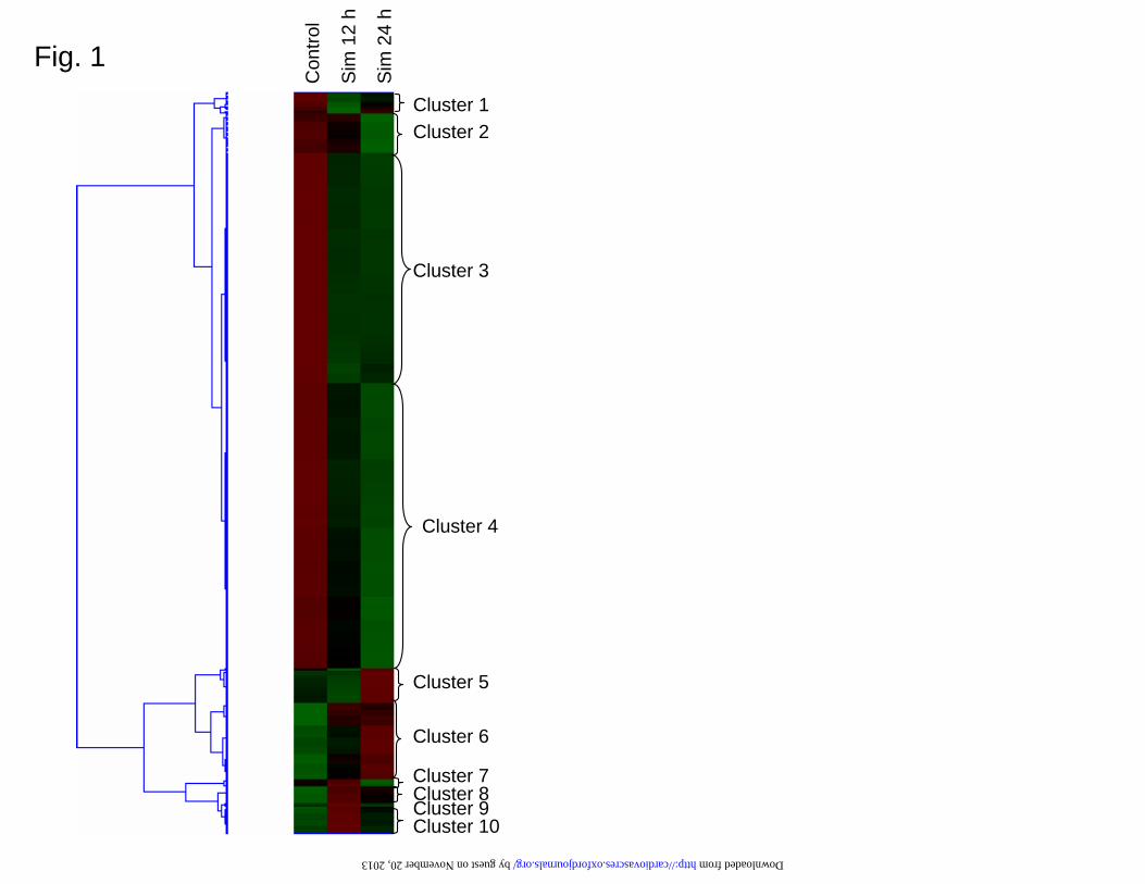
To assess the relationships and coordinate expression profiles between the regulated
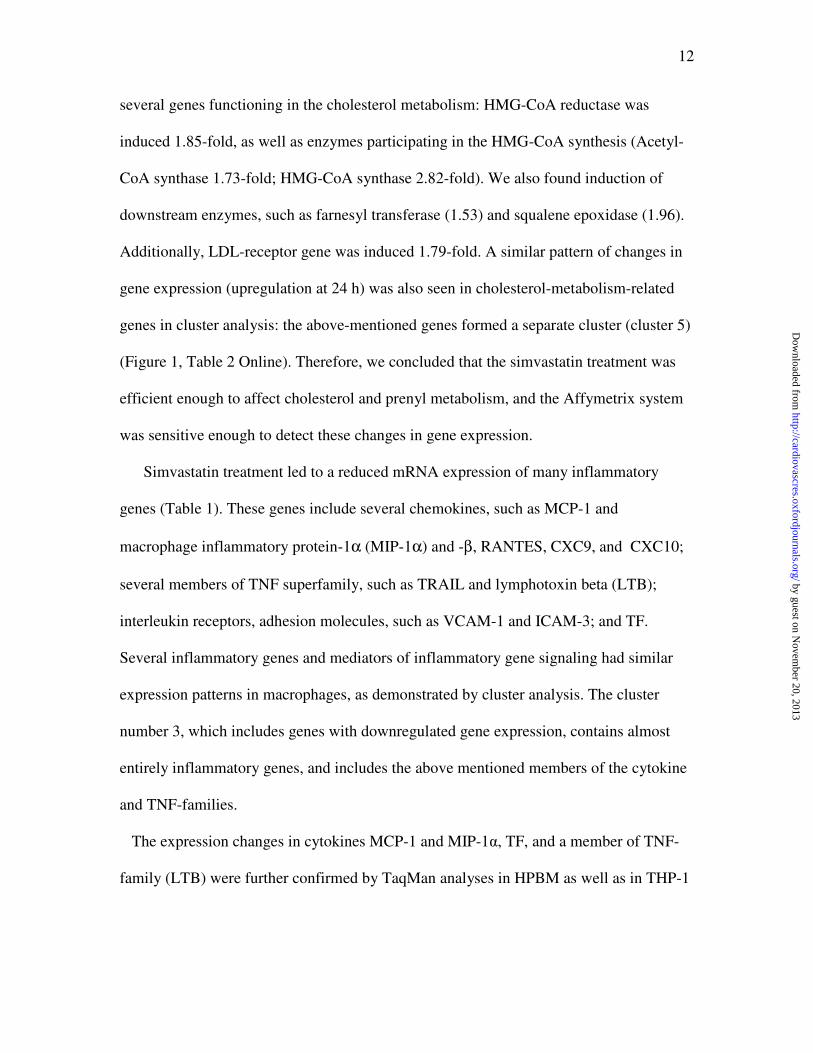
genes we performed a cluster analysis of the pooled data set. The analysis showed ten
well-defined clusters, which contained several genes that were related (e.g. the same gene
family or the functional group), for example "inflammatory gene cluster" (cluster 3), "cell
structure" (cluster 2), "cholesterol-metabolism-cluster" (cluster 5), "signal transduction
with downregulation" (cluster 4) and "signal transduction with upregulation" (cluster 6),
"KLF-cluster" (cluster 8), and "metallothionein cluster" (cluster 10) (Figure 1, Table 2
Online).
We found that the inhibition of HMG-CoA reductase in macrophages and the
subsequent reduction in the cholesterol synthesis lead to compensatory changes at 24 h in
by guest on Novem
ber 20, 2013http://cardiovascres.oxfordjournals.org/
Dow
nloaded from
12
several genes functioning in the cholesterol metabolism: HMG-CoA reductase was
induced 1.85-fold, as well as enzymes participating in the HMG-CoA synthesis (Acetyl-
CoA synthase 1.73-fold; HMG-CoA synthase 2.82-fold). We also found induction of
downstream enzymes, such as farnesyl transferase (1.53) and squalene epoxidase (1.96).
Additionally, LDL-receptor gene was induced 1.79-fold. A similar pattern of changes in
gene expression (upregulation at 24 h) was also seen in cholesterol-metabolism-related
genes in cluster analysis: the above-mentioned genes formed a separate cluster (cluster 5)
(Figure 1, Table 2 Online). Therefore, we concluded that the simvastatin treatment was
efficient enough to affect cholesterol and prenyl metabolism, and the Affymetrix system
was sensitive enough to detect these changes in gene expression.
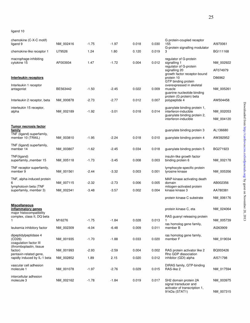
Simvastatin treatment led to a reduced mRNA expression of many inflammatory
genes (Table 1). These genes include several chemokines, such as MCP-1 and
macrophage inflammatory protein-1α (MIP-1α) and -β, RANTES, CXC9, and CXC10;
several members of TNF superfamily, such as TRAIL and lymphotoxin beta (LTB);
interleukin receptors, adhesion molecules, such as VCAM-1 and ICAM-3; and TF.
Several inflammatory genes and mediators of inflammatory gene signaling had similar
expression patterns in macrophages, as demonstrated by cluster analysis. The cluster
number 3, which includes genes with downregulated gene expression, contains almost
entirely inflammatory genes, and includes the above mentioned members of the cytokine
and TNF-families.
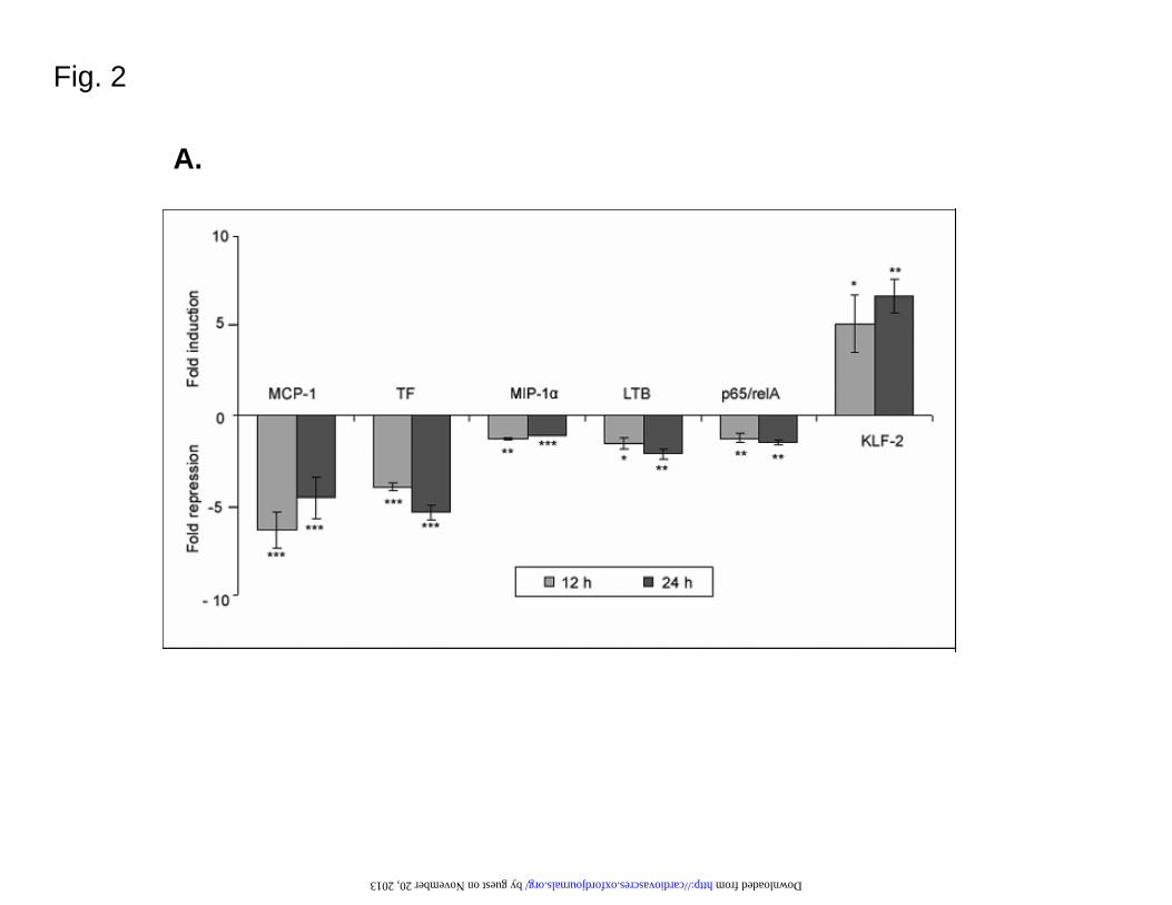
The expression changes in cytokines MCP-1 and MIP-1α, TF, and a member of TNF-
family (LTB) were further confirmed by TaqMan analyses in HPBM as well as in THP-1
by guest on Novem
ber 20, 2013http://cardiovascres.oxfordjournals.org/
Dow
nloaded from
13
derived macrophages (Figure 2A and B). Downregulation of MCP-1 and TF sustained for
48 and 72 h in THP-1 cells (results not shown).
Simvastatin attenuates inflammatory signalling in macrophages: downregulation of
NF-κκκκB relA/p65 subunit mRNA expression and upregulation of KLF 2 expression.
In Affymetrix analysis, simvastatin treatment lead to reduced expression of several genes
that mediate inflammatory signaling in cells, including several guanylate binding proteins
(GBP 1-5), Ras and Rho-proteins; and several transcription factors, such as NF-κB
relA/p65 protein, PPARδ, and v-ets. The expression of KLF-2 was strongly induced
(3.9-fold upregulation at 12 h, 9.3-fold at 24 h). Additionally, the KLF family members
KLF-3 and -4 had increased expression levels. Most of the inflammatory signaling-
related genes clustered together with other inflammatory genes to cluster 3, which shows
decreasing expression pattern, whereas KLFs 2-4 formed a separate cluster with
increasing expression patterns (cluster 8) (Figure 1, Table 2 online).
In Affymetrix analysis, several members of the NF-κB signaling pathway showed a
trend to reduced RNA expression levels, for example NF-κB /rel protein RelA/p65 (-2.0-
fold decrease at 24 h), RelB and NF-κB p50, but all the changes were not statistically
significant. Additionally, several members of the signaling cascades leading to NF-κB
activation had downregulated expression, e.g. protein kinase C (-1.4) and Akt and Cot (-
1.4). Also JAK-STAT (STAT -1.6 fold) signaling pathway was donwnregulated.
The expression changes of relA/p65 and KLF-2 were further studied with TaqMan in
HPBM and THP-1 macrophages (Figure 2), and KLF-2 protein expression by Western
blot analysis (Figure 2). TaqMan analysis confirmed the downregulated levels of
by guest on Novem
ber 20, 2013http://cardiovascres.oxfordjournals.org/
Dow
nloaded from
14
relA/p65 mRNA in HPBM cells (Figure 2A), as well as the induction of KLF-2
expression in HPBM and THP-1 macrophages (Figure 2A and B). There was also an
increase in the KLF-2 protein (Figure 2C). The effect of simvastatin on KLF-2, TF and
MCP-1 expression was detected also at concentration of 1 µM simvastatin as studied by
TaqMan analysis (data not shown). However, at lower concentrations of simvastatin
(0.025- 0.1 µM) the effect was not as remarkable.
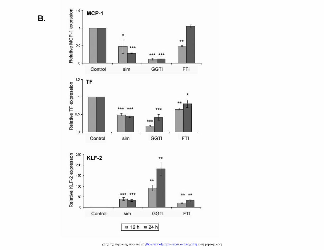
The inhibition of protein prenylation has the same effect on KLF-2, MCP-1 and TF
expression as simvastatin. THP-1 macrophages were treated with protein prenylation
inhibitors GGTI (inhibitor of geranyl-geranyl transferase) and FTI (inhibitor of farnesyl
transferase). Both GGTI and FTI treatments lead to the downregulation of MCP-1 and TF
mRNA expression, and to the upregulation of KLF-2 expression (Figure 2B). Moreover,
simvastatin, GGTI and FTI treatments had similar effects on KLF-2 protein expression
(Figure 2C).
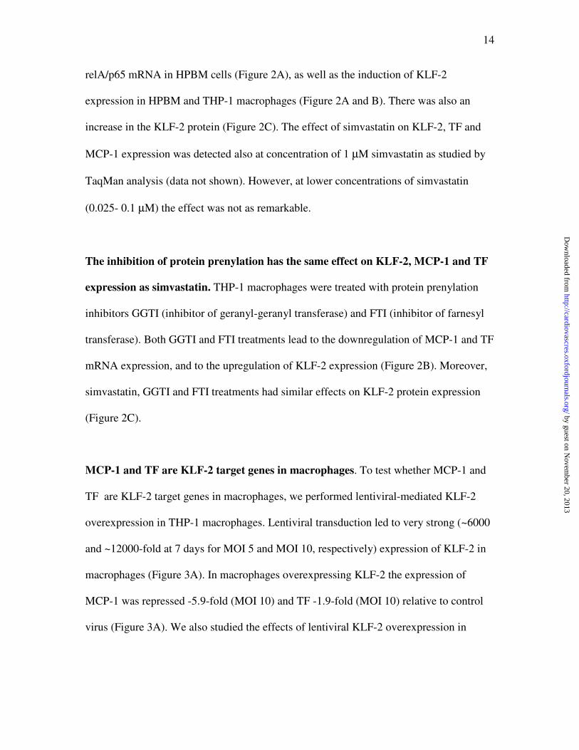
MCP-1 and TF are KLF-2 target genes in macrophages. To test whether MCP-1 and
TF are KLF-2 target genes in macrophages, we performed lentiviral-mediated KLF-2
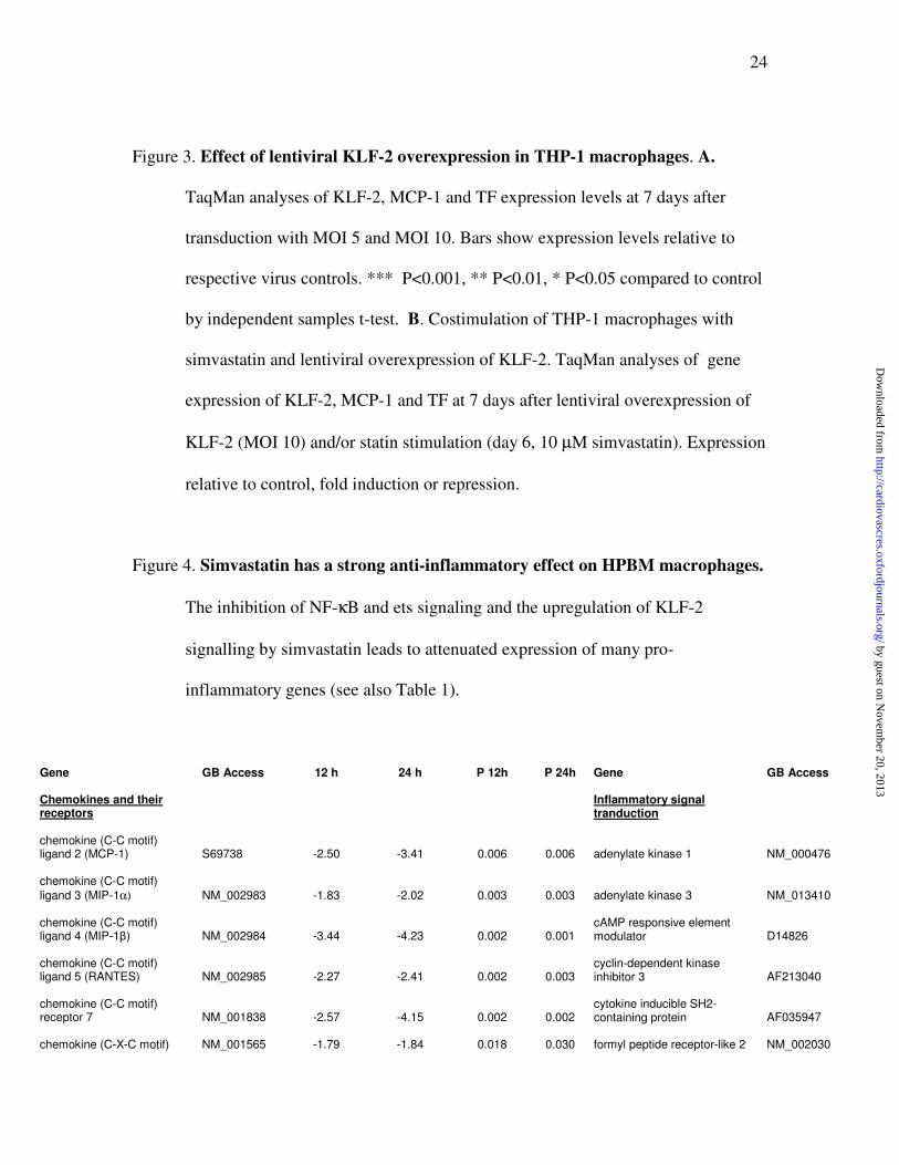
overexpression in THP-1 macrophages. Lentiviral transduction led to very strong (~6000
and ~12000-fold at 7 days for MOI 5 and MOI 10, respectively) expression of KLF-2 in
macrophages (Figure 3A). In macrophages overexpressing KLF-2 the expression of
MCP-1 was repressed -5.9-fold (MOI 10) and TF -1.9-fold (MOI 10) relative to control
virus (Figure 3A). We also studied the effects of lentiviral KLF-2 overexpression in
by guest on Novem
ber 20, 2013http://cardiovascres.oxfordjournals.org/
Dow
nloaded from
15
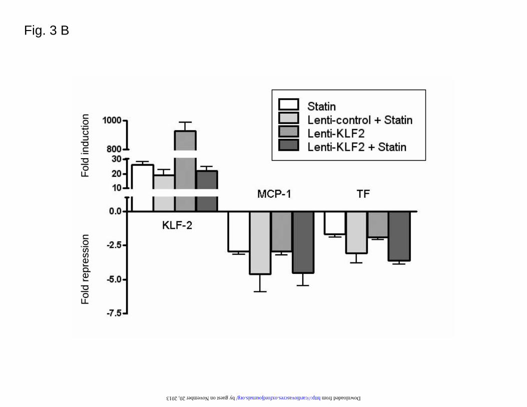
combination with statin treatment in THP-1 macrophages (Figure 3B). However,
simvastatin treatment had no additional effects over KLF-2 overexpression.
Discussion
This gene expression array study of simvastatin effects on macrophages shows that
simvastatin has a global anti-inflammatory effect on macrophages, which includes
attenuated expression of several pro-inflammatory cytokines, TNF family members, TF,
adhesion molecules and molecules mediating inflammatory signaling. All these
molecules play important roles in atherogenesis. Interestingly, also some less known
TNF-family members, such as TRAIL and LTB, were attenuated by simvastatin.
Clustering of gene expression data to several well-defined clusters suggests that the
effects on simvastatin on gene expression are mediated by several different pathways.
The potency of the cluster analysis in finding groups of genes with similar regulatory
mechanisms is demonstrated by the clustering of genes coding for proteins participating
in cholesterol and lipid metabolism to a separate cluster (cluster 5): this can be explained
by the control of their expression by sterol regulatory elements: cholesterol deprivation
induces their expression via SREBP1 25
. Additionally, many cytokines and other
inflammatory proteins had similar decreasing expression profiles (cluster 3), which might
suggest that the effects of simvastatin on proinflammatory gene expression are mediated
by the same mediators. Additionally, the KLFs 2-4 formed a separate cluster (cluster 8),
as well as metallothioneins (cluster 10). Several metallothioneins (MT 1E, X, G and 2A),
which participate in the metal homeostasis as well as the control of REDOX-balance,
by guest on Novem
ber 20, 2013http://cardiovascres.oxfordjournals.org/
Dow
nloaded from
16
inflammation and cell proliferation 26
, are induced at 12h by simvastatin in a similar
manner. However, their role in macrophages has not been established, and there is no
previous information about their regulation by statins.
The anti-inflammatory effect of simvastatin can be at least partly explained by its
impact on proinflammatory signal transduction. Several members of the NF-κB signaling
pathway had reduced mRNA expression levels, for example NF-κB complex proteins
p65/RelA, RelB and p50. Additionally, several members of the signaling cascades
leading to NF-kappaB activation had downregulated expression, e.g. Janus kinases,
protein kinase C, Akt and Cot. It has been previously shown that statins inhibit the
binding of NF-κB to nuclear proteins 11
, and inhibit the phosphorylation and degradation
of the NF-κB inhibitory protein IκB 27
. We show here that simvastatin downregulates
mRNA expression of the NF-κB subunits in HPBM cells. Besides NF-κB signaling, a
potent pro-inflammatory transcription factor c-ets, which mediates e.g. MCP-1 and
phospholipase expression 19;20
, as well as the family of guanylate binding proteins (GBPs
1-5), and JAK-STAT signaling pathway were downregulated by simvastatin.
We show here that KLF-2, as well as its family members KLF-3 and KLF-4, are
induced in macrophages by simvastatin. KLFs are recently characterized zinc finger
transcription factors that have several important functions; they modulate cell
differentiation, organ development and proliferation 16
. KLF-2, -4,-5 and -6 have
particular importance in the vasculature, and they have functional as well as pathological
roles 16
. KLF-2 has been shown to be essential for the vascular development 16
, and it has
been identified as part of the “atheroprotective phenotype” of ECs. It is induced by
steady laminar flow28;29
, and inhibited by TNF-α28
and IL-1β 14
in ECs. It has several
by guest on Novem
ber 20, 2013http://cardiovascres.oxfordjournals.org/
Dow
nloaded from
17
anti-inflammatory properties, such as reduction of adhesion molecule expression 14
.
Additionally, KLF-2 has been shown to regulate the thrombotic function of ECs, e.g. the
expression of thrombomodulin, eNOS and PAI-1 as wells as cytokine-mediated
production of TF 13
. Global analysis of gene expression changes after adenoviral
overexpression of KLF-2 in ECs showed that induction of KLF-2 resulted in the
regulation of endothelial transcription programs controlling inflammation,
thrombosis/hemostasis, vascular tone, and blood vessel development 12
. Moreover,
another microarray study showed that KLF-2 acts as a general transcriptional switch
point between the quiescent and activated status of ECs where lentiviral overexpression
of KLF-2 resulted in changes in several key functional pathways such as cell migration
via VEGFR2, inflammation and hemostasis 30
.
Recently, it has been shown that statins upregulate KLF-2 expression in ECs, which
might partly explain the atheroprotective effects of statins 12;15
. The statin effect on KLF-
2 was dependent on the ability of statins to inhibit Rho-pathway, as adenoviral
overexpression of Rho-protein decreased KLF-2 expression 15
.
Very little is known about the role of KLF-2 in macrophages. A recent study showed
that KLF-2 regulates proinflammatory activation of monocytes: overexpression of KLF-2
inhibited LPS-activated production of cytokines and reduced phagocytic activity 18
.
Interestingly, the expression of KLF-2 was reduced in circulating monocytes in patients
with CAD 18
. In this study we showed that KLF-2 is induced by simvastatin in mature
human macrophages. We also showed that the mechanism of induction of KLF-2 by
simvastatin in macrophages is dependent on the inhibition of protein prenylation.
Additionally, by lentiviral overexpression of KLF-2 we showed that MCP-1 and TF are
by guest on Novem
ber 20, 2013http://cardiovascres.oxfordjournals.org/
Dow
nloaded from
18
KLF-2 target genes in macrophages, and KLF-2 represses their expression. Moreover,
several KLF-2 endothelial cell target genes 12
, e.g. CXCL10, CCL5 and TNF had
changed expression levels in simvastatin-stimulated macrophages, which suggests them
to be potential KLF-2 target genes in macrophages as well. Taken together, this suggests
an anti-inflammatory and vasculoprotective role for KLF-2 in macrophages.
In addition to the induction of KLF-2, we showed that also its family members KLF-3
and KLF-4 are induced by simvastatin. However, the functions of KLF-3 and KLF-4 are
poorly known and further studies are needed to clarify their role in macrophages and
other cell types.
The inhibition of protein prenylation by inhibitors of geranyl-geranylation and
farnesylation had similar effects on KLF-2, MCP-1 and TF expression as simvastatin.
Isoprenoid binding to Ras and Rho proteins is essential for the initiation of G-protein
signaling, which controls for example cell proliferation and stress responses. Farnesylated
proteins include e.g. Ras, whereas Rho and Rac activation are dependent on geranyl-
geranylation9. The similar effects achieved by protein prenylation inhibitors and
simvastatin suggest that the simvastatin effects might be dependent on the inhibition of
protein prenylation. It would be interesting to study whether alterations in cellular
cholesterol content correlate with these effects and whether similar effects on cellular
transcriptional machinery could be achieved via other means that can alter cellular
cholesterol levels.
In conclusion, we show here that simvastatin has a strong anti-inflammatory effect on
macrophages including attenuated expression of several cytokines, TNF family members
and some proinflammatory signaling molecules. Most interestingly, transcription factor
by guest on Novem
ber 20, 2013http://cardiovascres.oxfordjournals.org/
Dow
nloaded from
19
KLF-2 is upregulated by simvastatin in macrophages. These findings support the notion
that statins have other antiatherogenic actions beyond their effects on plasma cholesterol.
Funding
This study has been supported by the Academy of Finland (211497), Finnish Foundation
for Cardiovascular Research, Sigrid Juselius Foundation, Finnish Cultural Foundation (to
T.T), Finnish Medical Foundation (to T.T), the Aarne Koskelo Foundation (to T.T), the
Paavo Nurmi Foundation (to T.T), and European Vascular Genomics Network (EVGN
LSHM-CT-2003-503254).
Acknowledgements
We thank Ms. Riina Kylätie, Ms. Mervi Nieminen, Ms. Anne Martikainen and Ms.
Marja Poikolainen for technical assistance.
Conflict of Interest
Conflict of Interest: none declared.
by guest on Novem
ber 20, 2013http://cardiovascres.oxfordjournals.org/
Dow
nloaded from
20
Reference List
1. Hansson GK. Inflammation, atherosclerosis, and coronary artery disease. N Engl J
Med. 2005;352:1685-1695.
2. Palinski W, Napoli C. Unraveling pleiotropic effects of statins on plaque rupture.
Arterioscler Thromb Vasc Biol. 2002;22:1745-1750.
3. Ray KK, Cannon CP. Intensive statin therapy in acute coronary syndromes: clinical
benefits and vascular biology. Curr Opin Lipidol. 2004;15:637-643.
4. Aikawa M, Rabkin E, Sugiyama S, Voglic SJ, Fukumoto Y, Furukawa Y et al. An
HMG-CoA reductase inhibitor, cerivastatin, suppresses growth of macrophages
expressing matrix metalloproteinases and tissue factor in vivo and in vitro.
Circulation. 2001;103:276-283.
5. Waehre T, Yndestad A, Smith C, Haug T, Tunheim SH, Gullestad L et al.
Increased expression of interleukin-1 in coronary artery disease with
downregulatory effects of HMG-CoA reductase inhibitors. Circulation.
2004;109:1966-1972.
6. Yoshida M, Sawada T, Ishii H, Gerszten RE, Rosenzweig A, Gimbrone MA, Jr et
al. Hmg-CoA reductase inhibitor modulates monocyte-endothelial cell interaction
under physiological flow conditions in vitro: involvement of Rho GTPase-
dependent mechanism. Arterioscler Thromb Vasc Biol. 2001;21:1165-1171.
7. Senokuchi T, Matsumura T, Sakai M, Yano M, Taguchi T, Matsuo T et al. Statins
suppress oxidized low density lipoprotein-induced macrophage proliferation by
inactivation of the small G protein-p38 MAPK pathway. J Biol Chem.
2005;280:6627-6633.
8. Tuomisto TT, Korkeela A, Rutanen J, Viita H, Brasen JH, Riekkinen MS et al.
Gene expression in macrophage-rich inflammatory cell infiltrates in human
atherosclerotic lesions as studied by laser microdissection and DNA array:
overexpression of HMG-CoA reductase, colony stimulating factor receptors,
CD11A/CD18 integrins, and interleukin receptors. Arterioscler Thromb Vasc Biol.
2003;23:2235-2240.
9. Denhardt DT. Signal-transducing protein phosphorylation cascades mediated by
Ras/Rho proteins in the mammalian cell: the potential for multiplex signalling.
Biochem J. 1996;318:729-747.
10. Laufs U, Liao JK. Targeting Rho in cardiovascular disease. Circ Res. 2000;87:526-
528.
by guest on Novem
ber 20, 2013http://cardiovascres.oxfordjournals.org/
Dow
nloaded from
21
11. Dichtl W, Dulak J, Frick M, Alber HF, Schwarzacher SP, Ares MP et al. HMG-
CoA reductase inhibitors regulate inflammatory transcription factors in human
endothelial and vascular smooth muscle cells. Arterioscler Thromb Vasc Biol.
2003;23:58-63.
12. Parmar KM, Nambudiri V, Dai G, Larman HB, Gimbrone MA, Jr., Garcia-Cardena
G. Statins exert endothelial atheroprotective effects via the KLF2 transcription
factor. J Biol Chem. 2005;280:26714-26719.
13. Lin Z, Kumar A, Senbanerjee S, Staniszewski K, Parmar K, Vaughan DE et al.
Kruppel-like factor 2 (KLF2) regulates endothelial thrombotic function. Circ Res.
2005;96:e48-e57.
14. Senbanerjee S, Lin Z, Atkins GB, Greif DM, Rao RM, Kumar A et al. KLF2 Is a
novel transcriptional regulator of endothelial proinflammatory activation. J Exp
Med. 2004;199:1305-1315.
15. Sen-Banerjee S, Mir S, Lin Z, Hamik A, Atkins GB, Das H et al. Kruppel-like
factor 2 as a novel mediator of statin effects in endothelial cells. Circulation.
2005;112:720-726.
16. Suzuki T, Aizawa K, Matsumura T, Nagai R. Vascular implications of the Kruppel-
like family of transcription factors. Arterioscler Thromb Vasc Biol. 2005;25:1135-
1141.
17. Dekker RJ, van Thienen JV, Rohlena J, de Jager SC, Elderkamp YW, Seppen J et
al. Endothelial KLF2 links local arterial shear stress levels to the expression of
vascular tone-regulating genes. Am J Pathol. 2005;167:609-618.
18. Das H, Kumar A, Lin Z, Patino WD, Hwang PM, Feinberg MW et al. Kruppel-like
factor 2 (KLF2) regulates proinflammatory activation of monocytes. Proc Natl
Acad Sci U S A. 2006;103:6653-6658.
19. Oettgen P. Regulation of vascular inflammation and remodeling by ETS factors.
Circ Res. 2006;99:1159-1166.
20. Zhan Y, Brown C, Maynard E, Anshelevich A, Ni W, Ho IC et al. Ets-1 is a critical
regulator of Ang II-mediated vascular inflammation and remodeling. J Clin Invest.
2005;115:2508-2516.
21. Pietarinen-Runtti P, Lakari E, Raivio KO, Kinnula VL. Expression of antioxidant
enzymes in human inflammatory cells. Am J Physiol Cell Physiol. 2000;278:C118-
C125.
22. Li C, Wong WH. Model-based analysis of oligonucleotide arrays: expression index
computation and outlier detection. Proc Natl Acad Sci U S A. 2001;98:31-36.
by guest on Novem
ber 20, 2013http://cardiovascres.oxfordjournals.org/
Dow
nloaded from
22
23. Eisen MB, Spellman PT, Brown PO, Botstein D. Cluster analysis and display of
genome-wide expression patterns. Proc Natl Acad Sci U S A. 1998;95:14863-
14868.
24. Makinen PI, Koponen JK, Karkkainen AM, Malm TM, Pulkkinen KH, Koistinaho J
et al. Stable RNA interference: comparison of U6 and H1 promoters in endothelial
cells and in mouse brain. J Gene Med. 2006;8:433-441.
25. Wang X, Sato R, Brown MS, Hua X, Goldstein JL. SREBP-1, a membrane-bound
transcription factor released by sterol-regulated proteolysis. Cell. 1994;77:53-62.
26. Haq F, Mahoney M, Koropatnick J. Signaling events for metallothionein induction.
Mutat Res. 2003;533:211-226.
27. Hilgendorff A, Muth H, Parviz B, Staubitz A, Haberbosch W, Tillmanns H et al.
Statins differ in their ability to block NF-kappaB activation in human blood
monocytes. Int J Clin Pharmacol Ther. 2003;41:397-401.
28. Dekker RJ, van Soest S, Fontijn RD, Salamanca S, de Groot PG, VanBavel E et al.
Prolonged fluid shear stress induces a distinct set of endothelial cell genes, most
specifically lung Kruppel-like factor (KLF2). Blood. 2002;100:1689-1698.
29. van Thienen JV, Fledderus JO, Dekker RJ, Rohlena J, van Ijzendoorn GA, Kootstra
NA et al. Shear stress sustains atheroprotective endothelial KLF2 expression more
potently than statins through mRNA stabilization. Cardiovasc Res. 2006;72:231-
240.
30. Dekker RJ, Boon RA, Rondaij MG, Kragt A, Volger OL, Elderkamp YW et al.
KLF2 provokes a gene expression pattern that establishes functional quiescent
differentiation of the endothelium. Blood. 2006;107:4354-4363.
by guest on Novem
ber 20, 2013http://cardiovascres.oxfordjournals.org/
Dow
nloaded from
23
Figure legends
Figure 1. Cluster analysis of gene expression data. Clustering reveals 10 well-defined
clusters including "leukotriene signalling" (cluster 1), "cell structure" (cluster 2),
“inflammatory genes” (cluster 3), "signal transduction, downregulated" (cluster
4), “cholesterol metabolism” (cluster 5), "signal transduction, upregulated"
(cluster 6), "orosomucoid-cluster" (cluster 7), “KLF-cluster” ( cluster 8), "tiny
cluster" (cluster 9) and “metallothioneins” (cluster 10). Red indicates upregulated
expression relative to control (baseline), green indicates downregulated
expression. The list of genes in different clusters are presented in Table 2 in
supplementary data.
Figure 2. Effect of simvastatin and protein prenylation inhibitors on gene expression
of selected genes in HPBM and THP-1 derived macrophages. A. TaqMan
analysis of mRNA expression of MCP-1, TF, MIP-1α, LTB, relA and KLF-2 in
HPBM macrophages after simvastatin (sim) treatment. Expression relative to
control, fold induction and repression. B. TaqMan analysis of mRNA expression
of MCP-1, TF and KLF-2 in THP-1 derived macrophages after simvastatin and
GGTI and FTI treatments. C. Effect of simvastatin and protein prenylation
inhibitors on protein expression of KLF-2. Western blot analysis.
Expression relative to control. *** P<0.001, ** P<0.01, * P<0.05 compared with
control by independent samples t-test.
by guest on Novem
ber 20, 2013http://cardiovascres.oxfordjournals.org/
Dow
nloaded from
24
Figure 3. Effect of lentiviral KLF-2 overexpression in THP-1 macrophages. A.
TaqMan analyses of KLF-2, MCP-1 and TF expression levels at 7 days after
transduction with MOI 5 and MOI 10. Bars show expression levels relative to
respective virus controls. *** P<0.001, ** P<0.01, * P<0.05 compared to control
by independent samples t-test. B. Costimulation of THP-1 macrophages with
simvastatin and lentiviral overexpression of KLF-2. TaqMan analyses of gene
expression of KLF-2, MCP-1 and TF at 7 days after lentiviral overexpression of
KLF-2 (MOI 10) and/or statin stimulation (day 6, 10 µM simvastatin). Expression
relative to control, fold induction or repression.
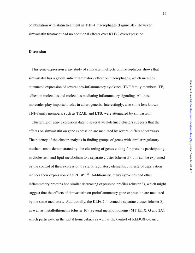
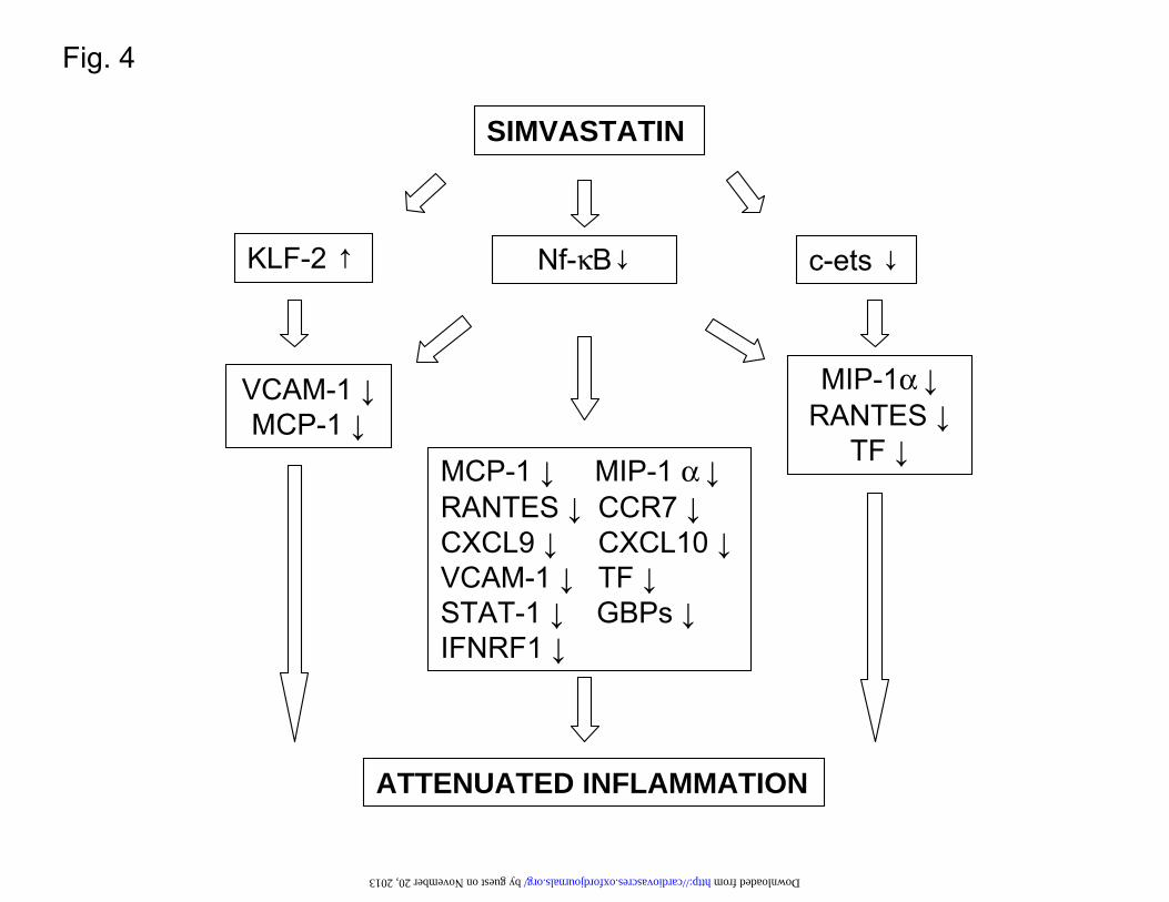
Figure 4. Simvastatin has a strong anti-inflammatory effect on HPBM macrophages.
The inhibition of NF-κB and ets signaling and the upregulation of KLF-2
signalling by simvastatin leads to attenuated expression of many pro-
inflammatory genes (see also Table 1).
Gene GB Access 12 h 24 h P 12h P 24h Gene GB Access Chemokines and their receptors
Inflammatory signal tranduction
chemokine (C-C motif) ligand 2 (MCP-1) S69738 -2.50 -3.41 0.006 0.006 adenylate kinase 1 NM_000476 chemokine (C-C motif)
ligand 3 (MIP-1α) NM_002983 -1.83 -2.02 0.003 0.003 adenylate kinase 3 NM_013410 chemokine (C-C motif) ligand 4 (MIP-1β) NM_002984 -3.44 -4.23 0.002 0.001
cAMP responsive element modulator D14826
chemokine (C-C motif) ligand 5 (RANTES) NM_002985 -2.27 -2.41 0.002 0.003
cyclin-dependent kinase inhibitor 3 AF213040
chemokine (C-C motif) receptor 7 NM_001838 -2.57 -4.15 0.002 0.002
cytokine inducible SH2-containing protein AF035947
chemokine (C-X-C motif) NM_001565 -1.79 -1.84 0.018 0.030 formyl peptide receptor-like 2 NM_002030
by guest on Novem
ber 20, 2013http://cardiovascres.oxfordjournals.org/
Dow
nloaded from
25
ligand 10
chemokine (C-X-C motif) ligand 9 NM_002416 -1.75 -1.97 0.018 0.030
G protein-coupled receptor 155 AI970061
chemokine-like receptor 1 U79526 1.24 1.80 0.120 0.019
G-protein signalling modulator 3 BG111168
macrophage-inhibiting cytokine 15 AF003934 1.47 -1.72 0.004 0.012
regulator of G-protein signalling 1 NM_002922
regulator of G-protein signalling 20 AF074979
Interleukin receptors
growth factor receptor-bound protein 10 D86962
Interleukin 1 receptor antagonist BE563442 -1.50 -2.45 0.022 0.009
GTP binding protein overexpressed in skeletal muscle NM_005261
Interleukin 2 receptor, beta NM_000878 -2.73 -2.77 0.012 0.007
guanine nucleotide binding protein (G protein) beta polypeptide 4 AW504458
interleukin 15 receptor, alpha NM_002189 -1.92 -3.01 0.018 0.014
guanylate binding protein 1, interferon-inducible NM_002053
guanylate binding protein 2, interferon-inducible NM_004120
Tumor necrosis factor family guanylate binding protein 3 AL136680 TNF (ligand) superfamily, member 10 (TRAIL) NM_003810 -1.95 -2.24 0.018 0.010 guanylate binding protein 4 AW392952 TNF (ligand) superfamily, member 14 NM_003807 -1.62 -2.45 0.034 0.018 guanylate binding protein 5 BG271923 TNF(ligand) superfamily.,member 15 NM_005118 -1.73 -3.45 0.008 0.003
insulin-like growth factor binding protein 6 NM_002178
TNF receptor superfamily, member 9 NM_001561 -2.44 -3.32 0.003 0.001
lymphocyte-specific protein tyrosine kinase NM_005356
TNF, alpha-induced protein 6 NM_007115 -2.32 -2.73 0.006 0.005
MAP-kinase activating death domain AB002356
lymphotoxin beta (TNF superfamily, member 3) NM_002341 -3.48 -3.57 0.002 0.004
mitogen-activated protein kinase kinase 3 AA780381
protein kinase C substrate NM_006176 Miscellaneous inflammatory genes protein kinase C, eta NM_024064 major histocompatibility complex, class II, DQ beta 1 M16276 -1.75 -1.84 0.028 0.013
RAS guanyl releasing protein 1 NM_005739
leukemia inhibitory factor NM_002309 -4.04 -6.48 0.009 0.011
ras homolog gene family, member B AI263909
dipeptidylpeptidase 4 (CD26) NM_001935 -1.70 -1.88 0.033 0.020
ras homolog gene family, member F NM_019034
coagulation factor III (thromboplastin, tissue factor) NM_001993 -2.93 -2.59 0.004 0.002 RAS protein activator like 2 BQ003426 pentaxin-related gene, rapidly induced by IL-1 beta NM_002852 1.89 2.15 0.020 0.012
Rho GDP dissociation inhibitor (GDI) alpha AI571798
vascular cell adhesion molecule 1 NM_001078 -1.97 -2.76 0.029 0.015
DIRAS family, GTP-binding RAS-like 2 NM_017594
intercellular adhesion molecule 3 NM_002162 -1.78 -1.84 0.019 0.017 SH2 domain protein 2A NM_003975
signal transducer and activator of transcription 1, 91kDa (STAT1) NM_007315
by guest on Novem
ber 20, 2013http://cardiovascres.oxfordjournals.org/
Dow
nloaded from
26
Lipid metabolism
suppressor of cytokine signaling 1 AB005043
sterol isomerase
NM_006579
-1.87
1.31
0.025
0.018
tyrosine kinase with immunoglobulin and epidermal growth factor homology domains NM_005424
HMG-CoA synthase
NM_002130.1
1.32
2.82
0.202
0.005
7-dehydrocholesterol reductase
AW150953
-1.63
2.89
0.030
0.004
Transcription factor activity acetoacetyl-CoA synthetase
NM_023928.1
-1.34
1.73
0.096
0.008
nuclear factor of kappaB (p65 protein, relA) AI703057
squalene epoxidase
AA639705
-1.53
1.96
0.046
0.010 c-rel NM_002908
low density lipoprotein receptor
NM_000527.2
-1.59
1.79
0.023
0.006
interferon regulatory factor 1 NM_002198
fatty acid desaturase 2
NM_004265.1
-1.62
1.82
0.017
0.005
Kruppel-like factor 2 NM_016270
HMG-CoA reductase
NM_000859.1
-1.44
1.85
0.062
0.007
Kruppel-like factor 3 AA130132
Farnesyl transferase
AA872727 -1,42 1,53 0,045 0,006
Kruppel-like factor 4 BF514079
rev-erbA-alpha-related receptor AI761621
Metallothionein superfamily
peroxisome proliferative activated receptor, delta NM_006238
metallothionein 1X
NM_002450
2.37
1.34
0.008
0.040
TGFB inducible early growth response 2
AA149594
metallothionein 1G NM_005950 2.86 1.39 0.002 0.026
v-ets erythroblastosis virus E26 oncogene homolog BE218980
metallothionein 2A NM_005953 1.99 1.18 0.004 0.092 metallothionein 1E (functional) AL031602 2.08 1.18 0.004 0.092 metallothionein 2A M10943 2.66 1.46 0.008 0.078
Fold induction and p-values at 12 h and 24 hours after simvastatin treatment as compare to control. Fold inductions ≥ ±1.5 were
included in the table.
by guest on Novem
ber 20, 2013http://cardiovascres.oxfordjournals.org/
Dow
nloaded from
Cluster 1
Cluster 2
Cluster 3
Cluster 4
Cluster 5
Cluster 6
Cluster 7
Cluster 8
Cluster 9
Cluster 10
Con
trol
Sim
12
h
Sim
24
h
Fig. 1 by guest on November 20, 2013 http://cardiovascres.oxfordjournals.org/ Downloaded from
A.
Fig. 2
by guest on November 20, 2013 http://cardiovascres.oxfordjournals.org/ Downloaded from
B.
by guest on November 20, 2013 http://cardiovascres.oxfordjournals.org/ Downloaded from
HPBM cells Simvastatin treatment ¯¯¯¯¯¯¯¯¯¯¯¯¯¯¯¯¯¯¯¯¯¯¯¯¯¯ c 12h 24h 48h 72h
THP-1 cells Simvastatin treatment ¯¯¯¯¯¯¯¯¯¯¯¯¯¯¯¯¯¯¯¯¯¯¯¯¯¯ c 12h 24h 48h 72h
FTI treatment ¯¯¯¯¯¯¯¯¯¯¯¯¯¯¯¯¯¯¯¯¯¯¯¯¯¯ c 12h 24h 48h 72h
GGTI treatment ¯¯¯¯¯¯¯¯¯¯¯¯¯¯¯¯¯¯¯¯¯¯¯¯¯¯ c 12h 24h 48h 72h
C.
40 kDa
40 kDa
40 kDa
40 kDa
by guest on November 20, 2013 http://cardiovascres.oxfordjournals.org/ Downloaded from
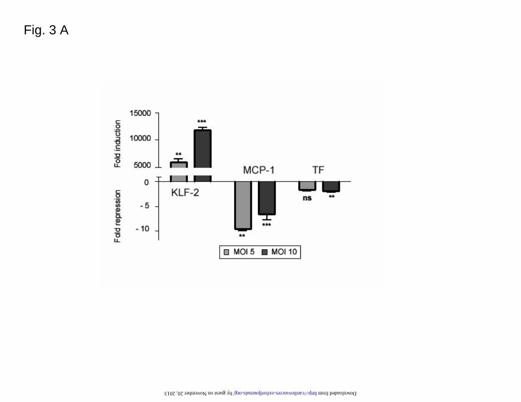
Fig. 3 A
by guest on November 20, 2013 http://cardiovascres.oxfordjournals.org/ Downloaded from
Fold
Fold
repr
essi
on
repr
essi
on
F
old
Fold
indu
ctio
nin
duct
ion
Fig. 3 B
by guest on November 20, 2013 http://cardiovascres.oxfordjournals.org/ Downloaded from
Nf-κB↓ KLF-2 ↑ c-ets ↓
MCP-1 ↓ MIP-1 α ↓ RANTES ↓ CCR7 ↓ CXCL9 ↓ CXCL10 ↓ VCAM-1 ↓ TF ↓ STAT-1 ↓ GBPs ↓ IFNRF1 ↓
VCAM-1 ↓ MCP-1 ↓
MIP-1α ↓ RANTES ↓
TF ↓
SIMVASTATIN
ATTENUATED INFLAMMATION
Fig. 4 by guest on November 20, 2013 http://cardiovascres.oxfordjournals.org/ Downloaded from
Copyright © 2022 FDOKUMEN