Simultaneous fat suppression and band reduction with large-angle multiple-acquisition balanced...
18
Simultaneous Fat Suppression and Band Reduction with Large- Angle Multiple-Acquisition bSSFP Brady Quist 1 , Brian A. Hargreaves 2 , Tolga Cukur 3 , Glen R. Morrell 4 , Garry E. Gold 2 , and Neal K. Bangerter 1,4 1 Department of Electrical & Computer Engineering, Brigham Young University, Provo, UT, USA 2 Department of Radiology, Stanford University, Stanford, CA, USA 3 Department of Electrical Engineering, Stanford University, Stanford, CA, USA 4 Department of Radiology, University of Utah, Salt Lake City, UT, USA Abstract Balanced steady-state free precession (bSSFP) MRI is a rapid and SNR-efficient imaging method, but suffers from characteristic bands of signal loss in regions of large field inhomogeneity. Several methods have been developed to reduce the severity of these banding artifacts, typically involving the acquisition of multiple bSSFP data sets (and the accompanying increase in scan time). Fat suppression with bSSFP is also challenging; most existing methods require an additional increase in scan time, and some are incompatible with bSSFP band-reduction techniques. This work was motivated by the need for both robust fat suppression and band reduction in the presence of field inhomogeneity when using bSSFP for flow-independent peripheral angiography. The large flip angles used in this application to improve vessel conspicuity and contrast lead to SAR considerations, longer TR, and increased severity of banding artifacts. In this work, a novel method that simultaneously suppresses fat and reduces bSSFP banding artifact with the acquisition of only two phase-cycled bSSFP data sets is presented. A weighted sum of the two bSSFP acquisitions is taken on a voxel-by-voxel basis, effectively synthesizing an off-resonance profile at each voxel that puts fat in the stop band while keeping water in the pass band. The technique exploits the near-sinusoidal shape of the bSSFP off-resonance spectrum for many tissues at large (>50 degrees) flip angles. Keywords bSSFP; SSFP; steady-state; fat suppression; artifact reduction; flow-independent angiography Introduction Balanced steady-state free precession (bSSFP) is a rapid and SNR-efficient imaging method, but suffers from characteristic bands of signal loss in regions of large field inhomogeneity. Several methods have been developed to reduce the severity of these banding artifacts, typically involving a penalty in scan time. Multiple phase-cycled bSSFP data sets can be acquired and combined in various ways (linear combination, maximum intensity, sum of squares) to reduce the severity of banding, as described in [1-9]. Another technique, wideband SSFP, utilizes two alternating repetition times (TR) with alternating RF phase to Correspondence to: Neal K. Bangerter, 469 Clyde Building, Brigham Young University, Provo, UT 84602, Phone: (801) 422-4869, [email protected]. NIH Public Access Author Manuscript Magn Reson Med. Author manuscript; available in PMC 2013 April 1. Published in final edited form as: Magn Reson Med. 2012 April ; 67(4): 1004–1012. doi:10.1002/mrm.23076. NIH-PA Author Manuscript NIH-PA Author Manuscript NIH-PA Author Manuscript
Transcript of Simultaneous fat suppression and band reduction with large-angle multiple-acquisition balanced...

Simultaneous Fat Suppression and Band Reduction with Large-Angle Multiple-Acquisition bSSFP
Brady Quist1, Brian A. Hargreaves2, Tolga Cukur3, Glen R. Morrell4, Garry E. Gold2, andNeal K. Bangerter1,4
1Department of Electrical & Computer Engineering, Brigham Young University, Provo, UT, USA2Department of Radiology, Stanford University, Stanford, CA, USA3Department of Electrical Engineering, Stanford University, Stanford, CA, USA4Department of Radiology, University of Utah, Salt Lake City, UT, USA
AbstractBalanced steady-state free precession (bSSFP) MRI is a rapid and SNR-efficient imaging method,but suffers from characteristic bands of signal loss in regions of large field inhomogeneity. Severalmethods have been developed to reduce the severity of these banding artifacts, typically involvingthe acquisition of multiple bSSFP data sets (and the accompanying increase in scan time). Fatsuppression with bSSFP is also challenging; most existing methods require an additional increasein scan time, and some are incompatible with bSSFP band-reduction techniques.
This work was motivated by the need for both robust fat suppression and band reduction in thepresence of field inhomogeneity when using bSSFP for flow-independent peripheral angiography.The large flip angles used in this application to improve vessel conspicuity and contrast lead toSAR considerations, longer TR, and increased severity of banding artifacts. In this work, a novelmethod that simultaneously suppresses fat and reduces bSSFP banding artifact with the acquisitionof only two phase-cycled bSSFP data sets is presented. A weighted sum of the two bSSFPacquisitions is taken on a voxel-by-voxel basis, effectively synthesizing an off-resonance profile ateach voxel that puts fat in the stop band while keeping water in the pass band. The techniqueexploits the near-sinusoidal shape of the bSSFP off-resonance spectrum for many tissues at large(>50 degrees) flip angles.
KeywordsbSSFP; SSFP; steady-state; fat suppression; artifact reduction; flow-independent angiography
IntroductionBalanced steady-state free precession (bSSFP) is a rapid and SNR-efficient imaging method,but suffers from characteristic bands of signal loss in regions of large field inhomogeneity.Several methods have been developed to reduce the severity of these banding artifacts,typically involving a penalty in scan time. Multiple phase-cycled bSSFP data sets can beacquired and combined in various ways (linear combination, maximum intensity, sum ofsquares) to reduce the severity of banding, as described in [1-9]. Another technique,wideband SSFP, utilizes two alternating repetition times (TR) with alternating RF phase to
Correspondence to: Neal K. Bangerter, 469 Clyde Building, Brigham Young University, Provo, UT 84602, Phone: (801) 422-4869,[email protected].
NIH Public AccessAuthor ManuscriptMagn Reson Med. Author manuscript; available in PMC 2013 April 1.
Published in final edited form as:Magn Reson Med. 2012 April ; 67(4): 1004–1012. doi:10.1002/mrm.23076.
NIH
-PA Author Manuscript
NIH
-PA Author Manuscript
NIH
-PA Author Manuscript
widen the band spacing in bSSFP, thereby reducing banding [10]. Slow modulations in thespectral offset frequency have also been used to reduce banding [11].
Fat suppression with bSSFP is also challenging. Numerous suppression techniques havebeen proposed, including linear combination [12], TIDE [13], FEMR [14], IDEAL and otherDixon methods [15-17], phase-sensitive fat detection [18,19], alternating TR (ATR) [20],and periodic fat saturation methods [21]. However, most methods require an additionalincrease in scan time, and some are incompatible with bSSFP band-reduction techniques[12]. Furthermore, these techniques have varying degrees of robustness to off resonance, andsome suffer from partial volume effects [18,19]. Some are transient techniques, relying on atransient magnetization preparation to suppress fat [13,21], while others work in the steadystate [12,14,15,19].
This work was motivated by the need for both robust fat suppression and band reduction inthe presence of field inhomogeneity when using bSSFP for flow-independent peripheralangiography [22, 23]. Relatively large flip angles (∼50 degrees or larger) are desirable whenusing bSSFP for peripheral angiography to increase vessel conspicuity and improve contrast[24]. This unfortunately can lead to SAR considerations and longer TR. As TR increases, thenulls in the SSFP spectral profile responsible for banding artifacts are more closely spacedin frequency. This can increase the number of signal null bands appearing in the image overa given range of off-resonance, while simultaneously making the transitions from signal tosignal null more abrupt spatially; the bands of signal null get thinner and closer together, andconsequently the edges get sharper. While the degree of band reduction is unaffected by TR,rapid spatial modulations in signal are typically less desirable than slower spatialmodulations. These problems are exacerbated at higher field strengths (e.g., 3 Tesla). Fatsuppression can also be compromised for a number of the aforementioned techniques as TRincreases [14,20]. Ideally, we would like a bSSFP technique that robustly suppresses fatwithout partial volume effects or artifacts, reduces bSSFP banding artifacts, works well atlarge flip angles, works in the presence of large field inhomogeneity, and keeps the scantime penalty to a minimum.
In this work, we present Large-Angle Multiple-Acquisition (LAMA) bSSFP, a novel bSSFPtechnique that simultaneously suppresses fat and reduces banding artifacts with theacquisition of only two phase-cycled bSSFP data sets. The technique relies on the near-sinusoidal shape of the bSSFP off-resonance spectrum for many tissues at large flip angle,and thus performs best under the large-flip-angle constraint of the flow-independentperipheral angiography application. Preliminary results are presented both in phantoms andin vivo, along with the results of a simulation and study assessing the performance of thetechnique across a range of flip angles.
TheoryAs the flip angle is increased, the bSSFP signal as a function of off-resonance frequencybegins to approximate a sinusoid for many biological tissues (Figure 1). (Note that thisapproximation strictly speaking requires echo time TE = TR/2. At other TE, a simple linearphase is introduced across the spectral profile and the signal can be modeled by a complexexponential.) This spectral profile can be arbitrarily shifted in frequency by incrementing thephase of the RF pulse by some Δϕ from excitation to excitation [3,4,7,8]. When two large-flip-angle (∼50° or greater) bSSFP acquisitions are performed with Δϕ = 0° and Δϕ =180°,the spectral profiles of the acquisitions approximate a sine and cosine, respectively, withperiod 2/TR Hz. A data set with spectral profile shifted by a desired spectral shift Δf canthen be synthesized from the two acquisitions, using the relationship
Quist et al. Page 2
Magn Reson Med. Author manuscript; available in PMC 2013 April 1.
NIH
-PA Author Manuscript
NIH
-PA Author Manuscript
NIH
-PA Author Manuscript

(1)
as illustrated in Figure 2.
Choice of TR such that TR = n / (2*CSf), where n = [1, 3, 5, …] and CSf is the absolutechemical shift of fat relative to water (in Hz), will place fat in a signal null whenever wateris at a signal maximum. Choice of TR to adjust the relative phase of fat and water is alsoexploited in [25]. If the appropriate frequency shift Δf needed to place water on a signalmaximum is then identified on a voxel-by-voxel basis, a fat-suppressed image withoutbanding can theoretically be formed from just two phase-cycled acquisitions. This forms thebasis for our large-angle multiple-acquisition (LAMA) bSSFP technique.
The required frequency shift for each voxel can be determined either through acquisition ofa field map, or through an intelligent search of voxel intensities over a range of Δf values(eliminating the need for field map acquisition and the associated scan time penalty). In thelatter case, a field map is essentially inferred by examining what values of Δf yield themaximum water and minimum fat signal for each voxel. When large jumps in these Δfvalues are observed in adjacent voxels, the voxels likely lie at a fat/water boundary. Region-growing algorithms can then be devised to identify voxels as predominantly fat orpredominantly water, and a smooth map of Δf values synthesized.
Once a smooth Δf map is generated, water-only or fat-only images can be reconstructed. LetS1n be the complex signal of the nth voxel from the first phase-cycled acquisition (with Δϕ =0°), S2n be the complex signal of the nth voxel from the second phase-cycled acquisition(with Δϕ = 180°), and Δfn be the frequency shift required to put water in the center of thepassband (and hence fat in the center of the null) for the nth voxel. The reconstructed signalintensity of the nth voxel in the water-only image (Wn) is then given by:
(2)
Similarly, the reconstructed signal intensity of the nth voxel in the fat-only image (Fn) isgiven by:
(3)
The above relations are clearly only valid if the off-resonant spectral profiles of the tissuesof interest are identically sinusoidal. While the assumption of sinusoidal spectral profiles isan approximation, we can expect reasonable results across a range of tissues even at flipangles down to approximately 50°. In Figure 3 the spectral profiles of several tissues (fat,arterial blood, venous blood, and muscle) at flip angles of 30°, 60°, and 90° are shown (firstcolumn). The second through fourth columns of Figure 3 show spectral profiles synthesizedfrom two phase-cycled acquisitions at Δf values of 1/(12TR), 1/(6TR), and 1/(4TR)respectively. Note that this range of Δf values spans the full range of distortion in thespectral profiles: the distortion at Δf =1/(3TR) is equivalent to that at 1/(6TR), and so forth.While the profiles are distorted enough at a flip angle of 30° to suggest that the LAMAbSSFP method is impractical at these lower flip angles, the distortion is relatively mild at60°, as illustrated in columns two through four of Figure 3. The distortion continues todecrease for flip angles approaching 180° (irrespective of T1/T2), although signalattenuation becomes significant at flip angles approaching 180°.
Quist et al. Page 3
Magn Reson Med. Author manuscript; available in PMC 2013 April 1.
NIH
-PA Author Manuscript
NIH
-PA Author Manuscript
NIH
-PA Author Manuscript

The warping of synthesized profiles at different Δf values has an effect on signal contrast.An analysis of arterial/venous, arterial/muscle, and arterial/fat contrast (defined as theabsolute signal difference between two tissues) is shown in Figure 4 as a function of bothflip angle and resonant shift Δf. As in Figure 3, graphs are shown for Δf = 0, 1/(12TR), 1/(6TR), and 1/(4TR). Interestingly, the distortion tends to improve contrast between therelevant tissues for our flow-independent peripheral angiography application. T1 and T2values assumed in all simulations for fat, muscle, arterial blood, and venous blood are listedin Table 1.
Materials and MethodsPhantom and Lower Leg Data Acquisition
As an initial proof-of-concept, two phase-cycled bSSFP 3D images and a 3D field map wereacquired of an oil/water phantom and the lower leg of a normal volunteer on a 1.5 T GEscanner (GE Healthcare, Waukesha, WI). All subject scans referenced in this work wereconducted according to the guidelines of our Institutional Review Board (IRB). Scanparameters for the bSSFP acquisitions were: flip angle = 90° (phantom) and 70° (volunteer),TR/TE = 6.6/3.3 ms, and phase cycling of Δϕ = 0° and 180°. Recall that TR for ourtechnique is constrained to:
(4)
where CSf is the absolute chemical shift of fat relative to water (in Hz). Assuming CSf = 226Hz at 1.5 T, allowable TRs are thus 2.2 ms, 6.6 ms, 11.1 ms, and so forth. A TR of 2.2 mscorresponds to placing fat in the null immediately adjacent to the water passband, and wouldbe expected to be the most robust. However, a TR this short is difficult to achieve. Ourchoice of TR = 6.6 ms places fat in a null that is one removed from the water passband. ATR of greater than approximately 7 or 8 ms begins to be impractical for SSFP imaging dueto the severity of off-resonance banding. As mentioned, increasing TR spatially compressesbands of signal null, leading to more rapid spatial variations in signal and sharper bandedges.
Field Map ReconstructionFor field map reconstruction, a field map was first acquired by taking the phase differencebetween two successive GRE images with ΔTE = 1/CSf (or 4.4 ms at 1.5T). This ensuresthat fat and water voxels will be in phase for a voxel that has no off resonance. The GREimages were acquired on the identical matrix as the bSSFP images. The phase difference ofeach voxel within the image can then be determined to calculate the off-resonance frequencyfor that voxel within a modulo of 1/ΔTE. The frequency Δfn for the nth voxel was calculatedby:
(5)
where Δθn is the phase difference in radians of the nth voxel. Once Δf is known for eachvoxel and both bSSFP images have been acquired, the fat and water images can be createdby inserting Δfn into equations (2) and (3).
Region-Growing ReconstructionTo eliminate the need for field map acquisition, a region-growing algorithm wasimplemented that synthesizes a Δf-map using only the two phase-cycled bSSFP acquisitions.This map can be used in place of the field map.
Quist et al. Page 4
Magn Reson Med. Author manuscript; available in PMC 2013 April 1.
NIH
-PA Author Manuscript
NIH
-PA Author Manuscript
NIH
-PA Author Manuscript

The two constituent phase-cycled images are first processed by creating reconstructed setsof images where each image is shifted by different values of Δf ranging from 0 to 1/TR.(Note that these values only span half of the period of the off-resonance spectra, as we areonly concerned with the magnitude of the final image). Afterwards the new set of images arescanned on a voxel-by-voxel basis to find Δfmax for each voxel, where Δfmax is defined asthe Δf value that produces the maximum intensity for that voxel, A maximum intensityimage is also produced by selecting the maximum intensity found for each voxel across thenew set of images. Voxels that are all fat or all water, at the same off-resonant frequency,will have Δfmax values that are separated in frequency by 1/(2TR).
In the peripheral angiography application, the brightest voxels are typically fat. Thus, theassumption is made that the brightest voxel in the maximum intensity image is always goingto be a fat voxel with little to no partial volume effects. The algorithm thus assigns the Δfmaxof that voxel to be Δffat and Δfwater to be Δffat - 1/(2TR). While this method performsreasonably well for our application, the identification of a fat pixel could be done manuallyby the MR operator if needed.
Once Δffat and Δfwater for the first voxel are determined, the Δfmax of each neighboringvoxel is compared to the first voxel to determine if it is fat or water. If the Δfmax of theneighboring voxel is within a predefined threshold of the surrounding Δffat or Δfwater values,then the new voxel is identified as either fat or water and Δfmax and Δfwater for that voxel areassigned accordingly. If the Δfmax is not close to the known neighboring Δffat or Δfwatervalues, the voxel is skipped and will not be identified as fat or water.
The algorithm continues in a region-growing fashion until no new neighbors can beidentified as either fat or water across the entire dataset. Two slight modifications to thealgorithm were found to be helpful in images with large FOV, low resolution, or less thanideal shims. The first modification involves adding magnitude thresholds to limit falseidentification for voxels close to the noise floor. The second modification is to only usepixels identified as fat for construction of the Δf-map, as these voxels typically have thehighest SNR. In some situations, using only fat voxels proved to be more robust in avoidingmisidentification in regions of large B0 inhomogeneity.
Once the region-growing algorithm has identified Δfwater and Δffat for any identifiablevoxels, a Δffat-map and Δfwater-map are created by interpolating Δfwater and Δffat to fill invoxels that were unidentifiable. When only fat voxels are used, the Δffat-map is created first,and the Δfwater-map synthesized by subtracting 1/(2TR) from the Δffat-map. The Δffat-mapand Δfwater-map are then filtered to enforce only gradual off-resonance variation on pixelsthat were not originally identified. The original images are then reprocessed using theΔfwater-map to make a water-only image and Δffat-map to make a fat-only image.
Contrast and Performance SimulationsA simulation was performed to analyze the performance and contrast of LAMA bSSFP atdifferent off-resonance values and at multiple flip angles. A 3D lower leg phantom wassimulated in Matlab (The MathWorks, Natick, Massachussetts) by creating a volumetricdataset consisting of a tapered cylinder with fat on the surface and muscle in the middle. Fatpockets of different sizes were randomly placed in the muscle portion. A bifurcated arteryand two veins were then added, running superior/inferior (vertically in the imagespresented). T1 and T2 values assumed for muscle, fat, arterial blood, and venous blood arelisted in Table 1. A linear variation in resonant frequency was added to the simulatedphantom, also in the vertical (superior/inferior) direction. The off-resonant shift ranged from-1/TR in the most inferior portion of the simulated phantom to 1/TR in the most superiorportion (-152 Hz to +152 Hz based on TR = 6.6 ms).
Quist et al. Page 5
Magn Reson Med. Author manuscript; available in PMC 2013 April 1.
NIH
-PA Author Manuscript
NIH
-PA Author Manuscript
NIH
-PA Author Manuscript

Using this simulated phantom, the expected signal was simulated for two phase-cycledbSSFP acquisitions at flip-angles of 30°, 40°, 50°, 60°, 70°, 80°, and 90°. Fat was assumedto be perfectly off-resonance at 226Hz (the chemical shift at 1.5T). Complex zero-meanGaussian noise was added with an SNR of 31.6. This data was then processed using theregion-growing algorithm as described.
Contrast and Performance Verification in VivoTo verify the contrast and performance simulation, 3D LAMA bSSFP images were acquiredof a normal volunteer in vivo using a 1.5T GE MRI scanner (GE Healthcare, Waukesha, WI)at flip angles of 30°, 50°, 70°, and 90°. Parameters were chosen assuming a CSf of 226 Hzsuch that TR = 6.6 ms and TE = 3.3 ms, and two datasets acquired at each flip angle withphase cycling of Δϕ = 0° and 180° respectively. Other parameters were: FOV = 28 × 14 × 14cm and matrix size = 256 × 64 × 128.
ResultsPhantom and Lower Leg Data Acquisition
Individual phase-cycled 1.5T acquisitions of both the oil/water phantom (Figure 5(a,b)) andthe lower leg of a normal volunteer (Figure 5(g,h)) are shown. Banding is evident in both thephantom and in vivo phase-cycled images. LAMA bSSFP water-only reconstructions usingthe acquired field maps are shown in Figure 5(c,i), and exhibit relatively good fatsuppression in both the phantom and the leg. However, fat suppression appears to be lessrobust in regions of large field inhomogeneity, such as in the subcutaneous fat. Furthermore,even in regions of relatively homogeneous field, fat suppression is less than optimal,possibly due to noise in the acquired field map.
LAMA bSSFP water-only images reconstructed using the region-growing algorithm areshown in Figure 5(d,j). The region-growing reconstruction effectively suppresses fat in boththe phantom and the leg. While better than the field map reconstruction, the region-growingreconstruction is still susceptible to misidentifications of fat in regions of large fieldinhomogeneity. Note that a small segment of subcutaneous fat was incorrectly identified aswater, and hence not suppressed. In regions where voxels were correctly identified aspredominantly fat or water, fat/water separation is excellent. For reference, the acquiredfield maps are shown in Figure 5(e,k), and the maps synthesized using the region-growingalgorithm are shown in Figure 5(f,l).
Contrast and Performance SimulationsResults of the contrast and performance simulations on the simulated lower leg phantom areshown in Figure 6 for flip angles ranging from 30° to 90°. All of the images are maximum-intensity projections (MIPs) of the reconstructed 3D dataset. The LAMA bSSFPreconstruction using the region-growing algorithm is shown in the lower panes, while asimple root sum-of-squares reconstruction of the two phase-cycled datasets is shown in theupper panes for comparison. The simulated images highlight the improved fat-suppressionof LAMA bSSFP at larger flip angles. Smaller flip angles show regions where the arterial/muscle contrast is less than ideal. The signal decreases from many tissues as the flip angleincreases. Arterial/venous and arterial/muscle contrast (defined as the absolute signaldifference between tissues) is maximized at flip angles of approximately 50-60°. Note thatthe region-growing algorithm performs very well in this simulation, given the enforced slowvariation in field homogeneity.
Quist et al. Page 6
Magn Reson Med. Author manuscript; available in PMC 2013 April 1.
NIH
-PA Author Manuscript
NIH
-PA Author Manuscript
NIH
-PA Author Manuscript

Contrast and Performance Verification in VivoResults of the contrast and performance verification in vivo at 1.5T are shown in Figure 7 forflip angles of 30°, 50°, 70°, and 90°. As with the simulation results, all images are MIPsthrough the reconstructed 3D dataset. As expected, some fat-suppression occurs at lower flipangles, but the fat-suppression performance of LAMA bSSFP improves as the flip angle isincreased. Residual off-resonance signal modulations (banding) also decrease as the flipangle is increased. The ability of the region-growing algorithm to correctly identify thefrequency shift (and consequently to classify voxels as either fat or water) improves as theflip angle increases. Less distortion is also expected in the reconstructed off-resonanceprofiles, as demonstrated in Figure 3. The in vivo results appear to correspond well with theresults expected given the simulations presented in Figure 6. However, focal signal loss thatmimics pathology is evident in some areas. We suspect this arises from errors in thesynthesis of the Δf-map, but a more thorough exploration of the source of these artifacts isneeded.
DiscussionThere are several clear limitations of the LAMA bSSFP technique that should be addressed.First, the requirement of relatively large flip angles (greater than ∼50°), while reasonable forthe flow-independent peripheral angiography work that motivated this research, could limitthe utility of the method for other bSSFP applications. In many situations, lower flip anglesare desired to maximize the bSSFP signal. Furthermore, the large flip angle requirementmay lead to SAR considerations, particularly at higher field strengths.
The constraints imposed on TR by the LAMA bSSFP technique may also make itimpractical for certain applications. As previously mentioned, TR must be chosen such thatTR = n / (2*CSf), where n = 1, 3, 5, … and CSf is the absolute chemical shift of fat relativeto water (in Hz). In practice, only TRs between approximately 2.5 ms and 10 ms arepractical for most bSSFP applications. This limits choice of TR in the above equation to n =3 for 1.5T and n = 5 for 3T. While we have achieved good results at both 1.5T (with n = 3)and 3T (with n = 5), n values of greater than 5 are likely to result in poor performance forLAMA bSSFP, as both the fat null and the water passband become narrower in frequencyand further removed from each other. At 7T, we expect LAMA bSSFP to be largelyimpractical.
In this work, the coronal orientation was used such that blood flow is mainly along thereadout (superior/inferior) direction, thus minimizing the impact of flow-dependent phasevariations. Flow-dependent phase variations are expected to have an adverse impact on theperformance of the LAMA bSSFP technique, as the technique relies on the predictablevariations in signal phase arising from off-resonance. Further work is needed to understandthe sensitivity of the technique to flow-induced phase.
As reported in the previous section, we were able to achieve better results with the region-growing reconstruction technique than by using a field map to inform LAMAreconstruction. The region-growing synthesis results in a Δf-map that partially compensatesfor the distortions caused by the fact that off-resonance spectra are not truly sinusoidal.However, there is still significant room for improvement and optimization of the LAMAtechnique. We have experimented with several different filtering methods to enforcesmoothness in the Δf-maps synthesized by the region-growing reconstruction method.Across regions with small and smooth variations in B0, these filtered Δf-maps yield verygood results. However (and not surprisingly), we have found that the region-growingtechnique achieves better fat suppression in regions of rapid B0 variations when the filtering
Quist et al. Page 7
Magn Reson Med. Author manuscript; available in PMC 2013 April 1.
NIH
-PA Author Manuscript
NIH
-PA Author Manuscript
NIH
-PA Author Manuscript

step is eliminated. Further work is needed to tailor and optimize LAMA bSSFP for variouspossible applications.
Synthesis of the Δf-map during region-growing reconstruction hinges on the successfulclassification of a sufficient number of voxels to allow interpolation of the missing points.One reason this classification might fail is simply due to insufficient signal for that voxel.However, failure can also result from partial volume effects (e.g., when a voxel hascomparable signal contributions from both fat and water). A region-growing LAMA bSSFPreconstruction can fail if too large a proportion of voxels split their volume across fat andwater. We have sometimes observed this failure in muscle tissue with significant fat content.In these cases, a field-map based reconstruction might perform more reliably.
While partial volume effects may cause synthesis of the Δf-map to fail, it is important topoint out that the LAMA bSSFP technique is in general robust to partial-volume effectsprovided an accurate Δf-map is available. If the true off-resonance shift of a given voxel isknown, fat signal from that voxel can be separated from water signal by the LAMAreconstruction technique. Thus, while partial-volume effects may result in synthesis of apoor Δf-map using a region-growing technique, they should not adversely affect a LAMAbSSFP reconstruction performed with an accurate Δf-map. However, it is important to notethat the relatively broad fat spectrum with multiple peaks will still degrade the performanceof the technique. Further work is needed to understand the magnitude of this degradation.
Future work will focus on optimization of the LAMA bSSFP technique for flow-independent peripheral angiography. A more detailed comparison and analysis of the Δf-maps synthesized from region-growing techniques vs. those derived directly from a fieldmap could help inform this optimization. In some cases, the small amount of time needed toperform a low-resolution field map for reconstruction may be a valid trade-off ifreconstruction is significantly improved. Hybrid techniques that employ both a field mapand a synthesized Δf-map could also be explored to make the technique more robust.Furthermore, a detailed analysis and comparison of the performance of the LAMA techniqueto other SSFP fat suppression and band reduction techniques needs to be performed.
ConclusionIn summary, large-angle multiple-acquisition (LAMA) bSSFP is a novel technique thatsimultaneously suppresses fat and reduces bSSFP banding artifacts with the acquisition ofonly two phase-cycled bSSFP data sets and a field map (in the case of a field-map basedreconstruction). Preliminary implementations have been demonstrated with promisingresults. In addition to the field-map based reconstruction, a region-growing reconstructionthat eliminates the need for field-map acquisition has been successfully demonstrated at bothfield strengths. The technique shows particular promise for flow-independent peripheralangiography using bSSFP, where large flip angles are desirable to achieve good vesselconspicuity. Further work is needed to fully optimize the proposed technique, compare itsperformance to other bSSFP techniques, and assess its applicability to other potentialapplications.
AcknowledgmentsThis work was supported by NIH 1 R01 HL075803-01, NIH R01 EB002524-01, NIH 5 K08 CA112449, The BenB. and Iris M. Margolis Foundation, and Brigham Young University.
Quist et al. Page 8
Magn Reson Med. Author manuscript; available in PMC 2013 April 1.
NIH
-PA Author Manuscript
NIH
-PA Author Manuscript
NIH
-PA Author Manuscript

References1. Carr HY. Steady-state free precession in nuclear magnetic resonance. Phys Rev. 1958; 112:1693–
1701.2. Freeman R, Hill HDW. Phase and intensity anomalies in Fourier transform NMR. J Magn Reson.
1971; 4:366–383.3. Schwenk A. NMR pulse techniques with high sensitivity for slowly relaxing systems. J Magn
Reson. 1971; 5:376–389.4. Hinshaw WS. Image Formation By Nuclear Magnetic Resonance: The Sensitive Point Method. J
Appl Phys. 1976; 47:3709.5. Oppelt A, Graumann R, Barfuss H, Fischer H, Hartl W, Shajor W. FISP—a new fast MRI sequence.
Electromedica. 1986; 54:15–18.6. Zur Y, Stokar S, Bendel P. An analysis of fast imaging sequences with steady-state transverse
magnetization refocusing. Magn Reson Med. 1988; 6:175–193. [PubMed: 3367775]7. Haacke EM, Wielopolski PA, Tkach JA, Modic MT. Steady-state free precession imaging in the
presence of motion: application for improved visualization of the cerebrospinal fluid. Radiology.1990; 175:545–552. [PubMed: 2326480]
8. Zur Y, Wood ML, Neuringer LJ. Motion-insensitive, steady-state free precession imaging. MagnReson Med. 1990; 16:444–459. [PubMed: 2077335]
9. Bangerter NK, Hargreaves BA, Vasanawala SS, Pauly JM, Gold GE, Nishimura DG. Analysis ofMultiple-Acquisition SSFP. Magn Reson Med. 2004; 51:1038–1047. [PubMed: 15122688]
10. Nayak KS, Hsu-Lei L, Hargreaves BA, Hu BS. Wideband SSFP: Alternating Repetition TimeBalanced Steady State Free Precession with Increased Band Spacing. Magn Reson Med. 2007;58:931–938. [PubMed: 17969129]
11. Foxall DL. Frequency-modulated steady-state free precession imaging. Magn Reson Med. 2002;48:502–508. [PubMed: 12210915]
12. Vasanawala SS, Pauly JM, Nishimura DG. Linear combination steady-state free precession MRI.Magn Reson Med. 2000; 43:82–90. [PubMed: 10642734]
13. Hennig J, Speck O, Scheffler K. Optimization of Signal Behavior in the Transition to DrivenEquilibrium in Steady-State Free Precession Sequences. Magn Reson Med. 2002; 48:801–809.[PubMed: 12417994]
14. Vasanawala SS, Pauly JM, Nishimura DG. Fluctuating equilibrium MRI. Magn Reson Med. 1999;42:876–883. [PubMed: 10542345]
15. Reeder SB, Wen Z, Yu H, Pineda AR, Gold GE, Markl M, Pelc NJ. Multicoil Dixon chemicalspecies separation with an iterative least-squares estimation method. Magn Reson Med. 2004;51:35–45. [PubMed: 14705043]
16. Reeder SB, Pineda AR, Wen Z, Shimakawa A, Yu H, Brittain JH, Gold GE, Beaulieu CH, Pelc NJ.Iterative Decomposition of Water and Fat with Echo Assymmetry and Least-squares estimation(IDEAL): Application with fast spin-echo imaging. Magn Reson Med. 2005; 54:636–644.[PubMed: 16092103]
17. Huang TY, Chung HW, Wang FN, Ko CW, Chen CY. Fat and water separation in balancedsteady-state free precession using the Dixon method. Magn Reson Med. 2004; 51:243–247.[PubMed: 14755647]
18. Hargreaves BA, Vasanawala SS, Nayak KS, Hu BS, Nishimura DG. Fat-suppressed steady-statefree precession imaging using phase detection. Magn Reson Med. 2003; 50:210–213. [PubMed:12815698]
19. Hargreaves BA, Bangerter NK, Shimakawa A, Vasanawala SS, Brittain JH, Nishimura DG. Dual-acquisition phases-sensitive fat-water separation using balanced steady-state free precession. MagnReson Imaging. 2006; 24(2):113–122. [PubMed: 16455400]
20. Cukur T, Nishimura DG. Fat-water separation with alternation repetition time balanced SSFP.Magn Reson Med. 2008; 60(2):479–484. [PubMed: 18666114]
21. Scheffler K, Heid O, Hennig J. Magnetization preparation during the steady-state: fat-saturated 3Dtrue FISP. Magn Reson Med. 2001; 45:1075–1080. [PubMed: 11378886]
Quist et al. Page 9
Magn Reson Med. Author manuscript; available in PMC 2013 April 1.
NIH
-PA Author Manuscript
NIH
-PA Author Manuscript
NIH
-PA Author Manuscript

22. Brittain JH, Olcott EW, Szuba A, Gold GE, Wright GA, Irarrazaval P, Nishimura DG. Three-dimensional flow-independent peripheral angiography. Magn Reson Med. 1997; 38(3):343–354.[PubMed: 9339435]
23. Cukur T, Lee JH, Bangerter NK, Hargreaves BA, Nishimura DG. Non-contrast-enhanced flow-independent peripheral MR angiography with balanced SSFP. Magn Reson Med. 2009; 61(6):1533–1539. [PubMed: 19365850]
24. Bangerter, NK. Doctoral thesis. Stanford University; Palo Alto, CA: 2004. Contrast Enhancementand Artifact Reduction in Steady-State Magnetic Resonance Imaging.
25. Yu, H.; Reeder, SB.; Markl, M.; Pelc, NJ. Single acquisition water fat separation for SSFP cardiacCINE imaging: Feasibility study. Proceedings of the 12th Annual Meeting of ISMRM; Kyoto.2004. p. 263
26. Wright GA, Hu BS, Macovski A. Estimating oxygen saturation of blood in vivo with MR imagingat 1.5 T. J Magn Reson Imaging. 1991; 1:275–283. [PubMed: 1802140]
27. Gronas R, Kalman PG, Kucey DS, Wright GA. Flow-independent angiography for peripheralvascular disease: initial in-vivo results. J Magn Reson Imaging. 1997; 7:637–643. [PubMed:9243381]
Quist et al. Page 10
Magn Reson Med. Author manuscript; available in PMC 2013 April 1.
NIH
-PA Author Manuscript
NIH
-PA Author Manuscript
NIH
-PA Author Manuscript

Figure 1.The bSSFP signal as a function of off-resonance (solid line) is compared to a sinusoid(dashed line) for several tissues (fat, muscle, arterial blood, and venous blood) at flip anglesof 30°, 60°, and 90°. As flip angle is increased, the bSSFP signal begins to more closelyapproximate a sinusoid for many tissues.
Quist et al. Page 11
Magn Reson Med. Author manuscript; available in PMC 2013 April 1.
NIH
-PA Author Manuscript
NIH
-PA Author Manuscript
NIH
-PA Author Manuscript

Figure 2.Two large-angle phase-cycled acquisitions can be combined to synthesize an arbitrarily-shifted bSSFP profile. Appropriate choice of TR then allows fat to be placed in the signalnull on a voxel-by-voxel basis.
Quist et al. Page 12
Magn Reson Med. Author manuscript; available in PMC 2013 April 1.
NIH
-PA Author Manuscript
NIH
-PA Author Manuscript
NIH
-PA Author Manuscript
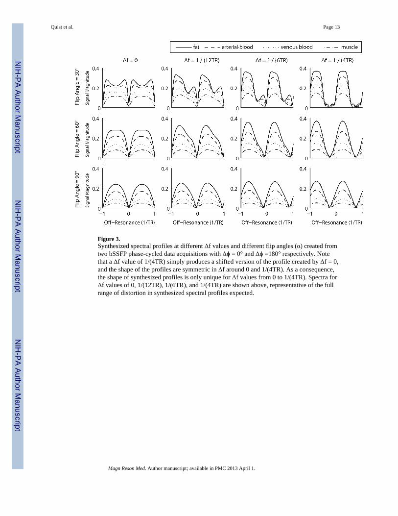
Figure 3.Synthesized spectral profiles at different Δf values and different flip angles (α) created fromtwo bSSFP phase-cycled data acquisitions with Δϕ = 0° and Δϕ =180° respectively. Notethat a Δf value of 1/(4TR) simply produces a shifted version of the profile created by Δf = 0,and the shape of the profiles are symmetric in Δf around 0 and 1/(4TR). As a consequence,the shape of synthesized profiles is only unique for Δf values from 0 to 1/(4TR). Spectra forΔf values of 0, 1/(12TR), 1/(6TR), and 1/(4TR) are shown above, representative of the fullrange of distortion in synthesized spectral profiles expected.
Quist et al. Page 13
Magn Reson Med. Author manuscript; available in PMC 2013 April 1.
NIH
-PA Author Manuscript
NIH
-PA Author Manuscript
NIH
-PA Author Manuscript

Figure 4.The warping of synthesized profiles at different Δf values has an effect on signal contrast.An analysis of arterial/venous, arterial/muscle, and arterial/fat contrast is shown above as afunction of both flip angle and resonant shift Δf. As in Figure 3, graphs are shown for Δf =0, 1/(12TR), 1/(6TR), and 1/(4TR). Interestingly, the distortion tends to improve the contrastof the relevant tissues for the peripheral angiography application.
Quist et al. Page 14
Magn Reson Med. Author manuscript; available in PMC 2013 April 1.
NIH
-PA Author Manuscript
NIH
-PA Author Manuscript
NIH
-PA Author Manuscript

Figure 5.Images from oil/water phantom (top) and lower-leg (bottom) experiments demonstratingLAMA bSSFP at 1.5T. Regular bSSFP acquisitions are shown in (a) and (g) for Δϕ = 0° andin (b) and (h) for Δϕ = 180°. LAMA bSSFP images using field map reconstruction areshown in (c) and (i), and the corresponding images using the region-growing reconstructionare shown in (d) and (j). Fat is well suppressed using both LAMA reconstruction techniques,although a notable improvement is evident with the region-growing reconstruction method.For reference, the acquired field maps are shown in (e) and (k), and the maps synthesizedusing the region-growing algorithm are shown in (f) and (l).
Quist et al. Page 15
Magn Reson Med. Author manuscript; available in PMC 2013 April 1.
NIH
-PA Author Manuscript
NIH
-PA Author Manuscript
NIH
-PA Author Manuscript

Figure 6.Simulation of LAMA bSSFP contrast across a range of flip angles in a simulated 3D lowerleg phantom. Two phase-cycled acquisitions of the 3D phantom were simulated, and thenreconstructed using a simple root sum-of-squares reconstruction (top row) and the region-growing LAMA reconstruction technique (bottom row). The images shown are MIPsthrough each reconstructed 3D dataset. A linear off-resonant shift in the vertical (superior/inferior) direction was simulated to assess the performance of each method given variationsin resonance frequency, and noise added to each phase-cycled dataset prior toreconstruction. Residual banding as a function of off-resonance is evident as signalmodulations in the vertical direction. Off-resonance values vary from 1/TR to -1/TR (152Hz to -152 Hz for an assumed TR of 6.6 ms).
Quist et al. Page 16
Magn Reson Med. Author manuscript; available in PMC 2013 April 1.
NIH
-PA Author Manuscript
NIH
-PA Author Manuscript
NIH
-PA Author Manuscript

Figure 7.Contrast and performance comparison of region-growing LAMA bSSFP in vivo at 1.5T.Lower leg MIPs of a normal volunteer are shown using a standard root sum-of-squaresreconstruction (top row) and the region-growing LAMA reconstruction technique (bottomrow) across a range of flip angles. The better fat-suppression performance of LAMA bSSFPat larger flip angles is clearly evident, although the technique performs surprisingly well atflip angles down to 30°.
Quist et al. Page 17
Magn Reson Med. Author manuscript; available in PMC 2013 April 1.
NIH
-PA Author Manuscript
NIH
-PA Author Manuscript
NIH
-PA Author Manuscript

NIH
-PA Author Manuscript
NIH
-PA Author Manuscript
NIH
-PA Author Manuscript
Quist et al. Page 18
Table 1
T1 and T2 values assumed in simulations for muscle, fat, arterial blood, and venous blood. Values wereinformed by parameters found in [22,26-27].
Tissue T1 (ms) T2 (ms)
Muscle 870 47
Fat 270 85
Arterial Blood 1000 200
Venous Blood 1000 100
Magn Reson Med. Author manuscript; available in PMC 2013 April 1.