Endoscopic management of hypertensive intraventricular haemorrhage with obstructive hydrocephalus
Upload
independentCategory
view
1download
0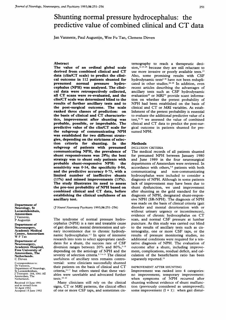
Journal ofNeurology, Neurosurgery, and Psychiatry 1993;56:251-256
Shunting normal pressure hydrocephalus: thepredictive value of combined clinical and CT data
Jan Vanneste, Paul Augustijn, Wee Fu Tan, Clemens Dirven
AbstractThe value of an ordinal global scalederived from combined clinical and CTdata (clin/CT scale) to predict the clini-cal outcome in 112 patients shunted forpresumed normal pressure hydro-cephalus (NPH) was analysed. The clini-cal data were retrospectively collected,all CT scans were re-evaluated, and theclinICT scale was determined blind to theresults of further ancillary tests and tothe post-surgical outcome. The scaleranked three classes of prediction: onthe basis of clinical and CT characteris-tics, improvement after shunting wasprobable, possible, or improbable. Thepredictive value of the clin/CT scale forthe subgroup of communicating NPHwas established for two different strate-gies, depending on the strictness of selec-tion criteria for shunting. In thesubgroup of patients with presumedcommunicating NPH, the prevalence ofshunt responsiveness was 29%; the beststrategy was to shunt only patients withprobable shunt-responsive NPH: thesensitivity was 054, the specificity 0i84,and the predictive accuracy 075, with alimited number of ineffective shunts(11%) and missed improvements (13%).The study illustrates its need to assessthe pre-test probability ofNPH based oncombined clinical and CT data, beforeestablishing the clinical usefulness of anancillary test.
Department ofNeurology, StLucasziekenhuis,AmsterdamJ Vanneste,P AugustijnDepartment ofNeurosurgery,Academic MedicalCentre, AmsterdamW F TanDepartment ofNeurosurgery,Academic Hospital,Free University ofAmsterdam, TheNetherlands.C DirvenCorrespondence to:Dr Vanneste,Department of Neurology,St Lucasziekenhuis,J Tooropstr. 164, 1061 AEAmsterdam, TheNetherlands.
Received 10 June 1991and in revised form30 April 1992.Accepted 15 June 1992
(7 Neurol Neurosurg Psychiatry 1993;56:251-256)
The syndrome of normal pressure hydro-cephalus (NPH) is a rare and treatable cause
of gait disorder, mental deterioration and uri-nary incontinence due to chronic hydrody-namic hydrocephalus.' 2 In spite of intensiveresearch into tests to select appropriate candi-dates for a shunt, the success rate of CSFdiversion ranges between 20% and 80%,3-15depending on the aetiology of NPH and theseverity of selection criteria.5 79'5 The clinicalusefulness of ancillary tests remains contro-versial: some clinicians successfully shuntedtheir patients on the basis of clinical and CTcriteria,'6 17 but others stated that these vari-ables were unreliable and advocated furthertests. 18-22Many clinicians still rely on the clinical
signs, CT or MRI patterns, the clinical effectof one or more CSF taps, and sometimes cis-
ternography to reach a therapeutic deci-sion,1623-25 because they are still reluctant touse more invasive or poorly available tests.23Also, some promising results with CSFhydrodynamic tests6 18 have not been redupli-cated in other studies.2&28 In addition, evenrecent articles describing the advantages ofancillary tests such as CSF hydrodynamicevaluation29 or MRI30 provide scant informa-tion on whether the pretest probability ofNPH had been established on the basis ofclinical and CT or MRI variables. As estab-lishment of the pretest probability is essentialto evaluate the additional predictive value of atest,3' 32 we assessed the value of combinedclinical and CT data to predict the post-sur-gical outcome in patients shunted for pre-sumed NPH.
MethodsINCLUSION CRITERIAThe medical records of all patients shuntedfor presumed NPH between January 1980and June 1989 in the four neurosurgicaldepartments of Amsterdam were reviewed. Inaccordance with others,58 patients with bothcommunicating and non-communicatinghydrocephalus were included to consider adiagnosis of NPH. Although in some patientslack of improvement may have been due toshunt dysfunction, we used improvementafter shunting as the gold standard for thediagnosis of NPH, designated shunt-respon-sive NPH (SR-NPH). The diagnosis of NPHwas made on the basis of clinical criteria (gaitdisorder and mental deterioration with orwithout urinary urgency or incontinences),evidence of chronic hydrocephalus on CTscan, and normal CSF pressure at lumbarpuncture. As this study was carried out blindto the results of ancillary tests such as cis-ternograhy, one or more CSF taps, or theresults of pressure monitoring studies, noadditional conditions were required for a ten-tative diagnosis of NPH. The evaluation ofoutcome after a shunt, including improve-ment, complications, residual deficit, and cal-culation of the benefit/harm ratio has beenseparately reported.33
IMPROVEMENT AFTER SHUNTINGImprovement was ranked into 4 categories:no improvement; temporary improvement:when symptoms of NPH recurred aftershunting without evidence of shunt malfunc-tion (previously considered as unimproved);some improvement (I + 1): when gait distur-
251
Vanneste, Augustijn, Tan, Dirven
bance, mental impairment, urinary inconti-nence or several of these signs improved onlyone point on pre-established ordinal scales, orwhen two--independent assessors estimatedthat there was only slight improvement of gaitor mental function; marked improvement(I + 2): when one or several of these func-tions improved at least two points on theordinal scales, or when at least two indepen-dent assessors agreed that the patient hadsubstantially improved. The term "missedimprovement" was applied to improvedpatients who would not have been shunted onthe basis of combined clinical and CT data(false negative predictions). The term "inef-fective shunt" referred to patients who wouldhave been shunted on the basis of clinical andCT data, but did not improve after shunting(false positive predictions).We could not avoid biases inherent in ret-
rospective studies, based on unstandardisedmedical recording. For example, the positiveeffect of shunting may have been overesti-mated: we tried to counterbalance potentialoveroptimistic evaluations of clinicians byscrutinising the notes of the nursing staff,and, when necessary, by soliciting the opin-ions of the patient, the family and the generalpractitioner.
THE PREDICTIVE VALUE OF A GLOBAL CLINICALPLUS CT SCALE (CLIN/CT SCALE)Calculation of the predictive value of com-bined clinical and CT data has been sum-marised elsewhere.'4 In brief, two clinicalassessors evaluated all available pre-operativeclinical data and CT scans without knowl-edge of the results of ancillary tests and to theoutcome after shunting. The CT scans werere-evaluated and a number of measurementswas carried out, according to a pre-estab-lished protocol. Thereafter, the clinical andCT variables were grouped together into anoverall estimation and classified as probable,possible, or improbable SR-NPH.
This retrospective collection of data fol-lowed by a prospective cohort study, includ-ing observer "blinding" to the results of atherapeutic intervention, has been labelled asa "retrolective" study." Such studies may beparticularly useful when results of prospectivestudies are not available. As we did not estab-lish the intra- and interrater variabilities ofpredictions established with the clin/CTscale, additional investigations should alsovalidate our results.
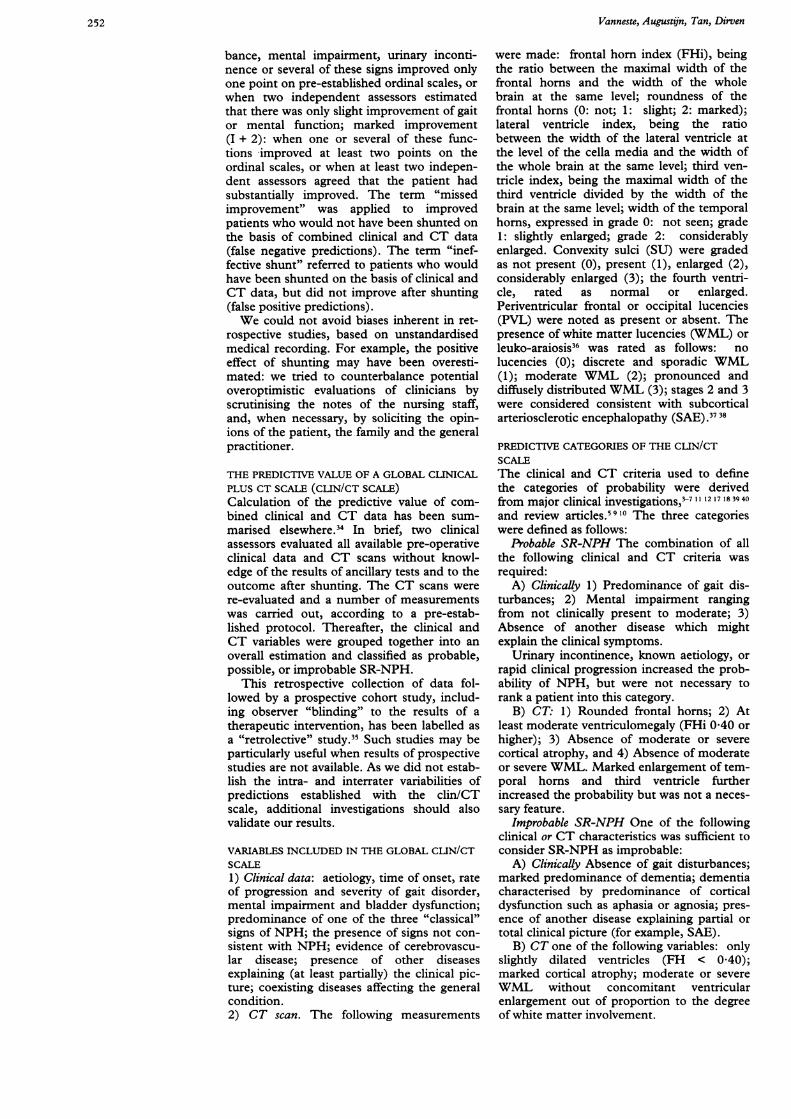
VARIABLES INCLUDED IN THE GLOBAL CLIN/CTSCALE1) Clinical data: aetiology, time of onset, rateof progression and severity of gait disorder,mental impairment and bladder dysfunction;predominance of one of the three "classical"signs of NPH; the presence of signs not con-sistent with NPH; evidence of cerebrovascu-lar disease; presence of other diseasesexplaining (at least partially) the clinical pic-ture; coexisting diseases affecting the generalcondition.2) CT scan. The following measurements
were made: frontal horn index (FHi), beingthe ratio between the maximal width of thefrontal horns and the width of the wholebrain at the same level; roundness of thefrontal horns (0: not; 1: slight; 2: marked);lateral ventricle index, being the ratiobetween the width of the lateral ventricle atthe level of the cella media and the width ofthe whole brain at the same level; third ven-tricle index, being the maximal width of thethird ventricle divided by the width of thebrain at the same level; width of the temporalhorns, expressed in grade 0: not seen; grade1: slightly enlarged; grade 2: considerablyenlarged. Convexity sulci (SU) were gradedas not present (0), present (1), enlarged (2),considerably enlarged (3); the fourth ventri-cle, rated as normal or enlarged.Periventricular frontal or occipital lucencies(PVL) were noted as present or absent. Thepresence of white matter lucencies (WML) orleuko-araiosis36 was rated as follows: nolucencies (0); discrete and sporadic WML(1); moderate WML (2); pronounced anddiffusely distributed WML (3); stages 2 and 3were considered consistent with subcorticalarteriosclerotic encephalopathy (SAE).3738
PREDICTIVE CATEGORIES OF THE CLIN/CTSCALEThe clinical and CT criteria used to definethe categories of probability were derivedfrom major clinical investigations,3-7 11 12 17 18 39 40and review articles.5910 The three categorieswere defined as follows:
Probable SR-NPH The combination of allthe following clinical and CT criteria wasrequired:
A) Clinically 1) Predominance of gait dis-turbances; 2) Mental impairment rangingfrom not clinically present to moderate; 3)Absence of another disease which mightexplain the clinical symptoms.
Urinary incontinence, known aetiology, orrapid clinical progression increased the prob-ability of NPH, but were not necessary torank a patient into this category.
B) CT: 1) Rounded frontal horns; 2) Atleast moderate ventriculomegaly (FHi 0 40 orhigher); 3) Absence of moderate or severecortical atrophy, and 4) Absence of moderateor severe WML. Marked enlargement of tem-poral horns and third ventricle furtherincreased the probability but was not a neces-sary feature.
Improbable SR-NPH One of the followingclinical or CT characteristics was sufficient toconsider SR-NPH as improbable:
A) Clinically Absence of gait disturbances;marked predominance of dementia; dementiacharacterised by predominance of corticaldysfunction such as aphasia or agnosia; pres-ence of another disease explaining partial ortotal clinical picture (for example, SAE).
B) CT one of the following variables: onlyslightly dilated ventricles (FH < 0 40);marked cortical atrophy; moderate or severeWML without concomitant ventricularenlargement out of proportion to the degreeof white matter involvement.
252

Shunting normal pressure hydrocephalus: the predictive value ofcombined clinical and CT data
Possible SR-NPH This category containedall patients who could not be ranked as prob-able or improbable SR-NPH. For example,although CT displays moderate or severewhite matter involvement, it is not sufficientto explain a huge ventriculomegaly. Possiblecombinations of 2 diseases such as SAE plusNPH or Alzheimer's disease plus NPH wereranked into this category.
THE PREDICTIVE VALUE OF A GLOBAL CLIN/CTSCALE IN COMMUNICATING NPH (CoNPH).As most problems of decision making areencountered in patients with CoNPH, we cal-culated the predictive value of a globalClin/CT scale in this subgroup. Two strategicscenarios were considered: 1) Strict selec-tion, consisting of shunting only patients withprobable SR-CoNPH; 2) Liberal shuntingpolicy, consisting of shunting all patients withprobable and possible SR-CoNPH.
THE PREDICTIVE VALUE OF THE CLIN/CT SCALECOMPARED WITH THAT OF A HYDRODYNAMICTESTMeasurement of the conductance to CSFoutflow (C0Ut) obtained during lumbo-ventric-ular perfusion tests in patients with presumedNPH has emerged as a highly predictive testby some investigators.64' We therefore calcu-lated the additional value of this test in ourpatients with idiopathic probable and possibleSR-CoNPH. This additional predictive valueof Co0t was calculated with the assumptionthat the same high sensitivity and specificitywould have been obtained as it was byB0rgesen et al.6
ResultsThe pre-operative clinical data of 166 consec-utive patients shunted for presumed NPHwere reviewed. Four patients were excludedbecause they had an accompanying diagnosis
Table 1 Correlations between predictive categories on a global clinical/CT scale andgrades of improvement
Improvement
Category of ClinICT scale Substantial Some No Total
Probable NPHsecondary, non-communicating 9 2 3 14secondary, communicating 4 - 5 9idiopathic 8 3 6 17
Possible NPH 5 3 25 33Improbable NPH 0 5 34 39Total 26 13 73 112
Table 2 Strategy 1: predictive value of strict selection (shunt only probable SR-CoNPH)
Improvement
Strategy Substantial Some No Total
Shunt probable SR-CoNPH 12 3 11 26Do not shunt possibleand improbable SR-CoNPH 5 8 59 72Total 17 11 70 98
Sensitivity for overall improvement: 0-54 (15/28)Sensitivity for substantial improvement: 0-71 (12/17)Specificity: 0-84 (59/70)Positive predictive value: 58% (15126)Negative predictive value: 82% (59/72)Accurate predictive rate: 75% (74/98).
which might have interfered with signs ofNPH and 10 were excluded because theirmedical records were lost or did not containsufficient information on post-surgical out-come.33 A further 40 patients were withheldfrom this study because of insufficient preop-erative clinical data (N = 8), unavailablepreoperative CT scans (N = 30), or both(N = 2).Thus 112 patients plus their CT scans
remained for further analysis. The aetiologyof hydrocephalus in this group was idiopathicin 91; secondary and communicating in 9;non-communicating in 14, and in 13 wasassociated with aqueduct stenosis.42
Table 1 correlates the degrees of improve-ment with the predictive categories of theClin/CT scale. Improvement after shuntingwas 35% in the whole group, and 29% inpatients with communicating hydrocephalus.
PREDICTIVE VALUE OF THE CLIN/CT SCALE INCONPHProbable SR-CoNPH. Twenty six patients(27% of the total CoNPH group) were allo-cated to this category. The median age was72 years, range 45-85; 11 were males. Ninehad communicating NPH with a knowncause and 17 were idiopathic. On CT themedian FH was 0-46, range 0-40-0-56. In 22patients the sulci were absent (0) or visible(1), in 18 the temporal horns were consider-ably enlarged; WML grade 1 were seen in 10patients. In this group, overall permanentimprovement was 58% (95% CI 37-77%),with no significant difference between idio-pathic (65%, 95% CI 38-86%) and sec-ondary (44%, 95% CI 14-79%) CoNPH.
Possible SR-CoNPH was considered in 33patients, with a median age of 76 years, range62-84; 23 were males. All had idiopathicNPH. On CT the median FH index was 0 45(range 0 39-0 57). Cortical sulci were absentor normal in 21/33, slightly enlarged in 10/33and considerably enlarged in 2/33. WMLwere absent in 18, grade 1 in 8 and grades 2or 3 in 7 patients. Shunting all patients with"possible SR-CoNPH" would have led tosubstantial improvement in 15% (95% CI5-32%).
Improbable SR-NPH was diagnosed in 39patients. All had idiopathic CoNPH. Themedian age was 74 years, range 45-84 years.On CT, the median FH was 0-42 (range0-37-0 50). The main reasons for rejectingSR-NPH were: marked predominance ofdementia (12); dementia including substan-tial cortical dysfunction consistent with seniledementia of Alzheimer's type (7); clinical andCT features consistent with SAE (29); CTshowed marked atrophy (3). Many patientshad more than one reason to be categorisedas improbable SR-NPH. Shunting patients ofthis category would have led to substantialamelioration in 0% (95% CI 0-9%) andslight improvement in 13% (95% CI 4-27%).
Table 2 shows the predictive value of thefirst strategy, that is, shunting only patientswith probable SR-CoNPH. It would have ledto a predictive accuracy of 75% (95% CI
253
Vanneste, Augustijn, Tan, Dirven
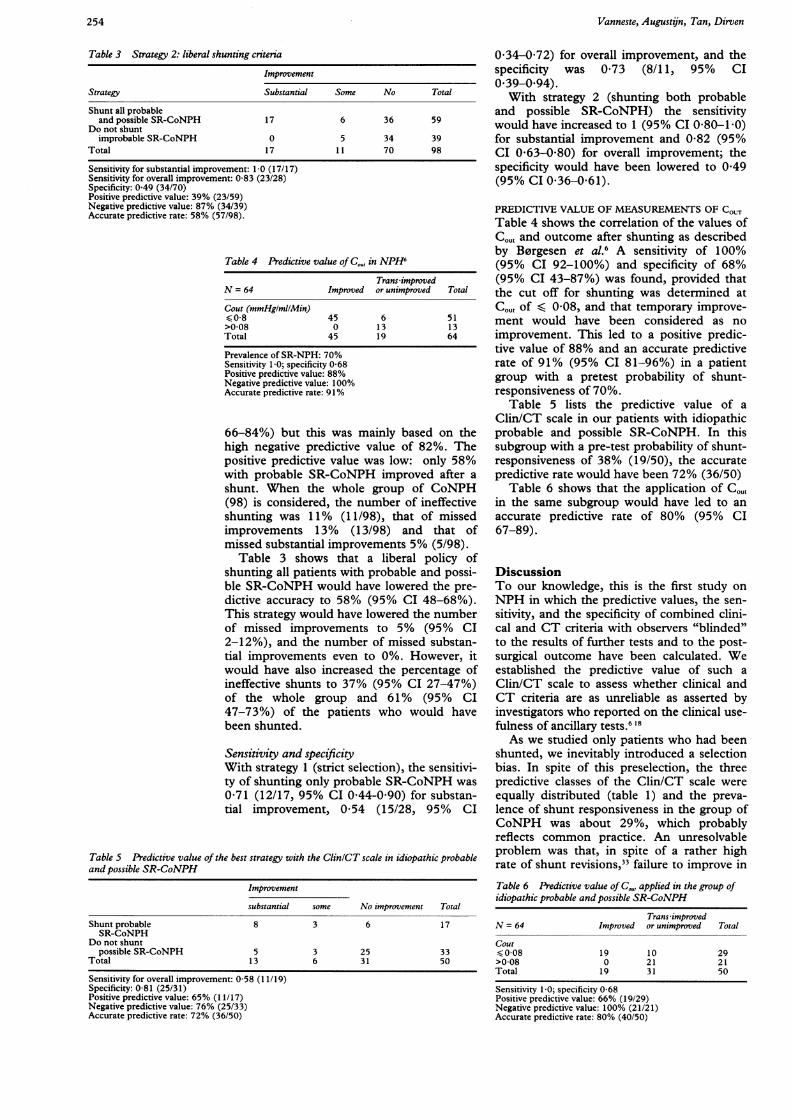
Table 3 Strategy 2: liberal shunting criteria
Improvement
Strategy Substantial Some No Total
Shunt all probableand possible SR-CoNPH 17 6 36 59
Do not shuntimprobable SR-CoNPH 0 5 34 39
Total 17 11 70 98
Sensitivity for substantial improvement: 1 0 (17/17)Sensitivity for overall improvement: 0-83 (23/28)Specificity: 0 49 (34/70)Positive predictive value: 39% (23/59)Negative predictive value: 87% (34/39)Accurate predictive rate: 58% (57/98).
Table 4 Predictive value of C,O, in NPH6
Trans improvedN = 64 Improved or unimproved Total
Cout (mmHg/ml/Min)<0-8 45 6 51>0-08 0 13 13Total 45 19 64
Prevalence of SR-NPH: 70%Sensitivity 1 0; specificity 0-68Positive predictive value: 88%Negative predictive value: 100%Accurate predictive rate: 91%
66-84%) but this was mainly based on thehigh negative predictive value of 82%. Thepositive predictive value was low: only 58%with probable SR-CoNPH improved after ashunt. When the whole group of CoNPH(98) is considered, the number of ineffectiveshunting was 11% (11/98), that of missedimprovements 13% (13/98) and that ofmissed substantial improvements 5% (5/98).
Table 3 shows that a liberal policy ofshunting all patients with probable and possi-ble SR-CoNPH would have lowered the pre-dictive accuracy to 58% (95% CI 48-68%).This strategy would have lowered the numberof missed improvements to 5% (95% CI2-12%), and the number of missed substan-tial improvements even to 0%. However, itwould have also increased the percentage ofineffective shunts to 37% (95% CI 27-47%)of the whole group and 61% (95% CI47-73%) of the patients who would havebeen shunted.
Sensitivity and specificityWith strategy 1 (strict selection), the sensitivi-ty of shunting only probable SR-CoNPH was071 (12/17, 95% CI 044-0 90) for substan-tial improvement, 054 (15/28, 95% CI
Table S Predictive value of the best strategy with the ClinlCT scale in idiopathic probableand possible SR-CoNPH
Improvement
substantial some No improvement Total
Shunt probable 8 3 6 17SR-CoNPH
Do not shuntpossible SR-CoNPH 5 3 25 33
Total 13 6 31 50
Sensitivity for overall improvement: 0-58 (11/19)Specificity: 0-81 (25/31)Positive predictive value: 65% (11/17)Negative predictive value: 76% (25/33)Accurate predictive rate: 72% (36/50)
034-072) for overall improvement, and thespecificity was 0 73 (8/11, 95% CI039-0 94).With strategy 2 (shunting both probable
and possible SR-CoNPH) the sensitivitywould have increased to 1 (95% CI 080-1 -0)for substantial improvement and 082 (95%CI 0 63-0 80) for overall improvement; thespecificity would have been lowered to 0A49(95% CI 0-36-0-61).
PREDICTIVE VALUE OF MEASUREMENTS OF COUTTable 4 shows the correlation of the values ofCG0t and outcome after shunting as describedby B0rgesen et al.6 A sensitivity of 100%(95% CI 92-100%) and specificity of 68%(95% CI 43-87%) was found, provided thatthe cut off for shunting was determined atCG,t of < 0-08, and that temporary improve-ment would have been considered as noimprovement. This led to a positive predic-tive value of 88% and an accurate predictiverate of 91% (95% CI 81-96%) in a patientgroup with a pretest probability of shunt-responsiveness of 70%.
Table 5 lists the predictive value of aClin/CT scale in our patients with idiopathicprobable and possible SR-CoNPH. In thissubgroup with a pre-test probability of shunt-responsiveness of 38% (19/50), the accuratepredictive rate would have been 72% (36/50)
Table 6 shows that the application of Coutin the same subgroup would have led to anaccurate predictive rate of 80% (95% CI67-89).
DiscussionTo our knowledge, this is the first study onNPH in which the predictive values, the sen-sitivity, and the specificity of combined clini-cal and CT criteria with observers "blinded"to the results of further tests and to the post-surgical outcome have been calculated. Weestablished the predictive value of such aClin/CT scale to assess whether clinical andCT criteria are as unreliable as asserted byinvestigators who reported on the clinical use-fulness of ancillary tests.6 18As we studied only patients who had been
shunted, we inevitably introduced a selectionbias. In spite of this preselection, the threepredictive classes of the Clin/CT scale wereequally distributed (table 1) and the preva-lence of shunt responsiveness in the group ofCoNPH was about 29%, which probablyreflects common practice. An unresolvableproblem was that, in spite of a rather highrate of shunt revisions," failure to improve in
Table 6 Predictive value of CO", applied in the group ofidiopathic probable and possible SR-CoNPH
Trans-improvedN = 64 Improved or unimproved Total
Cout0 08 19 10 29
>0-08 0 21 21Total 19 31 50
Sensitivity I 0; specificity 0-68Positive predictive value: 66% (19/29)Negative predictive value: 100% (21/21)Accurate predictive rate: 80% (40/50)
254
Shunting normal pressure hydrocephalus: the predictive value ofcombined clinical and CT data
some patients may have been due to inade-quate shunts rather than to a misdiagnosis ofNPH.'3
Shunting only patients with probably SR-CoNPH led to the highest predictive accura-cy. With this policy (table 2), neurologistswould have referred less than one third of theshunted patients to the neurosurgeon, withlimited erroneous decisions: only 5% of thewhole group with presumed SR-CoNPHwould have been erroneously withheld fromsubstantial improvement and 13% of thewhole group would have had ineffectivesurgery.Thus in centres with limited technological
facilities, shunting only patients with probableCoNPH on the basis of a global clinical/CTscale would be justified, especially in view ofthe serious surgical risks and the very low rateof marked improvement in the categories ofpossible and improbable SR-CoNPH. Themajor drawback is that these patients andtheir families must accept a 42% chance ofineffective surgery and a 5% chance of beingerroneously withheld a highly beneficial pro-cedure.
In patients with possible SR-CoNPH-thecategory with the greatest diagnostic uncer-tainty-the likelihood of improvement after ashunt is estimated at 30-40%.1 14 This con-trasts with our experience, as shunting allpatients with possible SR-CoNPH wouldhave resulted in a disappointingly low per-centage of substantial benefit (15%). As seri-ous shunt-related residual morbidityapproximates 7-8%,"3 a very reserved thera-peutic attitude towards patients with possibleSR-CoNPH would be justified, as the bene-fits would scarcely outweigh the risks of seri-ous post-surgical harm. It is possible that ourcriteria to classify hydrocephalic patients intothis category were too liberal, but it is alsopossible that some investigators includedimprovement of limited clinical importance tocalculate the benefits of a shunt.
Shunting patients with improbable SR-CoNPH appeared as a desperate attempt toreverse an irreversible process of mental dete-rioration, most probably due to degenerativebrain disease or to SAE. Our large retrospec-tive study33 revealed that many neurologistsand neurosurgeons submitted patients withimprobable NPH to a neurosurgical "last-chance" procedure. As very few patients ofthis group fare only slightly better after ashunt, submitting them to a shunting proce-dure is unjustified as the benefit/harm ratio isunacceptably low.
With a combined clin/CT scale the mostaccurate predictive rate in the whole group ofCoNPH was 75% (strategy 1). This predic-tive accuracy is higher than commonlyassumed, but was mainly due to the high pre-dictive value of a negative decision, that is,not to shunt patients with possible or improb-able SR-CoNPH. Even with strict selection,the positive predictive value of a combinedClin/CT scale remained low (58%).Thus by combining clinical and CT crite-
ria for a global predictive scale, we did not
succeed in eliminating the need for non-inva-sive, easily accessible tests with reproducibleresults, assuming, of course, that their sensi-tivity, specificity and predictive values wouldbe meaningfully higher than those obtainedwith combined clinical and CT criteria.Continuous intracranial CSF pressurerecording8 and calculation of C0,t641 havebeen described as the best predictive tests.Unfortunately, at least in the Netherlands,these techniques are inaccessible to most clin-icians.2' In the best hands, the accuracy ofC,Ut was very high, reaching levels of 90% oreven higher, but this was obtained in a groupof patients in whom a large majority mayhave presented with a typical SR-NPH, as70% improved after a shunt.6 Other reportshave illustrated that CSF infusion tests maybe less accurate.'$'8
In our 50 patients with idiopathic commu-nicating NPH there was a large spectrum ofprobabilities indicating SR-NPH, explainingthat the pretest probability of SR-NPH wasonly 39%: this approximates to the percent-age encountered in common neurologicalpractice. In this group, the most difficultregarding decision making, the predictiveaccuracy of C0Ut would have been 80%, whichis only 8% better than that obtained with thebest strategy based on clinical and CT crite-ria. The main advantage of assessing the C,would have been to avoid missed substantialimprovements in 10% of the patients.
In conclusion, our study suggests that fur-ther studies on NPH should include assess-ment of the pretest probability based oncombined clinical and CT variables. Onlythen can one assess the usefulness of an ancil-lary test to assist clinicians in their decisionsto shunt or not to shunt patients with pre-sumed NPH.
We acknowledge the useful suggestions of Professors H vanCrevel, M Vermeulen and Drs A Hijdra, GAG Davies and RPGolding.
1 Adams RD, Fisher CM, Hakim S, Ojemann RG, SweetWH. Symptomatic occult hydrocephalus with "normal"cerebrospinal fluid pressure. A treatable syndrome. NEngJMed 1965;273:117-26.
2 Hakim S, Adams RD. The special clinical problem ofsymptomatic hydrocephalus with normal cerebrospinalfluid pressure. Observations on cerebrospinal fluidhydrodynamics. J Neurol Sci 1965;2:307-27.
3 Jacobs L, Conti D, Kinkel WR, Manning EJ. "Normal-pressure" hydrocephalus. Relationship of clinical andradiographic findings to improvement following shuntsurgery. JAMA 1976;235:510-12.
4 Greenberg JO, Shenkin HA, Adam R. Idiopathic normalpressure hydrocephalus-a report of 73 patients. J NeurolNeurosurg Psychiatry 1977;40:336-4 1.
5 Katzman R. Normal pressure hydrocephalus. In:Katzman R, Terry RD, Bick K, eds. Alzheimer'sDisease: Senile dementia and related disorders. Aging1978;7:115-24.
6 Borgesen SE, Gjerris F. The predictive value of conduc-tance to outflow of cerebrospinal fluid in normal pres-sure hydrocephalus. Brain 1982;105:65-86.
7 Fisher CM. Hydrocephalus as a cause of disturbances ofgait in the elderly. Neurology 1982;32:1358-63.
8 Pickard JD. Normal pressure hydrocephalus- to shunt ornot to shunt. In: Warlow C, Garfield J, eds. Dilemmasin the management of the neurological patient. Edinburgh:Churchill Livingstone, 1984:207-14.
9 Fishman RA. Normal-pressure hydrocephalus and arthri-tis. NEnglJMed 1985;312:1255-6.
10 Black PMcL, Ojemann RG, Tzouras A. CSF shunts fordementia, incontinence, and gait disturbance. ClinNeurosurg 1985;32:632-51
11 Petersen RC, Mokri B, Laws ER Jr. Surgical treatment of
255
Vanneste, Augustijn, Tan, Dirven
idiopathic hydrocephalus in elderly patients. Neurology1985;35:307-1 1.
12 Graff-Radford NR, Godersky JC, Jones MP. Variablespredicting surgical outcome in symptomatic hydro-cephalus in the elderly. Neurology 1989;39:1601-4.
13 Black PMcL. The normal pressure hydrocephalus syn-drome. In: Scott RM, ed. Concepts in neurosurgery, vol 3.Hydrocephalus. Baltimore: Williams and Wilkins, 1990:109-14.
14 Bret P, Chazal J. Chronic hydrocephalus of the adult.Neurochirurgie 1990;36(Suppl.1): 1-159.
15 Anonymous. Normal-pressure hydrocephalus (editorial).Lancet 1990;335:22.
16 Alexander MP, Geschwind N. Dementia in the elderly.In: Albert ML, ed. Clinical neurology of aging. New-York: Oxford University Press, 1984:254-76.
17 Vassilouthis J. The syndrome of normal-pressure hydro-cephalus. J Neurosurg 1984;61:501-9.
18 Tans JTJ. Differentiation of normal pressure hydro-cephalus and cerebral atrophy by computed tomographyand spinal infusion test. J Neurol 1979;222: 109-18.
19 Pickard JD, Teasdale G, Matheson M, Lindsay K,Galbraith S, Wyper D, MacPherson P. Intraventricularpressure waves-the best predictive test for shunting innormal pressure hydrocephalus. In: Shulman K,Marmarou A, Miller JD, et al, eds Intracranial pressureIV. Berlin: Springer-Verlag, 1980:498-500.
20 Pappada G, Poletti C, Guazzoni A, Sani R, Colli M.Normal pressure hydrocephalus: relationship amongclinical picture, CT scan and intracranial pressure mon-itoring. J Neurosurg Sci 1986;30: 115-28.
21 Borgesen SE, Gjerris F. Relationships between intracra-nial pressure, ventricular size, and resistance to CSFoutflow. J Neurosurg 1987;67:535-9.
22 Price DJ. Attempts to predict the probability of clinicalimprovement following shunting of patients with pre-sumed normal pressure hydrocephalus. In: Hoff JT,Betz AL, eds. Intracranial pressure VII. Berlin: SpringerVerlag, 1989:390-3.
23 Vanneste J, van Acker R. Normal pressure hydrocephalus:did literature alter management ? J Neurol NeurosurgPsychiatry 1990;53:564-8.
24 Mulrow CD, Feussner JR, Williams BC, Vokaty KA. Thevalue of clinical findings in the detection of normal pres-sure hydrocephalus. J. Gerontol 1987;42:277-9.
25 Benzel EC, Pelletier Al, Levy PG. Communicating hydro-cephalus in adults: prediction of outcome after ventricu-lar shunting procedures. Neurosurgery 1990;26:655-60.
26 Vorstrup S, Christensen J, Gjerris F, Sorensen PS,Thomsen AM, Paulson OB. Cerebral blood flow inpatients with normal-pressure hydrocephalus before andafter shunting. _Neurosurg 1987;66:379-87.
27 Delwel EJ, de Jong DA, Avezaat Cnl, van EijndhovenJHM. CSF dynamics in patients with suspected normalpressure hydrocephalus. In: Hoff JT, Betz AL, eds.Intracranial pressure VII. Berlin: Springer Verlag,
1989:379-82.28 Kosteljanetz M, Westergaard L, Kaalund J, Nehen AM.
The significance of outflow resistance measurements inthe prediction of outcome after CSF-shunting inpatients with normal pressure hydrocephalus. In: HoffJT, Betz AL, eds. Intracranial pressure VII. Berlin:Springer Verlag, 1989:383-5.
29 Lundar T, Nornes H. Determination.of ventricular fluidoutflow resistance in patients with' ventriculomegaly. 3Neurol NeurosurgPsychiatny 1990;53:896-8.
30 Bradley WG Jr, Whittemore AR, Watanabe AS, Davis SJ,Teresi LM, Homyak M. Association of deep white mat-ter infarction with chronic communicating hydro-cephalus: imnplications regarding the possible origin ofnormal-pressure hydrocephalus. AJNR 199 1;12:31-9
31 Sackett DL, Haynes RB, Tugwell P. The selection ofdiagnostic tests. Clinical epidemiology. Boston/Toronto:Little Brown, 1991:51-68.
32 Longstreth WT Jr, Koepsell TD, van Belle G. ClinicalNeuroepidemiology. I. Diagnosis. Arch Neurol 1987;44:1091-9.
33 Vanneste J, Augustijn P, Dirven C, Tan WF, GoedhartZD. Shunting normal pressure hydrocephalus: do thebenefits outweigh the risks? A multicentre study and lit-erature review. Neurology 1992;42:54-59.
34 Vanneste J, Augustijn P, Tan WF, Dirven C. Shuntingnormal pressure hydrocephalus: is cisternography stilluseful? Arch Neurol 1992;49:366-70.
35 Feinstein AR. Retrolective and prolective studies. In: ARFeinstein, ed. Clinical epidemiology. The architecture ofclinical research. Philadelphia: WB Saunders, 1985:225-6.
36 Hachinski VC, Potter P, Merskey H. Leuko-araiosis. ArchNeurol 1987;44:21-3.
37 Zeumer H, Schonsky B, Sturn KW. Predominant whitematter involvement in subcortical arterioscleroticencephalopathy (Binswanger disease). .7 Comput AssistTomogr 1980;4:14-9.
38 Lotz PR, Ballinger WE Jr, Quisling RG. Subcortical arte-riosclerotic encephalopathy: CT spectrum and patho-logic correlation. AJNR 1986;7:817-22.
39 Laws ER Jr, Mokri B. Occult hydrocephalus: results ofshunting correlated with diagnostic tests. In: KeenerEB, ed. Clinical Neurosurgery, Baltimore: Williams andWilkins, 1977;24:316-33.
40 Black PMcL. Idiopathic normal-pressure hydrocephalus.Results of shunting in 62 patients. .7 Neurosurg1980;53:371-7.
41 Borgesen SE, Gjerris F, Schmidt J. Measurement of resis-tance to CSF outflow by subarachnoid perfusion. AlfredBenzon Symposium 27: Outflow of cerebrospinal fluid.Gjerris F, Borgesen SE, Sorensen PS, eds. Copenhagen:Munksgaard, 1989:121-133.
42 Vanneste J, Hyman R. Non-tumoural aqueduct stenosisand normal pressure hydrocephalus in the elderly. .7Neurol Neurosurg Psychiatry 1986;49:529-35.
256
Copyright © 2022 FDOKUMEN