"Duelo y fotografía post-mortem Contradicciones de una práctica vigente en el siglo XXI"

Neurochemistry International 62 (2013) 182–188
Contents lists available at SciVerse ScienceDirect
Neurochemistry International
journal homepage: www.elsevier .com/locate /nc i
Serotonin receptor of type 6 (5-HT6) in human prefrontal cortexand hippocampus post-mortem: An immunohistochemical andimmunofluorescence study
Donatella Marazziti a,⇑, Stefano Baroni a, Andrea Pirone b, Gino Giannaccini a, Laura Betti a,Giovanna Testa a, Lara Schmid a, Lionella Palego a, Franco Borsini d, Fabio Bordi d, Ilaria Piano e,Claudia Gargini a, Maura Castagna c, Mario Catena-Dell’Osso a, Antonio Lucacchini a
a Dipartimento di Psichiatria, Neurobiologia, Farmacologia e Biotecnologie, University of Pisa, Italyb Dipartimento di Scienze Fisiologiche, University of Pisa, Italyc Dipartimento di Chirurgia, University of Pisa, Italyd Sigma-Tau ‘‘Industrie Farmaceutiche Riunite’’, Roma, Italye G.B. Bietti Foundation for Ophthalmology Research, Roma, Italy
a r t i c l e i n f o
Article history:Received 29 August 2012Received in revised form 20 November 2012Accepted 25 November 2012Available online 3 December 2012
Keywords:SerotoninSerotonin receptorsSerotonin receptors of type 6Human brainAutopsy[125I]SB-258585 bindingPrefrontal cortexHippocampusImmunohistochemistryImmunofluorescence
0197-0186/$ - see front matter � 2012 Elsevier Ltd. Ahttp://dx.doi.org/10.1016/j.neuint.2012.11.013
⇑ Corresponding author. Address: Dipartimento dFarmacologia e Biotecnologie, University of Pisa, viaTel.: +39 050 2219768; fax: +39 050 2219787.
E-mail address: [email protected] (D. M
a b s t r a c t
Given the paucity of data on the distribution of serotonin (5-HT) receptors of type 6 (5-HT6) in the humanbrain, the aim of this study was to investigate their distribution in postmortem human prefrontal cortex,striatum and hippocampus by either immunohistochemical or immunofluorescence techniques.
The brain samples were obtained from 6 subjects who had died for causes not involving primarily orsecondarily the CNS. The 5-HT6 receptor distribution was explored by the [125I]SB-258585 binding tobrain membranes followed by immunohistochemical and immunofluorescence evaluations.
A specific [125I]SB-258585 binding was detected in all the regions under investigation, whilst the con-tent in the hippocampus and cortex being about 10–30 times lower than in the striatum. Immunohisto-chemistry and double-label immunofluorescence microscopy experiments, carried out in the prefrontalcortex and hippocampus only, since data in the striatum were already published, showed the presenceof 5-HT6 receptors in both pyramidal and glial cells of prefrontal cortex, while positive cells were mainlypyramidal neurons in the hippocampus.
The heterogeneous distribution of 5-HT6 receptors provides a preliminary explanation of how theymight regulate different functions in different brain areas, such as, perhaps, brain trophism in the cortexand neuronal firing in the hippocampus. This study, taking into account all the limitations due to thepostmortem model used, represents the starting point to explore the 5-HT6 receptor functionality andits sub-cellular distribution.
� 2012 Elsevier Ltd. All rights reserved.
1. Introduction
Serotonin (5-hydroxytryptamine, 5-HT) is a neurotransmitterinvolved in the modulation of several functions and behaviors,comprising mood, anxiety, temperature, appetite, sleep, memoryand learning, nociception and sexuality (Berger et al., 2009; Nord-quist and Oreland, 2010). Serotonin exerts its diverse physiologicaland pharmacological effects through the interaction with multiplereceptor subtypes (Hoyer et al., 2002). Up-to now, seven majorfamilies of 5-HT receptors have been identified (5-HT1–5-HT7),including at least 14 distinct receptor subtypes (Stahl, 2008). The
ll rights reserved.
i Psichiatria, Neurobiologia,Roma, 67, I-56100 Pisa, Italy.
arazziti).
5-HT6 receptor differs from all other subtypes in terms of bothstructural and pharmacological features (Marazziti et al., 2011). In-deed, this membrane-bound receptor protein is characterized by along C-terminal tail and a short third cytoplasmatic loop codifiedby an intron-containing gene region (Kohen et al., 1996). Moreover,either antidepressants or antipsychotics, both typical, such aschlorpromazine, and atypical, such as clozapine and olanzapine,behave as antagonists at the level of 5-HT6 receptors (Monsmaet al., 1993; Roth et al., 1994; Glatt et al., 1995; Kohen et al.,1996; Frederick and Meador-Woodruff, 1999; Branchek andBlackburn, 2000; Bymaster et al., 2001; Meltzer et al., 2003).Interestingly, 5-HT6 receptors have been found to influenceacetylcholine release in the frontal cortex (Riemer et al., 2003),while suggesting their involvement in cognition deficits, someforms of anxiety disorders (Branchek and Blackburn, 2000;

D. Marazziti et al. / Neurochemistry International 62 (2013) 182–188 183
Woolley et al., 2004; Codony et al., 2011), nociception (Castaneda-Corral et al., 2009; Freitas et al., 2009) seizure (Routledge et al.,2000) and drug reward (Frantz et al., 2002; Ferguson et al.,2007). For this reason, a number of 5-HT6 receptor ligands enteredthe clinical development as potential anti-dementia agents (John-son et al., 2008; Upton et al., 2008; Geldenhuys and van der Schyl,2008, 2009; Maher-Edwards et al., 2010; Witty et al., 2009; Rosséand Schaffhauser, 2010), antipsychotics (Arnt and Skarsfeldt, 1998;Minabe et al., 2004; Johnson et al., 2008; Li et al., 2007), or anti-obesity drugs (Heal et al., 2008).
The CNS distribution of 5-HT6 receptors is still a matter of con-troversies. In the rat brain, the highest 5-HT6 receptor content wasfound in the striatum, followed by nucleus accumbens, hippocam-pus and olfactory tubercle (Gérard et al., 1997; Yoshioka et al.,1998; Hamon et al., 1999; Hirst et al., 2000, 2003; Roberts et al.,2002), with, however, some region-dependent discrepanciesbetween protein binding and gene expression results. An intense5-HT6 immuno-autoradiographic labeling, together with a weak-to-moderate mRNA expression, was observed in the rat cerebellum,whilst the opposite situation being described in the hippocampus(Ward et al., 1995; Gérard et al., 1996; Hamon et al., 1999).
The data on the distribution of 5-HT6 receptors in the humanbrain is similarly controversial, mainly limited by the use of differ-ent, not easily comparable techniques and by the small samplenumbers. In most cases, the highest level of 5-HT6 receptor mRNAwas detected in the caudate and nucleus accumbens, followed bythe hippocampus and amygdala. Lowest concentrations were in-stead observed in the thalamus, subthalamic nucleus and substan-tia nigra (Kohen et al., 1996; Hirst et al., 2003). The postmortemautoradiography of brain cortex from schizophrenic subjects, car-ried out by means of the iodinated specific 5-HT6 antagonist[125I]SB-258585 (Hirst et al., 2000), revealed affinity (pKD � 9) andrelative distribution of 5-HT6 receptor binding sites comparableto those of the rat brain (striatum >> cortex � hippocampus) (Eastet al., 2002). The cellular expression of 5-HT6 receptors in the pre-frontal cortex has been further explored by applying immunohisto-chemistry techniques to brain tissue sections of patients withAlzheimer’s disease and normal control subjects (Lorke et al., 2006).
The present study represents an extension of a previous onecarried out by us showing the presence of a high-affinity (of nM or-der), specific and saturable [125I]SB-258585 binding in the humanstriatum postmortem (Marazziti et al., 2012). In that study, wecould quantify the presence of the 5-HT6 receptor by means ofthe Scatchard analysis in the striatum only, but not in the prefron-tal cortex and hippocampus, given the paucity of specific bounddetected in these two brain regions.
Our current aim was, therefore, to report binding data in stria-tum, cortex and hippocampus, as well as to further characterize thedistribution of 5-HT6 receptors in the prefrontal cortex and hippo-campus by either immunohistochemical or immunofluorescencetechniques.
2. Materials and methods
2.1. Collection of human brain tissues
The recognition of the three different brain areas, the prefrontalcortex, hippocampus and striatum, as well as sample collectionwere carried out during autopsy sessions by skilled and qualifiedanatomists from the Anatomy and Pathology Section of the ‘‘Dipar-timento di Chirurgia’’, University of Pisa. In every case, the brainsamples were taken as parts of tissues kept for diagnostic scopes,according to the procedure approved by the Ethics Committee ofthe University of Pisa. The brain specimens were obtained from 6subjects (3 men and 3 women, mean age ± SD: 52 ± 8 years), whohad died for causes not involving primarily or secondarily the
CNS (3 for myocardial infarction, 2 for pneumonia, 1 for pulmonaryembolism), and were not suffering from chronic metabolic diseasesor major psychiatric disorders, as shown by their medical charts.The postmortem delay (the time between demise and tissue dissec-tion/freezing) was within 48 h (mean ± SD: 39 ± 9 h). Brain tissueswere removed, rapidly dissected and transported in liquid nitrogento the laboratory, where they were frozen at �80 �C for [125I]SB-258585 (Perkin Elmer Life Sciences, Milan, Italy; specific activity:2200 Ci/mmol) binding experiments, carried out within 4 weeks.Conversely, for immunostaining/fluorescence techniques, hippo-campal and fronto-cortical samples were immediately treated.
2.2. Preparation of human brain membranes
The human brain membranes were prepared according to theHirst et al. ’s procedure (2000), as previously described in more de-tails (Marazziti et al., 2012). Final membrane pellets were assayedfor protein concentration by the Bradford method (Bio-Rad proteinassay kit; Bio-Rad, Hemel Hempstead, UK), by using c-globulins asthe standard.
2.3. [125I]SB-258585 binding
The [125I]SB-258585 binding experiments were carried out byincubating membranes (0.6–0.9 mg protein), obtained from thethree brain regions under investigation, for 60 min with 0.1 nM[125I]SB-258585 at 25 �C without (total binding) and in the pres-ence of (non specific binding) unlabeled 1 lM SB-399885 in a finalvolume of 0.5 ml assay buffer. The incubation was halted by a rapidfiltration under vacuum using a Brandel cell harvester apparatus,through Whatman GF/B filters pre-soaked with 0.3% polyethyleni-mine, washed and measured by a c-counter solid-phase scintilla-tion spectrometer (Perkin–Elmer, Wizard 1470), as previouslydescribed (Marazziti et al., 2012).
2.4. Tissue preparation for immunohistochemistry andimmunofluorescence assay
Blocks of prefrontal cortex and hippocampus were carefully dis-sected out from the brain and placed in 4% formalin in 0.1 M phos-phate buffer (PBS) for 24 h. The tissue blocks were cryoprotectedfirst in PBS-20% sucrose containing 0.1% Na-azide for 2–3 days,then in PBS-30% sucrose containing 0.1% Na-azide for 2–3 addi-tional days. After this step, the samples were snap-freezed on pow-dered dry ice, and stored at �80 �C.
Sectioning of tissue blocks was performed on a freezing micro-tome set at a thickness of 15 lm. The resulting sections were sub-sequently mounted on gelatin-coated slides and processed for theimmunohistochemical stain.
2.5. Single-antigen immunohistochemistry: 5-HT6 receptordistribution
Fronto-cortical and hippocampal sections were rinsed in PBSand incubated for 10 min in a 1% H2O2–PBS solution. Then, theywere pre-incubated in 5% normal goat serum (NGS) (Vector Labs,Peterborough, UK) in PBS containing 0.3% TritonX-100 (TX)(Sigma–Aldrich, Milan, Italy) to reduce unspecific staining. Afterthis washing procedure, sections were incubated overnight in ahumid chamber at 4 �C with a goat polyclonal antibody raisedagainst a specific human 5-HT6 receptor epitope, a peptide mappedwithin an internal region of the protein (sc-26668, Santa CruzBiotec., Inc., USA); the antibody was opportunely diluted in PBS(1:50) containing 0.3% TX and 1% NGS. Sections were than treated,for the immunoperoxidase activity, as previously described indetail (Marazziti et al., 2012).

0 0,5 1 1,5 2
Hippocampus
Striatum
Frontal Cortex
fmol/mg proteins
Fig. 1. Specific binding (fmol/mg proteins) of [125I]-SB258585 (0,1 nM,) in thehuman hippocampus, striatum and prefrontal cortex.
184 D. Marazziti et al. / Neurochemistry International 62 (2013) 182–188
2.6. Double-antigen immunofluorescence: Cellular localization
Sections were washed three times for 10 min (3 � 10 min) inPBS, then permeabilized and blocked with PBS containing 1%bovine serum albumin (BSA) and 0.3% Triton X-100 in a humidchamber at room temperature for 45 min. Sections were succes-sively incubated in a combination of the following antibodies:anti-5-HT6 (goat polyclonal antibody, clone A20, Santa Cruz Biotec.,Inc., USA) (1:100)/NeuN (1:250) (mouse monoclonal antibody,
Fig. 2. (A–D): 5-HT6 immunoreactivity localization (immunoperoxidase reaction) in the(A) some of these (arrow) cross perpendicularly the layer 1 (L1). (B, C) Immunostainedneuron in the layer 3. Scale bar = 50 lm (A, B), 10 lM (C, D).
clone A60, Millipore, Milan, Italy), anti-5-HT6 (1:100)/GFAP(1:500) (mouse monoclonal antibody, clone 6F2, code n. M0761,DakoCytomation) diluted in PBS containing 1% BSA and 0.03% Tri-tonX-100 (PBS-BT) overnight in a humid chamber at 4 �C. Afterwashing for 3 � 10 min in PBS, the slides were incubated in a com-bination of secondary antibodies (1:200): anti-mouse Alexa 568and anti-goat Alexa 488 (Invitrogen, Carlsbad, CA, USA). The sec-tions were further washed for 3 � 10 min in PBS and mounted inVectashield (Vector Labs, Peterborough, UK). The specificity ofthe staining reaction was checked in repeated trials through thesubstitution of either the primary or the secondary antibody withPBS. Slides were examined with a Leica TCS-NT confocal micro-scope equipped with a krypton–argon laser.
3. Results
3.1. [125I]SB-258585 Binding
Fig. 1 shows the specific [125I]SB-258585 binding (fmol/mg pro-teins) obtained in striatum, prefrontal cortex and hippocampus: atthe same final ligand concentration (0.1 nM), the specific bindingwas higher (approximately 10 and 30 times) in the striatum thanin the hippocampus and cortex.
3.2. Immunohistochemistry data
A significant and specific 5-HT6 receptor immunoreactivity wasobserved in both prefrontal cortex and hippocampus, with somepeculiar characteristics and cell distribution.
In fronto-cortical sections, the immunoreactivity appears to bepredominantly localized either in the layer I or the molecular one.It is noteworthy that, in the molecular layer, we observed positive
prefrontal cortex. (A, B) plexus of positive fibers (empty arrow) in the sub-pial area;cells with extensions in contact with blood vessels (arrow). (D) positive pyramidal

D. Marazziti et al. / Neurochemistry International 62 (2013) 182–188 185
cells in both the somata and processes: these cells were also foundto get contacts with blood capillaries. Further, still in this layer, aplexus of positive fibers was detected at the level of the sub-pial re-gion, some of these crossing perpendicularly all the layers.
In the deepest cortical layers (IV, V, VI), just a few positive so-mata were observed. Some immunoreactive cells found in layerIII had a typical morphology of pyramidal neurons (Fig. 2).
In the hippocampus, a widespread immunoreactivity was ob-served in the CA1, CA2, CA3 and CA4 fields. Parallel sections, trea-ted with the Nissl reagent, confirmed that the immunoreactivecells were pyramidal neurons (Fig. 3).
3.3. Immunofluorescence data
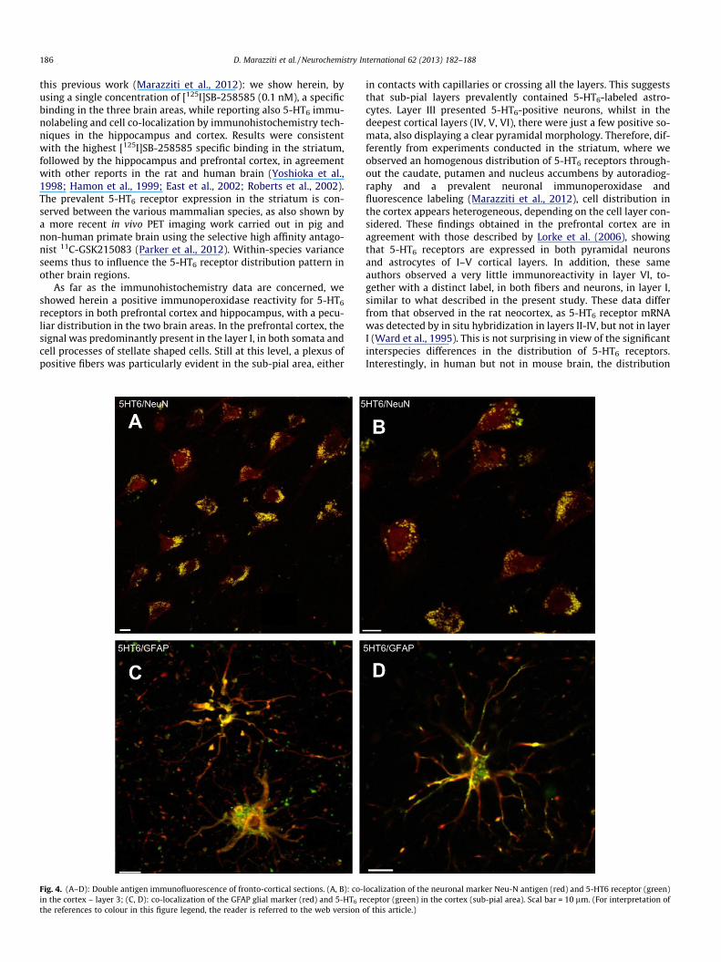
In order to better clarify the nature of the 5-HT6 labeled cells inthe cortex, double-labeling experiments were performed in thefronto-cortical sections, using two cell-specific antigens: the glialfibrillary acidic protein (GFAP) and the neuronal nuclear antigen(NeuN). The use of secondary antibodies conjugated with two dif-ferent (red and green) fluorophores allowed to better co-localize5-HT6 receptor with glial or pyramidal neurons in the hippocampusand cortex. In particular, the immunofluorescence data confirmedwhat supposed from the morphological analysis by the immunop-eroxidase technique: depending on the cell layer, glial orpyramidal cells were 5-HT6 positive (Fig. 4). The dual-antigenimmunofluorescence technique also confirmed the nature of
Fig. 3. (A–C): 5-HT6 immunoreactivity localization (immunoperoxidase reaction) in thenon-neuronal stain; (B) parallel section treated with the Nissl reagent show positive pcorresponding Nissl-treated section. Scale bar = 50 lm.
5-HT6 receptor labeled cells in the hippocampus: in this area,5-HT6 labeling resulted co-localized with the NeuN stain onlyand, therefore, with pyramidal neurons (Fig. 5).
4. Discussion
Serotonin 5-HT6 receptors have recently attracted much atten-tion for the potential clinical usefulness of selective agonists/antag-onists acting at their level in different neuropsychiatric conditions,such as psychoses, dementia, depression and obesity (East et al.,2002; Johnson et al., 2008; Heal et al., 2008; Wesołowska, 2010).However, relatively little information is available on the distribu-tion of 5-HT6 receptors in the human brain, and most literature iscentered on animal studies (Gérard et al., 1997; Yoshioka et al.,1998; Healy and Meador-Woodruff, 1999; Pouzet et al., 2002;Roberts et al., 2002; Dawson and Li, 2003). Data on the cellular dis-tribution of 5-HT6 receptors are even more limited (Lorke et al.,2006). In previous experiments carried out by our research groupthrough the selective antagonist [125I]SB-258585 (East et al.,2002; Hirst et al., 2003), we characterized the 5-HT6 receptor bind-ing in the human striatum (Marazziti et al., 2012). In that work, wecould apprise the binding properties of this radioligand in thestriatum only, because of the scarce specific bound measured inthe other two brain regions investigated, the prefrontal cortexand hippocampus. The present study represents an extension of
hippocampus. (A) Immunostained cells positive for 5-HT6 (arrows); empty arrow,yramidal cells. (A, B) The red square indicate positive pyramidal neurons and the
186 D. Marazziti et al. / Neurochemistry International 62 (2013) 182–188
this previous work (Marazziti et al., 2012): we show herein, byusing a single concentration of [125I]SB-258585 (0.1 nM), a specificbinding in the three brain areas, while reporting also 5-HT6 immu-nolabeling and cell co-localization by immunohistochemistry tech-niques in the hippocampus and cortex. Results were consistentwith the highest [125I]SB-258585 specific binding in the striatum,followed by the hippocampus and prefrontal cortex, in agreementwith other reports in the rat and human brain (Yoshioka et al.,1998; Hamon et al., 1999; East et al., 2002; Roberts et al., 2002).The prevalent 5-HT6 receptor expression in the striatum is con-served between the various mammalian species, as also shown bya more recent in vivo PET imaging work carried out in pig andnon-human primate brain using the selective high affinity antago-nist 11C-GSK215083 (Parker et al., 2012). Within-species varianceseems thus to influence the 5-HT6 receptor distribution pattern inother brain regions.
As far as the immunohistochemistry data are concerned, weshowed herein a positive immunoperoxidase reactivity for 5-HT6
receptors in both prefrontal cortex and hippocampus, with a pecu-liar distribution in the two brain areas. In the prefrontal cortex, thesignal was predominantly present in the layer I, in both somata andcell processes of stellate shaped cells. Still at this level, a plexus ofpositive fibers was particularly evident in the sub-pial area, either
Fig. 4. (A–D): Double antigen immunofluorescence of fronto-cortical sections. (A, B): co-in the cortex – layer 3; (C, D): co-localization of the GFAP glial marker (red) and 5-HT6 rethe references to colour in this figure legend, the reader is referred to the web version o
in contacts with capillaries or crossing all the layers. This suggeststhat sub-pial layers prevalently contained 5-HT6-labeled astro-cytes. Layer III presented 5-HT6-positive neurons, whilst in thedeepest cortical layers (IV, V, VI), there were just a few positive so-mata, also displaying a clear pyramidal morphology. Therefore, dif-ferently from experiments conducted in the striatum, where weobserved an homogenous distribution of 5-HT6 receptors through-out the caudate, putamen and nucleus accumbens by autoradiog-raphy and a prevalent neuronal immunoperoxidase andfluorescence labeling (Marazziti et al., 2012), cell distribution inthe cortex appears heterogeneous, depending on the cell layer con-sidered. These findings obtained in the prefrontal cortex are inagreement with those described by Lorke et al. (2006), showingthat 5-HT6 receptors are expressed in both pyramidal neuronsand astrocytes of I–V cortical layers. In addition, these sameauthors observed a very little immunoreactivity in layer VI, to-gether with a distinct label, in both fibers and neurons, in layer I,similar to what described in the present study. These data differfrom that observed in the rat neocortex, as 5-HT6 receptor mRNAwas detected by in situ hybridization in layers II-IV, but not in layerI (Ward et al., 1995). This is not surprising in view of the significantinterspecies differences in the distribution of 5-HT6 receptors.Interestingly, in human but not in mouse brain, the distribution
localization of the neuronal marker Neu-N antigen (red) and 5-HT6 receptor (green)ceptor (green) in the cortex (sub-pial area). Scal bar = 10 lm. (For interpretation off this article.)

Fig. 5. (A–C): Double antigen immunofluorescence of hippocampal sections. (A–C): the same section showing double positive neuronal cells for Neu-N antigen (red) and 5-HT6 receptor (green) at increasing enlargement. Scale bar = 10 lm. (For interpretation of the references to colour in this figure legend, the reader is referred to the webversion of this article.)
D. Marazziti et al. / Neurochemistry International 62 (2013) 182–188 187
of 5-HT6 receptor mRNA would parallel that of the binding (Hirstet al., 2003).
In the hippocampal sections, immunoreactive peroxidase posi-tive cells, were clearly pyramidal, in the CA1, CA2, CA3 and CA4fields. To our knowledge, this is the first report showing 5-HT6
receptor immunoreactivity in the human hippocampus and, specif-ically, in pyramidal neurons.
The double-labeling immunofluorescence experiments carriedout to additionally clarify the nature of the immunoperoxidase-po-sitive cells, confirmed that 5-HT6 receptors were co-localized withGFAP, the glial marker, in the sub-pial region and layer I, as sug-gested by the interaction of their cell processes with blood vessels.The deepest layers were instead co-localized with the neuronalantigen, NeuN. On the contrary, in the hippocampus, 5-HT6 recep-tors were undoubtedly co-localized with the NeuN stain, the neu-ronal marker. The observed diverse distribution of 5-HT6 receptorswithin the various cortical cell populations is an important findingwhich, possibly, mirrors the receptor functional heterogeneity inthis area, implying its contribution to neuronal trophism or firingdepending on the cell layer. This aspect provides support to theconviction that 5-HT receptors can significantly modulate the cog-nitive function (Harrison, 2004; Roth et al., 2004).
Either immunoperoxidase or immunofluorescence labeling re-sults, while showing the presence of positive cells in the hippo-campus and frontal cortex, seem in disagreement with bindingdata which report very low specific binding values. This controver-sial finding can be explained by several hypotheses. First of all, thediscrepancy can be due to the post-mortem and/or inter-individualvariance, since binding and immunohistochemistry data were notcarried out in brain samples obtained from the same autopsy sub-ject, which is always a main limit of the use of postmortem tissues,as already observed by others. Another possible reason can be thatthe receptor is coupled to different signal transduction systemsand/or that the receptor protein displays diverse phosphorylationstates within the various brain areas: this can affect affinity for li-gands, although the use of an antagonist tracer, like in the presentwork, limits this explanation. Marcos et al. (2008) found, in differ-ent autopsy subjects and experimental conditions, as compared tothose of present work, a noticeable [125I]SB-258585 binding in hu-man postmortem temporal cortex, depending on 5-HT tissue con-tent. This could provide support to the existence of 5-HT6 receptorisoforms present in CNS. Further, cells in different areas couldpresent a different compartmentalization of 5-HT6 receptors: forinstance, in the striatum, the expression would be almost in theplasma membrane, while, in the other brain regions, the proteinwould be also located inside the cell, like a non-mature isoform,still expressing the epitope recognized by the here-employed
5-HT6 antibody. This last hypothesis seems quite speculative, butwe cannot exclude that the use of detergents in sample prepara-tion could have contributed to the issue.
5. Conclusions
In summary, taken together, these findings indicate that in pre-frontal cortex, 5-HT6 receptors are expressed by both pyramidaland glial cells, while in the hippocampus they are prevalently ex-pressed by pyramidal neurons. It is likely possible that 5-HT6 dif-ferential labeling within the CNS is dependent upon the relativecell distribution (neurons:astrocytes) in the different brain areas;on the other hand, it is also plausible to suppose that differentastrocyte-sub-populations can differentially express 5-HT6 recep-tors. Anyway, the sharp cellular region-dependent 5-HT6 receptorlocation provides a preliminary explanation of how they might reg-ulate different functions in different brain areas, for instance braintrophism in the cortex and neuronal firing in the hippocampus.This can also explain the current pharmacological interest towardseither selective 5-HT6 agonists or antagonists in treating differentpathological conditions: their relative efficacy would be dependentupon the expression and functional profile of this receptor subtypewithin the brain. It is worth mentioning that 5-HT6 receptor antag-onists have been found to positively modify cognitive function andmemory (King et al., 2007; Mitchell and Neumaier, 2008; Marcoset al., 2010; Codony et al., 2011), while agonists are able to displaya stress-reduced, hedonic antidepressant activity (Di Chiara et al.,2009; Scheggi et al., 2011).
This study, taking into account all the limitations due to thepostmortem model used, represents, therefore, the startingpoint to explore the 5-HT6 receptor functionality as well as itssub-cellular distribution, together the receptor mRNA expressionor protein maturation/regulation by protein kinase activities inthe human brain.
References
Arnt, J., Skarsfeldt, T., 1998. Do novel antipsychotics have similar pharmacologicalcharacteristics? A review of the evidence. Neuropsychopharmacology 18 (2),63–101.
Berger, M., Gray, J.A., Roth, B.L., 2009. The expanded biology of serotonin. Annu. Rev.Med. 60, 355–366.
Branchek, T.A., Blackburn, T.P., 2000. 5-HT6 receptors as emerging targets for drugdiscovery. Ann. Rev. Pharmacol. 40, 319–334.
Bymaster, F.P., Falcone, J.F., Bauzon, D., Kennedy, J.S., Schenk, K., DeLapp, N.W.,Cohen, M.L., 2001. Potent antagonism of 5-HT3 and 5-HT6 receptors byolanzapine. Eur. J. Pharmacol. 430, 341–349.
Castaneda-Corral, G., Rocha-Gonzales, H.I., Araiza-Saldana, C.I., Ambriz-Tututi, M.,Vidal-Cantù, G.C., Granados-Soto, V., 2009. Role of peripheral and spinal 5-HT6
receptors according to the rat formalin test. Neuroscience 162, 444–452.

188 D. Marazziti et al. / Neurochemistry International 62 (2013) 182–188
Codony, X., Vela, J.M., Ramírez, M.J., 2011. 5-HT6 receptor and cognition. Curr. Opin.Pharmacol. 11 (1), 94–100.
Dawson, A.L., Li, P., 2003. Effects of 5-HT6 receptor blockade in the neurochemicaloutcome of antidepressant treatment in the frontal cortex of the rat. J. Neural.Transm. 110, 577–590.
Di Chiara, G., Valentini, V., Fenu, S., De Luca, M.A., Borsini, F., 2009. Reinforcing,rewarding and dopamine-stimulant properties of the 5-HT6 receptor agonistST1936: the first member of a new class of psychostimulants? Behav.Pharmacol. 20, S9.
East, S.Z., Burnet, P.W., Leslie, R.A., Roberts, J.C., Harrison, P.J., 2002a. 5-HT6 receptorbinding sites in schizophrenia and following antipsychotic drug administration:autoradiographic studies with [125I]SB-258585. Synapse 45, 191–199.
East, S.Z., Burnet, P.W., Kerwin, R.W., Harrison, P.J., 2002b. An RT-PCR study of 5-HT6
and 5-HT7 receptor mRNAs in the hippocampal formation and prefrontal cortexin schizophrenia. Schizophr. Res. 57, 15–26.
Ferguson, S.M., Mitchell, E.S., Neumaier, J.F., 2007. Increased expression of 5-HT6
receptors in the nucleus accumbens blocks the rewarding but not psychomotoractivating properties of cocaine. Biol. Psychiatry 63, 207–213.
Frantz, K.J., Hansson, K.J., Stouffer, D.G., Parson, L.H., 2002. 5-HT6 receptorantagonism potentiates the behavioral and neurochemical effects ofamphetamine but not cocaine. Neuropharmacology 42, 170–180.
Frederick, J.A., Meador-Woodruff, J.H., 1999. Effect of clozapine and haloperidol on5-HT6 receptor mRNA levels in rat brain. Schizophr. Res. 38 (1), 7–12.
Freitas, R.L., dos Reis Ferreira, C.M., Castiblanco Urbina, A.M., Marino, A.U., Carvalho,A.D., Butera, G., de Oliveira, A.M., Coimbra, N.C., 2009. 5-HT1A/1B, 5-HT6 and 5-HT7 serotonergic receptor recruitment in tonic-clonic seizure-inducedantinociception: role of dorsal raphe nucleus. Exp. Neurol. 217, 16–24.
Geldenhuys, W.J., Van der Schyl, C.J., 2008. Serotonin 5-HT6 receptor antagonists forthe treatment of Alzheimer’s disease. Curr. Top. Med. Chem. 8, 1035–1048.
Geldenhuys, W.J., Van der Schyl, C.J., 2009. The serotonin 5-HT6 receptor: a viabledrug target for treating cognitive deficits in Alzheimer’s disease. Expert. Rev.Neurother. 9, 1073–1085.
Gérard, C., El Mestkawy, S., Lebrand, C., Adrien, J., Ruat, M., Traiffort, E., Hamon, M.,Martres, M.-P., 1996. Quantitative RT-PCR distribution of serotonin 5-HT6receptor mRNA in the central nervous system of control or 5,7-dihydroxytryptramine-trated rats. Synapse 23, 164–173.
Gérard, C., Martres, M.-P., Lefevre, K., Miquel, M.-C., Vergé, D., Lanfumey, L., Doucet,E., Hamon, M., El Mestkawy, S., 1997. Immuno-localization of serotonin in 5-HT6 receptor-like material in the rat central nervous system. Brain Res. 746,207–219.
Glatt, C.E., Snowman, A.M., Sibley, D.R., Snyder, S.H., 1995. Clozapine: selectivelabeling of sites resembling 5-HT6 serotonin receptors may reflect psychoactiveprofile. Mol. Med. 1, 398–406.
Hamon, M., Doucet, E., Lefevre, K., Miquel, M.-C., Lanfumey, L., Insausti, R., Frechilla,D., Del Rio, J., Vergé, D., 1999. Antibodies and antisense oligonucleotide forprobing the distribution and putative functions of central 5-HT6 receptors.Neuropsychopharmacology 21, 68S–76S.
Harrison, P.J., 2004. The hippocampus in schizophrenia: a review of the neuro-pathological evidence and its pathophysiological implications. Psychophar-macology 174 (1), 151–162.
Heal, D.J., Smith, S.L., Fisas, A., Codony, X., Buschmann, H., 2008. Selective 5-HT6
receptor ligands: progress in the development of a novel pharmacologicalapproach to the treatment of obesity and related metabolic disorders.Pharmacol. Ther. 117, 207–231.
Healy, D.J., Meador-Woodruff, J.H., 1999. Ionotropic glutamate receptor modulationof 5-HT6 and 5-HT7 mRNA expression in rat brain. Neuropsychopharmacology21, 341–351.
Hirst, W.D., Minton, J.A.L., Bromidge, S.M., Moss, S.F., Latter, A.J., Riley, G., Routledge,C., Middlemiss, D.N., Price, G.W., 2000. Characterization of [125I]-SB-258585binding to human recombinant and native 5-HT6 receptors in rat, pig, andhuman brain tissue. Brit. J. Pharmacol. 130, 1597–1605.
Hirst, W.D., Abrahamsen, B., Blaney, F.E., Calver, A.R., Aloj, L., Price, G.W., Medhurst,A.D., 2003. Differences in the central nervous system distribution andpharmacology of the mouse 5-hydroxytryptamine-6 receptor compared withrat and human receptors investigated by radioligand binding, site-directedmutagenesis, and molecular modelling. Mol. Pharmacol. 64, 1295–1308.
Hoyer, D., Hannon, J.P., Martin, G.R., 2002. Molecular, pharmacological andfunctional diversity of 5-HT receptors. Pharmacol. Biochem. Behav. 71, 533–554.
Johnson, C.N., Ahmed, M., Miller, N.D., 2008. 5-HT6 receptor antagonists: prospectsfor the treatment of cognitive disorders including dementia. Curr. Opin. DrugDiscovery Dev. 11 (5), 642–654.
King, M.V., Fone, K.C.F., Shacham, S., Gannon, K.S., 2007. The novel 5-HT6 antagonist,PRX-07034, enhances memory and reduces food intake in a neurodevelop-mental model of schizophrenia. J. Psychopharmacol. 21, A57.
Kohen, R., Metcalf, M.A., Khan, N., Druck, T., Huebner, K., Lachwicz, J.E., Meltzer, H.Y.,Sibley, D.R., Roth, B.L., Hamblin, M.W., 1996. Cloning, characterization, andchromosomal localization of a human 5-HT6 serotonin receptor. J. Neurochem.66, 47–56.
Li, Z., Huang, M., Prus, A.J., Dai, J., Meltzer, H.Y., 2007. 5-HT6 receptor antagonist SB-399885 potentiates haloperidol and risperidone-induced dopamine efflux in themedial prefrontal cortex or hippocampus. Brain Res. 1134, 70–78.
Lorke, D.E., Lu, G., Cho, E., Yew, D.T., 2006. Serotonin 5-HT2A and 5-HT6 receptors inthe prefrontal cortex of Alzheimer and normal aging patients. BMC Neurosci. 7,36.
Maher-Edwards, G., Zvartau-Hind, M., Hunter, A.J., Gold, M., Hopton, G., Jacobs, G.,Davy, M., Williams, P., 2010. HYPERLINK ‘‘http://www.ncbi.nlm.nih.gov/pubmed/20043816’’ double-blind, controlled phase II study of a 5-HT6receptor antagonist, SB-742457. Curr. Alzheimer Res. 7 (5), 374–385.
Marazziti, D., Baroni, S., Pirone, A., Giannaccini, G., Betti, L., Schmid, L., Vatteroni, E.,Palego, L., Borsini, F., Bordi, F., Piano, I., Gargini, C., Castagna, M., Catena-Dell’osso, M., Lucacchini, A., 2012. Distribution of serotonin receptor of type 6(5-HT6) in human brain post-mortem (2012). A pharmacology, autoradiographyand immunohistochemistry study. Neurochem. Res. 37 (5), 920–927.
Marazziti, D., Baroni, S., Dell’Osso, M.C., Bordi, F., Borsini, F., 2011. Serotoninreceptors of type 6 (5-HT6): what can we expect from them? Curr. Med. Chem.18 (18), 2783–2790.
Marcos, B., Garcia-Allorca, M., Gil-Bea, F.J., Chuang, T.T., Francis, P.T., Chen, C.P.,Tsang, S.W.T.Y., Lai, M.K.P., Ramirez, M.J., 2008. Involvement of an altered 5-HT6
receptor function in behavioral symptoms of Alzheimer’s disease. J. AlzheimersDis. 14, 43–50.
Marcos, B., Cabero, M., Solas, M., Aisa, B., Ramirez, M.J., 2010. Signalling pathwaysassociated with 5-HT6 receptors: relevance for cognitive effects. Int. J.Neuropsychopharmacol. 13 (6), 775–784.
Meltzer, H.Y., Li, Z., Kaneda, Y., Ichikawa, J., 2003. Serotonin receptors: their key rolein drugs to treat schizophrenia. Prog. Neuropsychopharmacol. Biol. Psychiatry27 (7), 1159–1172.
Minabe, Y., Shirayama, Y., Hashimoto, K., Routledge, C., Hagan, J.J., Ashny Jr., C.R.,2004. Effect of acute and chronic administration of the selective 5-HT6 receptorantagonist SB-271046 on the activity of midbrain dopamine neurons in rats: anin vivo electrophysiological study. Synapse 52, 20–28.
Mitchell, E.S., Neumaier, J.F., 2008. 5-HT6 receptor antagonist of emotional learningand prepulse inhibition deficits induced by apomorphine or scopolamine.Pharmacol. Biochem. Behav. 88, 291–298.
Monsma Jr, F.J., Shen, Y., Ward, R.P., Hamblin, M.W., Sibley, D.R., 1993. Cloning andexpression of a novel serotonin receptor with affinity for tricyclic psychotropicdrugs. Mol. Pharmacol. 43, 320–327.
Nordquist, N., Oreland, L., 2010. Serotonin, genetic variability, behaviour, andpsychiatric disorders – a review. Ups. J. Med. Sci. 115 (1), 2–10.
Parker, C.A., Gunn, R.N., Rabiner, E.A., Slifstein, M., Comley, R., Salinas, C., Johnson,C.N., Jakobsen, S., Houle, S., Laruelle, M., Cunningham, V.J., Martarello, L., 2012.Radiosynthesis and characterization of 11C-GSK215083 as a PET radioligand forthe 5-HT6 receptor. J. Nucl. Med. 53, 295–303.
Pouzet, B., Didriksen, M., Arnt, J., 2002. Effects of the 5-HT6 receptor antagonist, SB-271046, in animal models for schizophrenia. Pharmacol. Biochem. Behav. 71,635–643.
Riemer, C., Borroni, E., Levet-Trafit, B., Martin, J.R., Poli, S., Porter, R.H.P., Bos, M.,2003. Influence of the 5-HT6 receptor on acetylcholine release in the cortex:pharmacological characterization of 4-(2-bromo-6-pyrrolidin-1-ylpyridine-4-sulfonyl)phenylamine, a potent and selective 5-HT6 receptor antagonist. J. Med.Chem. 46, 1273–1276.
Roberts, J.C., Reavill, C., East, S.Z., Harrison, P.J., Patel, S., Routledge, C., Leslie, R.A.,2002. The distribution of 5-HT6 receptors in rat brain: an autoradiographicbinding study using the radiolabeled 5-HT6 receptor antagonist [125I]SB-258585. Brain Res. 934, 49–57.
Rossé, G., Schaffhauser, H., 2010. 5-HT6 receptor antagonists as potentialtherapeutics for cognitive impairment. Curr. Topics Med. Chem. 10, 207–221.
Roth, B.L., Craigo, S.C., Choudhary, M.S., Uluier, A., Monsma Jr, F.J., Shen, Y., Meltzer,H.Y., Sibley, D.R., 1994. Binding to typical and atypical antipsychotic agents to5-hydroxytryptamine-6 and 5-hydroxytryptamine-7 receptors. J. Pharmacol.Exp. Ther. 268, 1403–1410.
Roth, B.L., Hanizavareh, S.M., Blum, A.E., 2004. Serotonin receptors represent highlyfavorable molecular targets for cognitive enhancement in schizophrenia andother disorders. Psychopharmacology 174, 17–24.
Routledge, C., Bromidge, S.M., Moss, S.F., Price, G.W., Hirst, W., Newmann, H., Riley,G., Gager, T., Stean, T., Upton, U., Clarke, S.E., Brown, A.M., Middlemiss, D.N.,2000. Characterization of SB-271046: a potent, selective and orally active 5-HT6receptor antagonist. Br. J. Pharmacol. 130, 1606–1612.
Scheggi, S., Marchese, G., Borsini, F., Bordi, F., De Montis, M.G., 2011. Effects of the 5-HT(6) receptor agonist ST 1936 on depression- and anhedonia-likeexperimental models. Behav. Brain Res. 10 (224), 35–43.
Stahl, S.M., 2008. Essential neuropsychopharmacology – neuroscientific basis andpractical applications. Cambridge University Press, Cambridge.
Upton, N., Chuang, T.T., Hunter, A.J., Virley, D.J., 2008. 5-HT6 receptor antagonists asnovel cognitive enhancing agents for Alzheimer’s disease. Neurotherapeutics 5,458–469.
Ward, R.P., Hamblin, M.W., Lachowicz, J.E., Hoffman, B.J., Sibley, D.R., Dorsa, D.M.,1995. Localization of serotonin subtype 6 receptor messenger RNA in the ratbrain by in situ hybridization histochemistry. Neuroscience 64, 1105–1111.
Wesołowska, A., 2010. Potential role of the 5-HT6 receptor in depression andanxiety: an overview of preclinical data. Pharmacol. Rep. 62 (4), 564–577.
Witty, D., Ahmed, M., Chuang, T.-T., 2009. Advances in the design of 5-HT6 receptorligands with therapeutical potential. Progress Med. Chem. 48, 163–225.
Woolley, M.L., Marsden, C.A., Fone, K.C.F., 2004. 5-HT6 receptors. Curr. DrugTargets-CNS & Neuro. Disorders 3 (1), 59–79.
Yoshioka, M., Matsumoto, M., Togashi, H., Mori, K., Saito, H., 1998. Centraldistribution and function of 5-HT6 receptor subtype in the rat brain. Life Sci.62, 1471–1473.
Copyright © 2022 FDOKUMEN