Semi-allogeneic dendritic cells can induce antigen-specific T-cell activation, which is not enhanced...
13
ORIGINAL ARTICLE Semi-allogeneic dendritic cells can induce antigen-specific T-cell activation, which is not enhanced by concurrent alloreactivity James W. Wells Chris J. Cowled David Darling Barbara-Ann Guinn Farzin Farzaneh Alistair Noble Joanna Galea-Lauri Received: 11 December 2006 / Accepted: 11 April 2007 / Published online: 9 May 2007 Ó Springer-Verlag 2007 Abstract Background Alloreactive T-cell responses are known to result in the production of large amounts of proinflamma- tory cytokines capable of activating and maturing dendritic cells (DC). However, it is unclear whether these allogeneic responses could also act as an adjuvant for concurrent antigen-specific responses. Objective To examine effects of simultaneous alloreac- tive and antigen-specific T-cell responses induced by semi- allogeneic DC. Methods Semi-allogeneic DC were generated from the F 1 progeny of inbred strains of mice (C57BL/6 and C3H, or C57BL/6 and DBA). We directly primed antigen- specific CD8 + and CD4 + T-cells from OT-I and OT-II mice, respectively, in the absence of allogeneic responses, in vitro, and in the presence or absence of alloreactivity in vivo. Results In vitro, semi-allogeneic DC cross-presented ovalbumin (OVA) to naı ¨ve CD8 + OT-I transgenic T-cells, primed naı ¨ve CD4 + OT-II transgenic T-cells and could stimulate strong alloreactive T-cell proliferation in a primary mixed lymphocyte reaction (MLR). In vivo, semi-alloge- neic DC migrated efficiently to regional lymph nodes but did not survive there as long as autologous DC. In addition, they were not able to induce cytotoxic T-lymphocyte (CTL) activity to a target peptide, and only weakly stimulated adoptively transferred OT-II cells. The CD4 + response was unchanged in allo-tolerized mice, indicating that alloreactive T-cell responses could not provide help for concurrently activated antigen-specific responses. In an EL4 tumour-treatment model, vaccination with semi-allogeneic DC/EL4 fusion hybrids, but not allogeneic DC/EL4 hybrids, significantly increased mouse survival. Conclusion Expression of self-Major histocompatibility complex (MHC) by semi-allogeneic DC can cause the induction of antigen-specific immunity, however, concur- rently activated allogeneic bystander responses do not provide helper or adjuvant effects. Keywords Semi-allogeneic Dendritic cell vaccination Adjuvant DC fusion Antigen specific T-cell Alloreactivity Abbreviations 51 Cr 51 Chromium CFSE Carboxy fluorescein succinimidyl ester CTL Cytotoxic T-lymphocyte DC Dendritic cell ES Embryonic stem cells MHC Major histocompatibility complex MLR Mixed lymphocyte reaction OVA Ovalbumin SEM Standard error of the mean TCR T-cell receptor Joanna Galea-Lauri and Alistair Noble share senior authorship of this work. J. W. Wells D. Darling B.-A. Guinn F. Farzaneh J. Galea-Lauri Department of Haematological and Molecular Medicine, King’s College London, The Rayne Institute, London SE5 9NU, UK J. W. Wells C. J. Cowled A. Noble (&) MRC & Asthma UK Centre in Allergic Mechanisms of Asthma, King’s College London, 5th Floor Thomas Guy House, Guy’s Hospital, London SE1 9RT, UK e-mail: [email protected] 123 Cancer Immunol Immunother (2007) 56:1861–1873 DOI 10.1007/s00262-007-0328-x
-
Upload
independent -
Category
Documents
-
view
1 -
download
0
Transcript of Semi-allogeneic dendritic cells can induce antigen-specific T-cell activation, which is not enhanced...

ORIGINAL ARTICLE
Semi-allogeneic dendritic cells can induce antigen-specific T-cellactivation, which is not enhanced by concurrent alloreactivity
James W. Wells Æ Chris J. Cowled Æ David Darling ÆBarbara-Ann Guinn Æ Farzin Farzaneh ÆAlistair Noble Æ Joanna Galea-Lauri
Received: 11 December 2006 / Accepted: 11 April 2007 / Published online: 9 May 2007
� Springer-Verlag 2007
Abstract
Background Alloreactive T-cell responses are known to
result in the production of large amounts of proinflamma-
tory cytokines capable of activating and maturing dendritic
cells (DC). However, it is unclear whether these allogeneic
responses could also act as an adjuvant for concurrent
antigen-specific responses.
Objective To examine effects of simultaneous alloreac-
tive and antigen-specific T-cell responses induced by semi-
allogeneic DC.
Methods Semi-allogeneic DC were generated from the
F1 progeny of inbred strains of mice (C57BL/6 and C3H,
or C57BL/6 and DBA). We directly primed antigen-
specific CD8+ and CD4+ T-cells from OT-I and OT-II
mice, respectively, in the absence of allogeneic responses,
in vitro, and in the presence or absence of alloreactivity
in vivo.
Results In vitro, semi-allogeneic DC cross-presented
ovalbumin (OVA) to naı̈ve CD8+ OT-I transgenic T-cells,
primed naı̈ve CD4+ OT-II transgenic T-cells and could
stimulate strong alloreactive T-cell proliferation in a primary
mixed lymphocyte reaction (MLR). In vivo, semi-alloge-
neic DC migrated efficiently to regional lymph nodes
but did not survive there as long as autologous DC.
In addition, they were not able to induce cytotoxic
T-lymphocyte (CTL) activity to a target peptide, and only
weakly stimulated adoptively transferred OT-II cells. The
CD4+ response was unchanged in allo-tolerized mice,
indicating that alloreactive T-cell responses could not
provide help for concurrently activated antigen-specific
responses. In an EL4 tumour-treatment model, vaccination
with semi-allogeneic DC/EL4 fusion hybrids, but not
allogeneic DC/EL4 hybrids, significantly increased mouse
survival.
Conclusion Expression of self-Major histocompatibility
complex (MHC) by semi-allogeneic DC can cause the
induction of antigen-specific immunity, however, concur-
rently activated allogeneic bystander responses do not
provide helper or adjuvant effects.
Keywords Semi-allogeneic � Dendritic cell vaccination �Adjuvant � DC fusion � Antigen specific T-cell �Alloreactivity
Abbreviations51Cr 51Chromium
CFSE Carboxy fluorescein succinimidyl ester
CTL Cytotoxic T-lymphocyte
DC Dendritic cell
ES Embryonic stem cells
MHC Major histocompatibility complex
MLR Mixed lymphocyte reaction
OVA Ovalbumin
SEM Standard error of the mean
TCR T-cell receptor
Joanna Galea-Lauri and Alistair Noble share senior authorship of this
work.
J. W. Wells � D. Darling � B.-A. Guinn � F. Farzaneh �J. Galea-Lauri
Department of Haematological and Molecular Medicine,
King’s College London, The Rayne Institute,
London SE5 9NU, UK
J. W. Wells � C. J. Cowled � A. Noble (&)
MRC & Asthma UK Centre in Allergic Mechanisms of Asthma,
King’s College London, 5th Floor Thomas Guy House,
Guy’s Hospital, London SE1 9RT, UK
e-mail: [email protected]
123
Cancer Immunol Immunother (2007) 56:1861–1873
DOI 10.1007/s00262-007-0328-x

Introduction
Advances in the generation of large numbers of dendritic
cells (DC) from blood monocytes, stem cells or bone
marrow, allowed DC to be generated and directly loaded
with antigen ex vivo, and induced powerful antigen-spe-
cific immune responses upon reintroduction into the patient
[1–3, 42]. Increasingly, however, the importance of DC
maturation in vaccination strategies has been realized [12,
14, 25, 31], with the use of immature DC in vaccination
strategies highlighted as a cause of antigen-specific inhi-
bition of immune responses in both humans and mice [13,
23]. In addition, it has been shown in mice that immature
DC, when matured in vivo in adjuvant-pre-treated sites,
were more effective at inducing anti-tumour immunity than
DC matured ex vivo [33].
Recent studies have found that the proinflammatory
cytokines IL-1b, TNF-a, IL-6 and IFN-c were released by
T-cells during the course of an allo-immune response, and
that the combination of these cytokines led to the activation
and maturation of bystander DC in humans [27, 39]. In
response to alloreactive T-cell-derived cytokines, it was
shown that the costimulatory molecules CD80, CD83,
CD86 and CD40, chemokine receptor CCR7, adhesion
molecule ICAM-1, DC-lysosomal associated membrane
protein (DC-LAMP) and HLA-DR were all up-regulated
[27, 39]. These studies suggest that the proinflammatory
cytokines induced by direct allorecognition could aid
concurrently activated, antigen-specific T-cell responses to
exogenous antigens or indirectly presented alloantigens. It
has been suggested that CD40L expression by activated
alloreactive CD4+ T-cells [7, 9] could condition DC via
CD40 and enable DC to facilitate antigen-specific CD8+ T-
cell priming. This would bypass the need for simultaneous
presentation of tumour peptides by both Major histocom-
patibility complex (MHC) classes I and II molecules and
increase the proportion of cancer patients in whom a
cytotoxic CD8+ T-cell response would be possible [15, 17].
This form of T-cell help could prove particularly potent as
it has been estimated that between 1 and 10% of a host’s T-
cells are potentially alloreactive [37]. All these studies
suggest that the presence of a concurrent allo-response
could induce a ‘‘cytokine storm’’ conducive for the mat-
uration of immature DC in vivo. Such a micro-environment
would be ideal for the maturation of DC as part of an
immunotherapy strategy, if the response by T-cells could
be shown to remain tumour antigen-specific.
In this study, we determined whether it would be pos-
sible to generate antigen-specific immune responses using
semi-allogeneic (partially MHC-matched) DC, thus pro-
viding a source of allo-derived cytokines and co-stimula-
tion to help drive a concurrent antigen-specific immune
response. This would replicate the partial mismatch situa-
tion seen in allogeneic stem cell transplantation and pro-
vide insight into whether allo-enhanced DC-induced
immune responses can aid the in vivo maturation needed
for effective DC-driven anti-tumour immunotherapy.
Materials and methods
Mice and cell lines
C57BL/6 (H-2b), C3H/HeN (H-2k) [C3H/HeN · C57BL/6]
F1 (B6C3F1, H-2k/b), DBA/2 (H-2d) and [DBA/2 ·C57BL/6] F1 (B6D2F1, H-2d/b) 6-8-week-old female mice
were purchased from Harlan, Oxford, UK. OT-I [22] and
OT-II [6] T-cell receptor (TCR) transgenic mice (both
H-2b) were bred in our facility. CD8+ OT-I T-cells
recognize Chicken Ovalbumin (OVA)257-264 peptide
(SIINFEKL) in association with MHC class I (H-2Kb),
whereas CD4+ OT-II T-cells recognize OVA323–339 peptide
(ISQAVHAAHAEINEAGR) in association with MHC
class II (I-Ab). All animal studies were carried out in
accordance with UK Home Office regulations and were
approved by our local ethical committee.
EL4, an H-2b thymoma, and the E.G7 derivative line,
which stably expresses chicken OVA on its cell surface
[32], were maintained in X-VIVO-15 (Biowhittaker,
Walkersville, MD, USA), 100 U/ml penicillin and 100 lg/
ml streptomycin (both from Sigma, Poole, UK). The B3Z
T-cell hybridoma [24], which is specific for the H-2Kb/
SIINFEKL complex, was maintained in RPMI (Sigma)
supplemented with 10% FCS (Sigma), 100 U/ml penicillin
and 100 lg/ml streptomycin. EL4, E.G7 and B3Z were a
kind gift from Dr J. Morrow, Department of Immunology,
St. Bartholomew’s and the Royal London School of
Medicine, London, UK.
Antibodies and other reagents
FITC-conjugated monoclonal antibodies AF6-88.5 (anti-
H-2Kb), 36-7-5 (anti-H-2Kk), SF1-1.1 (anti-H-2Kd) 28-18-
8S (anti-I-Ab) and PE-conjugated B20.1 (anti-Va2) were
purchased from BD Biosciences, Oxford, UK. Biotinylated
MR9-4 (Vb5.1, 5.2; BD) was detected using Streptavidin
Qdot-800 conjugate (Cambridge Bioscience, Cambridge,
UK). APC-conjugated RM4-5 (anti-CD4) and unlabelled
CT-17.1/CT-17.2 (mouse anti-mouse FcRcII/III-CD16/
CD32) were from Caltag-Medsystems, Silverstone, UK.
SIINFEKL peptide (OVA257–264) was purchased from
ProImmune Ltd., Oxford, UK. OVA323–339 peptide (IS-
QAVHAAHAEINEAGR) was synthesized by Mimotopes
Ltd., Wirral, UK. Purified Grade V OVA was from Sigma.
Irrelevant control peptide GAD65171–190 (IKTGH-
PRYFNQLSTGLDMVG) was donated by Dr. Tim Tree,
1862 Cancer Immunol Immunother (2007) 56:1861–1873
123

King’s College London, and control peptide Vesicular
Stomatitis Virus (VSV)52–59 (RGYVYQGL) was pur-
chased from the Department of Molecular Biology and
Biotechnology, University of Sheffield, UK. Anti-CD4
(L3T4), -CD8 (Ly-2) and -CD11c (HL3) Microbeads for
MACS were purchased from Miltenyi Biotec, Bisley, UK.
Bone marrow-derived dendritic cells and phenotypic
analysis
Bone marrow cells were cultured under serum-free con-
ditions in the presence of 5 ng/ml of GM-CSF (R&D
systems, Abingdon, UK) and 10 ng/ml of IL-4 (Pepro-
Tech EC Ltd., London, UK) as described previously [40].
Non-adherent or loosely adherent DCs were harvested
for use on day 7. Tolerogenic (immature) C3H DC
were generated following a modified version of the ‘‘OX
Method’’ [29]. C3H bone marrow cells were cultured at
2 · 106 cells/ml in X-VIVO-15 containing 1 ng/ml of
GM-CSF at 37�C/5% CO2. On day 2, the plate was
swirled gently and half the medium was removed and
replaced with the same volume of medium containing
1 ng/ml GM-CSF. On day 4, the contents of each well
were divided into two fresh wells, which were topped up
to 4 ml/well with medium containing 1 ng/ml GM-CSF.
Immature DC were harvested on day 6, washed twice in
PBS, and injected i.v. at 106 cells/mouse to induce tol-
erance. Mice were immunized with semi-allogeneic DC
i.d. 7 days later. DC were analysed by FACS (BD Bio-
science) following Fc-receptor-blocking using anti-CD16/
CD32 to prevent non-specific antibody staining. DC were
then incubated with H-2K antibodies for 15 min, washed
twice, and analysed.
Allogeneic mixed lymphocyte reaction
Dendritic cells were plated out in triplicate in 96-well U
bottomed plates (Greiner bio-one, Stonehouse, UK), serially
diluted 1:2 from 104 cells/well down to 6.25 · 102 cells/
well. Splenocytes were harvested from the spleens of naı̈ve
mice and depleted of red-blood cells using Red Blood Cell
Lysing Buffer (0.155 M ammonium chloride in 0.01 M
Tris–HCl buffer, Sigma), and B-cells using B220 Dyna-
beads (Dynal, Wirral, UK) according to the manufacturer’s
instructions. The splenocytes were then added to the plate
at 105 cells/well (final volume: 200 ll/well). After 5 days
at 37�C, splenocyte proliferation was assessed by [me-
thyl-3H] thymidine (1 lCi/well, Amersham Pharmacia
Biotech, Little Chalfont, UK) incorporation over 6 h using
a Liquid Scintillation Analyzer (TRI-CARB 2200CA,
Packard). Secondary mixed lymphocyte reactions (MLR’s)
were performed over 3 days using Inguinal LN cells from
immunized mice as responders.
B3Z colorimetric assay
Dendritic cells were pulsed for 4 h with various concen-
trations of SIINFEKL peptide or VSV52–59 irrelevant
peptide, washed twice and resuspended in phenol-red free
RPMI (Sigma) containing 100 U/ml penicillin and 100 lg/
ml streptomycin, 1% FCS and 2 mM L-Glutamine. DC
(104 cells/well) were then co-cultured in a 96-well U bot-
tomed plate with 5 · 104/B3Z cells/well, also suspended in
phenol-red free RPMI, overnight at 37�C (final well vol-
ume: 200 ll). 150 ll of supernatant was taken from each
well and replaced with 150 ll of PBS containing 5 mM
ONPG (Sigma) and 0.5% Nonidet-P40 (BDH, Bristol,
UK). The plate was incubated at 37�C for 2 h and optical
density measured using a Precision Microplate Reader
(Molecular Devices, Sunnyvale, CA, USA) at 450 nm with
wavelength correction set at 650 nm.
OVA presentation assays
Dendritic cells (104 cells/well) and various concentrations
of OVA323–339 peptide or whole OVA protein were co-
cultured with 3 · 104 naı̈ve OT-II CD4+ T-cells [from the
spleen, inguinal and mesenteric lymph nodes of OT-II
mice, purified using CD4+ Microbeads (Miltenyi Biotec)
by autoMACSTM selection, >95% purity]. GAD65171–190
peptide (10 lg/ml) and bovine serum albumin (BSA)
protein (1,000 lg/ml) were used as negative controls. After
90 (peptide) or 96 h (protein) lymphocyte proliferation was
assessed by 3H-thymidine incorporation over 6 (protein) or
18 h (peptide) of culture.
CD11c– CD8+ OT-I cells were purified using Microbe-
ads (Miltenyi Biotec) and autoMACS selection (>98%
purity) and cultured (105/well) with DC (104/well) and
OVA for 3 days at 37�C. BSA (1 mg/ml) was used as
negative control. Lymphocyte proliferation was assessed
by 3H-thymidine incorporation over the final 6 h of culture.
DC migration studies
Dendritic cell migration was quantified as described pre-
viously [26] with slight modifications. Briefly, DC were
washed twice in PBS, labelled with 2.5 lM carboxy fluo-
rescein succinimidyl ester (CFSE) (Invitrogen, Paisley,
UK; 10 min 37�C) and injected into naı̈ve mice (106 DC/
flank i.d.). 20, 44 and 68 h later the inguinal lymph nodes
were removed, pooled, counted and analysed by flow
cytometry.
DC competition experiments
OT-II CD4 cells or OT-I CD8 cells were labelled with
CFSE as above. 6 · 104 labelled cells were cultured with
Cancer Immunol Immunother (2007) 56:1861–1873 1863
123

6 · 103 C3H · C57 F1 DC in 1 ml, with or without a 100-
fold excess (6 · 106) of CD11c-depleted C3H · C57 F1
(autologous) or C57 (alloreactive) CD4 or CD8 T-cells.
OVA peptides (2 lg/ml) were added as above. After
3 days, cells were washed, stained for CD4/CD8 and the
entire sample was analysed by flow cytometry with gating
on CFSE+ events.
Activation of CD4+ OT-II T-cells in vivo
OVA-specific CD4+ T-cells from OT-II mice were wa-
shed twice in PBS, labelled with 2.5 lM CFSE, washed
and transferred into tail veins of mice (~5 · 106 each in
100 ll PBS). About 24 h later mice received DC previ-
ously pulsed with 150 lg/ml of OVA for 1 h (washed
extensively) at 106 DC/flank i.d. For allo-tolerization
experiments mice received 106 immature C3H DC i.v.
6 days before OT-II transfer. About 3 days following i.d.
immunization the inguinal lymph nodes were stained with
CD4, Va2 and Vb5 antibodies and analysed by flow
cytometry.
DC–EL4 fusion hybrid formation
Dendritic cell and EL4 (irradiated at 100 Gy) tumour
cells were mixed together at a ratio of 1:1, washed in
Mg2+/Ca2+-free PBS (ICN, Irvine, CA, USA) at 37�C, and
pelleted tightly together by centrifugation at 400g for
10 min. The supernatant was carefully removed and fu-
sion induced by gently stirring the pellet with the tip of a
pipette adding 1 ml of HYBRI-MAX� PEG/DMSO
solution (also at 37�C, Sigma) over the course of 1 min.
The cells were gently stirred for a further minute, and
then 10 ml of X-VIVO-15 medium (at 37�C) was added
drop-wise over the following 3 min. Cell hybrids were
then washed; hybrids were not isolated prior to immuni-
zation. To determine DC–EL4 fusion efficiency by flow
cytometry, DC were labelled with the red fluorescent
membrane dye PKH26 (Sigma) and EL4 cells with the
green fluorescent intracellular dye 5-chloromethylfluores-
cein diacetate (CMFDA, Molecular Probes, Eugene, OR,
USA). When DC fused to DC and EL4 cells fused to EL4
cells were mixed together they formed a small number of
cell aggregates that resulted in false ‘‘double positives’’.
When this had been taken into account, the efficiency of
successful DC to EL4 cell fusion was estimated to be
4.2%.
EL4 tumour experiments
For tumour prevention experiments, DC from C57, C3H,
DBA, C3H · C57 F1 or DBA · C57 F1 mice were fused
with irradiated EL4 cells, and injected s.c. into the flanks of
naive C57BL/6 mice (n = 8 per group, 106 starting DC/
mouse) on days –16 and –7. On day 0, mice were chal-
lenged with 5 · 104 viable EL4 cells injected s.c. The
control group was not immunized prior to tumour chal-
lenge. Tumour growth was monitored at regular intervals
and mice were culled when tumour diameters reached 16–
17 mm. For tumour treatment experiments, naı̈ve mice
were challenged s.c with 5 · 104 viable EL4 cells on day 0,
and then immunized on both flanks on days 3 and 7 with
DC/EL4 fusion hybrids.
Cytotoxicity assay
Dendritic cells were pulsed with 10 lg/ml of SIINFEKL
peptide and 5 lg/ml human b2-microglobulin (Sigma) for
3 h at 37�C, then washed and injected i.d. (106 cells/flank)
into the right and left flanks of naı̈ve C57BL/6 mice. About
7 days after immunization splenocytes from these mice
were co-cultured at a ratio of 10:1 with EG7 cells (irradi-
ated at 50 Gy) in X-VIVO-15 medium supplemented with
50 lM b-mercaptoethanol for 5 days. After 2 days 10 U/
ml of rmIL-2 (R&D) was added. On day 5 the ability of the
splenocytes (effectors) to kill 51Chromium (51Cr)-labelled
EG7 or EL4 cells (5 · 103cells/well, targets) was then
assessed over 4 h. Background 51Cr release was measured
from 51Cr labelled cells alone. Total 51Cr-released was
measured following treatment of 51Cr-labelled cells with
Triton X100. Target cell lysis was calculated using mean
counts per minute (cpm) of triplicate wells in the following
equation:
Cell Lysis
¼ Experimental 51Cr release�background 51Cr release
Total 51Cr release�background 51Cr release
� �
�100.
If background 51Cr release/total 51Cr release� �
� 100� �
was >25%, the experimental data were considered invalid.
Statistical analysis
Standard error of mean (SEM) and significance values were
calculated using GraphPad Prism Version 3.02 for Win-
dows, GraphPad Software, San Diego, CA, USA. Statisti-
cal comparisons of mean values were performed using
unpaired Student’s t-test. Statistical comparisons of sur-
vival curves were performed using the logrank test with the
null hypothesis that treatments did not change survival.
P < 0.05 (*) were considered significant. P < 0.005 (**)
and P < 0.001 (***) are indicated.
1864 Cancer Immunol Immunother (2007) 56:1861–1873
123

Results
Semi-allogeneic (F1) DC induce potent alloreactive
immune responses from both parental strains
Dendritic cells can induce particularly strong responses in
MHC-mismatched T-cell populations when combined in
primary MLRs in vitro. To determine whether semi-allo-
geneic DC could induce equally strong alloreactions we co-
cultured C3H/HeN · C57BL/6 F1 DC (C3H · C57 F1, H-
2k/b) with either C57BL/6 (C57, H-2b) or C3H/HeN (C3H,
H-2k) naı̈ve B-cell-depleted splenocytes (Fig. 1). Semi-
allogeneic DC were found to induce a similar alloreactive
response to that induced by fully allogeneic DC (C3H DC
and C57 DC, respectively), when co-cultured with either
C57 or C3H parental splenocytes. Therefore, the presence
of ‘‘self’’-MHC on the surface of semi-allogeneic DC does
not appear to affect their capacity to induce allo-responses.
As a negative control, semi-allogeneic DC (as well as C57
and C3H DC) were co-cultured with semi-allogeneic
splenocytes and were shown not to induce a response as
expected. These data demonstrate the potential of semi-
allogeneic DC to induce alloreactive immune responses.
Semi-allogeneic (F1) DC prime naı̈ve antigen-specific
T-cells in vitro
To determine whether semi-allogeneic C3H · C57 F1 DC
would be able to induce antigen-specific immune responses
on an H-2b background, we analysed the expression of H-
2Kb and I-Ab by flow cytometry (Fig. 2a, b). Semi-allo-
geneic DC were found to express both parental H-2K
haplotypes (H-2Kb and H-2Kk), although the level of H-
2Kb expression was just over half of that expressed by C57
DC as determined by the mean fluorescence intensity. The
MHC Class II positive cells in the C3H · C57 F1 DC
population expressed around 25% less I-Ab than C57
DC. The level of H-2Kk expressed by C3H · C57 F1 DC
however, was very similar to that expressed by C3H DC. A
similar H-2K expression pattern was observed between
C57 DC, DBA DC and semi-allogeneic DBA · C57 F1 DC
following staining for H-2Kb or H-2Kd (data not shown).
To analyse functional presentation of antigen,
C3H · C57 F1 DC were pulsed with various concentra-
tions of the H-2Kb-presented SIINFEKL peptide and
assayed for their ability to stimulate the B3Z (SIINFEKL/
H-2Kb-specific) T-cell hybridoma. Activation through the
TCR induces the expression of the reporter gene product
b-galactosidase, which is assayed after lysis. As shown in
Fig. 2c, semi-allogeneic C3H · C57 F1 DC but not allo-
geneic C3H DC were able to present SIINFEKL peptide to
the H-2b B3Z hybridoma in vitro. However, semi-alloge-
neic C3H · C57 F1 DC induced significantly weaker TCR-
mediated b-galactosidase production at each concentration
of SIINFEKL peptide tested as compared with autologous
C57 DC. As the B3Z hybridoma only requires the pre-
sentation of SIINFEKL in the context of H-2Kb to become
activated, and does not require co-stimulatory molecules,
this result was most likely due to reduced expression of
H-2Kb by C3H · C57 F1 DC as compared with C57 DC.
OT-I and OT-II transgenic T-cells were used to inves-
tigate whether semi-allogeneic DC were capable of prim-
ing naı̈ve antigen-specific T-cell responses in vitro. DC
were co-cultured with CD11c-depleted CD8+ OT-I T-cells
and the indicated concentration of whole OVA protein
(Fig. 2d). Semi-allogeneic C3H · C57 F1 DC and autolo-
gous C57 DC, but not allogeneic C3H DC, stimulated
naı̈ve CD8+ OT-I T-cells to proliferate. Interestingly, there
was no significant difference in the ability of semi-allo-
geneic C3H · C57 F1 DC to cross-present OVA peptides
to CD8+ OT-I T-cells as compared with autologous C57
DC. OT-I T-cells did not respond to the control protein,
BSA. When pulsed with OVA323–339 peptide (Fig. 2e) or
whole OVA protein (Fig. 2f), semi-allogeneic DC were
also capable of priming naı̈ve MHC class II-restricted
CD4+ OT-II T-cells in vitro. However, semi-allogeneic DC
Fig. 1 C3H · C57 F1 DC are just as potent as allogeneic DC in their
ability to stimulate alloreactive proliferation in a primary MLR. C57
(open squares), C3H (open triangles), or C3H · C57 F1 DC (solidcircles) were co-cultured with naı̈ve C57, C3H or C3H · C57 F1
B-cell-depleted splenocytes as indicated for 5 days at 37�C.
Proliferation was measured by the uptake of 3H-thymidine over 6 h.
Graphs represent combined data from two separate sets of experi-
ments carried out in triplicate. Error bars represent SEM
Cancer Immunol Immunother (2007) 56:1861–1873 1865
123
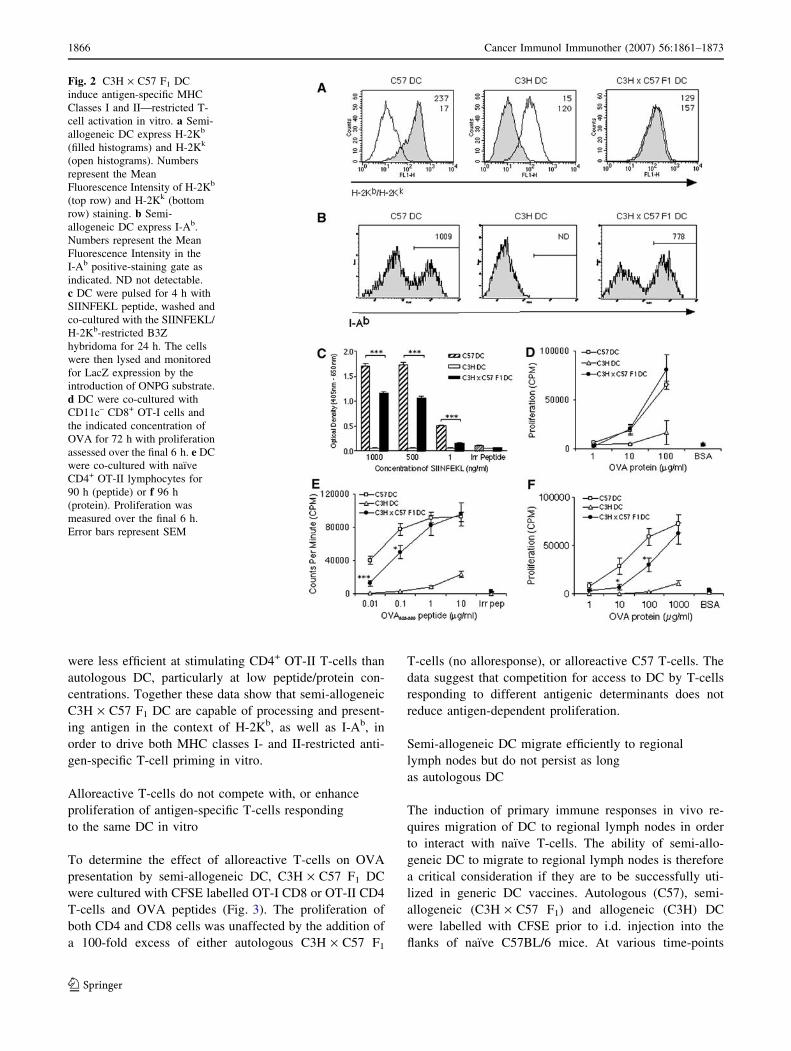
were less efficient at stimulating CD4+ OT-II T-cells than
autologous DC, particularly at low peptide/protein con-
centrations. Together these data show that semi-allogeneic
C3H · C57 F1 DC are capable of processing and present-
ing antigen in the context of H-2Kb, as well as I-Ab, in
order to drive both MHC classes I- and II-restricted anti-
gen-specific T-cell priming in vitro.
Alloreactive T-cells do not compete with, or enhance
proliferation of antigen-specific T-cells responding
to the same DC in vitro
To determine the effect of alloreactive T-cells on OVA
presentation by semi-allogeneic DC, C3H · C57 F1 DC
were cultured with CFSE labelled OT-I CD8 or OT-II CD4
T-cells and OVA peptides (Fig. 3). The proliferation of
both CD4 and CD8 cells was unaffected by the addition of
a 100-fold excess of either autologous C3H · C57 F1
T-cells (no alloresponse), or alloreactive C57 T-cells. The
data suggest that competition for access to DC by T-cells
responding to different antigenic determinants does not
reduce antigen-dependent proliferation.
Semi-allogeneic DC migrate efficiently to regional
lymph nodes but do not persist as long
as autologous DC
The induction of primary immune responses in vivo re-
quires migration of DC to regional lymph nodes in order
to interact with naı̈ve T-cells. The ability of semi-allo-
geneic DC to migrate to regional lymph nodes is therefore
a critical consideration if they are to be successfully uti-
lized in generic DC vaccines. Autologous (C57), semi-
allogeneic (C3H · C57 F1) and allogeneic (C3H) DC
were labelled with CFSE prior to i.d. injection into the
flanks of naı̈ve C57BL/6 mice. At various time-points
Fig. 2 C3H · C57 F1 DC
induce antigen-specific MHC
Classes I and II—restricted T-
cell activation in vitro. a Semi-
allogeneic DC express H-2Kb
(filled histograms) and H-2Kk
(open histograms). Numbers
represent the Mean
Fluorescence Intensity of H-2Kb
(top row) and H-2Kk (bottom
row) staining. b Semi-
allogeneic DC express I-Ab.
Numbers represent the Mean
Fluorescence Intensity in the
I-Ab positive-staining gate as
indicated. ND not detectable.
c DC were pulsed for 4 h with
SIINFEKL peptide, washed and
co-cultured with the SIINFEKL/
H-2Kb-restricted B3Z
hybridoma for 24 h. The cells
were then lysed and monitored
for LacZ expression by the
introduction of ONPG substrate.
d DC were co-cultured with
CD11c– CD8+ OT-I cells and
the indicated concentration of
OVA for 72 h with proliferation
assessed over the final 6 h. e DC
were co-cultured with naı̈ve
CD4+ OT-II lymphocytes for
90 h (peptide) or f 96 h
(protein). Proliferation was
measured over the final 6 h.
Error bars represent SEM
1866 Cancer Immunol Immunother (2007) 56:1861–1873
123

following injection the inguinal lymph nodes were re-
moved, pooled, and the number of CFSE+ DC in whole
pooled lymph node samples was quantified by flow
cytometry. As shown in Fig. 4a, autologous, semi-allo-
geneic and allogeneic DC displayed a similar capacity to
migrate to the inguinal lymph nodes 20 h after injection.
At 44 h post-injection the number of CFSE+ autologous
or semi-allogeneic DC continued to rise to a similar
degree, however, the number of CFSE+ allogeneic DC
appeared to decrease. At 68 h the numbers of CFSE+
semi-allogeneic or allogeneic DC had significantly
declined compared with autologous DC-injected mice.
The decrease in resident CFSE+ DC populations in mice
injected with semi-allogeneic DC at 68 h correlated with
a significant expansion in total lymph node cell counts
(Fig. 4b) suggesting that an alloreactive T-cell response
was mediating the destruction of semi-allogeneic DC. The
data indicate that semi-allogeneic DC, at least during
primary immunization, migrate efficiently to the regional
lymph nodes but are susceptible to killing mediated by
the ensuing alloreactive immune response.
Semi-allogeneic DC were not capable of inducing
cytotoxic T-lymphocyte (CTL)-activity
to a target peptide
In order to evaluate the potential of semi-allogeneic DC
to induce MHC class I-restricted antigen-specific cyto-
toxicity, autologous (C57), semi-allogeneic (C3H · C57
F1) and allogeneic (C3H) DC were pulsed with
SIINFEKL peptide and adoptively transferred i.d. into the
flanks of naı̈ve C57BL/6 mice. After 7 days spleens were
removed and cultured with irradiated EG7 cells for 5 days
prior to use in a standard 4-h chromium release assay
Fig. 3 Alloantigen-reactive
T-cells do not interfere with
T-cell responses to an
exogenous antigen in vitro.
OT-I CD8 T-cells (a) or OT-II
CD4 T-cells (b) were labelled
with CFSE and cultured with
C3H · C57 F1 DC + OVA
peptides, in the presence of a
100-fold excess of unlabelled
autologous (F1) or alloreactive
(C57) T-cells as indicated. After
3 days cells were stained for
CD4 or CD8 and analysed for
CFSE dilution. Data indicate %
divided cells in gated CFSE+
CD4/CD8+ populations. Similar
data were obtained in two
further experiments
Fig. 4 C3H · C57 F1 DC migrate to regional LN as efficiently as
C57 DC but do not persist as long. a Mice were injected i.d. on both
flanks with OVA-pulsed, CFSE-labelled DC. At the time-points
indicated inguinal LN were harvested, pooled (for each mouse),
digested with collagenase and analysed for the presence of CFSE+
cells by flow cytometry. b Pooled LN counts at each time-point
highlight the presence of an active allo-response. Data are represen-
tative of two separate experiments with similar results. Error bars
represent SD
Cancer Immunol Immunother (2007) 56:1861–1873 1867
123

(Fig. 5). Autologous C57 DC were found to induce
cytotoxic T-lymphocyte (CTL) activity directed against
EG7 target cells, but not against control EL4 target cells,
indicating that CTL activity was OVA-specific. No evi-
dence of CTL activity directed at either EG7 or EL4
target cells was seen in mice injected with peptide-pulsed
semi-allogeneic (C3H · C57 F1) DC or allogeneic (C3H)
DC. Similar results were seen when using pooled draining
lymph node (inguinal) and spleen cells as a source of
CTL (data not shown). Furthermore, multiple weekly
injections (up to four injections) of peptide-pulsed semi-
allogeneic DC did not result in induction of OVA-specific
CTL activity, suggesting that resident DC were not
stimulated to induce CTL activity to a foreign antigen
during the course of strong alloreactive immune responses
(data not shown). Pre-immunization of recipients with
OVA/alum also failed to result in detectable CTL, and
similar results were obtained using semi-allogeneic DC
derived from DBA · C57 F1 mice (H-2Kd/b, data not
shown). These results indicate that semi-allogeneic DC do
not directly, nor indirectly, induce antigen-specific CTL
activity in naı̈ve C57BL/6 mice. These results suggest that
semi-allogeneic DC do not become conditioned to stim-
ulate antigen-specific CD8+ T-cells to become CTL
through interactions with activated alloreactive CD4+
T-cells.
Semi-allogeneic DC induce weak activation
of adoptively transferred OT-II CD4 T-cells in vivo
Our data suggested that semi-allogeneic DC were not
capable of inducing effective cell-mediated immunity
in vivo, despite their effective APC functions in vitro. We
investigated whether this might be due to destruction of
semi-allogeneic DC or more competition from concurrent
alloreactive T-cell responses in vivo. CFSE+-labelled OT-
II CD4+ T-cells were adoptively transferred i.v. into groups
of C57BL/6 mice. After 24 h the mice were injected i.d.
with OVA-pulsed autologous (C57), semi-allogeneic
(C3H · C57 F1) or allogeneic (C3H) DC (day 0). One
group of mice that received OVA-pulsed semi-allogeneic
(C3H · C57 F1) DC had been pre-tolerized to C3H allo-
antigens through the i.v. injection of immature C3H DC
7 days prior to the start of the experiment (day 7 [29]).
Draining inguinal lymph nodes were harvested 72 h later
and pooled, total cell numbers were then noted and each
sample analysed for the division of CFSE+ cells by flow
cytometry. As shown in Fig. 6a, the total lymph node cell
count increased significantly in mice that received semi-
allogeneic (C3H · C57 F1) or allogeneic (C3H) DC as
compared to mice that received autologous (C57) DC,
indicative of alloreactivity. Mice that were pre-tolerized to
C3H allo-antigens however, did not show a significant in-
crease in total lymph node cell count in response to
immunization with semi-allogeneic (C3H · C57 F1) DC.
Furthermore, when lymph node cells from these mice were
cultured in a secondary MLR with semi-allogeneic
(C3H · C57 F1) DC, they failed to proliferate (Fig. 6b),
confirming that these mice were tolerized to C3H alloan-
tigens. In comparison to autologous (C57) DC, semi-allo-
geneic DC induced weaker OT-II proliferation in vivo,
however, this was not unexpected as they have a lower
expression of I-Ab. We did not observe a significant
difference in OT-II proliferation between naı̈ve and
allo-tolerized C57 mice injected with OVA-pulsed semi-
allogeneic (C3H · C57 F1) DC (Fig. 6c), indicating that
alloreactive T-cell responses do not provide help for anti-
gen-specific stimulation in vivo. Conversely, alloreactivity
does not appear to interfere with antigen-specific responses
through competition for proinflammatory cytokines or
interactions with DC.
Semi-allogeneic DC fusion hybrids increase survival
rates in EL4 tumour models
In previous studies we showed that the direct fusion of DC
with tumour cells provides an effective method for loading
DC with tumour antigens [19, 20]. We wished to determine
whether the presence of autologous MHC class I molecules
(H-2Kb) on tumour cells would enable semi-allogeneic DC
Fig. 5 C3H · C57 F1 DC were not capable of inducing CTL activity
to a target peptide (SIINFEKL). Naı̈ve C57 mice were immunized i.d.
with C57, C3H or C3H · C57 F1 DC pulsed with 10 lg/ml
SIINFEKL peptide. About 7 days later splenocytes from two mice/
group were harvested, pooled, restimulated for 5 days with irradiated
EG7 cells and then tested for their ability to kill 51Cr-labelled EG7
or EL4 target cells. Data are representative of five independent
experiments with similar results
1868 Cancer Immunol Immunother (2007) 56:1861–1873
123

fusion hybrids to stimulate tumour-specific CD8+ CTL
in vivo. In addition we hypothesized that expression of
autologous MHC class II molecules (I-Ab) by semi-allo-
geneic DC might provide CD4 T-cell help for tumour-
specific CD8+ T-cells, as DC/tumour cell fusion hybrids
have been shown to be able to directly activate tumour-
reactive CD4 T-cells [20]. To investigate this we hybrid-
ized autologous (C57), semi-allogeneic (C3H · C57 F1 or
DBA · C57 F1) or allogeneic (C3H or DBA) DC to irra-
diated EL4 cells (H-2Kb+ I-Ab–) and used these fusion
hybrids in the treatment of tumours induced by a lethal
dose of live EL4 cells. C57BL/6 mice were given two
identical immunizations of DC/EL4 fusion hybrids a week
apart, and were challenged with viable EL4 cells (day 0)
2 weeks after the second immunization. As shown in
Fig. 7a, untreated control mice were all culled by day 28.
Prophylactic vaccination, regardless of whether the DC
used were autologous, semi-allogeneic or allogeneic, re-
sulted in a significant increase in survival time when
compared with untreated controls, and in the long-term
tumour free survival of some mice within each vaccinated
group (other than in mice receiving DBA DC). All tumour-
free mice were re-challenged on day 91 with five times the
original tumour dose (2.5 · 105 cells/mouse), yet remained
tumour-free up to day 138 (data not shown), demonstrating
long-lived anti-tumour immunity had been induced in these
mice. However, that allogeneic DC/EL4 hybrids offer
some protection against a subsequent challenge with viable
EL4 cells might be taken to imply that host DC cross-
present EL4 antigens to the immune system in this sce-
nario. We therefore evaluated the influence of vaccination
in the treatment of mice pre-challenged with tumour cells.
Mice were given viable EL4 cells (day 0) and then two
immunizations of DC/EL4 fusion hybrids on days 3 and 7
(Fig. 7b). Untreated control mice were all culled by day 27.
Mice immunized with allogeneic DBA- or C3H DC/EL4
fusion hybrids did not display a significant difference
in survival time (P = 0.9827 and 0.2655, respectively),
despite the tumour-free survival of one mouse in each of
these groups. However, mice immunized with autologous
C57- or semi-allogeneic C3H · C57 F1- or DBA · C57 F1
DC/EL4 fusion hybrids survived significantly (P =
0.0054*, 0.0081* and 0.0034**, respectively), longer than
controls. These data suggest that semi-allogeneic DC
hybrids present tumour antigens to elicit tumour-specific
immunity. Thus it appears that semi-allogeneic DC that
express autologous MHC classes I- and II molecules may
offer an advantage over allogeneic DC in therapeutic DC/
tumour cell fusion hybrid therapy of established tumours.
Discussion
In recent years, the beneficial effects of the alloresponse in
cancer immunotherapy have been repeatedly demonstrated
in the clinic. Hemopoietic stem cell transplantation and
Fig. 6 Ability of C3H · C57 F1 DC to prime immune responses in
mice pre-tolerized to alloantigen. a Mice tolerized to C3H allo-
antigens do not have evidence of LN expansion in response to
injection with C3H · C57 F1 DC. Mice were tolerized to C3H-allo-
antigens through the injection of 1 · 106 immature (day 6) C3H DC
i.v. After 6 days the mice received 4–5 · 106 naı̈ve CFSE-labelled
CD4+ OT-II cells i.v. 24 h later mice were challenged with the
indicated DC pulsed with 150 lg/ml OVA i.d. on both flanks.
Inguinal LN were harvested 72 h later. Data represent average LN
cell counts ± SEM derived from five separate experiments. b LN cells
from mice tolerized to C3H allo-antigens do not respond to
C3H · C57 F1 DC in a secondary MLR. c OT-II cells adoptively
transferred into C3H allo-antigen-tolerized mice proliferate poorly
when stimulated by C3H · C57 F1 DC (as shown by CFSE dilution).
Numbers refer to the percentage of CFSE+ cells that divided ± SD for
three mice/group. Plots represent one experiment of five with similar
results
Cancer Immunol Immunother (2007) 56:1861–1873 1869
123

donor lymphocyte infusions have been shown to be par-
ticularly effective in the treatment of hematological
malignancies such as chronic and acute myeloid leukaemia
[4, 18], and also show promise in the therapy of solid
tumours [8, 10]. The vigorous induction of anti-tumour
T-cell responses, which results following presentation of
tumour antigens in the context of foreign MHC molecules,
as compared to self-MHC molecules, may in part be
explained by the absence of tolerance which normally
occurs during development in the thymus [41]. Alloreac-
tive responses in vivo are usually extremely potent and
the resultant massive release of cytokines has been well-
documented and termed a ‘‘cytokine storm’’ [16].
Our study addressed the role of alloreactivity in the
promotion of a concurrently activated antigen-specific
T-cell response. By using partially MHC-matched semi-
allogeneic DC derived from F1 strains, we were able to
examine the induction of antigen-specific CD8 and CD4
T-cell responses in vitro in the presence or absence of
allostimulation. We showed that in vitro, semi-allogeneic
C3H · C57 F1 (H-2k/b) DC induce alloreactivity in an
MLR, present peptide to B3Z hybridoma cells in the con-
text of H-2Kb, prime naı̈ve OT-II CD4 T-cells (H-2b) to
proliferate when pulsed with antigen, and are capable of
the cross-presentation of OVA peptides to MHC class I-
restricted OT-I T-cells. However, semi-allogeneic DC were
not as efficient as autologous DC in the presentation of
SIINFEKL peptide to the B3Z hybridoma or in the priming
of OT-II CD4 T-cells at low doses of protein or peptide,
which may result from the reduced expression of H-2Kb
and I-Ab by these DC in comparison to autologous DC.
In vivo, we showed that although semi-allogeneic DC
migrate efficiently to the regional lymph nodes following
adoptive transfer, they were unable to prime CTL to a
target peptide in naı̈ve mice. A recent study by Fukuma
et al. [17] showed that semi-allogeneic embryonic stem
cells (ES cells) engineered to express OVA and then sub-
sequently differentiated into DC were able to activate
OVA-specific CTL. The priming capacity of ES cell-de-
rived DC was suggested to be related to the expression of
SPI-6, a specific inhibitor of granzyme B not expressed by
bone-marrow-derived DC, which would make ES cell-de-
rived DC resistant to cytotoxicity by CTL [17]. However,
the use of whole OVA may have provided a source of class
II-restricted epitopes, resulting in antigen-specific CD4+
help for CD8+ CTL induction, as ES cell-derived DC
pulsed with SIINFEKL were weak stimulators of CTL
[17]. Our data suggest that CD4+ T-cells activated by
allogeneic MHC class II molecules co-expressed by bone-
marrow-derived, class I-peptide-pulsed semi-allogeneic
DC do not provide help for the priming of antigen-specific
CTL.
To ascertain the influence of allogeneic immune re-
sponses on the priming of antigen-specific CD4+ T-cells,
we studied the ability of OVA-pulsed semi-allogeneic DC
to prime adoptively transferred CD4+ OT-II cells in allo-
competent and allo-tolerized hosts. Mice were tolerized to
alloantigens with immature allogeneic DC, which are
known to induce alloantigen-specific tolerance resulting in
long-term survival of cardiac allografts in mice [5, 29].
Alloantigen-tolerized mice did not show lymph node
expansion in response to immunization with semi-alloge-
neic DC, and lymph-node-derived cells did not proliferate
in response to semi-allogeneic DC in a secondary MLR,
confirming that these mice were tolerized to (C3H) al-
loantigens. Compared to autologous DC, semi-allogeneic
Fig. 7 Semi-allogeneic DC/EL4 fusion hybrids increase survival
expectancy in EL4 tumour prevention (a) and b tumour treatment
experiments. a C57BL/6 mice were immunized twice with C57,
DBA, C3H, C3H · C57 F1, or DBA · C57 F1 DC fused to irradiated
EL4 cells (n = 8). Untreated mice were used as a control. Two weeks
later all the mice were challenged with a subcutaneous injection of
5 · 104 viable EL4 cells. The numbers in brackets in a represent the
number of tumour-free mice in each group at the end of the
experiment. b C57BL/6 mice were challenged with a subcutaneous
injection of 5 · 104 viable EL4 cells and treated on days 3 and 7 with
C57, DBA, C3H, C3H · C57 F1 or DBA · C57 F1 DC fused to
irradiated EL4 cells (n = 8). Mice were culled when tumour diameters
reached 16–17 mm
1870 Cancer Immunol Immunother (2007) 56:1861–1873
123

DC induced weak activation of OT-II cells in allo-com-
petent mice. Importantly, the ability of semi-allogeneic DC
to induce OT-II activation was not significantly altered in
allo-tolerized mice, suggesting that alloreactive T-cell re-
sponses do not provide help for antigen-specific CD4+ T-
cell responses in vivo, and additionally, that allogeneic
immune responses do not inhibit OT-II activation through
competition for proinflammatory cytokines, CD4+ growth
factors, or interactions with DC. The latter conclusion was
reinforced by our in vitro competition experiments. Al-
though the work of Willis et al. [43] demonstrates that T-
cells of differing specificities do compete for access to DC
in vivo, this study was performed in a syngeneic system
with two exogenous peptides. In our study, we observed a
loss of semi-allogeneic DC in draining lymph nodes, sug-
gesting that they are killed or receive cytokine signals that
prevent their retention in lymphoid tissue.
It is likely that semi-allogeneic DC survive for longer in
the lymph nodes of allo-tolerized mice than in the lymph
modes of naı̈ve mice [28]. However, allo-tolerization does
not appear to have a significant impact on OT-II-prolifer-
ation, so it seems unlikely that alloreactive cytotoxicity
directed against semi-allogeneic DC in the lymph node can
be held solely responsible for the poor stimulation of
antigen-specific T-cells. Although in this study we did not
look at the possibility that NK cells kill semi-allogeneic
DC, it has recently been shown that semi-allogeneic
DBA · C57 F1 DC adoptively transferred into Rag–/– (H-
2b) mice, which are deficient in T- and B-cells but not NK
cells, survive for >2 weeks, yet fully MHC mismatched
allogeneic DC are rapidly destroyed [44]. Therefore, the
presence of self-MHC molecules on semi-allogeneic DC
may well protect these cells from NK cell killing. It seems
more probable that the weaker direct stimulation of anti-
gen-specific CD4+ and CD8+ T-cell responses by semi-
allogeneic DC compared to autologous DC is due to low
‘‘self’’-MHC expression (H-2Kb/I-Ab) by these DC.
Clearly not just the presence, but also the level of self-MHC
expression is an important consideration when choosing
suitable semi-allogeneic DC for vaccination studies.
The fusion of DC to tumour cells results in the produc-
tion of hybrid cells capable of presenting endogenous tu-
mour antigens (from the tumour partner) in association with
the requisite adhesion and co-stimulatory molecules (from
the DC partner) to induce tumour-specific T-cell activation.
Fusion hybrids created using autologous or allogeneic DC
have been shown capable of inducing anti-tumour immune
responses [21, 30, 36], suggesting that MHC-restriction
may not be as important as the provision of co-stimulatory
signals. Our data suggest however, that it is advantageous
for the DC partner to express self-MHC molecules, as
recipients of semi-allogeneic DC/EL4 fusion hybrids
showed significantly increased survival time in an EL4
tumour treatment model, but mice receiving allogeneic DC/
EL4 hybrids did not. Since EL4 cells are MHC class II
negative, this observation suggests that the expression of
self-MHC class II molecules by semi-allogeneic DC, which
is absent on allogeneic DC, may be critical for the induction
of anti-tumour immune responses. In addition, the role of
MHC class I expression by semi-allogeneic DC should not
be overlooked. It is possible that semi-allogeneic DC, when
hybridized to tumour cells, may also provide the processing
machinery for MHC class I presentation of tumour antigens.
Our findings that DC/tumour hybrid-immunized mice show
increased survival following tumour cell challenge are
potentially at odds with our results suggesting a lack of CTL
and weak CD4+ T-cell induction in vivo. One possible
explanation could be that differences in processing and
presentation of endogenously expressed antigens by the
hybrids in vivo allowed a more efficient stimulation of the
host immune system, compared with exogenous addition of
antigen or peptide to DC ex vivo.
Semi-allogeneic DC do not appear to differ from auto-
logous DC in their ability to act as fusion partners for the
induction of anti-tumour immunity in a treatment model
and may also allow greater flexibility in these protocols.
Suitably MHC-matched DC lines or DC generated from
healthy volunteers in advance, for example, could negate
the requirement to generate DC from ill patients, and may
also allow rapid treatment at a considerably reduced cost.
While the induction of tolerance to alloantigens might
allow repeated use of the same semi-allogeneic DC popu-
lation, further work is required to define whether Th1-
inducing allogeneic responses [11, 34, 35, 39] would be
beneficial in polarizing concurrently activated cancer-spe-
cific immune responses towards a Th1 phenotype [38]. The
present findings demonstrate that the expression of self-
MHC by semi-allogeneic DC can lead to the induction of
antigen-specific T-cell responses, yet concurrently acti-
vated allogeneic responses do not provide help. The use-
fulness of partially MHC-matched semi-allogeneic DC in
vaccination strategies may therefore arise from the
expression of self-MHC molecules, but not the concurrent
activation of alloreactivity.
Acknowledgements This work was supported by Leukaemia Re-
search Fund and a JRC studentship from King’s College London
School of Medicine.
References
1. Avigan D, Vasir B, Gong J, Borges V, Wu Z, Uhl L, Atkins M,
Mier J, McDermott D, Smith T, Giallambardo N, Stone C, Schadt
K, Dolgoff J, Tetreault JC, Villarroel M, Kufe D (2004) Fusion
cell vaccination of patients with metastatic breast and renal
cancer induces immunological and clinical responses. Clin Can-
cer Res 10:4699–4708
Cancer Immunol Immunother (2007) 56:1861–1873 1871
123

2. Banchereau J, Palucka AK, Dhodapkar M, Burkeholder S, Taquet
N, Rolland A, Taquet S, Coquery S, Wittkowski KM, Bhardwaj
N, Pineiro L, Steinman R, Fay J (2001) Immune and clinical
responses in patients with metastatic melanoma to CD34+ pro-
genitor-derived dendritic cell vaccine. Cancer Res 61:6451–6458
3. Banchereau J, Schuler-Thurner B, Palucka AK, Schuler G (2001)
Dendritic cells as vectors for therapy. Cell 106:271–274
4. Barber LD, Madrigal JA (2006) Exploiting beneficial alloreactive
T cells. Vox Sang 91:20–27
5. Beriou G, Peche H, Guillonneau C, Merieau E, Cuturi MC (2005)
Donor-specific allograft tolerance by administration of recipient-
derived immature dendritic cells and suboptimal immunosup-
pression. Transplantation 79:969–972
6. Barnden MJ, Allison J, Heath WR, Carbone FR (1998) Defective
TCR expression in transgenic mice constructed using cDNA-
based alpha- and beta-chain genes under the control of heterol-
ogous regulatory elements. Immunol Cell Biol 76:34–40
7. Bingaman AW, Ha J, Durham MM, Waitze SY, Tucker-Burden
C, Cowan SR, Pearson TC, Larsen CP (2001) Analysis of the
CD40 and CD28 pathways on alloimmune responses by CD4+ T
cells in vivo. Transplantation 72:1286–1292
8. Bregni M, Ueno NT, Childs R (2006) The second international
meeting on allogeneic transplantation in solid tumors. Bone
Marrow Transplant 38:527–537
9. Buhlmann JE, Gonzalez M, Ginther B, Panoskaltsis-Mortari A,
Blazar BR, Greiner DL, Rossini AA, Flavell R, Noelle RJ (1999)
Cutting edge: sustained expansion of CD8+ T cells requires
CD154 expression by Th cells in acute graft versus host disease.
J Immunol 162:4373–4376
10. Childs R, Chernoff A, Contentin N, Bahceci E, Schrump D,
Leitman S, Read EJ, Tisdale J, Dunbar C, Linehan WM, Young
NS, Barrett AJ (2000) Regression of metastatic renal-cell carci-
noma after nonmyeloablative allogeneic peripheral-blood stem-
cell transplantation. N Engl J Med 343:750–758
11. Conrad R, Remberger M, Cederlund K, Hentschke P, Sundberg
B, Ringden O, Barkholt L (2006) Inflammatory cytokines pre-
dominate in cases of tumor regression after hematopoietic stem
cell transplantation for solid cancer. Biol Blood Marrow Trans-
plant 12:346–354
12. Dhodapkar MV, Steinman RM (2002) Antigen-bearing immature
dendritic cells induce peptide-specific CD8+ regulatory T cells
in vivo in humans. Blood 100:174–177
13. Dhodapkar MV, Steinman RM, Krasovsky J, Munz C, Bhardwaj
N (2001) Antigen-specific inhibition of effector T cell function in
humans after injection of immature dendritic cells. J Exp Med
193:233–238
14. Dubsky P, Ueno H, Piqueras B, Connolly J, Banchereau J,
Palucka AK (2005) Human dendritic cell subsets for vaccination.
J Clin Immunol 25:551–572
15. Fabre JW (2001) The allogeneic response and tumor immunity.
Nat Med 7:649–652
16. Ferrara JL (1993) Cytokine dysregulation as a mechanism of graft
versus host disease. Curr Opin Immunol 5:794–799
17. Fukuma D, Matsuyoshi H, Hirata S, Kurisaki A, Motomura Y,
Yoshitake Y, Shinohara M, Nishimura Y, Senju S (2005) Cancer
prevention with semi-allogeneic ES cell-derived dendritic cells.
Biochem Biophys Res Commun 335:5–13
18. Galea-Lauri J (2002) Immunological weapons against acute
myeloid leukaemia. Immunology 107:20–27
19. Galea-Lauri J, Darling D, Mufti G, Harrison P, Farzaneh F (2002)
Eliciting cytotoxic T lymphocytes against acute myeloid leuke-
mia-derived antigens: evaluation of dendritic cell-leukemia cell
hybrids and other antigen-loading strategies for dendritic cell-
based vaccination. Cancer Immunol Immunother 51:299–310
20. Galea-Lauri J, Wells JW, Darling D, Harrison P, Farzaneh F
(2004) Strategies for antigen choice and priming of dendritic
cells influence the polarization and efficacy of antitumor T-cell
responses in dendritic cell-based cancer vaccination. Cancer
Immunol Immunother 53:963–977
21. Gong J, Nikrui N, Chen D, Koido S, Wu Z, Tanaka Y, Cannistra
S, Avigan D, Kufe D (2000) Fusions of human ovarian carcinoma
cells with autologous or allogeneic dendritic cells induce anti-
tumor immunity. J Immunol 165:1705–1711
22. Hogquist KA, Jameson SC, Heath WR, Howard JL, Bevan MJ,
Carbone FR (1994) T cell receptor antagonist peptides induce
positive selection. Cell 76:17–27
23. Jiang HR, Muckersie E, Robertson M, Forrester JV (2003)
Antigen-specific inhibition of experimental autoimmune uveo-
retinitis by bone marrow-derived immature dendritic cells. Invest
Ophthalmol Vis Sci 44:1598–1607
24. Karttunen J, Sanderson S, Shastri N (1992) Detection of rare
antigen-presenting cells by the lacZ T-cell activation assay sug-
gests an expression cloning strategy for T-cell antigens. Proc Natl
Acad Sci USA 89:6020–6024
25. Kusmartsev S, Cheng F, Yu B, Nefedova Y, Sotomayor E, Lush
R, Gabrilovich D (2003) All-trans-retinoic acid eliminates
immature myeloid cells from tumor-bearing mice and improves
the effect of vaccination. Cancer Res 63:4441–4449
26. Lappin MB, Weiss JM, Delattre V, Mai B, Dittmar H, Maier C,
Manke K, Grabbe S, Martin S, Simon JC (1999) Analysis of
mouse dendritic cell migration in vivo upon subcutaneous and
intravenous injection. Immunology 98:181–188
27. Laurin D, Kanitakis J, Bienvenu J, Bardin C, Bernaud J, Le-
becque S, Gebuhrer L, Rigal D, Eljaafari A (2004) Allogeneic
reaction induces dendritic cell maturation through proinflamma-
tory cytokine secretion. Transplantation 77:267–275
28. Lee WC, Zhong C, Qian S, Wan Y, Gauldie J, Mi Z, Robbins PD,
Thomson AW, Lu L (1998) Phenotype, function, and in vivo
migration and survival of allogeneic dendritic cell progenitors
genetically engineered to express TGF-beta. Transplantation
66:1810–1817
29. Lutz MB, Suri RM, Niimi M, Ogilvie AL, Kukutsch NA, Rossner
S, Schuler G, Austyn JM (2000) Immature dendritic cells gen-
erated with low doses of GM-CSF in the absence of IL-4 are
maturation resistant and prolong allograft survival in vivo. Eur
J Immunol 30:1813–1822
30. Marten A, Renoth S, Heinicke T, Albers P, Pauli A, Mey U,
Caspari R, Flieger D, Hanfland P, Von Ruecker A, Eis-Hubinger
AM, Muller S, Schwaner I, Lohmann U, Heylmann G, Sauerb-
ruch T, Schmidt-Wolf IG (2003) Allogeneic dendritic cells fused
with tumor cells: preclinical results and outcome of a clinical
phase I/II trial in patients with metastatic renal cell carcinoma.
Hum Gene Ther 14:483–494
31. McIlroy D, Gregoire M (2003) Optimizing dendritic cell-based
anticancer immunotherapy: maturation state does have clinical
impact. Cancer Immunol Immunother 52:583–591
32. Moore MW, Carbone FR, Bevan MJ (1988) Introduction of
soluble protein into the class I pathway of antigen processing and
presentation. Cell 54:777–785
33. Nair S, McLaughlin C, Weizer A, Su Z, Boczkowski D, Dannull
J, Vieweg J, Gilboa E (2003) Injection of immature dendritic
cells into adjuvant-treated skin obviates the need for ex vivo
maturation. J Immunol 171:6275–6282
34. Noble A, Leggat JA, Inderberg EM (2003) CD8+ immunoregu-
latory cells in the graft-versus-host reaction: CD8 T cells activate
dendritic cells to secrete interleukin-12/interleukin-18 and induce
T helper 1 autoantibody. Immunology 109:476–486
35. Peng Q, Li K, Patel H, Sacks SH, Zhou W (2006) Dendritic cell
synthesis of C3 is required for full T cell activation and devel-
opment of a Th1 phenotype. J Immunol 176:3330–3341
36. Siders WM, Vergilis KL, Johnson C, Shields J, Kaplan JM (2003)
Induction of specific antitumor immunity in the mouse with the
1872 Cancer Immunol Immunother (2007) 56:1861–1873
123

electrofusion product of tumor cells and dendritic cells. Mol Ther
7:498–505
37. Suchin EJ, Langmuir PB, Palmer E, Sayegh MH, Wells AD,
Turka LA (2001) Quantifying the frequency of alloreactive T
cells in vivo: new answers to an old question. J Immunol
166:973–981
38. Suzuki T, Fukuhara T, Tanaka M, Nakamura A, Akiyama K,
Sakakibara T, Koinuma D, Kikuchi T, Tazawa R, Maemondo M,
Hagiwara K, Saijo Y, Nukiwa T (2005) Vaccination of dendritic
cells loaded with interleukin-12-secreting cancer cells augments
in vivo antitumor immunity: characteristics of syngeneic and
allogeneic antigen-presenting cell cancer hybrid cells. Clin
Cancer Res 11:58–66
39. Wallgren AC, Andersson B, Backer A, Karlsson-Parra A (2005)
Direct allorecognition promotes activation of bystander dendritic
cells and licenses them for Th1 priming: a functional link be-
tween direct and indirect allosensitization. Scand J Immunol
62:234–242
40. Wells JW, Darling D, Farzaneh F, Galea-Lauri J (2005) Influence
of interleukin-4 on the phenotype and function of bone marrow-
derived murine dendritic cells generated under serum-free con-
ditions. Scand J Immunol 61:251–259
41. Whitelegg AM, Oosten LE, Jordan S, Kester M, van Halteren
AG, Madrigal JA, Goulmy E, Barber LD (2005) Investigation of
peptide involvement in T cell allorecognition using recombinant
HLA class I multimers. J Immunol 175:1706–1714
42. Wierecky J, Mueller M, Brossart P (2006) Dendritic cell-based
cancer immunotherapy targeting MUC-1. Cancer Immunol Im-
munother 55:63–67
43. Willis RA, Kappler JW, Marrack PC (2006) CD8 T cell com-
petition for dendritic cells in vivo is an early event in activation.
Proc Natl Acad Sci USA 103:12063–12068
44. Yu G, Xu X, Vu MD, Kilpatrick ED, Li XC (2006) NK cells
promote transplant tolerance by killing donor antigen-presenting
cells. J Exp Med 203:1851–1858
Cancer Immunol Immunother (2007) 56:1861–1873 1873
123