A Case of Concurrent Molybdenosis, Secondary Copper ...
14
animals Communication A Case of Concurrent Molybdenosis, Secondary Copper, Cobalt and Selenium Deficiency in a Small Sheep Herd in Northern Germany Carina Helmer 1,† , Regina Hannemann 1,‡ , Esther Humann-Ziehank 1,§ , Sven Kleinschmidt 2 , Mareike Koelln 3, k , Josef Kamphues 3 and Martin Ganter 1, * Citation: Helmer, C.; Hannemann, R.; Humann-Ziehank, E.; Kleinschmidt, S.; Koelln, M.; Kamphues, J.; Ganter, M. A Case of Concurrent Molybdenosis, Secondary Copper, Cobalt and Selenium Deficiency in a Small Sheep Herd in Northern Germany. Animals 2021, 11, 1864. https://doi.org/10.3390/ ani11071864 Academic Editors: Ilektra A. Fragkou and Tomás Landete-Castillejos Received: 24 March 2021 Accepted: 18 June 2021 Published: 23 June 2021 Publisher’s Note: MDPI stays neutral with regard to jurisdictional claims in published maps and institutional affil- iations. Copyright: © 2021 by the authors. Licensee MDPI, Basel, Switzerland. This article is an open access article distributed under the terms and conditions of the Creative Commons Attribution (CC BY) license (https:// creativecommons.org/licenses/by/ 4.0/). 1 Clinic for Swine and Small Ruminants, University of Veterinary Medicine Hannover, Foundation, Bischofsholer Damm 15, 30173 Hannover, Germany; [email protected] (C.H.); [email protected] (R.H.); [email protected] (E.H.-Z.) 2 Lower Saxony State Office for Consumer Protection and Food Safety, Food- and Veterinary Institute Braunschweig/Hannover, Eintrachtweg 17, 30173 Hannover, Germany; [email protected] 3 Institute of Animal Nutrition, University of Veterinary Medicine Hannover, Foundation, Bischofsholer Damm 15, 30173 Hannover, Germany; [email protected] (M.K.); [email protected] (J.K.) * Correspondence: [email protected] † Current Address: AniCon Labor GmbH, Muehlenstrasse 13, 49685 Hoeltinghausen, Germany; [email protected]. ‡ Current Address: Veterinary Practice for Sheep, Goats, Llamas and Alpacas Dr. Hannemann, Domaene Ammerhof, 72070 Tuebingen, Germany; [email protected]. § Current Address: LABVETCON, Foehrenkamp 20, 31303 Burgdorf, Germany; [email protected]. k Current Address: Bosch Tiernahrung GmbH & Co. KG, Engelhardshauser Str. 55+57, 74572 Blaufelden-Wiesenbach, Germany; [email protected]. Simple Summary: Mineral deficiencies are very widespread among small ruminant herds through- out the world and play a big role in small ruminant herd health management. This paper describes the occurrence of a primary molybdenosis causing secondary copper deficiency, combined with ovine white liver disease (cobalt deficiency) and white muscle disease (selenium deficiency) in a group of pastured ram lambs in Northern Germany. To the authors’ knowledge, this is the first proven report of multiple trace element deficiencies in sheep in Germany for decades. Abstract: To the author’s knowledge this paper describes the first proven report of a combined primary molybdenosis, secondary copper (Cu) deficiency, Ovine White Liver Disease—Cobalt (Co) deficiency, and selenium (Se) deficiency in a small pedigree herd of White Horned Heath sheep in Germany (8 ewes, 2 rams, 3 yearling ewes, 17 lambs) for decades. Clinical signs associated with these mineral deficiencies in a group of pastured ram lambs included emaciation, conjunctivitis, anaemia, growth retardation, discolouration of the wool and photodermatitis. Morbidities and mortalities arose in 4–6-month-old lambs despite intensive veterinary treatment in the summer of 2014 and 2015 (n = 13, 23% died). Se (3/5), Cu (4/7), and Co (3/3) deficiencies in combination with elevated values for Molybdenum (Mo, 2/2) were found. Hamburg is a large industrial city and an input of heavy metals from surrounding industries and coal-fired power stations in combination with a sandy, non-fertilised soil and monoculture grass species might offer a potential explanation for the severity of mineral deficiencies observed in this herd. Keywords: cobalt; copper; selenium; deficiency; molybdenosis; sheep 1. Introduction Molybdenum (Mo) is an essential trace element and part of the so-called molybdenum cofactor complex, which is required for three mammalian enzymes: xanthine oxidase, alde- Animals 2021, 11, 1864. https://doi.org/10.3390/ani11071864 https://www.mdpi.com/journal/animals
-
Upload
khangminh22 -
Category
Documents
-
view
0 -
download
0
Transcript of A Case of Concurrent Molybdenosis, Secondary Copper ...
animals
Communication
A Case of Concurrent Molybdenosis, Secondary Copper,Cobalt and Selenium Deficiency in a Small Sheep Herd inNorthern Germany
Carina Helmer 1,†, Regina Hannemann 1,‡, Esther Humann-Ziehank 1,§, Sven Kleinschmidt 2 , Mareike Koelln 3,‖,Josef Kamphues 3 and Martin Ganter 1,*
�����������������
Citation: Helmer, C.; Hannemann,
R.; Humann-Ziehank, E.;
Kleinschmidt, S.; Koelln, M.;
Kamphues, J.; Ganter, M. A Case of
Concurrent Molybdenosis, Secondary
Copper, Cobalt and Selenium
Deficiency in a Small Sheep Herd in
Northern Germany. Animals 2021, 11,
1864. https://doi.org/10.3390/
ani11071864
Academic Editors: Ilektra A. Fragkou
and Tomás Landete-Castillejos
Received: 24 March 2021
Accepted: 18 June 2021
Published: 23 June 2021
Publisher’s Note: MDPI stays neutral
with regard to jurisdictional claims in
published maps and institutional affil-
iations.
Copyright: © 2021 by the authors.
Licensee MDPI, Basel, Switzerland.
This article is an open access article
distributed under the terms and
conditions of the Creative Commons
Attribution (CC BY) license (https://
creativecommons.org/licenses/by/
4.0/).
1 Clinic for Swine and Small Ruminants, University of Veterinary Medicine Hannover, Foundation,Bischofsholer Damm 15, 30173 Hannover, Germany; [email protected] (C.H.);[email protected] (R.H.); [email protected] (E.H.-Z.)
2 Lower Saxony State Office for Consumer Protection and Food Safety, Food- and Veterinary InstituteBraunschweig/Hannover, Eintrachtweg 17, 30173 Hannover, Germany;[email protected]
3 Institute of Animal Nutrition, University of Veterinary Medicine Hannover, Foundation,Bischofsholer Damm 15, 30173 Hannover, Germany; [email protected] (M.K.);[email protected] (J.K.)
* Correspondence: [email protected]† Current Address: AniCon Labor GmbH, Muehlenstrasse 13, 49685 Hoeltinghausen, Germany;
[email protected].‡ Current Address: Veterinary Practice for Sheep, Goats, Llamas and Alpacas Dr. Hannemann, Domaene
Ammerhof, 72070 Tuebingen, Germany; [email protected].§ Current Address: LABVETCON, Foehrenkamp 20, 31303 Burgdorf, Germany; [email protected].‖ Current Address: Bosch Tiernahrung GmbH & Co. KG, Engelhardshauser Str. 55+57,
74572 Blaufelden-Wiesenbach, Germany; [email protected].
Simple Summary: Mineral deficiencies are very widespread among small ruminant herds through-out the world and play a big role in small ruminant herd health management. This paper describesthe occurrence of a primary molybdenosis causing secondary copper deficiency, combined with ovinewhite liver disease (cobalt deficiency) and white muscle disease (selenium deficiency) in a group ofpastured ram lambs in Northern Germany. To the authors’ knowledge, this is the first proven reportof multiple trace element deficiencies in sheep in Germany for decades.
Abstract: To the author’s knowledge this paper describes the first proven report of a combinedprimary molybdenosis, secondary copper (Cu) deficiency, Ovine White Liver Disease—Cobalt (Co)deficiency, and selenium (Se) deficiency in a small pedigree herd of White Horned Heath sheep inGermany (8 ewes, 2 rams, 3 yearling ewes, 17 lambs) for decades. Clinical signs associated with thesemineral deficiencies in a group of pastured ram lambs included emaciation, conjunctivitis, anaemia,growth retardation, discolouration of the wool and photodermatitis. Morbidities and mortalitiesarose in 4–6-month-old lambs despite intensive veterinary treatment in the summer of 2014 and2015 (n = 13, 23% died). Se (3/5), Cu (4/7), and Co (3/3) deficiencies in combination with elevatedvalues for Molybdenum (Mo, 2/2) were found. Hamburg is a large industrial city and an input ofheavy metals from surrounding industries and coal-fired power stations in combination with a sandy,non-fertilised soil and monoculture grass species might offer a potential explanation for the severityof mineral deficiencies observed in this herd.
Keywords: cobalt; copper; selenium; deficiency; molybdenosis; sheep
1. Introduction
Molybdenum (Mo) is an essential trace element and part of the so-called molybdenumcofactor complex, which is required for three mammalian enzymes: xanthine oxidase, alde-
Animals 2021, 11, 1864. https://doi.org/10.3390/ani11071864 https://www.mdpi.com/journal/animals
Animals 2021, 11, 1864 2 of 14
hyde oxidase and sulphite oxidase [1]. Dietary Mo, iron (Fe) and Sulphur (S) concentrationsare important factors influencing the absorption and availability of copper (Cu) [2,3]. Allenand Gawthrone [4] identified that increased Mo concentrations in forage led to interactionsbetween molybdate and sulphide to form thiomolybdates in the rumen, which can bindCu to form nutritionally unavailable complexes. Therefore, high levels of Mo have beenassociated with secondary Cu deficiency. Former mining areas are potential risk areas forthe occurrence of high Mo concentrations in soil and grass [5]. Forage with moderate Moconcentrations of 5–10 mg/kg dry matter (DM) may affect bone development in youngcattle (focal widenings of the growth plate, uncalcified cartilage), while concentrationsgreater than 10 mg/kg DM may result in diarrhoea, emaciation and achromotrichia (loss ofhair pigment) [6]. However, the toxic level of Mo in forage consumed by ruminants is notwell defined due to several factors. Laiblin and Stöber [7] suggest that molybdenosis mightpossibly occur at Mo concentrations of 1–10 mg/kg fresh matter in pasture grass if the Cuconcentration is <5 mg/kg fresh matter, whilst, O’Connor et al. [8] suggested that plantMo concentrations of 10 mg/kg DM may be a very conservative estimate for the thresholdat which toxic effects may be observed. Miltimore and Mason [9] suggest that Cu:Moratios above 2 were suitable but lower ratios were associated with signs of Cu deficiency.The National Research Council [10] insists that Cu:Mo ratios of 10:1 are necessary forgood ruminant health. Mills and Davis [11] revealed that severe diarrhoea might be adirect effect of Mo toxicity. However, most clinical effects of high Mo intake appear to beassociated with the induced secondary Cu deficiency, called molybdenosis. This conditionoccurs under natural grazing conditions in many different parts of the world includingCanada [1], the USA [12], the UK [13] and Sweden [14]. As indicated, thiomolybdatesappear to induce Cu deficiency in several different ways [1]. Clinical manifestations ofCu deficiency include relatively unspecific signs such as ill thrift, emaciation and achro-motrichia (cattle) or wool changes (sheep). Others include cardiovascular disorders (cattle),anaemia, growth retardation, infertility [15] and bone fragility [16]. A specific conditionin sheep and goats related to Cu deficiency of pregnant ewes and young lambs resultsin enzootic ataxia of lambs. Most often ataxia is apparent immediately after birth, butit may also be delayed several weeks. Signs of ataxia include muscular incoordination,partial paralysis of the hindquarters, and degeneration of the myelin sheath of the nervefibres. Lambs may be born weak or die due to the inability to suckle [15–17]. In additionto Cu deficiency, sheep are very susceptible to Cu toxicosis as well. Cu concentrations inpastures vary, however, most cases were caused by unintended exposure of sheep to Cuoverload, induced by feeding concentrates or minerals produced for other farm animalsthan sheep, which provide higher Cu contents [18]. Unlike other species, sheep seem tohave a limited capacity to accumulate large amounts of Cu-metallothionein in the liver, andsaturation occurs very quickly. The factors precipitating clinical chronic copper toxicity areunclear, but mostly include stress, acute infections and poor nutrition [19]. Noteworthy, thedecrease of liver Cu concentration after exposure to Cu overload is very long with a half-lifeof liver Cu concentration >2 years as demonstrated in experimentally induced chronicCu poisoning [20]. According to EU legislation (Annex/2010/349/EU), the maximumconcentration of Cu in complete feedstuff with a moisture content of 12% is 15 mg/kgfor ovines.
Ovine white liver disease (OWLD) is a disorder of the lipometabolism of sheep, usu-ally growing lambs, and was first recorded in New Zealand in 1967 [21]. The trace elementcobalt (Co) is an essential component for the synthesis of vitamin B12 (Cobalamin) by rumenmicrobes [22]. Thus, Co deficiency secondary results in a vitamin B12 deficiency. Sheepappear to be susceptible to Co deficiency and develop normocytic and normochromicanaemia, anorexia, reduced weight gains and photosensitivity [23,24]. Moreover, lacrima-tion, scaly ears, discolouration of the wool, cardiovascular lesions and cerebrocorticalnecrosis [25–28] have been associated with low dietary levels of Co. For adult sheepgrazing Co deficient pastures the amount of Co necessary to ensure optimal growth is0.08 mg/animal/d. The requirement for young, rapidly growing lambs is greater during
Animals 2021, 11, 1864 3 of 14
the first few months and should be at least 0.2 mg/animal/day [29]. Clinicopathologi-cal abnormalities associated with Co deficiency are related to the inefficient metabolismof propionate [30]. Under normal conditions propionate is produced in the rumen, be-ing metabolized to succinate via methylmalonate by the vitamin B12-dependent enzymemethylmalonyl-CoA mutase. Succinate enters the citric acid circle to provide glucose as anenergy source [31]. A lack of Co and hence of vitamin B12 leads to insufficient β-oxidationof fatty acids which accumulate in the hepatocytes and result in hepatic lipidosis. Grosspathological findings of OWLD include a remarkably pale, beige-coloured and friableliver. Liver histopathology reveals fatty degenerations of hepatocytes and accumulationsof ceroid lipofuscin in Kupffer cells [32]. However, it must be admitted that even in casesof sufficient Co supplementation by the diet a ruminal function disorder might results in avitamin B12 deficiency due to the disfunction of the ruminal microbiota.
Deficiencies of vitamin E/Se are associated with nutritional myopathy with de-generative lesions of the skeletal muscles and heart muscle as well as hepatic necro-sis, also known as white muscle disease [33]. Clinical manifestations associated withVitamin E/Se deficiency in sheep include: ill-thrift in lambs, white muscle disease, low-ered fertility and embryonic death, prematurity and perinatal death and abortions andimmunosuppression [34,35].
This paper describes a combined occurrence of molybdenosis, secondary Cu deficiency,OWLD (Co deficiency) and selenium (Se) deficiency in a group of pastured ram lambs inNorthern Germany. To the authors‘ knowledge, this is the first report of multiple traceelement deficiencies in sheep in Germany for decades.
2. Materials and Methods2.1. Case Presentation
This field investigation did not require official or institutional ethical approval becauseall samples were taken during routine diagnostic procedures to improve the herd healthand were conducted in accordance with German animal welfare legislation and the EUDirective 2010/63/EU for animal experiments. All animals were handled according tohigh ethical standards and national legislation.
In October 2015 a German pedigree breeder with a small herd (8 ewes, 2 rams,3 yearling ewes, and 17 lambs) of White Horned Heath sheep contacted the Clinic forSwine and Small Ruminants of the University of Veterinary Medicine Hannover, Founda-tion for a veterinary investigation into issues with lamb morbidity and mortalities. Thefarm is located in the North-Western part of Germany close to the Free City of Hamburg.The herd is managed outside at pasture year-round with access to an open stable at lambingtime (March/April). Sheep are rotated regularly across two pastures during spring andsummer. Pasture 1, based on sandy soil, is 2 hectares (ha) of a monoculture grass species(bluegrass, Poa pratensis) and sheep have been reared on this pasture for 3 years. Pasture 2is a marshy soil type. It comprises 0.7 ha of diverse vegetation (pasture grass (Poa pratensis),timothy grass (Phleum pretense), meadow fescue (Festuca pratensis), velvet grass (Holcuslanatus), clover and legumes) and has been in use by sheep for 19 years. However, pasturegrass dominates pasture 2 as well. Pasture 3 (0.5 ha) is used for lambing in March andApril with the grazing of horses during the summer. No fertilizer is applied to pastures 1, 2and 3. During the winter months (November until March) the herd is reared on temporarygrassland which is fertilized (cattle manure and artificial fertilizer) and mowed regularly.Whilst sheep are at pasture, they are supplemented with a free-standing proprietary min-eral block containing zinc (6400 mg/kg), manganese (1300 mg/kg), Co (20 mg/kg), iodine(100 mg/kg) and Se (24 mg/kg). The mineral block does not contain any Cu, Mo, S or Fe.Average intake, estimated by the owner based on block consumption, is approximately2–4 g/animal/day, although significant intake deviations by some individuals, or in someperiods cannot be ruled out. The recommendation for daily intake of the manufacturer is20–30 g/animal/day. During the lambing period, the sheep are supplemented with adlibitum silage grass. The herd receives no concentrated feed and water access is freely

Animals 2021, 11, 1864 4 of 14
available on all pastures (quality tested well water). Prior to these veterinary investigations,there was minimal veterinary practice contact. There was no history of preventative diseasevaccination. The last deworming of the entire herd took place in spring 2015 with an oralmoxidectin compound (0.2 mg/kg body weight, Cydectin® 0.1%, Zoetis, Switzerland).No parasitological analysis or assessment of the FAMACHA© score was performed be-fore the anthelmintic treatment by the local veterinarian. The average number of raisedlambs/ewe/year was 1.3–1.5. Abortions and premature births had not been observed dur-ing the previous lambing periods. The shepherd reported that the lambs, especially the ramlambs aged 4–6 months, had shown poor growth performance over the previous two years(13 lambs were affected, 3 died). In 2014, the shepherd reported signs of emaciation andshaggy, pale wool. Post mortem investigation of one of these animals revealed cachexia,and the liver was pale and of friable consistency. Hepatic lipidosis was confirmed byhistopathology. Additionally, in 2015 the farmer reported that the lambs had shown signsof photodermatitis (crusty and ulcerated skin over the labia, nasal planum, ears and distalparts of the limbs) during the summer months. Three of the 13 (23%) affected lambs in 2014and 2015 died despite intensive treatment with oral moxidectin (0.2 mg/kg body weight,Cyedctin® 0.1%, Zoetis, Switzerland) and oral triclabendazole (10 mg/kg body weight,Fasinex® 5%, Novartis Tiergesundheit AG, Switzerland), supplementation of vitamin E/Se,and supplementation of B vitamins by the local veterinarian. Again no parasitologicalanalysis or assessment of the FAMACHA© score or Se or Co status was performed bythe local veterinarian prior to treatment. As the condition of the lambs did not improvedespite the implemented treatment, the local veterinarian submitted one affected ram lambto the clinic for further investigation on the 5th of October 2015. In autumn 2015, veterinaryinvestigations performed by the Clinic of Swine and Small Ruminants included clinicalexamination of the entire herd (30 animals) was conducted.
2.2. Clinical Examination
An intensive clinical examination of a six-month-old entire ram lamb (15.5 kg) submit-ted to the clinic was performed on 5 October 2015.
2.3. Farm Visit
On the 29th of October 2015, a farm visit was made by an experienced sheep veteri-narian (CH). The animals were reared in four different groups on four different pastures:group 1: Eight ram lambs kept on pasture 3 until slaughter; group 2: Three yearling ewesand one ram kept on temporary pasture grass until lambing; group 3: Eight ewes and oneram kept on temporary pasture grass until lambing; group 4: Nine female lambs kept ontemporary pasture grass until slaughter. The whole herd was clinically examined. Bloodsamples, faecal samples and liver samples were taken to the clinic for further analysis.
2.4. Further Diagnostics
An overview of samples gained for further diagnostics can be found in Table 1.
Table 1. Animals sampled for further investigations.
Id Sample Id Age Sex Health Status Tissue Examined Date
lamb 1–presented in the
clinic1 6 months m
emaciation,photodermatitis,
conjunctivitis, apathy
blood samples, faecalsample, post mortem
investigation, liversamples
5/10/15
lamb 2–farm visit 2 6 months m unremarkable blood samples,
pooled faecal sample 29/10/15
lamb 3–farm visit 3 6 months m
emaciation,photodermatitis in
healing
blood samples,pooled faecal sample 29/10/15
Animals 2021, 11, 1864 5 of 14
Table 1. Cont.
Id Sample Id Age Sex Health Status Tissue Examined Date
ewe 1–farm visit 4 7 years f emaciation, shaggy,
pale woolblood samples,
pooled faecal sample 29/10/15
ewe 2–farm visit 5 3 years f unremarkable blood samples,
pooled faecal sample 29/10/15
liver 1–farm visit, frozen 6 adult,
concrete age u f u liver samples slaughtered inautumn 2014
liver 2–farm visit, frozen 7 adult,
concrete age u f u liver samples slaughtered inautumn 2014
u = unknown, m = male, f = female.
2.4.1. Blood and Liver Samples
Blood samples were taken for further diagnostics (EDTA blood for haematology,Lithium-Heparin-Plasma and serum for biochemistry, serum for serology, Lithium-Heparin-Plasma and serum for investigation of mineral balance). In total 5 animals were sampled:1. The ram lamb submitted to the clinic, 2. two 6-month-old entire ram lambs (one affectedlamb showing clinical signs of emaciation and photodermatitis in healing and one lambin apparently healthy condition), 3. two ewes (one emaciated seven-year-old ewe withshaggy wool and a three-year-old ewe in good condition). EDTA anticoagulated bloodwas used to analyse, haemoglobin concentration and white and red blood cell count(Haematology analyser, Celltag alpha, Nihon Kohden, Kleinmachnow, Germany). Packedcell volume was analysed by centrifugation. Erythrocyte indices (MCH, MCV, MCHC)were calculated. Heparin plasma was analysed for total protein, albumin, bilirubin, plasmaenzyme activities of creatine kinase (CK), aspartate-amino-transferase (ASAT), glutamatedehydrogenase (GLDH), and gamma-glutamyl-transferase (GGT) were determined byroutine laboratory biochemistry according to Bickhardt and König [36]. Serum sampleswere analysed for Selenium (Se), copper (Cu), and cobalt (Co) concentrations. The resultsof the liver fluke Elisa were evaluated according to the manufacturer´s instructions (IDEXXFasciolosis Verification Test, IDEXX Laboratories, Ludwigsburg, Germany). Investigationsof Se, Cu, Co and Mo concentrations in liver samples gained from 2 ewes of unknownage and health status slaughtered in 2014 and the ram lamb submitted to the clinic andeuthanised due to grounds of animal welfare as well as plasma samples were performedby GFAAS and flame AAS (SOLAAR M, Thermo FisherScientific, Karlsruhe, Germany),respectively. Liver samples were digested using 4 mL HNO3 (65%) and 1ml H2O2 (30%) in amicrowave digestion unit (Start, MLS GmbH, Leutkirch, Germany) before analysis [37,38].
2.4.2. Faecal Samples
Pooled faecal samples were taken from each of the 4 different groups for endoparasiticinvestigation during the farm visit. Furthermore, a single faecal sample of the ram lambsubmitted to the clinic was gained. For examining the faecal samples, a modified versionof the combined sedimentation-flotation process according to Benedek [39] was used.
2.4.3. Post Mortem Investigation
After three days of hospitalization and repeated clinical examination, the animalsubmitted to the clinic remained inappetent and was euthanized on the grounds of animalwelfare. Post mortem investigation was performed at the Lower Saxony State Office for Con-sumer Protection and Food Safety, Food- and Veterinary Institute Braunschweig/Hannover.Gross necropsy examination was performed and tissue samples were collected from themajor parenchymatous organs (lungs, heart, brain, liver, spleen, and kidneys), musculatureand joints. Samples were fixed in 4% neutral buffered formalin and embedded in paraffinwax. Tissue sections (4 µm) were stained with haematoxylin and eosin (HE).
Animals 2021, 11, 1864 6 of 14
2.4.4. Nutritional Assessment
Feed samples of the permanent pasture grass from pastures 1 and 2, the pastureson which the sheep were reared during the summer months in which the clinical signswere observed, were collected (one rubbish bag (capacity 60 L) per pasture; samples wereobtained from all 4 corners and in a meandering pattern from the rest of the pasture) andsubmitted for feed analysis to the Institute of Animal Nutrition, University of VeterinaryMedicine Hannover, Foundation. Dry Matter, Calcium (Ca), Phosphorus (P) and S contentswere analysed according to the VDLUFA (book of methods) [40]. The reference rangesare set according to Kamphues et al. [41]. Sulphur was detected by using the Vario MaxCNS (Elementar®). The ground samples were mixed with wolfram (VI) oxide, weighed inceramic crucibles and incinerated in the Vario Max CNS by 1140 ◦C. The calcium contentand trace elements were determined by atomic absorption spectrometry, phosphoruscolourimetrically [42–45]. Molybdenum analysis was performed according to DIN ENISO 17294-2 (2005) in an external laboratory (Sys Analytics Germany GmbH, Weilheim,Germany). Cobalt analysis was performed as described by Lange et al. [46]. In addition,water samples from both wells of these pastures were collected (2 L/well collected in cleanplastic bottles) and also sent for analytical examination to the Institute of Animal Nutrition,University of Veterinary Medicine Hannover, Foundation.
3. Results3.1. Clinical Examination
Clinical examination of a six-month-old entire ram lamb (15.5 kg) submitted to theclinic revealed poor body condition with a body condition score (BCS) of 1.0 out of 5 [47],and a shaggy, discoloured coat. A bilateral mucopurulent nasal and ocular discharge, aswell as hyperaemia of the sub-conjunctival mucous membranes, was observed. Cardiopul-monary auscultation and palpation of superficial lymph nodes were unremarkable. Rectalbody temperature was 38.0 ◦C and the breech dirtiness (DAG score [48]) was 0 out of 5.The FAMACHA© score was 3–4 on a scale ranging from 1–5 (pale mucous membranes).One rumen contraction in two minutes was noticed. Assessment of the claws, joints andexternal reproductive organs was unremarkable. Severe multifocal pinhead-sized, en-crusted, hyperaemic skin lesions were observed around the labia, the nose, the eyes and theears. Externally, the nasal planum, periorbital skin and peri-auricular skin was bilaterallyswollen, warm and appeared painful to palpation. Crusting of the skin could not be re-moved without loss of substance. Pruritus was not present. Based on clinical examination,the key clinical signs of note were emaciation, conjunctivitis and photodermatitis.
3.2. Farm Visit
Clinical examination of the entire herd during the farm visit which took place onthe 29th of October 2015 revealed that ram lambs (group 1) were generally in poor bodycondition (average BCS was 1.5). Two ram lambs showed signs of conjunctivitis andphotodermatitis in healing. The rest of the herd appeared to be clinically in good healthexcept for one older ewe (7 years old), which was in a poor body condition (BCS 2) andalso showed shaggy, pale wool with an open fleece.
3.3. Further Diagnostics3.3.1. Blood and Liver Samples
The ram lamb admitted to the clinic (sample ID 1) showed a remarkable increaseof liver enzyme activities (aspartate aminotransferase (ASAT), glutamate dehydrogenase(GLDH), gamma-glutamyltransferase (GGT)) as well as an increase of total bilirubin.Microcytic, hypochromic anaemia, slight hyperproteinaemia and hypoalbuminaemia wereidentified. Analysis of liver tissue revealed that lamb 1 showed a severe copper and cobaltdeficiency. Se in serum was within the reference range. Two lambs were sampled duringthe farm visit (sample ID 2 and 3), both showed low copper concentrations in serum andslightly elevated GGT activity. Signs of anaemia were noted in the haematology profile of
Animals 2021, 11, 1864 7 of 14
lamb 2 (sample ID 2). Serum samples from ewes 1 and 2 (sample IDs 4 and 5) were onlyinvestigated for Se and Cu. Ewe 1 showed a moderate Se deficiency. Analysis of livers fromtwo slaughtered adult sheep (sample IDs 7 and 8) revealed both a Se and Co deficiency.A Cu deficiency was noted in sample ID 8 and high concentrations of Mo were found inboth liver samples. A summary of all results gained from blood and liver samples can befound in Table 2.
Table 2. Blood and liver results of tested sheep.
Sample ID 1 2 3 4 5 6 7
Selenium ***(s: 80–500 µg/L
l: 0.25–1.5 mg/kg FM)s: 83.5 s: 125.2 s: 112.3 s: 60.9 ↓ s: 125.3 l: 0.109 ↓ l: 0.099 ↓
Copper *** (s: 7–24µmol/L;
l: 10–120 mg/kg FM)
s: 10.9l: 1.45 ↓ s: 5.8 ↓ s: 5.7 ↓ s: 13.7 s: 12.3 l: 24.2 l: 9.1 ↓
Cobalt ***(l: 0.025–0.085 mg/kg FM) l: 0.008 ↓ - - - - l: 0.015 ↓ l: 0.020 ↓
Molybdenum ***(l: 1.5–6 mg/kg DM) - - - - - l: 7 ↑ l: 8.5 ↑
Haemoglobin * (90–140g/L) 84 ↓ 90 109 - - - -
Packed Cell volume *(0.27–0.41 L/L) 0.24 ↓ 0.3 0.36
MCH *(13–14 pg) 10.4 ↓ 10.5 ↓ 10.7 ↓ - - - -
MCV *(34–46 fL) 32 ↓ 35.1 35.3
MCHC *(290–340 g/L) 323 300 303
Bilirubin **(0–10 µmol/L) 13.13 ↑ - - - - - -
Protein **(52–70 g/L) 71.9 ↑ 62.5 66.2
Albumin **(27–39 g/L) 24.2 ↓ 36 31.8 - - - -
ASAT **(30–80 U/L) 589 ↑ 63 47 - - - -
CK **(10–230 U/L) 87 78 100 - - - -
GLDH **(2–12 U/L) 525 ↑ 7 9 - - - -
GGT **(5–32 U/L) 135 ↑ 34 ↑ 35 ↑ - - - -
Sample ID according to Table 1; s: serum; l: liver, MCH: Mean Corpuscular Haemoglobin, MCV: Mean Corpuscular Volume, MCHC:Mean Corpuscular Haemoglobin Concentration. The number in brackets marks the reference range according to Weiss and Wardrop [49] *,Bickardt et al. [50] **, and Puls (1995) [51] ***. Values, which are not reported (hyphen), were not tested. The deviating values are indicatedin bold and marked with ↓ for values that are too low and ↑ for elevated values.
3.3.2. Faecal Samples
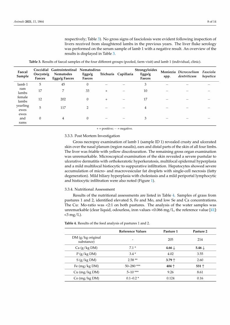
The single faecal sample of lamb 1, the pooled faecal sample of the group of ramlambs and the pooled faecal sample of the group of ewes and rams showed only low eggcounts. The pooled faecal samples of the female lambs and yearling ewes revealed moder-ate egg counts for gastrointestinal nematodes (202 eggs/g faeces and 117 eggs/g faeces,
Animals 2021, 11, 1864 8 of 14
respectively; Table 3). No gross signs of fasciolosis were evident following inspection oflivers received from slaughtered lambs in the previous years. The liver fluke serologywas performed on the serum sample of lamb 1 with a negative result. An overview of theresults is displayed in Table 3.
Table 3. Results of faecal samples of the four different groups (pooled, farm visit) and lamb 1 (individual, clinic).
FaecalSample
CoccidialOocysts/gFaeces
GastrointestinalNematodes
Eggs/g Faeces
NematodirusEggs/gFaeces
Trichuris CapillariaStrongyloides
Eggs/gFaeces
Monieziaspp.
Dicrocoeliumdentriticum
Fasciolahepatica
lamb 1 5 45 0 − − 3 − − −ram
lambs 17 7 33 + − 10 − − −
femalelambs 12 202 0 + − 17 − − −
yearlingewes 5 117 2 − − 4 − − −
ewesand
rams0 4 0 − − 3 − − −
+ = positive; − = negative.
3.3.3. Post Mortem Investigation
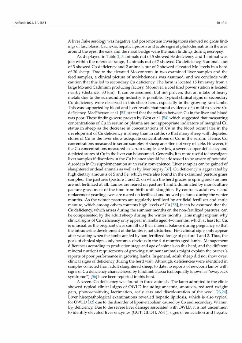
Gross necropsy examination of lamb 1 (sample ID 1) revealed crusty and ulceratedskin over the nasal planum (region nasalis), ears and distal parts of the skin of all four limbs.The liver was friable with yellow discolouration. The remaining gross organ examinationwas unremarkable. Microscopical examination of the skin revealed a severe pustular toulcerative dermatitis with orthokeratotic hyperkeratosis, multifocal epidermal hyperplasiaand a mild multifocal histiocytic to suppurative infiltration. Hepatocytes showed severeaccumulation of micro- and macrovesicular fat droplets with single-cell necrosis (fattydegeneration). Mild biliary hyperplasia with cholestasis and a mild periportal lymphocyticand histiocytic infiltration were also noted (Figure 1).
3.3.4. Nutritional Assessment
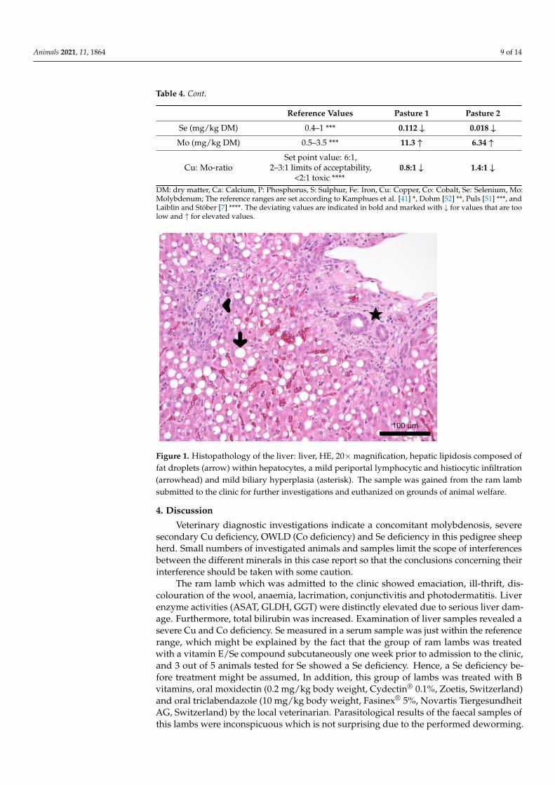
Results of the nutritional assessments are listed in Table 4. Samples of grass frompastures 1 and 2, identified elevated S, Fe and Mo, and low Se and Ca concentrations.The Cu: Mo-ratio was <2:1 on both pastures. The analysis of the water samples wasunremarkable (clear liquid, odourless, iron values <0.066 mg/L, the reference value [41]:<3 mg/L).
Table 4. Results of the feed analysis of pastures 1 and 2.
Reference Values Pasture 1 Pasture 2
DM (g/kg originalsubstance) - 205 214
Ca (g/kg DM) 7.1 * 4.66 ↓ 5.46 ↓P (g/kg DM) 3.4 * 4.02 3.55
S (g/kg DM) 2.58 ** 3.79 ↑ 2.60
Fe (mg/kg DM) 50–280 *** 404 ↑ 531 ↑Cu (mg/kg DM) 5–10 *** 9.26 8.61
Co (mg/kg DM) 0.1–0.2 * 0.124 0.16
Animals 2021, 11, 1864 9 of 14
Table 4. Cont.
Reference Values Pasture 1 Pasture 2
Se (mg/kg DM) 0.4–1 *** 0.112 ↓ 0.018 ↓Mo (mg/kg DM) 0.5–3.5 *** 11.3 ↑ 6.34 ↑
Cu: Mo-ratioSet point value: 6:1,
2–3:1 limits of acceptability,<2:1 toxic ****
0.8:1 ↓ 1.4:1 ↓
DM: dry matter, Ca: Calcium, P: Phosphorus, S: Sulphur, Fe: Iron, Cu: Copper, Co: Cobalt, Se: Selenium, Mo:Molybdenum; The reference ranges are set according to Kamphues et al. [41] *, Dohm [52] **, Puls [51] ***, andLaiblin and Stöber [7] ****. The deviating values are indicated in bold and marked with ↓ for values that are toolow and ↑ for elevated values.
Animals 2021, 11, x FOR PEER REVIEW 9 of 14
Figure 1. Histopathology of the liver: liver, HE, 20× magnification, hepatic lipidosis composed of fat droplets (arrow) within hepatocytes, a mild periportal lymphocytic and histiocytic infiltration (arrowhead) and mild biliary hyperplasia (asterisk). The sample was gained from the ram lamb submitted to the clinic for further investigations and euthanized on grounds of animal welfare.
3.3.4. Nutritional Assessment Results of the nutritional assessments are listed in Table 4. Samples of grass from
pastures 1 and 2, identified elevated S, Fe and Mo, and low Se and Ca concentrations. The Cu: Mo-ratio was <2:1 on both pastures. The analysis of the water samples was unremark-able (clear liquid, odourless, iron values <0.066 mg/L, the reference value [41]: <3 mg/L).
Table 4. Results of the feed analysis of pastures 1 and 2.
Reference Values Pasture 1 Pasture 2 DM (g/kg original substance) - 205 214
Ca (g/kg DM) 7.1 * 4.66 ↓ 5.46 ↓ P (g/kg DM) 3.4 * 4.02 3.55 S (g/kg DM) 2.58 ** 3.79 ↑ 2.60
Fe (mg/kg DM) 50–280 *** 404 ↑ 531 ↑ Cu (mg/kg DM) 5–10 *** 9.26 8.61 Co (mg/kg DM) 0.1–0.2 * 0.124 0.16 Se (mg/kg DM) 0.4–1 *** 0.112 ↓ 0.018 ↓ Mo (mg/kg DM) 0.5–3.5 *** 11.3 ↑ 6.34 ↑
Cu: Mo-ratio Set point value: 6:1,
2–3:1 limits of acceptability, <2:1 toxic ****
0.8:1 ↓ 1.4:1 ↓
DM: dry matter, Ca: Calcium, P: Phosphorus, S: Sulphur, Fe: Iron, Cu: Copper, Co: Cobalt, Se: Se-lenium, Mo: Molybdenum; The reference ranges are set according to Kamphues et al. [41] *, Dohm [52] **, Puls [51] ***, and Laiblin and Stöber [7] ****. The deviating values are indicated in bold and marked with ↓ for values that are too low and ↑ for elevated values.
4. Discussion Veterinary diagnostic investigations indicate a concomitant molybdenosis, severe
secondary Cu deficiency, OWLD (Co deficiency) and Se deficiency in this pedigree sheep herd. Small numbers of investigated animals and samples limit the scope of interferences between the different minerals in this case report so that the conclusions concerning their interference should be taken with some caution.
Figure 1. Histopathology of the liver: liver, HE, 20×magnification, hepatic lipidosis composed offat droplets (arrow) within hepatocytes, a mild periportal lymphocytic and histiocytic infiltration(arrowhead) and mild biliary hyperplasia (asterisk). The sample was gained from the ram lambsubmitted to the clinic for further investigations and euthanized on grounds of animal welfare.
4. Discussion
Veterinary diagnostic investigations indicate a concomitant molybdenosis, severesecondary Cu deficiency, OWLD (Co deficiency) and Se deficiency in this pedigree sheepherd. Small numbers of investigated animals and samples limit the scope of interferencesbetween the different minerals in this case report so that the conclusions concerning theirinterference should be taken with some caution.
The ram lamb which was admitted to the clinic showed emaciation, ill-thrift, dis-colouration of the wool, anaemia, lacrimation, conjunctivitis and photodermatitis. Liverenzyme activities (ASAT, GLDH, GGT) were distinctly elevated due to serious liver dam-age. Furthermore, total bilirubin was increased. Examination of liver samples revealed asevere Cu and Co deficiency. Se measured in a serum sample was just within the referencerange, which might be explained by the fact that the group of ram lambs was treatedwith a vitamin E/Se compound subcutaneously one week prior to admission to the clinic,and 3 out of 5 animals tested for Se showed a Se deficiency. Hence, a Se deficiency be-fore treatment might be assumed, In addition, this group of lambs was treated with Bvitamins, oral moxidectin (0.2 mg/kg body weight, Cydectin® 0.1%, Zoetis, Switzerland)and oral triclabendazole (10 mg/kg body weight, Fasinex® 5%, Novartis TiergesundheitAG, Switzerland) by the local veterinarian. Parasitological results of the faecal samples ofthis lambs were inconspicuous which is not surprising due to the performed deworming.
Animals 2021, 11, 1864 10 of 14
A liver fluke serology was negative and post-mortem investigations showed no gross find-ings of fasciolosis. Cachexia, hepatic lipidosis and acute signs of photodermatitis in the areaaround the eyes, the ears and the nasal bridge were the main findings during necropsy.
As displayed in Table 2, 3 animals out of 5 showed Se deficiency and 1 animal wasjust within the reference range, 4 animals out of 7 showed Cu deficiency, 3 animals outof 3 showed Co deficiency and 2 animals out of 2 showed elevated Mo levels in a herdof 30 sheep. Due to the elevated Mo contents in two examined liver samples and thefeed samples, a clinical picture of molybdenosis was assumed, and we conclude withcaution that this led to secondary Cu deficiency. The farm is located 15 km away from alarge Mo and Cadmium producing factory. Moreover, a coal fired power station is locatednearby (distance: 30 km). It can be assumed, but not proven, that an intake of heavymetals due to the surrounding industry is possible. Typical clinical signs of secondaryCu deficiency were observed in this sheep herd, especially in the growing ram lambs.This was supported by blood and liver results that found evidence of a mild to severe Cudeficiency. MacPherson et al. [53] stated that the relation between Cu in the liver and bloodwas poor. These findings were proven by West et al. [54] which suggested that measuringconcentrations of Cu in serum or plasma are not appropriate indicators of marginal Custatus in sheep as the decrease in concentrations of Cu in the blood occur later in thedevelopment of Cu deficiency in sheep than in cattle, so that many sheep with depletedstores of Cu in the liver show adequate concentrations of Cu in the serum. Hence, Cuconcentrations measured in serum samples of sheep are often not very reliable. However, ifthe Cu concentrations measured in serum samples are low, a severe copper deficiency anddepleted stores of Cu in the liver can be assumed. Generally, it is more useful to investigateliver samples if disorders in the Cu balance should be addressed to be aware of potentialdisorders in Cu supplementation at an early convenience. Liver samples can be gained ofslaughtered or dead animals as well as by liver biopsy [37]. Cu deficiency is aggravated byhigh dietary amounts of S and Fe, which were also found in the examined pasture grasssamples. The pastures (pasture 1 and 2), on which the herd grazes in spring and summerare not fertilized at all. Lambs are reared on pasture 1 and 2 dominated by monoculturepasture grass most of the time from birth until slaughter. By contrast, adult ewes andreplacement yearling ewes are reared on fertilized and mowed pastures during the wintermonths. As the winter pastures are regularly fertilized by artificial fertilizer and cattlemanure, which among others contents high levels of Cu [55], it can be assumed that theCu deficiency, which arises during the summer months on the non-fertilized pastures, canbe compensated by the adult sheep during the winter months. This might explain whyclinical signs of Cu deficiency only appear in lambs aged 4–6 months, which at least for Cu,is unusual, as the pregnant ewes can fill up their mineral balance during pregnancy so thatthe intrauterine development of the lambs is not disturbed. First clinical signs only appearafter weaning when the lambs are fed by non-fertilized forage of pasture 1 and 2. Thus, thepeak of clinical signs only becomes obvious in the 4–6 months aged lambs. Managementdifferences according to production stage and age of animals on this herd, and the differentmineral nutrient requirements of growing ruminant animals might explain the owner’sreports of poor performance in growing lambs. In general, adult sheep did not show overtclinical signs of deficiency during the herd visit. Although, deficiencies were identified insamples collected from adult slaughtered sheep, to date no reports of newborn lambs withsigns of Cu deficiency characterized by hindlimb ataxia (colloquially known as “swaybacksyndrome”) [56] have been reported in this herd.
A severe Co deficiency was found in three animals. The lamb admitted to the clinicshowed typical clinical signs of OWLD including anaemia, anorexia, reduced weightgain, photosensitivity, lacrimation, scaly ears and discolouration of the wool [23,24].Liver histopathological examinations revealed hepatic lipidosis, which is also typicalfor OWLD [32] due to the disorder of lipometabolism caused by Co and secondary VitaminB12 deficiency. Due to the severe liver damage associated with OWLD, it is not uncommonto identify elevated liver enzymes (GGT, GLDH, AST), signs of emaciation and hepatic
Animals 2021, 11, 1864 11 of 14
photodermatitis in affected animals [57]. Ulvund [58] found that in Norway especially thecoastal areas are affected by Co deficiency. These finding could be proven by Sivertsen andPlassen [59]. Hence, it might be assumed that light and sandy soils are predisposed to lowCo amounts. The absorption of Co through the plants depends on the pH value of the soil,and this decreases with increasing pH values. Additionally, the Co amount of the plantsdepends on the plant species. Grasses show the lowest Co content, whereas legumes, cloverand lucerne contain 3–5 times more Co compared to grasses [60]. Pastures 1 and 2 whichare used for growing lambs during summer are dominated by grass as a monoculture. LowCo contents in these forages are therefore not surprising.
Se deficiency is widespread in German sheep herds. Humann-Ziehank et al. [38]found that more than one-third of the investigated herds showed Se deficiency. Hence, if nomineral feed is provided or the Se content of the mineral feed is low then there is a risk forSe deficiency in grazing sheep herds. In this herd, the farmer reported that the sheep takeonly approximately 2–4 g mineral feed per animal per day of the offered licking mineralblock. To assume an adequate mineral supply, adult sheep of approximately 60 kg shouldbe fed 20–30 g/animal/day according to the manufacturer’s instructions. The licking blockis presented in a 2 kg bucket with ad libitum access on the ground with no protectionagainst bad weather conditions. Hence, much of the licking stone is washed out by rain.Moreover, contaminations due to urine, faeces and dirt might lead to diminished intake ofmineral feed. In this herd, 3 tested animals showed a Se deficiency and one animal was justwithin the reference range. The group of ram lambs had been treated with a vitamin E/Secompound subcutaneously one week prior to admission of lamb 1 to the clinic. Therefore,given the long-term nature of Se, it is unknown but was assumed that Se deficiency arosepre-treatment. Se deficiency might lead to nutritional muscular dystrophy (white muscledisease) which might clinically result in ill thrift in lambs, which could be observed in theram lambs of this herd.
As this herd is reared on pastures close to Hamburg, which is a large industrial city,an input of heavy metals from surrounding industries and coal-fired power stations incombination with a non-fertilized, mostly sandy soil and monoculture pasture grass mightbe a possible explanation for the disorders in the mineral balance of this herd. Hence, aninput of heavy metals from surrounding industries and coal mining and a prior lack ofveterinary input on the herd was considered to contribute to the severe and concomitanttrace element deficiencies identified.
As a result of veterinary diagnostic investigations, injecting 3 mL vitamin E/Se peranimal [Vitamin E-Selen (100 mg/mL + 0.658 mg/mL), CP-Pharma, Burgdorf, Germany]and vitamin B compounds (3 mL/animal, VITAMIN-B-KOMPLEX pro inj., SerumwerkBernburg, Bernburg, Germany) subcutaneously and providing a mineral feed containingCo, Cu and higher amounts of Se fed at a dose of at least 20–30 g/animal/day accordingto the manufacturer’s instructions the ram lambs recovered quickly and achieved theirslaughter weight (35 kg live weight on average), even though this was achieved 4–6 weekslater than expected. Raisbeck et al. [12] showed that moderate Cu supplementation permit-ted cows to graze on pastures heavily contaminated with Mo with no adverse effects ongeneral health or reproduction. Hence, a mineral feed for sheep containing Cu should beprovided from now on being freely accessible for all animals at any time. To ensure this, thefarmer was advised to build roofed facilities at head value and feed 20–30 g/animal/dayof a commercial mineral feed in powdered or pelletized form. To monitor the Cu intakeof the herd the farmer was advised to send in liver samples of slaughtered animals every3–6 months so that the risk of an accidental Cu intoxication can be minimized. In addition,all newborn lambs will from now on be treated with vitamin E/Se and vitamin B com-pounds within the first days of life. Se and Co statuses will also be addressed from liversamples of slaughtered animals regularly to monitor the general mineral status of the herd.
Animals 2021, 11, 1864 12 of 14
5. Conclusions
This case report shows that several mineral deficiencies might be present at the sametime in a sheep herd and that even under field conditions a detailed investigation is possible.Cases of poor growth performance in lambs should be investigated taking several mineraldeficiencies, particularly Co, Cu and Se, into account. Clinical examination can give oftenonly suspected diagnoses. To access possible mineral deficiencies, a nutritional assessmentshould be performed. Sampling should not only include blood, but also liver samples(biopsies, slaughter samples, or post mortem samples).
Author Contributions: Conceptualization, C.H. and M.G.; methodology, M.G., S.K. and M.K.;validation, C.H., R.H. and E.H.-Z.; formal analysis, C.H.; investigation, C.H. and R.H.; resources,M.G., S.K., M.K. and J.K.; data curation, C.H.; writing—original draft preparation, C.H.; writing—review and editing, R.H., E.H.-Z., S.K., M.K., J.K. and M.G.; visualization, C.H.; supervision, M.G.and J.K.; project administration, C.H. All authors have read and agreed to the published version ofthe manuscript.
Funding: This research did not receive any specific grant from funding agencies in the public,commercial, or not-for-profit sectors. This publication was supported by Deutsche Forschungs-gemeinschaft and University of Veterinary Medicine Hannover, Foundation within the fundingprogram Open Access Publishing.
Institutional Review Board Statement: Ethical review and approval were waived for the study wasperformed to evaluate and solve a clinical problem, which had been occurred spontaneously.
Data Availability Statement: Data is contained within the article.
Acknowledgments: The authors wish to thank the technical personnel of the Clinic for Swine andSmall Ruminants, the Institute for Animal Nutrition, University of Veterinary Medicine, Hannover aswell as the Lower Saxony State Office for Consumer Protection and Food Safety, Food and VeterinaryInstitute Braunschweig/Hannover for their excellent technical support. In addition, the authorswould like to thank Frances Sherwood-Brock and Clare Joan Phythian for proofreading the Englishmanuscript as well as the shepherd of the herd for their active involvement in these investigations.
Conflicts of Interest: The authors declare no conflict of interest.
References1. Sardesai, V.M. Molybdenum: An essential trace element. Nutr. Clin. Pract. 1993, 8, 277–281. [CrossRef]2. Gooneratne, S.R.; Buckley, W.T.; Christensen, D.A. Review of Copper deficiency and metabolism in ruminants. Can. J. Anim. Sci.
1989, 69, 819–845. [CrossRef]3. Majak, W.; Steinke, D.; Lysyk, T.; Ogilvie, K.; McGillivray, J. Efficacy of copper supplementation in the prevention of molybdenosis
in cattle. Rangel. Ecol. Manag. 2006, 59, 285–292. [CrossRef]4. Allen, J.D.; Gawthrone, J.M. Involvement of the solid phase rumen digesta in the interaction between copper, molybdenum and
sulfur in sheep. Br. J. Nutr. 1987, 58, 265–276. [CrossRef] [PubMed]5. Gardner, W.C.; Broersma, K.; Popp, J.D.; Mir, Z.; Mir, P.S.; Buckley, W.T. Copper and health status of grazing high-molybdenum
forage from reclaimed mining tailing site. Can. J. Anim. Sci. 2003, 83, 479–485. [CrossRef]6. National Research Council. Mineral Tolerance of Domestic Animals; National Academy Press: Washington, DC, USA, 1980.7. Laiblin, C.; Stöber, M. Fütterungs-, stoffwechsel-, mangel- und vergiftungs-bedingte Krankheiten mit Beteiligung mehrerer
Organsysteme. In Innere Medizin und Chirurgie des Rindes, 5th ed.; Dirksen, G., Gründer, H.D., Stöber, M., Eds.; Parey in MVSMedizinverlage: Stuttgart, Germany, 2006; pp. 1266–1272.
8. O’Connor, G.A.; Brobst, R.B.; Chaney, R.L.; Kincaid, R.L.; McDowell, L.R.; Pierzynski, G.M.; Rubin, A.; Van Riper, G.G. A modifiedrisk assessment to establish molybdenum standards for land application of biosolids. J. Environ. Qual. 2001, 30, 1490–1507.[CrossRef] [PubMed]
9. Miltimore, J.E.; Mason, J.L. Copper to molybdenum ratio and molybdenum and copper concentrations in ruminant feeds. Can. J.Anim. Sci. 1971, 51, 193–200. [CrossRef]
10. National Research Council. Nutritient Requirements of Beef Cattle, 7th ed.; National Academy Press: Washington, DC, USA, 2000.11. Mills, C.F.; Davis, G.K. Molybdenum. In Trace Elements in Human and Animal Nutrition; Academic Press Inc.: London, UK, 1987;
Volume 1, pp. 429–463.12. Raisbeck, M.F.; Siemion, R.S.; Smith, M.A. Modest copper supplementation blocks molybdenosis in cattle. J. Vet. Diagn. Invest.
2006, 18, 566–572. [CrossRef] [PubMed]
Animals 2021, 11, 1864 13 of 14
13. Humphries, W.R. Control of hypocupremia in cattle by addition of copper to water supplies. Vet. Rec. 1980, 106, 359–362.[CrossRef]
14. Frank, A. ‘Mysterious’ moose disease in Sweden. Similarities to copper deficiency and/or molybdenosis in cattle and sheep.Biochemical background of clinical signs and organ lesions. Sci. Total Environ. 1998, 209, 17–26. [CrossRef]
15. Howell, J.M. Nutrition and the nervous system in farm animals. World Rev. Nutr. Diet. 1970, 12, 377–412. [PubMed]16. Buck, B.C.; Ulrich, R.; Taube, V.; Jacobsen, B.; Ganter, M. Osteopenie in Folge eines Kupfermangels bei einer zwergwüchsigen
Thüringerwald Ziege. Tierärztl Prax 2012, 40, 45–52.17. Underwood, E.J. Mineral imbalances in farm animals and their study and diagnosis with isotopic tracers. Energy Rev. 1976,
14, 591–619.18. Humann, E. Investigations of Pathogenesis and Diagnostic of Chronic Copper Poisoning in Sheep. Ph.D. Thesis, University of
Vet Med Hannover, Hanover, Germany, 1997.19. López-Alonso, M.; Miranda, M. Copper Supplementation, a Challenge in Cattle. Animals 2020, 10, 1890. [CrossRef] [PubMed]20. Humann-Ziehank, E.; Coenen, M.; Ganter, M.; Bickhardt, K. Long-Term Observation of Subclinical Chronic Copper Poisoning in
Two Sheep Breeds. J. Vet. Med. A 2001, 48, 429–439. [CrossRef]21. Martinovich, D. Sheep diseases in Northland associated with suspected toxic forage. In Proceedings of the New Zealand Veterinary
Association Sheep Society; Annual Seminar 1974, Volume, Jan 1974; The Society of Sheep and Beef Cattle Veterinarians of the NewZealand Veterinary Association: Wellington, New Zealand, 1974; p. 99.
22. Smith, R.A.; Marston, H.R. Production, absorption and excretion of vitamin B12 in sheep. Br. J. Nutr. 1970, 24, 857–877. [CrossRef]23. Ulvund, M.J.; Pestalozzi, M. Ovine white-liver disease (OWLD) in Norway: Clinical symptoms and preventive measures. Acta
Vet. Scan. 1990, 31, 53–62. [CrossRef]24. Vellema, P.; Moll, L.; Barkema, H.W.; Schukken, Y.H. Effect of cobalt supplementation on serum vitamin B12 levels, weight gain
and survival rate in lambs grazing cobalt-deficient pastures. Vet. Quart. 1997, 19, 1–5. [CrossRef]25. Sargison, N.D.; Scott, P.R.; Wilson, D.J.; Bell, G.J.C.; Mauchline, S.; Rhind, S.M. Hepatic encephalopathy associated with cobalt
deficiency and white liver disease in lambs. Vet. Rec. 2001, 149, 770–772. [CrossRef]26. Rice, D.A.; McLoughlin, M.; Blachflower, W.J.; McMurray, C.H.; Goodall, E.A. Sequential changes in plasma methylmalonic acid
and vitamin B12 in sheep eating cobalt-deficient grass. Biol. Trace Elem. Res. 1989, 22, 153–164. [CrossRef]27. Mohammed, R.; Lamand, M. Cardivascular lesions in cobalt–vitamin B12 deficient sheep. Ann. Vet. Res. 1986, 17, 447–450.28. MacPherson, A.; Moon, F.E.; Voss, R.C. Biochemical aspects of cobalt deficiency in sheep with special reference to vitamin status
and a possible involvement in the aetiology of cerebrocortical necrosis. Br. Vet. J. 1976, 132, 294–308. [CrossRef]29. Lee, H.J.; Marston, H.R. Requirement for cobalt of sheep grazed on cobalt-deficient pastures. Aust. J. Agric. Res. 1969, 20, 905–918.
[CrossRef]30. Martson, H.R.; Allen, S.H.; Smith, R.H. Primary metablic defect supervening on vitamin B12 deficiency in sheep. Nature 1961,
190, 1085–1092.31. Gawthrone, J.M. The excretion of methylmalonic and formiminoglutamic acids during the induction and remission of vitamin
B12 deficiency in sheep. Aust. J. Biol. Sci. 1968, 21, 789–794. [CrossRef] [PubMed]32. Ulvund, M.J. Ovine white-liver disease (OWLD). Pathology. Acta Vet. Scand. 1990, 31, 309–324. [CrossRef]33. Muth, O.H.; Schubert, J.R.; Oldfield, J.E. White muscle disease (myopathy) in lambs and calves. VII. Etiology and prophylaxis.
Am. J. Vet. Res. 1961, 22, 466–469.34. Andrews, E.D.; Hartley, W.J.; Grant, A.B. Se-responsive diseases if animals in New Zealand. N. Z. Vet. J. 1968, 16, 3–17. [CrossRef]35. Bostedt, H. Serumenzymatische Untersuchungen bei Lämmern im Alter von 10–30 Tagen; gleichzeitig ein Beitrag zur Prophylaxe
der enzootischen Muskeldystrophie. Berl. Münch. Tierärztl. Wschr. 1976, 89, 169–174.36. Bickardt, K.; König, G. Blutmesswerte von gesunden Mutterschafen der Merino- und Schwarzkopfrasse zur Zeit der Geburt
(Referenzwerte). Dtsch Tierärztl. Wschr. 1999, 106, 445–451.37. Humann, E.; Risse, R.; Bruegmann, M.; Henze, P.; Ganter, M. Liver biopsy in sheep: Experiences with two different techniques.
Tierärztl. Umsch. 1999, 54, 151–157.38. Humann-Ziehank, E.; Tegtmeyer, P.; Seelig, B.; Roehrig, P.; Ganter, M. Variation of serum selenium concentrations in German
sheep flocks and implications for herd health management consultancy. Acta Vet. Scan. 2013, 55, 1–8. [CrossRef] [PubMed]39. Benedek, L. Allatorv. Lapok 1943, 66, 139.40. Naumann, C.; Bassler, R. Methoden der landwirtschaftlichen Forschungs- und Untersuchungsanstalt, Biochemische Untersuchung von
Futtermitteln, Methodenbuch 3 (Einschließlich der Achten Ergänzungen); VDLUFA: Darmstadt, Germany, 2012.41. Kamphues, J.; Coenen, M.; Wolf, P.; Liesegang, A.; Eder, K.; Männer, K.; Iben, C.; Zebeli, Q.; Kienzle, E.; Zentek, J. Supplemente zur
Tierernährung für Studium und Praxis; M. & H. Schaper GmbH: Hannover, Germany, 2014; pp. 232, 294–295.42. El-Wahab, A.A.; Visscher, C.; Teitge, F.; Steinhagen, D. Choice preference of diets with different protein levels depending on water
temperature in Nile tilapia. J. World Aquacult. Soc. 2020, 51, 512–526. [CrossRef]43. Lineva, A.; Kirchner, R.; Kienzle, E.; Kamphues, J.; Dobenecker, B. A pilot study on in vitro solubility of phosphorus from mineral
sources, feed ingredients and compound feed for pigs, poultry, dogs and cats. J. Anim. Physiol. Anim. Nutr. 2019, 103, 317–323.[CrossRef]
44. Visscher, C.; Middendorf, L.; Günther, R.; Engels, A.; Leibfacher, C.; Möhle, H.; Düngelhoef, K.; Weier, S.; Haider, W.; Radko, D.Fat content, fatty acid pattern and iron content in livers of turkeys with hepatic lipidosis. Lipids Health Dis. 2017, 16, 98. [CrossRef]
Animals 2021, 11, 1864 14 of 14
45. Neustädter, L.-T.; Kamphues, J.; Ratert, C. Influences of different dietary contents of macrominerals on the availability of traceelements in horse. J. Anim. Physiol. Anim. Nutr. 2018, 102, e633–e640. [CrossRef] [PubMed]
46. Lange, M.; Höltershinken, M.; Scholz, H.; Vogt, C. Critical evaluation of internal standards and measuring conditions for thesimultaneous determination of iodine and cobalt in bovine serum and urine (WedPo57). In Proceedings of the European WinterConference on Plasma Spectrochemistry, Taormina, Italy, 18–23 February 2007.
47. Russel, A. Body condition scoring of sheep. In Pract. 1984, 6, 91–93. [CrossRef] [PubMed]48. Dag Scoring, LambPlus, Profit through Science. Available online: http://www.sheep.ie/services/lambplus/files/dag_scoring.
pdf (accessed on 3 February 2016).49. Weiss, D.J.; Wardrop, K.J. Schalm’s Veterinary Hematology; Wiley-Blackwell: Ames, IA, USA, 2010.50. Bickhardt, K.; Dudziak, D.; Ganter, M.; Henze, P. Untersuchungen zur Altersabhängigkeit hämatologischer und blutchemischer
Meßgrößen bei gesunden Schaflämmern—Ein Beitrag zur Definition von Referenzwerten beim Schaf. Dtsch. Tierärztl. Wschr.1999, 106, 445–451.
51. Puls, R. Mineral Levels in Animal Health, 2nd ed.; Sherpa International: Clearbrook, BC, Canada, 1994.52. Dohm, A. Der Schwefel- und Sulfat-Gehalt in Grund- und Kraftfuttermitteln–Einflüsse und Mögliche Bedeutung (Sulfur and
Sulfate Contents in Roughages and Concentrates–Influence Sand Possible Significance). Ph.D. Thesis, University of VeterinaryMedicine Hannover, Hanover, Germany, 2015.
53. Mac Pherson, A.; Brown, N.A.; Hemingway, R.C. The relation between the concentration of copper in the blood and livers ofsheep. Vet. Rec. 1964, 76, 643–645.
54. West, D.M.; Bruère, A.N.; Ridler, A.L. Clinical aspects of trace element requirements of grazing ruminants with particularlyreference to sheep and cattle. In The Sheep: Health, Disease and Production, 3rd ed.; VetLearn®: Wellington, New Zealand, 2009;pp. 129–180.
55. Nookabkaew, S.; Rangkadilok, N.; Prachoom, N.; Satayavivad, J. Concentrations of Trace Elements in Organic Fertilizers andAnimal Manures and Feeds and Cadmium Contamination in Herbal Tea (Gynostemma pentaphyllum Makino). J. Agric. FoodChem. 2016, 64, 3119–3126. [CrossRef] [PubMed]
56. Barlow, R.M. Recent Advances in Swayback. Proc. R. Soc. Med. 1958, 51, 748–752. [CrossRef] [PubMed]57. Ulvund, M.J. Ovine white-liver disease (OWLD). Changes in blood chemistry. Acta Vet. Scand. 1990, 31, 277–286. [CrossRef]58. Ulvund, M.J. Ovine White-Liver Disease (OWLD). Manifestation of Cobalt/Vitamin B12 Deficiency in Lambs. Ph.D. Thesis,
Norwegian College of Veterinary Medicine, Oslo, Norway, 1990.59. Sivertsen, T.; Plassen, C. Hepatic Cobalt and Copper Levels in Lambs in Norway. Acta Vet. Scand. 2004, 45, 69–77. [CrossRef]
[PubMed]60. Ulvund, M.J.; Pestalozzi, M. Ovine white-liver disease (OWLD). Botanical and chemical composition of pasture grass. Acta Vet.
Scan. 1990, 31, 257–265. [CrossRef]