Self-assembled anchor layers/polysaccharide coatings on titanium surfaces: a study of...
15
617 Self-assembled anchor layers/polysaccharide coatings on titanium surfaces: a study of functionalization and stability Ognen Pop-Georgievski *,‡1 , Dana Kubies ‡1 , Josef Zemek 2 , Neda Neykova 2,3 , Roman Demianchuk 1 , Eliška Mázl Chánová 1 , Miroslav Šlouf 1 , Milan Houska 1 and František Rypáček 1 Full Research Paper Open Access Address: 1 Institute of Macromolecular Chemistry, Academy of Sciences of the Czech Republic, Heyrovsky sq. 2, 16206 Prague 6, Czech Republic, 2 Institute of Physics, Academy of Sciences of the Czech Republic, Cukrovarnicka 10, 16253 Prague 6, Czech Republic and 3 Czech Technical University in Prague, Faculty of Nuclear Sciences and Physical Engineering, Trojanova 13, 12000 Prague 2, Czech Republic Email: Ognen Pop-Georgievski * - [email protected] * Corresponding author ‡ Equal contributors Keywords: alginate; biomimetic surfaces; bisphosphonates; neridronate; poly(dopamine); spectroscopic ellipsometry; surface characterization; surface modification; titanium; XPS Beilstein J. Nanotechnol. 2015, 6, 617–631. doi:10.3762/bjnano.6.63 Received: 31 July 2014 Accepted: 05 February 2015 Published: 02 March 2015 This article is part of the Thematic Series "Self-assembly of nanostructures and nanomaterials". Guest Editor: I. Berbezier © 2015 Pop-Georgievski et al; licensee Beilstein-Institut. License and terms: see end of document. Abstract Composite materials based on a titanium support and a thin, alginate hydrogel could be used in bone tissue engineering as a scaf- fold material that provides biologically active molecules. The main objective of this contribution is to characterize the activation and the functionalization of titanium surfaces by the covalent immobilization of anchoring layers of self-assembled bisphosphonate neridronate monolayers and polymer films of 3-aminopropyltriethoxysilane and biomimetic poly(dopamine). These were further used to bind a bio-functional alginate coating. The success of the titanium surface activation, anchoring layer formation and algi- nate immobilization, as well as the stability upon immersion under physiological-like conditions, are demonstrated by different surface sensitive techniques such as spectroscopic ellipsometry, infrared reflection–absorption spectroscopy and X-ray photoelec- tron spectroscopy. The changes in morphology and the established continuity of the layers are examined by scanning electron microscopy, surface profilometry and atomic force microscopy. The changes in hydrophilicity after each modification step are further examined by contact angle goniometry. 617 Introduction Titanium and titanium alloys are widely used in medicine and dentistry to replace and support hard tissues [1]. The absence of toxic alloying metals [1], extraordinary specific strength, appro- priate Young’s modulus, outstanding biocompatibility and excellent corrosion resistance make commercially pure tita- nium a highly favored, biocompatible, metallic material [2].
Transcript of Self-assembled anchor layers/polysaccharide coatings on titanium surfaces: a study of...

617
Self-assembled anchor layers/polysaccharide coatings ontitanium surfaces: a study of functionalization and stabilityOgnen Pop-Georgievski*,‡1, Dana Kubies‡1, Josef Zemek2, Neda Neykova2,3,Roman Demianchuk1, Eliška Mázl Chánová1, Miroslav Šlouf1, Milan Houska1
and František Rypáček1
Full Research Paper Open Access
Address:1Institute of Macromolecular Chemistry, Academy of Sciences of theCzech Republic, Heyrovsky sq. 2, 16206 Prague 6, Czech Republic,2Institute of Physics, Academy of Sciences of the Czech Republic,Cukrovarnicka 10, 16253 Prague 6, Czech Republic and 3CzechTechnical University in Prague, Faculty of Nuclear Sciences andPhysical Engineering, Trojanova 13, 12000 Prague 2, Czech Republic
Email:Ognen Pop-Georgievski* - [email protected]
* Corresponding author ‡ Equal contributors
Keywords:alginate; biomimetic surfaces; bisphosphonates; neridronate;poly(dopamine); spectroscopic ellipsometry; surface characterization;surface modification; titanium; XPS
Beilstein J. Nanotechnol. 2015, 6, 617–631.doi:10.3762/bjnano.6.63
Received: 31 July 2014Accepted: 05 February 2015Published: 02 March 2015
This article is part of the Thematic Series "Self-assembly ofnanostructures and nanomaterials".
Guest Editor: I. Berbezier
© 2015 Pop-Georgievski et al; licensee Beilstein-Institut.License and terms: see end of document.
AbstractComposite materials based on a titanium support and a thin, alginate hydrogel could be used in bone tissue engineering as a scaf-
fold material that provides biologically active molecules. The main objective of this contribution is to characterize the activation
and the functionalization of titanium surfaces by the covalent immobilization of anchoring layers of self-assembled bisphosphonate
neridronate monolayers and polymer films of 3-aminopropyltriethoxysilane and biomimetic poly(dopamine). These were further
used to bind a bio-functional alginate coating. The success of the titanium surface activation, anchoring layer formation and algi-
nate immobilization, as well as the stability upon immersion under physiological-like conditions, are demonstrated by different
surface sensitive techniques such as spectroscopic ellipsometry, infrared reflection–absorption spectroscopy and X-ray photoelec-
tron spectroscopy. The changes in morphology and the established continuity of the layers are examined by scanning electron
microscopy, surface profilometry and atomic force microscopy. The changes in hydrophilicity after each modification step are
further examined by contact angle goniometry.
617
IntroductionTitanium and titanium alloys are widely used in medicine and
dentistry to replace and support hard tissues [1]. The absence of
toxic alloying metals [1], extraordinary specific strength, appro-
priate Young’s modulus, outstanding biocompatibility and
excellent corrosion resistance make commercially pure tita-
nium a highly favored, biocompatible, metallic material [2].
Beilstein J. Nanotechnol. 2015, 6, 617–631.
618
The biocompatibility and corrosion resistance of titanium
surfaces is closely related to the presence of a spontaneously
formed 3–6 nm thick layer of titanium oxides, mostly in the
form of titanium(IV) oxide (TiO2). The outermost surface of the
oxide is covered with a 2.8–9.5 Å thick hydroxy group layer
[3], which determines the reactivity of titanium surfaces [4] and
sets their isoelectric point in the range of 3.5–6.2 [5-7].
Different surface modifications have been proposed to take the
advantage of the titanium surface properties and to promote
beneficial interactions at tissue–titanium implant interfaces.
Established techniques use modifications of the titanium surface
morphology and variations in the inorganic surface chemistry
[8]. Procedures based on electrostatically driven adsorption
[9-11], covalent coupling [12], electrochemical surface modifi-
cations [13], self-organized organic layers [14,15], etc. have
been extensively studied for the immobilization of biologically
active molecules [16] on titanium surfaces. Bio-related tita-
nium surface modifications based on polysaccharides and syn-
thetic polymers have been performed by physisorption and elec-
trostatic interactions. In comparison with polylactide coatings,
physisorbed alginate coatings are capable of exhibiting
pronounced cell adhesion [17]. Chitosan/alginate, multilayered,
3D networks prepared by the layer-by-layer method enabled en-
capsulation of bone marrow stromal cells on the surface of
dental or joint implants [18]. Polyelectrolyte (chitosan, poly(L-
glutamic acid), and poly-L-lysine) coatings increased the
surface ionic nature and wettability of the surface, yielding
enhanced osteoblast differentiation [19].
The success of these modifications is highly dependent on the
chemical state, reactivity and surface concentration of the
hydroxy groups, as well as the presence of contaminants [12].
Therefore, one of the main objectives of this contribution is to
perform and precisely characterize the activation of commer-
cially pure titanium substrates for the realization of reactive tita-
nium surfaces without contaminants. Such activated surfaces
can be further functionalized by the covalent immobilization of
self-assembled anchoring layers of different organic com-
pounds, providing functional groups for further modification.
Covalent bonding, which provides a stable fixation of immobi-
lized compounds, is an alternative approach to coatings based
on adsorption processes. The most common strategies for the
formation of anchoring layers are thiol-based self-assembled
monolayers (SAMs) [20] and silanes [21,22]. Despite the ease
of preparation and high uniformity of the resulting layers, the
thiol–SAMs provide an anchoring chemistry scheme limited
only to noble metals. Furthermore, the established thiol bond is
prone to oxidation and can be displaced from the surface
[23,24]. Alkoxy- and chloro-silanes are widely used for the
modification of different surface oxides. The mechanism of the
layer formation includes replacement of a silane group by the
transfer of a proton from the activated surface hydroxy group.
This leads to the elimination of alcohol or hydrochloric acid,
depending on whether alkoxysilane or chlorosilane, respective-
ly, is used. In most cases the alkoxysilane treatment results in a
3D polymer network and extra precaution needs to be taken for
the creation of a monolayer [22,25]. Surface treatments using
3-aminopropyltriethoxysilane (APTES) can result in several
surface structures such as covalent attachment, self-assembly,
multilayer formation by surface-initiated (SI) polymerization
and particle adsorption [22]. The obstacles and limitations
inherent to thiol–SAMs and silanes can be circumvented by the
use of moieties bearing phosphonate [14,26,27] and bisphos-
phonate (BP) [28,29] groups. Upon hydrolysis, these form
strong mono- and bi-dentate coordination bonds with metal
surfaces [30]. Inspired by the composition of mussel adhesive
proteins, Messersmith et al. [31] proposed the formation of
poly(dopamine) (PDA) confluent films as a substrate-inde-
pendent modification approach. The ability of PDA to adhere to
solid surfaces stems from the reactivity of ortho-quinone/cate-
chol moieties that form coordination bonds with surface metal
oxides and covalent bonds with nucleophilic groups. In addi-
tion to this, the different PDA units can establish a wide range
of non-covalent bonds through π stacking, hydrogen bonding,
and van der Waals- and hydrophobic-interactions. PDA films
have been used as the anchor layers of non-fouling polymer
brushes [32-34], substrates for cell adhesion [35,36] and as plat-
forms for controlled cell adhesion [37]. The presence of amine
groups in PDA has been used for functionalization with
moieties for photo-induced grafting reactions [38,39].
In this work, we study the immobilization of three compounds
to the titanium surface: bisphosphonate neridronate, APTES and
PDA. The neridronate covalent coupling leads to immobiliza-
tion of the particular self-assembled molecules, whereas the im-
mobilization of APTES or dopamine monomers results in the
formation of partially or fully polymerized layers of APTES
siloxane or PDA, respectively. The reactive amino end groups
present in these anchor layers can be further utilized for the
covalent bonding of a biofunctional coating of compounds
bearing negatively charged functional groups. To this end, an
anionic polysaccharide alginate extracted from the cell walls of
brown algae (Phaeophyceae) was chosen as a model natural
polymer, which satisfies the set prerequisites. This polysaccha-
ride is biocompatible and degradable under normal physiolog-
ical conditions [40] and has been used in various biomedical
applications [41,42]. The presence of carboxyl groups in the
structure of β-D-mannuronate and α-L-guluronate monomer
units can be utilized for the immobilization of the polysaccha-
ride chains to the anchor layer amine groups through the
creation of amide bonds.

Beilstein J. Nanotechnol. 2015, 6, 617–631.
619
Table 1: Influence of the surface treatments on the surface concentration of elements present on pristine, activated and flat titanium surfaces, asdetermined by XPS. The ratio between the surface oxides and hydroxides determined from the analysis of the high resolution O 1s spectra is alsoreported.
Treatment Ti O C Al Si MtOH/Oxide(atom %)
Pristine 7.9 50.3 28.8 7.3 5.7 0.04NH4OH:H2O2:H2O 18.0 59.0 23.0 – – 0.2NaOH 17.0 58.4 21.5 3.1 – 0.1HCl/H2SO4 17.8 58.6 19.3 4.4 – 0.1H2SO4/H2O2 18.0 55.5 24.6 – 1.9 0.2Flat surface 19.5 56.7 23.8 – – 0.2
The success of the performed modifications and their short-term
stability in a phosphate buffer at 37 °C was probed by different
surface sensitive techniques such as X-ray photoelectron spec-
troscopy (XPS), spectroscopic ellipsometry (SE) and infrared
reflection–absorption spectroscopy (IRRAS). The changes in
topography and the established continuity of the layers were
revealed by scanning electron microscopy (SEM), stylus
profilometry (SP) and atomic force microscopy (AFM). The
changes in hydrophilicity after each modification and immer-
sion step are further examined by contact angle goniometry.
Results and DiscussionSurface analysis of activated titaniumsurfacesThe surface concentration of elements present on pristine, acti-
vated and flat titanium surfaces, as determined by XPS, is
summarized in Table 1. Considerable amounts of aluminum and
silicon were observed on the pristine surfaces, most likely from
the polishing pastes used by the producer. In order to produce a
consistent and reproducible titanium oxide surface layer, four
different chemical treatments were tested in both alkaline (using
alkaline piranha or 0.5 M NaOH) and acidic conditions (using
mixtures of H2SO4/HCl or H2SO4/H2O2). The chemical treat-
ments were followed by 5 min oxygen plasma treatments. The
tested activation procedures significantly decreased the concen-
tration of inorganic contaminants and caused a beneficial
increase in the surface concentration of titanium and oxygen.
The surfaces were free of inorganic contaminants when alka-
line piranha (NH4OH:H2O2:H2O) was used for the cleaning and
activation process. However, despite the rigorous chemical and
oxygen plasma treatments, as well as the precautions taken
during the sample preparation, it was not possible to completely
avoid hydrocarbon contamination. We presume that the hydro-
carbon contamination takes place mainly during the transfer of
the freshly oxidized titanium samples from the plasma reactor
to the desiccator. Irrespective of the surface treatment, the high
resolution C 1s spectra centered at 285.0 eV lacked the ex-
pected titanium carbide contribution at 281.6 eV.
Furthermore, the surfaces were free of metallic titanium (peak
at 454.1 eV). Similar to the observations on rough [19] and
ultra-flat titanium surfaces performed by template striping [43],
our high resolution titanium 2p spectra in the region of
450–468 eV showed the characteristic Ti 2p spin-split doublet
structure, with a separation of approximately 6 eV between the
Ti 2p1/2 and Ti 2p3/2 peaks (Figure 1). The binding energies of
the contributions within the Ti 2p3/2 envelope were found at
458.9 ± 0.1 and 457.4 ± 0.1 eV and were assigned to TiO2 and
Ti2O3, respectively. The activation treatments increased the
TiO2 concentration from 80% for the pristine surfaces to more
than 97% for the activated titanium surfaces.
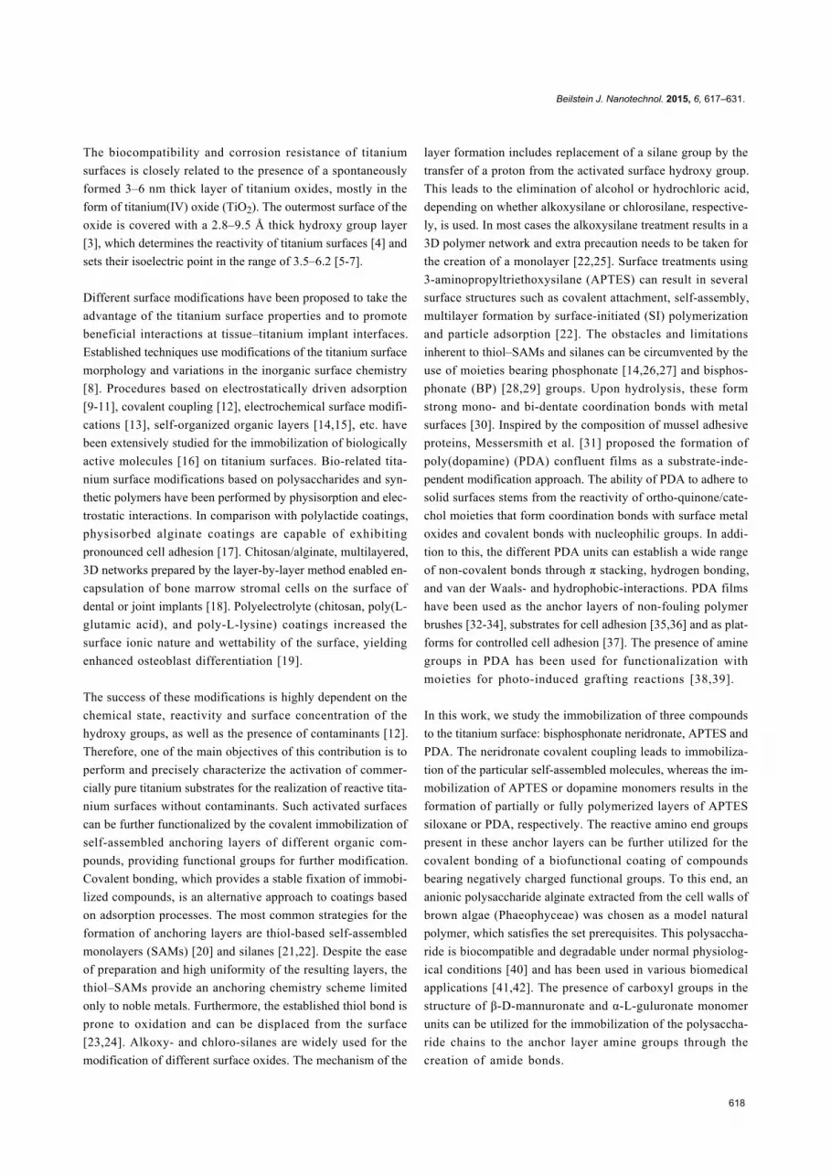
Figure 1 also reports the high resolution oxygen 1s XPS spectra
of the studied titanium surfaces. The O 1s envelope could be
resolved into surface oxide contributions at 530.3 ± 0.1 eV,
531.5 ± 0.1 eV and 533.0 ± 0.3 eV arising from titanium oxides
(TiO2 and Ti2O3), Al2O3 and SiO2, respectively. The presence
of hydroxy groups on the surface was evidenced by the pres-
ence of the peak at 531.8 ± 0.2 eV. The performed activation
treatments increased the contribution of the hydroxy from the
observed 2% for the pristine surfaces to more than 6% for the
activated titanium surfaces (Supporting Information File 1,
Table S1). This was evidenced by the change in the ratio
between surface hydroxy and oxide from 0.04 for the untreated
titanium surfaces to values in the range 0.1–0.2 for the acti-
vated ones. The careful analysis of the obtained high resolution
O 1s spectra enabled the estimation of the concentration of
hydroxy groups on the titanium surfaces according the method
proposed by McCafferty and Wightman [3]. The performed ac-
tivation treatments increased the surface density of the hydroxy
groups from the initial 2–4 hydroxy groups per nm2 on the pris-
tine titanium surfaces to 4–14 hydroxy groups per nm2. These
values are consistent with the range of values of 5–15 hydroxy
groups per nm2 reported for titanium foils [3] and titanium
dioxide powders [44]. The observed concomitant increase in
concentration of surface hydroxy groups and decrease in the
presence of surface contaminants inevitably leads to higher re-

Beilstein J. Nanotechnol. 2015, 6, 617–631.
620
Figure 1: High resolution Ti 2p (left) and O 1s (right) XPS spectra of pristine titanium surfaces (A) and surfaces treated using alkaline piranha (B),0.5 M NaOH (C), a HCl/H2SO4 mixture (D) and piranha solution (H2SO4/H2O2) (E). The spectrum of flat titanium surfaces deposited on siliconsubstrates (F) is given for comparison. The dominant contribution (more than 96.7%) within the Ti 2p3/2 envelope appears at 458.9 ± 0.1 eV and isidentified as TiO2. The measured O 1s spectra (points) was fitted (solid, colored lines) by resolving the individual surface oxide contributions (blacklines) centered at 530.3 ± 0.1, 531.5 ± 0.1 and 533.0 ± 0.3 eV arising from titanium oxides (TiO2 and Ti2O3), Al2O3 and SiO2, respectively. Thehydroxy groups on the surface gave rise to the peak at 531.8 ± 0.2 eV. The figure also reports the surface density of hydroxy groups (nOH).
activity of the treated surfaces [4,44]. In addition to an increase
in the number of surface sites available for binding, SEM
(Supporting Information File 1, Figure S1) and stylus profilom-
etry (Supporting Information File 1, Table S2) analysis showed
an increased microscale texture for all treated surfaces (alkaline
piranha, 0.5 M NaOH, and piranha (H2SO4/H2O2)) except for
those treated with a H2SO4/HCl solution. Microscale texturing
similar to that reported here has been obtained by treatments
such as machining [45,46], anodic oxidation [45,46] and chem-
ical oxidation using piranha [12]. The increase in the surface
roughness and the creation of a specific microscale texture due
to oxidative treatments as observed in our study have been
shown to enhance the rate of bone formation [12,45,46].
The decreased organic contamination and increased surface
density of hydroxy groups on the activated surfaces is further
evidenced by the higher hydrophilicity of the treated surfaces
(Supporting Information File 1, Table S2). The activation treat-
ments decrease the measured advancing water contact angles
from about 50° for the pristine titanium surfaces to values lower
than 30°. Almost completely wettable surfaces were obtained
when alkaline piranha was used as the surface activation
treatment.
It is worth mentioning that the chemical activation using alka-
line piranha simultaneously led to augmentation of the surface
composition, surface reactivity, topography and hydrophilicity.
Therefore, this chemical activation treatment is a potentially
valuable step in the treatment of titanium surfaces and possible
implants based on this material. Importantly, the evaporation-
deposited, flat, titanium reference samples (RRMS < 1.0 nm)
have the same surface composition and surface density of
hydroxy groups (Table 1 and Figure 1) as the activated pure
titanium surfaces. Therefore, it is reasonable to consider the flat
surfaces as a representative reference surface of the activated
pristine titanium for the verification of the surface modifica-
tions based on thin anchor layers and on the alginate monolayer.
The absence of surface irregularities on these mirror-like
substrates enables techniques such as SE, IRRAS and AFM to
be used for the characterization of sub- and mono-molecular,
organic overlayers.
Anchor layer depositionThe reproducibility in terms of continuity, uniformity, reactiv-
ity and adhesion properties of the anchor layers is a prerequi-
site for the creation of grafted adlayers with defined properties
[34]. The attachment of the anchor layers of three FDA
approved, organic compounds (neridronate, APTES and dopa-
mine) were performed on oxygen plasma-activated, flat tita-
nium substrates. As previously observed by XPS, the exposure
of titanium and titanium oxide surfaces to air resulted in a thin
adherent layer of organic contaminants. The presence of such an
organic contaminant layer influences the optical dispersion
function of the titanium films. These ill-defined optical parame-
ters of the titanium substrates decrease the precision of the ellip-
sometric data analysis during subsequent surface modifications.
A practical way to circumvent this problem is to perform the SE
measurements in different solvents (ethanol, isobutanol, tolu-
ene), a method referred as the multiple-environment method.
Due to the refractive index matching between the solvents and
the adsorbed organic contaminants, and the possible dissolution
of the contaminants, the multiple-environment method revealed
Beilstein J. Nanotechnol. 2015, 6, 617–631.
621
the intrinsic optical dispersion function of the flat titanium
surfaces. The measured data of a neat titanium layer in different
solvents was simultaneously fitted with the parameters of a
Drude–Lorentz and two Lorentz oscillator functions as
discussed in Supporting Information File 1, Figure S2.
The SE analysis showed formation of a 0.9 ± 0.3 nm thick
monolayer of neridronate. The concomitant processes of the
self-assembly of dopamine and its intermediates (dopamine-
quinone, 5,6-dihydroxyindole, etc.), of SI polymerization and of
adsorption of the resulting polymer molecules resulted in a
15.2 ± 0.5 nm thick confluent PDA layer. Despite the precau-
tions taken during the APTES deposition for the formation of
the SAM [21] (freshly distilled reagents, dry titanium substrates
and elevated temperature during the capping reaction), a
12.4 ± 1.7 nm thick APTES siloxane polymer multilayer was
formed [21]. The presence of the anchor layers increased the
water contact angles of completely wettable flat titanium
surfaces (oxygen plasma treated) to 40 ± 1°, 72 ± 1° and 60 ± 5°
for the substrates containing neridronate, APTES and PDA, res-
pectively. Here, the lower hydrophilicity is caused by the pres-
ence of organic molecules with increased hydrophobicity in
comparison to the neat titanium surface.
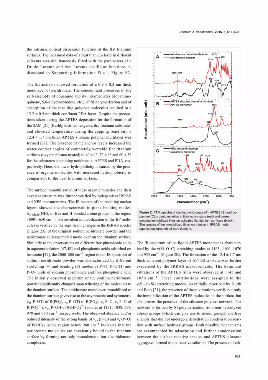
The surface immobilization of these organic moieties and their
covalent structure was further verified by independent IRRAS
and XPS measurements. The IR spectra of the resulting anchor
layers showed the characteristic in-plane bending modes,
δin-plane(NH), of free and H-bonded amino groups in the region
1600–1650 cm−1. The covalent immobilization of the BP mole-
cules is verified by the significant changes in the IRRAS spectra
(Figure 2A) of the original sodium neridronate powder and the
neridronate self-assembled monolayer on the titanium surface.
Similarly to the observations on different free phosphonic acids
in aqueous solution [47,48] and phosphonic acids adsorbed on
bentonite [49], the 2000–800 cm−1 region in our IR spectrum of
sodium neridronate powder was characterized by different
stretching (ν) and bending (δ) modes of P=O, P–O(H) and
P–O– units of sodium phosphonate and free phosphonic acid.
The initially observed spectrum of the sodium neridronate
powder significantly changed upon tethering of the molecules to
the titanium surface. The neridronate monolayer immobilized to
the titanium surface gives rise to the asymmetric and symmetric
νas P–OTi of R(PO3), νs P–OTi of R(PO3), νs P–O, νs P–O of
R(PO32−), νas P–OH of R(HPO3
2−) modes at 1121, 1038, 996,
978 and 960 cm−1, respectively. The observed absence and/or
reduced intensity of the strong bands of νas (P–O) and νs (P–O)
of P(OH)2 in the region below 960 cm−1 indicates that the
neridronate molecules are covalently bound to the titanium
surface by forming not only monodentate, but also bidentate
complexes.
Figure 2: FTIR spectra of starting neridronate (A), APTES (B) and do-pamine (C) organic moieties in their native state (red) and corres-ponding immobilized films on activated flat titanium surfaces (black).The spectra of the immobilized films were taken in IRRAS modeagainst backgrounds of bare titanium.
The IR spectrum of the liquid APTES monomer is character-
ized by the ν(Si–O–C) stretching modes at 1165, 1100, 1070
and 953 cm−1 (Figure 2B). The formation of the 12.4 ± 1.7 nm
thick adherent polymer layer of APTES siloxane was further
evidenced by the IRRAS measurements. The dominant
vibrations of the APTES films were observed at 1145 and
1050 cm−1. These contributions were assigned to the
ν(Si–O–Si) stretching modes. As initially described by Kurth
and Bein [21], the presence of these vibrations verify not only
the immobilization of the APTES molecules to the surface, but
also proves the presence of the siloxane polymer network. The
network is formed by SI polymerization from non-hydrolyzed
ethoxy groups (which can give rise to silanol groups) and free
silanols that did not undergo a dehydration condensation reac-
tion with surface hydroxy groups. Both possible mechanisms
are accompanied by adsorption and further condensation
between the surface reactive species and APTES siloxane
aggregates formed in the reactive solution. The presence of eth-

Beilstein J. Nanotechnol. 2015, 6, 617–631.
622
oxy groups, resulting in an incomplete cross-linking of the
APTES siloxane polymer network, can be seen by the signifi-
cant broadening of the dominate IR contributions toward the
initially observed main peaks of APTES "monomer". This may
be a source of the hydrolytical layer instability. In a water envi-
ronment, the primary amines present in the network can intra-
or inter- molecularly coordinate to a silicon center and catalyze
the hydrolysis reaction.
The spectrum of a solid dopamine monomer (Figure 2C) is
characterized by skeletal vibration modes of aromatic double
bonds (1650–1400 cm−1), stretching ν(C–O) modes of the cate-
chol moieties at 1283 cm−1, in-plane bending δin-plane(C–H) at
1170 cm−1 and stretching modes ν(C–C–N) of the aminoethyl
chain at 935 cm−1 [50]. The oxidative polymerization of dopa-
mine and the surface attachment of different monomer units
(dopamine-quinone, 5,6-dihydroxyindole, etc.) caused evident
changes in the IR spectra (Figure 2C) and resulted in a
confluent layer of PDA [32-34,51]. Similar to previous studies
on PDA modified materials [32-34,52], the spectrum of PDA
immobilized onto titanium substrates is characterized by poorly
resolved bands of many overlapping vibration modes of the
different monomer units. The most prominent contributions at
1615, 1510, 1445 cm−1 originate from the C=C vibrations of the
different monomer units, whereas the shoulder at 1715 cm−1
indicates the presence of quinone groups. The shift in
frequency, the broadening of the initial contributions, as well as
the appearance of new bands with respect to the IR spectra of
dopamine, proves not only the polymer nature of the resulting
films, but also their complex highly conjugated covalent
structure.
The complementary XPS measurements further verified the
successful formation of surface adherent films and their cova-
lent structure. The determined elemental compositions of the
neridronate, APTES siloxane and PDA anchor layers is reported
in Table 2. The covalent tethering of the organic moieties
caused an increase in the contributions of carbon and was asso-
ciated with the significant decrease in the surface concentration
of titanium. In the case of the thick polymer anchor films of
APTES and PDA, the contributions arising from the titanium
substrate were negligible. Importantly, the XPS spectra verified
the presence of nitrogen on the surface of different anchor
layers. Moreover, the obtained relative ratios N/P = 0.57 for the
neridronate and N/Si = 0.65 for APTES siloxane are reasonably
close to the expected values of 0.5 and 1, respectively. Figure 3
reports the high resolution carbon 1s XPS spectra for the
neridronate, APTES siloxane and PDA adherent films immobi-
lized on the flat titanium surfaces. The C 1s envelope of the
anchor layers could be resolved into contributions centered at
285 ± 0.1 eV arising from sp3 carbon (C–C and C–H functional-
ities), at 285.9 ± 0.1 eV arising from the C–N species of amines
and at 286.6 ± 0.2 eV arising from the C–O contribution of
hydroxy groups present in neridronate, the non-hydrolyzed
ethoxy groups of APTES and catechols of poly(dopamine).
The spectrum of APTES has an additional contribution at
284.3 ± 0.2 eV from the C–Si functionality. The PDA shows
contributions at 284.5 ± 0.1, 288.0 ± 0.2, 289.3 ± 0.3 and
291.2 ± 0.2 eV arising from the carbon species of sp2 carbon
(C=C functionality), the C=O functionality of the quinons, the
carboxylic carbon functionality (O–C=O groups) and the π–π*
transition (shake-up), respectively [53,54]. A rather unexpected
peak at 288.4 ± 0.1 eV was observed in the high resolution C 1s
spectra of neridronate and APTES siloxane. Although with a
large uncertainty, Acres et al. have tentatively assigned this
peak to the C–C=O functionality [55]. A similar contribution
was observed for micro-plasma polymerized APTES layers [56]
and was attributed to amide contribution. Since our immobiliza-
tion protocols lack harsh plasma deposition treatments, we
tentatively attribute this functionality to carbamate-like struc-
tures, which form due to the scrubbing effect of amines on CO2
from air [57].
Figure 3: High resolution C 1s XPS spectra of neridronate (A), APTESsiloxane (B) and PDA (C) films on the surfaces of activated flat tita-nium substrates (black). The unfilled circles represent the measureddata, while the red lines represent the fitted data. The individual contri-butions to the fitted data of different functional groups present in thefilms are represented with black lines.
The topography and homogeneity of the resulting anchor layers
on titanium substrates was monitored via AFM. The corres-
ponding images are presented in Figure 4. The AFM data
clearly evidence the functionalization of the flat titanium
surfaces with confluent anchor layers free of pinholes. The self-
assembly of neridronate molecules resulted in a fine-grained

Beilstein J. Nanotechnol. 2015, 6, 617–631.
623
Figure 4: AFM images of the neat, flat titanium surface (RRMS = 0.5 ± 0.3 nm) (A), and confluent anchor layers of neridronate (RRMS = 0.5 ± 0.2 nm)(B), APTES (RRMS = 1.1 ± 0.2 nm) (C) and PDA (RRMS = 3.6 ± 1.2 nm) (D). The figure also reports AFM images of ALG layers grafted ontoneridronate (RRMS = 0.7 ± 0.3 nm) (E), APTES (RRMS = 1.8 ± 0.2 nm) (F) and PDA (RRMS = 2.9 ± 1.0 nm) (G) anchor layers. The AFM measure-ments on the ALG surfaces were performed on predominantly flat regions with RRMS values similar to those of the initial anchor layers.
Table 2: Elemental compositions of anchor layers (neridronate,APTES siloxane and PDA) and ALG layers tethered to these surfaces,as determined by XPS.
Modification Ti O C N P Si(atom %)
Neat, flat titaniumsurface 19.5 56.7 23.8 – – –
Neridronate 13.8 55.6 25.9 1.7 3.0 –APTES siloxane – 27.1 54.6 7.2 – 11.1PDA 0.4 22.1 70.7 6.8 – –ALG/neridronate 1.8 22.2 70.1 5.8 – –ALG/APTES siloxane 0.6 27.9 60.5 6.3 – 4.7ALG/PDA – 30.7 61.9 7.4 – –
topography similar to the activated titanium surface (Figure 4A
and Figure 4B). The processes of self-assembly, SI polymeriza-
tion and adsorption of aggregates from the reactive solution
during adsorption and formation of the APTES siloxane and
PDA layers led to surfaces with an increased roughness of
1.1 ± 0.2 nm and 3.6 ± 1.2 nm, respectively (Figure 4C and
Figure 4D). The immobilized polymer surfaces exhibited a
more pronounced grain structure with nanoparticles having
average diameter of 12 nm for the APTES siloxane and of
38 nm for PDA anchor layers. While such a pronounced surface
topography is considered to be an inherent characteristic of the
PDA films [34,53], the presence of such surface objects on
APTES layers is rarely discussed. However, even in the case
when a APTES SAM was achieved [58], the presence of
surface adherent objects resembling aggregates having average
diameter of up 30 nm was inevitable. We presume that the
observed APTES siloxane aggregates are formed in the reac-
tive solution and further adsorbed and even grafted onto the
surface.
Covalent binding of alginateThe tethering of alginate (ALG) chains by reaction of the
carboxyl groups to the amine-functionalized titanium surfaces
was performed by following standard EDC/NHS protocols. The
binding reaction resulted in the formation of 5.0 ± 1.9 nm
thick alginate films, irrespective of the anchor layer. The
grafting density, calculated from the ellipsometric thickness and
the molecular weight of ALG (1.5 × 10⁶ g∙mol−1), was
1.3–3.0 × 10¹¹ chains/cm2. The presence of the polysaccharide
chains was obvious from the significant increase in the surface
hydrophilicity. The immobilization of hydrophilic alginate
chains resulted in a decrease in the contact angles from the
values determined for the anchor layers to 13 ± 3°, 19 ± 3° and
13 ± 1° for the alginate films bound to neridronate, APTES and
PDA, respectively.

Beilstein J. Nanotechnol. 2015, 6, 617–631.
624
The covalent tethering of the polysaccharide chains and the for-
mation of amide bonds between the activated carboxyl groups
of ALG and the amines present in the anchor layer were probed
by IRRAS measurements. A representative IR spectrum of free
alginate deposited on the flat titanium surface is presented in
Figure 5. The spectrum is dominated by the symmetric νsym
(C=O) and asymmetric νasym (C=O) modes of charged carboxyl
groups at 1630 and 1420 cm−1, respectively, in addition to the
stretching ν(C–O) modes of the pyranosyl ring, β-(1-4)-glyco-
sidic bonds and hydroxy groups of the polysaccharide in the
1200–1000 cm−1 region. The established covalent bonds
between the carboxyl groups of the alginate chains and the
amines present on the surface are evidenced by the appearance
of the highly specific amide I and amide II bands at 1650 and
1540 cm−1, respectively (Figure 5). Additionally, the differen-
tial spectra of ALG bound to neridronate and PDA show the
carbonyl band (1730 cm−1) and the bands characteristic for the
polysaccharide moieties (1200–1000 cm−1). In the same region,
the differential spectrum of ALG bound to the siloxane anchor
layer is characterized by a valley at 1580 cm−1 arising from
decreased in-plane bending δin-plane(NH) of amine contribu-
tions and at 1145 and 1037 cm-1 from decreased ν(Si–O–Si)
stretching contributions. The appearance of these bands is asso-
ciated with the amine-catalyzed hydrolysis of the siloxane
bonds of the polymer network [22] during the 24 h immersion
in MES buffer.
The IRRAS results of the immobilization of ALG to the anchor
layers are further supported by XPS measurements. The forma-
tion of the 5 nm thick polysaccharide layer on the surfaces with
different anchors serves to further decrease the contributions
from the titanium substrate. This corresponds to a concomitant
increase in the presence of the elements from the organic
moieties (Table 2). Compared to the high resolution C 1s
spectra of the anchor layers (Figure 3), the spectra of the bound
alginate films (Figure 6) show increased contributions at
286.5 ± 0.1 eV arising from the C–O moiety of the pyranosyl
ring, β-(1-4)-glycosidic bonds and hydroxy groups of the poly-
saccharide in addition to a peak at 289.2 ± 0.1 eV arising from
the O–C=O functionality of carboxylic groups. Importantly, the
presence of the peak at 288.1 ± 0.2 eV verifies the formation of
amide bonds (N–C=O) between the activated carboxyl groups
of ALG and the amines present in the anchor layers. Thus, the
XPS studies strongly prove the covalent immobilization of the
alginate films.
The tethering of ALG resulted in surfaces with a predominantly
flat topography (Figure 4E–G) similar to those observed for the
corresponding anchor layers (Figure 4B–D). The ALG films on
neridronate, APTES siloxane and PDA were characterized by
RRMS values of 0.7 ± 0.3 nm, 1.8 ± 0.2 nm and 2.9 ± 1.0 nm,
Figure 5: Differential IRRAS spectra of free alginate adsorbed onto aflat titanium surface (A) and covalently bound alginate molecules to theamines of the neridronate (B), APTES (C) and PDA (D) anchor layers.The IRRAS spectra of covalently bound alginate films was character-ized by the presence of the carbonyl band (1730 cm−1), amide I(1650 cm−1), amide II band (1540 cm−1) and ν(C–O) stretching modesof the pyranosyl ring, β-(1-4)-glycosidic bonds and hydroxy groups ofthe polysaccharide (1200–1000 cm−1). The spectra were referenced tocorresponding background spectra of bare titanium and titaniumbearing different anchor layers.
Figure 6: High resolution C 1s XPS spectra of alginate coatings onneridronate (A), APTES siloxane (B) and PDA (C) anchoring layers.The unfilled circles represent the measured data, while the red linesrepresent the fitted data. The individual contributions to the fitted dataof different functional groups present in the films are represented withblack lines.
respectively. The increase in the ellipsometric thickness of 5 nm
combined with the AFM findings of the surface roughness
(similar to the values characteristic for the anchor layers) indi-
cates the formation of continuous ALG films, which merely
replicate the surface underneath. However, although only occa-

Beilstein J. Nanotechnol. 2015, 6, 617–631.
625
sionally observed, the AFM measurement also showed the pres-
ence of regions of surface-immobilized ALG aggregates
composed of particles with an average diameter exceeding
40 nm (Supporting Information File 1, Figure S3). The
observed nanoparticle aggregates may have a physisorbed frac-
tion of loosely bound ALG chains that are a potential source of
instability and defects when these surfaces are exposed to phys-
iological conditions.
Stability of alginate filmsThe stability of the anchor layers and anchored ALG films is
crucial for their performance especially when biomedical and
tissue engineering applications are in question. The deteriora-
tion of these surface confluent layers could affect the surface
concentration of free carboxylic end groups that are essential in
the envisaged applications. Furthermore, the instability of the
layers that are in intimate contact with the titanium surface
could result in complete delamination of the potentially surface
adherent alginate gels.
The stability tests of the neat, anchor layers and the ALG/
anchor layers on flat titanium substrates were performed by
immersion in PBS buffer at 37 °C for a period of 7 days. The
ellipsometric thickness and water contact angles were measured
on dry films after 1, 3 and 7 days of incubation (Figure 7 and
Supporting Information File 1, Figure S4). The thickness of the
ALG adlayers was obtained from an optical model that consid-
ered a constant thickness of the neridronate and PDA anchor
layers as determined before the grafting. The adopted optical
model conforms with the observed stability of neat neridronate
and PDA films during the immersion in PBS (Supporting Infor-
mation File 1, Figures S4 and S5). When the polysaccharide
layer was bound to the APTES siloxane, the optical model
considered the instability of the APTES anchor in accordance
with the SE, CA (Supporting Information File 1, Figure S4) and
IRRAS findings (Supporting Information File 1, Figure S5).
This methodology enabled monitoring of the stability of the
ALG adlayer in the two-layer stack.
During the 7 days of incubation, only a minor decrease in the
measured ellipsometric thickness was observed (Figure 7). We
presume that the reduction in the ALG thickness is mainly
caused by the release of a physisorbed fraction present in the
surface adherent ALG aggregates. However, the water contact
angles of the ALG coatings remained rather constant. This indi-
cates that the observed, small decrease in thickness due to dete-
rioration of the ALG films does not reveal the less-wettable
anchor layers underneath (Supporting Information File 1, Figure
S4). The stability of the ALG/anchor layers and the neat anchor
layers was further verified by IRRAS measurements (Figure 8
and Supporting Information File 1, Figure S5). The IRRAS data
Figure 7: Ellipsometric thickness and water contact angle evolution ofALG bound to neridronate, APTES siloxane and PDA during theimmersion in PBS (37 °C, pH = 7.4) (mean value ± SD, n = 15).
enabled monitoring of the changes in the covalent structure of
the whole ALG/anchor double layer and allowed for the contri-
butions of both constituents of the stack to be separately
resolved. As depicted in Figure 8, the polysaccharide films
bound to neridronate and PDA anchors are rather stable without
any significant changes in the position and intensity of the main
vibrations of the double layer components. However, the ALG
films anchored to APTES showed continuous deterioration
during the 7 days of immersion. The main reduction in inten-
sity was observed for the bands centered at 1146 and 1046 cm−1
arising from the ν(Si–O–Si) stretching modes of the APTES
siloxane polymer network. The position of these bands corre-
sponds to the positions of the decreased contributions in the
IRRAS spectra of the deteriorated neat APTES siloxane films
(Supporting Information File 1, Figure S5). This implies that
the main components released during the immersion are of
siloxane nature. At the same time, the spectral contributions of
ALG were affected to a minor extent, most likely due to the
several grafting points through which the polysaccharide chains
are bound to the surface. However, the previously discussed
hydrolytic instability of the APTES siloxane anchor layer would
eventually lead to a complete cleavage of the polysaccharide
layer from the titanium surface during a long-term immersion.

Beilstein J. Nanotechnol. 2015, 6, 617–631.
626
Figure 8: Evolution of IRRAS spectra of ALG bound to neridronate (A),to APTES siloxane (B) and to PDA (C) upon immersion in PBS at37 °C for 7 days.
Based on the stability observations, the alginate monolayers
bound to the neridronate or PDA anchor layers can be poten-
tially used for the immobilization of a thin alginate hydrogel
carrier of bioactive compounds (such as calcium phosphates or
other biologically active molecules) formed by ionic cross-
linking [40]. The proposed architecture is envisaged to enhance
adhesion, proliferation, differentiation of osteoblasts, and thus
ultimately, to achieve a better integration of the titanium
implant into the bone tissue.
ConclusionIn the present contribution, we demonstrated the successful
covalent attachment of ALG chains to neridronate, APTES and
PDA anchor layers immobilized on activated titanium surfaces.
The formation of the ALG and anchor layer films was investi-
gated utilizing SE, AFM and contact angle goniometry. The
IRRAS analysis further evidenced the established amide bonds
between the carboxyl groups of ALG and amine groups of the
anchor layers. The immobilization of the organic moieties, as
well as the changes in the surface composition of pristine tita-
nium surfaces after different surface activation treatments, was
probed by XPS measurements. The changes in the surface
morphology and roughness parameters during the activation of
titanium surfaces were monitored by SEM and SP analysis. The
5 nm thick ALG layers anchored to neridronate and PDA were
stable during immersion under physiological-like conditions for
7 days. The hydrolysis of the anchoring APTES siloxane
network led to a higher deterioration tendency of the ALG/
APTES double layer. The presented surface modification
strategy of titanium can be an effective path for the formation of
ALG-based hydrogel coatings enriched with bioactive com-
pounds for bone tissue engineering applications.
ExperimentalMaterialsDopamine hydrochloride (98.5%) was purchased from Sigma
and 3-triethoxysilylpropan-1-amine from Aldrich. 3-(Ethylimi-
nomethyleneamino)-N,N-dimethylpropan-1-amine (EDC),
1-hydroxy-2,5-pyrrolidinedione (NHS) and 2-(morpholin-4-
yl)ethanesulfonic acid (MES) were obtained from Fluka.
Sodium alginate salt (ALG) derived from brown algae was
purchased from Sigma. The molecular weight of ALG was
determined by size exclusion chromatography (SEC) on a
gradient Knauer system with diode array detection (DAD) and
an Alltech 3300 evaporative light scattering detection (ELSD)
system. The SEC measurement was performed on a PolySept
GFC-P linear column using an isocratic system of 0.03 M am-
monium acetate buffer in acetonitrile/water (20/80 v/v).
The determined average molecular weight of ALG was
1.5 × 10⁶ g·mol−1 (PDI = 2.45) with the column calibration
carried out using PEO standards. The ALG peak had a
unimodal distribution without the presence of low molecular
weight degradation products.
All organic solvents (petroleum ether, methanol, ethanol,
isobuthanol and toluene) were of analytical grade (Lach-Ner,
Czech Republic) and used as received. Ultrapure water was
obtained with a Millipore Milli-Q system.
Substrate preparationClean, single-side-polished silicon wafers (CZ, orientation
<100>, B-doped, resistivity 5–20 Ω∙cm) with a ≈50 nm SiO2
thermal overlayer (Siegert Consulting e.K., Germany) were
used as substrates for the preparation of ultraflat titanium

Beilstein J. Nanotechnol. 2015, 6, 617–631.
627
surfaces. Flat, titanium reference surfaces (50 nm thickness)
were obtained by evaporation deposition (rate = 0.35 Å·s−1,
pressure = 6.66 × 10⁻⁶ Pa) using a COV AP SQC-310C deposi-
tion device (Angstrom Engineering, Canada). The coated
substrates were cut into 1.2 cm × 1.2 cm pieces, sonicated in
methanol, and deionized in water for 15 min, followed by
oxygen plasma oxidization (25 W, Plasma Cleaner/Sterilizer,
Harrick, USA) for 5 min directly before the anchor layer immo-
bilization.
Commercially available, paste-polished, pure titanium surfaces
(Beznoska, Czech Republic) were used to probe the presence of
inorganic and organic surface contaminants and to determine
the surface concentration of introduced hydroxy groups. After
the initial sonication in petrolether, methanol and deionized
water for 15 min, the following surface cleaning and activation
procedures were investigated: alkaline piranha treatment (mix-
ture of 25% NH3, 30% H2O2 and water at 1:1:5 v/v/v, at 70 °C
for 15 min), immersion in 0.5 M NaOH (60 °C for 24 h),
immersion in a mixture of concentrated HCl and H2SO4
(1:1 v/v, at room temperature for 20 min) and a piranha
cleaving treatment utilizing concentrated H2SO4 and 30% H2O2
(1:1 v/v, at room temperature for 20 min). The substrates were
subsequently thoroughly rinsed with ultrapure water, blow-
dried using nitrogen, and exposed to an oxygen plasma (25 W)
for 5 min just before the XPS analysis or the binding of the
anchor layers (Scheme 1).
Formation of anchoring layersNeridronate monosodium salt ((6-amino-1-hydroxy-1-phos-
phonohexyl)-hydroxyphosphinate sodium) was prepared
according previous reports [59]. The immobilization on the flat
titanium surfaces proceeded from a 0.005 M neridronate solu-
tion in water at 100 °C for 48 h. Afterwards, the samples were
rinsed in water 3 times for 5 min to remove the physisorbed
molecules, then blow-dried and kept in vacuum until further
use.
Siloxane anchor layers were prepared by exposing the activated
titanium substrates to 0.1% v/v APTES solutions in dry
toluene at 70 °C. After 12 h of exposure, the samples were soni-
cated in dry toluene for 15 min to remove the physisorbed
siloxane particles, then blow-dried and kept in vacuum until
further use.
A poly(dopamine) coating was deposited from a 2 mg·mL−1
solution prepared by dissolution of dopamine hydrochloride in
an air-saturated 10 mM Tris hydrochloride (pH 8.5) buffer.
After 3 h of polymerization, the PDA-coated surfaces were
rinsed with water, sonicated in water for 15 min and blow-dried
in a stream of nitrogen.
Stability tests were performed on alginate-containing and neat-
anchor layers deposited on flat titanium substrates, incubated in
PBS (pH 7.4, containing 0.02 wt % sodium azide) at 37 °C for
7 days. After the immersion period, the substrates were rinsed
with copious amounts of water and blow-dried in a stream of
nitrogen.
Tethering of alginate onto titanium layerscontaining anchor layersThe tethering of the alginate chains to the amine-functionalized
flat titanium substrates was performed employing a modified
EDC/NHS protocol based on the work of Rowley et al. [60].
Alginate was dissolved in mixture of 0.1 M MES and 0.15 M
NaCl, at pH 5, at a concentration of 1 wt %. Next, EDC was
added and the solution was stirred for 15 min. Afterwards, NHS
was added and the solution was stirred for an additional 15 min
until the production of bubbles diminished. The molar ratio
between reactants was uronic units/EDC/NHS (1:20:20). The
titanium substrates bearing the neridronate, APTES siloxane
and PDA anchors were placed in 12-well cultivation plates and
0.8 mL of the reaction solution was deposited on the surface
and allowed to react for 24 h. Subsequently the alginate-grafted
substrates were rinsed with water and blow-dried in a stream of
nitrogen.
MethodsSpectroscopic ellipsometry (SE): Ex situ and in situ ellipso-
metric data were acquired using a spectroscopic imaging, auto-
nulling ellipsometer (EP3-SE, Nanofilm Technologies,
Germany) equipped with a liquid cell (Vinternal = 0.7 mL) in
4-zone mode in the wavelength range of 398.9–811.0 nm
(source: Xe arc lamp, wavelength step: 10 nm) at an angle of
incidence of 60°. The cell windows (strain-free, optical BK-7
glass from Qioptiq, Germany) exhibited only small birefrin-
gence and dichroism causing errors in the ellipsometric angles
Δ and Ψ smaller than 0.3° and 0.1°, respectively. These errors
were corrected following the method of Azzam and Bashara
[61]. To increase the measurement precision and exclude errors
from the variations of layer thickness throughout the substrate
area, a 10× objective and position-calibrated sample stage were
utilized to perform repeated ex situ and in situ measurements
over the same sample area (1 × 2 mm). The obtained data were
analyzed with multilayer models using the EP4-SE analysis
software (Accurion GmbH, Germany).
The thickness and refractive index of the resulting organic
layers were obtained from simultaneous fitting of the obtained
ellipsometric data using the Cauchy dispersion function (n = An
+ Bn/λ, k = 0 with An = 1.412 ± 0.010, Bn = 5900 ± 80 nm2 for
neridronate and An = 1.413 ± 0.009, Bn = 6270 ± 70 nm2 for
APTES and An = 1.4714 ± 0.008, Bn = 13200 ± 1000 nm2 for

Beilstein J. Nanotechnol. 2015, 6, 617–631.
628
Scheme 1: Performed surface treatments and subsequent reactions for the activation and modification of titanium surfaces. (A) Cleaning and acti-vation procedures for the removal of inorganic and organic contaminants from the titanium surface and to increase the number of free hydroxy groups.(B) Immobilization of neridronate, APTES siloxane and poly(dopamine) anchor layers through surface specific reactions between the phosphonate,silane and catechol groups of corresponding compounds and hydroxy groups on the surface. (C) Covalent binding of ALG chains to amino groupspresent in the anchor layers by the EDC/NHS coupling reaction.
Beilstein J. Nanotechnol. 2015, 6, 617–631.
629
the ALG layers). The optical dispersion functions of PDA,
silicon dioxide and silicon were taken from previous reports
[34,62]. The optical dispersion functions of ethanol, isobutanol,
toluene and titanium dioxide were taken from the EP4-SE data-
base.
Contact angle measurement: The wettability of the organic
surfaces on flat, titanium reference surfaces was examined by a
static sessile water drop method using a DataPhysics OCA 20
contact angle system. Each sample was characterized using four
3 μL drops of material. The data were evaluated using the
Young–Laplace method.
The wettability of the commercially available, rough titanium
substrates upon different treatments was estimated by
measuring the advancing and receding water contact angles
utilizing the dynamic Wilhelmy plate method. The measure-
ments were performed on a Kruss K12 (Germany) tensiometer.
Infrared reflection–absorption spectroscopy (IRRAS): The
infrared spectra of neridronate, APTES and dopamine moieties
were recorded using a Perkin Elmer, Paragon 1000PC, FTIR
spectrometer, equipped with a MCT detector and a single
reflection, monolithic diamond, Golden Gate ATR accessory
(Specac, England). The spectra of the dry organic films formed
on titanium surfaces were recorded using a Bruker IFS 55 FTIR
spectrometer (Bruker Optics, Germany) equipped with a MCT
detector. The measurements were performed at a grazing angle
(80°, p-polarization) using the reflection spectroscopy acces-
sory. The measurement chamber was continuously purged with
dry air. The acquisition time was approximately 20 min at a
resolution of 2 cm−1. The spectra are reported as −log(R/R0),
where R is the reflectance of the sample and R0 is the
reflectance of bare titanium reference surfaces.
Atomic force microscopy (AFM): AFM characterization was
performed on a Dimension ICON (Bruker, USA) system in
peak force tapping mode in air using silicon probes (TAP150A,
Bruker, USA) with a typical force constant of 5 N∙m−1. The
images were taken using a scan rate in the range of 0.5−1.2 Hz
and a peak force set point of 0.02−0.2 V.
Surface profilometry: Macroscopic surface roughness and wavi-
ness measurements were performed using a Tencor P-10
(Texas, USA) surface profiler with 1 mm long scans at a speed
of 20 μm∙s−1 and a sampling rate of 200 Hz using a maximum
stylus force of 0.02 N.
Scanning electron microscopy (SEM): The SEM analysis was
performed on a Quanta 200 FEG (FEI, Czech Republic) micro-
scope. All micrographs presented are secondary electron images
taken under high vacuum using an accelerating voltage of
30 kV.
X-ray photoelectron spectroscopy (XPS): The core-level photo-
electron spectra were recorded using an angle-resolved photo-
electron spectrometer, ADES 400 (VG Scientific, UK), oper-
ating at a base pressure of 1.33 × 10⁻⁷ Pa. The system was
equipped with an X-ray excitation source and a rotatable hemi-
spherical electron energy analyzer. The photoelectron spectra
were recorded using Mg Kα radiation with a pass energy of
100 eV or 20 eV (high-energy resolution). The incidence angle
was 70° with respect to the sample surface normal and the emis-
sion angle along the surface normal. The atomic concentrations
of carbon, oxygen and nitrogen were determined from the C 1s,
O 1s, and N 1s photoelectron peak areas after a Shirley inelastic
background subtraction. Assuming a simple model of a semi-
infinite solid of homogeneous composition, the peak areas were
corrected for the photoelectric cross-sections [63], the inelastic
mean free paths of the electrons in question [64], and the trans-
mission function of the spectrometer [65]. The experimental
uncertainties in the quantitative analysis of XPS were assessed
in separate experiments with several standard materials and
were estimated to be below 7%. This value encompasses the
overall uncertainties of the method that are typically introduced
by the background subtraction.
Supporting InformationSupporting Information File 1Additional Experimental Information.
[http://www.beilstein-journals.org/bjnano/content/
supplementary/2190-4286-6-63-S1.pdf]
AcknowledgementsThe authors acknowledge the support of the Grant Agency of
the Ministry of Health of the Czech Republic (grant number:
NT/13297–4), of the Ministry of Education, Youth and Sports
of the Czech Republic (grant number: SGS No. 10/297/OHK4/
3T/14, EE2.3.30.0029 and the project “BIOCEV – Biotech-
nology and Biomedicine Centre of the Academy of Sciences
and Charles University” CZ.1.05/1.1.00/02.0109), and the
Czech Science Foundation (grant number: P108/11/1857).
References1. Elias, C. N.; Lima, J. H. C.; Valiev, R.; Meyers, M. A. JOM 2008, 60,
46–49. doi:10.1007/s11837-008-0031-12. Park, J. B.; Bronzino, J. D., Eds. Biomaterials - Principles and
Applications; CRC Press: Boca Raton, FL, USA, 2003.

Beilstein J. Nanotechnol. 2015, 6, 617–631.
630
3. McCafferty, E.; Wightman, J. P. Surf. Interface Anal. 1998, 26,549–564.doi:10.1002/(SICI)1096-9918(199807)26:8<549::AID-SIA396>3.0.CO;2-Q
4. Tanaka, Y.; Saito, H.; Tsutsumi, Y.; Doi, H.; Imai, H.; Hanawa, T.Mater. Trans. 2008, 49, 805–811.doi:10.2320/matertrans.MRA2007317
5. Kataoka, S.; Gurau, M. C.; Albertorio, F.; Holden, M. A.; Lim, S.-M.;Yang, R. D.; Cremer, P. S. Langmuir 2004, 20, 1662–1666.doi:10.1021/la035971h
6. Roessler, S.; Zimmermann, R.; Scharnweber, D.; Werner, C.;Worch, H. Colloids Surf., B 2002, 26, 387–395.doi:10.1016/S0927-7765(02)00025-5
7. Parks, G. A. Chem. Rev. 1965, 65, 177–198. doi:10.1021/cr60234a0028. Schliephake, H.; Scharnweber, D. J. Mater. Chem. 2008, 18,
2404–2414. doi:10.1039/b715355b9. Tosatti, S.; De Paul, S. M.; Askendal, A.; VandeVondele, S.;
Hubbell, J. A.; Tengvall, P.; Textor, M. Biomaterials 2003, 24,4949–4958. doi:10.1016/S0142-9612(03)00420-4
10. Schuler, M.; Owen, G. Rh.; Hamilton, D. W.; De Wild, M.; Textor, M.;Brunette, D. M.; Tosatti, S. G. P. Biomaterials 2006, 27, 4003–4015.doi:10.1016/j.biomaterials.2006.03.009
11. Tosatti, S.; Schwartz, Z.; Campbell, C.; Cochran, D. L.;VandeVondele, S.; Hubbell, J. A.; Denzer, A.; Simpson, J.;Wieland, M.; Lohmann, C. H.; Textor, M.; Boyan, B. D.J. Biomed. Mater. Res., Part A 2004, 68A, 458–472.doi:10.1002/jbm.a.20082
12. Nanci, A.; Wuest, J. D.; Peru, L.; Brunet, P.; Sharma, V.; Zalzal, S.;McKee, M. D. J. Biomed. Mater. Res. 1998, 40, 324–335.doi:10.1002/(SICI)1097-4636(199805)40:2<324::AID-JBM18>3.0.CO;2-L
13. Jimbo, R.; Ivarsson, M.; Koskela, A.; Sul, Y.-T.; Johansson, C.J. Oral Maxillofac. Res. 2010, 1, e3. doi:10.5037/jomr.2010.1303
14. Zorn, G.; Gotman, I.; Gutmanas, E. Y.; Adadi, R.; Sukenik, C. N.J. Mater. Sci.: Mater. Med. 2007, 18, 1309–1315.doi:10.1007/s10856-006-0117-7
15. Heijink, A.; Schwartz, J.; Zobitz, M. E.; Crowder, K. N.; Lutz, G. E.;Sibonga, J. D. Clin. Orthop. Relat. Res. 2008, 466, 977–984.doi:10.1007/s11999-008-0117-7
16. Kaigler, D.; Avila, G.; Wisner-Lynch, L.; Nevins, M. L.; Nevins, M.;Rasperini, G.; Lynch, S. E.; Giannobile, W. V. Expert Opin. Biol. Ther.2011, 11, 375–385. doi:10.1517/14712598.2011.554814
17. Bagno, A.; Genovese, M.; Luchini, A.; Dettin, M.; Conconi, M. T.;Menti, A. M.; Parnigotto, P. P.; Di Bello, C. Biomaterials 2004, 25,2437–2445. doi:10.1016/j.biomaterials.2003.09.018
18. Wu, M.-Y.; Chen, N.; Liu, L.-K.; Yuan, H.; Li, Q.-L.; Chen, S.-H.J. Bioact. Compat. Polym. 2009, 24, 301–315.doi:10.1177/0883911509105848
19. Park, J. H.; Schwartz, Z.; Olivares-Navarrete, R.; Boyan, B. D.;Tannenbaum, R. Langmuir 2011, 27, 5976–5985.doi:10.1021/la2000415
20. Chapman, R. G.; Ostuni, E.; Yan, L.; Whitesides, G. M. Langmuir 2000,16, 6927–6936. doi:10.1021/la991579l
21. Kurth, D. G.; Bein, T. Langmuir 1995, 11, 3061–3067.doi:10.1021/la00008a035
22. Smith, E. A.; Chen, W. Langmuir 2008, 24, 12405–12409.doi:10.1021/la802234x
23. Flynn, N. T.; Tran, T. N. T.; Cima, M. J.; Langer, R. Langmuir 2003, 19,10909–10915. doi:10.1021/la035331e
24. Sung, I.-H.; Kim, D.-E. Tribol. Lett. 2004, 17, 835–844.doi:10.1007/s11249-004-8091-z
25. Xiao, S.-J.; Textor, M.; Spencer, N. D.; Sigrist, H. Langmuir 1998, 14,5507–5516. doi:10.1021/la980257z
26. Dubey, M.; Weidner, T.; Gamble, L. J.; Castner, D. G. Langmuir 2010,26, 14747–14754. doi:10.1021/la1021438
27. Silverman, B. M.; Wieghaus, K. A.; Schwartz, J. Langmuir 2005, 21,225–228. doi:10.1021/la048227l
28. Lecollinet, G.; Delorme, N.; Edely, M.; Gibaud, A.; Bardeau, J.-F.;Hindré, F.; Boury, F.; Portet, D. Langmuir 2009, 25, 7828–7835.doi:10.1021/la8039576
29. Yoshinari, M.; Oda, Y.; Ueki, H.; Yokose, S. Biomaterials 2001, 22,709–715. doi:10.1016/S0142-9612(00)00234-9
30. Luschtinetz, R.; Gemming, S.; Seifert, G. Eur. Phys. J. Plus 2011, 126,98. doi:10.1140/epjp/i2011-11098-4
31. Lee, H.; Dellatore, S. M.; Miller, W. M.; Messersmith, P. B. Science2007, 318, 426–430. doi:10.1126/science.1147241
32. Pop-Georgievski, O.; Rodriguez-Emmenegger, C.;de los Santos Pereira, A.; Proks, V.; Brynda, E.; Rypáček, F.J. Mater. Chem. B 2013, 1, 2859–2867. doi:10.1039/c3tb20346h
33. Pop-Georgievski, O.; Verreault, D.; Diesner, M.-O.; Proks, V.;Heissler, S.; Rypáček, F.; Koelsch, P. Langmuir 2012, 28,14273–14283. doi:10.1021/la3029935
34. Pop-Georgievski, O.; Popelka, S.; Houska, M.; Chvostova, D.;Proks, V.; Rypáček, F. Biomacromolecules 2011, 12, 3232–3242.doi:10.1021/bm2007086
35. Ku, S. H.; Ryu, J.; Hong, S. K.; Lee, H.; Park, C. B. Biomaterials 2010,31, 2535–2541. doi:10.1016/j.biomaterials.2009.12.020
36. Ryu, J.; Ku, S. H.; Lee, H.; Park, C. B. Adv. Funct. Mater. 2010, 20,2132–2139. doi:10.1002/adfm.200902347
37. Proks, V.; Jaroš, J.; Pop-Georgievski, O.; Kučka, J.; Popelka, Š.;Dvořák, P.; Hampl, A.; Rypáček, F. Macromol. Biosci. 2012, 12,1232–1242. doi:10.1002/mabi.201200095
38. Kaupp, M.; Quick, A. S.; Rodriguez-Emmenegger, C.; Welle, A.;Trouillet, V.; Pop-Georgievski, O.; Wegener, M.; Barner-Kowollik, C.Adv. Funct. Mater. 2014, 24, 5649–5661. doi:10.1002/adfm.201400609
39. Rodriguez-Emmenegger, C.; Preuss, C. M.; Yameen, B.;Pop-Georgievski, O.; Bachmann, M.; Mueller, J. O.; Bruns, M.;Goldmann, A. S.; Bastmeyer, M.; Barner-Kowollik, C. Adv. Mater.2013, 25, 6123–6127. doi:10.1002/adma.201302492
40. Becker, T. A.; Kipke, D. R.; Brandon, T. J. Biomed. Mater. Res. 2001,54, 76–86.doi:10.1002/1097-4636(200101)54:1<76::AID-JBM9>3.0.CO;2-V
41. Li, Z.; Ramay, H. R.; Hauch, K. D.; Xiao, D.; Zhang, M. Biomaterials2005, 26, 3919–3928. doi:10.1016/j.biomaterials.2004.09.062
42. Chen, T. W.; Chang, S. J.; Niu, G. C.-C.; Hsu, Y. T.; Kuo, S. M.J. Appl. Polym. Sci. 2006, 102, 4528–4534. doi:10.1002/app.24945
43. Rossetti, F. F.; Reviakine, I.; Textor, M. Langmuir 2003, 19,10116–10123. doi:10.1021/la034280i
44. Feng, B.; Chen, J. Y.; Qi, S. K.; He, L.; Zhao, J. Z.; Zhang, X. D.J. Mater. Sci.: Mater. Med. 2002, 13, 457–464.doi:10.1023/A:1014737831371
45. Larsson, C.; Thomsen, P.; Aronsson, B.-O.; Rodahl, M.; Lausmaa, J.;Kasemo, B.; Ericson, L. E. Biomaterials 1996, 17, 605–616.doi:10.1016/0142-9612(96)88711-4
46. Larsson, C.; Thomsen, P.; Lausmaa, J.; Rodahl, M.; Kasemo, B.;Ericson, L. E. Biomaterials 1994, 15, 1062–1074.doi:10.1016/0142-9612(94)90092-2
47. Chapman, A. C.; Thirlwell, L. E. Spectrochim. Acta 1964, 20, 937–947.doi:10.1016/0371-1951(64)80094-1

Beilstein J. Nanotechnol. 2015, 6, 617–631.
631
48. Zenobi, M. C.; Luengo, C. V.; Avena, M. J.; Rueda, E. H.Spectrochim. Acta, Part A: Mol. Biomol. Spectrosc. 2008, 70, 270–276.doi:10.1016/j.saa.2007.07.043
49. Zenobi, M. C.; Luengo, C. V.; Avena, M. J.; Rueda, E. H.Spectrochim. Acta, Part A: Mol. Biomol. Spectrosc. 2010, 75,1283–1288. doi:10.1016/j.saa.2009.12.059
50. Lagutschenkov, A.; Langer, J.; Berden, G.; Oomens, J.; Dopfer, O.Phys. Chem. Chem. Phys. 2011, 13, 2815–2823.doi:10.1039/c0cp02133d
51. Proks, V.; Brus, J.; Pop-Georgievski, O.; Večerníková, E.;Wisniewski, W.; Kotek, J.; Urbanová, M.; Rypáček, F.Macromol. Chem. Phys. 2013, 214, 499–507.doi:10.1002/macp.201200505
52. Müller, M.; Keßler, B. Langmuir 2011, 27, 12499–12505.doi:10.1021/la202908b
53. Pop-Georgievski, O.; Neykova, N.; Proks, V.; Houdkova, J.;Ukraintsev, E.; Zemek, J.; Kromka, A.; Rypaček, F. Thin Solid Films2013, 543, 180–186. doi:10.1016/j.tsf.2012.11.128
54. Clark, M. B., Jr.; Gardella, J. A., Jr.; Schultz, T. M.; Patil, D. G.;Salvati, L., Jr. Anal. Chem. 1990, 62, 949–956.doi:10.1021/ac00208a011
55. Acres, R. G.; Ellis, A. V.; Alvino, J.; Lenahan, C. E.; Khodakov, D. A.;Metha, G. F.; Andersson, G. G. J. Phys. Chem. C 2012, 116,6289–6297. doi:10.1021/jp212056s
56. Bashir, M.; Rees, J. M.; Bashir, S.; Zimmerman, W. B. Thin Solid Films2014, 564, 186–194. doi:10.1016/j.tsf.2014.06.004
57. Gray, M. L.; Soong, Y.; Champagne, K. J.; Pennline, H.; Baltrus, J. P.;Stevens, R. W., Jr.; Khatri, R.; Chuang, S. S. C.; Filburn, T.Fuel Process. Technol. 2005, 86, 1449–1455.doi:10.1016/j.fuproc.2005.01.005
58. Pasternack, R. M.; Amy, S. R.; Chabal, Y. J. Langmuir 2008, 24,12963–12971. doi:10.1021/la8024827
59. Kieczykowski, G. R.; Jobson, R. B.; Melillo, D. G.; Reinhold, D. F.;Grenda, V. J.; Shinkai, I. J. Org. Chem. 1995, 60, 8310–8312.doi:10.1021/jo00130a036
60. Rowley, J. A.; Madlambayan, G.; Mooney, D. J. Biomaterials 1999, 20,45–53. doi:10.1016/S0142-9612(98)00107-0
61. Azzam, R. M. A.; Bashara, N. M. Ellipsometry and Polarized Light, 5thed.; Elsevier: Amsterdam, Netherlands, 2003.
62. Herzinger, C. M.; Johs, B.; McGahan, W. A.; Woollam, J. A.;Paulson, W. J. Appl. Phys. 1998, 83, 3323–3336.doi:10.1063/1.367101
63. Band, I. M.; Kharitonov, Yu. I.; Trzhaskovskaya, M. B.At. Data Nucl. Data Tables 1979, 23, 443–505.doi:10.1016/0092-640X(79)90027-5
64. Tanuma, S.; Powell, C. J.; Penn, D. R. Surf. Interface Anal. 1994, 21,165–176. doi:10.1002/sia.740210302
65. Jiříček, P. Czech. J. Phys. 1994, 44, 261–267.doi:10.1007/BF01694490
License and TermsThis is an Open Access article under the terms of the
Creative Commons Attribution License
(http://creativecommons.org/licenses/by/2.0), which
permits unrestricted use, distribution, and reproduction in
any medium, provided the original work is properly cited.
The license is subject to the Beilstein Journal of
Nanotechnology terms and conditions:
(http://www.beilstein-journals.org/bjnano)
The definitive version of this article is the electronic one
which can be found at:
doi:10.3762/bjnano.6.63