Selective cell death of latently HIV-infected CD4+ T ... - Nature
14
Zhang et al. Cell Death and Disease (2019)10:419 https://doi.org/10.1038/s41419-019-1661-7 Cell Death & Disease ARTICLE Open Access Selective cell death of latently HIV-infected CD4 + T cells mediated by autosis inducing nanopeptides Gang Zhang 1 , Brian T. Luk 2 , Xiaoli Wei 2 , Grant R. Campbell 1 , Ronnie H. Fang 2 , Liangfang Zhang 2 and Stephen A. Spector 1,3 Abstract Despite significant advances in the treatment of human immunodeficiency virus type-1 (HIV) infection, antiretroviral therapy only suppresses viral replication but is unable to eliminate infection. Thus, discontinuation of antiretrovirals results in viral reactivation and disease progression. A major reservoir of HIV latent infection resides in resting central memory CD4 + T cells (T CM ) that escape clearance by current therapeutic regimens and will require novel strategies for elimination. Here, we evaluated the therapeutic potential of autophagy-inducing peptides, Tat-Beclin 1 and Tat-vFLIP- α2, which can induce a novel Na + /K + -ATPase dependent form of cell death (autosis), to kill latently HIV-infected T CM while preventing virologic rebound. In this study, we encapsulated autophagy inducing peptides into biodegradable lipid-coated hybrid PLGA (poly lactic-co-glycolic acid) nanoparticles for controlled intracellular delivery. A single dose of nanopeptides was found to eliminate latent HIV infection in an in vitro primary model of HIV latency and ex vivo using resting CD4 + T cells obtained from peripheral blood mononuclear cells of HIV-infected patients on antiretroviral with fully suppressed virus for greater than 12 months. Notably, increased LC3B lipidation, SQSTM1/p62 degradation and Na + /K + -ATPase activity characteristic of autosis, were detected in nanopeptide treated latently HIV-infected cells compared to untreated uninfected or infected cells. Nanopeptide-induced cell death could be reversed by knockdown of autophagy proteins, ATG5 and ATG7, and inhibition or knockdown of Na + /K + -ATPase. Importantly, viral rebound was not detected following the induction of the Na + /K + -ATPase dependent form of cell death induced by the Tat-Beclin 1 and Tat-vFLIP-α2 nanopeptides. These findings provide a novel strategy to eradicate HIV latently infected resting memory CD4 + T cells, the major reservoir of HIV latency, through the induction of Na + /K + -ATPase dependent autophagy, while preventing reactivation of virus and new infection of uninfected bystander cells. Introduction At present, an estimated 37 million people live with human immunodeficiency virus type-1 (HIV) infection worldwide 1 . Despite the tremendous success of anti- retroviral therapy (ART) in suppressing the virus and changing HIV from an invariably fatal disease to a chronic illness, current treatment strategies have failed to eradi- cate the virus and achieve a virologic cure. Additionally, despite prolonged virologic suppression, the dis- continuation of ART in most cases leads to the rapid return of viremia 2–4 . The difficulty in eradicating HIV from latent reservoirs has changed the focus of much research to be directed towards achieving a “functional cure”. As part of a functional cure strategy, numerous investigators are attempting strategies to reactivate HIV from latent reservoirs (shock) followed by killing the virus. However, the “shock and kill” approach has, to date, been © The Author(s) 2019 Open Access This article is licensed under a Creative Commons Attribution 4.0 International License, which permits use, sharing, adaptation, distribution and reproduction in any medium or format, as long as you give appropriate credit to the original author(s) and the source, provide a link to the Creative Commons license, and indicate if changes were made. The images or other third party material in this article are included in the article’ s Creative Commons license, unless indicated otherwise in a credit line to the material. If material is not included in the article’s Creative Commons license and your intended use is not permitted by statutory regulation or exceeds the permitted use, you will need to obtain permission directly from the copyright holder. To view a copy of this license, visit http://creativecommons.org/licenses/by/4.0/. Correspondence: Stephen A. Spector ([email protected]) 1 Division of Infectious Diseases, Department of Pediatrics, University of California San Diego, La Jolla, CA, USA 2 Department of NanoEngineering and Moores Cancer Center, University of California San Diego, La Jolla, CA, USA Full list of author information is available at the end of the article. Edited by T. Kaufmann Official journal of the Cell Death Differentiation Association 1234567890():,; 1234567890():,; 1234567890():,; 1234567890():,;
-
Upload
khangminh22 -
Category
Documents
-
view
2 -
download
0
Transcript of Selective cell death of latently HIV-infected CD4+ T ... - Nature

Zhang et al. Cell Death and Disease (2019) 10:419
https://doi.org/10.1038/s41419-019-1661-7 Cell Death & Disease
ART ICLE Open Ac ce s s
Selective cell death of latently HIV-infectedCD4+ T cells mediated by autosis inducingnanopeptidesGang Zhang 1, Brian T. Luk2, Xiaoli Wei2, Grant R. Campbell 1, Ronnie H. Fang2, Liangfang Zhang2 andStephen A. Spector 1,3
AbstractDespite significant advances in the treatment of human immunodeficiency virus type-1 (HIV) infection, antiretroviraltherapy only suppresses viral replication but is unable to eliminate infection. Thus, discontinuation of antiretroviralsresults in viral reactivation and disease progression. A major reservoir of HIV latent infection resides in resting centralmemory CD4+ T cells (TCM) that escape clearance by current therapeutic regimens and will require novel strategies forelimination. Here, we evaluated the therapeutic potential of autophagy-inducing peptides, Tat-Beclin 1 and Tat-vFLIP-α2, which can induce a novel Na+/K+-ATPase dependent form of cell death (autosis), to kill latently HIV-infected TCMwhile preventing virologic rebound. In this study, we encapsulated autophagy inducing peptides into biodegradablelipid-coated hybrid PLGA (poly lactic-co-glycolic acid) nanoparticles for controlled intracellular delivery. A single doseof nanopeptides was found to eliminate latent HIV infection in an in vitro primary model of HIV latency and ex vivousing resting CD4+ T cells obtained from peripheral blood mononuclear cells of HIV-infected patients on antiretroviralwith fully suppressed virus for greater than 12 months. Notably, increased LC3B lipidation, SQSTM1/p62 degradationand Na+/K+-ATPase activity characteristic of autosis, were detected in nanopeptide treated latently HIV-infected cellscompared to untreated uninfected or infected cells. Nanopeptide-induced cell death could be reversed byknockdown of autophagy proteins, ATG5 and ATG7, and inhibition or knockdown of Na+/K+-ATPase. Importantly, viralrebound was not detected following the induction of the Na+/K+-ATPase dependent form of cell death induced bythe Tat-Beclin 1 and Tat-vFLIP-α2 nanopeptides. These findings provide a novel strategy to eradicate HIV latentlyinfected resting memory CD4+ T cells, the major reservoir of HIV latency, through the induction of Na+/K+-ATPasedependent autophagy, while preventing reactivation of virus and new infection of uninfected bystander cells.
IntroductionAt present, an estimated 37 million people live with
human immunodeficiency virus type-1 (HIV) infectionworldwide1. Despite the tremendous success of anti-retroviral therapy (ART) in suppressing the virus andchanging HIV from an invariably fatal disease to a chronic
illness, current treatment strategies have failed to eradi-cate the virus and achieve a virologic cure. Additionally,despite prolonged virologic suppression, the dis-continuation of ART in most cases leads to the rapidreturn of viremia2–4. The difficulty in eradicating HIVfrom latent reservoirs has changed the focus of muchresearch to be directed towards achieving a “functionalcure”. As part of a functional cure strategy, numerousinvestigators are attempting strategies to reactivate HIVfrom latent reservoirs (shock) followed by killing the virus.However, the “shock and kill” approach has, to date, been
© The Author(s) 2019OpenAccessThis article is licensedunder aCreativeCommonsAttribution 4.0 International License,whichpermits use, sharing, adaptation, distribution and reproductionin any medium or format, as long as you give appropriate credit to the original author(s) and the source, provide a link to the Creative Commons license, and indicate if
changesweremade. The images or other third partymaterial in this article are included in the article’s Creative Commons license, unless indicated otherwise in a credit line to thematerial. Ifmaterial is not included in the article’s Creative Commons license and your intended use is not permitted by statutory regulation or exceeds the permitted use, you will need to obtainpermission directly from the copyright holder. To view a copy of this license, visit http://creativecommons.org/licenses/by/4.0/.
Correspondence: Stephen A. Spector ([email protected])1Division of Infectious Diseases, Department of Pediatrics, University ofCalifornia San Diego, La Jolla, CA, USA2Department of NanoEngineering and Moores Cancer Center, University ofCalifornia San Diego, La Jolla, CA, USAFull list of author information is available at the end of the article.Edited by T. Kaufmann
Official journal of the Cell Death Differentiation Association
1234
5678
90():,;
1234
5678
90():,;
1234567890():,;
1234
5678
90():,;
unsuccessful because methods attempted have failed toreactivate latent virus5.The primary reservoirs of HIV latently infected cells are
thought to be long-lived, resting central memory CD4+
T cells (TCM), which are established early in infection,harbor integrated proviral DNA, and fail to producereplication-competent virus6–8. These latently infectedcells are not targeted by the immune system and ART isineffective in eradicating the virus. Although the precisemechanisms that promote the long-term survival of HIVlatently infected TCM are largely unknown, it is likely thatcell death pathways are critical to cell survival, and thatanti-apoptotic proteins and modulation of autophagy playan important role in prolonged cell survival.Macroautophagy (referred to here as autophagy) is a
critical cyto-adaptive response to environmental stressesincluding starvation, ischemia, cancer and infection9,10.The hallmark of autophagy is a double-membranedautophagosome that engulfs bulk cytoplasm and cyto-plasmic organelles, such as mitochondria and endo-plasmic reticulum11. The accumulation ofautophagosomes and autophagic proteins within a cellrepresents the failure of autophagy to rescue the cell fromtoxic stress12. Although the major role of autophagy is cellsurvival and maintenance of cellular homeostasis, the overinduction of autophagy can lead to autophagic celldeath13,14. During initial HIV infection of host cells, thereis an induction of autophagy and HIV uses autophagy-related proteins (ATG) to promote its own replication15.During permissive infection, HIV down-regulates autop-hagy to prolong cell survival. In the case of CD4+ T cells,autophagy is dysfunctional and most infected cells die16.However, infection of macrophages and microglial cellsleads to a balance between the levels of autophagyrequired for cell survival and low-level viral replicationwithout killing the cell.In addition to extending cell survival, autophagy plays a
central role in the degradation and elimination of intra-cellular pathogens10,17–19. Our laboratory has had a par-ticular interest in examining the role of autophagy in HIVpathogenesis20–23, and identified that the induction ofautophagy inhibits HIV replication and promotes thedegradation of viral proteins23–26. Once HIV establishes aproductive infection, HIV Nef binds Beclin 1 resulting inmTOR activation, TFEB phosphorylation and cytosolicsequestration, and the inhibition of autophagy23. Thesefindings help to explain how HIV modulates autophagy topromote cell survival and viral persistence, and furtherestablishes Beclin 1 as an important target to eliminatelatent reservoirs of HIV. The region of Beclin 1 binding toNef has been mapped to an 18 aa conserved region27. Inconjunction with the laboratory of Dr. Beth Levine, weshowed that a Tat-Beclin 1 fusion peptide consisting ofthe Tat transduction domain and the identified region of
Beclin 1 that binds Nef is a potent inducer of autophagy27.Additionally, we showed that pre-treatment of macro-phages with this Tat-Beclin 1 peptide inhibits HIVinfection. Similar proteins that inhibit autophagy havebeen found in other viruses. One of these, viral FLIP(vFLIP) present in Kaposi’s sarcoma-associated herpes-virus, herpesvirus saimiri and molluscum contagiosumvirus, was found to inhibit autophagy and cell death bypreventing Atg3 from binding and processing LC3, acritical protein in autophagosome biogenesis28.Liu et al. have described a novel form of autophagic cell
death that they have termed “autosis” which has uniquemorphologic features, depends on cellular Na+/K+-ATPase, and occurs during treatment with autophagyinducing peptides, starvation and hypoxia29,30. In a pre-vious study, we found that autophagy inducing peptides,Tat-Beclin 1 and Tat-vFLIP-α2, exhibit robust anti-HIVactivity through induction of autophagy31. Bothautophagy-inducing peptides induce cell death throughautosis that is dependent on alteration of Na+/K+-ATPase. Of interest, we found that intracellular deliveryof Tat-Beclin 1 and Tat-vFLIP-α2 peptides by lipid-coatedhybrid PLGA (poly lactic-co-glycolic acid) nanoparticlescan preferentially induce autosis and selectively killchronically infected macrophages. In this study, we fur-ther evaluated the potential of these nanoformulatedautophagy-inducing peptides (nanopeptides) to killlatently HIV-infected central memory CD4+ T cells (HIV-TCM). Our findings demonstrate that nanopeptides caninduce a unique Na+/K+-ATPase dependent form of celldeath that has the potential to eliminate latently HIV-infected TCM without reactivation of HIV replication, andprotect bystander cells.
ResultsNanopeptides preferentially kill CD4+ T memory cells withlatent infectionWe have previously shown the ability of lipid-coated
hybrid PLGA nanoparticles loaded with Tat-vFLIP-α2peptides to kill selectively HIV-infected macrophages31.However, the most common reservoir of HIV is thoughtto reside in resting central memory CD4+ T cells(TCM)
32,33. Thus, our initial experiments were designed todemonstrate the potential of these nanopeptides to pre-ferentially kill HIV-TCM. Because the frequency of latentlyHIV-infected CD4+ T cells in infected persons on fullysuppressive ART is extremely low, our laboratory hasadapted a primary in vitro latency model of HIV-infectedresting CD4+ central memory T cells for screening novelanti-HIV latency altering agents34. In this model, HIV-TCM lack both cell surface activation and cell cycle mar-kers, do not synthesize DNA, do not proliferate, andcontain an average of 1 copy per cell of integrated HIVDNA. Importantly, HIV can be reactivated from these
Zhang et al. Cell Death and Disease (2019) 10:419 Page 2 of 14
Official journal of the Cell Death Differentiation Association
cells using αCD3/αCD28-conjugated beads, phytohe-magglutinin, M form (PHA-M) or interleukin 7. In thestudies presented here, we further optimized this latencymodel through co-incubation with the HIV proteaseinhibitor, atazanavir, and the nucleoside reverse tran-scriptase inhibitor, tenofovir (Supplementary Fig. 1).We formulated and characterized nanopeptides fol-
lowing our previous established protocol31. Using singlestep nanoprecipitation, the formulated PLGA nano-particles loaded Tat-Beclin 1 (NP-Beclin 1) and Tat-vFLIP-α2 (NP-vFLIP-α2) had an average 127 nm and147 nm in diameter, respectively (Supplementary Fig. 2).All of the formulated nanopeptides had more than 15%(wt:wt) loading yield, were stable in both PBS and waterover the 96 h evaluation period, and did not exhibit anyburst release of Tat-Beclin 1 or Tat-vFLIP-α2 in peptiderelease kinetics studies (Supplementary Fig. 2).To assess the ability of our nanopeptides to induce
preferential killing, NP-Beclin 1 and NP-vFLIP-α2 atincreasing concentrations were incubated with HIV-TCM
and uninfected cells. After 24 h, both nanopeptidesdemonstrated a dose dependent killing of HIV-TCM. NP-
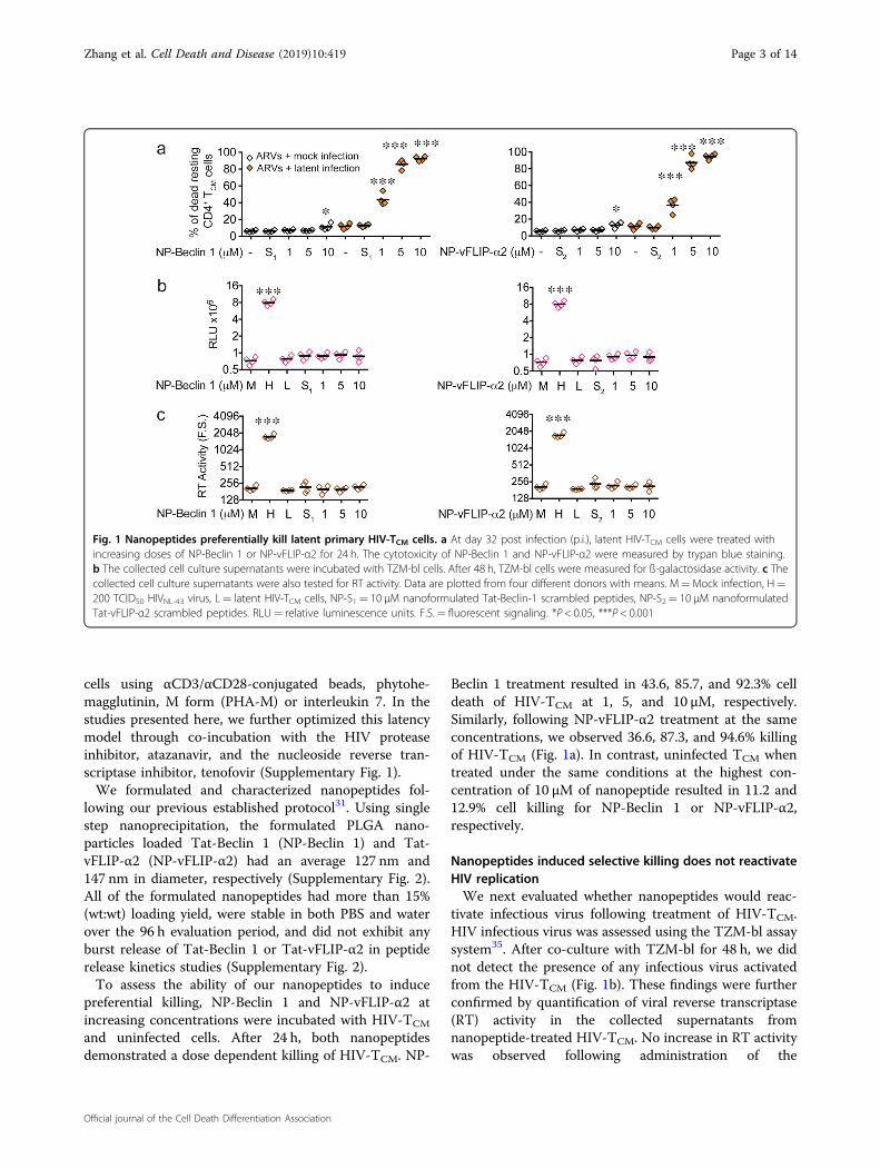
Beclin 1 treatment resulted in 43.6, 85.7, and 92.3% celldeath of HIV-TCM at 1, 5, and 10 µM, respectively.Similarly, following NP-vFLIP-α2 treatment at the sameconcentrations, we observed 36.6, 87.3, and 94.6% killingof HIV-TCM (Fig. 1a). In contrast, uninfected TCM whentreated under the same conditions at the highest con-centration of 10 µM of nanopeptide resulted in 11.2 and12.9% cell killing for NP-Beclin 1 or NP-vFLIP-α2,respectively.
Nanopeptides induced selective killing does not reactivateHIV replicationWe next evaluated whether nanopeptides would reac-
tivate infectious virus following treatment of HIV-TCM.HIV infectious virus was assessed using the TZM-bl assaysystem35. After co-culture with TZM-bl for 48 h, we didnot detect the presence of any infectious virus activatedfrom the HIV-TCM (Fig. 1b). These findings were furtherconfirmed by quantification of viral reverse transcriptase(RT) activity in the collected supernatants fromnanopeptide-treated HIV-TCM. No increase in RT activitywas observed following administration of the
Fig. 1 Nanopeptides preferentially kill latent primary HIV-TCM cells. a At day 32 post infection (p.i.), latent HIV-TCM cells were treated withincreasing doses of NP-Beclin 1 or NP-vFLIP-α2 for 24 h. The cytotoxicity of NP-Beclin 1 and NP-vFLIP-α2 were measured by trypan blue staining.b The collected cell culture supernatants were incubated with TZM-bl cells. After 48 h, TZM-bl cells were measured for ß-galactosidase activity. c Thecollected cell culture supernatants were also tested for RT activity. Data are plotted from four different donors with means. M=Mock infection, H=200 TCID50 HIVNL-43 virus, L= latent HIV-TCM cells, NP-S1= 10 μM nanoformulated Tat-Beclin-1 scrambled peptides, NP-S2= 10 μM nanoformulatedTat-vFLIP-α2 scrambled peptides. RLU= relative luminescence units. F.S.= fluorescent signaling. *P < 0.05, ***P < 0.001
Zhang et al. Cell Death and Disease (2019) 10:419 Page 3 of 14
Official journal of the Cell Death Differentiation Association
nanopeptides (Fig. 1c), further confirming that ournanopeptides kill latently infected TCM without reacti-vating HIV replication.
Killing of latently infected CD4+ T cells is autophagydependentBased on our previous research, we suspected that the
mechanism for the selective killing observed of HIV-TCM
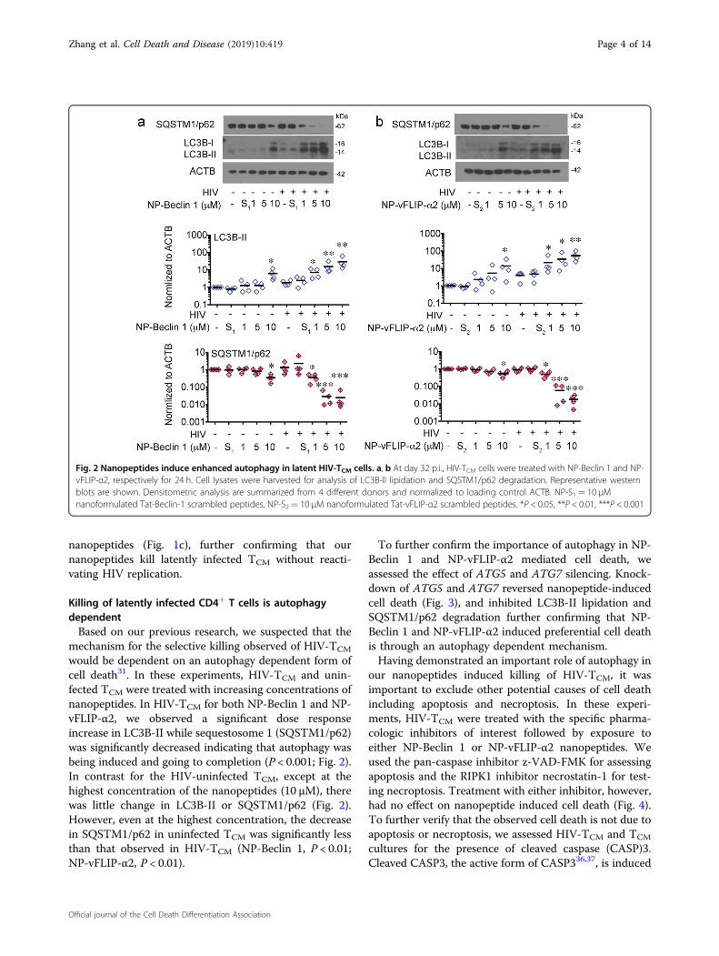
would be dependent on an autophagy dependent form ofcell death31. In these experiments, HIV-TCM and unin-fected TCM were treated with increasing concentrations ofnanopeptides. In HIV-TCM for both NP-Beclin 1 and NP-vFLIP-α2, we observed a significant dose responseincrease in LC3B-II while sequestosome 1 (SQSTM1/p62)was significantly decreased indicating that autophagy wasbeing induced and going to completion (P < 0.001; Fig. 2).In contrast for the HIV-uninfected TCM, except at thehighest concentration of the nanopeptides (10 μM), therewas little change in LC3B-II or SQSTM1/p62 (Fig. 2).However, even at the highest concentration, the decreasein SQSTM1/p62 in uninfected TCM was significantly lessthan that observed in HIV-TCM (NP-Beclin 1, P < 0.01;NP-vFLIP-α2, P < 0.01).
To further confirm the importance of autophagy in NP-Beclin 1 and NP-vFLIP-α2 mediated cell death, weassessed the effect of ATG5 and ATG7 silencing. Knock-down of ATG5 and ATG7 reversed nanopeptide-inducedcell death (Fig. 3), and inhibited LC3B-II lipidation andSQSTM1/p62 degradation further confirming that NP-Beclin 1 and NP-vFLIP-α2 induced preferential cell deathis through an autophagy dependent mechanism.Having demonstrated an important role of autophagy in
our nanopeptides induced killing of HIV-TCM, it wasimportant to exclude other potential causes of cell deathincluding apoptosis and necroptosis. In these experi-ments, HIV-TCM were treated with the specific pharma-cologic inhibitors of interest followed by exposure toeither NP-Beclin 1 or NP-vFLIP-α2 nanopeptides. Weused the pan-caspase inhibitor z-VAD-FMK for assessingapoptosis and the RIPK1 inhibitor necrostatin-1 for test-ing necroptosis. Treatment with either inhibitor, however,had no effect on nanopeptide induced cell death (Fig. 4).To further verify that the observed cell death is not due toapoptosis or necroptosis, we assessed HIV-TCM and TCM
cultures for the presence of cleaved caspase (CASP)3.Cleaved CASP3, the active form of CASP336,37, is induced
Fig. 2 Nanopeptides induce enhanced autophagy in latent HIV-TCM cells. a, b At day 32 p.i., HIV-TCM cells were treated with NP-Beclin 1 and NP-vFLIP-α2, respectively for 24 h. Cell lysates were harvested for analysis of LC3B-II lipidation and SQSTM1/p62 degradation. Representative westernblots are shown. Densitometric analysis are summarized from 4 different donors and normalized to loading control ACTB. NP-S1= 10 μMnanoformulated Tat-Beclin-1 scrambled peptides, NP-S2= 10 μM nanoformulated Tat-vFLIP-α2 scrambled peptides. *P < 0.05, **P < 0.01, ***P < 0.001
Zhang et al. Cell Death and Disease (2019) 10:419 Page 4 of 14
Official journal of the Cell Death Differentiation Association
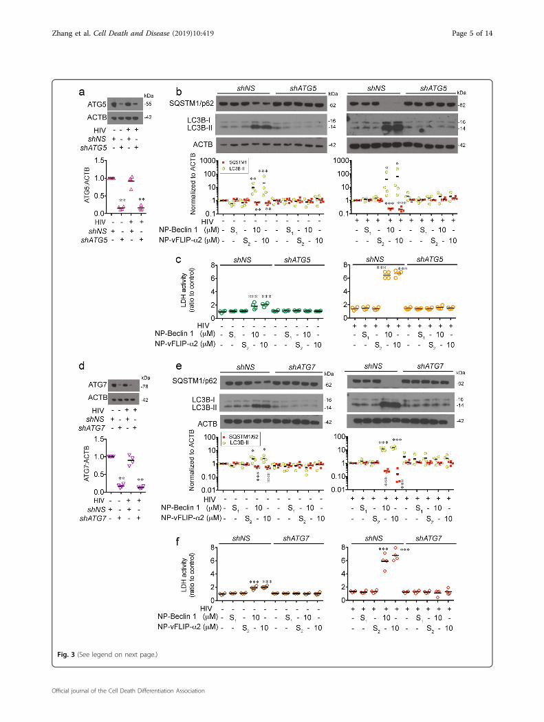
Fig. 3 (See legend on next page.)
Zhang et al. Cell Death and Disease (2019) 10:419 Page 5 of 14
Official journal of the Cell Death Differentiation Association
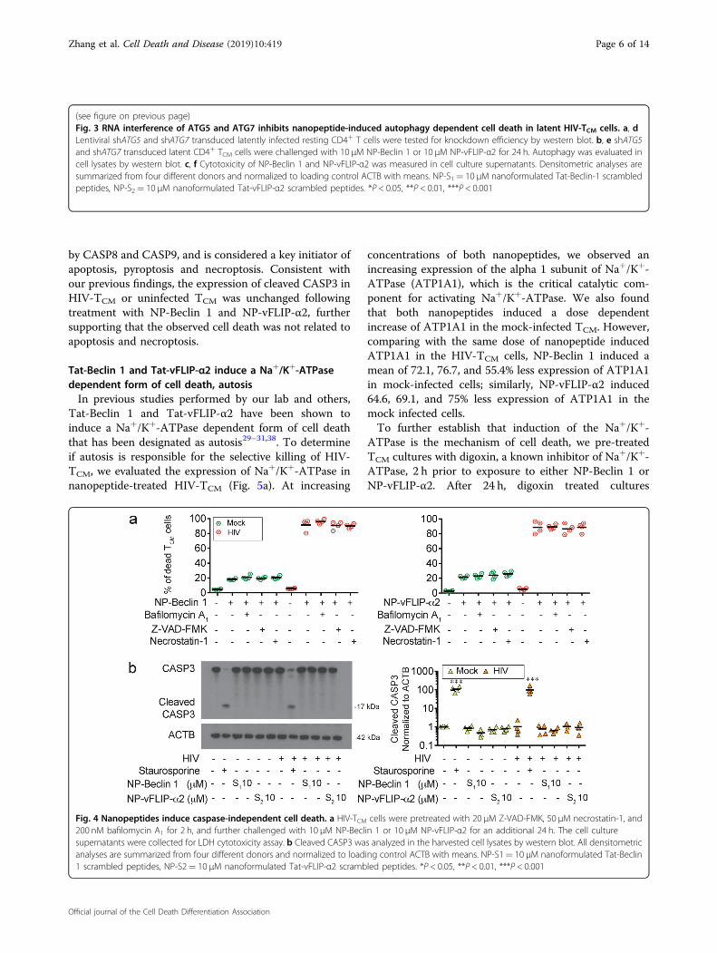
by CASP8 and CASP9, and is considered a key initiator ofapoptosis, pyroptosis and necroptosis. Consistent withour previous findings, the expression of cleaved CASP3 inHIV-TCM or uninfected TCM was unchanged followingtreatment with NP-Beclin 1 and NP-vFLIP-α2, furthersupporting that the observed cell death was not related toapoptosis and necroptosis.
Tat-Beclin 1 and Tat-vFLIP-α2 induce a Na+/K+-ATPasedependent form of cell death, autosisIn previous studies performed by our lab and others,
Tat-Beclin 1 and Tat-vFLIP-α2 have been shown toinduce a Na+/K+-ATPase dependent form of cell deaththat has been designated as autosis29–31,38. To determineif autosis is responsible for the selective killing of HIV-TCM, we evaluated the expression of Na+/K+-ATPase innanopeptide-treated HIV-TCM (Fig. 5a). At increasing
concentrations of both nanopeptides, we observed anincreasing expression of the alpha 1 subunit of Na+/K+-ATPase (ATP1A1), which is the critical catalytic com-ponent for activating Na+/K+-ATPase. We also foundthat both nanopeptides induced a dose dependentincrease of ATP1A1 in the mock-infected TCM. However,comparing with the same dose of nanopeptide inducedATP1A1 in the HIV-TCM cells, NP-Beclin 1 induced amean of 72.1, 76.7, and 55.4% less expression of ATP1A1in mock-infected cells; similarly, NP-vFLIP-α2 induced64.6, 69.1, and 75% less expression of ATP1A1 in themock infected cells.To further establish that induction of the Na+/K+-
ATPase is the mechanism of cell death, we pre-treatedTCM cultures with digoxin, a known inhibitor of Na+/K+-ATPase, 2 h prior to exposure to either NP-Beclin 1 orNP-vFLIP-α2. After 24 h, digoxin treated cultures
(see figure on previous page)Fig. 3 RNA interference of ATG5 and ATG7 inhibits nanopeptide-induced autophagy dependent cell death in latent HIV-TCM cells. a, dLentiviral shATG5 and shATG7 transduced latently infected resting CD4+ T cells were tested for knockdown efficiency by western blot. b, e shATG5and shATG7 transduced latent CD4+ TCM cells were challenged with 10 μM NP-Beclin 1 or 10 μM NP-vFLIP-α2 for 24 h. Autophagy was evaluated incell lysates by western blot. c, f Cytotoxicity of NP-Beclin 1 and NP-vFLIP-α2 was measured in cell culture supernatants. Densitometric analyses aresummarized from four different donors and normalized to loading control ACTB with means. NP-S1= 10 μM nanoformulated Tat-Beclin-1 scrambledpeptides, NP-S2= 10 μM nanoformulated Tat-vFLIP-α2 scrambled peptides. *P < 0.05, **P < 0.01, ***P < 0.001
Fig. 4 Nanopeptides induce caspase-independent cell death. a HIV-TCM cells were pretreated with 20 μM Z-VAD-FMK, 50 μM necrostatin-1, and200 nM bafilomycin A1 for 2 h, and further challenged with 10 μM NP-Beclin 1 or 10 μM NP-vFLIP-α2 for an additional 24 h. The cell culturesupernatants were collected for LDH cytotoxicity assay. b Cleaved CASP3 was analyzed in the harvested cell lysates by western blot. All densitometricanalyses are summarized from four different donors and normalized to loading control ACTB with means. NP-S1= 10 μM nanoformulated Tat-Beclin1 scrambled peptides, NP-S2= 10 μM nanoformulated Tat-vFLIP-α2 scrambled peptides. *P < 0.05, **P < 0.01, ***P < 0.001
Zhang et al. Cell Death and Disease (2019) 10:419 Page 6 of 14
Official journal of the Cell Death Differentiation Association
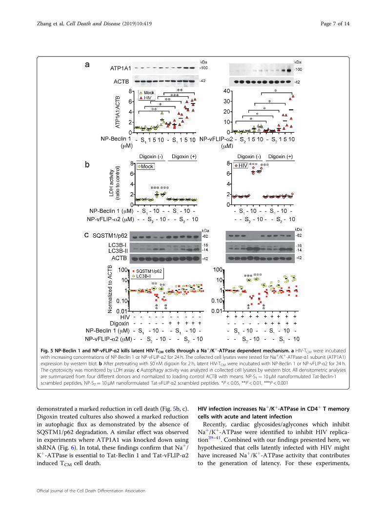
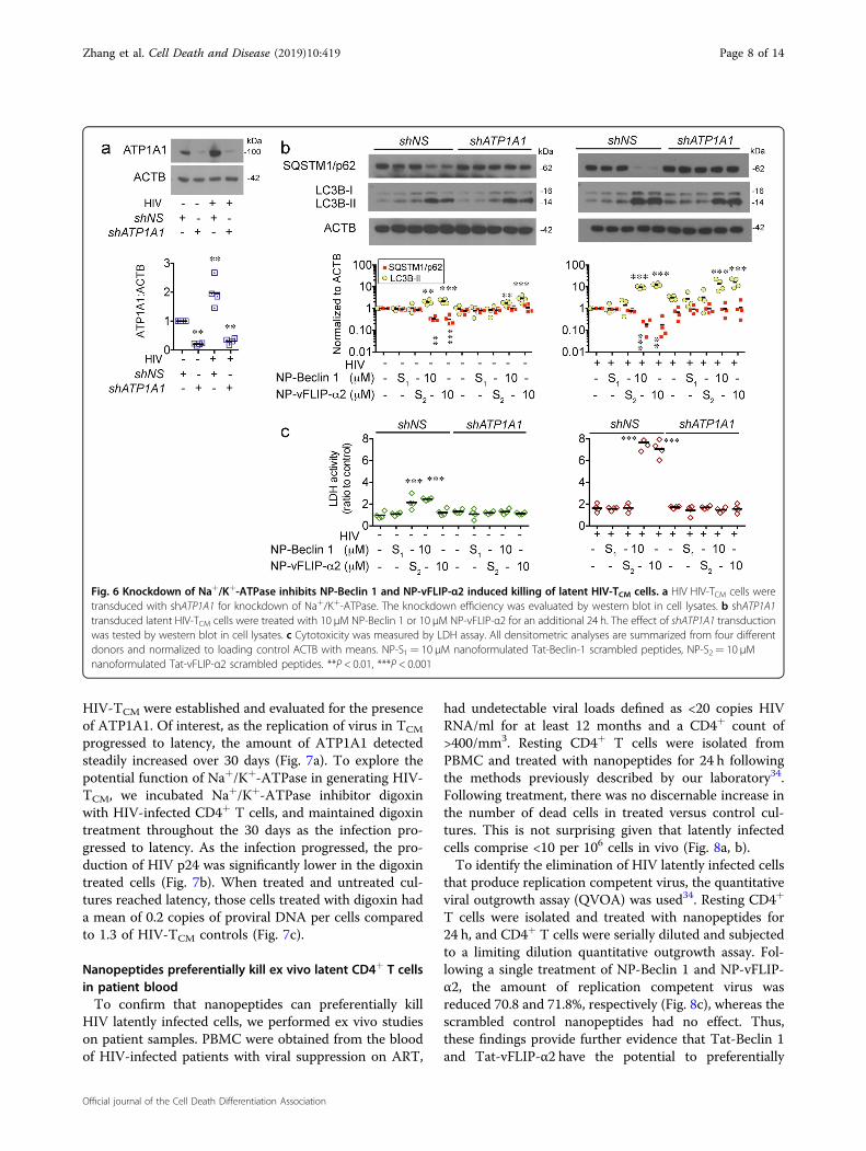
demonstrated a marked reduction in cell death (Fig. 5b, c).Digoxin treated cultures also showed a marked reductionin autophagic flux as demonstrated by the absence ofSQSTM1/p62 degradation. A similar effect was observedin experiments where ATP1A1 was knocked down usingshRNA (Fig. 6). In total, these findings confirm that Na+/K+-ATPase is essential to Tat-Beclin 1 and Tat-vFLIP-α2induced TCM cell death.
HIV infection increases Na+/K+-ATPase in CD4+ T memorycells with acute and latent infectionRecently, cardiac glycosides/aglycones which inhibit
Na+/K+-ATPase were identified to inhibit HIV replica-tion39–41. Combined with our findings presented here, wehypothesized that cells latently infected with HIV mighthave increased Na+/K+-ATPase activity that contributesto the generation of latency. For these experiments,
Fig. 5 NP-Beclin 1 and NP-vFLIP-α2 kills latent HIV-TCM cells through a Na+/K+-ATPase dependent mechanism. a HIV-TCM were incubatedwith increasing concentrations of NP-Beclin 1 or NP-vFLIP-α2 for 24 h. The collected cell lysates were tested for Na+/K+-ATPase-α1 subunit (ATP1A1)expression by western blot. b After pretreating with 50 nM digoxin for 2 h, latent HIV-TCM were incubated with NP-Beclin 1 or NP-vFLIP-α2 for 24 h.The cytotoxicity was monitored by LDH assay. c Autophagy activity was analyzed in collected cell lysates by western blot. All densitometric analysesare summarized from four different donors and normalized to loading control ACTB with means. NP-S1= 10 μM nanoformulated Tat-Beclin-1scrambled peptides, NP-S2= 10 μM nanoformulated Tat-vFLIP-α2 scrambled peptides. *P < 0.05, **P < 0.01, ***P < 0.001
Zhang et al. Cell Death and Disease (2019) 10:419 Page 7 of 14
Official journal of the Cell Death Differentiation Association
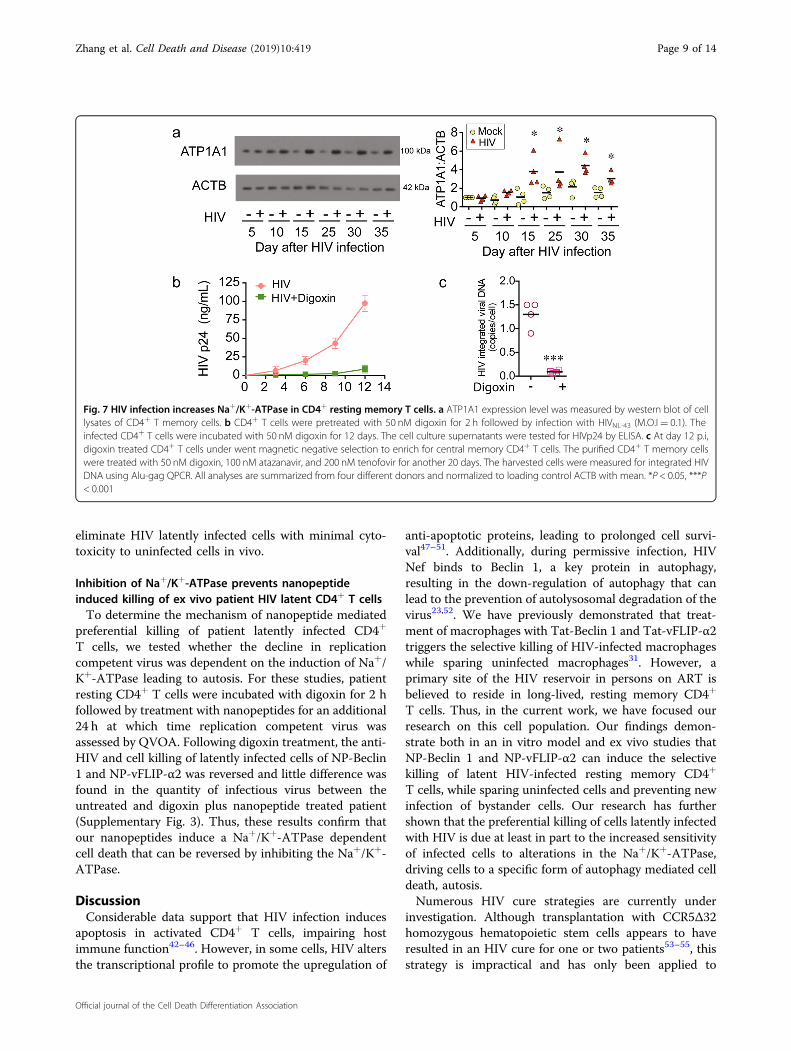
HIV-TCM were established and evaluated for the presenceof ATP1A1. Of interest, as the replication of virus in TCM
progressed to latency, the amount of ATP1A1 detectedsteadily increased over 30 days (Fig. 7a). To explore thepotential function of Na+/K+-ATPase in generating HIV-TCM, we incubated Na+/K+-ATPase inhibitor digoxinwith HIV-infected CD4+ T cells, and maintained digoxintreatment throughout the 30 days as the infection pro-gressed to latency. As the infection progressed, the pro-duction of HIV p24 was significantly lower in the digoxintreated cells (Fig. 7b). When treated and untreated cul-tures reached latency, those cells treated with digoxin hada mean of 0.2 copies of proviral DNA per cells comparedto 1.3 of HIV-TCM controls (Fig. 7c).
Nanopeptides preferentially kill ex vivo latent CD4+ T cellsin patient bloodTo confirm that nanopeptides can preferentially kill
HIV latently infected cells, we performed ex vivo studieson patient samples. PBMC were obtained from the bloodof HIV-infected patients with viral suppression on ART,
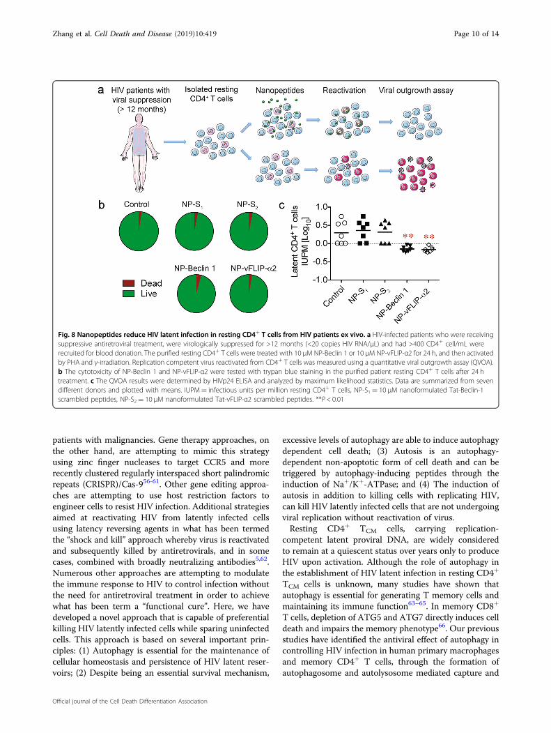
had undetectable viral loads defined as <20 copies HIVRNA/ml for at least 12 months and a CD4+ count of>400/mm3. Resting CD4+ T cells were isolated fromPBMC and treated with nanopeptides for 24 h followingthe methods previously described by our laboratory34.Following treatment, there was no discernable increase inthe number of dead cells in treated versus control cul-tures. This is not surprising given that latently infectedcells comprise <10 per 106 cells in vivo (Fig. 8a, b).To identify the elimination of HIV latently infected cells
that produce replication competent virus, the quantitativeviral outgrowth assay (QVOA) was used34. Resting CD4+
T cells were isolated and treated with nanopeptides for24 h, and CD4+ T cells were serially diluted and subjectedto a limiting dilution quantitative outgrowth assay. Fol-lowing a single treatment of NP-Beclin 1 and NP-vFLIP-α2, the amount of replication competent virus wasreduced 70.8 and 71.8%, respectively (Fig. 8c), whereas thescrambled control nanopeptides had no effect. Thus,these findings provide further evidence that Tat-Beclin 1and Tat-vFLIP-α2 have the potential to preferentially
Fig. 6 Knockdown of Na+/K+-ATPase inhibits NP-Beclin 1 and NP-vFLIP-α2 induced killing of latent HIV-TCM cells. a HIV HIV-TCM cells weretransduced with shATP1A1 for knockdown of Na+/K+-ATPase. The knockdown efficiency was evaluated by western blot in cell lysates. b shATP1A1transduced latent HIV-TCM cells were treated with 10 μM NP-Beclin 1 or 10 μM NP-vFLIP-α2 for an additional 24 h. The effect of shATP1A1 transductionwas tested by western blot in cell lysates. c Cytotoxicity was measured by LDH assay. All densitometric analyses are summarized from four differentdonors and normalized to loading control ACTB with means. NP-S1= 10 μM nanoformulated Tat-Beclin-1 scrambled peptides, NP-S2= 10 μMnanoformulated Tat-vFLIP-α2 scrambled peptides. **P < 0.01, ***P < 0.001
Zhang et al. Cell Death and Disease (2019) 10:419 Page 8 of 14
Official journal of the Cell Death Differentiation Association
eliminate HIV latently infected cells with minimal cyto-toxicity to uninfected cells in vivo.
Inhibition of Na+/K+-ATPase prevents nanopeptideinduced killing of ex vivo patient HIV latent CD4+ T cellsTo determine the mechanism of nanopeptide mediated
preferential killing of patient latently infected CD4+
T cells, we tested whether the decline in replicationcompetent virus was dependent on the induction of Na+/K+-ATPase leading to autosis. For these studies, patientresting CD4+ T cells were incubated with digoxin for 2 hfollowed by treatment with nanopeptides for an additional24 h at which time replication competent virus wasassessed by QVOA. Following digoxin treatment, the anti-HIV and cell killing of latently infected cells of NP-Beclin1 and NP-vFLIP-α2 was reversed and little difference wasfound in the quantity of infectious virus between theuntreated and digoxin plus nanopeptide treated patient(Supplementary Fig. 3). Thus, these results confirm thatour nanopeptides induce a Na+/K+-ATPase dependentcell death that can be reversed by inhibiting the Na+/K+-ATPase.
DiscussionConsiderable data support that HIV infection induces
apoptosis in activated CD4+ T cells, impairing hostimmune function42–46. However, in some cells, HIV altersthe transcriptional profile to promote the upregulation of
anti-apoptotic proteins, leading to prolonged cell survi-val47–51. Additionally, during permissive infection, HIVNef binds to Beclin 1, a key protein in autophagy,resulting in the down-regulation of autophagy that canlead to the prevention of autolysosomal degradation of thevirus23,52. We have previously demonstrated that treat-ment of macrophages with Tat-Beclin 1 and Tat-vFLIP-α2triggers the selective killing of HIV-infected macrophageswhile sparing uninfected macrophages31. However, aprimary site of the HIV reservoir in persons on ART isbelieved to reside in long-lived, resting memory CD4+
T cells. Thus, in the current work, we have focused ourresearch on this cell population. Our findings demon-strate both in an in vitro model and ex vivo studies thatNP-Beclin 1 and NP-vFLIP-α2 can induce the selectivekilling of latent HIV-infected resting memory CD4+
T cells, while sparing uninfected cells and preventing newinfection of bystander cells. Our research has furthershown that the preferential killing of cells latently infectedwith HIV is due at least in part to the increased sensitivityof infected cells to alterations in the Na+/K+-ATPase,driving cells to a specific form of autophagy mediated celldeath, autosis.Numerous HIV cure strategies are currently under
investigation. Although transplantation with CCR5Δ32homozygous hematopoietic stem cells appears to haveresulted in an HIV cure for one or two patients53–55, thisstrategy is impractical and has only been applied to
Fig. 7 HIV infection increases Na+/K+-ATPase in CD4+ resting memory T cells. a ATP1A1 expression level was measured by western blot of celllysates of CD4+ T memory cells. b CD4+ T cells were pretreated with 50 nM digoxin for 2 h followed by infection with HIVNL-43 (M.O.I= 0.1). Theinfected CD4+ T cells were incubated with 50 nM digoxin for 12 days. The cell culture supernatants were tested for HIVp24 by ELISA. c At day 12 p.i,digoxin treated CD4+ T cells under went magnetic negative selection to enrich for central memory CD4+ T cells. The purified CD4+ T memory cellswere treated with 50 nM digoxin, 100 nM atazanavir, and 200 nM tenofovir for another 20 days. The harvested cells were measured for integrated HIVDNA using Alu-gag QPCR. All analyses are summarized from four different donors and normalized to loading control ACTB with mean. *P < 0.05, ***P< 0.001
Zhang et al. Cell Death and Disease (2019) 10:419 Page 9 of 14
Official journal of the Cell Death Differentiation Association
patients with malignancies. Gene therapy approaches, onthe other hand, are attempting to mimic this strategyusing zinc finger nucleases to target CCR5 and morerecently clustered regularly interspaced short palindromicrepeats (CRISPR)/Cas-956-61. Other gene editing approa-ches are attempting to use host restriction factors toengineer cells to resist HIV infection. Additional strategiesaimed at reactivating HIV from latently infected cellsusing latency reversing agents in what has been termedthe “shock and kill” approach whereby virus is reactivatedand subsequently killed by antiretrovirals, and in somecases, combined with broadly neutralizing antibodies5,62.Numerous other approaches are attempting to modulatethe immune response to HIV to control infection withoutthe need for antiretroviral treatment in order to achievewhat has been term a “functional cure”. Here, we havedeveloped a novel approach that is capable of preferentialkilling HIV latently infected cells while sparing uninfectedcells. This approach is based on several important prin-ciples: (1) Autophagy is essential for the maintenance ofcellular homeostasis and persistence of HIV latent reser-voirs; (2) Despite being an essential survival mechanism,
excessive levels of autophagy are able to induce autophagydependent cell death; (3) Autosis is an autophagy-dependent non-apoptotic form of cell death and can betriggered by autophagy-inducing peptides through theinduction of Na+/K+-ATPase; and (4) The induction ofautosis in addition to killing cells with replicating HIV,can kill HIV latently infected cells that are not undergoingviral replication without reactivation of virus.Resting CD4+ TCM cells, carrying replication-
competent latent proviral DNA, are widely consideredto remain at a quiescent status over years only to produceHIV upon activation. Although the role of autophagy inthe establishment of HIV latent infection in resting CD4+
TCM cells is unknown, many studies have shown thatautophagy is essential for generating T memory cells andmaintaining its immune function63–65. In memory CD8+
T cells, depletion of ATG5 and ATG7 directly induces celldeath and impairs the memory phenotype66. Our previousstudies have identified the antiviral effect of autophagy incontrolling HIV infection in human primary macrophagesand memory CD4+ T cells, through the formation ofautophagosome and autolysosome mediated capture and
Fig. 8 Nanopeptides reduce HIV latent infection in resting CD4+ T cells from HIV patients ex vivo. a HIV-infected patients who were receivingsuppressive antiretroviral treatment, were virologically suppressed for >12 months (<20 copies HIV RNA/μL) and had >400 CD4+ cell/mL wererecruited for blood donation. The purified resting CD4+ T cells were treated with 10 μM NP-Beclin 1 or 10 μM NP-vFLIP-α2 for 24 h, and then activatedby PHA and γ-irradiation. Replication competent virus reactivated from CD4+ T cells was measured using a quantitative viral outgrowth assay (QVOA).b The cytotoxicity of NP-Beclin 1 and NP-vFLIP-α2 were tested with trypan blue staining in the purified patient resting CD4+ T cells after 24 htreatment. c The QVOA results were determined by HIVp24 ELISA and analyzed by maximum likelihood statistics. Data are summarized from sevendifferent donors and plotted with means. IUPM= infectious units per million resting CD4+ T cells, NP-S1= 10 μM nanoformulated Tat-Beclin-1scrambled peptides, NP-S2= 10 μM nanoformulated Tat-vFLIP-α2 scrambled peptides. **P < 0.01
Zhang et al. Cell Death and Disease (2019) 10:419 Page 10 of 14
Official journal of the Cell Death Differentiation Association
degradation of viral proteins24–26,31,34. In this study, wefurther identify that autophagy-inducing peptides havethe potential to eliminate HIV latent infection in TCM
cells using an autophagy-dependent mechanism.To improve the translational potential of our approach,
we have loaded the autophagy-inducing peptides intolipid-coated hybrid PLGA nanoparticles using bio-compatible and biodegradable materials. Combining theunique features of liposomes and polymeric nanoparticles,our lipid-coated hybrid PLGA nanoparticles are capableof effective delivery of encapsulated cargos67. In thisstudy, we successfully loaded Tat-Beclin 1 or Tat-vFLIP-α2 into the hybrid nanoparticles resulting in sustainedrelease of autophagy-inducing peptides over 96 h. Thislipid hybrid PLGA nanoformulation has been shown toextend the bioavailability of autophagy-inducing peptidesin human macrophages for almost one week in our pre-vious study31. Indeed, the hybrid nanoparticles haveimproved the intracellular bioavailability of our peptidesto HIV-infected cells, and transformed the encapsulatedpeptide drug into biocompatible and translational drugcandidate68. Recently, we developed a novel targetedversion of the PLGA nanoparticles with surface coating ofCD4+ T cell plasma membrane, which broadly recognizesHIV envelope protein, glycosylated protein 120 (gp120)69.This CD4+ T cell plasma membrane coated polymericnanoparticle has high binding affinity to HIV gp120leading to robust antiretroviral activity through neu-tralizing cell free HIV entering into CD4+ T cells andmacrophages. This new formulation has the potential toimprove the targeted killing of all HIVgp120 positive cellsincluding chronically infected and reactivated latentlyinfected cells.In summary, we have identified that two nanopeptides,
Tat-Beclin 1 and Tat-vFLIP-α2, targeting proteins criticalto autophagy induce Na+/K+-ATPase and selectively killHIV latently infected resting memory CD4+ T cells withlittle effect on uninfected cells. The preferential killing oflatently infected cells is dependent on increased Na+/K+-ATPase as the induction of cell death can be reversed bydigoxin, an inhibitor of Na+/K+-ATPase. These peptideswhen loaded into PLGA nanoparticles can be deliveredand incorporated into HIV-infected cells, and are syn-thesized from FDA-approved material that will facilitatetheir ability to be used in humans. Thus, we believe thatthese peptides have great potential to be used as part of anoverall strategy designed to eliminate HIV from infectedpersons.
Materials and methodsEthics statementVenous blood was obtained from HIV seronegative and
HIV seropositive donors. The protocol was reviewed andapproved by the Human Research Protections Program of
the University of California, San Diego (Project 09-0660).Written informed consent was obtained from all blooddonors.
Preparation of NP-Beclin 1 and NP-vFLIP-α2Autophagy-inducing peptides including Tat-Beclin 1
(RRRQRRKKRGY-GG-TGFEGDHWIEFTANFVNT), scr-ambled Tat-Beclin 1 (RRRQRRKKRGY-GG-WETAFGT-TEHNIFFDNGV), Tat-vFLIP-α2 (RRRQRRKKRGY-GFVNLLFLVVE) and scrambled Tat-vFLIP-α2 (RRRQRRKKRGY-GFVNLAAAVVE), were synthesized in D-isomer sequenceand obtained from New England Peptide, Inc. Autophagy-inducing peptides loaded lipid-coated PLGA nanoparticleswere synthesized using single step nanoprecipitation. Poly(D,L-lactic-co-glycolic acid) PLGA (50:50, 0.67 dl/g, Pelham,AL) with ester-terminated and autophagy-inducing peptideswere dissolved in organic solvent acetonitrile at 10mg/ml.Soybean lecithin and DSPE-PEG2000-COOH (1,2-distearoyl-sn-glycero-3-phosphoethanolamine-N-carboxy (polyenyleneglycol)2000) (Alabaster, AL) was mixed in chloroform at20% of the PLGA polymer weight and air-dried as a lipidfilm. The dissolved PLGA/autophagy-inducing peptidesorganic solution was added into the lipid film under gentlestirring and further vortexed for 3min. PLGA nanoparticleswere then washed with 10mM tris-HCl pH 8 buffer andloaded with autophagy-inducing peptides. The remainingacetonitrile and free polymers were removed by washingusing an Amicon Ultra-4 centrifugal filter (Millipore, Bill-erica, MA) with a molecular weight cut-off of 10 kDa.
Characterization of NP-Beclin 1 and NP-vFLIP-α2The morphological characteristics of NP-Beclin 1 and
NP-vFLIP-α2 were evaluated by JEOL Gatan transmissionelectron microscopy (TEM) using negative staining. Thesize, polydispersity index and surface zeta potential weremeasured by dynamic light scattering Malvern ZetasizerNano ZS for each of nanoformulations. The peptideloading capacity was evaluated by Slide-A-Yzer MINIdialysis microtube with a molecular weight cutoff of3.5 kDa (Thermofisher, Rockford, IL), and determined bythe weight ratio of the peptide payload to the PLGAnanoparticles.
Generation of HIV latently infected primary restingmemory CD4+ T cellsHIV latently infected resting memory T cells were
generated following our previously published protocol34.Briefly, resting memory CD4+ T cells from HIV-uninfected donors were suspended with RPMI 1640medium with 10% fetal bovine serum supplemented with29 nM CCL19 (PeproTech, Rocky Hill, NJ) for 48 h. Next,the CD4+ T cells were infected with HIVNL4-3 at a mul-tiplicity of infection (M.O.I.) of 0.1, and incubated with250 ng/mL staphylococcal enterotoxin B (Sigma, St.Louis,
Zhang et al. Cell Death and Disease (2019) 10:419 Page 11 of 14
Official journal of the Cell Death Differentiation Association

MO) and 25 U/mL IL-2 (Roche, Basel, Switzerland) for anadditional 3 days. After removal of staphylococcal enter-otoxin, cells were cultured with 25 U/mL IL-2 for another9 days. Then, the central memory CD4+ T cells werepurified from HIV-infected CD4+ T cells using negativemagnetic isolation, and further cultured with 1 ng/mL IL-7 (PeproTech, Rocky Hill, NJ), 100 nM atazanavir and200 nM tenofovir (Selleckchem, Houston, TX) for another20 days.
Quantitative viral out growth assay of HIV latent ex vivoCD4+ T in the isolated patient bloodOur laboratory has adapted the Quantitative viral out
growth assay (QVOA) assay previously established by theSiliciano laboratory34,70. HIV-infected patients who werevirologically suppressed on ART, had a viral load of <20copies/mL for at least 12 months and had a CD4+ countof >400 cells/mm3 were recruited and obtained informedconsent at University of California San Diego Mother-Child-Adolescent HIV Program clinic, and Peripheralblood mononuclear cells (PBMC) were isolated fromcollected venous blood using Ficoo-PaqueTM densitycentrifugation (GE Healthcare, Pittsburgh, PA). RestingCD4+ T cells were purified through negative magneticcell isolation of human CD25, CD69 and anti-HLA-DRfollowing the manufacturer’s protocols, and reactivatedwith gamma irradiation (5000R in Cs-source) and phy-tohemagglutinin (PHA-M, 1 μg/mL). The CD8+ T celldepleted PBMC were reactivated with PHA-M and usedas feeder cells to co-culture with limiting dilution ofresting CD4+ T cells for 3 weeks. The collected cell cul-tured supernatants were tested with HIV p24 antigenusing by ELISA.
Western blottingThe collected cell lysates were mixed with pierce lane
marker reducing sample buffer (ThermoFisher Scientific,Waltham, MA) and boiled for 5 min to achieve proteindenaturation. The protein samples were separated byExpressPlus PAGE Gels and electrophoretic transferred toPVDF or nitrocellulose membrane. The targeted proteinswere detected by primary anti-LC3B (Novus Biologicals,Littleton, CO), anti-ATP1A1, anti-ATG5, anti-ATG7(Cell Signaling Technology, Danvers, MA) and anti-SQSTM1 antibodies (Abcam, Cambridge, MA). Thehorseradish peroxidase conjugated goat anti-mouse andanti-rabbit secondary antibodies were used to amplify thedetected antigen.
Cytotoxicity assayThe collected cell culture supernatants were co-
incubated with LDH cytotoxicity assay reaction bufferfollowing the manufacture’s protocol (Clontech, Moun-tain View, CA). The colorimetric results were quantified
using a BioTek microplate reader at 490 nm wavelength.The dead cells were counterstained with 0.4% trypan bluesolution and quantified by Nexcelom cell counter.
TZM-bl HIV infectivity assayTZM-bl HIV infectivity was tested following an estab-
lished protocol35. Briefly, the collected cell culturesupernatants were loaded into 96-well plates containing50,000 TZM-bl cells per well in RPMI 1640 medium with10% FBS, and incubated at 37 °C for 48 h. After washing,the incubated TZM-bl cells were measured by Beta-Gloassay kit (Promega, Madison, WI).
Measurement of HIV infectionGenomic DNA from TCM was isolated through the
PureLink® Genomic DNA Kits (Invitrogen, Carlsbad,CA). Following the established protocol71–73, integratedviral DNA was measured with Alu-gag QPCR. The iso-lated genomic DNA from ACH-2 and 8E5 cell lines wasused as HIV standards. The collected cell culture super-naturants were tested for HIVp24 ELISA (PerkinElmer,Waltham, MA) and RT activity (ThermoFisher, Carlsbad,CA) following the manufacture protocol.
RNA interferenceShort hairpin RNA lentiviral transduction particles kits
were purchased from Sigma-Aldrich for silencing ATG5,ATG7 and ATP1A1 in primary central CD4+ T memorycells (ATG5-TRCN0000151963, ATG7-TRCN0000435480and ATP1A1-TRCN0000424769). The lentiviral shRNAwere transduced following the manufacture’s protocols.shRNA control vector was also obtained from Sigma-Aldrich used as non-specific targeting control (SHC002).
StatisticsAll results were assessed in GraphPad Prism 6.0
(GraphPad, La Jolla, CA). The normalized data includingfold and ratio changes were transformed into log2 value tonormalize the data. Two-tailed Student t test, ANOVA,Pearson correlation and Wilcoxon rank test were appliedfor statistical analysis. P values < 0.05 two-tailed wereconsidered statistically significant.
AcknowledgementsWe thank Erin Maule, Jonathan Hana and Morcel Hamidy for experimentalassistance, and Siyu Zhu and Zhe Zhong for assistance with illustration andstatistical analysis. This work was supported, in whole or in part, by the NationalInstitute of Neurological Disorders and Stroke of the NIH under Grant R01NS084912 and R01 NS104015; International Maternal Pediatric Adolescent AIDSClinical Trials Network. Overall support for the International Maternal PediatricAdolescent AIDS Clinical Trials (IMPAACT) Network was provided by theNational Institute of Allergy and Infectious Diseases (NIAID) of the NationalInstitutes of Health (NIH) under Grant UM1AI068632 (IMPAACT LOC),UM1AI068616 (IMPAACT SDMC) and UM1AI106716 (IMPAACT LC), with co-funding from the Eunice Kennedy Shriver National Institute of Child Health andHuman Development (NICHD) and the National Institute of Mental Health(NIMH), National Institute of Allergy and Infectious Diseases (NIAID)
Zhang et al. Cell Death and Disease (2019) 10:419 Page 12 of 14
Official journal of the Cell Death Differentiation Association
[UM1AI068632] and National Institute of Allergy and Infectious Diseases(NIAID) [UM1AI106716].
Author details1Division of Infectious Diseases, Department of Pediatrics, University ofCalifornia San Diego, La Jolla, CA, USA. 2Department of NanoEngineering andMoores Cancer Center, University of California San Diego, La Jolla, CA, USA.3Rady Children’s Hospital, San Diego, CA, USA
Authors contributionsG.Z., L.Z., and S.A.S designed and conceived the research. G.Z., B.T.L, X.W., G.R.C.,R.H.F. performed the experiments. G.Z., L.Z., and SAS analyzed the data. G.Z., L.Z., and S.A.S. wrote the manuscript.
Conflict of interestThe authors declare that they have no conflict of interest.
Publisher’s noteSpringer Nature remains neutral with regard to jurisdictional claims inpublished maps and institutional affiliations.
Supplementary Information accompanies this paper at (https://doi.org/10.1038/s41419-019-1661-7).
Received: 10 January 2019 Revised: 26 March 2019 Accepted: 3 April 2019
References1. Yoshimura, K. Current status of HIV/AIDS in the ART era. J. Infect. Chemother.
23, 12–16 (2017).2. Rainwater-Lovett, K., Luzuriaga, K. & Persaud, D. Very early combination anti-
retroviral therapy in infants: prospects for cure. Curr. Opin. Hiv Aids 10, 4–11(2015).
3. Chun, T. W. et al. Rebound of plasma viremia following cessation of anti-retroviral therapy despite profoundly low levels of HIV reservoir: implicationsfor eradication. Aids 24, 2803–2808 (2010).
4. Chun, T. W., Moir, S., Kovacs, C. & Fauci, A. S. Rebound of plasma viremiafollowing cessation of antiretroviral therapy despite profoundly low levels ofHIV reservoir: implications for eradication Reply. Aids 25, 872–873 (2011).
5. Deeks, S. G. HIV Shock and kill. Nature 487, 439–440 (2012).6. Finzi, D. et al. Identification of a reservoir for HIV-1 in patients on highly active
antiretroviral therapy. Science 278, 1295–1300 (1997).7. Murray, A. J., Kwon, K. J., Farber, D. L. & Siliciano, R. F. The latent reservoir for
HIV-1: how immunologic memory and clonal expansion contribute to HIV-1persistence. J. Immunol. 197, 407–417 (2016).
8. Chun, T. W. et al. Presence of an inducible HIV-1 latent reservoir during highlyactive antiretroviral therapy. Proc Natl Acad. Sci. USA 94, 13193–13197 (1997).
9. Deretic, V., Saitoh, T. & Akira, S. Autophagy in infection, inflammation andimmunity. Nat. Rev. Immunol 13, 722–737 (2013).
10. Choi, Y., Bowman, J. W. & Jung, J. U. Autophagy during viral infection—adouble-edged sword. Nat. Rev. Microbiol. 16, 340–353 (2018).
11. Lamb, C. A., Yoshimori, T. & Tooze, S. A. The autophagosome: originsunknown, biogenesis complex. Nat. Rev. Mol. Cell. Bio. 14, 759–774 (2013).
12. Button, R. W., Roberts, S. L., Willis, T. L., Hanemann, C. O. & Luo, S. Q. Accu-mulation of autophagosomes confers cytotoxicity. J. Biol. Chem. 292,13599–13614 (2017).
13. Fulda, S. & Kogel, D. Cell death by autophagy: emerging molecularmechanisms and implications for cancer therapy. Oncogene 34, 5105–5113(2015).
14. Doherty, J. & Baehrecke, E. H. Life, death and autophagy. Nat. Cell. Biol. 20,1110–1117 (2018).
15. Nardacci, R. et al. Role of autophagy in HIV infection and pathogenesis. J.Intern. Med. 281, 422–432 (2017).
16. Gomez-Mora, E. et al. Brief Report: Impaired CD4 T-Cell Response to Autop-hagy in Treated HIV-1-Infected Individuals. J. Acquir. Immune Defic. Syndr. 74,201–205 (2017).
17. Mao, K. & Klionsky, D. J. Xenophagy: a battlefield between host and microbe,and a possible avenue for cancer treatment. Autophagy 13, 223–224 (2017).
18. Wileman, T. Autophagy as a defence against intracellular pathogens. EssaysBiochem. 55, 153–163 (2013).
19. Nardacci, R. et al. Autophagy plays an important role in the containment ofHIV-1 in nonprogressor-infected patients. Autophagy 10, 1167–1178 (2014).
20. Zhou, D. J., Masliah, E. & Spector, S. A. Autophagy is increased in postmortembrains of persons with HIV-1-associated Encephalitis. J. Infect. Dis. 203,1647–1657 (2011).
21. Zhou, D. J., Kang, K. H. & Spector, S. A. Production of interferon alpha byhuman immunodeficiency virus type 1 in human plasmacytoid dendritic cellsis dependent on induction of autophagy. J. Infect. Dis. 205, 1258–1267 (2012).
22. Zhou, D. J. & Spector, S. A. Human immunodeficiency virus type-1 infectioninhibits autophagy. Aids 22, 695–699 (2008).
23. Campbell, G. R., Rawat, P., Bruckman, R. S. & Spector, S. A. Human immuno-deficiency virus type 1 Nef inhibits autophagy through transcription Factor EBsequestration. Plos Pathog. 11, e1005018 (2015).
24. Campbell, G. R. & Spector, S. A. Vitamin D Inhibits human immunodeficiencyvirus type 1 and Mycobacterium Tuberculosis infection in macrophagesthrough the induction of autophagy. Plos Pathog. 8, e1005018 (2012).
25. Campbell, G. R. & Spector, S. A. Hormonally active vitamin D3 (1 alpha,25-Dihydroxycholecalciferol) triggers autophagy in human macrophages thatinhibits HIV-1 infection. J. Biol. Chem. 286, 18890–18902 (2011).
26. Campbell, G. R. et al. Induction of autophagy by PI3K/MTOR and PI3K/MTOR/BRD4 inhibitors suppresses HIV-1 replication. J. Biol. Chem. 293, 5808–5820(2018).
27. Shoji-Kawata, S. et al. Identification of a candidate therapeutic autophagy-inducing peptide. Nature 494, 201–206 (2013).
28. Lee, J. S. et al. FLIP-mediated autophagy regulation in cell death control. Nat.Cell. Biol. 11, 1355–U1225 (2009).
29. Liu, Y. et al. Autosis is a Na+,K+-ATPase-regulated form of cell death triggeredby autophagy-inducing peptides, starvation, and hypoxia-ischemia. Proc. NatlAcad. Sci. USA 110, 20364–20371 (2013).
30. Liu, Y. & Levine, B. Autosis and autophagic cell death: the dark side ofautophagy. Cell Death Differ. 22, 367–376 (2015).
31. Zhang, G., Luk, B. T., Hamidy, M., Zhang, L. F. & Spector, S. A. Induction of a Na+/K+-ATPase-dependent form of autophagy triggers preferential cell deathof human immunodeficiency virus type-1-infected macrophages. Autophagy14, 1359–1375 (2018).
32. Soriano-Sarabia, N. et al. Quantitation of replication-competent HIV-1 inpopulations of resting CD4+ T cells. J. Virol. 88, 14070–14077 (2014).
33. Sung, J. M. & Margolis, D. M. HIV persistence on antiretroviral therapy andbarriers to a cure. Adv. Exp. Med. Biol. 1075, 165–185 (2018).
34. Campbell, G. R., Bruckman, R. S., Chu, Y. L., Trout, R. N. & Spector, S. A. SMACmimetics induce autophagy-dependent apoptosis of HIV-1-infected restingmemory CD4+T Cells. Cell Host Microbe 24, 689–702.e7 (2018).
35. Sanyal, A. et al. Novel assay reveals a large, inducible, replication-competentHIV-1 reservoir in resting CD4(+) T cells. Nat. Med. 23, 885–889 (2017).
36. Wolf, B. B., Schuler, M., Echeverri, F. & Green, D. R. Caspase-3 is the primaryactivator of apoptotic DNA fragmentation via DNA fragmentation factor-45/inhibitor of caspase-activated DNase inactivation. J. Biol. Chem. 274,30651–30656 (1999).
37. McIlwain, D. R., Berger, T. & Mak, T. W. Caspase functions in cell death anddisease. Csh Perspect. Biol. 5, a008656 (2013).
38. Kheloufi, M., Boulanger, C. M., Codogno, P. & Rautou, P. E. Autosis occurs in theliver of patients with severe anorexia nervosa. Hepatology 62, 657–658 (2015).
39. Wong, R. W., Lingwood, C. A., Ostrowski, M. A., Cabral, T. & Cochrane, A. Cardiacglycoside/aglycones inhibit HIV-1 gene expression by a mechanism requiringMEK1/2-ERK1/2 signaling. Sci. Rep-Uk 8, 850 (2018).
40. Laird, G. M., Eisele, E. E., Rabi, S. A., Nikolaeva, D. & Siliciano, R. F. A novel cell-based high-throughput screen for inhibitors of HIV-1 gene expression andbudding identifies the cardiac glycosides. J. Antimicrob. Chemoth. 69, 988–994(2014).
41. Wong, R. W., Balachandran, A., Ostrowski, M. A. & Cochrane, A. Digoxin sup-presses HIV-1 replication by altering viral RNA processing. PLoS Pathogens 9,e1003241 (2013).
42. Badley, A. D. et al. Upregulation of Fas ligand expression by human immu-nodeficiency virus in human macrophages mediates apoptosis of uninfectedT lymphocytes. J. Virol. 70, 199–206 (1996).
Zhang et al. Cell Death and Disease (2019) 10:419 Page 13 of 14
Official journal of the Cell Death Differentiation Association
43. Dyrhol-Riise, A. M. et al. The Fas/FasL system and T cell apoptosis in HIV-1-infected lymphoid tissue during highly active antiretroviral therapy. Clin.Immunol. 101, 169–179 (2001).
44. Fevrier, M., Dorgham, K. & Rebollo, A. CD4+ T cell depletion in humanimmunodeficiency virus (HIV) infection: role of apoptosis. Viruses 3, 586–612(2011).
45. Serrano, A. et al. Dysregulation of apoptosis and autophagy gene expressionin peripheral blood mononuclear cells of efficiently treated HIV-infectedpatients. Aids 32, 1579–1587 (2018).
46. Gougeon, M. L. & Piacentini, M. New insights on the role of apoptosis andautophagy in HIV pathogenesis. Apoptosis 14, 501–508 (2009).
47. Lopez-Huertas, M. R. et al. The presence of HIV-1 Tat protein second exondelays fas protein-mediated apoptosis in CD4+ T lymphocytes: a potentialmechanism for persistent viral production. J. Biol. Chem. 288, 7626–7644(2013).
48. Wolf, D. et al. HIV-1 Nef associated PAK and PI3-kinases stimulate Akt-independent Bad-phosphorylation to induce anti-apoptotic signals. Nat. Med.7, 1217–1224 (2001).
49. Kim, Y., Anderson, J. L. & Lewin, S. R. Getting the “Kill” into “Shock and Kill”:strategies to eliminate latent HIV. Cell Host Microbe 23, 14–26 (2018).
50. Cummins, N. W. & Badley, A. D. Anti-apoptotic mechanisms of HIV: lessons andnovel approaches to curing HIV. Cell Mol. Life Sci. 70, 3355–3363 (2013).
51. Kuo, H. H. et al. Anti-apoptotic protein BIRC5 maintains survival of HIV-1-infected CD4(+) T Cells. Immunity 48, 1183–1194 e1185 (2018).
52. Kyei, G. B. et al. Autophagy pathway intersects with HIV-1 biosynthesis andregulates viral yields in macrophages. J. Cell Biol. 186, 255–268 (2009).
53. Hutter, G. Stem cell transplantation in strategies for curing HIV/AIDS. AIDS Res.The.r 13, 31 (2016).
54. Kiem, H. P., Jerome, K. R., Deeks, S. G. & McCune, J. M. Hematopoietic-stem-cell-based gene therapy for HIV disease. Cell Stem Cell 10, 137–147 (2012).
55. Gupta, R. K. et al. HIV-1 remission following CCR5Delta32/Delta32 haemato-poietic stem-cell transplantation. Nature 568, 244–248 (2019).
56. Wang, G., Zhao, N., Berkhout, B. & Das, A. T. CRISPR-Cas based antiviral stra-tegies against HIV-1. Virus Res. 244, 321–332 (2018).
57. Wang, Z. et al. CRISPR/Cas9-derived mutations both inhibit HIV-1 replicationand accelerate viral escape. Cell Rep. 15, 481–489 (2016).
58. Owens, B. Zinc-finger nucleases make the cut in HIV. Nat. Rev. Drug Discov. 13,321–322 (2014).
59. Manjunath, N., Yi, G., Dang, Y. & Shankar, P. Newer gene editing technologiestoward HIV gene therapy. Viruses 5, 2748–2766 (2013).
60. Hu, W. et al. RNA-directed gene editing specifically eradicates latent andprevents new HIV-1 infection. Proc. Natl Acad. Sci. USA 111, 11461–11466(2014).
61. Drake, M. J. & Bates, P. Application of gene-editing technologies to HIV-1. Curr.Opin. Hiv Aids 10, 123–127 (2015).
62. Halper-Stromberg, A. et al. Broadly neutralizing antibodies and viral inducersdecrease rebound from HIV-1 latent reservoirs in humanized mice. Cell 158,989–999 (2014).
63. Murera, D. et al. CD4 T cell autophagy is integral to memory maintenance. Sci.Rep.-Uk 8, 5951 (2018).
64. Botbol, Y., Guerrero-Ros, I. & Macian, F. Key roles of autophagy in regulating T-cell function. Eur. J. Immunol. 46, 1326–1334 (2016).
65. Weil, J. et al. Autophagy enforces functional integrity of regulatory T cells bycoupling environmental cues and metabolic homeostasis. Nat. Immunol. 17,277–285 (2016).
66. Xu, X. J. et al. Autophagy is essential for effector CD8(+) T cell survival andmemory formation. Nat. Immunol. 15, 1152–1161 (2014).
67. Zhang, L. et al. Self-assembled lipid–polymer hybrid nanoparticles: a robustdrug delivery platform. ACS Nano 2, 1696–1702 (2008).
68. Hu, C. M. et al. Half-antibody functionalized lipid-polymer hybrid nanoparticlesfor targeted drug delivery to carcinoembryonic antigen presenting pancreaticcancer cells. Mol. Pharm. 7, 914–920 (2010).
69. Wei, X. et al. T-Cell-mimicking nanoparticles can neutralize HIV infectivity. Adv.Mater. 30, e1802233 (2018).
70. Laird, G. M. et al. Rapid quantification of the latent reservoir for HIV-1 using aviral outgrowth assay. Plos Pathog. 9, e1003398 (2013).
71. Zhang, G. et al. The mixed lineage kinase-3 inhibitor URMC-099 improvestherapeutic outcomes for long-acting antiretroviral therapy. Nanomedicine 12,109–122 (2016).
72. Guo, D. et al. Endosomal trafficking of nanoformulated antiretroviral therapyfacilitates drug particle carriage and HIV clearance. J. Virol. 88, 9504–9513(2014).
73. Puligujja, P. et al. Pharmacodynamics of long-acting folic acid-receptor tar-geted ritonavir-boosted atazanavir nanoformulations. Biomaterials 41, 141–150(2015).
Zhang et al. Cell Death and Disease (2019) 10:419 Page 14 of 14
Official journal of the Cell Death Differentiation Association