Schwann Cells Express Active Agrin and Enhance Aggregation of Acetylcholine Receptors on Muscle...
13
Schwann Cells Express Active Agrin and Enhance Aggregation of Acetylcholine Receptors on Muscle Fibers Jie-Fei Yang, Guan Cao, Samir Koirala, Linga V. Reddy, and Chien-Ping Ko Section of Neurobiology, Department of Biological Sciences, University of Southern California, Los Angeles, California 90089-2520 To explore novel roles of glial cells in synaptic function and formation, we examined the expression of agrin in frog Schwann cells and tested their role in the aggregation of ace- tylcholine receptors (AChRs). Using reverse transcription-PCR, we found that Schwann cells along nerve fibers in tadpoles expressed only the inactive agrin isoform B0 but began to also express active agrin isoforms B11 and B19 at approximately metamorphosis. During nerve regeneration in the adult, the expression of these active agrin isoforms in Schwann cells was upregulated, including the appearance of the most potent iso- form, B8. This upregulation was induced by regenerating axons but not by nerve injury per se. In muscle cultures, the presence of adult Schwann cells enhanced the number and the total area of AChR aggregates 2.2- and 4.5-fold, respectively, and this enhancement was eliminated by heparin treatment. Further- more, adult Schwann cells in culture expressed active agrin isoforms and produced agrin protein. Using a novel technique to selectively ablate perisynaptic Schwann cells (PSCs) at the neuromuscular junction, we found that PSCs also expressed active agrin isoforms B11 and B19, and these active isoforms were upregulated, including the appearance of B8, during re- innervation. Observation in vivo showed that extrajunctional AChR aggregates were associated with PSC sprouts after nerve injury and subsequent reinnervation. These results sug- gest that, contrary to the prevailing view that only neurons express active agrin, glial cells also express active agrin and play a role in the aggregation of AChRs both in vitro and in vivo. Key words: acetylcholine receptors; agrin; complement; frog; glia; muscle; neuromuscular junction; nerve regeneration; Schwann cells Glial cells outnumber neurons and are widely distributed throughout the nervous system, including at the chemical synapse. However, our knowledge of the role of glial cells in the synapse is rudimentary. As with other chemical synapses, the neuromuscu- lar junction (NMJ) is composed of three intimately juxtaposed cellular elements: the presynaptic nerve terminal, the postsynap- tic specializations, and synapse-associated glial cells. Previous studies on the NMJ, the best-understood synapse, have focused almost exclusively on the role of the presynaptic nerve terminal and the postsynaptic specializations (Sanes and Lichtman, 1999). The role of synapse-associated glial cells, which are called peri- synaptic Schwann cells (PSCs) (also known as terminal Schwann cells) at the NMJ, has been overlooked until recently. One of the key findings regarding the role of PSCs is that PSCs sprout profusely after nerve injury and lead regenerating axons and nerve terminal sprouts (Reynolds and Woolf, 1992; Son and Thompson, 1995a,b; O’Malley et al., 1999; Koirala et al., 2000). Nerve terminals also grow along the preceding PSC sprouts seen during synaptic remodeling in intact frog muscles (Chen et al., 1991; Chen and Ko, 1994; Ko and Chen, 1996), as well as during synaptogenesis in tadpole muscles (Herrera et al., 2000). These studies suggest that PSCs play important roles in synaptic repair and growth (Son et al., 1996). It has also been shown that glial cells modulate synaptic function at the NMJ (Robitaille, 1998; Castonguay and Robitaille, 2001) and the CNS synapse (Araque et al., 1999; Bacci et al., 1999). Thus, glial cells should be viewed as an active partner of the tripartite chemical synapse in both the PNS and the CNS. To f urther explore synapse –glial interactions, the present study aimed to test a hypothesis that glial cells play a role in the aggregation of acetylcholine receptors (AChRs). It has been well established that agrin plays a major role in the aggregation of AChRs and the differentiation of the postsynaptic apparatus at the NMJ (McMahan, 1990; Ruegg and Bixby, 1998; Sanes and Lichtman, 1999). Motor neurons express different isoforms of the agrin protein; three isoforms, B8, B11, and B19, with inserts of 8, 11, and 19 amino acids, respectively, at the B (for chick and frog)/Z (for rat) site are active in the aggregation of AChRs, and one isoform, B0, without the inserts, is inactive (Ferns et al., 1992, 1993; Ruegg et al., 1992; Tsim et al., 1992). Although various non-neuronal tissues, including muscle fibers, also express the inactive isoform of agrin (Ferns et al., 1992; Ruegg et al., 1992; Tsim et al., 1992; Ma et al., 1994; Smith and O’Dowd, 1994), the prevailing view is that only neurons express active isoforms of agrin and induce AChR aggregation (Ruegg and Bixby, 1998). However, it has not been rigorously tested whether and how Schwann cells express active agrin isoforms and whether Schwann cells also play a role in the aggregation of AChRs. The present study addressed these questions. Parts of this work have been published previously in abstract form (Qiang et al., 1998; Cao et al., 1999; Yang and Ko, 1999). Received Aug. 7, 2001; revised Sept. 19, 2001; accepted Oct. 1, 2001. This work was supported by National Institutes of Health Grant NS17954 and a Muscular Dystrophy Association research grant. We thank Dr. Earl W. Godfrey of Eastern Virginia Medical School for the generous gift of anti-agrin C3 monoclonal antibody and Dr. Bai Lu of National Institutes of Health for the generous gift of the anti-synapsin I antibody. We are grateful to Drs. John H. Caldwell, Earl W. Godfrey, and Karl W. K. Tsim for their critical comments. We also thank H. Qiang, T. Ma, Z. Feng, and C. David for their expert technical support. Correspondence should be addressed to Dr. Chien-Ping Ko, Section of Neurobi- ology, Department of Biological Sciences, University of Southern California, Los Angeles, CA 90089-2520. E-mail: [email protected]. Copyright © 2001 Society for Neuroscience 0270-6474/01/219572-13$15.00/0 The Journal of Neuroscience, December 15, 2001, 21(24):9572–9584
-
Upload
independent -
Category
Documents
-
view
2 -
download
0
Transcript of Schwann Cells Express Active Agrin and Enhance Aggregation of Acetylcholine Receptors on Muscle...

Schwann Cells Express Active Agrin and Enhance Aggregation ofAcetylcholine Receptors on Muscle Fibers
Jie-Fei Yang, Guan Cao, Samir Koirala, Linga V. Reddy, and Chien-Ping Ko
Section of Neurobiology, Department of Biological Sciences, University of Southern California, Los Angeles, California90089-2520
To explore novel roles of glial cells in synaptic function andformation, we examined the expression of agrin in frogSchwann cells and tested their role in the aggregation of ace-tylcholine receptors (AChRs). Using reverse transcription-PCR,we found that Schwann cells along nerve fibers in tadpolesexpressed only the inactive agrin isoform B0 but began to alsoexpress active agrin isoforms B11 and B19 at approximatelymetamorphosis. During nerve regeneration in the adult, theexpression of these active agrin isoforms in Schwann cells wasupregulated, including the appearance of the most potent iso-form, B8. This upregulation was induced by regenerating axonsbut not by nerve injury per se. In muscle cultures, the presenceof adult Schwann cells enhanced the number and the total areaof AChR aggregates 2.2- and 4.5-fold, respectively, and thisenhancement was eliminated by heparin treatment. Further-
more, adult Schwann cells in culture expressed active agrinisoforms and produced agrin protein. Using a novel techniqueto selectively ablate perisynaptic Schwann cells (PSCs) at theneuromuscular junction, we found that PSCs also expressedactive agrin isoforms B11 and B19, and these active isoformswere upregulated, including the appearance of B8, during re-innervation. Observation in vivo showed that extrajunctionalAChR aggregates were associated with PSC sprouts afternerve injury and subsequent reinnervation. These results sug-gest that, contrary to the prevailing view that only neuronsexpress active agrin, glial cells also express active agrin andplay a role in the aggregation of AChRs both in vitro and in vivo.
Key words: acetylcholine receptors; agrin; complement; frog;glia; muscle; neuromuscular junction; nerve regeneration;Schwann cells
Glial cells outnumber neurons and are widely distributedthroughout the nervous system, including at the chemical synapse.However, our knowledge of the role of glial cells in the synapse isrudimentary. As with other chemical synapses, the neuromuscu-lar junction (NMJ) is composed of three intimately juxtaposedcellular elements: the presynaptic nerve terminal, the postsynap-tic specializations, and synapse-associated glial cells. Previousstudies on the NMJ, the best-understood synapse, have focusedalmost exclusively on the role of the presynaptic nerve terminaland the postsynaptic specializations (Sanes and Lichtman, 1999).The role of synapse-associated glial cells, which are called peri-synaptic Schwann cells (PSCs) (also known as terminal Schwanncells) at the NMJ, has been overlooked until recently. One of thekey findings regarding the role of PSCs is that PSCs sproutprofusely after nerve injury and lead regenerating axons andnerve terminal sprouts (Reynolds and Woolf, 1992; Son andThompson, 1995a,b; O’Malley et al., 1999; Koirala et al., 2000).Nerve terminals also grow along the preceding PSC sprouts seenduring synaptic remodeling in intact frog muscles (Chen et al.,1991; Chen and Ko, 1994; Ko and Chen, 1996), as well as during
synaptogenesis in tadpole muscles (Herrera et al., 2000). Thesestudies suggest that PSCs play important roles in synaptic repairand growth (Son et al., 1996). It has also been shown that glialcells modulate synaptic function at the NMJ (Robitaille, 1998;Castonguay and Robitaille, 2001) and the CNS synapse (Araqueet al., 1999; Bacci et al., 1999). Thus, glial cells should be viewedas an active partner of the tripartite chemical synapse in both thePNS and the CNS.
To further explore synapse–glial interactions, the present studyaimed to test a hypothesis that glial cells play a role in theaggregation of acetylcholine receptors (AChRs). It has been wellestablished that agrin plays a major role in the aggregation ofAChRs and the differentiation of the postsynaptic apparatus atthe NMJ (McMahan, 1990; Ruegg and Bixby, 1998; Sanes andLichtman, 1999). Motor neurons express different isoforms of theagrin protein; three isoforms, B8, B11, and B19, with inserts of 8,11, and 19 amino acids, respectively, at the B (for chick andfrog)/Z (for rat) site are active in the aggregation of AChRs, andone isoform, B0, without the inserts, is inactive (Ferns et al., 1992,1993; Ruegg et al., 1992; Tsim et al., 1992). Although variousnon-neuronal tissues, including muscle fibers, also express theinactive isoform of agrin (Ferns et al., 1992; Ruegg et al., 1992;Tsim et al., 1992; Ma et al., 1994; Smith and O’Dowd, 1994), theprevailing view is that only neurons express active isoforms ofagrin and induce AChR aggregation (Ruegg and Bixby, 1998).However, it has not been rigorously tested whether and howSchwann cells express active agrin isoforms and whether Schwanncells also play a role in the aggregation of AChRs. The presentstudy addressed these questions.
Parts of this work have been published previously in abstractform (Qiang et al., 1998; Cao et al., 1999; Yang and Ko, 1999).
Received Aug. 7, 2001; revised Sept. 19, 2001; accepted Oct. 1, 2001.This work was supported by National Institutes of Health Grant NS17954 and a
Muscular Dystrophy Association research grant. We thank Dr. Earl W. Godfrey ofEastern Virginia Medical School for the generous gift of anti-agrin C3 monoclonalantibody and Dr. Bai Lu of National Institutes of Health for the generous gift of theanti-synapsin I antibody. We are grateful to Drs. John H. Caldwell, Earl W.Godfrey, and Karl W. K. Tsim for their critical comments. We also thank H. Qiang,T. Ma, Z. Feng, and C. David for their expert technical support.
Correspondence should be addressed to Dr. Chien-Ping Ko, Section of Neurobi-ology, Department of Biological Sciences, University of Southern California, LosAngeles, CA 90089-2520. E-mail: [email protected] © 2001 Society for Neuroscience 0270-6474/01/219572-13$15.00/0
The Journal of Neuroscience, December 15, 2001, 21(24):9572–9584
MATERIALS AND METHODSAnimals. Adult grass frogs (Rana pipiens) (7–8 cm rump-to-nose length;weighing 25–35 gm) were obtained from Charles Sullivan (Nashville,TN) and maintained in the laboratory for at least 2 weeks beforeexperiments. Frogs were kept at 24°C on a 12 hr light /dark cycle inindividual tanks and fed with mealworm (Tenebrio molitor) larvae twicea week. Adult Xenopus laevis were obtained from Nasco (Fort Atkinson,WI), bred following their methods, and embryos were staged accordingto the system of Nieuwkoop and Faber (1994). Tadpoles and juveniles ofbullfrog (Rana catesbeiana) at different stages were obtained fromCharles Sullivan.
Adult Schwann cell culture. Adult Xenopus sciatic nerves were dissectedout, and epineurial membranes were removed. Nerves were cut intosmall pieces (�2 mm) and digested with 0.3% collagenase and 0.25%trypsin-EDTA (Life Technologies, Gaithersburg, MD). Dissociated cellswere plated on laminin-1-coated culture dishes with culture mediumconsisting of 45% Leibovitz’s L-15 medium (Life Technologies), 45%Ringer’s solution (in mM: 115 NaCl, 2 CaCl2, 2.5 KCl, and 10 HEPES,pH 7.4) and 10% fetal calf serum (Life Technologies). Serum-freemedium, L-15/Ringer’s solution (1:1, v/v), was used from the secondweek and subsequently changed once every week. Because the sciaticnerve does not contain any neuronal cell bodies, it is virtually impossiblethat the Schwann cell culture would be contaminated with neurons. Theidentity of Schwann cells in culture was verified by staining with mono-clonal antibody (mAb) 2A12 (Astrow et al., 1998) or anti-glial fibrillaryacidic protein (GFAP) antibody (Georgiou et al., 1994). Only culturescontaining 90% or more Schwann cells (the rest were fibroblasts) wereused for coculturing with muscle. After Schwann cells had been culturedfor 3–4 weeks, Xenopus muscle was added. Pure embryonic Xenopusmuscle cultures were prepared according to Tabti and Poo (1994).Briefly, neural tubes and associated myotomal tissues of stage 21–23Xenopus embryos were dissected, and myotomal tissues were detachedfrom the neural tube after 15 min 0.1% collagenase treatment. Thedetached myotomal tissue was further dissociated in Ca 2�- and Mg 2�-free Ringer’s solution. The dissociated cells were then plated on eithercoverslips with adult Xenopus Schwann cells grown on them or coverslipscoated with laminin-1. The culture medium contained 50% L-15 and50% Ringer’s solution. For heparin treatment, 300 �g/ml heparin (H-3393; Sigma, St. Louis, MO) was included in the muscle medium orSchwann cell–muscle coculture medium. On day 7 in coculture, cultureswere fixed with 2% paraformaldehyde and stained with Texas Red-tagged �-bungarotoxin (�-BTX) (0.3 �g/ml; Molecular Probes, Eugene,OR) for AChR aggregates and mAb 2A12 for Schwann cells. Imageswere captured with a Spot Digital Camera (Diagnostic Instruments,Sterling Heights, MI), and the number and size of AChR aggregateswere analyzed using ImageTool (University of Texas Health ScienceCenter at San Antonio, San Antonio, TX).
Reverse transcription-PCR. Adult frogs were anesthetized with 15–30min immersion in 0.1% tricaine (3-aminobenzoic acid ethyl ester; Sig-ma). For the short-term denervation study, the sciatic nerve wastransected and allowed to regenerate. For the long-term denervationstudy, a 5 mm segment of the sciatic nerve was removed, and the severednerve was examined every 2 weeks to visually verify that the distal stumpwas completely segregated from the proximal stump. At different timepoints after axotomy, both distal and proximal nerve stumps were col-lected and analyzed by reverse transcription (RT)-PCR. Segments ofthese stumps were stained with anti-neurofilament 200 antibody (Sigma)to verify the presence or the absence of axons.
Tissues including spinal cord, sciatic nerve trunk, and cutaneous pec-toris (CP) muscle of frog, or cultured cells were collected, frozen inliquid nitrogen, and stored at �70°C. The epineurial sheath, whichcontains fibroblasts, was routinely removed. Total RNAs of these tissueswere isolated using QuickPrep Total RNA extraction kit (AmershamPharmacia Biotech, Arlington Heights, IL) and were reverse transcribedusing oligo-dT primer by First-Strand cDNA synthesis kit (AmershamPharmacia Biotech). PCR reaction mixtures were prepared with cDNAsfrom reverse transcription using the PCR Supermix (Life Technologies).The PCR reaction was performed using the Robocycler Gradient 40(Stratagene, La Jolla, CA) for 30 cycles of 94°C for 1 min, 57°C for 1.5min, and 72°C for 1.5 min in a 50 �l volume containing 0.8 mM dNTPs,1� PCR buffer (20 mM Tris-HCl, pH 8.4, and 50 mM KCl), 1.5 mMMgCl2, and 0.25 U of Taq DNA-polymerase. The PCR cycle numbersand composition of the PCR buffer were optimized to fall in the linearrange of signal. Primers flanking the frog agrin alternative splicing site Bwere designed based on the GenBank sequence under accession number
AF096690 (Werle et al., 1999): forward, 5� 574 TTT GAC GGA AAGACT TAC CTG 594 3�; and backward, 5� 726 GGC TTC AGT CTT TATGCT CAG CTC 702 3�. The PCR products were analyzed on polyacryl-amide gels following Sambrook and Russell (2001). After electrophore-sis, the gels were visualized by UV transilluminator and imaged with adigital camera. The PCR fragments were subcloned into pCR2 vector(Invitrogen, Carlsbad, CA), and the identity of each fragment wasconfirmed by DNA sequencing (performed by Research Genetics, Hunts-ville, AL). Nested PCR was used to further confirm that the PCRproducts represented the frog agrin gene fragments. The internal nestprimers (forward, 5� 590 CCT GGA GTA CCA CAA A 606 3�; andbackward, 5� 707 AGC TCA AAT TCA TTG GT 690 3�) were locatedwithin the PCR fragment generated from the previous PCR reaction andthus were used to ensure the identity of those PCR products. To revealthe relative abundance of different agrin isoforms, bands of RT-PCRdata were scanned, and the number of pixels in each band was calculatedand expressed as the percentage of the total agrin isoforms within thesame lane. To verify the absence of neuronal mRNA contamination inour samples, primers for Xenopus neurofilament were designed accordingto the GenBank sequence under accession number U85969 (Gervasi andSzaro, 1997): forward, 5� 298 TAC ATC GAG AAG GTC CAT 315 3�; andbackward, 5� 1169 AAA AGT TTC CTG TAT GCA 1152 3�.
SDS-PAGE and immunoblotting. Schwann cells in culture (5 � 10 6
cells per lane) were collected by 0.25% trypsin-EDTA treatment andlysed in a buffer containing 2% SDS and 62.5 mM Tris-HCl, pH 7.4.Conditioned media from Schwann cell and muscle cocultures or frompure muscle cultures were concentrated using Microcon-30 (Millipore,Bedford, MA). The concentrated media or the total lysate of Schwanncells were collected and prepared for SDS-PAGE (Sambrook and Rus-sell, 2001).
After electrophoresis, proteins in the polyacrylamide gels were trans-ferred to a polyvinylidene difluoride membrane (Immobilon-P; Milli-pore) using a Mini Trans-Blot Cell (Bio-Rad, Hercules, CA). The mem-brane was stained with Ponceau S [0.1% Ponceau S (w/v) in 5% aceticacid (v/v)] to confirm the presence of proteins. The membrane wasblocked with 5% dry nonfat milk in Tris-buffered saline–Tween 20(TBS-T) (20 mM Tris, 0.14 M NaCl, pH 7.6, and 0.1% Tween 20) for 1 hrat room temperature, followed by incubation with anti-agrin mAb C3 (akind gift from Dr. Earl W. Godfrey, Eastern Virginia Medical School,Norfolk, VA) (Godfrey et al., 1988) at 1:100 for 1 hr at room tempera-ture. The membrane was thoroughly washed with TBS-T before it wasfurther incubated with alkaline phosphatase-conjugated goat anti-mouseIgG secondary antibody for 1 hr at room temperature. After TBS-Trinses, immunoreactivity was detected using Alkaline Phosphatase Con-jugate Substrate kit (Bio-Rad).
PSC ablation in the cutaneous pectoris muscle. Affinity-purified mAb2A12 (Astrow et al., 1998) [60 �g/ml in 100 �l of normal frog Ringer’ssolution (NFR)] was injected bilaterally beneath the frog CP muscle. TheCP muscles were dissected the next day and incubated in guinea pigcomplement at 30°C for 1 hr. The guinea pig complement was dilutedwith additional 40% distilled water to maintain normal frog osmolarity.Immediately after the complement treatment, the CP muscle on one sidewas then taken for RT-PCR sampling by separating the NMJ-rich andNMJ-poor regions according to the innervation pattern visible under adissecting microscope. The contralateral muscle was used to confirm PSCcell death by staining with ethidium homodimer-1 (EthD-1) (1:500 inNFR; Molecular Probes) and FITC–peanut agglutinin (PNA) (1:100 inNFR) for 1 hr. EthD-1 labels nuclei of lysed cells, whereas PNA recog-nizes the extracellular matrix associated with PSCs (Ko, 1987).
Observation of extrajunctional AChR aggregates in the frog muscle.Adult frogs were anesthetized by 15–30 min immersion in 0.1% tricaine,and the nerve to the CP muscle was transected 1–2 mm from the muscle.This denervation procedure permits reinnervation of the muscle begin-ning at �14 d after axotomy. At 17–28 d after axotomy, animals werekilled, and CP muscles were dissected. Muscles in whole mount weretreated with 3% normal goat serum in NFR for 45–60 min, after whichthey were incubated overnight in 7.2 �g/ml mAb 2A12 in NFR. Biotin-ylated goat anti-mouse IgM �-specific secondary antibody (Sigma) wasapplied for 45 min at 1:50 in NFR, along with Texas Red-tagged �-BTXat 0.5 �g/ml. After 30 min wash with NFR, the muscle was fixed in 2%paraformaldehyde for 30 min. In some cases, instead of �-BTX, a mono-clonal antibody against the � subunit of the nicotinic AChR (AffinityBioReagents, Golden, CO) was used at 1:400 after fixation. 7-amino-4-methylcoumarin-3-acetic acid-tagged streptavidin at 1:50 or 1:75 wasapplied to reveal mAb 2A12, and Texas Red-tagged goat anti-mouse IgG
Yang et al. • Schwann Cell Agrin Expression J. Neurosci., December 15, 2001, 21(24):9572–9584 9573
at 1:400 revealed the anti-AChR � antibody. Muscles were rinsed againand then treated for 45–60 min with 5% goat serum in frog PBScontaining 0.5% Triton X-100 (PBS-T). Anti-synapsin I (polyclonal an-tibody kindly provided by Dr. Bai Lu, National Institutes of Health,Bethesda, MD) and anti-neurofilament 200 kDa (Sigma) antibodies,diluted 1:300 and 1:400, respectively, in 0.5% PBS-T containing 5% goatserum, were applied overnight. These antibodies were revealed using1:150 FITC-tagged goat anti-rabbit IgG and 1:400 FITC-tagged goatanti-mouse IgG �-specific secondary antibodies, respectively, in 0.3%PBS-T containing 5% goat serum. After rinsing with PBS and strippingthe muscles of excess connective tissue, the muscles were post-fixed for 30min in 2% paraformaldehyde. Histological staining for acetylcholinest-erase (AChE) was performed according to the method of Karnovsky(1964). Finally, the muscles were mounted on glass slides in Citifluormountant (Ted Pella Inc., Redding, CA) and viewed under fluorescenceoptics. Digital images were captured, and measurements of length andarea were made using the Scion Image program (Scion Corp., Frederick,MD). Measurements are mean � SEM.
To determine the properties of extrajunctional AChR aggregates andtheir association with PSCs, four measurements were made: 1, size(length along major axis) of AChR aggregate; 2, distance from closestaxon or nerve terminal; 3, distance from the closest PSC process; and 4,distance from the original synapse (marked by AChE stain). Extrajunc-tional AChR “aggregates” were defined as �-BTX- or anti-AChR �antibody-positive clusters that measured 10 �m or greater in length andwere at least 10 �m away from AChE-stained original sites. AChRaggregates were considered to be associated with PSCs alone when theaggregates overlapped with 2A12 staining and were at least 10 �m awayfrom synapsin I- or neurofilament 200-stained processes. All measure-ments were restricted to the area on each muscle fiber extending 200 �mbeyond either end of an NMJ; AChR aggregates were very rare outsideof this area.
It is important to rule out the possibility that AChR aggregates may beassociated with PSCs purely by chance. Our approach was to determinethe proportion of observed muscle area occupied by PSC sprouts andthen calculate the relative probability that randomly distributed AChRaggregates would fall within this area. For muscle fibers with PSC-associated AChR aggregates, the average PSC sprout area (withoutnerve terminals or axons) was �330 �m 2. The total observed areaper muscle fiber was �57350 �m 2. After excluding original junctionalsites, as well as areas under axons and nerve terminals, the total extra-junctional muscle area was �52950 �m 2. Within this extrajunctionalarea, an AChR aggregate could randomly either fall within the areaoccupied by PSC sprouts (330 �m 2) or outside (52620 �m 2). The ratio ofPSC sprout area to surrounding muscle area is �1:160 (330:52620).Therefore, if extrajunctional AChR aggregates were randomly distrib-uted over the muscle surface, only 0.6% (1 of 160) would be expected tocolocalize with PSC sprouts alone. Our observed results were comparedwith these expected results for random association using the � 2 test.
RESULTSExpression of agrin isoforms in developing and adultSchwann cellsPrevious molecular cloning studies have shown that motor neu-rons express four isoforms of agrin that contain either 0, 8, 11, or19 amino acids at the B (for chick and frog) or Z (for rat) site(Ferns et al., 1992; Ruegg et al., 1992; Tsim et al., 1992; Werle etal., 1999). Only B8, B11, and B19 isoforms are capable of inducingAChR aggregation at the NMJ, whereas B0 is inactive in induc-ing AChR aggregation (Ferns et al., 1992, 1993; Ruegg et al.,1992; Gesemann et al., 1995; Daggett et al., 1996). To investigateexpression of agrin isoforms in glial cells, Schwann cells along thesciatic nerves of adult frogs (Rana pipiens) were examined byRT-PCR. Although the predominant cell type in the sciatic nerveis the Schwann cell, the epineurial sheath, which is rich in fibro-blasts, was routinely removed to ensure that we used primarilySchwann cells for the RT-PCR study. We found that frogSchwann cells along the sciatic nerve expressed not only theinactive isoform B0, as shown previously (Werle et al., 1999), butalso the active isoforms B11 and B19 (Fig. 1A, lane 6). To confirmthe identity of agrin transcripts in Schwann cells, each PCR
fragment was subcloned and sequenced. PCR with nested primers(see Materials and Methods) were also used to further confirmthat these PCR products were frog agrin gene fragments (Werleet al., 1999). Our results showed that agrin expression in adultSchwann cells was similar to that in the spinal cord neurons,except for the absence of the B8 isoform. In contrast to Schwanncells, the epineurial sheath, which is rich in fibroblasts, of the frogsciatic nerve showed no agrin expression at all (Fig. 1B, lane 6).To exclude the possibility of contamination with neuronal mR-NAs in the sciatic nerve preparation, we performed RT-PCR forneurofilament, a neuron-specific marker. As expected, spinal cordtissue expressed neurofilament transcripts (Fig. 1C, lane 1, arrow).However, neurofilament transcripts were expressed in neitherSchwann cells along the sciatic nerve (Fig. 1C, lane 2) nor muscletissue (Fig. 1C, lane 3). Thus, it is unlikely that our sciatic nervesample is contaminated with neuronal mRNAs. It is most likelythat the active agrin isoforms seen in the sciatic nerve are ex-
Figure 1. Expression of agrin isoforms in developing and adult Schwanncells. A, Expression of agrin isoforms in Schwann cells along the sciaticnerve in adult and bullfrog tadpoles at different developmental stages wasexamined by RT-PCR. Lane 1, The spinal cord of stage XXII bullfrogtadpoles showed all four isoforms of agrin: B0, B8, B11, and B19. Lane 2,Schwann cells at stage XXI expressed only the inactive B0 isoform. Lane3, A trace amount of the B11 isoform was detected in Schwann cells atstage XXII. Lanes 4, 5, Schwann cells at stages XXIV and XXV ex-pressed active isoforms B11 and B19 besides B0. Lane 6, Adult Schwanncells expressed three agrin isoforms, B0, B11, and B19, but did not showB8. B, Fibroblasts in the epineurial sheath of the sciatic nerve in bullfrogtadpoles at stages XXI-XXV (lanes 2–5) and in adult (lane 6 ) did notexpress any agrin isoforms. As a positive control, the adult spinal cordexpressed all four agrin isoforms (lane 1). C, RT-PCR showed neurofila-ment mRNA fragment (0.87 kb) in the frog spinal cord tissue (lane 1,arrow) but not in Schwann cells along the sciatic nerve (lane 2) or muscle(lane 3).
9574 J. Neurosci., December 15, 2001, 21(24):9572–9584 Yang et al. • Schwann Cell Agrin Expression
pressed by Schwann cells rather than by neurons or fibroblasts(see Discussion).
To determine the expression of active agrin isoforms inSchwann cells during development, sciatic nerves from tadpolesof bullfrog (Rana catesbeiana) at different developmental stageswere examined. Bullfrog tadpoles were chosen because their largesize provided ample developing Schwann cells for analysis. Be-fore the onset of metamorphosis (stage XXI), Schwann cellsalong the sciatic nerve expressed only the inactive isoform B0(Fig. 1A, lane 2). During and after metamorphosis (stages XXIIto XXV), Schwann cells began to express active isoforms B11 andB19, in addition to B0 (Fig. 1A, lanes 3–5). Similar to adultSchwann cells (Fig. 1A, lane 6), developing Schwann cells did notexpress B8. In contrast, spinal cord neurons of the above devel-opmental stages, as well as in adult, expressed all four isoforms(Fig. 1A,B, lane 1). Similar to adult fibroblasts (Fig. 1B, lane 6),fibroblasts in the epineurial sheath of the sciatic nerve in tadpolesat various stages expressed neither B0 nor active agrin isoforms(Fig. 1B, lanes 2–5).
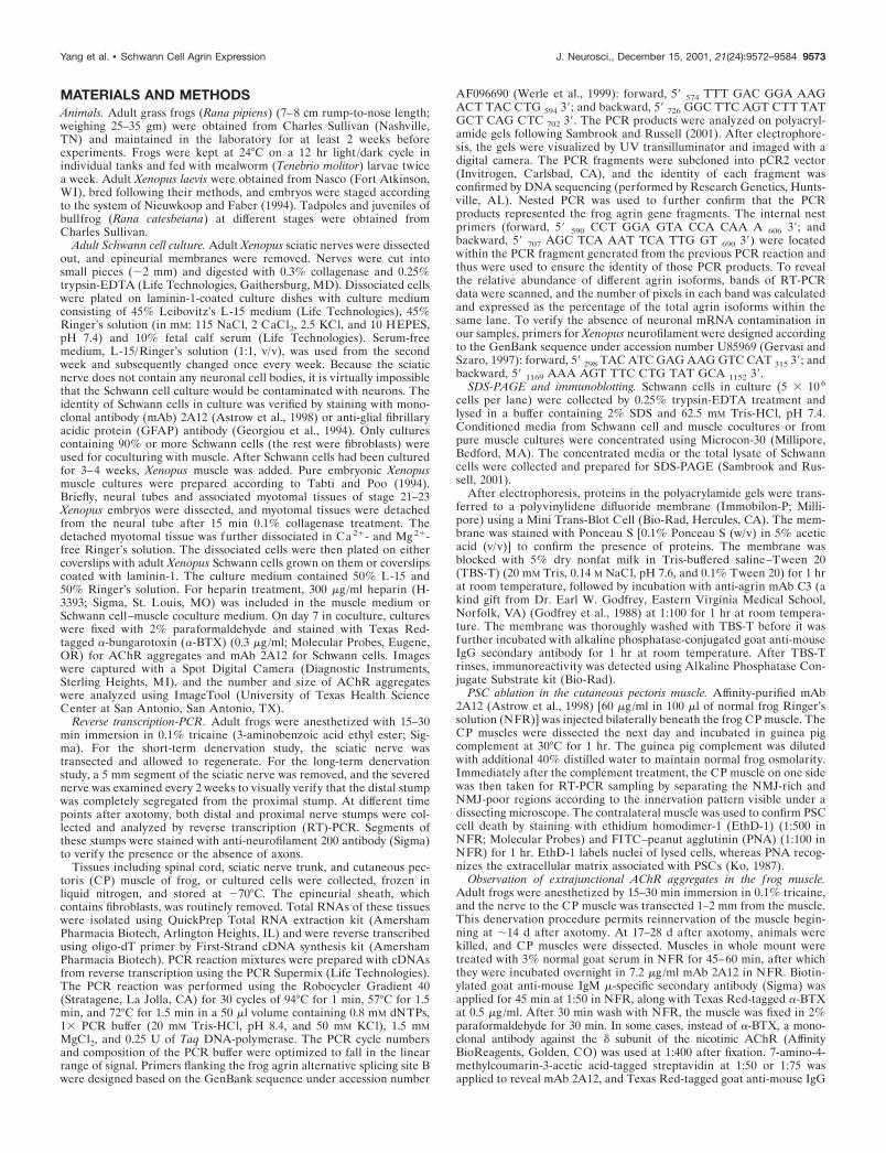
Expression of agrin isoforms in adult Schwann cellsafter axotomyTo examine whether and how axons regulate the expression ofadult Schwann cell agrin, we compared Schwann cells along thefrog (Rana pipiens) sciatic nerve after short-term denervation, inwhich nerve regeneration was allowed, with Schwann cells afterlong-term denervation, in which nerve regeneration was pre-vented. The result of the short-term denervation is shown inFigure 2, A and B. As expected from the above study (Fig. 1A,lane 6), adult Schwann cells in intact frog sciatic nerve expressedB0, B11, and B19 (Fig. 2A, Intact). Two weeks after axotomy,Schwann cells along nerve fibers (Fig. 2A, 2w) began to showupregulation of active agrin isoforms in both proximal and distalnerve segments. In addition to the upregulation of B11 and B19,the most potent isoform in AChR aggregation, B8, now appeared.At this time after the short-term denervation, axons began toreinnervate through the distal nerve stump, as revealed by anti-neurofilament 200 staining (data not shown). To examineSchwann cell agrin upregulation after axonal regeneration, eachlane in Figure 2A (Distal) was analyzed based on the area andintensity of each band (see Materials and Methods). This infor-mation allowed us to reveal the relative abundance, and changesin the expression pattern, of different agrin isoforms after nerveinjury. Figure 2B shows the percentage of all active agrin iso-forms combined (B8/B11/B19) relative to the total isoforms (ac-tive plus inactive) within each individual lane at various timeperiods after axotomy. The percentages of B8 and B0 in each lanewere also plotted individually. In Schwann cells along the intactnerve, B11/B19 constituted �45% of total agrin isoforms. Twoweeks after axotomy, the active isoforms, including B8, consti-tuted over 60% of total agrin. This upregulation of active agrinbecame more prominent as regeneration progressed and peaked�6–7 weeks after nerve transection. During this peak expression,there was a concomitant decrease in the relative percentage of B0,and the pattern of Schwann cell agrin expression closely resem-bled neuronal agrin expression (Fig. 2A, 4w–8w, compare withFig. 1A,B, lane 1). From 8 weeks after axotomy, the activeisoforms began to show downregulation (Fig. 2B). However, 12weeks after a single nerve transection, when nerve regenerationwas complete, the active agrin expression level in Schwann cellsremained slightly higher than that in Schwann cells along intactnerves, and a trace amount of B8 (3%) could still be detected
(Fig. 2A,B). Whether or not the normal expression pattern wouldbe totally restored beyond 12 weeks after axotomy was notexamined.
To confirm that the upregulation of active agrin was triggeredby axonal regeneration instead of simply by nerve injury per se,we also examined agrin expression in Schwann cells after long-term denervation (Fig. 2C,D). At 2 weeks after axotomy, theproximal nerve stump showed positive neurofilament staining(Fig. 2C, top right panel), indicative of nerve regeneration, but thedisconnected distal stump was absent of neurofilament staining(Fig. 2C, bottom right panel). As expected for regenerated nerves(Fig. 2A,B), Schwann cells in the proximal nerve segment ex-pressed all four isoforms of agrin (Fig. 2C, top lef t panel). Incontrast, Schwann cells in the chronically denervated distal seg-ment expressed only B0, B11, and B19 and not B8 (Fig. 2C, middlelef t panel). At 4–6 weeks after chronic denervation, the contrastbetween the upregulation of active agrin in the proximal stumpand its absence in the distal stump became even more prominent.Similar to the intact sciatic nerve (Fig. 1C), Schwann cells in theregenerated proximal stump 2–6 weeks after axotomy did notexpress neurofilament mRNA (Fig. 2C, bottom lef t panel). Thus,the appearance of B8 isoform in the proximal Schwann cells afternerve regeneration is likely not a contamination of neuronal agrinmRNA. The difference in the relative expression of active versusinactive isoforms between Schwann cells in the proximal anddistal segments is further shown in Figure 2D. Schwann cellsalong the proximal nerve stump showed upregulation of all activeagrin isoforms from 45% to over 65% and B8 from 0% to over17%, relative to the total agrin isoforms at 4–6 weeks afteraxotomy. In contrast, Schwann cells in the distal nerve segmentshowed a total absence of B8 and no upregulation of the otheractive isoforms, which remained �44–48% up to 6 weeks afterlong-term denervation. These results suggest that upregulation ofactive agrin isoforms in Schwann cells is not induced by nerveinjury per se but instead by nerve regeneration.
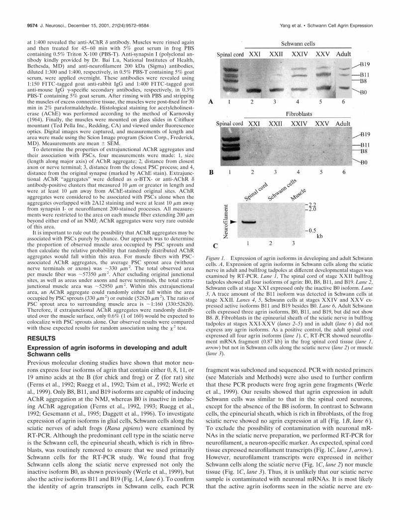
Adult Schwann cells enhance AChR aggregation onmuscle in cultureBecause adult Schwann cells express active agrin isoforms, weinvestigated whether these glial cells play a role in the aggregationof AChRs on muscle fibers. To address this question, Xenopuscultures were used for functional assay. Schwann cells obtainedfrom adult Xenopus sciatic nerves were cocultured with primaryXenopus myotubes, which were prepared from stage 21–23 em-bryos. In culture, these Schwann cells de-differentiate to a non-myelinating phenotype (Brockes et al., 1979), similar to PSCs. Allcultures contained similar density of Schwann cells at �15,000cells per dish (35 mm in diameter). The identity of Schwann cellsin vitro was confirmed by positive staining with mAb 2A12 thatrecognizes the Schwann cell membrane (Astrow et al., 1998) orwith anti-GFAP antibody (Georgiou et al., 1994). EmbryonicSchwann cells were not used because they expressed only theinactive B0 isoform as shown above. To analyze the effect ofSchwann cells on AChR aggregation, we compared AChR clus-ters on muscle fibers grown in the presence of adult Schwann cells(SC�M) with those on muscle grown alone (M) in culture.Neurons were not added to either of these cultures, and theabsence of neuronal contamination was further verified by theabsence of staining with anti-neurofilament antibody in thesecultures (data not shown). Figure 3 shows an example of anSC�M culture (A, A�) and an M culture (B, B�) 7 d in culture. The
Yang et al. • Schwann Cell Agrin Expression J. Neurosci., December 15, 2001, 21(24):9572–9584 9575

general cellular morphology in both cultures appeared similarunder phase-contrast optics (A, B). In addition, there was nosignificant difference in muscle fiber length between SC�M cul-ture (241.8 � 51.2 �m; mean � SD; n � 96) and M culture(221.6 � 33.2 �m; n � 98). However, as shown with fluorescencestaining, there were more and larger AChR clusters (arrowheadsin A� and B�) labeled with �-BTX in SC�M cultures than in M
cultures (compare A� with B�). The average number of AChRclusters per 100 �m muscle length in the SC �M culture was2.2-fold ( p � 0.001) of that in the M culture (Fig. 3C). Theaverage area of individual AChR “hotspots” in the SC�M culturewas 43.4 � 7.3 �m2, which was significantly larger ( p � 0.02) thanthe 22.7 � 3.7 �m2 in the M culture. Thus, the total area ofAChR clusters per 100 �m muscle length in the SC�M culture
Figure 2. Upregulation of active agrinisoforms in adult Schwann cells inducedby nerve regeneration. Expression ofagrin isoforms in the Schwann cellsalong the frog sciatic nerve after short-term denervation (A, B) and long-termdenervation (C, D) was examined byRT-PCR. A, Schwann cells in the intactsciatic nerve expressed only three agrinisoforms: B0, B11, and B19. However,after a single nerve transection that al-lowed nerve regeneration, Schwanncells in both the proximal and the distalnerve stumps upregulated the expres-sion of active isoforms, including theappearance of B8. The number of weeksafter axotomy is denoted on the top ofeach lane. B, A plot shows changes inthe percentage of B0 ( filled circles), B8(open triangles), and all active isoforms(B8/B11/B19; filled triangles) relative tothe total isoforms (see Materials andMethods) at different time points aftershort-term denervation (n � 3 experi-ments; mean � SEM). The increase inthe relative expression of active agrinand a concomitant decrease in B0 beganat �2 weeks and peaked at �6–7 weeksafter short-term denervation. A traceamount of B8 (3%) was still detected 12weeks after axotomy. C, Schwann cellsin the sciatic nerve segment proximal tothe transection site 2–6 weeks afterlong-term denervation upregulated theactive agrin isoforms compared with theintact nerve (top lef t panel ). The sameSchwann cell samples along the intactnerve and the proximal nerve segment2–6 weeks after axotomy did not showany neurofilament mRNA, in contrastto the spinal cord (bottom lef t panel ).The proximal segment contained regen-erating axons, as revealed by positiveanti-neurofilament staining (top rightpanel ). In contrast, the distal segment,which was chronically severed from theproximal segment, was absent of anti-neurofilament staining (bottom rightpanel ). Schwann cells in the distal seg-ment showed neither upregulation ofB11 and B19 nor appearance of B8(middle lef t panel ). D, A plot shows anincrease in the relative expression of allagrin isoforms (B8/ B11/ B19; filledtriangles) and B8 (open triangles) in theproximal segment after long-term de-nervation. However, in the chronicallysegregated distal segment, the relativeexpression of the total active isoforms(B8/B11/B19; filled squares) remainedunchanged, and no B8 (open squares)was detected up to 6 weeks after axo-tomy (n � 3 experiments; mean �SEM).
9576 J. Neurosci., December 15, 2001, 21(24):9572–9584 Yang et al. • Schwann Cell Agrin Expression

was also significantly enlarged ( p � 0.001) to 4.5-fold of that inthe M culture (Fig. 3D).
To test whether AChR aggregates colocalized with contactsbetween Schwann cell processes and muscle fibers, we double-labeled the SC�M culture with mAb 2A12 for Schwann cells (Fig.3F) and �-BTX for AChRs (Fig. 3G). As shown in Figure 3E,contacts (arrows) between Schwann cell processes and musclefibers were visualized with phase-contrast optics. Schwann cellprocesses were verified with mAb 2A12 staining (F). Althoughthere were numerous AChR clusters (Fig. 3G,H, arrowheads) onthe muscle fibers, most of them were not associated with Schwanncell processes. Only in rare cases were AChR clusters colocalizedwith Schwann cell–muscle contacts (Fig. 3E–H, asterisks). In 138muscle fibers from nine cultures observed, 98.5% of AChR ag-gregates were not colocalized with Schwann cell–muscle contacts.This contrasts with nerve–muscle contacts, in which AChRs areclustered (Cohen et al., 1979; Frank and Fischbach 1979). Ourresults suggest that the increase in AChR aggregation bySchwann cells is likely mediated by soluble factors, such as agrin,released into the tissue culture medium.
Is the increase in AChR aggregation by Schwann cellsmediated by agrin?To determine whether the enhancement of AChR aggregation bySchwann cells might be mediated by agrin, we first verified withRT-PCR the expression of active agrin transcripts in culturedSchwann cells. As shown in Figure 4A, lane 3, Schwann cellsgrown alone in culture expressed not only B0 but also the activeisoforms B11 and B19. The expression of these three isoformswas also seen in SC�M culture (Fig. 4A, lane 4) throughout thecoculture period. Similar to the expression pattern in adultSchwann cells after chronic denervation in vivo (Fig. 2C,D), B8was not expressed in these “denervated” Schwann cells, eithergrown alone or with muscle, in culture. The absence of neuronswas verified by the absence of neurofilament mRNA in thesecultures (data not shown). Muscle cells grown alone in culture(M) expressed only the inactive isoform B0 (Fig. 4A, lane 2),whereas spinal neurons in culture (Fig. 4A, lane 1) expressed allfour isoforms. Thus, Schwann cells, muscles, and neurons in vitroretain their characteristic expression pattern of agrin isoforms asseen in vivo.
To confirm that the expression of active agrin isoforms resultedin the production of agrin protein, lysate of cultured Schwann cellbodies was examined with Western blot using anti-Xenopus agrinmAb C3 (Godfrey et al., 1988). Similar to brain tissues (Fig. 4B,lane 2), cultured Schwann cells (Fig. 4B, lane 1) expressed nativeagrin protein at over 200 kDa. To determine whether agrinprotein was released into the culture medium, Western blot ofconditioned medium from Schwann cells cultured for 6 weeks wasexamined. As shown in Figure 4B, lane 3, the Schwann cell-conditioned medium did contain the agrin protein (arrow), as alsoobserved in the muscle-conditioned medium (lane 4) as a control.However, in contrast to the native agrin protein over 200 kDaseen in Schwann cell bodies, the 70 kDa band seen in the condi-tioned medium likely belongs to an inactive degraded product ofthe agrin protein, as shown previously in basal lamina extracts ofthe Torpedo electric organ (Nitkin et al., 1987). Consistent withthe absence of native agrin protein bands above 200 kDa, wefound that Schwann cell-conditioned medium added to M culturealone did not cause an increase in AChR aggregates (data notshown). This is contrary to the effect seen in SC�M coculture(Fig. 3), in which muscle fibers were probably exposed to a
continuous supply of native agrin protein from coculturedSchwann cells (see Discussion).
To further determine whether Schwann cell-derived agrinmight be involved in the enhancement of AChR aggregation,Schwann cell and muscle cocultures were treated with heparin(300 �g/ml), which has been shown to inhibit agrin-mediatedAChR clustering on muscle (Hopf and Hoch, 1997). As shown inFigure 3, C and D, the number and the area of AChR hotspots inSC�M cultures were significantly reduced by heparin treatment(SC�M�H). However, spontaneous AChR aggregation, which isindependent of agrin, was not affected in M cultures by heparintreatment. Although the direct evidence is lacking (see Discus-sion), the present data are consistent with the hypothesis that theenhancement of AChR aggregation seen in SC�M cultures ismediated by Schwann cell-derived agrin.
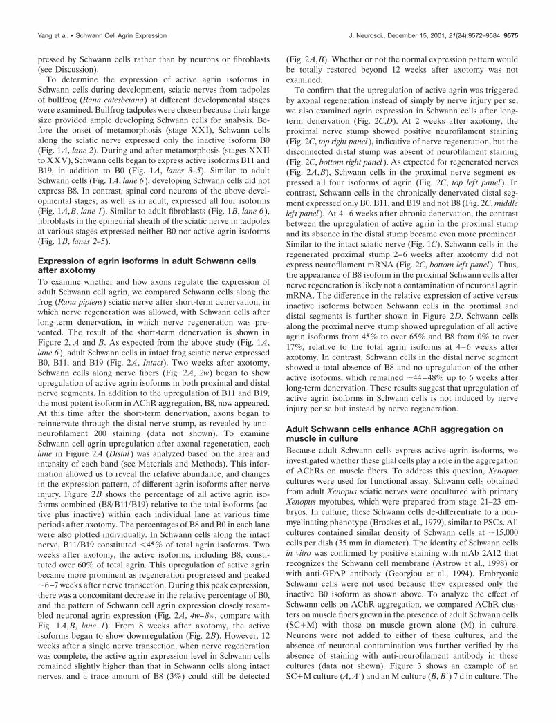
Agrin expression in the perisynaptic Schwann cell atthe NMJTo investigate whether Schwann cells at the NMJ are similar toSchwann cells along the nerve with respect to expression of activeagrin isoforms, we used a novel technique to selectively ablatePSCs in vivo (Reddy et al., 1999) (the detailed procedure of thistechnique will be described in a future paper). To ablate PSCs invivo, we took advantage of mAb 2A12, which specifically labelsthe PSC surface membrane (Astrow et al., 1998). Labeling ofPSCs with mAb 2A12 followed by treatment with guinea pigcomplement results in the formation of membrane-attack com-plexes on PSCs. Membrane-attack complexes form pores on thecell membrane and cause the antibody-labeled cells to lyse(Howard and Hughes-Jones, 1988). To verify PSC lysis, EthD-1,which stains the nuclei of cells with damaged membranes, wasapplied after the complement treatment. As shown in Figure 5A,EthD-1 labeling (in red, arrows) colocalized with the soma ofPSCs, which were revealed by mAb 2A12 immunofluorescentstaining (in green). Over 80% of PSCs were ablated using thistreatment. In contrast to PSCs, Schwann cells along axons are notlabeled with mAb 2A12 in whole mount because mAb 2A12 doesnot penetrate the perineurium surrounding nerve fibers (Astrowet al., 1998) and thus are not lysed. We did not observe anymorphological damage to nerve terminals or muscle fibers aftermAb 2A12 and complement treatment (L. V. Reddy, S. Koirala,Y. Sugiura, and C. P. Ko, unpublished observations). In controlexperiments using complement treatment alone, without 2A12application, over 95% of PSCs were not lysed as indicated by theabsence of EthD-1 labeling (Fig. 5B, arrows). Because mAb 2A12was not used for the control, PSCs were revealed with FITC-conjugated PNA (in green), which recognizes the extracellularmatrix associated with PSCs (Ko, 1987). The few EthD-1-positivecells not colocalized with PSC cell bodies in the control werelikely blood cells that are inevitably damaged during muscledissection. The above results indicate that PSCs in situ can beselectively ablated using mAb 2A12 followed by complementtreatment (Reddy, Koirala, Sugiura and Ko, unpublishedobservations).
To test whether PSCs express active agrin isoforms, intact andPSC-ablated muscles were examined by RT-PCR (Fig. 5C). Thecentral region of the CP muscle enriched in NMJs (NMJ�) wasseparated from the remaining NMJ-poor (NMJ�) region. Ininnervated muscles without PSC ablation (Fig. 5C), agrin iso-forms B0, B11, and B19 were detected in the NMJ-rich region(NMJ�, PSC�, lane 1). This expression pattern was similar tothat of adult Schwann cells along intact nerve (Fig. 2A). In
Yang et al. • Schwann Cell Agrin Expression J. Neurosci., December 15, 2001, 21(24):9572–9584 9577

Figure 3. Adult Schwann cells enhance AChR aggregation on muscle in culture. Aggregation of AChRs on embryonic Xenopus SC�M cells for 7 d wascompared with that in M. A, B, Phase-contrast images of an SC�M culture (A) and an M culture (B) show similar muscle morphology. A�, B�,Fluorescence images of the same cultures labeled with Texas Red-conjugated �-BTX show more AChR hotspots in SC�M than M. C, A plot shows asignificant increase in the number of AChR hotspots per 100 �m muscle length in SC�M, which was 2.2-fold of that in M. Treatment with heparin (300�g/ml) eliminated this increase in the coculture (SC�M�H), but the treatment did not show effect in pure muscle culture (M�H). D, A plot shows the
9578 J. Neurosci., December 15, 2001, 21(24):9572–9584 Yang et al. • Schwann Cell Agrin Expression

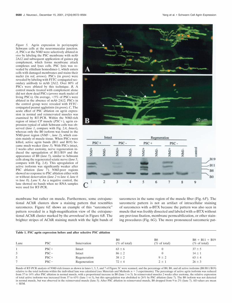
contrast, only the B0 isoform was found in the NMJ-poor region(Fig. 5C, NMJ�, lane 2), which consists mainly of muscle tissue.After acute ablation of PSCs in innervated muscle, active agrinbands (B11 and B19) became much weaker (Fig. 5C, lane 3; Table1). Thus, the reduction in B11 and B19 is correlated with theabsence of PSCs. The weaker bands of active isoforms in lane 3after PSC ablation may originate, in part, from the unablatedSchwann cells along intramuscular nerves (see Discussion). Theseaxonal Schwann cells constitute �20% of the total number ofSchwann cells as counted by nuclear staining (Hoechst 33342) inthe CP muscle.
To investigate whether and how agrin expression in Schwanncells responds to nerve injury, we performed RT-PCR of rein-nervated muscles (Fig. 5C). In reinnervated muscles 2 weeks afternerve transection, upregulation of B11/B19 and the appearanceof B8 were found in the NMJ-rich tissue with intact PSCs (lane 5;compare with lane 1). This result is similar to the upregulation ofactive agrin isoforms in Schwann cells along regenerating sciaticnerves (Fig. 2A). After acute PSC ablation, the active agrin bandsin the reinnervated NMJ-rich tissue were significantly weaker(compare lane 7 with lane 5), indicating that the active agrinmRNA was present in PSCs. These changes in agrin expressionwere quantified by comparing the relative intensity of variousbands within the same lane (Table 1). After PSC ablation, therelative expression of active agrin isoforms in the NMJ-rich tissuein innervated muscles was decreased from 37 to 14% of totalagrin isoforms and in reinnervated muscles from 63 to 26%(Table 1). NMJ-poor regions showed no response to PSC ablationeither with or without axotomy (Fig. 5C, lane 2 vs lane 4, lanes 6vs lane 8). Thus, PSC ablation does not affect the agrin expressionpattern in muscle.
Extrajunctional AChR clusters are associated with PSCsprouts in vivoThe expression of active agrin isoforms and their upregulation inPSCs after nerve regeneration suggested that PSCs might alsoplay a role in the aggregation of AChRs in vivo. Because of theclose apposition between PSCs and nerve terminals, it is impos-sible to distinguish the contribution of PSCs to AChR aggrega-tion from that of nerve terminals in normal muscles. To circum-vent this difficulty, we took advantage of the fact that PSCssprout, often tens of micrometers, beyond the tips of regeneratingnerve terminals during reinnervation (Koirala et al., 2000). Thisallowed us to examine in vivo whether PSCs play a role in AChRaggregation independent of nerve terminals. CP muscles of adultfrog (Rana pipiens) were excised 17–28 d after axotomy, whenmany NMJs bore PSC sprouts longer than corresponding regen-erating nerve terminals (Koirala et al., 2000) and then werefluorescently stained for AChRs, PSCs, axons, and nerve termi-nals. Figure 6 shows an example of an NMJ with extrajunctionalAChR aggregation associated with PSC sprouts 4 weeks afteraxotomy. A prominent PSC sprout labeled with mAb 2A12 (Fig.6A, arrowhead) extended over 100 �m beyond the boundary of
the original synaptic site (Fig. 6A,B,D, arrows), which was markedby AChE staining (Fig. 6D). Large and diffuse AChR aggregateslabeled with �-BTX staining (Fig. 6B, arrowhead) were associatedwith the PSC sprout. Although regenerating nerve terminals wereobserved in neighboring junctional branches (Fig. 6C, asterisk),nerve terminals and axons were absent along these extrajunc-tional AChR aggregates (Fig. 6C, arrowhead), suggesting thatPSCs directly induce such extrajunctional AChR aggregates. Incontrast to the junctional AChRs, which appeared as bright bandsand were sharply colocalized with the original junctional site, theextrajunctional AChRs appeared less dense and were not strictlyconfined by the boundary of the PSC sprouts (compare the regionmarked by the arrowhead in A with that in B). This lack of precisecolocalization between the extrajunctional AChRs and PSCsprouts indicates that the AChR clusters are not on the PSC
Figure 4. Agrin expression in adult Xenopus Schwann cells in vitro. A,Agrin isoform expression in cultured Schwann cells was examined byRT-PCR. Lane 1, As a positive control, spinal neurons in culture showedall four agrin isoforms. Lane 2, Muscle in culture displayed only the B0isoform. Lane 3, Schwann cells in culture expressed B0, B11, and B19isoforms. Lane 4, SC�M for 7 d also showed B0, B11, and B19 isoforms.Lane 5, As a negative control, the lane showed no bands when no RNAsamples were used for RT-PCR. B, Agrin protein in cultured Schwanncells and conditioned media was detected by Western blot using theanti-agrin antibody C3. Lane 1, Native agrin protein over 200 kDa wasdetected in the total cell lysate of cultured Schwann cells. Lane 2, As apositive control, brain tissues showed similar immunoreactivity over 200kDa. Conditioned media of Xenopus Schwann cells cultured for 6 weeks(SC-CM; lane 3) and Xenopus muscle conditioned media from day 7 inculture (Mu-CM; lane 4 ) showed a positive band at �70 kDa (arrow), butbands over 200 kDa were absent. The 70 kDa band may be an inactivedegradation product of native agrin protein.
4
total area (in square micrometers) of AChR clusters per 100 �m of muscle length in SC�M, M, and after heparin treatment in SC�M�H and M�H.Similar to C, the area of AChR clusters in SC�M was significantly enlarged to 4.5-fold of that in M, and this increase was eliminated by heparintreatment. In C and D, n denotes the number of muscle fibers observed, and all values are mean � SD. E–H, SC�M cocultures were examined todetermine the spatial relationship between AChR aggregates and Schwann cell–muscle contacts. The contacts (arrows) could be observed with phasecontrast optics (E) and confirmed with staining of Schwann cells with mAb 2A12 (F). The same culture double-labeled with �-BTX (G) showed thatthe majority of these contacts were not colocalized with AChR clusters (arrowheads). Only in rare cases were AChR clusters colocalized with theSchwann cell–muscle contact (asterisk). The spatial relationship between AChR clusters and the contacts is further shown in H, which is a merged imageof E–G.
Yang et al. • Schwann Cell Agrin Expression J. Neurosci., December 15, 2001, 21(24):9572–9584 9579
membrane but rather on muscle. Furthermore, some extrajunc-tional AChR clusters show a staining pattern that resemblessarcomeres. Figure 6E shows an example of this “sarcomeric”pattern revealed in a high-magnification view of the extrajunc-tional AChR cluster marked by the arrowhead in Figure 6B. Thebrighter stripes of AChR staining match with the light bands of
sarcomeres in the same region of the muscle fiber (Fig. 6F). Thesarcomeric pattern is not an artifact of intracellular stainingof sarcomeres with �-BTX because the pattern was also seen inmuscle that was freshly dissected and labeled with �-BTX withoutany previous fixation, membrane permeabilization, or other stain-ing procedures (Fig. 6G). The more pronounced sarcomeric pat-
Figure 5. Agrin expression in perisynapticSchwann cells at the neuromuscular junction.A, PSCs at the NMJ were selectively ablated invivo by labeling the PSC membrane with mAb2A12 and subsequent application of guinea pigcomplement, which forms membrane attackcomplexes and lyses cells. PSC lysis was re-vealed by ethidium homodimer-1, which enterscells with damaged membranes and stains theirnuclei (in red, arrows). PSCs (in green) wererevealed by labeling with FITC-conjugated sec-ondary antibody to mAb 2A12. Over 80% ofPSCs were ablated by this technique. B, Acontrol muscle treated with complement alonedid not show dead PSCs (arrows mark nuclei ofliving PSCs). On average, �5% of PSCs wereablated in the absence of mAb 2A12. PSCs inthe control group were revealed with FITC-conjugated peanut agglutinin (in green). C, Theacute effect of PSC ablation on agrin expres-sion in normal and reinnervated muscles wasexamined by RT-PCR. Within the NMJ-richregion of intact CP muscle (PSC�), agrin ex-pression typical of adult Schwann cells was ob-served (lane 1, compare with Fig. 2A, Intact),whereas only the B0 isoform was found in theNMJ-poor region (NMJ�, lane 2), which con-sists mainly of muscle tissue. When PSCs werekilled, active agrin bands (B11 and B19) be-came much weaker (lane 3). With PSCs intact,2 weeks after axotomy, nerve regeneration in-duced the upregulation of B11/B19 and theappearance of B8 (lane 5), similar to Schwanncells along the regenerated sciatic nerve (lane 5,compare with Fig. 2A). This upregulation ofactive isoforms was significantly weaker afterPSC ablation (lane 7 ). NMJ-poor regionsshowed no response to PSC ablation either withor without denervation (lane 2 vs lane 4, lane 6vs lane 8). Lane 9, As a negative control, thelane showed no bands when no RNA sampleswere used for RT-PCR.
Table 1. PSC agrin expression before and after selective PSC ablation
Lane PSC InnervationB0(% of total)
B8(% of total)
B8 � B11 � B19(% of total)
1 PSC� Intact 63 � 6 0 37 � 53 PSC� Intact 86 � 2 0 14 � 15 PSC� Regeneration 38 � 2 9 � 2 63 � 47 PSC� Regeneration 72 � 4 2 � 1 26 � 3
Bands of RT-PCR analysis of NMJ-rich tissues as shown in lanes 1, 3, 5, and 7 of Figure 5C were scanned, and the percentage of B0, B8, and all active isoforms (B8/B11/B19)relative to the total isoforms within the individual lane was calculated (see Materials and Methods: n � 3 experiments). The percentage of active agrin isoforms was reducedfrom 37 to 14% after PSC ablation in normal muscle, with a proportional increase in B0 (lane 1 vs 3). In reinnervated muscles 2 weeks after axotomy, the relative expressionof total active isoforms was increased from 37 to 63% (lane 1 vs 5), but this upregulation was abolished to 26% by PSC ablation (lane 7). The B8 isoform was not detectedin normal muscle, but was observed in the reinnervated muscle (lane 5). After PSC ablation in reinnervated muscle, B8 dropped from 9 to 2% (lane 7). All values are mean� SEM.
9580 J. Neurosci., December 15, 2001, 21(24):9572–9584 Yang et al. • Schwann Cell Agrin Expression
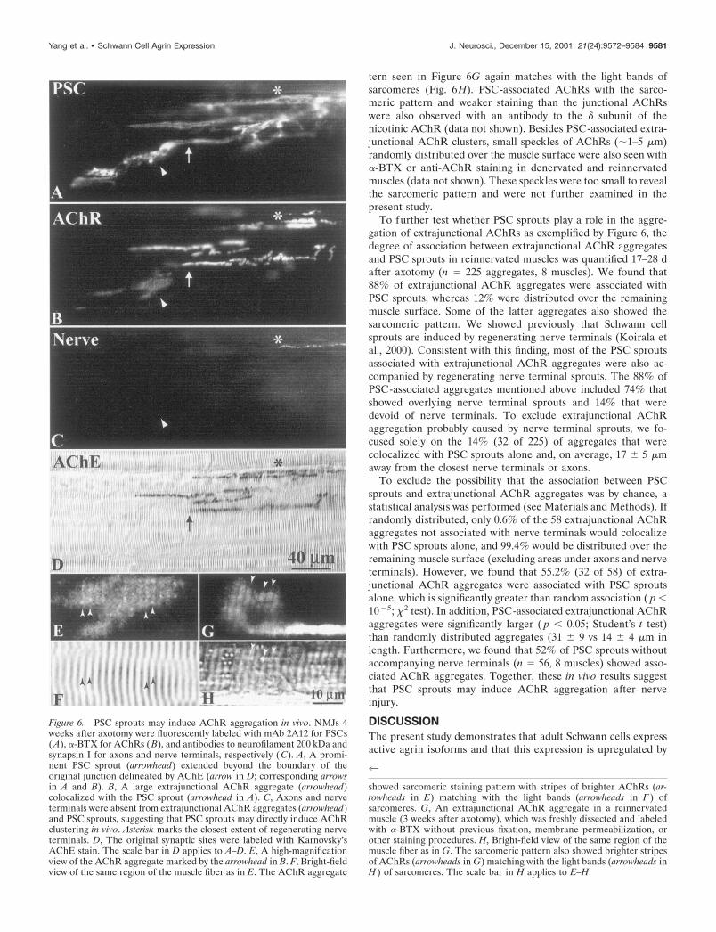
tern seen in Figure 6G again matches with the light bands ofsarcomeres (Fig. 6H). PSC-associated AChRs with the sarco-meric pattern and weaker staining than the junctional AChRswere also observed with an antibody to the � subunit of thenicotinic AChR (data not shown). Besides PSC-associated extra-junctional AChR clusters, small speckles of AChRs (�1–5 �m)randomly distributed over the muscle surface were also seen with�-BTX or anti-AChR staining in denervated and reinnervatedmuscles (data not shown). These speckles were too small to revealthe sarcomeric pattern and were not further examined in thepresent study.
To further test whether PSC sprouts play a role in the aggre-gation of extrajunctional AChRs as exemplified by Figure 6, thedegree of association between extrajunctional AChR aggregatesand PSC sprouts in reinnervated muscles was quantified 17–28 dafter axotomy (n � 225 aggregates, 8 muscles). We found that88% of extrajunctional AChR aggregates were associated withPSC sprouts, whereas 12% were distributed over the remainingmuscle surface. Some of the latter aggregates also showed thesarcomeric pattern. We showed previously that Schwann cellsprouts are induced by regenerating nerve terminals (Koirala etal., 2000). Consistent with this finding, most of the PSC sproutsassociated with extrajunctional AChR aggregates were also ac-companied by regenerating nerve terminal sprouts. The 88% ofPSC-associated aggregates mentioned above included 74% thatshowed overlying nerve terminal sprouts and 14% that weredevoid of nerve terminals. To exclude extrajunctional AChRaggregation probably caused by nerve terminal sprouts, we fo-cused solely on the 14% (32 of 225) of aggregates that werecolocalized with PSC sprouts alone and, on average, 17 � 5 �maway from the closest nerve terminals or axons.
To exclude the possibility that the association between PSCsprouts and extrajunctional AChR aggregates was by chance, astatistical analysis was performed (see Materials and Methods). Ifrandomly distributed, only 0.6% of the 58 extrajunctional AChRaggregates not associated with nerve terminals would colocalizewith PSC sprouts alone, and 99.4% would be distributed over theremaining muscle surface (excluding areas under axons and nerveterminals). However, we found that 55.2% (32 of 58) of extra-junctional AChR aggregates were associated with PSC sproutsalone, which is significantly greater than random association ( p �10�5; �2 test). In addition, PSC-associated extrajunctional AChRaggregates were significantly larger ( p � 0.05; Student’s t test)than randomly distributed aggregates (31 � 9 vs 14 � 4 �m inlength. Furthermore, we found that 52% of PSC sprouts withoutaccompanying nerve terminals (n � 56, 8 muscles) showed asso-ciated AChR aggregates. Together, these in vivo results suggestthat PSC sprouts may induce AChR aggregation after nerveinjury.
DISCUSSIONThe present study demonstrates that adult Schwann cells expressactive agrin isoforms and that this expression is upregulated by
Figure 6. PSC sprouts may induce AChR aggregation in vivo. NMJs 4weeks after axotomy were fluorescently labeled with mAb 2A12 for PSCs(A), �-BTX for AChRs ( B), and antibodies to neurofilament 200 kDa andsynapsin I for axons and nerve terminals, respectively (C). A, A promi-nent PSC sprout (arrowhead) extended beyond the boundary of theoriginal junction delineated by AChE (arrow in D; corresponding arrowsin A and B). B, A large extrajunctional AChR aggregate (arrowhead)colocalized with the PSC sprout (arrowhead in A). C, Axons and nerveterminals were absent from extrajunctional AChR aggregates (arrowhead)and PSC sprouts, suggesting that PSC sprouts may directly induce AChRclustering in vivo. Asterisk marks the closest extent of regenerating nerveterminals. D, The original synaptic sites were labeled with Karnovsky’sAChE stain. The scale bar in D applies to A–D. E, A high-magnificationview of the AChR aggregate marked by the arrowhead in B. F, Bright-fieldview of the same region of the muscle fiber as in E. The AChR aggregate
4
showed sarcomeric staining pattern with stripes of brighter AChRs (ar-rowheads in E) matching with the light bands (arrowheads in F ) ofsarcomeres. G, An extrajunctional AChR aggregate in a reinnervatedmuscle (3 weeks after axotomy), which was freshly dissected and labeledwith �-BTX without previous fixation, membrane permeabilization, orother staining procedures. H, Bright-field view of the same region of themuscle fiber as in G. The sarcomeric pattern also showed brighter stripesof AChRs (arrowheads in G) matching with the light bands (arrowheads inH ) of sarcomeres. The scale bar in H applies to E–H.
Yang et al. • Schwann Cell Agrin Expression J. Neurosci., December 15, 2001, 21(24):9572–9584 9581

axonal regeneration. Adult Schwann cells enhance the aggrega-tion of AChRs on muscle fibers in vitro. Although the evidence isindirect, our results are consistent with the hypothesis that theenhancement of AChR aggregation by cultured Schwann cells ismediated by agrin. Using complement-mediated lysis to selec-tively ablate PSCs at the NMJ in vivo, we show that thesesynapse-associated glial cells also express active agrin isoforms.Finally, we present evidence that PSCs may induce AChR clus-ters in vivo. These novel findings suggest that, in addition toneurons, glial cells also play a role in the aggregation of postsyn-aptic receptors. Receptor aggregation by glia may complementthe role of neurons and may be particularly important in ensuringrapid restoration of synaptic function during regeneration.
Adult Schwann cells express active agrin isoformsWerle et al. (1999) have reported that Schwann cells in the frogsciatic nerve express only the inactive B0 isoform. However, usingthe same preparation and the same primers for RT-PCR, wefound that frog Schwann cells do express the active agrin isoformsB11 and B19, besides the inactive B0. One possible explanationfor the conflicting result may be attributed to different amount ofRNA, or different temperature and number of cycles, used for ourRT-PCR. To exclude the possibility of false-positive bands in oursamples, we subcloned each PCR fragment and confirmed itssequence as agrin. The expression of active agrin isoforms is notan artifact of contamination by neuronal mRNA that might bepresent in axons or nerve terminals, because the sciatic nervesamples used for our RT-PCR study did not contain any neuro-filament mRNA. In addition, we showed that Schwann cellscocultured with muscles, but devoid of neurons, also expressactive agrin isoforms. Furthermore, if our RT-PCR samples werecontaminated with neuronal agrin mRNA, we would detect allfour agrin isoforms, including B8, as found in the spinal cord.However, the B8 isoform was not observed in Schwann cells alongnormal sciatic nerve, in nerve terminals, or in cultured Schwanncells. Moreover, B11 and B19 isoforms were also expressed in thechronically segregated distal segment of the sciatic nerve. Be-cause the half-life of mRNAs in animal cells is usually �24 hr(Lewin and Siliciano, 1996), the active agrin isoforms in the distalnerve segment seen even at 6 weeks after long-term denervationmust be actively transcribed, most likely by Schwann cells alongthe nerve. Contamination of active agrin from other cell types isalso unlikely; Schwann cells are the predominant cell type alongthe nerve fiber, and we found that fibroblasts in the epineurialsheath do not express any agrin isoforms. Together, these resultsdemonstrate that adult Schwann cells express active agrin iso-forms. Anti-agrin staining has also been observed at frog PSCs(Werle et al., 1999). However, it is not possible to distinguishbetween the active and inactive forms of agrin proteins using thecurrently available antibodies.
Expression of Schwann cell agrin during developmentand axonal regenerationSimilar to embryonic Schwann cells in the chick sciatic nerve,which express only B0 (Ruegg et al., 1992), developing Schwanncells in tadpoles do not express active agrin. Although it is notknown how the expression of B11 and B19 is triggered inSchwann cells during metamorphosis, it is clear that the expres-sion of these active isoforms occurs long after synaptogenesis.Thus, the expression of B11 and B19 isoforms by Schwann cellsduring development is not induced by innervation per se. Theabsence of active agrin expression in tadpole Schwann cells also
indicates that Schwann cell-derived agrin is unlikely to play a rolein the aggregation of AChRs during initial synaptogenesis. Con-sistent with this view, there is no AChR clustering along Schwanncell sprouts that extend beyond developing nerve terminals dur-ing synapse formation in tadpole muscles (Herrera et al., 2000).This is in contrast to adult muscles, in which aggregates ofAChRs are formed along PSC sprouts after nerve injury andreinnervation (see below).
In adult animals, expression of all active agrin isoforms inSchwann cells was upregulated after nerve injury. However, theupregulation is not attributable to nerve injury per se because noupregulation occurs in Schwann cells along chronically severednerve fibers. Rather, the upregulation of active agrin isoformscoincides with axonal regeneration, suggesting that regeneratingnerves play a role in the upregulation. This upregulation of activeagrin isoforms is reminiscent of the induction of PSC sprouting,which also occurs in response to nerve regeneration, but notnerve injury alone, at the frog NMJ (Koirala et al., 2000). Themechanism underlying activation of Schwann cells by regenerat-ing axons and nerve terminals is unknown.
Schwann cells enhance AChR aggregation in vitroKoenig et al. (1998) suggested that chick Schwann cells have thecapacity to enhance AChR aggregation on cultured myotubes.The present study has shown that cultured adult Schwann cellsexpress active agrin mRNAs and produce native agrin protein. Inaddition, adult Schwann cells increase the number and area ofAChR hotspots on Xenopus muscle in culture, and the increase inAChR aggregation is eliminated by heparin. These findings areconsistent with the hypothesis that the enhancement of AChRaggregation by Schwann cells is mediated by agrin. However,besides disrupting agrin function (Hopf and Hoch, 1997), heparinhas been shown to also interfere with the function of neuregulin,laminin, and other molecules that affect AChR synthesis andclustering (Yarden and Wen, 1994). Thus, we cannot exclude thepossibility that other soluble molecules released by Schwann cellsmay also play a role in the enhancement of AChR aggregation.Unfortunately, the lack of antibodies that perturb the function ofXenopus agrin prevented us from testing the direct involvement ofagrin in SC�M cocultures.
The absence of enhancement of AChR aggregation bySchwann cell-conditioned medium also precluded our attempt touse immunoprecipitation to directly test the role of Schwanncell-derived agrin. In the Schwann cell-conditioned medium, weonly detected a 70 kDa band, which likely represents a degrada-tion product of the agrin protein and is probably not active inAChR aggregation (Nitkin et al., 1987). The absence of proteinbands above 200 kDa in the conditioned medium may be attrib-utable to the fact that the conditioned medium was collected fromlong-term cultures, and thus most native agrin protein was de-graded. This is in contrast to the increase in AChR aggregationobserved in SC�M cocultures (Fig. 3). In the cocultures, Schwanncells may continuously release native agrin protein, which pre-sumably would have an immediate and cumulative effect onAChR aggregation before being degraded.
Expression of active agrin in the PSCThe present study has applied a novel technique usingcomplement-mediated lysis to selectively ablate PSCs (Reddy etal., 1999) (a full-length paper will be published in the future).Ablation of PSCs caused a reduction in the amount of activeagrin isoforms, indicating that, similar to axonal Schwann cells,
9582 J. Neurosci., December 15, 2001, 21(24):9572–9584 Yang et al. • Schwann Cell Agrin Expression

PSCs also express active agrin genes. As shown in Astrow et al.(1998), Schwann cells along axons are not labeled by mAb 2A12in whole-mount preparations and thus are not ablated by thecomplement treatment. Because �20% of the total Schwann cellsin the CP muscle belong to these axonal Schwann cells andtypically �20% of PSCs were not ablated by the mAb 2A12 pluscomplement treatment, these remaining Schwann cells mightcontribute to the active agrin isoforms still seen after the ablationtreatment in intact and reinnervated muscles (Table 1). Ourapproach using PSC ablation and RT-PCR has provided evi-dence, albeit indirect, that is consistent with the idea that PSCsalso express active agrin isoforms and the expression is upregu-lated by reinnervation.
Extrajunctional AChRs are associated with PSCsprouts in vivoThe present study has shown that, during muscle reinnervation,clusters of AChRs form outside of original synaptic sites and thatthese clusters colocalize with PSC sprouts. Because the density ofAChRs falls sharply within a few micrometers of nerve terminalsat the NMJ (Matthews-Bellinger and Salpeter, 1978), it is unlikelythat neuronal agrin would have a diffusible effect on the aggre-gation of extrajunctional AChRs located tens of micrometersaway as observed in the present study. It is also unlikely that theextrajunctional AChR aggregation is caused by nerve terminalsthat extended and then retracted before our observations. Usingrepeated observation of identified NMJs in living frogs, weshowed previously extension of PSC sprouts tens or hundreds ofmicrometers longer than nerve terminals; in many cases, PSCsprouts continued to extend even when nerve terminals showedno change in length during a period of 2–3 months of multipleobservations (Chen et al., 1991; Chen and Ko, 1994; Ko andChen, 1996). Thus, frog nerve terminals do not extend and thenretract through such long distances. Furthermore, we showedrecently that, at early stages of reinnervation (2–4 weeks afteraxotomy), there is substantial growth of nerve terminals butvirtually no observed retraction (Koirala et al., 2000). Thus, theclusters of extrajunctional AChRs were most likely caused by PSCsprouts rather than by nerve terminals that extended and thenretracted.
The association of AChR aggregates and PSC sprouts is notrandom. In stark contrast to the predicted value of 0.6% if theassociation were purely by chance, we showed that 55.2% ofextrajunctional AChR clusters (without accompanying nerve ter-minals) are located at PSC sprouts. Furthermore, 52% of PSCsprouts (without accompanying nerve terminals) showed thepresence of colocalized extrajunctional AChRs, consistent withthe suggestion that PSC sprouts play a role in the AChRaggregation.
The mechanism of PSC-induced AChR aggregation in vivo isnot known. Because PSCs express active agrin, it is tempting tospeculate that the aggregation of extrajunctional AChRs is me-diated by agrin released by PSCs. The colocalization betweenAChR clusters and PSC sprouts in vivo would suggest that agrinmolecules, if released by PSC sprouts, probably bind basal laminaand are concentrated locally, as is the case for neuronal agrin atthe NMJ. In contrast to the in vivo finding, Schwann cell-enhanced AChR aggregation in vitro is independent of cell con-tact, which may be attributed to the paucity of basal lamina onembryonic muscle fibers in culture.
As in the present study, discrete patches of extrajunctionalAChRs with different shape and lower density than the junctional
AChRs have been reported in denervated mammalian muscles(Ko et al., 1977). Clusters of AChR with various densities havealso been observed in muscles treated with agrin in vitro (Wallace,1992) or in vivo (Bezakova et al., 2001). In fact, the sarcomericpattern can also be discerned in some extrajunctional AChRclusters induced by agrin application to rat muscle (Bezakova etal., 2001, their Fig. 4). Because the sarcomeric pattern of AChRstaining matches with the light bands of the sarcomere, it is likelythat AChRs are located in and around the T tubules, which havemembrane invaginations near the Z-line in the light bands.Sheikh et al. (2001) have shown that sodium channels clusterselectively around the mouths of the T tubules in the frog skeletalmuscle fiber. It is possible that AChRs also cluster only aroundthe mouths of the T tubules. Alternatively, AChRs may be evenlydistributed along the entire length of the T tubules, but theirinvaginations give rise to a brighter signal of AChR staining inthe en face view.
The novel role of glial cells in receptor aggregation describedin this study may be important in laying the groundwork for rapidand successful restoration of synaptic function during regenera-tion. In addition to the presynaptic guidance of regeneratingnerve terminals as shown previously (for review, see Son et al.,1996), PSCs now appear to play a postsynaptic role in inducingAChR aggregation ahead of regenerating terminals. There isindirect evidence that PSC-induced AChR aggregates may con-stitute sites of subsequent synaptogenesis. PSCs sprout soon afterregenerating nerve terminals arrive at endplates, and nerve ter-minals grow along PSC sprouts (Koirala et al., 2000). BecauseAChR aggregates are present along PSC sprouts ahead of nerveterminals, these aggregates would very likely be “innervated” bythe regenerating nerve terminals. Consistent with this scenario,we observe that, at later stages of reinnervation, all extrajunc-tional AChR aggregates colocalize with overlying nerve termi-nals (Koirala et al., 2000). Apart from a preparatory role inreinnervation, our results suggest that PSCs could also play a rolein the maintenance of AChRs at the NMJ. Studies are underwayusing our novel PSC ablation technique to determine whetherPSCs play a role in the maintenance of AChR clustering atNMJs, particularly after nerve injury.
REFERENCESAraque A, Parpura V, Sanzgiri RP, Haydon PG (1999) Tripartite syn-
apses: glia, the unacknowledged partner. Trends Neurosci 22:208–215.Astrow SH, Qiang H, Ko C-P (1998) Perisynaptic Schwann cells at
neuromuscular junctions revealed by a novel monoclonal antibody.J Neurocytol 27:667–681.
Bacci A, Verderio C, Pravettoni E, Matteoli M (1999) The role of glialcells in synaptic function. Philos Trans R Soc Lond B Biol Sci354:403–409.
Bezakova G, Helm JP, Francolini M, Lomo T (2001) Effects of purifiedrecombinant neural and muscle agrin on skeletal muscle fibers in vivo.J Cell Biol 153:1441–1452.
Brockes JP, Fields KL, Raff MC (1979) Studies on cultured rat Schwanncells. I. Establishment of purified populations from cultures of periph-eral nerve. Brain Res 165:105–118.
Cao G, Qiang H, Ko C-P (1999) The effect of Schwann cells on acetyl-choline receptor aggregates in Xenopus nerve-muscle cultures. SocNeurosci Abstr 25:240.
Castonguay A, Robitaille R (2001) Differential regulation of transmitterrelease by presynaptic and glial Ca 2� internal stores at the neuromus-cular synapse. J Neurosci 21:1911–1922.
Chen L, Ko C-P (1994) Extension of synaptic extracellular matrix dur-ing nerve terminal sprouting in living frog neuromuscular junctions.J Neurosci 14:796–808.
Chen L, Folsom DB, Ko C-P (1991) The remodeling of synaptic extra-cellular matrix and its dynamic relationship with nerve terminals atliving frog neuromuscular junctions. J Neurosci 11:2920–2930.
Cohen MW, Anderson MJ, Zorychta E, Weldon PR (1979) Accumula-tion of acetylcholine receptors at nerve-muscle contacts in culture. ProgBrain Res 49:335–349.
Yang et al. • Schwann Cell Agrin Expression J. Neurosci., December 15, 2001, 21(24):9572–9584 9583

Daggett DF, Stone D, Peng HB, Nikolics K (1996) Full-length agrinisoform activities and binding site distributions on cultured Xenopusmuscle cells. Mol Cell Neurosci 7:75–88.
Ferns M, Hock W, Campanelli JT, Rupp F, Hall ZW, Scheller RH (1992)RNA spicing regulates agrin-mediated acetylcholine receptor cluster-ing activity on cultured myotubes. Neuron 8:1079–1086.
Ferns M, Campanelli JT, Hock W, Scheller RH, Hall ZW (1993) Theability of agrin to cluster AChRs depends on alternative splicing and oncell surface proteoglycans. Neuron 11:491–502.
Frank E, Fischbach GD (1979) Early events in neuromuscular junctionformation in vitro: induction of acetylcholine receptor clusters in thepostsynaptic membrane and morphology of newly formed synapses.J Cell Biol 83:143–158.
Georgiou J, Robitaille R, Trimble WS, Charlton MP (1994) Synapticregulation of glial protein expression in vivo. Neuron 12:443–455.
Gervasi C, Szaro BG (1997) Sequence and expression patterns of twoforms of the middle molecular weight neurofilament protein (NF-M) ofXenopus laevis. Mol Brain Res 48:229–242.
Gesemann M, Denzer A, Ruegg MA (1995) Acetylcholine receptor-aggregating activity of agrin isoforms and mapping of the active site.J Cell Biol 128:625–636.
Godfrey EW, Dietz ME, Morstad AL, Wallskog PA, Yorde DE (1988)Acetylcholine receptor-aggregating proteins are associated with theextracellular matrix of many tissue in Torpedo. J Cell Biol106:1263–1272.
Herrera AA, Qiang H, Ko C-P (2000) The role of perisynaptic Schwanncells in development of neuromuscular junctions in the frog (Xenopuslaevis). J Neurobiol 45:237–254.
Hopf C, Hoch W (1997) Heparin inhibits acetylcholine receptor aggre-gation at two distinct steps in the agrin-induced pathway. Eur J Neu-rosci 9:1170–1177.
Howard J, Hughes-Jones N (1988) Complement-mediated lysis withmonoclonal antibodies. Prog Allergy 45:1–15.
Karnovsky MJ (1964) The localization of cholinesterase activity in ratcardiac muscle by electron microscopy. J Cell Biol 23:217–232.
Ko C-P (1987) A lectin, peanut agglutinin, as a probe for the extracel-lular matrix in living neuromuscular junction. J Neurocytol 16:567–576.
Ko C-P, Chen L (1996) Synaptic remodeling revealed by repeated in vivoobservations and electron microscopy of identified frog neuromuscularjunctions. J Neurosci 16:1780–1790.
Ko PK, Anderson MJ, Cohen MW (1977) Denervated skeletal musclefibers develop discrete patches of high acetylcholine receptor density.Science 196:540–542.
Koenig J, de La Porte S, Chapron J (1998) The Schwann cell at theneuromuscular junction. J Physiol (Paris) 92:153–155.
Koirala S, Qiang H, Ko C-P (2000) Reciprocal interactions betweenperisynaptic Schwann cells and regenerating nerve terminals at the frogneuromuscular junction. J Neurobiol 44:343–360.
Lewin B, Siliciano P (1996) Genes VI, p 165. New York: Oxford UP.Ma E, Morgan R, Godfrey EW (1994) Distribution of agrin mRNAs in
the chick embryo nervous system. J Neurosci 14:2943–2952.Matthews-Bellinger J, Salpeter MM (1978) Distribution of acetylcholine
receptors at frog neuromuscular junctions with a discussion of somephysiological implications. J Physiol (Lond) 279:197–213.
McMahan UJ (1990) The agrin hypothesis. Cold Spring Harb SympQuant Biol 55:407–418.
Nieuwkoop PK, Faber J (1994) Normal table of Xenopus laevis (Dau-din): a systematical and chronological survey of development from the
fertilized egg till the end of metamorphosis, pp 162–188. New York:Garland.
Nitkin RM, Smith MA, Magill C, Fallon JR, Yao YM, Wallace BG,McMahan UJ (1987) Identification of agrin, a synaptic organizingprotein from Torpedo electric organ. J Cell Biol 105:2471–2478.
O’Malley JP, Waran MT, Balice-Gordon RJ (1999) In vivo observationsof terminal Schwann cells at normal, denervated, and reinnervatedmouse neuromuscular junctions. J Neurobiol 38:270–286.
Qiang H, Koirala S, Ko C-P (1998) Do Schwann cells play a role inaggregation of acetylcholine receptors at the neuromuscular junction?Soc Neurosci Abstr 24:1036.
Reddy LV, Koirala S, Ko C-P (1999) Selective ablation of perisynapticSchwann cells from the frog neuromuscular junction in vivo. Soc Neu-rosci Abstr 25:2032.
Reynolds ML, Woolf CJ (1992) Terminal Schwann cells elaborate ex-tensive processes following denervation of the motor endplate. J Neu-rocytol 21:50–66.
Robitaille R (1998) Modulation of synaptic efficacy and synaptic depres-sion by glial cells at the frog neuromuscular junction. Neuron21:847–855.
Ruegg MA, Bixby JL (1998) Agrin orchestrates synaptic differentiationat the vertebrate neuromuscular junction. Trends Neurosci 21:22–27.
Ruegg MA, Tsim KWK, Horton SE, Kroger S, Escher G, Gensch EM,McMahan UJ (1992) The agrin gene codes for a family of basal laminaproteins that differ in function and distribution. Neuron 8:691–699.
Sambrook J, Russell DW (2001) Molecular cloning—a laboratory man-ual (Argentine J ed), pp A8.40–A8.55. New York: Cold Spring HarborLaboratory.
Sanes JR, Lichtman JW (1999) Development of the vertebrate neuro-muscular junction. Annu Rev Neurosci 22:389–442.
Sheikh SM, Skepper JN, Chawla S, Vandenberg JI, Elnei S, Huang CL-H(2001) Normal conduction of surface action potentials in detubulatedamphibian skeletal muscle fibres. J Physiol (Lond) 535:579–590.
Smith MA, O’Dowd DK (1994) Cell-specific regulation of agrin RNAsplicing in the chick ciliary ganglion. Neuron 12:795–804.
Son YJ, Thompson WJ (1995a) Schwann cell processes guide regenera-tion of peripheral axons. Neuron 14:125–132.
Son YJ, Thompson WJ (1995b) Nerve sprouting in muscle is inducedand guided by processes extended by Schwann cells. Neuron14:133–141.
Son YJ, Trachtenberg JT, Thompson WJ (1996) Schwann cells induceand guide sprouting and reinnervation of neuromuscular junctions.Trends Neurosci 19:280–285.
Tabti N, Poo MM (1994) Study on the induction of spontaneous trans-mitter release at early nerve-muscle contacts in Xenopus cultures.Neurosci Lett 173:21–26.
Tsim KWK, Ruegg MA, Escher G, Kroger S, McMahan UJ (1992)cDNA that encodes active agrin. Neuron 8:677–689.
Wallace B (1992) Mechanism of agrin-induced acetylcholine receptoraggregation. J Neurobiol 23:592–604.
Werle MJ, Jones MA, Stanco AM (1999) Aggregates of acetylcholinereceptors are not observed under anti-agrin staining Schwann cellprocesses at the frog neuromuscular junction. J Neurobiol 40:45–54.
Yang JF, Ko C-P (1999) Expression of agrin by Schwann cells at intact,degenerating and developing nerves. Soc Neurosci Abstr 25:240.
Yarden Y, Wen D (1994) Neu differentiation factor (NDF) and theneuregulin (NRG) family. In: Guidebook to cytokines and their recep-tors (Nicola NA, ed), pp 146–149. New York: Oxford UP.
9584 J. Neurosci., December 15, 2001, 21(24):9572–9584 Yang et al. • Schwann Cell Agrin Expression