Salicylic acid differentially affects suspension cell cultures of Lotus japonicus and one of its...
15
Salicylic acid differentially affects suspension cell cultures of Lotus japonicus and one of its non-symbiotic mutants Fiorenza Bastianelli • Alex Costa • Marco Vescovi • Enrica D’Apuzzo • Michela Zottini • Maurizio Chiurazzi • Fiorella Lo Schiavo Received: 6 February 2009 / Accepted: 30 November 2009 / Published online: 10 December 2009 Ó Springer Science+Business Media B.V. 2009 Abstract Salicylic acid (SA) is known to play an important role in the interaction between plant and micro- organisms, both symbiotic and pathogen. In particular, high levels of SA block nodule formation and mycorrhizal colonization in plants. A mutant of Lotus japonicus, named Ljsym4-2, was characterized as unable to establish positive interactions with Rhizobium and fungi (NOD - , MYC - ); in particular, it does not recognize signal molecules released by symbiotic micro-organisms so that eventually, epider- mal cells undergo PCD at the contact area. We performed a detailed characterization of wild-type and Ljsym4-2 cul- tured cells by taking into account several parameters characterizing cell responses to SA, a molecule strongly involved in defense signaling pathways. In the presence of 0.5 mM SA, Ljsym4-2 suspension-cultured cells reduce their growth and eventually die, whereas in order to induce the same effects in wt suspension cells, SA concentration must be raised to 1.5 mM. An early and short production of nitric oxide (NO) and reactive oxygen species (ROS) was detected in wt-treated cells. In contrast, a continuous production of NO and a double-peak ROS response, similar to that reported after a pathogenic attack, was observed in the mutant Ljsym4-2 cells. At the molecular level, a con- stitutive higher level of a SA-inducible pathogenesis rela- ted gene was observed. The analysis in planta revealed a strong induction of the LjPR1 gene in the Ljsym4-2 mutant inoculated with Mesorhizobium loti. Keywords Lotus japonicus cell cultures Salicylic acid Cell death H 2 O 2 NO Introduction Plant-interacting microbes differ with respect to the nature of the response that they elicit in their respective hosts. In an incompatible plant-pathogen interaction, the host plant induces a defense response—be it the hypersensitive response (HR), systemic acquired resistance or both—that limits pathogen invasion and spreading. However, in the case of symbiotic bacteria, such as those of the genus Rhizobium, an obvious defense response is usually not elicited. Instead, a beneficial relationship is established that results in nodule formation and atmospheric nitrogen fixation. An emerging picture from recent studies indicates that legumes utilize similar mechanisms to recognize pathogens and symbiotic microbes (Miya et al. 2007; Wan et al. 2008). Both rhizobia and successful pathogens suppress plant defenses when establishing an infection. Plant defense-like phenotypes are induced by legumes or by bacterial mutants unable to carry out an efficient nodulation program (Carlson et al. 1987; Campbell et al. 2002; Veershlingam et al. 2004). In the same way, normally, after initial nodule formation, the host inhibits the progress of Fiorenza Bastianelli and Alex Costa are contributed equally to this work. This report is dedicated to the memory of Prof. M. Terzi. F. Bastianelli A. Costa (&) M. Vescovi M. Zottini F. Lo Schiavo Department of Biology, Padova University, Via U. Bassi 58/B, 35131 Padua, Italy e-mail: [email protected] E. D’Apuzzo M. Chiurazzi Institute of Genetics and Biophysics ‘‘A. Buzzati Traverso’’, Via P. Castellino 12, 80131 Naples, Italy 123 Plant Mol Biol (2010) 72:469–483 DOI 10.1007/s11103-009-9585-8
-
Upload
independent -
Category
Documents
-
view
1 -
download
0
Transcript of Salicylic acid differentially affects suspension cell cultures of Lotus japonicus and one of its...

Salicylic acid differentially affects suspension cell culturesof Lotus japonicus and one of its non-symbiotic mutants
Fiorenza Bastianelli • Alex Costa • Marco Vescovi •
Enrica D’Apuzzo • Michela Zottini •
Maurizio Chiurazzi • Fiorella Lo Schiavo
Received: 6 February 2009 / Accepted: 30 November 2009 / Published online: 10 December 2009
� Springer Science+Business Media B.V. 2009
Abstract Salicylic acid (SA) is known to play an
important role in the interaction between plant and micro-
organisms, both symbiotic and pathogen. In particular, high
levels of SA block nodule formation and mycorrhizal
colonization in plants. A mutant of Lotus japonicus, named
Ljsym4-2, was characterized as unable to establish positive
interactions with Rhizobium and fungi (NOD-, MYC-); in
particular, it does not recognize signal molecules released
by symbiotic micro-organisms so that eventually, epider-
mal cells undergo PCD at the contact area. We performed a
detailed characterization of wild-type and Ljsym4-2 cul-
tured cells by taking into account several parameters
characterizing cell responses to SA, a molecule strongly
involved in defense signaling pathways. In the presence of
0.5 mM SA, Ljsym4-2 suspension-cultured cells reduce
their growth and eventually die, whereas in order to induce
the same effects in wt suspension cells, SA concentration
must be raised to 1.5 mM. An early and short production of
nitric oxide (NO) and reactive oxygen species (ROS) was
detected in wt-treated cells. In contrast, a continuous
production of NO and a double-peak ROS response, similar
to that reported after a pathogenic attack, was observed in
the mutant Ljsym4-2 cells. At the molecular level, a con-
stitutive higher level of a SA-inducible pathogenesis rela-
ted gene was observed. The analysis in planta revealed a
strong induction of the LjPR1 gene in the Ljsym4-2 mutant
inoculated with Mesorhizobium loti.
Keywords Lotus japonicus cell cultures � Salicylic acid �Cell death � H2O2 � NO
Introduction
Plant-interacting microbes differ with respect to the nature
of the response that they elicit in their respective hosts. In
an incompatible plant-pathogen interaction, the host plant
induces a defense response—be it the hypersensitive
response (HR), systemic acquired resistance or both—that
limits pathogen invasion and spreading. However, in the
case of symbiotic bacteria, such as those of the genus
Rhizobium, an obvious defense response is usually not
elicited. Instead, a beneficial relationship is established that
results in nodule formation and atmospheric nitrogen
fixation.
An emerging picture from recent studies indicates that
legumes utilize similar mechanisms to recognize pathogens
and symbiotic microbes (Miya et al. 2007; Wan et al.
2008). Both rhizobia and successful pathogens suppress
plant defenses when establishing an infection. Plant
defense-like phenotypes are induced by legumes or by
bacterial mutants unable to carry out an efficient nodulation
program (Carlson et al. 1987; Campbell et al. 2002;
Veershlingam et al. 2004). In the same way, normally, after
initial nodule formation, the host inhibits the progress of
Fiorenza Bastianelli and Alex Costa are contributed equally to this
work.
This report is dedicated to the memory of Prof. M. Terzi.
F. Bastianelli � A. Costa (&) � M. Vescovi � M. Zottini �F. Lo Schiavo
Department of Biology, Padova University,
Via U. Bassi 58/B, 35131 Padua, Italy
e-mail: [email protected]
E. D’Apuzzo � M. Chiurazzi
Institute of Genetics and Biophysics ‘‘A. Buzzati Traverso’’,
Via P. Castellino 12, 80131 Naples, Italy
123
Plant Mol Biol (2010) 72:469–483
DOI 10.1007/s11103-009-9585-8

additional rhizobial infections in order to limit nodule
number, and this response leads to an HR-like response at
the sites of aborted infection threads in Medicago sativa
roots (Vasse et al. 1993). NO production in Lotus is
induced early and transiently after Mesorhizobium loti
inoculation (Shimoda et al. 2005; Nagata et al. 2008). At
the molecular level, these defense-like phenotypes are
associated to a transcript profiling showing an early tran-
sient up-regulation of several defense and stress-response
genes after Rhizobium inoculation (Kouchi et al. 2004;
Lohar et al. 2006).
Salicylic acid (SA) is a phenolic compound, which plays
important roles in plant physiology. SA has been shown to
be a key molecule in plant disease resistance, involved in
the induction of both HR and systemic acquired resistance
(Durner et al. 1997; Feys and Parker 2000). SA, when
exogenously added to plants, can enhance defense gene
induction (i.e., PR1), the production of hydrogen peroxide
(H2O2), and cell death (Draper 1997); moreover, its levels
can also affect interactions between plants and symbiotic
micro-organisms. Exogenous SA addition inhibited inde-
terminate nodulation of Vicia sativa (van Spronsen et al.
2003), and in plants expressing the bacterial NahG gene
(encoding salycilate hydroxylase), where SA was not
accumulated, both infection and nodulation were signifi-
cantly increased (Gaffney et al. 1993; Stacey et al. 2006).
Moreover, Medina et al. (2003) found that tobacco plants
expressing NahG showed enhanced mycorrhizal fungal
infection, whereas plants constitutive for SA expression
exhibited reduced infection.
Nodulation in legumes is activated in response to rhi-
zobial signaling molecules called nodulation (nod) factors
that are perceived by a few cells in the emerging root hair
zone behind the root apical meristem, gaining a transient
competence for entering the nodule developmental pro-
gram. In the last 10 years, several mutants blocked at the
early stages of the nod factor-dependent transduction
pathway, have been characterized (Schauser et al. 1999;
Stracke et al. 2002; Endre et al. 2002; Madsen et al. 2003;
Radutoiu et al. 2003; Ane et al. 2004; Levy et al. 2004;
Kalo et al. 2005; Imaizumi-Anraku et al. 2005; Smit et al.
2005; Kanamori et al. 2006; Gonzales-Rizzo et al. 2006;
Tirichine et al. 2007). Some of these caused inhibition both
for rhizobial and mycorrhizal symbiosis, leading to the
identification of common signaling steps (Stracke et al.
2002; Endre et al. 2002; Ane et al. 2004; Levy et al. 2004;
Imaizumi-Anraku et al. 2005; Kanamori et al. 2006).
Several genes such as ENOD11 that are induced during
nodulation are also activated during mycorrhizal infection
(Albrecht et al. 1999; Gualtieri and Bisseling 2000; Journet
et al. 2001), and there are parallels with some aspects of
both types of infection (Kistner and Parniske 2002). In
most of the mutants blocked in both symbiotic interactions,
the calcium spiking response, activated early during the
nodulation program, is repressed. This has been taken to
suggest that Ca2? spiking is a signaling step during
mycorrhizal symbiosis. Recently, rapid and transient ele-
vations in cytosolic free Ca2? were recorded in soybean
cell cultures in response to treatment with the culture
medium of Gigaspora margarita spores (Navazio et al.
2007).
The Ljsym4-2 mutant was isolated in Lotus japonicus by
EMS mutagenesis and reported as unable to form nodules
with M. loti and to form arbuscules upon inoculation with
either of the two arbuscular mycorrhizal fungi, Glomus
intraradices or Gigaspora margarita (Schauser et al. 1998;
Bonfante et al. 2000).
Microscope analysis suggested that LjSym4-2 is involved
in a process occurring in epidermal cells (Bonfante et al.
2000; Novero et al. 2002), where infection threads are not
formed upon contact with M. loti, and perifungal mem-
branes do not develop around G. margarita. The Ljsym4-2
mutant allele was subsequently isolated by positional
cloning leading to the identification of the CASTOR gene,
and fluorescence studies indicated that the nod-factor-
induced calcium spiking signal was lacking in this genetic
background (Imaizumi-Anraku et al. 2005).
As a consequence of the abortion of fungal infection, all
of the epidermal colonized cells of Ljsym4-2 appeared
morphologically dead (Bonfante et al. 2000), a commonly
observed response of resistant plants to pathogen attacks
(Heath 1997).
Suspension cell cultures, as already reported for dif-
ferent species, provide an excellent experimental system
where several parameters of cell growth and death can be
precisely defined (Carimi et al. 2003, 2004; Zottini et al.
2006). For this reason, we produced a suspension cell
culture from the L. japonicus mutant and wild type plants
to study the cell death events induced in Ljsym4-2 in
detail. In this work, the characterization of programmed
cell death (PCD) induced by SA is reported, and differ-
ences between the two cell lines are described. Low levels
of SA are able to induce PCD only in suspension
cell cultures of Ljsym4-2 where an higher level of a
SA-inducible LjPR1 gene is observed. Moreover, two key
molecules of the SA signaling pathway, nitric oxide (NO)
and H2O2, show different behavior in mutant cells with
respect to wt ones. Finally, the pattern of the LjPR1
expression in L. japonicus seedlings at 48 h after inocu-
lation with M. loti shows an induction in wild type seed-
lings and a clear burst of expression in the Ljsym4.2
mutant background. However, the analysis of transgenic
hairy roots transformed with the nahG gene suggests that
the triggering of the defense-like response in the mutant
roots does not seem to be the cause of the deficient
symbiotic phenotype.
470 Plant Mol Biol (2010) 72:469–483
123

Materials and methods
Cell cultures, plant materials and treatments
Wild type Lotus japonicus (ecotype Gifu B-129) cell lines,
and mutant cell lines Ljsym4-2 were generated from roots
of young seedlings and routinely subcultured in Gamborg’s
B5 medium with 9.2 lM 2,4-dichlorophenoxy-acetic acid
(2,4-D, Duchefa), 2% sucrose. For subculture cycles,
1.2 ml of packed cell volume was placed in 100-ml
Erlenmeyer flasks containing 20 ml liquid medium. Cells
were subcultured in fresh medium at 10-day intervals and
maintained in a climate chamber on a horizontal rotary
shaker (80 rpm) at 25 ± 1�C with a 16-h photoperiod.
The pH of the media was adjusted to 5.5 ± 0.1 with
NaOH before autoclaving at 121�C for 15 min. The growth
regulator (2,4 D), when needed, was filter-sterilized and
added directly to the medium. To determine the effect of
salicylic acid (SA), 4-d-old wild type and mutant cells were
incubated with 0.5 to 3 mM SA. Catalase (75 U/ml-1,
Sigma–Aldrich, Milan Italy) was added into the medium
together with SA.
The NOS L-arginine-based inhibitor NG-monomethyl-L-
arginine (L-NMMA) as well as the NO scavenger 2-(4-car-
boxyphenyl)-4,4,5,5-tetramethylimidazoline-1-oxyl-3-oxide
(cPTIO) were from Alexis Biochemicals, Vinci, Italy.
L-NMMA (1 mM) or cPTIO (0.4 mM), when required, were
added to the cells 1 h before SA treatments.
Cell death was determined by spectrophotometric mea-
surements of the uptake of Evan’s blue stain, as described
by Shigaki and Bhattacharya (1999).
To determine dry weight, integer cells were separated
from the culture medium and cell debris through a vac-
uum filtration unit (Sartorius, Florence, Italy). The col-
lected cells were dried overnight at 60�C. For cell
suspension-cultured experiments, a randomized complete
block design was used with three replicates (individual
Erlenmeyer flasks). Each experiment was repeated three
times.
The experiments on plant seedlings were performed on
Lotus japonicus wild type and Ljsym 4-2 mutant. Seeds
were surface-sterilized by immersion in 4% (v/v) sodium
hypochlorite, 2% (v/v) Triton X-100 for 20 min on a
rotating plate agitator, and rinsed five times (10 min each)
with sterile distilled water. Sterilized seeds were incubated
overnight at 4�C, then transferred to Petri dishes containing
1% solidified agar medium and incubated for 1 day at 4�C
and 1 day in the dark at 25�C. The dishes were then
transferred to the light at 25�C for 4 days. Successively, wt
and mutant roots were inoculated with M. loti (OD 580–
600 nm) for 48 h; then the roots were cut and collected for
RNA extractions.
Agrobacterium rhizogenes transformation
and plant inoculation
The procedure was previously described by Martirani et al.
(1999). Six day-old seedlings were cut in the root hair
emergence zone at about 0.5–1 cm from the growing root
tip. The freshly cut surface was inoculated with Agrobac-
terium rhizogenes grown overnight in liquid medium. After
10 days, when micro calli with emerging roots appeared
clearly at the wound sites, composite plants were trans-
ferred to slanted fresh nitrogen-free nodulation medium
agar. Every root tip was inoculated with 10 ll (5 9 106
Rhizobium cells) of an overnight-grown R. loti suspension
culture and roots were kept in the dark. The plants were
grown at 23�C with a 16-h photoperiod and nodulation was
tested at 4 weeks post inoculation.
DNA analysis
Cells grown in liquid media were harvested, frozen in
liquid N2 and stored at -80�C. For DNA extraction, cells
were ground using a mortar and pestle with liquid N2.
Genomic DNA was isolated as described by Doyle and
Doyle (1987) and quantified by measurement of the
OD-260 nm as described by Sambrook et al. (1989). For
DNA fragmentation analysis, 10 lg of each sample was
electrophoresed on 1% (w/v) agarose gels containing 19
TAE (40 mM Tris–acetate, 1 mM EDTA) and stained with
ethidium bromide.
Nuclear morphology
Lotus japonicus wt and mutant Ljsym4-2 cells were pre-
pared for microscope analysis according to a previously
described procedure (Traas et al. 1992) with minor modi-
fications. Briefly, cells were fixed by adding 0.5 ml of a
solution containing 4% (v/v) paraformaldehyde in Hepes,
EGTA, MgSO4 (HEM) buffer (100 mM Hepes pH 6.9,
10 mM EGTA, 10 mM MgSO4) to 0.5 ml culture. After
15 min, cells were washed three times in HEM buffer and
finally resuspended in HEM containing 0.2% (w/v) Triton
X-100 (Sigma–Aldrich, Milan Italy) and 1 lg ml-1 of the
DNA specific dye 40,6-diamidino-2-phenylidone (DAPI)
(Alexix Chemical, Vinci, Italy). The cells were overlaid on
poly-L-lysine-coated (Sigma–Aldrich) microscope slides,
and nuclei were visualized using a Leica DMR epifluo-
rescence microscope (Leica Microsystems Wetzlar GmbH,
Wetzlar, Germany) with an excitation filter of 330–380 nm
and a barrier filter of 400 nm. For nuclear morphology
experiments, a randomized complete block design was
used with three replicates (individual Erlenmeyer flasks).
Each experiment was repeated at least three times. For each
Plant Mol Biol (2010) 72:469–483 471
123
point and treatment, 100 representative nuclei were coun-
ted. Images acquired by fluorescence microscopy were
processed using Corel Photo-Paint (Corel Corporation,
Dallas, TX, USA).
Extracellular NO quantification by fluorimetric analysis
with DAF-2
NO was determined by binding to 4,5-diaminofluorescein
(DAF-2; Alexis Biochemicals) in a fluorimetric assay
(Nakatsubo et al. 1998). Fluorescence measurements were
performed with a Perkin Elmer LS-55 Luminescence
Spectrometer with an excitation wavelength of 495 nm, an
emission wavelength of 515 nm, and with a slit width of
3 nm. We followed the procedure of Carimi et al. (2005)
with slight modifications: 25.5 mg of cells were taken from
the flask and transferred into 2 ml buffered medium con-
sisting of 50 ll of 0.5 M sodium phosphate buffer (pH 7.2),
1 ml of H2O, 2.5 lM DAF-2 and B52 medium to the final
volume. After 15 min incubation at 25�C on a rotating
plate agitator, the DAF-2-loaded cell suspension was
transferred to a 10-mm quartz cuvette, and the fluorescence
emission was determined at 24�C with slow stirring. When
the assay was performed in presence of NOS inhibitor
L-NMMA or NO scavenger cPTIO, the cells were pre-
incubated 1 h with the respective inhibitor/scavenger
before SA addition. Then, at each considered time (1 and
5 h after SA addition), the cells were taken from the flask
and incubated with the DAF-2 containing buffer for
15 min, as mentioned above, before NO measurement. All
reactions were carried out at least in duplicate and their
reproducibility was checked. Each experiment was repe-
ated three times. Relative fluorescence is expressed as the
ratio to the control values obtained with untreated cells.
Imaging of intracellular NO productions
The cell-permeable diacetate derivative diaminofluorescin-
FM (DAF-FM DA, Alexis Biochemicals) was used as a
fluorescent probe for the detection of NO in cells (Kojima
et al. 1998). In the presence of NO and O2, DAF probes are
converted to the fluorescent triazole derivative thereby
increasing the quantum yield of fluorescence more than
180-fold (Nakatsubo et al. 1998). A 4-day-old suspension
cell culture, corresponding to 50 ll of packed cells, was
added to a solution containing 0.5 lM DAF-FM DA in a
final volume of 1 ml of B52 medium. After 15 min incu-
bation at 25�C on a rotating plate agitator, cells were
washed with 1.5 ml B52 medium for 20 min. For micro-
scopic analysis cells were layered onto a coverslip.
DAF-FM DA fluorescence was estimated using confocal
laser scanning microscopy (excitation 488 nm, emission
515–530 nm; Nikon PCM2000). The images acquired from
the confocal microscope were processed using the software
Corel Photo-Paint (Corel Corporation, Dallas, TX, USA)
and relative pixel intensities determined using Scion Image
software (Scion Corporation, Frederick, MD, USA).
Extracellular H2O2 quantifications
H2O2 was measured according to Jiang et al. (1990) in the
extracellular phase. Briefly, 1 ml of suspension cell cultures
was harvested by centrifugation (10,000 g, 3 min, 25�C),
and the H2O2 concentration was measured in the supernatant.
An aliquot of supernatant (500 ll) was added to 500 ll of
assay reagent (500 lM ferrous ammonium sulphate, 50 mM
H2SO4, 200 lM xylenol orange, and 200 mM sorbitol).
After 45 min of incubation, the peroxide-mediated oxidation
of Fe2? to Fe3? was determined by measuring the absor-
bance at 560 nm of the Fe3?-xylenol orange complex.
RNA analysis
Cells and seedlings were harvested, frozen in liquid N2 and
stored at –80�C. RNA was isolated using Trizol (Invitro-
gen, San Giuliano Milanese, Italy) following the manu-
facturer’s instructions and treated with DNase I (Ambion
Inc., Austin, TX, USA). Total RNA from each sample
(2.5 lg) was reverse-transcribed using PowerScript reverse
transcriptase (Clontech Laboratories, Palo Alto, CA, USA)
following the manufacturer’s instructions. For the semi-
quantitative RT-PCR, the 18S rRNA was used as an
internal standard using the primers from the Quantum
RNA Universal 18S Internal Standards Kit (Ambion).
The primers used for RT-PCR analysis of LjPR1 were
50-TGGGATGACACCGTAGCTGCTTTT-30 (forward) and
50-CCGCACTGTTTTCCACTAGCACAA-30 (reverse).
The following cycling conditions were used: 95�C for 60 s
followed by 26 and 28 cycles for LjPR1 and 11 cycles for
18S at 94�C for 30 s, 61�C for 30 s and 72�C for 30 s using
a Hybaid PCR express thermal cycler (VWR, Milan, Italy).
PCR products were visualized on 1% (w/v) agarose gels
containing ethidium bromide (Sambrook et al. 1989).
Products of RT-PCR were purified using the QIAquick
PCR purification Kit (Qiagen, Milan, Italy), and sequenced.
Real-time PCR was performed with a DNA Engine Opti-
con 2 System, MJ Research (MA, USA) using SYBR to
monitor dsDNA synthesis. The ubiquitin (UBI) gene
(AW719589) was used as an internal standard.
The sequences of the gene-specific primers used for
real-time RT PCR were the following: 50-GCCAATC
AACGTAAGGGAGA-30 (forward) and 50-CGCACTG
TTTTCCACTAGCA-30 (reverse). The concentration of
primers was optimized for each PCR reaction and each
amplification was carried out in triplicate. The PCR program
used was as follows: 95�C for 13 min and 39 cycles of 94�C
472 Plant Mol Biol (2010) 72:469–483
123
for 15 s, 63�C for 15 s. and 72�C for 15 s. Data were analyzed
using Opticon Monitor Analysis Software Version 2.01 (MJ
Research). The relative level of expression was calculated
with the following formula: relative expression ratio of the
gene of interest is 2-DCT with DCT = CTgene minus CTUBI.
Analysis of the melting curve of PCR products at the end of the
PCR run revealed a single narrow peak for each amplification
product, and fragments amplified from total cDNA were gel-
purified and sequenced to assure accuracy and specificity.
Statistical analysis
Statistical analyses were performed by the Student’s t-test.
Results
Cell growth and spontaneous senescence in suspension
cell cultures of L. japonicus wild type and Ljsym4-2
To study cell-death events induced in Ljsym4-2 in detail,
we produced suspension cell cultures from the L. japonicus
mutant and wild type plants. Both cell cultures were
characterized for their physiological parameters (Fig. 1).
Wild type and Ljsym4-2 cells were sub-cultured with a
cycle of *10 days: this period comprised the exponential
growth phase up to the stationary phase. If the medium was
not changed at the proper time (10–14 days), a decrease in
dry weight was observed (decline phase) (Fig. 1a, a0). This
decrease correlated well with a rapid and similar increase
in cell death in both cell lines, as suggested by the analysis
of their survival curves (Fig. 1b, b0). Agarose gel analysis
of DNA, isolated from wild type and mutant suspension
cultures at different times after culture initiation, showed
that DNA laddering (a characteristic feature of PCD)
occurred in a time-dependent fashion (Fig. 1c, c0) in both
cell lines. In fact, if the medium was not changed after
10 days, DNA laddering was observed 21 days after cul-
ture initiation (when cell death reaches 40%) and was more
pronounced afterwards.
Comparing the physiological parameters of the two cell
lines (Fig. 1), we observed some slight differences, i.e., the
growth peak was reached at 14 days in mutant cells instead
of 11 days of wt cells, and a slower increase in the percent
Fig. 1 Growth and senescence
of Lotus japonicus wt and
Ljsym4-2 mutant cell cultures.
Dry weight of wt (a) and
Ljsym4-2 (a0). Cell death
estimated by Evan’s blue
staining of wt (b) and Ljsym4-2(b0). Agarose gel analysis of
DNA extracted from wt (c) and
Ljsym4-2 (c0) collected at
different days (11, 14, 17, 21,
24, 27) after culture initiation
Plant Mol Biol (2010) 72:469–483 473
123
of cell death was detected in the mutant cell population.
However, these differences are commonplace among cell
lines generated independently.
Effects of different levels of SA on wild type
and mutant Lotus cell cultures
In plants, SA can affect interactions with both symbiotic
and pathogenic micro-organisms. For this reason, the
effects of different levels of SA were analyzed in our
experimental system. In particular, two different SA con-
centrations (0.5 and 1 mM) were tested on 4-day-old pro-
liferating cell-suspension cultures of wild type and Ljsym4-
2 for 5 days. The dry weight of Lotus cells was differen-
tially and significantly dependent on SA concentration
(Fig. 2a). The addition of 0.5 mM SA to the culture med-
ium of wt cells had a moderate effect on cell growth and on
cell viability, 10.5% cell death versus 6.5% present in
control cells (Fig. 2b), but in mutant cells, it reduced cell
growth to 50% and increased the percent of cell death
three-fold (Fig. 2b). A 1 mM concentration of SA affected
cell growth of both cell lines, but a more severe effect
(70% reduction) was reported for mutant cells. In the
presence of 1 mM SA, a four-fold increase in cell death
was reported for the mutant cell culture, whereas only a
doubling in cell death was detected in wt cells (Fig. 2b).
No intra-nucleosomal DNA cleavage was observed in
wt cells treated with 0.5 and 1 mM SA, whereas pro-
nounced nuclear DNA degradation was evident in mutant
cells treated with either SA concentration (Fig. 2c).
Nuclear morphology, observed at the two SA concen-
trations, was investigated using DAPI staining coupled
with fluorescence microscopy. A strong increase in the
percentage of condensed plus stretched nuclei was already
detected after 48 h of treatment in mutant but not in wt
cells (Fig. 2d).
This different sensitivity of wt and mutant cells to SA
was confirmed in three cell lines, independently produced
(data not shown).
Components of the signaling pathway induced by SA
To investigate components involved in the SA signaling
pathway, H2O2 and NO production were analyzed in wt
and mutant cells.
The accumulation of H2O2 was measured by xylenol
orange assay in wt and Ljsym4-2 cells incubated in the
presence of 0.5 and 1 mM SA. As shown in Fig. 3a, an
increase (24%) in H2O2 production can be detected 20 min
after treatment in wt cells incubated with 1 mM SA. No
effect was apparent when wt cells were treated with
0.5 mM SA. When Ljsym4-2 cells were similarly treated, a
Fig. 2 Effects of different levels of SA on Lotus japonicus wt and
Ljsym4-2 mutant cells. Four-day-old cells treated with two SA
concentrations (0.5 and 1 mM) for 5 days (a, b, c). a Cell dry weight
of wt and Ljsym4-2. b Cell death estimated by Evan’s blue staining of
wt and Ljsym4-2 cells. c Agarose gel analysis of DNA extracted from
wt and mutant cells. d Nuclear morphology of wt and Ljsym4-2 cells
treated for 48 h (Bar 20 lm). Values represent mean ± SE of six
independent experiments
474 Plant Mol Biol (2010) 72:469–483
123
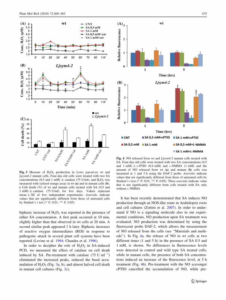
biphasic increase of H2O2 was reported in the presence of
either SA concentration. A first peak occurred at 10 min,
slightly higher than that observed in wt cells at 20 min. A
second similar peak appeared 1 h later. Biphasic increases
of reactive oxygen intermediates (ROI) in response to
pathogenic attack in several plant cell systems have been
reported (Levine et al. 1994; Chandra et al. 1996).
In order to decipher the role of H2O2 in SA-induced
PCD, we measured the effect of catalase on cell death
induced by SA. Pre-treatment with catalase (75 U ml-1)
eliminated the increased peaks, reduced the basal accu-
mulation of H2O2 (Fig. 3a, b), and almost halved cell death
in mutant cell cultures (Fig. 3c).
It has been recently demonstrated that SA induces NO
production through an NOS-like route in Arabidopsis roots
and cell cultures (Zottini et al. 2007). In order to under-
stand if NO is a signaling molecule also in our experi-
mental conditions, NO production upon SA treatment was
evaluated. NO production was determined by using the
fluorescent probe DAF-2, which allows the measurement
of NO released from the cells (see ‘‘Materials and meth-
ods’’). In Fig. 4a, the release of NO in wt cells at two
different times (1 and 5 h) in the presence of SA 0.5 and
1 mM, is shown. No differences in fluorescence levels
were detected in control and wild type SA treated cells,
while in mutant cells, the presence of both SA concentra-
tions induced an increase of the florescence level, at 5 h
treatment (Fig. 4b). Pre-treatment with the NO scavenger
cPTIO cancelled the accumulation of NO, while pre-
Fig. 3 Measure of H2O2 production in Lotus japonicus wt and
Ljsym4-2 mutant cells. Four-day-old cells were treated with two SA
concentrations (0.5 and 1 mM) ± catalase (75 U/ml), and H2O2 was
measured with xylenol orange assay in wt (a) and in mutant cells (b).
c Cell death (%) of wt and mutant cells treated with SA (0.5 and
1 mM) ± catalase (75 U/ml) for five days. Values represent
mean ± SE of five independent experiments. Asterisks indicate
values that are significantly different from those of untreated cells
by Student’s t test (* P, 0.01, ** P, 0.05)
Fig. 4 NO released from wt and Ljsym4-2 mutant cells treated with
SA. Four-day-old cells were treated with two SA concentrations (0.5
and 1 mM) ± c-PTIO (0.4 mM) and L-NMMA (1 mM) and the
amount of NO released from wt (a) and mutant (b) cells was
measured at 1 and 5 h using the DAF-2 probe. Asterisks indicate
values that are significantly different from those of untreated cells by
Student’s t test (* P, 0.01, ** P, 0.05). Three asterisks indicate value
that is not significantly different from cells treated with SA only
without L-NMMA
Plant Mol Biol (2010) 72:469–483 475
123
treatment with the NOS inhibitor L-NMMA does not seem
to induce a statistically significant effect. (Fig. 4b). The
effect of SA on NO accumulation was also determined at
the intracellular level by using the cell-permeable fluores-
cent probe DAF-FM DA combined with confocal micros-
copy analyses. NO levels increased more and constantly in
mutant cells compared to wt cells, in agreement with the
results obtained by measuring the NO released by the cells
(data not shown).
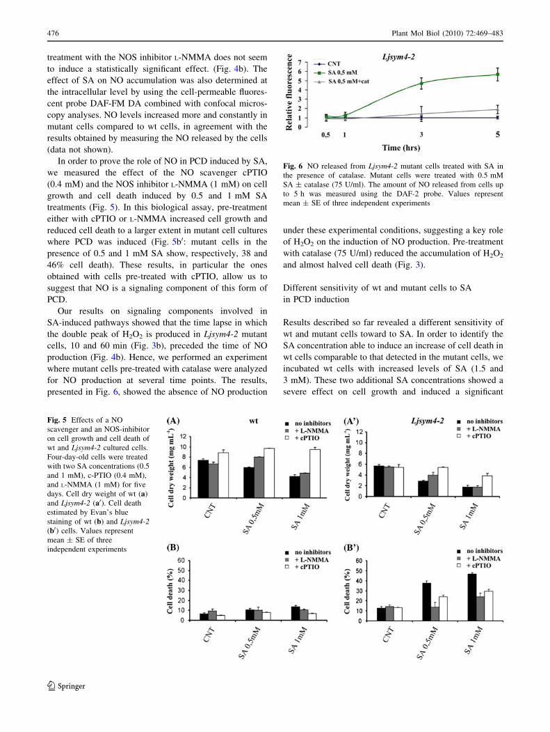
In order to prove the role of NO in PCD induced by SA,
we measured the effect of the NO scavenger cPTIO
(0.4 mM) and the NOS inhibitor L-NMMA (1 mM) on cell
growth and cell death induced by 0.5 and 1 mM SA
treatments (Fig. 5). In this biological assay, pre-treatment
either with cPTIO or L-NMMA increased cell growth and
reduced cell death to a larger extent in mutant cell cultures
where PCD was induced (Fig. 5b0: mutant cells in the
presence of 0.5 and 1 mM SA show, respectively, 38 and
46% cell death). These results, in particular the ones
obtained with cells pre-treated with cPTIO, allow us to
suggest that NO is a signaling component of this form of
PCD.
Our results on signaling components involved in
SA-induced pathways showed that the time lapse in which
the double peak of H2O2 is produced in Ljsym4-2 mutant
cells, 10 and 60 min (Fig. 3b), preceded the time of NO
production (Fig. 4b). Hence, we performed an experiment
where mutant cells pre-treated with catalase were analyzed
for NO production at several time points. The results,
presented in Fig. 6, showed the absence of NO production
under these experimental conditions, suggesting a key role
of H2O2 on the induction of NO production. Pre-treatment
with catalase (75 U/ml) reduced the accumulation of H2O2
and almost halved cell death (Fig. 3).
Different sensitivity of wt and mutant cells to SA
in PCD induction
Results described so far revealed a different sensitivity of
wt and mutant cells toward to SA. In order to identify the
SA concentration able to induce an increase of cell death in
wt cells comparable to that detected in the mutant cells, we
incubated wt cells with increased levels of SA (1.5 and
3 mM). These two additional SA concentrations showed a
severe effect on cell growth and induced a significant
Fig. 5 Effects of a NO
scavenger and an NOS-inhibitor
on cell growth and cell death of
wt and Ljsym4-2 cultured cells.
Four-day-old cells were treated
with two SA concentrations (0.5
and 1 mM), c-PTIO (0.4 mM),
and L-NMMA (1 mM) for five
days. Cell dry weight of wt (a)
and Ljsym4-2 (a0). Cell death
estimated by Evan’s blue
staining of wt (b) and Ljsym4-2(b0) cells. Values represent
mean ± SE of three
independent experiments
Fig. 6 NO released from Ljsym4-2 mutant cells treated with SA in
the presence of catalase. Mutant cells were treated with 0.5 mM
SA ± catalase (75 U/ml). The amount of NO released from cells up
to 5 h was measured using the DAF-2 probe. Values represent
mean ± SE of three independent experiments
476 Plant Mol Biol (2010) 72:469–483
123
increase in the percentage of cell death in wt suspension
cultures (Fig. 7a, d). The addition of 1.5 mM SA to the
medium of wt cells induced a level of cell death (40%)
similar to that detected in Ljsym4-2 cells incubated with
0.5 mM SA. Nuclear morphology observed at the two
concentrations showed an increase in the percentage of
condensed plus stretched nuclei, confirming the induction
of PCD in these treated cells (Fig. 7b).
As shown in Fig. 7c, an increase in H2O2 production can
be detected 20 min after treatment of wt cells with 1.5 mM
SA and a more relevant increase with 3 mM SA. The
presence of 3 mM SA induced a second peak at 70 min,
correlating a biphasic increase of reactive oxygen inter-
mediates with the induction of cell death. Also in this case,
as shown in Fig. 3, pre-treatment with catalase (75 U/ml)
reduced the accumulation of H2O2 and the percent of cell
death in the culture (Fig. 7d).
The NO released from cells incubated with 1.5 and
3 mM SA could not be directly measured because high
levels of SA (more than 1 mM) interfered with the DAF-2
assay. Thus, the NO accumulation in wt cells treated with
1.5 and 3 mM SA was determined at the intracellular level
by using the cell-permeable fluorescent probe DAF-FM
DA. The results (Fig. 8a) showed a clear increase of NO in
a dose- dependent manner.
In order to confirm the role of NO in the PCD pathway
induced by high levels of SA in wt cells, we measured the
effect of the NO scavenger cPTIO (0.4 mM) and the NOS
inhibitor L-NMMA (1 mM) on wt cell growth and cell
death induced by 1.5 and 3 mM SA treatment. Also in this
case, the results clearly showed that pre-treatment with
cPTIO and L-NMMA increased cell growth and reduced
cell death (Fig. 8b, c), confirming the role of NO in the
signaling pathway of this form of PCD.
To complete the picture, a last experiment was
performed. Wild type cell cultures were pre-treated with
L-NMMA (1 mM) and analyzed for H2O2 production. In
this case, cPTIO could not be used because of its inter-
ference with xylenol orange present in the H2O2 assay
under our experimental conditions. The results in Fig. 8d
show a reduction of H2O2 suggesting a role for L-NMMA
in this process.
Modulation of LjPR1 gene expression in wt and mutant
cell cultures
Pathogenesis related (PR) proteins are normally used as
markers of a SA-dependent plant defense response. In
order to test SA-dependent pathway in wild type and
Ljsym4-2 cells we used, as molecular marker, the
L. japonicus predicted gene LjSGA_043037.1 (http://www.
kazusa.or.jp/lotus/blast.html) (renamed here as LjPR1),
orthologue of the A. thaliana PR1 gene (At2G14610;
E value 3e-50).
The analysis of the amount of LjPR1 transcript, mea-
sured by semi-quantitative RT-PCR (Fig. 9a), showed very
low basal level of expression in control wild type cells,
whereas it was clearly induced 24 h after treatment with
0.1 and to a lesser extent, with 0.5 mM SA. In the Ljsym4-2
cells, the level of expression of LjPR1 was very high both
in untreated and SA-treated cells, confirming a constitutive
alteration of the SA sensing in this mutated background.
Fig. 7 Effect of 1.5 and 3 mM
SA concentrations on Lotusjaponicus wt cells. Four-day-old
wt cells were incubated for
5 days in presence of the two
SA concentrations: a Dry
weight, d Cell death estimated
by Evan’s blue staining with the
two SA
concentrations ± catalase
(75 U/ml). b Percentage of
normal and apoptotic
(condensed and stretched)
nuclei in cells treated with SA
1.5 and 3 mM for 48 h. c H2O2
production measured by xylenol
orange assay in four-day-old
cells treated with the two SA
concentrations ± catalase
(75 U/ml). Asterisks indicate
values that are significantly
different from those of untreated
cells by Student’s t test (* P,
0.01, ** P, 0.05)
Plant Mol Biol (2010) 72:469–483 477
123

Characterization of plant responses to M. loti in wild
type and mutant Ljsym4-2 seedlings
In order to test in planta the physiological relevance of the
altered level of the SA-dependent pathway characterized in
cells, we compared the basal level of expression of the
LjPR1 gene in wild type and Ljsym4-2 mutant L. japonicus
seedlings and followed the profile of its expression after
M. loti inoculation. Roots of wild type and mutant Ljsym4-
2 seedlings, grown in N starvation conditions, were inoc-
ulated with M. loti, and the level of expression of the LjPR1
gene was measured, by qRT-PCR analysis at the time of
inoculation 0 and 48 h later. As shown in Fig. 9b the
amount of LjPR1 transcript did not change in the Ljsym4-2
un-inoculated roots when compared to wild type. Further-
more, wild type roots showed a clear increase (about 2
fold) in the LjPR1 transcript level at 48 h p.i. consistently
with the reported early induction of defense genes during
the establishment of the symbiotic interaction (Kouchi
et al. 2004; Lohar et al. 2006). The level of expression of
LjPR1 at 48 h p-i. was of one order of magnitude higher in
the Ljsym4-2 seedlings (Fig. 9b).
In order to see whether the altered SA dependent path-
way could represent the cause of the nod-phenotype
reported for the Ljsym4-2 mutant (Bonfante et al. 2000) we
constructed composite L. japonicus transgenic plants using
transformation with Agrobacterium rhizogenes carrying the
pROK2-NAHG, or pHKN 29 (Kumagai and Kouchi 2003;
35S-GFP construct), or pIG121-HM (Hiei et al. 1994; 35S-
gusA construct) and the appearing roots were tested for
nodulation by inoculating with M. loti. Composite trans-
genic plants obtained after A. rhizogenes infection of wild
type seedlings showed a full nodulation capacity (7–10
nodules per plant), whereas Ljsym4-2 plants transformed
with each of the constructs were unable to form nodules
although the presence of the GFP and GUS markers
allowed an estimation of 30–60% of transformed roots in
three independent experiments (data not shown). This
result suggests that the alteration of the SA pathway in the
Ljsym4-2 background is not the main cause of the symbi-
otic phenotype.
Discussion
An intriguing behavior of Ljsym4-2 mutant plant is that, as
a consequence of the abortion of a symbiotic fungal
infection, all epidermal colonized cells appear morpho-
logically dead (Bonfante et al. 2000). This is a common
response of resistant plants to attack by pathogens, but not
an ordinary response of legume plants to a symbiotic
fungal infection. Hence, a mutation in the LjSym gene
prevents mycorrhizal formation and activates a cell death
pathway in the presence of symbiotic fungi.
We thought it worthwhile to study cell-death events
induced in Ljsym4-2 in detail, and for this reason, we
produced suspension cell cultures from the L. japonicus
mutant and wild type plants and characterized them. The
physiological parameters of cell growth and senescence
measured in both cell cultures showed similar patterns with
Fig. 8 SA-treated wt cells: intracellular levels of NO, cell growth,
cell death in the presence of a scavenger and an inhibitor of NO and
H2O2 in presence of a NOS inhibitor. Four-day-old cells were treated
with 1.5 and 3 mM SA for 7 h and a intracellular levels of NO were
visualized using a cell-permeable NO-sensitive probe (DAF-FM DA).
Cell dry weight (b) was measured and % cell death c estimated by
Evan’s blue staining in wt cells treated with SA and with cPTIO
(0.4 mM) or L-NMMA (1 mM). d Measure of H2O2 production in
Lotus japonicus wt cells treated with 3 mM of SA ± L-NMMA
(1 mM). Values represent mean ± SE of three independent
experiments
478 Plant Mol Biol (2010) 72:469–483
123

slight differences justified by the independent origin of the
two cell lines (Fig. 1). This result indicates that, in mutant
cells, the cell death events induced during spontaneous
senescence of the cell population occurred normally
(Carimi et al. 2003, 2004).
Because SA acts in plants as a signaling molecule
involved in several physio-pathological processes, we
decided to investigate its effects on wt and mutant sus-
pension cultures. Both cell lines were incubated in the
presence of two (0.5 and 1 mM) SA concentrations and
these treatments induced cell death in mutant but not in
wt cells (Fig. 2). This meant that we were imposing on
wt cells modulations in SA concentrations still perceived as
physiological, whereas on mutant cells, the same signal
induced cell death.
Then, we investigated the components of the signaling
pathway induced by SA, specifically, H2O2 and NO, which
have been reported to be important molecules in plant cell
death induction (Delledonne et al. 2001). Results involving
H2O2 production allowed us to distinguish between the two
cell lines: mutant cells respond to SA (0.5 and 1 mM) by
inducing the appearance of a double peak of H2O2 absent
in wt cells (Fig. 3b). Biphasic increases of reactive oxygen
intermediates (ROI) have been reported in several plant
cell systems in response to pathogenic attack (Levine et al.
1994; Chandra et al. 1996). Thus, this response of mutant
cells to SA suggests these SA concentrations, physiological
for wt cells, were perceived by mutant cells as a death
signal.
The total amount of NO (cell-released and intracellular)
differed in mutant and wt cells (Figs. 4, 5). In mutant cells,
the levels of NO is clearly increased in the presence of both
0.5 and 1 mM SA, whereas in wt cells, the levels remained
low. An accurate analysis of the two components, H2O2
and NO, involved in the signaling pathway induced by SA
allowed us to establish that H2O2 production preceded NO
formation, behaving potentially as a signaling molecule for
NO production (Fig. 6) (Lum et al. 2002; Bright et al.
2005). In addition, we demonstrated that both these com-
ponents, as previously shown in PCD induced during the
hypersensitive response (Delledonne et al. 2001; de Pinto
et al. 2006), were necessary for inducing cell death.
The data on NO detection were obtained by using the
DAF fluorometric method that we know to be an indirect
way to detect NO (Planchet and Kaiser 2006). However, in
the present paper experiments were always performed in
parallel with experiments including the use of cPTIO as
NO scavenger, a strategy usually adopted to confirm NO
results (Besson-Bard et al. 2008).
The effects of 0.5–1 mM SA on mutant cells were
comparable with those observed on 3 mM SA-treated wt
cells when we measured cell survival, appearance of a
double peak of H2O2, (Fig. 7) and production of NO
(which remained constant for a few hours) (Fig. 8a), thus
confirming a different sensitivity of the two lines with
respect to SA.
In order to try to define a possible sequence of events in
H2O2, and NO production, as last experiment, we pre-
treated wt cells with L-NMMA, H2O2, production was
analyzed and a drastic reduction of it, was observed
Fig. 9 Analysis of LjPR1 expression in wt and Ljsym4-2 mutant
cultured cells and seedlings. a LjPR1 expression in wt and Ljsym4-2cultured cells after incubation with different concentrations of SA (0.1
and 0.5 mM) for 24 h. The expression of LjPR1 was analyzed by
semiq RT-PCR at two different PCR amplification cycles (26 and 28)
whereas as internal standard the 18S transcript was analysed after 11
cycles of PCR amplification. The results are representative of two
independent biological repetition. b Relative LjPR1 transcript levels
in roots of wild type and Ljsym4-2 roots at the moment of M. lotiinoculation 0 and 48 h later. Transcript levels were determined by
qRT-PCR, normalized to that of the internal control ubiquitin (see
Materials and methods) and plotted relative to LjPR1 transcript levels
at time 0 wild type seedlings. Bars represent the mean and SD of data
obtained with RNA extracted from two biological replicates and three
technical replicates each
Plant Mol Biol (2010) 72:469–483 479
123
(Fig. 8d). This result is an intriguing one. In fact, even if
L-NMMA was not able to reduce in short times NO pro-
duction in SA treated cells (Fig. 4b), this NOS inhibitor
reduced drastically the cell death rate in a 4-day vitality
assay (Fig. 5b0). Unfortunately, this experiment was not
also performed in presence of cPTIO because of its inter-
ference with components present in the H2O2 assay.SA is a
key molecule in inducing the expression of defense genes.
In particular, in Arabidopsis, PR1 expression after a path-
ogenic attack correlates with plant response capability
(Shah 2003). Then, in order to verify at the molecular level
the different sensitivity of wt and mutant cells to SA, a
L. japonicus gene orthologue to the A. thaliana PR1 gene
was identified by a bioinformatic analysis. The induction of
LjPR1 by SA was clearly detected in wt Lotus cells
(Fig. 9a). An interesting result was obtained in Ljsym4-2
mutant cells where the LjPR1 gene resulted always well
expressed either in absence or in presence of SA, con-
firming a constitutive alteration of this mutant in this sig-
naling pathway.
The analysis of LjPR1 expression in plant seedlings did
not show the different basal level of expression between
wild type and mutant (Fig. 9b). This could be explained by
the less homogeneity of the seedlings, as experimental
system, compared to cell cultures. The induction of defense
and stress-responsive pathways during symbiotic interac-
tions has been reported previously (Colebatch et al. 2004;
Kouchi et al. 2004; Lohar et al. 2006) and an initial
induction followed by a rapid suppression of these genes
may be part of the physiological cascade of events leading
to a successful symbiotic interaction (Kouchi et al. 2004;
Lohar et al. 2006). In agreement with these reports we
observed a significant induction of the LjPR1 gene at 48 h
p.i. in wild type seedlings. Interestingly, the observed burst
of the level of expression observed in the Ljsym4-2 seed-
lings at 48 h p.i. suggests the triggering of a defense-
related pathway in the mutant background (Fig. 9b).
However, such a conclusion, must be further investigated
in planta through a more complete analysis of the molec-
ular pathway leading to the plant defense response.
Recently, a strong induction of genes encoding phenylal-
anine ammonia lyase (PAL), in L. japonicus roots inocu-
lated with M. loti mutants unable to form infection threads
structures (cgs mutants; D’Antuono et al. 2008) was
reported. A question that remains to be elucidated is to
what extent plant defense responses are physiologically
triggered during nodulation. The differences in the level of
LjPR1 between wt and Ljsym4-2 mutant at 48 h p.i. rep-
resent a striking picture of the physiological and patho-
logical level of plant defense response triggered by a
successful and unsuccessful symbiotic interaction,
respectively.
The interference between SA-dependent defense
response and symbiotic pathways has been previously
reported. In fact, addition of exogenous SA inhibits both
indeterminate and determinate nodulation in legume roots
(Martinez-Abarca et al. 1998; Lian et al. 2000; van
Spronsen et al. 2003). Recently, it was shown that reduc-
tion of the endogenous SA level by transgenic expression
of the bacterial nahG gene also correlated with a significant
increase in the number of infections and mean nodule
number when compared with wild type controls (Stacey
et al. 2006). The same approach in Nicotiana tabacum led
to an enhancement of mycorrhizal fungal infection (Med-
ina et al. 2003). However, the triggering of a defense-
related response is not necessarily directly involved in the
specific interaction with rhizobia mediated by Nod factors,
but could represent a parallel pathway triggered during the
infection. In the case of the Ljym4-2 mutant, we did not
observe the rescue of the nod- phenotype in transgenic
hairy roots transformed with a constitutively expressed
copy of the nahG. This result must be considered with
caution since we did not provide direct evidences for a
biologically significant increase of the SA level in the
Ljym4-2 roots. However, a possible interpretation would be
that the putative induction of the defense response,
deducible by the strong induction of the SA inducible
LjPR1 gene (Fig. 9), is not the main cause of the defective
symbiotic phenotype. In other words, the induction of the
defense response pathway could take place in the Ljsym4-2
genetic background after M. loti inoculation, probably as a
consequence of the block of the infection progress.
The plant mutation Ljsym4-2 identifies the CASTOR
gene and determines a block in the Nod factor-dependent
pathway by inhibiting the calcium spiking response but not
calcium influx in root hairs (Imaizumi-Anraku et al. 2005;
Miwa et al. 2006). CASTOR encodes for a nuclear mem-
brane-localized cation channel (Riely et al. 2007; Char-
pentier et al. 2008). It has been suggested that there are at
least two possible mechanisms by which CASTOR and its
orthologue POLLUX could contribute to Ca2? spiking. In
fact, potassium flowing from the root hair cytoplasm into
the nuclear envelope could either alter the membrane
potential, opening voltage-gated calcium channels respon-
sible for the spiking signal, or may act as a counter-ion,
which compensates for the rapid release of positive charge
from the calcium store during each spike (Oldroyd and
Downie 2008; Charpentier et al. 2008). Lack of calcium
spiking is associated to the block of cortical cell divisions
and nodule primordium formation because the Ca2?
oscillation would not be perceived by the calcium–cal-
modulin-dependent protein kinase CCaMK (Levy et al.
2004; Mitra et al. 2004), which controls the activity of
putative transcriptional activators, such as NSP1, NSP2 and
480 Plant Mol Biol (2010) 72:469–483
123
ERN (Kalo et al. 2005; Smit et al. 2005; Murakami et al.
2006; Middleton et al. 2007).
Our results performed in suspension cell cultures show
that a mutation in the allele Ljsym changes the cell sensi-
tivity to SA by inducing cell death in mutant cells when not
in wild type.
The different behavior, detectable at the cellular level,
invokes a general cell function for the Ljsym gene not
limited to root tissues. In this context, it is worth men-
tioning that the two orthologues CASTOR and POLLUX
are ubiquitously present and highly conserved in both
legumes and non-legumes (Chen et al. 2009). In tobacco
cell suspension culture, it has been reported that SA
induces calcium influx (Lin et al. 2005). We can hypoth-
esize that an increase in calcium concentration inside
mutant cells can be ‘‘misinterpreted’’ in cells lacking cal-
cium spiking and, as a consequence, a double peak of H2O2
and an increase in NO level are induced leading to a pro-
gram of cell death. At the moment, we are not able to
suggest a detailed molecular mechanism that links the
function of the Ljsym gene and perception and signal
transduction induced by SA, but investigations are under-
way to clarify these links.
Conclusions
In suspension cell cultures, we have compared the SA
sensitivity of wild type and Ljsym4-2 mutant showing clear
differences. Different parameters of the SA-dependent
signaling pathway were significantly altered in the Ljsym4-
2 cells compared to wt cells. The most relevant result is the
different sensitivity of wild type and mutant cells to SA in
cell death induction. These results gave us the opportunity
to begin some ad hoc experiments on plant seedlings that
suggest the induction of a plant defense-like response. The
double approach—cell culture and plant seedlings—should
help in the future to better understand the beneficial and
pathogenic role of SA in plants.
Acknowledgments We thank Dr. JP Metraux for providing us
pROK-2 vector. Lotus japonicus seeds (wild type and Ljsym4-2) were
kindly provided by Prof. P Bonfante. This research was supported by the
‘‘Ministero dell’Istruzione e della Ricerca, fondi PRIN’’ to F.L.S. and
by a grant from the EEC (INTEGRAL: MRTN-CT-2003-505227).
References
Albrecht C, Geurts R, Bisseling T (1999) Legume nodulation and
mycorrhizae formation, two extremes in host specificity meet.
EMBO J 18:281–288
Ane JM, Kiss GB, Riely BK, Penmetsa RV, Oldroyd D, Ayax C,
Levy J, Debelle F, Baek JM, Kalo P, Rosemberg C, Roe BA,
Long SR, Denarie J, Cook DR (2004) Medicago truncatula
DMI1 is required for bacterial and fungal symbioses in legumes.
Science 303:1364–1367
Besson-Bard A, Griveau S, Bedioui F, Wendehenne D (2008) Real-
time electrochemical detection of extracellular nitric oxide in
tobacco cells exposed to cryptogein, an elicitor of defence
responses. J Exp Bot 59:3407–3414
Bonfante P, Genre A, Faccio A, Martini I, Schauser L, Stougaard J,
Webb J, Parniske M (2000) The Lotus japonicus LjSym4 gene is
required for the successful symbiotic infection of root epidermal
cells. Mol Plant Microb Interact 13:1109–1120
Bright J, Desikan R, Hancock JT, Weir IS, Neill J (2005) ABA-
induced NO generation and stomatal closure in Arabidopsis are
dependent on H2O2 synthesis. Plant J 45:113–122
Campbell GR, Reuhs BL, Walker GC (2002) Chronic intracellular
infection of alfalfa nodules by Sinorhizobium meliloti requires
correct lipopolysaccharide core. Proc Natl Acad Sci USA 19:
3938–3943
Carimi F, Zottini M, Formentin E, Terzi M, Lo Schiavo F (2003)
Cytokinins: new apoptotic inducers in plants. Planta 216:
413–421
Carimi F, Terzi M, De Michele R, Zottini M, Lo Schiavo F (2004)
High levels of the cytokinin BAP induce PCD by accelerating
senescence. Plant Sci 166:963–969
Carimi F, Zottini M, Costa A, Cattelan I, De Michele R, Terzi M, Lo
Schiavo F (2005) NO signalling in cytokinin-induced pro-
grammed cell death. Plant Cell and Environ 28:1171–1178
Carlson RW, Kalembasa S, Turowski D, Pachori P, Noel KD (1987)
Characterization of the lipopolysaccharide from a Rhizobiumphaseoli mutant that is defective in infection thread develop-
ment. J Bacteriol 169:4923–4928
Chandra S, Martin MB, Low PS (1996) The Pto kinase mediates a
signaling pathway leading to the oxidative burst in tomato. Proc
Natl Acad Sci USA 93:13393–13397
Charpentier M, Bredemier R, Wanner G, Takeda N, Schleiff E,
Parniske M (2008) Lotus japonicus CASTOR and POLLUX are
ion channels essential for perinuclear calcium spiking in legume
root endosymbiosis. Plant Cell 20:3467–3479
Chen C, Fan C, Gao M, Zhu H (2009) Antiquity and function of
CASTOR and POLLUX, the twin ion channel-encoding genes
key to the evolution of root symbioses in plants. Plant Physiol
149:306–317
Colebatch G, Desbrosses G, Ott T, Krusell L, Montanari O, Kloska S,
Kopka J, Udvardi MK (2004) Global changes in transcription
orchestrate metabolic differentiation during symbiotic nitrogen
fixation in Lotus japonicus. Plant J 39:487–512
D’Antuono AL, Ott T, Krussell L, Voroshilova V, Ugalde RA,
Udvardi M, Lepek VC (2008) Defects in rhizobial cyclic glucan
and lypopolysaccharide synthesis alter legume gene expression
during nodule development. Mol Plant Microbe Interact 21:
50–60
de Pinto MC, Paradiso A, Leonetti P, De Gara L (2006) Hydrogen
peroxide, nitric oxide and cytosolic ascorbate peroxidase at the
crossroad between defence and cell death. Plant J 48:784–795
Delledonne M, Zeier J, Marocco A, Lamb C (2001) Signal
interactions between nitric oxide and reactive oxygen interme-
diates in the plant hypersensitive disease resistance response.
Proc Natl Acad Sci USA 98:13454–13459
Doyle JJ, Doyle JL (1987) A rapid DNA isolation procedure from
small quantities of fresh leaf tissue. Phytochem Bull 19:11–15
Draper J (1997) Salicylate, superoxide synthesis and cell suicide in
plant defence. Trends Plant Sci 2:162–165
Durner J, Shab J, Klessing DF (1997) Salicylic acid and disease
resistance in plants. Trends Plant Sci 2:266–277
Endre G, Kereszt A, Kevei Z, Mihacea S, Kalo P, Kiss GB (2002) A
receptor kinase gene regulating symbiotic nodule development.
Nature 417:962–966
Plant Mol Biol (2010) 72:469–483 481
123
Feys B, Parker JE (2000) Interplay of signaling pathways in plant
disease resistance. Trends Genet 16:449–455
Gaffney T, Friedrich L, Vernooij B, Negrotto D, Nye G, Uknes S,
Ward E, Kessmann H, Ryals J (1993) Requirement of salicylic
acid for the induction of systemic acquired resistance. Science
261:754–756
Gonzales-Rizzo S, Crespi M, Frugier F (2006) The Medicagotruncatula CRE1 cytokinin receptor regulates lateral root
development and early symbiotic interaction with Sinorhizobium
meliloti. Plant Cell 18:2680–2693
Gualtieri G, Bisseling T (2000) The evolution of nodulation. Plant
Mol Biol 42:181–194
Heath MC (1997) Signalling between pathogenic rust fungi and
resistant or susceptible host plants. Ann Bot 80:713–720
Hiei Y, Ohta S, Komari T, Kumashiro T (1994) Efficient transfor-
mation of rice mediated by Agrobacterium and sequence
analysis of the boundaries of the T-DNA. Plant J 6:271–282
Imaizumi-Anraku H, Takeda N, Charpentier M, Perry J, Miwa H,
Umehara Y, Kouci H, Murakami Y, Mulder L, Vickers K, Pike J,
Downie JA, Wang T, Sato S, Asamizu E, Tabata S, Yoshikawa
M, Murooka Y, Wu G-J, Kawaguchi M, Kawasaki S, Parniske
M, Hayashi M (2005) Plastid proteins crucial for symbiotic
fungal and bacterial entry into plant roots. Nature 433:527–531
Jiang Z, Wooland ACS, Wolff S (1990) Hydrogen peroxide
production during experimental protein glycation. FEBS Lett
268:69–71
Journet EP, El-Gachtouli N, Vernoud V, de Billy F, Pichon M, Dedieu
A, Arnould C, Morandi D, Barker DG, Gianinazzi-Pearson V
(2001) Medicago truncatula ENOD11: a novel RPRP-encoding
early nodulin gene expressed during mycorrhization in arbuscule-
containing cells. Mol Plant Microbe Interact 14:737–748
Kalo P, Gleason C, Edwards A, Marsh J, Mitra RM, Hirsh S, Jakab J,
Sims S, Long SR, Rogers J, Kiss GB, Downie JA, Oldroyd D
(2005) Nodulation signaling in legumes requires NSP2, a
member of the GRAS family of transcriptional regulators.
Science 308:1786–1789
Kanamori N, Madsen LH, Radutoiu S, Frantescu M, Quistgaard
EMH, Miwa H, Downie AJ, James EK, Felle HH, Haaning LL,
Jensen TH, Sato S, Nakamura Y, Tabata S, Sandal N, Stougaard
J (2006) A nucleoporin is required for induction of Ca2? spiking
in legume nodule development and essential for rhizobial and
fungal symbiosis. Proc Natl Acad Sci USA 103:359–364
Kistner C, Parniske M (2002) Evolution and signal transduction in
intracellular symbiosis. Trends Plant Sci 7:511–518
Kojima H, Nakatsubo N, Kikuchi K, Kawahara S, Kirino Y, Nagoshi
H, Hirata Y, Nagano T (1998) Detection and imaging of nitric
oxide with novel fluorescent indicators: diaminofluoresceins.
Anal Chem 70:2446–2453
Kouchi H, Shimomura K, Hata S, Hirota A, Wu G-J, Kumagai H,
Tajima S, Suganuma N, Suzuki A, Aoki T, Hayashi M,
Yokoyama T, Ohyama T, Asamizu E, Kuwata C, Shibata D,
Tabata S (2004) Large-scale analysis of gene expression profiles
during ealy stages of root nodule formation in a model legume,
Lotus japonicus. DNA Res 11:263–274
Kumagai H, Kouchi H (2003) Gene silencing by expression of hairpin
RNA in Lotus japonicus roots and root nodules. Mol Plant
Microbe Interact 16:663–668
Levine A, Tenhaken R, Dixon R, Lamb C (1994) H2O2 from the
oxidative burst orchestrates the plant hypersensitive disease
resistance response. Cell 94:491–501
Levy J, Bres C, Geurts R, Chalhoub B, Kukilova O, Duc G, Journet
EP, Ane JM, Lauber E, Bisseling T, Denarie J, Rosemberg C,
Debelle F (2004) A putative Ca2? and calmodulin-dependent
protein kinase required for bacterial and fungal symbioses.
Science 303:1361–1364
Lian B, Zhou X, Miransari M, Smith DL (2000) Effects of salycilic
acid on the development and root nodulation of soybean
seedlings. J Agron Crop Sci 185:187–192
Lin C, Yu Y, Kadono T, Iwata M, Umemura K, Furuichi T, Kuse M,
Isobe M, Yamamoto Y, Matsumoto H, Yoshizuka K, Kawano T
(2005) Action of aluminium, novel TPC1-type channel inhibitor,
against salicylate-induced and cold-shock-induced calcium
influx in tobacco BY-2 cells. BBRC 332:823–830
Lohar DP, Shaparova N, Endre G, Penuela S, Samac D, Town C,
Siverstein KA, VandenBosch KA (2006) Transcript analysis of
early events in Medicago truncatula. Plant Physiol 140:221–234
Lum HK, Butt YKC, Lo SCL (2002) Hydrogen peroxide induces a
rapid production of nitric oxide in mung bean (Phaseolusaureus). Nitric Oxide Biol Chem 6:205–213
Madsen EB, Madsen LH, Radutoiu S, Olbryt M, Szczyglowski K,
Sato S, Kaneko T, Tabata S, Sandal N, Stougaard J (2003) A
receptor kinase gene of LysM type is involved in legume
perception of rhizobial signals. Nature 425:637–640
Martinez-Abarca F, Herrera-Cervera JA, Bueno P, Sanjuan J,
Bisseling T, Olivares J (1998) Involvement of salicylic acid in
the establishment of the Rhizobium meliloti-alfalfa symbiosis.
Mol Plant Microbe Interact 11:153–155
Martirani L, Stiller J, Mirabella R, Alfano F, Lamberti A, Radutoiu
SE, Iaccarino M, Gresshoff PM, Chiurazzi M (1999) A fast and
efficient experimental system for T-DNA tagging in the model
legume Lotus japonicus. Trapping sequences frequencies,
expression patterns and potential for insertional mutagenesis.
Mol Plant Microbe Interact 12:275–284
Medina MJH, Gagnon H, Piche Y, Ocampo JA, Garrido JMG,
Vierheilig H (2003) Root colonization by arbuscular mycorrhizal
fungi is affected by the salicylic acid content of the plant. Plant
Sci 164:993–998
Middleton PH, Jakab J, Penmetsa RV, Starker CG, Doll J, Kalo P,
Prabhu R, Marsh JF, Mitra RM, Kereszt A, Dudas B,
VandenBosch K, Long SR, Cook GB, Oldroyd GE (2007) An
ERFtranscription factor in Medicago truncatula that is essential
for Nod factor signal transduction. Plant Cell 19:1221–1234
Mitra RM, Gleason CA, Edwards A, Hadfield J, Downie JA, Oldroyd
GE, Long SR (2004) A Ca2?/calmodulin-dependent protein kinase
required for symbiotic nodule development: gene identification by
transcript-based cloning. Proc Natl Acad Sci USA 101:4701–4705
Miwa H, Sun J, Oldroyd GED, Downie JA (2006) Analysis of nod-
factor-induced calcium signaling in root hairs of symbiotically
defective mutants of Lotus japonicus. Mol Plant Microbe Interact
19:914–923
Miya AF, Albert P, Shinya T, Desaki Y, Ichimura K, Shirasu K,
Narusaka Y, Kawakami N, Kaku H, Shibuya N (2007) CERK1, a
LysM receptor kinase, is essential for chitin elicitor signalling in
Arabidopsis. Proc Natl Acad Sci USA 104:19613–19618
Murakami Y, Miwa H, Imaizumi-Anraku H, Kouchi H, Downie JA,
Kawaguchi M, Kawasaki S (2006) Positional cloning identifies
Lotus japonicus NSP2, a putative transcription factor of the GRAS
family, required NIN and ENOD40 gene expression in nodule
initiation. DNA Res 13:255–265
Nagata M, Murakami E, Shimoda Y, Shimoda-Sasakura F, Kucho K,
Suzuki A, Abe M, Higashi S, Uchiumi T (2008) Expression of a
class 1 hemoglobin gene and production of nitric oxide in
response to symbiotic and pathogenic bacteria in Lotus japoni-cus. Mol Plant Microbe Interact 21:1175–1183
Nakatsubo N, Kojima H, Kikuchi K, Nagoshi H, Hirata Y, Maeda D, Imai
Y, Irimura T, Nagano T (1998) Direct evidence of nitric oxide
production from bovine aortic endothelial cells using new fluores-
cence indicators: diaminofluoresceins. FEBS Lett 427:263–266
Navazio L, Moscatiello R, Genre A, Novero M, Baldan B, Bonfante P,
Mariani P (2007) A diffusible signal from arbuscular mycorrhyzal
482 Plant Mol Biol (2010) 72:469–483
123

fungi elicits a transient cytosolic calcium elevation in host plant
cells. Plant Physiol 144:673–681
Novero M, Faccio A, Genre A, Stougaard J, Webb KJ, Mulder L,
Parniske M, Bonfante P (2002) Dual requirement of the LjSym4gene for micorrhizal development in epidermal and cortical cells
of Lotus japonicus roots. New Phytol 154:741–749
Oldroyd GED, Downie JA (2008) Coordinating nodule morphogen-
esis with rhizobial infection in legumes. Ann Rev Plant Mol Biol
59:519–546
Planchet E, Kaiser WM (2006) Nitric oxide (NO) detection by DAF
fluorescence and chemiluminescence: a comparison using abiotic
and biotic NO sources. J Exp Bot 57:3043–3055
Radutoiu S, Madsen LH, Madsen EB, Felle HH, Umehara Y,
Gronlund M, Sato S, Nakamura Y, Tabata S, Sandal N, Sougaard
J (2003) Plant recognition of symbiotic bacteria requires two
LysM receptor-like kinases. Nature 425:585–592
Riely BK, Lougnon G, Ane J-M, Cook DR (2007) The symbiotic ion
channel homolog DMI1 is localized in the nuclear membrane of
Medicago truncatula roots. Plant J 49:208–216
Sambrook J, Fritsch EF, Maniatis T (1989) Molecular cloning: a
laboratory manual, 2nd edn. Cold Spring Harbor, New York
Schauser L, Handberg K, Sandal N, Stiller J, Thykjaer T, Pajuelo E,
Nielsen A, Stougaard J (1998) Symbiotic mutants deficient in
nodule establishment identified after T-DNA transformation of
Lotus japonicus. Mol Gen Genet 259:414–423
Schauser L, Roussis A, Stiller J, Stougaard J (1999) A plant regulator
controlling development of symbiotic root nodules. Nature 402:
191–195
Shah J (2003) The salicylic acid loop in plant defense. Curr Opin
Plant Biol 6:365–371
Shigaki T, Bhattacharya MK (1999) Color coding the cell death status
of plant suspension cells. Biotechniques 26:1060–1062
Shimoda Y, Nagata M, Suzuki A, Abe M, Sato S, Kato T, Tabata S,
Higashi S, Uchiumi T (2005) Symbiotic rhizobium and nitric
oxide induce gene expression of non-symbiotic hemoglobin in
Lotus japonicus. Plant Cell Physiol 46:99–107
Smit P, Raedts J, Portyanko V, Debelle F, Gough C, Bisseling T,
Geurts R (2005) NSP1 of the GRAS protein family is essential
for rhizobial Nod factor-induced transcription. Science 308:
1789–1791
Stacey G, Mc Alvin CB, Kim S-Y, Olivares J, Soto MJ (2006) Effects
of endogenous salicylic acid on nodulation in the model legumes
Lotus japonicus and Medicago truncatula. Plant Physiol 141:
1473–1481
Stracke S, Kistner C, Yoshida S, Mulder L, Sato S, Kaneko T, Tabata
S, Sandal N, Stougaard J, Szczyglowski K, Parniske M (2002) A
plant receptor-like kinase required for both bacterial and fungal
symbiosis. Nature 417:959–996
Tirichine L, Sandal N, Madsen LH, Radutoiu S, Albrektsen AS, Sato
S, Asamizu E, Tabata S, Stougaard J (2007) A gain-of-function
mutation in a cytokinin receptor triggers spontaneous root
nodule organogenesis. Science 315:104–107
Traas JA, Beven AF, Doonan JH, Cordewener J, Shaw PJ (1992)
Cell-cycle-dependent changes in labelling of specific phospho-
proteins by monoclonal antibody MPM-2 in plant cells. Plant J
2:723–732
Van Spronsen PC, Tak T, Rood AMM, van Brussel AAN, Kijne JW,
Boot KJM (2003) Salycilic acid inhibits indeterminate-type
nodulation but not determinate-type nodulation. Mol Plant
Microb Interact 16:83–91
Vasse J, de Billy F, Truchet G (1993) Abortion of infection during the
Rhizobium meliloti-alfalfa symbiotic interaction is accompanied
by a hypersensitive reaction. Plant J 4:555–566
Veershlingam H, Haynes JG, Penmetsa RV, Cook DR, Sherrier DJ,
Dickstein R (2004) nip a symbiontic Medicago truncatulamutant that forms root nodules with aberrant infection threads
and plant defence-like response. Plant Physiol 136:3692–3702
Wan J, Zhang X-C, Neece D, Ramonell KM, Clough S, Kim S-Y,
Stacey MG, Stacey G (2008) A LysM receptor-like kinase plays
a critical role in chitin signalling and fungal resistance in
Arabidopsis. Plant Cell 20:182–481
Zottini M, Barizza E, Bastianelli F, Carimi F, Lo Schiavo F (2006)
Growth and senescence of Medicago truncatula cultured cells
are associated with characteristic mitochondrial morphology.
New Phytol 172:239–247
Zottini M, Costa A, De Michele M, Ruzzene M, Carimi C, Lo
Schiavo F (2007) Salicylic acid activates nitric oxide synthesis in
Arabidopsis. J Exp Bot 58:1397–1405
Plant Mol Biol (2010) 72:469–483 483
123