A Novel ARID DNA-Binding Protein Interacts with SymRK and Is Expressed during Early Nodule...
11
A Novel ARID DNA-Binding Protein Interacts with SymRK and Is Expressed during Early Nodule Development in Lotus japonicus 1[C][W][OA] Hui Zhu, Tao Chen, Maosheng Zhu, Qing Fang, Heng Kang, Zonglie Hong, and Zhongming Zhang* State Key Laboratory of Agricultural Microbiology, Huazhong Agricultural University, Wuhan 430070, China (H.Z., T.C., M.Z., Q.F., H.K., Z.Z.); and Department of Microbiology, Molecular Biology and Biochemistry, University of Idaho, Moscow, Idaho 83844–3052 (Z.H.) During the establishment of symbiosis in legume roots, the rhizobial Nod factor signal is perceived by the host cells via receptor- like kinases, including SymRK. The NODULE INCEPTION (NIN) gene in Lotus japonicus is required for rhizobial entry into root cells and for nodule organogenesis. We describe here a novel DNA-binding protein from L. japonicus, referred to as SIP1, because it was identified as a SymRK-interacting protein. SIP1 contains a conserved AT-rich interaction domain (ARID) and represents a unique member of the ARID-containing proteins in plants. The C terminus of SIP1 was found to be responsible for its interaction with the kinase domain of SymRK and for homodimerization in the absence of DNA. SIP1 specifically binds to the promoter of LjNIN but not to that of LjCBP1 (a calcium-binding protein gene), both of which are known to be inducible by Nod factors. SIP1 recognizes two of the three AT-rich domains present in the NIN gene promoter. Deletion of one of the AT-rich domains at the NIN promoter diminishes the binding of SIP1 to the NIN promoter. The protein is localized to the nuclei when expressed as a red fluorescence fusion protein in the onion (Allium cepa) epidermal cells. The SIP1 gene is expressed constitutively in the uninfected roots, and its expression levels are elevated after infection by Mesorhizobium loti. It is proposed that SIP1 may be required for the expression of NIN and involved in the initial communications between the rhizobia and the host root cells. Legume plants are capable of acquiring nitrogen from rhizobacteria maintained symbiotically in spe- cialized root organ nodules that form through a com- plex developmental process involving an exchange of signals between the host root cells and the bacteria. At the beginning of nodule organogenesis, specific flavo- noid metabolites released by the legume roots notify soil rhizobia that a suitable host is nearby. Inside the bacteria, the flavonoids are recognized by a receptor protein known as NodD. The binding of NodD to these flavonoids activates the protein and promotes the transcription of other nod genes involved in the synthesis and secretion of Nod factors, the rhizobial signaling molecules (Peck et al., 2006). The common feature of all Nod factors is the presence of a chitin backbone and a fatty acyl tail (Denarie et al., 1996). Rhizobial Nod factors, in turn, are capable of inducing a series of specific responses in the host root cells, including root hair deformation, alkalinization of the cytosol, depolarization of the plasma membrane, and calcium influx and spiking (Ehrhardt et al., 1992, 1996; Kurkdjian, 1995; Felle et al., 1995, 1996; Limpens and Bisseling, 2003; Gleason et al., 2006). Root hair defor- mation and curling are the early morphological changes induced by rhizobia. Purified Nod factors can cause root hair deformation at concentrations as low as 10 212 M (Lerouge et al., 1990; Spaink et al., 1991; Sanjuan et al., 1992; Margaert et al., 1993), although in most cases curling is only observed when the bacteria are present (Relic et al., 1993). The perception and signal transduction of Nod fac- tors in the host cells have been subject to intense mo- lecular and genetic studies in recent years. Rhizobial Nod factors have been shown to be recognized by lysin motif (LysM)-containing receptor-like kinases (RLKs), such as NFR1/NFR5 from Lotus japonicus and LYK3 and NFP from Medicago truncatula (Madsen et al., 2003; Radutoiu et al., 2003; Limpens et al., 2003; Arrighi et al., 2006). The LysM RLKs are localized to the plasma membrane with the LysM domain pro- truding to the extracellular space and the kinase domain facing the cytoplasm. The LysM domain in- teracts with the lipochitin-oligosaccharide backbone of Nod factors, resulting in Nod factor recognition (Steen et al., 2003, 2005; Radutoiu et al., 2007). Using forward genetics and map-based cloning approaches, a series 1 This work was supported by the National Natural Science Foundation of China (grant no. 30570056 to Z.Z.), by the National Basic Research Program of China (grant no. 01CB108901 to Z.Z.), and by the National Science Foundation (grant nos. NSF–MCB 0548525 and NSF–IOB 0543923 to Z.H.). * Corresponding author; e-mail [email protected]. The author responsible for distribution of materials integral to the findings presented in this article in accordance with the policy described in the Instructions for Authors (www.plantphysiol.org) is: Zhongming Zhang ([email protected]). [C] Some figures in this article are displayed in color online but in black and white in the print edition. [W] The online version of this article contains Web-only data. [OA] Open Access articles can be viewed online without a sub- scription. www.plantphysiol.org/cgi/doi/10.1104/pp.108.119164 Plant Physiology, September 2008, Vol. 148, pp. 337–347, www.plantphysiol.org Ó 2008 American Society of Plant Biologists 337
-
Upload
independent -
Category
Documents
-
view
3 -
download
0
Transcript of A Novel ARID DNA-Binding Protein Interacts with SymRK and Is Expressed during Early Nodule...

A Novel ARID DNA-Binding Protein Interacts withSymRK and Is Expressed during Early NoduleDevelopment in Lotus japonicus1[C][W][OA]
Hui Zhu, Tao Chen, Maosheng Zhu, Qing Fang, Heng Kang, Zonglie Hong, and Zhongming Zhang*
State Key Laboratory of Agricultural Microbiology, Huazhong Agricultural University, Wuhan 430070,China (H.Z., T.C., M.Z., Q.F., H.K., Z.Z.); and Department of Microbiology, Molecular Biology andBiochemistry, University of Idaho, Moscow, Idaho 83844–3052 (Z.H.)
During the establishment of symbiosis in legume roots, the rhizobial Nod factor signal is perceived by the host cells via receptor-like kinases, including SymRK. The NODULE INCEPTION (NIN) gene in Lotus japonicus is required for rhizobial entry into rootcells and for nodule organogenesis. We describe here a novel DNA-binding protein from L. japonicus, referred to as SIP1, becauseit was identified as a SymRK-interacting protein. SIP1 contains a conserved AT-rich interaction domain (ARID) and represents aunique member of the ARID-containing proteins in plants. The C terminus of SIP1 was found to be responsible for its interactionwith the kinase domain of SymRK and for homodimerization in the absence of DNA. SIP1 specifically binds to the promoter ofLjNIN but not to that of LjCBP1 (a calcium-binding protein gene), both of which are known to be inducible by Nod factors. SIP1recognizes two of the three AT-rich domains present in the NIN gene promoter. Deletion of one of the AT-rich domains at the NINpromoter diminishes the binding of SIP1 to the NIN promoter. The protein is localized to the nuclei when expressed as a redfluorescence fusion protein in the onion (Allium cepa) epidermal cells. The SIP1 gene is expressed constitutively in the uninfectedroots, and its expression levels are elevated after infection by Mesorhizobium loti. It is proposed that SIP1 may be required for theexpression of NIN and involved in the initial communications between the rhizobia and the host root cells.
Legume plants are capable of acquiring nitrogenfrom rhizobacteria maintained symbiotically in spe-cialized root organ nodules that form through a com-plex developmental process involving an exchange ofsignals between the host root cells and the bacteria. Atthe beginning of nodule organogenesis, specific flavo-noid metabolites released by the legume roots notifysoil rhizobia that a suitable host is nearby. Inside thebacteria, the flavonoids are recognized by a receptorprotein known as NodD. The binding of NodD tothese flavonoids activates the protein and promotesthe transcription of other nod genes involved in thesynthesis and secretion of Nod factors, the rhizobialsignaling molecules (Peck et al., 2006). The commonfeature of all Nod factors is the presence of a chitin
backbone and a fatty acyl tail (Denarie et al., 1996).Rhizobial Nod factors, in turn, are capable of inducinga series of specific responses in the host root cells,including root hair deformation, alkalinization of thecytosol, depolarization of the plasma membrane, andcalcium influx and spiking (Ehrhardt et al., 1992, 1996;Kurkdjian, 1995; Felle et al., 1995, 1996; Limpens andBisseling, 2003; Gleason et al., 2006). Root hair defor-mation and curling are the early morphological changesinduced by rhizobia. Purified Nod factors can causeroot hair deformation at concentrations as low as 10212
M
(Lerouge et al., 1990; Spaink et al., 1991; Sanjuan et al.,1992; Margaert et al., 1993), although in most casescurling is only observed when the bacteria are present(Relic et al., 1993).
The perception and signal transduction of Nod fac-tors in the host cells have been subject to intense mo-lecular and genetic studies in recent years. RhizobialNod factors have been shown to be recognized bylysin motif (LysM)-containing receptor-like kinases(RLKs), such as NFR1/NFR5 from Lotus japonicus andLYK3 and NFP from Medicago truncatula (Madsenet al., 2003; Radutoiu et al., 2003; Limpens et al.,2003; Arrighi et al., 2006). The LysM RLKs are localizedto the plasma membrane with the LysM domain pro-truding to the extracellular space and the kinasedomain facing the cytoplasm. The LysM domain in-teracts with the lipochitin-oligosaccharide backbone ofNod factors, resulting in Nod factor recognition (Steenet al., 2003, 2005; Radutoiu et al., 2007). Using forwardgenetics and map-based cloning approaches, a series
1 This work was supported by the National Natural ScienceFoundation of China (grant no. 30570056 to Z.Z.), by the NationalBasic Research Program of China (grant no. 01CB108901 to Z.Z.), andby the National Science Foundation (grant nos. NSF–MCB 0548525and NSF–IOB 0543923 to Z.H.).
* Corresponding author; e-mail [email protected] author responsible for distribution of materials integral to the
findings presented in this article in accordance with the policydescribed in the Instructions for Authors (www.plantphysiol.org) is:Zhongming Zhang ([email protected]).
[C] Some figures in this article are displayed in color online but inblack and white in the print edition.
[W] The online version of this article contains Web-only data.[OA] Open Access articles can be viewed online without a sub-
scription.www.plantphysiol.org/cgi/doi/10.1104/pp.108.119164
Plant Physiology, September 2008, Vol. 148, pp. 337–347, www.plantphysiol.org � 2008 American Society of Plant Biologists 337
of host genes involved in the perception of the Nodfactor signals, including SymRK (symbiosis RLK),Castor, Pollux, Nup133, Nup85, CCaMK, and Cyclops,have been characterized from the legume L. japonicus(Kistner et al., 2005). Mutations in any of these genesresult in defects in nodule initiation. Downstream ofthis pathway, putative transcription factors encodedby LjNIN, LjNSP1, and LjNSP2 (Schauser et al., 1999;Heckmann et al., 2006) are also required to initiatenodule organogenesis.
SymRK from L. japonicus encodes a protein withthree Leu-rich repeat (LRR) domains in the predictedextracellular region. Mutations in the SymRK geneabolish the host interaction with rhizobia (Strackeet al., 2002). Its orthologs, NORK, DMI2, and SYM19,from Medicago sativa, M. truncatula, and Pisum sativum,respectively, have been cloned (Endre et al., 2002;Stracke et al., 2002). The LRR-RKs represent the largestgroup of receptor kinases in plants, comprising ap-proximately one-half of the predicted receptor kinasesin Arabidopsis (Arabidopsis thaliana; Shiu et al., 2004).LRR-RKs have been implicated in diverse plant signal-ing pathways, including the perception of pathogensignals, brassinosteroid hormones, and the CLAVATApeptide hormone (Oldroyd and Dowine, 2004). It hasbeen proposed that the three LRRs in SymRK maybe involved in protein-protein interactions, protein-ligand interactions, or autophosphorylation-regulatedkinase activation (Yoshida and Parniske, 2005). Theobservation that the symRK/nork mutant fails to formarbuscular mycorrhiza suggests that SymRK/NORKplays a role in the exchange of signals with both sym-biotic bacteria and fungi. Although SymRK is knownto be essential in the Nod signaling pathway in L.japonicus, the biochemical mechanism leading to Nodfactor-induced transcriptional activation is obscure.Moreover, how the kinase activity of SymRK is regu-lated remains to be determined. Recently, a NORK-interacting partner in M. truncatula has been identifiedas 3-hydroxy-3-methylglutaryl-CoA reductase 1, whichmay link the Nod signaling pathway with the for-mation of isoprenoid-derived phytohormones (Keveiet al., 2007).
In this study, we demonstrate that a novel protein,designated as SIP1 for SymRK-interacting protein 1,interacts with the kinase domain of SymRK. SIP1 is atranscription factor containing an AT-rich interactiondomain (ARID) and may participate in Nod factor-induced transcriptional activation of genes requiredfor nodule initiation.
RESULTS
Characterization of a SymRK-Associated Protein
The SymRK peptide contains three LRRs, a trans-membrane domain, and an intracellular kinase do-main. The structure features suggest potential rolesof SymRK in the perception of extracellular signals
and transduction of the signals through the intra-cellular kinase domain (Stracke et al., 2002). In anattempt to identify SymRK-associated proteins, weused the kinase domain of SymRK as a bait of theyeast two-hybrid system and screened a Lotus cDNAlibrary constructed in the prey vector pGADT7-Rec2.Approximately five million yeast Saccharomyces cer-evisiae colonies expressing the cDNA library wereassayed for their ability to grow on selective syntheticdextrose medium (SD/-Leu-Trp-His-Ade). The baitplasmids were isolated from positive colonies andreintroduced back to yeast cells containing the preyplasmid. Colonies that failed to grow in the secondround of testing were considered as false positives.After eliminating false positives, several clones wereidentified as potential interaction partners of SymRK-protein kinase (PK). One positive cDNA was isolatedfrom three independent yeast colonies. It encoded anovel protein designated SIP1 (Fig. 1A; SupplementalFig. S1).
The full-length SIP1 cDNA (GenBank accession no.EU559710) contained an open reading frame of 1,224nucleotides encoding a peptide of 408 amino acidresidues with a predicted molecular mass of 45.7 kD.Analysis of the peptide sequence revealed the pres-ence of a conserved ARID (Fig. 1B) that has been im-plicated in sequence-specific DNA binding (Gregoryet al., 1996). ARID-containing proteins appear toplay important roles in diverse biological functions,including cell proliferation and differentiation, andorgan development (Herrscher et al., 1995; Gregoryet al., 1996; Kortschak et al., 2000). The mouse BRIGHT(B-cell regulator of immunoglobulin H transcription)protein and the fruit fly DRI (Drosophila dead ringer)are two well-characterized ARID-containing transcrip-tion factors (Herrscher et al., 1995; Gregory et al., 1996;Valentine et al., 1998; Iwahara et al., 2002; Wilskeret al., 2005). The predicted three-dimensional model ofthe ARID domain of SIP1 consists of eight a-helices,two b-strands, and four structure-undefined loops(Fig. 1B). The structural features of this 91-residuemotif were identical to those found in animal ARID-containing proteins such as DRI (Fig. 1B; Iwahara andClubb, 1999) and indicated that SIP1 might bind to theAT-rich region of plant promoters.
ARID-containing proteins are widely present inplant genomes. Ten such proteins have been foundin Arabidopsis (Fig. 1C) and can be grouped intofour subfamilies, designated high-mobility group(HMG), EGL-27 and MTA1 homology 2 (ELM2), planthomeodomain (PHD), and heat stress protein 20-like(Hsp20)-related proteins (Fig. 1D). Animal proteinscontaining HMG and ELM2 motifs have been impli-cated in DNA binding and transcription regulation(Ding et al., 2003; Stros et al., 2007). PHD-containingproteins are known to be involved in binding tomethylated histone H3 (Ramon-Maiques et al., 2007).Hsp20-like proteins, also known as a-crystallindomain-containing proteins, are believed to functionin protecting other proteins from denaturation by heat
Zhu et al.
338 Plant Physiol. Vol. 148, 2008
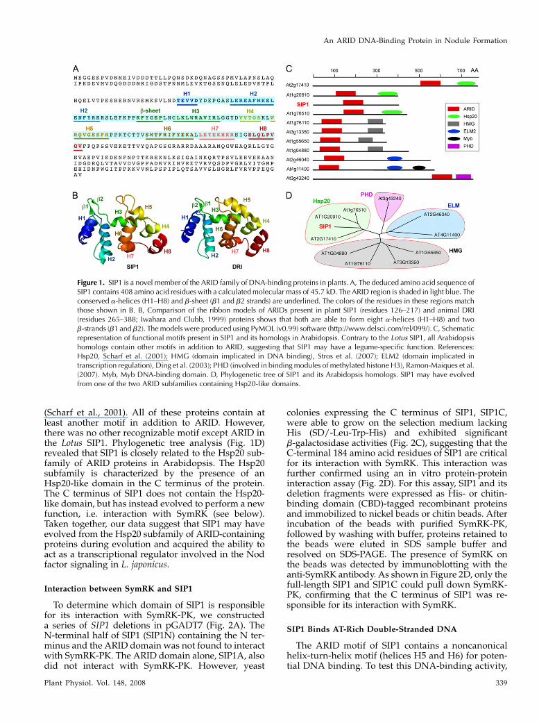
(Scharf et al., 2001). All of these proteins contain atleast another motif in addition to ARID. However,there was no other recognizable motif except ARID inthe Lotus SIP1. Phylogenetic tree analysis (Fig. 1D)revealed that SIP1 is closely related to the Hsp20 sub-family of ARID proteins in Arabidopsis. The Hsp20subfamily is characterized by the presence of anHsp20-like domain in the C terminus of the protein.The C terminus of SIP1 does not contain the Hsp20-like domain, but has instead evolved to perform a newfunction, i.e. interaction with SymRK (see below).Taken together, our data suggest that SIP1 may haveevolved from the Hsp20 subfamily of ARID-containingproteins during evolution and acquired the ability toact as a transcriptional regulator involved in the Nodfactor signaling in L. japonicus.
Interaction between SymRK and SIP1
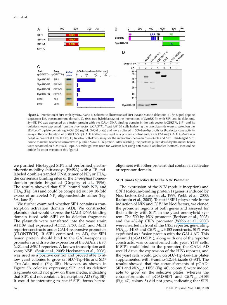
To determine which domain of SIP1 is responsiblefor its interaction with SymRK-PK, we constructeda series of SIP1 deletions in pGADT7 (Fig. 2A). TheN-terminal half of SIP1 (SIP1N) containing the N ter-minus and the ARID domain was not found to interactwith SymRK-PK. The ARID domain alone, SIP1A, alsodid not interact with SymRK-PK. However, yeast
colonies expressing the C terminus of SIP1, SIP1C,were able to grow on the selection medium lackingHis (SD/-Leu-Trp-His) and exhibited significantb-galactosidase activities (Fig. 2C), suggesting that theC-terminal 184 amino acid residues of SIP1 are criticalfor its interaction with SymRK. This interaction wasfurther confirmed using an in vitro protein-proteininteraction assay (Fig. 2D). For this assay, SIP1 and itsdeletion fragments were expressed as His- or chitin-binding domain (CBD)-tagged recombinant proteinsand immobilized to nickel beads or chitin beads. Afterincubation of the beads with purified SymRK-PK,followed by washing with buffer, proteins retained tothe beads were eluted in SDS sample buffer andresolved on SDS-PAGE. The presence of SymRK onthe beads was detected by immunoblotting with theanti-SymRK antibody. As shown in Figure 2D, only thefull-length SIP1 and SIP1C could pull down SymRK-PK, confirming that the C terminus of SIP1 was re-sponsible for its interaction with SymRK.
SIP1 Binds AT-Rich Double-Stranded DNA
The ARID motif of SIP1 contains a noncanonicalhelix-turn-helix motif (helices H5 and H6) for poten-tial DNA binding. To test this DNA-binding activity,
Figure 1. SIP1 is a novel member of the ARID family of DNA-binding proteins in plants. A, The deduced amino acid sequence ofSIP1 contains 408 amino acid residues with a calculated molecular mass of 45.7 kD. The ARID region is shaded in light blue. Theconserved a-helices (H1–H8) and b-sheet (b1 and b2 strands) are underlined. The colors of the residues in these regions matchthose shown in B. B, Comparison of the ribbon models of ARIDs present in plant SIP1 (residues 126–217) and animal DRI(residues 265–388; Iwahara and Clubb, 1999) proteins shows that both are able to form eight a-helices (H1–H8) and twob-strands (b1 and b2). The models were produced using PyMOL (v0.99) software (http://www.delsci.com/rel/099/). C, Schematicrepresentation of functional motifs present in SIP1 and its homologs in Arabidopsis. Contrary to the Lotus SIP1, all Arabidopsishomologs contain other motifs in addition to ARID, suggesting that SIP1 may have a legume-specific function. References:Hsp20, Scharf et al. (2001); HMG (domain implicated in DNA binding), Stros et al. (2007); ELM2 (domain implicated intranscription regulation), Ding et al. (2003); PHD (involved in binding modules of methylated histone H3), Ramon-Maiques et al.(2007). Myb, Myb DNA-binding domain. D, Phylogenetic tree of SIP1 and its Arabidopsis homologs. SIP1 may have evolvedfrom one of the two ARID subfamilies containing Hsp20-like domains.
An ARID DNA-Binding Protein in Nodule Formation
Plant Physiol. Vol. 148, 2008 339
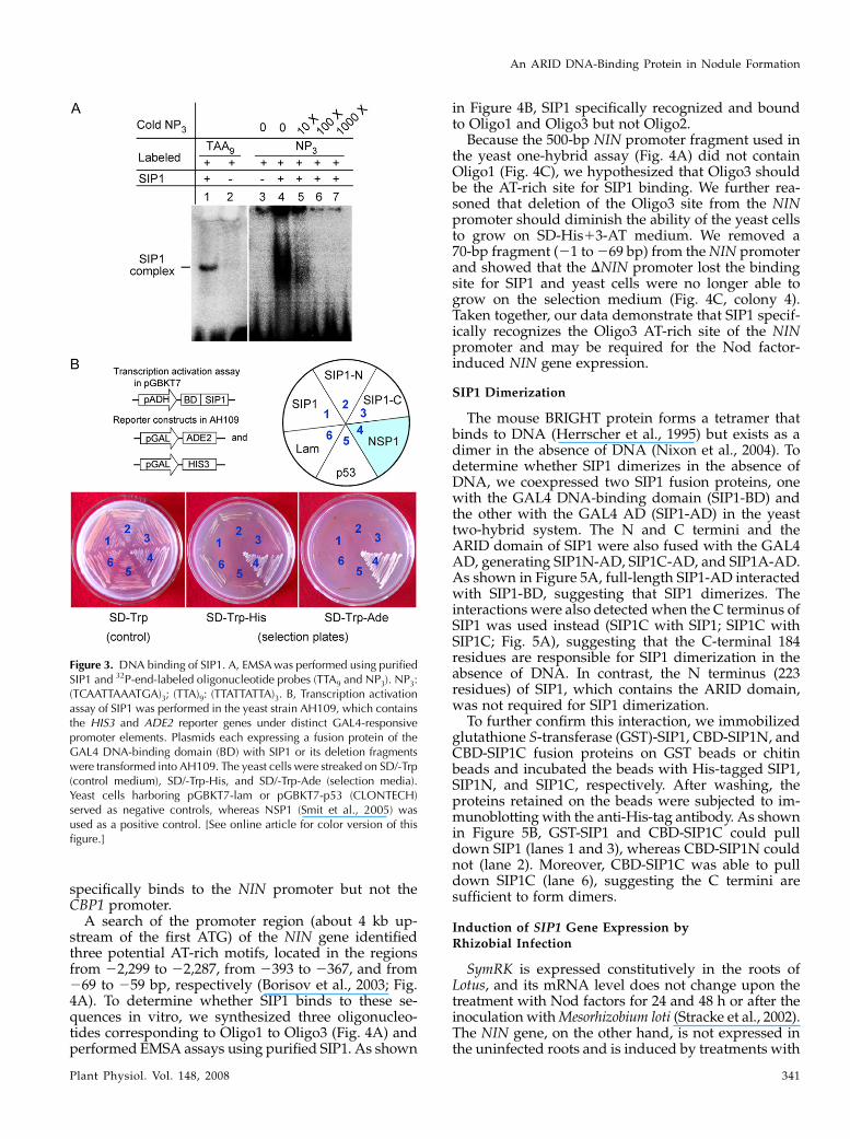
we purified His-tagged SIP1 and performed electro-phoretic mobility shift assays (EMSA) with a 32P-end-labeled double-stranded DNA trimer of NP3 or TTA9,the consensus binding sites of the Drosophila homeo-domain protein Engrailed (Gregory et al., 1996).The results showed that SIP1 bound both NP3 andTTA9 (Fig. 3A) and could be competed out by 10-foldexcess of unlabeled NP3 oligonucleotide trimer (Fig.3A, lane 5).
We further examined whether SIP1 contains a tran-scription activation domain (AD). We constructedplasmids that would express the GAL4 DNA-bindingdomain fused with SIP1 or its deletion fragments.The plasmids were transferred into the yeast strainAH109 that expressed ADE2, HIS3, lacZ, and MEL1reporter constructs under GAL4-responsive promoters(CLONTECH). If SIP1 contained an AD, the SIP1fusion protein should bind to the GAL4-responsivepromoters and drive the expression of the ADE2, HIS3,lacZ, and MEL1 reporters. A known transcription acti-vator, NSP1 (Smit et al., 2005; Heckmann et al., 2006),was used as a positive control and proved able to al-low yeast colonies to grow on SD/-Trp-His and SD/-Trp-Ade media (Fig. 3B). However, as shown inFigure 3B, colonies expressing SIP1 and its deletionfragments could not grow on these media, indicatingthat SIP1 did not contain a transcription AD (Fig. 3B).It would be interesting to test if SIP1 forms hetero-
oligomers with other proteins that contain an activatoror repressor domain.
SIP1 Binds Specifically to the NIN Promoter
The expression of the NIN (nodule inception) andCBP1 (calcium-binding protein 1) genes is induced byNod factors (Schauser et al., 1999; Webb et al., 2000;Radutoiu et al., 2003). To test if SIP1 plays a role in theinduction of NIN and CBP1 by Nod factors, we clonedthe promoter regions of both genes and assayed fortheir affinity with SIP1 in the yeast one-hybrid sys-tem. The 500-bp NIN promoter (Borisov et al., 2003)and the 482-bp CBP1 promoter (Webb et al., 2000)were inserted in front of the HIS3 reporter, generatingNINproTHIS3 and CBP1proTHIS3 constructs. SIP1 wasexpressed as a fusion protein with the GAL4 AD. Thisplasmid (pGAD-SIP1), along with one of the reporterconstructs, was cotransformed into yeast Y187 cells.If SIP1 could bind to the promoter, the GAL4 ADwould drive the expression of the HIS3 reporter, andthe yeast cells would grow on SD/-Trp-Leu-His platessupplemented with 3-amino-1,2,4-triazole (3-AT). Theresults showed that the cotransformants of pGAD-SIP1 and NINproTHIS3 (Fig. 4C, colony 3) were indeedable to grow on the selective plates, whereas thecotransformants of pGAD-SIP1 and CBP1proTHIS3(Fig. 4C, colony 5) did not grow, indicating that SIP1
Figure 2. Interaction of SIP1 with SymRK. A and B, Schematic illustrations of SIP1 (A) and SymRK deletions (B). SP, Signal peptidesequence; TM, transmembrane domain. C, Yeast two-hybrid assays of the interactions of SymRK-PK with SIP1 and its deletions.SymRK-PK was expressed as a fusion protein with the GAL4 DNA-binding domain in the bait vector (pGBKT7). SIP1 and itsdeletions were expressed from the prey vector (pGADT7). Yeast AH109 cells harboring the two plasmids were streaked on theSD/-Leu-Trp plate containing X-Gal (80 mg/mL; X-Gal plate) and were cultured in SD/-Leu-Trp broth for b-galactosidase activityassays. The combination of pGBKT7-53/pGADT7-SV40 was used as a positive control and pGBKT7-Lam/pGADT7-SV40 as anegative control (CLONTECH). D, In vitro pull-down assay for the interaction between SymRK-PK and SIP1. His-tagged SIP1bound to nickel beads was mixed with purified SymRK-PK protein. After washing, the proteins pulled down by the nickel beadswere separated on SDS-PAGE (top). A similar gel was used for western blot using anti-SymRK antibodies (bottom). [See onlinearticle for color version of this figure.]
Zhu et al.
340 Plant Physiol. Vol. 148, 2008
specifically binds to the NIN promoter but not theCBP1 promoter.
A search of the promoter region (about 4 kb up-stream of the first ATG) of the NIN gene identifiedthree potential AT-rich motifs, located in the regionsfrom 22,299 to 22,287, from 2393 to 2367, and from269 to 259 bp, respectively (Borisov et al., 2003; Fig.4A). To determine whether SIP1 binds to these se-quences in vitro, we synthesized three oligonucleo-tides corresponding to Oligo1 to Oligo3 (Fig. 4A) andperformed EMSA assays using purified SIP1. As shown
in Figure 4B, SIP1 specifically recognized and boundto Oligo1 and Oligo3 but not Oligo2.
Because the 500-bp NIN promoter fragment used inthe yeast one-hybrid assay (Fig. 4A) did not containOligo1 (Fig. 4C), we hypothesized that Oligo3 shouldbe the AT-rich site for SIP1 binding. We further rea-soned that deletion of the Oligo3 site from the NINpromoter should diminish the ability of the yeast cellsto grow on SD-His13-AT medium. We removed a70-bp fragment (21 to 269 bp) from the NIN promoterand showed that the DNIN promoter lost the bindingsite for SIP1 and yeast cells were no longer able togrow on the selection medium (Fig. 4C, colony 4).Taken together, our data demonstrate that SIP1 specif-ically recognizes the Oligo3 AT-rich site of the NINpromoter and may be required for the Nod factor-induced NIN gene expression.
SIP1 Dimerization
The mouse BRIGHT protein forms a tetramer thatbinds to DNA (Herrscher et al., 1995) but exists as adimer in the absence of DNA (Nixon et al., 2004). Todetermine whether SIP1 dimerizes in the absence ofDNA, we coexpressed two SIP1 fusion proteins, onewith the GAL4 DNA-binding domain (SIP1-BD) andthe other with the GAL4 AD (SIP1-AD) in the yeasttwo-hybrid system. The N and C termini and theARID domain of SIP1 were also fused with the GAL4AD, generating SIP1N-AD, SIP1C-AD, and SIP1A-AD.As shown in Figure 5A, full-length SIP1-AD interactedwith SIP1-BD, suggesting that SIP1 dimerizes. Theinteractions were also detected when the C terminus ofSIP1 was used instead (SIP1C with SIP1; SIP1C withSIP1C; Fig. 5A), suggesting that the C-terminal 184residues are responsible for SIP1 dimerization in theabsence of DNA. In contrast, the N terminus (223residues) of SIP1, which contains the ARID domain,was not required for SIP1 dimerization.
To further confirm this interaction, we immobilizedglutathione S-transferase (GST)-SIP1, CBD-SIP1N, andCBD-SIP1C fusion proteins on GST beads or chitinbeads and incubated the beads with His-tagged SIP1,SIP1N, and SIP1C, respectively. After washing, theproteins retained on the beads were subjected to im-munoblotting with the anti-His-tag antibody. As shownin Figure 5B, GST-SIP1 and CBD-SIP1C could pulldown SIP1 (lanes 1 and 3), whereas CBD-SIP1N couldnot (lane 2). Moreover, CBD-SIP1C was able to pulldown SIP1C (lane 6), suggesting the C termini aresufficient to form dimers.
Induction of SIP1 Gene Expression byRhizobial Infection
SymRK is expressed constitutively in the roots ofLotus, and its mRNA level does not change upon thetreatment with Nod factors for 24 and 48 h or after theinoculation with Mesorhizobium loti (Stracke et al., 2002).The NIN gene, on the other hand, is not expressed inthe uninfected roots and is induced by treatments with
Figure 3. DNA binding of SIP1. A, EMSA was performed using purifiedSIP1 and 32P-end-labeled oligonucleotide probes (TTA9 and NP3). NP3:(TCAATTAAATGA)3; (TTA)9: (TTATTATTA)3. B, Transcription activationassay of SIP1 was performed in the yeast strain AH109, which containsthe HIS3 and ADE2 reporter genes under distinct GAL4-responsivepromoter elements. Plasmids each expressing a fusion protein of theGAL4 DNA-binding domain (BD) with SIP1 or its deletion fragmentswere transformed into AH109. The yeast cells were streaked on SD/-Trp(control medium), SD/-Trp-His, and SD/-Trp-Ade (selection media).Yeast cells harboring pGBKT7-lam or pGBKT7-p53 (CLONTECH)served as negative controls, whereas NSP1 (Smit et al., 2005) wasused as a positive control. [See online article for color version of thisfigure.]
An ARID DNA-Binding Protein in Nodule Formation
Plant Physiol. Vol. 148, 2008 341
Nod factors or rhizobial infection (Schauser et al., 1999;Radutoiu et al., 2003). Using quantitative PCR, weexamined the SIP1 and NIN mRNA levels in differenttissues of L. japonicus. SIP1 mRNA was expressedconstitutively in leaves and control (uninoculated)roots (Fig. 6A). In the young roots (2 d old), SIP1expression levels were low but increased to a steadystate after 6 to 8 d. In the stem, its expression levelswere relatively low but detectable. In contrast, NINexpression levels were relatively low in stems, leaves,and control roots (Fig. 6B).
We then focused on the expression levels of SIP1and NIN in roots after infection with M. loti. Wheninoculated with M. loti, an induction of SIP1 mRNAwas observed as early as 5 h post inoculation (hpi). In24 hpi, the expression levels dropped down to a steadylevel, which was slightly higher than that observed inthe control roots. This expression pattern was distinctfrom that of NIN (Fig. 6B), which exhibited significantinduction 5 h after rhizobial inoculation and main-tained a high expression level in inoculated roots. It isimportant to note that the timing of the SIP1 induction(5 hpi) correlates well with that of NIN. After the initialinduction, NIN continued to be expressed at relativelyhigh levels. In conclusion, the SIP1 gene is expressedconstitutively in roots and leaves, and its expressionlevels are elevated in roots transiently (5 hpi–1 dpostinoculation [dpi]) after rhizobial infection. It mayplay a role in the induction of the NIN gene during theprocess of rhizobial entry and nodule organogenesis.
Subcellular Localization of SIP1
To determine the subcellular localization of SIP1, weexpressed SIP1 as a fusion protein with the Discosomared fluorescent protein (DsRed) under the control ofthe cauliflower mosaic virus 35S promoter. The fusionprotein was transiently expressed in the onion (Alliumcepa) epidermal cells via particle bombardment, and itsexpression was monitored using a confocal laser-scanning microscope. As expected in the control cellsexpressing DsRed alone, the red fluorescence was de-tected only in the cytoplasm (Fig. 7, D–F). In the onionepidermal cells expressing the SIP1-DsRed fusion pro-tein, the red fluorescence was concentrated to the nuclei(Fig. 7, A–C). This result is consistent with its potentialfunction in DNA binding and transcription regulation.However, it remains to be determined how SIP1 interactswith SymRK in Lotus roots after rhizobial infection.
DISCUSSION
SymRK is a member of a large family of LRR RLKsin plants and is required for the legume root cells to
Figure 4. Binding of SIP1 to the NIN promoter. A, A search of the 4-kb5# region of the NIN gene identified three AT-rich motifs. Three oligosused in this experiment correspond to the DNA sequences of theNIN promoter from 22,287 to 22,299 for Oligo1, 2367 to 2393 forOligo2, and 259 to 269 for Oligo3. Red letters correspond to theDNA sequences of the NIN promoter, whereas the underlined blackletters indicate a repeat (Oligo1) or double repeats (Oligo3). B, EMSAwas performed using purified SIP1 and 32P-end-labeled Oligos. Thecomponents of the binding reactions are indicated at the top ofthe lanes. C, In vivo assay of SIP1 binding to the NIN promoter. SIP1was expressed as a fusion protein with the GAL4 AD (pGADT7-SIP1)in yeast Y187 cells harboring the reporter construct NINproTHIS3,DNINproTHIS3, or CBP1proTHIS3, which expresses the HIS3 reportergene under the promoter of the NIN or CBP1 gene. The NIN promoteris a 500-bp fragment (21 to 2500 bp), whereas the DNIN promotercontains a 430-bp fragment (270 to 2500 bp) lacking the Oligo3AT-rich site. The CBP1 promoter (462 bp) was amplified by PCR(Webb et al., 2000). Yeast cells were examined for growth on theSD/-Trp-Leu (control) and SD/-Trp-Leu-His plates in the presence of
40 mM 3-AT (selection). Yeast cells harboring p53-HIS2 and pGADT7-53 were used as a positive control, whereas those containing p53-HIS2 and pGADT7-SIP1 served as negative control. [See onlinearticle for color version of this figure.]
Zhu et al.
342 Plant Physiol. Vol. 148, 2008
perceive the Nod factor signals released by the infect-ing rhizobia (Yoshida and Parniske, 2005). NIN is a keytranscription factor required for rhizobial entry intothe root cells and for nodule organogenesis (Schauseret al., 1999; Radutoiu et al., 2003). In this report, we
describe a novel DNA-binding protein, SIP1, whichmay provide a link between SymRK and NIN. Wedemonstrate that SIP1 interacts with the intracellularkinase domain of SymRK (Fig. 2) and specificallybinds to the Oligo3 AT-rich site of the NIN promoter(Fig. 4). Because SIP1 does not contain a transcriptionAD (Fig. 3), it may form hetero-oligomers with otherunidentified proteins that contain an activator or re-pressor domain. Plu-1, a human ARID-containing pro-tein, also lacks an AD but contains transcriptionalrepressor properties and forms hetero-oligomers withother transcription factors (Tan et al., 2003). The initialsignal communications between the rhizobia and rootsinvolve a number of different molecular exchangesculminating in calcium spiking in the host cells. SIP1,by linking the SymRK receptor kinase to the NIN tran-scription factor, may play a pivotal role for the success-ful establishment of rhizobia-legume symbiosis.
SIP1 represents a new member of the conservedARID family of proteins in plants. The ARID domainwas initially identified in the mouse BRIGHT protein
Figure 5. Dimerization of SIP1. A, Yeast AH109 cells were cotrans-formed with different combinations of the bait and prey constructscontaining either the full-length SIP1 or its deletion fragments. Theyeast cells were transferred from SD/-Leu-Trp-His-Ade to SD/-Leu-Trpplate containing X-Gal (80 mg/mL). Yeast cells harboring pGBKT7-53/pGADT7-SV40 were used as a positive control, whereas those con-taining pGBKT7-Lam/pGADT7-SV40 served as a negative control. B, Invitro protein-protein interaction assay. SIP1 and its deletion fragmentswere expressed as GST- or CBD-tagged proteins and immobilized toglutathione- or chitin beads. SIP1 and its deletion fragments werealso expressed and purified as His-tagged proteins, followed by elutionfrom the nickel-column with imidazole. The eluted soluble SIP1 or itsdeletion fragments were then mixed with glutathione beads or CBDbeads containing an immobilized peptide. After washing with buffer,proteins retained to the beads were solubilized in SDS sample bufferand separated on SDS-PAGE (top), and the interacting proteins weredetected using anti-His antibodies (bottom). [See online article for colorversion of this figure.] Figure 6. Expression of SIP1 and NIN genes in L. japonicus. Roots were
harvested 2, 5, and 12 hpi, and 1, 2, 4, 6, and 8 dpi with M. loti. Rootstreated with water served as the mock control. Total RNA was isolatedstems (S), leaves (L), control roots (R), and Rhizobium-inoculated roots(IR). Steady-state transcript levels of SIP1 (A) and NIN (B) weremeasured by quantitative PCR on a real-time PCR system (Radutoiuet al., 2003). The ATPase gene (AW719841) was used as an internalcontrol. Relative values of transcripts normalized to the control roots(4 dpi) are shown.
An ARID DNA-Binding Protein in Nodule Formation
Plant Physiol. Vol. 148, 2008 343

(Herrscher et al., 1995) and Drosophila DRI (Gregoryet al., 1996). ARID-containing proteins have now beenshown to be present in all sequenced eukaryotic or-ganisms, including mammals, insects, plants, nema-todes, and yeast (Wilsker et al., 2002). Although ARIDis an a-helix-based DNA-binding domain, the struc-tures and functions of ARID-containing proteins arehighly diverse (Wilsker et al., 2002). These proteinsappear to play vital roles in the regulation of devel-opment and tissue-specific gene expression.
The ARID domains, especially their 3-D structures,are conserved between animals and plants (Fig. 1B).However, other parts of the molecules can be verydifferent among ARID-containing proteins in an organ-ism. In Arabidopsis, the 10 ARID-containing proteinsvary in length from 319 residues in At3g13350 to 786residues in At2g17410. They can be grouped into foursubfamilies on the basis of their phylogenetic relation-ship and the presence of functional motifs (Fig. 1D). L.japonicus SIP1 is closely related to the Hsp20 subfamilyin Arabidopsis, although it does not contain an Hsp20-like domain. The Hsp20 motif is found in the C terminiof the Arabidopsis ARID proteins (Fig. 1C). In L.japonicus SIP1, the C terminus has evolved into adifferent domain that is responsible for interactingwith SymRK (Fig. 2) and for the homodimerization(Fig. 5). SIP1 represents a new ARID-containing tran-scription factor in plants and is potentially involvedin linking the Nod signal perception to the transcrip-tional regulation during nodule development in L.japonicus.
The ARID domain has not been found in Archea andeubacteria, but is widespread in protozoa, metazoans,green algae, fungi, plants, and animals. There are 15ARID proteins in the human genome and two in S.cerevisiae (Wilsker et al., 2002, 2005). The four Arabi-dopsis ARID subfamilies (Fig. 1D) do not correlateclosely with the seven subfamilies of the human ARIDproteins (Wilsker et al., 2005). At3g43240 is a single
member of the Arabidopsis subfamily that possessesan ARID and a PHD domain. In humans, the mostclosely related proteins are the four ARID proteins inthe JARID1 subfamily, which contain two or morePHD domains each, in addition to several other func-tional motifs (Wilsker et al., 2005), and are also twice aslarge as At3g43240. The remaining three subfamilies ofArabidopsis ARID proteins are plant specific, becauseno known human ARID protein contains an Hsp20,HMG, or ELM2 domain (Wilsker et al., 2002, 2005).This diversity in peptide length and motif arrange-ments suggests that the specific ARID subfamilieshave apparently evolved independently after the di-vergence of plant and animal lineages.
ARID proteins in higher eukaryotes are involved indifferentiation and transcriptional regulation of geneexpression (Wang et al., 2007). At least two humanARID proteins are known to be induced during tu-morigenesis. Plu-1 (JARID1B) was identified as an up-regulated gene product in human breast cancer and isnot expressed in normal adult tissues except testis.RBP1L1 (ARID4B) was identified as a tumor antigenand is highly expressed in all cancer cell lines (Wilskeret al., 2002; Tan et al., 2003). Nodule initiation involvesthe de-differentiation of the root cortical cells into themeristematic cells and the activation of a series ofnodule-specific genes. Our study shows that SIP1 isconstitutively expressed in roots, and its expression ishighly induced 5 h after rhizobial infection (Fig. 6).Although L. japonicus SIP1 is not homologous to thehuman Plu-1 and RBP1L1 except within the ARIDdomain, they share some similarity in their function inreprogramming the differentiated cells back to thedividing status, i.e. tumorigenesis in human and nod-ule organogenesis in the legume.
There are three AT-rich motifs in the promoter of theNIN gene, two of which showed affinity with SIP1 inin vitro assays (Fig. 4). In the yeast one-hybrid system,the 500-bp promoter region of the NIN gene was found
Figure 7. Subcellular localization of SIP1. SIP1 wasexpressed as a fusion protein with DsRed under thecontrol of the 35S promoter. The plasmid was deliv-ered to the onion epidermal cells via particle bom-bardment. The plasmid (35STDsRed) expressingDsRed alone served as a control. The onion cellswere observed with a confocal laser-scanning micro-scope, and pictures were taken in the light transmis-sion channel (A and D) or using the 583/558-nm filter(B and E). The photos were superimposed usingPhotoshop software (C and F). Note that DsRed isdistributed in the cytosol, whereas SIP1:DsRed goesto the nuclei.
Zhu et al.
344 Plant Physiol. Vol. 148, 2008

to contain cis-DNA elements for SIP1 binding. Thebinding of SIP1 to the NIN promoter was apparentlybrought about via the Oligo3 AT-rich motif, which islocalized to the promoter region between 259 to 269bp. Because SIP1 does not have a transcription AD(Fig. 3B), it is likely that SIP1 may function as a DNAsequence-specific binding protein and may form anoligomer with another transcription activator or re-pressor. We are currently in the process of searchingfor these potential SIP1-interacting partners and ex-amining the biological functions of SIP1 and its asso-ciated proteins in nodule organogenesis in L. japonicus.Further analyses of the signaling pathway involvingSIP1 may eventually lead to the identification of newmolecular events required for the later steps in thesymbiotic establishment in legume roots.
MATERIALS AND METHODS
Plant Materials
Seeds of Lotus japonicus MG-20 were surface-sterilized in 2% sodium hypo-
chlorite for 8 min and washed seven times with sterile water. The seeds were left
to germinate for 48 h at 22�C on sterile water-soaked filter paper in petri dishes
in a dark room. Seedlings were subsequently planted in pots on sterile sands
supplemented with nitrogen-free Fahraeus medium (Fahraeus, 1957) and were
grown in a growth chamber maintained at 22�C with a 16-h-light/8-h-dark
cycle. Five-day-old seedlings were inoculated with approximately 107 CFU/mL
of Mesorhizobium loti NZP2037. Roots were collected from plants 2, 4, 6, 8, and
12 d after rhizobial inoculation and were frozen in liquid nitrogen.
cDNA Library Construction
Total RNA was isolated from the equally mixed roots collected 2, 4, 6, 8,
and 12 d after rhizobial inoculation using the TRIzol reagent (Invitrogen).
Total RNA (2 mg) was reverse transcribed into single-stranded cDNA using
oligo(dT) as the primer. The cDNA was size-fractionated using BD Match-
maker library construction and screening kits (BD Biosciences CLONTECH).
The cDNA fragments longer than 500 bp were cotransformed with linearized
pGADT7-Rec into yeast (Saccharomyces cerevisiae) AH109 cells. The transfor-
mants were selected on SD/-Leu medium according to the manufacturer’s
instruction. The transformation efficiency was approximately 2 3 106 CFU/3
mg pGADT7-Rec.
Library Screening
The SymRK-PK cDNA (GenBank accession no. AF492655) was amplified
by reverse transcription-PCR with the following gene-specific primers:
5#-GGGCATATGATGGAGAGGTACAAAACCTTG-3# and 5#-AAAGAATTCT-
CTCGGCTGTGGGTGAG-3#. PCR was carried out using ExTaq DNA poly-
merase (TaKaRa) with an initial denaturation step of 95�C for 5 min, followed
by 30 cycles of 95�C for 15 s, 55�C for 30 s, and 72�C for 1 min. After the last
cycle, an extension step for 10 min at 72�C was carried out. Purified PCR
products were inserted into the Nde1-EcoR1 site of pGBKT7 vector. Screening
of interaction clones was carried out according to the manufacturer’s instruc-
tions (CLONTECH). A total of 1 3 107 transformants from the cDNA library
were screened for growth on the SD/-Leu-Trp-His-Ade media. Positive clones
were verified by retransformation of the rescued plasmid into the AH109
containing the bait plasmid (pGBKT7-SymRK-PK). Plasmid pGBKT7-Lam
(CLONTECH) was used as a negative control. Colonies growing on the
SD/-Leu-Trp-His-Ade media were transferred to selective media-containing
X-Gal (80 mg/mL) or were left on filters as described (Ma et al., 2007). The blue
colonies were characterized.
b-Galactosidase Assay
Yeast cells grown in liquid selection media were pelleted and washed twice
with Z-buffer (60 mM Na2HPO4, 40 mM NaH2PO2, 10 mM KCl, 1.0 mM MgSO2,
pH 7.0). The cells were resuspended in 300 mL of Z-buffer and permeabilized
by two freeze-thaw cycles in liquid nitrogen and 37�C. Cell extracts were
added 0.7 mL of Z-buffer containing 50 mM b-mercaptoethanol and 160 mL of
O-nitrophenyl b-D-galactopyranoside (4 mg/mL) in Z-buffer. After incubation
at 30�C for 30 min or until the yellow color appeared, the reaction was
terminated by the addition of 0.4 mL of 1.0 M Na2CO3. The reaction mixture
was centrifuged for 10 min at 14,000 rpm to remove cell debris. b-Galactosidase
activity in the supernatant was measured at OD420 and expressed in Miller units
(Miller, 1972).
Expression and Purification of Fusion Proteins
The full-length cDNA of SIP1 was inserted at the Nde1-Sal1 site of pET28
vector (Novagen), generating pET-SIP1. The 3# region of SIP1 was amplified
by PCR using primers 5#-CTTTCTAGACCATATGAAGTCCAGAGAAAG-
GA-3# and 5#-TTTTGTCGACAAGCTTCACGGCTCCCTG-3#. The fragment
was cloned into the Nde1-Sal1 site of pET28a vector, generating pET-SIP1C.
The 5# region of SIP1 was amplified using primers 5#-CCGGTACCCATATG-
GAAATTGTTGAT-3# and 5#-GGGAAGCTTGTCGACTTAGAGCTGTAACTC-3#.The fragment was cloned into the Nde1-Sal1 site of pTYB12 (New England
Biolabs), yielding pTYB-SIP1N. For protein expression, Escherichia coli BL21
(Novagen) or ER2566 cells (New England Biolabs) harboring the plasmid were
induced with 0.5 mM isopropyl 1-thio-b-D-galactopyranoside in Luria-Bertani
broth for 8 h at 16�C. His-tagged proteins were purified using nickel-agarose
beads (Qiagen) according to the manufacturer’s instruction. CBD-tagged pro-
teins were purified using chitin beads (New England Biolabs) as described
previously (Zhang et al., 2003). Purified proteins were desalted by dialysis in
PBS buffer and concentrated with PEG-8000 powder.
In Vitro Protein-Protein Interaction
To assay the interaction between SymRK-PK and SIP1, His-tagged SIP1
and SIP1C were bound to nickel-agarose beads, while CBD-SIP1N was
absorbed on the chitin beads. The beads were incubated with 5 mg of purified
SymRK-PK protein in the interaction buffer (20 mM Tris-HCl, 100 mM KCl,
2 mM MgCl2, 5% glycerol, pH 8.0) for 1 h on ice with gentle shaking. The nickel
beads were washed three times with a buffer solution (137 mM NaCl, 2.7 mM
KCl, 10 mM Na2HPO4, 2 mM KH2PO4, 100 mM imidazole, 5% glycerol, pH 8.0),
whereas the chitin beads were washed three times with 1.0 mL of TEG buffer
(20 mM Tris-HCl, 1 mM EDTA, 5% glycerol, pH 8.0). The retained proteins were
eluted by boiling for 3 min in 13 SDS sample buffer (2% SDS, 29.1 mmol/L
Tris, pH 6.8, 10% glycerol, and 0.01% bromphenol blue; Zhang et al., 2003).
Samples were analyzed on 10% SDS-PAGE, followed by immunoblotting with
the anti-LjSymRK antibody.
To assay SIP1 dimerization, GST-tagged SIP1 was expressed and purified
on glutathione-Sepharose 4B (Sigma) as described elsewhere (Zhang et al.,
2000). CBD-tagged SIP1N and SIP1C were immobilized on chitin beads. The
beads were incubated with purified His-SIP1, His-SIP1N, and His-SIP1C
proteins for 1 h on ice with gentle shaking. Following washing, the retained
proteins were resolved on 10% PAGE and detected using the anti-His-tag
antibody.
EMSA
EMSA of SIP1 was performed on 5% nondenaturing polyacrylamide gels
as described previously (Gregory et al., 1996). DNA-binding activity of SIP1
was examined using g-32P-end-labeled double-stranded oligonucleotide
trimer of the consensus Engrailed binding site NP3 [(TCAATTAAATGA)X3]
or trimer of TTA9 [(TTATTATTA)X3; Gregory et al., 1996]. To test if SIP1
specifically binds to the promoter of the NIN gene, Oligo1, Oligo2, and Oligo3
(see Fig. 4A for sequences) were synthesized and used for EMSA. His-tagged
SIP1 was purified with nickel-agarose beads and eluted using 500 mM
imidazole followed by dialysis. Approximately 10 ng of SIP1 protein was
added in a mixture containing 0.3 mM of g-32P-labeled double-stranded DNA
in 10 mM Tris-HCl, pH 7.5, 50 mM NaCl, 1.0 mM EDTA, 1.0 mM dithiothreitol,
50 mg bovine serum albumin, and 5% glycerol. After incubation at room
temperature for 20 min, the samples were subjected to electrophoresis on a 5%
polyacrylamide/bis (29:1) gel in 0.53 Tris-borate/EDTA buffer at a constant
voltage of 100 V. The gel was dried, and the radioactive bands were detected
using PhosphorImager (FUJIFILM).
An ARID DNA-Binding Protein in Nodule Formation
Plant Physiol. Vol. 148, 2008 345
Transient Expression
The SIP1 cDNA amplified using primers 5#-TATACTAGTATGGAAA-
TTGTTGAT-3#, and 5#-AAGGTCACCTTACACGGCTCCCTG-3# was cloned
into the Spe1/BstEII site of pCAMBIA1302 vector (CAMBIA). We amplified
DsRed coding sequence using primer 5#-CAACTAGTATGAGGTCTTCCAA-
GAATGT-3#, and 5#-TTACTAGTAAGGAACAGATGGTGGCGTC-3# from
the pDsRed (CLONTECH), and it was inserted into the Spe1 site of the
pCAMBIA1302-SIP1 construct, resulting in p35S-DsRed-SIP1. The plasmid
was used for transient expression in the onion (Allium cepa) epidermal cells by
particles bombardment using a Biolistic PDS-1000/He particle delivery sys-
tem (Bio-Rad). After incubation for 24 to 48 h at 25�C in dark, the epidermal
cell layers were examined using a confocal laser-scanning microscope (Leica).
Yeast One-Hybrid Assay
The SIP1 cDNA was fused to the GAL4 AD in pGADT7-Rec2 (CLONTECH),
generating pGADT7-SIP1. The promoter (21 bp to 2500 bp) of the NIN gene
(Schauser et al., 1999) was amplified using primers 5#-GGAATTCGGCGTG-
AGACTTTACATTAC-3# and 5#-CGACGCGTGAAAATGATTGCCTTGCC-3#.
A 430-bp fragment of the NIN promoter (270 to 2500 bp) lacking the Oligo3
AT-rich site (DNIN) was amplified by PCR using primers 5#-GGAAT-
TCGGCGTGAGACTTTACATTAC-3# and 5#-TTTTACGCGTCTTGCTAGG-
ACC-3#. The promoter of the LjCBP1 gene (Webb et al., 2000) was amplified
from genomic DNA using primers 5#-GGAATTCCCAAAGTCAGTCAATTT-
TTG-3# and 5#-CGACGCGTCCACTGACTTAATTCATG-3#. The promoter
fragments of the NIN, DNIN, and LjCBP1 genes were inserted into the
EcoRI-MluI site of pHIS2 (CLONTECH), producing NINproTHIS3, DNINproT
HIS3, and CBP1proTHIS3, respectively, which would express the HIS3 reporter
under the control of the corresponding promoter.
The plasmid was transformed into yeast Y187 cells harboring pGADT7-SIP1,
which expresses SIP1 fused with the GAL4 transcription AD. The DNA-binding
activity of SIP1 was determined by the expression of the HIS3 reporter.
Measurement of Transcript Level by Quantitative PCR
Total RNA was isolated from stems, leaves, control roots, and roots
inoculated with M. loti, using a TRIZOL reagent (Invitrogen). RNA samples
were treated with DNase I to remove potential contaminating genomic DNA,
followed by extraction with phenol:chloroform. The DNA-free RNA samples
were confirmed by lack of a PCR product using primers specific to the
untranscribed NIN gene promoter (5#-GTTTTCAAGAATGGGAGGGG-3#,
5#-CTCCTCTGGTTTCATTGGTG-3#). First-strand cDNA was prepared using
Oligo(dT) primer, and quantitative PCR was performed on a Mini Opticon
real-time PCR system (Bio-Rad) using One-Step SYBR PrimeScript RT-PCR kit
II (Takara). The same cDNA pool was used for amplification of all tested
transcripts. The relative quantification software (Bio-Rad) was used to deter-
mine the efficiency-corrected relative transcript concentration, normalized to a
calibrator sample (Radutoiu et al., 2003). ATP synthase was used as a reference
gene (AW719841). Melting curve analysis and sequencing of the amplified
products was used to determine the identity of the amplified PCR products.
Intron spanning primers were used for transcript amplification of the ATP
synthase (5#-CAATGTCGCCAAGGCCCATGGTG-3# and 5#-AACACCACT-
CTCGATCATTTCTCTG-3#), SIP1 (5#-CTTGCAGGTAAGTTCACAAC-3# and
5#-GTACGAGTCCAATCAGATCCAG-3#), and NIN (5#-AATGCTCTTGAT-
CAGGCTG-3# and 5#-AGGAGCCCAAGTGAGTGCTA-3#) genes. The same
experiment was performed three times, and the averages of measurements
were presented.
Sequence data from this article can be found in the GenBank/EMBL data
libraries under accession number EU559710.
Supplemental Data
The following materials are available in the online version of this article.
Supplemental Figure S1. Full-length cDNA sequence of L. japonicus SIP1.
ACKNOWLEDGMENTS
We thank Drs. Yongjun Lin, Chunli Chen, and Ms. Huazhi Song for advice
on transient expression and confocal laser-scanning microscope. We also
thank Dr. Allan Caplan for critical comments on the manuscript.
Received March 17, 2008; accepted June 13, 2008; published July 16, 2008.
LITERATURE CITED
Arrighi JF, Barre A, Ben Amor B, Bersoult A, Soriano LC, Mirabella R,
de Carvalho-Niebel F, Journet EP, Gherardi M, Huguet T, et al (2006) The
Medicago truncatula lysine motif-receptor-like kinase gene family includes
NFP and new nodule-expressed genes. Plant Physiol 142: 265–279
Borisov AY, Madsen LH, Tsyganov VE, Umehara Y, Voroshilova VA,
Batagov AO, Sandal N, Mortensen A, Schauser L, Ellis N, et al (2003)
The Sym35 gene required for root nodule development in pea is an
ortholog of NIN from Lotus japonicus. Plant Physiol 131: 1009–1017
Denarie J, Debelle F, Prome JC (1996) Rhizobium lipo-chitooligosaccharide
nodulation factors: signaling molecules mediating recognition and
morphogenesis. Annu Rev Biochem 65: 503–558
Ding Z, Gillespie L, Pate G (2003) Human MI-ER1 alpha and beta function
as transcriptional repressors by recruitment of histone deacetylase 1 to
their conserved ELM2 domain. Mol Cell Biol 23: 250–258
Ehrhardt DW, Atkinson EM, Long SR (1992) Depolarization of alfalfa root
hair membrane potential by Rhizobium meliloti Nod factors. Science 256:
998–1000
Ehrhardt DW, Wais R, Long SR (1996) Calcium spiking in plant root hairs
responding to Rhizobium nodulation signals. Cell 85: 673–681
Endre G, Kereszt A, Kevei Z, Mihacea S, Kalo P, Kiss GB (2002) A receptor
kinase gene regulating symbiosis nodule development. Nature 417:
962–966
Fahraeus A (1957) The infection of clover root hairs by nodule bacteria
studied by a simple glass slide technique. J Gen Microbiol 16: 374–381
Felle HH, Kondorosi E, Kondorosi A, Schultze M (1995) Nod signal-
induced plasma membrane potential changes in alfalfa root hairs are
differentially sensitive to structural modifications of the lipochitooligo-
saccharide. Plant J 7: 939–947
Felle HH, Kondorosi E, Kondorosi A, Schultze M (1996) Rapid alkalin-
azation in alfalfa root hairs in response to rhizobia lipochitooligosac-
charide signals. Plant J 10: 295–301
Gleason C, Chaudhuri S, Yang T, Munoz A, Poovaiah BW, Oldroyd GE
(2006) Nodulation independent of rhizobia induced by a calcium-
activated kinase lacking autoinhibition. Nature 441: 1149–1152
Gregory SL, Kortschak RD, Kalionis B, Saint R (1996) Characterization of
the dead ringer gene identifies a novel, highly conserved family of
sequence-specific DNA-binding proteins. Mol Cell Biol 16: 792–799
Heckmann AB, Lombardo F, Miwa H, Perry JA, Bunnewell S, Parniske M,
Wang TL, Downie JA (2006) Lotus japonicus nodulation requires two
GRAS domain regulators, one of which is functionally conserved in a
non-legume. Plant Physiol 142: 1739–1750
Herrscher RF, Kaplan MH, Lelsz DL, Das C, Scheuermann R, Tucker PW
(1995) The immunoglobulin heavy-chain matrix-associating regions are
bound by Bright: a B cell-specific trans-activator that describes a new
DNA-binding protein family. Genes Dev 9: 3067–3082
Iwahara J, Clubb RT (1999) Solution structure of the DNA binding domain
from Dead ringer, a sequence-specific AT-rich interaction domain
(ARID). EMBO J 18: 6084–6094
Iwahara J, Iwahara M, Daughdrill GW, Ford J, Clubb RT (2002) The
structure of the Dead ringer-DNA complex reveals how AT-rich inter-
action domains (ARIDs) recognize DNA. EMBO J 21: 1197–1209
Kevei Z, Lougnon G, Mergaert P, Horvath GV, Kereszt A, Jayaraman D,
Zaman N, Marcel F, Regulski Z, Kiss GB, et al (2007) 3-Hydroxy-3-
methylglutaryl coenzyme A reductase 1 interacts with NORK and is
crucial for nodulation in Medicago truncatula. Plant Cell 19: 3974–3989
Kistner C, Winzer T, Pitzschke A, Mulder L, Sato S, Kaneko T, Tabata S,
Sandal N, Stougaard J, Parniske M (2005) Seven Lotus japonicus genes
required for transcriptional reprogramming of the root during fungal
and bacterial symbiosis. Plant Cell 17: 2217–2229
Kortschak RD, Tucker PW, Saint R (2000) ARID proteins come in from the
desert. Trends Biochem Sci 25: 294–299
Kurkdjian AC (1995) Role of the differentiation of root epidermal cells in
Nod factor (from Rhizobium meliloti) induced root hair depolarization of
Medicago sativa. Plant Physiol 107: 783–790
Lerouge P, Roche P, Faucher C, Maillet F, Truchet G, Prome JC, Denarie J
(1990) Symbiotic host-specificity of Rhizoium meliloti is determined by
sulphated and acetylated glucosamine oligosaccharide signal. Nature
344: 781–784
Zhu et al.
346 Plant Physiol. Vol. 148, 2008

Limpens E, Bisseling T (2003) Signaling in symbiosis. Curr Opin Plant Biol
6: 343–350
Limpens E, Franken C, Smit P, Willemse J, Bisseling T, Geurts R (2003)
LysM domain receptor kinases regulating rhizobial Nod factor-induced
infection. Science 302: 630–633
Ma L, Hong Z, Zhang Z (2007) Perinuclear and nuclear envelop localiza-
tions of Arabidopsis Ran proteins. Plant Cell Rep 26: 1373–1382
Madsen EB, Madsen LH, Radutoiu S, Olbryt M, Rakwalska M,
Szczyglowski K, Sato S, Kaneko T, Tabata S, Sandal N, et al (2003) A
receptor kinase gene of the LysM type is involved in legume perception
of rhizobial signals. Nature 425: 637–640
Margaert P, Van Montagu M, Prome JC, Holsters M (1993) Three unusual
modifications, a D-arabinosyl, an N-methyl, and a carbamoyl group, are
present on the Nod factors of Azorhizobium caulinodans strain ORS571.
Proc Natl Acad Sci USA 90: 1551–1555
Miller J (1972) Experiments in Molecular Genetics. Cold Spring Harbor
Laboratory Press, Cold Spring Harbor, NY, pp 352–355
Nixon JC, Rajaiya J, Webb CF (2004) Characterization of dominant neg-
ative forms of the transcription factor, Bright. J Biol Chem 279: 52465–
52472
Oldroyd GED, Dowine JA (2004) Calcium kinase and nodulation signaling
in legumes. Nat Rev Mol Cell Biol 5: 566–576
Peck MC, Fisher RF, Long SR (2006) Diverse flavonoids stimulate NodD1
binding to nod gene promoters in Sinorhizobium meliloti. J Bacteriol 188:
5417–5427
Radutoiu S, Madsen LH, Madsen EB, Felle HH, Umehara Y, Gronlund M,
Sato S, Nakamura Y, Tabata S, Sandal N, et al (2003) Plant recognition
of symbiotic bacteria requires two LysM receptor-like kinase. Nature
425: 585–592
Radutoiu S, Madsen LH, Madsen EB, Jurkiewicz A, Fukai E, Quistgaard
EM, Albrektsen AS, James EK, Thirup S, Stougaard J (2007) LysM
domains mediate lipochitin-oligosaccharide recognition and Nfr genes
extend the symbiotic host range. EMBO J 26: 3923–3935
Ramon-Maiques S, Kuo AJ, Carney D, Matthews AG, Oettinger MA,
Gozani O, Yang W (2007) The plant homeodomain finger of RAG2
recognizes histone H3 methylated at both lysine-4 and arginine-2. Proc
Natl Acad Sci USA 104: 18993–18998
Relic B, Talmont F, Kopcinska J, Golinowski W, Prome JC, Broughton WJ
(1993) Biological activity of Rhizobium sp. NGR234 Nod-factors on
Macroptilium atropurpureum. Mol Plant Microbe Interact 6: 764–774
Sanjuan J, Carlson RW, Spaink HP, Bhat RU, Barbour MW, Glushka J,
Stacey G (1992) A 2-O-methylfucose moiety is present in the lipo-
oligosaccharide nodulation signal of Bradyrhizobium japonicu. Proc Natl
Acad Sci USA 89: 8789–8793
Scharf KD, Siddique M, Vierling E (2001) The expanding family of
Arabidopsis thaliana small heat stress proteins and a new family of
proteins containing alpha-crystallin domains (Acd proteins). Cell Stress
Chaperones 6: 225–237
Schauser L, Roussis A, Stiller J, Stougaard J (1999) A plant regulator
controlling development of symbiotic root nodules. Nature 402: 191–195
Shiu SH, Karlowski WM, Pan R, Tzeng YH, Mayer KFX, Li WH (2004)
Comparative analysis of the receptor-like kinase family in Arabidopsis
and rice. Plant Cell 16: 1220–1234
Smit P, Raedts J, Portyanko V, Debelle F, Gough C, Bisseling T, Geurts R
(2005) NSP1 of the GRAS protein family is essential for rhizobial Nod
factor-induced transcription. Science 308: 1789–1791
Spaink HP, Sheeley DM, van Brussel AA, Glushka J, York WS, Tak T,
Geiger O, Kennedy EP, Reinhold VN, Lugtenberg BJ (1991) A novel
highly unsaturated fatty acid moiety of lipo-oligosaccharide signals
determines host specificity of Rhizobium. Nature 354: 125–130
Steen A, Buist G, Horsburgh GJ, Venema G, Kuipers OP, Foster SJ, Kok J
(2005) AcmA of Lactococcus lactis is an N-acetylglucosamininidase with
an optimal number of LysM domains for proper functioning. FEBS J 272:
2854–2868
Steen A, Buist G, Leenhouts KJ, El Khattabi M, Grijpstra F, Zomer AL,
Venema G, Kuipers OP, Kok J (2003) Cell wall attachment of a widely
distributed peptidoglycan domain is hindered by cell wall constituents.
J Biol Chem 278: 23874–23881
Stracke S, Kistner C, Yoshida S, Mulder L, Sato S, Kaneko T, Sandal N,
Stougaard J, Szczyglowski K, Parniske M (2002) A plant receptor-
like kinase required for both fugal and bacterial symbiosis. Nature 417:
959–962
Stros M, Launholt D, Grasser KD (2007) The HMG-box: a versatile protein
domain occurring in a wide variety of DNA-binding proteins. Cell Mol
Life Sci 64: 2590–2606
Tan K, Shaw AL, Madsen B, Jensen K, Taylor-Papadimitriou J, Freemont
PS (2003) Human PLU-1 has transcriptional repression properties and
interacts with the developmental transcription factors BF-1 and PAX9.
J Biol Chem 278: 20507–20513
Valentine SA, Chen G, Shandala T, Fernandez J, Mische S, Saint R,
Courey AJ (1998) Dorsal-mediated repression requires the formation of
a multiprotein repression complex at the ventral silencer. Mol Cell Biol
18: 6584–6594
Wang CH, Su LH, Sun CH (2007) A novel ARID/Bright-like protein
involved in transcriptional activation of cyst wall protein 1 gene in
Giardia lamblia. J Biol Chem 282: 8905–8914
Webb KJ, Skot L, Nicholson MN, Jorgensen B, Mizen S (2000) Mesorhi-
zobium loti increases root-specific expression of a calcium-binding pro-
tein homologue identified by promoter tagging in Lotus japonicus. Mol
Plant Microbe Interact 13: 606–616
Wilsker D, Patsialou A, Dallas PB, Moran E (2002) ARID proteins: a
diverse family of DNA binding proteins implicated in the control of
cell growth, differentiation, and development. Cell Growth Differ 146:
95–106
Wilsker D, Probst L, Wain HM, Maltais L, Tucker PW, Moran E (2005)
Nomenclature of the ARID family of DNA-binding proteins. Genomics
86: 242–251
Yoshida S, Parniske M (2005) Regulation of plant symbiosis receptor
kinase (SymRK) through serine and threonine phosphorylation. J Biol
Chem 280: 9203–9209
Zhang Z, Hong Z, Verma DPS (2000) Phragmoplastin polymerizes into
spiral coiled structures via intermolecular interaction of two self-
assembly domains. J Biol Chem 275: 8779–8784
Zhang Z, Quick MK, Kanelakis KC, Gijzen M, Krishna P (2003) Charac-
terization of a plant homolog of Hop, a cochaperone of Hsp90. Plant
Physiol 103: 525–535
An ARID DNA-Binding Protein in Nodule Formation
Plant Physiol. Vol. 148, 2008 347