Safety of a Novel Calcium/Potassium Saltof (-)-Hydroxycitric Acid (HCA-SX): II.Developmental...
10
Toxicology Mechanisms and Methods, 18:443–451, 2008 Copyright c Informa Healthcare USA, Inc. ISSN: 1537-6516 print; 1537-6524 online DOI: 10.1080/15376510802055022 Safety of a Novel Calcium/Potassium Salt of (-)-Hydroxycitric Acid (HCA-SX): II. Developmental Toxicity Study in Rats N. S. Deshmukh INTOX Private Limited, Pune, MS412108, India M. Bagchi InterHealth Nutraceuticals Inc., Benicia, CA, 94510, USA T. Yasmin and D. Bagchi Creighton University Medical Center, Omaha, NE, 68178, USA ABSTRACT (-)-Hydroxycitric acid (HCA), active constituent (10%–30%) of the dried fruit rind of Garcinia cambogia, is commonly used as a dietary supplement for weight management. The objective of the present study was to evaluate the teratogenic potential of a novel calcium/potassium salt of HCA (HCA-SX) in Sprague-Dawley rats. Due to its potential to affect fat synthesis and reduce food intake, processes that are often crucial in normal fetal development, this teratology study was undertaken as part of a multigeneration reproductive investigation. The animals in this study were selected randomly after weaning from each F 2b litter of the F 1 generation from the two-generation reproductive toxicity study. To start the teratology study, Sprague-Dawley rat pups (∼30/sex/group) from the F 2b generation were allowed to grow up to 10 to 12 weeks of age before mating. The rats in the treatment group were exposed directly to HCA-SX through feed, while prior to their weaning, they had indirect exposure to the test material during lactation. The dietary exposure levels were the same as those employed for the two-generation reproductive toxicity study, viz. 1000, 3000, or 10,000 ppm. Following mating at maturity, the pregnant rats were observed daily for clinical signs of adverse effects, and body weight and feed consumption were recorded. On day 20 of gestation, animals were subjected to a necropsy and cesarean section to examine the uterus, ovaries, and fetuses for assessment of different parameters of pregnancy and embryo-fetal defects. Despite a slight (13%) lowering of maternal body weight gain during gestation period in the group receiving 10,000 ppm HCA-SX, no evidence of maternal toxicity, adverse effects on the parameters evaluated for the gravid uteri, external abnormalities in the fetuses, soft tissue abnormalities in the fetuses, or skeletal abnormalities in the fetuses were noted. Based on the results of this developmental toxicity study, conducted in continuation of a two-generation reproductive toxicity study, HCA-SX was not found to be teratogenic in the Sprague-Dawley rat at the dietary exposure levels of 1000, 3000, and 10,000 ppm, equivalent to the dose levels of 103, 352, or 1240 mg/kg/day, respectively. KEYWORDS Hydroxycitric Acid; Garcinia cambogia Extract; Weaning; Teratogenicity; Gestation; Embryo Received 29 January 2008; accepted 13 March 2008. Address correspondence to Debasis Bagchi, Ph.D., Creighton University Medical Center, 2500 California Plaza, Omaha, NE68178. E-mail: [email protected] 443
Transcript of Safety of a Novel Calcium/Potassium Saltof (-)-Hydroxycitric Acid (HCA-SX): II.Developmental...
Toxicology Mechanisms and Methods, 18:443–451, 2008Copyright ©c Informa Healthcare USA, Inc.ISSN: 1537-6516 print; 1537-6524 onlineDOI: 10.1080/15376510802055022
Safety of a Novel Calcium/Potassium Saltof (-)-Hydroxycitric Acid (HCA-SX): II.Developmental Toxicity Study in Rats
N. S. DeshmukhINTOX Private Limited, Pune,MS412108, India
M. BagchiInterHealth Nutraceuticals Inc.,Benicia, CA, 94510, USA
T. Yasmin and D. BagchiCreighton University MedicalCenter, Omaha, NE, 68178,USA
ABSTRACT (-)-Hydroxycitric acid (HCA), active constituent (10%–30%) ofthe dried fruit rind of Garcinia cambogia, is commonly used as a dietarysupplement for weight management. The objective of the present study wasto evaluate the teratogenic potential of a novel calcium/potassium salt ofHCA (HCA-SX) in Sprague-Dawley rats. Due to its potential to affect fatsynthesis and reduce food intake, processes that are often crucial in normal fetaldevelopment, this teratology study was undertaken as part of a multigenerationreproductive investigation. The animals in this study were selected randomlyafter weaning from each F2b litter of the F1 generation from the two-generationreproductive toxicity study. To start the teratology study, Sprague-Dawley ratpups (∼30/sex/group) from the F2b generation were allowed to grow up to 10to 12 weeks of age before mating. The rats in the treatment group were exposeddirectly to HCA-SX through feed, while prior to their weaning, they had indirectexposure to the test material during lactation. The dietary exposure levels werethe same as those employed for the two-generation reproductive toxicity study,viz. 1000, 3000, or 10,000 ppm. Following mating at maturity, the pregnantrats were observed daily for clinical signs of adverse effects, and body weightand feed consumption were recorded. On day 20 of gestation, animals weresubjected to a necropsy and cesarean section to examine the uterus, ovaries, andfetuses for assessment of different parameters of pregnancy and embryo-fetaldefects. Despite a slight (13%) lowering of maternal body weight gain duringgestation period in the group receiving 10,000 ppm HCA-SX, no evidence ofmaternal toxicity, adverse effects on the parameters evaluated for the graviduteri, external abnormalities in the fetuses, soft tissue abnormalities in thefetuses, or skeletal abnormalities in the fetuses were noted. Based on theresults of this developmental toxicity study, conducted in continuation ofa two-generation reproductive toxicity study, HCA-SX was not found to beteratogenic in the Sprague-Dawley rat at the dietary exposure levels of 1000,3000, and 10,000 ppm, equivalent to the dose levels of 103, 352, or 1240mg/kg/day, respectively.
KEYWORDS Hydroxycitric Acid; Garcinia cambogia Extract; Weaning; Teratogenicity;Gestation; Embryo
Received 29 January 2008;accepted 13 March 2008.
Address correspondence to DebasisBagchi, Ph.D., Creighton UniversityMedical Center, 2500 California Plaza,Omaha, NE68178. E-mail:[email protected]
443

INTRODUCTION(-)-Hydroxycitric acid (HCA), a weight management supple-
ment, is derived from the dried fruit rind of Garcinia cambogia,a plant native to Southeastern Asia. The dried fruit rind alsoknown as Malabar tamarind is commonly used as a preservative,carminative, flavoring, and pickling agent (Jena et al. 2002;Clouatre and Rosenbaum 1994) and is also included in theUSDA’s inventory of perennial edible fruits of the tropics(Martin et al. 1987). Structurally HCA is related to citric acid, theagent that gives citrus fruits their characteristic tart flavor. Thenatural occurrence of HCA was reported as early as 1883 (Bollet al. 1983). HCA, the organoleptically characterizing ingredientof G. cambogia, is a popular component of several dietarysupplements marketed under various trade names, includingCitriMax and Super CitriMax. As a dietary supplement in theUnited States, these products are regulated under the DietarySupplement Health and Education Act (DSHEA) of 1994(DSHEA 1994).
In published literature, several studies have demonstrated thesafety of HCA-SX (Shara et al. 2003, 2004; Soni et al. 2004). Ina number of clinical trials, HCA-SX extracts have been shownto lower body weight and reduce fat mass in humans withoutany significant adverse events (Preuss et al. 2004a, 2004b, 2005).The reported weight reduction effects of HCA-SX are based onits competitive inhibitory action toward the extramitochondrialenzyme ATP-citrate lyase. Inhibition of citrate lyase enzymeresults in decreased fatty acid synthesis (Watson et al. 1969;Watson and Lowenstein 1970; Lowenstein 1971). HCA iseffective at decreasing appetite, inhibiting fat synthesis, andreducing body weight (Sullivan et al. 1974a, 1974b; Preusset al. 2004a, 2004b, 2005). In recent years because of increasedobesity, the awareness and consumption of products containingHCA have increased.
Pragmatic maternal observations indicate that reducedweight gain during pregnancy can lead to maternal toxicityand consequently affect developmental parameters such asfetal size and ossification. Therefore, as HCA demonstratedto reduce weight in animals and humans, the possibility ofweight reduction-associated effects of HCA on developmentalparameters cannot be ignored.
The objective of the present study was to evaluate theeffects of HCA-SX (commercially known as Super CitriMax)on pregnant females and their developing fetuses, followingexposure of these rats to HCA-SX from fourth week of their life,through their mating and gestation, up to 1 day prior to theirexpected day of parturition. The major endpoints evaluated inthis study, conducted as a continuation of a two-generationreproductive toxicity study, were maternal toxicity and effectson the developing embryo, which included death, structuralanomalies, and altered or retarded growth.
MATERIALS AND METHODSProtocol Compliance
The study was performed in compliance with a standardstudy protocol based upon the U.S. Food and Drug Admin-istration (FDA) Redbook Guidelines for Reproduction StudiesIV.C.9.a and Guidelines for Developmental Toxicity StudiesIV.C.9.b., Feed Additive Safety (U.S. FDA 2000) and theOrganization for Economic Co-operation and Development
(OECD) principles of Good Laboratory Practice. As per U.S.FDA guidelines (U.S. FDA 2000), this developmental toxicity(teratology) study was incorporated into the multigenerationreproductive study as an optional phase (Part 2).
AnimalsMale and female Sprague-Dawley rats, 7 to 9 weeks old,
bred and reared at the animal breeding facility of INTOX Pvt.Ltd. (Pune, India), were used for the two-generation study. Theanimals were maintained under controlled conditions in a roomventilated with 100% fresh and filtered air, with 10 to 15 airchanges per hour. The rt was maintained between 19◦C and25◦C with relative humidity of 30% to 70% and a 12-hourlight/dark cycle. The animals were allowed to acclimatize at least7 days before the initiation of experiments with food and wateravailable ad libitum. Nutrilab-brand extruded rodent powderedfeed, manufactured by M/s Vetcare Pvt. Ltd., Bangalore, India,and tested for nutrients and contaminants, was provided adlibitum to the animals.
TreatmentHCA-SX was mixed with powdered rodent diet to obtain
the three concentration levels. Initially, a small volume of dietpremix was prepared, which was then mixed with the remainingportion of diet in a mechanized ribbon blender for about20 min to obtain the desired homogeneity of the test articleconcentration in diet.
The experimental diets were prepared once a week basedupon the results of the stability tests on HCA-SX. Thediet preparation procedure was subsequently validated byconducting stability studies and homogeneity on treated diets.
A total of 30 male and 30 female pups per dose group (exceptthe intermediate-dose or 3000-ppm group where only 25 of eachsex were available), including the concurrent control group,were selected randomly after weaning from each F2b litter ofthe F1 generation from the two-generation reproductive toxicitystudy (Deshmukh et al. 2008), and were allowed to grow. At10 to 12 weeks of age, animals were taken up for mating tostart the teratology study. One male and one female from thesame dose group were cohabited (1M:1F) overnight for mating.Each morning all females kept for mating were examined forthe presence of sperms via vaginal smear. An initial detectionof sperm was considered as day zero (0) of gestation and thefemale was housed separately. The male and female animalsfrom the treatment groups were subjected to indirect exposureto HCA-SX during lactation and to direct exposure of HCA-SXby dietary admixture from the time they were weaned (i.e.,from the fourth week of life) up to their termination (includinggrowth phase, mating period, and gestation (Fig. 1)). HCA-SXdietary exposure levels of 1000, 3000, or 10,000 ppm were thesame as those employed for the two-generation reproductivetoxicity study. The control group received normal untreateddiet. Pregnant females were observed throughout the gestationperiod for changes in body weight, feed consumption, andadverse clinical signs. The females were euthanized on the 20thday of gestation and subjected to necropsy and cesarean sectionto examine the uterus, ovaries, and fetuses for assessment ofdifferent parameters of pregnancy and embryo-fetal defects.
N. S. Deshmukh et al. 444

Treatment continued through diet from selection until termination
One male and one female pup from each litter of F2b generation of the reproductive toxicity studywere selected for the teratology study. In thereproductive toxicity study rats were fed dietcontaining 0, 1000, 3000, and 10,000 ppm of HCA-SX.
DEVELOPMENTAL TOXICITY
One male and female pup selectedfrom the F2b litter were grown up to10 weeks and mated.
Sperm-positive females werehoused singly and euthanized on the 20th day of gestation.
FIGURE 1 Flow chart for developmental toxicity (teratology)study.
Maternal Body Weightand Feed Intake
Animals weights were recorded on the day prior to mating,and then on gestation days 0, 7, 14, and 20 (at necropsy).The corrected body weight for each dam was calculated bysubtracting uterus weight from the 20th-day gestation bodyweight. In addition, body weight gain in terms of percentagewas calculated for gestation days 0 to 7, 7 to 14, and 14 to20 and for the entire gestation period (i.e., days 0–20). Thequantity of feed offered to and consumed by the female rat ineach cage was recorded for each week (days 0–7, 7–14, 14–20),and amount of feed consumed in grams per rat per day wascomputed for each week of gestation. Feed consumption wasdetermined based on the amount of HCA-SX intake.
Maternal Clinical ObservationsThroughout the study, all animals were observed twice daily
for clinical signs and mortality. For individual animals, allsigns of adverse health, together with any behavioral changesor reaction to treatment, was recorded. Records of appearancechange and disappearance of clinical signs were maintained onclinical history sheets. Necropsies were conducted on dead ormoribund animals.
Maternal Gross Pathologyand Uterine Observations
On day 20 of gestation, all surviving dams were weighed,euthanized using carbon dioxide, and observed for gross
pathological changes and findings. Tissues were preserved in10% neutral buffered formalin for possible histopathologicalexamination. The intact uterus from each dam was collectedand weighed. The gravid uterus was cut open at the left ovarianend and up to the cervix, and then starting at right ovary, itwas cut open up to the right cervix. Fetuses were removedfrom the uterus and placed in saline. The ovaries of eachgravid female were examined and total number of corpora luteawas counted. For each dam at term, pregnant/nonpregnantstatus, uterus weight, total number of implantations, totalnumber of live fetuses, total number of dead fetuses, andtotal number of early/late resorptions were recorded. Uterusof nonpregnant females was examined by staining with 10%ammonium sulphide solution for identifying and confirmingany resorption sites.
Fetal ObservationsFetuses removed from the uterus were weighed individually
and examined for gross external abnormalities. One-half of thefetuses from each litter (i.e., even number of fetuses from eachlitter) were observed for visceral and head-razor examinationusing modified Wilson’s technique (Wilson and Hayes 2001).The remaining half of the fetuses were processed with Alizarinred S staining techniques for skeletal observations (Lazcanoet al. 1993). The fetal parameters recorded for individual damincluded total number of fetuses, number of abnormal fetuses,number of dead fetuses, number of live fetuses, number of maleand female fetuses, weight of fetuses, and sex ratio.
Classification of Fetal AbnormalitiesThe abnormalities noted in fetuses were broadly classified as
normal variants, minor anomalies, or major malformations. Thenormal variants refer to those abnormalities whose incidenceis distributed throughout a population in a consistently largepercentage. The minor anomalies are those malformations thatwould not be expected to directly affect the survival of the fetus.The normal variants or minor anomalies are considered as thoseoccurring during the growth and development of fetuses due todelayed development, and usually revert to normalcy duringthe course of further development. The major malformationsare those that have a teratological significance and/or that wouldbe life threatening to the fetus.
Visceral and Skeletal ExaminationVisceral examination of rat fetuses was performed by fixation
of selected fetuses in 70% isopropyl alcohol. After completionof visceral examination the head was decapitated from eachfetus for razor blade sectioning (Wilson and Hayes 2001). Eachfetus selected for visceral examination was placed on paraffinwax block petri dish. Incision was made through ventral midlinefrom the umbilicus, cutting caudal to the genital tubercle andcutting cranially to the neck. A cut was made through the skin,so as to expose the sternebrae, and then through muscularabdominal wall and the rib cage (Wilson and Hayes 2001),slightly lateral and on the right side of the sternebrae. Oncethe cut was completed, the abdominal skin was pinned and acut was made on each side of the diaphragm so as to exposethe thoracic and abdominal visceral organs. Fetal viscera were
445 HCA Safety

then examined sequentially starting from organs situated inthe thoracic region followed by examination of abdominalviscera. Skeletal examinations were performed by employingthe single staining procedure with Alizarin red S stain. Thisstain specifically binds to ossified or developing bones and theskeleton appears to be red to purple color, thus permitting theevaluation of the deformities in ossified bone (Hadjidakis andAndroulakis 2006).
Evisceration and SkinningSelected fetuses from each litter for skeletal observations were
eviscerated and skinned after external examination. A ventralmidline incision was made from the umbilicus, cutting caudalto the genital tubercle and cutting cranially to the neck. The firstcut was made through the skin, so that sternebrae were visible,and then through the muscular abdominal wall and the rib cage,slightly lateral to and on the right of the sternebrae. Once theincision was completed, the visceral organs such as heart, veinsand vessels, liver, lungs, thymus, gastrointestinal tract, kidneys,adrenals, spleen, genital organs, and fat were carefully removed.Then, the skin was carefully removed starting from posteriorend toward the anterior end of the body.
Once the fetuses were skinned and eviscerated, skeletaltissues were fixed in 70% isopropyl alcohol. Following overnightfixation, tissues were transferred to 1% potassium hydroxide(KOH) solution containing Alizarin red S stain for about 24 h.The following day the stain was drained off and replaced witha fresh solution of 1% KOH solution for about 6 h and thenreplaced with fresh solution of 0.5% KOH solution overnight.The KOH solution was decanted and replaced with a clearingand hardening solution that consisted of 70% isopropyl alcohol,glycerol, and benzyl alcohol in ratio of 2:2:1 for 24 h. The nextmorning the clearing and hardening solution was poured offand replaced with a preservative and storage solution of 70%isopropyl alcohol and glycerol in a ratio of 1:1. The fixed tissueswere examined for skeletal tissue anomalies.
Statistical AnalysisFor statistical analysis, a litter was considered as the basic
sampling unit. To analyze the various parameters, differentand appropriate statistical methods were employed. Maternalbody weight and weight gain, feed intake, number of corporalutea, number of implantations, and mean fetal weight wereanalyzed by Bartlett’s test (Bartlett 1937) followed by ANOVAand Dunnett’s test with statistical significance at p < 0.05.
Litter size was analyzed by Student’s t-test. Number of fetuseswith major malformations and number of dams with majormalformed fetuses were analyzed by Z’ est. Preimplantation loss(%), postimplantation loss (%), and number of early and lateresorptions were analyzed by Mann Whitney U test. Results ofthese statistical analyses were described as significantly differentfrom control values at p < 0.05.
RESULTSMaternal Feed and Test Article
ConsumptionCompared to the control group of rats, feeding of HCA-SX
at a concentration up to 10,000 ppm in diet did not affect themean feed consumption in any of the treatment groups, duringthe gestation period. The daily amount of HCA-SX consumedat dietary feed levels of 1000, 3000, or 10,000 ppm (0.1%, 0.3%,and 1.1%) was calculated as 103, 352, or 1240 mg/kg bodyweight/day, respectively.
Maternal Pregnancy DataComparison of the incidence of different pregnancy-related
parameters was conducted. Sperm-positive females (matingbehavior), maternal deaths during pregnancy, number ofpregnant/nonpregnant females, pregnancy rate (%), and femaleswith resorptions (%) were evaluated between the control grouprats and HCA-SX groups at the doses mentioned. HCA-SX atdose levels of 1000, 3000, and 10,000 ppm did not reveal anyadverse effects of the treatment.
Maternal Body Weight ChangesEffects of HCA-SX on body weight and body weight gain
during the gestation period are presented in Table 1. HCA-SXfeeding at dose levels of 0, 1000, 3000, or 10,000 ppm didnot affect maternal body weight on days 0, 7, 14, and 20 ofgestation. The group mean values of body weight gain duringthis period did not show any significant differences in the treatedand control groups. On the 20th day of gestation, a significantdecrease in mean body weight of the rats maintained on thehighest dose (10,000 ppm) was noted. The values of meanmaternal percent body weight gain during gestation days 0 to20 were 44.1%, 44.0$, 43.8$, and 38.4% in the control, low-,intermediate-, and high-dose groups, respectively. The gain in
TABLE 1 Effect of HCA-SX on body weight and body weight gain during gestation
Body weight (g) on gestation days Body weight gain (%) on gestation daysHCA-SX levelin diet 0 7 14 20 0–7 7–14 14–20 0–20
Control0 ppm 228 ± 18.5 250 ± 19.9 279 ± 20.8 329 ± 26.2 9.5 ± 3.9 11.9 ± 4.0 17.8 ± 5.2 44.1 ± 8.21000 ppm 225 ± 22.3 245 ± 21.7 277 ± 22.5 324 ± 31.8 8.9 ± 3.8 13.0 ± 2.6 17.1 ± 4.7 44.0 ± 5.33000 ppm 216 ± 15.1 239 ± 18.0 268 ± 21.9 311 ± 28.8 10.5 ± 2.3 12.3 ± 2.8 15.9 ± 4.8 43.8 ± 8.310,000 ppm 204 ± 22.8 220 ± 26.0 249 ± 26.8 282 ± 38.2∗ 8.2 ± 7.2 13.5 ± 1.5 13.1 ± 7.9 38.4 ± 12.9
Values are means ± SD.∗Significant at p < 0.05.
N. S. Deshmukh et al. 446

body weight by females from the high-dose group during thisperiod was lower by about 13% compared to control (Table 1).
Maternal Clinical Signs and MortalityDuring the course of this study, no treatment-related clinical
abnormalities were observed in any of the groups. Isolatedincidences of clinical signs such as wryneck and a transientperiod of emaciation were observed in the females duringthe course of the study. However, these observations wereconsidered as incidental as their occurrence was minimal andwas not dose related. On day 17 of gestation, one pregnantfemale from the high-dose group died. Vaginal bleeding wasevident with pallor of liver and emphysema of lungs. Profusehemorrhage was identified as a probable cause of death.In the absence of any signs of toxicity in the precedingperiod of gestation, this isolated instance of sudden deathwas considered as incidental and unrelated to the treatment.No other mortalities were noted in any of the groups duringthe course of the study. All other female rats in these groupssurvived to their scheduled day of terminal necropsy.
At terminal necropsy on day 20 of gestation, no remarkablegross pathological changes were noted in dams of control or anyof the HCA-SX-treated groups. There were isolated instancesof gross pathological findings such as pulmonary alterations,including abscesses with/without pleural adhesion, emphysema,diffuse reddening, and presence of red foci on lobes. Thesenecropsy findings were isolated instances without any dose-dependent trend of HCA-SX feeding in their occurrence, andhence, not considered to be of any toxicological significance.
Maternal Uterine ObservationsObservations made on the gravid uteri of females euthanized
on day 20 of gestation did not reveal any remarkable alterationsindicative of adverse effects of HCA-SX on absolute andrelative uterus weight, number of corpora lutea, number ofimplantations, number of live and dead implants, numberof early and late resorptions, and pre- and postimplantationlosses (%). Group mean values of these parameters in femalestreated with HCA-SX were comparable to those of the controlgroup. On 20 day of gestation, the values of group meanabsolute gravid uterine weight and relative uterine weight (%of corrected body weight) of females treated with HCA-SXdid not differ significantly from those of the control groupfemales. The values of average number of corpora lutea in femalerats treated with HCA-SX at doses of up to 10,000 ppm didnot differ significantly from those of the controls. The groupmean numbers of corpora lutea were 11.7, 11.1, 10.0, and 10.1respectively in the control, low-, intermediate-, and high-dosegroups, respectively.
Compared to the control group, no significant differenceswere observed in the group mean values of number ofimplantations in the animals treated with HCA-SX. The meannumber of implants in the control, 1000, 3000, and 10,000ppm group were 10.4 (±3.0), 10.0 (±2.5), 8.5 (±2.9), and 8.4(±2.9), respectively. There was no incidence of dead implantsobserved in any of the females in this study. The group meanvalues of number of early resorptions were 0.09, 0.63, 0.16, and0.25 and those of late resorptions were 0.00, 0.04, 0.05, and0.11 in the control, low-, intermediate-, and high-dose groups,
respectively. There was no significant difference in the incidenceof preimplantation losses observed between the control (12%)and HCA-SX-treated dose groups (11%, 15%, and 18% at 1000,3000, and 10,000 ppm, respectively). The percent incidenceof postimplantation loss in control, low-, intermediate-, andhigh-dose groups were 0.8%, 5.8%, 3.5%, and 5.2%, respectively(Table 2).
Maternal Litter DataThe number of litters available for examination in the
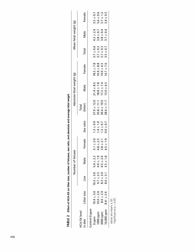
control, 1000, 3000, and 10,000 ppm group were 22, 23, 19,and 20, respectively. Observations made on the litters of femaleseuthanized on day 20 of gestation did not reveal any remarkabletreatment-related alterations in litter size, number of fetuses,sex ratios of the fetuses, and fetal weights. The mean littersize was 10.4 in the control group, while in the treatmentgroup there were 10.0, 8.5, and 8.4, respectively, at the doselevels of 1000, 3000, and 10,000 ppm of HCA-SX. The groupmean litter size from the highest-dose group of HCA-SX wassignificantly lower than the control group (p < 0.05). However,these observations were not biologically significant because thechanges were smaller in magnitude compared to the variationobserved in the historical control data (10.2 ± 2.1) (Table 2).The mean number of male fetuses per group was 5.4 in thecontrol group as compared to 4.9, 4.5, and 3.5, respectively,in the low-, intermediate-, and high-dose groups. The meannumber of female fetuses per group was 5.1, 4.7, 3.8, and 4.5,respectively, in the control, 1000, 3000, and 10,000 ppm groups.Compared to the control group, HCA-SX treatment did notsignificantly alter the mean sex ratio. The mean sex ratio of maleto female fetuses was 1.3 in the control group and 1.4, 1.3, and0.9 in the low-, intermediate-, and high-dose groups. The groupmean live fetal weight of male fetuses was 4.3 g in the controlgroup and 3.8, 3.8, and 3.7 g in the HCA-SX-treated groupsat the dose levels of 1000, 3000, and 10,000 ppm, respectively.Similar values for female fetuses were 3.5 g in the control groupand 3.6, 3.7, and 3.4 g at the dose levels of 1000, 3000, and10,000 ppm, respectively (Table 2).
Fetal ObservationsThe number of fetuses examined in the control, 1000-, 3000-,
and 10,000-ppm group were 226, 227, 158, and 160, respectively.The only “major malformation” noted was an omphalocele, abirth defect in which the intestine or other abdominal organsstick out of the belly button (navel), which was encounteredin two fetuses, one each in the control and high-dose groups.In view of its isolated incidence and its presence in both thecontrol and treated groups, this was considered incidental.Additionally, two other external abnormalities observed inthis study were classified as “minor malformations.” Thesemalformations included an incidence of a small hematoma atthe tip of the tail in three fetuses, one each in the control,low-dose, and intermediate-dose groups, and one fetus in thehigh-dose group was found to be small in size (runt).
Fetal Visceral ObservationsNo treatment-related significant incidence of soft tissue
alterations in fetuses of dams exposed to HCA-SX at the doses
447 HCA Safety
TAB
LE2
Eff
ect
of
HC
A-S
Xo
nlit
ter
size
,nu
mb
ero
ffe
tuse
s,se
xra
tio
,an
dab
solu
tean
dav
erag
efe
talw
eig
ht
Nu
mb
ero
ffe
tuse
sA
bso
lute
feta
lwei
gh
t(g
)M
ean
feta
lwei
gh
t(g
)
HC
A-S
Xle
vel
ind
iet
Litt
ersi
zeLi
veM
ale
Fem
ale
Sex
rati
oTo
tal
(lit
ter)
Mal
eFe
mal
eTo
tal
Mal
eFe
mal
e
Co
ntr
ol0
pp
m0
pp
m10
.4±
3.0
10.3
±3.
05.
4±
2.2
5.1
±2.
01.
3±
0.9
37.9
±12
.021
.4±
8.5
18.2
±7.
83.
7±
0.6
4.3
±2.
43.
5±
0.7
1000
pp
m10
.0±
2.6
9.7
±2.
54.
9±
1.9
4.8
±2.
51.
4±
1.0
35.8
±11
.118
.8±
7.8
16.9
±8.
83.
7±
0.2
3.8
±0.
73.
6±
0.6
3000
pp
m8.
6±
2.9
8.3
±3.
04.
5±
2.0
3.8
±2.
11.
3±
0.7
30.4
±10
.017
.2±
7.4
14.0
±6.
53.
7±
0.3
3.8
±0.
43.
7±
1.2
10,0
00p
pm
8.4∗
±2.
98.
0±
3.1
3.5
±1.
84.
5±
1.9
0.9
±0.
728
.6±
11.7
13.9
±6.
514
.7±
7.6
3.5
±0.
73.
7±
0.4
3.4
±0.
5
Valu
esar
em
eans
±SD
.∗ S
igni
fican
tat
p<
0.05
.
448

of 1000, 3000, and 10,000 ppm was noted. The number oftotal fetuses subjected to visceral examination from the controlgroup was 108, while that from HCA-SX-treated groups were106, 74, and 76, respectively, for the low-, intermediate-, andhigh-dose groups. The incidence of normal fetuses observedin the control, 1000-, 3000-, and 10,000-ppm group was 95%,98%, 99%, and 96%, respectively. Small incidences of “minoranomalies” such as globular heart and unilateral enlargement ofthe ventricle of heart, mottled lungs, unilateral displacement ofadrenal, and unilateral hypoplasia of kidney in fetal soft tissuesnoted in the study were considered as incidental and of notoxicological significance. The only abnormal finding classifiedunder “major” anomalies, viz. unilateral cerebral hypoplasia,was observed in one fetus from the low-dose group. This wasagain considered to be incidental in light of its isolated incidence(0.94%).
Fetal Skeletal ObservationsThere was no incidence of any significant and treatment-
related skeletal abnormalities in fetuses of dams treated withHCA-SX at the doses of 1000, 3000, or 10,000 ppm. All fetalskeletal abnormalities encountered in this study were consideredto be incidental or of no toxicological significance, eitherbecause of their being a normal variant or minor in nature,or their incidence being isolated and/or comparable to thatin the control group. There was no incidence of any “majormalformations” observed in any fetus. From the control, low-,intermediate-, and high-dose group, a total of 118, 121, 84, and84 fetuses were examined for skeletal abnormalities, respectively.The proportion of incidence of normal fetuses was 86.4%,91.7%, 80.9%, and 65.5% at the doses of 0 (control), 1000,3000, or 10,000 ppm, respectively, while the proportion oflitters without any abnormalities was 54.5%, 65.2%, 57.9%,and 60.0%, respectively, and was comparable between thegroups.
No treatment-related effects of HCA-SX on sternebrae,such as incomplete/partial ossification, poor ossification, orunossification of sternebrae, were noted. The variations notedin the treatment group were classified as normal and were alsoobserved in controls. In the control group, a small (2/118; 1.7%)incidence of split sternebrae was noted and was classified as a“minor anomaly.” In the intermediate-dose group (3000 ppm),“minor anomalies” such as displaced ribs (1.2%), undulationof the 13th rib (5.9%), and clubbed ribs (3.6%) were noted.In view of their occurrence only in the intermediate-dosegroup, these findings were considered as incidental in nature.As regards vertebrae, an incidence of “normal variants” suchas incomplete ossification of thoracic vertebrae (low dose:0.83%; high dose: 2.38%), poor ossification of thoracic (control:0.85%) and lumbar (control: 0.85%) vertebrae, and unossifiedcentrum of caudal vertebrae (high dose: 1.19%) was noted. Sincethis incidence was small and not dose dependent, it was notconsidered as treatment related. Additionally, the incidence of“minor anomalies,” such as split (control: 3.4%; high dose:3.6%) and dumbbell-shaped (control: 1.7%; intermediate dose:3.6%; high dose: 1.2%) thoracic vertebrae observed in thisstudy was considered as incidental in nature. An incidence ofabnormalities in the extremities, classified as “normal variants,”was noted in fetuses from one litter (among 20 litters) in thehigh-dose group only and was considered as incidental in nature.
DISCUSSIONHCA-SX has been shown to be effective in obesity man-
agement as evidenced by lowering of body weight and bodymass index, enhanced leptin and serotonin metabolism, andenhanced urinary excretion of fat metabolites. HCA-SX wasdemonstrated to perform through a number of mechanismsincluding ATP citrate lyase inhibition (a building block for fat,cholesterol, and triglyceride biosyntheses), appetite suppressionby enhancing serotonin release and its increased availability inthe brain, fat oxidation, maintaining healthy lipid profile, andtriggering genetic signaling to downregulate obesity (Downset al. 2005). Efficacious dosage, higher bioavailability, appro-priate and regular diet, and moderate exercise coupled withsynergistic influences of Ca2+ and K+ can significantly enhancethe therapeutic effects of HCA-SX in obesity regulation (Preusset al. 2004a, 2004b, 2005).
Oral HCA-SX supplementation was found to be bioavailablein human plasma. Oral LD50 of HCA-SX in rats was foundto be greater than 5000 mg/kg, which demonstrates its broad-spectrum safety. The dermal LD50 of HCA-SX was found tobe greater than 2000 mg/kg in rabbits. The results of the skinand eye irritation studies indicate that HCA-SX can cause mildirritation (Ohia et al. 2002).
In a subchronic study in rats, the oral administration ofHCA-SX at doses up to 2500 mg/kg/day for a period of90 days caused a significant decrease in body weight andreduction in feed consumption. Organ weights, hematology,and clinical chemistry analysis did not reveal any signs oftoxicity. Histological investigations of the major tissues didnot reveal any treatment-related pathological changes exceptfor minimal or mild hepatocyte vacuolation at 30 and 60days, but not at 90 days. The investigators suggested that thevacuolation may be related to the time duration between lastfeeding and euthanasia. Based on the data from this study,the no-observed-adverse-effect level (NOAEL) of HCA-SX formale and female rats was determined to be 2500 mg/kg bodyweight/day (Shara et al. 2003, 2004). Furthermore, HCA-SX didnot exhibit any mutagenicity (Soni et al. 2004).
In placebo-controlled, double-blind trials and one single-arm, open trial employing up to 2800 mg/day HCA or 4667mg/day (78 mg/kg/day) HCA-SX for up to 60 days, notreatment-related adverse effects were reported. The majorityof the side effects were minor and transient. In some of thesestudies, hematology and clinical chemistry parameters werestudied, and no treatment-related changes in these parameterswere noted. These studies demonstrate that HCA did not causeadverse effects and was well tolerated (Soni et al. 2004; Preusset al. 2004a, 2004b, 2005). Based on these animal and humansafety data, HCA-SX received self-affirmed GRAS status in 2003(Burdock Group 2003).
As HCA-SX reduces the weight gain in animals, the possibil-ity of weight loss-related effects of HCA-SX on reproductive anddevelopmental parameters, such as fetal size and ossification,needs special considerations. The results of the present studydemonstrate that exposure of HCA-SX for over two generationsdid not result in any significant treatment-related teratogeniceffects in Sprague-Dawley rats at the dietary dose levels of 1000,3000, and 10,000 ppm, equivalent to the dose levels of 103 mg,352 mg, or 1240 mg/kg body weight/day, respectively.
In the present study, at the highest dose level of HCA-SX(10,000 ppm) exposure, the group mean body weight of female
449 HCA Safety

rats on gestation day 20 was found to be slightly but statisticallysignificantly decreased. Similarly in this group, the body weightgain during the gestation period was also found to be lowered byabout 13% as compared to the control group. The rats used inthis study were from a two-generation HCA-SX developmentaltoxicity study and the initial body weights of the female rats werealready lower than the controls. The decreased body weights canbe considered as an effect of the test substance exposure for thetwo preceding generations. Apart from these observations, noother evidence of maternal toxicity was noted in pregnant ratsexposed to HCA-SX at dose levels of 1000, 3000, or 10,000ppm. There were no treatment-related alterations encounteredduring this study in the parameters of general toxicity, such asmortality, clinical signs, body weight gain (up to 3000 ppm),pregnancy data, and gross pathological findings.
In summary, except a slight (13%) decrease in body weightgain during the gestation period, at the highest dose HCA-SXdid not cause maternal toxicity, adverse effects on the graviduteri, external abnormalities in the fetuses of the treated animals,soft tissue abnormalities in the fetuses, or skeletal abnormalitiesin the fetuses. These results suggest that HCA-SX was notteratogenic in Sprague-Dawley rats at the dietary dose levelsof 1000, 3000, and 10,000 ppm, equivalent to the dose levelsof 103 mg, 352 mg, or 1240 mg/kg/day, respectively. Based onthe results of this study, the NOAEL of HCA-SX is determinedas 1240 mg/kg/day. The results of the present study and theearlier safety study confirm the safety of HCA-SX for long-termhuman consumption.
In conclusion, based on extensive scientific investigations,which include human, animal, analytical, and other mechanisticstudies, and history of exposure and use, the consumption ofHCA or HCA-SX at dose levels of 2800 or 4667 mg/day, respec-tively, is considered safe for human consumption, which mayeradicate obesity and related disorders including cardiovasculardysfunctions, diabetes, and arthritis.
ABBREVIATIONSFDA Food and Drug Administration;DSHEA Dietary Supplement Health and Education Act;HCA (-)-Hydroxycitric acid;OECD Organization for Economic Co-operation and De-
velopment;NOAEL no observed adverse effect level;
ppm = parts per million.
REFERENCESBartlett, M.S. 1937. Properties of sufficiency and statistical tests. Proc.
Royal Stat. Soc. Series A 160:268–282.Boll, P.M., Sorensen, E., and Balieu, E. 1969. Naturally occurring lactones
and lactames. The absolute configuration of hydroxycitric acidlactones: hibiscus acid and garcinia acid. Acta Chem. Scand.23:286–293.
Burdock Group. 2003. Opinion of an Expert Panel on the GenerallyRecognized As Safe (GRAS) Status of Super CitriMax R© as aFlavoring Agent. Received October 31, 2003.
Clouatre, D., and Rosenbaum, M. 1994. The Diet and Health Benefits ofHCA (Hydroxycitric acid). Keats Publishing Co., New Canaan, CT.9.
Deshmukh, N.S., Bagchi, M., Yasmin, T., and Bagchi, D. 2008. Safetyof a novel calcium/potassium salt of hydroxycitric acid (HCA-SX): I.
Two-generation reproduction toxicity study. Tox. Mech. Meth. (inpress).
Downs, B.W., Bagchi, M., Subbaraju, G.V., Shara, M.A., Preuss, H.G., andBagchi, D. 2005. Bioefficacy of a novel calcium-potassium salt of(-)-hydroxycitric acid. Mutat. Res. 579:149–162.
DSHEA. 1994. Dietary Supplements and Health and Education Act of1994. U.S. Food and Drug Administration, Center for Food Safetyand Applied Nutrition, Washington, DC.
Hadjidakis, D.J., and Androulakis, I.L. 2006. Bone remodeling. Ann. N. T.Acad. Sci. 1092:385–396.
Jena, B.S., Jayaprakasha, G.K., Singh, R.P. and Sakariah, K.K. 2002.Chemistry and biochemistry of (-)-hydroxycitric acid from Garcinia.J. Agric. Food Chem. 50:10–22.
Lazcano, O., Li, C.Y., Pierre, R.V., O’Duffy, J.D., Beissner, R.S., andAbell-Aleff, P.C. 1993. Clinical utility of the alizarin red S stain onpermanent preparations to detect calcium-containing compoundsin synovial fluid. Am. J. Clin. Pathol. 99:90–96.
Lowenstein, J.M. 1970. Experiments with (-)-hydroxycitrate. in Essays inCell Metabolism, (W. Bartley, H. Hornber and J. Qualyle, Eds.), WileyInterscience, New York, NY, 153–156.
Martin, F.W., Campbell, C.W., and Ruberte, R.M. 1987.Perennial ediblefruits of the tropics: an inventory. U.S. Department of Agriculture,Agricultural Research Service, Washington, DC, 212.
Ohia, S.E., Opere, C.A., LeDay, A.M., Bagchi, M., Bagchi, D., and Stohs,S.J. 2002. Safety and mechanism of appetite suppression by anovel hydroxycitric acid extract (HCA-SX). Mol. Cell. Biochem.238:89–103.
Preuss, H.G., Bagchi, D., Bagchi, M., Rao, C.V., Dey, D.K., and Satya-narayana, S. 2004a. Effects of a natural extract of (-)-hydroxycitricacid (HCA-SX) and a combination of HCA-SX plus niacin-boundchromium and Gymnema sylvestre extract on weight loss. DiabetesObes. Metab. 6:171–180.
Preuss, H.G., Garis, R.I., Bramble, J.D., Bagchi, D., Bagchi, M., Rao, C.V.,and Satyanarayana, S. 2005. Efficacy of a novel calcium/potassiumsalt of (-)-hydroxycitric acid in weight control. Int. J. Clin. Pharmacol.Res. 25:133–144.
Preuss, H.G., Rao, C.V., Garis, R., Bramble, J.D., Ohia, S.E., Bagchi, M.,and Bagchi, D. 2004b. An overview of the safety and efficacy ofa novel, natural (-)-hydroxycitric acid extract (HCA-SX) for weightmanagement. J. Med. 35:33–48.
Roy, S., Shah, H., Rink, C., Khanna, S., Bagchi, D., Bagchi, M., and Sen,C.K. 2007. Transcriptome of primary adipocytes from obese womenin response to a novel hydroxycitric acid-based dietary supplement.DNA Cell Biol. 26:627–639.
Shara, M., Ohia, S.E., Yasmin, T., Zardetto-Smith, A., Kincaid, A., Bagchi,M., Chatterjee, A., Bagchi, D., and Stohs, S.J. 2003. Dose- andtime-dependent effects of a novel (-)-hydroxycitric acid extracton body weight, hepatic and testicular lipid peroxidation, DNAfragmentation and histopathological data over a period of 90 days.Mol. Cell Biochem. 254:339–346.
Shara, M., Ohia, S.E., Yasmin, T., Zardetto-Smith, A., Kincaid, A., Bagchi,M., Chatterjee, A., Bagchi, D., and Stohs, S.J. 2004. Physico-chemical properties of a novel (-)-hydroxycitric acid extract andits effect on body weight, selected organ weights, hepatic lipidperoxidation and DNA fragmentation, hematology and clinicalchemistry, and histopathological changes over a period of 90 days.Mol. Cell Biochem. 260:171–186.
Soni, M.G., Burdock, G.A., Preuss, H.G., Stohs, S.J., Ohia, S.E., andBagchi, D. 2004. Safety assessment of (-)-hydroxycitric acid andSuper CitriMax, a novel calcium/potassium salt. Food Chem. Toxicol.42:1513–1529.
Sullivan, A.C., Triscari, J., Hamilton, J.G., Miller, O.N., and Wheatley, V.R.1974a. Effect of (-)-hydroxycitrate upon the accumulation of lipidin the rat. I. Lipogenesis, Lipids 9:121–128.
Sullivan, A.C., Triscari, J., Hamilton, J.G., and Miller, O.N. 1974b. Effectof (-)-hydroxycitrate upon the accumulation of lipid in the rat. II.Appetite. Lipids 9:129–134.
N. S. Deshmukh et al. 450
U.S. FDA. 2000. Toxicological Principles for the Safety Assessment ofDirect Food Additives and Color Additives Used in Food (‘Redbook’).U.S. Food and Drug Administration, Washington, DC.
Watson, J.A., Fang, M., and Lowenstein, J.M. 1969. Tricarballylate andhydroxycitrate: substrate and inhibitor of ATP: citrate oxaloacetatelyase. Arch. Biochem. Biophys. 135:209–217.
Watson, J.A., and Lowenstein, J.M. 1970. Citrate and the conversionof carbohydrate into fat. Fatty acid synthesis by a combination ofcytoplasm and mitochondria. J. Biol. Chem. 245:5993–6002.
Wilson, N.H., and Hayes, J.R. 2001.Short-term repeated dosing andsubchronic toxicity studies. In Principles and Methods of Toxicology,Ed. W. Hayes, Raven Press, New York, 649–672.
451 HCA Safety