Identification of Serologic Markers for School-Aged Children With Congenital Rubella Syndrome
Upload
independentCategory
view
2download
0
RTS,S Vaccination Is Associated WithSerologic Evidence of Decreased Exposure toPlasmodium falciparum Liver- and Blood-StageParasites*□S
Joe J. Campo‡§� �, John J. Aponte‡§, Jeff Skinner¶, Rie Nakajima‡‡,Douglas M. Molina�, Li Liang‡‡, Jahit Sacarlal§**, Pedro L. Alonso‡§,Peter D. Crompton¶, Philip L. Felgner‡‡�, and Carlota Dobano‡§
The leading malaria vaccine candidate, RTS,S, targets thesporozoite and liver stages of the Plasmodium falciparumlife cycle, yet it provides partial protection against diseaseassociated with the subsequent blood stage of infection.Antibodies against the vaccine target, the circumsporo-zoite protein, have not shown sufficient correlation withrisk of clinical malaria to serve as a surrogate for protec-tion. The mechanism by which a vaccine that targetsthe asymptomatic sporozoite and liver stages protectsagainst disease caused by blood-stage parasites remainsunclear. We hypothesized that vaccination with RTS,Sprotects from blood-stage disease by reducing the num-ber of parasites emerging from the liver, leading to pro-longed exposure to subclinical levels of blood-stage par-asites that go undetected and untreated, which in turnboosts pre-existing antibody-mediated blood-stage im-munity. To test this hypothesis, we compared antibodyresponses to 824 P. falciparum antigens by protein arrayin Mozambican children 6 months after receiving a fullcourse of RTS,S (n � 291) versus comparator vaccine (n �297) in a Phase IIb trial. Moreover, we used a nestedcase-control design to compare antibody responses ofchildren who did or did not experience febrile malaria.Unexpectedly, we found that the breadth and magnitudeof the antibody response to both liver and asexual blood-
stage antigens was significantly lower in RTS,S vaccin-ees, with the exception of only four antigens, including theRTS,S circumsporozoite antigen. Contrary to our initialhypothesis, these findings suggest that RTS,S confersprotection against clinical malaria by blocking sporozoiteinvasion of hepatocytes, thereby reducing exposure to theblood-stage parasites that cause disease. We also foundthat antibody profiles 6 months after vaccination did notdistinguish protected and susceptible children during thesubsequent 12-month follow-up period but were stronglyassociated with exposure. Together, these data provideinsight into the mechanism by which RTS,S protects frommalaria. Molecular & Cellular Proteomics 14: 10.1074/mcp.M114.044677, 519–531, 2015.
The RTS,S malaria vaccine candidate provides partial pro-tection against clinical malaria in African children, which hasbeen repeatedly demonstrated in Phase IIb and Phase IIIclinical trials (1–5). The RTS,S target is the Plasmodium fal-ciparum circumsporozoite protein (CSP), and it has beenshown to generate high antibody titers that remain abovelevels acquired naturally for years (6). However, it remainsunclear how the vaccine, which targets sporozoites, providesprotection against disease caused by blood-stage parasites.A rational mechanism has been proposed, based on antibodyand T cell responses to the CSP (7), but antibodies have notconsistently correlated with protection when clinical diseasewas the trial end point (8). We and others hypothesized thatpartial blockage of pre-erythrocytic development would resultin low-level blood-stage infections that go untreated in RTS,Svaccinees and that this would boost the blood-stage immuneresponse, contributing to protection from malaria disease(8–10).
We set out to address the question of how the vaccine worksby investigating the response to malaria parasites in the contextof RTS,S vaccination. However, until recently, the means ofassessing the response to malaria parasites has been limited toa sparse selection of recombinant proteins or parasite lysates.
‡ISGlobal, Barcelona Ctr. Int. Health Res. (CRESIB), Hospital Clínic—Universitat de Barcelona, Barcelona, Spain; §Manhica Health Re-search Centre, Manhica, Mozambique; ¶Laboratory of Immunogenet-ics, National Institute of Allergy and Infectious Diseases, NationalInstitutes of Health, Rockville, MD; ‡‡Department of Medicine, Uni-versity of California Irvine, Irvine, CA; �Antigen Discovery Inc., Irvine,CA; **Faculty of Medicine, Universidade Eduardo Mondlane, Maputo,Mozambique
Received September 17, 2014, and in revised form, December 1,2014
Published, MCP Papers in Press, December 13, 2014, DOI10.1074/mcp. M114.044677
Author contributions: J.J.C., J.J.A., P.L.A., P.L.F., and C.D. de-signed the research; J.J.C., R.N., D.M.M., and J. Sacarlal performedthe research; J.J.C., J.J.A., J. Skinner, D.M.M., L.L., P.L.A., P.D.C.,P.L.F., and C.D. analyzed the data; J.J.C., J. Sacarlal, P.L.A., P.D.C.,P.L.F., and C.D. wrote the paper.
Research© 2015 by The American Society for Biochemistry and Molecular Biology, Inc.This paper is available on line at http://www.mcponline.org
Molecular & Cellular Proteomics 14.3 519

The P. falciparum (Pf) proteome contains more than 5,300 pro-teins, and, until recently, less than 0.5% of them have beenclosely investigated (11). Similar to the approach taken withgene expression microarrays, protein arrays offer the opportu-nity to screen antibody responses to partial or complete pro-teomes (12). This approach was taken in this study to identifythe breadth and magnitude of naturally acquired immune re-sponses in Mozambican children vaccinated with RTS,S/AS021, the predecessor to the RTS,S/AS01 formulation used inthe current Phase III trial, or comparator vaccine.
In addition to characterizing the RTS,S mode of action, weaimed to identify biomarker correlates of protection againstclinical malaria. Malaria vaccinology is lacking in surrogatemarkers of protection, and such biomarkers would be a highlyuseful measure for assessment of vaccine efficacy, especiallywhen control or placebo vaccine groups are no longer available(13). This could mitigate the current inefficient means of mea-suring efficacy in clinical trials. In the post-genomic era, withsystems approaches employed for questions to complex prob-lems in biology and medicine, perhaps alternative thinking isrequired to tackle the question of how to assess vaccines (14,15). In this study, we took steps in that direction in order toidentify antibody signatures of protection that contribute towarda surrogate marker for the RTS,S and other vaccines.
EXPERIMENTAL PROCEDURES
Ethics Statement—The study was approved by the MozambicanNational Health and Bioethics Committee (Ref. 237/CNBS), the Hos-pital Clínic of Barcelona Ethics Committee (Registro 2008/4444), andthe PATH Research Ethics Committee (Study file number HS 482) andwritten informed consent was gathered from parents/guardians.
Study Design—Serum samples were obtained from a Phase IIbrandomized, controlled trial of the RTS,S/AS02 vaccine administeredto 1–4-year-old Mozambican children (ClinicalTrials.gov registrynumber NCT00197041). A nested case-control study was designed
using the cross-sectional survey at study month 8.5 (M8.5), 6 monthsafter the third dose, as the sampling time point (Fig. 1). Cases ofclinical malaria, irrespective of treatment group, were identified duringthe 12-month follow-up period from M8.5 to study month 21 (M21),following the trial secondary case definition of P. falciparum asexualblood-stage parasitemia of �0 parasite/�l of blood and an axillarytemperature � 37.5 °C. For the cases that had available serum sam-ples at the time of the study, controls were matched to cases 2 to 1by random selection of non-cases. A total of 623 samples (207 casesand 416 controls), 588 (196 cases and 392 controls) of which passedfiltering criteria, was probed at the Protein Microarray Laboratory atthe University of California Irvine (UCI).
The clinical trial enrolled two study cohorts from different areas ofManhica District to measure different efficacy endpoints, cohort 1 inManhica and Maragra for efficacy against clinical malaria and cohort 2from Ilha Josina for efficacy against time to first infection (1). Only cohort1 of the trial was selected since efficacy had waned in cohort 2 (16), andthe time point was selected to allow 6 months of post-vaccinationnatural exposure before sampling and a 1-year follow-up timeframeafter sampling. This was chosen as opposed to a longer follow-up toincrease the specificity of antibody responses measured at M8.5 andassociation with subsequent clinical cases. At the pre-vaccination base-line time point, serum samples were tested for antibody titers to infectedred blood cells by immunofluorescence antibody test, as previouslydescribed (1). These data were used to demonstrate the higher trans-mission intensity in cohort 2, and these data are used herein to comparebasal levels of blood-stage antibody responses to control for bias.
Protein Array Construction—Proteins were expressed using theEscherichia coli cell-free Rapid Translation System (RTS) kit (5 Prime,Gaithersburg, MD). A library of Pf partial or complete open readingframes (ORFs) cloned into a T7 expression vector pXT7 has beenestablished at Antigen Discovery, Inc. (ADi, Irvine, CA). This librarywas created through an in vivo recombination cloning process withPCR-amplified Pf ORFs, and a complementary linearized expressedvector transformed into chemically competent E. coli was amplifiedby PCR and cloned into pXI vector using a high-throughput PCRrecombination cloning method described elsewhere (17). Each ex-pressed protein includes a 5� polyhistidine (HIS) epitope and 3� he-magglutinin (HA) epitope. After expressing the proteins according tomanufacturer instructions, translated proteins were printed onto ni-trocellulose-coated glass AVID slides (Grace Bio-Labs, Inc., Bend,OR) using an Omni Grid Accent or Omni Grid 100 robotic microarrayprinter (Digilabs, Inc., Marlborough, MA). Microarray chip printing andprotein expression were quality checked by probing random slideswith anti-HIS and anti-HA monoclonal antibodies with fluorescentlabeling, as shown in Fig. S1.
1 The abbreviations used are: AS02, adjuvant system associatedwith RTS,S malaria vaccine candidate; CSP, circumsporozoite pro-tein, the immunodominant surface protein of Plasmodium sporozo-ites; HA, hemagglutinin epitope, HIS, polyhistidine epitope; ORF,open reading frame; Pf, Plasmodium falciparum; RTS, rapid transla-tion system; RTSS, malaria vaccine candidate.
FIG. 1. Trial and nested case-control study design. Samples for this study were taken from cohort 1 of a Phase IIb trial of RTS,S/AS02in Mozambican children. The children were followed by passive case detection for 45 months from enrollment. The nested case-control studywas designed by selecting children with cases of clinical malaria and those without during the follow-up period between study months 8 1⁄2and 21. The blood sample taken at a cross-sectional survey at the beginning of that period was used for antibody profiling.
Malaria Antibody Responses Associated With RTS,S Vaccination
520 Molecular & Cellular Proteomics 14.3
Protein Array Chip Design—A down-selected array was designed(PF11� chip, ADi) to include eight “pads,” or replicated grids ofspotted proteins totaling 960 features per pad, each available toprobe a single specimen. Each pad contains 94 control spots, com-posed of the following: (i) 16 purified human IgG or a mouse anti-human IgG positive control spots, printed in quadruplicate at twodifferent concentrations; (ii) 38 tris-based buffering solution “blanks”with or without Tween-20 (12 and 16 spots, respectively) or nothing atall (four spots); (iii) 40 RTS reactions without Pf ORFs (NoDNA), asshown in Fig. S2. The NoDNA controls were randomly distributedacross the pad subarrays. Printed features do not vary in positionbetween pads or slides. Each pad contains 824 peptide fragmentsexpressed from Pf ORFs representing 702 unique proteins and threeconcentrations of 14 full-length proteins expressed using clinical-grade manufacturing procedures. The protein targets on the arraywere selected from published studies done on a larger microarraycontaining 2,323 protein features after interrogating specimens fromnaturally exposed individuals (18), and specimens from sporozoitevaccine trials (19). The top 824 most immunoreactive antigens fromthese studies were printed on this down-selected array.
Sample Probing—Serum samples were diluted 1:100 in a 10%E. coli lysate solution in protein arraying buffer (Maine Manufacturing,Sanford, ME) and incubated at room temperature for 30 min. Chipswere rehydrated in blocking buffer for 30 min. Blocking buffer wasremoved, and chips were probed with preincubated serum samplesusing sealed chambers fitted to eight-pad slides to ensure no cross-contamination of sample between pads. Chips were incubated over-night at 4 °C with agitation. Chips were washed five times with TBS-0.05% Tween 20, followed by incubation with biotin-conjugated goatanti-human IgG (Jackson ImmunoResearch, West Grove, PA) diluted1:200 in blocking buffer at room temperature. Chips were washedthree times with TBS-0.05% Tween 20, followed by incubation withstreptavidin-conjugated SureLight P-3 (Columbia Biosciences, Fred-erick, MD) at room temperature protected from light. Chips werewashed three times with TBS-0.05% Tween 20, three times with TBS,and once with water. Chips were air dried by centrifugation at 1,000 �g for 4 min and scanned on a ScanArray Express HT spectral scanner(Perkin-Elmer, Waltham, MA), and spot and background intensitieswere measured using an annotated grid file (.GAL). Data were ex-ported in Microsoft Excel.
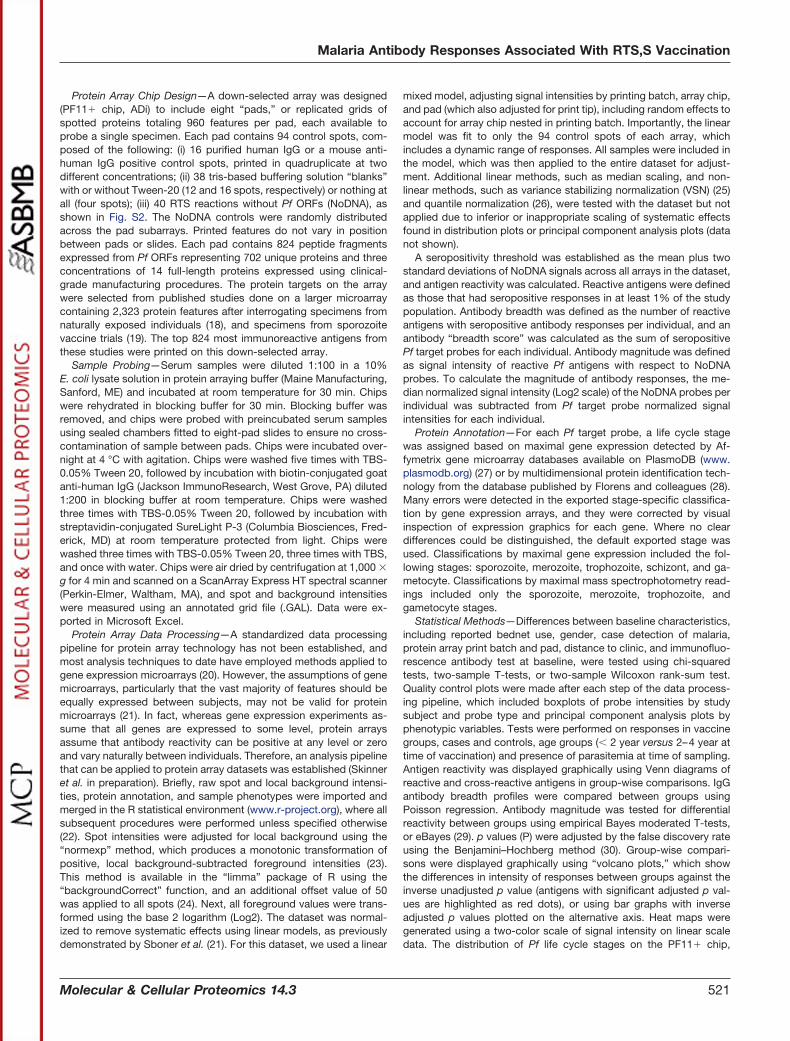
Protein Array Data Processing—A standardized data processingpipeline for protein array technology has not been established, andmost analysis techniques to date have employed methods applied togene expression microarrays (20). However, the assumptions of genemicroarrays, particularly that the vast majority of features should beequally expressed between subjects, may not be valid for proteinmicroarrays (21). In fact, whereas gene expression experiments as-sume that all genes are expressed to some level, protein arraysassume that antibody reactivity can be positive at any level or zeroand vary naturally between individuals. Therefore, an analysis pipelinethat can be applied to protein array datasets was established (Skinneret al. in preparation). Briefly, raw spot and local background intensi-ties, protein annotation, and sample phenotypes were imported andmerged in the R statistical environment (www.r-project.org), where allsubsequent procedures were performed unless specified otherwise(22). Spot intensities were adjusted for local background using the“normexp” method, which produces a monotonic transformation ofpositive, local background-subtracted foreground intensities (23).This method is available in the “limma” package of R using the“backgroundCorrect” function, and an additional offset value of 50was applied to all spots (24). Next, all foreground values were trans-formed using the base 2 logarithm (Log2). The dataset was normal-ized to remove systematic effects using linear models, as previouslydemonstrated by Sboner et al. (21). For this dataset, we used a linear
mixed model, adjusting signal intensities by printing batch, array chip,and pad (which also adjusted for print tip), including random effects toaccount for array chip nested in printing batch. Importantly, the linearmodel was fit to only the 94 control spots of each array, whichincludes a dynamic range of responses. All samples were included inthe model, which was then applied to the entire dataset for adjust-ment. Additional linear methods, such as median scaling, and non-linear methods, such as variance stabilizing normalization (VSN) (25)and quantile normalization (26), were tested with the dataset but notapplied due to inferior or inappropriate scaling of systematic effectsfound in distribution plots or principal component analysis plots (datanot shown).
A seropositivity threshold was established as the mean plus twostandard deviations of NoDNA signals across all arrays in the dataset,and antigen reactivity was calculated. Reactive antigens were definedas those that had seropositive responses in at least 1% of the studypopulation. Antibody breadth was defined as the number of reactiveantigens with seropositive antibody responses per individual, and anantibody “breadth score” was calculated as the sum of seropositivePf target probes for each individual. Antibody magnitude was definedas signal intensity of reactive Pf antigens with respect to NoDNAprobes. To calculate the magnitude of antibody responses, the me-dian normalized signal intensity (Log2 scale) of the NoDNA probes perindividual was subtracted from Pf target probe normalized signalintensities for each individual.
Protein Annotation—For each Pf target probe, a life cycle stagewas assigned based on maximal gene expression detected by Af-fymetrix gene microarray databases available on PlasmoDB (www.plasmodb.org) (27) or by multidimensional protein identification tech-nology from the database published by Florens and colleagues (28).Many errors were detected in the exported stage-specific classifica-tion by gene expression arrays, and they were corrected by visualinspection of expression graphics for each gene. Where no cleardifferences could be distinguished, the default exported stage wasused. Classifications by maximal gene expression included the fol-lowing stages: sporozoite, merozoite, trophozoite, schizont, and ga-metocyte. Classifications by maximal mass spectrophotometry read-ings included only the sporozoite, merozoite, trophozoite, andgametocyte stages.
Statistical Methods—Differences between baseline characteristics,including reported bednet use, gender, case detection of malaria,protein array print batch and pad, distance to clinic, and immunofluo-rescence antibody test at baseline, were tested using chi-squaredtests, two-sample T-tests, or two-sample Wilcoxon rank-sum test.Quality control plots were made after each step of the data process-ing pipeline, which included boxplots of probe intensities by studysubject and probe type and principal component analysis plots byphenotypic variables. Tests were performed on responses in vaccinegroups, cases and controls, age groups (� 2 year versus 2–4 year attime of vaccination) and presence of parasitemia at time of sampling.Antigen reactivity was displayed graphically using Venn diagrams ofreactive and cross-reactive antigens in group-wise comparisons. IgGantibody breadth profiles were compared between groups usingPoisson regression. Antibody magnitude was tested for differentialreactivity between groups using empirical Bayes moderated T-tests,or eBayes (29). p values (P) were adjusted by the false discovery rateusing the Benjamini–Hochberg method (30). Group-wise compari-sons were displayed graphically using “volcano plots,” which showthe differences in intensity of responses between groups against theinverse unadjusted p value (antigens with significant adjusted p val-ues are highlighted as red dots), or using bar graphs with inverseadjusted p values plotted on the alternative axis. Heat maps weregenerated using a two-color scale of signal intensity on linear scaledata. The distribution of Pf life cycle stages on the PF11� chip,
Malaria Antibody Responses Associated With RTS,S Vaccination
Molecular & Cellular Proteomics 14.3 521

reactive antigens, and differentially reactive antibodies was graphi-cally displayed using pie graphs. Differential IgG antibody reactivityby Pf life cycle stages was tested using Fisher’s exact test. A largeproportion of Pf targets were unidentified or were expressed equallybetween some stages in the multidimensional protein identificationtechnology database. Therefore, life cycle stage comparisons areonly shown with the maximal expression by gene arrays. Between-group baseline characteristics were tested using chi-squared tests.For comparisons of baseline characteristics, results were consideredstatistically significant for a p value � 0.05. For all other comparisons,results were considered statistically significant for an adjusted pvalue � 0.05. All antibody data were analyzed in R (22), except foranalysis of baseline characteristics, which were performed in STATA(StataCorp LP, College Station, TX).
RESULTS
Characteristics of the Study Population—Baseline charac-teristics of 588 Mozambican children with samples probed on
PF11� protein arrays are shown by vaccination group, agegroup, and case-control group (Table 1). There were slightlyhigher, statistically significant proportions of cases to controlsand infections at the time of sampling in the comparatorvaccine group (p � 0.04 and p � 0.03, respectively), which isconsistent with the lower proportions observed in the clinicaltrial (16). No differences were observed in age, reported bed-net use, gender, or previous clinical malaria episodes (p �
0.05). The distribution of RTS,S and comparator vaccine sam-ples across print batches and pads was balanced, althoughthere were slight, statistically significant differences in theproportions of RTS,S or comparator samples across pads,particularly between pads 4 and 7 (p � 0.05, Table S1).Between age groups, the younger age group had a higherproportion of cases to controls and proportion of children with
TABLE IBaseline characteristics of 588 Mozambican children included in the study
Comparatorvaccine (%)
or �S.D.
RTS,S vaccine(%) or �S.D. p valuea � 2 years
(%)2–4 years
(%) p value Control(%)
Case(%) p value
Probed 297 (51) 291 (49) 157 (27) 431 (73) 392 (67) 196 (33)Age group
� 2 year 79 (13) 78 (13) 0.96 – – – –2–4 year 218 (37) 213 (36) – – – –
Nested case-controlCase 111 (19) 85 (14) 0.04 66 (11) 130 (22) 0.01 – –Control 186 (32) 206 (35) 91 (15) 301 (51) – –
Bednet use at D0b
Yes 14 (2) 10 (2) 0.43 6 (1) 18 (3) 0.85 12 (2) 12 (2) 0.08No 283 (48) 281 (48) 151 (26) 413 (70) 380 (65) 184 (31)
GenderFemale 144 (24) 127 (22) 0.24 71 (12) 200 (34) 0.80 179 (30) 92 (16) 0.77Male 153 (26) 164 (28) 86 (15) 231 (39) 213 (36) 108 (18)
Previous clinical malaria0 197 (34) 210 (36) 0.13 95 (16) 312 (53) 0.01 308 (52) 99 (17) �0.01� 1 100 (17) 81 (14) 62 (11) 119 (20) 84 (14) 97 (16)
Infection M8.5c
Neg 243 (41) 257 (44) 0.03 136 (23) 364 (62) 0.51 348 (59) 152 (26) �0.01Pos 54 (9) 34 (6) 21 (4) 67 (11) 44 (7) 44 (7)
Protein array print batchAccent A 121 (21) 129 (22) 0.46 157 (27) 93 (16) �0.01 162 (28) 88 (15) 0.09Accent B 168 (29) 153 (26) 0 (0) 321 (55) 214 (36) 107 (18)Omni100a 6 (1) 4 (1) 0 (0) 10 (2) 10 (2) 0 (0)Omni100b 2 (0.3) 5 (1) 0 (0) 7 (1) 6 (1) 1 (0.2)
Protein array pad1 34 (6) 37 (6) 0.01 21 (4) 50 (9) 0.99 45 (8) 26 (4) 0.682 33 (6) 39 (7) 20 (3) 52 (9) 54 (9) 18 (3)3 34 (6) 36 (6) 19 (3) 51 (9) 46 (8) 24 (4)4 27 (5) 45 (8) 19 (3) 53 (9) 49 (8) 23 (4)5 32 (5) 44 (7) 20 (3) 56 (10) 45 (8) 31 (5)6 42 (7) 36 (6) 20 (3) 58 (10) 52 (9) 26 (4)7 50 (9) 28 (5) 20 (3) 58 (10) 54 (9) 24 (4)8 45 (8) 26 (4) 18 (3) 53 (9) 47 (8) 24 (4)
Distance to clinicd
km 1.7 �1.1 1.8 �1.0 0.14e 1.7 �1.0 1.8 �1.1 0.41e 1.7 �1.0 1.8 �1.2 0.36e
IFAT titer at screeningf
GMT 2712 �13.4 2029 �11.8 0.18g 1046 �10.9 3147 �12.5 �0.01g 1750 �12.4 4220 �11.8 �0.01g
a Reported p values are chi-squared tests unless otherwise stated; b Reported bednet use at the time that the first dose of RTS,S/AS02 orcomparator vaccine was administered; c Study month 8.5 corresponding to 6 months post-vaccination. d Mean distance in km to nearesthospital or health care facility; e Two-sample T-test; f Immunofluorescence antibody test (IFAT) at screening is during visit before administrationof first dose of vaccine, reported as geometric mean endpoint titer; g Two-sample Wilcoxon rank-sum test.
Malaria Antibody Responses Associated With RTS,S Vaccination
522 Molecular & Cellular Proteomics 14.3

previous malaria cases than the older age group during the 6months of follow-up prior to sampling (p � 0.01 for bothcomparisons), following the expected pattern of risk of clinicalmalaria with age. There was a marked difference in print batchover age groups (p � 0.01), with all of the younger age groupconcentrated in a single print batch, the “Accent A.” This flawin the sampling design was a result of correlation in age andthe sequence of subjects, although the primary comparisongroups were randomized, and this may introduce bias in thecomparison of age groups. Age groups were evenly balancedacross array pads. Younger children had significantly lowerbaseline blood-stage antibody levels, as measured by immu-nofluorescence antibody test (p � 0.01), which was an ex-pected characteristic of the study population. Cases and con-trols had significantly different proportions of those withprevious clinical malaria cases and infection at time of sam-pling (p � 0.01 for both comparisons) but were balancedacross print batches and array pads. Cases had significantlyhigher levels of baseline blood stage antibodies (p � 0.01).
Effect of Vaccination with RTS,S/AS02 on P. falciparum-Specific IgG Responses 6 Months After Vaccination—First,we tested the hypothesis that RTS,S vaccination would elicitan overall stronger blood-stage response from untreated in-fections by examining the breadth (number of seropositiveresponses per individual) and magnitude of the antibody re-sponse to the 824 Pf antigens on the PF11� array. We foundthat a total of 579 Pf protein fragments were reactive in atleast 1% of the study population, and 245 targets were non-reactive. Table S2 lists the shared and unique reactive anti-gens between comparison groups. The majority of reactiveantigens were shared between vaccine groups (576 of 579).Differences in antibody breadth profiles were significant (p �
1E-15), with mean breadth scores of 49 (standard deviation(S.D.): 53) in children vaccinated with RTS,S and 62 (S.D.: 69)in children vaccinated with comparator vaccine. A total of 85antigens had differentially reactive antibody magnitude be-tween vaccination groups. However, to filter out antigens withvery low group mean antibody levels, only antigens with re-sponses above the median of the NoDNA background in atleast one of the comparison groups were considered signifi-cant. Henceforth, only differentially reactive antigens meetingthis significance criterion are reported. A total of 32 differen-tially reactive antigens were identified among proteins thathad group mean antibody levels higher than the medianNoDNA. The list of antigens with differential antibody reactiv-ity by comparison group is shown in Table S3. Fig. 2 showsthe profile of vaccine group comparisons. Surprisingly, of the32 differentially reactive antigens, 28 had higher responses inthe comparator vaccines, and four were higher in RTS,S vac-cinees, including the CSP. Notably, the extreme significanceand fold change of the CSP antigen is consistent with vaccineimmunogenicity monitoring in the clinical trials of RTS,S (31).
Antibody Responses in Cases Versus Controls—Next, weaimed to identify antibody signatures of protection within the
nested case-control study design. The profile of children whohad a subsequent episode(s) of malaria in the 12 monthsfollowing sample collection (cases) compared with childrenwho did not (controls) is shown in Fig. 3. All reactive antigenswere shared between cases and controls. Interestingly, pre-follow-up antibody breadth profiles were higher in cases(mean: 68, S.D.: 67) than controls (mean: 50, S.D.: 58; p �
1E-15). Likewise, there were 22 differentially reactive proteinsin the case-control study, all of which were more reactive inthe cases.
Effect of P. falciparum Exposure and Age on IgG AntibodyResponses—The findings from the nested case-control studyled us to more closely examine factors in the study popula-tion, such as exposure during and prior to sampling and age.We did a stratified analysis taking into account parasitemia attime of sampling or having had previous malaria episodes.Fig. S3 shows the profile of parasitemia group comparisons.Again, all reactive antigens were shared between childrenwho had parasitemia at the time of sampling and those thatdid not. Differences in antibody breadth profiles were highlysignificant (p � 1E-15) for having parasitemia. Mean breadthscore of aparasitemic children was 45 (S.D.: 50) versus 120(S.D.: 80) for parasitemic children (with or without previousmalaria). A total of 319 Pf targets was differentially reactivebetween children with or without parasitemia. All 319 differ-entially reactive antigens were higher in the parasitemic chil-dren. Furthermore, a gradient pattern was observed betweenexposure status when stratifying the population by childrenwith parasitemia, children without parasitemia but with previ-ously documented cases of malaria, and children with noparasitemia and no documented cases of malaria. The heatmap in Fig. 4A shows the differential reactivity profiles of eachexposure group.
To account for the effect of exposure on antibody levels, weperformed a stratified analysis of the vaccine group andnested case-control comparisons by exposure group. No dif-ferentially reactive targets were identified when comparingRTS,S and comparator vaccinees within parasitemic childrenor children who had no parasitemia but had documentedcases of malaria in the 6 months prior to sampling (Fig. 4B).Children who had no documented exposure showed multipledifferentially reactive antibodies for both comparator andRTS,S vaccinees with a tendency toward more reactivity inRTS,S vaccinees. No differentially reactive targets were iden-tified when comparing cases and controls within like exposuregroups (Fig. 4C). Stratified analysis of antibody breadth isshown in Fig. S4. Among parasitemic children, antibodybreadth score was higher in comparator vaccinees (mean:128, S.D.: 81) than in RTS,S vaccinees (mean: 106, S.D.: 76;p � 1E-15), and breadth was lower in cases (mean: 113, S.D.:78) than in controls (mean: 126, S.D.: 81; p � 5E-8). Amongchildren with previously documented cases of malaria, anti-body breadth was higher in comparator vaccinees (mean: 83,S.D.: 70) than in RTS,S vaccinees (mean: 61, S.D.: 53; p �
Malaria Antibody Responses Associated With RTS,S Vaccination
Molecular & Cellular Proteomics 14.3 523

1E-15), and breadth was higher in cases (mean: 80, S.D.: 66)than in controls (mean: 67, S.D.: 62; p � 1E-15). In childrenwith no documented exposure to malaria, antibody breadthwas higher in comparator vaccinees (mean: 32, S.D.: 40) thanin RTS,S vaccinees (mean: 35, S.D.: 38; p � 1E-7), andbreadth was higher in cases (mean: 35, S.D.: 40) than incontrols (mean: 33, S.D.: 39; p � 0.0003).
All reactive antigens were shared between children youngerthan 2 years of age and children from 2–4 years of age at timeof vaccination (Fig. S5). Antibody breadth profiles were similarbetween age groups, although there was a statistically signif-icant difference, with older children having slightly lower an-tibody breadth scores (p � 0.02). Mean breadth score for theyounger age group was 57 (S.D.: 68) versus 55 (S.D.: 60) for
FIG. 2. Antibody reactivity, breadth, and magnitude profiles by vaccination group. A Antigen reactivity between RTS,S and comparatorvaccine groups is shown by Venn diagram with nonreactive antigens shown outside of the circles and shared antigens shown within the circles.B Antibody breadth profiles are shown in dotplots with measurements as antibody breadth scores (p � 1E-15). Means are displayed withstandard deviation error bars. C A bar graph of antibody levels between vaccine groups is represented by colored overlapping bars for eachantigen, sorted by antibody level in the RTS,S group, and the red bars on the alternative y axis represent inverse adjusted p values with thedashed red line representing the significance threshold. The gray dashed line represents median NoDNA levels. D The volcano plot showsinverse unadjusted p values plotted against fold change. Red dots appearing to the right of zerofold change represent significant antigens afteradjustment favoring the RTS,S-vaccinated children, while those appearing to the left represent significant antigens after adjustment favoringthe comparator vaccine immunized children. The red dashed line represent the threshold of statistical significance (p � 0.05).
Malaria Antibody Responses Associated With RTS,S Vaccination
524 Molecular & Cellular Proteomics 14.3

older children. There were 114 antigens with differential anti-body magnitude between older and younger children. Ofthese, 41 antigens had higher reactivity in the younger agegroup, while 73 were higher in the older age group. Stratifyingby exposure group, antibody breadth was higher in youngerchildren (mean: 137, S.D.: 86) than in older children (mean:114, S.D.: 77; p � 1E-15) for parasitemic children, higher in
younger children (mean: 80, S.D.: 66) than older (mean: 70,S.D.: 63; p � 1E-11) for children with previously documentedmalaria, and lower in younger children (mean: 26, S.D.: 37)than older (mean: 36, S.D.: 39; p � 1E-15) for children with nodocumented exposure to malaria (Fig. S4C). Stratification ofthe dataset by age group gave similar antibody profiles in bothyounger and older children for the primary comparisons as the
FIG. 3. Antibody reactivity, breadth, and magnitude profiles in nested case-control study. A Antigen reactivity between cases andcontrols is shown by Venn diagram with nonreactive antigens shown outside of the circles and shared antigens shown within the circles. BAntibody breadth profiles are shown in dotplots with measurements as antibody breadth scores (p � 1E-15). Means are displayed withstandard deviation error bars. C A bar graph of antibody levels between susceptibility groups is represented by colored overlapping bars foreach antigen, sorted by antibody level in the control group, and the red bars on the alternative y axis represent inverse adjusted p values withthe dashed red line representing the significance threshold. The gray dashed line represents median NoDNA levels. D The volcano plot showsinverse unadjusted p values plotted against fold change. Red dots appearing to the right of zerofold change represent significant antigens afteradjustment favoring the case children, while those appearing to the left represent significant antigens after adjustment favoring the controlchildren. The red dashed line represent the threshold of statistical significance (p � 0.05).
Malaria Antibody Responses Associated With RTS,S Vaccination
Molecular & Cellular Proteomics 14.3 525

FIG. 4. Antibody reactivity by exposure group. A The heatmap shows antibody levels for 382 Pf antigens in rows and individual samplesin columns. Children were stratified by exposure group (No Pf: children with no documented exposure, Prev Pf: previous exposures, Pf �:exposure at time of sampling), and columns were sorted by mean signal intensity within each strata. Antibody levels are shown on the linearscale, with NoDNA subtraction occurring on the linear scale with transformation of negatives to zero. The color scale is graduated by signalintensity, as shown on the right of the heatmap. B and C The volcano plots show differentially reactive antibody responses, stratified byexposure groups, between vaccine group and nested case-control comparisons, respectively. The red dashed lines represent the thresholdof statistical significance (p � 0.05).
Malaria Antibody Responses Associated With RTS,S Vaccination
526 Molecular & Cellular Proteomics 14.3

full dataset, namely overall higher antibody levels in compar-ator vaccinees than RTS,S vaccinees and lower antibodylevels in controls than cases, with the exception of fewerantibody targets surviving correction for the false discoveryrate (data not shown).
Life Cycle Stage Differential Reactivity—In continuation withour hypothesis, we sought to identify the life cycle stage ofdifferentially reactive antibody targets in RTS,S vaccinees andamong those associated with risk of malaria. We classifiedantigens for life cycle stage-specific expression of the 824 Pfprotein fragments on the Pf11� Chip using the best informa-tion publicly available on PlasmoDB and individually checkingantigens for most approximate stage classification. To do this,we used either maximal gene expression or mass spectrometry,as shown in Fig. S6A, and the proportional constitution of re-active antigens on the chip are shown in Fig. S6B. Differentiallyreactive antibody levels per comparison are shown in Fig. 5, andthe full list of antigens and life cycle stages is shown in Table S4.There were no specific differences detected in the number ofdifferentially reactive antigens by vaccination group or agegroup (Table S5), where the asexual blood stages were com-bined into a single category. Asexual blood-stage antigens wererepresented as 79% (22/28) of differentially reactive antigens incomparator vaccinees and 50% (2/4) in RTS,S vaccinees. Theywere represented as 78% (32/41) of differentially reactive anti-gens in younger children and 81% (59/73) in older children. Bothmalaria cases in the nested case-control study and parasitemia
at time of sampling resulted in an increase in antibody re-sponses to antigens from all stages.
Protein Array Quality Control—Of the 623 samples probed,two slides were flagged as bad slides due to no signal in thepositive controls or high signal in the blanks. Additionally, fourslides were invalidated during assaying. The final number ofsamples after filtering was 588. Quality control plots for eachstep in the data processing pipeline are shown in Fig. S7. Dataclustering in the PCA quality control plots revealed a pro-nounced pad effect and, to a lesser extent, print batch andslide effect in the raw data (Fig. S8). The normalization pro-cedures lessened batch and slide effects, but the pad effectremained and may have been exacerbated at the end of dataprocessing. The remaining pad effect was mitigated by arelatively balanced sample distribution across pads.
DISCUSSION
In this study, we aimed to test a hypothesis that partialprotection against clinical malaria afforded by vaccinationwith RTS,S would boost naturally acquired immune re-sponses against Pf. Significant differentially reactive antibod-ies against Pf were found in both the RTS,S and comparatorvaccine groups, but, interestingly, there was a clear trendtoward overall higher IgG levels and also slightly higher anti-body breadth in the comparator group. There was a trendtoward increased antibodies to asexual blood-stage antigensin the comparator group, which taken together, indicates that
FIG. 5. Maximum stage-specific expression of differentially reactive antibodies. The Pf life cycle stage at maximum gene expression fordifferentially reactive antibodies are shown for the following comparisons: A vaccination groups, B nested case-control study, C parasitemiaat time of sampling, and D age groups. Total number of differentially reactive antigens (Ag) per group is written in the upper right of each piegraph.
Malaria Antibody Responses Associated With RTS,S Vaccination
Molecular & Cellular Proteomics 14.3 527

RTS,S vaccinees probably have lower antibodies through areduction in blood-stage infections, a finding consistent withthe observation that RTS,S reduces incidence of infection andmalaria disease. Surprisingly, sporozoite and liver-stage anti-gens were also found in the list of differentially reactive targetshigher in comparator vaccinees, suggesting that exposuremay even be reduced in the liver stage, probably via a mech-anism of RTS,S-induced inhibition of sporozoite invasion ofliver cells or development within. The clinical relevance of thisreduction in naturally acquired immune responses remainsunclear, given that antibodies in these children appear to beshort-lived and that a causal relationship between antibodiesand protection in this age group has not been clearly estab-lished. Indeed, when stratifying by exposure groups, the dif-ferences disappear in the two exposed groups. Thus, theobserved difference in the overall study population is likelydue to the reduction in risk of infection, which translates to alower probability of exposure near the time of sampling (16).Interestingly, the trend is reversed in the group with no doc-umented exposure in the 6 months prior to sampling—thiscould be either higher basal levels within the no-exposureRTS,S vaccinees or an enrichment of differential antibodyacquisition in the RTS,S group via non-documented expo-sures. Differences may also be of less importance, sinceantibody levels overall in the no-exposure group were low. Atthis time, the best measure of the effect of RTS,S on naturallyacquired immunity is still long-term follow-up and monitoringof vaccinated individuals for rebound in risk of malaria, butthese results show that monitoring the acquisition of blood-stage antibodies in RTS,S vaccinees could provide indica-tions of vaccine efficacy or the onset of waning. In otherdisease models, such as Borrelia burgdorferi, serologic test-ing has been used to assess vaccine efficacy for symptomaticor asymptomatic infection (32). Likewise, antibody testingcould be used to improve specificity of exposed, asymptom-atic participants of malaria clinical trials.
In the nested case-control study, antibody breadth andmagnitude were unexpectedly higher in cases than in con-trols, suggesting that antibodies were associated with in-creased susceptibility to malaria. This could be an erroneousconclusion since it is known that antibodies protect againstclinical malaria from experimental evidence of passive transferof IgG from immune sera to clinically ill patients (33) andthrough in vitro evidence that IgG antibody-dependent cellularinhibitory activity correlate with clinical outcomes (34). Theseresults could illustrate a design flaw, in that the comparisongroups may not be equally matched. Indeed, we found herethat children with previous episodes of malaria, those withalready elevated antibody levels, were at greater risk of sub-sequent episodes of malaria, and previous malaria cases hasbeen a common risk factor in our other studies (9, 35, 36).Thus, the clustering of malaria phenomenon, whereby chil-dren with malaria tend to get malaria again, makes studydesigns assessing antibody correlates of risk problematic.
This is consistent with other studies that failed to identify“protective” antibodies (37, 38). And, protein arrays probed bysamples from Malian individuals at the end of the transmissionseason did not associate with risk, whereas samples takenbefore the transmission season did (18). Among the 22 differ-entially reactive antibody responses found in this comparison,most antibody targets were asexual blood-stage antigens,and eight were PfEMP1 proteins, which are major proteinsthat are clonally variant antigens expressed on the surface ofinfected red blood cells and targets of protective antibodies(39). This finding suggests that the higher antibody levelsidentified in cases were associated with a latent variable, likelygreater malaria exposure. We show here that parasitemicchildren have broadly higher antibodies than those withoutparasitemia, followed by those who had documented malariain the 6 months prior to sampling, and, lastly, children whohad no documented exposure prior to sampling (but who mayhave had before follow-up began) had the lowest antibodylevels. Antibodies in these children may be short-lived, andpeak antibody levels are not maintained for even a period ofmonths. We can assert that the effect of exposure confoundsthe protective effects of antibodies. In cross-sectional stud-ies, individuals with current or recent exposure to malaria mayhave transient antibody levels that add noise to the compar-isons. This is in contrast to other studies that suggest com-paring only between individuals with a current infection (40).However, based on our findings, comparisons within para-sitemic individuals would assume that peak antibody levelsare more important than steady-state levels for protectionagainst subsequent malaria challenges. When comparing likeexposure groups, we found no antigens that were associatedwith a change in risk of malaria, which is less surprising afterthe observation of the rate at which antibodies disappear inthese children (shown in Fig. 4A and unpublished longitudinaldata). Therefore, predictive models and cross-validation todetermine a “signature” of protection were not utilized in thisdataset. One of the most immediate challenges to malariaresearchers is finding a means to control for heterogeneity ofexposure in immunoepidemiology studies. It is likely that lon-gitudinal study designs with frequent sampling may be re-quired to resolve exposure effects and identify protectiveantibodies, which would need a strong commitment fromclinical and field study teams wishing to include an immuno-logical component in clinical trials.
In this study, we used a protein array of 824 selected ORFscomposed of 702 unique Pf proteins covering 13% of the Pfproteome. The selection of targets based on previously pub-lished reactive proteins was done to allow for a larger samplesize in the experiment and since children were expected to havelower reactivity than the adults probed in previous studies (18,41). The large sample size and comparability of the vaccinegroups with that of the main trial allowed us to make inferenceson associations between variables such as vaccine group, ex-posure, and age, as well as the nested case-control study.
Malaria Antibody Responses Associated With RTS,S Vaccination
528 Molecular & Cellular Proteomics 14.3

The majority of antigens on the PF11� chip were asexualblood-stage antigen, which was not an a priori chip design butrather a result of selecting highly reactive antigens based onthe experience of previous studies. Even so, 90 sexual-stageantigens and 64 sporozoite antigens were included on thechip, as well as 23 antigens of unknown life cycle stageexpression. Conserved or hypothetical proteins made up alarge portion (58%) of the chip targets. This serves to highlightthe work that remains in characterizing the Pf proteome. Here,we show that Mozambican children at an age range of 1.75 to5.75 years react and produce antibodies to a large number ofPf antigens. For most antigens on the chip, at least 1% of thestudy population was seropositive. Reactive antigens werecommonly shared between all comparison groups, whereasantibody levels and, to a lesser extent, antibody breadthshowed differential reactivity, suggesting that seropositiveresponses to antigens were acquired rapidly and maintainedin a minimal proportion of any strata of the study populationbut that exposure-related factors such as age drove furtheracquisition of antibodies.
There was an imbalance in the distribution of samples byage group across protein microarray printing batches, whichaccounts for an unintentional limitation in the study. Since agegroup is confounded with printing batch effects, the compar-ison of older and younger children should be interpretedcautiously. The primary comparison of RTS,S and comparatorvaccinees was equally distributed between age groups, butsome bias may exist in the comparison of post-samplingmalaria cases and controls, which had a higher proportion ofcases to controls in the younger age group. We believe thatthis bias was mitigated by the normalization procedure fittingof the control probes in all samples, and similarity in differen-tial antibody profiles between the primary comparisons whenstratified by age group support our findings. Older andyounger children had similar antibody breadth profiles, likelyreflecting the acquisition and maintenance of seropositiveresponses (Fig. S5), even though there was a trend for peakantibody levels near the time of exposure to be lost rapidly.
We used maximum gene expression from gene microarrayexperiments published on PlasmoDB to classify proteins inlife cycle stages. This was done for completeness, as theavailable mass spectrometry data had a large proportion ofthe chip with unidentifiable maximum expression or no de-tected expression. Interpretation of life cycle stages should bedone with care. Many genes have high expression in multiplestages, and many are not highly expressed in any stage,making compartmentalization of proteins into stages difficult.Additionally, it should be considered that genes maximallyexpressed during schizogony may in fact be merozoite pro-teins, as seen in Table S4, where multiple MSP proteins hadmaximal gene expression in the schizont stage. Some liver-stage antigens, such as LSA-1 and LSA-3 were classified asmerozoite proteins, likely due to continued expression afterbursting from infected hepatocytes into the blood stream and
because the published gene expression studies have a gap ininformation on Pf liver-stage expression. Finally, the PF11�
chip is not representative of the entire Pf proteome, and this“enrichment” per se can only be defined in this study asbetween group comparisons and not overall Pf reactivity.
This study used the PF11� 8-pad chip, which had limitedcoverage of the Pf proteome. Thus, there may be importantantigens in the remaining 87% of the proteome that we wereunable to detect. Another limitation of this study is the use ofan E. coli translation system with no confirmation of correctfolding or post-translational modifications, which may reduceor ablate presentation of antigenic epitopes. Including suchsteps would greatly reduce the throughput of the technology.However, the research questions posed in this study aimedto broadly describe antibody responses or discover newprotective antigens, and a high throughput approach wastherefore appropriate. We also noticed that the Mozambi-can children in this study had significantly higher antibodyresponses to the NoDNA background than nonimmune U.S.volunteers (data not included), which could translate toreduced sensitivity. Finally, the PF11� chip used gene tar-gets based on the published 3D7 genome and includes noother Pf strain targets and limited variant antigen targets.New chips can be produced that include greater variety ofstrain targets or antigenic epitopes for important antigens,such as PfEMP1 targets (42, 43) or AMA-1 SNP variantsBailey et al. (44). It is possible that greater breadth ofantigenic variants may have a stronger association withprotection in children than the selection of 3D7 strain anti-gens tested in this study.
CONCLUSION
Unexpectedly, exposure-related IgG breadth and magni-tude of antibody response to both liver and blood stageantigens were lower in RTS,S vaccinees, with the exception ofonly four antigens, including the CSP. Contrary to our initialhypothesis, these results highlight a pre-erythrocytic mecha-nism of partial protection whereby RTS,S confers protectionagainst clinical malaria by blocking sporozoite invasion ofhepatocytes or development therein, thereby reducing expo-sure to blood-stage parasites. Exposed children respond to awide range of Pf antigens, but they appear to be of shortduration, which raises new questions about the mediators ofnaturally acquired immunity. Improved sample collection reg-imens and study designs for controlling the confounding fac-tor of exposure could make the protein array platformespecially useful for discovery of antibodies implicated inprotection and development of surrogate markers that aid invaccine efficacy testing. Protein arrays may be used in thecontext of vaccine trials to identify the effects of vaccinationon naturally acquired immunity to malaria.
Acknowledgments—We recognize and thank the children and fam-ilies in Manhica that volunteered to participate in the clinical trial andancillary studies, without whom this study would not be possible. We
Malaria Antibody Responses Associated With RTS,S Vaccination
Molecular & Cellular Proteomics 14.3 529

wish to thank the fieldwork teams for efficient and careful conduct ofthe study, the CISM data center, and support staff at CISM. We thankLaura Puyol, Pau Cistero, Alfons Jimenez, and Diana Barrios ofCRESIB for assistance in laboratory procurement. We would like tothank Arlo Randall from Antigen Discovery, Inc. for additional assis-tance with data analysis. We thank Marc Lievens of GSK Biologicalsfor study design critique and our collaborators at GSK Biologicals forcritically reviewing the manuscript. We give special thanks to DavidKaslow, Didier Leboulleux, and Karen Ivinson from PATH-MVI forassistance in critically reviewing the manuscript.
* This work was supported by the Program for Appropriate Tech-nology in Health-Malaria Vaccine Initiative; the Fundación RamonAreces; the Agéncia de Gestió d’Ajuts Universitaris i de Recerca[2010FI B 00168 to JJC]; the Instituto de Salud Carlos III (grantPS11/00423); and the Spanish Ministry of Science and Innovation[RYC-2008-02631 to CD]. The following NIH funding contributed tothe development and fabrication of the protein array chips used in thisstudy: U54-AI065359, R44-AI066791, U19-AI089686, and R01-AI095916. The funders had no role in study design, data collectionand analysis, or decision to publish.
□S This article contains supplemental Tables S1–S4 and Figs. S1–S8.
�� To whom correspondence should be addressed: Antigen DiscoveryInc., 1 Technology Drive, Suite E309, Irvine, CA 92618. Tel.: �1-949-679-4068; Fax: �1-949-679-4069; E-mail: [email protected].
REFERENCES
1. Alonso, P. L., Sacarlal, J., Aponte, J. J., Leach, A., Macete, E., Milman, J.,Mandomando, I., Spiessens, B., Guinovart, C., Espasa, M., Bassat, Q.,Aide, P., Ofori-Anyinam, O., Navia, M. M., Corachan, S., Ceuppens, M.,Dubois, M. C., Demoitie, M. A., Dubovsky, F., Menendez, C., Torn-ieporth, N., Ballou, W. R., Thompson, R., and Cohen, J. (2004) Efficacy ofthe RTS, S/AS02A vaccine against Plasmodium falciparum infection anddisease in young African children: randomised controlled trial. Lancet364, 1411–1420
2. Bejon, P., Lusingu, J., Olotu, A., Leach, A., Lievens, M., Vekemans, J.,Mshamu, S., Lang, T., Gould, J., Dubois, M.-C., Demoitie, M.-A., Stal-laert, J.-F., Vansadia, P., Carter, T., Njuguna, P., Awuondo, K. O., Ma-labeja, A., Abdul, O., Gesase, S., Mturi, N., Drakeley, C. J., Savarese, B.,Villafana, T., Ballou, W. R., Cohen, J., Riley, E. M., Lemnge, M. M., Marsh,K., and von Seidlein, L. (2008) Efficacy of RTS,S/AS01E vaccine againstmalaria in children 5 to 17 months of age. N. Engl. J. Med. 359,2521–2532
3. Abdulla, S., Oberholzer, R., Juma, O., Kubhoja, S., Machera, F., Membi, C.,Omari, S., Urassa, A., Mshinda, H., Jumanne, A., Salim, N., Shomari, M.,Aebi, T., Schellenberg, D. M., Carter, T., Villafana, T., Demoitie, M.-A.,Dubois, M.-C., Leach, A., Lievens, M., Vekemans, J., Cohen, J., Ballou,W. R., and Tanner, M. (2008) Safety and immunogenicity of RTS,S/AS02D malaria vaccine in infants. N. Engl. J. Med. 359, 2533–2544
4. Agnandji, S. T., Lell, B., Soulanoudjingar, S. S., Fernandes, J. F., Abossolo,B. P., Conzelmann, C., Methogo, B. G., Doucka, Y., Flamen, A., Mord-muller, B., Issifou, S., Kremsner, P. G., Sacarlal, J., Aide, P., Lanaspa, M.,Aponte, J. J., Nhamuave, A., Quelhas, D., Bassat, Q., Mandjate, S.,Macete, E., Alonso, P., Abdulla, S., Salim, N., Juma, O., Shomari, M.,Shubis, K., Machera, F., Hamad, A. S., Minja, R., Mtoro, A., Sykes, A.,Ahmed, S., Urassa, A. M., Ali, A. M., Mwangoka, G., Tanner, M., Tinto, H.,D’Alessandro, U., Sorgho, H., Valea, I., Tahita, M. C., Kabore, W.,Ouedraogo, S., Sandrine, Y., Guiguemde, R. T., Ouedraogo, J. B.,Hamel, M. J., Kariuki, S., Odero, C., Oneko, M., Otieno, K., Awino, N.,Omoto, J., Williamson, J., Muturi-Kioi, V., Laserson, K. F., Slutsker, L.,Otieno, W., Otieno, L., Nekoye, O., Gondi, S., Otieno, A., Ogutu, B.,Wasuna, R., Owira, V., Jones, D., Onyango, A. A., Njuguna, P., Chilengi,R., Akoo, P., Kerubo, C., Gitaka, J., Maingi, C., Lang, T., Olotu, A., Tsofa,B., Bejon, P., Peshu, N., Marsh, K., Owusu-Agyei, S., Asante, K. P.,Osei-Kwakye, K., Boahen, O., Ayamba, S., Kayan, K., Owusu-Ofori, R.,Dosoo, D., Asante, I., Adjei, G., Chandramohan, D., Greenwood, B.,Lusingu, J., Gesase, S., Malabeja, A., Abdul, O., Kilavo, H., Mahende, C.,
Liheluka, E., Lemnge, M., Theander, T., Drakeley, C., Ansong, D., Agbe-nyega, T., Adjei, S., Boateng, H. O., Rettig, T., Bawa, J., Sylverken, J.,Sambian, D., Agyekum, A., Owusu, L., Martinson, F., Hoffman, I., Mvalo,T., Kamthunzi, P., Nkomo, R., Msika, A., Jumbe, A., Chome, N., Nyak-uipa, D., Chintedza, J., Ballou, W. R., Bruls, M., Cohen, J., Guerra, Y.,Jongert, E., Lapierre, D., Leach, A., Lievens, M., Ofori-Anyinam, O.,Vekemans, J., Carter, T., Leboulleux, D., Loucq, C., Radford, A., Sava-rese, B., Schellenberg, D., Sillman, M., and Vansadia, P. (2011) Firstresults of phase 3 trial of RTS,S/AS01 malaria vaccine in African children.N. Engl. J. Med. 365, 1863–1875
5. Agnandji, S. T., Lell, B., Fernandes, J. F., Abossolo, B. P., Methogo,B. G. N. O., Kabwende, A. L., Adegnika, A. A., Mordmuller, B., Issifou, S.,Kremsner, P. G., Sacarlal, J., Aide, P., Lanaspa, M., Aponte, J. J.,Machevo, S., Acacio, S., Bulo, H., Sigauque, B., Macete, E., Alonso, P.,Abdulla, S., Salim, N., Minja, R., Mpina, M., Ahmed, S., Ali, A. M., Mtoro,A. T., Hamad, A. S., Mutani, P., Tanner, M., Tinto, H., D’Alessandro, U.,Sorgho, H., Valea, I., Bihoun, B., Guiraud, I., Kabore, B., Sombie, O.,Guiguemde, R. T., Ouedraogo, J. B., Hamel, M. J., Kariuki, S., Oneko, M.,Odero, C., Otieno, K., Awino, N., McMorrow, M., Muturi-Kioi, V., Laser-son, K. F., Slutsker, L., Otieno, W., Otieno, L., Otsyula, N., Gondi, S.,Otieno, A., Owira, V., Oguk, E., Odongo, G., Woods, J. B., Ogutu, B.,Njuguna, P., Chilengi, R., Akoo, P., Kerubo, C., Maingi, C., Lang, T.,Olotu, A., Bejon, P., Marsh, K., Mwambingu, G., Owusu-Agyei, S., As-ante, K. P., Osei-Kwakye, K., Boahen, O., Dosoo, D., Asante, I., Adjei, G.,Kwara, E., Chandramohan, D., Greenwood, B., Lusingu, J., Gesase, S.,Malabeja, A., Abdul, O., Mahende, C., Liheluka, E., Malle, L., Lemnge, M.,Theander, T. G., Drakeley, C., Ansong, D., Agbenyega, T., Adjei, S.,Boateng, H. O., Rettig, T., Bawa, J., Sylverken, J., Sambian, D., Sarfo, A.,Agyekum, A., Martinson, F., Hoffman, I., Mvalo, T., Kamthunzi, P., Nk-omo, R., Tembo, T., Tegha, G., Tsidya, M., Kilembe, J., Chawinga, C.,Ballou, W. R., Cohen, J., Guerra, Y., Jongert, E., Lapierre, D., Leach, A.,Lievens, M., Ofori-Anyinam, O., Olivier, A., Vekemans, J., Carter, T.,Kaslow, D., Leboulleux, D., Loucq, C., Radford, A., Savarese, B., Schel-lenberg, D., Sillman, M., and Vansadia, P. (2012) A phase 3 trial ofRTS,S/AS01 malaria vaccine in African infants. N. Engl. J. Med. 367,2284–2295
6. Regules, J. A., Cummings, J. F., and Ockenhouse, C. F. (2011) The RTS,Svaccine candidate for malaria. Expert Rev. Vaccines 10, 589–599
7. Moorthy, V. S., and Ballou, W. R. (2009) Immunological mechanisms un-derlying protection mediated by RTS,S: a review of the available data.Malar. J. 8, 312
8. Guinovart, C., Aponte, J. J., Sacarlal, J., Aide, P., Leach, A., Bassat, Q.,Macete, E., Dobano, C., Lievens, M., Loucq, C., Ballou, W. R., Cohen, J.,and Alonso, P. L. (2009) Insights into long-lasting protection induced byRTS,S/AS02A malaria vaccine: further results from a phase IIb trial inMozambican children. PLoS One 4, e5165
9. Campo, J. J., Dobano, C., Sacarlal, J., Guinovart, C., Mayor, A., Angov, E.,Dutta, S., Chitnis, C., Macete, E., Aponte, J. J., and Alonso, P. L. (2011)Impact of the RTS,S malaria vaccine candidate on naturally acquiredantibody responses to multiple asexual blood stage antigens. PLoS One6, e25779
10. Sutherland, C. J., Drakeley, C. J., and Schellenberg, D. (2007) How ischildhood development of immunity to Plasmodium falciparum en-hanced by certain antimalarial interventions? Malar. J. 6, 161
11. Doolan, D. L. (2011) Plasmodium immunomics. Int. J. Parasitol. 41, 3–2012. Templin, M. F., Stoll, D., Schwenk, J. M., Potz, O., Kramer, S., and Joos,
T. O. (2003) Protein microarrays: promising tools for proteomic research.Proteomics 3, 2155–2166
13. Hudgens, M. G., Gilbert, P. B., and Self, S. G. (2004) Endpoints in vaccinetrials. Stat. Methods Med. Res. 13, 89–114
14. Pulendran, B., Li, S., and Nakaya, H. I. (2010) Systems vaccinology. Immu-nity 33, 516–529
15. Tran, T. M., Samal, B., Kirkness, E., and Crompton, P. D. (2012) Systemsimmunology of human malaria. Trends Parasitol. 28, 248–257
16. Alonso, P. L., Sacarlal, J., Aponte, J. J., Leach, A., Macete, E., Aide, P.,Sigauque, B., Milman, J., Mandomando, I., Bassat, Q., Guinovart, C.,Espasa, M., Corachan, S., Lievens, M., Navia, M. M., Dubois, M. C.,Menendez, C., Dubovsky, F., Cohen, J., Thompson, R., and Ballou, W. R.(2005) Duration of protection with RTS,S/AS02A malaria vaccine in pre-vention of Plasmodium falciparum disease in Mozambican children: sin-gle-blind extended follow-up of a randomised controlled trial. Lancet
Malaria Antibody Responses Associated With RTS,S Vaccination
530 Molecular & Cellular Proteomics 14.3

366, 2012–201817. Davies, D. H., Liang, X., Hernandez, J. E., Randall, A., Hirst, S., Mu, Y.,
Romero, K. M., Nguyen, T. T., Kalantari-Dehaghi, M., Crotty, S., Baldi, P.,Villarreal, L. P., and Felgner, P. L. (2005) Profiling the humoral immuneresponse to infection by using proteome microarrays: high-throughputvaccine and diagnostic antigen discovery. Proc. Natl. Acad. Sci. U.S.A.102, 547–552
18. Crompton, P. D., Kayala, M. A., Traore, B., Kayentao, K., Ongoiba, A.,Weiss, G. E., Molina, D. M., Burk, C. R., Waisberg, M., Jasinskas, A., Tan,X., Doumbo, S., Doumtabe, D., Kone, Y., Narum, D. L., Liang, X.,Doumbo, O. K., Miller, L. H., Doolan, D. L., Baldi, P., Felgner, P. L., andPierce, S. K. (2010) A prospective analysis of the Ab response to Plas-modium falciparum before and after a malaria season by protein microar-ray. Proc. Natl. Acad. Sci. U.S.A. 107, 6958–6963
19. Trieu, A., Kayala, M. A., Burk, C., Molina, D. M., Freilich, D. A., Richie, T. L.,Baldi, P., Felgner, P. L., and Doolan, D. L. (2011) Sterile protectiveimmunity to malaria is associated with a panel of novel P. falciparumantigens. Mol. Cell Proteomics 10, M111.007948
20. Sundaresh, S., Doolan, D. L., Hirst, S., Mu, Y., Unal, B., Davies, D. H.,Felgner, P. L., and Baldi, P. (2006) Identification of humoral immuneresponses in protein microarrays using DNA microarray data analysistechniques. Bioinformatics 22, 1760–1766
21. Sboner, A., Karpikov, A., Chen, G., Smith, M., Mattoon, D., Dawn, M.,Freeman-Cook, L., Schweitzer, B., and Gerstein, M. B. (2009) Robust-linear-model normalization to reduce technical variability in functionalprotein microarrays. J. Proteome Res. 8, 5451–5464
22. R Development Core Team (2013) R: a language and environment forstatistical computing. R Foundation for Statistical Computing, Vienna,Austria.
23. Ritchie, M. E., Silver, J., Oshlack, A., Holmes, M., Diyagama, D., Holloway,A., and Smyth, G. K. (2007) A comparison of background correctionmethods for two-colour microarrays. Bioinformatics 23, 2700–2707
24. Smyth, G. K. (2005) Bioinformatics and computational biology solutionsusing R and Bioconductor, Statistics for Biology and Health., eds. Gen-tleman, R., Carey, V., Huber, W., Irizarry, R., and Dudoit, S. (SpringerNew York), 397–420
25. Huber, W., von Heydebreck, A., Sultmann, H., Poustka, A., and Vingron, M.(2002) Variance stabilization applied to microarray data calibration and tothe quantification of differential expression. Bioinformatics 18 Suppl 1,S96-S104
26. Bolstad, B. M., Irizarry, R. A., Astrand, M., and Speed, T. P. (2003) Acomparison of normalization methods for high density oligonucleotidearray data based on variance and bias. Bioinformatics 19, 185–193
27. The Plasmodium Genome Database Collaborative (2001) PlasmoDB: Anintegrative database of the Plasmodium falciparum genome. NucleicAcids Res. 29, 66–69
28. Florens, L., Washburn, M. P., Raine, J. D., Anthony, R. M., Grainger, M.,Haynes, J. D., Moch, J. K., Muster, N., Sacci, J. B., Tabb, D. L., Witney,A. A., Wolters, D., Wu, Y., Gardner, M. J., Holder, A. A., Sinden, R. E.,Yates, J. R., and Carucci, D. J. (2002) A proteomic view of the Plasmo-dium falciparum life cycle. Nature 419, 520–526
29. Smyth, G. K. (2004) Linear models and empirical Bayes methods for as-sessing differential expression in microarray experiments. Stat. Appl.Genet. Mol. Biol. 3, 1–69
30. Benjamini, Y., and Yosef, H. (1995) Controlling the false discovery rate: apractical and powerful approach to multiple testing. J. R. Stat. Soc. 57,289–300
31. Campo, J. J., Sacarlal, J., Aponte, J. J., Aide, P., Nhabomba, A. J., Dobano,C., and Alonso, P. L. (2014) Duration of vaccine efficacy against malaria:5th year of follow-up in children vaccinated with RTS,S/AS02 in Mozam-bique. Vaccine 32, 2209–2216
32. Steere, A. C., Sikand, V. K., Meurice, F., Parenti, D. L., Fikrig, E., Schoen,R. T., Nowakowski, J., Schmid, C. H., Laukamp, S., Buscarino, C., andKrause, D. S. (1998) Vaccination against Lyme disease with recombinant
Borrelia burgdorferi outer-surface lipoprotein A with adjuvant. Lyme Dis-ease Vaccine Study Group. N. Engl. J. Med. 339, 209–15
33. Cohen, S., McGregor, I. A., and Carrington, S. (1961) Gamma-globulin andacquired immunity to human malaria. Nature 192, 733–737
34. Bouharoun-Tayoun, H., Attanath, P., Sabchareon, A., Chongsuphajaisid-dhi, T., and Druilhe, P. (1990) Antibodies that protect humans againstPlasmodium falciparum blood stages do not on their own inhibit parasitegrowth and invasion in vitro, but act in cooperation with monocytes. J.Exp. Med. 172, 1633–1641
35. Quelhas, D., Puyol, L., Quinto, L., Serra-Casas, E., Nhampossa, T., Macete,E., Aide, P., Mayor, A., Mandomando, I., Sanz, S., Aponte, J. J., Chau-han, V. S., Chitnis, C. E., Alonso, P. L., Menendez, C., and Dobano, C.(2008) Impact of intermittent preventive treatment with sulfadoxine-py-rimethamine on antibody responses to erythrocytic-stage Plasmodiumfalciparum antigens in infants in Mozambique. Clin. Vaccine Immunol. 15,1282–1291
36. Aponte, J. J., Menendez, C., Schellenberg, D., Kahigwa, E., Mshinda, H.,Vountasou, P., Tanner, M., and Alonso, P. L. (2007) Age interactions inthe development of naturally acquired immunity to Plasmodium falcipa-rum and its clinical presentation. PLoS Med. 4, e242
37. Greenhouse, B., Ho, B., Hubbard, A., Njama-Meya, D., Narum, D. L., Lanar,D. E., Dutta, S., Rosenthal, P. J., Dorsey, G., and John, C. C. (2011)Antibodies to Plasmodium falciparum antigens predict a higher risk ofmalaria but protection from symptoms once parasitemic. J. Infect. Dis.204, 19–26
38. Bejon, P., Cook, J., Bergmann-Leitner, E., Olotu, A., Lusingu, J., Mwa-charo, J., Vekemans, J., Njuguna, P., Leach, A., Lievens, M., Dutta, S.,von Seidlein, L., Savarese, B., Villafana, T., Lemnge, M. M., Cohen, J.,Marsh, K., Corran, P. H., Angov, E., Riley, E. M., and Drakeley, C. J.(2011) Effect of the Pre-erythrocytic candidate malaria vaccine RTS,S/AS01E on blood stage immunity in young children. J. Infect. Dis. 204,9–18
39. Hviid, L. (2005) Naturally acquired immunity to Plasmodium falciparummalaria in Africa. Acta Trop. 95, 270–275
40. Bejon, P., Warimwe, G., Mackintosh, C. L., Mackinnon, M. J., Kinyanjui,S. M., Musyoki, J. N., Bull, P. C., and Marsh, K. (2009) Analysis ofimmunity to febrile malaria in children that distinguishes immunity fromlack of exposure. Infect. Immun. 77, 1917–1923
41. Doolan, D. L., Mu, Y., Unal, B., Sundaresh, S., Hirst, S., Valdez, C., Randall,A., Molina, D., Liang, X., Freilich, D. A., Oloo, J. A., Blair, P. L., Aguiar,J. C., Baldi, P., Davies, D. H., and Felgner, P. L. (2008) Profiling humoralimmune responses to P. falciparum infection with protein microarrays.Proteomics 8, 4680–4694
42. Barry, A. E., Trieu, A., Fowkes, F. J. I., Pablo, J., Kalantari-Dehaghi, M.,Jasinskas, A., Tan, X., Kayala, M. A., Tavul, L., Siba, P. M., Day, K. P.,Baldi, P., Felgner, P. L., and Doolan, D. L. (2011) The stability andcomplexity of antibody responses to the major surface antigen of Plas-modium falciparum are associated with age in a malaria endemic area.Mol. Cell Proteomics 10, M111.008326
43. Travassos, M. A., Niangaly, A., Bailey, J. A., Ouattara, A., Coulibaly, D.,Laurens, M. B., Pablo, J., Jasinskas, A., Nakajima-Sasaki, R., Berry,A. A., Takala-Harrison, S., Kouriba, B., Rowe, J. A., Lyke, K. E., Doumbo,O. K., Thera, M. A., Felgner, P. L., and Plowe, C. V (2013) Seroreactivityto Plasmodium falciparum erythrocyte membrane protein 1 intracellulardomain in malaria-exposed children and adults. J. Infect. Dis. 208,1514–1519
44. Bailey, J. A., Pablo, J., Niangaly, A., Travassos, M. A., Ouattara, A., Cou-libaly, D., Laurens, M. B., Takala-Harrison, S. L., Lyke, K. E., Skinner, J.,Berry, A. A., Jasinskas, A., Nakajima-Sasaki, R., Kouriba, B., Thera,M. A., Felgner, P. L., Doumbo, O. K., and Plowe, C. V. (2015) Seroreac-tivity to a large panel of field-derived Plasmodium falciparum ApicalMembrane Antigen 1 and Merozoite Surface Protein 1 variants reflectsseasonal and lifetime acquired responses to malaria. Am. J. Trop. Med.Hyg. 92, 9–12
Malaria Antibody Responses Associated With RTS,S Vaccination
Molecular & Cellular Proteomics 14.3 531
Copyright © 2022 FDOKUMEN