Root coverage procedures for treating single and multiple ...
45
This article is protected by copyright. All rights reserved. Root coverage procedures for treating single and multiple recession-type defects: A Cochrane Systematic Review Updated Leandro Chambrone,* Maria Aparecida Salinas Ortega,* Flávia Sukekava, † Roberto Rotundo, ‡ Zamira Kalemaj, § Jacopo Buti ‡ and Giovan Paolo Pini Prato ¶ *MSc Dentistry Program, Ibirapuera University, São Paulo, Brazil † Private practice, Curitiba, Brazil ‡ Unit of Periodontology, UCL Eastman Dental Institute, London, UK § Private practice, Milan, Italy ¶ Tuscany Academy of Dental Research (ATRO), Florence, Italy This paper is based on a Cochrane Review 1 published in The Cochrane Library 2018, Issue 10 (see www.thecochranelibrary.com for information). Cochrane Reviews are regularly updated as new evidence emerges and in response to feedback, and The Cochrane Library should be consulted for the most recent version of the review Correspondence : Dr. Leandro Chambrone, Rua da Moóca, 2518, cj13 03104-002, São Paulo, SP, Brazil. E-mail:[email protected] Words: 3,887 Number of tables: 3 Number of Figures: 2 References: 83 Number of online supplemental appendixes: 8 Short running title: Treatment of recession type-defects Summary sentence: Subepithelial connective tissue grafts, coronally advanced flaps alone or associated with other allogenous/xenogenous soft tissue substitutes can be used as root coverage procedures for the treatment of recession-type defects ABSTRACT
-
Upload
khangminh22 -
Category
Documents
-
view
4 -
download
0
Transcript of Root coverage procedures for treating single and multiple ...
This article is protected by copyright. All rights reserved.
Root coverage procedures for treating single and multiple recession-type
defects: A Cochrane Systematic Review Updated
Leandro Chambrone,* Maria Aparecida Salinas Ortega,* Flávia Sukekava, † Roberto
Rotundo,‡ Zamira Kalemaj,§ Jacopo Buti‡ and Giovan Paolo Pini Prato¶
*MSc Dentistry Program, Ibirapuera University, São Paulo, Brazil
† Private practice, Curitiba, Brazil
‡ Unit of Periodontology, UCL Eastman Dental Institute, London, UK
§Private practice, Milan, Italy
¶Tuscany Academy of Dental Research (ATRO), Florence, Italy
This paper is based on a Cochrane Review1 published in The Cochrane Library 2018, Issue 10 (see
www.thecochranelibrary.com for information). Cochrane Reviews are regularly updated as new evidence
emerges and in response to feedback, and The Cochrane Library should be consulted for the most recent
version of the review
Correspondence :
Dr. Leandro Chambrone, Rua da Moóca, 2518, cj13
03104-002, São Paulo, SP, Brazil.
E-mail:[email protected]
Words: 3,887 Number of tables: 3 Number of Figures: 2 References: 83
Number of online supplemental appendixes: 8
Short running title: Treatment of recession type-defects
Summary sentence: Subepithelial connective tissue grafts, coronally advanced flaps alone or associated with other allogenous/xenogenous soft tissue substitutes can be used as root coverage procedures for the treatment of recession-type defects
ABSTRACT
This article is protected by copyright. All rights reserved.
Background: This updated Cochrane systematic review (SR) evaluated the efficacy of
different root coverage (RC) procedures in the treatment of single and multiple gingival
recessions (GR).
Material and Methods: We included randomized controlled trials (RCTs) only of at least 6
months‟ duration evaluating Miller‟s Class I or II GR (≥ 3 mm) treated by means of RC
procedures. Five databases were searched up to January 16, 2018. Random effects meta-
analyses were conducted thoroughly.
Results: We included 48 RCTs in the SR. The results indicated a greater GR reduction for
subepithelial connective tissue grafts (SCTG) + coronally advanced flap (CAF) compared to
guided tissue regeneration with resorbable membranes (GTR rm) + CAF (mean difference
[MD]: -0.37 mm). There was insufficient evidence of a difference in GR reduction between
acellular dermal matrix grafts (ADMG) + CAF and SCTG + CAF or between enamel matrix
derivative (EMD) + CAF and SCTG + CAF. Greater gains in the keratinized tissue width
(KTW) were found for SCTG + CAF when compared to EMD + CAF (MD: -1.06 mm), and
SCTG + CAF when compared to GTR rm + CAF (MD: -1.77 mm). There was insufficient
evidence of a difference in KTW gain between ADMG + CAF and SCTG + CAF.
Conclusions: SCTG, CAF alone or associated with another biomaterial may be for treating
single or multiple GR. There is also some evidence suggesting that ADMG appear as the
soft tissue substitute that may provide the most similar outcomes to those achieved by
SCTG.
KEY WORDS (MESH verified): Gingival recession; therapy; surgery; tooth root; surgical flaps.
INTRODUCTION
This article is protected by copyright. All rights reserved.
Different systematic reviews (SR) have been published focusing on the effect of root
coverage (RC) procedures on the treatment of single gingival recessions (GR).2-7 These
authors reported that different surgical techniques led to statistically significant
improvements in recession depth (RD), clinical attachment level (CAL) and in the keratinized
tissue width (KTW) (when indicated).2-7 Also, it was recommended for clinical practice that
when RC is indicated, subepithelial connective tissue grafts (SCTG), should be considered
as the „gold standard‟ procedure.2-7 Moreover, the use of other biomaterials of allogenous
(acellular dermal matrix graft [ADMG]8) or xenogenous (i.e. collagen membranes,9,10 enamel
matrix derivative [EMD]11 and collagen bilayer matrix graft [XCM]12) origin has been broadly
studied since the late 1990s to treat GR.
The previous version of this Cochrane Review13,14 endorsed these outcomes, and also
emphasized the importance of SCTG in improving the KTW. Since its original publication in
the Cochrane Database of Systemtatic Reviews in 200913 and in the Journal of
Periodontology in 2010,14 the knowledge on RC procedures and materials have evolved and
new randomized clinical trials (RCT) have been published so far. Thus, this updated version
of the original Cochrane SR13,14 evaluated the efficacy of different RC procedures in the
treatment of single and multiple GR.
MATERIALS & METHODS
Detailed descriptions of the SR protocol (i.e., criteria for considering studies for the review,
search methods for identification of studies, and data collection and analysis) used in this
paper have been published previously.13,14 The following sections provide a brief description
of the overall specific methodologic aspects of the 2018 version of the review.1
Criteria for considering studies for this review
This article is protected by copyright. All rights reserved.
Types of studies and participants: RCTs > 6 months‟ duration and reporting patient-
based analysis. Studies were included if they reported the treatment of single or multiple
Miller‟s15 Class I or II GR (RD > 3 mm), as well as at least 10 participants per group at final
examination (with a follow-up < 5 years).
Exclusion criteria: Studies including Miller's15 Class III and IV and restored root surfaces
were not included.
Types of interventions: The interventions of interest were: a) free gingival grafts (FGG); b)
laterally positioned flap (LPF); c) CAF; d) SCTG alone or in combination with LPF or CAF;
and e) CAF in association with allograft (e.g., ADMG, others), GTR (with resorbabable [rs] or
non-resorbable membranes [nrm]), EMD, XCM or other biomaterial. In addition, RCTs
comparing variations of the same procedure (e.g. CAF with vertical incisions versus CAF
without vertical incisions, etc) were also considered eligible for inclusion in the review.
Outcome measures: Primary outcome measures included aesthetic condition change
(ACC) related to patient's opinion, complete root coverage (CRC) and RD change.
Secondary outcome measures were as follows: CAL change, KTW change, mean root
coverage (MRC), patients' preference for a specific RC procedure (in split-mouth trials),
occurrence of adverse effects and/or postoperative complications. Outcome measures were
separated into short-term (as evaluated 6 months to 12 months following interventions),
medium-term (13 months to 59 months) or long-term (≥ 5 years).
Search methods for identification of studies (for details see supplementary Appendix
1 in online Journal of Periodontology).
Data collection and analysis
Details regarding data collection until October 2008 were reported previously.13,14
Identification of studies conducted from November 2008 to January 16, 2018 were
performed by two independent reviewers (LC and MASO). Agreement between review
authors was assessed calculating Kappa scores. Disagreement between the review authors
This article is protected by copyright. All rights reserved.
was resolved by discussion with the inclusion of another review author (RR). Risk of bias
(low, high, or unclear) of each included study was assessed using the Cochrane domain-
based, two-part tool as described in the Cochrane Handbook for Systematic Reviews of
Interventions.17
Data synthesis
Data were collated into evidence tables. Random-effects meta-analyses were used
throughout. For continuous data, pooled outcomes were expressed as weighted mean
differences (MD) with their associated 95% confidence intervals (CI). For dichotomous data,
these were predominately pooled odds ratios (OR) and associated 95% CI. The analyses
were conducted using the generic inverse variance statistical method where the MD or
log[OR] and standard error (SE) are entered for all studies. Becker-Balagtas method18 was
used to calculate MD and log ORs, as indicated by Curtin et al.19 to accommodate data
pooling from split-mouth and parallel-group studies in a single meta-analysis, and facilitate
data synthesis.18 For split-mouth trials it was assumed a intracluster correlation co-efficient
of 0.05, while for parallel trials a co-efficient of zero for the calculation of SE. Statistical
heterogeneity was assessed by calculation of the Q statistic. Analyses were performed using
RevMan software. ||
||Review Manager software, version 5.3; The Nordic Cochrane Centre, The Cochrane Collaboration, Copenhagen, Denmark
Variance imputation methods were conducted to estimate appropriate variance estimates in
some split-mouth studies, where the appropriate standard deviation of the differences was
not included in the trials.20 The significance of discrepancies in the estimates of the
This article is protected by copyright. All rights reserved.
treatment effects from the different trials was assessed by means of Cochran's test for
heterogeneity and the I2 statistic.
Presentation of main results: 'Summary of findings' tables for the main comparisons
on single GR involving the "gold-standard" procedure (i.e., SCTG-based procedures
versus other root coverage procedures) and the currently used alternative
approaches (i.e., CAF, CAF + ADMG, CAF + EMD and CAF + XCM)5,21,22 were produced
for the following outcomes: a) CRC; b) GR change; c) CAL change; and d) KTW
change. GRADE methods,23 and the GRADEpro online tool were used for developing
'Summary of findings' tables (www.guidelinedevelopment.org). The quality of the
body of evidence was assessed for each comparison and outcome by considering the
overall risk of bias of the included studies, the directness of the evidence, the
inconsistency of the results, the precision of the estimates, and the risk of publication
bias. The quality of each body of evidence was categorised as high, moderate, low, or
very low.
RESULTS
Results of the search and included studies
A total of 1714 records were retrieved from the searches (see supplementary Appendix 2 in
online Journal of Periodontology). After the removal of duplicates, 724 records were
screened for eligibility. 530 records were discarded, and the full-texts of 194 articles were
assessed. From the 194 papers, 137 did not meet the criteria of eligibility and the reasons
for exclusion were reported in the supplementary Appendix 3 in online Journal. Kappa
scores for the searches conducted from November 2008 to January 2018 for title and/or
abstract review, and full texts screening were 0.88 and 0.87, respectively.
Forty-eight studies (reported in 57 papers8-12,24-75) were included in the review, with 20
providing data for meta-analyses. Nine RCTs had their data reported in two articles each
This article is protected by copyright. All rights reserved.
(i.e. according to the follow-up period or type of data (i.e. clinical or patient-reported
outcomes).12,31,32,34,35,44,45,47-51,61-64,66,67 Consequently, the papers with a shorter follow-up
period were included under the one study name (e.g. papers with the longer follow-
up),32,35,45,48,50,51,61,67 while one article reporting patient-reported outcomes was included
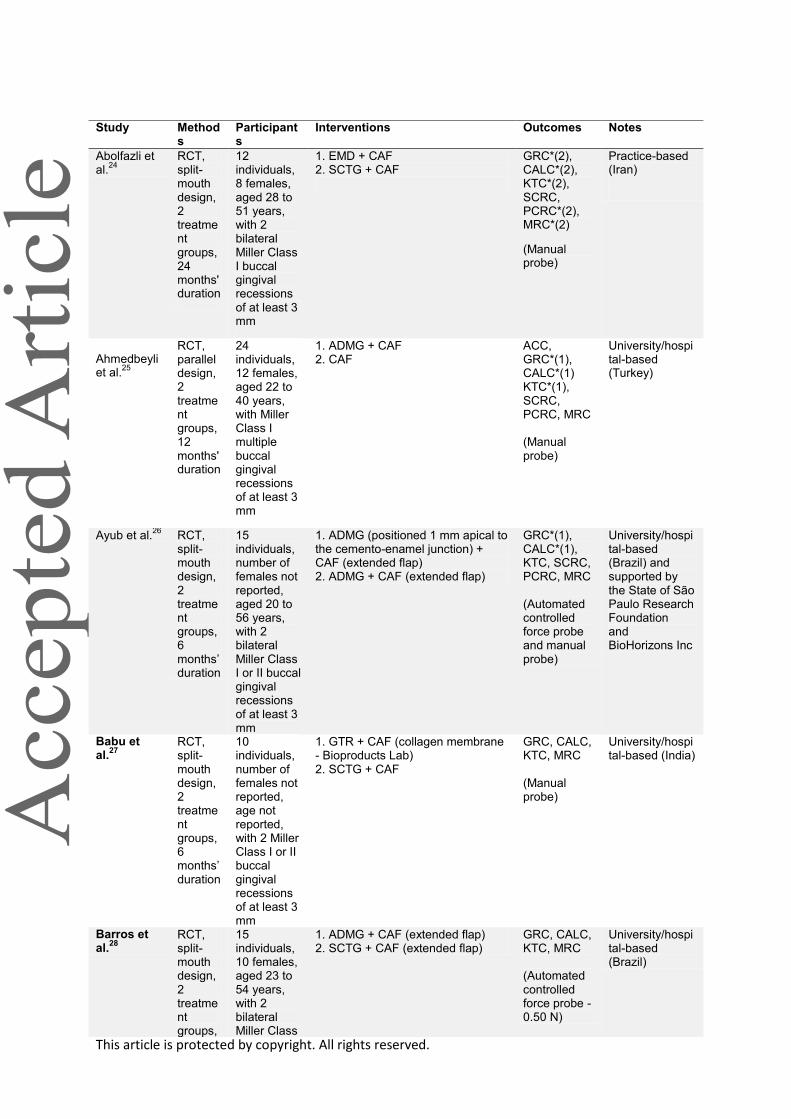
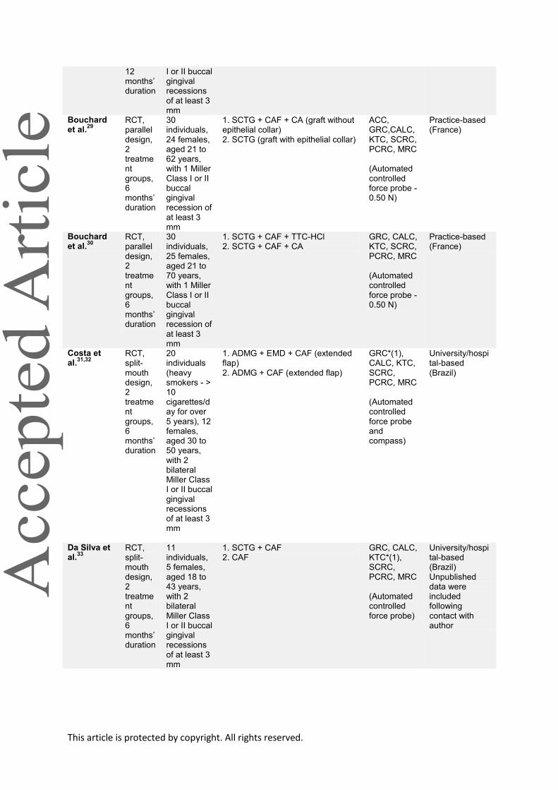
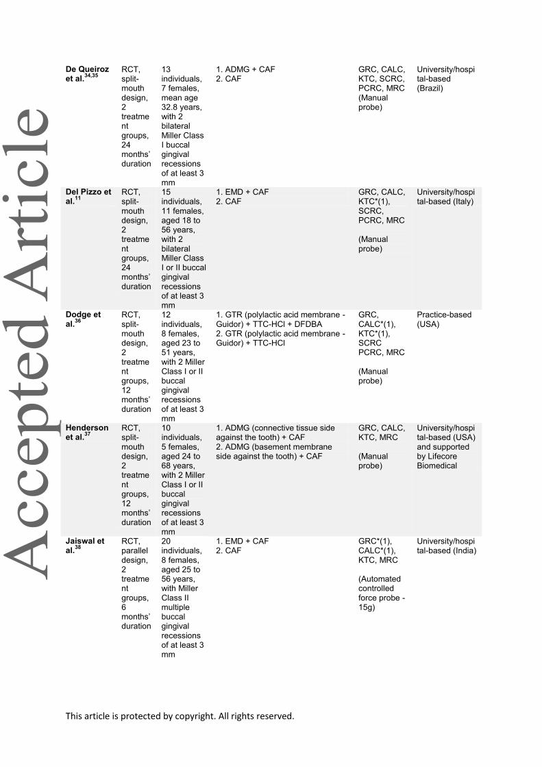
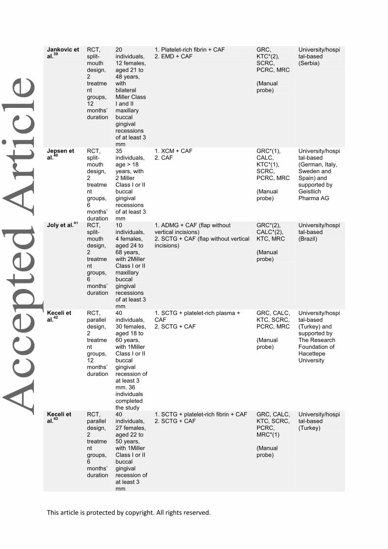
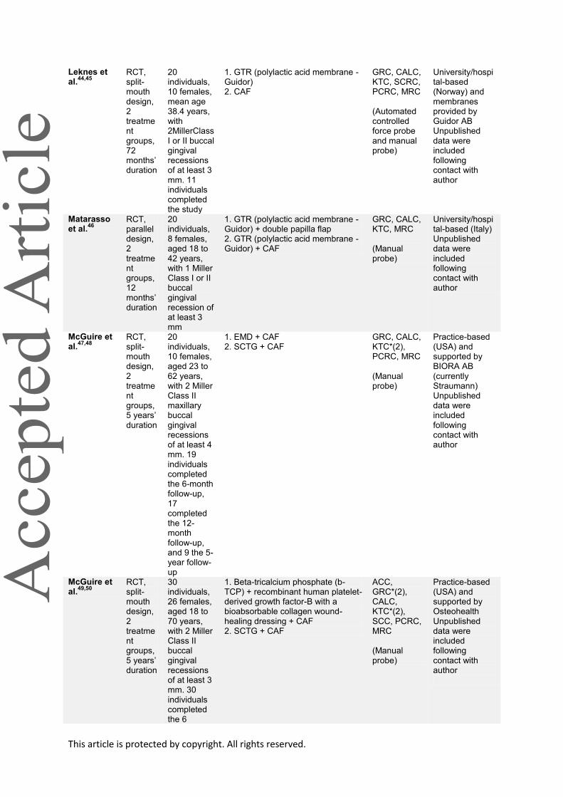
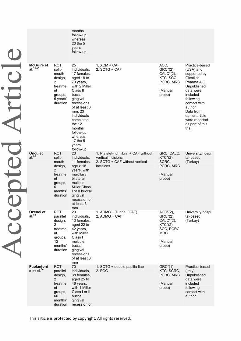
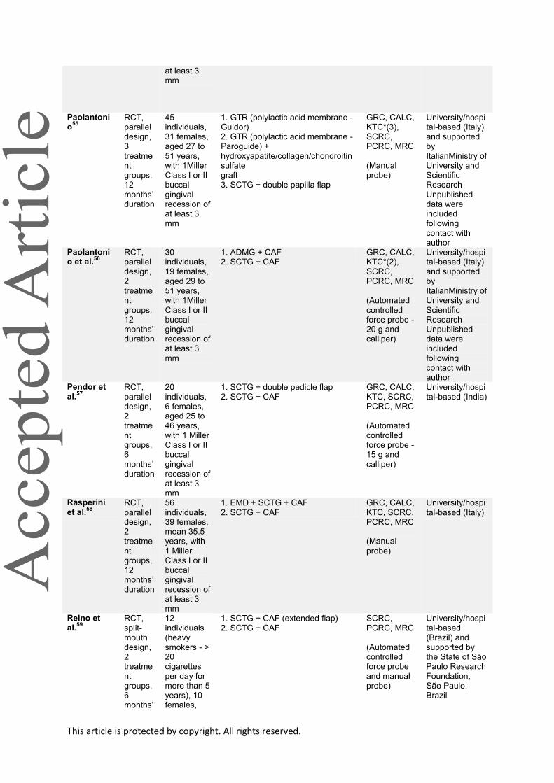
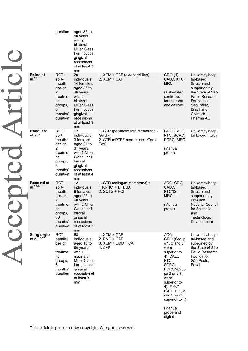
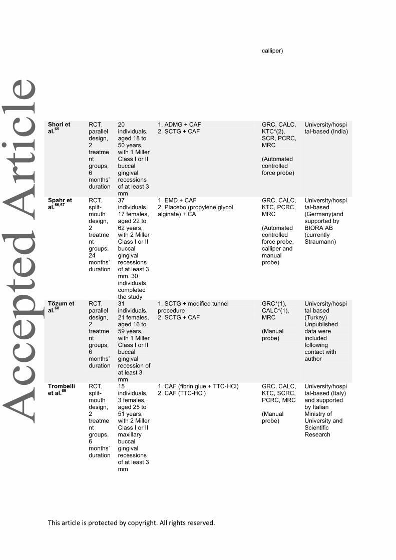
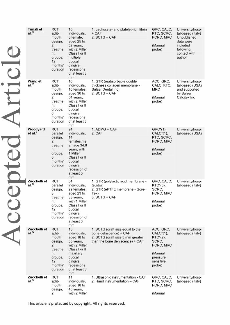
under the name of the clinical outcomes paper.64 Data on the type of study design, location
and country of trial are described in Table 1. Five studies evaluated multiple GR,25,38,52,53,70
whereas the others single defects. Two studies32,59 evaluated exclusively outcomes of
smokers (i.e. 10 or more cigarettes per day for more than 5 years). In addition, the majority
of trials followed participants during a short-term period (6 months to 12 months). Only five
publications with medium-term follow-up11,24,35,61,67 and five with long-term follow-up45,48,50,51,54
were included. In total, 1227 patients were treated and details on the different treatment
modalities are depicted in Table 1.
Risk of bias in included studies
Only one study was considered to be at a low overall risk of bias (Figure 1).64 According to
GRADE methods23 all evidence was considered to be of low to very low quality, mainly for
imprecision and inconsistency (see supplementary Appendix 4 in online Journal of
Periodontology).
Effects of interventions
ACC, GR change, CAL change and KTW change: ACC related to patient‟s opinion was
reported in 10 RCTs25,29,48,50,51,53,61,72,74,75 (Table 2) Given the heterogeneity of
methods/criteria used to assess this outcome and types of procedures compared, formal
pooling of data via meta-analysis was precluded. Of the 48 included trials, 18 evaluating
single GR8-11,24,28,35,36,40,41,48,55,56,61,64,65,67,71 and two multiple GR52,70 were included into 11 sets
This article is protected by copyright. All rights reserved.
of meta-analyses (Table 3). In addition, data from studies not included in meta-analyses are
presented in supplementary Appendix 5 in online Journal.
Single GR: With respect to RD change, there was evidence of greater RD reduction for
EMD + CAF when compared to CAF alone (short/medium term; P = 0.005, MD: 0.32 mm),
for SCTG + CAF when compared to GTR rm + CAF (P = 0.002, MD: -0.37 mm), for GTR rm
+ CAF associated with bone substitutes compared to GTR rm + CAF (P = 0.02, MD: 0.48
mm) and for XCM + CAF compared to CAF alone (P = 0.006, MD: 0.40 mm). Regarding
CAL change, there was evidence of greater reduction of CAL for EMD + CAF when
compared to CAF alone (short/medium-term, P = 0.009, MD: 0.35 mm), and for GTR rm +
CAF compared to SCTG + CAF (P = 0.02, MD: of 0.35 mm). For KTW change, there was
evidence of greater gain in the KTW for EMD + CAF when compared to CAF alone (short-
term, P = 0.001, MD: 0.35 mm; short/medium term, P = 0.0005, MD: 0.40 mm), for SCTG +
CAF when compared to EMD + CAF (P < 0.00001, MD: -1.06 mm), for SCTG + CAF when
compared to GTR rm + CAF (P < 0.0001, mean difference -1.77 mm), for SCTG + CAF
when compared to GTR rm + CAF associated with bone substitutes (P < 0.00001, MD: -2.38
mm), and for XCM + CAF when compared to CAF alone (P = 0.03, MD: 0.44 mm). Multiple
GR: There was evidence of greater reduction of CAL for SCTG + CAF compared to PRF +
CAF (P = 0.02, MD: -0.37 mm).
CRC
CRC was reported in 34 studies (Table 2) Among the included RCTs designed to evaluate
single GR (excluding the data from Costa et al.31,32 and Reino et al.59 who included only
heavy smokers), CRC varied from 0%26 to 91.6%8 for ADMG; 18.1%33 to 95.6%12,51 for
SCTG; 25%24 to 89.5%47,48 for EMD; 7.7 %34,35 to 81.8%73 for CAF; 33.3%36 to 53.3%55 for
GTR rm; and 28%10 to 41.6%9 for GTR nrm. Also, OR analyses of six comparisons did not
find statistical differences between procedures (Table 3). For XCM + CAF versus CAF, the
This article is protected by copyright. All rights reserved.
combined therapy improved the achievement of sites displaying CRC compared to the use
of CAF alone (OR of 4.73, 95% CI 2.35 to 9.50).
MRC
All included trials reported the MRC. Within studies evaluating single GR (excluding the data
from two RCT31,32,59 who included heavy smokers), this outcome varied from 50%41 to 96%8
for ADMG, 64.7%29 to 99.3%12,51 for SCTG, 70.5%39 to 95.1%47,48 for EMD, 55.9%34,35 to
95.4%73 for CAF, 62.5%46 to 73.7%36 for GTR rm, 84.2 %61,62 to 89.9%36 for GTR rm
associated with bone substitutes, and 80.5%10 to 82.4%9 for GTR nrm (Table 2).
Patients’ preference for a specific RC procedure in split-mouth trials
This update did not identify additional data to those already publish by the previous version
of this SR.13,14 Details on this outcome are described in supplementary Appendix 6 in online
Journal of Periodontology.
Occurrence of adverse effects and/or postoperative complications
Occurrence of adverse effects and/or postoperative complications during the postsurgical
period was reported in 15 trials,12,28,36,39,40,42,45,47,49,52,66,71,72,74,75 but restricted to a limited
number of patients/cases (see supplementary Appendix 7 in online Journal). Overall, the
most common adverse outcomes were postsurgical pain/swelling within the first days after
surgery, ADMG graft or membrane exposure and postoperative pain in donor site of SCTG.
DISCUSSION
This article is protected by copyright. All rights reserved.
Summary of main results
The main changes since the last version13,14 are reported in Figure 2. In spite of aesthetics
being considered the primary goal of RC procedures, few studies had evaluated ACC related
to patients‟ opinion.12,24,29,47-51,53,61,62,72,74,75 In these studies, the majority of the patients were
satisfied with the final aesthetic result achieved (Table 2). Also, procedures that make a
reduction in the operatory time possible, that eliminate the need for a second surgical site
and that use smaller palatal grafts72,74 were better accepted by the patients. In terms of RD
reduction, results from meta-analyses demonstrated evidence that at short-term: SCTG +
CAF promoted additional gains to those achieved by GTR rm + CAF; XCM + CAF improved
the gains obtained by CAF alone; EMD + CAF led to better stability of the gingival margin
after treatment than CAF alone; and GTR rm + bone substitutes + CAF provided better
outcomes than GTR rm + CAF (Table 3).
There was a marked variation between procedures in terms of the achievement of CRC at
short-term (Table 2): 0% to 95.6%. OR analyses on CRC did not reveal evidence of
differences between procedures in none of the available comparisons, except for XCM +
CAF versus CAF (i.e. the combined therapy promoted better outcomes). Additionally, some
studies showed a decrease in the number of sites displaying CRC over time.12,33,34,47-51
With respect to secondary outcomes, four comparisons showed evidence that SCTG + CAF
promoted additional gains in the KTW compared to EMD + CAF, GTR rm + CAF, or GTR rm
+ bone substitutes + CAF. Similarly, the use of EMD + CAF or XCM + CAF promoted
additional gains in the KTW compared to the use of CAF alone (Table 3). Regarding CAL
changes, there was evidence that SCTG + CAF promoted additional gains to those achieved
by platelet-rich fibrin (PRF) + CAF, and that GTR rm + CAF promoted additional gains
compared to SCTG + CAF. Also, there was a markedly variation in the amount of RC
This article is protected by copyright. All rights reserved.
achieved. MRC varied from 44% to 99.3% (Table 2). Furthermore, data from some medium-
and long-term trials12,33,34,47-51 showed that MRC decreased over time.
Patients‟ preference for a specific RC procedure followed the same pattern as ACC.9,71,72
Occurrence of an early discomfort with or without pain was related to donor sites of
SCTG.47,48,52,71,72 This aspect may be related to the size of the graft obtained from the palate
and the surgical approach used.72 Moreover, „bigger grafts‟ were more associated to
shrinkage of the covering flap with graft exposure when compared to „small grafts‟.72,74 In
terms of flap preparation, the removal of the labial submucosal tissue, in the area of lower
incisors, led to a reduction in the number of sites experiencing covering flap shrinkage than
sites where the submucosal tissue was not removed.75
Although 48 RCTs were included in this Cochrane SR, it was difficult to combine data from
these trials due to a great variability of comparisons between the various RC procedures and
the inexistence of a unique gold standard control group in all studies. Consequently, only 20
trials were incorporated into meta-analyses8-12,24,28,34-36,40,41,47,48,52,55,56,61-67,70,71 in 11 different
group comparisons (Table 3). Few studies reported a follow-up period superior to 12
months.12,24,34,35,44,45,47-51,54,61,62,66,67 In six of these studies a chronological evaluation of the
results evidenced loss in the amount of RC obtained (e.g. MRC and CRC) between the 6
months to 12 months period of evaluation11,34,35,66,67 and between the first year and 5-and
10-year follow-ups.12,47-51 This assumption was evidenced by the findings of pooled
estimates on EMD + CAF versus CAF (Table 3). Two trials31,32,59 evidenced the detrimental
impact of smoking on root coverage outcomes (i.e. MRC and CRC decrease) within patients
who smoke ≥ 10 cigarettes per day for more than 5 years.
Overall, both the individual studies‟ outcomes (i.e. within-group comparisons reported by
each individual trial) and findings of pooled estimates clearly demonstrated that all RC
procedures included in this Cochrane Review promoted reduction in the extent of GR and
This article is protected by copyright. All rights reserved.
concomitant gain in the CAL for both single and multiple GR. Likewise, it was evidenced that
KTW augmentation of these sites was associated to the use of SCTG or allogenous
(ADMG)/xenogenous (XCM) soft tissue substitutes.
Quality of the evidence
Only one study was considered to be at a low overall risk of bias. GRADE methods23 were
used to assess the quality of the body of evidence of our main comparisons and our
assessment is presented in the supplementary Appendix 4 in online Journal of
Periodontology with all evidence considered to be of low to very low quality, mainly for
imprecision and inconsistency.
Potential biases in the review process
In this review, only defects ≥ 3 mm were included in order to minimize heterogeneity
between the trials. However, this inclusion criterion could have eliminated data from studies
that could be incorporated into meta-analyses.
Agreements and disagreements with other studies or reviews
Important aspects already described in both the previous13,14 and current versions of this
Cochrane SR are depicted in supplementary Appendix 8 in online Journal. The current
version of this SR evidenced that both patients and clinicians seem to agree that, in terms of
aesthetic perception, CRC is perceived as the primary ‟successful outcome‟ of a RC
procedure.76 However, it is important to highlight that patients‟ perception of buccal
recessions is not high (approximately half of the patients with one gingival recession do not
perceive them), as well as that the majority of those defects do not lead to functional or
aesthetic concerns.77
This article is protected by copyright. All rights reserved.
It has been demonstrated by an individual patient data meta-analysis of 602 Miller Class I
and II recession defects4 that the greater the baseline RD, the smaller the chance of CRC. It
should also be noted that the inclusion of studies with recession defects ≥ 4 mm tends to
show greater differences between baseline and follow-up means (i.e. outcome change), a
factor that may influence the calculation of meta-analyses.4,13,14 Another couple of studies78,79
demonstrated that sites in which the gingival margin was sutured at the level of the cemento-
enamel junction the achievement of CRC was inferior to those sites where a trapezoidal flap
was sutured coronal (approximately 1 mm to 2 mm) (i.e. the more apical the gingival margin
after surgery, the smaller the chance of CRC). Moreover, other anatomic aspects related to
the interproximal dental papillae were already described previously13,14 (see supplementary
Appendix 8 in online Journal of Periodontology). Consequently, all these factors make
comparisons and combination of data from different trials a critical issue.
It has been shown that smoking can affect the results obtained by RC procedures.5 Two
RCTs31,32,59 evaluated only patients who smoked ≥ 10 cigarettes per day for at least 5 years,
and their results showed that heavy smokers may be benefited by RC therapy, as well.
However, MRC and CRC were clearly inferior to the outcomes achieved by trials evaluating
non-smokers (Table 2). Eight trials29,30,40,12,51,73-75 reported the inclusion of smokers who
smoked less than 10 cigarettes per day. None of them performed comparisons between
smokers and non-smokers. Zucchelli et al.10 commented only that patients who smoke more
than 10 cigarettes a day presented the worst percentage of RC. This is in line with included
RCTs on smokers31,32,59 and the data from other studies that have assessed the amount of
RC obtained by smokers and non-smokers through CAF and SCTG.5,3,14
The present version of this Cochrane Review is completely in line with data from the recent
American Academy of Periodontology Regeneration Workshop SR5 that concluded that: 1)
“all RC procedures can provide significant reduction in RD and CAL gain without alteration of
This article is protected by copyright. All rights reserved.
probing depth for Miller Class I and II single GR, but multiple GR seems to be benefit as well
despite the reduced quantity of information available;” 2) “SCTG-based procedures provided
the best outcomes for clinical practice because of their superior percentages of MRC and
CRC and the significant increase of KTW when compared with most of the other procedures”
(as reported by the individual studies‟ outcomes, Table 2); 3) “the use of CAF with ADMG,
EMD, and XCM also provided gains, many of them similar to SCTG-based procedures, and
thus these may be considered as adequate substitute treatment approaches”; and 4)
“smoking may decrease the expected results”.5
It is also important to highlight that recent evidence from three long-term non-randomized
studies, that followed patients for at least 20 years, found that GR relapse appears to be
associated to sites lacking an attached KT band of at least 2mm.80-82 Similarly, a recent SR83
evaluating the long-term outcomes of untreated buccal GR (in terms of associated reported
aesthetic and functional alterations and factors influencing the progression/worsening of
dental and periodontal tissue conditions) found that: a) untreated GR in individuals with good
oral hygiene are highly likely to experience RD increase during long-term follow-up (78% of
the defects displayed clinical worsening); and b) the presence of KTW and/or greater KTW
decrease the chance of RD increase or the development of new recessions. Nonetheless,
individual data from some of the studies included in the present SR suggest that SCTG
promoted better stability of the gingival margin/some degree of creeping attachment over
time, compared to other surgical approaches.12,24,40,51,70
CONCLUSIONS
All the analyzed RC procedures led to RD reduction and CAL gain and thus can be used
in clinical practice. However, there was a great variability in the percentages of CRC and
MRC.
The available evidence base indicates that the most suitable options for RC of single GR,
in terms of clinical outcomes and cost-to-benefit ratio, are: (1) SCTG plus CAF; (2) ADMG
This article is protected by copyright. All rights reserved.
plus CAF; (3) EMD + CAF; (4) XCM + CAF; and (5) CAF alone. Despite of the restricted
number of studies on multiple GR included in this SR, this „hierarchy criterion‟ may be
applied for the treatment of such defects, as well.
GTR could be used to treat single GR, but most the information on these procedures
were obtained from studies published up to the early 2000‟0.
Individual studies‟ outcomes and the available pooled estimates suggest that SCTG plus
CAF may be considered as „gold standard‟ procedure for the treatment of single and
multiple GR. Moreover, evidence suggests that SCTG promoted better stability of the
gingival margin/some degree of creeping attachment over time, compared to other
surgical approaches.
ADMG (primarily) and XCM (secondly) may be considered as alternative soft tissue
grafting materials.
Outcome measures of the evaluated surgical techniques were not improved by the use of
root modification agents or the type of mechanical root scaling during surgery.
The incidence of adverse effects, such as discomfort with or without pain, was mainly
related to donor sites of SCTG. However, these conditions occurred mainly within the first
week after surgery and did not influence on RC outcomes.
Implications for research
• Limited data exist on ACC related to patient‟s opinion, thus further RCTs are still required
to evaluate this primary outcome variable. The use of the VAS (or other „standardized
scales‟) will allow more precise evaluations of patient-based outcomes.
• Future split-mouth trials should focus on patients‟ preference for a specific RC procedure.
• The inclusion of baseline and final individual defect measurements will allow more precise
evaluations, as well as subgroup evaluations (e.g. patients presenting similar defects) and
future comparisons via meta-analyses. These outcome measures should include GR depth
and width, CAL, KTW and thickness, and root surface conditions (i.e. presence of caries,
abrasions or restorations). Also, in order to draw more robust conclusions about treatment of
This article is protected by copyright. All rights reserved.
sites lacking attached gingiva: a) the number of Miller Class I and II should be balanced and
equally distributed in the study groups (i.e., test and control); and b) the differences in
response to treatment between these sites should be considered.
• Comparisons between different operators (i.e. with respect to the degree of operator‟s
experience) remain necessary to evaluate differences in the expected outcome measures.
• Considering the proposed inclusion criteria, no data were available for LPF and there is
limited information for FGG and platelet-rich fibrin. These procedures might be evaluated by
future research.
ACKNOWLEDGEMENTS
The review authors would like to acknowledge Anne Littlewood for her assistance on the
search strategy section and Helen Worthington, Ian Needleman, Luisa Fernandez
Mauleffinch and Marco Esposito from Cochrane Oral Health for their help with the
preparation of the protocol and full text of the review. We would like to thank Professor Kevin
Seymour from Division of Dentistry, School of Medical Sciences, Faculty of Biology,
Medicine and Health, the University of Manchester for providing comments on this update.
REFERENCES
1. Chambrone L, Salinas Ortega MA, Sukekava F, et al. Root coverage procedures for treating localised and multiple recession-type defects. Cochrane Database Syst Rev 2018; 10: CD007161.
2. Chambrone L, Chambrone D, Pustiglioni FE, Chambrone LA, Lima LA. Can subepithelial
connective tissue grafts be considered the gold standard procedure in the treatment of Miller Class I and II recession-type defects?. J Dent 2008;36:659–671.
3. Chambrone L, Faggion CM Jr, Pannuti CM, Chambrone LA. Evidence-based periodontal plastic surgery: an assessment of quality of systematic reviews in the treatment of recession-type defects. J Clin Periodontology 2010;37:1110–1118.
4. Chambrone L, Pannuti CM, Tu YK, Chambrone LA. Evidence-based periodontal plastic surgery. II. An individual data meta-analysis for evaluating factors in achieving complete root coverage. Journal of Periodontology 2012;83:477–490.
This article is protected by copyright. All rights reserved.
5. Chambrone L, Tatakis DN. Periodontal soft tissue root coverage procedures: a systematic review from the AAP Regeneration Workshop. Journal of Periodontology 2015;86 (2 Suppl):S8–51.
6. Buti J, Baccini M, Nieri M, La Marca M, Pini-Prato GP. Bayesian network meta-analysis of root coverage procedures ranking efficacy and identification of best treatment. J Clin Periodontol 2013;40:372–386.
7. Pini Prato G, Nieri M, Pagliaro U, et al. Surgical treatment of single gingival recessions: clinical guidelines. Eur J Oral Implantol 2014;7:9–43.
8. Woodyard JG, Greenwell H, Hill M, Drisko C, Iasella JM, Scheetz J. The clinical effect of acellular dermal matrix on gingival thickness and root coverage compared to coronally positioned flap alone. J Periodontol 2004;75:44–56.
9. Roccuzzo M, Lungo M, Corrente G, Gandolfo S. Comparative study of a bioresorbable and a non-resorbable membrane in the treatment of human buccal gingival recessions. J Periodontol 1996;67:7–14.
10. Zucchelli G, Clauser C, De Sanctis M, Calandriello M. Mucogingival versus guided tissue regeneration procedures in the treatment of deep recession type defects. J Periodontol 1998;69:138–145.
11. Del Pizzo M, Zucchelli G, Modica F, Villa R, Debernardi C. Coronally advanced flap with or without enamel matrix derivative for root coverage: a 2-year study. J Clin Periodontol 2005;32:1181–1187.
12. McGuire MK, Scheyer ET. Xenogeneic collagen matrix with coronally advanced flap compared to connective tissue with coronally advanced flap for the treatment of dehiscence-type recession defects. J Periodontol 2010;81:1108–1117.
13. Chambrone L, Sukekava F, Araújo MG, Pustiglioni FE, Chambrone LA, Lima LA. Root coverage procedures for the treatment of localised recession-type defects. Cochrane Database Syst Rev 2009; 2: CD007161
14. Chambrone L, Sukekava F, Araújo MG, Pustiglioni FE, Chambrone LA, Lima LA. Root-coverage procedures for the treatment of localized recession-type defects: a Cochrane systematic review. J Periodontol 2010;81:452–478.
15. Miller PD Jr. A classification of marginal tissue recession. Int J Periodontics Restorative Dent 1985;5(2):8–13.
16. Lefebvre C, Manheimer E, Glanville J. Chapter 6: Searching for studies. In: Higgins JP, Green S, editor(s). Cochrane Handbook for Systematic Reviews of Interventions Version 5.1.0 (updated March 2011). The Cochrane Collaboration, 2011. Available from http://handbook-5-1.cochrane.org/.
This article is protected by copyright. All rights reserved.
17. Higgins JPT, Green S, editor(s). Cochrane Handbook for Systematic Reviews of Interventions Version 5.1.0 (updated March 2011). The Cochrane Collaboration, 2011. Available from http://handbook-5-1.cochrane.org/.
18. Stedman MR, Curtin F, Elbourne DR, Kesselheim AS, Brookhart MA. Meta-analyses involving cross-over trials: methodological issues. International J Epidemiol 2011;40:1732–1734.
19. Curtin F, Elbourne D, Altman DG. Meta-analysis combining parallel and cross-over clinical trials. II: Binary outcomes. Stat Med 2002;21:2145–2159.
20. Follmann D, Elliott P, Suh I, Cutler J. Variance imputation for overviews of clinical trials with continuous response. J Clin Epidemiol 1992;45:769–773.
21. Tatakis DN, Chambrone L, Allen EP, et al. Periodontal soft tissue root coverage procedures: a consensus report from the AAP Regeneration Workshop. J Periodontol 2015;86 (2 Suppl):S52-55.
22. Richardson CR, Allen EP, Chambrone L, et al. Periodontal soft tissue root coverage procedures: practical applications from the AAP Regeneration Workshop. Clin Adv Periodontics 2015;5:2–10.
23. Atkins D, Best D, Briss PA, et al; GRADE Working Group. Grading quality of evidence and strength of recommendations. BMJ 2004;328:1490–1494.
24. Abolfazli N, Saleh-Saber F, Eskandari A, Lafzi A. A comparative study of the long term results of root coverage with connective tissue graft or enamel matrix protein: 24-month results. Med Oral, Patol Oral, Cir Bucal 2009;14:E304–309.
25. Ahmedbeyli C, Ipci SD, Cakar G, Kuru BE, Y lmaz S. Clinical evaluation of coronally advanced flap with or without acellular dermal matrix graft on complete defect coverage for the treatment of multiple gingival recessions with thin tissue biotype. J Clin Periodontol 2014;41:303–310.
26. Ayub LG, Ramos UD, Reino DM, et al. Randomized comparative clinical study of two surgical procedures to improve root coverage with the acellular dermal matrix graft. J Clin Periodontol 2012;39:871-878.
27. Babu HM, Gujjari SK, Prasad D, Sehgal PK, Srinivasan A. Comparative evaluation of a bioabsorbable collagen membrane and connective tissue graft in the treatment of localized gingival recession: a clinical study. J Indian Soc Periodontol 2011;15:353–358.
28. Barros RRM, Macedo GO, de Queiroz AC, Novaes Jr AB. A modified surgical flap for root coverage in association with grafting materials. J Esthetic Restorative Dent 2015;27:84–91.
29. Bouchard P, Etienne D, Ouhayoun JP, Nilveus R. Subepithelial connective tissue grafts in the treatment of gingival recessions. A comparative study of 2 procedures. J Periodontol 1994;65:929–936.
This article is protected by copyright. All rights reserved.
30. Bouchard P, Nilveus R, Etienne D. Clinical evaluation of tetracycline HCl conditioning in the treatment of gingival recessions. A comparative study. J Periodontol 1997;68:262–269.
31. Alves LB, Costa PP, de Souza SLS, et al. Acellular dermal matrix graft with or without enamel matrix derivative for root coverage in smokers: a randomised clinical study. J Clin Periodontol 2012;39:393–399.
32. Costa PP, Alves LB, Souza SL, et al. Root coverage in smokers with acellular dermal matrix graft and enamel matrix derivative: a 12-month randomized clinical trial. Int J Periodontics Restorative Dent 2016;36:525–531.
33. da Silva RC, Joly JC, de Lima AF, Tatakis DN. Root coverage using the coronally positioned flap with or without a subepithelial connective tissue graft. J Periodontol 2004;75:413–419.
34. de Queiroz Cortes A, Martins AG, Nociti FH Jr, Sallum AW, Casati MZ, Sallum EA. Coronally positioned flap with or without acellular dermal matrix graft in the treatment of Class I gingival recessions: a randomized controlled clinical study. J Periodontol 2004;75:1137–1144.
35. de Queiroz Cortes A, Sallum AW, Casati MZ, Nociti FH Jr, Sallum EA. A two-year prospective study of coronally positioned flap with or without acellular dermal matrix graft. J Clin Periodontol 2006;33:683–689.
36. Dodge JR, Greenwell H, Drisko C, Wittwer JW, Yancey J, Rebitski G. Improved bone regeneration and root coverage using a resorbable membrane with physically assisted cell migration and DFDBA. Int J Periodontics Restorative Dent 2000;20:398–411.
37. Henderson RD, Greenwell H, Drisko C, et al. Predictable multiple site root coverage using an acellular dermal matrix allograft. J Periodontol 2001;72:571–582.
38. Jaiswal GR, Kumar R, Khatri PM, Jaiswal SG, Bhongade ML. The effectiveness of enamel matrix protein (Emdogain) in combination with coronally advanced flap in the treatment of multiple marginal tissue recession: a clinical study. J Indian Soc Periodontol 2012; 16:224–230.
39. Jankovic S, Aleksic Z, Milinkovic I, Dimitrijevic B. The coronally advanced flap in combination with platelet rich fibrin (PRF) and enamel matrix derivative in the treatment of gingival recession: a comparative study. Eur J Esthet Dent 2010;5:260–273.
40. Jepsen K, Jepsen S, Zucchelli G, et al. Treatment of gingival recession defects with a coronally advanced flap and a xenogeneic collagen matrix: a multicenter randomized clinical trial. J Clin Periodontol 2013;40:82–89.
41. Joly JC, Carvalho AM, da Silva RC, Ciotti DL, Cury PR. Root coverage in isolated gingival recessions using autograft versus allograft: a pilot study. J Periodontol 2007;78:1017–1022.
42. Keceli HG, Sengun D, Berberoglu A, Karabulut E. Use of platelet gel with connective tissue grafts for root coverage: a randomized-controlled trial. J Clin Periodontol 2008;35:255–262.
This article is protected by copyright. All rights reserved.
43. Keceli HG, Kamak G, Erdemir EO, Evginer MS, Dolgun A. The adjunctive effect of platelet-rich fibrin to connective tissue graft in the treatment of buccal recession defects: results of a randomized, parallel-group controlled trial. J Periodontol 2015;86:1221–1230.
44. Amarante ES, Leknes KN, Skavland J, Lie T. Coronally positioned flap procedures with or without a bioabsorbable membrane in the treatment of human gingival recession. J Periodontol 2000;71:989–998.
45. Leknes KN, Amarante ES, Price DE, Boe OE, Skavland RJ, Lie T. Coronally positioned flap procedures with or without a biodegradable membrane in the treatment of human gingival recession. A 6-year follow-up study. J Clin Periodontol 2005;32:518–529.
46. Matarasso S, Cafiero C, Coraggio F, Vaia E, de Paoli S. Guided tissue regeneration versus coronally repositioned flap in the treatment of recession with double papillae. Int J Periodontics Restorative Dent 1998;18:444–453.
47. McGuire MK, Nunn M. Evaluation of human recession defects treated with coronally advanced flaps and either enamel matrix derivative or connective tissue. Part 1: comparison of clinical parameters. J Periodontol 2003;74:1110–1125.
48. McGuire MK, Scheyer ET, NunnM. Evaluation of human recession defects treated with coronally advanced flaps and either enamel matrix derivative or connective tissue: comparison of clinical parameters at 10 years. J Periodontol 2012;83:1353–1362.
49. McGuire MK, Scheyer ET, Schupbach P. Growth factor mediated treatment of recession defects: a randomized controlled trial and histologic and microcomputed tomography examination. J Periodontol 2009; 80:550–564.
50. McGuire MK, Scheyer ET, Snyder MB. Evaluation of recession defects treated with coronally advanced flaps and either recombinant human platelet-derived growth factor-BB plus b-tricalcium phosphate or connective tissue: comparison of clinical parameters at 5 years. J Periodontol 2014;85:1361–1370.
51. McGuire MK, Scheyer ET. Long-term results comparing xenogeneic collagen matrix and autogenous connective tissue grafts with coronally advanced flaps for treatment of dehiscence-type recession defects. J Periodontol 2016;87:221–227.
52. Öncü E. The use of platelet-rich fibrin versus subepithelial connective tissue graft in treatment of multiple gingival recessions: a randomized clinical trial. Int J Periodontics Restorative Dent 2017;37:265–271.
53. Ozenci I, Ipci SD, Cakar G, Yilmaz S. Tunnel technique versus coronally advanced flap with acellular dermal matrix graft in the treatment of multiple gingival recessions. J Clin Periodontol 2015;42:1135-1142.
54. Paolantonio M, di Murro C, Cattabriga A, Cattabriga M. Subpedicle connective tissue graft versus free gingival graft in the coverage of exposed root surfaces. A 5-year clinical study. J Clin Periodontol 1997;24:51–56.
This article is protected by copyright. All rights reserved.
55. Paolantonio M. Treatment of gingival recessions by combined periodontal regenerative technique, guided tissue regeneration, and subpedicle connective tissue graft. A comparative clinical study. J Periodontol 2002;73:53–62.
56. Paolantonio M, Dolci M, Esposito P, et al. Subpedicle acellular dermal matrix graft and autogenous connective tissue graft in the treatment of gingival recessions: a comparative 1-year clinical study. J Periodontol 2002;73:1299–1307.
57. Pendor S, Baliga V, Bhongade ML, Turakia V, Shori T. A comparison between connective tissue grafts combined with either double pedicle grafts or coronally positioned pedicle grafts: a clinical study. J Indian Soc Periodontol 2014;18:326–330.
58. Rasperini G, Roccuzzo M, Francetti L, Acunzo R, Consonni D, Silvestri M. Subepithelial connective tissue graft for treatment of gingival recessions with and without enamel matrix derivative: a multicenter, randomized controlled clinical trial. Int J Periodontics Restorative Dent 2011;31:133–139.
59. Reino DM, Novaes Jr AB, Maia LP, et al. Treatment of gingival recessions in heavy smokers using two surgical techniques: a controlled clinical trial. Braz Dent J 2012;23:59–67.
60. Reino DM, Maia LP, Fernandes PG, et al. A randomized comparative study of two techniques to optimize the root coverage using a porcine collagen matrix. Braz Dent J 2015;26:445–450.
61. Rosetti EP, Marcantonio E Jr, Zuza EP, Marcantonio RAC. Root coverage stability of the subepithelial connective tissue graft and guided tissue regeneration: a 30-month follow-up clinical trial. J Dent 2013;41:114–120.
62. Rosetti EP, Marcantonio RA, Rossa C Jr, Chaves ES, Goissis G, Marcantonio E Jr. Treatment of gingival recession: comparative study between subepithelial connective tissue graft and guided tissue regeneration. J Periodontol 2000;71:1441–1447.
63. Rocha Dos Santos M, Sangiorgio JPM, Neves FLDS, et al. Xenogenous collagen matrix and/or enamel matrix derivative for treatment of localized gingival recessions: a randomized clinical trial. Part II: patient-reported outcomes. J Periodontol 2017;88:1319–1328.
64. Sangiorgio JPM, Neves FLDS, Rocha Dos Santos M, et al. Xenogenous collagen matrix and/or enamel matrix derivative for treatment of localized gingival recessions: a randomized clinical trial. Part I: clinical outcomes. J Periodontol 2017;88:1309–1318.
65. Shori T, Kolte A, Kher V, Dharamthok S, Shrirao T. A comparative evaluation of the effectiveness of subpedicle acellular dermal matrix allograft with subepithelial connective tissue graft in the treatment of isolated marginal tissue recession: a clinical study. J Indian Soc Periodontol 2013;17:78–81.
66. Hagewald S, Spahr A, Rompola E, Haller B, Heijl L, Bernimoulin JP. Comparative study of Emdogain and coronally advanced flap technique in the treatment of human gingival recessions. A prospective controlled clinical study. J Clin Periodontol 2002;29:35–41.
This article is protected by copyright. All rights reserved.
67. Spahr A, Haegewald S, Tsoulfidou F, et al. Coverage of Miller class I and II recession defects using enamel matrix proteins versus coronally advanced flap technique: a 2-year report. J Periodontol 2005;76:1871–1880.
68. Tozum TF, Keceli HG, Guncu GN, Hatipoglu H, Sengun D. Treatment of gingival recession: comparison of two techniques of subepithelial connective tissue graft. J Periodontol 2005;76:1842–1848.
69. Trombelli L, Scabbia A, Wikesjö UM, Calura G. Fibrin glue application in conjunction with tetracycline root conditioning and coronally positioned flap procedure in the treatment of human gingival recession defects. J Clin Periodontol 1996;23:861–867.
70. Tunali M, Ozdemir H, Arabaci T, Pikdoken ML. Clinical evaluation of autologous platelet-rich fibrin in the treatment of multiple adjacent gingival recession defects: a 12-month study. Intl J Periodontics Restorative Dent 2015;35:105–114.
71. Wang HL, Bunyaratavej P, Labadie M, Shyr Y, MacNeil RL. Comparison of 2 clinical techniques for treatment of gingival recession. J Periodontol 2001;72:1301–1311.
72. Zucchelli G, Amore C, Sforzal NM, Montebugnoli L, De Sanctis M. Bilaminar techniques for the treatment of recession-type defects. A comparative clinical study. J Clin Periodontol 2003;30:862–870.
73. Zucchelli G, Mounssif I, Stefanini M, Mele M, Montebugnoli L, Sforza NM. Hand and ultrasonic instrumentation in combination with root-coverage surgery: a comparative controlled randomized clinical trial. J Periodontol 2009;80:577–585.
74. Zucchelli G, Mounssif I, Mazzotti C, et al. Does the dimension of the graft influence patient morbidity and root coverage outcomes? A randomized controlled clinical trial. J Clin Periodontol 2014;41:708-716.
75. Zucchelli G, Marzadori M, Mounssif I, Mazzotti C, Stefanini M. Coronally advanced flap + connective tissue graft techniques for the treatment of deep gingival recession in the lower incisors. A controlled randomized clinical trial. J Clin Periodontol 2014;41:806-813.
76. Rotundo R, Nieri M, Mori M, Clauser C, Pini Prato G. Aesthetic perception after root coverage
procedure. J Clin Periodontol 2008;35:705–712.
77. Nieri M, Pini Prato GP, Giani M, Magnani N, Pagliaro U, Rotundo R. Patient perceptions of buccal gingival recessions and requests for treatment. J Clin Periodontol 2013;40:707–712.
78. Nieri M, Rotundo R, Franceschi D, Cairo F, Cortellini P, Pini Prato G. Factors affecting the outcome of the coronally advanced flap procedure: a Bayesian network analysis. J Periodontol 2009;80:405–410.
79. Pini Prato GP, Baldi C, Nieri M, et al. Coronally advanced flap: the post-surgical position of the gingival margin is an important factor for achieving complete root coverage. J Periodontol 2005;76:713–722.
This article is protected by copyright. All rights reserved.
80. Agudio G, Chambrone L, Pini Prato G. Biologic remodeling of periodontal dimensions of areas treated with gingival augmentation procedure: a 25-year follow-up observation. J Periodontol 2017;88:634–642.
81. Pini Prato G, Magnani C, Chambrone L. Long-term evaluation (20 years) of the outcomes of coronally advanced flap in the treatment of single recession-type defects. J Periodontol 2018; 89:265-274.
82. Pini Prato GP, Franceschi D, Cortellini P, Chambrone L. Long-term evaluation (20 years) of the outcomes of subepithelial connective tissue graft plus coronally advanced flap in the treatment of maxillary single recession-type defects. J Periodontol 2018;89:1290-1299.
83. Chambrone L, Tatakis DN. Long-term outcomes of untreated buccal gingival recessions: a systematic review and meta-analysis. J Periodontol 2016;87:796–808.
Figure legends.
Figure 1 - Risk of bias summary: review authors' judgements about each risk of bias item for each included study.
Figure 2 - „What‟s new‟ table - changes since the last version.
Table 1 - Characteristics of included studies
This article is protected by copyright. All rights reserved.
Study Methods
Participants
Interventions Outcomes Notes
Abolfazli et al.
24 RCT, split-mouth design, 2 treatment groups, 24 months' duration
12 individuals, 8 females, aged 28 to 51 years, with 2 bilateral Miller Class I buccal gingival recessions of at least 3 mm
1. EMD + CAF 2. SCTG + CAF
GRC*(2), CALC*(2), KTC*(2), SCRC, PCRC*(2), MRC*(2)
(Manual probe)
Practice-based (Iran)
Ahmedbeyli et al.
25
RCT, parallel design, 2 treatment groups, 12 months' duration
24 individuals, 12 females, aged 22 to 40 years, with Miller Class I multiple buccal gingival recessions of at least 3 mm
1. ADMG + CAF 2. CAF
ACC, GRC*(1), CALC*(1) KTC*(1), SCRC, PCRC, MRC (Manual probe)
University/hospital-based (Turkey)
Ayub et al.26
RCT, split-mouth design, 2 treatment groups, 6 months‟ duration
15 individuals, number of females not reported, aged 20 to 56 years, with 2 bilateral Miller Class I or II buccal gingival recessions of at least 3 mm
1. ADMG (positioned 1 mm apical to the cemento-enamel junction) + CAF (extended flap) 2. ADMG + CAF (extended flap)
GRC*(1), CALC*(1), KTC, SCRC, PCRC, MRC (Automated controlled force probe and manual probe)
University/hospital-based (Brazil) and supported by the State of São Paulo Research Foundation and BioHorizons Inc
Babu et al.
27 RCT, split-mouth design, 2 treatment groups, 6 months‟ duration
10 individuals, number of females not reported, age not reported, with 2 Miller Class I or II buccal gingival recessions of at least 3 mm
1. GTR + CAF (collagen membrane - Bioproducts Lab) 2. SCTG + CAF
GRC, CALC, KTC, MRC (Manual probe)
University/hospital-based (India)
Barros et al.
28 RCT, split-mouth design, 2 treatment groups,
15 individuals, 10 females, aged 23 to 54 years, with 2 bilateral Miller Class
1. ADMG + CAF (extended flap) 2. SCTG + CAF (extended flap)
GRC, CALC, KTC, MRC (Automated controlled force probe - 0.50 N)
University/hospital-based (Brazil)
This article is protected by copyright. All rights reserved.
12 months‟ duration
I or II buccal gingival recessions of at least 3 mm
Bouchard et al.
29 RCT, parallel design, 2 treatment groups, 6 months‟ duration
30 individuals, 24 females, aged 21 to 62 years, with 1 Miller Class I or II buccal gingival recession of at least 3 mm
1. SCTG + CAF + CA (graft without epithelial collar) 2. SCTG (graft with epithelial collar)
ACC, GRC,CALC, KTC, SCRC, PCRC, MRC (Automated controlled force probe - 0.50 N)
Practice-based (France)
Bouchard et al.
30 RCT, parallel design, 2 treatment groups, 6 months‟ duration
30 individuals, 25 females, aged 21 to 70 years, with 1 Miller Class I or II buccal gingival recession of at least 3 mm
1. SCTG + CAF + TTC-HCl 2. SCTG + CAF + CA
GRC, CALC, KTC, SCRC, PCRC, MRC (Automated controlled force probe - 0.50 N)
Practice-based (France)
Costa et al.
31,32 RCT, split-mouth design, 2 treatment groups, 6 months‟ duration
20 individuals (heavy smokers - > 10 cigarettes/day for over 5 years), 12 females, aged 30 to 50 years, with 2 bilateral Miller Class I or II buccal gingival recessions of at least 3 mm
1. ADMG + EMD + CAF (extended flap) 2. ADMG + CAF (extended flap)
GRC*(1), CALC, KTC, SCRC, PCRC, MRC (Automated controlled force probe and compass)
University/hospital-based (Brazil)
Da Silva et al.
33 RCT, split-mouth design, 2 treatment groups, 6 months‟ duration
11 individuals, 5 females, aged 18 to 43 years, with 2 bilateral Miller Class I or II buccal gingival recessions of at least 3 mm
1. SCTG + CAF 2. CAF
GRC, CALC, KTC*(1), SCRC, PCRC, MRC (Automated controlled force probe)
University/hospital-based (Brazil) Unpublished data were included following contact with author
This article is protected by copyright. All rights reserved.
De Queiroz et al.
34,35 RCT, split-mouth design, 2 treatment groups, 24 months‟ duration
13 individuals, 7 females, mean age 32.8 years, with 2 bilateral Miller Class I buccal gingival recessions of at least 3 mm
1. ADMG + CAF 2. CAF
GRC, CALC, KTC, SCRC, PCRC, MRC (Manual probe)
University/hospital-based (Brazil)
Del Pizzo et al.
11 RCT, split-mouth design, 2 treatment groups, 24 months‟ duration
15 individuals, 11 females, aged 18 to 56 years, with 2 bilateral Miller Class I or II buccal gingival recessions of at least 3 mm
1. EMD + CAF 2. CAF
GRC, CALC, KTC*(1), SCRC, PCRC, MRC (Manual probe)
University/hospital-based (Italy)
Dodge et al.
36 RCT, split-mouth design, 2 treatment groups, 12 months‟ duration
12 individuals, 8 females, aged 23 to 51 years, with 2 Miller Class I or II buccal gingival recessions of at least 3 mm
1. GTR (polylactic acid membrane - Guidor) + TTC-HCl + DFDBA 2. GTR (polylactic acid membrane - Guidor) + TTC-HCl
GRC, CALC*(1), KTC*(1), SCRC PCRC, MRC (Manual probe)
Practice-based (USA)
Henderson et al.
37 RCT, split-mouth design, 2 treatment groups, 12 months‟ duration
10 individuals, 5 females, aged 24 to 68 years, with 2 Miller Class I or II buccal gingival recessions of at least 3 mm
1. ADMG (connective tissue side against the tooth) + CAF 2. ADMG (basement membrane side against the tooth) + CAF
GRC, CALC, KTC, MRC (Manual probe)
University/hospital-based (USA) and supported by Lifecore Biomedical
Jaiswal et al.
38 RCT, parallel design, 2 treatment groups, 6 months‟ duration
20 individuals, 8 females, aged 25 to 56 years, with Miller Class II multiple buccal gingival recessions of at least 3 mm
1. EMD + CAF 2. CAF
GRC*(1), CALC*(1), KTC, MRC (Automated controlled force probe - 15g)
University/hospital-based (India)
This article is protected by copyright. All rights reserved.
Jankovic et al.
39 RCT, split-mouth design, 2 treatment groups, 12 months‟ duration
20 individuals, 12 females, aged 21 to 48 years, with bilateral Miller Class I and II maxillary buccal gingival recessions of at least 3 mm
1. Platelet-rich fibrin + CAF 2. EMD + CAF
GRC, KTC*(2), SCRC, PCRC, MRC (Manual probe)
University/hospital-based (Serbia)
Jepsen et al.
40 RCT, split-mouth design, 2 treatment groups, 6 months‟ duration
35 individuals, age > 18 years, with 2 Miller Class I or II buccal gingival recessions of at least 3 mm
1. XCM + CAF 2. CAF
GRC*(1), CALC, KTC*(1), SCRC, PCRC, MRC (Manual probe)
University/hospital-based (German, Italy, Sweden and Spain) and supported by Geistlich Pharma AG
Joly et al.41
RCT, split-mouth design, 2 treatment groups, 6 months‟ duration
10 individuals, 4 females, aged 24 to 68 years, with 2Miller Class I or II maxillary buccal gingival recessions of at least 3 mm
1. ADMG + CAF (flap without vertical incisions) 2. SCTG + CAF (flap without vertical incisions)
GRC*(2), CALC*(2), KTC, MRC (Manual probe)
University/hospital-based (Brazil)
Keceli et al.
42 RCT, parallel design, 2 treatment groups, 12 months‟ duration
40 individuals, 30 females, aged 18 to 60 years, with 1Miller Class I or II buccal gingival recession of at least 3 mm. 36 individuals completed the study
1. SCTG + platelet-rich plasma + CAF 2. SCTG + CAF
GRC, CALC, KTC, SCRC, PCRC, MRC (Manual probe)
University/hospital-based (Turkey) and supported by The Research Foundation of Hacettepe University
Keceli et al.
43 RCT, parallel design, 2 treatment groups, 6 months‟ duration
40 individuals, 27 females, aged 22 to 50 years, with 1Miller Class I or II buccal gingival recession of at least 3 mm
1. SCTG + platelet-rich fibrin + CAF 2. SCTG + CAF
GRC, CALC, KTC, SCRC, PCRC, MRC*(1) (Manual probe)
University/hospital-based (Turkey)
This article is protected by copyright. All rights reserved.
Leknes et al.
44,45 RCT, split-mouth design, 2 treatment groups, 72 months‟ duration
20 individuals, 10 females, mean age 38.4 years, with 2MillerClass I or II buccal gingival recessions of at least 3 mm. 11 individuals completed the study
1. GTR (polylactic acid membrane - Guidor) 2. CAF
GRC, CALC, KTC, SCRC, PCRC, MRC (Automated controlled force probe and manual probe)
University/hospital-based (Norway) and membranes provided by Guidor AB Unpublished data were included following contact with author
Matarasso et al.
46 RCT, parallel design, 2 treatment groups, 12 months‟ duration
20 individuals, 8 females, aged 18 to 42 years, with 1 Miller Class I or II buccal gingival recession of at least 3 mm
1. GTR (polylactic acid membrane - Guidor) + double papilla flap 2. GTR (polylactic acid membrane - Guidor) + CAF
GRC, CALC, KTC, MRC (Manual probe)
University/hospital-based (Italy) Unpublished data were included following contact with author
McGuire et al.
47,48 RCT, split-mouth design, 2 treatment groups, 5 years‟ duration
20 individuals, 10 females, aged 23 to 62 years, with 2 Miller Class II maxillary buccal gingival recessions of at least 4 mm. 19 individuals completed the 6-month follow-up, 17 completed the 12-month follow-up, and 9 the 5-year follow-up
1. EMD + CAF 2. SCTG + CAF
GRC, CALC, KTC*(2), PCRC, MRC (Manual probe)
Practice-based (USA) and supported by BIORA AB (currently Straumann) Unpublished data were included following contact with author
McGuire et al.
49,50 RCT, split-mouth design, 2 treatment groups, 5 years‟ duration
30 individuals, 26 females, aged 18 to 70 years, with 2 Miller Class II buccal gingival recessions of at least 3 mm. 30 individuals completed the 6
1. Beta-tricalcium phosphate (b-TCP) + recombinant human platelet-derived growth factor-B with a bioabsorbable collagen wound-healing dressing + CAF 2. SCTG + CAF
ACC, GRC*(2), CALC, KTC*(2), SCC, PCRC, MRC (Manual probe)
Practice-based (USA) and supported by Osteohealth Unpublished data were included following contact with author
This article is protected by copyright. All rights reserved.
months follow-up, whereas 20 the 5 years follow-up
McGuire et al.
12,51 RCT, split-mouth design, 2 treatment groups, 5 years‟ duration
25 individuals, 17 females, aged 18 to 70 years, with 2 Miller Class II buccal gingival recessions of at least 3 mm. 23 individuals completed the 12 months follow-up, whereas 17 the 5 years follow-up
1. XCM + CAF 2. SCTG + CAF
ACC, GRC*(2), CALC*(2), KTC, SCC, PCRC, MRC (Manual probe)
Practice-based (USA) and supported by Giestlich Pharma AG Unpublished data were included following contact with author Data from earlier article were reported as part of this trial
Öncü et al.
52 RCT, split-mouth design, 2 treatment groups, 6 months‟ duration
20 individuals, 11 females, age > 18 years, with maxillary bilateral multiple Miller Class I or II buccal gingival recession of at least 3 mm
1. Platelet-rich fibrin + CAF without vertical incisions 2. SCTG + CAF without vertical incisions
GRC, CALC, KTC*(2), SCRC, PCRC, MRC (Manual probe)
University/hospital-based (Turkey)
Ozenci et al.
53 RCT, parallel design, 2 treatment groups, 12 months‟ duration
20 individuals, 13 females, aged 22 to 42 years, with Miller Class I multiple buccal gingival recessions of at least 3 mm
1. ADMG + Tunnel (CAF) 2. ADMG + CAF
ACC*(2), GRC*(2), CALC*(2), KTC*(2), SCC, PCRC, MRC (Manual probe)
University/hospital-based (Turkey)
Paolantonio et al.
54 RCT, parallel design, 2 treatment groups, 60 months‟ duration
70 individuals, 38 females, aged 25 to 48 years, with 1 Miller Class I or II buccal gingival recession of
1. SCTG + double papilla flap 2. FGG
GRC*(1), KTC, SCRC, PCRC, MRC (Manual probe)
Practice-based (Italy) Unpublished data were included following contact with author
This article is protected by copyright. All rights reserved.
at least 3 mm
Paolantonio
55 RCT, parallel design, 3 treatment groups, 12 months‟ duration
45 individuals, 31 females, aged 27 to 51 years, with 1Miller Class I or II buccal gingival recession of at least 3 mm
1. GTR (polylactic acid membrane - Guidor) 2. GTR (polylactic acid membrane - Paroguide) + hydroxyapatite/collagen/chondroitinsulfate graft 3. SCTG + double papilla flap
GRC, CALC, KTC*(3), SCRC, PCRC, MRC (Manual probe)
University/hospital-based (Italy) and supported by ItalianMinistry of University and Scientific Research Unpublished data were included following contact with author
Paolantonio et al.
56 RCT, parallel design, 2 treatment groups, 12 months‟ duration
30 individuals, 19 females, aged 29 to 51 years, with 1Miller Class I or II buccal gingival recession of at least 3 mm
1. ADMG + CAF 2. SCTG + CAF
GRC, CALC, KTC*(2), SCRC, PCRC, MRC (Automated controlled force probe - 20 g and calliper)
University/hospital-based (Italy) and supported by ItalianMinistry of University and Scientific Research Unpublished data were included following contact with author
Pendor et al.
57 RCT, parallel design, 2 treatment groups, 6 months‟ duration
20 individuals, 6 females, aged 25 to 46 years, with 1 Miller Class I or II buccal gingival recession of at least 3 mm
1. SCTG + double pedicle flap 2. SCTG + CAF
GRC, CALC, KTC, SCRC, PCRC, MRC (Automated controlled force probe - 15 g and calliper)
University/hospital-based (India)
Rasperini et al.
58 RCT, parallel design, 2 treatment groups, 12 months‟ duration
56 individuals, 39 females, mean 35.5 years, with 1 Miller Class I or II buccal gingival recession of at least 3 mm
1. EMD + SCTG + CAF 2. SCTG + CAF
GRC, CALC, KTC, SCRC, PCRC, MRC (Manual probe)
University/hospital-based (Italy)
Reino et al.
59 RCT, split-mouth design, 2 treatment groups, 6 months‟
12 individuals (heavy smokers - > 20 cigarettes per day for more than 5 years), 10 females,
1. SCTG + CAF (extended flap) 2. SCTG + CAF
SCRC, PCRC, MRC (Automated controlled force probe and manual probe)
University/hospital-based (Brazil) and supported by the State of São Paulo Research Foundation, São Paulo, Brazil
This article is protected by copyright. All rights reserved.
duration aged 35 to 50 years, with 2 bilateral Miller Class I or II buccal gingival recessions of at least 3 mm
Reino et al.
60 RCT, split-mouth design, 2 treatment groups, 6 months‟ duration
20 individuals, 14 females, aged 26 to 46 years, with 2 bilateral Miller Class I or II buccal gingival recessions of at least 3 mm
1. XCM + CAF (extended flap) 2. XCM + CAF
GRC*(1), CALC, KTC, MRC (Automated controlled force probe and calliper)
University/hospital-based (Brazil) and supported by the State of São Paulo Research Foundation, São Paulo, Brazil and Geistlich Pharma AG
Roccuzzo et al.
9 RCT, split-mouth design, 2 treatment groups, 6 months‟ duration
12 individuals, 3 females, aged 21 to 31 years, with 2 Miller Class I or II buccal gingival recessions of at least 4 mm
1. GTR (polylactic acid membrane - Guidor) 2. GTR (ePTFE membrane - Gore-Tex)
GRC, CALC, KTC, SCRC, PCRC, MRC (Manual probe)
University/hospital-based (Italy)
Rossetti et al.
61,62 RCT, split-mouth design, 2 treatment groups, 30 months‟ duration
12 individuals, 9 females, aged 25 to 60 years, with 2 Miller Class I or II buccal gingival recessions of at least 3 mm
1. GTR (collagen membrane) + TTC-HCl + DFDBA 2. SCTG + HCl
ACC, GRC, CALC, KTC*(2), MRC (Manual probe)
University/hospital-based (Brazil) and supported by Brazilian National Council for Scientific and Technologic Development
Sangiorgio et al.
62,64 RCT, parallel design, 4 treatment groups, 6 months‟ duration
68 individuals, aged 18 to 60 years, with 1 maxillary Miller Class I or II buccal gingival recession of at least 3 mm
1. XCM + CAF 2. EMD + CAF 3. XCM + EMD + CAF 4. CAF
ACC, GRC*(Groups 1, 2 and 3 were superior to 4), CALC, KTC SCRC, PCRC*(Groups 2 and 3 were superior to 4), MRC* (Groups 1, 2 and 3 were superior to 4) (Manual probe and digital
University/hospital-based and supported by the State of São Paulo Research Foundation, São Paulo, Brazil
This article is protected by copyright. All rights reserved.
calliper)
Shori et al.
65 RCT, parallel design, 2 treatment groups, 6 months‟ duration
20 individuals, aged 18 to 50 years, with 1 Miller Class I or II buccal gingival recessions of at least 3 mm
1. ADMG + CAF 2. SCTG + CAF
GRC, CALC, KTC*(2), SCR, PCRC, MRC (Automated controlled force probe)
University/hospital-based (India)
Spahr et al.
66,67 RCT, split-mouth design, 2 treatment groups, 24 months‟ duration
37 individuals, 17 females, aged 22 to 62 years, with 2 Miller Class I or II buccal gingival recessions of at least 3 mm. 30 individuals completed the study
1. EMD + CAF 2. Placebo (propylene glycol alginate) + CA
GRC, CALC, KTC, PCRC, MRC (Automated controlled force probe, calliper and manual probe)
University/hospital-based (Germany)and supported by BIORA AB (currently Straumann)
Tözum et al.
68 RCT, parallel design, 2 treatment groups, 6 months‟ duration
31 individuals, 21 females, aged 16 to 59 years, with 1 Miller Class I or II buccal gingival recession of at least 3 mm
1. SCTG + modified tunnel procedure 2. SCTG + CAF
GRC*(1), CALC*(1), MRC (Manual probe)
University/hospital-based (Turkey) Unpublished data were included following contact with author
Trombelli et al.
69 RCT, split-mouth design, 2 treatment groups, 6 months‟ duration
15 individuals, 3 females, aged 25 to 51 years, with 2 Miller Class I or II maxillary buccal gingival recessions of at least 3 mm
1. CAF (fibrin glue + TTC-HCl) 2. CAF (TTC-HCl)
GRC, CALC, KTC, SCRC, PCRC, MRC (Manual probe)
University/hospital-based (Italy) and supported by Italian Ministry of University and Scientific Research
This article is protected by copyright. All rights reserved.
Tunali et al.
70 RCT, split-mouth design, 2 treatment groups, 12 months‟ duration
10 individuals, 6 female, aged 25 to 52 years, with 2 Miller Class I or II multiple buccal gingival recessions of at least 3 mm
1. Leukocyte- and platelet-rich fibrin + CAF 2. SCTG + CAF
GRC, CALC, KTC, SCRC, PCRC, MRC (Manual probe)
University/hospital-based (Italy) Unpublished data were included following contact with author
Wang et al.
71 RCT, split-mouth design, 2 treatment groups, 6 months‟ duration
16 individuals, 10 females, aged 30 to 54 years, with 2 Miller Class I or II buccal gingival recessions of at least 3 mm
1. GTR (reabsorbable double thickness collagen membrane - Sulzer Dental Inc) 2. SCTG + CAF
ACC, GRC, CALC, KTC, MRC (Manual probe)
University/hospital-based (USA) and supported by Sulzer Calcitek Inc
Woodyard et al.
8 RCT, parallel design, 2 treatment groups, 6 months‟ duration
24 individuals, 14 females,mean age 34.6 years, with 1 Miller Class I or II buccal gingival recession of at least 3 mm
1. ADMG + CAF 2. CAF
GRC*(1), CALC*(1), KTC, SCRC, PCRC, MRC (Manual probe)
University/hospital-based (USA)
Zucchelli et al.
10 RCT, parallel design, 3 treatment groups, 12 months‟ duration
54 individuals, 29 females, aged 23 to 33 years, with 1 Miller Class I or II buccal gingival recession of at least 3 mm
1. GTR (polylactic acid membrane - Guidor) 2. GTR (ePTFE membrane - Gore-Tex) 3. SCTG + CAF
GRC, CALC, KTC*(3), SCRC, PCRC, MRC (Manual probe)
University/hospital-based (Italy)
Zucchelli et al.
72 RCT, split-mouth design, 2 treatment groups, 12 months‟ duration
15 individuals, aged 18 to 35 years, with 2 Miller Class I or II maxillary buccal gingival recessions of at least 3 mm
1. SCTG (graft size equal to the bone dehiscence) + CAF 2. SCTG (graft size 3 mm greater than the bone dehiscence) + CAF
ACC, GRC, CALC*(1), KTC*(2), SCRC, PCRC, MRC (Manual pressure sensitive probe)
University/hospital-based (Italy)
Zucchelli et al.
73 RCT, split-mouth design, 2
11 individuals, aged 18 to 40 years, with 2 Miller
1. Ultrasonic instrumentation - CAF 2. Hand instrumentation – CAF
GRC, CALC, KTC, SCRC, PCRC, MRC (Manual
University/hospital-based (Italy)
This article is protected by copyright. All rights reserved.
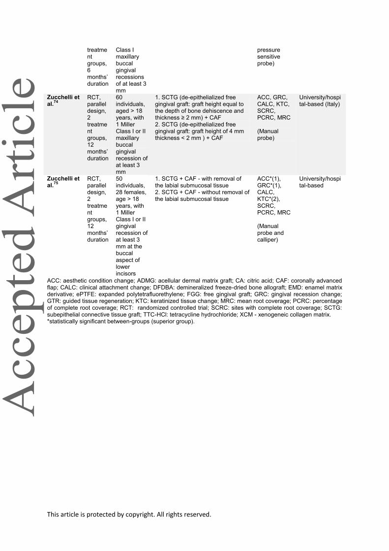
ACC: aesthetic condition change; ADMG: acellular dermal matrix graft; CA: citric acid; CAF: coronally advanced flap; CALC: clinical attachment change; DFDBA: demineralized freeze-dried bone allograft; EMD: enamel matrix derivative; ePTFE: expanded polytetrafluorethylene; FGG: free gingival graft; GRC: gingival recession change; GTR: guided tissue regeneration; KTC: keratinized tissue change; MRC: mean root coverage; PCRC: percentage of complete root coverage; RCT: randomized controlled trial; SCRC: sites with complete root coverage; SCTG: subepithelial connective tissue graft; TTC-HCl: tetracycline hydrochloride; XCM - xenogeneic collagen matrix. *statistically significant between-groups (superior group).
treatment groups, 6 months‟ duration
Class I maxillary buccal gingival recessions of at least 3 mm
pressure sensitive probe)
Zucchelli et al.
74 RCT, parallel design, 2 treatment groups, 12 months‟ duration
60 individuals, aged > 18 years, with 1 Miller Class I or II maxillary buccal gingival recession of at least 3 mm
1. SCTG (de-epithelialized free gingival graft: graft height equal to the depth of bone dehiscence and thickness ≥ 2 mm) + CAF 2. SCTG (de-epithelialized free gingival graft: graft height of 4 mm thickness < 2 mm ) + CAF
ACC, GRC, CALC, KTC, SCRC, PCRC, MRC (Manual probe)
University/hospital-based (Italy)
Zucchelli et al.
75 RCT, parallel design, 2 treatment groups, 12 months‟ duration
50 individuals, 28 females, age > 18 years, with 1 Miller Class I or II gingival recession of at least 3 mm at the buccal aspect of lower incisors
1. SCTG + CAF - with removal of the labial submucosal tissue 2. SCTG + CAF - without removal of the labial submucosal tissue
ACC*(1), GRC*(1), CALC, KTC*(2), SCRC, PCRC, MRC (Manual probe and calliper)
University/hospital-based
This article is protected by copyright. All rights reserved.
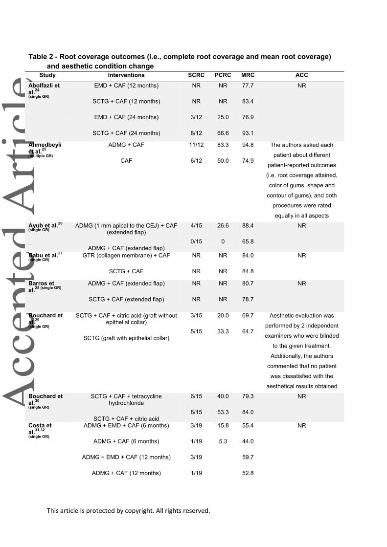
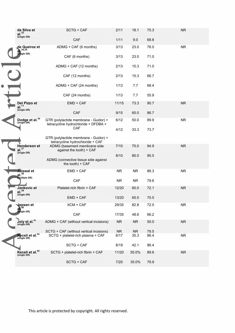
Table 2 - Root coverage outcomes (i.e., complete root coverage and mean root coverage)
and aesthetic condition change
Study Interventions SCRC PCRC MRC ACC
Abolfazli et al.
24
(single GR)
EMD + CAF (12 months)
SCTG + CAF (12 months)
EMD + CAF (24 months)
SCTG + CAF (24 months)
NR
NR
3/12
8/12
NR
NR
25.0
66.6
77.7
83.4
76.9
93.1
NR
Ahmedbeyli et al.
25
(multiple GR)
ADMG + CAF
CAF
11/12
6/12
83.3
50.0
94.8
74.9
The authors asked each
patient about different
patient-reported outcomes
(i.e. root coverage attained,
color of gums, shape and
contour of gums), and both
procedures were rated
equally in all aspects
Ayub et al.26
(single GR)
ADMG (1 mm apical to the CEJ) + CAF (extended flap)
ADMG + CAF (extended flap)
4/15
0/15
26.6
0
88.4
65.8
NR
Babu et al.27
(single GR) GTR (collagen membrane) + CAF
SCTG + CAF
NR
NR
NR
NR
84.0
84.8
NR
Barros et al.
28 (single GR) ADMG + CAF (extended flap)
SCTG + CAF (extended flap)
NR
NR
NR
NR
80.7
78.7
NR
Bouchard et al.
29
(single GR)
SCTG + CAF + citric acid (graft without epithelial collar)
SCTG (graft with epithelial collar)
3/15
5/15
20.0
33.3
69.7
64.7
Aesthetic evaluation was
performed by 2 independent
examiners who were blinded
to the given treatment.
Additionally, the authors
commented that no patient
was dissatisfied with the
aesthetical results obtained
Bouchard et al.
30
(single GR)
SCTG + CAF + tetracycline hydrochloride
SCTG + CAF + citric acid
6/15
8/15
40.0
53.3
79.3
84.0
NR
Costa et al.
31,32
(single GR)
ADMG + EMD + CAF (6 months)
ADMG + CAF (6 months)
ADMG + EMD + CAF (12 months)
ADMG + CAF (12 months)
3/19
1/19
3/19
1/19
15.8
5.3
55.4
44.0
59.7
52.8
NR
This article is protected by copyright. All rights reserved.
da Silva et al.
33
(single GR)
SCTG + CAF
CAF
2/11
1/11
18.1
9.0
75.3
68.8
NR
de Queiroz et al.
34,35
(single GR)
ADMG + CAF (6 months)
CAF (6 months)
ADMG + CAF (12 months)
CAF (12 months)
ADMG + CAF (24 months)
CAF (24 months)
3/13
3/13
2/13
2/13
1/13
1/13
23.0
23.0
15.3
15.3
7.7
7.7
76.0
71.0
71.0
66.7
68.4
55.9
NR
Del Pizzo et al.
11
(single GR)
EMD + CAF
CAF
11/15
9/15
73.3
60.0
90.7
86.7
NR
Dodge et al.36
(single GR) GTR (polylactide membrane - Guidor) + tetracycline hydrochloride + DFDBA +
CAF
GTR (polylactide membrane - Guidor) + tetracycline hydrochloride + CAF
6/12
4/12
50.0
33.3
89.9
73.7
NR
Henderson et al.
37
(single GR)
ADMG (basement membrane side against the tooth) + CAF
ADMG (connective tissue side against the tooth) + CAF
7/10
8/10
70.0
80.0
94.9
95.5
NR
Jaiswal et al.
38
(multiple GR)
EMD + CAF
CAF
NR
NR
NR
NR
86.3
79.6
NR
Jankovic et al.
39
(single GR)
Platelet-rich fibrin + CAF
EMD + CAF
12/20
13/20
60.0
65.0
72.1
70.5
NR
Jepsen et al.
40
(single GR)
XCM + CAF
CAF
29/35
17/35
82.8
48.6
72.0
66.2
NR
Joly et al.41
(single GR) ADMG + CAF (without vertical incisions)
SCTG + CAF (without vertical incisions)
NR
NR
NR
NR
50.0
79.5
NR
Keceli et al.42
(single GR) SCTG + platelet-rich plasma + CAF
SCTG + CAF
6/17
8/19
35.3
42.1
86.4
86.4
NR
Keceli et al.43
(single GR) SCTG + platelet-rich fibrin + CAF
SCTG + CAF
11/20
7/20
55.0%
35.0%
89.6
79.9
NR
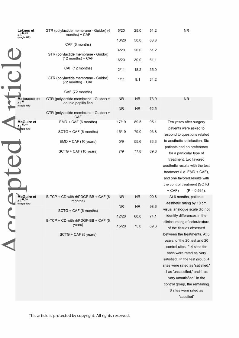
This article is protected by copyright. All rights reserved.
Leknes et al.
44,45
(single GR)
GTR (polylactide membrane - Guidor) (6 months) + CAF
CAF (6 months)
GTR (polylactide membrane - Guidor) (12 months) + CAF
CAF (12 months)
GTR (polylactide membrane - Guidor) (72 months) + CAF
CAF (72 months)
5/20
10/20
4/20
6/20
2/11
1/11
25.0
50.0
20.0
30.0
18.2
9.1
51.2
63.8
51.2
61.1
35.0
34.2
NR
Matarasso et al.
46
(single GR)
GTR (polylactide membrane - Guidor) + double papilla flap
GTR (polylactide membrane - Guidor) + CAF
NR
NR
NR
NR
73.9
62.5
NR
McGuire et al.
47,48
(single GR)
EMD + CAF (6 months)
SCTG + CAF (6 months)
EMD + CAF (10 years)
SCTG + CAF (10 years)
17/19
15/19
5/9
7/9
89.5
79.0
55.6
77.8
95.1
93.8
83.3
89.8
Ten years after surgery
patients were asked to
respond to questions related
to aesthetic satisfaction. Six
patients had no preference
for a particular type of
treatment, two favored
aesthetic results with the test
treatment (i.e. EMD + CAF),
and one favored results with
the control treatment (SCTG
+ CAF) (P = 0.564).
McGuire et al.
49,50
(single GR)
B-TCP + CD with rhPDGF-BB + CAF (6 months)
SCTG + CAF (6 months)
B-TCP + CD with rhPDGF-BB + CAF (5 years)
SCTG + CAF (5 years)
NR
NR
12/20
15/20
NR
NR
60.0
75.0
90.8
98.6
74.1
89.3
At 6 months, patients
aesthetic rating by 10 cm
visual analogue scale did not
identify differences in the
clinical rating of color/texture
of the tissues observed
between the treatments. At 5
years, of the 20 test and 20
control sites, "14 sites for
each were rated as 'very
satisfied.' In the test group, 4
sites were rated as 'satisfied,'
1 as 'unsatisfied,' and 1 as
'very unsatisfied.' In the
control group, the remaining
6 sites were rated as
'satisfied'
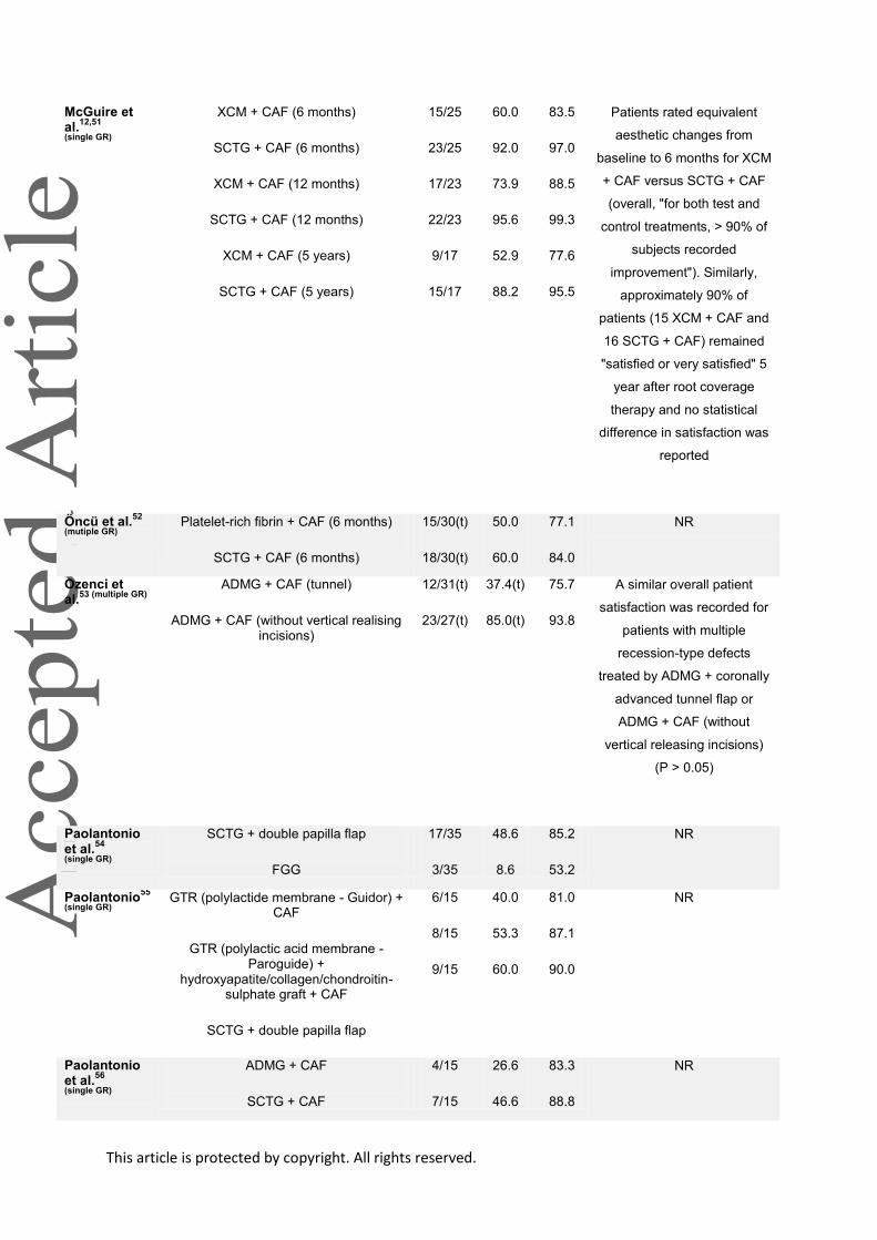
This article is protected by copyright. All rights reserved.
McGuire et al.
12,51
(single GR)
XCM + CAF (6 months)
SCTG + CAF (6 months)
XCM + CAF (12 months)
SCTG + CAF (12 months)
XCM + CAF (5 years)
SCTG + CAF (5 years)
15/25
23/25
17/23
22/23
9/17
15/17
60.0
92.0
73.9
95.6
52.9
88.2
83.5
97.0
88.5
99.3
77.6
95.5
Patients rated equivalent
aesthetic changes from
baseline to 6 months for XCM
+ CAF versus SCTG + CAF
(overall, "for both test and
control treatments, > 90% of
subjects recorded
improvement"). Similarly,
approximately 90% of
patients (15 XCM + CAF and
16 SCTG + CAF) remained
"satisfied or very satisfied" 5
year after root coverage
therapy and no statistical
difference in satisfaction was
reported
Öncü et al.52
(mutiple GR) Platelet-rich fibrin + CAF (6 months)
SCTG + CAF (6 months)
15/30(t)
18/30(t)
50.0
60.0
77.1
84.0
NR
Ozenci et al.
53 (multiple GR) ADMG + CAF (tunnel)
ADMG + CAF (without vertical realising incisions)
12/31(t)
23/27(t)
37.4(t)
85.0(t)
75.7
93.8
A similar overall patient
satisfaction was recorded for
patients with multiple
recession-type defects
treated by ADMG + coronally
advanced tunnel flap or
ADMG + CAF (without
vertical releasing incisions)
(P > 0.05)
Paolantonio et al.
54
(single GR)
SCTG + double papilla flap
FGG
17/35
3/35
48.6
8.6
85.2
53.2
NR
Paolantonio55
(single GR)
GTR (polylactide membrane - Guidor) + CAF
GTR (polylactic acid membrane - Paroguide) +
hydroxyapatite/collagen/chondroitin-sulphate graft + CAF
SCTG + double papilla flap
6/15
8/15
9/15
40.0
53.3
60.0
81.0
87.1
90.0
NR
Paolantonio et al.
56
(single GR)
ADMG + CAF
SCTG + CAF
4/15
7/15
26.6
46.6
83.3
88.8
NR
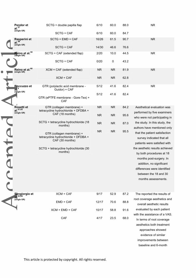
This article is protected by copyright. All rights reserved.
Pendor et al.
57
(single GR)
SCTG + double papilla flap
SCTG + CAF
6/10
6/10
60.0
60.0
88.0
84.7
NR
Rasperini et al.
58
(single GR)
SCTG + EMD + CAF
SCTG + CAF
16/26
14/30
61.5
46.6
90.7
76.6
NR
Reino et al.59
(single GR) SCTG + CAF (extended flap)
SCTG + CAF
2/20
0/20
10.0
0
44.5
43.2
NR
Reino et al.60
(single GR) XCM + CAF (extended flap)
XCM + CAF
NR
NR
NR
NR
81.9
62.8
NR
Roccuzzo et al.
9
(single GR)
GTR (polylactic acid membrane - Guidor) + CAF
GTR (ePTFE membrane - Gore-Tex) + CAF
5/12
5/12
41.6
41.6
82.4
82.4
NR
Rosetti et al.
61,62
(single GR)
GTR (collagen membrane) + tetracycline hydrochloride + DFDBA +
CAF (18 months)
SCTG + tetracycline hydrochloride (18 months)
GTR (collagen membrane) + tetracycline hydrochloride + DFDBA +
CAF (30 months)
SCTG + tetracycline hydrochloride (30 months)
NR
NR
NR
NR
NR
NR
NR
NR
84.2
95.6
87.0
95.5
Aesthetical evaluation was
performed by five examiners
who were not participating in
the study. In this study, the
authors have mentioned only
that the patient satisfaction
survey indicated that all
patients were satisfied with
the aesthetic results achieved
by both procedures at 18
months post-surgery. In
addition, no significant
differences were identified
between the 18 and 30
months assessments.
Sangiorgio et al.
63,64
(single GR)
XCM + CAF
EMD + CAF
XCM + EMD + CAF
CAF
9/17
12/17
10/17
4/17
52.9
70.6
58.8
23.5
87.2
88.8
91.6
68.0
The reported the results of
root coverage aesthetics and
overall aesthetic results
evaluated by each patient
with the assistance of a VAS.
In terms of root coverage
aesthetics both treatment
approaches showed
evidence of similar
improvements between
baseline and 6-month
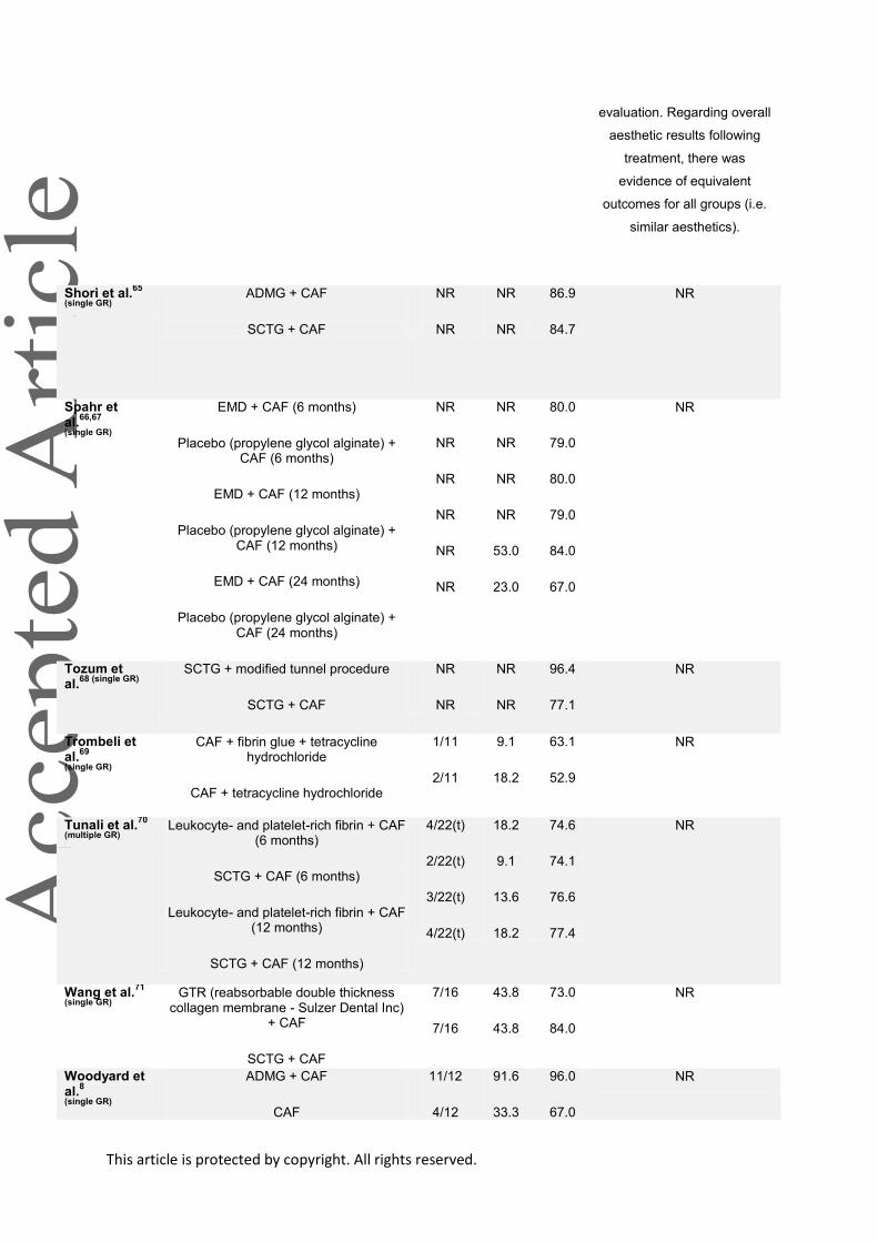
This article is protected by copyright. All rights reserved.
evaluation. Regarding overall
aesthetic results following
treatment, there was
evidence of equivalent
outcomes for all groups (i.e.
similar aesthetics).
Shori et al.65
(single GR) ADMG + CAF
SCTG + CAF
NR
NR
NR
NR
86.9
84.7
NR
Spahr et al.
66,67
(single GR)
EMD + CAF (6 months)
Placebo (propylene glycol alginate) + CAF (6 months)
EMD + CAF (12 months)
Placebo (propylene glycol alginate) + CAF (12 months)
EMD + CAF (24 months)
Placebo (propylene glycol alginate) + CAF (24 months)
NR
NR
NR
NR
NR
NR
NR
NR
NR
NR
53.0
23.0
80.0
79.0
80.0
79.0
84.0
67.0
NR
Tozum et al.
68 (single GR) SCTG + modified tunnel procedure
SCTG + CAF
NR
NR
NR
NR
96.4
77.1
NR
Trombeli et al.
69
(single GR)
CAF + fibrin glue + tetracycline hydrochloride
CAF + tetracycline hydrochloride
1/11
2/11
9.1
18.2
63.1
52.9
NR
Tunali et al.70
(multiple GR) Leukocyte- and platelet-rich fibrin + CAF
(6 months)
SCTG + CAF (6 months)
Leukocyte- and platelet-rich fibrin + CAF (12 months)
SCTG + CAF (12 months)
4/22(t)
2/22(t)
3/22(t)
4/22(t)
18.2
9.1
13.6
18.2
74.6
74.1
76.6
77.4
NR
Wang et al.71
(single GR) GTR (reabsorbable double thickness
collagen membrane - Sulzer Dental Inc) + CAF
SCTG + CAF
7/16
7/16
43.8
43.8
73.0
84.0
NR
Woodyard et al.
8
(single GR)
ADMG + CAF
CAF
11/12
4/12
91.6
33.3
96.0
67.0
NR
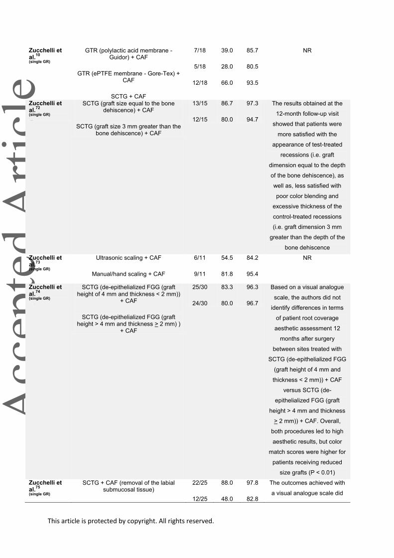
This article is protected by copyright. All rights reserved.
Zucchelli et al.
10
(single GR)
GTR (polylactic acid membrane - Guidor) + CAF
GTR (ePTFE membrane - Gore-Tex) + CAF
SCTG + CAF
7/18
5/18
12/18
39.0
28.0
66.0
85.7
80.5
93.5
NR
Zucchelli et al.
72
(single GR)
SCTG (graft size equal to the bone dehiscence) + CAF
SCTG (graft size 3 mm greater than the bone dehiscence) + CAF
13/15
12/15
86.7
80.0
97.3
94.7
The results obtained at the
12-month follow-up visit
showed that patients were
more satisfied with the
appearance of test-treated
recessions (i.e. graft
dimension equal to the depth
of the bone dehiscence), as
well as, less satisfied with
poor color blending and
excessive thickness of the
control-treated recessions
(i.e. graft dimension 3 mm
greater than the depth of the
bone dehiscence
Zucchelli et al.
73
(single GR)
Ultrasonic scaling + CAF
Manual/hand scaling + CAF
6/11
9/11
54.5
81.8
84.2
95.4
NR
Zucchelli et al.
74
(single GR)
SCTG (de-epithelialized FGG (graft height of 4 mm and thickness < 2 mm))
+ CAF
SCTG (de-epithelialized FGG (graft height > 4 mm and thickness > 2 mm) )
+ CAF
25/30
24/30
83.3
80.0
96.3
96.7
Based on a visual analogue
scale, the authors did not
identify differences in terms
of patient root coverage
aesthetic assessment 12
months after surgery
between sites treated with
SCTG (de-epithelialized FGG
(graft height of 4 mm and
thickness < 2 mm)) + CAF
versus SCTG (de-
epithelialized FGG (graft
height > 4 mm and thickness
> 2 mm)) + CAF. Overall,
both procedures led to high
aesthetic results, but color
match scores were higher for
patients receiving reduced
size grafts (P < 0.01)
Zucchelli et al.
75
(single GR)
SCTG + CAF (removal of the labial submucosal tissue)
22/25
12/25
88.0
48.0
97.8
82.8
The outcomes achieved with
a visual analogue scale did

This article is protected by copyright. All rights reserved.
SCTG + CAF not show differences
between procedures in terms
of root coverage, but color
match was identified by
patients as better when the
labial submucosal tissue was
removed
ADMG: acellular dermal matrix graft; B-TCP: Beta-tricalcium phosphate; CAF: coronally advanced flap; CEJ: cemento-enamel junction; DFDBA: demineralized freeze-dried bone allograft; EMD: enamel matrix derivative; ePTFE: expanded polytetrafluorethylene; FGG: free gingival graft; GTR: guided tissue regeneration; MRC: mean root coverage; NR: not reported; PCRC: percentage of complete root coverage; rhPDGF-BB: recombinant human platelet-derived growth factor-BB; SCRC: sites with complete root coverage; SCTG: subepithelial connective tissue graft; t: teeth; XCM: xenogeneic collagen matrix;
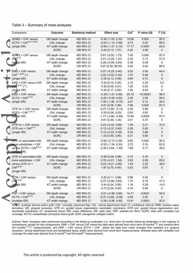
This article is protected by copyright. All rights reserved.
Table 3 – Summary of meta-analyses
Comparison Outcome Statistical method Effect size Chi2 P value (Q) I2 (%)
ADMG + CAF versus
SCTG + CAF28,41,56,65
(single GR)
GR depth change
CAL change
KT width change
SCRC
MD 95% CI
MD 95% CI
MD 95% CI
OR 95% CI
-0.36 (-1.03, 0.30)
-0.53 (-1.14, 0.08)
-0.59 (-1.27, 0.10)
0.43 (0.13, 1.37)
15.06
9.73
17.17
0.00
0.002
0.02
0.0007
0.96
80.0
69.0
83.0
0
ADMG + CAF versus
CAF8,35
(single GR)
GR depth change
CAL change
KT width change
SCRC
MD 95% CI
MD 95% CI
MD 95% CI
OR 95% CI
0.61 (-0.52, 1.73)
0.51 (-0.25, 1.27)
0.28 (-0.08, 0.64)
3.97 (0.20, 80.50)
7.45
2.32
0.30
5.03
0.006
0.13
0.59
0.02
87.0
57.0
0
80.0
EMD + CAF versus
CAF11,64,67 (*)
(single GR)
GR depth change
CAL change
KT width change
MD 95% CI
MD 95% CI
MD 95% CI
0.07 (-0.25, 0.40)
0.22 (-0.02, 0.45)
0.35 (0.13, 0.56)
5.62
1.57
0.64
0.06
0.46
0.73
64.0
0
0
EMD + CAF versus CAF
(2)11,64,67 (**)
(single GR)
GR depth change
CAL change
KT width change
MD 95% CI
MD 95% CI
MD 95% CI
0.32 (0.10, 0.55)
0.35 (0.09, 0.61)
0.40 (0.17, 0.62)
2.10
1.25
1.63
0.35
0.53
0.44
5.0
0
0
EMD + CAF versus
SCTG + CAF24,48
(single GR)
GR depth change
CAL change
KT width change
SCRC
MD 95% CI
MD 95% CI
MD 95% CI
OR 95% CI
-0.39 (-1.27, 0.48)
-0.25 (-0.69, 0.20)
-1.06 (-1.36, -0.76)
0.61 (0.05, 7.86)
25.79
2.95
2.47
7.86
<0.00001
0.09
0.12
0.005
96.0
66.0
59.0
87.0
GTR rm + CAF versus
SCTG + CAF10,55,71
(single GR)
GR depth change
CAL change
KT width change
SCRC
MD 95% CI
MD 95% CI
MD 95% CI
OR 95% CI
-0.37 (-0.60, -0.13)
0.35 (0.06, 0.63)
-1.77 (-2.66, -0.89)
0.61 (0.30, 1.24)
0.25
0.93
15.84
2.01
0.88
0.63
0.0004
0.37
0
0
87.0
0
GTR rm + CAF versus
GTR nrm + CAF9,10
(single GR)
GR depth change
CAL change
KT width change
SCRC
MD 95% CI
MD 95% CI
MD 95% CI
OR 95% CI
0.23 (-0.22, 0.68)
0.12 (-0.37, 0.60)
0.12 (-0.23, 0.48)
1.33 (0.46, 3.85)
1.59
0.28
0.03
0.21
0.21
0.60
0.86
0.65
37.0
0
0
0
GTR rm associated with
bone substitutes + CAF
versus SCTG + CAF55,61
(single GR)
GR depth change
CAL change
KT width change
MD 95% CI
MD 95% CI
MD 95% CI
-0.82 (-2.13, 0.49)
-0.52 (-1.34, 0.30)
-2.38 (-2.84, -1.92)
9.92
2.72
1.86
0.002
0.10
0.17
90.0
63.0
46.0
GTR rm associated with
bone substitutes + CAF
versus GTR rm +
CAF36,55
(single GR)
GR depth change
CAL change
KT width change
SCRC
MD 95% CI
MD 95% CI
MD 95% CI
OR 95% CI
0.48 (0.09, 0.88)
0.76 (-0.01, 1.54)
0.23 (-0.21, 0.68)
1.87 (0.75, 4.64)
0.10
2.83
1.63
0.03
0.76
0.09
0.20
0.87
0
65.0
39.0
0
XCM + CAF versus
CAF40,64
(single GR)
GR depth change
CAL change
KT width change
SCRC
MD 95% CI
MD 95% CI
MD 95% CI
OR 95% CI
0.40 (0.11, 0.68)
0.37 (-0.09, 0.83)
0.44 (0.04, 0.85)
4.73 (2.35, 9.50)
0.86
1.70
1.16
0.16
0.35
0.19
0.28
0.69
0
41.0
14.0
0
PRF + CAF versus
SCTG + CAF52,70
(multiple GR)
GR depth change
CAL change
KT width change
MD 95% CI
MD 95% CI
MD 95% CI
-0.01 (-0.89, 0.86)
-0.37 (-0.69, -0.06)
-0.26 (-0.98, 0.45)
14.71
0.58
13.41
0.0001
0.45
0.0003
93.0
0
93.0
ADMG: acellular dermal matrix graft; CAF: coronally advanced flap; CAL: clinical attachment level; CI: confidence interval; EMD: enamel matrix derivative; GR: gingival recession; GTR rm: guided tissue regeneration resorbable membrane; GTR nrm: guided tissue regeneration non-resorbable membrane; KT: keratinized tissue; MD: mean difference; OR: odds ratio; PRF: platelet-rich fibrin; SCRC: sites with complete root coverage; SCTG: subepithelial connective tissue graft; XCM: xenogeneic collagen matrix.
Authors‟ Note: Analyses were performed according to the follow-up evaluation (i.e. short term (6 months follow-up preferably) in the majority of comparisons, except for two comparisons: EMP + CAF versus CAF where the data were derived from short-term (6 months
64)* and medium-term
(24 months11,67
)** measurements; and EMP + CAF versus SCTG + CAF where the data from mean changes from baseline (i.e. gingival recession, clinical attachment level and keratinized tissue width) were derived from short-term measurements, whereas sites with complete root coverage the data were derived from 6-month
48 and 24-month
24 measurements.
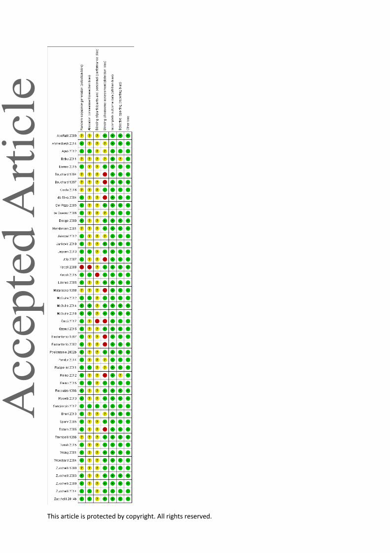
This article is protected by copyright. All rights reserved.
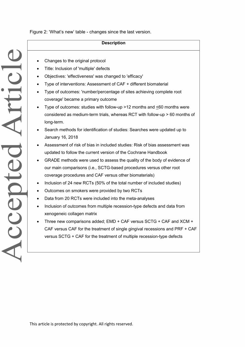
This article is protected by copyright. All rights reserved.
Figure 2: „What‟s new‟ table - changes since the last version.
Description
Changes to the original protocol
Title: Inclusion of 'multiple' defects
Objectives: 'effectiveness' was changed to 'efficacy'
Type of interventions: Assessment of CAF + different biomaterial
Type of outcomes: 'number/percentage of sites achieving complete root
coverage' became a primary outcome
Type of outcomes: studies with follow-up >12 months and <60 months were
considered as medium-term trials, whereas RCT with follow-up > 60 months of
long-term.
Search methods for identification of studies: Searches were updated up to
January 16, 2018
Assessment of risk of bias in included studies: Risk of bias assessment was
updated to follow the current version of the Cochrane Handbook
GRADE methods were used to assess the quality of the body of evidence of
our main comparisons (i.e., SCTG-based procedures versus other root
coverage procedures and CAF versus other biomaterials)
Inclusion of 24 new RCTs (50% of the total number of included studies)
Outcomes on smokers were provided by two RCTs
Data from 20 RCTs were included into the meta-analyses
Inclusion of outcomes from multiple recession-type defects and data from
xenogeneic collagen matrix
Three new comparisons added; EMD + CAF versus SCTG + CAF and XCM +
CAF versus CAF for the treatment of single gingival recessions and PRF + CAF
versus SCTG + CAF for the treatment of multiple recession-type defects