Characterization of some fungal pathogens causing ... - PLOS
Upload
independentCategory
view
1download
0

Genomics of Disease

J.P. Gustafson, J. Taylor, and G. StaceyEditors
Genomics of Disease
EditorsJ.P. GustafsonUSDA-ARSUniversity of MissouriDepartment of Agronomy205 Curtis HallColumbia MO [email protected]
J. TaylorUniversity of MissouriDivision Animal SciencesColumbia, MO [email protected]
G. StaceyUniversity of MissouriPlant Sciences UnitColumbia, MO [email protected]
ISBN: 978-0-387-76722-2 e-ISBN: 978-0-387-76723-9
Library of Congress Control Number: 2007943040
c© 2008 Springer Science+Business Media, LLCAll rights reserved. This work may not be translated or copied in whole or in part without the writtenpermission of the publisher (Springer Science+Business Media, LLC., 233 Spring Street, New York,NY10013, USA), except for brief excerpts in connection with reviews or scholarly analysis. Use inconnection with any form of information storage and retrieval, electronic adaptation, computer software,or by similar or dissimilar methodology now known or hereafter developed is forbidden.The use in this publication of trade names, trademarks, service marks, and similar terms, even if they arenot identified as such, is not to be taken as an expression of opinion as to whether or not they are subject toproprietary rights.While the advice and information in this book are believed to be true and accurate at the date of goingto press, neither the authors nor the editors nor the publisher can accept any legal responsibility for anyerrors or omissions that may be made. The publisher makes no warranty, express or implied, with respectto the material contained herein.
Printed on acid-free paper
9 8 7 6 5 4 3 2 1
springer.com

To Dr. Gordon Kimber in recognition of his effort, support, and commitment tocontinuation of the Stadler Genetics Symposium over many years. Gordon has beenthe strongest supporter of the Stadler Genetics Symposium since he co-organizedthe very first one with Dr. George Redei in May 1969. He continued organizing andediting the Symposium series for several years and has always been tireless in hisattendance and willingness to help in identifying excellent topics and speakers.
Acknowledgments
The editors gratefully acknowledge the generous support of the USDA-ARS and thefollowing units from the University of Missouri: the College of Agriculture, Foodand Natural Resources; the Molecular Biology Program; the Office of Research;the Interdisciplinary Plant Group; the College of Arts and Sciences; the School ofVeterinary Medicine; the Plant Sciences Unit; and the School of Medicine. NationalResearch Initiative Grant No. 2006-35205-17455 from the USDA CooperativeState Research, Education, and Extension Service also supported the Symposium.The contributors’ continued support made the 24th Stadler Genetics Symposium asuccess.
The speakers, who spent a tremendous amount of time preparing their presen-tations and manuscripts, are gratefully acknowledged. Without their expertise anddedication, the Symposium could not have taken place.
We wish to thank all of the local chairpersons for ensuring that all of the speakerswere well taken care of during the Symposium.
Sandi Strother from Conferences and Specialized Courses, University ofMissouri, who tirelessly handled all of our requirements and made sure thateverything was well organized, excellently handled the behind-the-scenes andon-site preparations. Without her, none of this would have been possible
A very special thanks goes to Arturo Garcia, USDA-ARS, Columbia, Missouri,for all of his excellent suggestions in editing the figures and the text to fit a bookformat.
vii
Contents
Roles of Plant Hormones in Plant Resistanceand Susceptibility to Pathogens . . . . . . . . . . . . . . . . . . . . . . . . . . . . . . . . . . . . . . 1Lionel Navarro, Rajendra Bari, Alexandre Seilaniantz, Adnane Nemri,and Jonathan D.G. Jones
Canine Genetics Facilitates Understanding of Human Biology . . . . . . . . . . . 11Elaine A. Ostrander, Heidi G. Parker, and Nathan B. Sutter
Xanthomonas oryzae pv. oryzae AvrXA21 Activity Is Dependenton a Type One Secretion System, Is Regulated by a Two-ComponentRegulatory System that Responds to Cell Population Density,and Is Conserved in Other Xanthomonas spp. . . . . . . . . . . . . . . . . . . . . . . . . . . 25Sang-Won Lee, Sang-Wook Han, Laura E. Bartley, and Pamela C. Ronald
Unraveling the Genetic Mysteries of the Cat: New Discoveriesin Feline-Inherited Diseases and Traits . . . . . . . . . . . . . . . . . . . . . . . . . . . . . . . . 41Leslie A. Lyons
Variation in Chicken Gene Structure and Expression Associatedwith Food-Safety Pathogen Resistance: Integrated Approachesto Salmonella Resistance . . . . . . . . . . . . . . . . . . . . . . . . . . . . . . . . . . . . . . . . . . . . 57S.J. Lamont
Functional Genomics and Bioinformatics of the Phytophthora sojaeSoybean Interaction . . . . . . . . . . . . . . . . . . . . . . . . . . . . . . . . . . . . . . . . . . . . . . . . 67Brett M. Tyler, Rays H.Y. Jiang, Lecong Zhou, Sucheta Tripathy,Daolong Dou, Trudy Torto-Alalibo, Hua Li, Yongcai Mao, Bing Liu,Miguel Vega-Sanchez, Santiago X. Mideros, Regina Hanlon, Brian M. Smith,Konstantinos Krampis, Keying Ye, Steven St. Martin, Anne E. Dorrance,Ina Hoeschele, and M.A. Saghai Maroof
ix
x Contents
Canine SINEs and Their Effects on Phenotypes of the Domestic Dog . . . . . 79Leigh Anne Clark, Jacquelyn M. Wahl, Christine A. Rees, George M. Strain,Edward J. Cargill, Sharon L. Vanderlip, and Keith E. Murphy
Ovine Disease Resistance: Integrating Comparative and FunctionalGenomics Approaches in a Genome Information-Poor Species . . . . . . . . . . . 89H.W. Raadsma, K.J. Fullard, N.M. Kingsford, E.T. Margawati,E. Estuningsih, S. Widjayanti, Subandriyo, N. Clairoux, T.W. Spithill,and D. Piedrafita
Integrating Genomics to Understand the Marek’s DiseaseVirus – Chicken Host–Pathogen Interaction . . . . . . . . . . . . . . . . . . . . . . . . . . . 115Hans H. Cheng
Combining Genomic Tools to Dissect Multifactorial Virulencein Pseudomonas aeruginosa . . . . . . . . . . . . . . . . . . . . . . . . . . . . . . . . . . . . . . . . . . 127Daniel G. Lee, Jonathan M. Urbach, Gang Wu, Nicole T. Liberati,Rhonda L. Feinbaum, and Frederick M. Ausubel
Genetic Dissection of the Interaction Between the Plant PathogenXanthomonas campestris pv. vesicatoria and Its Host Plants . . . . . . . . . . . . . . 151Ulla Bonas, Doreen Gurlebeck, Daniela Buttner, Monique Egler,Simone Hahn, Sabine Kay, Antje Kruger, Christian Lorenz, Robert Szczesny,and Frank Thieme
Structure and Function of RXLR Effectorsof Plant Pathogenic Oomycetes . . . . . . . . . . . . . . . . . . . . . . . . . . . . . . . . . . . . . . 161William Morgan, Jorunn Bos, Catherine Bruce, Minkyoung Lee,Hsin-Yen Liu, Sang-Keun Oh, Jing Song, Joe Win, Carolyn Young,and Sophien Kamoun
The Biotrophic Phase of Ustilago maydis: Novel Determinantsfor Compatibility . . . . . . . . . . . . . . . . . . . . . . . . . . . . . . . . . . . . . . . . . . . . . . . . . . . 173Thomas Brefort, Kerstin Schipper, Gunther Dohlemann,and Regine Kahmann
Virulence Evolution in Malaria . . . . . . . . . . . . . . . . . . . . . . . . . . . . . . . . . . . . . . 183M.J. Mackinnon
The Ins and Outs of Host Recognition of Magnaporthe oryzae . . . . . . . . . . . 199Sally A. Leong
Index . . . . . . . . . . . . . . . . . . . . . . . . . . . . . . . . . . . . . . . . . . . . . . . . . . . . . . . . . . . . . 217

Contributors
Frederick M. AusubelDepartment of Molecular Biology, Massachusetts General Hospital and Departmentof Genetics, Harvard Medical School, Boston, MA, USA
Rajendra BariThe Sainsbury Laboratory, John Innes Centre, Colney Lane, Norwich NR4 7UH,UK
Laura E. BartleyDepartment of Plant Pathology, One Shields Avenue, University of California,Davis, CA 95616, USA
Thomas BrefortMax Planck Institute for Terrestrial Microbiology, Department OrganismicInteractions, Karl-von-Frisch-Strasse, D-35043 Marburg, Germany
Ulla BonasDepartment of Genetics, Martin-Luther-University Halle-Wittenberg, Halle,[email protected]
Jorunn BosDepartment of Plant Pathology, Ohio State University Ohio Agricultural Researchand Development Center, Wooster, OH, USA
Catherine BruceDepartment of Plant Pathology, Ohio State University Ohio Agricultural Researchand Development Center, Wooster, OH, USA
Daniela ButtnerDepartment of Genetics, Martin-Luther-University Halle-Wittenberg, Halle,Germany
Edward J. CargillCanine Genetics Laboratory, Department of Pathobiology, College of VeterinaryMedicine, Texas A&M University, College Station, TX 77843-4467, USA
xi
xii Contributors
Hans H. ChengUSDA, ARS, Avian Disease and Oncology Laboratory, 3606 E. Mount Hope Rd.,East Lansing, MI 48823, USA
N. ClairouxCentre for Host-Parasite Interactions, Institute of Parasitology, McGill University,Ste Anne de Bellevue, Quebec, Canada, H9X 3V9
Leigh Anne ClarkCanine Genetics Laboratory, Department of Pathobiology, College of VeterinaryMedicine, Texas A&M University, College Station, TX 77843-4467, USA
Anne E. DorranceDepartment of Plant Pathology, The Ohio State University, OARDC, 1680 MadisonAve., Wooster, OH 44691, USA
Gunther DohlemannMax Planck Institute for Terrestrial Microbiology, Department OrganismicInteractions, Karl-von-Frisch-Strasse, D-35043 Marburg, Germany
Daolong DouVirginia Bioinformatics Institute, Virginia Polytechnic Institute and StateUniversity, Blacksburg, VA 24061, USA
Monique EglerDepartment of Genetics, Martin-Luther-University Halle-Wittenberg, Halle,Germany
E. EstuningsihIndonesian Veterinary Science Research Institute, Bogor, West Java, Indonesia
Rhonda L. FeinbaumDepartment of Molecular Biology, Massachusetts General Hospital and Departmentof Genetics, Harvard Medical School, Boston, MA, USA
K.J. FullardReprogen-Centre for Advanced Technologies in Animal Genetics and Repro-duction, Faculty of Veterinary Science, University of Sydney, Camden NSW,Australia
Doreen GurlebeckDepartment of Genetics, Martin-Luther-University Halle-Wittenberg, Halle,Germany
Sang-Wook HanDepartment of Plant Pathology, One Shields Avenue, University of California,Davis, CA 95616, USA
Simone HahnDepartment of Genetics, Martin-Luther-University Halle-Wittenberg, Halle,Germany
Contributors xiii
Regina Hanlon
Virginia Bioinformatics Institute, Virginia Polytechnic Institute and StateUniversity, Blacksburg, VA 24061, USA
Ina Hoeschele
Virginia Bioinformatics Institute, Virginia Polytechnic Institute and StateUniversity, Blacksburg, VA 24061, USA
Rays H.Y. Jiang
Virginia Bioinformatics Institute, Virginia Polytechnic Institute and StateUniversity, Blacksburg, VA 24061, USA
Present address: Laboratory of Phytopathology, Wageningen University,Binnenhaven 5, NL6709 PD Wageningen, The Netherlands
Jonathan D.G. Jones
The Sainsbury Laboratory, John Innes Centre, Colney Lane, Norwich NR4 7UH,UK
Sophien Kamoun
Department of Plant Pathology, Ohio State University Ohio Agricultural Researchand Development Center, Wooster, OH, [email protected]
Regine Kahmann
Max Planck Institute for Terrestrial Microbiology, Department Organis-mic Interactions, Karl-von-Frisch-Strasse, D-35043 Marburg, Germany,[email protected]
Sabine Kay
Department of Genetics, Martin-Luther-University Halle-Wittenberg, Halle,Germany
N.M. Kingsford
Reprogen-Centre for Advanced Technologies in Animal Genetics and Repro-duction, Faculty of Veterinary Science, University of Sydney, Camden NSW,Australia
Konstantinos Krampis
Department of Crop Soil and Environmental Science, Virginia BioinformaticsInstitute, Virginia Polytechnic Institute and State University, Blacksburg,VA 24061
Antje Kruger
Department of Genetics, Martin-Luther-University Halle-Wittenberg, Halle,Germany
xiv Contributors
S.J. LamontDepartment of Animal Science, Iowa State University, 2255 Kildee Hall, Ames, IA50011, [email protected]
Daniel G. LeeDepartment of Molecular Biology, Massachusetts General Hospital and Departmentof Genetics, Harvard Medical School, Boston, MA, USA
Minkyoung LeeDepartment of Plant Pathology, Ohio State University Ohio Agricultural Researchand Development Center, Wooster, OH, USA
Hua LiVirginia Bioinformatics Institute, Virginia Polytechnic Institute and StateUniversity, Blacksburg, VA 24061, USA
Nicole T. LiberatiDepartment of Molecular Biology, Massachusetts General Hospital and Departmentof Genetics, Harvard Medical School, Boston, MA, USA
Bing Liu
Virginia Bioinformatics Institute, Virginia Polytechnic Institute and StateUniversity, Blacksburg, VA 24061, USAPresent address: The Monsanto Company, 3302 SE Convenience Blvd, Ankeny, IA50021, USA
Hsin-Yen LiuDepartment of Plant Pathology, Ohio State University Ohio Agricultural Researchand Development Center, Wooster, OH, USA
Sally A. LeongUSDA, ARS CCRU, Department of Plant Pathology, University of Wisconsin,1630 Linden Dr., Madison, WI 53706, 608-262-6309, [email protected]
Sang-Won LeeDepartment of Plant Pathology, One Shields Avenue, University of California,Davis, CA 95616, USA
Christian LorenzDepartment of Genetics, Martin-Luther-University Halle-Wittenberg, Halle,Germany
Leslie A. LyonsDepartment of Population Health & Reproduction, School of Veterinary Medicine,University of California, Davis, CA 95616, USA
Contributors xv
M.J. MackinnonDepartment of Pathology, University of Cambridge, Tennis Court Road, CambridgeCB2 1QP and KEMRI-Wellcome Trust Research Programme, Centre forGeographic Medicine Research – Coast, Kilifi 80108, [email protected]
E.T. MargawatiLIPI, Cibinong, Indonesia
Yongcai MaoVirginia Bioinformatics Institute, Virginia Polytechnic Institute and StateUniversity, Blacksburg, VA 24061, USA
Forest Research Institute, Harborside Financial Center, Plaza V, Jersey City, NJ07311, USA
Steven St. MartinDepartment of Horticulture and Crop Science, The Ohio State University,Columbus, OH 43210-1086, USA
William MorganDepartment of Plant Pathology, Ohio State University Ohio Agricultural Researchand Development Center, Wooster, OH, USA
Santiago X. MiderosDepartment of Plant Pathology, The Ohio State University, OARDC, 1680 MadisonAve., Wooster, OH 44691, USA
Present address: Department of Plant Pathology, 334 Plant Science, CornellUniversity, Ithaca, NY, 14853, USA
Keith E. MurphyCanine Genetics Laboratory, Department of Pathobiology, College of VeterinaryMedicine, Texas A&M University, College Station, TX 77843-4467, USA
Lionel NavarroThe Sainsbury Laboratory, John Innes Centre, Colney Lane, Norwich NR47UH, UK; Institut de Biology Moleculaire des Plantes du Centre National de laRecherche Scientifique, 67084 Strasbourg Cedex, [email protected]
Adnane NemriThe Sainsbury Laboratory, John Innes Centre, Colney Lane, Norwich NR4 7UH,UK
Sang-Keun OhDepartment of Plant Pathology, Ohio State University Ohio Agricultural Researchand Development Center, Wooster, OH, USA
xvi Contributors
Elaine A. OstranderNational Human Genome Research Institute, National Institutes of Health, 50South Drive, Building 50, Room 5351, Bethesda, MD 20892, USA
Heidi G. ParkerNational Human Genome Research Institute, National Institutes of Health, 50South Drive, Building 50, Room 5351, Bethesda, MD 20892, USA
D. PiedrafitaCentre for Animal Biotechnology, School of Veterinary Science, The Universityof Melbourne, Victoria, 3010, Australia
H.W. RaadsmaReprogen-Centre for Advanced Technologies in Animal Genetics and Repro-duction, Faculty of Veterinary Science, University of Sydney, Camden NSW,Australia
Christine A. ReesCanine Genetics Laboratory, Department of Pathobiology, College of VeterinaryMedicine, Texas A&M University, College Station, TX 77843-4467, USA
Pamela C. RonaldDepartment of Plant Pathology, One Shields Avenue, University of California,Davis, CA 95616, USA
M.A. Saghai MaroofDepartment of Crop Soil and Environmental Science, Virginia Polytechnic Instituteand State University, Blacksburg, VA 24061, USA
Kerstin SchipperMax Planck Institute for Terrestrial Microbiology, Department OrganismicInteractions, Karl-von-Frisch-Strasse, D-35043 Marburg, Germany
Alexandre SeilaniantzThe Sainsbury Laboratory, John Innes Centre, Colney Lane, Norwich NR4 7UH,UK
Brian M. SmithVirginia Bioinformatics Institute, Virginia Polytechnic Institute and StateUniversity, Blacksburg, VA 24061, USA
Jing SongDepartment of Plant Pathology, Ohio State University Ohio Agricultural Researchand Development Center, Wooster, OH, USA
T.W. SpithillInstitute of Parasitology, Centre for Host-Parasite Interactions, McGill University,Ste Anne de Bellevue, Quebec, Canada, H9X 3V9
Contributors xvii
George M. StrainCanine Genetics Laboratory, Department of Pathobiology, College of VeterinaryMedicine, Texas A&M University, College Station, TX 77843-4467, USA
SubandriyoIndonesian Animal Production Research Institute, Bogor, West Java, Indonesia
Nathan B. SutterNational Human Genome Research Institute, National Institutes of Health, 50South Drive, Building 50, Room 5351, Bethesda, MD 20892, USARobert SzczesnyDepartment of Genetics, Martin-Luther-University Halle-Wittenberg, Halle,Germany
Frank ThiemeDepartment of Genetics, Martin-Luther-University Halle-Wittenberg, Halle,Germany
Sucheta TripathyVirginia Bioinformatics Institute, Virginia Polytechnic Institute and StateUniversity, Blacksburg, VA 24061, USA
Trudy Torto-AlaliboVirginia Bioinformatics Institute, Virginia Polytechnic Institute and StateUniversity, Blacksburg, VA 24061, USA
Brett M. TylerVirginia Bioinformatics Institute, Virginia Polytechnic Institute and StateUniversity, Blacksburg, VA 24061, USA
Jonathan M. UrbachDepartment of Molecular Biology, Massachusetts General Hospital and Departmentof Genetics, Harvard Medical School, Boston, MA, USA
Sharon L. VanderlipCanine Genetics Laboratory, Department of Pathobiology, College of VeterinaryMedicine, Texas A&M University, College Station, TX 77843-4467, USA
Miguel Vega-SanchezDepartment of Plant Pathology, The Ohio State University, OARDC, 1680 MadisonAve., Wooster, OH 44691, USA
Present address: Department of Plant Pathology, The Ohio State University, 2021Coffey Rd.Columbus, OH 43210, USA
Jacquelyn M. WahlCanine Genetics Laboratory, Department of Pathobiology, College of VeterinaryMedicine, Texas A&M University, College Station, TX 77843-4467, USA
S. WidjayantiResearch Institute for Veterinary Science, Bogor, West Java, Indonesia
xviii Contributors
Joe WinDepartment of Plant Pathology, Ohio State University Ohio Agricultural Researchand Development Center, Wooster, OH, USA
Gang WuDepartment of Molecular Biology, Massachusetts General Hospital and Departmentof Genetics, Harvard Medical School, Boston, MA, USAKeying YeDepartment of Statistics, Virginia Polytechnic Institute and State University,Blacksburg, VA24061, USA
Present address: Department of Management Science and Statistics, University ofTexasat San Antonio, 6900 North Loop 1604 West, San Antonio, TX 78249-0632,USA
Carolyn YoungDepartment of Plant Pathology, Ohio State University Ohio Agricultural Researchand Development Center, Wooster, OH, USA
Lecong ZhouDepartment of Crop Soil and Environmental Science, Virginia BioinformaticsInstitute, Virginia Polytechnic Institute and State University, Blacksburg, VA24061, USA
Roles of Plant Hormones in Plant Resistanceand Susceptibility to Pathogens
Lionel Navarro, Rajendra Bari, Alexandre Seilaniantz, Adnane Nemri,and Jonathan D.G. Jones
Abstract Plants and animals trigger an innate immune response upon perceptionof pathogen-associated molecular patterns (PAMPs) such as flagellin. In Arabidop-sis, flagellin perception elevates resistance to Pseudomonas syringae pv. tomatoDC3000 (Pst DC3000), although the molecular mechanisms involved remain elu-sive. A flagellin-derived peptide transiently enhances the accumulation of a plantmicroRNA that directs degradation of mRNA for TIR1, an F-box auxin receptor.The resulting repression of auxin signaling effectively restricts Pst DC3000 growth,implicating this previously unsuspected miRNA-mediated switch in bacterial dis-ease resistance. These data suggest that elevation of auxin levels constitute a bacte-rial pathogenicity strategy that is suppressed during the innate immune response toPAMPs. In a separate work, we showed that DELLA proteins, which are normallyassociated with gibberellin responses, play a role in the balance between salicylicacid and jasmonic acid–mediated defense signaling pathways. DELLA loss-of-function mutants show reduced growth inhibition in response to flg22, enhancedsusceptibility to necrotrophic pathogens, and enhanced resistance to Pst DC3000.
1 Introduction
Plants perceive a 22 amino acid–conserved peptide flg22, located in the N-terminalpart of eubacterial flagellin (Felix, 1998). In Arabidopsis, recognition of flg22 isassociated with early changes in host transcript levels, including a rapid down-regulation of a subset of genes (Navarro et al., 2004). This down-regulationis potentially post-transcriptional because promoter analysis does not identifyover-representation of common cis-regulatory elements within the promotersof flg22-repressed genes (Navarro et al., 2004). Plants use RNA silencing forpost-transcriptional gene regulation. This sequence-specific mRNA degradation
L. NavarroThe Sainsbury Laboratory, John Innes Centre, Colney Lane, Norwich NR4 7UH, UK; Institut deBiology Moleculaire des Plantes du Centre National de la Recherche Scientifique, 67084Strasbourg Cedex, Francee-mail: [email protected]
J.P. Gustafson et al. (eds.), Genomics of Disease, 1C© Springer Science+Business Media, LLC, 2008

2 L. Navarro et al.
mechanism is mediated by small (21-24nt) RNAs known as short interfering(si)RNAs and micro (mi)RNAs. Both siRNAs and miRNAs are derived fromdouble-stranded (ds)RNA by the action of homologues of the RNaseIII-typeenzymes called Dicer (Dicer-like, or DCLs). In Arabidopsis, miRNAs are excisedfrom intergenic stem-loop transcripts by DCL-1, and direct cleavage of cellularmRNAs carrying miRNA-complementary sequences (Bartel, 2004). Because thesteady-state level of several plant miRNAs—-and possibly several plant siRNAs—varies in response to exogenous stresses (Jones-Rhoades and Bartel, 2004; Sunkarand Zhu, 2004), we tested if those molecules could account for some of therapid post-transcriptional changes elicited by flg22 treatment. We used transgenicArabidopsis expressing plant virus–encoded proteins that suppress miRNA- andsiRNA-guided functions, anticipating that transcripts repressed by flg22-stimulatedsmall RNAs would likely be more elevated in those transgenic lines. Comparativetranscript profiling identified a subset of mRNAs fulfilling this criterion, amongwhich was an mRNA for the plant auxin receptor TIR1 (T ransport InhibitorResponse 1).
2 Flg22 Triggers Auxin-Signaling Repression by Inducinga Specific miRNA
TIR1 mRNA was previously identified as a target of miR393, a canonicalmiRNA conserved across plant species (Bonnet et al., 2004; Jones-Rhoades andBartel, 2004; Wang et al., 2004). To identify whether miR393 plays a role in flg22-reponse, we examined its levels over a time course of flg22 treatment. Northernanalysis revealed a transient and biphasic 2-fold increase in miR393 accumulationin flg22-treated Arabidopsis seedlings, whereas the levels of the unrelated miR171remained unaffected (Navarro et al., 2006). In addition, the miR393 levels wereunchanged in seedlings treated with flg22A.tum, an inactive peptide derived fromAgrobacterium tumefaciens flagellin (Felix, 1998). Quantitative RT-PCR (RT-qPCR) analyses (employing primers flanking the miR393 cleavage site) revealed aprogressive decrease of TIR1 transcript accumulation upon flg22 but not flg22A.tum
treatments, leading to an overall ∼3-fold reduction in TIR1 mRNA 60 minutesafter elicitation. This progressive flg22-dependent reduction of the TIR1 mRNAlevels was also reflected at the protein level, as assessed in Arabidopsis transfor-mants expressing a myc epitope-tagged form of TIR1 under the dexamethasone(Dex)-inducible promoter (Dex::TIR1-Myc) (Navarro et al., 2006). Collectively,these results indicate that flg22 triggers the rapid and specific repression of TIR1accumulation through transient up-regulation of miR393 levels.
In addition to its role in auxin perception, TIR1 is part of the ubiquitin-ligase complex SCFTIR1 that interacts with Aux/IAA transcriptional repressorproteins to promote their ubiquitylation and subsequent degradation by the
Roles of Plant Hormones in Plant Resistance and Susceptibility to Pathogens 3
26S-proteasome (Gray et al., 2001). We used transgenic lines expressing a heatshock–inducible AXR3/IAA17 protein fused with the �-glucuronidase (GUS)reporter (HS::AXR3NT-GUS) (Gray et al., 2001). Seedlings were exposed to hightemperature and treated with either flg22 or flg22A.tum for 2 hours; GUS staining wassubsequently performed. Flg22, but not flg22A.tum, triggered a strong stabilizationof AXR3NT-GUS in roots and leaves. In contrast, no AXR3NT-GUS was observedwhen seedlings were treated with flg22 at room temperature, indicating that flg22does not activate the heat shock promoter. Time-course analysis revealed thatstabilization of AXR3NT-GUS starts 1.5 hours after flg22 elicitation, consistentwith the kinetics of TIR1-Myc protein repression (Navarro et al., 2006).
Aux/IAA proteins act as repressors of auxin signaling through heterodimer-ization with ARF (Auxin Response Factor) transcription factors (Liscum andReed, 2002). ARFs bind directly to auxin responsive elements (AuxRE) found in thepromoters of primary auxin-response genes, leading to their transcriptional activa-tion (or repression in some cases) (Hagen and Guilfoyle, 2002). The flg22-inducedstabilization of AXR3/IAA17, and presumably other Aux/IAA proteins, promptedus to investigate if flg22 inhibits ARF protein function resulting in transcriptionalinactivation of primary auxin-response genes. To address this point, Arabidopsisseedlings were challenged for 1.5 hours with either flg22 or flg22A.tum and thetranscript levels of the primary auxin-response genes GH3-like, BDL/IAA12, andAXR3/IAA17 were monitored by RT-qPCR. This time point was chosen based onthe flg22-induced stabilization profile of the AXR3/IAA17 protein. We found thatall the three auxin-response genes were indeed repressed at this time point (Navarroet al., 2006). Collectively, these results indicate that flg22 triggers, through theaction of miR393, a series of molecular events that ultimately lead to the rapiddown-regulation of primary auxin-response genes.
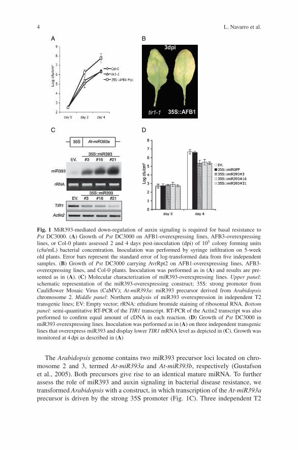
To assess if auxin is involved in disease resistance and susceptibility, we usedArabidopsis transgenic lines that overexpress myc epitope-tagged versions of thetwo TIR1 functional paralogs AFB1 and AFB3 (for auxin signaling F-Box proteins1 and 3). Both act in a redundant manner with TIR1 to mediate auxin perceptionand signaling. However, the AFB1 transcript, unlike the AFB3 transcript, is partiallyresistant to miR393-guided cleavage, presumably because of a single nucleotidepolymorphism introducing a synonymous mutation in the miRNA complementarysite. Therefore, overexpression of AFB1 should have dominant-negative effectsupon a putative miR393-mediated defense response. When inoculated with virulentP. syringae pv. tomato (Pst) DC3000, AFB1- but not AFB3-overexpressing plantsdisplayed a ∼100-fold higher bacterial titers compared to non-transformed plants,as assessed at 2 and 4 days post-inoculation (Fig. 1A). In contrast, no differencewas observed with avirulent Pst DC3000 carrying AvrRpt2, which encodes the elic-itor of race-specific resistance controlled by the Arabidopsis gene RPS2 (Fig. 1B;Dong et al., 1991; Whalen et al., 1991; Kunkel et al., 1993). These results sug-gest that miR393 specifically promotes basal resistance to virulent Pst DC3000but is not implicated in race-specific resistance mediated by the RPS2 resistanceprotein.
4 L. Navarro et al.
A
DC
B
Fig. 1 MiR393-mediated down-regulation of auxin signaling is required for basal resistance toPst DC3000. (A) Growth of Pst DC3000 on AFB1-overexpressing lines, AFB3-overexpressinglines, or Col-0 plants assessed 2 and 4 days post-inoculation (dpi) of 105 colony forming units(cfu/mL) bacterial concentration. Inoculation was performed by syringe infiltration on 5-weekold plants. Error bars represent the standard error of log-transformed data from five independentsamples. (B) Growth of Pst DC3000 carrying AvrRpt2 on AFB1-overexpressing lines, AFB3-overexpressing lines, and Col-0 plants. Inoculation was performed as in (A) and results are pre-sented as in (A). (C) Molecular characterization of miR393-overexpressing lines. Upper panel:schematic representation of the miR393-overexpressing construct; 35S: strong promoter fromCauliflower Mosaic Virus (CaMV); At-miR393a: miR393 precursor derived from Arabidopsischromosome 2. Middle panel: Northern analysis of miR393 overexpression in independent T2transgenic lines; EV: Empty vector; rRNA: ethidium bromide staining of ribosomal RNA. Bottompanel: semi-quantitative RT-PCR of the TIR1 transcript. RT-PCR of the Actin2 transcript was alsoperformed to confirm equal amount of cDNA in each reaction. (D) Growth of Pst DC3000 inmiR393 overexpressing lines. Inoculation was performed as in (A) on three independent transgeniclines that overexpress miR393 and display lower TIR1 mRNA level as depicted in (C). Growth wasmonitored at 4 dpi as described in (A)
The Arabidopsis genome contains two miR393 precursor loci located on chro-mosome 2 and 3, termed At-miR393a and At-miR393b, respectively (Gustafsonet al., 2005). Both precursors give rise to an identical mature miRNA. To furtherassess the role of miR393 and auxin signaling in bacterial disease resistance, wetransformed Arabidopsis with a construct, in which transcription of the At-miR393aprecursor is driven by the strong 35S promoter (Fig. 1C). Three independent T2
Roles of Plant Hormones in Plant Resistance and Susceptibility to Pathogens 5
transgenic lines were selected based on high miR393 accumulation. Accordingly,these transgenic lines had low levels of TIR1 mRNAs, as compared to lines trans-formed with an empty vector (Fig. 1C). Upon infection with virulent Pst DC3000,all the three miR393-overexpressing lines, but not the empty vector transformants,displayed ∼100-fold lower bacterial titers at 4 dpi, confirming that miR393 restrictsPst DC3000 growth (Fig. 1D). Furthermore, no difference in bacterial growth wasobserved in transgenic lines overexpressing an artificial miRNA directed againstthe green fluorescence protein (gfp) mRNA (Parizotto et al., 2004), indicating amiR393-specific effect (data not shown).
3 Does Auxin Play a Role in Bacterial Pathogenenity?
Besides its role in plant growth and development, auxin was also reported toaffect plant–pathogen interactions (Yamada, 1993). For example, the tumorigenicP. syringae pv. savastanoi produces high level of IAA that is implicated in thedevelopment of oleander knots (Yamada et al., 1991). The role of auxin in diseasesusceptibility is not restricted to tumorogenic bacteria because exogenously appliedauxin was also reported to induce susceptibility of maize to Helminthosporiumleaf spot and of tobacco to Tobacco Mosaic Virus (TMV) (Simons et al., 1972;Hoffman, 1973). Moreover, auxin can suppress the hypersensitive response (HR),a plant-triggered cell death process often induced to restrict pathogen growth(Novacky, 1972; Matthysse, 1987; Robinette and Matthysse, 1990). Interestingly,we note that most P. syringae strains produce IAA and Pst DC3000 infectiontriggers higher accumulation of free IAA in Arabidopsis (Glickmann et al., 1998;O’Donnell et al., 2003). In addition, the virulent AvrRpt2 type-III secreted pro-tein appears to promote the auxin-signaling pathway in an Arabidopsis rps2mutant background, resulting in enhanced bacterial disease symptoms and growth(B. Kunkel, personal communication). Consistent with the role of auxin in bacterialpathogenicity, we found that delivery of the auxin analog 2,4-dichlorophenoxyaceticacid (2,4-D) at 20 �M concentration together with Pst DC3000 (105 cfu/mL) sig-nificantly promotes bacterial disease symptoms as early as 3 days post-inoculation(data not shown). However, only a mild effect on bacterial growth was observed(∼1.5-fold higher bacterial titer compared to plantstreated with Pst DC3000 alone).Lower concentrations of 2,4-D still promote Pst DC3000 disease symptoms withouthaving a significant effect on bacterial growth (data not shown). These results sug-gest that exogenous auxin predominantly promotes Pst DC3000 disease symptomdevelopment.
We show here that a bacterial PAMP down-regulates auxin signaling by enhanc-ing the endogenous levels of miR393. Overexpressing auxin signaling through aTIR1 paralog that is partially refractory to miR393 guided–cleavage enhances sus-ceptibility to virulent Pst DC3000 and, conversely, repressing this hormonal path-way through miR393 overexpression induces resistance to the bacterium. Theseresults indicate that down-regulation of auxin signaling is part of a plant-inducedimmune response. They also suggest that auxin might promote disease susceptibility
6 L. Navarro et al.
to bacteria. Consistent with this hypothesis, we found that the auxin-analog 2,4-Dpromotes significantly Pst DC3000 disease symptom development (data not shown).These data suggest that elevation of auxin levels constitute a bacterial pathogenicitystrategy that is suppressed during the PAMP-triggered response.
4 Flg22 Triggers Growth Inhibition of Arabidopsis Seedlings
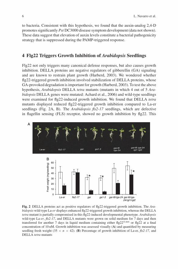
Flg22 not only triggers many canonical defense responses, but also causes growthinhibition. DELLA proteins are negative regulators of gibberellin (GA) signalingand are known to restrain plant growth (Harberd, 2003). We wondered whetherflg22-triggered growth inhibition involved stabilization of DELLA proteins, whoseGA-provoked degradation is important for growth (Harberd, 2003). To test the abovehypothesis, Arabidopsis DELLA tetra mutants (mutants in which 4 out of 5 Ara-bidopsis DELLA genes were mutated: Achard et al., 2006) and wild-type seedlingswere examined for flg22-induced growth inhibition. We found that DELLA tetramutants displayed reduced flg22-triggered growth inhibition compared to La-erseedlings (Fig. 2A, B). The Arabidopsis fls2-17 seedlings, which are defectivein flagellin sensing (FLS) receptor, showed no growth inhibition by flg22. This
A
0
Fre
sh w
eigh
t (m
g/se
edlin
g)
10
20
30
40
50
60
La-er fls2-17 gai ga1-3 gai-t6/rga-24 gai-t6/rga-24/rgl1/rgl2
B
Fig. 2 DELLA proteins act as positive regulators of flg22-triggered growth inhibition. The Ara-bidopsis wild-type La-er displays enhanced flg22-triggered growth inhibition, whereas the DELLAtetra mutant is partially compromised in this flg22-induced developmental phenotype. Arabidopsiswild-type La-er, fls2-17, and DELLA mutants were grown on solid medium for 7 days and thentransferred for another 7 days in liquid medium containing either flg22A.tum or flg22 at a finalconcentration of 10 nM. Growth inhibition was assessed visually (A) and quantified by measuringseedling fresh weight (35 < n < 42). (B) Percentage of growth inhibition of La-er, fls2-17, andDELLA tetra mutants

Roles of Plant Hormones in Plant Resistance and Susceptibility to Pathogens 7
suggested that flg22 treatment might stabilize DELLA proteins. In another stud-ies, we found that flg22 delays the GA-induced disappearance of GFP-RGA (datanot shown), which is consistent with the induction of DELLA protein stabilizationby flg22.
5 Role of DELLA Proteins in Plant Disease Resistanceand Susceptibility
The involvement of DELLA proteins in plant defense was examined by challengingDELLA tetra mutants with bacterial and fungal pathogens. When DELLA mutantplants were infiltrated with a biotrophic pathogen, Pst DC3000, they showed
2
3
4
5
6
7
8
9
10
day 0 day 2 day 4
La-erga1-3sly1-10gai
sly1-10
La-er
A
C D
0
1
2
3
4
5
6
7
day 0 day 2
Lergai-t6/rga-24tetra
Log cfu/cm2Log cfu/cm2
B
Fig. 3 DELLA proteins act as negative regulators of plant defense against Pst DC3000 and aspositive regulators of defense against A. brassicicola. (A) DELLA tetra mutants are more resistantto Pst DC3000. Growth of Pst DC3000 on DELLA tetra (gai-t6/rga-t2/rgl1-1/rgl2-1) and La-erplants was assessed 2 days post-inoculation (dpi) of 105 colony-forming units (cfu/ml). Inocula-tion was performed by syringe infiltration on 4-week old plants grown in long-day conditions.Error bars represent the standard error of log-transformed data from five independent samples.(B) DELLA tetra mutants are more susceptible to A. brassicicola. Four-week old plants (grownin long-day conditions) were sprayed with A. brassicicola spores at a concentration of 5 × 10⁵
spores/ml and pictures were taken at 7 dpi

8 L. Navarro et al.
enhanced resistance compared to wild-type plants (Fig. 3A). Salicylic acid (SA) isthe major signaling molecule involved in the resistance of plants against biotrophicpathogens (Durner et al., 1997). To check whether DELLA proteins interferewith the SA-dependent plant defense pathway, the expression of SA-dependentmarker genes PR1 and PR2 were monitored in DELLA tetra and wild-type plantschallenged with Pst DC3000. We found an earlier and stronger induction of bothPR1 and PR2 genes in Pst DC3000-inoculated tetra plants compared to wild-typeplants (data not shown).
Since the SA-dependent plant defense pathway is mutually antagonistic to thejasmonate/ethylene (JA/ET)-dependent pathway, we next investigated whether thestronger induction of SA-dependent genes, in the tetra infected mutant, leads tothe suppression of JA/ET-dependent gene expression. The expression of JA/ET-dependent marker genes PDF1.2 and VSP1 were reduced in Pst DC3000-challengedtetra plants compared to wild-type plants (data not shown). This indicates thatDELLA proteins are involved in the repression of SA-dependent gene expressionand activation of JA/ET-dependent gene expression in Arabidopsis.
The involvement of DELLA proteins in the positive regulation of the JA/ET-pathway led us to examine whether DELLA tetra mutants were altered in theirresistance to the necrotrophic pathogen Alternaria brassicicola Interestingly, 7 daysafter inoculation with A. brassicicola, DELLA tetra mutant leaves were diseasedand heavily colonized with fungal hyphae, indicating enhanced susceptibility toA. brassicicola compared to wild-type plants (Fig. 3B). This indicates that DELLAproteins act as positive regulators of plant defense responses to A. brassicicola.
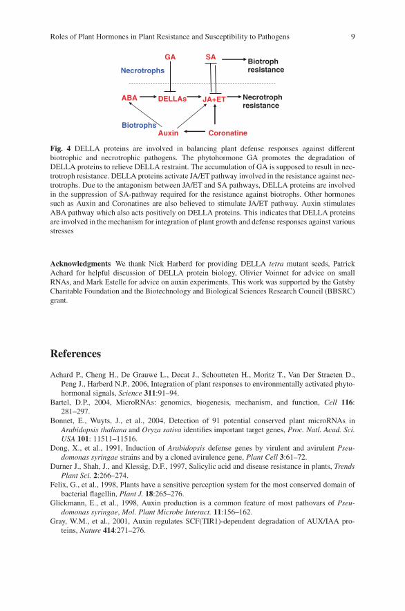
6 Are DELLA Proteins Integrators of Plant Defense Pathways?
These data show that DELLA proteins are involved in the repression of SA-dependent plant defense and activation of the JA/ET-dependent plant defense path-way. This suggest that DELLA proteins play an important role in establishing thebalance of defense responses mounted by a plant and suggest new mechanismsfor crosstalk between different plant-signaling pathways involved in growth anddefense (Fig. 4). It is conceivable that abscisic acid (ABA), by antagonizing GAaction, could have the opposite effect. ABA and ethylene pathways are involvedin plant responses to diverse abiotic and biotic stresses, but particularly in thedrought response and wound response respectively. Recently, it has been shown thattwo independent salt stress-activated phytohormonal signaling pathways (ABA andethylene) regulate plant development through integration at the level of DELLAfunction (Achard et al., 2006). These data, together with the data on miR393 andauxin signaling during defense, suggest that crosstalk between hormone pathwayshas insufficiently been acknowledged as an important determinant in the outcomeof plant–pathogen interactions. It seems likely that DELLA proteins will turn out toplay an important role in this crosstalk.
Roles of Plant Hormones in Plant Resistance and Susceptibility to Pathogens 9
JA+ET
SA
Necrotrophresistance
DELLAs
GA
CoronatineAuxin
ABA
Necrotrophs
Biotrophs
Biotrophresistance
Fig. 4 DELLA proteins are involved in balancing plant defense responses against differentbiotrophic and necrotrophic pathogens. The phytohormone GA promotes the degradation ofDELLA proteins to relieve DELLA restraint. The accumulation of GA is supposed to result in nec-trotroph resistance. DELLA proteins activate JA/ET pathway involved in the resistance against nec-trotrophs. Due to the antagonism between JA/ET and SA pathways, DELLA proteins are involvedin the suppression of SA-pathway required for the resistance against biotrophs. Other hormonessuch as Auxin and Coronatines are also believed to stimulate JA/ET pathway. Auxin stimulatesABA pathway which also acts positively on DELLA proteins. This indicates that DELLA proteinsare involved in the mechanism for integration of plant growth and defense responses against variousstresses
Acknowledgments We thank Nick Harberd for providing DELLA tetra mutant seeds, PatrickAchard for helpful discussion of DELLA protein biology, Olivier Voinnet for advice on smallRNAs, and Mark Estelle for advice on auxin experiments. This work was supported by the GatsbyCharitable Foundation and the Biotechnology and Biological Sciences Research Council (BBSRC)grant.
References
Achard P., Cheng H., De Grauwe L., Decat J., Schoutteten H., Moritz T., Van Der Straeten D.,Peng J., Harberd N.P., 2006, Integration of plant responses to environmentally activated phyto-hormonal signals, Science 311:91–94.
Bartel, D.P., 2004, MicroRNAs: genomics, biogenesis, mechanism, and function, Cell 116:281–297.
Bonnet, E., Wuyts, J., et al., 2004, Detection of 91 potential conserved plant microRNAs inArabidopsis thaliana and Oryza sativa identifies important target genes, Proc. Natl. Acad. Sci.USA 101: 11511–11516.
Dong, X., et al., 1991, Induction of Arabidopsis defense genes by virulent and avirulent Pseu-domonas syringae strains and by a cloned avirulence gene, Plant Cell 3:61–72.
Durner J., Shah, J., and Klessig, D.F., 1997, Salicylic acid and disease resistance in plants, TrendsPlant Sci. 2:266–274.
Felix, G., et al., 1998, Plants have a sensitive perception system for the most conserved domain ofbacterial flagellin, Plant J. 18:265–276.
Glickmann, E., et al., 1998, Auxin production is a common feature of most pathovars of Pseu-domonas syringae, Mol. Plant Microbe Interact. 11:156–162.
Gray, W.M., et al., 2001, Auxin regulates SCF(TIR1)-dependent degradation of AUX/IAA pro-teins, Nature 414:271–276.
10 L. Navarro et al.
Gustafson, A.M., et al., 2005, ASRP: the Arabidopsis Small RNA Project Database, Nucleic AcidsRes. 33:D637–D640.
Hagen, G., and Guilfoyle, T., 2002, Auxin-responsive gene expression: genes, promoters and reg-ulatory factors, Plant Mol. Biol. 49:373–385.
Hoffman, S.E., 1973, Leaf bioassay for Helminthosporium carbonum toxin-search for phytoalexin,Phytopath. 63:729–734.
Jones-Rhoades, M.W., and Bartel, D.P., 2004, Computational identification of plant microRNAsand their targets, including a stress-induced miRNA, Mol. Cell 14:787–799.
Kunkel, B.N., et al., 1993, RPS2: an Arabidopsis disease resistance locus specifying recognition ofPseudomonas syringae strains expressing the avirulence gene avrRpt2, Plant Cell 5:865–875.
Liscum, E., and Reed, J.W., 2002, Genetics of Aux/IAA and ARF action in plant growth anddevelopment, Plant Mol. Biol. 49:387–400.
Matthysse, A.G., 1987, A method for the bacterial elicitation of a hypersensitive-like response inplant cell cultures, J. Microbiol. Methods 7:183–191.
Navarro, L., et al., 2004, The transcriptional innate immune response to flg22. Interplay andoverlap with Avr gene-dependent defense responses and bacterial pathogenesis, Plant Physiol.135:1113–1128.
Navarro, L, Dunoyer, P., Jay, F., Arnold, B., Dharmasiri, N., Estelle, M., Voinnet, O., andJones, J.D., 2006, A plant miRNA contributes to antibacterial resistance by repressing auxinsignaling, Science 21:436–439.
Novacky, A., 1972, Suppression of the bacterially induced hypersensitive reaction by cytokinins,Physiol. Plant Pathol. 2:101–104.
O’Donnell, P.J. et al., 2003, Susceptible to intolerance—-a range of hormonal actions in a suscep-tible Arabidopsis pathogen response, Plant J. 33:245–257.
Parizotto, E.A. et al., 2004, In vivo investigation of the transcription, processing, endonucleolyticactivity, and functional relevance of the spatial distribution of a plant miRNA, Genes Dev.18:2237–2342.
Harberd, N.P., 2003, Relieving DELLA restraint, Science 21:1853–1854.Robinette, D., and Matthysse, A.G., 1990, Inhibition by Agrobacterium tumefaciens and Pseu-
domonas savastanoi of development of the hypersensitive response elicited by Pseudomonassyringae pv. phaseolicola, J. Bacteriol. 172:5742–5749.
Simons, T.J., et al., 1972, Effect of 2,4-dichlorophenoxyacetic acid on tobacco mosaic virus lesionsin tobacco and on the structure of adjacent cell, Virology 48:502–515.
Sunkar, R., and Zhu, J.K., 2004, Novel and stress-regulated microRNAs and other small RNAsfrom Arabidopsis, Plant Cell 16:2001–2019.
Wang, X.J., et al., 2004, Prediction and identification of Arabidopsis thaliana microRNAs and theirmRNA targets, Genome Biol. 5: R65.
Whalen, M.C., et al., 1991, Identification of Pseudomonas syringae pathogens of Arabidopsis anda bacterial locus determining avirulence on both Arabidopsis and soybean, Plant Cell 3:49–59.
Yamada, T., 1993, The role of auxin in plant-disease development, Annu. Rev. Phytopathol.31:253–273.
Yamada, T., et al., 1991, The role of indoleacetic acid biosynthetic genes in tumorigenicity, In:S.S. Patil, S. Ouchi, D. Mills, C. Vance (eds.), Mol. Strat. Pathogens Host Plants, New York:Springer-Verlag, pp. 83–94.
Canine Genetics Facilitates Understandingof Human Biology
Elaine A. Ostrander, Heidi G. Parker, and Nathan B. Sutter
Abstract In the past 15 years the field of canine genetics has advanced dramatically.Dense comparative maps, production of ×1.5 and ×7.5 genome sequences, SNPchips, and a growing sophistication regarding how to tackle problems in complexgenetics have all propelled the canine system from a backwater to the forefront ofthe genomics landscape. In this chapter, we explore some of the critical advances inthe field that have occurred in the past 5 years. We discuss the implications of eachon disease gene mapping. Complex trait genetics and advances related to findinggenes associated with morphology are also discussed. Finally, we speculate on whatadvances will likely define the field in the coming 5 years.
1 Introduction to Dogs and Breeds
The domestic dog is believed to be the most recently evolved species from the familyCanidae. Within the Canidae there are three distinct phylogenetic groups (Wayneet al., 1997; 1987a, b). The domestic dog shares a clade with the wolf-like canidssuch as the gray wolf, coyote, and jackals. Dogs are thought to have arisen in quiterecent time, perhaps as little as 40,000 years ago, with the initial domesticationevents occurring in eastern Asia (Savolainen et al., 2002; Vila et al., 1997).
Most dog breeds arose in the last 200–300 years and many of the most commonmodern breeds were developed in Europe in the 1800s. Currently, there are over 400recognized and distinct dog breeds of which 155 are registered by the AmericanKennel Club (AKC) in the United States (American Kennel Club, 1998). While abreed of dog can be recognized by its physical attributes such as size, shape, coatcolor, head shape, leg length, etc., the concept of a breed has been formally definedby both dog fanciers and geneticists.
According to registering bodies like the AKC, becoming a registered member ofa breed simply requires that both of a dog’s parents are documented members ofthe same breed, and that a small fee be paid. As a result, dog breeds are essentially
E.A. OstranderNational Human Genome Research Institute, National Institutes of Health, BethesdaMD 20892, USA
J.P. Gustafson et al. (eds.), Genomics of Disease, 11C© Springer Science+Business Media, LLC, 2008

12 E.A. Ostrander et al.
closed-breeding populations with little opportunity for introduction of new alleles.Dog breeds are characterized by a lower level of genetic heterogeneity than thatseen in mixed breed dogs as a result of small numbers of founders, population bot-tlenecks, and the over representation of some males (popular sires) who performwell in dog shows (Parker et al., 2004; Parker and Ostrander, 2005). As a result,the current population of ∼10 million purebred dogs in the United States repre-sents an ideal group in which to study the genetics of both simple and complextraits.
Recently, attempts have been made to define the concept of a breed at thegenetic level (Koskinen, 2003; Koskinen and Bredbacka, 2000; Parker et al., 2004).For example, Parker et al. (2004) utilized data from 96 (CA)n repeat-basedmicrosatellite markers spanning all dog autosomes on 414 dogs to determinethe degree to which dogs could be assigned to their appropriate breed using aclustering algorithm. Only a small set of closely related breed pairs (i.e., Whippetand Greyhound; Alaskan Malamute and Siberian Husky) could not be reproduciblydistinguished when compared to other breeds. Similarly, using the Doh assignmenttest, 99% of the dogs tested were correctly assigned to their distinct breed groupusing only the microsatellite data.
The above results are interesting in light of studies on genetic diversity in humanpopulations. In the Parker et al. (2004) study, we showed that humans and dogs havesimilar levels of overall nucleotide diversity, 8 × 10⁻⁴. Genetic variation betweendog breeds, however, is much greater than the observed variation between humanpopulations (27.5% versus 5.4% by AMOVA). The degree of genetic homogeneity,not unexpectedly, is much greater within the membership of any given individualdog breed than it is within distinct human populations. So the concept of a dog“breed” is much more definitive, at the genetic level, than is the concept of a human“population” or a human “race.”
2 Mapping Disease Genes in Dogs
Because dog breeds represent closed-breeding populations, they offer unique oppor-tunities for disease gene mapping (Ostrander and Kruglyak, 2000). Diseases thatare problems for both human and companion animal health are excellent candi-dates for study, particularly those associated with complicated phenotypes. Themapping of complex traits in humans, such as cancer, diabetes, epilepsy, andheart disease, has been stymied by the lack of large pedigrees, limited statisti-cal methods, and both locus and phenotypic heterogeneity. As a result, the abil-ity to unambiguously identify critical susceptibility loci for diseases like cancerhas been problematic (Ostrander et al., 2004). By working with canine families,researchers are able to overcome many of these disadvantages. Dog pedigrees arelarge, and often permit collection of several generations. For example, the pedi-grees used to find the genes for a variety of forms of progressive retinal atrophy(PRA) (Acland et al., 1994, 1998, 1999; Kukekova et al., 2006; Moody et al., 2005;Sidjanin et al., 2002), copper toxicosis (Yuzbasiyan-Gurkan et al., 1997), renal
Canine Genetics Facilitates Understanding of Human Biology 13
cancer (Jonasdottir et al., 2000), narcolepsy (Lin et al., 1999; Mignot et al., 1991),hyperuricosuria (Safra et al., 2006), pancreatic acinar atrophy (Clark et al., 2005),and epilepsy (Lohi et al., 2005) all involved large, multigenerational families ofthe sort unheard of in human genetics. In addition, the fact that all the dogs sharea common, often inbred genetic background means that phenotypic expressionamong individuals with the disease is usually very similar. This latter point shouldprove particularly useful as the community moves from the mapping of singlegene Mendelian disorders to identifying loci associated with complex traits suchas behavior and morphology.
We have appreciated the importance of genetic predisposition in the occurrenceof canine diseases for years (Patterson, 2000; Patterson et al., 1982). Indeed, thedog is second only to human in the attention to which clinicians offer their clientsand the number of dollars spent on health care (American Veterinary Medical Asso-ciation, 2002; Patterson, 2000). As a result, several hundred genetic diseases havebeen identified in the dog (Sargan, 2004), many of which share strong phenotypicsimilarities with human diseases. Many of these are collated in an online databasecalled IDID (Inherited Disease in Dogs), which is similar to the Online MendelianInheritance of Man (OMIM) database (Sargan, 2004).
To date, dozens of loci have been identified for canine-inherited diseases andin many cases the causative genes have been identified (reviewed in Parker andOstrander, 2005; Sutter et al., 2004; Switonski et al., 2004). Specific exam-ples include metabolic disorders (van De Sluis et al., 2002; Yuzbasiyan-Gurkanet al., 1997), blindness (Acland et al., 1998, 1999; Aguirre et al., 1978; Aguirre andAcland, 1988, 1998; Kukekova et al., 2006; Moody et al., 2005), cancer (Jonasdottiret al., 2000; Lingaas et al., 2003), neurologic disorders (Lin et al., 1999; Lingaaset al., 1998), hip dysplasia (Chase et al., 2004), osteoarthritis (Chase et al., 2005b),hyperuricosuria (Safra et al., 2006), pancreatic acinar atrophy (Clark et al., 2005),Addison’s disease (Chase et al., 2006), and epilepsy (Lohi et al., 2005).
The lessons learned have been plentiful. We have gleaned insight into newgenetic mechanisms responsible for disease as well as learned something aboutthe genes and pathways associated with many diseases. In some cases knowledgegained about the underlying disease genes have enlightened us about human condi-tions for which we had little prior knowledge, such as the inherited sleep disordernarcolepsy. In this case, the underlying mutation found in the genetically susceptibleDoberman Pinscher was a splicing defect in the gene for the hypocretin 2 receptor(Lin et al., 1999).
In other cases we have learned about new types of genetic aberrations that cancause disease. Recalling again the example of narcolepsy, the disease has beenshown to be caused at the molecular level by insertion of a canine-specific, shortinterspersed nuclear element (SINE; Bentolila et al., 1999; Minnick et al., 1992;Vassetzky and Kramerov, 2002). These retrotransposons are derived from a tRNA-Lys and occur frequently throughout the canine genome (Bentolila et al., 1999;Coltman and Wright, 1994; Kirkness et al., 2003). In addition to narcolepsy, aber-rant insertion of SINEC Cf elements are associated with centronuclear myopathyin the Labrador Retriever (Pele et al., 2005) as well as the gray merle coat coloringthat appears in many breeds (Clark et al., 2006).
14 E.A. Ostrander et al.
Another interesting example is found in a form of epilepsy similar to humanLafora disease. The canine disease affects several breeds including the miniaturewirehaired dachshund. Lohi and collaborators have shown that the disease is causedby expansion of an unstable dodecamer repeat in the Epm2b (Nhlrc1) gene (Lohiet al., 2005). While trinucleotide repeat expansion has been reported in associa-tion with several human neurologic disorders, this is the first report of a dodecamerrepeat expansion causing a disease in any species.
By far, the most interesting advances have been those that highlighted not onlynew mechanisms of disease, but new genes as well. For instance, extensive progresshas been made in understanding the genetic basis of PRA in the dog (Aclandet al., 1994, 1998, 1999; Aguirre et al., 1978, 1998; Aguirre and Acland, 1988;Kukekova et al., 2006; Lowe et al., 2003; Moody et al., 2005; Sidjanin et al., 2002).PRA refers to a collection of ocular disorders reminiscent of the constellation ofhuman diseases known as retinitis pigmentosa (reviewed by Petersen-Jones (2005)).Recently, a gene for progressive rod cone degeneration (prcd) was identified inthe Poodle, Labrador, and several other breeds (Goldstein et al., 2006; Zangerlet al., 2006). This disease had previously been mapped to a gene-rich region ofcanine chromosome 9 (CFA9) (Acland et al., 1998). As the disease is present inseveral related breeds, the authors used linkage disequilibrium (LD) data from acombination of 14 breeds to reduce the disease-associated interval from severalmegabases (Mb) to just 106 Kb (Goldstein et al., 2006) (Fig. 1). They then iden-tified a single missense mutation that accounted for both the canine disease andthe autosomal recessive retinitis pigmentosa in a patient from Bangladesh (Zangerlet al., 2006).
In many cases, disease genes have been found in dogs after identification inhumans, or simultaneous with the disease gene in humans. For example, the genefor canine renal cancer in the German Shepherd Dog, although linkage mapped first
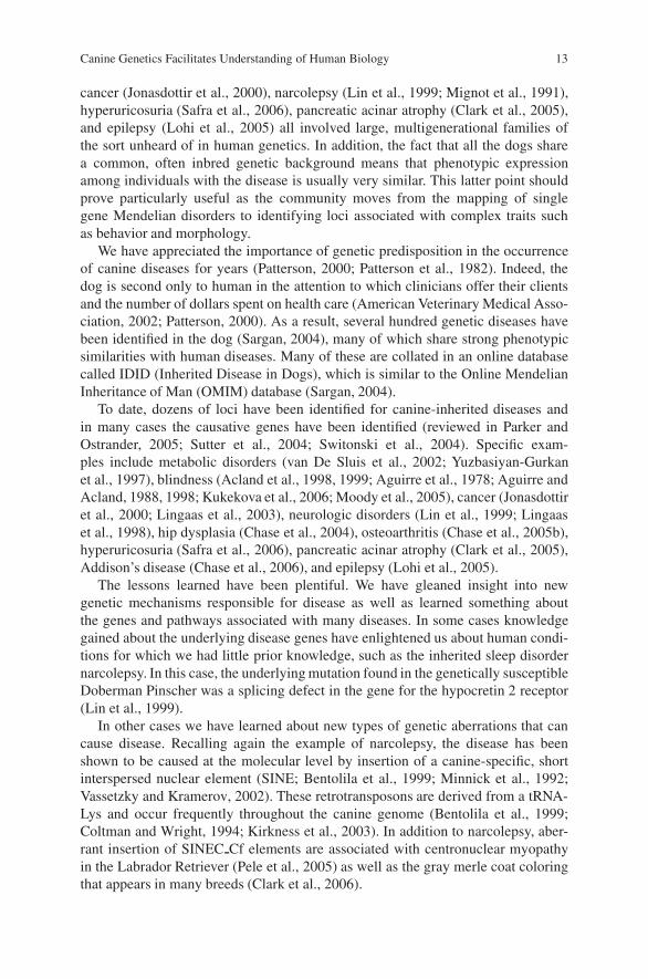
Fig. 1 Linkage disequilibrium. The top chromosome represents an ancient chromosome with aninitial ancestral mutation as marked by the X. Meiotic recombination whittles away the sharedhaplotype around the chromosome. Modern day chromosomes will share only a small regionof commonality around the mutation. Identification of this shared haplotype by SNP genotypingfacilitates fine mapping studies
Canine Genetics Facilitates Understanding of Human Biology 15
in the dog (Jonasdottir et al., 2000), was actually found (Lingaas et al., 2003) afterthe orthologous human gene, which causes a similar disease called Birt-Hogg-DubeSyndrome, was identified (Nickerson et al., 2002). The example of identifying thegene for prcd remains one of the few, together with the identification of the gene forcopper toxicosis in the Bedlington Terrier, where the canine community has led thehuman genetics community in the hunt for truly novel susceptibility genes (van DeSluis et al., 2002).
3 Canine Breed Relationships
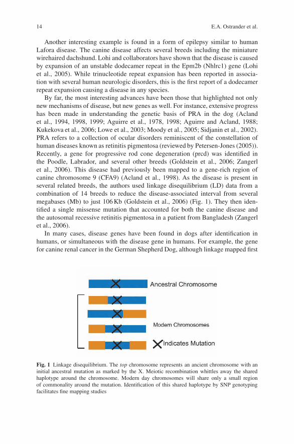
The above study by Goldstein et al. provides a nice example of how data can be com-bined across breeds to identify disease loci of interest (Goldstein et al., 2006). Togeneralize this concept, Parker et al. have studied over 85 breeds using a clusteringalgorithm to understand the relatedness of one breed to another (Parker et al., 2004).In their initial analysis, 85 breeds were ordered into four clusters, generating what isnow considered to be a new canine classification system for dog breeds (Ostranderand Wayne, 2005) based on similar patterns of alleles, presumably from a sharedancestral pool (Fig. 2). Cluster 1 comprised dogs of Asian and African origin as
Fig. 2 Population structure of the domestic dog. Figure is derived from the work of Parkeret al. (2004). Five dogs from each of 85 breeds were genotyped using 85 (CA)n repeat-basedmicrosatellites. Markers spanned all autosomes at 30 Mb density. Analysis was performed using thecomputer program structure. Analysis at K = 2, 3, and 4 divided the population of 85 breeds intothe most likely groups based on allele sharing. Group 1 is comprised largely of Asian breeds suchas the Lhasa Apso, Shar Pei, and Akita. Group 2 is the mastiff group and includes, for example,the Boxer, Bull Dog, and Presa Canario. Group 4 includes a mixture of dogs including workingbreeds. Group 4 is enriched for sight and scent hounds and includes breeds such as the spanielsand retrievers
16 E.A. Ostrander et al.
well as gray wolves. Cluster 2 is typified by mastiff-type dogs with big, boxy headsand strong, sturdy bodies such as the Boxer, Mastiff, and Bulldog. The third andfourth clusters split a group of herding dogs and sight hounds away from the gen-eral population of modern hunting dogs comprised of terriers, hounds, and gun dogbreeds. Ongoing studies are underway to expand this work to include more breeds.It is expected that this should allow even higher resolution of the breed relationshipspicture, and a clearer understanding of how to best combine data across breeds forfine resolution mapping studies.
4 Advances in Canine Genomics
While canine genetics has demonstrated significant progress in the past few years,the rate at which we can expect new discoveries will accelerate dramatically in thecoming months. This is due almost exclusively to two major advances. First, thepublication of a gene dense canine radiation hybrid (RH) map allowed us, for thefirst time, to understand the evolutionary relationship between the canine and thehuman genomes (Hitte et al., 2005). In this study, a well-spaced set of 9850 sequencetagged sites (STS) corresponding to a set of evenly spaced human genes selectedfrom the then available ×1.5 poodle sequence (Kirkness et al., 2003) were localizedon an RH map using a 9000 rad panel. Mutual-Blast alignments identified the besttarget (human) gene sequence using the dog sequence as a probe to ensure thatwe were, in fact, mapping the canine ortholog. A total of 9850 gene fragments wereeventually mapped, which corresponds to approximately half of the genes in the doggenome, identifying some 264 conserved segments (CS) between dog and human.
Interestingly, most of these fragments (243) were later identified by the wholegenome assembly (CanFam1.0) of the dog (Lindblad-Toh et al., 2005), generatedfrom the ×7.5 sequencing effort. This suggests that a dense RH map provides asmuch information for comparative genome mapping studies as a ×7–10 wholegenome shotgun sequence. In addition, detailed comparison of the canine ×7.5whole genome assembly (CanFam 1.0) to the 9000 rad RH map showed that 99.3%of the chromosomal assignments predicted by the RH map were in complete agree-ment with the sequence assembly. Those that were not were quickly resolved andfound to represent issues such as the orientation of internal chromosomal frag-ments. This advance was critical in allowing scientists to move between the canineand the human maps, in assembling the canine genome sequence, and in find-ing the precise breakpoints between the canine and the human genomes (Murphyet al., 2005).
In addition to the above, the availability of both a ×1.5 poodle survey sequenceand a whole genome assembly of a ×7.5 boxer sequence is sure to impact caninegenetics research at every level (Kirkness et al., 2003; Lindblad-Toh et al., 2005).We now know that the dog euchromatic genome is approximately 2.4 billion basesand is comprised of about 243 conserved segments when compared to the human

Canine Genetics Facilitates Understanding of Human Biology 17
genome. The assembled sequence is estimated to cover 98–99% of the genome,with the majority of the sequence contained within two supercontigs per chro-mosome. That is, on average, two segments of continuous sequence cover eachof the dogs’ 38 autosomes. The gene count, at ∼19, 000, is less than what hasbeen predicted for the human genome, perhaps due to complexities associated withsplicing and gene families. There is a 1-1-1 correspondence between orthologsof human, mouse, and dog for 75% of the genes. The full genome sequence canbe accessed through http://www.genome.ucsc.edu; http://www.ncbi.nih.gov, andhttp://www.ensembl.org. A discussion of mining the canine genome sequence isreviewed in O’Rourke (2005).
In addition to the Boxer sequence, a ×1.5 partial sequence of the Standard Poo-dle is available (Kirkness et al., 2003). While in itself less complete than the Boxersequence, together these two resources have enabled the identification of more than2 million single nucleotide polymorphisms (SNPs). We now know that a SNP occursabout once in every 1000 bases in dogs (Lindblad-Toh et al., 2005) and a first gen-eration canine SNP chip is now available from Affymetrix. The chip contains some24,000 working SNPs that will change the landscape of whole genome associa-tion studies in the dog. While microsatellites have proven sufficient for mappingsingle gene traits, it has generally not been possible to analyze enough markers tofully interrogate the genome in a complex trait association study. With thousands ofSNPs available on a single chip, we believe that it is now possible to identify subtlevariants responsible for a host of phenotypic observations.
Key to the development of the canine SNP chip were studies by both Lindblad-Toh et al. (Lindblad-Toh et al., 2005) and Sutter et al. (Sutter et al., 2004) whoaddressed the issue of how many SNPs are “enough” for doing whole genomeassociation studies in the dog. Sutter and colleagues examined the extent of LDin five breeds with distinct breed histories and reported that the average length ofLD in these five breeds is approximately 2 Mb (Fig. 3). This is 40–100 times further
Fig. 3 Divergent populationhistories of dog breeds.Breeds were selected bySutter et al. (2004) in theirstudy of linkagedisequilibrium in dogs.Breeds were chosen torepresent a variety ofmorphologic types, levels ofpresent-day and historicalpopularity, populationstructure, and history (Wilcoxand Walkowicz, 1995)
18 E.A. Ostrander et al.
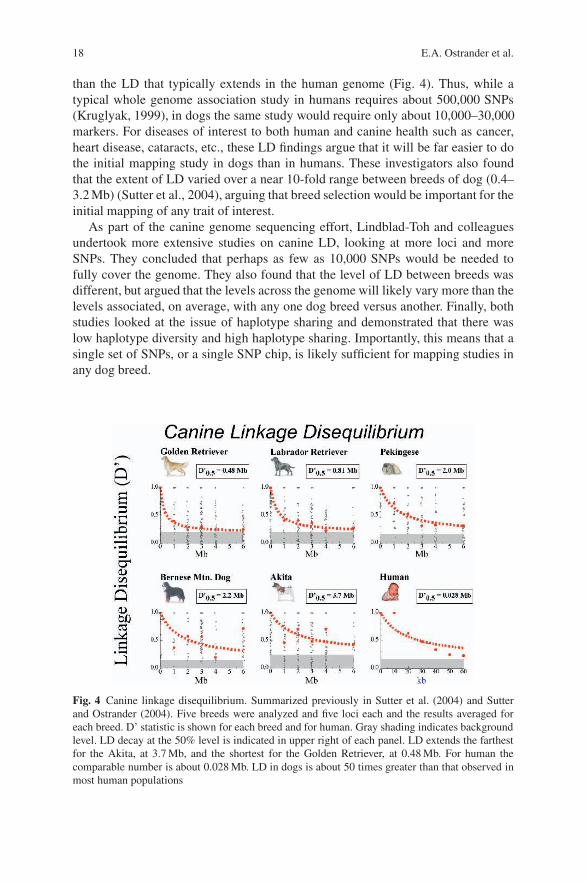
than the LD that typically extends in the human genome (Fig. 4). Thus, while atypical whole genome association study in humans requires about 500,000 SNPs(Kruglyak, 1999), in dogs the same study would require only about 10,000–30,000markers. For diseases of interest to both human and canine health such as cancer,heart disease, cataracts, etc., these LD findings argue that it will be far easier to dothe initial mapping study in dogs than in humans. These investigators also foundthat the extent of LD varied over a near 10-fold range between breeds of dog (0.4–3.2 Mb) (Sutter et al., 2004), arguing that breed selection would be important for theinitial mapping of any trait of interest.
As part of the canine genome sequencing effort, Lindblad-Toh and colleaguesundertook more extensive studies on canine LD, looking at more loci and moreSNPs. They concluded that perhaps as few as 10,000 SNPs would be needed tofully cover the genome. They also found that the level of LD between breeds wasdifferent, but argued that the levels across the genome will likely vary more than thelevels associated, on average, with any one dog breed versus another. Finally, bothstudies looked at the issue of haplotype sharing and demonstrated that there waslow haplotype diversity and high haplotype sharing. Importantly, this means that asingle set of SNPs, or a single SNP chip, is likely sufficient for mapping studies inany dog breed.
Fig. 4 Canine linkage disequilibrium. Summarized previously in Sutter et al. (2004) and Sutterand Ostrander (2004). Five breeds were analyzed and five loci each and the results averaged foreach breed. D’ statistic is shown for each breed and for human. Gray shading indicates backgroundlevel. LD decay at the 50% level is indicated in upper right of each panel. LD extends the farthestfor the Akita, at 3.7 Mb, and the shortest for the Golden Retriever, at 0.48 Mb. For human thecomparable number is about 0.028 Mb. LD in dogs is about 50 times greater than that observed inmost human populations
Canine Genetics Facilitates Understanding of Human Biology 19
5 Mapping Genes for Morphology in the Dog
Breeds of dog differ by over 40-fold in size and display an amazing level of mor-phologic variation. Indeed, Wayne et al. (1986a, b) have argued that the diversityin skeletal size and proportion of dogs is greater than that observed in any otherterrestrial mammal. Studies to map quantitative trait loci (QTLs) in the dog asso-ciated with body conformation have been led by Gordon Lark, Kevin Chase andcollaborators and are based upon their work with the Portuguese Water Dog (PWD)(Chase et al., 1999, 2002). They chose the PWD because the breed offers severaladvantages for complex trait mapping. There are only about 10,000 living AKCregistered PWD, and they derive historically largely from just two kennels (Chaseet al., 1999). The breed standard allows for significant variation, offering a greateropportunity for mapping traits associated with morphology than would studies ofother breeds.
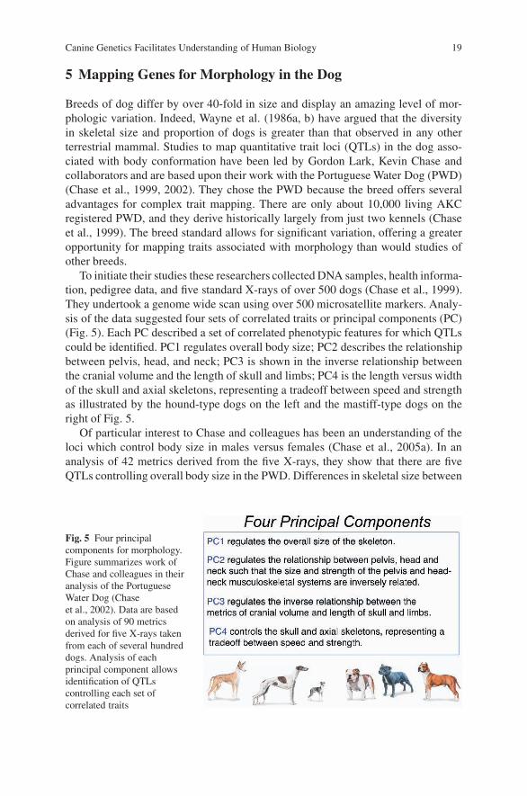
To initiate their studies these researchers collected DNA samples, health informa-tion, pedigree data, and five standard X-rays of over 500 dogs (Chase et al., 1999).They undertook a genome wide scan using over 500 microsatellite markers. Analy-sis of the data suggested four sets of correlated traits or principal components (PC)(Fig. 5). Each PC described a set of correlated phenotypic features for which QTLscould be identified. PC1 regulates overall body size; PC2 describes the relationshipbetween pelvis, head, and neck; PC3 is shown in the inverse relationship betweenthe cranial volume and the length of skull and limbs; PC4 is the length versus widthof the skull and axial skeletons, representing a tradeoff between speed and strengthas illustrated by the hound-type dogs on the left and the mastiff-type dogs on theright of Fig. 5.
Of particular interest to Chase and colleagues has been an understanding of theloci which control body size in males versus females (Chase et al., 2005a). In ananalysis of 42 metrics derived from the five X-rays, they show that there are fiveQTLs controlling overall body size in the PWD. Differences in skeletal size between
Fig. 5 Four principalcomponents for morphology.Figure summarizes work ofChase and colleagues in theiranalysis of the PortugueseWater Dog (Chaseet al., 2002). Data are basedon analysis of 90 metricsderived for five X-rays takenfrom each of several hundreddogs. Analysis of eachprincipal component allowsidentification of QTLscontrolling each set ofcorrelated traits
20 E.A. Ostrander et al.
females and males are due to an interaction between a QTL on CFA15, adjacent tothe insulin-like growth factor-1 (IGF-1) gene, and a locus on the X-chromosomedefined by the CHM marker. The locus on CFA15 is defined by marker FH2017.Analysis of FH2017 genotypes suggests that in females the CFA15 allele control-ling small size is dominant. However, in males the reverse is true and the genotypeassociated with large size appears dominant. The situation is partly explained byconsideration of the QTL on the X-chromosome. Females that are homozygousat the CHM marker and homozygous for the large size CFA15 genotype are, onaverage, as large as the very largest males in the breed. However, any female that isheterozygous at the CHM locus will be small, regardless of her FH2017 genotype.Overall, this interaction explains about 50% of sexual dimorphism in the breed.
Why are these observations so important? First, they demonstrate that the caninesystem is amenable to mapping of complex traits that are of interest to all mam-malian biologists. Second, these studies highlight the value of studying complextraits first in a single breed, especially one with small numbers of founders, buta large amount of phenotypic variation. Finally, the results demonstrate that thenumber of genes controlling complex traits is not so large as to be intractable. Thatis, body size is likely controlled by a small number of QTLs that are identifiablein the canine system. This has important implications for the mapping of complexdiseases as well as truly complex phenotypes such as those associated with behavior.
6 Summary and Future Aims
Until recently, advancement in the study of companion animal health has relied ondata from human and mouse studies. With the development of a ×7.5 whole genomesequence assembly of the dog, a SNP chip, studies of canine breed relationships,and a growing understanding of the architecture of the canine genome we are, forthe first time, mapping genetic traits of interest first in the dog. Our understanding ofdisease genes important for both simple and complex traits is advancing at a rapidrate, informing us about the underlying biology of diseases critical to both humansand companion animals.
In the coming decade, the dog is likely not only to lead man in the discovery ofdisease genes but to provide novel insights into our understanding of truly complexphenotypes. How many of those will be relevant to the human condition? Only timewill tell, but we can rest assured that the dog, ever man’s faithful companion, willbe by our side as we unravel the mysteries of disease susceptibility, behavior, andmorphology.
Acknowledgments We thank our colleagues who read the chapter during preparation and madeexcellent suggestions and the many dog owners, breeders, and supporters who provided us withsamples and information about their beloved pets. This work is supported by the IntramuralProgram of the National Human Genome Research Institute, a Burroughs Wellcome InnovationAward, and The American Kennel Club Canine Health Foundation.
Canine Genetics Facilitates Understanding of Human Biology 21
During preparation of this chapter, the 12-year-old Border Collie of one of the authors died aftera short and unexpected illness. We dedicate this chapter to the many pet owners who, in similarsituations, have shown us how to deal with our loss with grace and dignity.
References
Acland, G.M., Blanton, S.H., Hershfield, B., and Aguiree, G.D., 1994, XLPRA: a canine retinaldegeneration inherited as an X-linked trait, Am. J. Med. Genet. 52:27–33.
Acland, G.M., Ray, K., Mellersh, C.S., Gu, W., Langston, A.A., Rine, J., Ostrander, E.A., andAguirre, G.D., 1998, Linkage analysis and comparative mapping of canine progressive rod-cone degeneration (prcd) establishes potential locus homology with retinitis pigmentosa (RP17)in humans, Proc. Natl. Acad. Sci. USA 95:3048–3053.
Acland, G.M., Ray, K., Mellersh, C.S., Gu, W., Langston, A.A., Rine, J., Ostrander, E.A., andAguirre, G.D., 1999, A novel retinal degeneration locus identified by linkage and comparativemapping of canine early retinal degeneration, Genomics 59:134–142.
Aguirre, G., Lolley, R., Farber, D., Fletcher, T., and Chader, G., 1978, Rod-cone dysplasia in IrishSetter dogs: a defect in cyclic GMP metabolism in visual cells, Science 201:1133.
Aguirre, G.D., and Acland, G.M., 1988, Variation in retinal degeneration phenotype inherited atthe prcd locus, Exp. Eye Res. 46:663–687.
Aguirre, G.D., Baldwin, V., Pearce-Kelling, S., Narfstrom ,K., Ray, K., and Acland, G.M., 1998,Congenital stationary night blindness in the dog: common mutation in the RPE65 gene indicatesfounder effect. Mol. Vis. 4:23.
American Kennel Club, 1998, The Complete Dog Book, Howell Book House, New York, NY.American Veterinary Medical Association, 2002, U.S. Pet Onwership and Demographics Source-
book, pp. 1–136, American Veterinary Medical Association, Schauburg.Bentolila, S., Bach, J.M., Kessler, J.L., Bordelais, I., Cruaud, C., Weissenbach, J., and Panthier, J.J.,
1999, Analysis of major repetitive DNA sequences in the dog (Canis familiaris) genome,Mamm. Genome 10:699–705.
Chase, K., Adler, F.R., Miller-Stebbings, K., and Lark, K.G., 1999, Teaching a new dog old tricks:identifying quantitative trait loci using lessons from plants, J. Hered. 90:43–51.
Chase, K., Carrier, D.F., Adler, F.R., Ostrander, E.A., and Lark, K.G., 2005a, Size sexual dimor-phism in Portugese Water Dogs: interaction between an autosome and the X chromosome,Genome Res. 15:1820–1824.
Chase, K., Carrier, D.R., Adler, F.R., Jarvik, T., Ostrander, E.A., Lorentzen, T. D., and Lark, K.G.,2002, Genetic basis for systems of skeletal quantitative traits: principal component analysis ofthe canid skeleton, Proc. Natl. Acad. Sci. USA 99:9930–9935.
Chase, K., Lawler, D.F., Adler, F.R., Ostrander, E.A., and Lark, K.G., 2004, Bilaterally asym-metric effects of quantitative trait loci (QTLs): QTLs that affect laxity in the right versus leftcoxofemoral (hip) joints of the dog (Canis familiaris), Am. J. Med. Genet. 124:239–247.
Chase, K., Lawler, D.F., Carrier, D.R., and Lark, K.G., 2005b, Genetic regulation of osteoarthritis:a QTL regulating cranial and caudal acetabular osteophyte formation in the hip joint of the dog(Canis familiaris), Am. J. Hum. Genet. 135:334–335.
Chase, K., Sargan, D., Miller, K., Ostrander, E.A., and Lark, K.G., 2006, Understanding the genet-ics of autoimmune disease: two loci that regulate late onset Addison’s disease in PortugueseWater Dogs, Int. J. Immunogenet. 33:179–84.
Clark, L.A., Wahl, J.M., Rees, C.A., and Murphy, K.E., 2006, Retrotransposon insertion in SILVis responsible for merle patterning of the domestic dog, Proc. Natl. Acad. Sci. USA 103:1376–1381.
Clark, L.A., Wahl, J.M., Steiner, J.M., Zhou, W., Ji, W., Famula, T.R., Williams, D.A., andMurphy, K.E., 2005, Linkage analysis and gene expression profile of pancreatic acinar atrophyin the German Shepherd Dog, Mamm. Genome 16:955–962.
22 E.A. Ostrander et al.
Coltman, D.W., and Wright, J.M., 1994, Can SINEs: a family of tRNA-derived retroposons specificto the superfamily Canoidea, Nucleic Acids Res. 22:2726–2730.
Goldstein, O., Zangerl, B., Pearce-Kelling, S., Sidjanin, D.J., Kijas, J.W., Felix, J., Acland, G.M.,and Aguirre, G.D., 2006, Linkage disequilibrium mapping in domestic dog breeds narrows theprogressive rod-cone degeneration interval and identifies ancestral disease-transmitting chro-mosome, Genomics 88:551–563.
Hitte, C., Madeoy, J., Kirkness, E.F., Priat, C., Lorentzen, T.D., et al., 2005, Facilitating genomenavigation: survey sequencing and dense radiation-hybrid gene mapping, Nat. Rev. Genet.6:643–8.
Jonasdottir, T.J., Mellersh, C.S., Moe, L., Heggebo, R., Gamlem, H., Ostrander, E.A., and Lin-gaas, F., 2000, Genetic mapping of a naturally occurring hereditary renal cancer syndrome indogs, Proc. Natl. Acad. Sci. USA 97:4132–4137.
Kirkness, E.F., Bafna, V., Halpern, A.L., Levy, S., Remington, K., Rusch, D.B., Delcher, A.L.,Pop, M., Wang, W., Fraser, C.M., and Venter, J.C., 2003, The dog genome: survey sequencingand comparative analysis, Science 301:1898–1903.
Koskinen, M.T., 2003, Individual assignment using microsatellite DNA reveals unambiguous breedidentification in the domestic dog, Anim. Genet. 34:297–301.
Koskinen, M.T., and Bredbacka, P., 2000, Assessment of the population structure of five Finnishdog breeds with microsatellites, Anim. Genet. 31:310–317.
Kruglyak, L., 1999, Prospects for whole-genome linkage disequilibrium mapping of common dis-ease genes, Nat. Genet. 22:139–144.
Kukekova, A.V., Nelson, J., Kuchtey, R.W., Lowe, J.K., Johnson, J.L., Ostrander, E.A.,Aguirre, G.D., and Acland, G.M., 2006, Linkage mapping of canine rod cone dysplasia type2 (rcd2) to CFA7, the canine orthologue of human 1q32, Invest. Ophthalmol. Vis. Sci. 47:1210–1215.
Lin, L., Faraco, J., Li R., Kadotani, H., Rogers, W., Lin, X., Qiu, X., Jong, P.J., Nishino, S., andMignot, E., 1999, The sleep disorder canine narcolepsy is caused by a mutation in the hypocre-tin (orexin) receptor 2 gene, Cell 98:365–376.
Lindblad-Toh, K., Wade, C.M., Mikkelsen, T.S., Karlsson, E.K., Jaffe, D.B., et al., 2005, Genomesequence, comparative analysis and haplotype structure of the domestic dog, Nature 438:803–819.
Lingaas, F., Aarskaug, T., Sletten, M., Bjerkas, I., Grimholt, U., Moe, L., Juneja, R.K.,Wilton, A.N., Galibert, F., Holmes, N.G., and Dolf, G., 1998, Genetic markers linked to neu-ronal ceroid lipofuscinosis in English setter dogs, Anim. Genet. 29:371–376.
Lingaas, F., Comstock, K.E., Kirkness, E.F., Sorensen, A., Aarskaug, T., Hitte, C., Nickerson, M.L.,Moe, L., Schmidt, L.S., Thomas, R., Breen, M., Galibert, F., Zbar, B., and Ostrander, E.A.,2003, A mutation in the canine BHD gene is associated with hereditary multifocal renal cys-tadenocarcinoma and nodular dermatofibrosis in the German Shepherd dog, Hum. Mol. Genet.12:3043–3053.
Lohi, H., Young, E.J., Fitzmaurice, S.N., Rusbridge, C., Chan, E.M., Vervoort, M., Turnbull, J.,Zhao, X.C., Ianzano, L., Paterson, A.D., Sutter, N.B., Ostrander, E.A., Andre, C., Shelton, G.D.,Ackerley, C.A., Scherer, S.W., and Minassian, B.A., 2005, Expanded repeat in canine epilepsy,Science 307:81.
Lowe, J.K., Kukekova, A.V., Kirkness, E.F., Langlois, M.C., Aguirre, G.D., Acland, G.M., andOstrander, E.A., 2003, Linkage mapping of the primary disease locus for collie eye anomaly,Genomics 82:86–95.
Mignot, E., Wang, C., Rattazzi, C., Gaiser, C., Lovett, M., Guilleminault, C., Dement, W.C.,and Grumet, F.C., 1991, Genetic linkage of autosomal recessive canine narcolepsy with a muimmunoglobulin heavy-chain switch-like segment, Proc. Natl. Acad. Sci. USA 88:3475–3478.
Minnick, M.F., Stillwell, L.C., Heineman, J.M., and Stiegler, G.L., 1992, A highly repetitive DNAsequence possibly unique to canids, Gene 110:235–238.
Moody, J.A., Famula, T.R., Sampson, R.C., and Murphy, K.E., 2005, Identification of microsatel-lite markers linked to progressive retinal atrophy in American Eskimo Dogs, Am. J. Vet. Res.66:1900–1902.
Canine Genetics Facilitates Understanding of Human Biology 23
Murphy, W.J., Larkin, D.M., Everts-van der Wind, A., Bourque, G., Tesler, G., et al., 2005, Dynam-ics of mammalian chromosome evolution inferred from multispecies comparative maps, Sci-ence 309:613–617.
Nickerson, M., Warren, M., Toro, J., Matrosova, V., Glenn, G., Turner, M., Duray, P., Merino, M.,Choyke, P., Pavlovich, C., Sharma, N., Walther, M., Munroe, D., Hill, R., Maher, E.,Greenberg, C., Lerman, M., Linehan, W., Zbar, B., and Schmidt, L., 2002, Mutations in a novelgene lead to kidney tumors, lung wall defects, and benign tumors of the hair follicle in patientswith the Birt-Hogg-Dube syndrome, Cancer Cell 2:157.
O’Rourke, K., 2005, Mining the canine genome. Identification of genes helps breeders andresearchers, J. Am. Vet. Med. Assoc. 226:863–864.
Ostrander, E.A., and Kruglyak, L., 2000, Unleashing the canine genome, Genome Res. 10:1271–1274.
Ostrander, E.A., Markianos, K., and Stanford, J.L., 2004, Finding prostate cancer susceptibilitygenes, Annu. Rev. Genomics Hum. Genet. 5:151–175.
Ostrander, E.A., and Wayne, R.K., 2005, The canine genome, Genome Res. 15:1706–1716.Parker, H.G., Kim, L.V., Sutter, N.B., Carlson, S., Lorentzen, T.D., Malek, T.B., Johnson, G.S.,
DeFrance, H.B., Ostrander, E.A., and Kruglyak, L., 2004, Genetic structure of the purebreddomestic dog, Science 304:1160–1164.
Parker, H.G., and Ostrander, E.A., 2005, Canine genomics and genetics: Running with the pack,PLoS Genet. 1:e58.
Patterson, D., 2000, Companion animal medicine in the age of medical genetics, J. Vet. Internal.Med. 14:1–9.
Patterson, D.F., Haskins, M.E., and Jezyk, P.F., 1982, Models of human genetic disease in domesticanimals, Adv. Hum. Genet. 12:263–339.
Pele M., Tiret L., Kessler J. L., Blot S., and Panthier J. J. 2005. SINE exonic insertion in thePTPLA gene leads to multiple splicing defects and segregates with the autosomal recessivecentronuclear myopathy in dogs, Hum. Mol. Genet. 14:1417–1427.
Petersen-Jones S. 2005. Advances in the molecular understanding of canine retinal diseases, J.Small Anim Pract. 46:371–380.
Safra N., Schaible R.H., and Bannasch D.L. 2006. Linkage analysis with an interbreed backcrossmaps Dalmatian hyperuricosuria to CFA03, Mamm. Genome. 17: 340–345.
Sargan D.R. 2004. IDID: inherited diseases in dogs: web-based information for canine inheriteddisease genetics, Mamm. Genome 15:503–506.
Savolainen, P., Zhang, Y.P., Luo, J., Lundeberg, J., and Leitner, T., 2002, Genetic evidence for anEast Asian origin of domestic dogs, Science 298:1610–1613.
Sidjanin, D.J., Lowe, J.K., McElwee, J.L., Milne, B.S., Phippen, T.M., Sargan, D.R., Aguirre, G.D.,Acland, G.M., and Ostrander, E.A. 2002, Canine CNGB3 mutations establish cone degen-eration as orthologous to the human achromatopsia locus ACHM3, Hum. Mol. Genet. 11:1823–1833.
Sutter, N.B., Eberle, M.A., Parker, H.G., Pullar, B.J., Kirkness, E.F., Kruglyak, L., andOstrander, E.A., 2004, Extensive and breed-specific linkage disequilibrium in Canis familiaris,Genome Res. 14:2388–2396.
Sutter, N.B., and Ostrander, E.A., 2004, Dog star rising: the canine genetic system, Nat. Rev. Genet.5:900–910.
Switonski, M., Szczerbal, I., and Nowacka, J., 2004, The dog genome map and its use in mam-malian comparative genomics, J. Appl. Genet. 45:195–214.
van De Sluis, B., Rothuizen, J., Pearson, P.L., van Oost, B.A., and Wijmenga, C., 2002, Identifi-cation of a new copper metabolism gene by positional cloning in a purebred dog population,Hum. Mol. Genet. 11:165–173.
Vassetzky, N.S., and Kramerov, D.A., 2002, CAN—a pan-carnivore SINE family, Mamm. Genome13:50–57.
Vila, C., Savolainen, P., Maldonado, J.E., Amorim, I.R., Rice, J.E., Honeycutt, R.L.,Crandall, K.A., Lundeberg, J., and Wayne, R K., 1997, Multiple and ancient origins of thedomestic dog, Science 276:1687–1689.

24 E.A. Ostrander et al.
Wayne, R.K., 1986a, Cranial morphology of domestic and wild canids the influence of develop-ment on morphological change, Evolution 40:243–261.
Wayne, R.K., 1986b, Limb morphology of domestic and wild canids: the influence of developmenton morphologic change, J. Morphol. 187:301–319.
Wayne, R.K., Geffen, E., Girman, D.J., Koeppfli, K.P., Lau, L.M., and Marshall, C.R., 1997,Molecular systematics of the Canidae, Syst. Biol. 46:622–653.
Wayne, R.K., Nash, W.G., and O’Brien, S.J., 1987a, Chromosomal evolution of the Canidae. I.Species with high diploid numbers, Cytogenet. Cell Genet. 44:123–133.
Wayne, R.K., Nash, W.G., and O’Brien, S.J., 1987b, Chromosomal evolution of the Canidae. II.Divergence from the primitive carnivore karyotype, Cytogenet. Cell Genet. 44:134–141.
Wilcox, B., and Walkowicz, C., 1995, “Atlas of Dog Breeds of the World,” T.F.H. Publications,Neptune City, NJ.
Yuzbasiyan-Gurkan, V., Blanton, S.H., Cao, V., Ferguson, P., Li, J., Venta, P.J., and Brewer, G.J.,1997, Linkage of a microsatellite marker to the canine copper toxicosis locus in Bedlingtonterriers, Am. J. Vet. Res. 58:23–27.
Zangerl ,B., Goldstein, O., Philp, A.R., Lindauer, S.J., Pearce-Kelling, S.E., Mullins, R.F.,Graphodatsky, A.S., Ripoll, D., Felix, J.S., Stone, E.M., Acland, G.M., and Aguirre, G.D.,2006, Identical mutation in a novel retinal gene causes progressive rod-cone degeneration indogs and retinitis pigmentosa in humans, Genomics 26:26.
Xanthomonas oryzae pv. oryzae AvrXA21Activity Is Dependent on a Type One SecretionSystem, Is Regulated by a Two-ComponentRegulatory System that Responds to CellPopulation Density, and Is Conserved in OtherXanthomonas spp.
Sang-Won Lee, Sang-Wook Han, Laura E. Bartley, and Pamela C. Ronald
Abstract The rice pathogen recognition receptor, XA21, confers resistance toXanthomonas oryzae pv. oryzae (Xoo) strains expressing the pathogen-associatedmolecule, AvrXA21. XA21 codes for a receptor-like kinase consisting of an extra-cellular leucine rich repeat (LRR) domain, a transmembrane domain, and a cyto-plasmic kinase domain (Ronald et al., 1992; Song et al., 1995). We show thatAvrXA21 activity requires the presence of rax (required for AvrXA21) A, raxB,and raxC genes that encode components of a type one secretion system (TOSS).In contrast, an hrpC− strain deficient in type three secretion maintains AvrXA21activity. Xanthomonas campestris pv. campestris (Xcc) can express AvrXA21 activ-ity if raxST, encoding a putative sulfotransferase, and raxA are provided in trans.Expression of rax genes is dependent on population density and other functioningrax genes, suggesting that AvrXA21 is involved in quorum sensing and that theAvrXA21 pathogen-associated molecule represents an entirely new class of Gram-negative bacterial signaling molecules. We discuss the implications of these resultsfor models of plant innate immunity.
Here, we provide a brief overview of some of the major concepts and molecularfeatures of plant and animal innate immune system perception. We then describenew results from our studies of the XA21–AvrXA21 interaction and discuss howthese results call for some modifications in the way we think about plant innateimmunity strategies.
S.-W. LeeDepartment of Plant Pathology, One Shields Avenue, University of California, DavisCA 95616, USA
J.P. Gustafson et al. (eds.), Genomics of Disease, 25C© Springer Science+Business Media, LLC, 2008
26 S.-W. Lee et al.
1 Detection of Pathogens by Plants and Animal Hosts
Animals and plants both have well-developed immune systems for protectionagainst pathogen challenges. Adaptive immunity system, specific to animals,depends on somatic gene rearrangements for generation of antigen receptors withrandom specificities. In contrast, innate immunity is common to metazoans andplants and involves perception of pathogen associated molecular patterns (PAMPs)by pathogen recognition receptors (PRRs) (Girardin et al., 2002). PAMPs forplants and/or animals have been defined as microbe-associated molecules thatare relatively conserved and required for the microbe’s lifecycle (Medzhitovet al., 1997; Janeway et al., 2002). Representative PAMPs that have been iden-tified to date are flagellin, a proteinasceous component of bacterial polar flag-ella (Ramos et al., 2004), the peptidoglycan of Gram-positive bacteria (Leulieret al., 2003), lipopolysaccharide of Gram-negative bacteria (Erbs et al., 2003),single-stranded viral RNA (Jurk et al., 2002), and oomycete transglutaminase(Brunner et al., 2002).
In animals, recognition of PAMPs in extracellular compartments is largely car-ried out by the Toll-like receptor (TLR) family, which contains extracellular leucinerich repeats (LRR) that act in ligand recognition and an intracellular Toll-Interleukin1 (TIR) domain (Werling et al., 2003). Although TLRs recognize diverse molecules,they activate a common signaling pathway to induce a core set of defense responses(Barton et al., 2003). Intracellular recognition is largely carried out by the cytoplas-mic nucleotide-binding oligomerization domain (NOD)–protein family. The NODfamily contains a large number of proteins from animals, plants, fungi, and bacteria(Inohara et al., 2003).
Evidence has accumulated that plants also detect PAMPs, but unlike animal sys-tems, which have PRRs for cytoplasmic and extracellular perception of PAMPs,all biochemically characterized phytopathogen PAMPs are active at the cell surface(Nurnberger et al., 2004). With notable exceptions, another general characteristic ofPAMP recognition by plants is that, while promoting expression of pathogenesis-related proteins and other characteristics of pathogen response, it does not lead toclear disease resistance or a related hypersensitive response (HR), which includeslocalized plant cell death.
Surprisingly, little is known about PAMP receptors in plants. The only well-characterized plant PRR for a PAMP is the Arabidopsis thaliana receptor-like kinase(RLK), FLS2 (flagellin sensing 2), which includes an extracellular LRR ligand-binding domain and an intracellular serine/threonine kinase, and directly recog-nizes a conserved N-terminal fragment of bacterial flagellin (Felix et al., 1999;Chinchilla et al., 2006). Stimulation of FLS2 by flagellin activates pathogenesis-related gene expression and pretreatment with it leads to resistance (Gomez-Gomezet al., 1999; Sun et al., 2006). Recent studies have shown that the presence ofFLS2 decreases host susceptibility to a pathovar of Pseudomonas syringae whenthe pathogen is applied to leaves by spraying, but not infiltration (Zipfel et al., 2004;Sun et al., 2006). This demonstrates a direct link between disease resistance andPAMP perception that had previously been lacking in plants.

Activity of Xanthomonas oryzae pv. oryzae AvrXA21 27
In addition to recognition of conserved PAMPs, plant PRRs also recognize strain-specific molecules produced by phytopathogens, termed pathogen avirulence (Avr)factors (van’t Slot et al., 2002). This specific recognition generally triggers a strongdefense response, often including the HR. To explain the observation of interactionsbetween dominant PRR-genes and bacterial avr genes, Flor proposed the gene-for-gene hypothesis in which a single plant-gene product recognizes a single bacterialavr gene product and blocks disease formation (Flor, 1971).
The majority of plant PRR-genes cloned to date code for cytoplasmically local-ized NOD family members, with a domain that contains a nucleotide-binding siteand a domain with leucine rich repeats (NBS-LRRs). All characterized NBS-LRRsrecognize Avr proteins from pathogenic bacteria that are secreted through a largebacterial complex called a type three secretion system (TTSS). The TTSS is thoughtto function by transporting molecules directly into the cytoplasm of the host celland it is known to be an essential transport system for disease development and bac-terial multiplication (Henderson et al., 2004; Staskawicz et al., 2001). As recentlyreviewed (Mudgett, M.B., 2005; Chisholm, 2006), examples of the profusion ofAvr molecules that rely on the TTSS for secretion, type III effectors, are AvrRpt2,AvrB, AvrRpm1, HopPtoD2, AvrPphB, and AvrPto from P. syringae; and XopD,AvrXv4, and AvrBsT from X. campestris. However, only a few type III effectorshave known biochemical functions. Of those that have been characterized, severalare enzymes, such as proteases and phosphatases, that act on host protein substratesto interfere with, suppress, and manipulate host basal defense and signaling bydefense hormones such as salicylic acid, jasmonic acid, and ethylene (Thommaet al., 2001). A recent elegant example of this comes from the human pathogenYersinia, in which the type III effector YopJ acetylates a mitogen activate kinase(MAPK) protein kinase, blocking the site of its activation by phosphorylation(Mukherjee, 2005). Others encode proteins with a nuclear localization signal andmore directly modulate host transcription. For example, the AvrBs3/PthA familyhas a nuclear localization signal and an acidic transcription activation domain thatis required for AvrBs3-dependant HR (Szurek et al., 2001). Few direct interactionshave been reported between NBS-LRR PRRs and their corresponding effectors(Dodds et al., 2006). Instead, the defense response is often triggered by interactionbetween the NBS-LRR and another plant protein, which is targeted or modified bythe type III effector (Dangl, 2001).
In addition to the cytoplasmic NBS-LRRs, two other classes of plant PRR thatrecognize Avr proteins have been described. These are the RLKs, composed of var-ious putative ligand binding extracellular domains and an intracellular kinase, andreceptor-like proteins (RLPs), composed only of a membrane anchored extracellu-lar domain or a presumed secreted extracellular domain (Shiu, 2003; Wang, 1998).Relatively few plant RLKs and RLPs have been cloned and characterized to date,though, like NBS-LRRs, sequence analyses indicate that there are a large number ofgenes of these classes in plants (Shiu et al., 2003). The best-studied PRR RLK thatconfers a race-specific response is the rice XA21 protein, which recognizes Xoostrains carrying AvrXA21 activity (Song et al., 1995). The three other dominantRLKs cloned and characterized to date are the Xa26 and Pi-d2 proteins in rice and
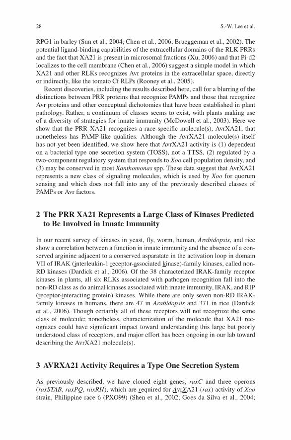
28 S.-W. Lee et al.
RPG1 in barley (Sun et al., 2004; Chen et al., 2006; Brueggeman et al., 2002). Thepotential ligand-binding capabilities of the extracellular domains of the RLK PRRsand the fact that XA21 is present in microsomal fractions (Xu, 2006) and that Pi-d2localizes to the cell membrane (Chen et al., 2006) suggest a simple model in whichXA21 and other RLKs recognizes Avr proteins in the extracellular space, directlyor indirectly, like the tomato Cf RLPs (Rooney et al., 2005).
Recent discoveries, including the results described here, call for a blurring of thedistinctions between PRR proteins that recognize PAMPs and those that recognizeAvr proteins and other conceptual dichotomies that have been established in plantpathology. Rather, a continuum of classes seems to exist, with plants making useof a diversity of strategies for innate immunity (McDowell et al., 2003). Here weshow that the PRR XA21 recognizes a race-specific molecule(s), AvrXA21, thatnonetheless has PAMP-like qualities. Although the AvrXA21 molecule(s) itselfhas not yet been identified, we show here that AvrXA21 activity is (1) dependenton a bacterial type one secretion system (TOSS), not a TTSS, (2) regulated by atwo-component regulatory system that responds to Xoo cell population density, and(3) may be conserved in most Xanthomonas spp. These data suggest that AvrXA21represents a new class of signaling molecules, which is used by Xoo for quorumsensing and which does not fall into any of the previously described classes ofPAMPs or Avr factors.
2 The PRR XA21 Represents a Large Class of Kinases Predictedto Be Involved in Innate Immunity
In our recent survey of kinases in yeast, fly, worm, human, Arabidopsis, and riceshow a correlation between a function in innate immunity and the absence of a con-served arginine adjacent to a conserved asparatate in the activation loop in domainVII of IRAK (interleukin-1 receptor-associated kinase)-family kinases, called non-RD kinases (Dardick et al., 2006). Of the 38 characterized IRAK-family receptorkinases in plants, all six RLKs associated with pathogen recognition fall into thenon-RD class as do animal kinases associated with innate immunity, IRAK, and RIP(receptor-interacting protein) kinases. While there are only seven non-RD IRAK-family kinases in humans, there are 47 in Arabidopsis and 371 in rice (Dardicket al., 2006). Though certainly all of these receptors will not recognize the sameclass of molecule; nonetheless, characterization of the molecule that XA21 rec-ognizes could have significant impact toward understanding this large but poorlyunderstood class of receptors, and major effort has been ongoing in our lab towarddescribing the AvrXA21 molecule(s).
3 AVRXA21 Activity Requires a Type One Secretion System
As previously described, we have cloned eight genes, raxC and three operons(raxSTAB, raxPQ, raxRH), which are required for AvrXA21 (rax) activity of Xoostrain, Philippine race 6 (PXO99) (Shen et al., 2002; Goes da Silva et al., 2004;
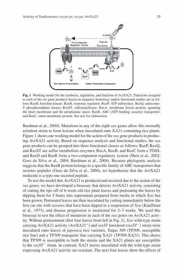
Activity of Xanthomonas oryzae pv. oryzae AvrXA21 29
RaxH
RaxS
RaxA
RaxC
Two-ComponentRegulatory System
Type one
Secretion
Hi
PSO3 APS PAPS
PAP
AvrXa21
RaxR
Rax Operon Transcription
RaxP
Sulfate Metabolism Components
RaxB
Xa21
RaxQ
-S-S
-S
P
Fig. 1 Working model for the synthesis, regulation, and function of AvrXA21. Functions assignedto each of the rax gene products based on sequence homology and/or functional studies are as fol-lows RaxH, histidine kinase; RaxR, response regulator; RaxP, ATP sulfurylase; RaxQ, adenosine-5′-phosphosulphate kinase; RaxST, sulfotransferase; RaxA, membrane fusion protein, spanningthe inner membrane and the periplasmic space; RaxB, ABC (ATP binding cassette) transporter;and RaxC, outer-membrane protein. See text for elaboration
Burdman et al., 2004). Mutations in any of the eight rax genes allow this normallyavirulent strain to form lesions when inoculated onto XA21-containing rice plants.Figure 1 shows our working model for the action of the rax gene products in produc-ing AvrXA21 activity. Based on sequence analysis and functional studies, the raxgene products can be grouped into three functional classes as follows: RaxP, RaxQ,and RaxST are sulfur metabolism enzymes; RaxA, RaxB, and RaxC form a TOSS;and RaxH and RaxR form a two-component regulatory system (Shen et al., 2002;Goes da Silva et al., 2004; Burdman et al., 2004). Because phylogenic analysissuggests that the RaxB protein belongs to a specific family of ABC transporters thatsecretes peptides (Goes da Silva et al., 2004), we hypothesize that the AvrXA21molecule is a type one secreted peptide.
To test the model that AvrXA21 is produced and secreted due to the action of therax genes, we have developed a bioassay that detects AvrXA21 activity, consistingof cutting the tips off of 6-week-old rice plant leaves and pretreating the leaves bydipping them for 5 hours into supernatant prepared from media in which Xoo hasbeen grown. Pretreated leaves are then inoculated by cutting immediately below thefirst cut site with scissors that have been dipped in a suspension of Xoo (Kauffmanet al., 1973), and disease progression is monitored for 2–3 weeks. We used thisbioassay to test the effect of mutations in each of the rax genes on AvrXA21 activ-ity. Without pretreatment (first four leaves from left in Fig. 2), Xoo wild-type straincarrying AvrXA21 activity (AvrXA21+) and raxST knockout (raxST−) strain wereinoculated onto leaves of japonica rice varieties, Taipei 309 (TP309, susceptiblerice line) and a TP309 transgenic line carrying XA21 (TP309-XA21). This showsthat TP309 is susceptible to both the strains and the XA21 plants are susceptibleto the raxST− strain. In contrast, XA21 leaves inoculated with the wild-type strainexpressing AvrXA21 activity are resistant. The next four leaves show the effects of
30 S.-W. Lee et al.
pretreatment with supernatants from strain supernatant onto various Xoo genotypes.Pretreatment of wild-type leaves in XA21 rice plants prevents infection by theraxST− strain. In contrast, pretreatment with supernatant from the AvrXA21−,rax-gene knock-out mutants, including raxST− (Fig. 2), raxA− (Fig. 2), and raxB−,raxC−, raxP−, and raxQ− strains (data not shown), does not prevent disease by theraxST− strain. However, a hrpC−, TTSS-deficient strain (Zhu et al., 2000) has noeffect on AvrXA21 activity as pretreatment with supernatant from this strain blockslesion development by the raxST− strain. These results indicate that AvrXA21activity is dominant, i.e., it can block lesion development by AvrXA21 minusstrains, and is secreted by a TOSS but not the TTSS, leading to recognition byXA21 at the cell surface.
This is the first discovery of type I secreted factor that can trigger innate immu-nity. While there are five systems (type I–V) for protein secretion in Gram-negativebacteria (Henderson et al., 2004), only the TTSS has previously been shown tosecrete Avr factors, which are then detected intracellularly. We hypothesize that
Fig. 2 Bioassay showing that AvrXA21 activity is present in medium of PXO99 wild-type andTTSS deficient mutant (hrpC−) strains, but not in a TOSS-deficient mutant (raxA−) strain or asulfuryltransferase-deficient (raxST−) strain. Rice leaves from cultivar TP309 and TP309 trans-genic for XA21 (TP309-XA21) were inoculated with PXO99 wild type (first and third leaves fromleft) or raxST− (second and fourth leaves from left) strains using the standard clipping method inwhich scissors are dipped in a solution of bacteria (OD600 = 0.5), the rice leaves are clipped, andthen lesions are measured 2 weeks later. To measure AvrXA21 activity, the raxST− strain was usedfor inoculations onto TP309-XA21 following pretreatment with cell-free supernatant of PXO99wild-type, raxST− , raxA− , and hrpC− strains. Shown are representative leaves from one of threeindependent experiments
Activity of Xanthomonas oryzae pv. oryzae AvrXA21 31
other members of the large complement of plant non-RD IRAK-family recep-tor kinases will be found to detect molecules secreted by other systems (Dardicket al., 2006). Indeed, indirect evidence suggests that type II secreted molecules areinvolved in eliciting plant defense (Jha et al., 2005).
4 The AVRXA21 Pathogen-Associated Molecule Is Conservedin Xanthomonas campestris pv. campestris
XA21 confers resistance to 29 out of 32 tested Xoo strains, which suggests that all 29strains carry AvrXA21 activity (Wang et al., 1996). In our previous report, the Xoostrain KR1, which lacks AvrXA21 activity, acquired this activity when the raxSTABoperon was provided on a plasmid (Goes da Silva et al., 2004). We hypothesizedthat other Xanthomonas species carry the cognate molecules that confers AvrXA21activity but lack the appropriate sulfation and secretion systems. To test this possibil-ity, we introduced the PXO99 raxSTAB region into a bacteria that is non-pathogenicon rice, Xcc ATCC 33913. We chose this strain because it carries closely relatedhomolog of all the rax genes except for the raxSTAB operon.
We carried out the AvrXA21 activity assay with supernatant prepared fromXcc transformed with the PXO99 raxSTAB genes and compared the lesion lengths(Table 1). Pretreatment with the supernatant of Xoo wild-type strain and the Xccstrain carrying the raxSTAB genes induces resistance against infection by theraxST− Xoo (lesion lengths of 1.3 ± 0.43 and 1.8 ± 0.8 cm, respectively); whereas,leaves pretreated with the supernatant of Xoo raxST− strain and Xcc wild-type strainhave long lesions (10.1 ± 5.2 and 10.0 ± 4.8 cm). Thus, the supernatant of Xcccarrying raxST, raxA, and raxB genes possesses AvrXA21 activity. Furthermore,Xcc carrying Xoo raxST and raxA genes showed full AvrXA21 activity but not withraxST alone (data not shown), suggesting that AvrXA21 modification and secretionmay be a sequential process requiring both the raxST and the RaxA gene productsor that the activity or stability is altered in the absence of RaxA.
Importantly, these results suggest that the core AvrXA21 molecule is conservedamong different species, a key component of the definition of a PAMP. Nonetheless,the requirement for raxST and raxA suggests that XA21-mediated recognition ofthe AvrXA21 molecule requires a specific TOSS inner membrane protein (RaxA)and/or a post-translational modification, sulfation, that is likely catalyzed by theputative sulfotransferase, RaxST (Goes da Silva et al., 2004). There are many exam-ples of post-translational modification affecting extracellular recognition. Typically,
Table 1 Comparison of lesion lengths on rice leaves. The data are averages from 10 scored leaves
Pretreatment Xccof supernatant PXO99 raxST− Xcc (raxSTAB)
Inoculation raxST−
Lesion length a 1.3 ± 0.43 10.1 ± 5.2 10.0 ± 4.8 1.8 ± 0.8a cm, average ± standard deviation

32 S.-W. Lee et al.
sulfated molecules are directed outside of the cell to serve as modulators of cell–cell interactions (Bowman et al., 1999). A notable example pertinent to agricultureis sulfation of the Sinorhizobium meliloti Nod factor that is required for specificrecognition by its host alfalfa (Roche et al., 1991). In plants, tyrosine sulfation ofphytosulfokine is required for recognition by the phytosulfokine receptor kinase, aplasma membrane RLK for proliferation of plant cells (Matsubayashi et al., 2002).
The other element of the definition of a PAMP is that it is essential for thepathogen. The fact that the core of AvrXA21 appears to be conserved in Xcc sug-gests that the molecule(s) has a function that makes it selectively advantageous.In addition, while we did not observe differences in virulence in the raxSTAB andraxC knockout strains under controlled conditions, in a field study, 37 Xoo Koreanstrains lacking AvrXA21 activity appeared to have reduced fitness (Choi, 2003). Anadditional study showed that Xoo strains that lost AvrXA21 activity did not persistat the same field site into the next season suggesting that these strains were at acompetitive disadvantage (C.M. Vera Cruz, personal communication). These resultssuggest both that the core AvrXA21 molecule is conserved between Xoo and Xccand that loss of AvrXA21 results in a fitness cost to Xoo. Therefore, AvrXA21, thedetection of which by PRR XA21 confers dominant resistance, shares characteris-tics of PAMPs and Avr proteins.
5 Cell Density Dependent Expression of Rax Genes
What is the function of AvrXA21? Like other living organisms, bacteria have devel-oped multiple systems for responding to environmental variation, such as changesin temperature, osmolarity, pH, nutrient availability, and even population size. Two-component systems, composed of histidine kinases (HK) and response regulators(RR), have an important role in environment sensing. Phosphorylation of the RRby the HK regulates gene expression and governs the response to an environmentalstimulus (Charles et al., 1992). Bacteria themselves produce some stimuli. In a pro-cess called quorum sensing (QS), small molecules serve as signals to recognize cellpopulation, leading to changes in expression of specific genes when the signal hasaccumulated to some threshold concentration (Fuqua, et al., 1994). QS moleculesregulate their own expression and are also called autoinducers (Waters et al., 2005).
Preliminary studies using the raxST promoter in front of GFP suggested thatrax gene expression may be dependent on cell density (data not shown). To testthis more thoroughly, the wild-type strain (PXO99) was cultured for 72 hours andthen diluted. The diluted cultures were returned to the incubator and, as they grew,aliquots were removed for RNA isolation. Gene expression was measured usingreal-time quantitative PCR and primers specific for the following four rax genes,one in each operon: raxST, in raxSTAB (Fig. 3); raxP, in raxPQ; raxR, in raxRH;and raxC (data not shown). Remarkably, the expression of all four rax genes wasonly observed in the wild-type strain at high cell densities; whereas, levels of aribosomal RNA were unaffected. We also observed rax gene expression in a strainconstitutively expressing RaxR. Figure 3 shows that the density dependence of rax
Activity of Xanthomonas oryzae pv. oryzae AvrXA21 33
Log (cell population)1e+6 1e+7 1e+8
Rel
ativ
e ra
xST
exp
ress
ion
(%)
0
20
40
60
80
100
120
Fig. 3 Cell-density-dependent expression of the raxST gene. Cultures of wild-type (circles) andraxR constitutive expression mutant (PXO99raxR− /pML122raxR: squares) strains were grown tosaturation in NB media for 72 hours at 28 ◦C (cell density at OD600 ≥ 1×10⁸ colony forming units(CFU)/mL and then diluted to 1×10⁵ CFU/mL. We measured expression of the raxST gene (closedsymbols, solid lines) by preparing RNA from samples at various cell densities after dilution andperforming real-time quantitative PCR with three primers pair corresponding to different region ofthe gene. Ribosomal RNA (open symbols, dashed lines) in wild-type and the mutant strains wereused as a control. Percent expression at each cell density was calculated relative to the copy numberof the sample at 1 × 10⁵. Data are from one of two independent experiments. Expression is highat 105 CFU/ml because the strains were grown to high density and diluted, i.e., the bacteria in thissample behave as if they are still at high density
gene expression in the raxR overexpressing strain is partially lost. This result indi-cates that RaxR is involved in regulation of the density-dependent expression ofthe rax genes, including itself. This auto-induction is consistent with a roll of theproduct of the rax gene pathway, namely AvrXA21, in QS (Waters et al., 2005).
The characteristics of the Xoo rax-genes suggest a QS system with features sim-ilar to that typically found in Gram-positive bacteria; thus, the AvrXA21 QS systemrepresents a novel class of QS in Gram-negative bacteria. Oligpeptides are the pre-dominant signaling molecules for genetic competence in Bacillus substilis (Tortosaet al., 1999), virulence in Staphylococcus aureus (Novick et al., 1999), and the pro-duction of antimicrobial peptides, inducing bacteriocins and lantibiotics, in lacticacid bacteria (Risoen et al., 2000; Kleerebezem et al., 1997). As QS molecules,these peptides also activate their own expression and have been given the name auto-inducible peptide (AIP). AIPs are known to be a secreted by ABC-transporters thatare similar to RaxB and are sensed by two-component systems. Significantly, whileQS via two-component systems is not unique to Gram-positive bacteria, secretionof the putative AvrXA21 peptide by a TOSS would be the first described occurrenceof an AIP in Gram-negative bacteria.
In summary, our data suggest that the AvrXA21 PAM is a secreted peptide, andhint that the biological function of the AvrXA21 PAM is as a QS signal molecule, the

34 S.-W. Lee et al.
production of which is regulated in a cell-density-dependent manner in the Gram-negative bacteria, Xoo, by a two-component system. Thus, the rice XA21 PRReffectively eavesdrops on the AvrXA21-mediated molecular conversation amongXoo, using AvrXA21 as an indicator of the presence of a pathogenic bacterialpopulation of significant size and leading to plant responses that effectively haltinfection (Fig. 1). Sequence analysis of the genomes of several phytopathogenicbacteria that are similar to Xoo reveals the abundance of the rax classes of molecularcomponents. Xoo, Xcc, Xanthomonas axonopodis pv. citri, and Xylella fastidiosaeach possess genes for approximately 30 two-component systems together withthree to five TOSSs that, like RaxB, are predicted to transport peptides. Further-more, it is apparent that X. fastidiosa does not use TTSS for pathogenesis becausegenome analysis has revealed the absence of these components in this bacterium.Rather, X. fastidiosa contains TOSS genes similar to raxA, raxB, and raxC as wellas sequences encoding as many as 10–12 peptides that are candidates for secretion(C. Dardick and P.C. Ronald, unpublished results). These observations highlight thatthe interactions among phytopathogens, their hosts, and the environment are moresophisticated than currently recognized.
Our data suggesting that the function of the AvrXA21 pathogen-associatedmolecule is a QS signaling molecule may present a critical clue to answering thequestion, why is AvrXA21 maintained in Xoo regardless of its use for detectionby a host protein? Based on others’ results that suggest decreased fitness of raxmutants, AvrXA21 may be essential for Xoo as a signal molecule for cell–cell com-munication. We carefully hypothesize that Xoo may use QS to coordinate infec-tion and production of virulence factors. QS has global effects on bacteria growth,survival, and interactions with eukaryotes as QS-responsive genes compose up to10% of the Pseudomonas aeruginosa trascriptome (Wagner et al., 2003). In thatreport, a large portion of QS-promoted genes code for membrane proteins and pro-tein export apparatuses involved in secretion of virulence factors. A relationshipbetween QS and virulence has also been described for the human pathogen Vibriocholerae (Miller et al., 2002). Interestingly, in another case a host can produce QSmimic molecules that interfere the QS regulated behaviors of the infecting bacteria(Teplitski et al., 2004). However, given the abundance of other QS peptides andsmall molecules such as bacteriocins and lantibiotics in the host vicinity, it willnot be surprising if these peptides are shown to play a role in the interaction ofother bacteria with their hosts. In principle, recognition of TOSS elicitors could beexploited to generate new types of specific and environmentally benign pesticidesfrom those elicitors.
6 Perspective
The data presented here as well as results of others in recent years establish thatthere is not a clear dichotomy between PAMP recognition and Avr recognition;rather, plant innate immunity functions as a continuum between the two generaltypes (Table 2) (Sun et al., 2006).
Activity of Xanthomonas oryzae pv. oryzae AvrXA21 35
Table 2 General characteristics of PAMPs (pathogen-associated molecular patterns) and the PRR(pathogen recognition receptors) responsible for their recognition as opposed to the features ofAvr (avirulence) proteins and their corresponding PRR proteins. Characteristics of AvrXA21 andXA21 are shown in bold if they appear to be unique compared with known systems
PAMP·PRR Generalcharacteristics
AvrXA21·XA21characteristics
Avr·PRR Generalcharacteristics
PAMP (e.g., flagellin)
- Conserved amongdiverse species
- Required for lifecycle
- Not typically activelysecreted
AvrXA21
- Strain specific
- Activity is regulated in acell-density dependent by atwo-component system
- Activity requiresmodification enzymes
- Synthesized in absence ofplant host (thus part oflifecycle)
- Absence compromisesvirulence in field
- Secreted by TOSS
Avr (e.g., AvrRpt2)
- Strain specific
- Many synthesized only inpresence of host
- Absence compromises viru-lence
- From bacteria, secreted byTTSS
PRR (e.g., FLS2)
- Extracellular,(intracellualr recognitiononly shown in animals)
- Recognition andresponse does not lead toresistance in most cases
XA21
- Recognition likelyextracellular
- Confers dominant resistance
PRR (e.g., Rps2)
- In dicots, recognition ismostly intracellular.
- Confers dominant resistance
PAMPs have been defined as being derived from conserved structures requiredfor pathogen function; whereas, Avr factors are thought to be maintained by spe-cific strains as virulence factors. The features of AvrXA21 form a hybrid betweenthese two categories. The AvrXA21 molecule appears to be conserved in Xcc, butsulfation seems to provide specificity to the system, just as flagellin recognitionby rice is modulated by glycosylation (Taguchi et al., 2003, 2003). Further evi-dence that plant-recognized PAMPs are not entirely conserved comes from a recentpaper showing that sequence variation in Xcc flagellin modulates FLS2-dependentpathogen recognition by Arabidopsis (Sun et al., 2006). Also, compromised vir-ulence and survival of avrXA21− strains in the field is consistent with a crucialrole for AvrXA21 in Xanthomonad quorum sensing. On the other hand, AvrXA21and XA21 fulfill the standard gene-for-gene model of the dominant resistance geneinteracting with the bacterial virulence factor as well. Thus, AvrXA21 has featuresthat are similar to both PAMPs and other Avr’s. For this reason, we propose theterminology pathogen-associated molecule (PAM) to describe both PAMPs and Avrfactors.
36 S.-W. Lee et al.
The fusion of PRR recognition of Avr and PAMP classes proposed here hasimplications for the mechanism of plant innate immunity. Recognition of PAMPsby the animal innate immune system has been proposed to act as guide for theadaptive immune system as to the nature of the infecting pathogen (Medzhitovet al., 1997). There is not an obvious need for such a function in plants since eachreceptor can be tied to a specific response through signal transduction. Nonethe-less, it is clear that the plant innate immune system recognizes both conserved andless conserved molecules. As articulated by Ausubel in 2005, the fact that plantresponses to PAMPs often do not lead to resistance may be due to evolutionaryselection for plant innate immune perception to require “priming” or “two-hits”so that resources are not unnecessarily wasted in responding to nonpathogenic orsmall populations of bacteria. In such a model, PAMP perception constitutes anearly warning, and indeed pretreatment with lipopolysaccharide or flagellin-derivedpeptide leads to resistance to subsequent treatments with normally pathogenic bac-teria (Erbs et al., 2003; Sun et al., 2006; Zipfel et al., 2004). However, it may bethat the necessity for the factor that primes the plant innate immune system to bea PAMP rather than an Avr molecule is simply a matter of experimental history.Because they are less useful in an agricultural context, we have not looked for PRRsand corresponding Avr molecules that do not lead to significant resistance. Thoughthe XA21D receptor, which confers only partial resistance (Wang et al., 1998), maybe such a molecule. Conversely, as mentioned above, FLS2-dependent resistanceto bacteria that express flagellin has also been observed (Zipfel et al., 2004). Phys-iologically relevant experiments in diverse genetic backgrounds may lead to fur-ther evidence that responses mediated by PRRs for both more and less conservedPAMs (i.e., PAMPs and Avr’s) are also part of a continuum, with some function-ing more often in priming and others more often leading to resistances. Alreadyamong described Avr PRRs there is a diversity of strengths, with some leading tosuper-resistance (Greenberg et al., 2004). Thus, in possessing receptors for moreconserved molecules, it may be that plants are simply taking advantage of the con-served nature of PAMPs as targets for recognition, rather than making a functionaldistinction in terms of responses.
Several examples support the idea that plants gain a selective advantage by mak-ing use of a variety of strategies for pathogen recognition beyond the classes ofmolecules that have typically been discussed (Chisholm, 2006; Dangl, 2001), bothin terms of the PAM detected and the cognate PRRs. AvrXA21 is currently the onlyknown PAM that is secreted by a TOSS. Until recently LRRs constituted the onlysignal recognition domain described for a cloned PRR RLK; however, a recentlycloned RLK conferring rice blast resistance, PID2, breaks from this by possessingan extracellular lectin-binding domain (Chen et al., 2006). Moreover, sequence anal-yses suggest that RLKs and RLPs with other, diverse extracellular binding domainsare also involved in plant innate immunity (Dardick et al., 2006; Shiu et al., 2003).Similarly, plants appear to use other entirely different classes of molecules as PRRsas well. Indeed, the first cloned PAMP high-affinity binding site was the soybean�-glucan elicitor binding protein (Umemoto, 1997). Localized to the extracellularside of the cell membrane and containing two glucan binding sites, it is predicted to

Activity of Xanthomonas oryzae pv. oryzae AvrXA21 37
be tethered to the membrane via interactions in a receptor complex (Mithofer, 2000).Yet another perception strategy is represented by Xa27, which codes for a noveldefense protein the promoter of which serves as the PRR by directly or indirectlyinteracting with the cognate nuclear-localized AvrXA27 type III effector (Gu, 2005).
In conclusion, our recent results suggest that the AvrXA21 PAM is a TOSS-dependent secreted peptide that represents a new family of signaling molecules forQS via a two-component system. Although QS molecules are known to be widelyconserved and essential for bacterial communication, they have never before beenshown to trigger innate immune responses. Our continuing work to identify theAvrXA21 molecule and understand the physical basis of its recognition may allowidentification of similar PAMs from plant and animal pathogens and their cognatehost receptors.
References
Ausubel, F.M., 2005, Are innate immune signaling pathways in plants and animals conserved? Nat.Immunol. 6:973–979.
Barton, G.M., and Medzhitov, R., 2003, Toll-like receptor signaling pathways, Science 300:1524–1525.
Bowman, K.G., and Bertozzi, C.R., 1999, Carbohydrate sulfotransferases: mediators of extracel-lular communication, Chem. Biol. 6:R9.
Brueggeman, R., Rostoks, N., Kudrna, D., Kilian, A., Han, F., Chen, J., Druka, A., Steffenson, B.,and Kleinhofs, A., 2002, The barley stem rust-resistance gene Rpg1 is a novel disease-resistancegene with homology to receptor kinases, Proc. Nat. Acad. Sci. USA 99:9328–9333.
Brunner, F., Rosahl, S., Lee, J., Rudd, J. J., Geiler, C., Kauppinen, S., Rasmussen, G., Scheel, D.,and Nuernberger, T., 2002, Pep-13, a plant defence-inducing pathogen-associated pattern fromPhytophthora transglutaminases, EMBO 21:6681–6688.
Burdman, S., Shen, Y., Lee, S.-W., Xue, Q., and Ronald, P., 2004, RaxH/RaxR: A Two-componentregulatory system in Xanthomonas oryzae pv. oryzae required for AvrXa21 activity, MPMI17:602–612.
Charles, T.C., Jin, S., and Nester, E.W., 1992, Two-component sensory transduction systems inphytobacteria, Ann. Rev. Phytopath. 30:463–484.
Chen, X., Shang, J., Chen, D., Lei, C., Zou, Y., Zhai, W., Liu, G., Xu, J., Ling, Z., Cao, G., Ma, B.,Wang, Y., Zhao, X., Li, S., and Zhu, L., 2006, A B-lectin receptor kinase gene conferring riceblast resistance, Plant J. 46:794–804.
Chinchilla, D., Bauer, Z., Regenass, M., Boller, T., and Felix, G., 2006, The Arabidopsis recep-tor kinase FLS2 binds flg22 and determines the specificity of flagellin perception, Plant Cell18:465–476.
Chisholm, S. T., Coaker, G., Day, B., and Staskawicz, B.J., 2006, Host-microbe interactions: shap-ing the evolution of the plant immune response, Cell 124:803–814.
Dangl, J.L., and Jones, J.D.G., 2001, Plant pathogens and integrated defence responses to infection,Nature 411:826.
Dardick, C., and Ronald, P., 2006, Plant and animal pathogen recognition receptors signal throughnon-RD kinases, PLoS Pathog 2:e2.
Dodds, P.N., Lawrence, G.J., Catanzariti, A.M., Teh, T., Wang, C.I., Ayliffe, M.A., Kobe, B., andEllis, J.G., 2006, Direct protein interaction underlies gene-for-gene specificity and coevolutionof the flax resistance genes and flax rust avirulence genes, Proc. Nat. Acad. Sci. USA 103:8888–8893.
38 S.-W. Lee et al.
Erbs, G., and Newman, M.-A., 2003, The role of lipololysaccharides in induction of plant defenceresponses, Mol. Plant Path. 4:421–425.
Fuqua, W.C., Winans, S.C., and Greenberg, E.P., 1994, Quorum sensing in bacteria: the LuxR-Lux1 family of cell density-responsive transcriptional regulators, J. Bacteriol. 176:269–275.
Felix, G., Duran, J.D., Volko, S., and Boller, T., 1999, Plants have a sensitive perception systemfor the most conserved domain of bacterial flagellin, Plant J. 18:265–279.
Flor, H.H., 1971, Current status of the gene for gene concept. Ann. Rev. Phytopath. 9:275–296.Girardin, S.E., Sansonetti, P.J., and Philpott, D.J., 2002, Intracellular vs. extracellular recognition
of pathogens—common concepts in mammals and flies, Trends Microbiol. 10:193–199.Goes da Silva, F., Shen, Y., Dardick, C., Burdman, S., Yadav, R., Sharma, P., and Ronald, P.,
2004, Bacterial genes involved in type I secretion and sulfation are required to elicit the riceXa21-mediated innate immune response, MPMI 17:593–601.
Gomez-Gomez, L., Felix, G., and Boller, T. (1999) A single locus determines sensitivity to bacte-rial flagellin in Arabidopsis thaliana, Plant J. 18:277–284.
Greenberg, J.T., and Yao, N., 2004, The role and regulation of programmed cell death in plant–pathogen interactions, Cell. Microbiol. 6:201–211.
Henderson, I.R., Navarro-Garcia, F., Desvaux, M., Fernandez, R.C., and Ala’Aldeen, D., 2004,Type V protein secretion pathway: the autotransporter Story. Microbiol. Mol. Biol. Rev. 68:692–744.
Inohara, N., and Nunez, G., 2003, NODs: intracellular proteins involved in inflammation and apop-tosis, Nat. Rev. Immunol. 3:371–382.
Janeway, C.A., Jr., and Medzhitov, R., 2002, Innate immune recognition, Annu. Rev. Immunol.20:197–216.
Jha, G., Rajeshwari, R., and Sonti, R.V., 2005, Bacterial type two secretion system secreted pro-teins: double edged swords for plant pathogens, Mol. Plant In. 18:891–898.
Jurk, M., Heil, F., Vollmer, J., Schetter, C., Krieg, A. M., Wagner, H., Lipford, G., and Bauer, S.,2002, Human TLR7 and TLR8 independently confer responsiveness to the antiviral compoundR-848, Nat. Immunol. 3:499.
Kauffman, H.E., Reddy, A.P.K., Hsieh, S.P.Y., and Merca, S.D., 1973. An improved tech-nique for evaluating resistance of rice varieties to Xanthomonas oryzae, Plant Dis. Rep. 57:537–541.
Kleerebezem, M., Quadri, L.E., Kuipers, O.P., and de Vos, W.M., 1997, Quorum sensing by peptidepheromones and two-component signal-transduction systems in Gram-positive bacteria. Mol.Microbiol. 24:895–904.
Leulier, F., Parquet, C., Pili-Floury, S., Ryu, J.H., Caroff, M., Lee, W. J., Mengin-Lecreulx, D., andLemaitre, B., 2000, The Drosophila immune system detects bacteria through specific peptido-glycan recognition, Nat. Immunol. 4:478–484.
Matsubayashi, Y., Ogawa, M., Morita, A., and Sakagami, Y., 2002, An LRR receptorkinase involved in perception of a peptide plant hormone, phytosulfokine, Science 296:1470–1472.
McDowell, J.M., and Woffenden, B.J., 2003, Plant defense, one post, multiple guards? TrendsBiotechnol. 21:178–183.
Medzhitov, R., and Janeway, C.A., Jr., 1997, Innate immunity: the virtues of a nonclonal system ofrecognition, Cell 91:295–298.
Miller, M.B., Skorupski, K., Lenz, D.H., Taylor, R.K., and Bassler, B.L., 2002, Parallel quorumsensing systems converge to regulate virulence in Vibrio cholerae, Cell 110:303–314.
Mudgett, M.B., 2005, New insights into the function of phytopathogenic bacterial type III effectorsin plants, Ann. Rev. Plant Biol. 56:509–531.
Novick, R.P., and Muir, T.W., 1999, Virulence gene regulation by peptides in staphylococci andother Gram-positive bacteria, Curr. Opin. Microbiol. 2:40–45.
Nurnberger, T., Brunner, F., Kemmerling, B., and Piater, L., 2004, Innate immunity in plants andanimals: striking similarities and obvious differences, Immunol. Rev. 198:249–266.
Ramos, H.C., Rumbo, M., and Sirard, J.C., 2004 Bacterial flagellins: mediators of pathogenicityand host immune responses in mucosa, Trends Microbiol. 12:509–517.
Activity of Xanthomonas oryzae pv. oryzae AvrXA21 39
Risoen, P.A., Brurberg, M.B., Eijsink, V.G., and Nes, I.F., 2000, Functional analysis of promotersinvolved in quorum sensing-based regulation of bacteriocin production in Lactobacillus, Mol.Microbiol. 37:619–628.
Roche, P., Debelle, F., Maillet, F., Lerouge, P., Faucher, C., Truchet, G., Denarie, J., andProme, J.-C., 1991, Molecular basis of symbiotic host specificity in Rhizobium meliloti: nodHand nodPQ genes encode the sulfation of lip-oligosaccharide signals, Cell 67:1131
Ronald, P.C., Albano, B., Tabien, R., Abenes, L., Wu, K.S., McCouch, S., and Tanksley, S.D.,1992, Genetic and physical analysis of the rice bacterial blight disease resistance locus, Xa-21,Mol. Gen. Genet. 236:113–120.
Rooney, H.C., Van’t Klooster, J.W., van der Hoorn, R.A., Joosten, M.H., Jones, J.D., andde Wit, P.J., 2005, Cladosporium Avr2 inhibits tomato Rcr3 protease required for Cf-2-dependent disease resistance, Science 308:1783–1786.
Shen, Y., Sharma, P., da Silva, F.G., and Ronald, P., 2002, The Xanthomonas oryzae pv. oryzaeRaxP and RaxQ genes encode an ATP sulphurylase and adenosine-5’-phosphosulphate kinasethat are required for AvrXa21 avirulence activity, Mol. Microbiol. 44:37–48.
Shiu, S.H., and Bleecker, A.B., 2003, Expansion of the receptor-like kinase/Pelle gene family andreceptor-like proteins in Arabidopsis, Plant Physiol. 132:530–543.
Song, W.Y., Wang, G.L., Chen, L.L., Kim, H.S., Pi, L.Y., et al., 1995, A receptor kinase-like proteinencoded by the rice disease resistance gence, Xa-21, Science 270:1804–1806.
Staskawicz, B.J., Mudgett, M.B., Dangl, J.L., and Galan, J.E., 2001, Common and contrastingthemes of plant and animal diseases, Science 292:2285–2289.
Sun, X., Cao, Y., Yang, Z., Xu, C., Li, X., Wang, S., and Zhang, Q., 2004, Xa26, a gene conferringresistance to Xanthomonas oryzae pv. oryzae in rice, encodes an LRR receptor kinase-likeprotein, Plant J. 37:517–527.
Sun, W., Dunning, F.M., Pfund, C., Weingarten, R., and Bent, A.F., 2006, Within-species flag-ellin polymorphism in Xanthomonas campestris pv. campestris and its impact on elicitation ofArabidopsisFLAGELLIN SENSING2-dependent defense, Plant Cell 18:764–779.
Szurek, B., Marois, E., Bonas, U., and Van den Ackerveken, G., 2001, Eukaryotic features of theXanthomonas type III effector AvrBs3: protein domains involved in transcriptional activationand the interaction with nuclear import receptors from pepper, Plant J. 26:523–534.
Taguchi, F., Shimizu, R., Inagaki, Y., Toyoda, K., Shiraishi, T., and Ichinose, Y., 2003, Post-translational modification of flagellin determines the specificity of HR production, Plant CellPhysiol. 44:342–349.
Takeuchi, K., Taguchi, F., Inagaki, Y., Toyoda, K., Shiraishi, T., and Ichinose, Y., 2003, Flagellinglycosylation island in Pseudomonas syringae pv. glycinea and its role in host specificity, J.Bacteriol. 185:6658–6665.
Thomma, B.P., Penninckx, I.A., Broekaert, W.F., and Cammue, B.P., 2001, The complexity ofdisease signaling in Arabidopsis, Curr. Opin. Immunol. 13:63–68.
Teplitski, M., Chen, H., Rajamani, S., Gao, M., Merighi, M., Sayre, R.T., Robinson, J.B.,Rolfe, B.G., and Bauer, W.D., 2004, Chlamydomonas reinhardtii secretes compounds thatmimic bacterial signals and interfere with quorum sensing regulation in bacteria, Plant Physiol.134:137–146.
Tortosa, P., and Dubnau, D., 1999, Competence for transformation: a matter of taste, Curr. Opin.Microbiol. 2:588–592.
van’t Slot, K., and Knogge, W,K., 2002, A dual role for microbial pathogen-derived effector pro-teins in plant disease and resistance, Crit. Rev. Plant Sci. 21:229–271.
Wagner, V.E., Bushnell, D., Passador, L., Brooks, A.I., and Iglewski, B.H., 2003, Microarrayanalysis of Pseudomonas aeruginosa quorum-sensing regulons: effects of growth phase andenvironment, J. Bacteriol. 185:2080–2095.
Wang, G.L., Ruan, D.L., Song, W.Y., Sideris, S., Chen, L., Pi, L.Y., Zhang, S., Zhang, Z.,Fauquet, C., Gaut, B.S., Whalen, M.C., and Ronald, P.C., 1998, Xa21D encodes a receptor-likemolecule with a leucine-rich repeat domain that determines race-specific recognition and issubject to adaptive evolution, Plant Cell 10:765–779.

40 S.-W. Lee et al.
Wang, G.L., Song, W.Y., Ruan, D.L., Sideris, S., and Ronald, P.C., 1996, The cloned gene, Xa21,confers resistance to multiple Xanthomonas oryzae pv. oryzae isolates in transgenic plants,Mol. Plant Microbe Interact. 9:850–855.
Waters, C.M., and Bassler, B.L., 2005, Quorum sensing: Cell-to cell communication in bacteria,Ann. Rev. Cell Develop. Biol. 21:319–346.
Werling, D., and Jungi, T.W., 2003, TOLL-like receptors linking innate and adaptive immuneresponse, Vet. Immunol. Immunop. 91:1–12.
Zhu, W., MaGbanua, M.M., and White, F.F., 2000, Identification of two novel hrp-associated genesin the hrp gene cluster of Xanthomonas oryzae pv. Oryzae, J. Bacteriol. 182:1844–1853.
Zipfel, C., Robatzek, S., Navarro, L., Oakeley, E.J., Jones, J.D., Felix, G., and Boller, T., 2004,Bacterial disease resistance in Arabidopsis through flagellin perception, Nature 428:764–767.
Unraveling the Genetic Mysteries of the Cat:New Discoveries in Feline-Inherited Diseasesand Traits
Leslie A. Lyons
Abstract The domestic cat is one of man’s most beloved species, living in house-holds as companions, working on farms for vermin control, and co-habitating inurban environments as semi-feral occupants. Advances in veterinary medicine pro-vide health care and diagnostics for the domestic cat on a comparable level tohumans. Fancy breeds result in the selection of aesthetically pleasing traits and,sometimes, undesired health conditions, both of which can be useful as models forhuman development, physiology, and health. Reproductive techniques have allowedresearch in the cat to expand into cloning studies and genomic tools are assisting thecat with the same research scrutiny as the more prominent research animal models,rodents and humans. This chapter explores the genetic mysteries of the cat and pro-vides a current state of the union for cat genetic research.
Cat \’kat\ – a long domesticated carnivorous mammal that is usually regardedas a distinct species though probably ultimately derived by selection, among thehybrid progeny of several small Old World wildcats that occurs in several varietiesdistinguished chiefly by length of coat body and presence or absence of tail and thatmakes the pet valuable in controlling rodents and other small vermin but tends torevert to a feral state if not housed and cared for (Webster 3rd New InternationalDictionary).
1 Cat Phenotypes
The domestic cat is one of 36 extant species of felids (Kitchener, 1991; Seidenstickerand Lumpkin, 1991; Sunquist and Sunquist, 2002), grouping with four other OldWorld wildcat species in the domestic cat “Felis” lineage, including, Felis bieti, theChinese desert cat, Felis margarita, the sand cat, Felis nigripes, the black-footed cat,and Felis chaus, the jungle cat (Johnson et al., 2006). Two additional wildcats, theEuropean wildcat, Felis silvestris, and the African wildcat, Felis libyca, are usuallyconsidered separate species and distinct from the domestic cat. However, these two
L.A. LyonsDepartment of Population Health & Reproduction, School of Veterinary Medicine, University ofCalifornia, Davis, CA 95616, USA
J.P. Gustafson et al. (eds.), Genomics of Disease, 41C© Springer Science+Business Media, LLC, 2008
42 L.A. Lyons
wildcats produce fertile hybrids with domestic cats and the speciation of these threecats remains a mystery, as genetic studies have not yet been able to focus on theirrecent divergence. Many other sub-species of European, Asian, and African wildcatshave been described; however, the true origins and the number of domesticationevents that led to the domestication of the cat has not been resolved and also remaina mystery.
The term “domesticate” has various definitions, including “to adapt to life inintimate association with and to the advantage of man.” Signs of domesticationinclude docility, retention of juvenile features, smaller brain size, raised tail car-riage, droopy ears, and pelage coloration, fur types, and morphologies that wouldlikely have a selective disadvantage in the wild (Hemmer, 1972, 1976, 1978, 1990;Kukekova et al., 2006; Wayne, 1986). Cats have been recognized to have “sevenancient mutations” that were evident before the advent of breeds (Wastlhuber, 1991).These mutations, melanism, dilution, long fur, orange, white spotting, the classictabby markings, and dominant white, all affect pelage coloration or length and werelikely the focus of early phenotypic selections by humans (Fig. 1). Many studieshave examined the distribution of coat colors in various populations throughout theworld, which revealed a “cline” of color variation frequencies across the Old World(Clark, 1975; Davis and Davis, 1977; Todd and Lloyd, 1984; Todd et al., 1974,1975; Wagner and Wolsan, 1987). These same phenotypic mutations segregate ina variety of modern cat breeds (Robinson, 1991; Vella et al., 1999). The genescontrolling these cat phenotypes and their associated mutations are presented inTable 1. Two of the mutations, dominant white and long fur, are yet to have directscientific study in the cat, but candidate genes such as KIT (Brooks and Bailey, 2005;Cooper et al., 2006; Johansson Moller et al., 1996; Marklund et al., 1998; Terryet al., 2001) and fibroblast growth factor 5 (FGF5) (Housley and Venta, 2006;Sundberg et al., 1997) have been shown to cause similar phenotypes in other species.The mutations for melanism and dilution have been determined (Eizirik et al., 2003;Ishida et al., 2006); however, only genes or genetic regions have been implicated fororange, white spotting, and tabby (Cooper et al., 2006; Grahn et al., 2005; Lyonset al., 2006).
The advent of the cat fancy in England during the late 1800s fostered the develop-ment of cat breeds (CFA, 2004). Although over 50 cat “breeds” can be demarcatedby breeders today (Morris, 1999a b), many of the cat breeds are single gene vari-ants of a parent breed and hence these different breeds are considered “variants” or“classes” in one cat fancy association or a distinctive breed by a different cat fancyassociation (http://www.cfainc.org/ and http://www.tica.org/). For example, ExoticShorthairs are a short-haired variety of Persians, long hair being the recessive trait.Himalayans are “pointed” Persians, often representing a color class or variety. Onebreed that had early recognition by the cat fancy is the Siamese. These cats havea distinctive temperature-sensitive pattern to their coloration; melanin is producedwhere the cats body temperature is lower. Hence, the “points” of a Siamese cat is aresult of proper color production at the extremities, including the face, ears, paws,and tail. A close cousin to the Siamese, the Burmese, has a less-severe temperature-sensitive color restriction; hence lower production of eumelanin occurs, resulting in
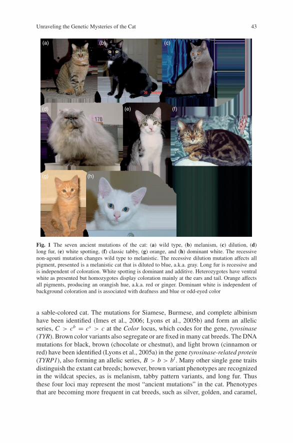
Unraveling the Genetic Mysteries of the Cat 43
(a)
(d)
(h)(g)
(e) (f)
(b) (c)
Fig. 1 The seven ancient mutations of the cat: (a) wild type, (b) melanism, (c) dilution, (d)long fur, (e) white spotting, (f) classic tabby, (g) orange, and (h) dominant white. The recessivenon-agouti mutation changes wild type to melanistic. The recessive dilution mutation affects allpigment, presented is a melanistic cat that is diluted to blue, a.k.a. gray. Long fur is recessive andis independent of coloration. White spotting is dominant and additive. Heterozygotes have ventralwhite as presented but homozygotes display coloration mainly at the ears and tail. Orange affectsall pigments, producing an orangish hue, a.k.a. red or ginger. Dominant white is independent ofbackground coloration and is associated with deafness and blue or odd-eyed color
a sable-colored cat. The mutations for Siamese, Burmese, and complete albinismhave been identified (Imes et al., 2006; Lyons et al., 2005b) and form an allelicseries, C > cb = cs > c at the Color locus, which codes for the gene, tyrosinase(TYR). Brown color variants also segregate or are fixed in many cat breeds. The DNAmutations for black, brown (chocolate or chestnut), and light brown (cinnamon orred) have been identified (Lyons et al., 2005a) in the gene tyrosinase-related protein(TYRP1), also forming an allelic series, B > b > bl . Many other single gene traitsdistinguish the extant cat breeds; however, brown variant phenotypes are recognizedin the wildcat species, as is melanism, tabby pattern variants, and long fur. Thusthese four loci may represent the most “ancient mutations” in the cat. Phenotypesthat are becoming more frequent in cat breeds, such as silver, golden, and caramel,
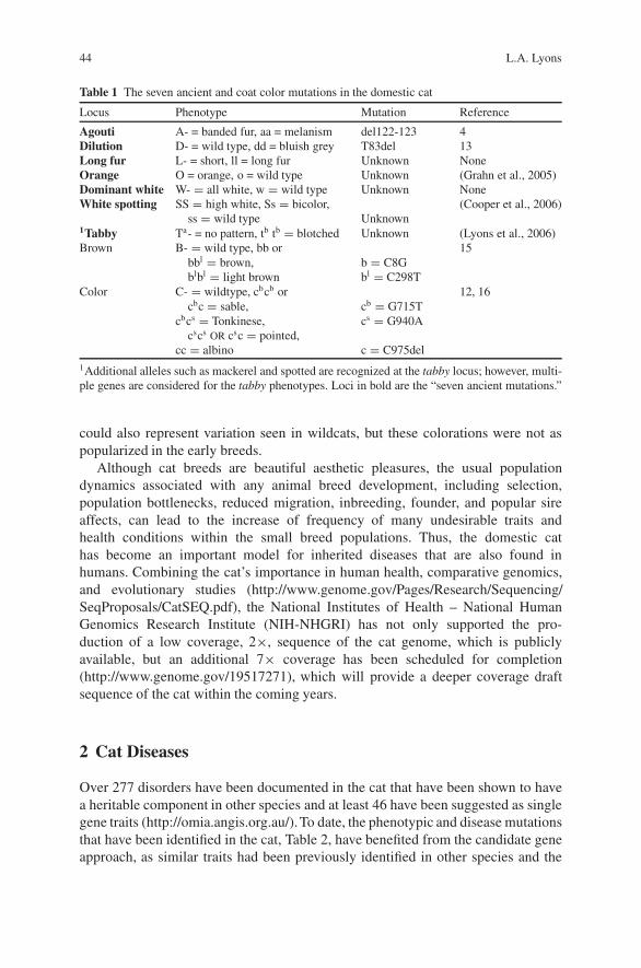
44 L.A. Lyons
Table 1 The seven ancient and coat color mutations in the domestic cat
Locus Phenotype Mutation Reference
Agouti A- = banded fur, aa = melanism del122-123 4Dilution D- = wild type, dd = bluish grey T83del 13Long fur L- = short, ll = long fur Unknown NoneOrange O = orange, o = wild type Unknown (Grahn et al., 2005)Dominant white W- = all white, w = wild type Unknown NoneWhite spotting SS = high white, Ss = bicolor, (Cooper et al., 2006)
ss = wild type Unknown1Tabby Ta- = no pattern, tb tb = blotched Unknown (Lyons et al., 2006)Brown B- = wild type, bb or 15
bbl = brown,blbl = light brown
b = C8Gbl = C298T
Color C- = wildtype, cbcb or 12, 16cbc = sable, cb = G715T
cbcs = Tonkinese,cscs OR csc = pointed,
cs = G940A
cc = albino c = C975del1Additional alleles such as mackerel and spotted are recognized at the tabby locus; however, multi-ple genes are considered for the tabby phenotypes. Loci in bold are the “seven ancient mutations.”
could also represent variation seen in wildcats, but these colorations were not aspopularized in the early breeds.
Although cat breeds are beautiful aesthetic pleasures, the usual populationdynamics associated with any animal breed development, including selection,population bottlenecks, reduced migration, inbreeding, founder, and popular sireaffects, can lead to the increase of frequency of many undesirable traits andhealth conditions within the small breed populations. Thus, the domestic cathas become an important model for inherited diseases that are also found inhumans. Combining the cat’s importance in human health, comparative genomics,and evolutionary studies (http://www.genome.gov/Pages/Research/Sequencing/SeqProposals/CatSEQ.pdf), the National Institutes of Health – National HumanGenomics Research Institute (NIH-NHGRI) has not only supported the pro-duction of a low coverage, 2×, sequence of the cat genome, which is publiclyavailable, but an additional 7× coverage has been scheduled for completion(http://www.genome.gov/19517271), which will provide a deeper coverage draftsequence of the cat within the coming years.
2 Cat Diseases
Over 277 disorders have been documented in the cat that have been shown to havea heritable component in other species and at least 46 have been suggested as singlegene traits (http://omia.angis.org.au/).To date, the phenotypic and disease mutationsthat have been identified in the cat, Table 2, have benefited from the candidate geneapproach, as similar traits had been previously identified in other species and the
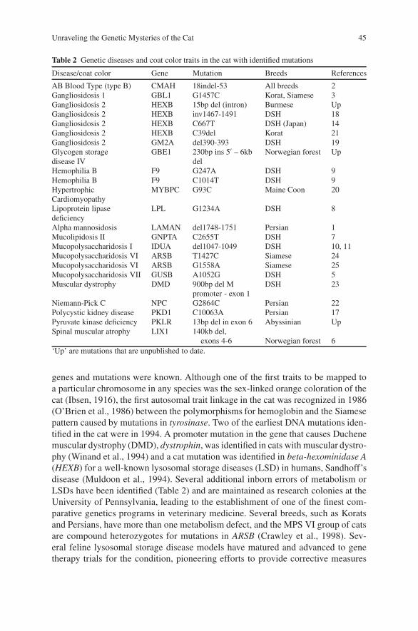
Unraveling the Genetic Mysteries of the Cat 45
Table 2 Genetic diseases and coat color traits in the cat with identified mutations
Disease/coat color Gene Mutation Breeds References
AB Blood Type (type B) CMAH 18indel-53 All breeds 2Gangliosidosis 1 GBL1 G1457C Korat, Siamese 3Gangliosidosis 2 HEXB 15bp del (intron) Burmese UpGangliosidosis 2 HEXB inv1467-1491 DSH 18Gangliosidosis 2 HEXB C667T DSH (Japan) 14Gangliosidosis 2 HEXB C39del Korat 21Gangliosidosis 2 GM2A del390-393 DSH 19Glycogen storagedisease IV
GBE1 230bp ins 5′ – 6kbdel
Norwegian forest Up
Hemophilia B F9 G247A DSH 9Hemophilia B F9 C1014T DSH 9HypertrophicCardiomyopathy
MYBPC G93C Maine Coon 20
Lipoprotein lipasedeficiency
LPL G1234A DSH 8
Alpha mannosidosis LAMAN del1748-1751 Persian 1Mucolipidosis II GNPTA C2655T DSH 7Mucopolysaccharidosis I IDUA del1047-1049 DSH 10, 11Mucopolysaccharidosis VI ARSB T1427C Siamese 24Mucopolysaccharidosis VI ARSB G1558A Siamese 25Mucopolysaccharidosis VII GUSB A1052G DSH 5Muscular dystrophy DMD 900bp del M
promoter - exon 1DSH 23
Niemann-Pick C NPC G2864C Persian 22Polycystic kidney disease PKD1 C10063A Persian 17Pyruvate kinase deficiency PKLR 13bp del in exon 6 Abyssinian UpSpinal muscular atrophy LIX1 140kb del,
exons 4-6 Norwegian forest 6‘Up’ are mutations that are unpublished to date.
genes and mutations were known. Although one of the first traits to be mapped toa particular chromosome in any species was the sex-linked orange coloration of thecat (Ibsen, 1916), the first autosomal trait linkage in the cat was recognized in 1986(O’Brien et al., 1986) between the polymorphisms for hemoglobin and the Siamesepattern caused by mutations in tyrosinase. Two of the earliest DNA mutations iden-tified in the cat were in 1994. A promoter mutation in the gene that causes Duchenemuscular dystrophy (DMD), dystrophin, was identified in cats with muscular dystro-phy (Winand et al., 1994) and a cat mutation was identified in beta-hexominidase A(HEXB) for a well-known lysosomal storage diseases (LSD) in humans, Sandhoff’sdisease (Muldoon et al., 1994). Several additional inborn errors of metabolism orLSDs have been identified (Table 2) and are maintained as research colonies at theUniversity of Pennsylvania, leading to the establishment of one of the finest com-parative genetics programs in veterinary medicine. Several breeds, such as Koratsand Persians, have more than one metabolism defect, and the MPS VI group of catsare compound heterozygotes for mutations in ARSB (Crawley et al., 1998). Sev-eral feline lysosomal storage disease models have matured and advanced to genetherapy trials for the condition, pioneering efforts to provide corrective measures
46 L.A. Lyons
that will be of benefit to humans with the same conditions (for reviews see Casaland Haskins, 2006; Desnick et al., 1982; Haskins, 1996; Haskins et al., 2002). Themutations for several types of LSDs have been identified and different therapeuticstrategies have been tested in afflicted cats, including enzyme replacement therapy(ERT), heterologous bone marrow transplantation (BMT), and gene therapy. Local-ized ERT in the joint space has successfully N-acetylgalactosamine-4-sulfatasedeficiency in the MPS VI feline model (Auclair et al., 2006). Adeno-associatedvirus vectors containing the normal cDNA of alpha-mannosidase have been usedto intracranially administer corrected enzymes in successful therapeutic trials forcats with mannosidosis, an LSD that attacks the central nervous system (Viteet al., 2005). Several feline disorders, including the mannosidosis, mucolipidosesII, and MPS VI cats, have all been models for BMT (Dial et al., 1997; Haskinset al., 1984; Simonaro et al., 1999), which have been some of the earliest attemptsto corrective therapies. Thus, as the genetic causes of feline diseases are identified,many are translating into therapeutic models to correct both the feline and the humanhomologous diseases.
Approximately 16 different genes account for the 22 mutations causing diseasesin cats. However, only eight disease-causing mutations (Table 2) are segregating incat breeds and not maintained only in research colonies. Two of the most prevalentmutations cause cardiac and renal disease. Hypertrophic cardiomyopathy (HCM) isa heterogeneous cardiac disease that has been recognized is several breeds, includ-ing Bengals, Ragdolls, and Sphynx; however, HCM is scientifically documented asheritable only in the Maine Coon cat (Kittleson et al., 1999). A mutation in myosinC binding protein (MYCBP) is highly correlated with clinical presentation of HCMin the Maine Coon cat (Meurs et al., 2005) and is considered the causative mutation.Although the frequency of the disease has not been clearly established for the MaineCoon breed, combined with echocardiogram evaluations, an active genetic typingprogram is assisting the reduction of this disease within the Maine Coon breed. Thesame mutation has been evaluated in other breeds with HCM but, as in humans,HCM appears to be heterogenic in cats and the mutation is not correlated with dis-ease in other breeds (K. Meurs, personal communication, L. Lyons, data not shown).
Other diseases found in the cat, such as polycystic kidney disease (PKD) inPersian cats, have benefited from a combined genetic linkage analysis and can-didate gene approach (Lyons et al., 2004; Young et al., 2005). Feline PKD wasfirst clinically described as an autosomal dominant inherited trait in 1990 (Billeret al., 1990, 1996). Persian cats are the most popular breed worldwide, and togetherwith other breeds that use Persians as a parent breed (Exotics and Himalayans)or for outcrosses (Selkirk Rex, Scottish Folds and American Shorthairs), Persiansrepresent nearly 60% of the cats in the cat fancy. Approximately 38% of Persianshave PKD worldwide (Barrs et al., 2001; Barthez et al., 2003; Cannon et al., 2001),making PKD the most common inherited disease in the domestic cat. PKD affects600,000 people in the US alone, and 12.5 million worldwide. There are more peopleafflicted with PKD than cystic fibrosis, muscular dystrophy, hemophilia, and Downssyndrome and sickle cell anemia combined. Over 90% of PKD is heritable. Morethan 60% of the individuals with PKD develop kidney failure, or end-stage renaldisease (ESRD). Patients with ESRD are generally on dialysis for approximately
Unraveling the Genetic Mysteries of the Cat 47
7 years, significantly lower than their life span (see reviews in Sessa et al., 2004;Tahvanainen et al., 2005). Feline and human PKD have similar clinical features(Eaton et al., 1997; Pedersen et al., 2003). Over 95% of cats with PKD will developrenal cysts by 8 months of age, which can be accurately determined by ultrasound.Depending on disease severity, cats can live a normal lifespan, 10–14 years, withPKD, or succumb within a few years of onset; similar disease variation and pro-gressions are also seen in humans. Ultrasonographic identification of renal cystsallowed the accurate ascertainment of large pedigrees of Persian cats with PKD.In humans, two genes are responsible for a majority of PKD, PKD1 and PKD2.Although PKD1 may present more severely, the clinical presentations are not dis-tinctive and can only be determined by mutation analyses. Approximately 85%of cases are caused by mutations related to the PKD1 gene, while the remaining15% are attributed to the PKD2 gene (Peters et al., 1993). The two major genesproduce very large transcripts and because limited domestic cat genetic sequencewas available, a linkage analysis was first conducted to implicate a candidate genefor feline PKD prior to a candidate gene approach (Young et al., 2005). Once acat microsatellite marker showed significant linkage to the region of the cat genomewith PKD1, a gene scan identified a C10063A transversion that changes an argenineto an OPA stop codon, which should disrupt approximately 25% of the polycystin-1protein (1).
Twenty-one mutations have been identified in the cat that cause diseases, fourcoat color loci represent an additional seven mutations, and a blood group mutationproduces the Type B cat. Cats have even been shown to lack a “sweet tooth” due tothe pseudogenization of Tas1r2 sweet receptor gene (Li et al., 2006; Li et al., 2005).Veterinary medicine has identified over 277 traits that could be heritable conditionsin the cat and many have been well documented. The identification of several addi-tional mutations should be on the horizon as active linkage studies have implicatedgenes or genetic regions for white spotting, tabby, orange, progressive retinal atro-phy in the Persian and the Abyssinian, and a craniofacial defect in the Burmese.Improved genetic resources will expedite mutation discoveries for the single genetraits of the cat.
3 Feline Genetic Resources
Early chromosome banding studies of the domestic cat revealed an easily dis-tinguishable karyotype consisting of 18 autosomal and the XY sex chromosomepair, resulting in a 2N complement of 38 cat chromosomes (Wurster-Hill andGray, 1973). The traditional groupings of chromosomes into alphabetic groupsbased on size and centromeric position has only relatively recently been renamed tomore standard nomenclature for the cat (Cho et al., 1997). Basic light microscopyand giemsa banding also showed that domestic cats have a chromosomal archi-tecture that is highly representative for all felids and even carnivores (Modi andO’Brien, 1988). Only minor chromosomal rearrangements are noted amongst the36 extant felids, most noticeably a robertsonian fusion in the South American ocelot
48 L.A. Lyons
lineage leads to a reduced complement, 2N = 36 (Wurster-Hill and Gray, 1973).The variation of chromosomal sizes allowed for the easy development of chromo-some paints by flow sorting (Wienberg et al., 1997). Chromosome painting tech-niques supported early somatic cell hybrid maps in that the cat appeared to behighly conserved in chromosomal arrangement to humans, specifically as com-pared to mice (Stanyon et al., 1999). Hence, chromosome painting gave an excel-lent overview of cat genome organization, which greatly facilitates candidate geneapproaches since the location of particular genes can be anticipated in cats fromcomparison with the genetic map of humans.
The low-resolution genetic comparisons provided by chromosomal studies havebeen augmented and complimented by medium resolution genetic and radiationhybrid maps of the cat. The current 5,000 Rad radiation hybrid map of the catconsists of 1784 markers (Menotti-Raymond et al., 2003a; Murphy et al., 2006,1999, 2000) and the interspecies hybrid-based linkage map contains approximately250 microsatellite markers (Menotti-Raymond et al., 2003b, 1999) that are affectivefor the initiation of linkage studies in families segregating for phenotypic traits.The linkage map is due for updating, which is likely in the coming year; however,already available linkage maps have assisted targeted candidate gene approaches, asseen for PKD (Young et al., 2005), linkage analyses for Tabby (Lyons et al., 2006),white spotting (Cooper et al., 2006) and Orange (Grahn et al., 2005), and the geneticmap has led to the first disease gene isolated by positional cloning, LIX1, whichcauses spinal muscular atrophy in the Norwegian forest cat (Fyfe et al., 2006;He et al., 2005). Updated maps, deeper genomic sequence, and the RPCI-85 catBAC library (http://bacpac.chori.org/) should provide sufficient and highly efficientgenetic resources for future genetic studies in felids.
4 Reproductive Technologies
Even with limited genetic resources, the domestic cat has become the focus ofresearch technologies that use reproductive methods to manipulate the cat genome.Although gene knock-outs for cat allergens have only been proposed, the cur-rently available “hypoallergenic” cats have been developed by natural breedingmethods (http://www.allerca.com/). However, cloning of the cat by either nuclearor chromatin transfer has been successful and even commercialized. To date, sev-eral domestic cats and the African wildcat have been successfully cloned (Gomezet al., 2004; Shin et al., 2002). At least one company has used client-provided mate-rials to clone a pet as a commercial adventure (http://www.savingsandclone.com/).Clients are now asking veterinarians to collect viable cells as sample from theircats and send them to cloning companies for the eventual use of cloning the donorcat. Cat cloning has exemplified important aspects regarding this highly contro-versial topic. The first cloned cat, CC, was cloned from a calico cat, a female cat(Rainbow) that had white spotting and different alleles at the Orange locus on theX chromosome. The cloned cat, CC, did not express the orange allelic variants;
Unraveling the Genetic Mysteries of the Cat 49
hence, X-inactivation did not get reprogrammed during the cloning process. Thisevent highlights two important concepts: (1) researchers still do not recognize orunderstand all the normal biological processes during embryo development and (2)cloned animals can definitely look different that the original, donor individual. Thesuccessful cloning of the African wildcat potentially opens avenues for the reintro-duction of the genetics of individuals that have been lost to conservation breedingprograms, such as animals that have been sterilized or are too old or unhealthy tobreed. However, domestic cat eggs were used for the cloning of the African wildcats,hence their mtDNA is not representative of the endangered species. But overall,cloning may provide unexpected opportunities in conservation, or perhaps in thereplication of individuals with certain phenotypes of interest in research. Represent-ing the ultimate of an inbred species, a series of clones from the same individualcould foster interesting studies of nature versus nurture.
5 Future of Cat Genetics
Once causative mutations for heritable conditions are identified in cats, felinesbecome a more important asset to human health. Several inborn errors ofmetabolism in cats are already exploring gene therapy approaches. Many murinemodels exist for the study of cystogenesis, the hallmark of PKD; however, eachrodent model has its shortcomings. The cat is similar to human autosomal dominantPKD in several important aspects, including (1) a causative mutation in PKD1,(2) a similar type of mutation that causes a similar protein disruption, (3) similarvariability in disease progression, (4) cystogenesis in other organs, including theliver and pancreas, (5) homozygotes for the mutation are lethal, and (6) cats areintermediate in regard to genetic variation, mimicking human populations andethnic groups more closely than inbred strains of mice. None of the rodent modelshave a mutation in PKD1 and many lack the other noted aspects that are importantto human drug and gene therapy trials.
Thus, cats can be an alternative and supportive animal model to rodent modelsfor many heritable conditions because (1) they provide a balance between cost andefficiency, (2) drug dosages are more easily translated between cats and humans, (3)the longer life span of the cat allows repeated therapy trials and longer term studies,(4) cats have strong conservation of biology, anatomy, and physiology to humans,(5) cats provide a second animal for validation and efficacy, (6) the larger size of thecat and its organs are more amenable to therapies, and (7) cats are intermediate inregard to genetic variation, mimicking human populations and ethnic groups moreclosely than inbred strains of mice.
The deeper sequencing of the cat genome and the investigation of variation byresequencing in different cat breeds will allow the cat to leap forward in genetics,from the analysis of single gene traits to the investigation of more complex traits,such as asthma, diabetes, and obesity. Cats are notorious for viral infections, hencethe identification of susceptibility and resistance genes for acquired diseases should
50 L.A. Lyons
become a mainstay in cat research. The determination of linkage disequilibrium mayassist disease studies in many breeds by potentially lowering the number of markers,likely SNP-based, that are required for full genome scans in complex trait analyses.However, many of the common diseases that plague humans, which are found incats, will likely be examined in the outbred populations of non-purebred housecats,as only 10–15% of cats in the US are representatives by a fancy breed, a proportionthat is higher than most other nations (Louwerens et al., 2005).
The available genetic resources for the cat now places the research bottleneckfor felines back to the acquisition of appropriate patients with sufficient cases andcontrols. Hence, the primary care veterinarians, the veterinary specialists, and vet-erinary researchers need to join forces to properly characterize diseases and rou-tinely collect research materials so that patients are not lost to important studies andhealth investigations. The development of the DNA tests for parentage and identi-fication (http://www.isag.org.uk/), coat colors, and the prominent diseases, such asPKD and HCM, has encouraged cat breeders to explore genetic research in a morepositive manner and has encouraged their participation in research studies. Foren-sic applications (Menotti-Raymond et al., 1997a, 1997b, 2005) and mass disastershave brought to the forefront the DNA profiling of loved ones, including our pets.For these reasons, more cat breeders are banking DNA sources on their animalsor providing DNA to service and research laboratories. Many veterinary hospitalsand large clinical conglomerates are developing electronic database systems thatcould facilitate the identification of proper patients, cases, and controls. Combinedwith DNA banking and specialty health care, the veterinary world stands to enhancethe possibilities of complex disease research in the cat by leaps and bounds. Eventhough feline origins remain a mystery and “domesticated” may not be the mostappropriate term for the domestic cat, the cat is unlocking its genetic secrets thatexplain the cat’s form and function.
References
Auclair, D., Hein, L.K., Hopwood, J.J., and Byers, S., 2006, Intra-articular enzyme administrationfor joint disease in feline mucopolysaccharidosis VI: enzyme dose and interval, Pediatr. Res.59:538–543.
Barrs, V.R., Gunew, M., Foster, S.F., Beatty, J.A., and Malik, R., 2001, Prevalence of autosomaldominant polycystic kidney disease in Persian cats and related-breeds in Sydney and Brisbane,Aust. Vet. J. 79:257–259.
Barthez, P.Y., Rivier, P., and Begon, D., 2003, Prevalence of polycystic kidney disease in Persianand Persian related cats in France, J. Feline Med. Surg. 5:345–347.
Biller, D.S., Chew, D.J., and DiBartola, S.P., 1990, Polycystic kidney disease in a family of Persiancats, J. Am. Vet. Med. Assoc. 196:1288–1290.
Biller, D.S., DiBartola, S.P., Eaton, K.A., Pflueger, S., Wellman, M.L., and Radin, M.J., 1996,Inheritance of polycystic kidney disease in Persian cats, J. Hered. 87:1–5.
Brooks, S.A., and Bailey, E., 2005, Exon skipping in the KIT gene causes a Sabino spotting patternin horses, Mamm. Genome 16:893–902.
Cannon, M.J., MacKay, A.D., Barr, F.J., Rudorf, H., Bradley, K.J., and Gruffydd-Jones, T.J., 2001,Prevalence of polycystic kidney disease in Persian cats in the United Kingdom, Vet. Rec.149:409–411.
Unraveling the Genetic Mysteries of the Cat 51
Casal, M., and Haskins, M., 2006, Large animal models and gene therapy, Eur. J. Hum. Genet.14:266–272.
CFA 2004, The Cat Fanciers’ Association Complete Cat Book, HarperCollins Publishers,New York.
Cho, K.W., Youn, H.Y., Watari, T., Tsujimoto, H., Hasegawa, A., and Satoh, H., 1997, A proposednomenclature of the domestic cat karyotype, Cytogenet. Cell Genet. 79:71–78.
Clark, J.M., 1975, The effects of selection and human preference on coat colour gene frequenciesin urban cats, Heredity 35:195–210.
Cooper, M.P., Fretwell, N., Bailey, S.J., and Lyons, L.A., 2006, White spotting in the domestic cat(Felis catus) maps near KIT on feline chromosome B1, Anim. Genet. 37:163–165.
Crawley, A.C., Yogalingam, G., Muller, V.J., and Hopwood, J.J., 1998, Two mutations within afeline mucopolysaccharidosis type VI colony cause three different clinical phenotypes, J. Clin.Invest. 101:109–119.
Davis, B.K., and Davis, B.P., 1977, Allele frequencies in a cat population in Budapest, J. Hered.68:31–34.
Desnick, R.J., McGovern, M.M., Schuchman, E.H., and Haskins, M.E., 1982, Animal analogues ofhuman inherited metabolic diseases: molecular pathology and therapeutic studies, Prog. Clin.Biol. Res. 94:27–65.
Dial, S.M., Byrne, T., Haskins, M., Gasper, P.W., Rose, B., Wenger, D.A., and Thrall, M.A., 1997,Urine glycosaminoglycan concentrations in mucopolysaccharidosis VI-affected cats followingbone marrow transplantation or leukocyte infusion, Clin. Chim. Acta 263:1–14.
Eaton, K.A., Biller, D.S., DiBartola, S.P., Radin, M.J., and Wellman, M.L., 1997, Autosomal dom-inant polycystic kidney disease in Persian and Persian-cross cats, Vet. Pathol. 34:117–126.
Eizirik, E., Yuhki, N., Johnson, W.E., Menotti-Raymond, M., Hannah, S.S., and O’Brien, S.J.,2003, Molecular genetics and evolution of melanism in the cat family, Curr. Biol. 13:448–453.
Fyfe, J.C., Menotti-Raymond, M., David, V.A., Brichta, L., Schaffer, A.A., Agarwala, R.,Murphy, W.J., Wedemeyer, W.J., Gregory, B.L., Buzzell, B.G., Drummond, M.C., Wirth, B.,and O’Brien, S.J., 2006, An approximately 140-kb deletion associated with feline spinal mus-cular atrophy implies an essential LIX1 function for motor neuron survival, Genome Res. 16:1084–1090.
Gomez, M.C., Pope, C.E., Giraldo, A., Lyons, L.A., Harris, R.F., King, A.L., Cole, A.,Godke, R.A., and Dresser, B.L., 2004, Birth of African Wildcat cloned kittens born fromdomestic cats, Cloning Stem Cells 6:247–258.
Grahn, R.A., Lemesch, B.M., Millon, L.V., Matise, T., Rogers, Q.R., Morris, J.G., Fretwell, N.,Bailey, S.J., Batt, R.M., and Lyons, L.A., 2005, Localizing the X-linked orange colour pheno-type using feline resource families, Anim. Genet. 36:67–70.
Haskins, M., 1996, Bone marrow transplantation therapy for metabolic disease: animal models aspredictors of success and in utero approaches, Bone Marrow Transpl 18:S25–S27.
Haskins, M., Casal, M., Ellinwood, N.M., Melniczek, J., Mazrier, H., and Giger, U., 2002, Animalmodels for mucopolysaccharidoses and their clinical relevance, Acta Paediatr. Suppl. 91:88–97.
Haskins, M.E., Wortman, J.A., Wilson, S., and Wolfe, J.H., 1984, Bone marrow transplantation inthe cat, Transplantation 37:634–636.
He Q., Lowrie, C., Shelton, G.D., Castellani, R.J., Menotti-Raymond, M., Murphy, W.,O’Brien, S.J., Swanson, W.F., and Fyfe, J.C., 2005, Inherited motor neuron disease in domesticcats: a model of spinal muscular atrophy, Pediatr. Res. 57:324–330.
Hemmer, H., 1972, Variations in brain size in the species Felis silvestris, Experientia 28:271–272.
Hemmer, H., 1976, Man’s strategy in domestication – a synthesis of new research trends, Experi-entia 32:663–666.
Hemmer, H., 1978, The evolutionary systematics of living Felidae: present status and current prob-lems, Carnivore B:71–79.
Hemmer, H., 1990, “Domestication: The decline of environmental appreciation,” Cambridge Uni-versity Press, Cambridge.
52 L.A. Lyons
Housley, D.J., and Venta, P.J. 2006, The long and the short of it: evidence that FGF5 is a majordeterminant of canine ’hair’-itability, Anim. Genet. 37:309–315.
Ibsen, H.L., 1916, Tricolor inheritance. III. Tortoiseshell cats, Genetics 1:377–386.Imes, D.L., Geary, L.A., Grahn, R.A., and Lyons, L.A., 2006, Albinism in the domestic cat (Felis
catus) is associated with a tyrosinase (TYR) mutation, Anim. Genet. 37:175–178.Ishida, Y., David, V.A., Eizirik, E., Schaffer, A.A., Neelam, B.A., Roelke, M.E., Hannah, S.S.,
O’Brien, S.J., and Menotti-Raymond, M., 2006, A homozygous single-base deletion in MLPHcauses the dilute coat color phenotype in the domestic cat, Genomics 88:698–705. Epub.
Johansson Moller, M., Chaudhary, R., Hellmen, E., Hoyheim, B., Chowdhary, B., andAndersson, L., 1996, Pigs with the dominant white coat color phenotype carry a duplication ofthe KIT gene encoding the mast/stem cell growth factor receptor, Mamm. Genome 7:822–830.
Johnson, W.E., Eizirik, E., Pecon-Slattery, J., Murphy, W.J., Antunes, A., Teeling, E., andO’Brien, S.J., 2006, The late Miocene radiation of modern Felidae: a genetic assessment, Sci-ence 311:73–77.
Kitchener, A., 1991, “The natural history of wild cats,” Cornell University Press, New York.Kittleson, M.D., Meurs, K.M., Munro, M.J., Kittleson, J.A., Liu, S.K., Pion, P.D., and Towbin, J.A.,
1999, Familial hypertrophic cardiomyopathy in maine coon cats: an animal model of humandisease, Circulation 99:3172–3180.
Kukekova, A.V., Acland, G.M., Oskina, I.N., Kharlamova, A.V., Trut, L.N., Chase, K., Lark, K.G.,Erb, H.N., and Aguirre, G.D., 2006, The genetics of domesticated behavior in Canids: what candogs and Silver foxes tell us about each other? In E.A. Ostrander, U. Giger, and K. Lindblad-Tioh, Eds., “The Dog and Its Genome”, pp. 515–537, Cold Spring Harbor Laboratory Press,Cold Spring Harbor.
Li X., Li, W., Wang, H., Cao, J., Maehashi, K., Huang, L., Bachmanov, A.A., Reed, D.R., Legrand-Defretin, V., Beauchamp, G.K., and Brand, J.G., 2005, Pseudogenization of a sweet-receptorgene accounts for cats’ indifference toward sugar, PLoS Genet. 1:27–35.
Li, X., Li, W., Wang, H., Bayley, D.L., Cao, J., Reed, D.R., Bachmanov, A.A., Huang, L., Legrand-Defretin, V., Beauchamp, G.K., and Brand, J.G., 2006, Cats lack a sweet taste receptor, J. Nutr.136:1932S–1934S.
Louwerens, M., London, C.A., Pedersen, N.C., and Lyons, L.A., 2005, Feline lymphoma in thepost-feline leukemia virus era, J. Vet. Intern. Med. 19:329–335.
Lyons, L.A., Bailey, S.J., Baysac, K.C., Byrns, G., Erdman, C.A., Fretwell, N., Froenicke, L.,Gazlay, K.W., Geary, L.A., Grahn, J.C., Grahn, R.A., Karere, G.M., Lipinski, M.J., Rah, H.,Ruhe, M.T., and Bach, L.H., 2006, The Tabby cat locus maps to feline chromosome B1, Anim.Genet. 37:383–386.
Lyons, L.A., Biller, D.S., Erdman, C.A., Lipinski, M.J., Young, A.E., Roe, B.A., Qin, B., andGrahn, R.A., 2004, Feline polycystic kidney disease mutation identified in PKD1, J. Am. Soc.Nephrol. 15:2548–2555.
Lyons, L.A., Foe, I.T., Rah, H., and Grahn, R.A., 2005a, Chocolate coated cat: TYRP1 mutationsfor brown color in domestic cats, Mamm. Genome 16:356–366.
Lyons, L.A., Imes, D.L., Rah, H.C., and Grahn, R.A., 2005b, Tyrosinase mutations associated withSiamese and Burmese patterns in the domestic cat (Felis catus), Anim. Genet. 36:119–126.
Marklund, S., Kijas, J., Rodriguez-Martinez, H., Ronnstrand, L., Funa, K., Moller, M., Lange, D.,Edfors-Lilja, I., and Andersson, L., 1998, Molecular basis for the dominant white phenotype inthe domestic pig, Genome Res. 8:826–833.
Menotti-Raymond, M., David, V.A., Agarwala, R., Schaffer, A.A., Stephens, R., O’Brien, S.J., andMurphy, W.J., 2003a, Radiation hybrid mapping of 304 novel microsatellites in the domesticcat genome, Cytogenet. Genome Res. 102:272–276.
Menotti-Raymond, M., David, V.A., Chen, Z.Q., Menotti, K.A., Sun, S., Schaffer, A.A., Agarwala,R., Tomlin, J.F., O’Brien, S.J., and Murphy, W.J., 2003b, Second-generation integrated geneticlinkage/radiation hybrid maps of the domestic cat (Felis catus), J. Hered. 94:95–106.
Menotti-Raymond, M., David, V.A., Lyons, L.A., Schaffer, A.A., Tomlin, J.F., Hutton, M.K., andO’Brien, S.J., 1999, A genetic linkage map of microsatellites in the domestic cat (Felis catus),Genomics 57:9–23.
Unraveling the Genetic Mysteries of the Cat 53
Menotti-Raymond, M., David, V.A., Stephens, J.C., Lyons, L.A., and O’Brien, S.J., 1997a, Geneticindividualization of domestic cats using feline STR loci for forensic applications, J. ForensicSci. 42:1039–1051.
Menotti-Raymond, M.A., David, V.A., and O’Brien, S.J., 1997b, Pet cat hair implicates murdersuspect, Nature 386:774.
Menotti-Raymond, M.A., David, V.A., Wachter, L.L., Butler, J.M., and O’Brien, S.J., 2005, AnSTR forensic typing system for genetic individualization of domestic cat (Felis catus) samples,J. Forensic Sci. 50:1061–1070.
Meurs, K.M., Sanchez, X., David, R.M., Bowles, N.E., Towbin, J.A., Reiser, P.J., Kittleson, J.A.,Munro, M.J., Dryburgh, K., Macdonald, K.A., and Kittleson, M.D., 2005, A cardiac myosinbinding protein C mutation in the Maine Coon cat with familial hypertrophic cardiomyopathy,Hum. Mol. Genet. 14:3587–3593.
Modi, W.S., and O’Brien, S.J., 1988, “Quantitative cladistic analysis of chromosomal banding dataamong species in three orders of mammals: Hominoid primates, felids, and arvicolid rodents,”Plenum Publishing, New York.
Morris, D., 1999a, “Cat breeds of the world,” Penguin Books, New York.Morris, D., 1999b, “Cat Breeds of the World: A Complete Illustrated Encyclopedia,” Viking
Penquin, New York.Muldoon, L.L., Neuwelt, E.A., Pagel, M.A., and Weiss, D.L., 1994, Characterization of the molec-
ular defect in a feline model for type II GM2-gangliosidosis (Sandhoff disease), Am. J. Pathol.144:1109–1118.
Murphy, W.J., Davis, B., David, V.A., Agarwala, R., Schaffer, A.A., Pearks Wilkerson, A.J.,Neelam, B., O’Brien, S.J., and Menotti-Raymond, M., 2006, A 1.5-Mb-resolution radiationhybrid map of the cat genome and comparative analysis with the canine and human genomes,Genomics 89:189–196.
Murphy, W.J., Menotti-Raymond, M., Lyons, L.A., Thompson, M.A., and O’Brien, S.J., 1999,Development of a feline whole genome radiation hybrid panel and comparative mapping ofhuman chromosome 12 and 22 loci, Genomics 57:1–8.
Murphy, W.J., Sun, S., Chen, Z., Yuhki, N., Hirschmann, D., Menotti-Raymond, M., andO’Brien, S.J., 2000, A radiation hybrid map of the cat genome: implications for comparativemapping, Genome Res. 10:691–702.
O’Brien, S.J., Haskins, M.E., Winkler, C.A., Nash, W.G., and Patterson, D.F., 1986, Chromosomalmapping of beta-globin and albino loci in the domestic cat. A conserved mammalian chromo-some group, J. Hered. 77:374–378.
Pedersen, K.M., Pedersen, H.D., Haggstrom, J., Koch, J., and Ersboll, A.K., 2003, Increased meanarterial pressure and aldosterone-to-renin ratio in Persian cats with polycystic kidney disease,J. Vet. Intern. Med. 17:21–27.
Peters, D.J., Spruit, L., Saris, J.J., Ravine, D., Sandkuijl, L.A., Fossdal, R., Boersma, J., vanEijk, R., Norby, S., Constantinou-Deltas, C.D., and et al. 1993, Chromosome 4 localizationof a second gene for autosomal dominant polycystic kidney disease, Nat. Genet. 5:359–362.
Robinson, R., 1991, “Genetics for Cat Breeders,” Pergamon Press, Oxford.Seidensticker, J., and Lumpkin, S., “Great Cats: majestic creatures of the wild,” Rodale Press,
Enormaus, PA.Sessa, A., Righetti, M., and Battini, G., 2004, Autosomal recessive and dominant polycystic kidney
diseases, Minerva Urol. Nefrol. 56:329–338.Shin, T., Kraemer, D., Pryor, J., Liu, L., Rugila, J., Howe, L., Buck, S., Murphy, K., Lyons, L., and
Westhusin, M., 2002, A cat cloned by nuclear transplantation, Nature 415:859.Simonaro, C.M., Haskins, M.E., Abkowitz, J.L., Brooks, D.A., Hopwood, J.J., Zhang, J., and
Schuchman, E.H., 1999, Autologous transplantation of retrovirally transduced bone marrowor neonatal blood cells into cats can lead to long-term engraftment in the absence of myeloab-lation, Gene Ther. 6:107–113.
Stanyon, R., Yang, F., Cavagna, P., O’Brien, P.C., Bagga, M., Ferguson-Smith, M.A., andWienberg, J., 1999, Reciprocal chromosome painting shows that genomic rearrangement
54 L.A. Lyons
between rat and mouse proceeds ten times faster than between humans and cats, Cytogenet.Cell Genet. 84:150–155.
Sundberg, J.P., Rourk, M H., Boggess, D., Hogan, M.E., Sundberg, B.A., and Bertolino, A.P., 1997,Angora mouse mutation: altered hair cycle, follicular dystrophy, phenotypic maintenance ofskin grafts, and changes in keratin expression, Vet. Pathol. 34:171–179.
Sunquist, M., and Sunquist, F., 2002, “Wild Cats of the World,” University of Chicago Press,Chicago.
Tahvanainen, E., Tahvanainen, P., Kaariainen, H., and Hockerstedt, K. 2005, Polycystic liver andkidney diseases, Ann. Med. 37:546–555.
Terry, R.R., Bailey, E., Bernoco, D., and Cothran, E.G., 2001, Linked markers exclude KIT asthe gene responsible for appaloosa coat colour spotting patterns in horses, Anim. Genet. 32:98–101.
Todd, N.B., and Lloyd, A.T., 1984, Mutant allele frequencies in domestic cats of Portugal and theAzores, J. Hered. 75:495–497.
Todd, N.B., Robinson, R., and Clark, J.M., 1974, Gene frequencies in domestic cats of Greece,J. Hered. 65:227–231.
Todd, N.E., Fagen, R.M., and Fagen, K., 1975, Gene frequencies in Icelandic cats, Heredity35:173–183.
Vella, C.M., Shelton, L.M., McGonagle, J.J., and Stanglein, T.W., 1999, “Robinson’s Genetics forCat Breeders and Veterinarians,” Butterworth Heinemann, Boston.
Vite, C.H., McGowan, J.C., Niogi, S.N., Passini, M.A., Drobatz, K.J., Haskins, M.E., andWolfe, J.H., 2005, Effective gene therapy for an inherited CNS disease in a large animal model,Ann. Neurol. 57:355–364.
Wagner, A., and Wolsan, M., 1987, Pelage mutant allele frequencies in domestic cat populationsof Poland, J. Hered. 78:197–200.
Wastlhuber, J., 1991, History of domestic cats and cat breeds, In N.C. Pedersen, Ed., “FelineHusbandry: Diseases and management in the multiple-cat environment”, pp. 1–59, AmericanVeterinary Publications, Inc., Goleta, CA.
Wayne, R.K., 1986, Cranial morphology of domestic and wild canids: the influence of developmenton morphological change, Evolution 40:243–261.
Wienberg, J., Stanyon, R., Nash, W.G., O’Brien, P.C., Yang, F., O’Brien, S.J., and Ferguson-Smith, M.A. 1997, Conservation of human vs. feline genome organization revealed by recipro-cal chromosome painting, Cytogenet. Cell Genet. 77:211–217.
Winand, N.J., Edwardsm M., Pradhanm D., Berianm C.A., and Cooper, B.J., 1994, Deletion of thedystrophin muscle promoter in feline muscular dystrophy, Neuromuscul. Disord. 4:433–445.
Wurster-Hill, D.H., and Gray, C.W., 1973, Giemsa banding patterns in the chromosomes of twelvespecies of cats (Felidae), Cytogenet. Cell Genet. 12:388–397.
Young, A.E., Biller, D.S., Herrgesell, E.J., Roberts, H.R., and Lyons, L.A., 2005, Feline polycystickidney disease is linked to the PKD1 region, Mamm. Genome 16:59–65.
APPENDIX: Table references
1. Berg, T., Tollersrud, O.K., Walkley, S.U., Siegel, D., and Nilssen, O., 1997, Purification offeline lysosomal alpha-mannosidase, determination of its cDNA sequence and identificationof a mutation causing alpha-mannosidosis in Persian cats, Biochem. J. 328:863–870.
2. Bighignoli, B., Grahn, R.A., Millon, L.V., Longeri, M., Polli, M., and Lyons, L.A., 2007,Genetic mutations for the feline AB blood group identified in CMAH mutations associatedwith the domestic cat AB blood group, MBC Genet. 8:27.
3. De Maria, R., Divari, S., Bo, S., Sonnio, S., Lotti, D., Capucchio, M.T., and Castagnaro, M.,1998, Beta-galactosidase deficiency in a Korat cat: a new form of feline GM1-gangliosidosis,Acta Neuropathol. (Berl) 96:307–314.
Unraveling the Genetic Mysteries of the Cat 55
4. Eizirik, E., Yuhki, N., Johnson, W.E., Menotti-Raymond, M., Hannah, S.S., O’Brien, andS.J., 2003, Molecular genetics and evolution of melanism in the cat family, Curr. Biol.13:448–453.
5. Fyfe, J.C., Kurzhals, R.L., Lassaline, M.E., Henthorn, P.S., Alur, P.R., Wang, P., Wolfe, J.H.,Giger, U., Haskins, M.E., Patterson, D.F., Sun, H., Jain, S., and Yuhki, N., 1999, Molecularbasis of feline beta-glucuronidase deficiency: an animal model of mucopolysaccharidosisVII, Genomics 58:121–128.
6. Fyfe, J.C., Menotti-Raymond, M., David, V.A., Brichta, L, Schaffer, A.A., Agarwala, R.,Murphy, W.J., Wedemeyer, W.J., Gregory, B.L., Buzzell, B.G., Drummond, M.C., Wirth,B., and O’Brien, S.J., 2006, An approximately 140-kb deletion associated with feline spinalmuscular atrophy implies an essential LIX1 function for motor neuron survival, Genome Res.16: 1084–1090.
7. Giger, U., Tcherneva, E., Caverly, J., Seng, A., Huff, A. M., Cullen, K., Van Hoeven, M.,Mazrier, H., and Haskins, M. E., 2006, A missense point mutation in N-acetylglucosamine-1-phospotrans-ferase causes mucolipidosis II in domestic shorthair cats, J. Vet. Intern. Med.20:781.
8. Ginzinger, D.G., Lewis, M.E., Ma, Y., Jones, B.R., Liu, G., and Jones, S.D., 1996, A muta-tion in the lipoprotein lipase gene is the molecular basis of chylomicronemia in a colony ofdomestic cats, J. Clin. Invest. 97:1257–1266.
9. Goree, M., Catalfamo, J.L., Aber, S., and Boudreaux, M.K., 2005, Characterization of themutations causing hemophilia B in 2 domestic cats, J. Vet. Intern. Med. 19:200–204.
10. Haskins, M., Jezyk, P., and Giger, U. 2005, Diagnostic tests for mucopolysaccharidosis. J.Am. Vet. Med. Assoc. 226:1047.
11. He, X., Li, C.M., Simonaro, C.M., Wan, Q., Haskins, M.E., Desnick, R.J., and Schuchman,E.H., 1999, Identification and characterization of the molecular lesion causing mucopolysac-charidosis type I in cats, Mol. Genet. Metab. 67:106–112.
12. Imes, D.L., Geary, L.A., Grahn, R.A., and Lyons, L.A., 2006, Albinism in the domestic cat(Felis catus) is associated with a tyrosinase (TYR) mutation, Anim. Genet. 37:175–178.
13. Ishida, Y., David, V.A., Eizirik, E., Schaffer, A.A., Neelam, B.A., Roelke, M.E., Hannah,S.S., O’Brien, S.J., and Menotti-Raymond, M., 2006, A homozygous single-base deletion inMLPH causes the dilute coat color phenotype in the domestic cat, Genomics 88:698–705.
14. Kanae, Y., Endoh, D., Yamato, O., Hayashi, D., Matsunaga, S., Ogawa, H., Maede, Y., andHayashi, M., 2007, Nonsense mutation of feline beta-hexosaminidase beta-subunit (HEXB)gene causing Sandhoff disease in a family of Japanese domestic cats, Res. Vet. Sci. 82:54–60.
15. Lyons, L.A., Foe, I.T., Rah, H.C., and Grahn, R.A., 2005, Chocolate coated cats: TYRP1mutations for brown color in domestic cats, Mamm. Genome. 16:356–366.
16. Lyons, L.A., Imes, D.L., Rah, H.C., and Grahn, R.A., 2005, Tyrosinase mutations associ-ated with Siamese and Burmese patterns in the domestic cat (Felis catus), Anim. Genet. 36:119–126.
17. Lyons, L.A., Biller, D.S., Erdman, C.A., Lipinski, M.J., Young, A.E., Roe, B.A., Qin, B.,and Grahn, R.A., 2004, Feline polycystic kidney disease mutation identified in PKD1, J. Am.Soc. Nephrol. 15:2548–2555.
18. Martin, D.R., Krum, B.K., Varadarajan, G.S., Hathcock, T.L., Smith, B.F., and Baker, H.J.,2004, An inversion of 25 base pairs causes feline GM2 gangliosidosis variant, Exp. Neurol.187: 30–37.
19. Martin, D.R., Cox, N.R., Morrison, N.E., Kennamer, D.M., Peck, S.L., Dodson, A.N., Gen-try, A.S., Griffin, B., Rolsma, M.D., and Baker, H.J., 2005, Mutation of the GM2 activatorprotein in a feline model of GM2 gangliosidosis, Acta Neuropathol. (Berl.) 110:443–450.
20. Meurs, K., Sanchez, X., David, R., Bowles, N.E., Towbin, J.A., Reiser, P.J., Kittleson, J.A.,Munro, M.J., Dryburgh, K., Boyer, M., Mathur, D., MacDonald, K.A., and Kittleson, M.D.,2005, Identification of a missense mutation in the cardiac myosin binding protein C gene in
56 L.A. Lyons
a family of Maine Coon cats with hypertrophic cardiomyopathy, Am. Col. Vet. Intern. Med.Forum 14:3587–3593.
21. Muldoon, L.L., Neuwelt, E.A., Pagel, M.A., and Weiss, D.L., 1994, Characterization of themolecular defect in a feline model for type II GM2-gangliosidosis (Sandhoff disease), Am. J.Pathol. 144:1109–1118.
22. Somers, K.L., Royals, M.A., Carstea, E.D., Rafi, M.A., Wenger, D.A., and Thrall, M.A.,2003, Mutation analysis of feline Niemann-Pick C1 disease, Mol Genet Metab. 79:99–103.
23. Winand, N.J., Edwards, M., Pradhan, D., Berian, C.A., and Cooper, B.J., 1994, Deletion ofthe dystrophin muscle promoter in feline muscular dystrophy, Neuromuscul. Disord. 4:433–445.
24. Yogalingam, G., Litjens, T., Bielicki, J., Crawley, A.C., Muller, V., Anson, D.S., andHopwood, J.J., 1996, Feline mucopolysaccharidosis type VI. Characterization of recombi-nant N-acetylgalactosamine 4-sulfatase and identification of a mutation causing the disease,J. Biol. Chem. 271:27259–27265.
25. Yogalingam G, Hopwood J. J., Crawley A., and Anson, D. S., 2004, Mild felinemucopolysaccharidosis type VI. Identification of an N-acetylgalactosamine-4-sulfatasemutation causing instability and increased specific activity, Hum. Mutat. 24:199–207.
Variation in Chicken Gene Structureand Expression Associated with Food-SafetyPathogen Resistance: Integrated Approachesto Salmonella Resistance
S.J. Lamont
Abstract The use of genetics to enhance immune response and microbial resistancein poultry is an environmentally sound approach to incorporate into comprehensivehealth programs. Many research strategies can be used to investigate the relation-ship of host genetics with immune response and disease resistance. Gene discoveryto enhance poultry health and food safety should build upon well-defined geneticpopulations, cell lines, gene identification, genome maps, comparative genomics,and analysis of gene expression. Because each investigative approach has its ownshortfalls, the strongest level of confidence comes from the convergence of evidencefrom an integrated approach of several independent experimental designs, such aswhole-genome scans, candidate gene analyses, and functional genomics studies, allsupporting the relationship of a specific gene with a resistance or immunity trait.Defining the causal genes, including genomic location and organization, epistaticand pleiotropic effects, and the encoded protein function, opens the door for geneticselection to improve health and also for enhancement of vaccine efficacy and innateimmunity. This chapter reviews the rationale and strategies for uncovering geneticresistance to food-safety pathogens in poultry and summarizes successes in eluci-dating the genetic control of host resistance to Salmonella.
1 Rationale and Strategies for Uncovering Genetic Resistanceto Food-Safety Pathogens in Poultry
Comprehensive programs of disease management should include a genetic enhance-ment approach, along with appropriate use of vaccination and approved therapeuticantibiotics, and strong biosecurity and sanitation programs. Genetic improvementsare permanent and cumulative, thus they are a long-term, cost-effective, and envi-ronmentally friendly solution to maintaining bird health (Lamont, 1998; Lamont,2003). Genetic enhancement of immunity can decrease disease and the need forpharmaceutical treatments, thus reducing drug residues in food. Genetic selection
S.J. LamontDepartment of Animal Science, Iowa State University, 2255 Kildee Hall, Ames, IA 50011, USA,e-mail: [email protected]
J.P. Gustafson et al. (eds.), Genomics of Disease, 57C© Springer Science+Business Media, LLC, 2008
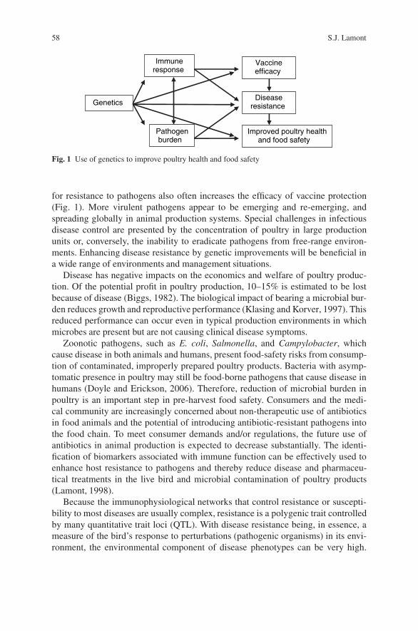
58 S.J. Lamont
Immuneresponse
Diseaseresistance
Vaccineefficacy
Improved poultry healthand food safety
Pathogenburden
Genetics
Fig. 1 Use of genetics to improve poultry health and food safety
for resistance to pathogens also often increases the efficacy of vaccine protection(Fig. 1). More virulent pathogens appear to be emerging and re-emerging, andspreading globally in animal production systems. Special challenges in infectiousdisease control are presented by the concentration of poultry in large productionunits or, conversely, the inability to eradicate pathogens from free-range environ-ments. Enhancing disease resistance by genetic improvements will be beneficial ina wide range of environments and management situations.
Disease has negative impacts on the economics and welfare of poultry produc-tion. Of the potential profit in poultry production, 10–15% is estimated to be lostbecause of disease (Biggs, 1982). The biological impact of bearing a microbial bur-den reduces growth and reproductive performance (Klasing and Korver, 1997). Thisreduced performance can occur even in typical production environments in whichmicrobes are present but are not causing clinical disease symptoms.
Zoonotic pathogens, such as E. coli, Salmonella, and Campylobacter, whichcause disease in both animals and humans, present food-safety risks from consump-tion of contaminated, improperly prepared poultry products. Bacteria with asymp-tomatic presence in poultry may still be food-borne pathogens that cause disease inhumans (Doyle and Erickson, 2006). Therefore, reduction of microbial burden inpoultry is an important step in pre-harvest food safety. Consumers and the medi-cal community are increasingly concerned about non-therapeutic use of antibioticsin food animals and the potential of introducing antibiotic-resistant pathogens intothe food chain. To meet consumer demands and/or regulations, the future use ofantibiotics in animal production is expected to decrease substantially. The identi-fication of biomarkers associated with immune function can be effectively used toenhance host resistance to pathogens and thereby reduce disease and pharmaceu-tical treatments in the live bird and microbial contamination of poultry products(Lamont, 1998).
Because the immunophysiological networks that control resistance or suscepti-bility to most diseases are usually complex, resistance is a polygenic trait controlledby many quantitative trait loci (QTL). With disease resistance being, in essence, ameasure of the bird’s response to perturbations (pathogenic organisms) in its envi-ronment, the environmental component of disease phenotypes can be very high.
Variation in Chicken Gene Structure and Expression Associated with Food-Safety 59
Heritabilities for overall resistance may be low to moderate, but dividing resistanceinto its component properties can identify immune phenotypes that each have mod-erate to high heritabilities that are amenable to genetic selection.
The complexity of disease resistance genetics has led to many different historicalapproaches being taken to improve health in commercial poultry stock. These haveincluded genetic selection of breeders after pathogenic challenge and assessmentof their relatives (sibs or progeny) for resistance traits, selection on positively cor-related immune traits such as antibody level to vaccines or other antigens such assheep red blood cells (Lamont et al., 2003), and indirect selection on protein markers(e.g., B blood group antigen for Marek’s disease resistance).
The feasibility of using whole genome scan approaches to identify genomicregions controlling important biological traits in chickens, including traits related todisease resistance, has been amply demonstrated (for reviews, see Hocking, 2005;Abasht et al., 2006). The complete draft sequence of the chicken genome wasrecently released (Hillier et al., 2004), along with a companion map of 2.8 millionsingle nucleotide polymorphisms (SNP) in the genome (Wong et al., 2004). Know-ing the specific points of genetic variation in the chicken genome will accelerateidentification of the genes that control health in poultry, by studying associations ofthe genomic variation with traits of host resistance to disease (Burt, 2005). Recentadvances in genome sequencing and high-throughput genotyping technology haveenabled genome-wide screening for QTL using high-throughput genotyping tech-nologies.
Molecular markers associated with health traits must be identified in order toapply marker-assisted selection in breeding programs for enhanced health. Thesemarkers are two general types: within genes or linked to genes. Using linked mark-ers has the advantages of not needing to know the specific identity or sequence ofany gene; the goal is to localize the positions of resistance QTL in the genome.Markers closely linked to the causative variation can be effectively used in breedingprograms until the linkage is lost to recombination, and can serve as the foundationto positionally identify the causative gene and mutation. Whole genome scans withmicrosatellites have been successfully used to identify QTL for disease resistance(e.g., McElroy et al., 2005). The current availability of the dense SNP map in nowshifting investigations from medium-density microsatellite-based genome scans tohigh-density SNP-based studies.
Whole-genome scans can be complemented with studies of specific candidategenes that control resistance traits. These genes may be positional candidatesbecause of their genomic position (mapping to a QTL location revealed througha genome scan), or they may be biological candidate genes because of a reportedassociation with resistance or immunity traits in chickens or other species. Genestructural variation assessed by SNP has successfully been associated with diseaseresistance traits (e.g., Lamont et al., 2002).
Research approaches that evaluate DNA structural variation are powerful but lackthe ability to characterize differences in gene expression, which for many traits maybe the major genetic mechanism that modulates biological performance. Studying
60 S.J. Lamont
gene expression on a large scale has recently become a reality for poultry. The com-plementary characterization of both genome structural variation and gene expres-sion differences is a very effective tool to identify genes affecting important traitsand the biological networks involved. Chicken microarrays can be used to assesschanges in gene expression that accompany different infection or resistance states(Morgan et al., 2001). The genes that show large expression differences are likelyto be involved in some aspect of the relevant pathways, although it is not possiblefrom these studies to determine whether the expression differences are a cause of,or a result of, the phenotypic differences in resistance. The convergence of associ-ations of both structure and expression of the same genes with disease resistancephenotypes provides additional evidence of an important role of the identified genes(Liu et al., 2001).
Proteomics, which is currently in early stages of use in poultry, can help to iden-tify the genes underlying resistance and immunity by profiling the proteins that existin specific tissues. Molecular genetics also provides other approaches to improvepoultry health. Genes that encode immune-system proteins, such as cytokines, canbe transferred to expression systems that produce large quantities of pure recombi-nant protein. These chicken proteins may then be used as natural immune enhancersor vaccine adjuvants. Production of transgenic poultry that are completely resistantto specific pathogens offers possibilities for those diseases with single-gene control(such as viral receptors), but only if there is an increase in the general efficiency oftechniques used to produce transgenic animals and public acceptance of transgenicanimals for food.
The complicated genetic architecture of disease resistance is shaped by variationin both structure and expression of many genes. Thus, a comprehensive picture ofthe genetics of poultry health is obtained only by integrating approaches that com-bine genome-wide and gene-specific studies of structural and expressional variation.Each of the individual research approaches to identify markers has special strengths,as well as limitations, in the type of information that it can yield.
Some genes are expected to control responses that are very specific to the individ-ual pathogen. However, other genes are likely to function at critical control pointsin host defense mechanisms and may have more general beneficial effects, such asaiding resistance to a whole group of similar pathogens (e.g., intracellular bacteria).Identifying biochemical pathways that have general effects on resistance proper-ties will be important, especially in the cases of pathogens of emerging impor-tance such as Campylobacter, or pathogens that frequently mutate and appear innew antigenic combinations, such as avian influenza. The major diseases studiedto define host genetic resistance in poultry include Marek’s disease, a viral disease(Liu et al., 2001; Morgan et al., 2001; Yonash et al., 1999; McElroy et al., 2005),Salmonellosis, a bacterial disease (Hu et al., 1997; Mariani et al., 2001; Krameret al., 2003; Liu and Lamont, 2003; Malek et al., 2004), and coccidiosis, a par-asitic disease (Lillehoj et al., 1989). As an example illustrating the principles ofeludicating the genetic control of complex resistance traits in poultry, this chapterreviews the research to determine the molecular genetic control of host response toSalmonella, a food-safety pathogen.
Variation in Chicken Gene Structure and Expression Associated with Food-Safety 61
2 Genetic Control of Salmonella Resistance in Poultry
Poultry salmonellosis is a food-safety concern and Salmonella enteritidis is a majorfood-borne pathogen, accounting for 82% of food-borne salmonellosis (Krameret al., 1998). Contaminated eggs are a leading cause of Salmonella food poison-ing and poultry flocks with high levels of S. enteritidis and contaminated manurepose the largest threat of egg contamination (Kramer et al., 1998). Understandinggenetic mechanisms controlling bacterial and cellular interactions to decrease bacte-rial colonization and fecal shedding and manure contamination would significantlydecrease this microbial contamination problem.
Some species of Salmonella bacteria are highly pathogenic in poultry, while otherspecies cause little response in the host birds, which can then become asymptomaticcarriers. Birds with subclinical salmonellosis can transmit the zoonotic bacteriainto the human food chain. Gast and Holt (1998) showed that chicks infected withSalmonella immediately after hatching can be persistently colonized to maturity,when the bacteria are shed vertically to infect table or hatching eggs, or horizontallyto infect other hens. Thus, control of salmonellosis presents special challenges tothe poultry industry.
To establish the feasibility of host genetic approaches to help improve resistanceto salmonellosis, it was first necessary to establish that there was, indeed, a mea-sure of genetic control over these traits. The heritability of chick mortality afterSalmonella challenge was estimated at 0.14 and 0.62 for sire and dam components,respectively (Beaumont et al. 1999). Estimated heritability of resistance to cecalcarrier state, measured by enrichment culture, in laying hens was 0.20 (Berthelotet al., 1998). Heritabilities of the number of bacteria persisting in internal organsranged from 0.02 to 0.29 (Girard-Santosuosso et al., 2002). Estimated heritabil-ity of antibody response to Salmonella ranged widely, from 0.03 to 0.26 (Kaiseret al., 1997; Beaumont et al., 1999). Measuring antibody response is more econom-ical and less laborious than measuring bacterial colonization and because vaccineantibody has a negative genetic correlation with cecal colonization, vaccine antibodyis a useful biomarker to improve resistance to colonization (Kaiser et al., 2002). Her-itability estimates of various parameters of Salmonella response, therefore, indicatethat genetic selection to improve resistance to salmonellosis is feasible.
As a polygenic trait, many genes are associated with genetic resistance toSalmonella species. Most genes have an individual effect on the disease phenotypicvariation that is relatively small, often only 3–5% of the total variation, which is con-sistent with complex control of the disease by many genes. Factors such as geneticline, population structure, definition of host response (antibody, mortality, systemic,or enteric colonization), and serovar of Salmonella may result in some variation inresults. However, a generally consistent picture of the existence of genetic controlof host resistance to Salmonella has emerged.
Comparative genomics strategies have been very effectively used to iden-tify genes controlling salmonellosis resistance in chickens, because response toSalmonella was previously studied in detail in the mouse as a model organism.Chicken homologs of major loci controlling natural resistance of mice to infection
62 S.J. Lamont
with S. typhimurium were examined as candidate genes. Variation in NRAMP1(natural resistance-associated macrophage protein 1, now called SLC11A1) andTNC were shown to account for 33% of the differential resistance in Salmonella-induced mortality in a backcross population of inbred lines (Hu et al., 1997).The TNC locus is closely linked to LPS, now known as TLR4, which bindslipopolysaccharide – a major component of Gram-negative bacteria membranes.The NRAMP1 association was additionally confirmed for many different specifictraits of host response to Salmonella in other chicken populations (Beaumontet al., 2003; Kramer et al., 2003; Liu et al., 2003), as was the TLR4 association(Beaumont et al., 2003; Leveque et al., 2003; Malek et al., 2004).
Success in the comparative genomics approach provided a foundation to selectadditional candidate genes to test. Some genes were positional candidates basedupon genomic position, such as CD28 and VIL1 in the NRAMP1 region (Girard-Santosuosso et al., 1997). The CD28 gene was found to be associated with entericSalmonella infection (Malek et al., 2004), and VIL1 with visceral infection (Girard-Santosuosso et al., 2002).
Involvement in pathways that are hypothesized to be important in host responseto Salmonella has been a source of information for selection of other candidategenes to study. The product of the MD2 gene interacts with the TLR4 receptor onthe cell surface, and SNPs in MD2 established to be associated with persistenceof Salmonella colonization in the cecum (Malek et al., 2004). The MHC, becauseof its crucial role in antigen processing and presentation, has been investigated.In a series of 12 B-complex congenic lines, line differences occurred in morbid-ity and mortality after Salmonella challenge (Cotter et al., 1998). Variation in theMHC class I gene was associated with resistance to Salmonella colonization inthe spleen in an experimental cross (Liu et al. 2002). Genes in apoptotic pathwaysinclude CASP1 and IAP1. A CASP1 SNP was associated with Salmonella persis-tence in the spleen and cecum in an experimental cross (Liu and Lamont, 2003)and in the liver and cecum in commercial broilers (Kramer et al., 2003). TheIAP1 gene was associated with spleen (Liu and Lamont, 2003) and cecum bacte-rial load (Kramer et al., 2003). The genomic region on chromosome 3 that con-tains a cluster of 13 gallinacin genes, which encode a newly identified family ofantimicrobial peptides, was associated with Salmonella vaccine antibody response(Hasenstein et al., 2006).
Most candidate gene experiments, because of linkage disequilibrium, cannotexclude the possibility that the causal gene could be a nearby gene rather than thespecific one studied. Supporting lines of evidence, such as confirmation in inde-pendent populations, with QTL scans, by gene expression data, or from compara-tive genomics, adds confidence in the detected gene-resistance associations. Geneexpression studies in Salmonella challenged versus unchallenged, or resistant versussusceptible animals, are currently revealing differential expression in genes that maybe active in pathways controlling resistance (Cheeseman et al., 2007; Kaiser et al.,2006; Zhou and Lamont, 2007). Combining data on Salmonella-response associ-ations with variation in gene structure and in gene expression, all conducted onthe same resource populations in the Lamont lab, has identified some predominant
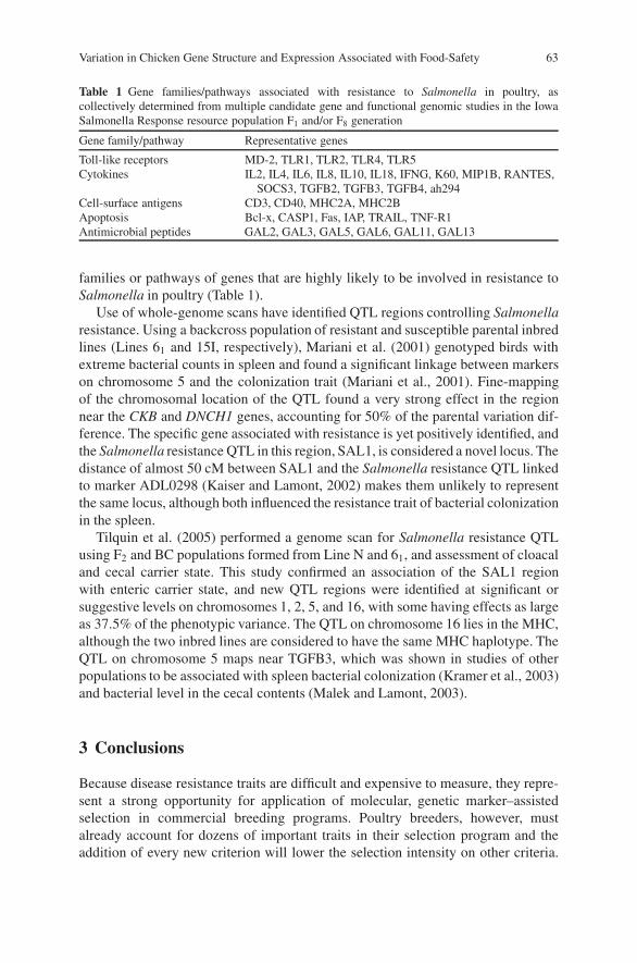
Variation in Chicken Gene Structure and Expression Associated with Food-Safety 63
Table 1 Gene families/pathways associated with resistance to Salmonella in poultry, ascollectively determined from multiple candidate gene and functional genomic studies in the IowaSalmonella Response resource population F1 and/or F8 generation
Gene family/pathway Representative genes
Toll-like receptors MD-2, TLR1, TLR2, TLR4, TLR5Cytokines IL2, IL4, IL6, IL8, IL10, IL18, IFNG, K60, MIP1B, RANTES,
SOCS3, TGFB2, TGFB3, TGFB4, ah294Cell-surface antigens CD3, CD40, MHC2A, MHC2BApoptosis Bcl-x, CASP1, Fas, IAP, TRAIL, TNF-R1Antimicrobial peptides GAL2, GAL3, GAL5, GAL6, GAL11, GAL13
families or pathways of genes that are highly likely to be involved in resistance toSalmonella in poultry (Table 1).
Use of whole-genome scans have identified QTL regions controlling Salmonellaresistance. Using a backcross population of resistant and susceptible parental inbredlines (Lines 61 and 15I, respectively), Mariani et al. (2001) genotyped birds withextreme bacterial counts in spleen and found a significant linkage between markerson chromosome 5 and the colonization trait (Mariani et al., 2001). Fine-mappingof the chromosomal location of the QTL found a very strong effect in the regionnear the CKB and DNCH1 genes, accounting for 50% of the parental variation dif-ference. The specific gene associated with resistance is yet positively identified, andthe Salmonella resistance QTL in this region, SAL1, is considered a novel locus. Thedistance of almost 50 cM between SAL1 and the Salmonella resistance QTL linkedto marker ADL0298 (Kaiser and Lamont, 2002) makes them unlikely to representthe same locus, although both influenced the resistance trait of bacterial colonizationin the spleen.
Tilquin et al. (2005) performed a genome scan for Salmonella resistance QTLusing F2 and BC populations formed from Line N and 61, and assessment of cloacaland cecal carrier state. This study confirmed an association of the SAL1 regionwith enteric carrier state, and new QTL regions were identified at significant orsuggestive levels on chromosomes 1, 2, 5, and 16, with some having effects as largeas 37.5% of the phenotypic variance. The QTL on chromosome 16 lies in the MHC,although the two inbred lines are considered to have the same MHC haplotype. TheQTL on chromosome 5 maps near TGFB3, which was shown in studies of otherpopulations to be associated with spleen bacterial colonization (Kramer et al., 2003)and bacterial level in the cecal contents (Malek and Lamont, 2003).
3 Conclusions
Because disease resistance traits are difficult and expensive to measure, they repre-sent a strong opportunity for application of molecular, genetic marker–assistedselection in commercial breeding programs. Poultry breeders, however, mustalready account for dozens of important traits in their selection program and theaddition of every new criterion will lower the selection intensity on other criteria.
64 S.J. Lamont
Therefore, the use of new selection tools, such as molecular markers, needs tobe carefully balanced so that the health improvements will complement, but notcompromise, current successes in poultry breeding.
Multiple approaches can be taken to identify genes associated with importantbiological traits such as resistance to Salmonella. The integration of multiple, com-plementary approaches helps to resolve the shortfalls inherent in each approach.Genome scans and candidate gene studies can reveal associations with disease resis-tance, and techniques to study gene expression are also powerful tools in diseasegenetics research. The newly available chicken genome sequence will accelerate thediscovery process. Because many of these resources, however, have only recentlycome on line, a limited number of specific genes or regions of the genome associatedwith poultry health have yet been identified.
Genetic selection for improved immunity and resistance traits, which can beaccomplished after sufficient genetic markers are defined, can improve poultryhealth and production efficiency, as well as reducing the potential for the introduc-tion of food-borne pathogens into the human food chain. Improving poultry healthby increasing genetic resistance to disease is essential to meet the increasing empha-sis of consumers and the industry on animal welfare, food safety, environmentalconcerns, and efficiency of production.
Acknowledgments I gratefully acknowledge the many colleagues and research group memberswho have contributed to this discovery program in the genetics of host resistance to Salmonella.The work was funded by the Iowa Agriculture and Home Economics Experiment Station, Ames,Iowa; Animal Health, Hatch Act, State of Iowa and Iowa State University Center for IntegratedAnimal Genomics Funds; Midwest Poultry Research Program, National Research Initiative Grantno. 2004-35205-14234 from the USDA Cooperative State Research, Education, and ExtensionService; and Research Grant US-3408-03 from BARD, the Binational Agriculture Research andDevelopment fund.
References
Abasht, B., Dekkers, J.C.M., and Lamont, S.J., 2006, Review of quantitative trait loci identified inthe chicken, Poultry Sci. 85:2079–2096.
Beaumont, C., Protais, J., Guillot, J. F., Colin, P., Proux, K., Millet, N., and Pardon, P., 1999,Genetic resistance to mortality of day-old chicks and carrier-state of hens after inoculationwith Salmonella enteritidis, Avian Pathol. 28:131–135.
Beaumont, C., Protais, J., Pitel, F., Leveque, G., Malo, D., Lantier, F., Plisson-Petit, F., Colin, P.,Protais, M., Le Roy, P., Elsen, J. M., Milan, D., Lantier, I., Neau, A., Salvat, G., and Vignal, A.,2003, Effect of two candidate genes on the Salmonella carrier state in fowl, Poultry Sci. 82:721–726.
Berthelot, F., Beaumont, C., Mompart, F., Girard-Santosuosso, O., Pardon, P., and Duchet-Suchaux, M., 1998, Estimated heritability of the resistance to cecal carrier state of Salmonellaenteritidis in chickens, Poultry Sci. 77:797–801.
Biggs, P.M., 1982, The world of poultry diseases, Avian Path. 11:281–300.Burt, D.W., 2005, Chicken genome: Current status and future opportunities, Genome Res. 15:
1692–1698.
Variation in Chicken Gene Structure and Expression Associated with Food-Safety 65
Cheeseman, J.H., Kaiser, M.G., Ciraci, C., Kaiser, P., and Lamont, S.J., 2007, Breed effect onearly cytokine mRNA expression in spleen and cecum of chickens with and without Salmonellaenteritidis infection, Dev. Comp. Immunol. 31:52–60.
Cotter, P.F., Taylor, R.L., Jr., and Abplanalp, H., 1998, B-complex associated immunity toSalmonella enteritidis challenge in congenic chickens, Poultry Sci. 77:1846–1851.
Doyle, M., and Erickson, M.C., 2006, Reducing the carriage of foodborne pathogens in livestockand poultry, Poultry Sci. 85:960–973.
Gast, R.K. and Holt, P.S., 1998, Persistence of Salmonella enteritidis from one day of age untilmaturity in experimentally infected layer chickens, Poultry Sci. 77:1759–1762.
Girard-Santosuosso, O., Bumstead, N., Lantier, I., Protais, J., Colin, P., Guillot, J.F., Beaumont, C.,Malo, D., and Lantier, F., 1997, Partial conservation of the mammalian NRAMP1 syntenicgroup on chicken chromosome 7, Mamm. Genome 8:614–616.
Girard-Santosuosso, O., Lantier, F., Lantier, I., Bumstead, N., Elsen, J. M., and Beaumont, C.,2002, Heritability of susceptibility to Salmonella enteritidis infection in fowls and test of therole of the chromosome carrying the NRAMP1 gene, Genet. Sel. Evol. 34:211–219.
Hasenstein, J.R., Zhang, G., and Lamont, S.J., 2006, Analyses of five gallinacin genes and theSalmonella enterica serovar Enteritidis response in poultry, Infect. Immun. 74:3375–3380.
Hillier, L.W., Miller, W., Birney, E., Warren, W., Hardison, R.C., et al., 2004, Sequence and com-parative analysis of the chicken genome provide unique perspectives on vertebrate evolution,Nature 432:695–716.
Hocking, P.M., 2005, Review on QTL mapping results in chickens, World’s Poult. Sci. J. 61:215–226.
Hu, J., Bumstead, N., Barrow, P., Sebastiani, G., Olien, L., Morgan, K., and Malo, D., 1997, Resis-tance to salmonellosis in the chicken is linked to NRAMP1 and TNC, Genome Res. 7:693–704.
Kaiser, M.G., Cheeseman, J.H., Kaiser, P., and Lamont, S.J 2006, Cytokine expression in chickenperipheral blood mononuclear cells after in vitro exposure to Salmonella enterica serovar Enter-itidis, Poultry Sci. 85:1907–1911.
Kaiser, M.G., Lakshmanan, N., Wing, T., and Lamont, S.J., 2002, Salmonella enterica serovarenteritidis burden in broiler breeder chicks genetically associated with vaccine antibodyresponse, Avian Dis. 46:25–31.
Kaiser, M.G., and Lamont, S.J., 2002, Microsatellites linked to Salmonella enterica serovar Enter-itidis burden in spleen and cecal content of young F1 broiler-cross chicks, Poultry Sci. 81:657–663.
Klasing, K.C., and Korver, D.R., 1997, Leukocytic cytokines regulate growth rate and compositionfollowing activation of the immune system, J. Anim. Sci. 75:58–67.
Kaiser, M.G., Wing, T., Cahaner, A., and Lamont, S.J., 1997, Aviagen, 12th International Sympo-sium on Current Problems in Avian Genetics, Prague, Czech Republic.
Kramer, J., Malek, M., and Lamont, S.J., 2003, Association of twelve candidate gene polymor-phisms and response to challenge with Salmonella enteritidis in poultry, Anim. Genet. 34:339–348.
Kramer, T.T., Reinke, C.R., and James, M., 1998, Reduction of fecal shedding and egg contami-nation of Salmonella enteritidis by increasing the number of heterophil adaptations, Avian Dis.42:585–588.
Lamont, S.J., 1998, Impact of genetics on disease resistance, Poult Sci. 77:1111–1118.Lamont, S.J., Kaiser, M.G., and Liu, W., 2002, Candidate genes for resistance to Salmonella enter-
itidis colonization in chickens as detected in a novel genetic cross, Vet. Immunol. Immunop.87:423–428.
Lamont, S.J., Pinard-van der Laan, M.-H., Cahaner, A., van der Poel, J.J., and Parmentier, H.K.,2003, Selection for disease resistance: direct selection on the immune response, in W.M. Muirand S.E. Aggrey, eds., Poultry Genetics, Breeding and Biotechnology, CAB International, OxonUK, pp. 399–418.
Leveque, G., Forgetta, V., Morroll, S., Smith, A.L., Bumstead, N., Barrow, P., Loredo-Osti, J.C.,Morgan, K., and Malo, D., 2003, Allelic variation in TLR4 is linked to susceptibility toSalmonella enterica serovar Typhimurium infection in chickens, Infect. Immun. 71:1116–1124.
66 S.J. Lamont
Lillehoj, H.S., Ruff, M.D., Bacon, L.D., Lamont, S.J., and Jeffers, T.K., 1989, Genetic control ofimmunity to Eimeria tenella. Interaction of MHC genes and non-MHC linked genes influenceslevels of disease susceptibility in chickens, Vet. Immunol. Immunop. 20:135–148.
Liu, H.C., Cheng, H.H., Tirunagaru, V., Sofer, L., and Burnside, J., 2001, A strategy to identifypositional candidate genes conferring Marek’s disease resistance by integrating DNA microar-rays and genetic mapping, Anim. Genet. 32:351–359.
Liu, W., Kaiser, M.G., and Lamont, S.J., 2003, Natural resistance-associated macrophage protein 1gene polymorphisms and response to vaccine against or challenge with Salmonella enteritidisin young chicks, Poultry Sci. 82:259–266.
Liu, W., and Lamont, S.J., 2003, Candidate gene approach: potentional association of caspase-1,inhibitor of apoptosis protein-1, and prosaposin gene polymorphisms with response toSalmonella enteritidis challenge or vaccination in young chicks, Anim. Biotechnol. 14:61–76.
Liu, W., Miller, M. M., and Lamont, S.J., 2002, Association of MHC class I and class II genepolymorphisms with vaccine or challenge response to Salmonella enteritidis in young chicks,Immunogenetics 54:582–590.
Malek, M., Hasenstein, J.R., and Lamont, S.J., 2004, Analysis of chicken TLR4, CD28, MIF,MD-2, and LITAF genes in a Salmonella enteritidis resource population, Poultry Sci. 83:544–549.
Malek, M., and Lamont, S.J., 2003, Association of INOS, TRAIL, TGF-beta2, TGF-beta3, andIgL genes with response to Salmonella enteritidis in poultry, Genet. Sel. Evol. 35 (Suppl 1):S99–S111.
Mariani, P., Barrow, P.A., Cheng, H.H., Groenen, M.M., Negrini, R., and Bumstead, N., 2001,Localization to chicken chromosome 5 of a novel locus determining salmonellosis resistance,Immunogenetics 53:786–791.
McElroy, J.P., Dekkers, J.C.M., Fulton, J.E., O’Sullivan, N.P., Soller, M., Lipkin, E., Zhang, W.,Koehler, K.J., Lamont, S.J., and Cheng, H.H., 2005, Microsatellite markers associated withresistance to Marek’s disease in commercial layer chickens, Poultry Sci. 84:1678–1688.
Morgan, R.W., Sofer, L., Anderson, A.S., Bernberg, E.L., Cui, J., and Burnside, J., 2001, Induc-tion of host gene expression following infection of chicken embryo fibroblasts with oncogenicMarek’s disease virus, J. Virol. 75:533–539.
Tilquin, P., Barrow, P.A., Marly, J., Pitel, F., Plisson-Petit, F., Velge, P., Vignal, A., Baret, P.V.,Bumstead, N., and Beaumont, C., 2005, A genome scan for quantitative trait loci affecting theSalmonella carrier-state in the chicken, Genet. Sel. Evol. 37:539–561.
Wong, G.K., Liu, B., Wang, J., Zhang, Y., Yang, X., et al., 2004, A genetic variation map forchicken with 2.8 million single-nucleotide polymorphisms, Nature 432:717–722.
Yonash, N., Bacon, L.D., Witter, R.L., and Cheng, H.H., 1999, High resolution mapping and iden-tification of new quantitative trait loci (QTL) affecting susceptibility to Marek’s disease, Anim.Genet. 30:126–135.
Zhou, H., and Lamont, S.J., 2007, Global gene expression profile after Salmonella enterica Serovarenteritidis challenge in two F8 advanced intercross chicken lines. Cytogenet, Genome Res.117:131–138.

Functional Genomics and Bioinformaticsof the Phytophthora sojae Soybean Interaction
Brett M. Tyler, Rays H.Y. Jiang, Lecong Zhou, Sucheta Tripathy,Daolong Dou, Trudy Torto-Alalibo, Hua Li, Yongcai Mao, Bing Liu,Miguel Vega-Sanchez, Santiago X. Mideros, Regina Hanlon, Brian M. Smith,Konstantinos Krampis, Keying Ye, Steven St. Martin, Anne E. Dorrance,Ina Hoeschele, and M.A. Saghai Maroof
Abstract Oomycete plant pathogens such as Phytophthora species and downymildews cause destructive diseases in an enormous variety of crop plant species aswell as forests and native ecosystems. These pathogens are most closely related toalgae in the kingdom Stramenopiles, and hence have evolved plant pathogenicityindependently of other plant pathogens such as fungi. We have used bioinfor-matic analysis of genome sequences and EST collections, together with functionalgenomics to identify plant and pathogen genes that may be key players in the inter-action between the soybean pathogen Phytophthora sojae and its host. In P. sojae,we have identified many rapidly diversifying gene families that encode potentialpathogenicity factors including protein toxins, and a class of proteins (avirulenceor effector proteins) that appear to have the ability to penetrate plant cells. Tran-scriptomic analysis of quantitative or multigenic resistance against P. sojae in soy-bean has revealed that there are widespread adjustments in host gene expressionin response to infection, and that some responses are unique to particular resistantcultivars. These observations lay the foundation for dissecting the interplay betweenpathogen and host genes during infection at a whole-genome level.
1 Introduction
Plant pathogens from the genus Phytophthora cause destructive diseases in anenormous variety of crop plant species as well as forests and native ecosystems(Erwin and Ribiero, 1996). The soybean pathogen Phytophthora sojae is alsocausing serious losses to the United States soybean crop on the order of US$100–200 million annually (Grau et al., 2004). The newly emerged Phytophthoraspecies, Phytophthora ramorum, is attacking trees and shrubs of coastal oak forests
B.M. TylerVirginia Bioinformatics Institute, Virginia Polytechnic Institute and State University, Blacksburg,VA 24061, USA
J.P. Gustafson et al. (eds.), Genomics of Disease, 67C© Springer Science+Business Media, LLC, 2008
68 B.M. Tyler et al.
in California, including the keystone live oak species (sudden oak death disease)(Rizzo et al., 2002). Taxonomically, Phytophthora pathogens are oomycetes,which are organisms that resemble fungi but belong to a kingdom of life calledStramenopiles that are most closely related to algae such as kelp and diatoms.Hence conventional fungal control measures often fail against these pathogens.Oomycetes include many other destructive plant pathogens in addition to Phytoph-thora, in particular the downy mildews and more than 100 species of the genusPythium. The downy mildew of Arabidopsis thaliana, caused by Hyaloperonosporaparasitica, is an important model system for understanding plant responses toinfection (Slusarenko and Schlaich, 2003). P. sojae and the potato and tomatopathogen Phytophthora infestans have been used for many basic studies of Phy-tophthora because these species are easy to genetically manipulate (Judelson, 1997;Tyler, 2007). Protection of soybean against P. sojae infection has traditionallyrelied on major resistance genes (R genes) (Schmitthenner, 1985). At least 14 Rgenes that protect against P. sojae infection (Rps genes) have been identified insoybean (Burnham et al., 2003b). At least 12 avirulence genes have been identifiedgenetically in P. sojae that interact in a gene-for-gene manner with Rps genes(Gijzen et al., 1996; May et al., 2002; Tyler et al., 1995; Tyler, 2002; Whissonet al., 1994, 1995). One avirulence gene, Avr1b-1, has been cloned and extensivelycharacterized (Shan et al., 2004). Cloning of Avr1a (MacGregor et al., 2002) andAvr46 (Whisson et al., 2004) is also close to completion.
Although major gene resistance has historically been effective against P.sojae, changes in pathogen populations over the last 10–15 years have progres-sively eroded the effectiveness of these genes (Dorrance et al., 2003b; Schmit-thenner, 1985; Schmitthenner et al., 1994). In particular, sexual reproductionappears to have greatly increased the complexity of the P. sojae population inthe US and Canada, resulting in genotypes of the pathogen able to overcomemost combinations of Rps genes (Dorrance et al., 2003b; Fsrster et al., 1994;Schmitthenner et al., 1994). As a result, there has been increased interest inquantitative resistance against P. sojae (also called partial resistance, multigenicresistance, field resistance, rate-reducing resistance, general resistance, or toler-ance) (Dorrance and Schmitthenner, 2000; Dorrance et al., 2003a; Tooley andGrau, 1982, 1984). Quantitative resistance has also been the main focus ofbreeding efforts against P. infestans in tomato and potato (Colon et al., 1995),where R gene resistance has been much more rapidly overcome (Fry andGoodwin, 1997). This form of resistance is more durable against changes inpathogen populations because it relies on small contributions from multiplegenes and does not depend on the presence of specific genes in the pathogenfor effectiveness. The principal mechanism of quantitative resistance in thesoybean lines characterized to date is the ability to reduce the rate of lesionexpansion following infection (Mideros et al., 2007; Tooley and Grau, 1982,1984). Quantitative resistance in soybean is highly heritable, but the multigenicnature of the resistance makes it difficult to introduce by conventional breed-ing. Although there has been some molecular characterization of resistant lines(Burnham et al., 2003a; Vega-Sanchez et al., 2005), the multigenic nature of
Functional Genomics and Bioinformatics of P. sojae Soybean Interaction 69
the resistance as well as the small contributions of each gene also impede thedevelopment of a better understanding of the molecular mechanisms of quantitativeresistance.
2 Sequencing of Oomycete Genomes
The complete genome sequence of an organism is an excellent starting point foridentifying genes involved in pathogenicity or any other process of interest. Thegenome sequence can also aid substantially in developing genetic tools for detectingand tracking the pathogen. The draft genome sequences of P. sojae and P. ramo-rum have been completed (Tyler et al., 2006). The 95-Mb genome of P. sojae wassequenced to a depth of ×9 while the 65-Mb genome of P. ramorum was sequencedto a depth of ×7, both by whole-genome shotgun sequencing. Automated genecalling software has identified 19,027 predicted genes (called gene models) in thegenome of P. sojae and 15,743 in the genome of P. ramorum. Analysis and visual-ization of gene predictions and annotations of the P. sojae and P. ramorum genomesare available at the VBI Microbial Database (VMD) (phytophthora.vbi.vt.edu) andthe JGI Genome Portals (www.jgi.doe.gov/genomes). VMD stores not only genomesequence data, but also Expressed Sequence Tag (EST) and microarray data andincludes a Genome Community Annotation Tool (GCAT) (Tripathy et al., 2006).Genome sequencing of four other osmycetes, P. infestans, Phytophthora capsici,Hyaloperonospora parasitica, and Pythium ultimum, is also under way (Govers andGijzen 2006).
One of the principal interests in the genomes is to identify genes that potentiallycontribute to the ability of P. sojae and P. ramorum to infect their plant hosts. Theavailability of the two genome sequences for comparison enables the identifica-tion of rapidly evolving genes. Such genes are candidates for being involved ininfection because the co-evolutionary battle between the infection mechanism ofthe pathogens and the defense systems of the plants is expected to accelerate theevolution of these genes. The different host ranges of the two pathogens also predictextensive variation in the genes that control host specificity. P. sojae is principallyconfined to soybean while P. ramorum infects a wide range of woody trees andshrubs.
Of the predicted genes in P. sojae and P. ramorum, 9,768 are similar enough insequence between the two species that they are predicted to have the same function.These genes, often called orthologs, are arranged in very similar orders along thechromosomes (i.e., exhibit synteny) (Tyler et al., 2006; Jiang et al., 2006). However,a large proportion of the predicted genes (49% in P. sojae; 38% in P. ramorum) havediverged so much that they have no matching gene in the other species. Of thesedivergent genes, 1,755 genes are unique to P. sojae and 624 are unique to P. ramo-rum. The remainder of the predicted genes represent gene families that are presentin both species but whose members do not match one another precisely. Proteins thatare secreted by the pathogen during infection and are also divergent between P. sojaeand P. ramorum are excellent candidates for proteins that function to promote the
70 B.M. Tyler et al.
infection process. An example is the explosive expansion and divergence of onesecreted protein family, NPP1, which encodes a phytotoxin (Fellbrich et al., 2002;Qutob et al., 2002; Tyler et al., 2006). Although the genomes of some fungal andbacterial species contain up to four NPP1 genes, P. sojae and P. ramorum have 29and 40 copies, respectively, and H. parasitica also has multiple NPP1 genes.
Comparisons of the mechanisms of pathogenicity in osmycetes to mechanisms inother plant pathogens such as bacteria fungi and nematodes is also of considerableinterest (Latijnhouwers et al., 2003). The pathogens in these diverse kingdoms oflife have presumably acquired the ability to attack plants by a process of convergentevolution. Identifying common mechanisms among these pathogens may advanceour understanding of the fundamental mechanisms required by all plant pathogens,and from there we may be able to better understand what are the essential compo-nents of the plant defense mechanisms. To facilitate the identification of commonmechanisms of pathogenesis, the Plant Associated Microbe Gene Ontology Project(pamgo.vbi.vt.edu) was initiated in 2005 to add standardized terms to The GeneOntology (The Gene Ontology Consortium 2000) for annotating the contributionsof genes to interactions between microbes and hosts. This multi-institution collabo-ration, coordinated by B. Tyler, recently added 472 new terms to the Gene Ontologyand is currently engaged in using the terms to annotate genomes of nine pathogensfrom diverse kingdoms including P. sojae and P. ramorum.
3 Effector Genes in Oomycete Genomes
Effector proteins are produced by pathogens to modify the physiology of the hostcells to facilitate infection. Effector proteins have been characterized extensively inbacteria (Chang et al., 2004). Many bacterial effector proteins were initially dis-covered as the products of avirulence genes, which are genes whose products aredetected by receptors encoded by plant major resistance genes (R genes). Almost allbacterial effector proteins are introduced into the cytoplasm of the host plant cell bymeans of a specialized type III secretion system (Chang et al., 2004), where manyof them interfere with the signal transduction pathways responsible for triggeringplants’ defense responses to infection (see other chapters in this book).
R genes also protect plants against oomycetes (Tyler, 2002). Several oomyceteavirulence genes have been identified that interact genetically with these R genes(Tyler, 2002), including 10 in P. sojae. In addition to the P. sojae avirulencegene Avr1b-1 (Shan et al., 2004) mentioned above, a number of other oomyceteavirulence genes have been cloned including Atr1 (Rehmany et al., 2005) andAtr13 (Allen et al., 2004) from H. parasitica and Avr3a from P. infestans (Arm-strong et al., 2005). All four of these cloned oomycete avirulence genes encodesa small secreted protein and in each case the cognate resistance gene encodes anintracellular NBS-LRR protein (Allen et al., 2004; Armstrong et al., 2005; Bhat-tacharyya et al., 2005; Rehmany et al., 2005; Shan et al., 2004). Particle bombard-ment experiments with Avr1b-1 (N. Bruce, S. Kale, B. Tyler, unpublished), Avr3a
Functional Genomics and Bioinformatics of P. sojae Soybean Interaction 71
(Armstrong et al., 2005), Atr13 (Allen et al., 2004) and Atr1 (Rehmany et al., 2005)have confirmed that the pathogen proteins are recognized inside the plant cell. Byinference, therefore, there must be a mechanism for those proteins to enter the plantcells. However, the nature of that mechanism is presently unknown. Since manybacterial avirulence genes encode effector proteins that enter the cytoplasm of plantcells via a type III secretion pathway to increase the susceptibility of the plant tissueto infection (Chang et al., 2004), there is a strong likelihood that oomycete aviru-lence gene products also act to increase susceptibility of the host tissue to infection.Consistent with this hypothesis, transient expression of the P. infestans Avr3a genein Nicotiana benthamiana suppressed the hypersensitive defense response triggeredby the ubiquitous Phytophthora protein elicitin (Bos et al., 2006). Avr1b-1 alsoencodes an effector protein that contributes positively to virulence, since P. sojaetransformants that overexpress the Avr1b-1 gene have increased virulence on somesoybean cultivars (D. Dou and B. Tyler, unpublished).
The sequencing of the P. sojae and P. ramorum genomes revealed a very large,very diverse superfamily of genes with similarity to the four cloned osmycete avir-ulence genes. There are more than 350 members of this superfamily in each of theP. sojae and P. ramorum genomes (Tyler et al., 2006) (Fig. 1), and large numbersof these genes can also be detected in the draft genome sequences of P. infestans,P. capsici, and H. parasitica. The members of the superfamily share two conservedmotifs, RXLR and dEER, a short distance from the end of the secretory leader (Birchet al., 2006; Rehmany et al., 2005; Tyler et al., 2006). Since the four characterizedoomycete avirulence proteins are inferred to have the ability to enter the plant cell, itwas hypothesized that the RXLR motif, with or without the dEER motif, is involvedin the ability of these proteins to enter the plant cell (Birch et al., 2006; Rehmanyet al., 2005; Tyler et al., 2006). In support of this hypothesis, mutations in theRXLR motif of Avr1b-1 block the ability of the protein to interact with the plantwhen the protein is secreted into the extracellular space, but not when the protein issynthesized inside the plant cell (D. Dou, S. Kale and B. Tyler, unpublished). Themechanism by which the RXLR motif is utilized to transport oomycete proteins intoplant cells is currently not known.
A motif similar to the RXLR motif is found in proteins secreted by malariaparasites (Plasmodium species) that have the ability to cross the membrane enclos-ing the parasite (the parasitiphorous vacuolar membrane) into the lumen of the redblood cell. The Plasmodium motif, called Pexel (Marti et al., 2004) or VTF (Hilleret al., 2004), is also located near the secretory leader (Fig. 2). The similarity betweenthe RXLR and the Pexel/VTF motifs suggests that the two pathogens use a similarmechanism to introduce virulence proteins into the cytoplasm of their host cells(Fig. 2). Indeed, Bhattacharjee et al. (2006) demonstrated that RXLR motifs fromP. infestans effector proteins could replace the VTF motif in transporting proteinsacross the parasitiphorous vacuolar membrane of the red blood cell.
The physical and functional resemblance between the Plasmodium and theoomycete RXLR motifs is fascinating because the two groups of pathogens belongto different kingdoms of life (Alveolata and Stramenopila respectively), whilethe same is also true for their host species (animalia and planta respectively)
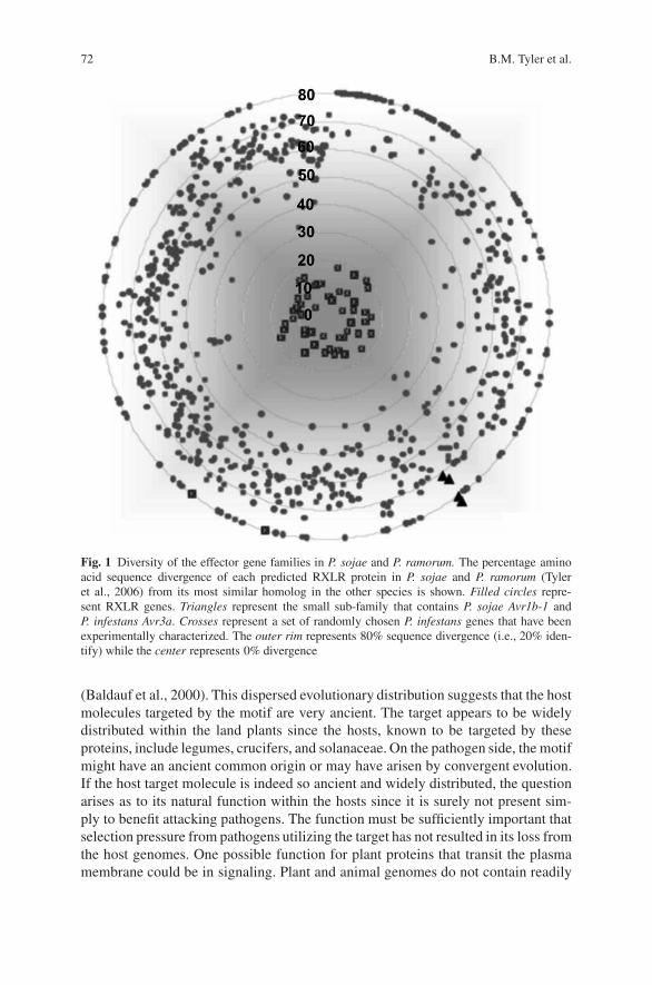
72 B.M. Tyler et al.
Fig. 1 Diversity of the effector gene families in P. sojae and P. ramorum. The percentage aminoacid sequence divergence of each predicted RXLR protein in P. sojae and P. ramorum (Tyleret al., 2006) from its most similar homolog in the other species is shown. Filled circles repre-sent RXLR genes. Triangles represent the small sub-family that contains P. sojae Avr1b-1 andP. infestans Avr3a. Crosses represent a set of randomly chosen P. infestans genes that have beenexperimentally characterized. The outer rim represents 80% sequence divergence (i.e., 20% iden-tify) while the center represents 0% divergence
(Baldauf et al., 2000). This dispersed evolutionary distribution suggests that the hostmolecules targeted by the motif are very ancient. The target appears to be widelydistributed within the land plants since the hosts, known to be targeted by theseproteins, include legumes, crucifers, and solanaceae. On the pathogen side, the motifmight have an ancient common origin or may have arisen by convergent evolution.If the host target molecule is indeed so ancient and widely distributed, the questionarises as to its natural function within the hosts since it is surely not present sim-ply to benefit attacking pathogens. The function must be sufficiently important thatselection pressure from pathogens utilizing the target has not resulted in its loss fromthe host genomes. One possible function for plant proteins that transit the plasmamembrane could be in signaling. Plant and animal genomes do not contain readily
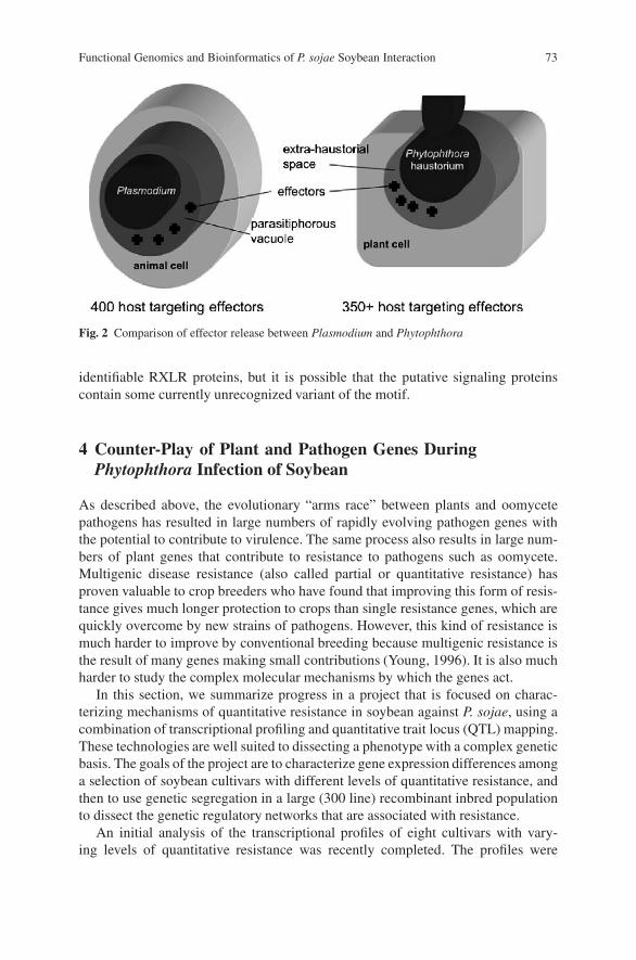
Functional Genomics and Bioinformatics of P. sojae Soybean Interaction 73
Fig. 2 Comparison of effector release between Plasmodium and Phytophthora
identifiable RXLR proteins, but it is possible that the putative signaling proteinscontain some currently unrecognized variant of the motif.
4 Counter-Play of Plant and Pathogen Genes DuringPhytophthora Infection of Soybean
As described above, the evolutionary “arms race” between plants and oomycetepathogens has resulted in large numbers of rapidly evolving pathogen genes withthe potential to contribute to virulence. The same process also results in large num-bers of plant genes that contribute to resistance to pathogens such as oomycete.Multigenic disease resistance (also called partial or quantitative resistance) hasproven valuable to crop breeders who have found that improving this form of resis-tance gives much longer protection to crops than single resistance genes, which arequickly overcome by new strains of pathogens. However, this kind of resistance ismuch harder to improve by conventional breeding because multigenic resistance isthe result of many genes making small contributions (Young, 1996). It is also muchharder to study the complex molecular mechanisms by which the genes act.
In this section, we summarize progress in a project that is focused on charac-terizing mechanisms of quantitative resistance in soybean against P. sojae, using acombination of transcriptional profiling and quantitative trait locus (QTL) mapping.These technologies are well suited to dissecting a phenotype with a complex geneticbasis. The goals of the project are to characterize gene expression differences amonga selection of soybean cultivars with different levels of quantitative resistance, andthen to use genetic segregation in a large (300 line) recombinant inbred populationto dissect the genetic regulatory networks that are associated with resistance.
An initial analysis of the transcriptional profiles of eight cultivars with vary-ing levels of quantitative resistance was recently completed. The profiles were

74 B.M. Tyler et al.
determined in both mock and P. sojae-inoculated roots, 3 or 5 days after inoculation.The transcriptional profiles were measured using an Affymetrix GeneChip that con-tains probes for 37,590 soybean genes and 15,800 P. sojae genes. The experimentaldesign, summarized in Fig. 3, included the pooling of samples from 60 plants foreach measurement, and four overall replications of the experiment. The data werenormalized using GC-RMA (Wu et al. 2004), and analyzed by linear mixed modelanalysis. Genes showing significant variation in response to each modeled factorwere identified using the two-step false discovery rate (TST-FDR) estimation (Ben-jamini et al., 2006).
Table 1 shows the number of genes that show significant variation in response tothe various factors, such as cultivar differences, the presence of the pathogen and thetime of sampling, as well as interactions among those factors. A major finding of thisanalysis is the very large numbers of genes showing statistically significant variationin each category. For example, 97% of all the genes with detectable mRNAs showedsignificant variation among the eight cultivars, irrespective of the infection status or
Fig. 3 Experimental design for expression profiling of infection responses of soybean cultivarsdiffering in quantitative resistance. Mock or infected tissue samples were collected 3 or 5 daysafter inoculation from a region spanning 7.5 mm each side of the upper edge of the visible lesion.RNA was extracted from powdered frozen tissue from pools of 30 seedlings using Qiagen RNeasyMini Plant Kits and submitted to the Virginia Bioinformatics Institute Core Laboratory Facilityfor Affymetrix GeneChip hybridization. Soybean seedlings were inoculated using the slant boardmethod (McBlain et al., 1991; Olah and Schmitthenner, 1985) after growth in vermiculite in thegreenhouse for 7 days. A 5 mm scrape wound on the root 2 cm below the stem was inoculated with0.5 ml of macerated lima bean agar which was either sterile (mock inoculation) or infested withP. sojae. Four resistant cultivars (V71-370, Conrad, General, and Athow), two moderately resistantcultivars (Williams and PI), and two susceptible cultivars (Sloan and OX20-8) were tested
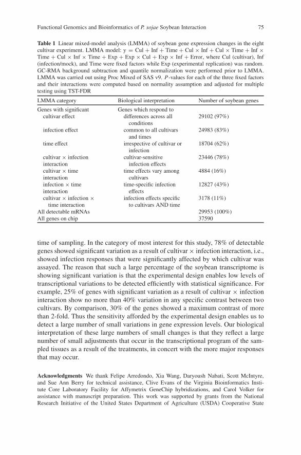
Functional Genomics and Bioinformatics of P. sojae Soybean Interaction 75
Table 1 Linear mixed-model analysis (LMMA) of soybean gene expression changes in the eightcultivar experiment. LMMA model: y = Cul + Inf + Time + Cul × Inf + Cul × Time + Inf ×Time + Cul × Inf × Time + Exp + Exp × Cul + Exp × Inf + Error, where Cul (cultivar), Inf(infection/mock), and Time were fixed factors while Exp (experimental replication) was random.GC-RMA background subtraction and quantile normalization were performed prior to LMMA.LMMA was carried out using Proc Mixed of SAS v9. P-values for each of the three fixed factorsand their interactions were computed based on normality assumption and adjusted for multipletesting using TST-FDR
LMMA category Biological interpretation Number of soybean genes
Genes with significant Genes which respond tocultivar effect differences across all
conditions29102 (97%)
infection effect common to all cultivarsand times
24983 (83%)
time effect irrespective of cultivar orinfection
18704 (62%)
cultivar × infectioninteraction
cultivar-sensitiveinfection effects
23446 (78%)
cultivar × timeinteraction
time effects vary amongcultivars
4884 (16%)
infection × timeinteraction
time-specific infectioneffects
12827 (43%)
cultivar × infection ×time interaction
infection effects specificto cultivars AND time
3178 (11%)
All detectable mRNAs 29953 (100%)All genes on chip 37590
time of sampling. In the category of most interest for this study, 78% of detectablegenes showed significant variation as a result of cultivar × infection interaction, i.e.,showed infection responses that were significantly affected by which cultivar wasassayed. The reason that such a large percentage of the soybean transcriptome isshowing significant variation is that the experimental design enables low levels oftranscriptional variations to be detected efficiently with statistical significance. Forexample, 25% of genes with significant variation as a result of cultivar × infectioninteraction show no more than 40% variation in any specific contrast between twocultivars. By comparison, 30% of the genes showed a maximum contrast of morethan 2-fold. Thus the sensitivity afforded by the experimental design enables us todetect a large number of small variations in gene expression levels. Our biologicalinterpretation of these large numbers of small changes is that they reflect a largenumber of small adjustments that occur in the transcriptional program of the sam-pled tissues as a result of the treatments, in concert with the more major responsesthat may occur.
Acknowledgments We thank Felipe Arredondo, Xia Wang, Daryoush Nabati, Scott McIntyre,and Sue Ann Berry for technical assistance, Clive Evans of the Virginia Bioinformatics Insti-tute Core Laboratory Facility for Affymetrix GeneChip hybridizations, and Carol Volker forassistance with manuscript preparation. This work was supported by grants from the NationalResearch Initiative of the United States Department of Agriculture (USDA) Cooperative State
76 B.M. Tyler et al.
Research, Education and Extension Service, numbers 2001-35319-14251, 2002-35600-12747,2004-35600-15055, and 2005-35600-16370, from the US National Science Foundation, numbersMCB-0242131, DBI-0211863, EF-0412213 and EF-0523736, and by funds from the US Depart-ment of Energy Joint Genome Institute and the Virginia Bioinformatics Institute.
References
Allen, R.L., Bittner-Eddy, P.D., Grenville-Briggs, L.J., Meitz, J.C., Rehmany, A.P., Rose, L.E.,and Beynon, J.L., 2004, Host-parasite coevolutionary conflict between Arabidopsis and downymildew, Science 306:1957–1960.
Armstrong, M.R., Whisson, S.C., Pritchard, L., Bos, J.I., Venter, E., Avrova, A.O., Rehmany, A.P.,Bohme, U., Brooks, K., Cherevach, I., Hamlin, N., White, B., Fraser, A., Lord, A., Quail, M.A.,Churcher, C., Hall, N., Berriman, M., Huang, S., Kamoun, S., Beynon, J.L., and Birch, P.R.,2005, An ancestral oomycete locus contains late blight avirulence gene Avr3a, encoding a pro-tein that is recognized in the host cytoplasm, Proc. Natl. Acad. Sci. USA 102:7766–7771.
Baldauf, S.L., Roger, A.J., Wenk-Siefert, I., and Doolittle, W.F., 2000, A kingdom-level phylogenyof eukaryotes based on combined protein data, Science 290:972–977.
Benjamini, Y., Krieger, A.M., and Yekutieli, D., 2006, Adaptive linear step-up procedures thatcontrol the false discovery rate, Biometrika 93:491–507.
Bhattacharjee, S., Hiller, N.L., Liolios, K., Win, J., Kanneganti, T.D., Young, C., Kamoun, S., andHaldar, K., 2006, The malarial host-targeting signal is conserved in the Irish potato faminepathogen, PLoS Pathog. 2:e50.
Bhattacharyya, M.K., Narayanan, N.N., Gao, H., Santra, D.K., Salimath, S.S., Kasuga, T., Liu, Y.,Espinosa, B., Ellison, L., Marek, L., Shoemaker, R., Gijzen, M., and Buzzell, R.I., 2005, Identi-fication of a large cluster of coiled coil-nucleotide binding site—-leucine rich repeat-type genesfrom the Rps1 region containing Phytophthora resistance genes in soybean, Theor. Appl. Genet.111:75–86.
Birch, P.R., Rehmany, A.P., Pritchard, L., Kamoun, S., and Beynon, J.L., 2006, Trafficking arms:oomycete effectors enter host plant cells, Trends Microbiol. 14:8–11.
Bos, J.I.B., Kanneganti, T.-D., Young, C., Cakir, C., Huitema, E., Win, J., Armstrong, M.,Birch, P.R.J., and Kamoun, S., 2006, The C-terminal half of Phytophthora infestansRXLR effector AVR3a is sufficient to trigger R3a-mediated hypersensitivity and suppressINF1induced cell death in Nicotiana benthamiana, Plant J. 48:165–176.
Burnham, K.D., Dorrance, A.E., VanToai, T.T., and St. Martin, S.K., 2003a, Quantitative trait locifor partial resistance to Phytophthora sojae in soybean, Crop Sci. 43:1610–1617.
Burnham, K.D., Dorrance, A.E., Francis, D.M., Fioritto, R.J., and St-Martin, S.K., 2003b, Rps8,A New Locus in Soybean for Resistance to Phytophthora sojae, Crop Sci. 43:101–105.
Chang, J.H., Goel, A.K., Grant, S.R., and Dangl, J.L. 2004. Wake of the flood: ascribing functionsto the wave of type III effector proteins of phytopathogenic bacteria, Curr. Opin. Microbiol.7:11–18.
Colon, L.T., Budding, D.J., Keizer, L.C.P., and Pieters, M.M.J., 1995, Components of resistanceto late blight (Phytophthora infestans) in eight South American Solanum species, Eur. J. PlantPathol. 101:441–456.
Dorrance, A.E., and Schmitthenner, A.F., 2000, New sources of resistance to Phytophthora sojaein the soybean plant introductions, Plant Dis. 84:1303–1308.
Dorrance, A.E., McClure, S.A., and St. Martin, S.K., 2003a, Effect of partial resistance on Phy-tophthora stem rot incidence and yield of soybean in Ohio, Plant Dis. 87:308–312.
Dorrance, A.E., McClure, S.A., and DeSilva, A., 2003b, Pathogenic diversity of Phytophthorasojae in Ohio soybean fields, Plant Dis. 87:139–146.
Erwin, D.C., and Ribiero, O.K., 1996, Phytophthora Diseases Worldwide, APS Press, St. Paul,Minnesota.
Functional Genomics and Bioinformatics of P. sojae Soybean Interaction 77
Fellbrich, G., Romanski, A., Varet, A., Blume, B., Brunner, F., Engelhardt, S., Felix, G.,Kemmerling, B., Krzymowska, M., and Nurnberger, T., 2002, NPP1, A Phytophthora asso-ciated trigger of plant defense in parsley and Arabidopsis, Plant J. 32:375–390.
Fsrster, H., Tyler, B.M., and Coffey, M.D., 1994, Phytophthora sojae races have arisen by clonalevolution and by rare outcrosses, Mol. Plant Microbe In. 7:780–791.
Fry, W.E., and Goodwin, S.B., 1997, Re-emergence of potato and tomato late blight in the UnitedStates, Plant Dis. 81:1349–1357.
Gijzen, M., Forster, H., Coffey, M.D., and Tyler, B.M., 1996, Cosegregation of Avr4 and Avr6 inPhytophthora sojae, Can. J. Bot. 74:800–802.
Govers, F., and Gijzen, M., 2006, Phytophthora genomics: the plant destroyers’ genome decoded,Mol. Plant Microbe In. 19:1295–1301.
Grau, C.R., Dorrance, A.E., Bond, J., and Russin, J.S., 2004, Chapter 14. Fungal diseases, inH.R. Boerma and J.E. Specht, eds., Soybeans: Improvement, Production, and Uses, Amer. Soc.Agron., Madison, Wisconsin.
Hiller, N.L., Bhattacharjee, S., van-Ooij, C., Liolios, K., Harrison, T., Lopez-Estra–o, C., andHaldar, K., 2004, A host-targeting signal in virulence proteins reveals a secretome in malarialinfection, Science 306:1934–1937.
Jiang, R.H., Tyler, B.M., and Govers, F., 2006, Comparative analysis of Phytophthora genes encod-ing secreted proteins reveals conserved synteny and lineage-specific gene duplications anddeletions, Mol. Plant Microbe Interact. 19:1311–1321.
Judelson, H.S., 1997, The genetics and biology of Phytophthora infestans: modern approaches toa historical challenge, Fungal Genet. Biol. 22:65–76.
Latijnhouwers, M., de Wit, P.J., and Govers, F., 2003, Oomycetes and fungi: similar weaponry toattack plants, Trends Microbiol. 11:462–469.
MacGregor, T., Bhattacharyya, M., Tyler, B.M., Bhat, R., Schmitthenner, A.F., and Gijzen, M.,2002, Genetic and physical mapping of Avr1a in Phytophthora sojae, Genetics 160:949–959.
Marti, M., Good, R.T., Rug, M., Knuepfer, E., and Cowman, A.F., 2004, Targeting malaria viru-lence and remodeling proteins to the host erythrocyte, Science 306:1930–1933.
May, K.J., Whisson, S.C., Zwart, R.S., Searle, I.R., Irwin, J.A.G., Maclean, D.J., Carroll, B.J., andDrenth, A., 2002, Inheritance and mapping of eleven avirulence genes in Phytophthora sojae,Fungal Genet. Biol. 37:1–12.
McBlain, B.A., Zimmerly, M.M., Schmitthenner, A.F., and Hacker, J.K., 1991, Tolerance tophytophthora rot in soybean: I. Studies of the cross Ripley × Harper, Crop Sci. 31:1405–1411.
Mideros, S., Nita, M., and Dorrance, A.E., 2007, Characterization of components of partial resis-tance, Rps2, and root resistance to Phytophthora sojae in soybean, Phytopath. 97:655–662.
Olah, A.F., and Schmitthenner, A.F., 1985, A growth chamber test for measuring phytophthoraroot rot tolerance in soybean [Glycine max] seedlings. Phytopath. 75:546–548.
Qutob, D., Kamoun, S., and Gijzen, M., 2002, Expression of a Phytophthora sojae necrosis-inducing protein occurs during transition from biotrophy to necrotrophy, Plant J. 32:361–373.
Rehmany, A.P., Gordon, A., Rose, L.E., Allen, R.L., Armstrong, M.R., Whisson, S.C., Kamoun, S.,Tyler, B.M., Birch, P.R., and Beynon, J.L., 2005, Differential recognition of highly divergentdowny mildew avirulence gene alleles by RPP1 resistance genes from two Arabidopsis lines,Plant Cell 17:1839–1850.
Rizzo, D.M., Garbelotto, M., Davidson, J.M., Slaughter, G.W., and Koike, S.T., 2002, Phytoph-thora ramorum as the cause of extensive mortality of Quercus spp. and Lithocarpus densiflorusin California, Plant Dis. 86:205–214.
Schmitthenner, A.F., 1985, Problems and progress in controlling Phytophthora root rot of soybean,Plant Dis. 69:362–368.
Schmitthenner, A.F., Hobe, M., and Bhat, R.G. 1994. Phytophthora sojae races in Ohio over a10-year interval. Plant Dis. 78: 269–276.
Shan, W., Cao, M., Leung, D., and Tyler, B.M., 2004, The Avr1b locus of Phytophthora sojaeencodes an elicitor and a regulator required for avirulence on soybean plants carrying resistancegene Rps1b, Mol. Plant Microbe In.. 17:394–403.
78 B.M. Tyler et al.
Slusarenko, A.J., and Schlaich, N.L., 2003, Pathogen profile. Downy mildew of Arabidopsisthaliana caused by Hyaloperonospora parasitica (formerly Peronospora parasitica), Mol.Plant Path. 4:159–170.
The Gene Ontology Consortium, 2000, Gene ontology: tool for the unification of biology, Nat.Genet. 25:25–29.
Tooley, P.W., and Grau, C.R., 1982, Identification and quantitative characterization of rate-reducing resistance to Phytophthora megasperma f.sp. glycinea in soybean seedlings, Phy-topath. 72:727–733.
Tooley, P.W., and Grau, C.R., 1984, Field characterization of rate-reducing resistance to Phytoph-thora megasperma f.sp. glycinea in soybean, Phytopath. 74:1201–1208.
Tripathy, S., Pandey, V.N., Fang, B., Salas, F., and Tyler, B.M., 2006, VMD: A community anno-tation database for microbial genomes, Nucl. Acids Res. 34:D379–D381.
Tyler, B., Forster, H., and Coffey, M.D., 1995, Inheritance of avirulence factors and restrictionfragment length polymorphism markers in outcrosses of the oomycete Phytophthora sojae,Mol. Plant Microbe In. 8:515–523.
Tyler, B.M., 2002, Molecular basis of recognition between Phytophthora species and their hosts,Annu. Rev. Phytopath. 40:137–167.
Tyler, B.M., 2007, Phytophthora sojae: root rot pathogen of soybean and model oomycete, Mol.Plant Path. 8:1–8.
Tyler, B.M., Tripathy, S., Zhang, X., Dehal, P., Jiang, R.H.Y., and et al., 2006, Phytophthoragenome sequences uncover evolutionary origins and mechanisms of pathogenesis, Science313:1261–1266.
Vega-Sanchez, M.E., Redinbaugh, M.G., Costanzo, S., and Dorrance, A.E., 2005, Spatial and tem-poral expression analysis of defense-related genes in soybean cultivars with different levels ofpartial resistance to Phytophthora sojae, Physiol. Mol. Plant Path. 66:175–182.
Whisson, S.C., Drenth, A., Maclean, D.J., and Irwin, J.A.G., 1994, Evidence for outcrossing inPhytophthora sojae and linkage of a DNA marker to two avirulence genes, Curr. Genet. 27:77–82.
Whisson, S.C., Drenth, A., Maclean, D.J., and Irwin, J.A.G., 1995, Phytophthora sojae avirulencegenes, RAPD and RFLP markers used to construct a detailed genetic linkage map, Mol. PlantMicrobe In. 8:988–995.
Whisson, S.C., Basnayake, S., Maclean, D.J., Irwin, J.A., and Drenth, A., 2004, Phytophthorasojae avirulence genes Avr4 and Avr6 are located in a 24kb, recombination-rich region ofgenomic DNA, Fungal Genet. Biol. 41:62–74.
Wu, Z., Irizarry, R.A., Gentleman, R., Martinez-Murillo, F., and Spencer, F., 2004, A model-basedbackground adjustment for oligonucleotide expression arrays, J. Am. Stat. Assoc. 99:909–917.
Young, N.D., 1996, QTL mapping and quantitative disease resistance in plants, Ann. Rev. Phy-topathol. 34:479–501.

Canine SINEs and Their Effects on Phenotypesof the Domestic Dog
Leigh Anne Clark, Jacquelyn M. Wahl, Christine A. Rees, George M. Strain,Edward J. Cargill, Sharon L. Vanderlip, and Keith E. Murphy
Abstract Short interspersed elements (SINEs) are mobile elements that contributeto genomic diversity through the addition of genetic material. Recent genomic anal-yses have vastly augmented our knowledge of both human- and canine-specificSINEs. SINEC Cf is a major SINE of the canid family that has undergone recentexpansion and is thought to be present in half of all genes. To date, only three phe-notypes of the domestic dog have been attributed to a SINE. One of these is merle,a coat pattern characterized by patches of full color on a diluted background andassociated with ocular and auditory anomalies. A SINEC Cf in the SILV gene causesmerle patterning by altering the cDNA transcript and has unique characteristics thatare likely responsible for the random nature of the phenotype.
1 Short Interspersed Elements
Short interspersed elements (SINEs) are non-autonomous transposons of approxi-mately 100–400 bp. SINEs have an internal promoter and are transcribed by RNApolymerase III but rely on long interspersed elements (LINEs) for reverse transcrip-tion and integration into the genome (Dewannieux et al., 2003). A typical SINE ischaracterized by three features: the head, which contains the promoter necessary forinitiation of transcription; the body, which is similar at the 3’ end to LINE RNA andis necessary for reverse transcription; and the tail, which is usually A- or AT-rich butcan vary in sequence and length and may be involved in transcription termination,RNA delivery, and/or RNA stability (Kramerov and Vassetzky, 2005).
Our knowledge of SINEs has increased greatly in recent years, largely due to theassembly of whole genome sequences. Analyses of the human sequence revealedthat SINEs comprise 13% of the genome (Lander et al., 2001). In humans, thedominant SINE is the Alu element, which is also the only active SINE family inthe genome (Lander et al., 2001). The promoter region of Alu elements is derivedfrom 7SL RNA sequences. This is different from most SINEs in that the majority
K.E. MurphyCanine Genetics Laboratory, Department of Pathobiology, College of Veterinary Medicine, TexasA&M University, College Station, TX 77843-4467
J.P. Gustafson et al. (eds.), Genomics of Disease, 79C© Springer Science+Business Media, LLC, 2008
80 L.A. Clark et al.
of SINEs have promoters derived from tRNA sequences (Ullu and Tschudi, 1984;Lander et al., 2001). It is estimated that 75% of human genes contain Alu elements,but many of these were inserted prior to divergence and thus are fixed in the popu-lation (Wang and Kirkness, 2005).
In 1992, Minnick et al. described a family of SINEs specific to canids. TheseSINEs, which are tRNA-Lys derived, are characterized by a (TC) repeat and anoligo-dA rich tail (Minnick et al., 1992). A subfamily of these SINEs is SINEC Cf.It is estimated that there are 170,000 SINEC Cf elements in the genome and thathalf of all genes contain a SINEC Cf insertion (Wang and Kirkness, 2005). Analy-ses of the first available canine genome sequence, which provides ×1.5 coverage(a Standard Poodle was used), suggest that SINEC Cf elements have experienced alarge expansion in recent history and that this subfamily is highly conserved, exhibit-ing only 4.8% divergence from the consensus sequence (Kirkness et al., 2003).When this sequence was compared to the ×7.5 coverage sequence (from a Boxer),10,000 insertion sites were found to be bimorphic (differing by the presence orabsence of a SINE), indicating that there are a substantial number of bimorphicsites present in the entire canine population (Lindblad-Toh et al., 2005).
The first published report of a disease-causing retrotransposon in the dog camein 1999 when Lin et al. discovered that narcolepsy in the Doberman Pinscher resultsfrom a 226 bp SINEC Cf insertion in intron 3 of Hcrtr2 (Lin et al., 1999). The SINE,located 35 bp upstream of the exon 4 splice acceptor, causes skipping of exon 4.Exon skipping as a result of an intronic SINE is also reported in humans, causingautoimmune lymphoproliferative syndrome (Tighe et al., 2002) and hemophilia A(Ganguly et al., 2003). In both organisms, this phenomenon presumably results fromdisplacement of the lariat branch point sequence.
A second canine disorder was recently attributed to a retrotransposon. Peleet al. (2005) showed that exon 2 of PTPLA is disrupted by a 236 bp SINEC Cfelement in Labrador retrievers having autosomal recessive centronuclear myopa-thy. The result of the insertion is variable; seven different cDNA transcripts areidentified. Exon-skipping and exonization (the incorporation of SINE sequence intomRNA) are observed in the mutant transcripts. Interestingly, one of the seven tran-scripts is wild-type, indicating that the SINE is spliced out.
2 Merle Patterning
While the most devastating impact of SINE insertions is disease such as the twomentioned above, SINEs may also have a dramatic effect on physical appearanceof dogs, without necessarily causing overt clinical disease. An example of this is aSINEC Cf insertion in the gene SILV that causes merle patterning in several breedsof dog (Clark et al., 2006). In a single dose, the mutation causes dilution of thebase fur color, which is determined by a separate locus, and can also cause blue eyecolor. The predominant characteristic of merling is random patches of full color dis-tributed in various sizes and patterns across the coat. In some breeds (e.g., Australian
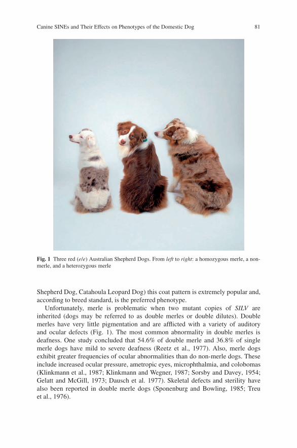
Canine SINEs and Their Effects on Phenotypes of the Domestic Dog 81
Fig. 1 Three red (e/e) Australian Shepherd Dogs. From left to right: a homozygous merle, a non-merle, and a heterozygous merle
Shepherd Dog, Catahoula Leopard Dog) this coat pattern is extremely popular and,according to breed standard, is the preferred phenotype.
Unfortunately, merle is problematic when two mutant copies of SILV areinherited (dogs may be referred to as double merles or double dilutes). Doublemerles have very little pigmentation and are afflicted with a variety of auditoryand ocular defects (Fig. 1). The most common abnormality in double merles isdeafness. One study concluded that 54.6% of double merle and 36.8% of singlemerle dogs have mild to severe deafness (Reetz et al., 1977). Also, merle dogsexhibit greater frequencies of ocular abnormalities than do non-merle dogs. Theseinclude increased ocular pressure, ametropic eyes, microphthalmia, and colobomas(Klinkmann et al., 1987; Klinkmann and Wegner, 1987; Sorsby and Davey, 1954;Gelatt and McGill, 1973; Dausch et al. 1977). Skeletal defects and sterility havealso been reported in double merle dogs (Sponenburg and Bowling, 1985; Treuet al., 1976).
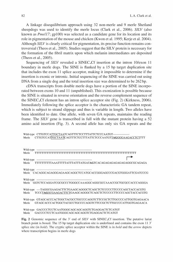
82 L.A. Clark et al.
A linkage disequilibrium approach using 32 non-merle and 9 merle Shetlandsheepdogs was used to identify the merle locus (Clark et al., 2006). SILV (alsoknown as Pmel17; gp100) was selected as a candidate gene for its location and itsrole in pigmentation of the mouse and chicken (Kwon et al. 1995; Kerje et al. 2004).Although SILV is clearly critical for pigmentation, its precise function remains con-troversial (Theos et al., 2005). Studies suggest that the SILV protein is necessary forthe formation of the fibril matrix upon which melanin intermediates are deposited(Theos et al., 2005).
Sequencing of SILV revealed a SINEC Cf insertion at the intron 10/exon 11boundary in merle dogs. The SINE is flanked by a 15 bp target duplication sitethat includes the exon 11 splice acceptor, making it impossible to determine if theinsertion is exonic or intronic. Initial sequencing of the SINE was carried out usingDNA from a single dog and the total insertion size was determined to be 262 bp.
cDNA transcripts from double merle dogs have a portion of the SINE incorpo-rated between exons 10 and 11 (unpublished). This exonization is possible becausethe SINE is situated in reverse orientation and the reverse complement sequence ofthe SINEC Cf element has an intron splice acceptor site (Fig. 2) (Kirkness, 2006).Immediately following the splice acceptor is the characteristic GA tandem repeat,which is subject to strand slippage and thus is variable in length. Two alleles havebeen identified to date. One allele, with seven GA repeats, maintains the readingframe. The SILV gene is transcribed in full with the mutant protein having a 52amino acid insertion (Fig. 3). A second allele has only six GA repeats and the
Wild-type CTTGTCCATTGCTAATCAGTTTCTCCTTTATTCTCCCAATGT------------------Merle CTTGTCCATTGCTAATCAGTTTCTCCTTTATTCTCCCAATGTTAGGGGAAGACCTCTTTT
Wild-type ------------------------------------------------------------Merle TTTTTTTTTTTTTTTTTTTTTTTTTTTTTTTTTTTTTTTTTTTTTTTTTTTTTTTTTTTT
Wild-type ------------------------------------------------------------Merle TTTTTTTTTTAAATTTTTATTTATTTATGATAGTCACAGAGAGAGAGAGAGGCGCAGAGA
Wild-type ------------------------------------------------------------Merle CACAGGCAGAGGGAGAAGCAGGCTCCATGCACCGGGAGCCCGACGTGGGATTCGATCCCG
Wild-type ------------------------------------------------------------Merle GGTCTCCAGGATCGCGCCCTGGGCCAAAGGCAGGCGCCAAACCGCTGCGCCACCCAGGGA
Wild-type ----TAGGCGAAGACTTCTGAAGCAGGGCTCAGCTCTCCCCCTTCCCCAGCTACCACGTG Merle TCCCTAGGCGAAGACTTCTGAAGCAGGGCTCAGCTCTCCCCCTTCCCCAGCTACCACGTG
Wild-type GTAGCACCCACTGGCTACGCCTGCCCCAGGTCTTCCGCTCTTGCCCCATTGGTGAGAACA Merle GTAGCACCCACTGGCTACGCCTGCCCCAGGTCTTCCGCTCTTGCCCCATTGGTGAGAACA
Wild-type GACCCCTCCTCAATGGGCAGCAGCAGGTCTGAGGACTCTCATGT Merle GACCCCTCCTCAATGGGCAGCAGCAGGTCTGAGGACTCTCATGT
Fig. 2 Genomic sequence of the 3′ end of SILV with SINEC Cf insertion. The putative lariatbranch point is boxed. The 15 bp target duplication site is underlined and contains the exon 11 3′
splice site (in bold). The cryptic splice acceptor within the SINE is in bold and the arrow depictswhere transcription begins in merle dogs
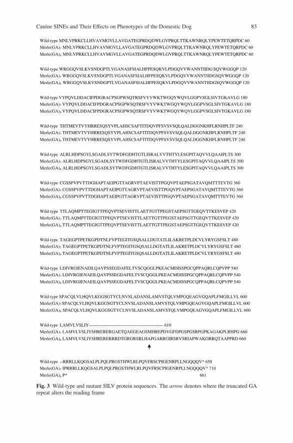
Canine SINEs and Their Effects on Phenotypes of the Domestic Dog 83
Wild-type MNLVPRKCLLHVAVMGVLLAVGATEGPRDQDWLGVPRQLTTKAWNRQLYPEWTETQRPDC 60
Merle(GA)7 MNLVPRKCLLHVAVMGVLLAVGATEGPRDQDWLGVPRQLTTKAWNRQLYPEWTETQRPDC 60
6Merle(GA) MNLVPRKCLLHVAVMGVLLAVGATEGPRDQDWLGVPRQLTTKAWNRQLYPEWTETQRPDC 60
Wild-type WRGGQVSLKVSNDGPTLVGANASFSIALHFPESQKVLPDGQVVWANNTIIDG SQVWGGQP 120
Merle(GA)7 WRGGQVSLKVSNDGPTLVGANASFSIALHFPESQKVLPDGQVVWANNTIIDGSQVWGGQP 120
Merle(GA)6 WRGGQVSLKVSNDGPTLVGANASFSIALHFPESQKVLPDGQVVWANNTIIDGSQVWGGQP 120
Wild-type VYPQVLDDACIFPDGRACPSGPWSQTRSFVYVWKTWGQYWQVLGGPVSGLSIVTGKAVLG 180
Merle(GA)7 VYPQVLDDACIFPDGRACPSGPWSQTRSFVYVWKTWGQYWQVLGGPVSGLSIVTGKAVLG 180
Merle(GA)6 VYPQVLDDACIFPDGRACPSGPWSQTRSFVYVWKTWGQYWQVLGGPVSGLSIVTGKAVLG 180
Wild-type THTMEVTVYHRRESQSYVPLAHSCSAFTITDQVPFSVSVSQLQALDGGNKHFLRNHPLTF 240
Merle(GA)7 THTMEVTVYHRRESQSYVPLAHSCSAFTITDQVPFSVSVSQLQALDGGNKHFLRNHPLTF 240
Merle(GA)6 THTMEVTVYHRRESQSYVPLAHSCSAFTITDQVPFSVSVSQLQALDGGNKHFLRNHPLTF 240
Wild-type ALRLHDPSGYLSGADLSYTWDFGDHTGTLISRALVVTHTYLESGPITAQVVLQAAIPLTS 300
Merle(GA)7 ALRLHDPSGYLSGADLSYTWDFGDHTGTLISRALVVTHTYLESGPITAQVVLQAAIPLTS 300
Merle(GA)6 ALRLHDPSGYLSGADLSYTWDFGDHTGTLISRALVVTHTYLESGPITAQVVLQAAIPLTS 300
Wild-type CGSSPVPVTTDGHAPTAEIPGTTAGRVPTAEVISTTPGQVPTAEPSGATAVQMTTTEVTG 360
Merle(GA)7 CGSSPVPVTTDGHAPTAEIPGTTAGRVPTAEVISTTPGQVPTAEPSGATAVQMTTTEVTG 360
Merle(GA)6 CGSSPVPVTTDGHAPTAEIPGTTAGRVPTAEVISTTPGQVPTAEPSGATAVQMTTTEVTG 360
Wild-type TTLAQMPTTEGIGTTPEQVPTSEVISTTLAETTGTTPEGSTAEPSGTTGEQVTTKESVEP 420
Merle(GA)7 TTLAQMPTTEGIGTTPEQVPTSEVISTTLAETTGTTPEGSTAEPSGTTGEQVTTKESVEP 420
Merle(GA)6 TTLAQMPTTEGIGTTPEQVPTSEVISTTLAETTGTTPEGSTAEPSGTTGEQVTTKESVEP 420
Wild-type TAGEGPTPETKGPDTNLFVPTEGITGSQSALLDGTATLILAKRETPLDCVLYRYGSFSLT 480
Merle(GA)7 TAGEGPTPETKGPDTNLFVPTEGITGSQSALLDGTATLILAKRETPLDCVLYRYGSFSLT 480
Merle(GA)6 TAGEGPTPETKGPDTNLFVPTEGITGSQSALLDGTATLILAKRETPLDCVLYRYGSFSLT 480
Wild-type LDIVRGIENAEILQAVPSSEGDAFELTVSCQGGLPKEACMDISSPGCQPPAQRLCQPVPP 540
Merle(GA)7 LDIVRGIENAEILQAVPSSEGDAFELTVSCQGGLPKEACMDISSPGCQPPAQRLCQPVPP 540
Merle(GA)6 LDIVRGIENAEILQAVPSSEGDAFELTVSCQGGLPKEACMDISSPGCQPPAQRLCQPVPP 540
Wild-type SPACQLVLHQVLKGGSGTYCLNVSLADANSLAMVSTQLVMPGQEAGVGQAPLFMGILLVL 600
Merle(GA)7 SPACQLVLHQVLKGGSGTYCLNVSLADANSLAMVSTQLVMPGQEAGVGQAPLFMGILLVL 600
Merle(GA)6 SPACQLVLHQVLKGGSGTYCLNVSLADANSLAMVSTQLVMPGQEAGVGQAPLFMGILLVL 600
Wild-type LAMVLVSLIY-------------------------------------------------- 610
Merle(GA)7 LAMVLVSLIYSHRERERGAETQAEGEAGSMHREPDVGFDPGSPGSRPGPKAGAKPLRHPG 660
Merle(GA)6 LAMVLVSLIYSHRERERRRDTGRGRSRLHAPGARRGIRSRVSRIAPWAKGRRQTAAPPRD 660
Wild-type --RRRLLKQGSALPLPQLPRGSTHWLRLPQVFRSCPIGENRPLLNGQQQV* 658
Merle(GA)7 IPRRRLLKQGSALPLPQLPRGSTHWLRLPQVFRSCPIGENRPLLNGQQQV* 710
Merle(GA)6 P* 661
Fig. 3 Wild-type and mutant SILV protein sequences. The arrow denotes where the truncated GArepeat alters the reading frame
84 L.A. Clark et al.
insertion disrupts the reading frame. Fifty-one amino acids are incorporated afterexon 10, and a premature stop codon occurs near the end of the insertion (Fig. 3).
3 A-Tails Are Important
Gel eletrophoresis analysis of the SILV SINE showed that the insertion size variesfrom dog to dog. Further sequencing revealed that the variability in the SINE isfound in the poly(A) tail. A-tail length is an important factor in retrotransposi-tion. Roy-Engel et al. (2002) analyzed A-tail length in Alu elements and foundthat insertions that result in disease (many are de novo events) have a mean lengthnearly twofold longer than other insertions. They also observed that overall, youngerAlu elements have longer A-tails than older Alu elements (Roy-Engel et al., 2002).These data suggest that the A-tail is evolutionarily unstable and subject to mutationand degradation over time. This phenomenon may exist in part because A-tails aresubject to strand slippage during replication and unequal crossing over (Roy-Engelet al., 2002).
The SINE element that disrupts the SILV gene is longer than other SINEC Cfelements that have been described in the dog (Pele et al., 2005; Fletcher et al., 2001;Jeoung et al., 2000). The variation in length is again found in the A-tail, which wedefine as the region after the (TC) repeat, between the last G and the duplicatedsequence. The A-tail of the SINEs in the PTPLA, dystrophin, and D2 dopaminereceptor genes are 64, 50, and 46 bp long, respectively (Pele et al., 2005; Fletcheret al., 2001; Jeoung et al., 2000). In merle dogs, the tail length of the SILV SINEinsertion ranges from 91 to 101 bp.
Along with changes in length, A-tails accumulate non-A bases that disrupt thepure A stretches. These interruptions reduce the likelihood of strand slippage, result-ing in greater tail stability. The SILV SINE has fewer A-tail interruptions than do thePTPLA and dystrophin SINEs. Consequently, the former SINE has pure A stretchesextending as long as 83 bp, while the latter SINEs have maximum pure A stretchesof 14 and 15 bp, respectively. The length and purity of the A-tail of the SILV inser-tion suggest that it is a young SINEC Cf and is subject to greater levels of instability.
A surprising find is the presence of the SILV SINE in dogs that do not have themerle phenotype (Clark et al., 2006). In these dogs, the poly(A)-tail is shortened,ranging from 54 to 65 bp. This finding suggests that the SINE insertion is necessary,but not sufficient for the merle pattern (Cordaux and Batzer, 2006). Although thethreshold has not been determined, it is apparent that a long A-tail is requisite toproduce the phenotype. A smaller insertion size may bring the lariat branch point toa more reasonable distance with the true splice site, encouraging proper splicing.
These data offer a possible explanation for merle patterning. During develop-ment, melanoblasts migrate from the neural crest and differentiate into the pigment-producing melanocytes (Steingrimsson et al., 2004). Instability of the poly(A)-tailduring this migration could result in cell populations with varying tail lengths.Melanocytes having a larger SILV SINE insertion would produce diluted pig-ment, while those with a truncated A-tail would produce full pigment. A similar
Canine SINEs and Their Effects on Phenotypes of the Domestic Dog 85
mechanism is found in pigs: black spotting results from somatic reversions of a Cexpansion in the MC1R gene (Kijas et al., 2001).
The randomness of merling may also provide an explanation for the variabil-ity of the abnormalities associated with the double merle phenotype. In 2006, ahearing test known as the brainstem auditory evoked response (BAER), whichdetects electrical activity in the cochlea and auditory pathways in the brain (Wilsonand Mills, 2005), was performed on 70 young to middle-aged merles representingfive breeds (unpublished). Twenty-two double merles were examined: eight werebilaterally deaf and two were unilaterally deaf. Although Reetz et al. (1977) sug-gested that about one-third of heterozygous merles have deafness, only one of the48 heterozygous merles in this study was deaf (unilateral). This dog, a Great Dane,was also piebald and consequently the deafness cannot be positively attributed tothe SILV mutation (Strain, 1999).
Fifteen of the double merles tested were Catahoula Leopard Dogs and of theseonly four were deaf. Although the sample size is small, this study suggests thatonly about 26% of double merle Catahoula Leopard Dogs are deaf while roughly85% of double merles from other breeds tested (Australian Shepherd, Collie, andShetland Sheepdog) are deaf. This finding is not entirely surprising because doublemerle Catahoula Leopard Dogs exhibit larger amounts of pigmentation than do otherbreeds. These phenotypic differences may result from a shorter average poly(A)-taillength in the breed or from modifying genes.
4 Summary
Mobile elements promote genomic diversity through the addition, and occasionaldeletion, of genetic material. Those mutations, which occur in or near coding regionsmay also influence phenotypic diversity by altering gene expression and function.These changes may manifest themselves as detrimental genetic disorders or as sim-ple physical traits. In addition, variability within phenotypes may result from insta-bility of the causative element. This is exemplified by merle patterning in the dog inwhich random spotting is determined by the total size of the SINE insertion.
Acknowledgments This research was supported in part by the National Organization for HearingResearch Foundation. We are grateful to Don Abney, Kerry Kirtley, Sherry Lindsey, and SusanSchroeder for providing tissue samples.
References
Clark, L.A., Wahl, J.M., Rees, C.A., and Murphy, K.E., 2006, Retrotransposon insertion in SILVis responsible for merle patterning of the domestic dog, Proc. Natl. Acad. Sci. USA 103:1376–1381.
Cordaux, R., and Batzer, M.A., 2006, Teaching an old dog new tricks: SINEs of canine genomicdiversity, Proc. Natl. Acad. Sci. USA 103:1157–1158.
Dausch, D., Wegner, W., Michaelis, M., and Reetz, I., 1977, Ophthalmologische befunde in einermerlezucht. Dtsch. Tierarztl. Wschr. 84:453–492.
86 L.A. Clark et al.
Dewannieux M., Esnault, C., and Heidmann, T., 2003, LINE-mediated retrotransposition ofmarked Alu sequences. Nat. Genet. 35:41–48.
Fletcher, S., Carville, K.S., Howell, J.M., Mann, C.J., and Wilton, S.D., 2001, Evaluation of a shortinterspersed nucleotide element in the 3’ untranslated region of the defective dystrophin geneof dogs with muscular dystrophy, Am. J. Vet. Res. 62:1964–1968.
Ganguly, A., Dunbar, T., Chen, P., Godmilow, L., and Ganguly, T., 2003, Exon skipping caused byan intronic insertion of a young Alu Yb9 element leads to severe hemophilia A, Hum. Genet.113:348–352.
Gelatt, K.N., and McGill, L.D., 1973, Clinical characteristics of microphthalmia with colobomasof the Australian shepherd dog, J. Am. Vet. Med. Assoc. 162:393–396.
Jeoung, D., Myeong, H., Lee, H., Ha, J., Galibert, F., Hitte, C., and Park, C., 2000, A SINE ele-ment in the canine D2 dopamine receptor gene and its chromosomal location, Anim. Genet. 31:333–346.
Kerje, S., Sharma, P., Gunnarsson, U., Kim, H., Bagchi, S., Fredriksson, R., Schultz, K., Jensen, P.,von Heijne, G., Okimoto, R., and Andersson, L., 2004, The dominant white, dun and smokeycolor variants in chickens are associated with insertion/deletion polymorphisms in the PMEL17gene, Genetics 168:1507–1518.
Kijas, J.M.H., Moller, M., Plastow, G., and Anderson, L., 2001, A frameshift mutation in MCR1and a high frequency of somatic reversions cause black spotting in pigs, Genetics 158:779–785.
Kirkness, E.F., 2006, SINEs of canine genomic diversity, in Elaine A. Ostrander, Urs Giger, KerstinLindblad-Toh, eds., The Dog and Its Genome, Cold Spring Harbor Laboratory Press, Wood-bury, New York, pp. 209–219.
Kirkness, E.F., Bafna, V., Halpern, A.L., Levy, S., Remington, K., Rusch, D.B., Delcher, A.L., Pop,M., Wang, W., Fraser, C.M., and Venter, J.C., 2003, The dog genome: survey sequencing andcomparative analysis, Science 301:1898–1903.
Klinkmann, G., Koniszewski, G., and Wegner, W., 1987, Lichtmikroskopische untersuchungen anden corneae von merle-Dachshunden, Dtsch. Tierarztl. Wschr. 94:338–341.
Klinkmann, G., and Wegner, W., 1987, Tonometry in merle dogs, Dtsch. Tierarztl. Wschr. 94:337–338.
Kramerov, D.A., and Vassetzky, N.S., 2005, Short retroposons in eukaryotic genomes, Int. Rev.Cytol. 247:165–221.
Kwon, B.S., Halaban, R., Ponnazhagan, S., Kim, K., Chintamaneni, C., Bennett, D., and Pickard,R.T., 1995, Mouse silver mutation is caused by a single base insertion in the putative cytoplas-mic domain of Pmel 17, Nucleic Acids Res. 23:154–158.
Lander, E.S., and the International Human Genome Sequencing Consortium, 2001, Initial sequenc-ing and analysis of the human genome, Nature 409:860–921.
Lin, L., Faraco, J., Li, R., Kadotani, H., Rogers, W., Lin, X., Qiu, X., de Jong, P.J., Nishino, S.,and Mignot, E., 1999, The sleep disorder canine narcolepsy is caused by a mutation in theHypocretin (Orexin) Receptor 2 gene, Cell 98:365–376.
Lindblad-Toh, K., Wade, C.M., Mikkelsen, T.S., Karlsson, E.K., Jaffe, D.B., Kamal, M., Clamp,M., Chang, J.L., Kulbokas III, E.J., Zody, M.C. et al., 2005, Genome sequence, comparativeanalysis and haplotype structure of the domestic dog, Nature 438:803–819.
Minnick, M.F., Stillwell, L.C., Heineman, J.M., and Stiegler, G.L., 1992, A highly repetitive DNAsequence possibly unique to canids, Gene 110:235–238.
Pele, M., Tiret, L., Kessler, J.L., Blot, S., and Panthier, J.J., 2005, SINE exonic insertion in thePTPLA gene leads to multiple splicing defects and segregates with the autosomal recessivecentronuclear myopathy in dogs, Hum. Mol. Genet. 14:1417–1427.
Reetz, I., Stecker, M., and Wegner, W., 1977, Audiometrische befunde in einer merlezucht, Dtsch.Tierarztl. Wschr. 84:253–292.
Roy-Engel, A.M., Salem, A.H., Oyeniran, O.O., Deininger, L., Hedges, D.J., Kilroy, G.E., Batzer,M.A., and Deininger, P.L., 2002, Active Alu element “A-tails”: size does matter, Gen. Res.12:1333–1344.
Canine SINEs and Their Effects on Phenotypes of the Domestic Dog 87
Sorsby, A., and Davey, J.B., 1954, Ocular accociations of dappling (or merling) in the coat colorof dogs, J. Genet. 54:425–440.
Sponenburg, D.P., and Bowling, A.T., 1985, Heritable syndrome of skeletal defects in a family ofAustralian shepherd dogs, J. Hered. 76:393–394.
Steingrimsson, E., Copeland, N.G., and Jenkins, N.A., 2004, Melanocytes and the microphthalmiatranscription factor network, Annu. Rev. Genet. 38:365–411.
Strain, G.M., 1999, Congenital deafness and its recognition, Vet. Clin. North Am. Small Anim.Pract. 29:895–907.
Theos, A.C., Truschel, S.T., Raposo, G., and Marks, M.S., 2005, The Silver locus prod-uct Pmel17/gp100/Silv/ME20: controversial in name and function, Pigm. Cell Res. 18:322–336.
Tighe, P.J., Stevens, S.E., Dempsey, S., Le Deist, F., Rieux-Laucat, F., and Edgar, J.D.M., 2002,Inactivation of the Fas gene by Alu insertion: retrotransposition in an intron causing splicingvariation and autoimmune lymphoproliferative syndrome, Genes Immun. 3:S66–S70.
Treu, H., Reetz, I., Wegner, W., Krause, D., 1976, Andrological findings in merled Dachshunds,Zuchthygiene 11:49–61.
Ullu, E., and Tschudi, C., 1984, Alu sequences are processes 7SL RNA genes, Nature 312:171–172.
Wang, W., and Kirkness, E.F., 2005, Short interspersed elements (SINEs) are a major source ofcanine genomic diversity, Gen. Res. 15:1798–1808.
Wilson, W.J., and Mills, P.C., 2005, Brainstem auditory-evoked response in dogs, Am. J. Vet. Res.66:2177–2187.
Ovine Disease Resistance: IntegratingComparative and Functional GenomicsApproaches in a Genome Information-PoorSpecies
H.W. Raadsma, K.J. Fullard, N.M. Kingsford, E.T. Margawati,E. Estuningsih, S. Widjayanti, Subandriyo, N. Clairoux, T.W. Spithill,and D. Piedrafita
Abstract In combination with goats, sheep represent the two most numerous agri-cultural species for which no cultural or ethical restrictions apply in their useas a source for milk, fibre and red meat. Particularly, in the developing worldthese species often represent the sole asset base for small-holder livestock farmers.Despite their global significance, genomic tools and approaches in disease resis-tance have lagged behind the efforts in the economically more influential beef anddairy cattle industries. In particular, infectious diseases have a significant economicimpact on livestock production systems worldwide. The most frequently investi-gated diseases in sheep have focused on the economically important burdens includ-ing gastrointestinal nematodes, dermatophilosis, footrot, myiases and fasciolosis. Inthis study we describe the use of Indonesian Thin Tail sheep (ITT) as a resourcewhich has been shown to have innate and acquired resistance to tropical fasciolosis(Fasciola gigantica). Using the contrast between the resistant ITT and the highlysusceptible Merino in a combined functional and comparative genomics approach,we have identified putative QTL (quantitative trait loci) for an extensive panel of par-asite and immune response phenotypes and putative resistance pathways and effec-tor molecules. On refinement of candidate gene analyses and effector mechanismswe propose to map these in the economically important target species, namely cattleand buffalo. In addition we exploit the relative susceptibility of ITT sheep to temper-ate fasciolosis (Fasciola hepatica) to contrast parasite–host interactions and identifyparasite immune evasion strategies to boost the discovery of new vaccine candidatesand effector pathways, which may be amenable to exogenous control. The studyhighlights the power and utility of direct gene discovery in ruminant model sys-tems. To overcome the shortage of genomic tools required for such investigations,we have drawn on the development of integrated comparative maps and alignment
H.W. RaadsmaReprogen-Centre for Advanced Technologies in Animal Genetics and Reproduction, Facultyof Veterinary Science, University of Sydney, Camden NSW, Australia
J.P. Gustafson et al. (eds.), Genomics of Disease, 89C© Springer Science+Business Media, LLC, 2008
90 H.W. Raadsma et al.
to the genome information–rich species such as human, murine and recently cattle.Similarly the use of bovine transcriptome tools have shown cross utility in sheep.The only species-specific requirement is the development of genome-wide high res-olution SNP mapping tools which are now under development.
1 Introduction
Diseases in livestock can broadly be divided into three classes: inherited disease,environmental/metabolic disease and infectious disease. Due to the complicatedinteractions between host, parasite and the environment, the latter has proved mostrefractory to amelioration. Given the global estimate of 8.74 billion head of cattleand 1.05 billion sheep estimates in 2004 (GLiPHA website in July 2007), it becomesobvious that the economic impact of infectious disease is substantial. It is thereforenot surprising that ongoing research has focussed on this aspect, considering thepotential benefits from successful preventative measures or integrating complemen-tary strategies for disease control. While much of the work has been done on cattle,this chapter discusses the use of sheep as a model organism for ruminants.
In sheep, predominant diseases concern endo- and ecto-parasites. In particulargastro-intestinal nematode (GIN) infestation causes significant loss of productivityand death, through inefficiency of feed utilization, anaemia, loss of body condi-tion, poor growth, reproductive fitness and lactation performance. Ovine footrot,flystrike and body strike (cutaneous myasis) are the three cutaneous diseases ofgreatest economic impact on sheep production after internal parasitism. Other dis-eases of relevance are parasitical (trypanosomes, ticks and trematodes), protozoal(toxoplasmosis, coccidiosis, giardia and cryptosporidia), bacterial (brucellosis, mas-titis, paratuberculosis or Johne’s disease, dermatophilosis and salmonellosis) andviral (foot and mouth, bovine leukaemia virus, sheep pox, scabby mouth and bluetongue), with prion diseases such as scrapie also possibly falling within the infec-tious disease category. Some of the diseases are present in both cattle and sheep anda comparative genomics approach will be relevant for both species.
To date, a combination of disease control strategies are utilised to minimiseinfection and maximise survival of flocks or herds, relying on vaccination,sanitation, therapeutic/pharmaceutical applications, quarantine/isolation, cullingand eradication. All of these approaches have limitations including, variously, thecost of implementation, the need for farmer education, the difficulty of eradicationpractices where farmers depend on the existence of the animal, and the develop-ment of drug resistance to multiple classes of pharmaceutical control measures. Indeveloping countries, different control measures to those employed in more affluentregions may be appropriate.
The selection of genetically resistant, tolerant or less susceptible hosts as a fur-ther arm of disease control strategies has been encouraged by the widespread evi-dence for host genetic variation for most diseases of interest in livestock production(as reviewed by Axford et al., 2000). Gibson and Bishop (2005) elegantly describeepidemiological models, which incorporate host resistance to disease, and suggest
Ovine Disease Resistance 91
that success in selection for resistance to disease is strongly dependent on the trans-mission pathway, virulence and initial level of resistance in the host population.Diseases brought about by fast-replicating highly infectious pathogens, for instancefoot and mouth, may be less amenable to genetic improvement. In contrast, forinternal parasites with a slower generation time and an intermediate reservoir stage,elimination of the most susceptible sub-population may prove beneficial. In suchcases, it is envisaged that conventional breeding strategies would be able to pro-duce stock with improved levels of innate resistance to infection. The relativelyseamless integration of “ready-made” resistant stock into existing breeding pro-grammes would be especially beneficial to developing countries. However, con-ventional genetic improvement programmes aimed at increasing profitability havefound it difficult to incorporate disease resistance in multi-trait breeding objectives.
One of the hurdles has been the problem of accurately identifying resistant breed-ing replacements, given the low to moderate heritabilities of such traits, the oftenlow and sporadic expression of disease, and the frequent use of surrogate traits foractual infection (that are often the only convenient phenotypes to measure). Morework is needed to ensure that the genetic correlation is high between the diseasetraits (such as parasite burden) and the surrogate trait such as faecal egg count andantibody titre. A distinction is also made between the ability to prevent or counteractinitial infection (resistance) and the ability to survive and thrive despite infection(resilience).
A further difficulty with incorporating disease resistance in animal productionsystems is the need for selection for resistance to multiple diseases simultaneously,with evidence mounting that there is no “silver bullet” of a gene that will conveyprotection against multiple pathogens. In addition, selection pressures on pathogensmay well force the evolution of parasite resistance in the same manner as for chemi-cal prophylaxis, potentially necessitating the stacking of favourable alleles for mul-tiple resistance mechanisms per pathogen. Finally for most disease resistance traits,there is a potentially negative impact of reduced selection efficiency on productiontraits.
Many of these problems could be solved by a better understanding of the mecha-nisms underpinning the resistance, the genes involved and the use of marker-assistedselection. Research would also help to identify multiple resistance pathways, toallow multiple breeding targets and suggest novel chemical prophylaxis or immu-nization strategies.
There are many tools that may be used to achieve gene discovery for diseaseresistance, thereby addressing the above points. Thus far, an important researchtool has been the numerous quantitative trait loci (QTL) studies employed in live-stock research (Brenig et al., 2004). QTL analyses employ statistical methods tocorrelate phenotypic variation with the correspondingly underlying genetic varia-tion in the genome. Regions of the genome that segregate together with a partic-ular phenotype across multiple meiotic events are likely to hold genes influenc-ing that phenotype. Published QTL analyses almost invariably identify significantchromosomal regions. Unfortunately, as more studies are conducted, more regionsare identified with a bewildering lack of repeatability and concordance between
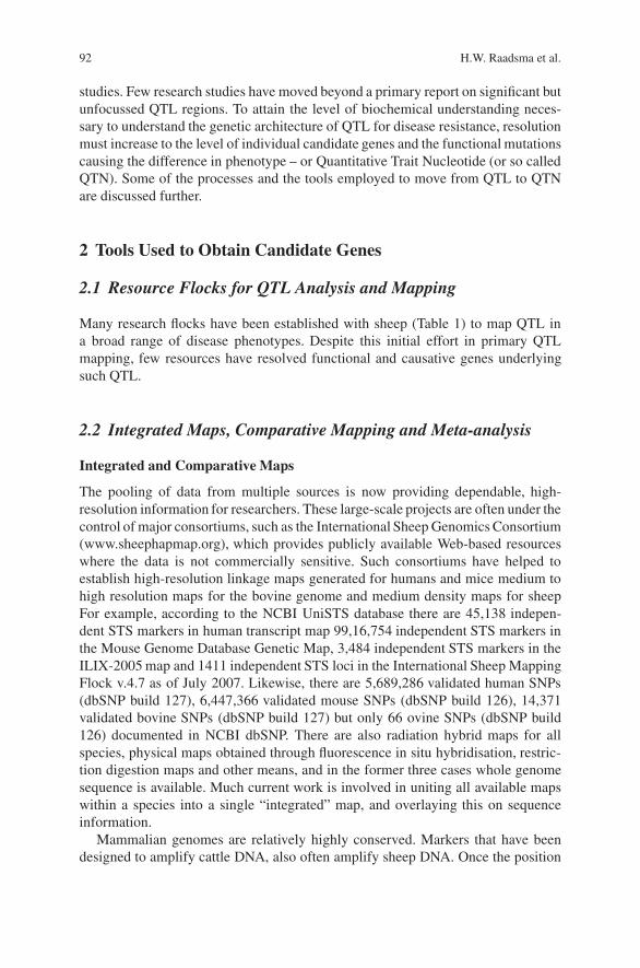
92 H.W. Raadsma et al.
studies. Few research studies have moved beyond a primary report on significant butunfocussed QTL regions. To attain the level of biochemical understanding neces-sary to understand the genetic architecture of QTL for disease resistance, resolutionmust increase to the level of individual candidate genes and the functional mutationscausing the difference in phenotype – or Quantitative Trait Nucleotide (or so calledQTN). Some of the processes and the tools employed to move from QTL to QTNare discussed further.
2 Tools Used to Obtain Candidate Genes
2.1 Resource Flocks for QTL Analysis and Mapping
Many research flocks have been established with sheep (Table 1) to map QTL ina broad range of disease phenotypes. Despite this initial effort in primary QTLmapping, few resources have resolved functional and causative genes underlyingsuch QTL.
2.2 Integrated Maps, Comparative Mapping and Meta-analysis
Integrated and Comparative Maps
The pooling of data from multiple sources is now providing dependable, high-resolution information for researchers. These large-scale projects are often under thecontrol of major consortiums, such as the International Sheep Genomics Consortium(www.sheephapmap.org), which provides publicly available Web-based resourceswhere the data is not commercially sensitive. Such consortiums have helped toestablish high-resolution linkage maps generated for humans and mice medium tohigh resolution maps for the bovine genome and medium density maps for sheepFor example, according to the NCBI UniSTS database there are 45,138 indepen-dent STS markers in human transcript map 99,16,754 independent STS markers inthe Mouse Genome Database Genetic Map, 3,484 independent STS markers in theILIX-2005 map and 1411 independent STS loci in the International Sheep MappingFlock v.4.7 as of July 2007. Likewise, there are 5,689,286 validated human SNPs(dbSNP build 127), 6,447,366 validated mouse SNPs (dbSNP build 126), 14,371validated bovine SNPs (dbSNP build 127) but only 66 ovine SNPs (dbSNP build126) documented in NCBI dbSNP. There are also radiation hybrid maps for allspecies, physical maps obtained through fluorescence in situ hybridisation, restric-tion digestion maps and other means, and in the former three cases whole genomesequence is available. Much current work is involved in uniting all available mapswithin a species into a single “integrated” map, and overlaying this on sequenceinformation.
Mammalian genomes are relatively highly conserved. Markers that have beendesigned to amplify cattle DNA, also often amplify sheep DNA. Once the position
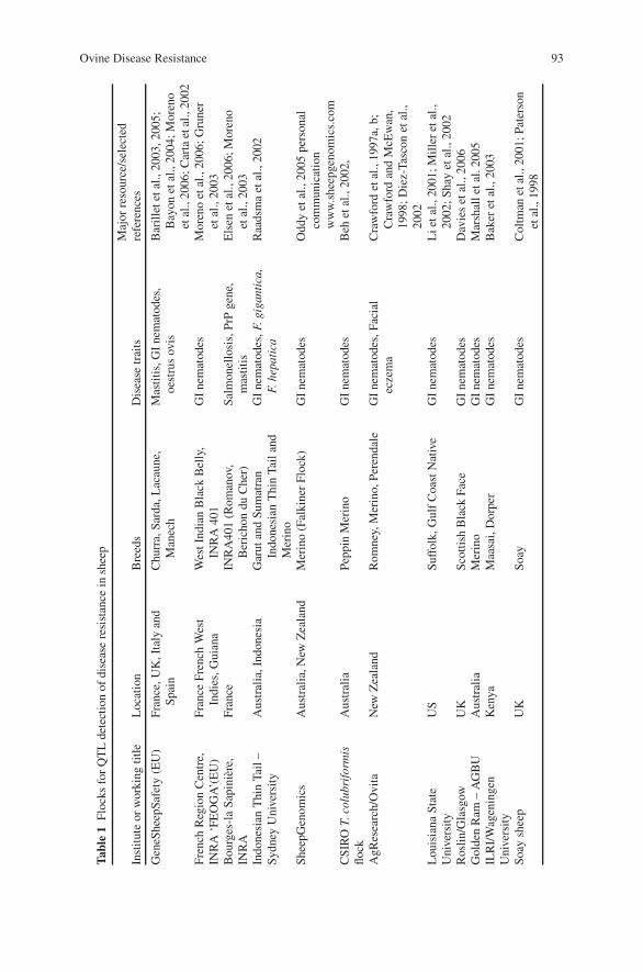
Ovine Disease Resistance 93
Tab
le1
Floc
ksfo
rQ
TL
dete
ctio
nof
dise
ase
resi
stan
cein
shee
p
Inst
itut
eor
wor
king
titl
eL
ocat
ion
Bre
eds
Dis
ease
trai
tsM
ajor
reso
urce
/sel
ecte
dre
fere
nces
Gen
eShe
epSa
fety
(EU
)Fr
ance
,UK
,Ita
lyan
dSp
ain
Chu
rra,
Sard
a,L
acau
ne,
Man
ech
Mas
titi
s,G
Ine
mat
odes
,oe
stru
sov
isB
aril
lete
tal.,
2003
,200
5;B
ayon
etal
.,20
04;M
oren
oet
al.,
2006
;Car
taet
al.,
2002
Fren
chR
egio
nC
entr
e,IN
RA
‘FE
OG
A’(
EU
)Fr
ance
Fren
chW
est
Indi
es,G
uian
aW
estI
ndia
nB
lack
Bel
ly,
INR
A40
1G
Ine
mat
odes
Mor
eno
etal
.,20
06;G
rune
ret
al.,
2003
Bou
rges
-la
Sapi
nier
e,IN
RA
Fran
ceIN
RA
401
(Rom
anov
,B
eric
hon
duC
her)
Salm
onel
losi
s,Pr
Pge
ne,
mas
titi
sE
lsen
etal
.,20
06;M
oren
oet
al.,
2003
Indo
nesi
anT
hin
Tail
–Sy
dney
Uni
vers
ity
Aus
tral
ia,I
ndon
esia
Gar
utan
dSu
mat
ran
Indo
nesi
anT
hin
Tail
and
Mer
ino
GI
nem
atod
es,F
.gig
anti
ca,
F.he
pati
caR
aads
ma
etal
.,20
02
Shee
pGen
omic
sA
ustr
alia
,New
Zea
land
Mer
ino
(Fal
kine
rFl
ock)
GI
nem
atod
esO
ddy
etal
.,20
05pe
rson
alco
mm
unic
atio
nw
ww
.she
epge
nom
ics.
com
CSI
RO
T.co
lubr
ifor
mis
flock
Aus
tral
iaPe
ppin
Mer
ino
GI
nem
atod
esB
ehet
al.,
2002
,
AgR
esea
rch/
Ovi
taN
ewZ
eala
ndR
omne
y,M
erin
o,Pe
rend
ale
GI
nem
atod
es,F
acia
lec
zem
aC
raw
ford
etal
.,19
97a,
b;C
raw
ford
and
McE
wan
,19
98;D
iez-
Tasc
onet
al.,
2002
Lou
isia
naSt
ate
Uni
vers
ity
US
Suff
olk,
Gul
fC
oast
Nat
ive
GI
nem
atod
esL
ieta
l.,20
01;M
ille
ret
al.,
2002
;Sha
yet
al.,
2002
Ros
lin/G
lasg
owU
KSc
ottis
hB
lack
Face
GI
nem
atod
esD
avie
set
al.,
2006
Gol
den
Ram
–A
GB
UA
ustr
alia
Mer
ino
GI
nem
atod
esM
arsh
alle
tal.
2005
ILR
I/W
agen
inge
nU
nive
rsit
yK
enya
Maa
sai,
Dor
per
GI
nem
atod
esB
aker
etal
.,20
03
Soay
shee
pU
KSo
ayG
Ine
mat
odes
Col
tman
etal
.,20
01;P
ater
son
etal
.,19
98
94 H.W. Raadsma et al.
of matching markers or genes have been established in two genomes, it is pos-sible to deduce the chromosomal rearrangements that have taken place betweenthe two. Marker order by and large remains conserved in those regions betweenhistorical breakpoints, and so-called “comparative mapping” allows informationto be transferred between the relevant genomes. Such comparisons have becomevital for those species whose genomes have not been sequenced, such as the sheep.As of 15 November 2006, a virtual sequence has been established for the sheepgenome, based entirely on the ordering of sequenced BAC ends relative to estab-lished human (bg17), cattle (Btau2.6) and dog (canFam2) frameworks (Interna-tional Sheep Genomics Consortium). Information on virtual sheep chromosomesare found at www.livestockgenomics.csiro.au. For sheep researchers, this providesaccess to the far greater functional and positional knowledge available in otherspecies. Likewise, knowledge generated in the sheep genome can be transferredto other species through the vehicle of integrated maps and Oxford grids, in whichshared features between two species are used to align the two integrated maps ina grid format as is shown for sheep, cattle and human in Fig. 1 (Nicholas, 2005;Liao et al., 2007).
2.2.1 Meta-analysis
Once a number of QTL studies have been undertaken on a particular disease,meta-analyses may be employed to help eliminate type 2 errors and suggest crit-ical genomic regions affecting a disease across multiple populations. Meta-analysisemploys comparable data from all such studies in a joint statistical analysis. Bycombining the information across studies, refined confidence intervals of criticalregions are also possible. In sheep, the possibility of collating the data gathered ongastro-intestinal nematodes is being considered (Jill Maddox, personal Communica-ton, 2006). A summary of chromosomes with significant or suggestive QTL for GInematode resistance/susceptibility is depicted in Fig. 2 and includes a crude crossspecies analysis, in which QTL identified on bovine chromosomes are translated totheir corresponding ovine chromosomes according to the appropriate Oxford grid.Even this rudimentary depiction suggests a major gene for GI nematode resistanceon OAR3 and possibly OAR1, OAR2, OAR6, OAR14 and OAR20. For a compre-hensive statistical meta-analyses, Khatkar et al. (2004) detail the general proceduresand requirements from the data, to provisionally locate QTL to best-bet locationsand make distinctions between single QTL and multiple QTL in target regions.
2.3 Association Studies, SNP Chips and LD Mapping
Most primary QTL screens have confidence intervals that are typically in the orderof 50–100 cM, thus encompassing potentially thousands of positional candidategenes. The first step towards refining the position of the actual QTL is to extractmaximal information from such studies by adding more markers to the same linkagemapping population, and if possible adding more individuals. This approach is
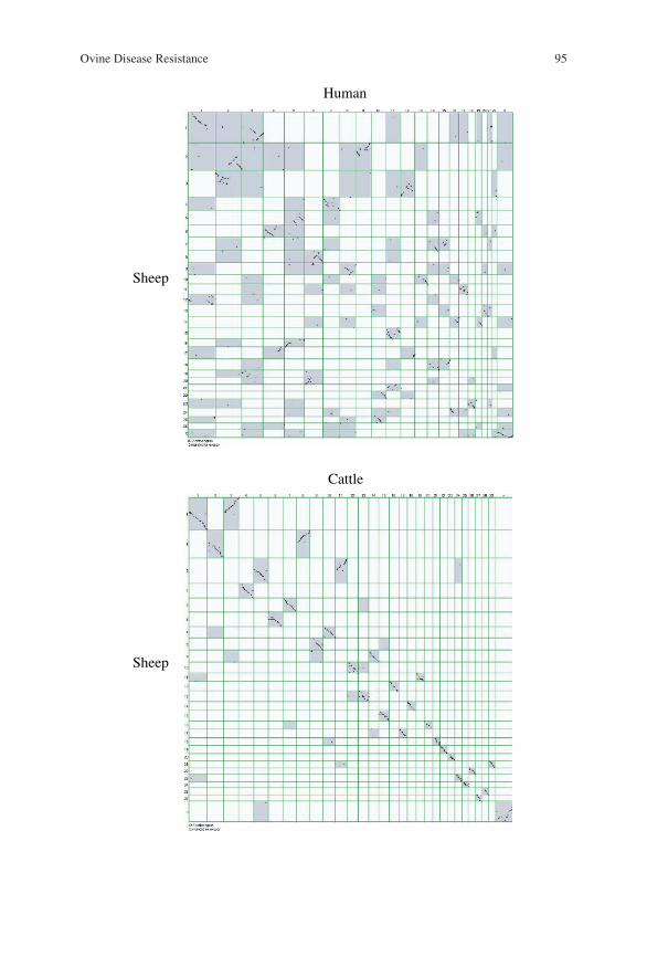
Ovine Disease Resistance 95
Sheep
Sheep
Human
Cattle
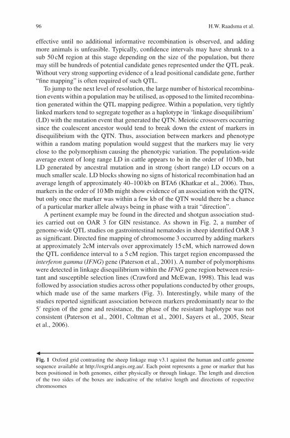
96 H.W. Raadsma et al.
effective until no additional informative recombination is observed, and addingmore animals is unfeasible. Typically, confidence intervals may have shrunk to asub 50 cM region at this stage depending on the size of the population, but theremay still be hundreds of potential candidate genes represented under the QTL peak.Without very strong supporting evidence of a lead positional candidate gene, further“fine mapping” is often required of such QTL.
To jump to the next level of resolution, the large number of historical recombina-tion events within a population may be utilised, as opposed to the limited recombina-tion generated within the QTL mapping pedigree. Within a population, very tightlylinked markers tend to segregate together as a haplotype in ‘linkage disequilibrium’(LD) with the mutation event that generated the QTN. Meiotic crossovers occurringsince the coalescent ancestor would tend to break down the extent of markers indisequilibrium with the QTN. Thus, association between markers and phenotypewithin a random mating population would suggest that the markers may lie veryclose to the polymorphism causing the phenotypic variation. The population-wideaverage extent of long range LD in cattle appears to be in the order of 10 Mb, butLD generated by ancestral mutation and in strong (short range) LD occurs on amuch smaller scale. LD blocks showing no signs of historical recombination had anaverage length of approximately 40–100 kb on BTA6 (Khatkar et al., 2006). Thus,markers in the order of 10 Mb might show evidence of an association with the QTN,but only once the marker was within a few kb of the QTN would there be a chanceof a particular marker allele always being in phase with a trait “direction”.
A pertinent example may be found in the directed and shotgun association stud-ies carried out on OAR 3 for GIN resistance. As shown in Fig. 2, a number ofgenome-wide QTL studies on gastrointestinal nematodes in sheep identified OAR 3as significant. Directed fine mapping of chromosome 3 occurred by adding markersat approximately 2cM intervals over approximately 15 cM, which narrowed downthe QTL confidence interval to a 5 cM region. This target region encompassed theinterferon gamma (IFNG) gene (Paterson et al., 2001). A number of polymorphismswere detected in linkage disequilibrium within the IFNG gene region between resis-tant and susceptible selection lines (Crawford and McEwan, 1998). This lead wasfollowed by association studies across other populations conducted by other groups,which made use of the same markers (Fig. 3). Interestingly, while many of thestudies reported significant association between markers predominantly near to the5′ region of the gene and resistance, the phase of the resistant haplotype was notconsistent (Paterson et al., 2001, Coltman et al., 2001, Sayers et al., 2005, Stearet al., 2006).
�Fig. 1 Oxford grid contrasting the sheep linkage map v3.1 against the human and cattle genomesequence available at http://oxgrid.angis.org.au/. Each point represents a gene or marker that hasbeen positioned in both genomes, either physically or through linkage. The length and directionof the two sides of the boxes are indicative of the relative length and directions of respectivechromosomes

Ovine Disease Resistance 97
Oar Refs. (First Author date)GIN (FEC 1)
1
1 ,,,
2
2
3
3
4
4
5
5
6
6
7
7
8
8
10
10
11
11
12
12
13
13
14
14
15
15
16
16
17
17
18
18
19
19
20
20
22
22
23
23
24
24
25
25
26
26 272829
21
21
9
9
Beh 2002GIN (FEC 1) Bayon 2004GIN (FEC 1a) Moreno 2006GIN (FEC 1b) Moreno 2006GIN (IgA 1) Bayon 2004GIN (PCV 1) Moreno 2006GIN (FEC 1a) Benavides 2002GIN (FEC 1b) Benavides 2002GIN (FEC 1) Janssen 2002GIN (Haemocrit 1) Janssen 2002GIN (IgL 1) Janssen 2002GIN (FEC >1) Beh 2002GIN (FEC >1) Miller 2002, Shay 2002GIN (FEC >1) Barillet 2003GIN (FEC >1a) Moreno 2006GIN (FEC >1b) Moreno 2006GIN (Adult >1) Crawford 2006GIN (PCV >1) Moreno 2006GIN (IgG>1) Crawford 2006GIN (IgE>1) Crawford 2006GIN (FEC >1) Davies 2006GIN (IgA >1) Davies 2006BtaGIN (FEC 1) Gasbarre 2004GIN (Pepsinogen 1) Gasbarre 2004
Sheep
Cow
Fig. 2 QTL studies of nematode infection in sheep (Oar = Ovis aries) and cattle (Bta = Bostaurus). Cattle chromosomes are aligned with homologous sheep chromosomes. Dark grey =autosomal chromosomes reported as significant or indicative, light grey = non-significant chro-mosomes, white = unexamined. GIN = gastro intestinal nematode, FEC = faecal egg count, 1 =naıve infection, >1 = secondary infection, IgA/E/G = immunoglobulin A/E/G, Adult = adultworm count, Pepsinogen = serum pepsinogen level, PCV = packed cell volume, a/b = separateflocks within the same study
190
200
210
220
230
240
CSS
M34
BL4
LYZ
o-IF
NG
BMS1
617
Oar
VH34
MAF
23O
arC
P43
Texa
n15
IL2R
B
cM
Romney selection lines; Crawford and McEwan 1998
c
t
6
5
g
a
A
B
Fine mapping (Paterson et al. 2001)
p≤0.05
Romney selection lines; Paterson et al. 2001
Soaysheep; Coltman et al. 2001Texel; Sayers et al. 2005
Perendal selection lines; Paterson et al. 2001 -
Suffolk; Sayers et al. 2005 -
Romney Selection lines; Dukkipati et al. 2005 -
=
=
=
R
S
Male Scottish Blackface; Stear et al. 2006R
S
SS
R
RR
S
NS
NS
NS
Fig. 3 Schematic representation of fine mapping and haplotype association studies of the Inter-feron Gamma gene. R haplotype = resistant and S haplotype = susceptible phenotypes, respec-tively. Coloured blocks indicate which of the four SNP polymorphisms were analysed in eachstudy

98 H.W. Raadsma et al.
In addition, three studies reported a lack of significant association. Thefirst used Perendale selection lines for high and low levels of GIN resistance(Paterson et al., 2001). Romney lines selected for traits independent of parasiteresistance also proved non-significant (Dukkipati et al., 2005). A proposed foundereffect is unlikely to be the sole explanation for association between IFNG and GINresistance, given the number of studies, and the broad base of breeds reporting asso-ciation. The last was in the Suffolk breed (Sayers et al., 2005), which had four segre-gating haplotypes, instead of the two present in other breeds, representing an ances-tral and potentially highly informative recombination between two closely linkedmarkers. Taken together, the combined work by many laboratories suggests that themarkers that have been used in association studies to date are not in sufficiently closelinkage disequilibrium to be in perfect phase with the real resistance polymorphism.
To date, such fine mapping and association studies have primarily been con-ducted on a targeted basis across limited regions in the genome. However, usinghigh-density SNP genotyping technologies, whole-genome scans of a similar levelof resolution are now possible. Single nucleotide polymorphisms (SNPs) offer aplentiful supply of bi-allelic variability throughout the genome, occurring on aver-age once in every 100 bp. Advances in technology now make it possible to take fulladvantage of the dense genome-wide information that these markers can provide,with mapping arrays of up to 1,000,000 markers now available in humans. Witha 100,000 K chip, marker density on a map is approximately in the order of 1 inevery 30 kb. At this level, population-wide levels of linkage disequilibrium maybe exploited. The transition from targeted QTL mapping studies to genome wideselection (GWS) studies with sufficiently dense SNP arrays will allow markers sig-nificantly associated with a trait to be used directly as selection aids in breedingprogrammes. Development of a 60,000 ovine SNP array has already been proposed(www.sheephapmap.org).
2.4 Microarrays, SELDI-TOF MS and Other High DensityGenomic or Proteomic Functional Tools
Functional genomic approaches offer other means to narrow down the approxi-mately 30,000 genes found within a genome to a few candidates, which influ-ence a particular trait. Micro-arrays, whether they be oligo-nucleotide-based chipsor c-DNA arrays, seek to detect changes in expression levels between two states(i.e. resistant vs. susceptible, or infected vs. non-infected). The caveat to bear inmind is that genes demonstrating changed expression levels may be downstreamin a biochemical pathway from influential “controller” genes with small changesin their expression or undetectable changes to their conformation state, or indeedmay not be represented on the array and therefore undetectable in the target stud-ies. Nevertheless, sophisticated algorithms are now available to group differentiallyexpressed genes according to their response profiles or promoter sequences, therebysuggesting functional links to lead pathways. This information, in conjunction withknowledge of the pertinent biochemical pathways obtained in other species, maylead to a number of candidate genes near the top of cascades.

Ovine Disease Resistance 99
Only a single large ovine array has been established based on 27 different tis-sues, with 19,968 spots representing 9,238 RefSeqs, which has been used suc-cessfully to examine baseline differences between GIN resistant and susceptiblePerendale sheep (Keane et al., 2006). Sheep researchers also have access to multi-ple large bovine arrays. The commercially available bovine Affymetrix array rep-resents approximately 19,000 unigene clusters and has a 75% cross hybridisationto the ovine genome (Ross Tellam, personal communication). In addition, a 10,204bovine cDNA array has successfully been utilised for cross species hybridisationwith sheep, in which duodenal tissue from infected GIN resistant and susceptiblePerendale sheep was compared (Diez-Tascon et al., 2005). A specialist ruminantinnate immune response array has been constructed with 1480 characterised genesand 5376 cDNAs derived from subtracted and normalised libraries, which detected94% of ovine transcripts tested (Donaldson et al., 2005). Other high throughput tran-scriptome techniques of a similar nature include serial analysis of gene expression(SAGE) used to investigate the response to trypanosome infection in cattle (Berthieret al., 2003) and massively parallel signature sequencing (MPSS).
A proteomic approach using SELDI-TOF mass spectroscopy, or similar, mayalso be used to identify differentially expressed genes based on their protein geneproducts as leads for further analyses. Again, the process interrogates samples fromanimals subjected to two different states and attempts to identify proteins that aredifferentially up or down regulated. This process has the benefit of being one stepcloser to the actual functional cellular mechanism, given that even though genesmay be up or down regulated, there are still cellular controls that may prevent acorresponding up or down translation of the transcripts into proteins. Once again,differentially expressed proteins may not necessarily represent the most importantgenes controlling the trait, but may well contribute to a better-characterised path-way analysis. A major limitation to the proteomic approach has been the limitedannotation of the many thousands of peptide fragments to genes and their function.
2.5 Positional Functional Integration
Despite large expenditure, most positional mapping studies are unsuccessful inrefining the number of candidate genes to a manageable number, especially sincemany of the putative genes lying under the most likely positions remain uncharac-terised. Further study would require exhaustive functional investigation of each inrelation to the specific disease trait of interest. Likewise, candidates derived fromfunctional transcriptomic, immunological or proteomic studies could number inthe hundreds, without any clear idea of which ones, if any, were causative, merelyreflecting a differentially expressed pathway, or even worse were spurious in oftenunder powered micro-array studies.
However, a combination of the two approaches may give maximum power toidentify true candidates. Transecting functional studies based on full transcriptomeanalyses with comprehensive micro-array and/or alternative techniques, such as
100 H.W. Raadsma et al.
SAGE and MPSS, with QTL studies has the potential to yield functional informa-tion on relevant genes underlying QTL or so-called functional-positional candidategenes. Joint candidates obtained through both approaches may typically be screenedin vitro and in vivo and sequenced to identify putative QTN.
Unequivocal proof that a specific mutation is causative of a QTL or a phenotypechange remains elusive in livestock, with limited scope for gene substitution andtargeted mutation induction to obtain such proof. A rare example of an appropriateexpression study has used a murine NRAMP1-susceptible (now known as solutecarrier family II member A1 gene) macrophage cell line as a constant backgroundto investigate the effect of a mutation in the bovine NRAMP gene under the controlof the bovine NRAMP1 promoter, with concommittant differences in the levels ofgene expression, as described in Barthel et al. (2001).
Functional transcriptome analyses and co-localisation studies with known QTLregions have been conducted for mastitis (Schwerin et al., 2003), trypanosomeresistance using micro-array and SAGE approaches, respectively (Hill et al., 2005,Berthier et al., 2003) and for GIN in cattle (Gasbarre et al., 2004) and recently insheep (Keane et al., 2006).
A technique that has long been proposed, but is still uncommon, is that of“microarray QTLs” or “e-QTLs” in which a suite of phenotypically variable animalsare used and micro-array analyses of each individual animal used as the phenotypein QTL studies. Those genes in common between QTL location and expressionprofiles (cis acting) may lead to so-called master regulatory genes, whereas thosenot collocated with the QTL but differentially expressed or trans acting may leadto major pathway analyses in regulating gene expression. The difficulty of linkingsuch e-QTL and their candidate genes to defined disease traits is still a complexissue, although once again meta-analyses of all functional and positional mappingstudies should reveal functionally important genes for further analysis.
3 An Example: Mapping Genes for Ruminant Fasciolosis
An example of an integrated gene mapping and functional genomics programme toidentify genes for resistance to fasciolosis has been described in full by Raadsmaet al. (2005). In collaboration with many others, this example is informative inthat multiple tools have been utilised in a targeted gene discovery programme. Twoprimary research arms are involved, a QTL mapping experiment to identify majorgenes for innate and acquired resistance to F. gigantica and an immunological char-acterisation of resistance mechanisms to fasciolosis to identify candidate gene(s)and pathways. The combination of information derived from both arms allowedgreater refinement of a list of candidate genes than would have been possible usingeach approach alone.
Fasciolosis is a major parasitic disease of livestock with over 700 million pro-duction animals at risk of infection and worldwide economic losses estimatedat >US$3.2 billion per annum (Spithill et al., 1999). The parasite is especially
Ovine Disease Resistance 101
prevalent in tropical regions of Asia and Africa, where it is considered thesingle most important helminth infection of cattle (Fabiyi, 1987; Schillhornvan Veen, 1980; Roy and Tandon, 1992; Pholpark and Srikitjakarn, 1989; Soe-setya, 1975; Spithill et al., 1999). Infected animals show lower weight gain,anaemia, reduced fertility, reduced milk production, lower feed conversion effi-ciency and a diminished work capacity. In African and Southeast Asian developingcountries, the latter trait significantly impacts on production, where ruminantsprovide 80% of the draught power (Spithill et al., 1999, Sukhapesna et al., 1994).In addition, there is a significant zoonotic infection rate in humans with the WHOrecognising >2.4 million cases of infection.
Anthelminthics are currently utilised against fasciolosis, but they are expensiveand require frequent administration, which prevents their application in many devel-oping countries (Spithill et al., 1999). In addition, there is growing evidence ofresistance to Triclabendazole, which has seriously compromised its use (Overendand Bowen, 1995; Spithill and Dalton, 1998). Vaccination is a second control mech-anism that might be used and, indeed, experimental vaccination against Fasciolais well established (Spithill and Dalton, 1998). However, the vaccine-induced andnatural acquired immune mechanisms linked to rejection of Fasciola infection inruminants are not well defined (Piedrafita et al., 2004).
3.1 Resistance to Fasciola
At the outset of this programme observations had been made that the IndonesianThin Tail (ITT) sheep breed expressed relative resistance to fasciolosis, whereasthe Merino was shown to be highly susceptible (Wiedosari and Copeman, 1990;Roberts et al., 1997a, b, c). ITT sheep expressed resistance to F. gigantica within theprepatent period of a primary infection and acquired a higher level of resistance afterexposure (Roberts et al., 1997a, c). The acquired resistance of ITT sheep, expressedwithin 3–4 weeks of infection, could be suppressed by dexamethasone, implyingthat this resistance was immunologically based and that the killing of many migrat-ing parasites occurred within 2–4 weeks of infection (Roberts et al., 1997b; Spithillet al., 1999). Despite displaying resistance to F. gigantica, ITT sheep are fully sus-ceptible to F. hepatica (Roberts et al., 1997a). These findings suggest that F. hep-atica and F. gigantica differ in some fundamental biological trait(s), which rendersF. gigantica more susceptible to immune effector mechanisms (Spithill et al., 1997;Piedrafita et al., 2004). The factors responsible for this apparent natural resistanceto Fasciola infection in certain sheep breeds are currently unknown.
3.2 The Resource Flock for Mapping Fasciolosis Resistance
The resistance of ITT sheep represents a unique biological situation, which has beenexploited in the construction of a QTL mapping resource flock. ITT sheep were
102 H.W. Raadsma et al.
backcrossed into susceptible Merino (Fig. 4) to generate 10 F1 ram families. Twostrains of ITT sheep were used, namely Sumatra and Garut (West Java) to allow forpossible strain variation in genetic resistance. Reciprocal matings were used in thecreation of the F1 sires, to allow for recognition of sex-linked or imprinted genes.Four families were augmented in number by further matings of the F1 sire. In twocases, these augmented matings involved alternate backcross to ITT ewes and F2intercross matings. Animals were maintained parasite free in holding pens aboveground at the Indonesian Institute for Science, Bogor, Indonesia, under natural lightconditions, and were fed ad libertum. A total of 694 animals from 10 linkage fam-ilies were genotyped and phenotyped but to achieve this number, a total of 1435animals were bought or born in the flock. As for all experimental flocks, a largeinvestment is necessary to achieve a reasonable number of experimental animals.
Over 3 years, naıve experimental animals were subjected to two experimentallycontrolled Haemonchus contortus infections prior to a midyear challenge (average373 days of age) with 300 metacercariae of F. gigantica. In the fourth year fol-lowing the H. contortus trials, animals were sensitised with 50 metacercariae ofF. gigantica (sensitization), drenched after 6 weeks, and then challenged with 250metacercariae of F. gigantica (challenge at average 446 days of age). Challengeswere slaughtered 12–15 weeks post-infection. Various phenotypes were measuredincluding repeat measures of live weight (LW), packed cell volume (PCV), antibodylevels, eosinophil, neutrophil and lymphocyte levels and liver enzymes. Flukes wererecovered post slaughter (Fl), liver damage was assessed as a score (LvrSc) from 1(least damage) to 5 (most damage) and liver weight (LvrWT) together with flukebiomass (WW) was obtained. In addition, use of this major resource flock was opti-mised by taking detailed carcass, growth and fleece production data to provide abetter understanding of major genes affecting production traits.
3.3 Linkage and QTL Analysis for Fasciolosis
A low density genome screen of autosomal chromosomes was undertaken using 138microsatellite markers selected on the basis of map location (Maddox et al., 2001)and polymorphic information content (PIC) scores from the Australian SheepGene Mapping Resource (http://rubens.its.unimelb.edu.au/∼jillm/jill.htm). This isone of a few free Web-based resources with updated information on the Inter-national Mapping Flock sheep linkage map and associated tools such as CMapallowing comparative analysis to human, canine and bovine linkage and sequenceinformation.
QTL analysis was undertaken for a range of parasite resistance and immuneresponse traits, using the half-sib analysis capability of QTL Express (Seatonet al., 2002). This Web-based analysis tool stemming from collaborative workbetween the Roslin Institute and the University of Edinburgh is widely usedand designed to allow user friendly regression-based QTL analysis of outbredpopulations. An initial analysis across all families for each trait included screens
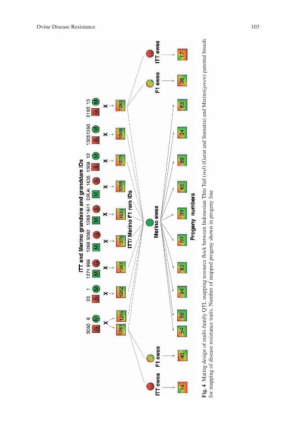
Ovine Disease Resistance 103
Fig
.4M
atin
gde
sign
ofm
ulti-
fam
ilyQ
TL
map
ping
reso
urce
flock
betw
een
Indo
nesi
anT
hin
Tail
(red
)(G
arut
and
Sum
atra
)an
dM
erin
o(gr
een)
pare
ntal
bree
dsfo
rm
appi
ngof
dise
ase
resi
stan
cetr
aits
.Num
ber
ofm
appe
dpr
ogen
ysh
own
inpr
ogen
yli
ne
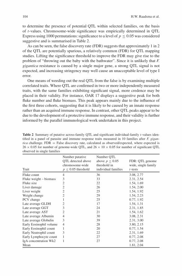
104 H.W. Raadsma et al.
to determine the presence of potential QTL within selected families, on the basisof t-values. Chromosome-wide significance was empirically determined in QTLExpress using 1000 permutations: significance to a level of p ≤ 0.05 was consideredsuggestive and is summarised in Table 2.
As can be seen, the false discovery rate (FDR) suggests that approximately 1 in 2of the QTL are potentially spurious, a relatively common (FDR) for QTL mappingstudies. Lifting the significance threshold to improve the FDR may give rise to theproblem of “throwing out the baby with the bathwater”. Since it is unlikely that F.gigantica resistance is caused by a single major gene, a strong QTL signal is notexpected, and increasing stringency may well cause an unacceptable level of type Ierror.
One means of weeding out the real QTL from the false is by examining multiplecorrelated traits. Where QTL are confirmed in two or more independently measuredtraits, with the same families exhibiting significant signal, more credence may beplaced in their validity. For instance, OAR 17 displays a suggestive peak for bothfluke number and fluke biomass. This peak appears mainly due to the influence ofthe first three cohorts, suggesting that it is likely to be caused by an innate responserather than an acquired immune response. In contrast, other QTL peaks appear to bedue to the development of a protective immune response, and their validity is furtherinformed by the parallel immunological work undertaken in this project.
Table 2 Summary of putative across-family QTL and significant individual-family t-values iden-tified in a panel of parasite and immune response traits measured in 10 families after F. gigan-tica challenge. FDR = False discovery rate, calculated as observed/expected, where expected is26 × 0.05 for number of genome-wide QTL, and 26 × 10 × 0.05 for number of significant QTLobserved in single families
Trait
Number putativeQTL detected abovechromosome-widep ≤ 0.05 threshold
Number QTLabove p ≤ 0.05threshold inindividual families
FDR: QTL genomewide, single familyt-tests
Fluke count 4 36 3.08, 2.77Fluke weight – biomass 3 33 2.31, 2.54Fluke size 2 22 1.54, 1.69Liver damage 2 26 1.54, 2.00Liver weight 2 25 1.54, 1.92Weight change 2 29 1.54, 2.23PCV change 1 25 0.77, 1.92Late average GLDH 2 17 1.54, 1.31Late average GGT 3 24 2.31, 1.85Late average AST 2 21 1.54, 1.62Late average Albumin 4 30 3.08, 2.31Late average Globulin 3 39 2.31, 3.00Early Eosinophil volume 4 28 3.80, 2.15Early Eosinophil count 1 20 0.77, 1.54Early Neutrophil count 3 22 2.31, 1.69Early Lymphocyte count 1 27 0.77, 2.08IgA concentration Wk2 1 27 0.77, 2.08Mean 1.81, 2.04
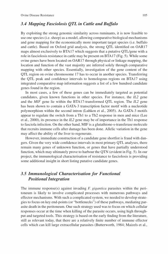
Ovine Disease Resistance 105
3.4 Mapping Fasciolosis QTL in Cattle and Buffalo
By exploiting the strong genomic similarity across ruminants, it is now feasible touse one species (i.e. sheep) as a model, allowing comparative biological mechanismsand gene mapping for the economically more important target species (i.e. buffaloand cattle). Based on Oxford grid analysis, the strong QTL identified on OAR17maps almost exclusively to BTA17 which suggests that a putative QTL/gene with arole in fasciolosis resistance in cattle may be present on BTA17 (Fig. 5). While someovine genes have been located on OAR17 through physical or linkage mapping, thelocation and function of the vast majority are inferred solely through comparativemapping with other species. Essentially, investigation of the gene content of theQTL region on ovine chromosome 17 has to occur in another species. Transferringthe QTL peak and confidence intervals to homologous regions on BTA17 usingintegrated comparative map information suggests a list of a few hundred candidategenes found in the region.
In most cases, a few of these genes can be immediately targeted as potentialcandidates, given known functions in other species. For instance, the IL2 geneand the MIF gene lie within the BTA17-transformed QTL region. The IL2 genehas been shown to contain a GATA-3 transcription factor motif with a nucleotidepolymorphism within the second intron (Luhken et al., 2005). As GATA-3 motifsappear to regulate the switch from a Th1 to a Th2 response in men and mice (Leeet al., 2000), its presence in the IL2 gene may be of importance in the Th1 responseto fasciola infection. On the other hand, MIF is a glutathione-s-transferase moleculethat recruits immune cells after damage has been done. Allelic variation in the genemay affect the ability of the liver to regenerate.
However, immediate construction of a candidate gene shortlist is fraud with dan-gers. Given the very wide confidence intervals in most primary QTL analyses, thereremain many genes of unknown function, or genes that have partially understoodfunction, which may ultimately prove to harbour the QTN (evident in Fig. 5). In ourproject, the immunological characterisation of resistance to fasciolosis is providingsome additional insight in short listing putative candidate genes.
3.5 Immunological Characterisation for FunctionalPositional Integration
The immune response(s) against invading F. gigantica parasites within the peri-toneum is likely to involve complicated processes with numerous pathways andeffector mechanisms. With such a complicated system, we needed to develop strate-gies to focus on key end-points (or “bottlenecks”) of these pathways, mediating par-asite death in the peritoneum. One such strategy used was to focus on which cellularresponses occur at the time when killing of the parasite occurs, using high through-put and targeted tools. This strategy is based on the early finding from the literature,still as relevant today, that there are a relatively finite number of immune effectorcells which can kill large extracellular parasites (Butterworth, 1984; Maizels et al.,
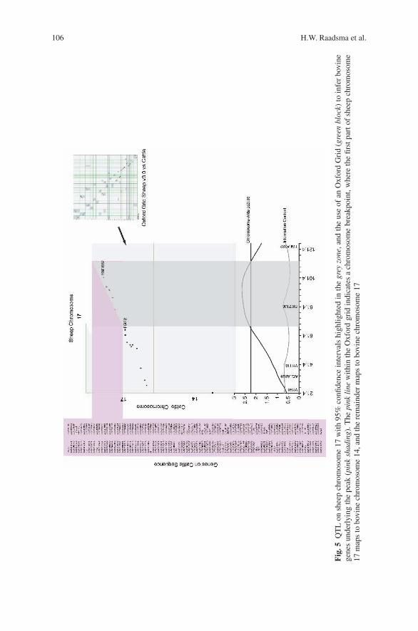
106 H.W. Raadsma et al.
Fig
.5Q
TL
onsh
eep
chro
mos
ome
17w
ith
95%
confi
denc
ein
terv
als
high
ligh
ted
inth
egr
eyzo
ne,a
ndth
eus
eof
anO
xfor
dG
rid
(gre
enbl
ock)
toin
fer
bovi
nege
nes
unde
rlyi
ngth
epe
ak(p
ink
shad
ing)
.The
pink
line
wit
hin
the
Oxf
ord
grid
indi
cate
sa
chro
mos
ome
brea
kpoi
nt,w
here
the
first
part
ofsh
eep
chro
mos
ome
17m
aps
tobo
vine
chro
mos
ome
14,a
ndth
ere
mai
nder
map
sto
bovi
nech
rom
osom
e17

Ovine Disease Resistance 107
1993). The concept of the research was that by investigating their role in killing invitro, and the toxic molecules produced which can kill the parasite, we would be ableto predict the central pathway(s) directing the protective immune response(s) andidentify the underlying resistance mechanism in ITT sheep. This would then allowidentification of candidate genes, which control these pathways, to be comparedwith that generated through the QTL analyses.
High resistance of ITT sheep to F. gigantica appeared to follow a model wherebyparasites were killed within the gut wall and/or peritoneum or shortly after reachingthe liver, and that the newly excysted immature juvenile parasite (NEJ) was theprimary target of an effective immune response in ITT sheep. We therefore decidedto study the peritoneal cavity for immune mediators correlating with resistance inITT sheep, using peritoneal lavage cells obtained from ITT and Merino sheep.
Our studies in this regard have shown that an antibody-dependent cell cyto-toxicity (ADCC) mechanism is expressed in ITT sheep (Piedrafita et al., 2007).NEJ of F. gigantica are susceptible to killing in vitro by peritoneal macrophagesand eosinophils present in the peritoneal lavage cells. The mechanism involvessuperoxide radicals as evidenced by reversal of cytotoxicity following inclusion ofsuperoxide dismutase (SOD) in the culture; SOD neutralizes immune cell–generatedsuperoxide radicals. We suggest that this killing mechanism may be important in theresistance of ITT sheep to F. gigantica infection (Piedrafita et al., 2004; Meeusenand Piedrafita, 2003). In contrast, ITT sheep do not acquire resistance to F. hepat-ica infections (Roberts et al., 1997a) and the identified superoxide-mediated killingmechanisms effective against F. gigantica are ineffective against F. hepatica in vitro(Piedrafita et al., 2000, 2001, 2007).
3.6 High Density Proteomic and Genomic Functional Screening
In an attempt to unravel host responses expressed in sheep against Fasciola sp., wehave recently begun to apply SELDI-TOF Mass Spectrometry (Issaq et al., 2002;Tang et al., 2004) to derive a protein profile of serum during infection of sheepwith F. hepatica. Although these data are preliminary, the results suggest that serumbiomarkers associated with F. hepatica infection may be readily discernible in sheep(Fig. 6). Sequence identification of these markers may reveal insights into the patho-biology of liver fluke infections in sheep. Comparative biomarker studies are under-way in the resistant ITT and susceptible Merino after F. gigantica challenge. Wehave now commenced gene-specific expression profiles by RT-PCR analyses duringdifferent stages of infection in ITT and Merino backgrounds for a broad repertoireof cytokine profiles. In addition, we will be conducting transcriptome analyses usingbovine micro-arrays and ruminant immune-response arrays.
3.7 Future Studies and Potential Applications
To our knowledge, this is the first genome screen conducted in ruminants for iden-tification of resistance genes and their functional importance in fasciolosis. The
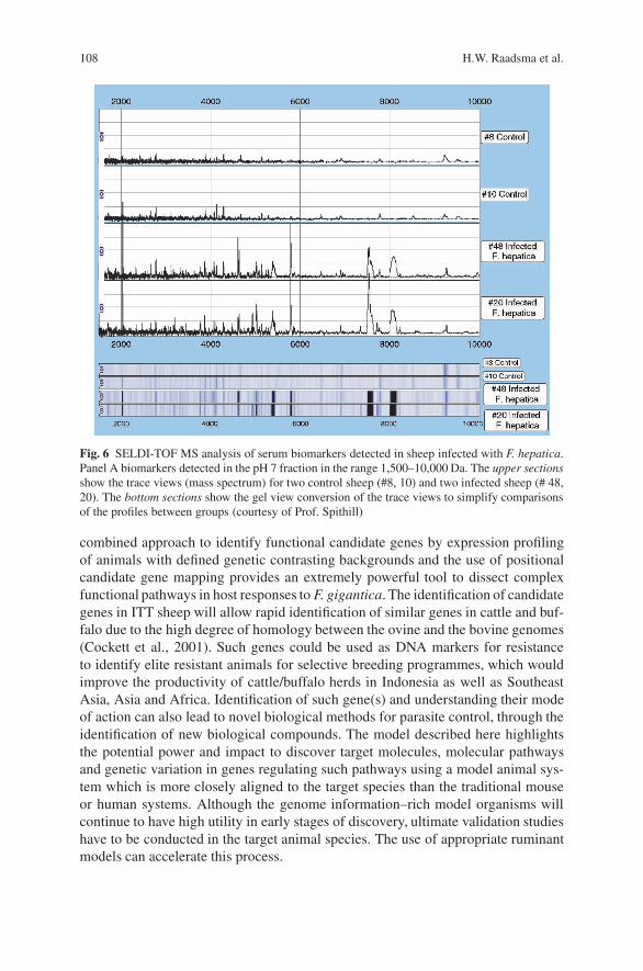
108 H.W. Raadsma et al.
Fig. 6 SELDI-TOF MS analysis of serum biomarkers detected in sheep infected with F. hepatica.Panel A biomarkers detected in the pH 7 fraction in the range 1,500–10,000 Da. The upper sectionsshow the trace views (mass spectrum) for two control sheep (#8, 10) and two infected sheep (# 48,20). The bottom sections show the gel view conversion of the trace views to simplify comparisonsof the profiles between groups (courtesy of Prof. Spithill)
combined approach to identify functional candidate genes by expression profilingof animals with defined genetic contrasting backgrounds and the use of positionalcandidate gene mapping provides an extremely powerful tool to dissect complexfunctional pathways in host responses to F. gigantica. The identification of candidategenes in ITT sheep will allow rapid identification of similar genes in cattle and buf-falo due to the high degree of homology between the ovine and the bovine genomes(Cockett et al., 2001). Such genes could be used as DNA markers for resistanceto identify elite resistant animals for selective breeding programmes, which wouldimprove the productivity of cattle/buffalo herds in Indonesia as well as SoutheastAsia, Asia and Africa. Identification of such gene(s) and understanding their modeof action can also lead to novel biological methods for parasite control, through theidentification of new biological compounds. The model described here highlightsthe potential power and impact to discover target molecules, molecular pathwaysand genetic variation in genes regulating such pathways using a model animal sys-tem which is more closely aligned to the target species than the traditional mouseor human systems. Although the genome information–rich model organisms willcontinue to have high utility in early stages of discovery, ultimate validation studieshave to be conducted in the target animal species. The use of appropriate ruminantmodels can accelerate this process.
Ovine Disease Resistance 109
Acknowledgments The research on fasciolosis reported in this chapter was strongly supported byfunds from the Australian Centre for International Agricultural Research (ACIAR) in a bilateralresearch project between Australia and Indonesia. The input from Dr P. Thomson in the develop-ment of the statistical procedures for segregation analyses is gratefully acknowledged. The strongtechnical and managerial support from a very large team of highly dedicated technical staff both inIndonesia and in Australia has made this project possible, for which we are very grateful. Studiesin Canada are supported by grants from the National Sciences and Engineering Research Councilof Canada and the Canada Research Chair Program.
References
Axford, R.F.E., Bishop, S.C., Nicholas, F.W., and Owen, J.B., 2000, Genetics of susceptibility toproduction diseases in cattle and sheep, in R.F.E. Axford, S.C. Bishop, F.W. Nicholas, and J.B.Owen, eds., Breeding for Disease Resistance in Farm Animals, 418pp.
Baker, R.L., Gibson, J.P., Iraqi, F.A., Menge, D.M., Mugambi, J.M., Hanotte, O., Nagda, S.,Wakelin, D., and Behnke, J.M., 2003, Exploring the genetic control of resistance to gastroin-testinal helminth infections in sheep and mice, Proc. 15th Conf., Assoc. Advan. Anim. Breed.Genet., Melbourne, Australia, 7–11 July 2003, pp. 183–190.
Barillet, F., Arranz, J.J., and Carta, A., 2005, Mapping quantitative trait loci for milk pro-duction and genetic polymorphisms of milk proteins in dairy sheep, Genet. Sel. Evol. 37:S109–S123.
Barillet, F., Carta, A., Allain, D., Amigues, Y., Bodin, L., Casu, S., Cribiu, E.P., Bed’hom, B.,Boichard, D., Boscher, M.Y., Elsen, J.M., Fraghi, A., Gruner, L., Jacquiet, P., Ligios, S., Marie-Etancelin, C., Mura, L., Piredda, G., Roig, A., Rupp, R., Sanna, S.R., Scala, A., Schibler, L.,Sechi, T., and Casu, S., 2003, Detection of QTL influencing present and future economic traitsin dairy sheep in France and Italy, in 10eemes Rencontres autour des Recherches sur les Rumi-nants, Institut National de la Recherche Agronomique, Paris, France, 3–4 Decembre 2003,pp. 57–60.
Barthel, R., Feng, J.W., Piedrahita, J.A., McMurray, D.N., Templeton, J.W., and Adams, L.G.,2001, Stable transfection of the bovine NRAMP1 gene into murine RAW264.7 cells: effect onBrucella abortus survival, Infect. Immun. 69:3110–3119.
Bayon, Y., Gutierrez-Gil, B., De La Fuente, L.F., San Primitivo Tirados, F., El Zarei, M.F., AlvarezCastelo, L., Arranz, J.J., Perez, J., and Rojo Vasquez, F.A., 2004, Busqueda de regionesgenomicas con influencia sobre la resistencia a las tricostrongilidosis en el ganado ovino deraza churra ITEA, Prod. Anim. 100:197–201.
Beh, K.J., Hulme, D.J., Callaghan, M.J., Leish, Z., Lenane, I., Windon, R.G., and Maddox, J.F.,2002, A genome scan for quantitative trait loci affecting resistance to Trichostrongylus colubri-formis in sheep, Anim. Genet. 33:97–106.
Benavides, M.V., Weimer, T.A., Borba, M.F.S., Berne, M.E.A., and Sacco, A.M.S., 2002, Asso-ciation between microsatellite markers of sheep chromosome 5 and faecal egg counts, SmallRuminant Res. 46:97–105.
Berthier, D., Quere, R., Thevenon, S., Belemsaga, D., Piquemal, D., Marti, J., and Maillard,J.C., 2003, Serial analysis of gene expression (SAGE) in bovine trypanotolerance: preliminaryresults, Genet. Sel. Evol. 35:S35–S47.
Brenig, B., Broad, T.E., Cockett, N.E., and Eggen, A., 2004, Achievements of research inthe field of molecular genetics, WAAP Book of the Year, 2003: A Review on Develop-ments and Research in Livestock Systems, Wageningen Universiteit (Wageningen University),Wageningen Netherlands, pp. 73–84.
Butterworth, A.E. 1984. Cell-mediated damage to helminths. Adv. Parasit. 23:143–235.Carta, A., Barillet, F., Allain, D., Amigues, B., Bibe, B., Bodin, L., Casu, S., Cribiu, E., Elsen, J.M.,
Fraghi, A., Gruner, L., Jacuiet, P., Ligios, S., Marie-Etancelin, C., Mura, L., Piredda, G.,

110 H.W. Raadsma et al.
Rupp, R., Sanna, S.R., Scala, A., Scala, A., Schibler, L., and Casu, S., 2002, QTL Detec-tion with genetic markers in a dairy sheep backcross Sarda × Lacaune Resource Population,Proc. 7th World Congr. Genet. Appl. Livest. Prod. Montpellier, France, August 2002, CD-Rom,Com.No. 09–07.
Cockett, N.E., Shay, T.L., and Smit, M., 2001, Analysis of the sheep genome, Physiol. Genomics7:69–78.
Coltman, D.W., Wilson, K., Pilkington, J.G., Stear, M.J., and Pemberton, J.M., 2001, A microsatel-lite polymorphism in the gamma interferon gene is associated with resistance to gastrointestinalnematodes in a naturally-parasitized population of Soay sheep, Parasitology 122:571–582.
Crawford, A.M., and McEwan, J.C., 1998, Identification of animals resistant to nematode parasiteinfection New Zealand Provisional Patent 330201, New Zealand, p. 46.
Crawford, A.M., McEwan, J.C., Dodds, K.G., Wright, C.S., Bisset, S.A., Macdonald, P.A.,Knowler, K.J., Greer, G.J., Green, R.S., Shaw, R.J., Paterson, K.A., Cuthbertson, R.P.,Vlassoff, A., Squire, D.R., West, C.J., and Phua, S.H., 1997a, Resistance to nematode parasitesin sheep: how important are the MHC genes? Assoc. Advanc. Anim. Breeding and Genet. Proc.Twelfth Conf., Dubbo, NSW, Australia 6–10 April 1997, Part 1, pp. 58–62.
Crawford, A.M., Phua, S.H., McEwan, J.C., Dodds, K.G., Wright, C.C., Morris, C.A., Bisset, S.A.,and Green, R.S., 1997b, Finding disease resistance QTL in sheep, Anim. Biotechnol. 8:13–22.
Crawford, A.M., Paterson, K.A., Dodds, K.G., Tascon, C.D., Williamson, P.A., Thomson, M.R.,Bisset, S.A., Beattie, A.E., Greer, G.J., Green, R.S., Wheeler, R., Shaw, R.J., Knowler, K., andMcEwan, J.C., 2006, Discovery of quantitative trait loci for resistance to parasitic nematodeinfection in sheep: I. Analysis of outcross pedigrees, BMC Genomics 7:2164–2178.
Davies, G., Stear, M.J., Benothman, M., Abuagob, O., Kerr, A., Mitchell, S., and Bishop, S.C.,2006, Quantitative trait loci associated with parasitic infection in Scottish blackface sheep,Hered. 96:252–258.
Diez-Tascon, C., Keane, O.M., Wilson, T., Zadissa, A., Hyndman, D.L., Baird, D.B., McEwan,J.C., and Crawford, A.M., 2005, Microarray analysis of selection lines from outbred popula-tions to identify genes involved with nematode parasite resistance in sheep, Physiol. Genomics21:59–69.
Diez-Tascon, C., MacDonald, P.A., Dodds, K.G., McEwan, J.C., and Crawford, A.M., 2002,A screen of chromosome 1 for QTL affecting nematode resistance in an ovine outcrosspopulation, Proc. 7th World Congr. Genet. Appl. Livestock Prod., Institut National de laRecherche Agronomique (INRA), Montpellier, France, August 2002, Session 13, pp. 1–4,Communication 37.
Donaldson, L., Vuocolo, T., Gray, C., Strandberg, Y., Reverter, A., McWilliam, S., Wang, Y.,Byrne, K., and Tellam, R., 2005, Construction and validation of a Bovine Innate ImmuneMicroarray, BMC Genomics 6:135.
Dukkipati, V.S.R., Blair, H.T., Johnson, P.L., Murray, A., and Garrick, D.J., 2005, A study on theassociation of genotypes at the interferon gamma microsatellite locus with faecal strongyleegg counts in sheep, Proc. 16th Conf. Assoc. Advan. Anim. Breeding Genet., Noosa Lakes,Australia, pp. 119–122.
Elsen, J.M., Moreno, C.R., Bodin, L., Francois, D., Bouix, J., Barillet, F., Allain, D., Lantier, F.,Lantier, I., Schibler, L., Roig, A., Brunel, J.C., and Vitezica, Z.G., 2006, Selectio for scrapieresistance in France, Is there evidence of negative effects on production and health traits?, Proc.8th World Cong. Genet. Appl. Livestock Prod., Belo Horizonte, MG, Brazil, pp. 15–15.
Fabiyi, J.P., 1987, Production Losses and Control of Helminths in Ruminants of Tropical Regions,Int. J. Parasitol. 17:435–442.
Gasbarre, L.C., Sonstegard, T., VanTassell, C.P., and Araujo, R., 2004, Symposium: Newapproaches in the study of animal parasites, Vet. Parasitol. 125:147–161.
Gibson, J.R., and Bishop, S.C., 2005, Use of molecular markers to enhance resistance of livestockto disease: a global approach, Rev. Sci. Tech. OIE. 24:343–353.
GLIPHA: Global Livestock Production and Health Atlas www.fao.org/ag/aga/glipha.Gruner, L., Aumont, G., Getachew, T., Brunel, J.C., Pery, C., Cognie, Y., and Guerin, Y., 2003,
Experimental infection of Black Belly and INRA 401 straight and crossbred sheep with tri-chostrongyle nematode parasites, Vet. Parasitol. 116:239–249.
Ovine Disease Resistance 111
Hill, E.W., O’Gorman, G.M., Agaba, M., Gibson, J.P., Hanotte, O., Kemp, S.J., Naessens, J.,Coussens, P.M., and MacHugh, D.E., 2005, Understanding bovine trypanosomiasis and try-panotolerance: the promise of functional genomics, Vet. Immun. Immunop. 105:247–258
Issaq, H.J., Veenstra, T.D., Conrads, T.P., and Felschow, D., 2002, The SELDI-TOF MS approachto proteomics: Protein profiling and biomarker identification, Biochem. Biophys. Res. Commun.292:587–592.
Janssen, M., Weimann, C., Gauly, M., and Erhardt, G., 2002, Associations between infectionswith Haemonchus contortus and genetic markers on ovine chromosome 20, Proc. 7th WorldCong. Genet. Appl. Livestock Prod., Montpellier, France, August 2002, Session 13, pp. 1–4,Communication 11.
Keane, O.M., Zadissa, A., Wilson, T., Hyndman, D.L., Greer, G.J., Baird, D.B., McCulloch, A.F.,Crawford, A.M., and McEwan, J.C., 2006, Gene expression profiling of naive sheep geneticallyresistant and susceptible to gastrointestinal nematodes, BMC Genomics 7:42.
Khatkar, M.S., Collins, A., Cavanagh, J.A.L., Hawken, R.J., Hobbs, M., Zenger, K.R., Barris, W.,McClintock, A.E., Thomson, P.C., Nicholas, F.W., and Raadsma, H.W., 2006, A first-generationmetric linkage disequilibrium map of bovine chromosome 6, Genetics 174:79–85.
Khatkar, M.S., Thomson, P.C., Tammen, I., and Raadsma, H.W., 2004, Quantitative trait loci map-ping in dairy cattle: review and meta-analysis, Genet. Select. Evol. 36:163–190.
Lee, H.J., Tokemoto, N., Kurata, H., Kamogawa, Y., Miyatake, S., o’Garra, A., and Arai, N., 2000,GATA-3 induces Thelper cell type 2 (Th2) cytokine expression and chromatin remodelling incommitted Thi cells, J. Exp. Med. 192:105–115.
Li, Y., Miller, J.E., and Franke, D.E., 2001, Epidemiological observation and heterosis of gastroin-testinal nematode infection in Suffolk, Gulf Coast Native and crossbred lambs, Vet. Parasitol.98:273–283.
Liao, W., Collins, A., Hobbs, M., Khatkar, M.S., Luo, J., and Nicholas, F.W., 2007, A comparativelocation database (CompLDB): map integration within and between species, Mamm. Genome18:287–299.
Luhken, G., Stamm, V., Menge, C., and Erhardt, G., 2005, Functional analysis of a singlenucleotide polymorphism in a potential binding site for GATA transcription factors in the ovineinterleukin 2 gene, Vet. Immun. Immunop. 107:51–56.
Maddox, J.F., Davies, K.P., Crawford, A.M., Hulme, D.J., Vaiman, D., et al., 2001, An enhancedlinkage map of the sheep genome comprising more than 1000 loci, Genome Res. 11:1275–1289.
Maizels, R.M., Bundy, D.A.P., Selkirk, M.E., Smith, D.F., and Anderson, R.M., 1993, Immunolog-ical modulation and evasion by helminth-parasites in human-populations, Nature 365:797–805.
Marshall, K., van der Werf, J.H.J., Maddox, J.F., Graser, H.-U., Zhang, Y., Walkden-Brown, S.W.,and Khan, L., 2005, A genome scan for quantitative trait loci for resistance to the gastointesti-nal parasite Haemonchus contortus in sheep, Proc. Assoc. Advan. Anim. Breed. Genet., NoosaLakes, Queensland, Australia, pp. 115–118
Meeusen, E.N.T., and Piedrafita, D., 2003, Exploiting natural immunity to helminth parasites forthe development of veterinary vaccines, Int. J. Parasitol. 33:1285–1290.
Miller, J.E., Cocket, N.E., Walling, G.A., Shay, T.A., McGraw, R.A., Bishop, S.C., and Haley, C.A.,2002, Segregation of resistance to nematode infection in F2 lambs of Suffolk × Gulf CoastNative sheep and associated QTL, Proc. Int. Soc. Anim. Genet., Gottingen, Germany, pp. 160.
Moreno, C.R., Gruner, L., Scala, A., Mura, L., Schibler, L., Amigues, Y., Sechi, T., Jacquiet, P.,Francois, D., Sechi, S., Roig, A., Casu, S., Barillet, F., Brunel, J.C., Bouix, J., Carta, A., andRupp, R., 2006, QTL for resistance to internal parasites in two designs based on natural andexperimental conditions of infection, Proc. 8th World Cong. Genet. Appl. Livestock Prod., BeloHorizonte, MG, Brazil, pp. 15–05.
Moreno, C.R., Lantier, F., Berthon, P., Gautier, A.V., Bouchardon, R., Boivin, R., Lantier, I.,Brunel, J.-C., Weisbecker, J.-L., Francois, D., Bouix, J., and Elsen, J.-M., 2003, Genetic param-eters for resistance to the salmonella abortosovis vaccinal nstrain RU6 in sheep, Gen. Sel. Evol.35:199–217.
Nicholas, F.W., 2005, Integrated and comparative maps in livestock genomics, Austr. J. Exper.Agric. 45:1017–1020.
112 H.W. Raadsma et al.
Overend, D.J., and Bowen, F.L., 1995, Resistance of Fasciola hepatica to Triclabendazole, Aust.Vet. J. 72:275–276.
Paterson, K.A., McEwan, J.C., Dodds, K., Morris, C.A., and Crawford, A.M., 2001, Fine mappinga locus affecting host resistance to internal parasites in sheep, Proc. Assoc. Advan. Anim. Breed.Genet., Queenstown, New Zealand, pp. 91.
Paterson, S., Wilson, K., and Pemberton, J.M., 1998, Major histocompatibility complex variationassociated with juvenile survival and parasite resistance in a large unmanaged ungulate popu-lation (Ovis aries L.), Proc. Nat. Acad. Sci. USA 95:3714–3719.
Pholpark, M., and Srikitjakarn, L., 1989, The control of parasitism in swamp buffalo and cattlein north-east Thailand, International Seminar on Animal Health and Production Services forVillage Livestock, Khon Kaen, Thailan, pp. 244–249.
Piedrafita, D., Estuningsih, E., Pleasance, J., Prowse, R., Raadsma, H.W., Meeusen E.N.T.,and Spithill, T.W., 2007, Peritoneal lavage cells of Indonesian thin-tail sheep mediateantibody-dependent superoxide radical cytotoxicity in vitro against newly excysted juvenileFasciola gigantica but not juvenile Fasciola hepatica, Infect. Immun. 75:1954–1963.
Piedrafita, D., Parsons, J.C., Sandeman, R.M., Wood, P.R., Estuningsih, S.E., Partoutomo, S., andSpithill, T.W., 2001, Anti body-dependent cell-mediated cytotoxicity to newly excysted juve-nile Fasciola hepatica in vitro is mediated by reactive nitrogen intermediates, Parasite Immun.23:473–482.
Piedrafita, D., Raadsma, H.W., Prowse, R., and Spithill, T.W., 2004, Immunology of the host-parasite relationship in fasciolosis (Fasciola hepatica and Fasciola gigantica), Can. J. Zoo.82:233–250.
Piedrafita, D., Spithill, T.W., Dalton, J.P., Brindley, P.J., Sandeman, M.R., Wood, P.R., and Parsons,J.C., 2000, Juvenile Fasciola hepatica are resistant to killing in vitro by free radicals comparedwith larvae of Schistosoma mansoni, Parasite Immun. 22:287–295.
Raadsma, H.W., Margawati, E.T., Piedrafita, D., Estuningsih, E., Widjayanti, S., Beriajaja,Subandriyo, Thomson, P., and Spithill, T., 2002, Towards molecular genetic characterisation ofhigh resistance to internal parasites in Indonesian Thin Tail sheep, Proc. 7th World Cong. Genet.Appl. Livest. Prod., August 2002, Institut National de la Recherche Agronomique (INRA),Montpellier, France, Session 13, pp. 1–4, Communication 18.
Raadsma, H.W., Piedrafita, D., Kingsford, N.M., Fullard, K.J., Margawati, E.T., Estuningsih, E.,Widjayanti, S., Subandriyo, Clairoux, N., and Spithill, T., 2005, A functional and comparativegenomics approach to characterize the high resistance of Indonesian Thin Tail (ITT) sheep tofasciolosis as a model for ruminants, In Brief and In Depth, CABI Publishing, AgBiotechNet.
Roberts, J.A., Estuningsih, E., Widjayanti, S., Wiedosari, E., Partoutomo, S., and Spithill, T.W.,1997a, Resistance of Indonesian thin tail sheep against Fasciola gigantica and F. hepatica, Vet.Parasitol. 68:69–78.
Roberts, J.A., Estuningsih, E., Wiedosari, E., and Spithill, T.W., 1997b, Acquisition of resistanceagainst Fasciola gigantica by Indonesian thin tail sheep, Vet. Parasitol. 73:215–224.
Roberts, J.A., Widjayanti, S., Estuningsih, E., and Hetzel, D.J., 1997c, Evidence for a major genedetermining the resistance of Indonesian thin tail sheep against Fasciola gigantica, Vet. Para-sitol. 68:309–314.
Roy, B., and Tandon, V., 1992, Seasonal prevalence of some zoonotic trematode infections in cattleand pigs in the north-east Montane zone in India, Vet. Parasitol. 41:69–76.
Sayers, G., Good, B., Hanrahan, J.P., Ryan, M., and Sweeney, T., 2005, Intron 1 of the interferongamma gene: its role in nematode resistance in Suffolk and Texel sheep breeds, Res. Vet. Sci.79:191–196.
Schillhorn van Veen, T.W., 1980, Fascioliasis (Fasciola gigantica) in West Africa: a review, Vet.Bull. 50:529–533.
Schwerin, M., Czernek-Schafer, D., Goldammer, T., Kata, S.R., Womack, J.E., Pareek, R.,Pareek, C., Walawski, K., and Brunner, R.M., 2003, Application of disease-associated differen-tially expressed genes – Mining for functional candidate genes for mastitis resistance in cattle,Genet. Sel. Evol. 35:S19–S34.
Ovine Disease Resistance 113
Seaton, G., Haley, C.S., Knott, S.A., Kearsey, M., and Visscher, P.M., 2002, QTL Express: mappingquantitative trait loci in of simple and complex pedigrees, Bioinform. 18:339–340.
Shay, T.L., Miller, J.E., McGraw, R.A., Walling, G.A., Bishop, S.C., Haley, C.A., and Cocket, N.E.,2002, Characterisation of QTL associated with resistance to nematode infection in sheep, Int.Soc. Anim. Genet., Gottingen, Germany, pp. 174.
Soesetya, R.H.B., 1975, The prevalence of Fasciola gigantica infection in cattle in East Java,Indonesia, Malay. Vet. J. 6:5–8.
Spithill, T.W., and Dalton, J.P., 1998, Progress in development of liver fluke vaccines, Parasitol.Today 14:224–228.
Spithill, T.W., Piedrafita, D., and Smooker, P.M.,1997, Immunological approaches for the controlof fasciolosis, Int. J. Parasitol. 27:1221–1235.
Spithill, T.W., Smooker, P.M., and Copeman, D.B., 1999, Fasciola gigantica: epidemiology, con-trol, immunology and molecular biology, in J.P. Dalton, ed., Fasciolosis, CAB International,Oxford, pp. 465–525.
Stear, M.J., Abuagob, O., Ben Othman, M., and Bishop, S.C., 2006, Major genes and resistanceto nematode infection in naturally infected Scottish Blackface lambs, Proc. 8th World Cong.Genet. Appl. Livest. Prod., Belo Horizonte, M.G., Brazil, pp. 15–21.
Sukhapesna, V., Tantasuvan, D., Sarataphan, N., and Imsup, K., 1994, Economic impact of fasci-olosis in buffalo production, J. Thai Vet. Med. Assoc. 45:45–52.
Tang, N., Tornatore, P., and Weinberger, S.R., 2004, Current developments in SELDI affinity tech-nology, Mass Spect. Rev. 23:34–44.
Wiedosari, E., and Copeman, D.B., 1990, High-resistance to experimental-infection with Fasciolagigantica in Javanese thin-tailed sheep, Vet. Parasitol. 37:101–111.
Integrating Genomics to Understandthe Marek’s Disease Virus – ChickenHost–Pathogen Interaction
Hans H. Cheng
1 Introduction
Poultry is the third largest agricultural commodity (larger than any plant species)and the primary meat consumed in the US. According to the USDA AgriculturalStatistics (www.nass.usda.gov), in 2004 (latest year with complete information), theUS produced 45.8 billion pounds of chicken meat, 7.3 billion pounds of turkey meat,and 87.5 billion eggs for combined sales totaling $28.9 billion (up 24% from 2003!),and the industry is the largest producer and exporter of poultry meat in the world.Primarily, due to advanced breeding programs, tremendous progress in productiontraits has been made to meet the growing demands of consumers.
Several major issues confront the poultry industry today. Infectious diseases arecertainly at or near the top of the list. Avian influenza, exotic Newcastle’s disease,and Salmonella are just a few pathogens well known to the public that harm thepoultry industry through loss of birds, reduced public confidence, and lost mar-ket accessibility via trade restrictions. Disease outbreaks or the potential for themto occur are enhanced by more concentrated chicken rearing and reduced geneticdiversity from industry consolidation. Changes in animal husbandry (e.g., “all in,all out” rearing), new vaccines, etc. have helped to alleviate some of the problems;however, improved or alternative control measures are still needed in the near futureto address current diseases and impede emerging threats.
The field of genomics offers one of the more exciting avenues for solving manyof these issues. While still in its formative years, by identifying quantitative traitloci (QTL) and genes that control heritable traits of agricultural importance, itis possible to select for birds with superior agricultural traits via marker-assistedselection (MAS). Other positive attributes commonly cited for MAS include greaterspeed and accuracy compared to traditional breeding. Furthermore, for infectiousdiseases, MAS would eliminate the exposure risk to elite flocks associated withhandling a hazardous pathogen. The recent release of the chicken genome sequence
H.H. ChengUSDA, ARS, Avian Disease and Oncology Laboratory, 3606 E. Mount Hope Rd., East Lansing,MI 48823, USA
J.P. Gustafson et al. (eds.), Genomics of Disease, 115C© Springer Science+Business Media, LLC, 2008
116 H.H. Cheng
(Hillier et al., 2004; ∼×6.6 coverage) and funded improvements to finish the assem-bly only increase the power of this discipline. The ultimate goal is to address thelong-standing question of how genetic variation explains the observed phenotypicvariation.
In this review, I will briefly describe Marek’s disease (MD), the most seriouschronic disease problem facing the poultry industry, and a very interesting model forcancer, vaccines, viral evolution, and host–pathogen interactions. Then I will discusshow we are using genomic and functional genomic approaches to identify genes andpathways that confer resistance to MD. Given that most labs have limited financesand resources, this integrated genomics strategy may be appealing to others. Finally,the impact of the chicken genome sequence on our approach is also described.
2 Marek’s Disease
MD is a T cell lymphoma disease of domestic chickens induced by a naturally onco-genic, highly cell-associated α-herpesvirus referred to as the Marek’s disease virus(MDV) (Marek, 1907; Churchill and Biggs, 1967; Nazerian and Burmester, 1968;Solomon et al., 1968). The disease is characterized by a mononuclear infiltrationof the peripheral nerves, gonads, iris, various viscera, muscles, and the skin. Par-tial or complete paralysis is a common symptom of MD due to accumulation andproliferation of tumor cells in peripheral nerves.
During the 1960s as the industry converted to high-intensity rearing, MD gener-ated tremendous economic losses. Since the 1970s, MD has been controlled throughthe use of vaccination and improved animal husbandry. However, even with vac-cines, annual losses in the US. by MD due to meat condemnation and reduced eggproduction exceed $160 million (Purchase, 1985), which is a minimum estimatesince the figure has not been revised to reflect inflation, new disease outbreaks, orMDV-induced immunosuppression. Although vaccination prevents the formation oflymphoma and other MD symptoms, it does not prevent MDV infection, replication,or horizontal spread (Purchase and Okazaki, 1971). Moreover, even though avail-able vaccines protect chickens against the disease, MD still remains a threat dueto increasingly frequent outbreaks of highly virulent strains of the MDV combinedwith the incomplete immunity that is elicited by vaccination (Witter et al., 1980;Schat et al., 1981; Osterrieder et al., 2006).
By inoculating susceptible chickens with oncogenic MDV, four different phasesof infection have been established: (1) Early cytolytic infection of the lymphoidorgans, (2) a period of latent infection, (3) late cytolytic infection, and (4) the trans-formation of T-lymphocytes. In genetically resistant hosts, only the early cytolyticinfection followed by the establishment of lifelong latency has been observed.Transformation is known to occur only in T cells. It is also believed that T cells aresusceptible to MDV infection only after activation. The mechanism that leads fromlatency to transformation is not well understood. Yet, current evidence suggests thatlatent infection is a prerequisite to transformation.
Integrating Genomics to Understand the Marek’s Disease Virus 117
MDV, the causative pathogen, is a herpesvirus that has lymphotropic propertiessimilar to those of γ-herpesviruses, such as Epstein–Barr virus (EBV) in humans,where the host range is restricted and latency is often evident in lymphoid tissue.Viruses of this group are capable of transforming cells in natural hosts. MDV wasre-classified, however, because its molecular structure and genomic organizationmore closely resembles that of α-herpesviruses, such as herpes simplex virus (HSV)and Varicella-Zoster virus (VZV) (Buckmaster et al., 1988). The complete sequenceof several MDV strains has been determined (Tulman et al., 2000; Lee et al., 2000;Niikura et al., 2006).
2.1 MD as a Model
In addition to its agricultural importance, MD is a fantastic model for studyingvaccine efficacy and protection. In the US, ∼1 million chickens are processed perhour. The vast majority of these chickens are vaccinated in ovo with MD vaccines,which probably makes them the most widely administered vaccine in the world.These vaccines are very effective as the latest incidence rates for leukosis fromthe USDA Agricultural Statistics are <0.01%. The problem is MDV strains evolveand show regular increases in virulence (Witter, 1997). This means that the vaccineindustry must vigilant and produce a new generation of MD vaccine every 10 yearsor so to stay ahead of the game. And it appears that the current and most protectivevaccine (CVI988) has reached the maximum efficacy (Witter and Kreager, 2004),which suggests the need for either new vaccines (e.g., recombinant) or supplemen-tation with other controls measures (e.g., genetic resistance).
Furthermore, MDV is one of very few herpesviruses that are naturally oncogenicin its host, which makes it a model for viral-induced transformation. And since thevaccines prevent tumor formation rather than viral replication or spread, it presentsan opportunity to examine vaccinal immunity to cancer.
2.2 Genetic Resistance
Chickens resistant to MD are those that fail to develop characteristic symptoms uponexposure to MDV. Genetic differences in resistance to fowl paralysis, assumed to beMD, have been reported for 70 years (Asmundson and Biely, 1932). Since that time,chickens have been selected for resistance to MD and several inbred lines have beendeveloped. In 1939, the Regional Poultry Research Laboratory (renamed ADOL in1990) initiated the development of inbred lines to study what was then known asthe avian leukosis complex. Of the fifteen lines initially developed, two proved tobe the most interesting with respect to MD. When Line 6 chicks are inoculated withthe JM strain of MDV at 1 day of age, less than 3% of the chicks will succumb tothe disease. In contrast, similar inoculations into Line 7 chicks will result in greaterthan 85% mortality. These lines are maintained at ADOL and are over 99% inbred.
118 H.H. Cheng
MD resistance is controlled by multiple genes or QTL. For example, the level ofdisease resistance as measured by mortality among F1 siblings of a cross betweenLines 6 and 7 is intermediate to the parents, approximately 60% (Stone, 1975).The levels of resistance observed in an F2 population encompass a large spectrumindicating that there is more than one gene for resistance involved. Further analysisof additional crosses suggests that there are only very few loci of sizeable effectthat encode resistance to MD, which makes individual gene identification difficultas most of the genes will contribute only a small portion of the measurable effect.
The best understood mechanism for the involvement of genetic resistance to MDinvolves the MHC or, as it is known in the chicken, the B complex. The MHCcontains three tightly linked regions known as B-F , B-G, and B-L which controlcell surface antigens. The B-G (class IV) locus is expressed in erythrocytes, whichenables convenient typing of blood groups. By measuring the frequency of spe-cific blood groups or using B congenic chickens (lines that have a common geneticbackground and differ only in the MHC), it has been observed that certain B allelescan be associated with resistance or susceptibility. In general, chickens with theB21 allele have been found to be more resistant than those with other B haplotypes(Bacon, 1987; Bacon and Witter, 1992). Other studies have allowed for the relativeranking of the other B alleles: moderate resistance, B2, B6, B14 and susceptibility,B1, B3, B5, B13, B15, B19, B27 (Longenecker and Mosmann, 1981). This relation-ship with MD resistance has been often cited as the classic example of the MHChaplotype effect, which is facilitated by the relative simplicity of the chicken MHC.However, the B haplotype effect is dependent on the genetic background as shownby several studies (e.g., Bacon et al., 1981; Hartman, 1989). The B-haplotype alsoinfluences vaccinal immunity as some haplotypes develop better protection withvaccines of one serotype than of a different serotype (Bacon and Witter, 1994a;b).
Besides the MHC, other genetic factors are known to exist that have a majorinfluence on MD resistance. For example, Lines 6 and 7 chickens share the sameB haplotype, B2 (Hunt and Fulton, 1998), yet differ greatly with respect to resis-tance to MD. In contrast to MHC-controlled resistance, non-MHC genetic resistancemay be related to the number of target cells. Spleen and thymus cells from Line 6chickens absorb less MDV than do similar cells from Line 7 chickens (Gallatinand Longenecker, 1979; Powell et al., 1982). The size of the primary lymphoidorgans (thymus and spleen) and the number of lymphocytes in Line 6 chickens arealso significantly smaller than those found in Line 7 chickens (Fredericksen andGilmour, 1981; Lee et al., 1981).
3 Integrating Genomics, Version 1.0 (Beforethe Genome Sequence)
Genetic resistance is another control strategy that can augment MD vaccinal protec-tion. Ideally, selection for MD resistance would be based on simple immunologicalassays that are associated with MD resistance, or genetic markers for the diseaseresistance genes, or ones that are tightly linked to them. The latter case is known as
Integrating Genomics to Understand the Marek’s Disease Virus 119
MAS and is expected to accelerate genetic progress by increasing the accuracy andtiming of selection. Furthermore, for use in disease resistance, challenging breedingstock with hazardous pathogens could be avoided with either screen.
With this goal in mind, we have been implementing and integrating genomicapproaches that identify QTL, genes, and proteins that are associated with resistanceto MD. The rationale for using more than one approach is that the strengths of eachsystem can be combined to yield results of higher confidence. Another justificationis that given the large volume of data produced by genomics, each method providesan additional screen to limit the number of targets to verify and characterize in futureexperiments. The current methods used are briefly described.
3.1 Genome-Wide QTL Scans
Myself and others have for the past 14+ years been contributing to the develop-ment of a molecular genetic map of the chicken genome (e.g., Cheng et al., 1995;Groenen et al., 2000). With the advent of this powerful tool, it became possible toidentify QTL or regions in the genome that contain one or more genes controllingcomplex traits, such as disease resistance. Our efforts have utilized resource popu-lations based on experimental inbred lines (Vallejo et al., 1998; Yonash et al., 1999)and commercial strains (McElroy et al., 2005). The use of the experimental linesallows for more precisely controlled environmental conditions and trait measure-ments while the use of commercial strains is more agriculturally relevant and per-mits many more birds to be produced and evaluated. In all populations, many QTLof small-to-moderate effect were identified, which agrees well with earlier estimatesof the multigenic nature of MD resistance (Stone, 1975). Many of the QTL arecommon across populations, which provides additional confidence on these QTLand suggests that results from experimental populations can be transferred to com-mercial birds.
Genome-wide scans for QTL conferring MD resistance have been with ADOLLines 6 (MD resistant) and 7 (MD susceptible), two highly (99+%) inbred parentalexperimental lines. Specifically, 272 unvaccinated F2 progeny were challenged withJM strain MDV and measured for MD as well as a variety of MD-associated traitssuch as viral titer, number of tumors, and length of survival (Vallejo et al., 1998;Yonash et al., 1999). Using 135+ markers (mostly microsatellites) that cover2, 500+ cM or ∼65% of the chicken genome, 14 QTL (7 significant and 7 sug-gestive) were discovered that explain one or more MD-associated traits. The QTLwere of small-to-moderate effect as they explained 2–10% of the variance, withadditive gene substitution effects from 0.01 to 1.05 phenotypic standard deviations.Collectively, the QTL explained up to 75% of the genetic variance. Interestingly,10 of the 14 QTL displayed non-additive gene action, 3 with overdominance, and 7were recessive. Theoretically, non-additive QTL should be among the most usefulfor MAS. With multiple traits being measured, the QTL could be grouped. In thefirst set, 3 of the QTL were associated almost exclusively with viremia levels while
120 H.H. Cheng
the remaining QTL could account for disease, survival, tumors, nerve enlargement,and other disease-associated traits. This suggests that disease resistance occurs atleast at two levels: initial viral replication and cellular transformation, which occurslater. It also highlights the added value of measuring several components as it mayfunctionally separate a complex trait as well as provide clues on positional candidategenes.
3.2 Gene Profiling
It is clear from theory and our results that it will be extremely difficult to identifypositional candidate genes for MD resistance using genetic approaches only. Geneexpression profiling using microarray hybridization technology is a powerful toolfor gene function studies. Our hope is that DNA microarrays will identify genesand pathways involved in MD resistance, which combined with genetic mappingcan reveal positional candidate genes (Liu et al., 2001a). In other words, positionalcandidate genes are those that have a genetic association and are identified as beingrelevant through gene expression analyses.
Gene profiling has been conducted to identify differentially expressed genesbetween Lines 6 and 7 after MDV challenge (Liu et al., 2001a), among B (MHC)congenic lines of chicken following inoculation with different MD vaccines (unpub-lished), and in chicken embryo fibroblasts (CEF) infected with MDV (Morganet al., 2001). Analyses of these experiments have identified a number of genes andpathways that are consistently associated with either MD resistance or MDV infec-tion. More importantly, the results suggest that chickens with immune systems thatare more stimulated by MDV infection are more susceptible. Initially, this seemscounterintuitive but upon further reflection, MDV is thought to only infect activatedlymphocytes and, thus, chickens with immune systems that are more responsivemay present more targets for MDV to infect and later transform.
3.3 Virus–Host Protein–Protein Interaction Screens
As the third component of our integrative approach, we have been systematicallyexamining protein variation and interactions associated with MD resistance. Specif-ically, we have screened for MDV–chicken protein–protein interactions using atwo-hybrid screen. Briefly describing the method, the two-hybrid system screensa cDNA (prey) library that has been fused to the activation domain (AD) of a tran-scriptional activator to identify proteins that interact with bait (protein of interest)that is fused with the DNA-binding domain (BD). As the AD and BD do not needto be physically connected to promote transcription, if the two fusion proteins inter-act, a reporter gene is expressed. Our hypothesis was that some chicken proteinsthat interact with MDV proteins are involved in the immune response and geneticresistance to MD. Thus, we could utilize the two-hybrid system to quickly identify
Integrating Genomics to Understand the Marek’s Disease Virus 121
interacting (and interesting) proteins, which when combined with genetic mappingwould identify positional candidate genes for MD resistance.
Our initial MDV bait highlights the success we have achieved (Liu et al., 2001b).We initially chose MDV SORF2 gene as bait since SORF2 overexpression inthe RM1 strain may account for the reduced virulence compared to its parentalJM/102W strain (Jones et al., 1996). Using the yeast two-hybrid system and asplenic cDNA library, growth hormone (GH) was found to specifically interactwith SORF2 (Liu et al., 2001b). It is critical to confirm the two-hybrid results,as this method is known to have a high false-positive rate. Thus, to corroboratethe detected interaction, in vitro protein binding assay using GST-fusion proteinswas performed to confirm direct binding of GH to SORF2. Our results showedthat, while SORF2 protein was not retained by GST protein alone, SORF2 couldbe retained by GST–GH fusion protein presumably due to the presence of GH.This result was in agreement with the result of the yeast two-hybrid system assayand indicated that the interaction between SORF2 and GH is a direct and specificprotein–protein interaction without other intermediary factors (e.g., yeast proteins)involved.
Having confirmed the SORF2–GH interaction, we treated the GH gene (GH1) asa candidate gene for MD resistance. To see if GH1 had a genetic basis, an associ-ation study was conducted in our MD resource population derived from commer-cial White Leghorn lines. GH1 variation was significantly associated (P ≤ 0.01)with a number of MD-associated traits in MHC B2/B15 chicks (Liu et al., 2001b).Furthermore, to provide some functional information support, our DNA microarrayresults indicate that GH is differentially expressed between MD resistant (Line 6)and susceptible (Line 7) chicks following MDV challenge (Liu et al., 2001a).
Thus, the combined results of a specific MDV–chicken protein interaction, dif-ferential expression of GH between MD resistant and susceptible chickens, andassociation of GH1 with MD disease-related traits and selected lines for MD resis-tance, all strongly suggested that GH1 is a MD-resistance gene. This conclusion issupported by reports demonstrating that GH modulates the immune system in manyspecies (e.g., reviews by Gala, 1991; Auernhammer and Strasburger, 1995), andGH1 alleles change in chicken strains in response to selection for MD resistance(Kuhnlein et al., 1997). Most importantly, it exemplifies the power of combininggenetic and molecular approaches to identify positional candidate genes for QTL.
While effective, the number of baits that could be screened by an investigatorwould be limited using the yeast two-hybrid system. Fortunately, two-hybrid sys-tems based in Escherichia coli have become commercially available. With the com-plete sequence of the MDV genome (Lee et al., 2000; Tulman et al., 2000), it becamefeasible to conduct a systematic screen of the relevant MDV genes for interactingchicken partners; there are ∼100 different MDV genes and proteins (Lee et al., 2000;Tulman et al., 2000; Liu et al., 2006). We screened all the MDV genes that are con-sidered unique to serotype I (virulent) strains, and all potential MDV–host proteininteractions were tested by an in vitro binding assay to confirm the initial two-hybridresults. As a result, eight new MDV–chicken protein interactions were identified(Niikura et al., 2004). More importantly, genetic mapping and association analyses
122 H.H. Cheng
of the encoding chicken genes revealed that LY6E [lymphocyte complex 6, locusE, aka, stem cell antigen 2 (SCA2) and thymic-shared antigen 1 (TSA1)] is anotherMD-resistant gene (Liu et al., 2003) and suggest that BLB, the gene for MHC classII � chain, is a strong positional candidate gene (Niikura et al., 2004).
4 Integrating Genomics, Version 2.0 (After the GenomeSequence)
The field of genomics is strongly influenced by technological advancements. DNAsequencing, molecular markers, and DNA microarrays are just a few examples thatemphasize this point. Consequently, investigators must remain vigilant to recentdevelopments and ready to adopt new methods.
In 2004, chicken joined the “genome sequence” club. With the draft chickengenome sequence assembly (Hillier et al., 2004) and ∼2.8 SNPs (Wong et al., 2004),researchers obtained a near complete “parts list” of the chicken and a tremendousstep toward understanding how genetic variation leads to phenotypic variation. Theimpact of these invaluable tools and resources changed or refined our strategy foridentifying MD resistance genes.
4.1 Genome-Wide QTL Scans
With the genome sequence, millions of high-confidence SNPs, and cost-efficienthigh-throughput genotyping systems, it is possible to genotype large resource pop-ulations very quickly and economically. Recently, we were able to re-evaluate ourLine 6 × 7 F2 MD resource population. Specifically, 578 additional genetic markerswere scored, which provided denser and superior coverage (Cheng et al., 2007).Most interestingly, partly due to the higher accuracy of our current genetic markers,we could conduct a search for two-locus epistatic interactions. A large number ofhighly significant two-way interactions were found that could account for viremia;no highly significant interactions were revealed for MD or survival. A total of 239highly significant interactions (LRS ≥ 57.8; genome-wide P ≤ 0.001) were identi-fied (Cheng et al., 2007). The location of loci and their interacting partners appearsto be distributed throughout the genome. On the other hand, loci in specific regionson chromosomes 1 and 4 are frequently involved; a single region on chromosome1 accounted for 166 of the 239 highly significant interactions. Interestingly, most ifnot all of the interacting loci are not in the QTL regions previously identified.
These results imply (1) interactions between loci can make a substantial contribu-tion to variation in complex traits, (2) because of the growing awareness of biolog-ical complexity, genome-wide QTL scans should attempt to incorporate resourcepopulations that can utilize the genotyping and analytical capabilities availabletoday or in the near future, and (3) the challenge remains to identify the underlyinggenes and genetic variation for QTL.
Integrating Genomics to Understand the Marek’s Disease Virus 123
4.2 Gene Profiling
While DNA microarrays experiments can be very powerful and the results enlight-ening, our experiences also suggest that it is critical to study the response of cells ina limited or local environment. For example, MHC class I expression was increasedin MDV-infected CEF (Morgan et al., 2001). However, upon closer examination ofindividual cells, virus-infected cells actually expressed reduced levels of MHC ontheir cell surfaces but neighboring uninfected cells had elevated levels of MHC classI (Hunt et al., 2001). To partially circumvent the problem of heterogeneous samples,laser-capture microdissection (LCM) and cell sorting has been used to isolate thecells of interest.
Another method that we plan to investigate further is the use of allele-specificgene expression. Specifically, we will conduct a genome-wide screen for genes thatshow allele-specific gene expression in response to MDV challenge using traditionalDNA microarrays. For the differentially expressed genes, the 3’ UTR of each genewill be sequenced to screen for SNPs, the only requirement for the assay to work.Intermatings of the parental lines will produce progeny that are heterozygous forthe SNP, and RNA from several tissues and time points measured for allele-specificgene expression. Cis-acting elements will be declared for a gene when allele-specificgene expression is found. Since the gene is in the same cell and bird, monitoring theexpression of each allele avoids issues like sampling, RNA quality, or environmentaleffects. Furthermore, because we are evaluating only allele-specific gene expressiondifferences, the analysis of the datasets are greatly simplified and the result is aclear identification of functional elements with a genetic basis. In short, we hope todemonstrate a simple and efficient genome-wide method for identifying cis-actingelements that influence gene expression which does not require knowledge of spe-cific regulatory variants and should be simpler and more cost effective to performthan similar eQTL experiments.
4.3 Virus–Host Protein–Protein Interaction Screens
Higher order protein–protein interactions can be revealed with gentle cell lysis andimmunoprecipitation of a protein complex using antibodies directed against onemember of the complex. The identity of the other interacting proteins can be quicklyrevealed through mass spectrometry, which is facilitated with the whole genomesequence.
To extend the MDV–chicken protein interactions, we propose to tandem affinitypurification (TAP) tag most, if not all, of the MDV proteins. And rather than express-ing the viral gene product alone, we plan to take advantage of our infection MDVBAC clones (Niikura et al., 2006) and the ability to make defined recombinantviruses. The use of these viruses expressing TAP-tagged MDV proteins duringinfection allows us to use a system closer to normal viral infection, which willhopefully give us useful information on the biology of viral infection and the role ofhost–virus interactions in that context.
124 H.H. Cheng
5 Some Final Thoughts
It has been over 30 years since MDV was identified as the causative agent of MDand effective vaccines produced. In spite of our ability to detect and protect againstMD, there is a surprising lack of information on what components of the chickenimmune system confer disease resistance and contribute to vaccinal immunity. Withthe advent of the new technologies and the impending release of the whole genomesequence, we believe that our ability to tangibly improve upon MD control mecha-nisms is at hand. Key to making rapid gains will be the ability to integrate variousmethods and information. If this occurs, then we should be able to accelerate thetransition from serendipity to rational, mechanism-based control of disease and foodproduction in the poultry industry.
Acknowledgments I would like to thank current members of my laboratory (Masahiro Niikura,Taejoong Kim, Kyle MacLea, Weifeng Mao, Laurie Molitor, and Tom Goodwill) and my manycollaborators especially Henry D. Hunt (ADOL); Mary Delany (UC Davis); Robin W. Morgan andJoan Burnside (University of Delaware); Janet E. Fulton (Hy-Line); Susan L. Lamont and JackC.M. Dekkers (Iowa State University); Morris Soller (Israel); Jerry Dodgson (Michigan State Uni-versity); Bill Muir (Purdue University); and Martien Groenen (The Netherlands). Financial supportwas provided in part from USDA awards 00-03473, 2001-52100, 2002-03407, 2003-05414, and2004-05434.
References
Asmundson, V.S., and Biely, J., 1932, Inheritance and resistance to fowl paralysis (neuro-lymphomatosis gallinarum). I. Differences in susceptibility, Can. J. Res. 6:171–176.
Auernhammer, C.J., and Strasburger, C.J., 1995, Effects of growth hormone and insulin-like growthfactor I on the immune system, Eur. J. Endocrinol. 133:635–645.
Bacon, L.D., 1987, Influence of the major histocompatibility complex on disease resistance andproductivity, Poult. Sci. 66:802–811.
Bacon, L.D., Polley, C.R., Cole, R.K., and Rose, N.R., 1981, Genetic influences on spontaneousautoimmune thyroiditis in (CSXOS) F2 chickens, Immunogen. 12:339–349.
Bacon, L.D., and Witter, R.L., 1992, Influence of turkey herpesvirus vaccination on theB-haplotype effect on Marek’s disease resistance in 15.B-congenic chickens, Avian Dis. 36:378–385.
Bacon, L.D., and Witter, R.L, 1994a, Serotype specificity of B-haplotype influence on the relativeefficacy of Marek’s disease vaccines, Avian Dis. 38:65–71.
Bacon, L.D., and Witter, R.L., 1994b, B-haplotype influence on the relative efficacy of Marek’sdisease vaccines in commercial chickens, Poultry Sci. 73:481–487.
Buckmaster, A.E., Scott, S.D., Sanderson, M.J., Boursnell, M.E., Ross, N.L., and Binns, M.M.,1988, Gene sequence and mapping data from Marek’s disease virus and herpesvirus of turkeys:implications for herpesvirus classification, J. Gen. Virol. 69:2033–2042.
Cheng, H.H, Levin, I., Vallejo, R.L., Khatib, H., Dodgson, J.B., Crittenden, L.B., and Hillel, J.,1995, Development of a genetic map of the chicken with markers of high utility, Poultry Sci.74:1855–1874.
Cheng, H.H., Zhang, Y., and Muir, W.M., 2007, Evidence for widespread epistatic interactionsinfluencing Marek’s disease virus viremia levels in chicken. Cytogen. Genome Res., 117:313–318.
Integrating Genomics to Understand the Marek’s Disease Virus 125
Churchill, A.E., and Biggs, P.M., 1967, Agent of Marek’s disease in tissue culture. Nature215:528–530.
Fredericksen, T.L., and Gilmour, D.G. 1981. Chicken lymphocyte alloantigen genes and respon-siveness of whole blood cells to concanavalin A, Fed. Proc. 40:977.
Gala, R.R., 1991, Prolactin and growth hormone in the regulation of the immune system, Proc.Soc. Exp. Biol. Med. 198:513–527.
Gallatin, W.M., and Longenecker, B.M., 1979, Expression of genetic resistance to an oncogenicherpesvirus at the target cell level, Nature 280:587–589.
Groenen, M.A.M., Cheng, H.H., Bumstead, N., Benkel, B.F., Briles, W.E., Burke, T., Burt,D.W., Crittenden, L.B., Dodgson, J., Hillel, J., Lamont, S., Ponce de Leon, A., Soller, M.,Takahashi, H., and Vignal, A., 2000, A consensus linkage map of the chicken genome, GenomeRes. 10:137–147.
Hartmann, W., 1989, Evaluation of “major genes” affecting disease resistance in poultry in respectto their potential for commercial breeding in recent advances, in B.S. Bhogal and G. Koch.Alan R. Liss, eds, Recent Advances in Avian Immunology Research, New York, pp. 221–231.
Hillier, L. W., Miller, W., Birney, E., Warren, W., Hardison, R. C., et al. International ChickenGenome Sequencing Consortium, 2004, Sequence and comparative analysis of the chickengenome provide unique perspectives on vertebrate evolution, Nature 432:695–716.
Hunt, H.D., and Fulton, J.E., 1998, Analysis of polymorphisms in the major expressed class I locus(B-FIV) of the chicken, Immunogen. 47:456–476.
Hunt, H.D., Lupiani, B., Miller, M.M., Gimeno, I., Lee, L.F., and Parcells, M.S., 2001, Marek’s dis-ease virus down regulates surface expression of MHC (B complex) class I (BF) glycoproteinsduring active but not latent infection of chicken cells, Virology 282:198–205.
Jones, D., Brunovskis, P., Witter, R., and Kung, H., 1996, Retroviral insertional activation in aherpesvirus:, transcriptional activation of Us genes by an integrated long terminal repeat in aMarek’s disease virus clone, J. Virol. 70:2460–2467.
Kuhnlein, U., Ni, L., Weigend, S., Gavora, J.S., Fairfull, W., and Zadworny, D., 1997, DNA poly-morphisms in the chicken growth hormone gene: response to selection for disease resistanceand association with egg production, Anim. Genet. 28:116–123.
Lee, L.F., Powell, P.C., Rennie, M., Ross, L.J.N., and Payne, L.N., 1981, Nature of genetic resis-tance to Marek’s disease, J. Natl. Canc. Inst. 66:789–796.
Lee. L.F., Wu, P., Sui, D., Ren, D., Kamil, J., Kung, H.J., and Witter, R.L., 2000, The completeunique long sequence and the overall genomic organization of the GA strain of Marek’s diseasevirus, Proc. Natl. Acad. Sci. USA 97:6091–6096.
Liu, H.-C., Cheng, H.H., Sofer, L., and Burnside, J., 2001a, A strategy to identify positional can-didate genes conferring Marek’s disease resistant by integrating DNA microarrays and geneticmapping, Anim. Genet. 32:351–359.
Liu, H.-C., Kung, H.-J., Fulton, J.E., Morgan, R.W., and Cheng, H.H., 2001b, Growth hormoneinteracts with the Marek’s disease virus SORF2 protein and is associated with disease resistancein chicken, Proc. Natl. Acad. Sci. USA 98:9203–9208.
Liu, H.C., Niikura, M., Fulton, J., and Cheng, H.H., 2003, Identification of chicken stem lympho-cyte antigen 6 complex, locus E (LY6E, alias SCA2) as a putative Marek’s disease resistancegene via a virus–host protein interaction screen, Cytogen. Genome Res. 102:304–308.
Liu, H.C., Soderblom, E.J., and Goshe, M.B., 2006, A mass spectrometry-based proteomicapproach to study Marek’s disease virus gene expression, J. Virol. Methods 135:66–75.
Longenecker, B.M., and Mosmann, T.R., 1981, Structure and properties of the major histocompat-ibility complex of the chicken. Speculations on the advantages and evolution of polymorphism,Immunogen. 13:1–23.
Marek, J., 1907, Mutiple Nervenentzundung (Polyneuritis) bei Huhnern, Dtsch. Tierarztl. Wschr.15:417–421.
McElroy, J.P., Dekkers, J.C.M., Fulton, J.E., O’Sullivan, N.P., Soller, M., Lipkin, E., Zhang, W.,Koehler, K.J., Lamont, S.J., and Cheng, H.H., 2005, Microsatellite markers associated withresistance to Marek’s disease in commercial layer chickens, Poultry Sci. 84:1678–1688.
126 H.H. Cheng
Morgan, R.W., Sofer, L., Anderson, A.S., Bernberg, E.L., Cui, J., and Burnside, J., 2001, Induc-tion of host gene expression following infection of chicken embryo fibroblasts with oncogenicMarek’s disease virus, J. Virol. 75:533–539.
Nazerian, K., and Burmester, B.R., 1968, Electron microscopy of a herpes virus associated withthe agent of Marek’s disease in cell culture, Cancer Res. 28:2454–2462.
Niikura, M., Liu, H.-C., Dodgson, J.B., and Cheng, H.H., 2004, A comprehensive screen forchicken proteins that interact with proteins unique to virulent strains of Marek’s disease virus,Poultry Sci. 83:1117–1123.
Niikura, M., Dodgson, J, and Cheng, H.H., 2006, Direct evidence of host genome acquisition bythe alphaherpesvirus Marek’s disease virus, Arch. Virol. 151:537–549.
Osterrieder, N., Kamil, J.P., Schumacher, D., Tischer, B.K., and Trapp, S., 2006, Marek’s diseasevirus: from miasma to model, Nature Rev. Micro. 4:283–294.
Powell, P.C., Lee, L.F., Mustill, B.M., and Rennie, M., 1982, The mechanism of genetic resistanceto Marek’s disease in chickens, Int. J. Canc. 29:169–174.
Purchase, H.G., 1985, Clinical disease and its economic impact, in L. N. Payne, ed., Marek’sDisease, Scientific Basis and Methods of Control, Martinus Nkjhoff Publishing, Boston, pp.17–42.
Purchase, H.G., and Okazaki, W., 1971, Effect of vaccination with herpesvirus of turkeys (HVT)on horizontal spread of Marek’s disease herpesvirus, Avian Dis. 15:391–397.
Schat, K.A., Calnek, B.W., Fabricant, J., and Abplanalp, H., 1981, Influence of oncogenicity ofMarek’ disease virus on evaluation of genetic resistance, Poult. Sci. 60:2559–2566.
Solomon, J.J., Witter, R.L., Nazerian, K., and Burmester, B.R., 1968, Studies on the etiology ofMarek’s disease. I. Propagation of the agent in cell culture, Proc. Soc. Exp. Biol. Med. 127:173–177.
Stone, H.A., 1975, Use of highly inbred chickens in research, USDA Technical Bulletin No. 1514.Tulman, E.R., Afonso, C.L., Lu, Z., Zsak, L., Rock, D.L., and Kutish, G.F., 2000, The genome of
a very virulent Marek’s disease virus, J. Virol. 74:7980–7988.Vallejo, R.L., Bacon, L.D., Liu, H.C., Witter, R.L., Groenen, M.A., Hillel, J., and Cheng, H.H.,
1998, Genetic mapping of quantitative trait loci affecting susceptibility to Marek’s disease virusinduced tumors in F2 intercross chickens, Genetics 148:349–360.
Witter, R.L., 1997, Increased virulence of Marek’s disease virus field isolates, Avian Dis. 41:149–163.
Witter, R.L., and Kreager, K.S., 2004, Serotype 1 viruses modified by backpassage or insertionalmutagenesis: approaching the threshold of vaccine efficacy in Marek’s disease, Avian Dis.48:768–782.
Witter, R.L., Sharma, J.M., and Fadly, A.M., 1980, Pathogenicity of variants Marek’s disease iso-lates in vaccinated and unvaccinated chickens, Avian Dis. 24:210–232.
Wong, G. K., Liu, B., Wang, J., Zhang, Y., Yang, X., et al.; International Chicken PolymorphismMap Consortium, 2004, A genetic variation map for chicken with 2.8 million single-nucleotidepolymorphisms, Nature 432:717–722.
Yonash, N., Bacon, L.D., Witter, R.L., and Cheng, H.H., 1999, High resolution mapping and iden-tification of new quantitative trait loci (QTL) affecting susceptibility to Marek’s disease, Anim.Genet. 30:126–135.
Combining Genomic Tools to DissectMultifactorial Virulence in Pseudomonasaeruginosa
Daniel G. Lee, Jonathan M. Urbach, Gang Wu, Nicole T. Liberati,Rhonda L. Feinbaum, and Frederick M. Ausubel
1 Introduction
The growing number of sequenced bacterial genomes, including those of importantpathogenic isolates, has made a significant impact on the field of bacterial pathogen-esis (Raskin et al., 2006). Combined with other technical advances in the laboratory,this wealth of information has made possible the development and widespread adop-tion of genomic tools for the study of infectious disease. This chapter focuses on thesequencing and functional analysis of PA14, a clinical isolate of the ubiquitous envi-ronmental bacterium and important opportunistic human pathogen, Pseudomonasaeruginosa. By comparing PA14 to the sequence of the less virulent P. aerugi-nosa isolate, PAO1, we have identified genomic sequences absent in one or theother strain in order to examine the relationship between genomic content andpathogenicity.
To assess the importance of strain-specific genes and virulence, we tested 20diverse P. aeruginosa strains, including PA14 and PAO1, in a Caenorhabdidits ele-gans pathogenesis model and observed a wide range of pathogenic potential; how-ever, genotyping these strains using a custom microarray showed that the presenceof genes present in PA14 and absent in PAO1 did not correlate with the virulenceof these strains. To further examine the roles in virulence of PA14 genes absent inPAO1, we utilized a full-genome non-redundant mutant library of PA14 to identifyfive ORFs (absent in PAO1) required for C. elegans killing. Surprisingly, althoughthese five genes are present in many other P. aeruginosa strains, they do not corre-late with virulence in C. elegans. Genes required for pathogenicity in one strain areneither required for nor predictive of virulence in other strains. We therefore proposethat virulence in this organism is a complex process that is both multifactorial andcombinatorial. Not only are multiple pathogenicity determinants acting in parallelwithin a given strain, but also, when comparing different strains, different combina-tions of pathogenicity factors may be selected to determine the ultimate virulence
D.G. LeeDepartment of Molecular Biology, Massachusetts General Hospital and Department of Genetics,Harvard Medical School, Boston, MA, USA
J.P. Gustafson et al. (eds.), Genomics of Disease, 127C© Springer Science+Business Media, LLC, 2008
128 D.G. Lee et al.
phenotype. Additionally, this work highlights the importance of combining multi-ple genomic tools to address complex biological problems. These genomic toolsincluded traditional comparative genome alignment algorithms, a microarray-basedmethod for rapidly genotyping multiple strains, and a non-redundant, genome-widelibrary of mutants to examine pathogenesis in a model high-throughput host infec-tion system. Finally, although this work has focussed on the use of genomic tools todissect the pathogen, model hosts that are amenable to genetic manipulation makepossible the use of comparable genome-wide approaches on the host side, ultimatelyallowing for the combination of sophisticated tools in both organisms to elucidatethe mechanisms underlying host–pathogen interactions.
2 Background
2.1 Pseudomonas aeruginosa is an Opportunistic HumanPathogen
Pseudomonas aeruginosa is a ubiquitous Gram negative soil bacterium that hasbeen shown to colonize and thrive in a wide range of environments. In the clinicalsetting, P. aeruginosa is also an important opportunistic human pathogen, infect-ing patients that are injured, burned, immunodeficient, or immunocompromised.P. aeruginosa also causes persistent respiratory infections in individuals sufferingfrom cystic fibrosis (CF) and remains the primary cause of illness and death in thesepatients (Doring, 1993; Wood, 1976). The first genome sequence of P. aeruginosawas that of strain PAO1, a clinical isolate that is currently the most widely stud-ied strain in the laboratory. The PAO1 isolate contains a large number of genesinvolved in regulation, catabolism, transport, and efflux of organic compounds andseveral potential chemotaxis systems (Stover et al., 2000), all potentially contribut-ing to the remarkable ability of this bacterium to adapt to diverse environmentalniches.
In general, the genomes of P. aeruginosa isolates share a high degree of sequencesimilarity. In microarray-based genotyping experiments, in which genomic DNAfrom test strains were hybridized to an array of sequences from PAO1, between 89and 98% of the reference sequences were detected (Ernst et al., 2003; Wolfganget al., 2003). Furthermore, whole-genome shotgun sequencing of two CF isolatesand one environmental isolate revealed that these strains share a large core of highlyconserved genes with PAO1. The major differences between strains were due to thepresence of strain-specific islands of genes, consisting either of genes with similaror related function but divergent DNA sequence, or genes that are entirely absentin some strains (Spencer et al., 2003). Despite this high level of overall similarity,complex phenotypes such as pathogenicity can vary greatly. The clinical isolatePA14, which is the subject of this chapter, is significantly more virulent than PAO1
Combining Genomic Tools to Dissect Multifactorial Virulence in P. aeruginosa 129
(in mammalian as well as invertebrate models of infection described below; Choiet al., 2002; Rahme et al., 1995; Tan et al., 1999a).
What accounts for the significant difference in pathogenicity between these iso-lates? In dedicated human pathogens, the potential virulence of a strain is generallydictated by the presence or absence of pathogenicity islands (Hacker et al., 1997;Oelschlaeger and Hacker, 2004), clusters of one or more virulence-related genesoften acquired by horizontal gene transfer. Screens for PA14 genes required for vir-ulence have identified novel genes absent in PAO1 (Choi et al., 2002; He et al., 2004;Mahajan-Miklos et al., 1999; Rahme et al., 1997; Tan et al., 1999b), consistent withthis model. However, other genes identified in these studies include genes commonto many if not all P. aeruginosa strains, including global transcriptional regulatorssuch as gacA, genes involved in pathogenesis-related processes such as motility,quorum sensing, and phenazine biosynthesis, and genes that encode secreted cel-lulytic factors and toxins such as ExoU, exotoxin A, phospholipase C, and elas-tase (Jander et al., 2000; Mahajan-Miklos et al., 1999; Miyata et al., 2003; Rahmeet al., 1995; Rahme et al., 1997; Tan et al., 1999a; Tan et al., 1999b). Therefore,pathogenesis in P. aeruginosa appears to be multifactorial, in that multiple geneproducts and processes act in parallel to result in a virulent phenotype. The multiplevirulence traits may be part of a common, core genome, which contributes to a baselevel of pathogenicity. In addition, the acquisition of strain-specific genes can mod-ify and augment this basal activity, resulting in the enhanced virulence observed inisolates such as PA14. To test this hypothesis on a genome-wide level, we sequencedthe genome of strain PA14 and compared it to that of PAO1. This traditional compar-ative genomic analysis was supplemented with the use of additional genomic toolsdeveloped in our lab: (1) A custom microarray for genotyping of additional strainsto determine the extent to which sequences absent in either PA14 or PAO1 wereconserved in other strains, and (2) a non-redundant mutant library of transposoninsertions in strain PA14 to identify genes (absent in PAO1) that were required forpathogenesis. To make maximal use of these tools, we chose to study virulence inthe context of a well-established model host infection system using the nematode,C. elegans.
2.2 The Model Host System for Studying Pathogenesis
Traditionally, the study of bacterial infectious disease has relied on one of twogeneral (non-mutually exclusive) approaches. The first simplifies the complex host–pathogen interaction by focussing narrowly on a bacterial activity or process thoughtto be important for infection (such as attachment to surfaces or production of toxiccompounds) that is amenable to detailed mechanistic study in the absence of a host.Such studies allow for the elucidation of mechanistic details but run the risk ofover-simplifying and potentially focussing on aspects of virulence that only havea minor contribution to the overall infectious process. Alternatively, whole-animal
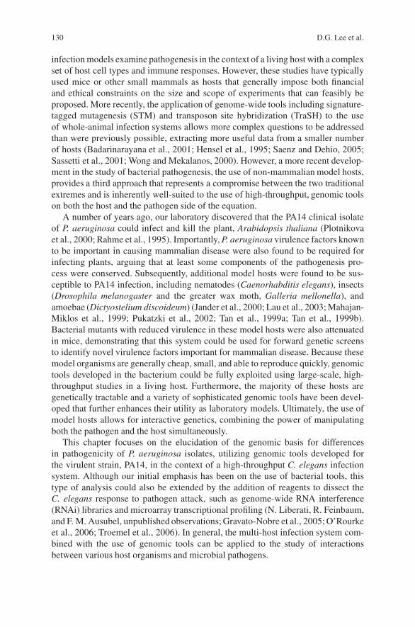
130 D.G. Lee et al.
infection models examine pathogenesis in the context of a living host with a complexset of host cell types and immune responses. However, these studies have typicallyused mice or other small mammals as hosts that generally impose both financialand ethical constraints on the size and scope of experiments that can feasibly beproposed. More recently, the application of genome-wide tools including signature-tagged mutagenesis (STM) and transposon site hybridization (TraSH) to the useof whole-animal infection systems allows more complex questions to be addressedthan were previously possible, extracting more useful data from a smaller numberof hosts (Badarinarayana et al., 2001; Hensel et al., 1995; Saenz and Dehio, 2005;Sassetti et al., 2001; Wong and Mekalanos, 2000). However, a more recent develop-ment in the study of bacterial pathogenesis, the use of non-mammalian model hosts,provides a third approach that represents a compromise between the two traditionalextremes and is inherently well-suited to the use of high-throughput, genomic toolson both the host and the pathogen side of the equation.
A number of years ago, our laboratory discovered that the PA14 clinical isolateof P. aeruginosa could infect and kill the plant, Arabidopsis thaliana (Plotnikovaet al., 2000; Rahme et al., 1995). Importantly, P. aeruginosa virulence factors knownto be important in causing mammalian disease were also found to be required forinfecting plants, arguing that at least some components of the pathogenesis pro-cess were conserved. Subsequently, additional model hosts were found to be sus-ceptible to PA14 infection, including nematodes (Caenorhabditis elegans), insects(Drosophila melanogaster and the greater wax moth, Galleria mellonella), andamoebae (Dictyostelium discoideum) (Jander et al., 2000; Lau et al., 2003; Mahajan-Miklos et al., 1999; Pukatzki et al., 2002; Tan et al., 1999a; Tan et al., 1999b).Bacterial mutants with reduced virulence in these model hosts were also attenuatedin mice, demonstrating that this system could be used for forward genetic screensto identify novel virulence factors important for mammalian disease. Because thesemodel organisms are generally cheap, small, and able to reproduce quickly, genomictools developed in the bacterium could be fully exploited using large-scale, high-throughput studies in a living host. Furthermore, the majority of these hosts aregenetically tractable and a variety of sophisticated genomic tools have been devel-oped that further enhances their utility as laboratory models. Ultimately, the use ofmodel hosts allows for interactive genetics, combining the power of manipulatingboth the pathogen and the host simultaneously.
This chapter focuses on the elucidation of the genomic basis for differencesin pathogenicity of P. aeruginosa isolates, utilizing genomic tools developed forthe virulent strain, PA14, in the context of a high-throughput C. elegans infectionsystem. Although our initial emphasis has been on the use of bacterial tools, thistype of analysis could also be extended by the addition of reagents to dissect theC. elegans response to pathogen attack, such as genome-wide RNA interference(RNAi) libraries and microarray transcriptional profiling (N. Liberati, R. Feinbaum,and F. M. Ausubel, unpublished observations; Gravato-Nobre et al., 2005; O’Rourkeet al., 2006; Troemel et al., 2006). In general, the multi-host infection system com-bined with the use of genomic tools can be applied to the study of interactionsbetween various host organisms and microbial pathogens.
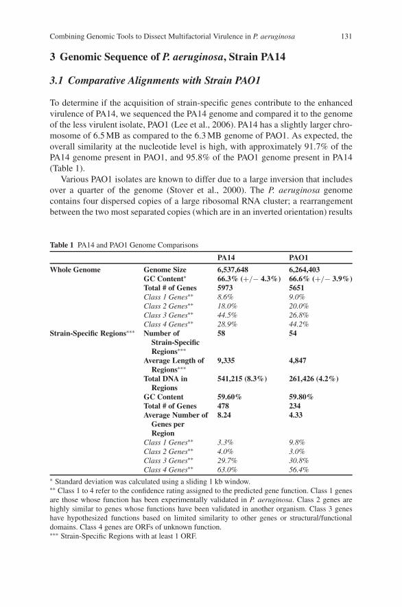
Combining Genomic Tools to Dissect Multifactorial Virulence in P. aeruginosa 131
3 Genomic Sequence of P. aeruginosa, Strain PA14
3.1 Comparative Alignments with Strain PAO1
To determine if the acquisition of strain-specific genes contribute to the enhancedvirulence of PA14, we sequenced the PA14 genome and compared it to the genomeof the less virulent isolate, PAO1 (Lee et al., 2006). PA14 has a slightly larger chro-mosome of 6.5 MB as compared to the 6.3 MB genome of PAO1. As expected, theoverall similarity at the nucleotide level is high, with approximately 91.7% of thePA14 genome present in PAO1, and 95.8% of the PAO1 genome present in PA14(Table 1).
Various PAO1 isolates are known to differ due to a large inversion that includesover a quarter of the genome (Stover et al., 2000). The P. aeruginosa genomecontains four dispersed copies of a large ribosomal RNA cluster; a rearrangementbetween the two most separated copies (which are in an inverted orientation) results
Table 1 PA14 and PAO1 Genome Comparisons
PA14 PAO1
Whole Genome Genome Size 6,537,648 6,264,403GC Content∗ 66.3% (+/− 4.3%) 66.6% (+/− 3.9%)Total # of Genes 5973 5651Class 1 Genes∗∗ 8.6% 9.0%Class 2 Genes∗∗ 18.0% 20.0%Class 3 Genes∗∗ 44.5% 26.8%Class 4 Genes∗∗ 28.9% 44.2%
Strain-Specific Regions∗∗∗ Number ofStrain-SpecificRegions∗∗∗
58 54
Average Length ofRegions∗∗∗
9,335 4,847
Total DNA inRegions
541,215 (8.3%) 261,426 (4.2%)
GC Content 59.60% 59.80%Total # of Genes 478 234Average Number of
Genes perRegion
8.24 4.33
Class 1 Genes∗∗ 3.3% 9.8%Class 2 Genes∗∗ 4.0% 3.0%Class 3 Genes∗∗ 29.7% 30.8%Class 4 Genes∗∗ 63.0% 56.4%
∗ Standard deviation was calculated using a sliding 1 kb window.∗∗ Class 1 to 4 refer to the confidence rating assigned to the predicted gene function. Class 1 genesare those whose function has been experimentally validated in P. aeruginosa. Class 2 genes arehighly similar to genes whose functions have been validated in another organism. Class 3 geneshave hypothesized functions based on limited similarity to other genes or structural/functionaldomains. Class 4 genes are ORFs of unknown function.∗∗∗ Strain-Specific Regions with at least 1 ORF.
132 D.G. Lee et al.
38.8%
PAO1
1
2
5
6
6,264,403 MB
F
AB
J
I
H
GE
D
K
L
C
1
F
AB
J
I
H
GE
D
K
L
C
AB
C
D
E
FG
H
I
K
L
J PA14
3
4
5
6
6,537,648MB
49.2%
AB
C
D
E
FG
H
K
L
3
4
5
6
MB
3
4
5
63
4
5
63
4
5
6
PA14
PAO
1
PAK
S350
04
X132
73
CF1
27
CF1
8
CF2
7
CF5
1966
0
6077
JJ69
2
S544
85
U25
04
UD
L
X245
09
62 E2
MSH
10
MSH
3
--PCR
A
BProducts 1 & 2 (PAO1 specific)
PCR Products 3 & 4 (PA14-specific)-
Fig. 1 Chromosomal rearrangement in PAO1 repositions the replication terminus relative to theorigin. (A) Schematic of PAO1 and PA14 chromosomes. The region with the same orientation inboth strains is shown with a thick red line; a thin blue line represents the inverted region. Arrowsrepresent the positions and orientations of four ribosomal RNA clusters. PCR products spanningeach rRNA cluster are indicated by numbers next to each arrow. PCR products 1 and 2 (purplenumbers) are derived from sequences diagnostic for the PAO1 chromosome (with the inversion).PCR products 3 and 4 (black numbers) are derived from sequences diagnostic for the PA14 chro-mosome (without the inversion). Products 5 and 6 (grey numbers) are common to both PA14 andPAO1. The origin of replication is at position “A” at the top of each chromosome. The position ofthe presumptive terminus of replication in each strain (see text) is indicated by an orange trianglemarked with the corresponding position along the chromosome (expressed as the percentage of thewhole chromosome, starting from the origin of replication and moving in a clockwise direction).(B) Diagnostic long-range PCR spanning ribosomal RNA repeats demonstrates the inversion inPAO1 but not in other P. aeruginosa strains. Diagnostic PCR primer pairs for products 1 and 2,diagnostic of the PAO1 arrangement (upper panels) or for products 3 and 4, diagnostic of the PA14arrangement (lower panels) were used to analyze genomic DNA isolated from PA14, PAO1, and18 additional strains. For each strain, a pair of lanes is used for the pair of PCR products (either1 and 2 for the upper panels, or 3 and 4 for the lower panels). Primers for PCR product 1 (upperpanel, first lane in each pair) resulted in a faint background band present with all P. aeruginosaisolates tested but absent when no genomic DNA template was added to the reaction
in the inversion (Fig. 1). The genome sequence of PA14 indicated that it alsocontains this inversion relative to the sequenced PAO1 isolate and we used long-range PCR to confirm the presence of the inversion (Fig. 1B). Furthermore, whenwe surveyed 18 additional diverse P. aeruginosa isolates, none gave PCR productsdiagnostic for the PAO1 chromosomal arrangement (upper panels) and most gener-ated one or both PCR products indicative of the PA14 arrangement (lower panels).These data suggest that the genome organization found in PA14 is more common

Combining Genomic Tools to Dissect Multifactorial Virulence in P. aeruginosa 133
among P. aeruginosa isolates than the genome organization of the sequenced PAO1strain.
In canonical bacterial genomes, the origin and terminus of replication are atopposite ends of the circular chromosome. Furthermore, prokaryotic genomes showa bias toward containing G over C on the leading strand of DNA synthesis (Bentleyand Parkhill, 2004). This bias can be measured as the GC-skew, defined as the mea-sure of G–C/G+C for regular intervals and tends to be positive on the leading strandand negative on the lagging strand. Therefore, by beginning at the origin of replica-tion and calculating GC-skew values on the leading strand, the putative replicationterminus can be identified as the region of the genome where the GC-skew valuesshift from a tendency toward positive values to a tendency toward negative values, ormore simply, the peak of the cumulative GC-skew values as one proceeds along thechromosome (Bentley and Parkhill, 2004). We therefore mapped cumulative GC-skews for both PA14 and PAO1. For PA14, the peak GC-skew was mapped oppositethe replication origin (at 49.2% of the genome; Figs. 1A) as is typical for otherbacterial genomes. In contrast, the position of the terminus in the sequenced PAO1chromosome is shifted relative to the origin (at 38.8% of the genome). Combinedwith the PCR-based survey of additional strains, this terminus mapping analysisargues that the PA14 chromosome (with respect to this large inversion) is morerepresentative of the canonical or ancestral P. aeruginosa genome than that of thesequenced PAO1 isolate. The physiological consequences of such an inversion andasymmetric replication cycle, however, are not clear.
Other than the presence of the large inversion, the genomes of PA14 and PAO1are largely colinear. Using the MUMmer 3.0 software package (Kurtz et al., 2004),we aligned the PA14 and PAO1 genomes (Fig. 2) and observed that the two genomeswere remarkably similar. The majority of the PA14 genome aligns with PAO1, in
PAO1
PA14
Fig. 2 Alignments of PA14 and PAO1 show that the two genomes are largely colinear. The MUM-mer 3.0 software package was utilized to align PA14 and PAO1 with respect to each other based onnucleotide sequence. The PAO1 genome is represented on top, and the PA14 genome is on the bot-tom. Lines connecting the two genomes represent the boundaries of regions that align. Alignmentsin the forward direction are in red, and reverse alignments are in blue. Gaps in a genome representregions of that genome that fail to align with the other genome (i.e., they are strain-specific regions)
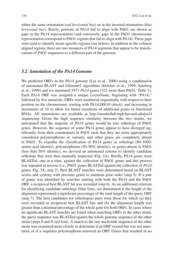
134 D.G. Lee et al.
either the same orientation (red horizontal bar) or in the inverted orientation (bluehorizontal bar). Rarely, portions of PA14 fail to align with PAO1 are shown asgaps in the PA14 representation (and conversely, gaps in the PAO1 chromosomerepresentation correspond to PAO1-regions that fail to align with PA14). These gapswere used to identify strain-specific regions (see below). In addition to the colinearaligned regions, there are rare instances of PA14 segments that appear to be translo-cations of PAO1 sequences to a different part of the genome.
3.2 Annotation of the PA14 Genome
We predicted ORFs in the PA14 genome (Lee et al., 2006) using a combinationof automated BLAST and Glimmer2 algorithms (Delcher et al., 1999; Salzberget al., 1998) and we annotated 5973 PA14 genes (322 more than PAO1; Table 1).Each PA14 ORF was assigned a unique LocusName, beginning with “PA14 ”followed by five numerals. ORFs were numbered sequentially with respect to theirposition on the chromosome, starting with PA14 00010 (dnaA), and increasing inincrements of 10 to allow for future insertions of additional genes or functionalRNAs. All annotations are available at http://ausubellab.mgh.harvard.edu/pa14sequencing. Given the high sequence similarity between the two strains, weanticipated that the majority of PA14 genes would be true orthologs of PAO1genes. However, the sequence of some PA14 genes appear to have diverged sig-nificantly from their counterparts in PAO1 such that they are more appropriatelyconsidered polymorphisms or variants, and other genes are completely absentin PAO1. To expedite the classification of PA14 genes as orthologs (90–100%amino acid identity), polymorphisms (50–90% identity), or genes absent in PAO1(less than 50% identity), we devised an automated scheme to identify candidateorthologs that were then manually inspected (Fig. 3A). Briefly, PA14 genes wereBLASTed, one at a time, against the collection of PAO1 genes and this processwas repeated in reverse (i.e., PAO1 genes BLASTed against the collection of PA14genes; Fig. 3A, step 2). Best BLAST matches were determined based on BLASTscores and synteny with previous genes to maintain gene order (step 3). If a pairof genes was identified by searches starting with both the PA14 and the PAO1ORF, a reciprocal best BLAST hit was recorded (step 6). As an additional criterionfor identifying candidate orthologs (blue font), we determined if the length of thealignment represented a significant percentage of the total length of the query ORF(step 7). The best candidates for orthologous pairs were those for which (a) theywere recorded as reciprocal best BLAST hits and (b) the alignment length wasgreater than a minimal percentage of the whole gene for both ORFs. In cases whereno significant BLAST matches are found when searching ORFs in the other strain,the query sequence was BLASTed against the whole genome sequence of the otherstrain (steps 8 and 9; red font). A match to the raw nucleotide sequence of the otherstrain was examined more closely to determine if an ORF existed but was not anno-tated, or if a sequence polymorphism removed an ORF. Genes that resulted in no
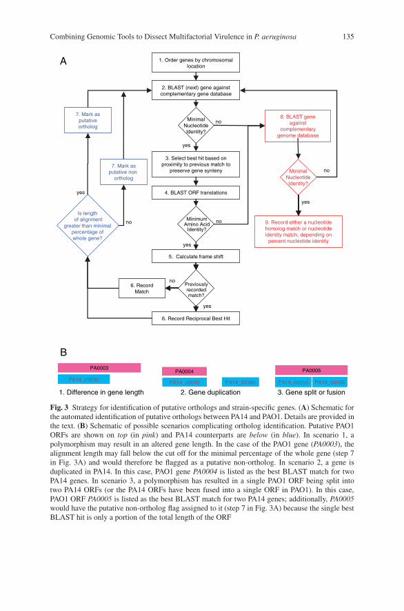
Combining Genomic Tools to Dissect Multifactorial Virulence in P. aeruginosa 135
1. Difference in gene length
PA0003
PA14_00030
PA0005
PA14_00050 PA14_00060
3. Gene split or fusion2. Gene duplication
PA0004
PA14_00040 PA14_03160
B
A
yes
yes
Minimal Nucleotide Identity?
8. BLAST gene against
complementary genome database
9. Record either a nucleotide homolog match or nucleotide identity match, depending on
percent nucleotide identity
no
Is lengthof alignment
greater than minimalpercentage ofwhole gene?
7. Mark as putativeortholog
7. Mark as putative non
ortholog
no
6. Record Match
1. Order genes by chromosomal location
yes
no
MinimumAmino Acid
Identity?
2. BLAST (next) gene against complementary gene database
Minimal Nucleotide Identity?
Previouslyrecordedmatch?
3. Select best hit based on proximity to previous match to
preserve gene synteny
4. BLAST ORF translations
6. Record Reciprocal Best Hit
5. Calculate frame shift
yes
yes
no
no
Fig. 3 Strategy for identification of putative orthologs and strain-specific genes. (A) Schematic forthe automated identification of putative orthologs between PA14 and PAO1. Details are provided inthe text. (B) Schematic of possible scenarios complicating ortholog identification. Putative PAO1ORFs are shown on top (in pink) and PA14 counterparts are below (in blue). In scenario 1, apolymorphism may result in an altered gene length. In the case of the PAO1 gene (PA0003), thealignment length may fall below the cut off for the minimal percentage of the whole gene (step 7in Fig. 3A) and would therefore be flagged as a putative non-ortholog. In scenario 2, a gene isduplicated in PA14. In this case, PAO1 gene PA0004 is listed as the best BLAST match for twoPA14 genes. In scenario 3, a polymorphism has resulted in a single PAO1 ORF being split intotwo PA14 ORFs (or the PA14 ORFs have been fused into a single ORF in PAO1). In this case,PAO1 ORF PA0005 is listed as the best BLAST match for two PA14 genes; additionally, PA0005would have the putative non-ortholog flag assigned to it (step 7 in Fig. 3A) because the single bestBLAST hit is only a portion of the total length of the ORF
136 D.G. Lee et al.
BLAST match to both the genes and the genome of the other strain were flagged asputative strain-specific genes.
Manual inspection of the automated ortholog predictions revealed several sce-narios that typically yielded ambiguous results (Fig. 3B). Scenario 1 depicts a situa-tion in which a sequence polymorphism has truncated the PA14 gene relative to thePAO1 copy. This gene pair would be identified as reciprocal best BLAST hits, butthe length of the alignment would potentially be shorter than the minimal percentageof the whole gene and would therefore be flagged as a putative NON-ortholog. Inscenario 2, a PAO1 gene is duplicated in PA14. The PAO1 gene will be paired withone of the two PA14 genes as a putative orthologous set; however, the second PA14gene would also be linked to the same PAO1 gene as its best BLAST hit (i.e., thePAO1 gene would have two entries). Manual inspection is required to ensure thatthe correct PA14 gene is selected as the ortholog. Scenario 3 shows a polymorphismthat results in one PAO1 gene split into two PA14 ORFs (corresponding to the N-and C-termini of the PAO1 gene). Again, the PAO1 gene appears as the best BLASThit for two different PA14 genes. By manually inspecting the ambiguous cases, weare gathering likely candidates for refinements to our search parameters that willoptimize the automated analysis.
Even though the portions of PA14 absent in PAO1 are a small fraction of thetotal genomic content (8.3% of the genome, Table 1), we were most interestedto see which genes were found in these regions and whether or not they con-tribute to virulence. We therefore identified 58 PA14 regions absent in PAO1 (con-taining 478 genes) and 54 PAO1 regions absent in PA14 (containing 234 genes).We refer to these as PA14-specific or PAO1-specific regions for the purposes ofthis discussion (recognizing that these genes may be present in other isolates ofP. aeruginosa and are not strictly strain-specific). Many of these gene clustershave hallmarks of horizontally transferred DNA, including boundary sequencesthat include direct repeats, insertion sequences, and tRNA genes, as well as GCcontents that differ significantly from the average GC content of the genome as awhole (PA14-specific clusters have an average GC content of 59.6%, more thanone standard deviation below the genome average of 66.3%, Table 1). Many ofthe defined PA14-specific and PAO1-specific regions are small, containing onlya few genes (only 22 of the 58 PA14-specific regions contain 5 or more genes,and only 18 of the 54 PAO1-specific regions contain 5 or more genes; Fig. 4).Therefore, only a subset of these putative gene clusters potentially includes func-tionally related genes encoding a complete pathogenicity “system” in the classicalsense. Nevertheless, we included all of these strain-specific regions in our subse-quent analysis to ensure that we were not excluding genes with potential roles invirulence.
These strain-specific regions contain an unusually high percentage of genes ofunknown function as compared to the whole genome. Each PA14 annotation hasan associated numerical value that reflects the confidence with which the genefunction is described. At the highest level of confidence are genes with a class 1designation, which are genes whose functions have been experimentally validatedin P. aeruginosa. Class 2 genes are highly similar to genes whose functions have
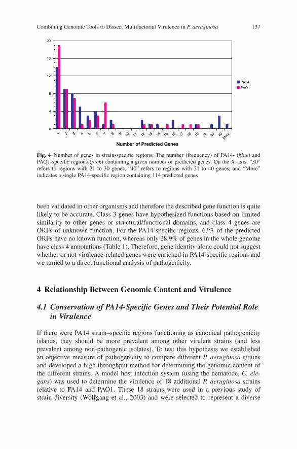
Combining Genomic Tools to Dissect Multifactorial Virulence in P. aeruginosa 137
Number of Predicted Genes
PA14
PAO1
7 8 9 13 14 17 20 30M
ore
0
4
8
12
16
20
10 11 12 15 16 18 19 401 2 3 4 5 6
Fig. 4 Number of genes in strain-specific regions. The number (frequency) of PA14- (blue) andPAO1-specific regions (pink) containing a given number of predicted genes. On the X -axis, “30”refers to regions with 21 to 30 genes, “40” refers to regions with 31 to 40 genes, and “More”indicates a single PA14-specific region containing 114 predicted genes
been validated in other organisms and therefore the described gene function is quitelikely to be accurate. Class 3 genes have hypothesized functions based on limitedsimilarity to other genes or structural/functional domains, and class 4 genes areORFs of unknown function. For the PA14-specific regions, 63% of the predictedORFs have no known function, whereas only 28.9% of genes in the whole genomehave class 4 annotations (Table 1). Therefore, gene identity alone could not suggestwhether or not virulence-related genes were enriched in PA14-specific regions andwe turned to a direct functional analysis of pathogenicity.
4 Relationship Between Genomic Content and Virulence
4.1 Conservation of PA14-Specific Genes and Their Potential Rolein Virulence
If there were PA14 strain–specific regions functioning as canonical pathogenicityislands, they should be more prevalent among other virulent strains (and lessprevalent among non-pathogenic isolates). To test this hypothesis we establishedan objective measure of pathogenicity to compare different P. aeruginosa strainsand developed a high throughput method for determining the genomic content ofthe different strains. A model host infection system (using the nematode, C. ele-gans) was used to determine the virulence of 18 additional P. aeruginosa strainsrelative to PA14 and PAO1. These 18 strains were used in a previous study ofstrain diversity (Wolfgang et al., 2003) and were selected to represent a diverse
138 D.G. Lee et al.
array of strain sources, including 13 clinical isolates from several types of infec-tions (CF lung infections, urinary tract infections, ocular infections, and blood iso-lates), four environmental isolates, and one laboratory strain. We then determinedthe genomic content and the relatedness of these 20 strains (18 plus PA14 andPAO1) using a microarray-based genotyping assay (see below) to assess the correla-tion between PA14-specific genes present in other isolates and their contribution tovirulence.
In the laboratory, C. elegans is grown on a lawn of E. coli, which serves asits food source. In the C. elegans model pathogenicity system (Tan et al., 1999a),pathogens such as PA14 are used as the sole source of food in place of E. coli, andthe longevity of the nematodes is monitored. Remarkably, nematodes feeding ona wide variety of human pathogens exhibit significantly shortened longevity com-pared to their longevity when feeding on E. coli. We consider shortened C. eleganslongevity as an indication of active killing since, in general, shortened longevity onpathogenic bacteria is dependent on live bacteria; i.e., dead pathogens do not killC. elegans. The nematodes are exposed to a maximal dose of pathogen in this assay(the bacteria are already grown to a lawn before the nematodes are added to theassay plate). Therefore, mutations or strain differences that effect the growth rateor overall fitness of the bacterium tend to have less of an impact on the virulencephenotype than infection models in which an initial low titer of bacteria is injectedinto the host and must survive long enough to reach a density sufficient to causedisease and death (as, e.g., in plant, insect, and mouse models). A number of studieshave demonstrated that the relative virulence of different P. aeruginosa strains inC. elegans correlates with their virulence in mice (Mahajan-Miklos et al., 1999; Tanet al., 1999a, b).
We tested the full set of 20 P. aeruginosa strains using the C. elegans killingmodel and we observed a full range of virulence phenotypes, including both theupper and the lower limits of what this assay system can measure (Lee et al., 2006).When we compared strains derived from a common infection type, we observed noconsistent clustering with respect to their virulence phenotype in C. elegans. Forexample, among the CF isolates were both the most virulent and the least virulentstrains in our collection. Importantly, among the environmental isolates, two closelyrelated strains (MSH3 and MSH10, both obtained from the same geographical site)were the fourth and the fifth most virulent strains tested. This latter result was inter-esting because it shows that strains isolated directly from the soil can have similarinfectious potential as strains isolated in a clinical setting.
To test the hypothesis that the virulence of the 20 P. aeruginosa strains correlateswith the presence of particular virulence islands, we determined which strain-specific genes were present or absent in each isolate by performing a microarray-based analysis of genomic content (a process described as genomotyping; Kimet al., 2002). We designed a custom array of spotted (70-mer) oligonucleotides,including 285 oligonucleotides corresponding to PA14 genes that are absent inPAO1 and 130 oligonucleotides corresponding to PAO1 genes that are absent inPA14 (along with additional sequences serving as positive and negative controls).Genomic DNA was isolated from these strains, fragmented, labeled, and hybridized
Combining Genomic Tools to Dissect Multifactorial Virulence in P. aeruginosa 139
to the microarray to determine if genes corresponding to each oligonucleotide werepresent, absent, or indeterminate (the hybridization intensity was too close to theempirically determined cut-off to make a confident present/absent call). Using thisarray data, we used hierarchical clustering algorithms to determine the relatednessthe 20 P. aeruginosa strains and found no strong correlation between genomic con-tent and neither the source of the strain nor the relative virulence of the strain (Leeet al., 2006).
4.2 Identification of PA14-specific Virulence Genesand Their Conservation in Other Strains
The experiments described above showed that there was no correlation betweenthe PA14-specific sequences in general and virulence in the C. elegans killingassay. We therefore performed a functional analysis of PA14-specific ORFs,directly identifying genes required for pathogenicity and subsequently assess-ing their distribution among the other strains. Our laboratory has constructeda genome-wide, non-redundant transposon insertion mutant library in PA14(http://ausubellab.mgh.harvard.edu/cgi-bin/pa14/home.cgi; Liberati et al., 2006).Using this library, we conducted a screen for mutants in PA14-specific genes thathad reduced virulence in C. elegans (Lee et al., 2006).
We identified four mutations in genes that are present in all P. aeruginosa strainsbut whose sequences are highly divergent (i.e., different strains contain differentvariants or alleles of functionally similar genes). These included two mutations ingenes required for the biosynthesis of the O-antigen component of the P. aeruginosalipopolysaccharide, and two mutations in genes involved in type 4 fimbrial biogen-esis. More importantly, we identified five genes required for PA14 virulence that areentirely absent in PAO1; four are genes of unknown function and one has hallmarksof a putative transcription factor. None of these five genes were previously identifiedas virulence determinants.
To determine whether or not these five PA14-specific pathogenicity genes werepredictive of pathogenicity in other strains, we used the microarray genotyping datato compute Spearman’s rank correlation coefficients for each PA14-specific gene,relating its presence, absence, or indeterminate status with the (rank order) virulenceof the strain. Figure 5 summarizes these data. Each of the 20 strains tested are rep-resented in columns arranged from left to right in order of decreasing pathogenicity.The PA14-specific genes tested (shown in rows) are described as present (blue),absent (yellow), or indeterminate (intermediate intensity that was too close to thecut-off between present and absent calls; red). These genes were then arranged withrespect to their Spearman’s rank order correlation coefficients, with those genesmost tightly correlated with virulence on top and those least correlated on the bot-tom. This gene order therefore does not reflect a linear order on the chromosome.As shown on the left side of Fig. 5, the correlation coefficients ranged from a highof 0.63 to a low of −0.20. A perfect (positive) correlation would result from a

140 D.G. Lee et al.
= present
= unclear
= absent
Legend
Correlation Coefficient
Range
Mutant Number
(Correlation coefficient)1,21,2 (0.63)(0.63)
3,43,4 (0.44)(0.44)
55 (0.02)(0.02)0 to 0.10.2 to 0
0.1 to 0.2
0.2 to 0.3
0.3 to 0.4
0.4 to 0.5
0.5 to 0.6
0.6 to 0.63
Mutant Number
1,21,2 (0.63)(0.63)
3,43,4 (0.44)(0.44)
55 (0.02)(0.02)
1,21,2 (0.63)(0.63)
3,43,4 (0.44)(0.44)
55 (0.02)(0.02)
1,21,2 (0.63)(0.63)
3,43,4 (0.44)(0.44)
55 (0.02)(0.02)–
2 4 6 8 10 12 14 16 181 3 5 7 9 11 13 15 17 19
20
Strain Number
Strain name Strain Source
1 CF18 CF2 PA14 burn3 UDL UTI4 MSH3 environmental5 MSH10 environmental6 U2504 UTI7 62 environmental8 6077 occular9 S54485 UTI10 X13273 blood11 PAO1 wound12 JJ692 UTI13 PAK lab14 19660 occular15 X24509 UTI16 CF127 CF17 E2 environmental18 CF27 CF19 S35004 blood20 CF5 CF
Fig. 5 Range of correlation coefficients between genomic content and virulence. A subset of themicroarray-based genotyping of 20 strains is shown for PA14-specific genes. Data for each straintested is presented in columns. Strains are arranged from left to right in order of decreasing vir-ulence; column headers refer to the 20 strain numbers in the accompanying key listing the strainname and strain source (UTI = urinary tract infection). The strain sources are color coded tohighlight that they do not cluster according to virulence. Some strains shown in adjacent columnswere equally virulent (strains 4 and 5; strains 8, 9, and 10; strains 14, 15, and 16). Genes arerepresented as individual rows and are described as present (blue), absent (yellow), or indetermi-nate (red). Genes are arranged by Spearman’s rank correlation coefficients relating the presence ofPA14 genes in a strain and the strain’s virulence (highest correlations at the top, lowest correlationsat the bottom). Brackets on the left indicate groups of genes with similar correlation coefficients,with the range of coefficients shown next to the bracket. Lines to the right indicate genes requiredfor maximal C. elegans virulence; the associated correlation coefficient for these genes is shownin red parentheses
Combining Genomic Tools to Dissect Multifactorial Virulence in P. aeruginosa 141
row with a clustering of blue on the left (a gene present in the most pathogenicstrains) and yellow clustered on the right (a gene absent in the least virulentstrains).
The positions of the genes identified as novel PA14-virulence genes are indicatedon the right side of Fig. 5 (with correlation coefficients shown in red parentheses).Genes 1 (unknown ORF) and 2 (putative transcription factor) are among thosewith the highest observed correlation coefficient (0.63). These two ORFs are foundtogether in an 8-gene cluster known as the clone C-specific region common to cloneC isolates (members of a clonal family associated with CF infections; Romlinget al., 2005). This genomic region is known to be hyper-variable among clone Cisolates (and absent in PAO1): two Clone C strains (C and SG17M) each containthe short clone C-specific region adjacent to large strain-specific islands PAGI-2(strain C, 113 ORFs) or PAGI-3 (strain SG17M, 105 ORFs) (Larbig et al., 2002).The corresponding PA14 locus possesses only orthologs to the genes containedin the shorter clone C-specific region; PA14 does not have orthologs to genesdescribed within the larger strain-specific islands. This cluster of clone C-specificgenes was found to be present in 14 of the 18 tested strains, and the 4 strainsthat lacked these genes were among the 5 least pathogenic isolates tested. How-ever, two exceptions to this general trend include PAO1 itself, which lacks thissequence and whose virulence is intermediate among those strains possessing thiscluster, and strain CF5 which contains this cluster and is the least virulent of allisolates.
Genes 3 and 4 in Fig. 5 (both ORFs of unknown function) have intermedi-ate correlation coefficients (0.44). They are contained within a previously identi-fied pathogenicity island (PAPI-1, containing 112 ORFs) shown to encode genesrequired for pathogenicity in plants and mammals (He et al., 2004). Although thetwo mutations in genes that we identified in this cluster were not among thosepreviously examined, they are adjacent to or nearby ORFs that had phenotypesin plants and mice. These two genes show a weak correlation with virulence andare present in 7 of the 9 strains more virulent than PAO1 (CF18, MSH3, MSH10,62, 6077, S54485, and X13273, in order of most to least virulent), and absent in6 of the 9 strains less pathogenic than PAO1 (JJ6992, PAK, 19660, CF27, E2, andCF5). However, these two genes are absent in the third and sixth most pathogenicstrains (UDL and U2504), and present in three avirulent strains (X24509, CF127,and S35004).
Gene 5 (also of unknown function) is part of a 14-gene region that was previouslyuncharacterized; this gene shows essentially no correlation with strain virulence(correlation coefficient of 0.02). In fact, this gene tends to be more representedin avirulent strains than pathogenic isolates. Taken together, the five functionallydefined PA14 genes required for C. elegans killing each had exceptions to thegeneral trend (being present in some attenuated strains and/or absent in somevirulent strains) and are neither required for nor necessarily predictive of anotherstrain’s ability to be pathogenic.

142 D.G. Lee et al.
5 Future Directions: Testing Additional Model Hosts
We were surprised that genes identified as PA14-virulence determinants are notnecessarily correlated with the virulence of other strains in which they are found,because this observation suggests that the action of some virulence determinantsmay depend on the total genomic content. This view contrasts with canonicalpathogenicity islands that contain a set of genes acting as an autonomous set, thepresence of which directly contributes to overall strain virulence. Since most stud-ies of pathogenicity islands have been carried out in more traditional experimentalsystems, it is possible that our unexpected observations are a result of using a modelhost such as C. elegans. Traditional infection systems using mice or other mammalsinvolve a single dose of bacteria, often administered at a low titer and requiringthe initial population of pathogens to proliferate and avoid detection by the host’simmune response long enough to reach a pathogenic or lethal dose. In contrast,the C. elegans infection system constantly exposes the host to a maximal does ofpathogen. The former approach has conceptual similarity to an acute infection inhumans, whereas the latter has aspects that more closely approximate a chronicinfection.
An advantage of the model host approach to studying infectious disease is thatmultiple hosts can be used and compared, each with their respective strengths andweaknesses. One alternate host that we are currently examining is the greater waxmoth caterpillar, G. mellonella. Wax moth caterpillars are commercially available,inexpensive, easy to use, and yield data rapidly, making them an ideal candi-date for pilot experiments to extend our observations in C. elegans to other hostorganisms.
5.1 Wax Moth Injection Model
The wax moth caterpillar infection system involves the sub-dermal injection of a lowdose of bacteria into the caterpillar and monitoring viability over several days. Byinjecting a dilution series of the bacteria, quantitative measurements can be madeof the LD50, the initial dose required to cause 50% lethality. Wild-type PA14 isan incredibly potent killer, with an LD50 of between 1 and 2 bacteria, whereasattenuated mutants have elevated LD50s (indicating a higher initial does requiredfor comparable disease symptoms). When comparing the virulence of various PA14mutants in wax moth caterpillars and mice, a positive correlation is observed, mak-ing this insect an excellent model system for the identification of pathogenesis fac-tors important in mammalian disease (Jander et al., 2000).
As a preliminary test of the viability of repeating our studies in wax moth cater-pillars, we calculated the LD50s for five mutants identified as avirulent in the C. ele-gans screen (Fig. 6A and Table 2). These included mutations in genes PA14 03370and PA14 27680, both genes of unknown function in regions completely absentin PAO1 (genes 5 and 1, respectively in Fig. 5). Also shown are three mutants in
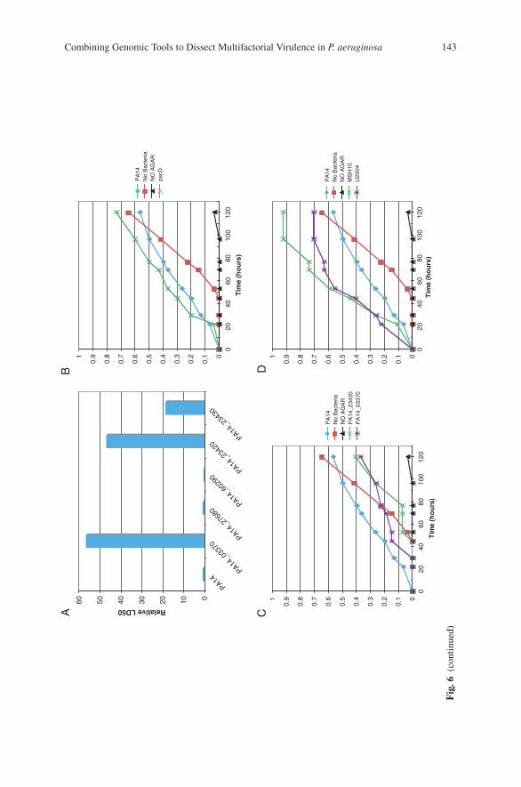
Combining Genomic Tools to Dissect Multifactorial Virulence in P. aeruginosa 143
0102030405060
PA14PA14
_033
70PA14
_276
80PA14
_602
90PA14
_234
20PA14
_234
30
Relative LD50
0
0.1
0.2
0.3
0.4
0.5
0.6
0.7
0.8
0.91
020
4060
8010
012
0
Tim
e (h
ou
rs)
PA
14N
o B
acte
riaN
O A
GA
Rps
cD
BA
0
0.1
0.2
0.3
0.4
0.5
0.6
0.7
0.8
0.91
020
4060
8010
012
0
Tim
e (h
ou
rs)
PA
14N
o B
acte
riaN
O A
GA
RP
A14
_234
20P
A14
_033
70
0
0.1
0.2
0.3
0.4
0.5
0.6
0.7
0.8
0.91
020
4060
8010
012
0T
ime
(ho
urs
)
PA
14N
o B
acte
riaN
O A
GA
RM
SH
10U
2504
DC
Fig
.6(c
onti
nued
)
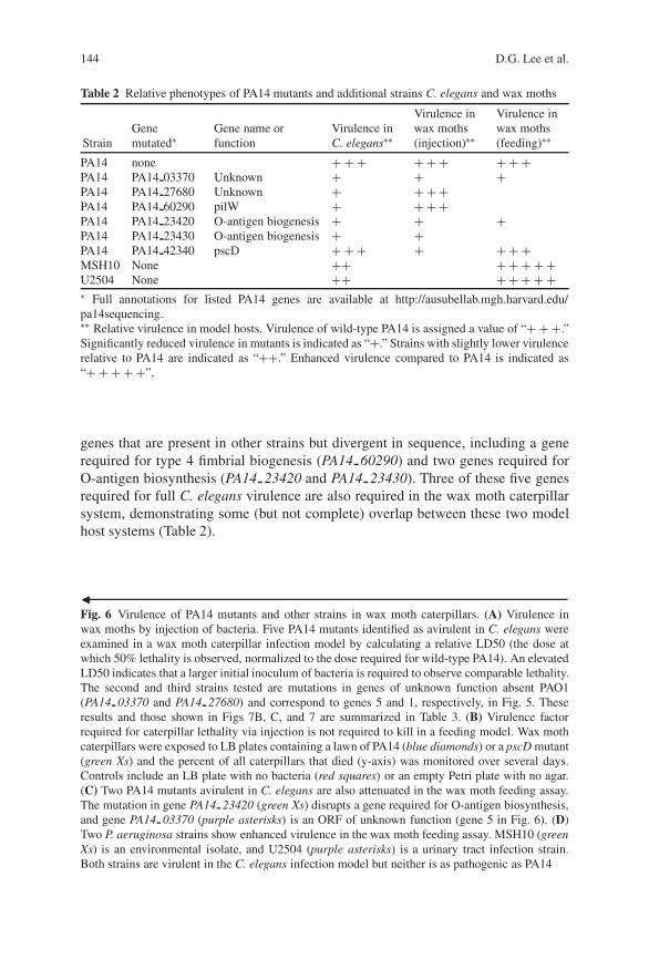
144 D.G. Lee et al.
Table 2 Relative phenotypes of PA14 mutants and additional strains C. elegans and wax moths
StrainGenemutated∗
Gene name orfunction
Virulence inC. elegans∗∗
Virulence inwax moths(injection)∗∗
Virulence inwax moths(feeding)∗∗
PA14 none + + + + + + + + +PA14 PA14 03370 Unknown + + +PA14 PA14 27680 Unknown + + + +PA14 PA14 60290 pilW + + + +PA14 PA14 23420 O-antigen biogenesis + + +PA14 PA14 23430 O-antigen biogenesis + +PA14 PA14 42340 pscD + + + + + + +MSH10 None ++ + + + + +U2504 None ++ + + + + +∗ Full annotations for listed PA14 genes are available at http://ausubellab.mgh.harvard.edu/pa14sequencing.∗∗ Relative virulence in model hosts. Virulence of wild-type PA14 is assigned a value of “+ + +.”Significantly reduced virulence in mutants is indicated as “+.” Strains with slightly lower virulencerelative to PA14 are indicated as “++.” Enhanced virulence compared to PA14 is indicated as“+ + + + +”.
genes that are present in other strains but divergent in sequence, including a generequired for type 4 fimbrial biogenesis (PA14 60290) and two genes required forO-antigen biosynthesis (PA14 23420 and PA14 23430). Three of these five genesrequired for full C. elegans virulence are also required in the wax moth caterpillarsystem, demonstrating some (but not complete) overlap between these two modelhost systems (Table 2).
�Fig. 6 Virulence of PA14 mutants and other strains in wax moth caterpillars. (A) Virulence inwax moths by injection of bacteria. Five PA14 mutants identified as avirulent in C. elegans wereexamined in a wax moth caterpillar infection model by calculating a relative LD50 (the dose atwhich 50% lethality is observed, normalized to the dose required for wild-type PA14). An elevatedLD50 indicates that a larger initial inoculum of bacteria is required to observe comparable lethality.The second and third strains tested are mutations in genes of unknown function absent PAO1(PA14 03370 and PA14 27680) and correspond to genes 5 and 1, respectively, in Fig. 5. Theseresults and those shown in Figs 7B, C, and 7 are summarized in Table 3. (B) Virulence factorrequired for caterpillar lethality via injection is not required to kill in a feeding model. Wax mothcaterpillars were exposed to LB plates containing a lawn of PA14 (blue diamonds) or a pscD mutant(green Xs) and the percent of all caterpillars that died (y-axis) was monitored over several days.Controls include an LB plate with no bacteria (red squares) or an empty Petri plate with no agar.(C) Two PA14 mutants avirulent in C. elegans are also attenuated in the wax moth feeding assay.The mutation in gene PA14 23420 (green Xs) disrupts a gene required for O-antigen biosynthesis,and gene PA14 03370 (purple asterisks) is an ORF of unknown function (gene 5 in Fig. 6). (D)Two P. aeruginosa strains show enhanced virulence in the wax moth feeding assay. MSH10 (greenXs) is an environmental isolate, and U2504 (purple asterisks) is a urinary tract infection strain.Both strains are virulent in the C. elegans infection model but neither is as pathogenic as PA14

Combining Genomic Tools to Dissect Multifactorial Virulence in P. aeruginosa 145
5.2 Wax Moth Feeding Model
In addition to the established injection model of infection, we examined whether ornot a chronic exposure of the wax moth caterpillars to a lawn of PA14 could resultin pathogenicity. This approach is significantly faster than manually injecting eachbacterial strain to be tested, and we are tentatively referring to this assay systemas a wax moth “feeding” model (although we have yet to determine that bacteriaare ingested by feeding rather than entering the host by some other mechanism).We placed wax moth caterpillars onto LB plates containing a lawn of PA14, LBplates with no bacteria, or an empty Petri dish with no agar (Fig. 6B). In the absenceof agar, the caterpillars survived with little or no adverse effects over the courseof 5 days (black triangles). When placed on an empty LB plate with no addedbacteria, a lawn of an (as yet) uncharacterized microorganism grew up over thecourse of the experiment and the caterpillars began dying at 53 hours (red squares).We have not yet determined whether this microbe was present on the surface ofthe caterpillars or is initially present in the gut of the animal and seeded onto theplates by excretion. Exposure of the wax moth caterpillars to a pre-formed lawnof PA14 resulted in more rapid death, beginning at 22 hours (blue diamonds).Interestingly, we also tested a previously characterized mutant in PA14, a lesionin the pscD gene, which is a structural component of the type III secretion appara-tus. Type III secreted bacterial products are required for mammalian pathogenesisand have also been shown to be required for virulence when bacteria are injectedinto G. mellonella caterpillars (but not required for virulence in C. elegans; Miy-ata et al., 2003). Surprisingly, the pscD mutant did not show a defect in the waxmoth feeding system (green Xs), suggesting that the two G. mellonella infectionsystems are monitoring different sets of virulence determinants (summarized inTable 2).
We next examined two mutations in genes required for pathogenicity in C. ele-gans (Fig. 6C and Table 2). We selected mutations in genes PA14 23420 (an O-antigen biosynthetic gene) and PA14 03370 (an ORF of unknown function; gene#5 in Fig. 5), because they were also required for full virulence in the wax mothcaterpillar injection system. Both mutants tested showed reduced pathogenicity inthe feeding assay (green Xs and purple asterisks), with less overall host lethalitythan the control containing agar but no bacteria. In all cases in which bacteria werepre-seeded on the LB plates (either wild-type or mutant PA14), the uncharacterizedmicroorganism was unable to grow, making it possible to observe lethality due onlyto the P. aeruginosa strain of interest. These results demonstrate that the caterpillar-feeding assay can identify mutants with reduced virulence, and that some mutationsresult in avirulence in all three assay systems (C. elegans, wax moth caterpillarinjection, and wax moth caterpillar feeding; Table 2).
Finally, we examined the ability of the wax moth feeding system to detect differ-ences in virulence among P. aeruginosa isolates. Strains MSH10 (an environmentalisolate) and U2504 (a urinary tract infection isolate) are virulent in C. elegans, butneither is as pathogenic as PA14 (rank order virulence of 5 and 6, respectively, ver-sus a rank order of 2 for PA14 within the group of 20 strains tested). Surprisingly,
146 D.G. Lee et al.
both strains showed elevated virulence compared to PA14 (Fig. 6D and Table 2).In the injection system, the virulence of wild-type PA14 is so potent (requiringbetween 1 and 2 live bacteria in the initial dose for 50% lethality) that enhancedvirulence cannot be measured. However, the feeding system can potentially iden-tify strains (or mutants) with both enhanced and decreased virulence comparedto PA14.
The wax moth caterpillar pathogenicity system is a promising alternative modelto extend our virulence studies that were initially carried out with C. elegans. Bothinjection and feeding assays can be used to assess the relative virulence of the 20P. aeruginosa strains and to screen the PA14 mutant library. Given that the threeapproaches (nematode feeding, caterpillar injection, and caterpillar feeding) resultin overlapping but occasionally distinct observations, it is likely that different setsof PA14 mutants will be identified as avirulent when the mutant library is tested ineach model. It will be particularly interesting to see if, for a given assay system,the identified PA14-virulence factors each fail to correlate with virulence in otherstrains (as was the case for C. elegans), or whether this surprising lack of correlationis specific to the nematode system.
6 Discussion
To study the relationship between P. aeruginosa PA14 genomic content and viru-lence, we combined several genomic tools including comparative genomic analyses,functional genome-wide analyses of virulence, and a model host infection systemamenable to high-throughput and low-cost screening. In the case of C. elegans, wehave found that genes identified in PA14 as required for virulence (and absent inthe less virulent PAO1) do not correlate strongly with the pathogenicity of otherstrains in which they are also present. In contrast, canonical pathogenicity islandstend to contain genes that contribute directly to the virulence of isolates in whichthey are found, such as the P. aeruginosa gene cluster encoding the type III secretioneffector ExoU (present in PA14 but absent in PAO1), which increases the cytotox-icity of strains toward mammalian cells (Sato and Frank, 2004) and is required forpathogenicity in G. mellonella and the amoeba, D. discoideum (Miyata et al., 2003;Pukatzki et al., 2002). However, it appears that PA14 also contains virulence deter-minants that may not function autonomously to affect virulence. The data shownin Fig. 5 suggest that, at least in the case of C. elegans pathogenicity, the effectsof multiple PA14 virulence factors combine in different combinations in differentstrains to result in an overall virulence phenotype.
What are the mechanistic underpinnings for the apparent combinatorial natureof P. aeruginosa virulence factors and how definitive is our analysis with respectto their conservation in other P. aeruginosa strains? First, our microarray-basedgenotyping analysis is only an initial step toward identifying strain similarities anddifferences at a functional level. The hybridization experiments can only identify(highly similar) sequences that are present in other isolates, but it cannot determine
Combining Genomic Tools to Dissect Multifactorial Virulence in P. aeruginosa 147
whether or not they encode functional proteins that are similarly regulated. Also, theapparent absence of a gene in a given strain cannot exclude the existence of a geneof divergent sequence that is nevertheless functionally analogous. Second, giventhe apparent multifactorial and combinatorial nature of P. aeruginosa virulence,elucidating the molecular basis of virulence in a particular P. aeruginosa strainwill require an understanding of how groups of virulence factors interact with eachother as well as how they interact with the core genome. With respect to the lattergoal, our laboratory is currently extending the screen for PA14 mutants attenuatedin C. elegans killing to include the entire genome (rather than focussing only onputative strain-specific genes). Third, as stated above, the examination of additionalmodel hosts will be a key step toward determining whether the lack of observedcorrelations between genomic content and strain virulence is a general phenomenonor specific to nematodes. In addition to the described use of G. mellonella (withboth an injection and a feeding model of pathogenesis), fruit flies and plants areinteresting candidate hosts for future study.
In presenting this work, we have aimed to provide an example of how genomicshas impacted our understanding of disease. The rapid development of genomic toolsin both the pathogen and the host has greatly expanded the breadth and depthof experimental questions that can be asked in the laboratory. In particular, webelieve that the model host infection system, in which multiple, genetically tractableorganisms are substituted for traditional mammalian hosts, provides a powerfulopportunity to utilize both pathogen and host genomic tools, either individuallyor combinatorially. The low cost, relatively small size, and low level of complex-ity of model hosts allow for high-throughput assays that are ideal for exploitingthe power inherent in large reagents such as genome-wide mutant libraries. Morerecently, a C. elegans–Enterococcus faecalis infection system has been used toscreen large chemical libraries for novel compounds acting as antimicrobials andanti-infectives (Moy et al., 2006); these compounds could serve as vital reagentsboth in clinical settings and in the lab as molecular probes to dissect the pathogen-esis process. With model hosts such as C. elegans, genome-wide RNA interference(RNAi) libraries and transcriptional arrays can be used to identify host gene prod-ucts involved in the innate immune response to pathogens (N. Liberati, R. Fein-baum and F. M. Ausubel, unpublished observations; Gravato-Nobre et al., 2005;O’Rourke et al., 2006; Troemel et al., 2006). As the use of genomic tools and modelhost infection systems matures, the true power of these approaches will be in theability to combine both host and pathogen reagents to understand, at a mechanis-tic level, the complex interactions between the two organisms that occur during aninfection.
Acknowledgments We are grateful to J. Decker, W. Brown, K. Osborn, A. Perera, R. Elliott, L.Gendal, K. Montgomery, G. Grills, and L. Li for sequencing PA14; R. Jackson for suggestionson genomic DNA preparations for Microarray analysis; N. El Massadi and J. Frietas for sugges-tions on sample labeling, hybridizations, and scanning for microarray experiments; and D. Parkfor assistance with microarray data analysis. Previously described data was obtained using fundsprovided by NIH grants AI064332-02 and HL66678, and DOE grant DE-FG02-ER63445.
148 D.G. Lee et al.
References
Badarinarayana, V., Estep, P.W., 3rd, Shendure, J., Edwards, J., Tavazoie, S., Lam, F., and Church,G.M., 2001, Selection analyses of insertional mutants using subgenic-resolution arrays, Nat.Biotechnol. 19:1060–1065.
Bentley, S.D., and Parkhill, J., 2004, Comparative genomic structure of prokaryotes, Annu. Rev.Genet. 38:771–792.
Choi, J.Y., Sifri, C.D., Goumnerov, B.C., Rahme, L.G., Ausubel, F.M., and Calderwood, S.B.,2002, Identification of virulence genes in a pathogenic strain of Pseudomonas aeruginosa byrepresentational difference analysis, J. Bacteriol. 184:952–961.
Delcher, A.L., Harmon, D., Kasif, S., White, O., and Salzberg, S.L., 1999, Improved microbialgene identification with GLIMMER, Nucleic Acids Res. 27:4636–4641.
Doring, D., 1993, Chronic Pseudomonas aeruginosa lung infection in cystic fibrosis patients, Pseu-domonas aeruginosa as an opportunistic pathogen, Plenum Press, New York, pp. 245–273.
Ernst, R.K., D’Argenio, D.A., Ichikawa, J.K., Bangera, M.G., Selgrade, S., Burns, J.L., Hiatt, P.,McCoy, K., Brittnacher, M., Kas, A., et al., 2003, Genome mosaicism is conserved but notunique in Pseudomonas aeruginosa isolates from the airways of young children with cysticfibrosis, Environ. Microbiol. 5:1341–1349.
Gravato-Nobre, M.J., Nicholas, H.R., Nijland, R., O’Rourke, D., Whittington, D.E., Yook, K.J.,and Hodgkin, J., 2005, Multiple genes affect sensitivity of Caenorhabditis elegans to the bac-terial pathogen Microbacterium nematophilum, Genetics 171:1033–1045.
Hacker, J., Blum-Oehler, G., Muhldorfer, I., and Tschape, H., 1997, Pathogenicity islands ofvirulent bacteria: structure, function and impact on microbial evolution, Mol. Microbiol. 23:1089–1097.
He, J., Baldini, R.L., Deziel, E., Saucier, M., Zhang, Q., Liberati, N.T., Lee, D., Urbach, J.,Goodman, H.M., and Rahme, L.G., 2004, The broad host range pathogen Pseudomonas aerug-inosa strain PA14 carries two pathogenicity islands harboring plant and animal virulence genes,Proc. Natl. Acad. Sci. USA 101:2530–2535.
Hensel, M., Shea, J.E., Gleeson, C., Jones, M.D., Dalton, E., and Holden, D.W., 1995, Simultane-ous identification of bacterial virulence genes by negative selection, Science 269: 400–403.
Jander, G., Rahme, L.G., and Ausubel, F.M., 2000, Positive correlation between virulence of Pseu-domonas aeruginosa mutants in mice and insects, J. Bacteriol. 182:3843–3845.
Kim, C.C., Joyce, E.A., Chan, K., and Falkow, S., 2002, Improved analytical methodsfor microarray-based genome-composition analysis, Genome Biol. 3, RESEARCH0065.1–0065.17.
Kurtz, S., Phillippy, A., Delcher, A.L., Smoot, M., Shumway, M., Antonescu, C., andSalzberg, S.L., 2004, Versatile and open software for comparing large genomes, Genome Biol.5:R12.
Larbig, K.D., Christmann, A., Johann, A., Klockgether, J., Hartsch, T., Merkl, R., Wiehlmann, L.,Fritz, H.J., and Tummler, B., 2002, Gene islands integrated into tRNA (Gly) genes confergenome diversity on a Pseudomonas aeruginosa clone, J. Bacteriol. 184:6665–6680.
Lau, G.W., Goumnerov, B.C., Walendziewicz, C.L., Hewitson, J., Xiao, W., Mahajan-Miklos, S.,Tompkins, R.G., Perkins, L.A., and Rahme, L.G., 2003, The Drosophila melanogaster toll path-way participates in resistance to infection by the gram-negative human pathogen Pseudomonasaeruginosa, Infect. Immun. 71:4059–4066.
Lee, D.G., Urbach, J.M., Wu, G., Liberati, N.T., Feinbaum, R.L., Miyata, S., Diggins, L.T., He, J.,Saucier, M., Deziel, E., et al., 2006, Genomic analysis reveals that Pseudomonas aeruginosavirulence is combinatorial, Genome Biol. 7:R90.
Liberati, N.T., Urbach, J.M., Miyata, S., Lee, D.G., Drenkard, E., Wu, G., Villanueva, J., Wei, T.,and Ausubel, F.M., 2006, An ordered, nonredundant library of Pseudomonas aeruginosa strainPA14 transposon insertion mutants, Proc. Natl. Acad. Sci. USA 103:2833–2838.
Mahajan-Miklos, S., Tan, M.W., Rahme, L.G., and Ausubel, F.M., 1999, Molecular mechanismsof bacterial virulence elucidated using a Pseudomonas aeruginosa–Caenorhabditis eleganspathogenesis model, Cell 96:47–56.
Combining Genomic Tools to Dissect Multifactorial Virulence in P. aeruginosa 149
Miyata, S., Casey, M., Frank, D.W., Ausubel, F.M., and Drenkard, E., 2003, Use of the Galleriamellonella caterpillar as a model host to study the role of the type III secretion system inPseudomonas aeruginosa pathogenesis, Infect. Immun. 71:2404–2413.
Moy, T.I., Ball, A.R., Anklesaria, Z., Casadei, G., Lewis, K., and Ausubel, F.M., 2006, Identifi-cation of novel antimicrobials using a live-animal infection model, Proc. Natl. Acad. Sci. USA103:10414–10419.
O’Rourke, D., Baban, D., Demidova, M., Mott, R., and Hodgkin, J., 2006, Genomic clusters,putative pathogen recognition molecules, and antimicrobial genes are induced by infection ofC. elegans with M. nematophilum, Genome Res. 16:1005–1016.
Oelschlaeger, T.A., and Hacker, J., 2004, Impact of pathogenicity islands in bacterial diagnostics,Apmis 112:930–936.
Plotnikova, J.M., Rahme, L.G., and Ausubel, F.M., 2000, Pathogenesis of the human oppor-tunistic pathogen Pseudomonas aeruginosa PA14 in Arabidopsis, Plant Physiol. 124:1766–1774.
Pukatzki, S., Kessin, R.H., and Mekalanos, J.J., 2002, The human pathogen Pseudomonas aerug-inosa utilizes conserved virulence pathways to infect the social amoeba Dictyostelium dis-coideum, Proc. Natl. Acad. Sci. USA 99:3159–3164.
Rahme, L.G., Stevens, E.J., Wolfort, S.F., Shao, J., Tompkins, R.G., and Ausubel, F.M., 1995,Common virulence factors for bacterial pathogenicity in plants and animals, Science 268:1899–1902.
Rahme, L.G., Tan, M.W., Le, L., Wong, S.M., Tompkins, R.G., Calderwood, S.B., andAusubel, F.M., 1997, Use of model plant hosts to identify Pseudomonas aeruginosa virulencefactors, Proc. Natl. Acad. Sci. USA 94:13245–13250.
Raskin, D.M., Seshadri, R., Pukatzki, S.U., and Mekalanos, J.J., 2006, Bacterial genomics andpathogen evolution, Cell 124:703–714.
Romling, U., Kader, A., Sriramulu, D.D., Simm, R., and Kronvall, G., 2005, Worldwide distribu-tion of Pseudomonas aeruginosa clone C strains in the aquatic environment and cystic fibrosispatients, Environ. Microbiol. 7:1029–1038.
Saenz, H.L., and Dehio, C., 2005, Signature-tagged mutagenesis: technical advances in a negativeselection method for virulence gene identification, Curr. Opin. Microbiol. 8:612–619.
Salzberg, S.L., Delcher, A.L., Kasif, S., and White, O., 1998, Microbial gene identification usinginterpolated Markov models, Nucleic Acids Res. 26:544–548.
Sassetti, C.M., Boyd, D.H., and Rubin, E.J., 2001, Comprehensive identification of conditionallyessential genes in mycobacteria, Proc. Natl. Acad. Sci. USA 98:12712–12717.
Sato, H., and Frank, D.W., 2004, ExoU is a potent intracellular phospholipase, Mol. Microbiol.53:1279–1290.
Spencer, D.H., Kas, A., Smith, E.E., Raymond, C.K., Sims, E.H., Hastings, M., Burns, J.L.,Kaul, R., and Olson, M.V., 2003, Whole-genome sequence variation among multiple isolatesof Pseudomonas aeruginosa, J. Bacteriol. 185:1316–1325.
Stover, C.K., Pham, X.Q., Erwin, A.L., Mizoguchi, S.D., Warrener, P., et al., 2000, Com-plete genome sequence of Pseudomonas aeruginosa PA01, an opportunistic pathogen, Nature406:959–964.
Tan, M.W., Mahajan-Miklos, S., and Ausubel, F.M., 1999a, Killing of Caenorhabditis elegans byPseudomonas aeruginosa used to model mammalian bacterial pathogenesis, Proc. Natl. Acad.Sci. USA 96:715–720.
Tan, M.W., Rahme, L.G., Sternberg, J.A., Tompkins, R.G., and Ausubel, F.M., 1999b, Pseu-domonas aeruginosa killing of Caenorhabditis elegans used to identify P. aeruginosa virulencefactors, Proc. Natl. Acad. Sci. USA 96:2408–2413.
Troemel, E.R., Chu, S.W., Renke, V., Lee, S.S., Ausubel, F.M., and Kim, D.H., 2006, p38 MAPKRegulates expression of immune response genes and contributes to longevity in C. elegans.PLoS, Genetics, 2:e183.
Wolfgang, M.C., Kulasekara, B.R., Liang, X., Boyd, D., Wu, K., Yang, Q., Miyada, C.G., andLory, S., 2003, Conservation of genome content and virulence determinants among clinical

150 D.G. Lee et al.
and environmental isolates of Pseudomonas aeruginosa, Proc. Natl. Acad. Sci. USA 100:8484–8489.
Wong, S.M., and Mekalanos, J.J., 2000 Genetic footprinting with mariner-based transposition inPseudomonas aeruginosa, Proc. Natl. Acad. Sci. USA 97:10191–10196.
Wood, R.E., 1976, Pseudomonas: the compromised host, Hosp. Pract. 11:91–100.
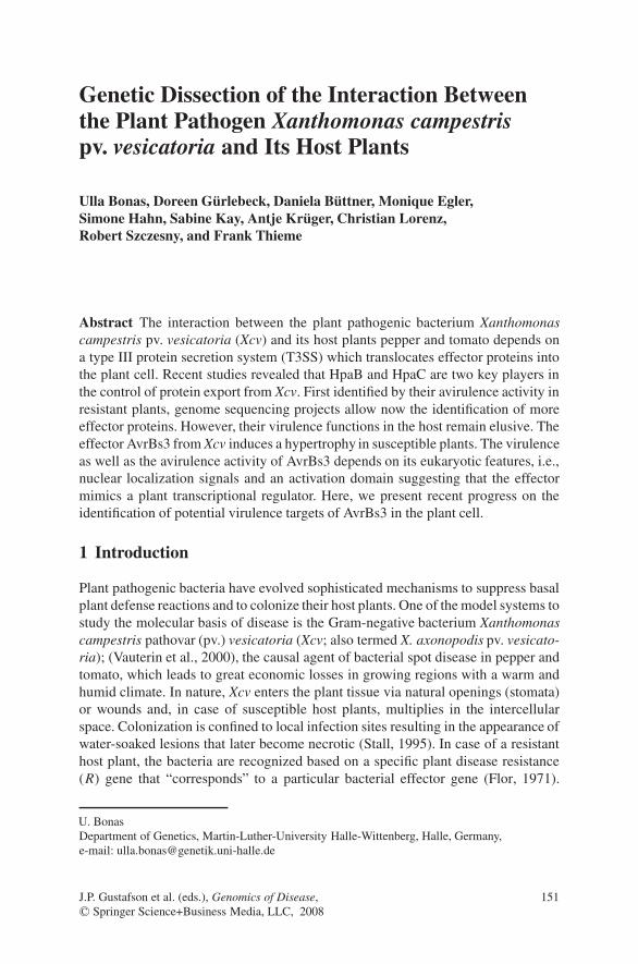
Genetic Dissection of the Interaction Betweenthe Plant Pathogen Xanthomonas campestrispv. vesicatoria and Its Host Plants
Ulla Bonas, Doreen Gurlebeck, Daniela Buttner, Monique Egler,Simone Hahn, Sabine Kay, Antje Kruger, Christian Lorenz,Robert Szczesny, and Frank Thieme
Abstract The interaction between the plant pathogenic bacterium Xanthomonascampestris pv. vesicatoria (Xcv) and its host plants pepper and tomato depends ona type III protein secretion system (T3SS) which translocates effector proteins intothe plant cell. Recent studies revealed that HpaB and HpaC are two key players inthe control of protein export from Xcv. First identified by their avirulence activity inresistant plants, genome sequencing projects allow now the identification of moreeffector proteins. However, their virulence functions in the host remain elusive. Theeffector AvrBs3 from Xcv induces a hypertrophy in susceptible plants. The virulenceas well as the avirulence activity of AvrBs3 depends on its eukaryotic features, i.e.,nuclear localization signals and an activation domain suggesting that the effectormimics a plant transcriptional regulator. Here, we present recent progress on theidentification of potential virulence targets of AvrBs3 in the plant cell.
1 Introduction
Plant pathogenic bacteria have evolved sophisticated mechanisms to suppress basalplant defense reactions and to colonize their host plants. One of the model systems tostudy the molecular basis of disease is the Gram-negative bacterium Xanthomonascampestris pathovar (pv.) vesicatoria (Xcv; also termed X. axonopodis pv. vesicato-ria); (Vauterin et al., 2000), the causal agent of bacterial spot disease in pepper andtomato, which leads to great economic losses in growing regions with a warm andhumid climate. In nature, Xcv enters the plant tissue via natural openings (stomata)or wounds and, in case of susceptible host plants, multiplies in the intercellularspace. Colonization is confined to local infection sites resulting in the appearance ofwater-soaked lesions that later become necrotic (Stall, 1995). In case of a resistanthost plant, the bacteria are recognized based on a specific plant disease resistance(R) gene that “corresponds” to a particular bacterial effector gene (Flor, 1971).
U. BonasDepartment of Genetics, Martin-Luther-University Halle-Wittenberg, Halle, Germany,e-mail: [email protected]
J.P. Gustafson et al. (eds.), Genomics of Disease, 151C© Springer Science+Business Media, LLC, 2008
152 U. Bonas et al.
In most cases that are studied on the molecular level the outcome of resistance isa hypersensitive reaction (HR), a rapid and localized programmed cell death thatrestricts further spread of the pathogen (Klement, 1982).
Recently, the genome sequence of Xcv strain 85–10 has been elucidated. Besidesthe chromosome (5.17 Mb) this strain contains four plasmids 1.8 kb to 183 kb in size(Thieme et al., 2005). Essential for pathogenicity of Xcv is a type III protein secre-tion system (T3SS), which is highly conserved in many Gram-negative plant andanimal pathogenic bacteria and some symbionts (Buttner and Bonas, 2006; Cornelisand Van Gijsegem, 2000; Marie et al., 2001; Tampakaki et al., 2004). Expression ofthe T3SS is induced in the plant and in certain minimal media. The T3SS secretesa small number of proteins into the extra cellular milieu, but most proteins (termed“effectors”) are translocated into the host cell cytosol (Buttner and Bonas, 2002;Gurlebeck et al., 2006) (Fig. 1). To date, more than 20 type III effector proteins are
Fig. 1 Model of type III secretion of Xcv. The T3SS spans both bacterial membranes and is con-nected to an extracellular Hrp pilus and the T3S translocon, which inserts into the plant plasmamembrane. The T3SS injects bacterial effector proteins into the host cell. Secretion of effectors andnon-effectors is controlled by HpaB and HpaC which interact with each other and bring effectorsinto contact with the T3S apparatus (Buttner et al., 2006). Once in the plant cell, effector proteinsmodulate the host cell metabolism to the advantage of the pathogen leading to the formation ofdisease symptoms. Some type III effectors are recognized mediated by corresponding R proteinsleading to the induction of the hypersensitive reaction. CW, cell wall; PM, plasma membrane; IM,inner membrane; OM, outer membrane
Interaction Between Xanthomonas and the Plant 153
known in Xcv (Thieme et al., 2005). Effector proteins probably manipulate host cellprocesses to the need of the pathogen. Key questions studied in our laboratory are(i) regulation of type III secretion, (ii) identification of type III effector proteins andtheir host targets, and (iii) dissection of specific plant disease resistance to Xcv.
The first type III effectors were isolated some 15–20 years ago because oftheir role in plant resistance. These so-called “avirulence (Avr) proteins” weredefined genetically as pathogen determinants that are recognized by resistant hostplants expressing a corresponding resistance gene. Upon recognition, the Avr pro-tein induces plant defense reactions, often manifested as an HR (Keen, 1990;Klement, 1982). Later, mutant studies demonstrated that the primary role of Avrproteins is in pathogen virulence, i.e., avr genes can contribute to bacterial multi-plication in the plant and to symptom development (Gurlebeck et al., 2006). Manynew effectors were identified by bioinformatic analysis of the respective bacterialgenome sequence. Criteria which can be used are conserved features of the pro-moter regions of the corresponding genes. For example, all P. syringae genes fortype III system components and effectors contain an hrp box in their promoter(Xiao and Hutcheson, 1994), whereas many Xcv effector genes carry a PIP-box (seebelow). In addition, many effector proteins can be grouped into families based ontheir predicted function and/or similarity to known effectors. Finally, the presenceof eukaryotic features such as leucine-rich-repeats (LRR) or nuclear localizationsignals (NLS) might be indicative of an effector candidate.
To verify effector candidates their translocation into the plant cell has to bedemonstrated. This can be monitored using a translational fusion of the N-terminalregion, which presumably contains the type III secretion signal, to a suitable reporterprotein. Successfully used are known effector domains, e.g., AvrBs3 that is deletedin its N-terminus but still causes the HR in a resistant plant when expressionwas mediated by Agrobacterium (Szurek et al., 2002). An alternative method isthe so-called “Cya assay” using an adenylate cyclase as reporter (Casper-Lindleyet al., 2002). Based on sequence conservation or predicted enzymatic functions mosttype III effector proteins can be grouped into families. It turned out that the bacteriaoften express effector proteins that use molecular mimicry to fulfill their functionin the plant cell (Gurlebeck et al., 2006). Examples are XopD from Xcv, which is aputative cysteine protease that cleaves SUMOylated proteins (Hotson et al., 2003),and HopPtoD2, a protein phosphatase of P. syringae (Bretz et al., 2003; Espinosaet al., 2003). Another large effector family is the AvrBs3 family which is expressedalmost exclusively in Xanthomonas spp.
2 Results and Discussion
2.1 The T3SS of Xcv
The genes encoding the T3SS of Xcv are localized in a 35.3 kb pathogenicity islandon the bacterial chromosome. The core gene cluster consists of six hrp (hypersensi-tive reaction and pathogenicity) operons which encode the components of the T3SS

154 U. Bonas et al.
and three Hrp-associated proteins (HpaA-C), which are non-essential virulenceproteins (Gurlebeck et al., 2006). Expression of the T3SS is induced in the plantand in certain minimal media and is controlled by two regulators, HrpG and HrpX,which are encoded outside of the hrp region. HrpG, a member of the OmpR familyof transcriptional regulators, is constitutively expressed but needs to be activatedvia an unknown signal from the plant. Active HrpG then induces the transcrip-tion of hrpX, which encodes an AraC-type regulator that in turn controls expres-sion of most genes in the hrp region and genes elsewhere in the genome (for arecent comprehensive review, see Gurlebeck et al., 2006). Most HrpX-regulatedpromoters contain a conserved sequence element, the PIP (plant-inducible promoter;consensus TTCG-N16-TTCG) box, which is specifically bound by HrpX (Koebniket al., 2006). More than 100 hrpG- and hrpX-induced genes have been identifiedby cDNA-AFLP and microarray analyses (Noel et al., 2001; Koebnik et al., 2006;B. Baumgarth, A. Becker, F. Thieme and U. Bonas, unpublished data).
On the bacterial surface the T3SS is extended by the Hrp pilus through whichtranslocon and effector proteins are transported (Weber et al., 2005). In Xcv strain85–10 more than 20 different effector proteins are known (Gurlebeck et al., 2006;Thieme et al., 2005; F. Thieme and U. Bonas, unpublished data).
The role of most effectors in virulence is not clear, because often a deletion of therespective gene does not alter bacterial virulence in the plant (Noel et al., 2003; Noelet al., 2001, 2002; F. Thieme, A. Kruger, A. Urban and U. Bonas, unpublished data).
2.2 Control of T3S by Xcv
How are bacterial proteins recognized by the T3SS? The export signal of mostproteins probably resides in the N-terminal amino acid sequence of the protein. Thesecretion signal is not conserved although there is some amino acid bias. In contrastto proteins secreted by the type II system, the T3S signal is not cleaved (Buttner andBonas, 2006). Recent studies revealed that HpaB and HpaC, which are not secreted,are two key players in the control of type III protein export. Interestingly, there aretwo classes of type III effectors based on their need for HpaB. The latter might deter-mine the hierarchy of protein secretion; however, this intriguing process remainsenigmatic. HpaB is a general T3S chaperone which interacts with HpaC and witheffector proteins and hence directs them to the T3SS (Buttner et al., 2004, 2006).HpaC interacts with HpaB and appears to increase the efficiency of protein export.Once the predicted translocon complex consisting of HrpF and possibly XopA isinserted into the plant plasma membrane effector proteins are injected directly fromthe bacterium into the plant cell cytoplasm (Buttner et al., 2004, 2002; Gurlebecket al., 2006; Noel et al., 2002).
2.3 The AvrBs3 Effector Protein
AvrBs3 is encoded on a self-transmissible plasmid that is present in some strains ofXcv. Xcv strains that express the 122-kDa AvrBs3 protein are specifically recognized
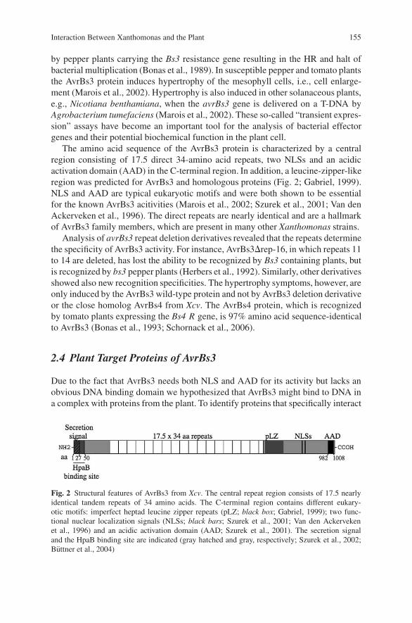
Interaction Between Xanthomonas and the Plant 155
by pepper plants carrying the Bs3 resistance gene resulting in the HR and halt ofbacterial multiplication (Bonas et al., 1989). In susceptible pepper and tomato plantsthe AvrBs3 protein induces hypertrophy of the mesophyll cells, i.e., cell enlarge-ment (Marois et al., 2002). Hypertrophy is also induced in other solanaceous plants,e.g., Nicotiana benthamiana, when the avrBs3 gene is delivered on a T-DNA byAgrobacterium tumefaciens (Marois et al., 2002). These so-called “transient expres-sion” assays have become an important tool for the analysis of bacterial effectorgenes and their potential biochemical function in the plant cell.
The amino acid sequence of the AvrBs3 protein is characterized by a centralregion consisting of 17.5 direct 34-amino acid repeats, two NLSs and an acidicactivation domain (AAD) in the C-terminal region. In addition, a leucine-zipper-likeregion was predicted for AvrBs3 and homologous proteins (Fig. 2; Gabriel, 1999).NLS and AAD are typical eukaryotic motifs and were both shown to be essentialfor the known AvrBs3 acitivities (Marois et al., 2002; Szurek et al., 2001; Van denAckerveken et al., 1996). The direct repeats are nearly identical and are a hallmarkof AvrBs3 family members, which are present in many other Xanthomonas strains.
Analysis of avrBs3 repeat deletion derivatives revealed that the repeats determinethe specificity of AvrBs3 activity. For instance, AvrBs3�rep-16, in which repeats 11to 14 are deleted, has lost the ability to be recognized by Bs3 containing plants, butis recognized by bs3 pepper plants (Herbers et al., 1992). Similarly, other derivativesshowed also new recognition specificities. The hypertrophy symptoms, however, areonly induced by the AvrBs3 wild-type protein and not by AvrBs3 deletion derivativeor the close homolog AvrBs4 from Xcv. The AvrBs4 protein, which is recognizedby tomato plants expressing the Bs4 R gene, is 97% amino acid sequence-identicalto AvrBs3 (Bonas et al., 1993; Schornack et al., 2006).
2.4 Plant Target Proteins of AvrBs3
Due to the fact that AvrBs3 needs both NLS and AAD for its activity but lacks anobvious DNA binding domain we hypothesized that AvrBs3 might bind to DNA ina complex with proteins from the plant. To identify proteins that specifically interact
Fig. 2 Structural features of AvrBs3 from Xcv. The central repeat region consists of 17.5 nearlyidentical tandem repeats of 34 amino acids. The C-terminal region contains different eukary-otic motifs: imperfect heptad leucine zipper repeats (pLZ; black box; Gabriel, 1999); two func-tional nuclear localization signals (NLSs; black bars; Szurek et al., 2001; Van den Ackervekenet al., 1996) and an acidic activation domain (AAD; Szurek et al., 2001). The secretion signaland the HpaB binding site are indicated (gray hatched and gray, respectively; Szurek et al., 2002;Buttner et al., 2004)
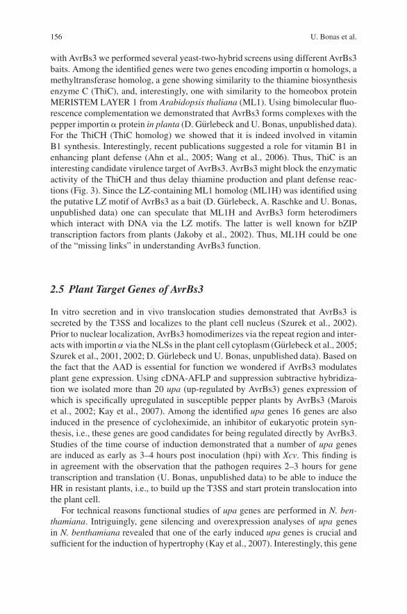
156 U. Bonas et al.
with AvrBs3 we performed several yeast-two-hybrid screens using different AvrBs3baits. Among the identified genes were two genes encoding importin � homologs, amethyltransferase homolog, a gene showing similarity to the thiamine biosynthesisenzyme C (ThiC), and, interestingly, one with similarity to the homeobox proteinMERISTEM LAYER 1 from Arabidopsis thaliana (ML1). Using bimolecular fluo-rescence complementation we demonstrated that AvrBs3 forms complexes with thepepper importin � protein in planta (D. Gurlebeck and U. Bonas, unpublished data).For the ThiCH (ThiC homolog) we showed that it is indeed involved in vitaminB1 synthesis. Interestingly, recent publications suggested a role for vitamin B1 inenhancing plant defense (Ahn et al., 2005; Wang et al., 2006). Thus, ThiC is aninteresting candidate virulence target of AvrBs3. AvrBs3 might block the enzymaticactivity of the ThiCH and thus delay thiamine production and plant defense reac-tions (Fig. 3). Since the LZ-containing ML1 homolog (ML1H) was identified usingthe putative LZ motif of AvrBs3 as a bait (D. Gurlebeck, A. Raschke and U. Bonas,unpublished data) one can speculate that ML1H and AvrBs3 form heterodimerswhich interact with DNA via the LZ motifs. The latter is well known for bZIPtranscription factors from plants (Jakoby et al., 2002). Thus, ML1H could be oneof the “missing links” in understanding AvrBs3 function.
2.5 Plant Target Genes of AvrBs3
In vitro secretion and in vivo translocation studies demonstrated that AvrBs3 issecreted by the T3SS and localizes to the plant cell nucleus (Szurek et al., 2002).Prior to nuclear localization, AvrBs3 homodimerizes via the repeat region and inter-acts with importin α via the NLSs in the plant cell cytoplasm (Gurlebeck et al., 2005;Szurek et al., 2001, 2002; D. Gurlebeck und U. Bonas, unpublished data). Based onthe fact that the AAD is essential for function we wondered if AvrBs3 modulatesplant gene expression. Using cDNA-AFLP and suppression subtractive hybridiza-tion we isolated more than 20 upa (up-regulated by AvrBs3) genes expression ofwhich is specifically upregulated in susceptible pepper plants by AvrBs3 (Maroiset al., 2002; Kay et al., 2007). Among the identified upa genes 16 genes are alsoinduced in the presence of cycloheximide, an inhibitor of eukaryotic protein syn-thesis, i.e., these genes are good candidates for being regulated directly by AvrBs3.Studies of the time course of induction demonstrated that a number of upa genesare induced as early as 3–4 hours post inoculation (hpi) with Xcv. This finding isin agreement with the observation that the pathogen requires 2–3 hours for genetranscription and translation (U. Bonas, unpublished data) to be able to induce theHR in resistant plants, i.e., to build up the T3SS and start protein translocation intothe plant cell.
For technical reasons functional studies of upa genes are performed in N. ben-thamiana. Intriguingly, gene silencing and overexpression analyses of upa genesin N. benthamiana revealed that one of the early induced upa genes is crucial andsufficient for the induction of hypertrophy (Kay et al., 2007). Interestingly, this gene
Interaction Between Xanthomonas and the Plant 157
Fig. 3 Current model of action of AvrBs3. HpaB enhances the secretion and translocation ofAvrBs3 by binding to the N-terminal 50 amino acids of the effector within Xcv (1–2). After injec-tion into the plant cell, AvrBs3 dimerizes in the cytoplasm (3). Importin � bound to importin �binds to the NLS of AvrBs3 (4) and thus mediates the nuclear import of the effector (5). In thenucleus the importin molecules dissociate and are recycled to the cytoplasm of the plant cell (6)so that AvrBs3 can act as transcriptional activator (7). For that AvrBs3 might directly (7a) bind topromoters of upa genes or in a complex with a virulence target protein, e.g., the homeobox proteinML1 homolog (7b). The induction of gene expression leads then to the growth of the plant cell(hypertrophy). Prior to nuclear import, AvrBs3 molecules might transiently interact with a ThiChomolog (ThiCH; 8) to inhibit its enzymatic activity, which could lead to a delay of plant defensereactions
158 U. Bonas et al.
encodes a protein with similarity to transcriptional regulators and thus might be thekey regulator for the induction of the hypertrophy (Fig. 3). It is now interesting toinvestigate if this Upa protein induces the expression of other, later induced upagenes, e.g., genes encoding expansin � or pectate lyases that are probably involvedin the development of hypertrophy (Marois et al., 2002). The model in Fig. 3 sum-marizes the current view on plant targets of the type III effector AvrBs3 from Xcv.
Acknowledgments This work has been supported by grants from the DFG (SFB 648 “Molecularmechanisms of information processing in plants”) and the BMBF (GenoMik) to Ulla Bonas.
References
Ahn, I.P., Kim, S., and Lee, Y.H., 2005, Vitamin B1 functions as an activator of plant diseaseresistance, Plant Physiol. 138:1505–1515.
Bonas, U., Stall, R.E., and Staskawicz, B., 1989, Genetic and structural characterization of theavirulence gene avrBs3 from Xanthomonas campestris pv. vesicatoria, Mol. Gen. Genet. 218:127–136.
Bonas, U., Conrads-Strauch, J., and Balbo, I., 1993, Resistance in tomato to Xanthomonascampestris pv. vesicatoria is determined by alleles of the pepper-specific avirulence geneavrBs3, Mol. Gen. Genet. 238:261–269.
Bretz, J.R., Mock, N.M., Charity, J.C., Zeyad, S., Baker, C.J., and Hutcheson, S.W., 2003, Atranslocated protein tyrosine phosphatase of Pseudomonas syringae pv. tomato DC3000 mod-ulates plant defence response to infection, Mol. Microbiol. 49:389–400.
Buttner, D., and Bonas, U., 2002, Getting across—bacterial type III effector proteins on their wayto the plant cell, EMBO J. 21:5313–5322.
Buttner, D., and Bonas, U., 2006, Who comes first? How plant pathogenic bacteria orchestrate typeIII secretion, Curr. Opin. Microbiol. 9:193–200.
Buttner, D., Nennstiel, D., Klusener, B., and Bonas, U., 2002, Functional analysis of HrpF, a puta-tive type III translocon protein from Xanthomonas campestris pv. vesicatoria, J. Bacteriol.184:2389–2398.
Buttner, D., Gurlebeck, D., Noel, L.D., and Bonas, U., 2004, HpaB from Xanthomonas campestrispv. vesicatoria acts as an exit control protein in type III-dependent protein secretion, Mol.Microbiol. 54:755–768.
Buttner, D., Lorenz, C., Weber, E., and Bonas, U., 2006, Targeting of two effector protein classesto the type III secretion system by a HpaC- and HpaB-dependent protein complex from Xan-thomonas campestris pv. vesicatoria, Mol. Microbiol. 59:513–527.
Casper-Lindley, C., Dahlbeck, D., Clark, E.T., and Staskawicz, B.J., 2002, Direct biochemicalevidence for type III secretion-dependent translocation of the AvrBs2 effector protein into plantcells, Proc. Natl. Acad. Sci. USA 99:8336–8341.
Cornelis, G.R., and Van Gijsegem, F., 2000, Assembly and function of type III secretory systems,Annu. Rev. Microbiol. 54:35–774.
Espinosa, A., Guo, M., Tam, V.C., Fu, Z.Q., and Alfano, J.R., 2003, The Pseudomonas syringaetype III-secreted protein HopPtoD2 possesses protein tyrosine phosphatase activity and sup-presses programmed cell death in plants, Mol. Microbiol. 49:377–387.
Flor, H.H.,1971, Current status of the gene-for-gene concept, Annu. Rev. Phytopathol. 9:275–296.Gabriel, D.W., 1999, The Xanthomonas avr/pth gene family, in G. Stacey and N.T. Keen, eds,
Plant–Microbe Interactions, St. Paul, Minnesota, APS Press, pp. 39–55.Gurlebeck, D., Szurek, B., and Bonas, U., 2005, Dimerization of the bacterial effector protein
AvrBs3 in the plant cell cytoplasm prior to nuclear import, Plant J. 42:175–187.

Interaction Between Xanthomonas and the Plant 159
Gurlebeck, D., Thieme, F., and Bonas, U., 2006, Type III effector proteins from the plant pathogenXanthomonas and their role in the interaction with the host plant, J. Plant Physiol. 163:233–255.
Herbers, K., Conrads-Strauch, J., and Bonas, U., 1992, Race-specificity of plant resistance to bac-terial spot disease determined by repetitive motifs in a bacterial avirulence protein, Nature356:172–174.
Hotson, A., Chosed, R., Shu, H., Orth, K., and Mudgett, M.B., 2003, Xanthomonas type III effectorXopD targets SUMO-conjugated proteins in planta, Mol. Microbiol. 50:377–389.
Jakoby, M., Weisshaar, B., Droge-Laser, W., Vicente-Carbajosa, J., Tiedemann, J., Kroj, T., andParcy, F., 2002, bZIP transcription factors in Arabidopsis, Trends Plant Sci. 7:106–111.
Kay, S., Hahn, S., Marois, E., Hause, G., and Bonas, U., 2007, A bacterial effector as a planttranscription factor and induces a cell size regulator, Science 318:648–351.
Keen, N.T., 1990, Gene-for-gene complementarity in plant–pathogen interactions, Annu. Rev.Genet. 24:447–463.
Klement, Z., 1982, Hypersensitivity, in M.S. Mount and G.H. Lacy, eds, Phytopathogenic Prokary-otes, New York, Academic Press, pp. 149–177.
Koebnik, R., Kruger, A., Thieme, F., Urban, A., and Bonas, U., 2006, Specific binding of theXanthomonas campestris pv. vesicatoria AraC-type transcriptional activator HrpX to plant-inducible promoter boxes, J. Bacteriol. 188:7652–7660.
Marie, C., Broughton, W.J., and Deakin, W.J., 2001, Rhizobium type III secretion systems: legumecharmers or alarmers? Curr. Opin. Plant Biol. 4:336–342.
Marois, E., Van den Ackerveken, G., and Bonas, U., 2002, The Xanthomonas type III effectorprotein AvrBs3 modulates plant gene expression and induces cell hypertrophy in the susceptiblehost, Mol. Plant Microbe Interact. 15:637–646.
Noel, L., Thieme, F., Nennstiel, D., and Bonas, U., 2001, cDNA-AFLP analysis unravels a genome-wide hrpG-regulon in the plant pathogen Xanthomonas campestris pv. vesicatoria, Mol. Micro-biol. 41:1271–1281.
Noel, L., Thieme, F., Nennstiel, D., and Bonas, U., 2002, Two novel type III-secreted proteinsof Xanthomonas campestris pv. vesicatoria are encoded within the hrp pathogenicity island,J. Bacteriol. 184:1340–1348.
Noel, L., Thieme, F., Gabler, J., Buttner, D., and Bonas, U., 2003, XopC and XopJ, two noveltype III effector proteins from Xanthomonas campestris pv. vesicatoria, J. Bacteriol. 185:7092–7102.
Schornack, S., Meyer, A., Romer, P., Jordan, T., and Lahaye, T., 2006, Gene-for-gene-mediatedrecognition of nuclear-targeted AvrBs3-like bacterial effector proteins, J. Plant Physiol.163:256–272.
Stall, R.E., 1995, Xanthomonas campestris pv. vesicatoria, in R.P.S. U.S. Singh, and K. Kohmoto,eds, Pathogenesis and Host-Parasite Specificity in Plant Diseases, Tarrytown, NY, Pergamon,Elsevier Science Inc., pp. 167–184.
Szurek, B., Marois, E., Bonas, U., and Van den Ackerveken, G., 2001, Eukaryotic features of theXanthomonas type III effector AvrBs3: protein domains involved in transcriptional activationand the interaction with nuclear import receptors from pepper, Plant J. 26:523–534.
Szurek, B., Rossier, O., Hause, G., and Bonas, U., 2002, Type III-dependent translocation of theXanthomonas AvrBs3 protein into the plant cell, Mol. Microbiol. 46:13–23.
Tampakaki, A.P., Fadouloglou, V.E., Gazi, A.D., Panopoulos, N.J., and Kokkinidis, M., 2004,Conserved features of type III secretion, Cell. Microbiol. 6:805–816.
Thieme, F., Koebnik, R., Bekel, T., Berger, C., Boch, J., Buttner, D., Caldana, C., Gaigalat, L.,Goesmann, A., Kay, S., Kirchner, O., Lanz, C., Linke, B., McHardy, A.C., Meyer, F.,Mittenhuber, G., Nies, D.H., Niesbach-Klosgen, U., Patschkowski, T., Ruckert, C., Rupp, O.,Schneiker, S., Schuster, S.C., Vorholter, F.J., Weber, E., Puhler, A., Bonas, U., Bartels, D.,and Kaiser, O., 2005, Insights into genome plasticity and pathogenicity of the plant pathogenicbacterium Xanthomonas campestris pv. vesicatoria revealed by the complete genome sequence,J. Bacteriol. 187:7254–7266.

160 U. Bonas et al.
Van den Ackerveken, G., Marois, E., and Bonas, U., 1996, Recognition of the bacterial avirulenceprotein AvrBs3 occurs inside the host plant cell, Cell 87:1307–1316.
Vauterin, L., Rademaker, J., and Swings, J., 2000, Synopsis on the taxonomy of the genus Xan-thomonas, Phytopathology 90:677–682.
Wang, G., Ding, X., Yuan, M., Qiu, D., Li, X., Xu, C., and Wang, S., 2006, Dual function of riceOsDR8 gene in disease resistance and thiamine accumulation, Plant Mol. Biol. 60:437–449.
Weber, E., Ojanen-Reuhs, T., Huguet, E., Hause, G., Romantschuk, M., Korhonen, T.K., Bonas, U.,and Koebnik, R., 2005, The type III-dependent Hrp pilus is required for productive interac-tion of Xanthomonas campestris pv. vesicatoria with pepper host plants, J. Bacteriol. 187:2458–2468.
Xiao, Y., and Hutcheson, S.W., 1994, A single promoter sequence recognized by a newly iden-tified alternate sigma factor directs expression of pathogenicity and host range determinantsin Pseudomonas syringae [published erratum appears in J. Bacteriol. 176:6158], J. Bacteriol.176:3089–3091.
Structure and Function of RXLR Effectorsof Plant Pathogenic Oomycetes
William Morgan, Jorunn Bos, Catherine Bruce, Minkyoung Lee,Hsin-Yen Liu, Sang-Keun Oh, Jing Song, Joe Win, Carolyn Young,and Sophien Kamoun
1 Introduction
A diverse number of plant pathogens, including bacteria, fungi, oomycetes andnematodes, secrete effector proteins to different cellular compartments of theirhosts to modulate plant defense circuitry and enable parasitic colonization (Birchet al., 2006; Chisholm et al., 2006; Kamoun, 2006; O′Connell and Panstruga, 2006).The current paradigm in the study of plant–microbe interactions is that unravel-ing the molecular function of effectors is central to a mechanistic understandingof pathogenicity. Indeed, significant progress has been made in elucidating the viru-lence functions of bacterial effectors and the biochemical activities that enable theseproteins to perturb host defense processes and facilitate pathogenicity (Chisholmet al., 2006). In contrast, little is known about the biology of effectors of eukaryoticplant pathogens. Nonetheless, this area of research is progressing rapidly as illus-trated by the recent identification of effectors from the flax rust and barley powderymildew fungi (Catanzariti et al., 2006; Dodds et al., 2004; Ridout et al., 2006),the oomycetes Phytophthora and Hyaloperonospora (Allen et al., 2004; Armstronget al., 2005; Rehmany et al., 2005; Shan et al., 2004), as well as root-knot nematodes(Huang et al., 2006a; Huang et al., 2006b).
Oomycetes form a distinct group of eukaryotic microorganisms that includessome of the most notorious pathogens of plants (Kamoun, 2003). Research onoomycete effectors has accelerated in recent years due in great part to the avail-ability of resources stemmed from genomics. The emerging picture is complexand fascinating. Oomycetes are now thought to secrete hundreds of effector pro-teins belonging to two classes that target distinct sites in the host plant (Birchet al., 2006; Kamoun, 2006; Tyler et al., 2006). Apoplastic effectors are secretedinto the plant extracellular space, whereas cytoplasmic effectors are translocatedinside the plant cell, where they target different subcellular compartments (Birchet al., 2006; Kamoun, 2006). Several apoplastic effectors have been determined
S. KamounDepartment of Plant Pathology, Ohio State University Ohio Agricultural Research and Develop-ment Center, Wooster, OH, USAe-mail: [email protected]
J.P. Gustafson et al. (eds.), Genomics of Disease, 161C© Springer Science+Business Media, LLC, 2008
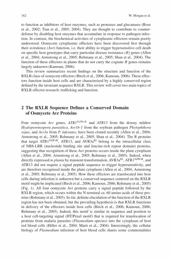
162 W. Morgan et al.
to function as inhibitors of host enzymes, such as proteases and glucanases (Roseet al., 2002; Tian et al., 2005; 2004). They are thought to contribute to counter-defense by disabling host enzymes that accumulate in response to pathogen infec-tion. In contrast, the biochemical activities of cytoplasmic effectors remain poorlyunderstood. Oomycete cytoplasmic effectors have been discovered first throughtheir avirulence (Avr) function, i.e. their ability to trigger hypersensitive cell deathon specific host genotypes that carry particular disease resistance (R) genes (Allenet al., 2004; Armstrong et al., 2005; Rehmany et al., 2005; Shan et al., 2004). Thefunction of these effectors in plants that do not carry the cognate R genes remainslargely unknown (Kamoun, 2006).
This review summarizes recent findings on the structure and function of theRXLR class of oomycete effectors (Birch et al., 2006; Kamoun, 2006). These effec-tors function inside host cells and are characterized by a highly conserved regiondefined by the invariant sequence RXLR. This review will cover two main topics ofRXLR effector research: trafficking and function.
2 The RXLR Sequence Defines a Conserved Domainof Oomycete Avr Proteins
Four oomycete Avr genes, ATR1NdWs B and ATR13 from the downy mildewHyaloperonospora parasitica, Avr1b-1 from the soybean pathogen Phytophthorasojae, and Avr3a from P. infestans, have been cloned recently (Allen et al., 2004;Armstrong et al., 2005; Rehmany et al., 2005; Shan et al., 2004). The R proteinsthat target ATR1NdWsB, ATR13, and AVR3aKI belong to the intracellular classof NBS-LRR (nucleotide binding site and leucine-rich repeat domain) proteins,suggesting that recognition of these Avr proteins occurs inside the plant cytoplasm(Allen et al., 2004; Armstrong et al., 2005; Rehmany et al., 2005). Indeed, whendirectly expressed in planta by transient transformation, AVR3aKI, ATR1NdWsB, andATR13 did not require a signal peptide sequence to trigger hypersensitivity, andare therefore recognized inside the plant cytoplasm (Allen et al., 2004; Armstronget al., 2005; Rehmany et al., 2005). How these effectors are translocated into hostcells during infection is unknown but a conserved sequence centered on the RXLRmotif might be implicated (Birch et al., 2006; Kamoun, 2006; Rehmany et al., 2005)(Fig. 1). All four oomycete Avr proteins carry a signal peptide followed by theRXLR region, which occurs within the N-terminal ca. 60 amino acids of these pro-teins (Rehmany et al., 2005). So far, definite elucidation of the function of the RXLRregion has not been obtained, but the prevailing hypothesis is that RXLR functionsin delivery of the effectors inside host cells (Birch et al., 2006; Kamoun, 2006;Rehmany et al., 2005). Indeed, this motif is similar in sequence and position toa host cell-targeting signal (HT/Pexel motif) that is required for translocation ofproteins from malaria parasites (Plasmodium species) into the cytoplasm of hostred blood cells (Hiller et al., 2004; Marti et al., 2004). Interestingly, the cellularbiology of Plasmodium infection of host blood cells shares some commonalities
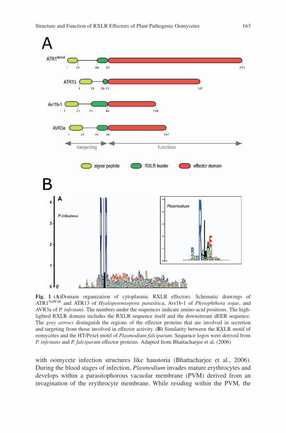
Structure and Function of RXLR Effectors of Plant Pathogenic Oomycetes 163
Fig. 1 (A)Domain organization of cytoplasmic RXLR effectors. Schematic drawings ofATR1NdWsB and ATR13 of Hyaloperonsopora parasitica, Avr1b-1 of Phytophthora sojae, andAVR3a of P. infestans. The numbers under the sequences indicate amino-acid positions. The high-lighted RXLR domain includes the RXLR sequence itself and the downstream dEER sequence.The gray arrows distinguish the regions of the effector proteins that are involved in secretionand targeting from those involved in effector activity. (B) Similarity between the RXLR motif ofoomycetes and the HT/Pexel motif of Plasmodium falciparum. Sequence logos were derived fromP. infestans and P. falciparum effector proteins. Adapted from Bhattacharjee et al. (2006)
with oomycete infection structures like haustoria (Bhattacharjee et al., 2006).During the blood stages of infection, Plasmodium invades mature erythrocytes anddevelops within a parasitophorous vacuolar membrane (PVM) derived from aninvagination of the erythrocyte membrane. While residing within the PVM, the
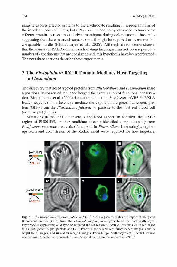
164 W. Morgan et al.
parasite exports effector proteins to the erythrocyte resulting in reprogramming ofthe invaded blood cell. Thus, both Plasmodium and oomycetes need to translocateeffector proteins across a host-derived membrane during colonization of host cellssuggesting that the conserved sequence motif might be required to overcome thiscomparable hurdle (Bhattacharjee et al., 2006). Although direct demonstrationthat the oomycete RXLR domain is a host-targeting signal has not been reported, anumber of experiments that are consistent with this hypothesis have been performed.The next three sections describe these experiments.
3 The Phytophthora RXLR Domain Mediates Host Targetingin Plasmodium
The discovery that host-targeted proteins from Phytophthora and Plasmodium sharea positionally conserved sequence begged the examination of functional conserva-tion. Bhattacharjee et al. (2006) demonstrated that the P. infestans AVR3aKI RXLRleader sequence is sufficient to mediate the export of the green fluorescent pro-tein (GFP) from the Plasmodium falciparum parasite to the host red blood cell(erythrocyte) (Fig. 2).
Mutations in the RXLR consensus abolished export. In addition, the RXLRregion of PH001D5, another candidate effector identified computationally fromP. infestans sequences, was also functional in Plasmodium. Interestingly, regionsupstream and downstream of the RXLR motif were required for host targeting,
Fig. 2 The Phytophthora infestans AVR3a RXLR leader region mediates the export of the greenfluorescent protein (GFP) from the Plasmodium falciparum parasite to the host erythrocyte.Erythrocytes expressing wild-type or mutated RXLR region of AVR3a (residues 21 to 69) fusedto a P. falciparum signal peptide and GFP. Panels ii and v represent fluorescence images, i and ivbright field images, and iii and vi merged images. Parasite (p), erythrocyte (e), Hoechst stainednucleus (blue), scale bar represents 2 �m. Adapted from Bhattacharjee et al. (2006)
Structure and Function of RXLR Effectors of Plant Pathogenic Oomycetes 165
suggesting that the RXLR sequence defines a ca. 30 amino acid leader sequence.Thus the targeting domain extends beyond the RXLR sequence. Consistent withthis view, sequence biases were observed in the regions flanking RXLR, particularlya high rate of E/D residues following RXLR. In summary, these findings suggestthat plant and animal eukaryotic pathogens share similar secretory signals for effec-tor delivery into host cells (Bhattacharjee et al., 2006). At this stage, it is unclearwhether this functional conservation reflects conserved export machinery betweenthese divergent eukaryotes (see below for further discussion on this topic). However,the functional analogy with bacterial type III secretion system is remarkable. In bothcases, export signals are highly conserved across unrelated pathogen species but theeffector secretome that is delivered to the host is highly divergent (Bhattacharjeeet al., 2006).
4 The RXLR Domain Is Not Required for Effector Activities
The view that the RXLR region functions in translocation into host cells sug-gests that RXLR effectors are organized into two main functional domains(Kamoun, 2006) (Fig. 1). The first domain encompassing the signal peptide andRXLR leader functions in secretion and targeting, while the remaining C-terminaldomain carries the effector activity. This model predicts that the RXLR regionshould not be required for activity when the effector is expressed inside host cells.Indeed, Bos et al. (2006) recently showed that mutation of P. infestans AVR3aKI
RXLR sequence into AXAA did not interfere with induction of R3a hypersensitivitywhen the protein is directly expressed in N. benthamiana leaves. In fact, deletionanalyses of AVR3aKI showed that the C-terminal 75-amino acid, which excludesthe RXLR region but includes the two polymorphic amino acids K80 and I103 thatare mutated in the nonfunctional allele, was sufficient for avirulence function whenexpressed directly inside plant cells (Bos et al., 2006). These findings are consistentwith the view that the N-terminal region of AVR3aKI and other RXLR effectors isinvolved in secretion and targeting but is not required for effector activity.
5 The C-Terminal Region of RXLR Effectors Is Typically MorePolymorphic than the Signal Peptide and RXLR Domains
Higher levels of polymorphisms, particularly non-synonymous substitutions, havebeen detected in the C-terminal regions of RXLR effectors than in the signal pep-tide and RXLR leader region. For example, the C-terminal regions of H. parasit-ica ATR1 and ATR13 exhibit higher levels of non-synonymous polymorphismsthan the N-terminal regions, suggesting that the effector activity is localized tothe C-terminal domain (Allen et al., 2004; Rehmany et al., 2005). Also, two outof the three polymorphic residues between the two Avr3a alleles of P. infestans,amino acids 80 and 103, are located in the C-terminal effector domain (Armstronget al., 2005). The observation that these effectors are under diversifying selectionis consistent with the view that pathogen effectors with avirulence functions are
166 W. Morgan et al.
caught in a coevolutionary arms race with host factors, particularly their cognate Rgenes (Allen et al., 2004). Signatures of selection are expected in regions involved ineffector activity rather than targeting. Thus, the observation that the RXLR domainis less polymorphic than the C-terminal region of RXLR proteins is consistent withthe view that it is not exposed to selection pressure by host defenses and that itfunctions in targeting.
6 Can RXLR Effectors Enter Host Plants in the Absenceof the Pathogen?
Whether RXLR effectors require pathogen machinery or structures (e.g., haustoria)to enter plant cells is currently unclear. In our laboratory, we have failed so farto trigger R3a hypersensitivity using recombinant AVR3aKI proteins (unpublisheddata). Shan et al. (2004) reported that infiltration of P. sojae RXLR effector Avr1b-1, produced in Pichia pastoris, into Rps1b soybean leaves resulted in cell death.However, it is unknown (although highly likely) whether Rps1b is a cytoplasmicprotein and confirmation of Avr1b-1 activity by in planta expression inside soybeancells has not been reported yet. Clearly, it would be highly informative to test Avr1b-1 recombinant proteins mutated in the RXLR sequence using the infiltration assayof Shan et al. (2004).
The issue of host translocation is also relevant to cytoplasmic effectors of fungalpathogens. In their work with Avr proteins of the flax rust fungus Melampsora lini,Catanzariti et al. (2006) concluded that the avirulence protein AvrM enters plantcells in the absence of the pathogen. AvrM is a secreted protein with a canonicalsignal peptide that is recognized inside flax cells in an M gene dependent man-ner. In addition, Agrobacterium tumefaciens-mediated expression of a full-lengthAvrM construct in flax plants carrying the M gene resulted in hypersensitive celldeath. Interestingly, similar constructs carrying the ER retention sequence HDELwere unable to trigger cell death. The authors concluded from this experimentthat AvrM first exits plant cells into the apoplast and then reenters through anunknown process (Catanzariti et al., 2006). We are not in absolute agreement withthis interpretation and are of the opinion that this experiment is inconclusive inevaluating whether AvrM can enter plant cells in the absence of the pathogen. Anequally plausible explanation is that the signal peptide carrying AvrM is translo-cated back from the ER into the cytosol through the well-established retrogradetransport pathway (Brandizzi et al., 2003; Di Cola et al., 2001). In such case, theHDEL motif would retain the protein into the ER and prevent retrograde translo-cation. In summary, although the experiments of Catanzariti et al. (2006) suggestthat AvrM needs to shuttle through the ER, perhaps to achieve maturation, infil-tration experiments with purified AvrM proteins are necessary to obtain conclusiveevidence as to the ability of this protein to enter host cells in the absence of thepathogen. Such data has been obtained conclusively with ToxA, a host-selectivetoxin produced by the plant pathogenic fungus Pyrenophora tritici-repentis. Man-ning and Ciuffetti (2005) demonstrated elegantly that a recombinant ToxA protein
Structure and Function of RXLR Effectors of Plant Pathogenic Oomycetes 167
tagged with GFP internalizes inside sensitive wheat cells, where it localizes tocytoplasmic compartments and chloroplasts. This implies that effector proteins cantranslocate inside host cells in the absence of pathogen machinery, probably usinghost-derived machinery. Host-translocation of ToxA is likely to occur via receptor-mediated endocytosis, and might implicate the RGD sequence that is identical toa cell adhesion motif of mammalian extracellular matrix proteins (Manning andCiuffetti, 2005).
In our own work with AVR3aKI, we noted that high levels of expression of a full-length construct resulted in R3a-dependent hypersensitivity (Bos et al., 2006). Theseexperiments are not particularly informative since they could be equally explainedby (1) mis-targeting of the protein to the cytoplasm or (2) secretion of AVR3aKI
followed by re-entry of the protein inside the plant cell resulting in R3a activation.Also, in other experiments, attempts to exploit this full-length AVR3aKI to evaluatethe role of the RXLR motif in translocation inside host cells were inconclusivesince a RXLR to AXAA mutant retained the ability to elicit R3a response (Boset al., 2006).
7 A Model for RXLR Effector Delivery into the Host
The process through which the RXLR leader sequence might function in host tar-geting of effectors remains unknown. Many key questions still need to be addressed.What is the export machinery of effectors in eukaryotic pathogens? Is it derived fromthe pathogen or are the effectors exploiting host transport systems? Is the exportmachinery conserved between oomycetes and Plasmodium? Despite these persist-ing questions, some reasonable assumptions about the translocation process can bemade. For instance, it seems sensible to break down the export process into two steps(Bhattacharjee et al., 2006). First, the effectors are secreted outside the pathogencell through the general secretory pathway and endoplasmic reticulum (ER) typesignal peptides. Then, the secreted effectors are transported across a host-derivedmembrane, most likely the haustorial membrane, via the RXLR leader. In the GFPexport experiments of Bhattacharjee et al. (2006), constructs with a mutated RXLRsequence accumulated GFP outside the parasite but within the parasitophorous vac-uole suggesting that the main function of the RXLR leader consists of transportacross this host-derived membrane.
Here, we propose a model for effector delivery (illustrated in Fig. 3). The modelis based on the fact that mechanisms of protein transport across membranes followcanonical processes involving recurrent themes (Wickner and Schekman, 2005). Wepropose that host translocation of the effectors via the RXLR leader involves at leasta RXLR leader binding protein, one or more chaperones, and a translocon, whichcould be of either pathogen or plant origin. Translocation into host cells initiateswith the RXLR binding protein recruiting secreted mature effectors in coordinationwith the chaperones. The effector cargo is then transferred to a translocon embeddedin the haustorial membrane, and is then released across the membrane into the plantcytosol. The chaperones are important for maintaining the folding state of the trans-ported effectors both prior and after transit through the translocon.
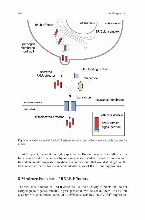
168 W. Morgan et al.
Fig. 3 A hypothetical model for RXLR effector secretion and delivery into host cells (see text fordetails)
At this point, this model is highly speculative. But our purpose is to outline a use-ful working model to serve as a hypothesis generator and help guide future research.Indeed, the model suggests immediate research avenues that would shed light on thetranslocation process, for instance the identification of RXLR binding proteins.
8 Virulence Functions of RXLR Effectors
The virulence function of RXLR effectors, i.e. their activity in plants that do notcarry cognate R genes, remains in great part unknown. Bos et al. (2006), in an effortto assign virulence-related functions to AVR3a, discovered that AVR3aKI suppresses
Structure and Function of RXLR Effectors of Plant Pathogenic Oomycetes 169
the hypersensitive cell death induced by the major P. infestans elicitin INF1 in Nico-tiana benthamiana. The cell death suppression activity of AVR3aKI exhibited somelevel of specificity. AVR3aKI did not suppress the cell death induced by other P.infestans effectors, like PiNPP1 and CRN2, which elicit distinct and antagonisticcell death signaling pathways compared to INF1 (Kanneganti et al., 2006). Thebiological relevance of this activity of AVR3aKI is unknown but could be signifi-cant considering that suppression of innate immunity is a widespread function ofplant pathogen effectors, particularly the type III secretion system (TTSS) effectorsof bacterial phytopathogens (Espinosa and Alfano 2004). AVR3aKI could interferewith the avirulence activity of INF1 or other unidentified effectors that trigger hyper-sensitivity using similar pathways as INF1 (Bos et al., 2006). Future work is neededto clarify these issues and determine whether cell death suppression is a commonfunction among RXLR effectors.
9 Outlook: Too Many Effectors, Too Little Time
Considering that five oomycete species, H. parasitica, P. capsici, P. infestans, P.ramorum, and P. sojae, are undergoing genome sequencing and annotation, we aremoving rapidly toward genome-wide catalogues of RXLR effectors. Already it isevident that the RXLR effector secretome of plant pathogenic oomycetes is muchmore complex than expected, with perhaps several hundred proteins dedicated tomanipulating host cells (Kamoun, 2006; Tyler et al., 2006). Recent estimates ofRXLR effectors based on the draft genome sequences of P. sojae and P. ramorumrange from ca. 150 to 350 per genome (Bhattacharjee et al., 2006; Tyler et al., 2006).The task of tackling the study of so many effectors is daunting. One of the challengesis to establish “effectoromics” approaches, or global studies of effector functionand activity. Ultimately, comprehensive understanding of RXLR effector activitiesand the perturbations they cause in plants is a precondition for understanding themolecular basis of oomycete pathogenesis and disease.
Acknowledgments We thank collaborators and colleagues in the oomycete field for useful discus-sions and insight. This work was supported by NSF Plant Genome Research Program grant DBI-0211659. Salaries and research support were provided, in part, by State and Federal Funds appro-priated to the Ohio Agricultural Research and Development Center, The Ohio State University.
References
Allen, R.L., Bittner-Eddy, P.D., Grenville-Briggs, L.J., Meitz, J.C., Rehmany, A.P., Rose, L.E.,and Beynon, J.L., 2004, Host–parasite coevolutionary conflict between Arabidopsis and downymildew, Science 306:1957–1960.
Armstrong, M.R., Whisson, S.C., Pritchard, L., Bos, J.I., Venter, E., Avrova, A.O., Rehmany, A.P.,Bohme, U., Brooks, K., Cherevach, I., Hamlin, N., White, B., Fraser, A., Lord, A., Quail, M.A.,Churcher, C., Hall, N., Berriman, M., Huang, S., Kamoun, S., Beynon, J.L., and Birch, P.R.,2005, An ancestral oomycete locus contains late blight avirulence gene Avr3a, encoding

170 W. Morgan et al.
a protein that is recognized in the host cytoplasm, Proc. Natl. Acad. Sci. USA 102:7766–7771.
Bhattacharjee, S., Hiller, N.L., Liolios, K., Win, J., Kanneganti, T.D., Young, C., Kamoun, S., andHaldar, K., 2006. The malarial host-targeting signal is conserved in the Irish potato faminepathogen. PLoS Pathog. 2:e50.
Birch, P.R., Rehmany, A.P., Pritchard, L., Kamoun, S., and Beynon, J.L., 2006, Trafficking arms:oomycete effectors enter host plant cells, Trends Microbiol. 14:8–11.
Bos, J.I., Kanneganti, T.D., Young, C., Cakir, C., Huitema, E., Win, J., Armstrong, M.R.,Birch, P.R., and Kamoun, S., 2006, The C-terminal half of Phytophthora infestans RXLR effec-tor AVR3a is sufficient to trigger R3a-mediated hypersensitivity and suppress INF1-inducedcell death in Nicotiana benthamiana, Plant J. 48:165–176.
Brandizzi, F., Hanton, S., DaSilva, L.L., Boevink, P., Evans, D., Oparka, K., Denecke, J., andHawes, C., 2003, ER quality control can lead to retrograde transport from the ER lumen to thecytosol and the nucleoplasm in plants, Plant J. 34:269–281.
Catanzariti, A.M., Dodds, P.N., Lawrence, G.J., Ayliffe, M.A., and Ellis, J.G., 2006, Haustoriallyexpressed secreted proteins from flax rust are highly enriched for avirulence elicitors, PlantCell 18:243–256.
Chisholm, S.T., Coaker, G., Day, B., and Staskawicz, B.J., 2006, Host–microbe interactions: shap-ing the evolution of the plant immune response, Cell 124:803–814.
Di Cola, A., Frigerio, L., Lord, J.M., Ceriotti, A., and Roberts, L.M., 2001, Ricin A chain withoutits partner B chain is degraded after retrotranslocation from the endoplasmic reticulum to thecytosol in plant cells, Proc. Natl. Acad. Sci. USA 98:14726–14731.
Dodds, P.N., Lawrence, G.J., Catanzariti, A.M., Ayliffe, M.A., and Ellis, J.G., 2004, The Melamp-sora lini AvrL567 avirulence genes are expressed in haustoria and their products are recognizedinside plant cells, Plant Cell 16:755–768.
Espinosa, A., and Alfano, J.R., 2004, Disabling surveillance: bacterial type III secretion systemeffectors that suppress innate immunity, Cell Microbiol. 6:1027–1040.
Hiller, N.L., Bhattacharjee, S., van Ooij, C., Liolios, K., Harrison, T., Lopez-Estrano, C., and Hal-dar, K., 2004, A host-targeting signal in virulence proteins reveals a secretome in malarialinfection, Science 306:1934–1937.
Huang, G., Allen, R., Davis, E.L., Baum, T.J., and Hussey, R.S., 2006a, Engineering broad root-knot resistance in transgenic plants by RNAi silencing of a conserved and essential root-knotnematode parasitism gene, Proc. Natl. Acad. Sci. USA, 103:14302–14306.
Huang, G., Dong, R., Allen, R., Davis, E.L., Baum, T.J., and Hussey, R.S., 2006b, A root-knotnematode secretory peptide functions as a ligand for a plant transcription factor, Mol. PlantMicrobe Interact. 19:463–470.
Kamoun, S., 2003, Molecular genetics of pathogenic oomycetes, Eukaryotic Cell 2:191–199.Kamoun, S., 2006, A Catalogue of the Effector Secretome of Plant Pathogenic Oomycetes, Annu.
Rev. Phytopathol. 44:41–60.Kanneganti, T.D., Huitema, E., Cakir, C., and Kamoun, S., 2006, Synergistic interactions of the
plant cell death pathways induced by Phytophthora infestans Nepl-like protein PiNPP1.1 andINF1 elicitin, Mol. Plant Microbe Interact. 19:854–863.
Manning, V.A., and Ciuffetti, L.M., 2005, Localization of Ptr ToxA Produced by Pyrenophoratritici-repentis Reveals Protein Import into Wheat Mesophyll Cells, Plant Cell 17:3203–3212.
Marti, M., Good, R.T., Rug, M., Knuepfer, E., and Cowman, A.F., 2004, Targeting malaria viru-lence and remodeling proteins to the host erythrocyte, Science 306:1930–1933.
O′Connell, R.J., and Panstruga, R., 2006, Tete a tete inside a plant cell: establishing compatibilitybetween plants and biotrophic fungi and oomycetes, New Phytol. 171:699–718.
Rehmany, A.P., Gordon, A., Rose, L.E., Allen, R.L., Armstrong, M.R., Whisson, S.C., Kamoun,S., Tyler, B.M., Birch, P.R., and Beynon, J.L., 2005, Differential recognition of highly divergentdowny mildew avirulence gene alleles by RPP1 resistance genes from two Arabidopsis lines,Plant Cell 17:1839–1850.
Structure and Function of RXLR Effectors of Plant Pathogenic Oomycetes 171
Ridout, C.J., Skamnioti, P., Porritt, O., Sacristan, S., Jones, J.D., and Brown, J.K., 2006, Multipleavirulence paralogues in cereal powdery mildew fungi may contribute to parasite fitness anddefeat of plant resistance, Plant Cell 18:2402–2414.
Rose, J.K., Ham, K.S., Darvill, A.G., and Albersheim, P., 2002, Molecular cloning and character-ization of glucanase inhibitor proteins: coevolution of a counterdefense mechanism by plantpathogens, Plant Cell 14:1329–1345.
Shan, W., Cao, M., Leung, D., and Tyler, B.M., 2004, The Avr1b locus of Phytophthora sojaeencodes an elicitor and a regulator required for avirulence on soybean plants carrying resistancegene Rps1b, Mol. Plant Microbe Interact. 17:394–403.
Tian, M., Benedetti, B., and Kamoun, S., 2005, A Second Kazal-like protease inhibitor fromPhytophthora infestans inhibits and interacts with the apoplastic pathogenesis-related proteaseP69B of tomato, Plant Physiol. 138:1785–1793.
Tian, M., Huitema, E., da Cunha, L., Torto-Alalibo, T., and Kamoun, S., 2004, A Kazal-like extra-cellular serine protease inhibitor from Phytophthora infestans targets the tomato pathogenesis-related protease P69B, J. Biol. Chem. 279:26370–26377.
Tyler, B.M., Tripathy, S., Zhang, X., Dehal, P., Jiang, R.H., et al., 2006, Phytophthora genomesequences uncover evolutionary origins and mechanisms of pathogenesis, Science 313:1261–1266.
Wickner, W., and Schekman, R., 2005, Protein translocation across biological membranes, Science310:1452–1456.
The Biotrophic Phase of Ustilago maydis:Novel Determinants for Compatibility
Thomas Brefort, Kerstin Schipper, Gunther Dohlemann,and Regine Kahmann
Abstract The basidiomycete fungus Ustilago maydis establishes a biotrophicrelationship with its host plant maize which is maintained throughout diseasedevelopment. Recent insights from the genome sequence have revealed that thisinteraction is largely governed by a set of novel secreted proteins that are only foundin U. maydis. Many of the respective genes are clustered and appear co-regulatedduring late stages of pathogenesis. Mutants in most of these gene clusters arrestdevelopment at distinct stages, suggesting that the secreted proteins fulfill discretefunctions in the interaction with the host. One of the cluster mutants, however,displays increased virulence suggesting that it is not in the interest of U. maydis touse its full potential as a pathogen. In this chapter we will review these findings andplace them in perspective for a comprehensive understanding of biotrophy.
1 Introduction
Smut diseases of grasses are caused by basidiomycetes of the order Ustilaginales.Because most smuts develop in kernel tissue they cause considerable yield losses.Smut fungi can initiate an infection only in their dikaryotic stage, which is gener-ated by mating of compatible haploid cells. Smuts differ in host range as well asthe site of symptom development, with symptoms in most cases being restrictedto the inflorescences. One notable exception to this is Ustilago maydis, a pathogeninfecting maize, which causes tumors on all aerial parts of the plant. Maize seedlingsare efficiently infected and symptom development can be scored in less than aweek after inoculation. This is one of the reasons why the U. maydis/maize sys-tem has become the model for plant pathogenic basidiomycetes. The other assetsof the system are the availability of efficient techniques for gene replacement,regulatable promoters for analyzing gene function, as well as advanced cytol-ogy and live cell imaging (Basse and Steinberg, 2004). The 20.5 Mb genome of
R. KahmannMax Planck Institute for Terrestrial Microbiology, Department Organismic Interactions, Karl-von-Frisch-Strasse, D-35043 Marburg, Germanye-mail: [email protected]
J.P. Gustafson et al. (eds.), Genomics of Disease, 173C© Springer Science+Business Media, LLC, 2008
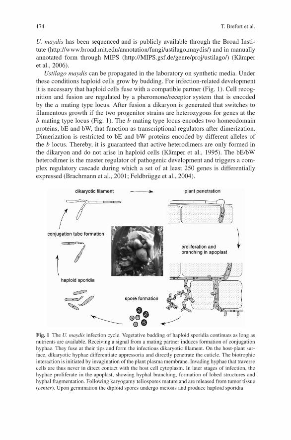
174 T. Brefort et al.
U. maydis has been sequenced and is publicly available through the Broad Insti-tute (http://www.broad.mit.edu/annotation/fungi/ustilago maydis/) and in manuallyannotated form through MIPS (http://MIPS.gsf.de/genre/proj/ustilago/) (Kamperet al., 2006).
Ustilago maydis can be propagated in the laboratory on synthetic media. Underthese conditions haploid cells grow by budding. For infection-related developmentit is necessary that haploid cells fuse with a compatible partner (Fig. 1). Cell recog-nition and fusion are regulated by a pheromone/receptor system that is encodedby the a mating type locus. After fusion a dikaryon is generated that switches tofilamentous growth if the two progenitor strains are heterozygous for genes at theb mating type locus (Fig. 1). The b mating type locus encodes two homeodomainproteins, bE and bW, that function as transcriptional regulators after dimerization.Dimerization is restricted to bE and bW proteins encoded by different alleles ofthe b locus. Thereby, it is guaranteed that active heterodimers are only formed inthe dikaryon and do not arise in haploid cells (Kamper et al., 1995). The bE/bWheterodimer is the master regulator of pathogenic development and triggers a com-plex regulatory cascade during which a set of at least 250 genes is differentiallyexpressed (Brachmann et al., 2001; Feldbrugge et al., 2004).
Fig. 1 The U. maydis infection cycle. Vegetative budding of haploid sporidia continues as long asnutrients are available. Receiving a signal from a mating partner induces formation of conjugationhyphae. They fuse at their tips and form the infectious dikaryotic filament. On the host-plant sur-face, dikaryotic hyphae differentiate appressoria and directly penetrate the cuticle. The biotrophicinteraction is initiated by invagination of the plant plasma membrane. Invading hyphae that traversecells are thus never in direct contact with the host cell cytoplasm. In later stages of infection, thehyphae proliferate in the apoplast, showing hyphal branching, formation of lobed structures andhyphal fragmentation. Following karyogamy teliospores mature and are released from tumor tissue(center). Upon germination the diploid spores undergo meiosis and produce haploid sporidia
The Biotrophic Phase of Ustilago maydis 175
In nature, mating occurs on the plant surface. The resulting filamentous dikaryonforms appressoria (Fig. 1), which allow direct penetration of the plant cuticle ina process presumably aided by lytic enzymes (Schirawski et al., 2005). Duringthis stage hyphae show tip growth but do not enlarge their cytoplasm and do notundergo nuclear division. As a result such hyphae contain cytoplasm only in thetip compartment while the older sections are highly vacuolated, and are sealed offby regularly spaced septa (Fig. 1). The dikaryon is arrested in the G2 phase of thecell cycle (Flor-Parra et al., 2006) and this block is released only after penetra-tion (Banuett and Herskowitz, 1996). The biz1 gene is among the genes indirectlyregulated through the bE/bW heterodimer. Biz1 encodes a zinc finger transcriptionfactor and triggers the G2 cell cycle arrest through down-regulation of the cyclinclb1. biz1 mutants are unable to cause disease symptoms and this was attributedto a 10-fold reduction in appressorium formation as well as arrested growth imme-diately after penetration. This illustrates that the cell cycle arrest is necessary forproper infection-related development (Flor-Parra et al., 2006). Between 1 and 2 daysafter penetration the growth mode changes, mitotic divisions occur, and branchingis observed (Fig. 1). How this is triggered is one of the unresolved mysteries. Acrucial role for coordinated mitotic divisions during dikaryotic growth was recentlyascribed to clp1, a direct target of the bE/bW heterodimer (Scherer et al., 2006),that is related to clp1 in coprinus cinerea (Inada et al., 2001) . clp1 mutants have nodiscernible phenotype during axenic culture and mate like wild type cells. However,after plant penetration they arrest growth prior to the first mitotic division, fail todevelop branch-like structures (Fig. 1), and are nonpathogenic. Conspicuously, thebranch-like structures were shown to arise in the most apical cell of the dikaryotichyphae at a point where new septa are later formed. By using strains with fluores-cently labeled nuclei it was subsequently shown that one of the four mitotic nucleibecomes trapped in these structures after septum formation and moves from thereto the subapical cell presumably through a septal pore (Scherer et al., 2006). Thisprocess is closely related to clamp formation in other basidiomycetes, but lacksthe feature that clamps fuse with the subapical cell to donate the trapped nucleus(Brown and Casselton, 2002). This illustrates that processes regulated through thebE/bW heterodimer are at least to some extent conserved in related fungal species.In addition, these findings clearly show that the regulatory function of the bE/bWheterodimer is required during extended stages of pathogenic development.
Around 5 days after infection by U. maydis the plant reacts by forming tumor-like structures (Fig. 1), which are most likely induced by the fungus throughinterference with phytohormone signaling. In these structures the dikaryotic hyphaeshow massive proliferation in the apoplast, round up, their nuclei fuse and hyphalfragmentation occurs followed by the formation of darkly pigmented spores (Fig. 1)(Snetselaar and Mims, 1994; Banuett and Herskowitz, 1996). U. maydis is abiotrophic pathogen, i.e. host cells stay alive throughout the course of an infectionuntil very late when tumor tissue dries up. This leads to cell wall rupture andrelease of the spores (Snetselaar and Mims, 1994; Banuett and Herskowitz, 1996).Biotrophy is established after an invagination of the host plasma membrane at thesite of fungal entry. This membrane surrounds the fungal hyphae and prevents
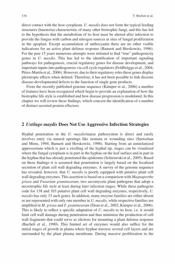
176 T. Brefort et al.
direct contact with the host cytoplasm. U. maydis does not form the typical feedingstructures (haustoria) characteristic of many other biotrophic fungi, and this has ledto the hypothesis that the metabolism of its host must be altered after infection toprovide the fungus with carbon and nitrogen sources at sites of fungal proliferationin the apoplast. Except accumulation of anthocyanin there are no other visibleindications for an active plant defense response (Banuett and Herskowitz, 1996).For the past 15 years numerous attempts were initiated to find “true” pathogenicitygenes in U. maydis. This has led to the identification of important signalingpathways for pathogenesis, crucial regulatory genes for disease development, andimportant inputs into pathogenesis via cell cycle regulators (Feldbrugge et al., 2004;Perez-Martın et al., 2006). However, due to their regulatory roles these genes displaypleiotropic effects when deleted. Therefore, it has not been possible to link discretedisease-developmental defects to the function of single gene products.
From the recently published genome sequence (Kamper et al., 2006) a numberof features have been recognized which begin to provide an explanation of how thebiotrophic life style is established and how disease progression is modulated. In thischapter we will review these findings, which concern the identification of a numberof distinct secreted protein effectors.
2 Ustilago maydis Does Not Use Aggressive Infection Strategies
Hyphal penetration in the U. maydis/maize pathosystem is direct and rarelyinvolves entry via natural openings like stomata or wounding sites (Snetselaarand Mims, 1994; Banuett and Herskowitz, 1996). Starting from an unmelanizedappressorium which is just a swelling of the hyphal tip, stages can be visualizedwhere the fungal cytoplasm is in part in the hyphae on the leaf surface and in part inthe hyphae that has already penetrated the epidermis (Schirawski et al., 2005). Basedon these findings it is assumed that penetration is largely based on the localizedsecretion of plant cell wall degrading enzymes. A survey of the genome sequencehas revealed, however, that U. maydis is poorly equipped with putative plant cellwall degrading enzymes. This assertion is based on a comparison with Magnaporthegrisea and Fusarium graminearum, two ascomycete plant pathogens that adopt anecrotrophic life style at least during later infection stages. While these pathogenscode for 138 and 103 putative plant cell wall degrading enzymes, respectively, U.maydis has only 33 such genes. In addition, many enzyme families are either absentor are represented with only one member in U. maydis, while respective families areamplified in M. grisea and F. graminearum (Dean et al., 2002; Kamper et al., 2006).This is likely to reflect a specific adaptation of U. maydis to its host, i.e. it wouldlimit cell wall damage during penetration and thus minimize the production of cellwall fragments that could serve as elicitors for mounting a plant defense response(Bucheli et al., 1990). This limited set of enzymes would also suffice for theinitial stages of growth in planta where hyphae traverse several cell layers and aresurrounded by the plant plasma membrane. During massive proliferation in the

The Biotrophic Phase of Ustilago maydis 177
apoplast at later stages of the infection the hyphae are often branched, sometimeswith extensions reaching into plant cells. The plant cells appear enlarged and areoften separated from their neighboring cells while their plasma membranes arestill intact (Snetselaar and Mims, 1994). During these stages the fungal hyphaeare imbedded in a mucilaginous material of unknown composition. We considerit likely that this material acts as a shield against antimicrobial defenses by thehost. In future, it will be interesting to elucidate which of the secreted plant cellwall degrading enzymes are actually needed during penetration and which arerequired at the stage of tumor formation. In this regard it is of interest that it hasrecently been accomplished to induce appressorium formation on artificial surfaces(Mendoza-Mendoza and Kahmann, unpublished). Transcription profiles of thisstage in comparison to the tumor stage is likely to provide important insights intowhich plant cell wall degrading enzymes play a role during these discrete stages ofdevelopment.
3 Ustilago maydis Regulates its Interaction with the Hostvia a Set of Novel Secreted Protein Effectors
While the number of U. maydis genes encoding secreted plant cell wall degrad-ing enzymes is small, the number of genes encoding secreted proteins of unknownfunction is unexpectedly high: of the 426 proteins predicted to be secreted by bothSignalP and ProtComp programs, 298 are without predicted function and of these198 are only found in U. maydis (Kamper et al., 2006). This prompted a closer look,which revealed that 72 genes coding for secreted proteins are arranged in 12 geneclusters (Kamper et al., 2006). Clusters are defined by at least three consecutivegenes encoding secreted proteins. Within the gene clusters small gene families con-sisting of two to five genes were frequent. Through microarray analysis in which theexpression of these genes was compared in tumor tissue and in a haploid strain it wasrecognized that most of the clustered genes are significantly upregulated in tumortissue (Kamper et al., 2006). Based on these results deletion mutants were generatedfor all 12 gene clusters. Five of the respective mutants were significantly alteredin virulence (Fig. 2). The virulence phenotypes ranged from a mere reduction ofpathogenicity symptoms (clusters 6A and 10A), to dramatically reduced virulence(cluster 19A), complete failure to cause disease (cluster 5B) to increased virulence(cluster 2A) (Fig. 2). A microscopic analysis revealed that cluster 5B mutant developlike wild type strains on the plant surface and form appressoria. However, theyspecifically arrest development immediately after penetration. Cluster 19A mutantsare indistinguishable from wild type during the early stages of infection-relateddevelopment. They infect plants and proliferate, but do not induce the formationof tumors. For mutants in clusters 2A, 6A, and 10A it has not yet been determinedat which stages of pathogenic development they are affected (Kamper et al., 2006).In seven cases the cluster deletions did not cause a phenotype. However, as relatedgenes were found elsewhere in the genome for four of these cluster genes, it is likely
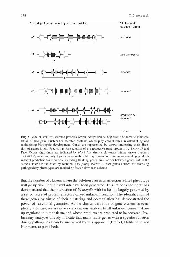
178 T. Brefort et al.
Fig. 2 Gene clusters for secreted proteins govern compatibility. Left panel: Schematic represen-tation of five gene clusters for secreted proteins which play crucial roles in establishing andmaintaining biotrophic development. Genes are represented by arrows indicating their direc-tion of transcription. Predictions for secretion of the respective gene products by SIGNALP andPROTCOMP algorithms are indicated by black line frames. Asterisks within arrows denote aTARGETP prediction only. Open arrows with light gray frames indicate genes encoding productswithout prediction for secretion, including flanking genes. Similarities between genes within thesame cluster are indicated by identical grey filling shades. Cluster genes deleted for assessingpathogenicity phenotypes are marked by lines below each scheme
that the number of clusters where the deletion causes an infection related phenotypewill go up when double mutants have been generated. This set of experiments hasdemonstrated that the interaction of U. maydis with its host is largely governed bya set of secreted protein effectors of yet unknown function. The identification ofthese genes by virtue of their clustering and co-regulation has demonstrated thepower of functional genomics. As the chosen definition of gene clusters is com-pletely arbitrary, we are now extending our analysis to all unknown genes that areup-regulated in tumor tissue and whose products are predicted to be secreted. Pre-liminary analyses already indicate that many more genes with a specific functionduring pathogenesis can be uncovered by this approach (Brefort, Dohlemann andKahmann, unpublished).
The Biotrophic Phase of Ustilago maydis 179
4 Discussion and Outlook
The identification of the U. maydis gene clusters encoding secreted proteins specif-ically required during pathogenesis raises a number of interesting questions andperspectives. In particular, it would be important to know which gene(s) from theclusters are responsible for the observed phenotype. Will it be possible to link theobserved phenotype to individual genes (or gene families) or will there be coopera-tivity in function especially in the larger clusters? Where are the effectors localizedand what is their function in the respective compartments? It is generally believedthat biotrophic pathogens have to suppress plant defense responses and redirect hostmetabolism to the site of infection. In the U. maydis system there is the additionalfeature of tumor induction, which may require changes in phytohormone signalingin the host. Based on insights from bacterial pathogens that use type III secretionsystems to translocate proteins directly into host cells (Mudgett, 2005), it is gener-ally assumed that other types of pathogens have also developed the means to directproteins from the pathogen to the host. Recently it was shown that a conservedpentameric sequence (Pexel motif, RXLX(E/Q)) located within 60 amino acidsdownstream of the signal sequence is responsible for trafficking into host cells inmalaria parasites (Tonkin et al., 2006). For a group of plant pathogenic microbes, theoomycetes, which are related to brown algae and diatoms, it was shown recently thatseveral avirulence gene products function inside host cells. These proteins, whichare unrelated by primary sequence also display a conserved RxLR motif locatedwithin 30 amino acids downstream of the signal sequence (Birch et al., 2006).Hundreds of additional genes harboring this motif were identified in oomycetegenome sequences, and these are therefore likely effectors for shaping the inter-action with the respective plant host. Recently evidence has been provided thatthese host-targeting signals are equivalent in function across species (Bhattacharjeeet al., 2006). Fungal plant pathogens do not code for proteins with such a motif.Nevertheless, it was demonstrated that several avirulence gene products of fungalpathogens elicit a hypersensitive response when expressed in the cytoplasm of plantcells. This is taken as evidence that these proteins are also translocated to the hostand interact with their cognate cytoplasmically located receptors (Jia et al., 2000;Dodds et al., 2004; Catanzariti et al., 2006). In the bean rust fungus system ahaustoria-specifically expressed protein (Rtp1) was shown to accumulate in the hostcytoplasm (Kemen et al., 2005). This demonstrates that these fungal pathogens areable to translocate proteins to host cells via an as yet unknown mechanism. Onthese grounds we speculate that some of the secreted effectors of U. maydis mayalso be targeted to the host. As most fungal pathogens where protein transloca-tion to the host has been shown/inferred are obligate parasites and cannot be trans-formed the U. maydis system may provide an attractive resource for gene/functionanalysis.
Another pertinent question is why the effectors identified in the U. maydis systemappear to be U. maydis specific. Based on preliminary analyses in Sporisorium reil-ianum, a pathogen closely related to U. maydis, it was recently shown that at leastsome of the clustered genes are conserved (Schirawski, Schoning and Kahmann,
180 T. Brefort et al.
unpublished). Thus, the assertion that these genes are classified as being U. maydisspecific is likely to change with more genome sequences becoming available.
These studies begin to provide an understanding of how a biotrophic fungusmodulates its interaction with its host. The goals for the future will lie in elucidatingwhere the now identified secreted proteins function and which processes they target.By starting out with distinct pathogenesis-related phenotypes this follow-up work isexpected to illuminate the molecular basis of biotrophic interactions.
References
Banuett, F., and Herskowitz, I., 1996, Discrete developmental stages during teliospore formationin the corn smut fungus, Ustilago maydis, Development 122:2965–2976.
Basse, C., and Steinberg, G., 2004, Ustilago maydis, model system for analysis of the molecularbasis of fungal pathogenicity, Mol. Plant. Pathol. 5:83–92.
Bhattacharjee, S., Hiller, N.L., Liolios, K., Win, J., Kanneganti, T., Young, C., Kamoun, S., andHaldar, K., 2006, The malarial host-targeting signal is conserved in the Irish potato faminepathogen, PloS Pathogens 2:453–465.
Birch, P.R.J., Rehmany, A.P., Pritchard, L., Kamoun, S., and Beynon, J.L., 2006, Trafficing arms:oomycete effectors enter host plant cells, Trends Microbiol. 14:8–11.
Brachmann, A., Weinzierl, G., Kamper, J., and Kahmann, R., 2001, Identification of genes in thebW/bE regulatory cascade in Ustilago maydis, Mol. Microbiol. 42:1047–1063.
Brown, J.B., and Casselton, L.A., 2002, Mating in mushrooms: Increasing the changes but pro-longing the affair. Trends Genet. 17:393–399.
Bucheli, P., Doares, S.H., Albersheim, P., Darvill, A., 1990, Host–pathogen interactions. XXXVI.Partial purification and characterization of heat–labile molecules secreted by the rice blastpathogen that solubilize plant cell wall fragments that kill plant cells, Physiol. Mol. Plant.Pathol. 1:159–173.
Catanzariti, A.M., Dodds, P.N., Lawrence, G.J., 2006, Haustorially expressed secreted proteinsfrom flax rust are highly enriched for avirulence elicitors, Plant Cell 18:243–256.
Dean, R.A., Talbot, N.J., Ebbole, D.J., Farman, M.L., Mitchell, T.K. et al., 2002, The genomesequence of the rice blast fungus Magnaporthe grisea. Nature 434:980–986.
Dodds, P.N., Lawrence G.J., Catanzariti, A.M., Ayliffe, M.A., and Ellis, J.G., 2004, The Melamp-sora lini AvrL567 avirulence genes are expressed in haustoria and their products are recognizedinside plant cells, Plant Cell 16:755–768.
Feldbrugge, M., Kamper, J., Steinberg, G., and Kahmann, R., 2004, Regulation of mating andpathogenic development in Ustilago maydis. Curr. Opin. Microbiol. 7:666–672.
Flor-Parra, I., Vranes, M., Kamper, J., and Perez-Martın, J., 2006, Biz1, a zinc finger proteinrequired for plant invasion by Ustilago maydis, regulates the levels of a mitotic cycle, PlantCell 18:2369–2387.
Inada, K., Morimoto, Y., Arima, T., Murata, Y., and Kamada T., 2001, The clp1 gene of themushroom Coprinus cinereus is essential for A-regulated sexual development, Genetics 157:133–140.
Jia, Y., McAdams, S., Bryan, G.T., Hershey, H.P., and Valent, B., 2000, Direct interaction of resis-tant gene and avirulence gene products confers rice blast resistance, EMBO J. 19:4004–4014.
Kamper, J., Reichmann, M., Romeis, T., Bolker, M., and Kahmann, R., 1995, Multiallelic recog-nition: nonself-dependent dimerization of the bE and bW homeodomain proteins in Ustilagomaydis, Cell 81:73–83.
Kamper, J., Kahmann, R., Bolker, M., Ma, L.-J., Brefort, T., et al., 2006, Living in pretend har-mony: insights from the genome of the biotrophic fungal plant pathogen Ustilago maydis,Nature 444:97–101.

The Biotrophic Phase of Ustilago maydis 181
Kemen, E., Kemen A.C., Rafiqui, M., Hempel, U., Mendgen, K., Hahn, M., and Voegele, R.T.,2005, Identification of proteins from rust fungi transferred from haustoria into infected plantcells, MPMI 18:1130–1139.
Mudgett, M.B., 2005, New insights to the function of phytopathogenic bacterial type III effectorsin plants. Annu Rev Plant Biol. 56:509–531.
Perez-Martın, J., Castillo-Lluva S., Sgarlata, C., Flor-Parra, I., Mielnichuk, N., Torreblanca, J., andCarbo, N., 2006, Pathocycles: Ustilago maydis as a model to study the relationships betweencell cycle and virulence in pathogenic fungi, Mol. Genet. Genomics 276:211–229.
Scherer, M., Heimel, K., Starke, V., and Kamper, J., 2006, The Clp1 protein is required for clampformation and pathogenic development of Ustilago maydis, Plant Cell 18:2388–2401.
Schirawski, J., Bohnert, H.U., Steinberg, G., Snetselaar, K., Adamikowa, L., and Kahmann, R.,2005, Endoplasmic reticulum glucosidase II is required for pathogenicity of Ustilago maydis,Plant Cell 17:3532–3543.
Snetselaar, K.M., and Mims, C.W., 1994, Light and electron microscopy of Ustilago maydishyphae in maize, Mycol. Res. 98:347–355.
Tonkin, C.J., Pearce, J.A., McFadden, G.I., and Cowman, A.F., 2006, Protein targeting to desti-nations of the secretory pathway in the malaria parasite Plasmodium falciparum, Curr. Opin.Microbiol. 9:381–387.
Virulence Evolution in Malaria
M.J. Mackinnon
Abstract One evolutionary theory of why some pathogens kill their host (i.e. arevirulent) is that they need to extract host resources in order to transmit to new hosts.We have tested this theory in malaria and find it to be a likely explanation for themaintenance of this parasite’s virulence in nature.
1 A Hypothesis for Pathogen Virulence
Pathogens that kill their host lose their source of ongoing survival and transmission.So why are they virulent? This question captured the attention of evolutionary biolo-gists in the early 1980s: until then, it was generally accepted that, given enough evo-lutionary time, all parasites would evolve to be non-harmful to their hosts. However,this is clearly not true: many ancient host–parasite associations are still problematicfor both host and pathogen. Furthermore, throughout human history, infectious dis-eases have remained a major cause of mortality. So what is it that maintains thepathogen’s virulence in nature? If we knew the answer to this question, would thishelp us design vaccines and other control measures that might drive the pathogentowards lower virulence (Williams and Nesse, 1991; Dieckmann et al., 2002)?
Throughout this chapter, the term “virulence” is used generally to describe levelsof morbidity or mortality: for modelling purposes, it is more strictly defined as therate of host mortality induced by the pathogen. It is not replication rate or trans-missibility or persistence – these, as it turns out, are correlates of virulence, but notvirulence per se.
One answer to the question of why pathogens sometimes kill their hosts is thatvirulence is a mistake by the pathogen. For example, a mutant pathogen may havea short-term competitive advantage within a host, but is unable to transmit: alterna-tively, a pathogen may end up in the wrong tissue, also leading to no transmission.
M.J. MackinnonDepartment of Pathology, University of Cambridge, Tennis Court Road, Cambridge CB2 1QP andKEMRI-Wellcome Trust Research Programme, Centre for Geographic Medicine Research, Kilifi80108, Kenyae-mail: [email protected]
J.P. Gustafson et al. (eds.), Genomics of Disease, 183C© Springer Science+Business Media, LLC, 2008
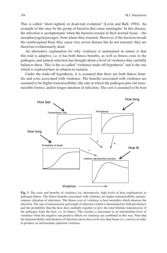
184 M.J. Mackinnon
This is called “short-sighted, or dead-end evolution” (Levin and Bull, 1994). Anexample of this may be the group of bacteria that cause meningitis. In this disease,the infection is asymptomatic when the bacteria remain in their normal tissue – thenasopharyngeal passages, from where they transmit. However, if the bacteria invadethe cerebrospinal fluid, they cause very severe disease but do not transmit: they aretherefore evolutionarily dead.
An alternative explanation for why virulence is maintained in nature is thatthis trait is adaptive, i.e. it has both fitness benefits, as well as fitness costs to thepathogen, and natural selection has brought about a level of virulence that carefullybalances these. This is the so-called “virulence trade-off hypothesis” and is the onewhich is explored here in relation to malaria.
Under the trade-off hypothesis, it is assumed that there are both fitness bene-fits and costs associated with virulence. The benefits associated with virulence areassumed to be higher transmissibility (the rate at which the pathogen puts out trans-missible forms), and/or longer duration of infection. The cost is assumed to be host
How fastHow long
How long
How fit
Transmission rate
Lifetime reproductive success
Virulence
++
–
Persistence
Probability of death
Fig. 1 The costs and benefits of virulence (or, alternatively, high levels of host exploitation) topathogen fitness. The fitness benefits associated with virulence are higher transmissibility and per-sistence (duration of infection). The fitness cost of virulence is host mortality which shortens theinfection. The rate of transmission and length of infection (which is determined by both persistenceand the probability that the host dies) multiply together to give the total lifetime transmission ofthe pathogen from the host, i.e. its fitness. This reaches a maximum at an intermediate level ofvirulence when the negative and positive effects on virulence are combined in this way. Note thatthe transmissibility and duration of infection curves have to be less than linear (i.e. convex) in orderto produce an intermediate optimum virulence
Virulence Evolution in Malaria 185
death because, for most pathogens, when the host dies, transmission ceases. Thetotal transmission per lifetime of the pathogen (i.e. one infection length), which isdirectly related to its fitness, is the rate of transmission multiplied by the length ofthe infection. Thus, if death shortens the infection, the parasite’s fitness is decreased.Thus, the pathogen is playing a dangerous game: it is trying to maximize transmis-sibility and infection length while also keeping its host alive. This, in principle, canlead to the evolution of an intermediate level of virulence, which balances the costsand benefits of virulence to the pathogen’s fitness (Fig. 1).
Although mooted earlier (Topley, 1919; Levin and Pimentel, 1981), the trade-off idea only really took hold when Anderson and May (1982) applied it to thecase of myxomatosis in rabbits. The myxoma virus was extremely virulent whenreleased into rabbit populations in Australia and Britain in the early 1950s, but overthe next decade, the virus evolved to an intermediate and stable level of virulence(Fenner and Ratcliffe, 1965). Anderson and May (1982) and Massad (1987) showedthat this was because, although the more virulent strains of the virus had longerinfections and higher transmissibility, their risk of killing the host was high enoughto outweigh these transmission benefits. Therefore the virus evolved to a lower levelof virulence where the transmission costs of mortality balanced the transmissionbenefits of longer infections and higher transmissibility.
2 Malaria
The myxomatosis example is somewhat artificial because the released virus was notfound naturally in the rabbit populations and so had not co-evolved with its host.Its relevance to human disease is therefore questionable (Ebert and Bull, 2003).Unfortunately, there are few data in any host–pathogen system, medically relevantor not, that can be used to support or refute the trade-off hypothesis. Examplesof pathogens where positive relationships between virulence, transmissibility andpersistence (hereon termed V–T–P), as assumed by the hypothesis, have been foundinclude trypanosomes in mice (Diffley et al., 1987; Turner et al., 1995; Masumuet al., 2006a; Masumu et al., 2006b), microsporidia in Daphnia (Ebert, 1994; Ebertand Mangin, 1997), bacteria in Daphnia (Jensen et al., 2006), Sarcosystis (an api-complexan protozoa) in rats (JSkel et al., 2001), schistosomes in mice (Davieset al., 2001), bacteria in mice (Greenwood et al., 1936), and phage in bacteria(Messenger et al., 1999). Examples that directly illustrate the cost of host deathto lifetime transmission are even more difficult to find (Jensen et al., 2006). Thislack of empirical data, particularly for medically relevant pathogens, spurred us toformally test the hypothesis for a major pathogen of humans – the malaria parasite.
The causative agent of the most pathogenic form of human malaria, Plasmodiumfalciparum, is a typical microparasite, i.e. it replicates rapidly within its host, as domany viral, bacterial and other protozoal pathogens. Unlike viruses and bacteria,however, the malaria parasite has a separate transmission stage form in its life cyclecalled gametocyte. Gametocytes, the sexual form of the parasite, are differentiated
186 M.J. Mackinnon
from the asexual, replicating blood-stage forms. Gametocytes do not themselvesreplicate, and thus are seen at low densities in the bloodstream. But, as they are theonly forms that survive in the mosquito vector, their mass production is critical tothe pathogen’s transmission.
2.1 Mouse Malaria
We began by testing the assumptions of the trade-off hypothesis using the mousemalaria model, Plasmodium chabaudi, as an experimental model. Using parasiteclones obtained from their natural host in the wild (Beale et al., 1978) to infectgroups of inbred mice in the laboratory, we measured the transmissibility, viru-lence and persistence of these clones and the genetic (i.e. across-clone) relation-ships among these traits (Mackinnon and Read, 1999a, 2003; Ferguson et al., 2003).Our experiments showed that clones that had higher levels of virulence had highertransmissibility (both production of gametocytes and infectivity to mosquitoes) andcleared their infections more slowly in the absence of host death (Fig. 2) (Fergusonand Read, 2002; Mackinnon and Read, 1999a, b, 2003). Virulent parasites werealso those that generated highest parasite densities and thus exploited more of thehost’s resources (red cells). Thus the V–T–P relationships assumed by the trade-offhypothesis were supported in this experimental model. Furthermore, if the host died,the total amount of gametocytes produced during the infection (i.e. the potentialtransmission) was less than if the host lived (Fig. 2e), thus demonstrating a clearcost of host mortality to the parasite’s fitness (Mackinnon et al., 2002). We foundthe V–T–P relationships to be qualitatively robust to host genotype (Mackinnonet al., 2002), host sex (Mackinnon and Read, 2004a), serial passage (Mackinnon andRead, 1999b, 2004a; Mackinnon et al., 2002), mosquito passage (Mackinnon andRead, 2004a; Mackinnon et al., 2005) and, importantly, to levels of host immunity(Mackinnon and Read, 2003, 2004a). Immunity acted to reduce all three traits –transmissibility, virulence and persistence – in line with that expected from the rela-tionships among these traits in naive hosts. In other words, the parasite lines thatreplicated well, transmitted faster, lasted longer and caused more morbidity wheninfecting naive hosts also did so in semi-immune hosts, but just at a lower level.
2.2 Human Malaria
We next turned our attention to human malaria, asking the question as to whetherthe relationships we observed in our mouse model in the laboratory also existedin human malaria in its natural environment. Using data from a large longitudinalcross-sectional population survey in Nigeria (the Garki project), it was observed thatvirulence, persistence and transmissibility were positively correlated when assessedacross the whole population (Fig. 3). In these data, however, the relationships areundoubtedly driven by age-acquired immunity, i.e. by levels of host defence rather

Virulence Evolution in Malaria 187
Survived Died
2345
Virulence (minimum red blood celldensity, no. × 109/ml)
–5.0
–4.5
–4.0
–3.5
–3.0
Tra
nsm
issi
bilit
y (
log 10
ave
rage
no.
ga
met
ocyt
es p
er r
ed b
lood
cel
l)
(a) (b)
(c) (d)
(e)
Virulence (minimum red blood celldensity, no. × 109/ml)
0.55
0.45
0.35
0.25
0.152345
Rat
e of
rec
over
y (lo
g 10no
. p
aras
ites
x 10
9 /ml p
er d
ay)
1
Viru
lenc
e (m
inim
um r
ed b
lood
cel
lde
nsity
, no.
× 1
09 /ml)
Maximum asexual parasitaemia(proportion of cells infected)
0.0 0.1 0.2 0.3 0.4 0.5
5
4
3
2
P < 0.05
40
30
20
10
0
Tot
al n
o. g
amet
ocyt
es in
the
infe
ctio
n (n
o. x
106 /m
l)
Virulence (minimum red blood celldensity, no. × 109/ml)
Infe
ctiv
ity to
mos
quito
es (
%)
1234
40
30
20
10
0
Fig. 2 Virulence–transmissibility–persistence relationships in the mouse model of malaria, P.chabaudi. Each point is the average value of groups of mice (n = 5–10) infected with one parasiteclone: thus a line fitted to the points would represent the parasite genetic relationship among thetraits. Filled symbols indicate naive mice and open symbols indicate mice made semi-immuneby previous infection and drug clearance. Virulence was measured as the mouse’s minimum redblood cell density reached during the infection. It was shown to positively relate to transmissibilityas measured by (a) daily average gametocyte density and (b) infectivity to mosquitoes on 2–4 daysduring the peak of gametocyte production. (c) Recovery rate, which is inversely related to infec-tion length, was measured as the rate at which the infection declined after peak parasitemia andwas negatively related to virulence, as expected. (d) Virulence was positively related to maximumparasitemia, an indicator of host exploitation. Clones broadly retained their rankings for all traitswhen infecting naıve vs. immunised mice (Pearson correlations across treatments of 0.59–0.66for the four traits used here.) In the experiments described in panels (a–d), very few mice diedand so a virulence cost to transmission was not observable. However, in another experiment usingless resistant host genotypes, mortality was high (23%) and was shown to severely reduce thetotal number of gametocytes produced by the infection, shown in (e). Data were reproduced fromMackinnon et al. (2002) and Mackinnon and Read (2003)
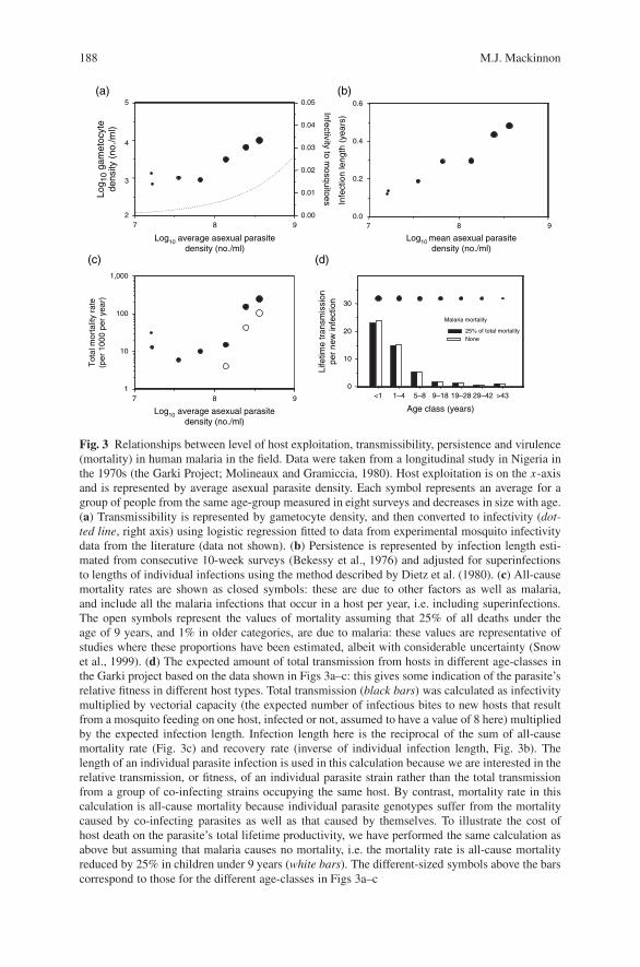
188 M.J. Mackinnon
Log10 average asexual parasitedensity (no./ml)
Log10 mean asexual parasitedensity (no./ml)
9872
3
4
5 0.05 0.6
0.4
0.2
0.0
0.04
0.03
0.02
0.01
0.00
Infectivityto m
osquitoes
(a) (b)
(d)
987
Infe
ctio
n le
ngth
(ye
ars)
0
10
20
30
>439–18 19–28 29–425–8
Age class (years)
1–4<1
Malaria mortality
25% of total mortalityNone
Life
time
tran
smis
sion
per
new
infe
ctio
n
Log 1
0 ga
met
ocyt
ede
nsity
(no
./ml)
1,000
100
10
(c)
1
Tot
al m
orta
lity
rate
(per
100
0 pe
r ye
ar)
987
Log10 average asexual parasitedensity (no./ml)
Fig. 3 Relationships between level of host exploitation, transmissibility, persistence and virulence(mortality) in human malaria in the field. Data were taken from a longitudinal study in Nigeria inthe 1970s (the Garki Project; Molineaux and Gramiccia, 1980). Host exploitation is on the x-axisand is represented by average asexual parasite density. Each symbol represents an average for agroup of people from the same age-group measured in eight surveys and decreases in size with age.(a) Transmissibility is represented by gametocyte density, and then converted to infectivity (dot-ted line, right axis) using logistic regression fitted to data from experimental mosquito infectivitydata from the literature (data not shown). (b) Persistence is represented by infection length esti-mated from consecutive 10-week surveys (Bekessy et al., 1976) and adjusted for superinfectionsto lengths of individual infections using the method described by Dietz et al. (1980). (c) All-causemortality rates are shown as closed symbols: these are due to other factors as well as malaria,and include all the malaria infections that occur in a host per year, i.e. including superinfections.The open symbols represent the values of mortality assuming that 25% of all deaths under theage of 9 years, and 1% in older categories, are due to malaria: these values are representative ofstudies where these proportions have been estimated, albeit with considerable uncertainty (Snowet al., 1999). (d) The expected amount of total transmission from hosts in different age-classes inthe Garki project based on the data shown in Figs 3a–c: this gives some indication of the parasite’srelative fitness in different host types. Total transmission (black bars) was calculated as infectivitymultiplied by vectorial capacity (the expected number of infectious bites to new hosts that resultfrom a mosquito feeding on one host, infected or not, assumed to have a value of 8 here) multipliedby the expected infection length. Infection length here is the reciprocal of the sum of all-causemortality rate (Fig. 3c) and recovery rate (inverse of individual infection length, Fig. 3b). Thelength of an individual parasite infection is used in this calculation because we are interested in therelative transmission, or fitness, of an individual parasite strain rather than the total transmissionfrom a group of co-infecting strains occupying the same host. By contrast, mortality rate in thiscalculation is all-cause mortality because individual parasite genotypes suffer from the mortalitycaused by co-infecting parasites as well as that caused by themselves. To illustrate the cost ofhost death on the parasite’s total lifetime productivity, we have performed the same calculation asabove but assuming that malaria causes no mortality, i.e. the mortality rate is all-cause mortalityreduced by 25% in children under 9 years (white bars). The different-sized symbols above the barscorrespond to those for the different age-classes in Figs 3a–c
Virulence Evolution in Malaria 189
than by parasite variation in virulence. They do not reflect the parasite V–T–Pgenetic correlations observed in mice in a uniform host type, and therefore do notdirectly support the assumptions of the trade-off model. Nevertheless, the humandata are consistent with the general pattern of higher parasite densities giving riseto more transmission forms, longer infections and being associated with highermortality, with all of these being reduced by immunity in an apparently straight-forward way. Thus these phenotypic relationships from the field data, which coverthe entire spectrum of immunity (and host and parasite genetics), may well besimple extensions of the intrinsic biological links among V–T–P that are rootedin the parasite’s ability to replicate. In other words, a pathogen’s ability to repli-cate in the face of host defence mechanisms is correlated to its ability to trans-mit to new hosts, and hence its fitness. Thus host immunity favours more virulentparasites.
The other requirement for the trade-off hypothesis to be accepted as applicableto human malaria is a direct demonstration of the cost to total transmission of hostdeath. To investigate this, we calculated the expected lifetime transmission fromeach host assuming that death did or did not occur (Fig. 3d). The penalties to totaltransmission were around 5% in young children. Whether this cost is sufficient toput the brake on virulence evolution, as supposed by the trade-off hypothesis, is notyet known. This requires an analysis of both the benefits, as well as the costs, ofhigher virulence, and this at the parasite genetic level: we are not yet able to do this.At the moment however, it seems plausible, at least, that death in children imposessome selection against higher virulence.
The analysis of total lifetime transmission (Fig. 3d) also reveals that adults trans-mit around 20 times less than children, but suffer little death. The latter means thatmore virulent parasites, which can “get away with it” in these protected hosts, maybe maintained in the parasite population. Thus, while these more virulent parasitesmay help the parasite transmit from the semi-immune hosts, they would do so at theexpense of the children. Furthermore, if the shape of the trade-off is as we assume(Fig. 1), parasites in such hosts have higher selection coefficients per unit increasein virulence and would therefore be more strongly selected, thus putting furtherpressure on increased virulence. On the other hand, adult hosts contribute muchless to the total transmission population, and so this source of selection may nothave a significant impact. The question is, then, what will happen to virulence if weeffectively turned all children into adults by immunizing with a (as yet hypothetical)malaria vaccine?
2.3 Consequences of Malaria Vaccination
The data so far indicate that in malaria the common factor underlying the V–T–Prelationships is asexual parasite density. We reasoned, therefore, that any controlmeasures that brought about a reduction in asexual parasite densities, such as anasexual stage, anti-replication vaccine, would bring about evolution for increasedintrinsic virulence because of its consequences to transmissibility, persistence and
190 M.J. Mackinnon
host mortality (Gandon et al., 2001). If this evolution were to happen, the unpro-tected (e.g. unvaccinated) people in the population would then be exposed to a morevirulent parasite and their risk of death would be higher than before. On the otherhand, it might be argued, the vaccine, if used widely, would protect many morepeople from disease so that the population-wide reduction in mortality may welloutweigh the increased mortality among the unvaccinated few. Thus, as is often thecase in vaccination programmes, even without evolution, the benefits to the majorityof the population have to be weighed against the risk to a few individuals. Therefore,to determine the benefit to the whole population of vaccination allowing for an evo-lutionary response in the parasite’s virulence, we modelled the case of virulence evo-lution under an imperfect asexual stage malaria vaccine incorporating the feedbacksbetween the epidemiology (force of infection) and the parasite evolution (Gandonet al., 2001). Parameter values were chosen to mimic an area of high year roundmalaria transmission such as central Kenya or Tanzania. As predicted, the parasiteevolved higher virulence under vaccination so that the case fatality rate amongstunvaccinated naive hosts was higher than if evolution had not occurred. Moreover,the total mortality across the whole population was also higher, especially when thevaccine was given to a moderate fraction of the population. Thus, all the expectedbenefits of vaccination were eroded by parasite evolution.
We also modelled other types of vaccines or devices that block infection (e.g.liver stage vaccines), or stop it transmitting from humans to mosquitoes (e.g. bed-nets), but that do not act directly on the asexual stage parasites and hence the V–T–P relationships. These control measures were predicted to have no impact or, asexplained below, could even lead to a decrease in virulence (Gandon et al., 2001).Thus, priority should be given to control measures that prevent pathogens fromentering or leaving the host rather than measures that directly target the infectionitself. In malaria, the former would include bednets, vector control and liver-stagevaccines (unless they also reduce multiplication rates): the latter includes asexual-stage vaccines, anti-toxin vaccines and drug use. Thus, in the face of a large potentialfor pathogen evolution, the hygiene principle would appear to be an excellent oneto follow (where possible) for evolutionary reasons as well as for its more obviousepidemiological applications.
In nature, malaria infections are frequently genetically diverse, raising the ques-tion of the relative fitness of virulent and avirulent strains when present in the samehost. Theoreticians have envisaged a range of possible outcomes, and hence effectson virulence evolution (reviewed in Read and Taylor 2001). In the P. chabaudimodel, more virulent clones have a competitive advantage: they are able to competi-tively suppress or even exclude less virulent clones during the acute phase of mixed-clone infections (de Roode et al., 2005a). The competitive outcome is reflected inrelative gametocyte production (Wargo et al., in preparation) and hence transmissionto mosquitoes (de Roode et al., 2005b). The competitive outcome is qualitativelyrobust to host genotype (de Roode et al., 2004) and host immune status (Raberget al., 2006).
The theoretical conclusion that using devices that prevent infection or blocktransmission will lead to reduced virulence results from the lower force of
Virulence Evolution in Malaria 191
infection caused by the control programme. This leads to lower levels of co-infection or super-infection and hence a lower level of competition among parasitesoccupying the same host. The reduction in virulence due to reduced competitioncan be understood by taking the view of a resident parasite that is either oustedby (super-infection) or forced to share with (co-infection) another parasite type. Inthe case of super-infection, the resident parasite has its infection shortened by theinvading parasite: the parasite then compensates by evolving a higher level of hostexploitation, or intrinsic virulence, up to the point when the benefits are balancedby the costs. In the case of co-infection, the risk of mortality for the residentparasite is increased by the presence of another parasite, thus increasing the costs,or the infection of a less virulent genotype is curtailed by that of a more virulentgenotype. This is especially so where virulence is associated with competitiveability. Whether the latter is true is still being worked out, but the data from the P.chabaudi-mouse model so far suggest that it is (Read and Taylor, 2001; De Roodeet al., 2004; De Roode et al., 2005a,b; Raberg et al., 2006). Thus so far, we believethat reducing the force of infection through vaccination will relieve the amountof within-host competition on the parasite and hence selection pressure on highvirulence.
As we cannot test the vaccination hypothesis in the field until it is too late, wewould ideally test it in the laboratory by experimental evolution. However, it isextremely difficult in the laboratory to repeatedly transmit multiple parasite linesof malaria through mosquitoes for the entire period of their transmissibility: it isalso unethical to allow host mortality to act as the selecting agent. Therefore, wedesigned an experiment to test one component of the vaccine hypothesis, namelythat immunity selects for higher rates of host exploitation (regardless of the trans-mission consequences). This was done by transferring a standard number of par-asites between mice every 7 days for 20 passages, thus bypassing the mosquitostage of the life cycle. At the end of this selection phase, the replicated lines werepassaged once through mosquitoes and tested for virulence and other characteristicsin either naıve or semi-immune mice. We found that parasite lines that had evolvedin immunised hosts were more virulent than parasite lines that had evolved in naıvehosts (Fig. 4). This was true whether the lines were infecting naıve or immunisedmice, and whether or not the lines had been passaged through mosquitoes prior totesting them (Mackinnon and Read, 2004b). The immune-selected lines also hadhigher intrinsic replication rates than the naıve-selected lines (Fig. 4). Thus immu-nity had more efficiently selected the parasites with greater ability to exploit thehost and thus cause more virulence. This result is consistent with our theory thatblood-stage vaccines would select for more virulent parasites, but the mechanismof selection in this experiment must have been different to that we proposed forhuman malaria in the field, since the mortality costs of virulence were unaltered inour experiments. The results from this experiment nevertheless demonstrate rapidresponse to selection imposed by immunity, and support the underlying basis ofthe trade-off model, that replication rate is the key to maximizing parasite fitnessin the face of immunity, with virulence being an unfortunate side-effect, especiallyfor non-immune hosts. It will be impossible to do a direct test of the vaccination
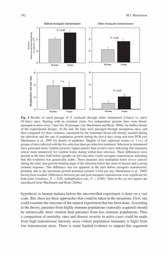
192 M.J. Mackinnon
4
3
2
Viru
lenc
e(m
inim
um r
ed b
lood
cel
l den
sity
,no
. cel
ls/m
l × 1
09 )
Before mosquito transmission
N-lines
P < 0.051
I-lines4
3
2
After mosquito transmission
N-lines
P < 0.05
1
I-lines
3
4
5
6
)yadrep(
etarnoitacilpitlu
M
N-lines
n.s.
I-lines3
4
5
6
N-lines
P < 0.05
I-lines
Fig. 4 Results of serial passage of P. chabaudi through either immunised (I-lines) vs. naıve(N-lines) mice. Starting with an avirulent clone, five independent parasite lines were blood-passaged in mice every 7 days for 20 passages (see Mackinnon and Read, 2004a, for further detailsof the experimental design). At the end, the lines were passaged through mosquitoes once, andthen compared for their virulence (measured by the minimum blood cell density reached duringthe infection) and the rate of population growth during the first 6 days using real-time PCR (seeMackinnon et al., 2005 for details of methods). Heights of bars represent means (± 1.s.e.) ofgroups of mice infected with the five selection lines per selection treatment. Selection in immunisedmice generated more virulent parasites (upper panels) than in naıve mice indicating that immunityselects more intensively for virulent forms during within-host selection. These differences werepresent in the lines both before (graphs on left) and after (right) mosquito transmission indicatingthat this evolution was genetically stable. These parasites also multiplied faster (lower panels)during the early, non-growth-limiting stage of the infection before the onset of disease and a strongimmune response. This difference was not apparent in the lines before mosquito transmission,probably due to the maximum growth potential (around 5-fold per day, Mackinnon et al., 2005)having been reached. Differences between pre-and post-mosquito transmission were significant forboth traits (virulence, P < 0.05; multiplication rate, P < 0.001). Data in the top two figures arereproduced from Mackinnon and Read (2004a)
hypothesis in human malaria before the uncontrolled experiment is done on a vastscale. But, there are three approaches that could be taken in the meantime. First, onecould examine the outcome of the natural experiment that has been done. Accordingto the theory, parasites from highly immune populations (naturally acquired) shouldbe intrinsically more virulent than parasites from less immune populations. Thusa comparison of mortality rates and disease severity in naıve cases could be madefrom high transmission intensity areas (where population immunity is high) fromlow transmission areas. There is some limited evidence to support this argument
Virulence Evolution in Malaria 193
(Murphy and Breman, 2001; Idro et al., 2006), but see Reyburn et al. (2005), thoughmore data are required and it will be a substantial challenge to disentangle the effectsof immunity and infection rate, which both covary with transmission rate. Anotherapproach might be to compare mortality rates of malaria-naıve travellers who haveacquired their infection from high vs. low transmission areas, or to compare in vitrothe virulence-related molecular phenotypes of parasites from areas with contrastingtransmission intensities.
3 Vaccine-Driven Virulence Evolution in Other Diseases
Are there any examples where vaccines have driven the pathogen towards highervirulence? Vaccines against smallpox, measles and polio have been remarkably suc-cessful. Among these, only polio virulence has been shown to increase in responseto vaccination pressure (Kew et al., 2002): in this case it was the attenuated vac-cine strain that reverted to virulence and transmissibility rather than the wild-typestrain changing in response to vaccination. In the case of measles and smallpox, thevaccine induces near-sterilizing immunity, and so it is therefore not surprising thatevolution seems not to have occurred.
On the other hand, for diseases where vaccines are more imperfect (i.e. lesseffective), changes in pathogen virulence following vaccination have been docu-mented, but not always in the direction we predict here, e.g. diphtheria, pneumo-coccal disease and whooping cough (pertussis). Such cases highlight an importantdistinction between the type of virulence evolution we have been discussing herefor malaria (“generalised virulence” evolution) and that which has been discussedfar more widely in the literature in relation to vaccine-driven evolution (“antigenicescape” evolution) (McLean, 1995; Lipsitch and Moxon, 1997; Gupta et al., 1997).In the case of diphtheria, virulence has been observed to decrease after vaccina-tion (Pappenheimer, 1984). However, in this case, the vaccine was directed onlyat the virulence-causing (tox+) strain, thus causing replacement by the non-toxinproducing, avirulent strain. A similar pattern has been observed for Streptococcuspneumoniae, for which the vaccine targets only the most virulent serotypes (Kaplanet al., 2004). With this sort of antigenic escape evolution (also know as “serotypereplacement”), the successful genotypes following vaccination are those that do notcarry the particular antigenic type in the vaccine. However, a priori, they may ornot be more intrinsically virulent than those that they replace: this will dependon which strains the vaccine targets. In this chapter on malaria we have been dis-cussing the possibility of “generalised virulence” evolution in which the successfultypes are those that are able to “grow over” vaccine-induced immunity by virtue oftheir higher intrinsic replication rate: these types are therefore also more virulentin unvaccinated hosts. In doing so, this does not discount the importance of theantigenic escape form of evolution in malaria: indeed it has already been observedfollowing a malaria vaccine trial (Genton et al., 2002), and its potential is implied
194 M.J. Mackinnon
from field studies of naturally acquired immunity in which selection away from theimmunising types is observed (Bull et al., 1999, 2000; Nielsen et al., 2002, 2004).
In pertussis, there appears to have been changes in the pathogen’s virulencegenes in response to vaccination (Mooi et al., 2001), but it is not clear whetherthese changes constitute the “antigenic escape” or the “generalized virulence” formof evolution. On the other hand, there is a very clear example of vaccine-drivenevolution in the Marek’s disease virus of chickens where vaccines have been inwidespread use for 40 years (Witter, 1998): while the precise reasons for this evolu-tion are not yet known, it has all the hallmarks of being driven by the “generalizedvirulence” form of evolution that we have been discussing here (Read et al., 2004).A further example of generalized virulence evolution is the increased virulence ofmyxomatosis virus in response to the evolution of higher levels of resistance inrabbits in Australia (Fenner and Fantini, 1999).
4 Conclusions
In the case of malaria, for which a vaccine is not yet available but is being intensivelypursued, the case has been presented for why generalised virulence may evolve inresponse to vaccines, what might be done to avoid it and what needs to be doneto further determine the likelihood of it happening. The key points are virulence,transmissibility and persistence appear to be intrinsically linked through the para-site’s biology/life history, and these links are maintained over different levels of hostimmunity. Immunity has a very strong impact on parasite fitness through its effecton reducing all three V–T–P traits in accordance with their biological links. Moredata are required in human malaria to determine whether these links are encodedby the parasite’s genes and to determine how much virulence variation exists in thefield.
Immunity is expected to select for higher virulence because, under the coverof immunity, the most virulent parasites can “get away with it”. In naıve hosts,they cannot. Thus highly immune host populations should select for more virulentpathogens. In malaria, children suffer as a result of selection by immune adults.
Devices that reduce asexual density but do not block infection, such as anti-asexual stage vaccines and drugs, are expected to lead to evolution of higher viru-lence, while hygiene (i.e. preventing infection and transmission) is expected to leadto evolution of lower virulence.
Ineffective control measures are expected to minimize undesirable virulence evo-lution. Therefore, age-targeted interventions that minimize selection while protect-ing the most vulnerable may be desirable.
Acknowledgments This chapter is published with the permission of the director of KEMRI. Thework was supported by the Leverhulme Trust and the University of Edinburgh.
Virulence Evolution in Malaria 195
References
Anderson, R.M., May, R.M., 1982, Co-evolution of hosts and parasites, Parasitology 85:411–426.Beale, G.H., Carter, R., Walliker, D., 1978, Genetics, in R. Killick-Kendrick, W. Peters, eds.,
Rodent Malaria, Academic Press, London, pp. 213–245.Bekessy, A., Molineaux, L., and Storey, J., 1976, Estimation of incidence and recovery rates
of Plasmodium falciparum parasitaemia from longitudinal data, Bull. World Health Organ.54:685–693.
Bull, P.C., Kortok, M., Kai, O., Ndungu, F., Ross, A., Lowe, B.S., Newbold, C.I., and Marsh, K.,2000, Plasmodium falciparum-infected erythrocytes: agglutination by diverse Kenyan plasmais associated with severe disease and young host age, J. Infect. Dis. 182:252–259.
Bull, P.C., Lowe, B.S., Kortok, M., and Marsh, K., 1999, Antibody recognition of Plasmodiumfalciparum erythrocyte surface antigens in Kenya: evidence for rare and prevalent variants,Infect. Immun. 67:733–739.
Davies, C.M., Webster, J.P., and Woolhouse, M.E.J., 2001, Trade-offs in the evolution of virulencein an indirectly transmitted macroparasite, Proc. R. Soc. Lond. B. 268:251–257.
De Roode, J.C., Culleton, R., Bell, A.S., and Read, A.F., 2004, Competitive release of drug resis-tance following drug treatment of mixed Plasmodium chabaudi infections, Malar. J. 3:33.
De Roode, J.C., Helsinki, M.E.H., Anwar, M.A., and Read, A.F., 2005a, Dynamics of multi-ple infection and within-host competition in genetically diverse malaria infections, Am. Nat.166:531–542.
De Roode, J.C., Pansini, R., Cheesman, S.J., Helsinki, M.E.H., Huijben, S., Wargo, A.R.,Bell, A.S., Chan, B.H.K., Walliker, D., and Read, A.F., 2005b, Virulent clones are competitivelysuperior in genetically diverse malaria infections, Proc. Natl. Acad. Sci. USA 102:7624–7628.
Dieckmann, U., Metz, J.A.J., Sabelis, M.W., and Sigmund, K., 2002, Virulence Management: theAdaptive Dynamics of Pathogen-Host Interactions, Cambridge University Press, Cambridge.
Dietz, K., Molineaux, L., and Thomas, A., 1980, The mathematical model of transmission, inL. Molineaux, G. Gramiccia, eds., The Garki Project. Research on the Epidemiology and Con-trol of Malaria in the Sudan Savanna of West Africa, World Health Organization, Geneva,pp. 262–289.
Diffley, P., Scott, J.O., Mama, K., and Tsen, T.N.R., 1987, The rate of proliferation among Africantrypanosomes is a stable trait that is directly related to virulence, Am. J. Trop. Med. Hyg. 36:533–540.
Ebert, D., 1994, Virulence and local adaptation of a horizontally transmitted parasite. Science265:1084–1086.
Ebert, D., and Bull, J.J., 2003, Challenging the trade-off model for the evolution of virulence: isvirulence management feasible? Trends Microbiol. 11:15–20.
Ebert, D., and Mangin, K.L., 1997, The influence of host demography on the evolution of virulenceof a microsporidian gut parasite, Evolution 51:1828–1837.
Fenner, F., and Fantini, B., 1999, Biological Control of Vertebrate Pests, CABI Publishing,Wallingford, UK.
Fenner, F., and Ratcliffe, R.N., 1965, Myxomatosis, Cambridge University Press, London.Ferguson, H.M., Mackinnon, M.J., Chan, B.H.K., and Read, A.F., 2003, Mosquito mortality and
the evolution of malaria virulence, Evolution 57:2792–2804.Ferguson, H.M., and Read, A.F., 2002, Genetic and environmental determinants of malaria parasite
virulence in mosquitoes, Proc. R. Soc. Lond. B. 269:1217–1224.Gandon, S., Mackinnon, M.J., Nee, S., and Read, A.F., 2001, Imperfect vaccines and the evolution
of parasite virulence, Nature 414:751–755.Genton, B., Betuela, I., Felger, I., Al-Yaman, F., Anders, R.F., Saul, A., Rare, L., Baisor, M.,
Lorry, K., Brown, G.V., Pye, D., Irving, D.O., Smith, T.A., Beck, H.-P., and Alpers, M.P., 2002,A recombinant blood-stage malaria vaccine reduces Plasmodium falciparum density and exertsselective pressure on parasite populations in a phase 1–2b trial in Papua New Guinea, J. Infect.Dis. 185:820–827.
196 M.J. Mackinnon
Greenwood, M., Bradford Hill, A., Topley, W. W. C., and Wilson, J., 1936, Experimental Epidemi-ology. Medical Research Council. 209. London, HMSO. Special Report Series.
Gupta, S., Ferguson, N.M., and Anderson, R.M., 1997, Vaccination and the population struc-ture of antigenically diverse pathogens that exchange genetic material, Proc. R. Soc. Lond.B. 264:1435–1443.
Idro, R., Aloyo, J., Mayende, L., Bitarakwate, E., John, C.C., and Kivumbi, G.W., 2006, Severemalaria in children in areas with low, moderate and high transmission intensity in Uganda,Trop. Med. Int. Health 11:115–124.
JSkel, T., Khoprasert, Y., and Mackenstedt, U., 2001, Immunoglobulin subclass responses of wildbrown rats to Sarcocystis singaporensis, Int. J. Parasitol. 31:273–283.
Jensen, K.H., Little, T.J., Skorping, A., and Ebert, D., 2006, Empirical support for optimal viru-lence in a castrating parasite. PLoS Biol. 4:e197
Kaplan, S.K., Mason, E.O., Wald, E.R., Schutze, G.E., Bradley, J.S., Tan, T.Q., Hoffman, J.A.,Givner, L.B., Yogev, R., and Barson, W.J., 2004, Decrease in invasisve pneumococcal infec-tions in children among 8 children’s hospitals in the United States after the introduction of the7-valent pneumococcal conjugate vaccine, Paediatrics 113:443–449.
Kew, O.M., Sutter, R.W., De Gourville, E.M., Dowdle, W.R., and Pallansch, M.A., 2002, Vaccine-derived polioviruses and the endgame strategy for global polio eradication, Annu. Rev. Micro-biol. 59:587–635.
Levin, B.R., and Bull, J.J., 1994, Short-sighted evolution and the virulence of pathogenic micro-organisms, Trends Microbiol. 2:76–81.
Levin, S.A., and Pimentel, D., 1981, Selection of intermediate rates of increase in parasite-hostsystems, Am. Nat. 117:308–315.
Lipsitch, M., and Moxon, E.R., 1997, Virulence and transmissibility of pathogens: what is therelationship? Trends Microbiol. 5:31–36.
Mackinnon, M.J., Bell, A.S., and Read, A.F., 2005, The effects of mosquito transmission andpopulation bottlenecking on virulence, multiplication rate and rosetting in rodent malaria, Int.J. Parasitol. 35:145–153.
Mackinnon, M.J., Gaffney, D.J., and Read, A.F., 2002, Virulence in malaria parasites: host geno-type by parasite genotype interactions, Infect. Genet. Evol. 1:287–296.
Mackinnon, M.J., and Read, A.F., 1999a, Genetic relationships between parasite virulence andtransmission in the rodent malaria Plasmodium chabaudi, Evolution 53:689–703.
Mackinnon, M.J., and Read, A.F., 1999b, Selection for high and low virulence in the malariaparasite Plasmodium chabaudi. Proc. R. Soc. Lond. B. 266:741–748.
Mackinnon, M.J., and Read, A.F., 2003, Effects of immunity on relationships between growth rate,virulence and transmission in semi-immune hosts, Parasitology 126:103–112.
Mackinnon, M.J., and Read, A.F., 2004a, Immunity promotes virulence in a malaria model, PLoSBiol. 2:1286–1292.
Mackinnon, M.J., and Read, A.F., 2004b, Virulence in malaria: an evolutionary viewpoint. Phil.Trans. R. Soc. Lond. B. 359:965–986.
Massad, E., 1987, Transmission Rates and the evolution of pathogenicity, Evolution 41:1127–1130.Masumu, J., Marcotty, T., Geysen, D., Geerts, S., Vercruysse, J., Dorny, P., and Van den Bossche,
P., 2006a, Comparison of the virulence of Trypanosoma congolense strains isolated from cattlein a trypanosomiasis endemic area of eastern Zambia, Int. J. Parasitol. 36:497–501.
Masumu, J., Marcotty, T., Ndeledje, N., Kubi, C., Geerts, S., Veycruysse, J., Dorny, P., and Vanden Bossche, P., 2006b, Comparison of transmissibility of Trypanosoma congolense strains,isolated in trypanosomiasis endemic area of eastern Zambia, by Glossina morsitans morsitans,Parasitology 133:331–334.
McLean, A.R., 1995, Vaccination, evolution and changes in the efficacy of vaccines: a theoreticalframework, Proc. R. Soc. Lond. B. 261:389–393.
Messenger, S.L., Molineux, I.J., and Bull, J.J., 1999, Virulence evolution in a virus obeys a trade-off, Proc. R. Soc. Lond. B. 266:397–404.
Virulence Evolution in Malaria 197
Molineaux, L., and Gramiccia, G., 1980, The Garki Project: Research on the Epidemiology andControl of Malaria in the Sudan Savanna of West Africa, World Health Organization, Geneva.
Mooi, F.R., Van Loo, I.H.M., and King, A.J., 2001, Adaptation of Bordetella pertussis to vaccina-tion: a cause for its reemergence, Emerg. Infect. Dis. 7:S526–S528.
Murphy, S.C., and Breman, J.G., 2001, Gaps in the childhood malaria burden in Africa: cerebralmalaria, neurological sequelae, anemia, respiratory distress, hypoglycaemia, and complicationsof pregnancy, Am. J. Trop. Med. Hyg. 64:57–67.
Nielsen, M.A., Staalsoe, T., Kurtzhals, J.A.L., Goka, B.Q., Dodoo, D., Alifrangis, M.,Theander, T.G., Akanmori, B.D., and Hviid, L., 2002, Plasmodium falciparum variant sur-face antigen expression varies between isolates causing severe and nonsevere malaria and ismodified by acquired immunity, J. Immunol. 168:3444–3450.
Nielsen, M.A., Vestergaard, L.S., Lusingu, J.P., Kurtzhals, J.A.L., Giha, H.A., Grevstad, B.,Goka, B.Q., Lemnge, M.M., Akanmori, B.D., Theander, T.G., and Hviid, L., 2004, Geograph-ical and temporal conservation of antibody recognition of Plasmodium falciparum to variantsurface antigens, Infect. Immun. 72:3531–3535.
Pappenheimer, A.M., 1984, Diphtheria,in R. Germanier, ed., Bacterial Vaccines, Academic Press,US, pp. 1–36.
Raberg, L., De Roode, J.C., Bell, A.S., Stamou, P., Gray, D., and Read, A.F., 2006, The roleof immune-mediated apparent competition in genetically diverse malaria infections, Am. Nat.168:41–53.
Read, A.F., Gandon, S., Nee, S., and Mackinnon, M.J., 2004, The evolution of parasite virulence inresponse to animal and public health interventions,in D. Dronamraj, ed., Evolutionary Aspectsof Infectious Diseases, Cambridge University Press, Cambridge.
Read, A.F., and Taylor, L.H., 2001, The ecology of genetically diverse infections, Science292:1099–1102.
Reyburn, H., Mbatia, R., Drakeley, C.J., Bruce, J., Carneiro, I., Olomi, R., Cox, J.,Nkya, W.M.M.M., Lemnge, M.M., Greenwood, B.M., and Riley, E.M., 2005, Association oftransmission intensity and age with clinical manifestations and case fatality of severe Plasmod-ium falciparum malaria, JAMA 293:1461–1470.
Snow, R.W., Craig, M., Deichmann, U., and Marsh, K., 1999, Estimating mortality, morbidity,and disability due to malaria among Africa’s non-pregnant population, B. World Health Organ.77:624–640.
Topley, W.W.C., 1919, The spread of bacterial infection, Lancet 194:1–5.Turner, C.M.R., Aslam, N., and Dye, C., 1995, Replication, differentiation, growth and the viru-
lence of Trypanosoma brucei infections, Parasitology 111:289–300.Williams, G.C., and Nesse, R.M., 1991, The dawn of Darwinian medicine, Q. Rev. Biol. 66:1–22.Witter, R.L., 1998, The changing landscape of Marek’s disease, Avian Pathol. 27:S47–S63.
The Ins and Outs of Host Recognitionof Magnaporthe oryzae
Sally A. Leong
Flor first proposed the gene-for-gene hypothesis to describe the relationship ofraces of the flax rust and cultivars of its flax host (Flor, 1955). In its simplestform, this hypothesis states that for every resistance gene in the host plant, thereexists a complementary avirulence (cultivar specificity) gene in the pathogen thatallows the host to recognize the pathogen and resist development of the diseasedstate. Since its first proposal, the gene-for-gene hypothesis has been found to beapplicable to many host-pathogen interactions including that of the rice blast fun-gus Magnaporthe orzyae and its host Oryza sativa (e.g., Ellingboe, 1992; Leunget al., 1988; Silue et al., 1992a,b; Smith and Leong, 1994; Valent et al., 1991) as wellas subspecific groups of M. orzyae and their respective hosts (Kato, 1983; Valentet al., 1986; Valent and Chumley, 1994). This fundamental relationship is of greatpractical interest as M. orzyae is rapidly able to overcome new disease resistancesin rice soon after their deployment (Bonman et al., 1992). Moreover, M. oryzaeand the closely related Magnaporthe grisea together exist as complex taxa withnumerous subspecific groups that are sometimes interfertile but differ in their hostrange (Couch and Kohn, 2002; Kato et al., 2000; Valent et al., 1991). How thesedifferent subspecific groups interrelate evolutionarily is of great concern as some ofthese alternate hosts are frequently found growing in close proximity to or in rota-tion with rice, and M. orzyae isolates infecting these alternate hosts can sometimesalso infect rice (Kato, 1983; Kato et al., 2000; Mackill and Bonman, 1986; Y. Jia,personal communication). In Japan, barley and rice are alternatively cultivated in thesame field, and rice-infecting strains of M. orzyae also infect barley. Blast diseaseof barley is apparently minimized when M. oryzae infection is limited by coolertemperatures (Yukio Tosa, personal communication). With the advance of globalwarming, these environmental limitations of blast prevalence may soon disappear,as is already being seen with the emergence of blast in California. Pathogens andpests that were a problem only in more southern regions of the US are now becom-ing serious in northern US regions like Wisconsin (Judy Reith-Rozelle, personal
S.A. LeongUSDA, ARS CCRU, Department of Plant Pathology, University of Wisconsin, 1630 LindenDr., Madison, WI 53706, 608-262-6309e-mail: [email protected]
J.P. Gustafson et al. (eds.), Genomics of Disease, 199C© Springer Science+Business Media, LLC, 2008
200 S.A. Leong
communication). This observation can be likely attributed to the warmer wintersthat have occurred in recent years.
The molecular bases of host/cultivar specificity and pathogenic variability inM. orzyae is only beginning to be understood with the cloning of the avirulencegenes for cultivar specificity: AVR2-YAMO/ (AVR- Pi-ta) (Orbach et al., 2000; Jiaet al., 2000; Valent and Chumley, 1994), AVRI-CO39 (Farman and Leong, 1998),and AVR-IRAT7 (Bohnert et al., 2004), and an avirulence gene for host specificityPWL2 (Sweigard et al., 1995) from rice pathogenic isolates of M. orzyae. Thesegenes function as classical avirulence genes (Flor, 1955) by preventing infectionof a specific cultivar or host. AVR-IRAT7 encodes a polyketide synthetase (Bohnertet al., 2004) and other genes may be involved in cultivar specificity. By contrast,AVR2-YAMO/(AVR-Pi-ta) encodes a 223 amino acid protein with homology to a zincmetalloprotease (Jia et al., 2000) while PWL2 encodes a 145 amino acid polypep-tide that is glycine-rich (Sweigard et al., 1995); both genes are predicted to havesecretion signals. Homologs of both genes appear to be widely distributed in riceas well as grass-infecting isolates of M. grisea (e.g., Kang et al., 1995; Valentand Chumley, 1994) and confirm the prediction obtained through genetic analysisthat M. grisea isolates infecting monocots other than rice contain cultivar speci-ficity genes for rice (Yaegashi and Asaga, 1981; Valent and Chumley, 1994; Valentet al., 1991) as well as host specificity genes that preclude infection of other hosts(Valent et al., 1986; Yaegashi, 1978). Rice pathogenic isolates of M. orzyae containcultivar specificity genes for rice (see above) and other grass hosts (Kato et al., 2000)as well as host specificity genes that prevent infection of other monocot hosts (Valentand Chumley, 1994).
Both AVR2-YAMO/(AVR-Pi-ta) and PWL2 are unusually unstable (Valent andChumley, 1994). Molecular characterization of a number of mutant alleles hasrevealed point, deletion, or insertion mutations (Valent and Chumley, 1994). In onecase, an inverted repeat transposon was found in the promoter of AVR2-YAMO/(AVR-Pi-ta). These studies illustrate the potential array of mutational events that can leadto increased virulence and host range in M. orzyae. How frequently these eventscontribute to pathogenic variability in M. orzyae existing in a natural agroecosystemis beginning to be understood for the AVR2-YAMO/(AVR- Pi-ta) gene (Y. Jia, per-sonal communication). The genome of M. orzyae contains a number of transposableelements (e.g., Nitta et al., 1997; Dobinson et al., 1993 Farman et al., 1996; Kachrooet al., 1994) and genomic alterations associated with transposon activity have beendocumented. Further examination of how these elements affect genome evolutionand expression in the context of pathogenic variability deserves close attention.
The cloning and molecular characterization of numerous disease resistance genesfrom different plant species is beginning to provide clues on how plants perceiveinvading pathogens and how these genes may be evolving (e.g., Meyers et al., 1999;Nimchuk et al., 2001). The many common structural features of these genes,nucleotide binding sites (NBS) and leucine-rich repeats (LRR), suggest that com-mon mechanisms of perception and transmission of the pathogen signals exist inthese diverse plant species (McDowell and Dangl, 2000). The finding that func-tional homologs of disease resistance genes exist in different plants species (Dangl
The Ins and Outs of Host Recognition of Magnaporthe oryzae 201
et al., 1992; Innes et al., 1993) and that the same disease resistance locus can showspecificity for different AVR genes as well as pathogens further supports this conclu-sion (e.g., Bisgrove et al., 1994; Grant et al., 1995; Rossi et al., 1998; van der Vossenet al., 2000). Finally, expression of resistance genes from one species of plant canprevent infection of pathogens carrying the corresponding avirulence gene whenintroduced as a transgene in another species of plant (e.g., Tai et al., 1999; Whithamet al., 1996).
The structure of the rice Xa21 (Song et al., 1995) and Xa-1 (Yoshimuraet al., 1998) loci and Pi-b (Wang et al., 1999) and Pi-ta (Jia et al., 2000) lociconferring resistance to bacterial blight or rice blast diseases, respectively, indicatesthat resistance genes from rice share these common NBS and LRR features. Therecent genome sequence of rice variety Nipponbare has provided for the first timea comprehensive view of the number and distribution of the NBS/LRR of genes inrice (International Rice Genome Sequencing Consortium, 2005; Consortia for theSequencing of Rice Chromosomes 11 and 12, 2005; Fritz-Laylin et al., 2005). TheLRR domain was found 1589 times and was one of the 50 most frequently observedclasses of domains in the rice genome (International Rice Genome SequencingConsortium, 2005). Furthermore, the NBS–LRR class of genes was found to occurpredominantly in clusters, which has allowed continuing evolution of the genesthrough recombination. The function of these candidate resistance genes remainsunknown; however, not all LRR-type genes are expected to function exclusivelyor solely in disease resistance as genes controlling plant development are alsoknown to have LRR (e.g., Arabidopsis ERECTA, Godiard et al., 2003; Fritz-Laylinet al., 2005; Arabidopsis CLAVATA, Rojo et al., 2002).
Recent comparative sequencing analysis of alleles of AVR- Pi-ta in M. oryzaeisolates and that of Pi-ta in Oryza species has provided evidence to support a modelof “trench warfare” for this host–parasite interaction. Most sequence diversity isfound in the coding region of AVR- Pi-ta leading to functional alleles while the Pi-taprotein is extremely conserved with a single functional nucleotide polymorphism ina conserved protease motif that distinguishes resistant from susceptible alleles andthe majority of sequence diversity localizing to the intron region (Jia, 2007; Jiaet al., 2003).
The molecular genetic characterization of additional disease resistance genes forrice blast is needed to provide a better understanding of how rice recognizes M.orzyae and how changes in the corresponding M. orzyae AVR genes defeat thisresistance, as well as to clarify the relationship of host and cultivar specificity.Moreover, the availability of cloned disease resistance genes to blast may facilitatethe genetic identification and cloning of other genes conferring resistance to thisdisease in other cereals as well as provide insight on how rice resists infection byother host-specific groups of M. orzyae. The genomes of members of the Poaceaefamily, which includes the many hosts of M. orzyae, are highly syntenic and can beconsidered as essentially one genome (Bennetzen and Freeling, 1993; Devos, 2005).Disease resistance gene homologs have been mapped to syntenic locations acrossgrass genomes (Leister et al., 1998). It will be interesting to learn whether homologsof disease resistance genes to blast are found in the same genomic location of
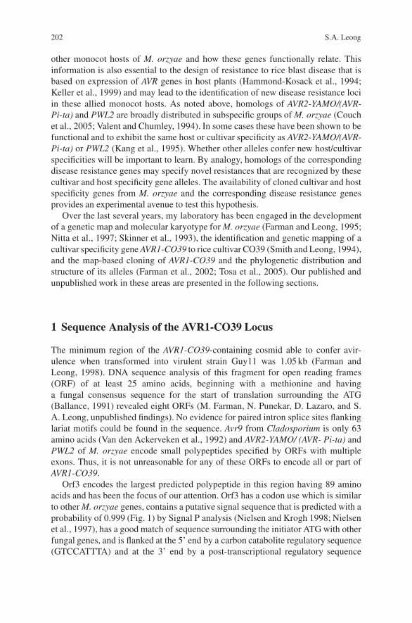
202 S.A. Leong
other monocot hosts of M. orzyae and how these genes functionally relate. Thisinformation is also essential to the design of resistance to rice blast disease that isbased on expression of AVR genes in host plants (Hammond-Kosack et al., 1994;Keller et al., 1999) and may lead to the identification of new disease resistance lociin these allied monocot hosts. As noted above, homologs of AVR2-YAMO/(AVR-Pi-ta) and PWL2 are broadly distributed in subspecific groups of M. orzyae (Couchet al., 2005; Valent and Chumley, 1994). In some cases these have been shown to befunctional and to exhibit the same host or cultivar specificity as AVR2-YAMO/(AVR-Pi-ta) or PWL2 (Kang et al., 1995). Whether other alleles confer new host/cultivarspecificities will be important to learn. By analogy, homologs of the correspondingdisease resistance genes may specify novel resistances that are recognized by thesecultivar and host specificity gene alleles. The availability of cloned cultivar and hostspecificity genes from M. orzyae and the corresponding disease resistance genesprovides an experimental avenue to test this hypothesis.
Over the last several years, my laboratory has been engaged in the developmentof a genetic map and molecular karyotype for M. orzyae (Farman and Leong, 1995;Nitta et al., 1997; Skinner et al., 1993), the identification and genetic mapping of acultivar specificity gene AVR1-CO39 to rice cultivar CO39 (Smith and Leong, 1994),and the map-based cloning of AVR1-CO39 and the phylogenetic distribution andstructure of its alleles (Farman et al., 2002; Tosa et al., 2005). Our published andunpublished work in these areas are presented in the following sections.
1 Sequence Analysis of the AVR1-CO39 Locus
The minimum region of the AVR1-CO39-containing cosmid able to confer avir-ulence when transformed into virulent strain Guy11 was 1.05 kb (Farman andLeong, 1998). DNA sequence analysis of this fragment for open reading frames(ORF) of at least 25 amino acids, beginning with a methionine and havinga fungal consensus sequence for the start of translation surrounding the ATG(Ballance, 1991) revealed eight ORFs (M. Farman, N. Punekar, D. Lazaro, and S.A. Leong, unpublished findings). No evidence for paired intron splice sites flankinglariat motifs could be found in the sequence. Avr9 from Cladosporium is only 63amino acids (Van den Ackerveken et al., 1992) and AVR2-YAMO/ (AVR- Pi-ta) andPWL2 of M. orzyae encode small polypeptides specified by ORFs with multipleexons. Thus, it is not unreasonable for any of these ORFs to encode all or part ofAVR1-CO39.
Orf3 encodes the largest predicted polypeptide in this region having 89 aminoacids and has been the focus of our attention. Orf3 has a codon use which is similarto other M. orzyae genes, contains a putative signal sequence that is predicted with aprobability of 0.999 (Fig. 1) by Signal P analysis (Nielsen and Krogh 1998; Nielsenet al., 1997), has a good match of sequence surrounding the initiator ATG with otherfungal genes, and is flanked at the 5’ end by a carbon catabolite regulatory sequence(GTCCATTTA) and at the 3’ end by a post-transcriptional regulatory sequence
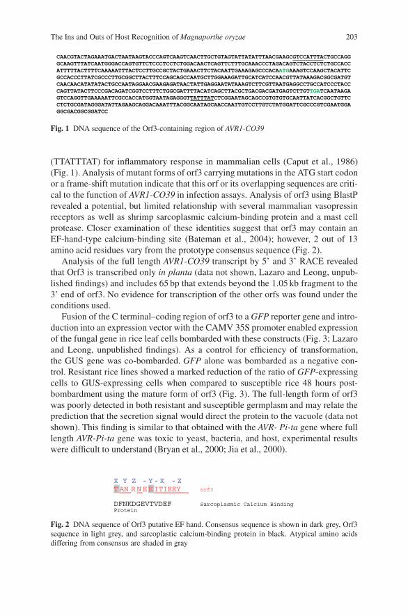
The Ins and Outs of Host Recognition of Magnaporthe oryzae 203
CAACGTACTAGAAATGACTAATAAGTACCCAGTCAAGTCAACTTGCTGTAGTATTATATTTAACGAAGCGTCCATTTACTGCCAGGGCAAGTTTATCAATGGGACCAGTGTTCTCCCTCCTCTGGACAACTCAGTTCTTTGCAAACCCTAGACAGTCTACCTCTCTGCCACCATTTTTACTTTTCAAAAATTTACTCCTTGCCGCTACTGAAACTTCTACAATTGAAAGAGCCCACAATGAAAGTCCAAGCTACATTCGCCACCCTTATCGCCCTTGCGGCTTACTTTCCAGCAGCCAATGCTTGGAAAGATTGCATCATCCAACGTTATAAAGACGGCGATGTCAACAACATATATACTGCCAATAGGAACGAAGAGATAACTATTGAGGAATATAAAGTCTTCGTTAATGAGGCCTGCCATCCCTACCCAGTTATACTTCCCGACAGATCGGTCCTTTCTGGCGATTTTACATCAGCTTACGCTGACGACGATGAGTCTTGTTGATCAATAAGAGTCCAGGTTGAAAAATTCGCCACCATGGTAATAGAGGGTTATTTATCTCGGAATAGCAGCCGTGTGTGCAATTATCACGGCTGTTCCTCTGCGATAGGGATATTAGAAGCAGGACAAATTTACGGCAATAGCAACCAATTGTCCTTGTCTATGGATTCGCCCGTCGAATGGAGGCGACGGCGGATCC
Fig. 1 DNA sequence of the Orf3-containing region of AVR1-CO39
(TTATTTAT) for inflammatory response in mammalian cells (Caput et al., 1986)(Fig. 1). Analysis of mutant forms of orf3 carrying mutations in the ATG start codonor a frame-shift mutation indicate that this orf or its overlapping sequences are criti-cal to the function of AVR1-CO39 in infection assays. Analysis of orf3 using BlastPrevealed a potential, but limited relationship with several mammalian vasopressinreceptors as well as shrimp sarcoplasmic calcium-binding protein and a mast cellprotease. Closer examination of these identities suggest that orf3 may contain anEF-hand-type calcium-binding site (Bateman et al., 2004); however, 2 out of 13amino acid residues vary from the prototype consensus sequence (Fig. 2).
Analysis of the full length AVR1-CO39 transcript by 5’ and 3’ RACE revealedthat Orf3 is transcribed only in planta (data not shown, Lazaro and Leong, unpub-lished findings) and includes 65 bp that extends beyond the 1.05 kb fragment to the3’ end of orf3. No evidence for transcription of the other orfs was found under theconditions used.
Fusion of the C terminal–coding region of orf3 to a GFP reporter gene and intro-duction into an expression vector with the CAMV 35S promoter enabled expressionof the fungal gene in rice leaf cells bombarded with these constructs (Fig. 3; Lazaroand Leong, unpublished findings). As a control for efficiency of transformation,the GUS gene was co-bombarded. GFP alone was bombarded as a negative con-trol. Resistant rice lines showed a marked reduction of the ratio of GFP-expressingcells to GUS-expressing cells when compared to susceptible rice 48 hours post-bombardment using the mature form of orf3 (Fig. 3). The full-length form of orf3was poorly detected in both resistant and susceptible germplasm and may relate theprediction that the secretion signal would direct the protein to the vacuole (data notshown). This finding is similar to that obtained with the AVR- Pi-ta gene where fulllength AVR-Pi-ta gene was toxic to yeast, bacteria, and host, experimental resultswere difficult to understand (Bryan et al., 2000; Jia et al., 2000).
X Y Z -Y-X -ZTAN RNEEITIEEY orf3
DFNKDGEVTVDEF Sarcoplasmic Calcium BindingProtein
Fig. 2 DNA sequence of Orf3 putative EF hand. Consensus sequence is shown in dark grey, Orf3sequence in light grey, and sarcoplastic calcium-binding protein in black. Atypical amino acidsdiffering from consensus are shaded in gray
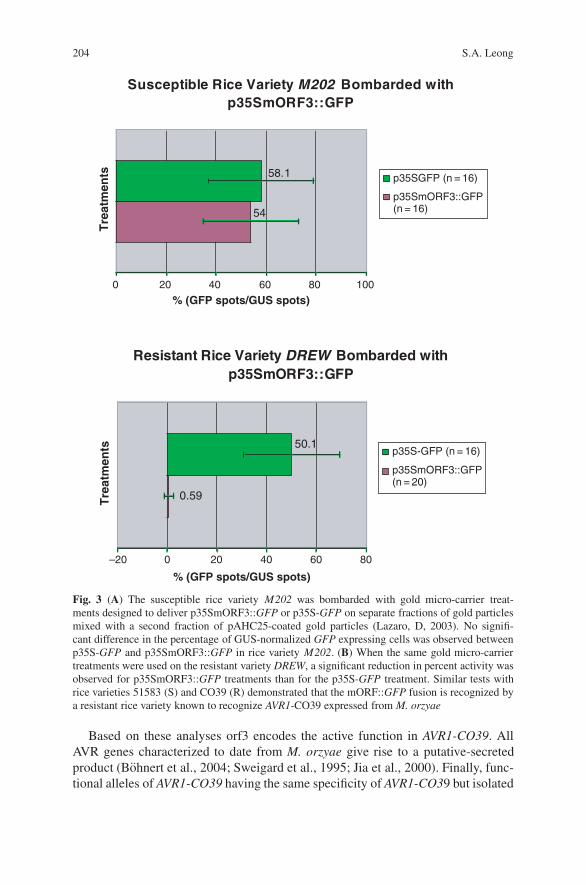
204 S.A. Leong
Susceptible Rice Variety M202 Bombarded withp35SmORF3::GFP
54
58.1
100
Tre
atm
ents
Tre
atm
ents
% (GFP spots/GUS spots)
% (GFP spots/GUS spots)
p35SGFP (n = 16)
p35SmORF3::GFP(n = 16)
Resistant Rice Variety DREW Bombarded withp35SmORF3::GFP
0.59
50.1
–20
0 20 40 60 80
0 20 40 60 80
p35S-GFP (n = 16)
p35SmORF3::GFP(n = 20)
Fig. 3 (A) The susceptible rice variety M202 was bombarded with gold micro-carrier treat-ments designed to deliver p35SmORF3::GFP or p35S-GFP on separate fractions of gold particlesmixed with a second fraction of pAHC25-coated gold particles (Lazaro, D, 2003). No signifi-cant difference in the percentage of GUS-normalized GFP expressing cells was observed betweenp35S-GFP and p35SmORF3::GFP in rice variety M202. (B) When the same gold micro-carriertreatments were used on the resistant variety DREW, a significant reduction in percent activity wasobserved for p35SmORF3::GFP treatments than for the p35S-GFP treatment. Similar tests withrice varieties 51583 (S) and CO39 (R) demonstrated that the mORF::GFP fusion is recognized bya resistant rice variety known to recognize AVR1-CO39 expressed from M. orzyae
Based on these analyses orf3 encodes the active function in AVR1-CO39. AllAVR genes characterized to date from M. orzyae give rise to a putative-secretedproduct (Bohnert et al., 2004; Sweigard et al., 1995; Jia et al., 2000). Finally, func-tional alleles of AVR1-CO39 having the same specificity of AVR1-CO39 but isolated
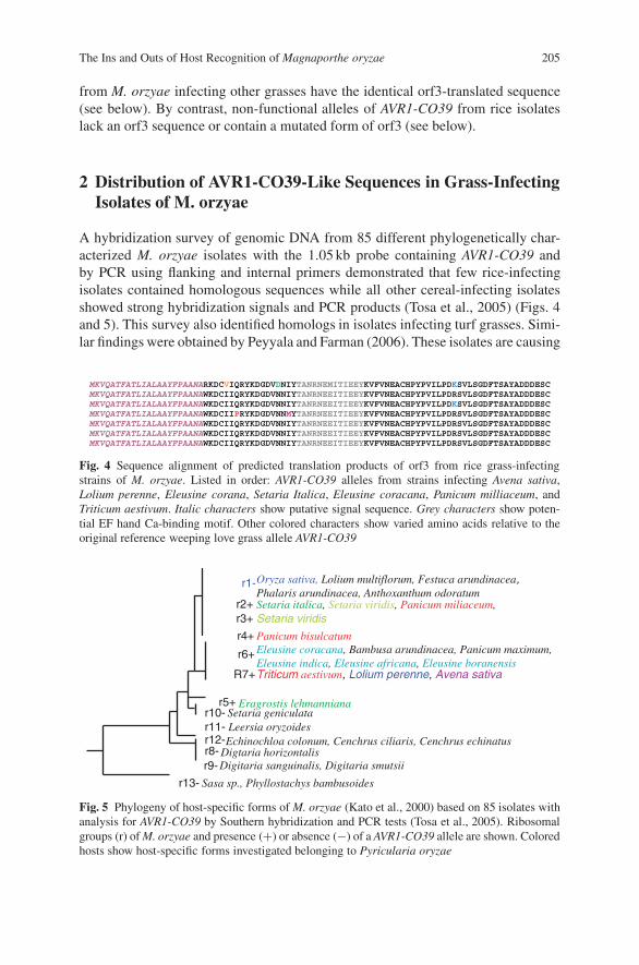
The Ins and Outs of Host Recognition of Magnaporthe oryzae 205
from M. orzyae infecting other grasses have the identical orf3-translated sequence(see below). By contrast, non-functional alleles of AVR1-CO39 from rice isolateslack an orf3 sequence or contain a mutated form of orf3 (see below).
2 Distribution of AVR1-CO39-Like Sequences in Grass-InfectingIsolates of M. orzyae
A hybridization survey of genomic DNA from 85 different phylogenetically char-acterized M. orzyae isolates with the 1.05 kb probe containing AVR1-CO39 andby PCR using flanking and internal primers demonstrated that few rice-infectingisolates contained homologous sequences while all other cereal-infecting isolatesshowed strong hybridization signals and PCR products (Tosa et al., 2005) (Figs. 4and 5). This survey also identified homologs in isolates infecting turf grasses. Simi-lar findings were obtained by Peyyala and Farman (2006). These isolates are causing
MKVQATFATLIALAAYFPAANARKDCVIQRYKDGDVDNIYTANRNEMITIEEYKVFVNEACHPYPVILPDKSVLSGDFTSAYADDDESCMKVQATFATLIALAAYFPAANAWKDCIIQRYKDGDVNNIYTANRNEEITIEEYKVFVNEACHPYPVILPDRSVLSGDFTSAYADDDESCMKVQATFATLIALAAYFPAANAWKDCIIQRYKDGDVNNIYTANRNEEITIEEYKVFVNEACHPYPVILPDKSVLSGDFTSAYADDDESCMKVQATFATLIALAAYFPAANAWKDCIIPRYKDGDVNNMYTANRNEEITIEEYKVFVNEACHPYPVILPDRSVLSGDFTSAYADDDESCMKVQATFATLIALAAYFPAANAWKDCIIQRYKDGDVNNIYTANRNEEITIEEYKVFVNEACHPYPVILPDRSVLSGDFTSAYADDDESCMKVQATFATLIALAAYFPAANAWKDCIIQRYKDGDVNNIYTANRNEEITIEEYKVFVNEACHPYPVILPDRSVLSGDFTSAYADDDESCMKVQATFATLIALAAYFPAANAWKDCIIQRYKDGDVNNIYTANRNEEITIEEYKVFVNEACHPYPVILPDRSVLSGDFTSAYADDDESC
Fig. 4 Sequence alignment of predicted translation products of orf3 from rice grass-infectingstrains of M. orzyae. Listed in order: AVR1-CO39 alleles from strains infecting Avena sativa,Lolium perenne, Eleusine corana, Setaria Italica, Eleusine coracana, Panicum milliaceum, andTriticum aestivum. Italic characters show putative signal sequence. Grey characters show poten-tial EF hand Ca-binding motif. Other colored characters show varied amino acids relative to theoriginal reference weeping love grass allele AVR1-CO39
r5+r10-r11-r12-r8-r9-
r13-
r1-Oryza sativa, Lolium multiflorum, Festuca arundinacea,Phalaris arundinacea, Anthoxanthum odoratum
R7+Triticum aestivum, Lolium perenne, Avena sativa
Eragrostis lehmanniana
r2+ Setaria italica, Setaria viridis, Panicum miliaceum,r3+ Setaria viridis
r4+ Panicum bisulcatum
Setaria geniculataLeersia oryzoidesEchinochloa colonum, Cenchrus ciliaris, Cenchrus echinatus
Digtaria horizontalis
Sasa sp., Phyllostachys bambusoides
Digitaria sanguinalis, Digitaria smutsii
r6+ Eleusine coracana, Bambusa arundinacea, Panicum maximum,Eleusine indica, Eleusine africana, Eleusine boranensis
Fig. 5 Phylogeny of host-specific forms of M. orzyae (Kato et al., 2000) based on 85 isolates withanalysis for AVR1-CO39 by Southern hybridization and PCR tests (Tosa et al., 2005). Ribosomalgroups (r) of M. orzyae and presence (+) or absence (−) of a AVR1-CO39 allele are shown. Coloredhosts show host-specific forms investigated belonging to Pyricularia oryzae
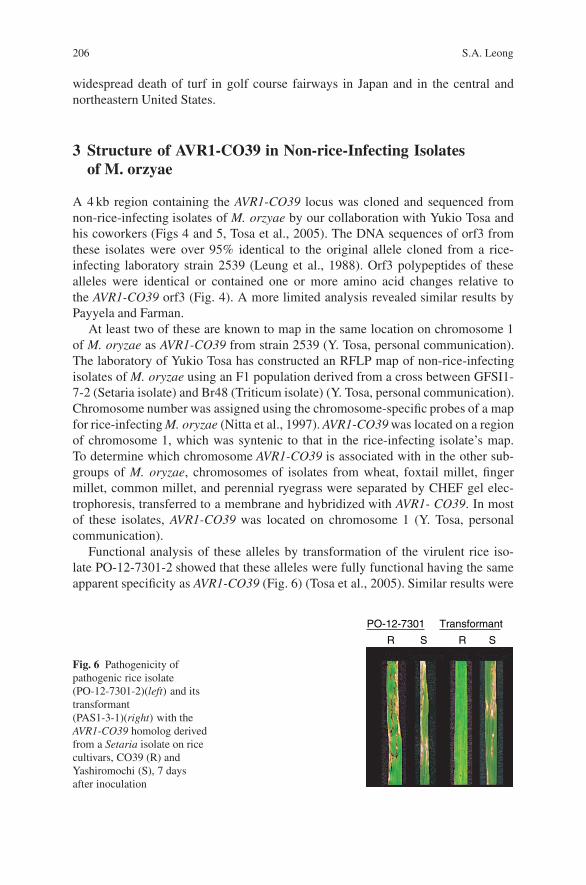
206 S.A. Leong
widespread death of turf in golf course fairways in Japan and in the central andnortheastern United States.
3 Structure of AVR1-CO39 in Non-rice-Infecting Isolatesof M. orzyae
A 4 kb region containing the AVR1-CO39 locus was cloned and sequenced fromnon-rice-infecting isolates of M. orzyae by our collaboration with Yukio Tosa andhis coworkers (Figs 4 and 5, Tosa et al., 2005). The DNA sequences of orf3 fromthese isolates were over 95% identical to the original allele cloned from a rice-infecting laboratory strain 2539 (Leung et al., 1988). Orf3 polypeptides of thesealleles were identical or contained one or more amino acid changes relative tothe AVR1-CO39 orf3 (Fig. 4). A more limited analysis revealed similar results byPayyela and Farman.
At least two of these are known to map in the same location on chromosome 1of M. oryzae as AVR1-CO39 from strain 2539 (Y. Tosa, personal communication).The laboratory of Yukio Tosa has constructed an RFLP map of non-rice-infectingisolates of M. oryzae using an F1 population derived from a cross between GFSI1-7-2 (Setaria isolate) and Br48 (Triticum isolate) (Y. Tosa, personal communication).Chromosome number was assigned using the chromosome-specific probes of a mapfor rice-infecting M. oryzae (Nitta et al., 1997). AVR1-CO39 was located on a regionof chromosome 1, which was syntenic to that in the rice-infecting isolate’s map.To determine which chromosome AVR1-CO39 is associated with in the other sub-groups of M. oryzae, chromosomes of isolates from wheat, foxtail millet, fingermillet, common millet, and perennial ryegrass were separated by CHEF gel elec-trophoresis, transferred to a membrane and hybridized with AVR1- CO39. In mostof these isolates, AVR1-CO39 was located on chromosome 1 (Y. Tosa, personalcommunication).
Functional analysis of these alleles by transformation of the virulent rice iso-late PO-12-7301-2 showed that these alleles were fully functional having the sameapparent specificity as AVR1-CO39 (Fig. 6) (Tosa et al., 2005). Similar results were
Fig. 6 Pathogenicity ofpathogenic rice isolate(PO-12-7301-2)(left) and itstransformant(PAS1-3-1)(right) with theAVR1-CO39 homolog derivedfrom a Setaria isolate on ricecultivars, CO39 (R) andYashiromochi (S), 7 daysafter inoculation
PO-12-7301 Transformant
R S R S
The Ins and Outs of Host Recognition of Magnaporthe oryzae 207
obtained by Payyela and Farman. However, the function of these alleles in non-riceisolates is unknown. They may function in host resistance to rice but few rice linescarry the corresponding PiCO39 (t) (Tosa et al., 2005; S. Leong, unpublished find-ings). Recent work has suggested evidence for race-specific resistance in barley tothe blast fungus indicating that AVR1-CO39 might function in race-specific resis-tance in other cereals (Chen et al., 2003). It will be interesting to learn if homologsof Pi-CO39 (t) in barley exist and if they are involved in blast resistance.
4 Structure of AVR1-CO39 Locus in Rice Isolates of M. oryzae
Further analysis of the structure of the AVR1-CO39 locus from 45 rice-infectingisolates from several geographical regions indicated that this region has undergonea number of changes involving insertion of transposable elements, single base pairchanges, and deletion events (Farman et al., 2002) giving rise to null alleles. At leastthree classes of alleles were discovered through PCR and DNA sequence analyses(Fig. 7). In all cases, orf3 was either deleted completely or had undergone mutationleading to an extension of the polypeptide as a result of a nonsense mutation. Thesedata suggest that this locus has been under strong selection for loss of function andthat these changes may have occurred early in or prior to the evolution of the ricelineage of M. oryzae since other lineages of cereal-infecting isolates all contain afunctional copy of the gene.
5 Genetic and Physical Mapping of the Pi-CO39 (t) Locus
Crosses were made between the rice lines CO39 (resistant) and 51583 (susceptible)to map the Pi-CO39 (t) gene. The resistance phenotype in resistant offspring wasreaction type 1, while that of susceptible seedlings was reaction type 4 (range of
Fig. 7 Structure of avr1-CO39 loci from rice-infecting isolates of M. orzyae (J, G1, G2 types)compared with a weeping love grass allele (W) (Farman et al., 2002). The 1.05 kb AVR1-CO39gene region is shown in black. The G1-type is found in Guy11
208 S.A. Leong
typing scale 0–4). Both of the reciprocal F1s tested (i.e., CO39 X 51583 or 51583 XCO39) were resistant to M. orzyae strain 6082 that contains AVR1-CO39, indicatingthat resistance is controlled by a dominant locus in the nuclear genome of CO39.Resistance to M. orzyae strain 6082 in 604 F2 progenies derived from three differentF1 plants segregated in a 3:1 ratio, characteristic of single dominant locus. Resis-tance to a Guy 11 (AVR1-CO39) transformant in one F2 population consisting of 235F2 progenies also segregated as a single dominant locus. Inheritance of resistancewas also confirmed in F3 families derived from the F2 populations used for map-ping the resistance locus. Testing of 78 F3 families with the Guy11 (AVRI-CO39)transformant and 59 F3 families with the M. orzyae progeny 6082 also showed thatresistance is controlled by a single dominant locus. The segregation ratio of F3families was all resistant: segregating for resistance: all susceptible; and fit a 1:2:1ratio. The disease resistance locus in CO39 was designated as Pi-CO39(t) (Chauhanet al., 2002).
6 Comparative DNA Sequence Analysis of Resistantand Susceptible Cultivars at the Pi-CO39 (t) Locus
A large insert DNA (100 kb average insert size) library of CO39 was constructedin a binary plant transformation cosmid vector pCLD04541 and screened with co-segregating marker RGA38 as well as BAC end sequence probes from the Nip-ponbare BAC libraries of Clemson University Institute for Genomics. Single BACclones hybridizing to co-segregating markers were identified in the Nipponbare (sus-ceptible) BAC library.
The DNA sequence of 6 BAC clones (phrap score >30) at the Pi-CO39 (t)locus was determined in collaboration with Frederick Blattner of the Univer-sity of Wisconsin, Madison. Comparative sequence analysis of blast resistant(CO39 indica) and susceptible (Nipponbare japonica) rice cultivars at genomicregions co-segregating with Pi-CO39(t) showed that these two haplotypes havediverged with respect to the relative type, number of paralogs, and the orientationand location of resistance gene homologs within each cluster (Fig. 8). Severaldisease resistance (NBS–LRR)-like genes were identified with similarity to theother rice resistance-associated genes RPR1, Xa1, Pi-ta, and Pib. RPR1 (riceprobenazole-responsive) is associated with probenazole-induced resistance to blastin rice (Sakamoto et al., 1999); Xa1 confers resistance to race 1 of bacterial blight(Xanthomonas oryzae pv. oryzae) (Yoshimura et al., 1998); Pi-ta is a major riceblast resistance gene related to RPM1 of Arabidopsis (Boyes et al., 1998; Bryanet al., 2000); Pib is another major rice blast resistance gene (Wang et al., 1999). Allfour genes belong to a non-TIR NBS–LRR subfamily of plant disease resistancegenes (S.A. Leong, R. Chauhan, T. Durfee, F. Blattner, and J. Holt, unpublisheddata; Consortia for the Sequencing of Rice Chromosomes 11 and 12, 2005; andMeyers et al., 1999). Polymorphic regions were found in LRR units of Pi-CO39,whether one of these LRR units is the determinant of resistance in CO39 is underinvestigation.

The Ins and Outs of Host Recognition of Magnaporthe oryzae 209
Currently, the gene identified as COR8 is the most promising candidate in thisregion based on its presence by PCR and Southern blot analysis in all resistantgermplasm tested and preliminary complementation studies by transient expression.COR8 is predicted to be the product of a recombination event involving three LRR-type genes or pseudogenes in the susceptible haplotype (Fig. 8).
Other candidate genes exist at the Pi-CO39 (t) locus. COR6 is a unique NBS–LRR type gene present only in the CO39 haplotype. Receptor kinases are presentin the locus and can also function as resistance genes (Becraft, 1998; Brueggemanet al., 2002; Nimchuk et al., 2001). Finally, a new non-LRR-type, class of resistancegene was recently described from Arabidopsis that shares common signaling path-ways with NBS–LRR type genes (Xiao et al., 2005), and the pepper Bs3 resistancegene was reported to be a flavin monooxygenase (Genetic Dissection of the Inter-action Between the Plant Pathogen Xanthomonas campestris pv. vesicatoria and itsHost Plants) requiring caution in assignment of gene function at the Pi-CO39 (t)locus based on classic structural features of plant disease resistance genes.
The LRR of four members of the RPR1 family in this region including COR8were compared and revealed that most variation occurs outside of the inner coreof the LRR, which is attributed to specificity in other LRR-containing proteins.In addition, the COR8 gene has two extra LRR that are separated by a putativetransmembrane domain (Fig. 9).
Cysteine proteases have been clearly associated with disease resistance inplants (e.g., Coaker et al., 2005; Holt et al., 2005; Kruger et al., 2002; Solomonet al., 1999), while serpins (serine/cysteine protease inhibitors) have been asso-ciated with innate immunity in insects (Huntington 2006). In Drosophila, theserine protease Persephone cleaves a Toll ligand named Spatzel, thus activatingbinding to Toll, which in turn signals the production of antifungal peptides. Theserpin Necrotic inhibits Persephone (Fig. 10). Protease mutants have increasedfungal infections while serpin mutants are melanized at wound sites and die earlyin life. These observations are of interest in the context of finding that the ricepathogen’s AVR1-CO39 has evolved by insertion/deletion and nucleotide changes
Fig. 8 Comparative analysis of CO39 (Indica) (bottom) and Nipponbare (Japonica) (top) genomesat regions co-segregating with Pi-CO39(t). Shown are RSNB (Nipponbare serpin-like genes),RSCO (CO39 serpin- like genes), NBR (Nipponbare NBS–LRR disease resistance-like genes),COR (CO39- NBS–LRR disease resistance-like genes), NKin (Nipponbare kinase-like genes),CKin (CO39 kinase-like genes), ProN (Nipponbare protease genes), ProC (CO39 protease genes).Putative orthologous gene relationships are shown with dotted lines
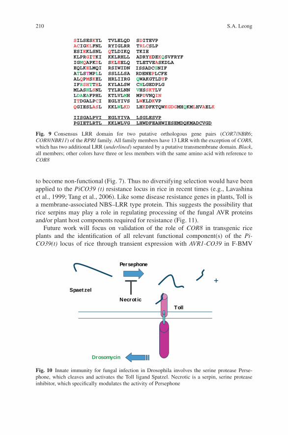
210 S.A. Leong
SILSESKYL TVLELQD SDITEVPACIGKLFNL RYIGLRR TRLCSLPESIEKLSNL QTLDIKQ TKIE KLPRGITKI KKLRHLL ADRYEDEKQSVFRYFIGMQAPKDL SKLEELQ TLETVEASKDLAEQLKELMQI RSIWIDNATLSTMPLL SSLLLSAALQPMSKEL HRLIIRG QWAKGTLDYPIFRSHTTHL KYLALSW CNLGEDPLGMLASHLSNL TYLRLNN VHSSKTLVLDAEAFPHL KTLVLMH MPDVNQINITDGALPCI EGLYIVS LWKLDKVPQGIESLASL KKLWLKD LHKDFKTQWKGDGMHQKMLHVAELK
IISGALPVI EGLYIVA LSGLESVPPGIETLRTL KKLWLVG LHWDFEAHWIESEMDQKMADCVGD
RDENEPLCFEISSADCGNIF
Fig. 9 Consensus LRR domain for two putative orthologous gene pairs (COR7/NBR6;COR8/NBR11) of the RPRI family. All family members have 13 LRR with the exception of COR8,which has two additional LRR (underlined) separated by a putative transmembrane domain. Black,all members; other colors have three or less members with the same amino acid with reference toCOR8
to become non-functional (Fig. 7). Thus no diversifying selection would have beenapplied to the PiCO39 (t) resistance locus in rice in recent times (e.g., Lavashinaet al., 1999; Tang et al., 2006). Like some disease resistance genes in plants, Toll isa membrane-associated NBS–LRR type protein. This suggests the possibility thatrice serpins may play a role in regulating processing of the fungal AVR proteinsand/or plant host components required for resistance (Fig. 11).
Future work will focus on validation of the role of COR8 in transgenic riceplants and the identification of all relevant functional component(s) of the Pi-CO39(t) locus of rice through transient expression with AVR1-CO39 in F-BMV
+
Toll
Persephone
Spaetzel
Necrotic
Drosomycin
Fig. 10 Innate immunity for fungal infection in Drosophila involves the serine protease Perse-phone, which cleaves and activates the Toll ligand Spatzel. Necrotic is a serpin, serine proteaseinhibitor, which specifically modulates the activity of Persephone
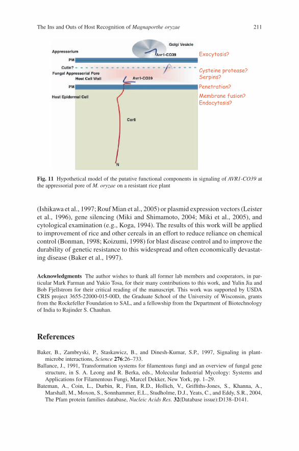
The Ins and Outs of Host Recognition of Magnaporthe oryzae 211
Fig. 11 Hypothetical model of the putative functional components in signaling of AVR1-CO39 atthe appressorial pore of M. oryzae on a resistant rice plant
(Ishikawa et al., 1997; Rouf Mian et al., 2005) or plasmid expression vectors (Leisteret al., 1996), gene silencing (Miki and Shimamoto, 2004; Miki et al., 2005), andcytological examination (e.g., Koga, 1994). The results of this work will be appliedto improvement of rice and other cereals in an effort to reduce reliance on chemicalcontrol (Bonman, 1998; Koizumi, 1998) for blast disease control and to improve thedurability of genetic resistance to this widespread and often economically devastat-ing disease (Baker et al., 1997).
Acknowledgments The author wishes to thank all former lab members and cooperators, in par-ticular Mark Farman and Yukio Tosa, for their many contributions to this work, and Yulin Jia andBob Fjellstrom for their critical reading of the manuscript. This work was supported by USDACRIS project 3655-22000-015-00D, the Graduate School of the University of Wisconsin, grantsfrom the Rockefeller Foundation to SAL, and a fellowship from the Department of Biotechnologyof India to Rajinder S. Chauhan.
References
Baker, B., Zambryski, P., Staskawicz, B., and Dinesh-Kumar, S.P., 1997, Signaling in plant-microbe interactions, Science 276:26–733.
Ballance, J., 1991, Transformation systems for filamentous fungi and an overview of fungal genestructure, in S. A. Leong and R. Berka, eds., Molecular Industrial Mycology: Systems andApplications for Filamentous Fungi, Marcel Dekker, New York, pp. 1–29.
Bateman, A., Coin, L., Durbin, R., Finn, R.D., Hollich, V., Griffiths-Jones, S., Khanna, A.,Marshall, M., Moxon, S., Sonnhammer, E.L., Studholme, D.J., Yeats, C., and Eddy, S.R., 2004,The Pfam protein families database, Nucleic Acids Res. 32(Database issue):D138–D141.
212 S.A. Leong
Becraft, P.W., 1998, Receptor kinases in plant development, Trends Plant Sci. 3:384–388.Bennetzen, J.L., and Freeling, M., 1993, Grasses as a single genetic system: genome composition,
collinearity and compatibility, Trends Genet 9:259–260.Bisgrove, S.R., Simonich, M.T., Smith, N.M., Sattler, A., and Innes, R.W., 1994, A disease resis-
tance gene in Arabadopsis with specificity for two different pathogen avirulence genes, PlantCell 6:927–933.
Bohnert, H.U., Fudal, I. Dioh, W., Tharreau, D., Notteghem, J.L., and Lebrun, M.H., 2004, A puta-tive polyketide synthase/peptide synthetase from Magnaporthe grisea signals pathogen attackto resistant rice, Plant Cell 16:2499–2513.
Bonman, M., 1998, Rice disease management: industry approaches and perspectives. Abstract3.6.7S of the International Congress on Plant Pathology, Edinburgh.
Bonman, M., Khush, G.S., and Nelson, R.J., 1992, Breeding rice for resistance to pests, Annu. Rev.Phytopathol. 30:507–528.
Boyes, D.C., Nam, J., and Dangl, J.L., 1998, The Arabidopsis thaliana RPM1 disease resistancegene product is a peripheral plasma membrane protein that is degraded coincident with thehypersensitive response, Proc. Natl. Acad. Sci. USA 95:15849–15854.
Bryan, G.T., Wu, K.S., Farrall, L., Jia, Y.L., Hershey, H.P., McAdams, S.A., Faulk, K.N.,Donaldson, G.K., Tarchini, R., and Valent, B., 2000, A single amino acid difference distin-guishes resistant and susceptible alleles of the rice blast resistance gene Pi-ta, Plant Cell12:2033–2045.
Brueggeman, R., Rostoks, N., Kudrna, D., Kilian, A., Han, F., Chen, J., Druka, A., Steffenson, B.,and Kleinhofs, A., 2002, The barley stem rust-resistance gene Rpg1 is a novel disease-resistancegene with homology to receptor kinases, Proc. Natl. Acad. Sci. USA 299:9328–9333.
Caput, D., Beutler, B., Hartog, K., Thayer, R., Brown-Shimer, S., and Cerami, A., 1986, Identi-fication of a common nucleotide sequence in the 3’-untranslated region of mRNA moleculesspecifying inflammatory mediators, Proc. Natl. Acad. Sci. USA 83:1670–1674.
Chauhan, R.S., Farman, M.L., Zhang, H.-B., and Leong, S.A., 2002, Genetic and physical mappingof a rice blast resistance locus, Pi-CO39(t), Corresponding to AVR1-CO39 of Magnaporthegrisea, Mol. Genet. Genomics 267:603–612.
Chen, H., Wang, S., Xing, Y., Xu, C., Hayes, P.M., and Zhang, Q., 2003, Comparative analyses ofgenomic location of specificities of loci for quantitative resistance to Pyricularia grisea in riceand barley, Proc. Natl. Acad. Sci. USA 100:2544–2549.
Coaker, G., Falick, A., and Staskawicz, B., 2005, Activation of a phytopathogenic bacterial effectorprotein by a eukaryotic cyclophilin, Science 308:548–550.
Couch, B.C., Fudal, I., Lebrun, M-H., Tharreau, D., Valent, B., van Kim, P., Notteghem, J.-L., andKohn, L. M., 2005., Origins of host-specific populations of the blast pathogen, Magnaportheoryzae, in crop domestication with subsequent expansion of pandemic clones on rice and weedsof rice, Genetics 170:613–630.
Couch, B.C., and Kohn, L.M., 2002, A multilocus gene genealogy concordant with host prefer-ence indicates segregation of a new species, Magnaporthe oryzae, from M. grisea, Mycologia,94:683–693.
Dangl, J.L., Ritter, C., Gibbon, M.J., Mur, L.A.J., Wood, J.R., Goss, S., Mansfield, J., Taylor, J.D.,and Vivian, A., 1992, Functional homologs of the Arabadopsis RPM1 disease resistance genein bean and pea, Plant Cell 4:1359–1369.
Devos, K.M., 2005, Updating the ‘crop circle,’ Curr. Opin. Plant Biol. 8:155–162.Dobinson, K., Harris, R., and Hamer, J.E., 1993, Grasshopper, a long terminal repeat (LTR)
retroelement in the phytopathogenic fungus Magnaporthe grisea, Mol. Plant Microbe Interact.6:114–126.
Ellingboe, A., 1992, Segregation of avirulence/virulence on three rice cultivars in 16 crosses ofMagnaporthe grisea, Phytopathology 82:597–601.
Farman, M.L., Eto, Y., Nakao, Y., Tosa, Y., Nakayashiki, H., Mayama, S., and Leong, S.A., 2002,Analysis of the structure of the Avr1-CO39 avirulence locus in virulent rice-infecting isolatesof Magnaporthe grisea, Mol. Plant Microbe Interact. 15:6–16.

The Ins and Outs of Host Recognition of Magnaporthe oryzae 213
Farman, M.L., and Leong, S.A., 1995, Physical and genetic mapping of telomeres of Magnaporthegrisea, Genetics 140:479–492.
Farman, M.L., and Leong, S.A., 1998, Chromosome walking to the AVR1-CO39 avirulence geneof Magnaporthe grisea: discrepancy between the physical and genetic maps, Genetics 150:1049–1058.
Farman, M.L., Tosa, Y., Nitta, N., and Leong, S.A., 1996, MAGGY, a retrotransposon in thegenome of the rice blast fungus Magnaporthe grisea, Mol. Gen. Genet. 251:665–674.
Flor, H.H., 1955. Host-parasite interaction in flax rust: Its genetics and other implications, Phy-topathology 45:680–685.
Fritz-Laylin, L.K., Krishnamurthy, N., Tor, M., Sjolander, K.V., and Jones, J.D.G., 2005, Phy-logenomic analysis of the receptor-like proteins of rice and Arabidopsis, Plant Physiol. 138:611–623.
Godiard, L., Sauviac, L., Torri, K.U., Grenon, O., Mangin, B., Griimsley, N.H., and Marco, Y.,2003, ERECTA, an LRR receptor-like kinase protein controlling development pleiotropicallyaffects resistance to bacterial wilt, Plant J. 36:353–365.
Grant, M.R., Godiard, L., Straube, E., Ashfield, T., Lewald, J., Sattler, A., Innes, R.W., andDangl, J.L., 1995, Structure of the Arabidopsis RPM1 gene enabling dual specificity diseaseresistance, Science 269:843–846.
Hammond-Kosack, K.E., Staskawicz, B.J., Jones, J.D.G., and Baulcombe, D.C., 1994, Functionalexpression of a fungal avirulence gene from a modified potato virus X genome, Mol. PlantMicrobe Interact. 8:181–185.
Holt, B.F., Belkahdir, Y., and Dangl, J.L., 2005, Antagonistic control of disease resistance proteinstability in the plant immune system, Science 309:929–932.
Huntington, J.A., 2006, Shape-shifting serpins – advantages of a mobile mechanism, TrendsBiochem. Sci. 31:427–435.
Innes, R.W., Bisgrove, S.R., Smith, N.M., Bent, A.F., Staskawicz, B.J., and Liu,Y-C., 1993, Iden-tification of a disease resistance locus in Arabidopsis that is functionally homologous to theRPG1 locus of soybean, Plant J 4:813–820.
International Rice Genome Sequencing Project, 2005, The map-based sequence of the rice genome,Nature 436:793–800.
Ishikawa, M., Janda, M., Krol, M.A., and Ahlquist, P., 1997, In vivo DNA expression of functionalbrome mosaic virus RNA replicons in Saccharomyces cerevisiae, J. Virology. 71:7781–7790.
Jia, Y., 2007, Plants and pathogens engage in trench warfare-knowledge learned from natural vari-ation of rice blast resistance gene Pi-ta. Abstract PAGXV, San Diego, CA, January 2007.
Jia, Y., Bryan, G.T., Farrall, L., and Valent, B., 2003, Natural variation at the Pi-ta rice blast resis-tance locus, Phytopathology 93:1452–1459.
Jia, Y., McAdams, S.A., Bryan, G.T., Hershey, H.P., and Valent, B., 2000, Direct interactionof resistance gene and avirulence gene products confers rice blast resistance, EMBO J. 19:4004–4014.
Kachroo, P., Leong, S.A., and Chattoo, B.B., 1994, Pot2, an inverted repeat transposon from therice blast fungus Magnaporthe grisea, Mol. Gen. Genet. 245:339–348.
Kang, S., Sweigard, J., and Valent, B., 1995, The PWL host-species specificity gene family in theblast fungus Magnaporthe grisea, Mol. Plant Microbe Interact. 8:939–948.
Kato, H., 1983, Responses of Italian millet, oat, timothy, Italian ryegrass and perennial ryegrassto Pyricularia species isolated form cereals and grasses, Proc. Kanto-Tosan Plant Protect. Soc.30:22–23.
Kato, H., Yamamoto, M., Yamaguchi-ozaki, T., Kadouchi, H., Iwamoto, Y., Nakayashiki, H.,Tosa, Y., Mayama, S., and Mori, N., 2000, Pathogenicity, mating ability and DNA restrictionfragment length polymorphisms of Pyricularia populations isolated from Gramineae, Bam-busideae and Zingiberaceae plants, J. Gen. Plant Pathol. 66:30–47.
Keller, H., Pamboukdjian, N., Ponchet, M., Poupet, A., Delon, R., Verrier, J.L., Roby, D., andRicci, P., 1999, Pathogen-induced elicitin production in transgenic tobacco generates a hyper-sensitive response and nonspecific disease resistance, Plant Cell 11:223–235.
214 S.A. Leong
Koga, H., 1994, Hypersensitive death, autofluorescence, and ultrastructural changes in cells of leafsheaths of susceptible and resistant near isogenic lines of rice (Pi-zt ) in relation to penetrationand growth of Pyricularia grisea, Can. J. Bot. 72:1463–1477.
Koizumi, S. 1998, New fungicide use on rice in Japan. Abstract 5.6.3S of the InternationalCongress on Plant Pathology, Edinburgh.
Kruger, J., Thomas, C.M., Golstein, C., Dixon, M.S., Smoker, M., Tang, S., Mulder, L., andJones, J.D., 2002, A tomato cysteine protease required for Cf-2-dependent disease resistanceand suppression of auto-necrosis, Science 296:744–747.
Lavashina, E.A., Langley, E., Green, C., Gubb, D., Ashburner, M., and Hoffmann, J.A., 1999, Con-stitutive activation of Toll-mediated antifungal defense in serpin-deficient Drosophila, Science285:1917–1919.
Lazarro, D., 2003, Characterization of the AVR1-CO39 Locus of Magnaporthe grisea. Master’sThesis, University of Wisconsin, Madison.
Leister, D., Kurth, J., Laurie, D.A., Yano, M., Sasaki, T., Devos, K., Graner, A., and Schulze-Lefert, P., 1998, Rapid reorganization of resistance gene homologues in cereal genomes, Proc.Natl. Acad. Sci. USA 95:370–375.
Leister, R.T., Ausubel, F.M., and Katagiri, F., 1996, Molecular recognition of pathogen attackoccurs inside of plant cells in plant disease resistance specified by the Arabidopsis genes RPS2and RPM1, Proc. Natl. Acad. Sci. USA 93:15497–15502.
Leung, H., Borromeo, E.S., Bernardo, M.A., and Notteghem, J.L., 1988, Genetic analysis of viru-lence in the rice blast fungus Magnaporthe grisea, Phytopathology 78:1227–1233.
Mackill, D., and Bonman, J.M., 1986, New hosts of Pyricularia grisea, Plant Dis. 70:125–127.McDowell, J.M., and Dangl, J.L., 2000, Signal transduction in the plant immune response, Trends
Plant Sci. 25:79–82.Meyers, B.C., Dickerman, A.W., Michelmore, R.W., Sivaramakrishnan, S., Sobral, B.W., and
Young, N.D., 1999, Plant disease resistance genes encode members of an ancient and diverseprotein family within the nucleotide-binding superfamily, Plant J. 20:317–332.
Miki, D., Itoh, R., and Shimamoto, K., 2005, RNA silencing of single and multiple members in agene family of rice, Plant Physiol. 138:1903–1913.
Miki, D., and Shimamoto, K., 2004, Simple RNAi vectors for stable and transient suppression ofgene function in rice, Plant Cell Physiol. 45:490–495.
Nielsen, H., and Krogh, A., 1998, Prediction of signal peptides and signal anchors by a hiddenMarkov model, Proc.Sixth Int. Conf. Int. Syst.Mol. Biol. (ISMB 6), AAAS Press, Menlo Park,California, pp. 122–130.
Nielsen, H., Engelbrecht, J., Brunak, S., and von Heijne, G., 1997, Identification of prokary-otic and eukaryotic signal peptides and prediction of their cleavage sites, Protein Eng. 10:1–6.
Nimchuk, Z., Rohmer, L., Chang, J.H., and Dangl, J.L., 2001, Knowing the dancer from the dance:R-gene products and their interactions with other proteins from host and pathogen, Curr. Opin.Plant Biol. 4:288–294.
Nitta, N., Farman, M., and Leong, S.A., 1997, Genome organization of Magnaporthe grisea:Integration of genetic maps, clustering of transposable elements, and identification of genomeduplications and rearrangements, Theor. Appl. Genet. 95:20–32.
Orbach, M.J., Farrall, L., Sweigard, J.A., Chumley, F.G., and Valent, B., 2000, A telomericavirulence gene determines efficacy for the rice blast resistance gene Pi-ta, Plant Cell 12:2019–2032.
Peyyala, R., and Farman, M.L., 2006, Magnaporthe oryzae isolates causing grey leaf spot of peren-nial ryegrass possess a functional copy of the AVRI-CO39 avirulence gene, Mol. Plant Pathol.7:157–165.
Rice Consortium Consortiums for Sequencing Rice Chromosomes 11 and 12, 2005, The sequenceof rice chromosomes 11 and 12, rich in disease resistance genes and recent gene duplications.BMC Biology http://www.biomedcentral.com/1741-7007/3/20.

The Ins and Outs of Host Recognition of Magnaporthe oryzae 215
Rojo, E., Sharma, V.K., Kovaleva, V.K., Kovaleva, V., Raikhel, N.V., and Flectecher, J.C., 2002,CLV3 is localized to the extracellular space, where it activates the Arabidopsis CLAVATA stemcell signaling pathway, Plant Cell 14:969–977.
Rossi, M., Goggin, F.L., Milligan, S.B., Kaloshian, I., Ullman, D.E., and Williamson, V.M., 1998,The nematode resistance gene Mi of tomato confers resistance against the potato aphid, Proc.Natl. Acad. Sci. USA 95:9750–9754.
Rouf Mian, M.A., Zwonitzer, J.C., Hopkins, A.A., Ding, X.S., and Nelson, R.S., 2005, Responseof tall fescue genotypes to a new strain of Brome Mosaic Virus, Plant Dis. 89:224–227.
Sakamoto, K., Tada, Y., Yokozeki, Y., Akagi, H., Hayashi, N., Fujimura, T., and Ichikawa, N.,1999, Chemical induction of disease resistance in rice is correlated with the expression of agene encoding a nucleotide binding site and leucine-rich repeats, Plant Mol. Biol. 40:847–855.
Silue, D., Tharreau, D., and Notteghem, J.L., 1992a, Evidence for a gene-for-gene relationship inthe Oryza sativa-Magnaporthe grisea pathosystem, Phytopathology 82:577–580.
Silue, D., Tharreau, D., and Notteghem, J.L., 1992b, Identification of Magnaporthe grsiea aviru-lence genes to seven rice cultivars, Phytopathology 82:1462–1467.
Skinner, D.Z., Budde, A., Farman, M., Smith, R., Leung, H., and Leong, S.A., 1993, Genetic map,molecular karyotype and occurrence of repeated DNAs in the rice blast fungus Magnaporthegrisea, Theor. Appl. Genet. 87:545–557.
Smith, J.R., and Leong, S.A., 1994, Mapping of a Magnaporthe grisea locus affecting rice (Oryzasativa) cultivar specificity, Theor. Appl. Genet. 88:901–908.
Solomon, M., Belenghia, B., Delledonneb, M., Menachema, E., and Levine, A., 1999, The involve-ment of cysteine proteases and protease inhibitor genes in the regulation of programmed celldeath in plants, Plant Cell 11:431–444.
Song, W.-Y., Wang, G.-L., Chen, L.-L., Kim, H.-S., Pi, L.-Y., Holsten, T., Gardner, J., Wang, B.,Zhai, W.-X., Zhu, L.-H., Fauquet, C., and Ronald, P., 1995, A receptor kinase-like proteinencoded by the rice disease resistance gene, Xa21, Science 270:1804–1806.
Sweigard, J.A., Carroll, A.M., Kang, S., Farrall, L., Chumley, F.G., and Valent, B., 1995, Identi-fication, cloning and characterization of PWL2, a gene for host species specificity in the riceblast fungus, Plant Cell 7:1221–1233.
Tai, T.H., Dahlbeck, D., Clark, E.T., Gajiwala, P., Pasion, R., Whalen, M.C., Stall, R.E., andStaskawicz, B.J., 1999, Expression of the Bs2 pepper gene confers resistance to bacterial spotdisease in tomato, Proc. Natl. Acad. Sci. USA 96:14153–14158.
Tang, H., Zakaria, K., Lemaitre, B., and Hashimoto, C., 2006, Two proteases defining a melaniza-tion cascade in the immune system of Drosophila, J. Biol. Chem. 281:28097–28104.
Tang, X., Frederick, R.D., Zhou, J., Halterman, D.A., Jia, Y., and Martin, G.B., 1996, Physicalinteraction of AvrPto and Pto Kinase, Science 274:2060–2063.
Tosa, Y., Osue, J., Eto, Y., Tamba, H., Tanaka, K., Nakayashiki, H., Mayama, S., and Leong, S.A.,2005, Evolution of an avirulence gene AVR1-CO39 concomitant with the evolution and differ-entiation of Magnaporthe oryzae, Mol. Plant Microbe Interact. 18:1148–1160.
Valent, B., and Chumley, F.G., 1994, Avirulence genes and mechanisms of genetic instability inthe rice blast fungus, in R. Zeigler, S. A. Leong, and P. Teng, eds., Rice Blast Disease, CABI,London, pp. 111–134.
Valent, B., Crawford, M.S., Weaver, C.G., and Chumley, F.G., 1986, Genetic studies of fertilityand pathogenicity in Magnaporthe grisea (Pyricularia grisea), Iowa State J. Res. 60:569–594.
Valent, B., Farrall, L., and Chumley, F., 1991, Magnaporthe grisea genes for pathogenicity andvirulence identified through a series of backcrosses, Genetics 127:87–101.
Van den Ackerveken, G.F., Van, J.M., Kan, J.A.L., and DeWit, P.G.M., 1992, Molecular analysisof the avirulence gene avr9 of the fungal pathogen Cladesporium fulvum fully supports thegene-for-gene hypothesis, Plant J. 2:359–366.
Van der Vossen, E.A.G., Rouppe van der Voort, J.N.A.M., Kanyuka, K., Bendahmane, A.,Sandbrink, H., Baulcombe, D.C., Bakker, J., Striekema, W.J., and Klein-Lankhorst, R.M., 2000,Homologues of a single resistance gene cluster in potato confer resistance to distinct pathogens:a virus and a nematode, Plant J. 23:567–576.

216 S.A. Leong
Wang, Z.X., Yano, M., Yamanouchi, U., Iwamoto, M., Monna, L., Hayasaka, H., Katayose, Y., andSasaki, T., 1999, The Pib gene for rice blast resistance belongs to the nucleotide binding andleucine-rich repeat class of plant disease resistance genes, Plant J. 19:55–64.
Whitham, S., McCormick, S., and Baker, B., 1996, The N gene of tobacco confers resistance totobacco mosaic virus in transgenic tomato, Proc. Natl. Acad. Sci. USA 93:8776–8781.
Xiao, S., Calis, O., Patrick, E., Zhang, G., Charoenwattana, P., Muskett, P., Parker, J.E., andTurner, J.G., 2005, The atypical resistance gene, RPW8, recruits components of basal defensefor powdery mildew resistance in Arabidopsis, Plant J. 42:95–110.
Yaegashi, H., 1978, Inheritance of pathogenicity in crosses of Pyricularia isolates from weepinglovegrass and finger millet, Ann. Phytopathol. Soc. Jpn 44:626–632.
Yaegashi, H., and Asaga, K., 1981, Further studies on the inheritance of pathogenicity in crossesof Pyricularia grisea with Pyricularia sp. from finger millet, Ann. Phytopathol. Soc. Jpn 47:677–679.
Yoshimura, S., Yamanouchi, U., Katayose, Y., Toki, S., Wang, Z.X., Kono, I., Kurata, N., Yano, M.,Iwata, N., and Sasaki, T., 1998, Expression of Xa1, a bacterial blight-resistance gene in rice, isinduced by bacterial inoculation, Proc. Natl. Acad. Sci. USA 95:1663–1668.

Index
Aa mating type locus, 174activation domain (AD), 120Agrobacterium tumefaciens, 155allele-specific gene expression, 123Alternaria brassicicola., 8Alu element, 79Anthelminthics, 101Apoplastic effectors, 161asymmetric replication cycle, 133auto-inducible peptide (AIP), 33automated BLAST, 134autosomal recessive centronuclear myopathy,
80auxin, 5Auxin Response Factor, 3auxin responsive elements, 3Avian influenza, 115avirulence, 199avirulence genes, 200avirulence protein AvrM, 166AVR-IRAT7, 200Avr1b-1, 68AVR2-YAMO/ (AVR-Pi-ta), 200
BB congenic chickens, 118β-glucan elicitor binding protein, 36β-glucuronidase (GUS) reporter, 3b mating type locus, 174BAC ends, 94bacterial diseases, 90bacterial type III secretion system, 165beta-hexominidase A (HEXB), 45Birt-Hogg-Dube Syndrome, 15biz1 gene, 175BLASTed, 134BlastP, 203boundary sequences, 136
bovine Affymetrix array, 99bovine transcriptome tools, 90brainstem auditory evoked response
(BAER), 85
C‘controller’ genes, 98C-terminal domain, 165Campylobacter, 60candidate genes, 59canonical bacterial genomes, 133canonical pathogenicity islands, 137, 142cDNA (prey) library, 120cDNA-AFLP, 156cell adhesion motif, 167cell death signaling pathways, 169cell sorting, 123chaperones, 167CHEF gel electrophoresis, 206Cis-acting elements, 123CKB genes, 63Cladosporium, 202clamp formation, 175clp1 mutants, 175co-infection, 191coalescent ancestor, 96coccidiosis, 60comparative mapping, 94conventional breeding strategies, 91copper toxicosis, 12counter-defense, 162Cya assay, 153cyclin clb1, 175Cysteine proteases, 209cystic fibrosis (CF), 128cytoplasmic effectors, 162cytoplasmic nucleotide-binding oligomeriza-
tion domain (NOD)-protein family, 26
217

218 Index
D(ds)RNA, 22,4-D, 5dEER motif, 71DELLA tetra mutants, 6dexamethasone (Dex)-inducible promoter, 2Dicer, 2differentially expressed proteins, 99differentially-expressed genes, 123disease resistance (R) gene, 151DNA binding domain, 155DNA-binding domain (BD), 120DNCH1 genes, 63dodecamer repeat, 14Duchene muscular dystrophy (DMD), 45dystrophin gene, 45dystrophin SINEs, 84
Eeffector proteins, 154effectoromics, 169epidemiological models, 90epidemiology, 190epilepsy, 13Epm2b (Nhlrc1) gene, 14ER retention sequence HDEL, 166erythrocyte membrane, 163Exon-skipping, 80exonization, 80
FFalse discovery rate (FDR), 104Fasciola gigantica, 89, 102Fasciola hepatica, 89Fasciolosis, 100Felis, 41fibroblast growth factor 5 (FGF5), 42fine mapping, 96Fine-mapping, 63flg22, 1FLS2 (flagellin sensing 2), 26fluorescence in situ hybridisation, 92Functional transcriptome analyses, 100Fusarium graminearum, 176
GGA tandem repeat, 82gastro-intestinal nematodes, 94gastro-intestinal nematodes (GIN), 90GATA-3 transcription factor motif, 105GC-skew, 133GC-skews, 133gene dense canine radiation hybrid (RH)
map, 16
Gene expression profiling, 120gene knock-outs, 48gene-for-gene hypothesis, 199generalized virulence, 194Genome Community Annotation Tool, 69genome wide selection (GWS) studies, 98genome-wide RNA interference (RNAi), 130Glimmer2 algorithms, 134Gram negative, 128
Hhaplotype diversity, 18haplotype sharing, 18haustorial membrane, 167Hcrtr2, 80herpes simplex virus (HSV), 117hierarchical clustering algorithms, 139high-density SNP genotyping, 98histidine kinases (HK), 32horizontal gene transfer, 129horizontally transferred DNA, 136host-parasite interaction, 201host-pathogen interaction, 129host-targeting signals, 179hrp box, 153Hrp-associated proteins (HpaA-C), 154HrpX-regulated promoters, 154Hyaloperonospora parasitica, 68Hyaloperonospora parasitica, 162hypersensitive cell death, 169hypersensitive reaction (HR), 152hypersensitive response, 5hyperuricosuria, 13hypocretin 2 receptor, 13
IIndonesian Thin Tail (ITT) sheep, 101infectious diseases, 183infiltration assay, 166insulin-like growth factor-1 (IGF-1), 20intercross matings, 102interferon gamma (IFNG) gene, 96intracellular Toll-Interleukin 1
(TIR), 26IRAK (interleukin-1 receptor-associated
kinase)-family kinases, 28
JJGI Genome Portals, 69
Kkey end-points (or ‘bottlenecks’), 105KIT gene, 42
Index 219
Llaser-capture microdissection (LCM), 123lectin binding domain, 36leucine rich repeats (LRR), 26leucine-rich repeats (LRR), 200leucine-rich-repeats (LRR), 153leucine-zipper-like region, 155linkage disequilibrium, 14linkage disequilibrium (LD), 96long interspersed elements (LINEs), 79long-range PCR, 132
MM gene, 166(mi)RNAs, 2Magnaporthe grisea, 176, 199Marek’s disease, 60, 116, 194Marek’s disease resistance, 59Marek’s disease virus (MDV), 116marker-assisted selection (MAS), 115massively parallel signature sequencing
(MPSS), 99mastitis trypanosome resistance, 100MD2 gene, 62MDV SORF2 gene, 121MDV-infected CEF, 123Melampsora lini, 166Mendelian disorders, 13meningitis, 184merle phenotype, 84Meta-analysis, 94methyltransferase, 156MHC class II � chain, 122microarray hybridization technology, 120microarray QTLs or e-QTLs, 100microarray transcriptional profiling, 130microsatellite markers, 102microsatellites, 17mitogen activate kinase (MAPK) protein
kinase, 27MPS VI feline model, 46MUMmer 3.0 software, 133murine NRAMP1-susceptible, 100myosin C binding protein (MYCBP), 46
N(NBS-LRR)-like genes, 208N. benthamiana, 165narcolepsy, 13natural resistance-associated macrophage
protein 1, SLC11A1, 62nave hosts, 186NBS-LRR (nucleotide binding site and
leucine-rich repeat domain), 162
NBS-LRR protein, 70Necrotic, 209Newcastle’s disease, 115Nicotiana benthamiana, 71, 169Nicotiana benthamiana, 155non-LRR-type, 209non-redundant transposon insertion PA14
mutant library, 139novel resistances, 202nuclear localization signals (NLS), 153nucleotide binding sites (NBS), 200
OO-antigen biosynthetic gene, 145Osmycetes, 68OmpR family of transcriptional regulators, 154oomycete avirulence proteins, 71Oomycete cytoplasmic effectors, 162oomycete effectors, 161, 162open reading frames (ORF), 202Oxford grid analysis, 105
PP. aeruginosa isolate, PAO1, 127PAMP-triggered response, 6pancreatic acinar atrophy, 13parasites, 90parasitic colonization, 161parasitophorous vacuolar membrane
(PVM), 163pathogen associated molecular patterns
(PAMPs), 26Persephone, 209Pexel/VTF motifs, 71Phytophthora infestans, 68Phytophthora sojae, 67, 162phytosulfokine receptor kinase, 32Pi-CO39(t), 208Pichia pastoris, 166PIP-box, 153plant auxin receptor TIR1, 2plant pathogenic oomycetes, 169Plasmodium falciparum, 164, 185Plasmodium, 162polycystic kidney disease (PKD), 46polymorphic information content (PIC), 102positional candidate genes, 94principal components (PC), 19prion diseases, 90probenazole-induced resistance, 208progressive retinal atrophy, 12progressive rod cone degeneration, 14protein-protein interactions, 123protozoa, 90
220 Index
pscD gene, 145Pseudomonas aeruginosa isolate PA14, 127Pseudomonas syringae pv. savastanoi, 5PWL2, 200
QQTL Express, 102quantitative resistance, 68Quantitative RT-PCR, 2quantitative trait loci (QTL), 58, 91, 115quantitative trait loci (QTLs), 19Quantitative Trait Nucleotide (QTN), 92
Rradiation hybrid maps, 92rax-gene knock-out mutants, 30reaction type 1, 207reaction type 4, 207Receptor kinases, 209renal cancer, 13repeat-based microsatellite markers, 12resistant haplotype, 96response regulators (RR), 32restriction digestion maps, 92retinitis pigmentosa, 14rice blast fungus Magnaporthe orzyae, 199RIP (receptor-interacting protein)
kinases, 28Rps genes, 68RT-PCR, 107ruminant innate immune response array, 99RXLR region, 162
S(si)RNAs, 2SA-dependent plant defense pathway, 8SAL1 locus, 63Salicylic acid, 8Salmonella, 115Salmonella enteritidis, 61Salmonellosis, 60secreted cellulytic factors, 129SELDI-TOF mass spectroscopy, 99semi-immune hosts, 189sequence tagged sites, 16serial analysis of gene expression
(SAGE), 99Short interspersed elements (SINEs), 79short interspersed nuclear element, 13short-sighted, or dead-end evolution, 184signature-tagged mutagenesis (STM), 130SILV, 82SINEC Cf elements, 13Single nucleotide polymorphisms (SNPs), 98
single nucleotide polymorphisms (SNPs), 17Smut diseases, 173SNP map, 59Spearman’s rank correlation coefficients, 139splenic cDNA library, 121structural/functional domains, 137super-infection, 191superoxide dismutase (SOD), 107susceptible haplotype, 209
TT cell lymphoma disease, 116T2 transgenic lines, 5thiamine biosynthesis enzyme C, 156TIR1 functional paralogs, 3TNC locus, 62Toll, 209, 210Toll-like receptor (TLR), 26transcriptional activator, 120transcriptional regulators, 174translocon, 167translocon complex, 154translocon proteins, 154transposable elements, 200transposon site hybridization (TraSH), 130tRNA-Lys, 13type III effector proteins, 152type III protein secretion system (T3SS), 151,
152type III secretion effector ExoU, 146type III secretion system (TTSS), 169type one secretion system (TOSS), 28type three secretion system (TTSS), 27tyrosinase gene (TYR), 43tyrosinase-related protein gene (TYRP1), 43
Uubiquitin-ligase complex, 2upa genes, 156Ustilago maydis, 173
VV-T-P relationships, 186Varicella-Zoster virus (VZV), 117VBI Microbial Database, 69viral diseases, 90viral receptors, 60virtual sequence, 94virulence determinants, 139virulence trade-off hypothesis, 184virulence-related genes, 129
Wwax moth caterpillar infection system, 142whole-animal infection models, 130

Index 221
whole-genome scans, 63, 98whole-genome shotgun sequencing, 128
XXa21, 201Xanthomonas campestris, 151
Xanthomonas campestris pv. vesicatoria(Xcv), 151
Xanthomonas oryzae pv. oryzae, 25
Zzinc finger transcription, 175Zoonotic pathogens, 58
Copyright © 2022 FDOKUMEN