Role of Novel Drug Delivery Vehicles in Nanobiomedicine
196
Role of Novel Drug Delivery Vehicles in Nanobiomedicine Edited by Rajeev K. Tyagi, Neeraj Garg, Rahul Shukla and Prakash Singh Bisen
-
Upload
khangminh22 -
Category
Documents
-
view
2 -
download
0
Transcript of Role of Novel Drug Delivery Vehicles in Nanobiomedicine

Role of Novel Drug Delivery Vehicles in Nanobiomedicine
Edited by Rajeev K. Tyagi, Neeraj Garg, Rahul Shukla and Prakash Singh Bisen
Edited by Rajeev K. Tyagi, Neeraj Garg, Rahul Shukla and Prakash Singh Bisen
Medical Nanotechnology and Nanomedicine introduces non-experts to the world of nanomedicine and its evolving organizational infrastructure. Considering the
fluid nature of nano breakthroughs and the delicate balance between benefits and consequences as they apply to medicine, readers at all levels will gain a practical, understandable base of information on these developments so that they may take the greatest advantage of them. This practical reference investigates the impact of
nanotechnology on applications in medicine and biomedical sciences, and the broader societal and economic effects. Eschewing technological details, it focuses on enhancing
awareness of the business, regulatory, and administrative aspects of medical applications. It gives readers a critical, balanced, and realistic evaluation of existing nanomedicine developments and future prospects and provides an ideal foundation
upon which to plan and make decisions.
Published in London, UK
© 2020 IntechOpen © Olique / iStock
ISBN 978-1-78923-985-0
Role of Novel D
rug Delivery Vehicles in N
anobiomedicine


Role of Novel Drug Delivery Vehicles in Nanobiomedicine
Edited by Rajeev K. Tyagi, Neeraj Garg, Rahul Shukla and Prakash Singh Bisen
Published in London, United Kingdom


Supporting open minds since 2005

Role of Novel Drug Delivery Vehicles in Nanobiomedicinehttp://dx.doi.org/10.5772/intechopen.77468Edited by Rajeev K. Tyagi, Neeraj Garg, Rahul Shukla and Prakash Singh Bisen
ContributorsMuniasamy Neerathilingam, Chetan Chandola, Arturo Santos, Alejandro Gonzalez-De La Rosa, Jose Navarro-Partida, Juan Carlos Altamirano-Vallejo, Jane Hsiao, Nayab Tahir, Muhammad Tahir Haseeb, Asadullah Madni, Farzana Parveen, Muhammad Muzamil Khan, Safiullah Khan, Nasrullah Jan, Arshad Khan, Jinu George, Reshma Joy, Franklin John, Subas Chandra Dinda, Bibhash Mohanta, Narahari Narayan Palei, Jyotirmoy Deb, Aadesh Kumar, Mahendra Rana, Amita Rana, Chander Amgoth, Chiuyen Phan, Murali Banavoth, Santosh Rompivalasa, Guping Tang, Weifen Zhang, Xiuwen Guan
© The Editor(s) and the Author(s) 2020The rights of the editor(s) and the author(s) have been asserted in accordance with the Copyright, Designs and Patents Act 1988. All rights to the book as a whole are reserved by INTECHOPEN LIMITED. The book as a whole (compilation) cannot be reproduced, distributed or used for commercial or non-commercial purposes without INTECHOPEN LIMITED’s written permission. Enquiries concerning the use of the book should be directed to INTECHOPEN LIMITED rights and permissions department ([email protected]).Violations are liable to prosecution under the governing Copyright Law.
Individual chapters of this publication are distributed under the terms of the Creative Commons Attribution 3.0 Unported License which permits commercial use, distribution and reproduction of the individual chapters, provided the original author(s) and source publication are appropriately acknowledged. If so indicated, certain images may not be included under the Creative Commons license. In such cases users will need to obtain permission from the license holder to reproduce the material. More details and guidelines concerning content reuse and adaptation can be found at http://www.intechopen.com/copyright-policy.html.
NoticeStatements and opinions expressed in the chapters are these of the individual contributors and not necessarily those of the editors or publisher. No responsibility is accepted for the accuracy of information contained in the published chapters. The publisher assumes no responsibility for any damage or injury to persons or property arising out of the use of any materials, instructions, methods or ideas contained in the book.
First published in London, United Kingdom, 2020 by IntechOpenIntechOpen is the global imprint of INTECHOPEN LIMITED, registered in England and Wales, registration number: 11086078, 7th floor, 10 Lower Thames Street, London, EC3R 6AF, United KingdomPrinted in Croatia
British Library Cataloguing-in-Publication DataA catalogue record for this book is available from the British Library
Additional hard and PDF copies can be obtained from [email protected]
Role of Novel Drug Delivery Vehicles in NanobiomedicineEdited by Rajeev K. Tyagi, Neeraj Garg, Rahul Shukla and Prakash Singh Bisenp. cm.Print ISBN 978-1-78923-985-0Online ISBN 978-1-78923-986-7eBook (PDF) ISBN 978-1-78984-330-9

Selection of our books indexed in the Book Citation Index in Web of Science™ Core Collection (BKCI)
Interested in publishing with us? Contact [email protected]
Numbers displayed above are based on latest data collected. For more information visit www.intechopen.com
4,600+ Open access books available
151Countries delivered to
12.2%Contributors from top 500 universities
Our authors are among the
Top 1%most cited scientists
120,000+International authors and editors
135M+ Downloads
We are IntechOpen,the world’s leading publisher of
Open Access booksBuilt by scientists, for scientists


Meet the editors
Dr. Rajeev K. Tyagi completed his Master’s degree in Biotech-nology at Meerut University, India. He completed his doctorate (PhD) in Immunology/Parasitology/Infectiology with a special mention of “Tres Honorable” at the Biomedical Parasitology Unit, Institute Pasteur, Paris, France (2011) and developed a preclinical “humanized” mouse model(s) to study asexual blood and liver-stage infection of Plasmodium falciparum. Dr. Tyagi
worked at the University of South Florida, Augusta University, and Vanderbilt University Medical Center, USA, as a postdoctoral scientist and deployed human chimeric mice to study P. falciparum and understand the immunological pathways of colitis. He has been working to develop novel drug delivery systems to deliver methotrexate and aceclofenac to explore their antiinflammatory effect in breast cancer, rheumatoid arthritis, etc. Dr. Tyagi has been employing his efforts to com-bine translational biomedical research with nanotechnology to develop therapeutic interventional approaches for inflammatory diseases and beyond. Currently, he has been working as a scientist at the CSIR-Institute of Microbial Technology (IM-TECH), Chandigarh, India.
Dr. Neeraj K. Garg is presently working as a formulation scientist (Manager) in Sun Pharmaceutical Industries Limited, Vadodara, India. His doctoral research focused on-to develop and optimize the novel lipid and polymer based nanocarriers for delivery of immunosuppressive drugs. Dr Neeraj have been working in the field of Advanced Drug Delivery System for last 15 years. He is working in the area of targeted lipid and polymeric nanoparti-
cles in general and development of Lipid and polymer based drug delivery vehicles; polymeric nanoparticles, solid lipid nanoparticles, lipid-polymer hybrid nanoparti-cles, nanostructure lipid carriers (NLCs), in particular.
Dr. Rahul Shukla is presently working as an assistant profes-sor in the Department of Pharmaceutics, National Institute of Pharmaceutical Education and Research (Raebareli), India. He was awarded a PhD from JNU, New Delhi, a CSIR-CDRI and MPharm in Pharmaceutics from IIT-BHU, Varanasi, and a D.S. Kothari Post Doctoral Fellowship from Panjab University, Chandigarh. He had experience as a formulation scientist at Dr.
Reddy’s laboratory, Hyderabad. He is presently guiding both doctoral students and master’s students. He has published widely in areas of nanomedicine and targeted drug delivery systems, particle engineering, controlled release systems, neglect-ed and tropical disease therapeutics, product development, and nutraceuticals in internationally renowned journals and books. He has delivered various lectures on many national and international platforms.

Professor P.S. Bisen, received PhD in 1972 and D.Sc. in 1981 from Jabalpur University, Jabalpur, India, guided 65 doctoral theses having 350 research publications and reviews to his credit in indexed international journals of repute with 12 internation-al books (Springer, John Wiley, CRC Press and IntechOpen) on contemporary issues in Biological Sciences, served as Director of the Institute of Microbiology and Biotechnology, Barkatullah
University, Bhopal, Vice-Chancellor, Jiwaji University, Gwalior, Direcor, Madhav Institute of Technology and Science, Gwalior, Director and Dean, JC Bose Institute of Life Sciences, Bundelkhand University, Jhansi and Principal Director and Ad-visor, Jaipur National University, Jaipur. He was a fellow of the UNESCO/UNDP/ICRO and Hungarian Academy of Sciences at the Institute of Plant Physiology, Bio-logical Research Center, Szeged, Hungary; U.S. National Science Foundation Fellow in the Department of Biological Sciences at the University of Illinois at Chicago and in the Department of Bacteriology at the University of California, Davis; UNESCO/WHO fellow at the Institute of Microbiology, Czechoslovak Academy of Sciences, Budejovika, Praha, Czechoslovakia; European Commission’s Marie Curie fellow at the Institute of Environmental and Biological Sciences, University of Lancaster, United Kingdom; and DAAD (German Academic Exchange Fellowship) fellow at the University of Bonn, Germany, awardees of First Indira Gandhi Fellowship for his outstanding work on environment conservation and management, National Overseas Associate ship of the Department of Biotechnology, Ministry of Science and Technology, Govt. of India, UGC Career Award, State Level Kailash Nath Katju Award of Government of Madhya Pradesh for contributions in Life Sciences. He was awarded the Life Time Professorship, the highest honor of Bundelkhand University, Jhansi, India. He has also worked as emeritus scientist (CSIR) at Defense Research Development Establishment, Defence Research Development Organiza-tion, Ministry of Defense, Government of India, Gwalior and is currently pursuing his research in the field of medical biotechnology as Advisor in the Institute of Med-ical Sciences, Jaipur National University, Jaipur with a research project on tubercu-losis from Grand Challenges Canada, Govt. of Canada. Professor Bisen is the elected fellow of the National Academy of Sciences, India, for his enormous contributions to the field of biology. The diagnostic technology developed by Professor Bisen is unique in several respects and has received recognition worldwide. The technology has been patented in India, the United States, Europe, and Japan. He has served as the external scientific advisor to a Barcelona-based Spanish multinational com-pany, Biokit, involved in the manufacturing of diagnostic kits and also serving as Scientific Adviser of an upcoming food industry, Tropilite Foods, based at Gwalior involved on probiotic research, food biotechnology and develops several innovative products for the food industry. Beijing Doing Institute of Translational Medicine Medical Group, China and Nan Yang Academy of Science Singapore appointed him as Senior Research Advisor.
Contents
Preface III
Chapter 1 1Aptamers for Targeted Delivery: Current Challenges and Future Opportunitiesby Chetan Chandola and Muniasamy Neerathilingam
Chapter 2 23Breaking down the Barrier: Topical Liposomes as Nanocarriers for Drug Delivery into the Posterior Segment of the Eyeballby Arturo Santos, Juan C. Altamirano-Vallejo, José Navarro-Partida, Alejandro González-De la Rosa and Jane H. Hsiao
Chapter 3 59Lipid Polymer Hybrid Nanoparticles: A Novel Approach for Drug Deliveryby Nayab Tahir, Muhammad Tahir Haseeb, Asadullah Madni, Farzana Parveen, Muhammad Muzamil Khan, Safiullah Khan, Nasrullah Jan and Arshad Khan
Chapter 4 79Preparation and Characterisation of Niosomal Emulsions as Novel Drug Delivery Vehicle Derived from Natural Seaweedsby Reshma Joy, Franklin John and Jinu George
Chapter 5 95Solid Lipid Based Nano-particulate Formulations in Drug Targetingby Bibhash Chandra Mohanta, Subas Chandra Dinda, Narahari Narayan Palei and Jyotirmoy Deb
Chapter 6 125Drug Delivery through Targeted Approach with Special References to Phytosomesby Mahendra Rana, Aadesh Kumar and Amita J. Rana
Chapter 7 139Polymer Properties: Functionalization and Surface Modified Nanoparticlesby Chander Amgoth, Chiuyen Phan, Murali Banavoth, Santosh Rompivalasa and Guping Tang
Chapter 8 163Applications of Chitosan in Pulmonary Drug Deliveryby Xiuwen Guan and Weifen Zhang

Contents
Preface XIII
Chapter 1 1Aptamers for Targeted Delivery: Current Challenges and Future Opportunitiesby Chetan Chandola and Muniasamy Neerathilingam
Chapter 2 23Breaking down the Barrier: Topical Liposomes as Nanocarriers for Drug Delivery into the Posterior Segment of the Eyeballby Arturo Santos, Juan C. Altamirano-Vallejo, José Navarro-Partida, Alejandro González-De la Rosa and Jane H. Hsiao
Chapter 3 59Lipid Polymer Hybrid Nanoparticles: A Novel Approach for Drug Deliveryby Nayab Tahir, Muhammad Tahir Haseeb, Asadullah Madni, Farzana Parveen, Muhammad Muzamil Khan, Safiullah Khan, Nasrullah Jan and Arshad Khan
Chapter 4 79Preparation and Characterisation of Niosomal Emulsions as Novel Drug Delivery Vehicle Derived from Natural Seaweedsby Reshma Joy, Franklin John and Jinu George
Chapter 5 95Solid Lipid Based Nano-particulate Formulations in Drug Targetingby Bibhash Chandra Mohanta, Subas Chandra Dinda, Narahari Narayan Palei and Jyotirmoy Deb
Chapter 6 125Drug Delivery through Targeted Approach with Special References to Phytosomesby Mahendra Rana, Aadesh Kumar and Amita J. Rana
Chapter 7 139Polymer Properties: Functionalization and Surface Modified Nanoparticlesby Chander Amgoth, Chiuyen Phan, Murali Banavoth, Santosh Rompivalasa and Guping Tang
Chapter 8 163Applications of Chitosan in Pulmonary Drug Deliveryby Xiuwen Guan and Weifen Zhang

Preface
As we researched the currently available textbooks covering the basic principles, applications, and promises of nanotechnology as it applies to medicine, we noted adearth of introductory material tailored specifically for students. While a numberof comprehensive books exist outlining the promise of nanosciences applicable tomedical applications, including the most recent advances in medical research, thesetexts fail to properly introduce the student to nanoscience and nanotechnologybecause they apply first to biology and then to potential therapeutics and diagnos-tics applications. Thus, this textbook is devoted to the basic principles of nanotech-nology, focusing on nanomaterials and nanoparticles, because with respect to thewhole of nanoscience, these sectors hold promise for the future of medicine. It istailored towards real-world applications of medical nanotechnologies with a heavyemphasis on specific examples from the existing literature available in this area. This book is organized to naturally transition from a basic understanding of theprinciples, including physics, behind, for example, nanoparticles and nanomateri-als, to how these principles might be exploited and used to treat or at the very leastefficiently diagnose human diseases or anomalies. Each chapter introduces topicsand vocabulary at a very basic level and transitions to more advanced coverage asthe student’s knowledge level matures.
Dr. Rajeev K. Tyagi, PhDRamalingaswami Fellow and Faculty,
Division of Cell Biology and Immunology,Biomedical Parasitology and Nano-immunology Lab,
CSIR-Institute of Microbial Technology (IMTECH),Chandigarh, India
Dr. Rahul Shukla, PhDAssistant Professor,
Department of Pharmaceutics,National Institute of Pharmaceutical Education and Research,
Raebareli, Lucknow, India
Dr. Neeraj K. GargFormulation Scientist,
Sun Pharmaceutical Industries Limited,Vadodara, India
Prakash S. BisenProfessor and Advisor,
Institute of Medical Sciences,Jaipur National University,
Jaipur

Preface
As we researched the currently available textbooks covering the basic principles, applications, and promises of nanotechnology as it applies to medicine, we noted a dearth of introductory material tailored specifically for students. While a number of comprehensive books exist outlining the promise of nanosciences applicable to medical applications, including the most recent advances in medical research, these texts fail to properly introduce the student to nanoscience and nanotechnology because they apply first to biology and then to potential therapeutics and diagnos-tics applications. Thus, this textbook is devoted to the basic principles of nanotech-nology, focusing on nanomaterials and nanoparticles, because with respect to the whole of nanoscience, these sectors hold promise for the future of medicine. It is tailored towards real-world applications of medical nanotechnologies with a heavy emphasis on specific examples from the existing literature available in this area. This book is organized to naturally transition from a basic understanding of the principles, including physics, behind, for example, nanoparticles and nanomateri-als, to how these principles might be exploited and used to treat or at the very least efficiently diagnose human diseases or anomalies. Each chapter introduces topics and vocabulary at a very basic level and transitions to more advanced coverage as the student’s knowledge level matures.
Dr. Rajeev K. Tyagi, PhDRamalingaswami Fellow and Faculty,
Division of Cell Biology and Immunology,Biomedical Parasitology and Nano-immunology Lab,
CSIR-Institute of Microbial Technology (IMTECH),Chandigarh, India
Dr. Rahul Shukla, PhDAssistant Professor,
Department of Pharmaceutics,National Institute of Pharmaceutical Education and Research,
Raebareli, Lucknow, India
Dr. Neeraj K. GargFormulation Scientist,
Sun Pharmaceutical Industries Limited,Vadodara, India
Prakash S. BisenProfessor and Advisor,
Institute of Medical Sciences,Jaipur National University,
Jaipur

XIV
Emeritus Professor,School of Studies in Biotechnology,
Jiwaji University,Gwalior
1
Chapter 1
Aptamers for Targeted Delivery: Current Challenges and FutureOpportunitiesChetan Chandola and Muniasamy Neerathilingam
Abstract
Aptamers are synthetic ssDNA/RNA molecules that are emerging as novel toolsfor the development of therapeutics, especially for targeted delivery. Aptamersare comparable to monoclonal antibodies, which are well-established therapeuticmolecules, in terms of specificity and affinity to their target. The advantage ofaptamers over antibodies includes their high stability, ease of synthesis, less batch-to-batch variation, easy chemical modifications that allow different conjugationchemistries, small size for better tissue penetration and low immunogenicity. Theseadvantages make aptamers an important tool for use in therapeutics for targeted delivery. However, aptamers do have some limitations that have hindered theirwidespread clinical use as a therapeutic agent. Some of their common limitationsinclude serum stability, renal filtration and endocytic escape. Other limitationsthat are more specific to aptamers include lack of diversity in the aptamer library, nuclease susceptibility and claims of aptamer specificity as well. This book chap-ter sheds light on these challenges, and using examples, it explains the scientificadvancements that have been achieved in overcoming these limitations. We will end this chapter by discussing the use of high-throughput technology, which is the onlyway of truly industrializing the aptamer technology akin to the development ofsmall molecule drugs.
Keywords: aptamers, SELEX, targeted delivery, serum stability, endosomal escape,renal clearance, high-throughput
1. Introduction
Aptamers are small single-stranded RNA or DNA oligonucleotides that specifi-cally bind to their target due to their unique 3-dimensional structure. They werefirst independently developed by the groups of Gold and Szostak in 1990 [1, 2]. Aptamers are selected from a pool of random oligonucleotide library (>1015 randomsequences) by iterative rounds of selection and amplification by a process called Systematic Evolution of Ligands by EXponential Enrichment (SELEX). Differentintermolecular interactions facilitate the interaction of aptamers with their targetincluding van der Waal’s forces, electrostatic interactions between charged groups, three-dimensional shape, stacking and hydrogen bonds.
Functionally aptamers are same as antibodies; however, their advantages overantibodies include small size, less immunogenicity, ease of synthesis, easy chemical

1
Chapter 1
Aptamers for Targeted Delivery: Current Challenges and Future OpportunitiesChetan Chandola and Muniasamy Neerathilingam
Abstract
Aptamers are synthetic ssDNA/RNA molecules that are emerging as novel tools for the development of therapeutics, especially for targeted delivery. Aptamers are comparable to monoclonal antibodies, which are well-established therapeutic molecules, in terms of specificity and affinity to their target. The advantage of aptamers over antibodies includes their high stability, ease of synthesis, less batch-to-batch variation, easy chemical modifications that allow different conjugation chemistries, small size for better tissue penetration and low immunogenicity. These advantages make aptamers an important tool for use in therapeutics for targeted delivery. However, aptamers do have some limitations that have hindered their widespread clinical use as a therapeutic agent. Some of their common limitations include serum stability, renal filtration and endocytic escape. Other limitations that are more specific to aptamers include lack of diversity in the aptamer library, nuclease susceptibility and claims of aptamer specificity as well. This book chap-ter sheds light on these challenges, and using examples, it explains the scientific advancements that have been achieved in overcoming these limitations. We will end this chapter by discussing the use of high-throughput technology, which is the only way of truly industrializing the aptamer technology akin to the development of small molecule drugs.
Keywords: aptamers, SELEX, targeted delivery, serum stability, endosomal escape, renal clearance, high-throughput
1. Introduction
Aptamers are small single-stranded RNA or DNA oligonucleotides that specifi-cally bind to their target due to their unique 3-dimensional structure. They were first independently developed by the groups of Gold and Szostak in 1990 [1, 2]. Aptamers are selected from a pool of random oligonucleotide library (>1015 random sequences) by iterative rounds of selection and amplification by a process called Systematic Evolution of Ligands by EXponential Enrichment (SELEX). Different intermolecular interactions facilitate the interaction of aptamers with their target including van der Waal’s forces, electrostatic interactions between charged groups, three-dimensional shape, stacking and hydrogen bonds.
Functionally aptamers are same as antibodies; however, their advantages over antibodies include small size, less immunogenicity, ease of synthesis, easy chemical

Role of Novel Drug Delivery Vehicles in Nanobiomedicine
2
modification that allows conjugation with a variety of molecules and stability at a higher temperature. Aptamer is comparably smaller (~3 nm) than an antibody (10–15 nm) [3], thus, allowing higher penetration in tissues and the ability to bind more dense cellular epitopes in living cells. Unlike antibodies, aptamers can be selected against non-immunogenic molecules, including proteins or peptides as well as toxins. Aptamers have been successfully developed against proteins, peptides, dyes, metal ions, viruses, bacteria, toxins, and whole cells. They have high specific-ity and affinity often comparable with antibodies. Unlike antibodies, they can be selected at non-physiological conditions such as extremely high or low tempera-ture, or pH. Synthetic manufacturing of aptamers allows minimal batch-to-batch variation, which is a tedious task to maintain in antibody development. All these properties make aptamers as suitable candidates for therapeutic use, particularly targeted nano-delivery. Several aptamers have already been selected against cell surface targets for targeted delivery of therapeutic payload for different diseases, e.g., cancer and human immunodeficiency virus (HIV) infected cells [4]. Table 1 lists the common differences between antibodies and aptamers.
In this book chapter, we discuss the challenges that hinder the clinical use of aptamers as targeted delivery molecules. For example, the small size of aptamers is like a double-edged sword. Though it helps in higher tissue penetration, but also results in high renal filtration that causes loss of its therapeutic efficiency. Using examples we show that how challenges in serum stability, renal filtration, selection
Antibody (IgG) Aptamer
Size 10–15 nm ~3 nm
Target Only immunogenic targets. Can’t be raised against toxic or non-immunogenic proteins
Produced against immunogenic and non-immunogenic (e.g., metal ions, dyes, small peptides, toxins etc.) targets
Specificity High High
Affinity High High
Penetration Low tissue penetration due to large size
High tissue penetration due to small size
Stability (pH, Temperature)
Low High
Shelf life Few months (at low temperature; repeated freeze-thaw causes loss-of function)
Several months (at room temperature) to several years (frozen)
Nuclease susceptibility
Absent Present (modified nucleotides minimize nuclease susceptibility)
Immune response High (except humanized Ab) Absent or low (rare cases)
Batch-to-batch variation
Present Absent due to synthetic manufacture
Conjugation Less possibilities Easy conjugation due to chemical nature allows functionalization with a wide variety of molecules
Synthesis Only in physiological conditions Synthetic
Cost of synthesis High. Requires animal house facility or reactors
Low. Synthesized using table-top instruments
Scale-of-synthesis Low Scalable
Table 1. This table lists the common differences between an antibody (IgG) and an aptamer.
3
Aptamers for Targeted Delivery: Current Challenges and Future OpportunitiesDOI: http://dx.doi.org/10.5772/intechopen.84217
methodology and endocytic escape have been overcome. We explain the importance of proper controls and validation methods in SELEX methodology, which would otherwise result in the development of poor quality aptamers and, this might ultimately diminish the confidence in this technology. Lastly, we discuss about the development of automation in SELEX technology, an important step toward industrializing the aptamer selection process.
2. Serum stability
Currently, many aptamers are in different stages of clinical trials for various dis-eases [5]. Most of the drugs are administered systemically, i.e., intravenously. This method poses a challenge of overcoming serum stability, which involves overcom-ing nuclease activity and coagulation. Here in we discuss these two topics in detail:
2.1 Aggregation
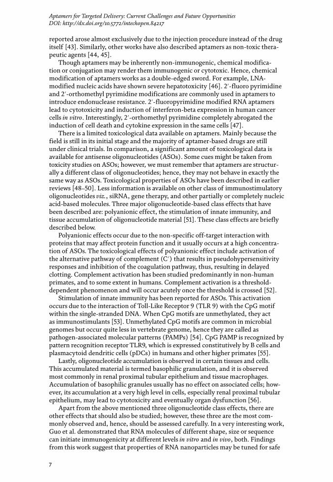
Aptamers have been used to functionalize liposomes to increase their bioavail-ability through targeted delivery (Figure 1) [4]. Since aptamers are negatively charged it is easier to pack them with cationic liposomes. However, serum proteins being charged molecules bind to cationic lipids, thus, lowering their targeting efficiency due to change in structure, aggregation or dissociation of the aptamer-cationic lipid complex [6, 7–9]. Polycationic lipids have also shown cyto(toxic) effects [10]. The problem of aggregation incurred by the charge of cationic lipids can be overcome by using neutral helper lipids [11, 12]. But another challenge is that lipid conjugated aptamers may also bind to erythrocytes, and filtered out through liver and spleen. For example, 1,2-dioleoyl phosphatidylethanolamine (DOPE), a neutral helper lipid, binds to erythrocytes. As an alternative, another neutral helper lipid, cholesterol, may be used which shows significantly lower binding to erythro-cytes as compared to DOPE [10, 13].
Cholesterol conjugated RNA aptamer against Hepatitis C virus (HCV) NS5B protein has been used to inhibit HCV replication in human liver cells without induction of cytotoxicity in vitro. Next, in this study, the same molecule was injected in a mouse model but found no induction of innate immunity. The half-life of the cholesterol-conjugated aptamer was longer than that of the
Figure 1. Illustration showing (a) non-functionalized liposome and (b) aptamer-functionalized liposome carrying drug molecules.

Role of Novel Drug Delivery Vehicles in Nanobiomedicine
2
modification that allows conjugation with a variety of molecules and stability at a higher temperature. Aptamer is comparably smaller (~3 nm) than an antibody (10–15 nm) [3], thus, allowing higher penetration in tissues and the ability to bind more dense cellular epitopes in living cells. Unlike antibodies, aptamers can be selected against non-immunogenic molecules, including proteins or peptides as well as toxins. Aptamers have been successfully developed against proteins, peptides, dyes, metal ions, viruses, bacteria, toxins, and whole cells. They have high specific-ity and affinity often comparable with antibodies. Unlike antibodies, they can be selected at non-physiological conditions such as extremely high or low tempera-ture, or pH. Synthetic manufacturing of aptamers allows minimal batch-to-batch variation, which is a tedious task to maintain in antibody development. All these properties make aptamers as suitable candidates for therapeutic use, particularly targeted nano-delivery. Several aptamers have already been selected against cell surface targets for targeted delivery of therapeutic payload for different diseases, e.g., cancer and human immunodeficiency virus (HIV) infected cells [4]. Table 1 lists the common differences between antibodies and aptamers.
In this book chapter, we discuss the challenges that hinder the clinical use of aptamers as targeted delivery molecules. For example, the small size of aptamers is like a double-edged sword. Though it helps in higher tissue penetration, but also results in high renal filtration that causes loss of its therapeutic efficiency. Using examples we show that how challenges in serum stability, renal filtration, selection
Antibody (IgG) Aptamer
Size 10–15 nm ~3 nm
Target Only immunogenic targets. Can’t be raised against toxic or non-immunogenic proteins
Produced against immunogenic and non-immunogenic (e.g., metal ions, dyes, small peptides, toxins etc.) targets
Specificity High High
Affinity High High
Penetration Low tissue penetration due to large size
High tissue penetration due to small size
Stability (pH, Temperature)
Low High
Shelf life Few months (at low temperature; repeated freeze-thaw causes loss-of function)
Several months (at room temperature) to several years (frozen)
Nuclease susceptibility
Absent Present (modified nucleotides minimize nuclease susceptibility)
Immune response High (except humanized Ab) Absent or low (rare cases)
Batch-to-batch variation
Present Absent due to synthetic manufacture
Conjugation Less possibilities Easy conjugation due to chemical nature allows functionalization with a wide variety of molecules
Synthesis Only in physiological conditions Synthetic
Cost of synthesis High. Requires animal house facility or reactors
Low. Synthesized using table-top instruments
Scale-of-synthesis Low Scalable
Table 1. This table lists the common differences between an antibody (IgG) and an aptamer.
3
Aptamers for Targeted Delivery: Current Challenges and Future OpportunitiesDOI: http://dx.doi.org/10.5772/intechopen.84217
methodology and endocytic escape have been overcome. We explain the importance of proper controls and validation methods in SELEX methodology, which would otherwise result in the development of poor quality aptamers and, this might ultimately diminish the confidence in this technology. Lastly, we discuss about the development of automation in SELEX technology, an important step toward industrializing the aptamer selection process.
2. Serum stability
Currently, many aptamers are in different stages of clinical trials for various dis-eases [5]. Most of the drugs are administered systemically, i.e., intravenously. This method poses a challenge of overcoming serum stability, which involves overcom-ing nuclease activity and coagulation. Here in we discuss these two topics in detail:
2.1 Aggregation
Aptamers have been used to functionalize liposomes to increase their bioavail-ability through targeted delivery (Figure 1) [4]. Since aptamers are negatively charged it is easier to pack them with cationic liposomes. However, serum proteins being charged molecules bind to cationic lipids, thus, lowering their targeting efficiency due to change in structure, aggregation or dissociation of the aptamer-cationic lipid complex [6, 7–9]. Polycationic lipids have also shown cyto(toxic) effects [10]. The problem of aggregation incurred by the charge of cationic lipids can be overcome by using neutral helper lipids [11, 12]. But another challenge is that lipid conjugated aptamers may also bind to erythrocytes, and filtered out through liver and spleen. For example, 1,2-dioleoyl phosphatidylethanolamine (DOPE), a neutral helper lipid, binds to erythrocytes. As an alternative, another neutral helper lipid, cholesterol, may be used which shows significantly lower binding to erythro-cytes as compared to DOPE [10, 13].
Cholesterol conjugated RNA aptamer against Hepatitis C virus (HCV) NS5B protein has been used to inhibit HCV replication in human liver cells without induction of cytotoxicity in vitro. Next, in this study, the same molecule was injected in a mouse model but found no induction of innate immunity. The half-life of the cholesterol-conjugated aptamer was longer than that of the
Figure 1. Illustration showing (a) non-functionalized liposome and (b) aptamer-functionalized liposome carrying drug molecules.

Role of Novel Drug Delivery Vehicles in Nanobiomedicine
4
non-cholesterol-conjugated aptamer, and in accordance, cholesterol conjugation reduced the clearance by 9-fold [14].
It is important to note that no one lipid composition induces cytotoxicity in all cell types, i.e., a conjugated lipid may be toxic to one cell type but not to another. Improvements of lipid formulations are constantly explored by the addition of different lipids, targeting molecules, or shielding moieties designed to prevent clearance in vivo. Use of neutral lipids for aptamer administration has been explored less due to the difficulty in loading hydrophilic aptamer molecules in the neutral lipids for formulation, as compared to (poly)cationic lipids. Liposomes are just one tool for enhancing the efficacy of aptamers for targeted delivery, and there are multitudes of vehicles that are being examined.
2.2 Nuclease activity
Nuclease activity of the serum is helpful in cleaving foreign DNA, thus, it is a way of prevention from pathogens. However, this activity is deleterious to the oligonucleotide-based therapeutics, including aptamers. Unmodified aptamers may have very short half-lives in vivo (<10 minutes) [15]. Two types of nucleases act on these aptamers, viz., exonucleases and endonucleases. Hence, different strategies have been designed to prevent the cleavage activity of each of the nucleases.
In-SELEX and post-SELEX strategies can be utilized to develop nuclease resis-tant aptamers. Each of these two strategies has its own advantage and limitation. We discuss these two below:
2.2.1 In-SELEX
In this methodology aptamers with the desired modification are generated during the aptamer selection process using modified nucleotides, e.g., modified 2′ sugar position using 2′-amino pyrimidine nucleosides [16, 17], 2′-fluoropyrimi-dine nucleosides [18, 19], 2′-O-methyl purine [20], and 2′-O-methyl pyrimidine nucleosides [21, 22] and locked nucleic acids (LNAs) [23, 24] have been commonly used for this purpose, preventing resistance from endonucleases. Macugen, the only FDA approved therapeutic aptamer, was also selected using this approach [25]. The Advantage of this methodology is that no further modification is required in the selected aptamers, thus, ruling out the possibility of loss of function due to post-SELEX modification. However, use of unnatural nucleotides posed a challenge to early researchers since wild-type DNA/RNA polymerases could not amplify the unnatural aptamers during preparative PCR round of aptamer selection. However, using synthetic biology researchers developed new polymerases that could amplify oligos with unnatural nucleotides. Still, amplification using these mutant polymer-ases is not comparable with natural polymerases; hence, aptamer library amplifica-tion is a bit challenging.
SOMAmers are another class of aptamers that bear dU residues at the 5′-posi-tion. These are uniformly functionalized at the 5′-position with various moieties (e.g., benzyl, 2-naphthyl, or 3-indolyl-carboxamide). These moieties can interact with the target molecule and form novel secondary and tertiary structural motifs within the SOMAmer. Introduction of dU at the 5′-position offers nuclease resis-tance to these molecules [26].
2.2.2 Post-SELEX
Post-SELEX involves the introduction of desired modification in pre-selected aptamers during solid-phase chemical synthesis. This method may often lead
5
Aptamers for Targeted Delivery: Current Challenges and Future OpportunitiesDOI: http://dx.doi.org/10.5772/intechopen.84217
to reduced affinity or specificity of the aptamers due to the structural change in the aptamer that is brought upon by the modification. Hence, a combination of methods has to be tested on the aptamer, and one that renders no loss of function has to be selected. Modification of 5′ and 3′ nucleotides is most common and effective in this case. Sometimes unmodified aptamers may demonstrate high resistance to serum nuclease activity [27]. This feature might develop due to the unique 3-D conformation of the aptamer that protects the 5′- and 3′-termini from exonucleases.
Spiegelmers are excellent nuclease resistant L-form RNA or DNA aptamers that are chiral inversions of the natural D-forms. Since naturally occurring nucleases can act only on D-form of nucleotides, they are very less likely to degrade the L-form spiegelmers [28]. Also, due to their unnatural conformation, they are less likely to generate an immune response. The unnatural conformation of spiegelmers also renders it unfit for processing by polymerases for amplification during SELEX process. Hence, in the first step, an aptamer library of natural D-oligonucleotides is selected against a synthetic enantiomer of the L-protein target. After the aptamer selection is done, aptamers are converted from D-form to L-form. As per the rules of symmetry, the resulting L-aptamers (spiegelmers) would bind to the natural D-protein target with the same affinity as the D-aptamers bind to the mirror image selection target [29].
3. Diversity of aptamer library
As we know, aptamer binding to its target depends on its 3-dimensional confor-mation, which is ultimately governed by its sequence. Hence, an aptamer library with a high degree of sequence variability will have a high variety of structurally variant aptamers; therefore, such a library will have a higher degree of success in developing tight binding aptamers to its target. In contrast to proteins, which are made of 20 different amino acids due to a variety of functional groups, nucleic acids possess a very limited variety of functional groups, i.e., only four bases (adenine, guanine, cytosine and thymine/uracil). Thus, aptamers can be endowed with protein-like properties by addition of functional groups that mimic amino acid side-chains to expand their chemical diversity [26].
SomaLogic, a leading company in aptamer development, pioneered in increas-ing the repertoire of aptamer sequences in a library. They introduced new func-tional groups, i.e., hydrophobic side chains (e.g., naphthyl, benzyl, tryptamino and isobutyl) at 5′-position of dUTP nucleotide. Aptamers developed using these modified nucleotides were called Slow Off-rate Modified Aptamers (SOMAmers), and they showed higher success rate in aptamer selection against difficult pro-teins with success rate going from <30% for unmodified aptamers to >90% for SOMAmers [30].
Kimoto et al. used an extra base pair Ds:Px in the initial library to introduce variety, and developed aptamers with 100-fold higher affinity as compared to the unmodified analogs against VEGF-165 (vascular endothelial growth factor) [31]. Sefah et al. developed introduced non-natural nucleosides Z and P to develop aptamers specifically targeting liver cancer cells [32]. In another work, glycosyl-ated aptamers were used in a method called SELection with Modified Aptamers (SELMA) to develop low-nanomolar affinity aptamers against HIV broadly neutral-izing antibody 2G12 [33]. These antibodies protect against HIV by recognizing the unique carbohydrate epitope on HIV. Here, the glycosylated aptamers worked as a scaffold that presented carbohydrate in a manner that mimics their multivalent presentation on HIV, thus it could be used as a vaccine component.

Role of Novel Drug Delivery Vehicles in Nanobiomedicine
6
4. Renal filtration
Renal filtration of small molecule drugs is a phenomenon that needs to be over-come for efficient therapeutic use. Owing to their small size, aptamers also undergo this challenge. An aptamer of 6–30 kDa mass has a size of <5 nm [34]. When an unmodified aptamer is administered intravenously, even using stabilizing backbone modifications, they are subjected to rapid excretion through renal filtration, hence, reduced circulation time. To overcome this challenge, aptamers are functionalized with bulky moieties viz., polyethylene glycol (PEG), liposomes, proteins, choles-terol, organic or inorganic nanomaterials, or multimerized to reach a mass above the threshold of glomerulus cut-off (30–50 kDa) [34].
4.1 PEG attachment
PEGylation is one of the most common methods to prevent renal filtration of aptamers. PEG is a flexible, uncharged and a highly hydrophilic polymer that is widely conjugated with therapeutic drugs to reduce reticuloendothelial clearance, extend circulation time and improve drug efficiency [35]. Macugen, which is the only FDA approved aptamer drug in the market is also PEGylated. PEG decreases aggregation and increases the solubility of the conjugates. PEGylation of Macugen increased its half-life to 9.5 –12.5 hours after intravenous and subcutaneous injection, respectively, in the plasma, and up to 94 hours in vitreous humor [36, 37]. Similarly, PEGylation also increased in vivo half-life of M7, a DNA aptamer, from <1 to 24–48 hours [38]. This modification did not cause any change in the specificity of M7 aptamer toward its target PD-1 (Programmed death protein 1) and suppressed the growth of PD-L1 (Programmed death-ligand 1) positive colon cancer carcinoma in vivo [39].
4.2 Cholesterol
A 29 nucleotide-log 2′-F pyrimidine modified RNA aptamer was reported to inhibit Hepatitis C virus (HCV) replication in vitro. In the follow-up study, this aptamer was derivatized with cholesterol to form cholesterol aptamer conjugate (chol-aptamer). This conjugate entered the cell and inhibited HCV RNA replication in a cell-based system successfully. Systemic administration of chol-aptamer was well tolerated by mice and increased retention time in plasma [14].
4.3 Dialkyl lipids
Willis et al. conjugated diacylglycerol (DAG) to the 5′-end of a nuclease resistant VEGF aptamer [40]. This DAG-aptamer conjugate was then incorporated to a lipo-some bilayer, which resulted in aptamers with improved anti-VEGF activity in vitro and in vivo, both. Importantly, this DAG-aptamer-liposome complex had a consid-erably better residence time in plasma as compared to unmodified aptamer [40].
5. Toxicity
Toxicity, side effects, and immunogenicity are one of the most important criteria in drug evaluation, apart from shelf life and efficacy of the drug. Aptamers have a significant advantage over antibodies due to their minimal or non-immunogenic nature. Early preclinical studies using Pegaptanib sodium (Macugen), the aptamer drug for preventing wet age related macular degeneration (AMD), did not exhibit any intrinsic toxicity during preclinical studies [37, 41, 42]. The only side effect
7
Aptamers for Targeted Delivery: Current Challenges and Future OpportunitiesDOI: http://dx.doi.org/10.5772/intechopen.84217
reported arose almost exclusively due to the injection procedure instead of the drug itself [43]. Similarly, other works have also described aptamers as non-toxic thera-peutic agents [44, 45].
Though aptamers may be inherently non-immunogenic, chemical modifica-tion or conjugation may render them immunogenic or cytotoxic. Hence, chemical modification of aptamers works as a double-edged sword. For example, LNA-modified nucleic acids have shown severe hepatotoxicity [46]. 2′-fluoro pyrimidine and 2′-orthomethyl pyrimidine modifications are commonly used in aptamers to introduce endonuclease resistance. 2′-fluoropyrimidine modified RNA aptamers lead to cytotoxicity and induction of interferon-beta expression in human cancer cells in vitro. Interestingly, 2′-orthomethyl pyrimidine completely abrogated the induction of cell death and cytokine expression in the same cells [47].
There is a limited toxicological data available on aptamers. Mainly because the field is still in its initial stage and the majority of aptamer-based drugs are still under clinical trials. In comparison, a significant amount of toxicological data is available for antisense oligonucleotides (ASOs). Some cues might be taken from toxicity studies on ASOs; however, we must remember that aptamers are structur-ally a different class of oligonucleotides; hence, they may not behave in exactly the same way as ASOs. Toxicological properties of ASOs have been described in earlier reviews [48–50]. Less information is available on other class of immunostimulatory oligonucleotides viz., siRNA, gene therapy, and other partially or completely nucleic acid-based molecules. Three major oligonucleotide-based class effects that have been described are: polyanionic effect, the stimulation of innate immunity, and tissue accumulation of oligonucleotide material [51]. These class effects are briefly described below.
Polyanionic effects occur due to the non-specific off-target interaction with proteins that may affect protein function and it usually occurs at a high concentra-tion of ASOs. The toxicological effects of polyanionic effect include activation of the alternative pathway of complement (C′) that results in pseudohypersensitivity responses and inhibition of the coagulation pathway, thus, resulting in delayed clotting. Complement activation has been studied predominantly in non-human primates, and to some extent in humans. Complement activation is a threshold-dependent phenomenon and will occur acutely once the threshold is crossed [52].
Stimulation of innate immunity has been reported for ASOs. This activation occurs due to the interaction of Toll-Like Receptor 9 (TLR 9) with the CpG motif within the single-stranded DNA. When CpG motifs are unmethylated, they act as immunostimulants [53]. Unmethylated CpG motifs are common in microbial genomes but occur quite less in vertebrate genome, hence they are called as pathogen-associated molecular patterns (PAMPs) [54]. CpG PAMP is recognized by pattern recognition receptor TLR9, which is expressed constitutively by B cells and plasmacytoid dendritic cells (pDCs) in humans and other higher primates [55].
Lastly, oligonucleotide accumulation is observed in certain tissues and cells. This accumulated material is termed basophilic granulation, and it is observed most commonly in renal proximal tubular epithelium and tissue macrophages. Accumulation of basophilic granules usually has no effect on associated cells; how-ever, its accumulation at a very high level in cells, especially renal proximal tubular epithelium, may lead to cytotoxicity and eventually organ dysfunction [56].
Apart from the above mentioned three oligonucleotide class effects, there are other effects that should also be studied; however, these three are the most com-monly observed and, hence, should be assessed carefully. In a very interesting work, Guo et al. demonstrated that RNA molecules of different shape, size or sequence can initiate immunogenicity at different levels in vitro and in vivo, both. Findings from this work suggest that properties of RNA nanoparticles may be tuned for safe
Role of Novel Drug Delivery Vehicles in Nanobiomedicine
8
use as therapeutic nanocarriers [57]. Findings from this work may be extrapolated to RNA aptamers to some extent.
Conjugated moiety may also cause an adverse affect in aptamer drug. For example, penivacogin, a PEGylated aptamer drug, induced severe allergic reac-tion during first exposure to the drug during phase III clinical trials. The allergy occurred due to pre-existing anti-PEG antibodies [58]. PEG is widely used in several consumer products including topical and parenteral medications. Possibly, exposure to these wide varieties of products leads to the development of anti-PEG antibodies [35].
Although less clinical data is available to assess the cytotoxicity of aptamers, studies in animal models show low or no cardiotoxicity, hepatotoxicity or renal toxicity. Doxorubicin (DOX), a widely used anticancer drug causes cardiotoxicity as a side-effect due to non-specific uptake. Neither liposomal DOX nor PEGylated DOX has resolved this issue. However, aptamer-DOX conjugate is known to reduce this cardiotoxicity along with increase in efficacy toward tumor suppression in comparison to non-targeted delivery [59] in vivo.
The hepatic uptake of aptamer functionalized gold nanostars (Apt-Au NS) was studied by Dam et al. They found high accumulation of the complex in liver; however, there was no hepatic acute toxicity. This was probably due to the high accumulation of the complex in macrophages instead of the hepatocytes [60]. Similarly, other works also demonstrated lack of hepatic and renal toxicity in vivo for aptamer-mediated tumor suppression [45, 61].
6. Selection process
Aptamers are selected from a pool of random single-stranded DNA/RNA oligonucleotide library (>1015 random sequences) by iterative rounds of selection and amplification by SELEX method as shown in Figure 2 [4]. Therapeutic aptam-ers have been developed against cell surface targets [62] as well as for delivery of therapeutic conjugates into the cells, i.e., for internalization [3]. Though the selec-tion method is fundamentally the same for developing aptamers for either use, the cell-internalizing aptamers require more points to be addressed to ensure a higher degree of success. Endosomal escape and effective delivery to the cytosol is an important aspect that should be considered for the effective biological activity of a cargo. Here we describe current selection methods commonly used for selection of cell-internalizing aptamers, and discuss the limitations of each method. For this purpose, here we compare the two commonly used strategies, i.e., SELEX and cell-SELEX.
6.1 SELEX
Aptamers have been historically selected against cell surface biomarkers using purified or recombinant proteins. Majority of aptamer therapeutics that are cur-rently in clinical trials have been selected using this methodology. However, there are problems associated with this selection method since the protein against which selection is done might not be in its natural conformation. Proteins are present in physiological milieu in a unique 3D conformation due to either posttranslational modification or their association with other interacting proteins. Proteins that are produced in vitro usually don’t possess these post-translational modifications, specifically, if they produced in a prokaryotic system, e.g., commonly used E. coli. Hence, it is advisable to use eukaryotic expression system viz., mammalian, yeast or insect cells as they would possess post-translational modifications which may
9
Aptamers for Targeted Delivery: Current Challenges and Future OpportunitiesDOI: http://dx.doi.org/10.5772/intechopen.84217
be crucial for the correct 3D conformation for selection of aptamers. However, low protein yield might be an issue if we chose a eukaryotic expression system over the prokaryotic one. There is also a possibility that recombinant cell surface proteins may be insoluble since they need interaction with other cell components (e.g., G-protein coupled receptor), or form multimeric and/or multivalent structures [63, 46]. Lastly, in the majority of cases, aptamer selection is done using only the extracellular domain of the target protein to avoid aptamer selection against the intracellular region of the protein; however, the absence of transmembrane domain may again alter the natural conformation of the protein. Therefore, there is a pos-sibility that aptamers selected using protein-based SELEX approach might not be able to bind their target in a physiological milieu. Liu et al. selected RNA aptamers against histidine-tagged epidermal growth factor receptor variant III (EGFRvIII) ectodomain. This receptor is present in glioblastoma, but not in normal brain tissue. This protein was expressed in an E. coli expression system, hence, lacked a specific post-translational modification, i.e., glycosylation. Lack of glycosylation at this particular site resulted in significant alteration of EGFRvIII protein structure and, hence, the selected aptamer was unable to bind this receptor protein in target cells [64]. Using recombinant protein with a tag-normally used for affinity column based purification of a recombinant protein, e.g., His tag-may result in non-specific aptamer selection against the protein tag; hence, it is always recommended to cleave the tag before a purified recombinant protein is used for aptamer selection. Lastly, attachment of a protein to the matrix may result in non-specific aptamer selection against the matrix itself.
Figure 2. Diagrammatic representation of the systematic evolution of ligands by EXponential enrichment (SELEX) method. A library of ~1015 different single-stranded DNA/RNA molecules is incubated with the target molecules followed by washing. Unbound sequences (non-specific aptamers) are discarded. Bound sequences are recovered and added to the negative control target. Here, the bound sequences (these are aptamers binding to common epitopes) are discarded, and the unbound sequences (target-specific aptamers) are recovered. These sequences are amplified and subjected to further rounds of in vitro selection followed by sequencing after the final selection round. The figure is adapted from Chandola et al. [4].

Role of Novel Drug Delivery Vehicles in Nanobiomedicine
10
6.2 Cell SELEX
Above mentioned limitations, viz., altered 3D conformation, undesired aptamer selection against the protein-tag or matrix can be overcome by using cell-SELEX methodology. In cell-SELEX aptamer selection is performed on live cells in vitro, hence, target surface proteins are present in their native conformation. Unlike protein-based SELEX, cell-SELEX does not need any prior information about the native conformation or biological function of the target protein. This method relies on the difference in the expression pattern of cell surface receptors between the dis-eased target cells (e.g., cancer cells) and the non-diseased cells (e.g., healthy cells). The aptamer selection is first performed on a negative control, i.e., healthy cells. From here, the unbound aptamers are collected and added to the test cell line, i.e., diseased cells. Only the unbound aptamers are collected from negative control cells because they are uncommon among the target and control cells, and it possibly has a set of aptamers that bind the upregulated cell surface receptors that are present only on target diseased cells. Using the above-mentioned methodology, cell-SELEX has been used to identify novel tumor biomarkers [65]. In our experience, major chal-lenge of cell-SELEX is the optimization of aptamer enrichment by PCR as compared to protein-based SELEX. This is due to the inhibitors or other cellular contaminants (e.g., DNA or RNA) that come in the aptamer pool which inhibit the polymerase-based PCR reaction for enriching aptamer pool during each round of selection. Also, lack of prior knowledge about the identity and expression level of the biomarker may lead to aptamer selection against unrelated/unwanted off-target surface molecules that are co-expressed on target cells; hence, more rounds of counter selection with the control cell line is required to improve the selectivity of aptamers.
An ideal aptamer candidate for internalization purpose should bind to a cell surface receptor that is present in high quantity, and it should have a high rate of endocytosis upon binding of a ligand. However, aptamers selected by cell-SELEX may not fall in this category always. Hence, researchers have developed novel strategies to facilitate enrichment of cell internalizing aptamers by alteration of the traditional cell-SELEX protocol. For this purpose, Thiel et al. developed a methodology called cell-internalization SELEX. Here, target cells were incubated with an RNA library fol-lowed by high-salt wash [66]. This method eliminates the non-internalizing surface bound aptamers or those that internalize at a slower rate. Cell internalizing aptamer against Human epidermal growth factor receptor (HER2), a transmembrane protein overexpressed in breast cancer cells was selected using this method [66]. Later studies reported that a high-salt wash is insufficient to remove the non-internalizing surface bound aptamers. Hence, Levy et al. (2012) used a cocktail of RNAses to digest the cell surface bound non-internalizing aptamers [67, 68]. Using this strategy, this group selected cell internalizing 2′-fluoropyrimidine RNA aptamer called c2 against human transferrin receptor (hTfR).
Proteinase K is a broad-spectrum serine protease that cleaves peptide bond adjacent to the carboxyl group of aliphatic and aromatic amino acids with blocked alpha-amino groups. Proteinase K was used as an alternate approach to digest protein domains present on the cell surface after incubation with aptamer library. Thus, digestion of protein domains would render loss of cognate epitopes needed for aptamer binding. After proteinase K treatment and washing of the cells, the aptamers non-specifically bound to the cell surface proteins would be removed, and only those which have internalized the cells can be obtained, as desired. Proteinase K treatment can be used for DNA and RNA aptamers both, unlike RNase treatment used by Levy et al. [68] which is applicable only for RNA aptamers. Iaboni et al. used this method to select cell-internalizing aptamers against insulin receptors on human glioma cells [69].
11
Aptamers for Targeted Delivery: Current Challenges and Future OpportunitiesDOI: http://dx.doi.org/10.5772/intechopen.84217
7. Strategies for endosomal escape of cargoes
The therapeutic efficiency of a cell-internalizing platform depends on its ability to escape from endosomes and deliver the therapeutic cargo to the cytoplasm. The efficiency of endosomal escape, which is 0.01% for traditional delivery systems, should be increased significantly to achieve higher therapeutic efficacy [70]. Two types of strategies are commonly employed to facilitate endosomal escape. First method uses endosomal destabilizing agents that alter the permeability of endosomal membranes, e.g., chloroquine. However, the limitation of this method is that these endosomolytic agents may reach lysosomes, thus, lysing them and lead to cytotoxicity due to the release of lysosomal lytic enzymes. Second method, which is found to be non-toxic in several studies, uses engineered viral or bacterial cell penetrating peptides (CPPs) (e.g., truncated HIV-1 TAT peptide or Aurein1,2) [71–73]. However, CPPs can increase the escape of cargoes from endosomes to the cytoplasm by only 5- to 8-fold, which is not enough to generate an effective plat-form for clinical applications [70]. Below we will discuss the strategies that could be used with aptamers to enhance their endosomal escape properties or that of the attached cargo.
Any targeted delivery platform for internalization of a cargo to cells is endo-cytosed by receptor-mediated endocytosis (RME) to a large extent [74], and aptamers fall in this category. The endocytosed cargo first reaches early endosomes, from where it is either sent back to cell surface (recycling endosomes) or sent to late endosomes, and finally lysosomes. The cargo may be degraded by hydrolytic enzymes in lysosomes. Therefore, if a cargo enters an endosome that is destined for lysosomes, it has a short period of time within which it has to escape to the cytosol to prevent its degradation. Different strategies have been developed that can be used in conjugation with aptamers, which may help in their endosomal escape. Endosomal sorting complex required for transport-I (ESCRT-I) is a protein complex that plays an important role in maturation and trafficking of endosomal cargoes. Wagenaar et al. silenced two components of ESCRT-1 (i.e., TSG101 and VPS28) to induce endosomal escape of delivered anti-miRNA in vitro and in vivo, both, and resulted in tumor regression [75].
Nieman-Pick type C1 (NPC1) is a late endosomal/lysosomal membrane protein required for extracellular recycling of endosomal contents. A small molecule inhibitor of NPC1 protein, termed NP3.47, was earlier used to delay maturation of non-recycling endosomes [76]. The Study found that NP3.47 caused 3-fold higher accumulation of lipid nanoparticle (LNP) formulations of siRNA, i.e., LNP-siRNA as compared to the controls in late endosomes/lysosomes in a variety of cell lines causing 4-fold higher gene silencing. Authors proposed that trapping LNP-siRNA in late endosomes extended the window of time for a more efficient escape into the cytosol to facilitate siRNA induced gene silencing [76].
pH-dependent cargo release is also an effective way for endosomal escape. Anti-PSMA aptamer-siRNA chimera was conjugated to a pH-dependent polyhisti-dine domain via dsRNA binding domain (dsRBD). In the acidic compartment of a mature endosome (pH ≤ 6), Histidine becomes protonated and facilitates osmotic swelling that leads to cargo release into the cytosol, a mechanism known as the proton sponge effect [77]. By this mechanism, the polyhistidine tag facilitated the escape of anti-PSMA aptamer-siRNA chimera from late endosome to cytosol result-ing in improved silencing of the target gene [78].
Another straightforward strategy for cytoplasmic delivery is to perform aptamer selection against a cell surface target protein that internalizes to other compart-ments by receptor-mediated endocytosis. RME facilitates more rapid internaliza-tion of the targeting moiety as compared to untargeted complexes. Depending on

Role of Novel Drug Delivery Vehicles in Nanobiomedicine
12
the intracellular receptor-dependent or independent trafficking path, the fate of targeting moiety can be controlled, e.g., macromolecules internalized by clathrin-dependent RME are mostly destined for lysosomal degradation whereas clathrin-independent internalization leads to endosomal accumulation and sorting to a non-degradative path [74]. However, this method can be used against those targets that have previously been characterized, e.g., nucleolin. Nucleolin is a nuclear ribonucleoprotein that is overexpressed in cancer cells where it shuttles among the cell-surface, cytoplasm and the nucleus. AS1411 aptamer, a G-quadruplex forming oligonucleotide, specifically binds nucleolin and displays antiproliferative activity against cancer cells [79]. However, there are conflicting reports regarding nucleolin-mediated uptake of AS1411 since one study suggest that it is nucleolin dependent and the other suggests that it is nucleolin independent. Though the mechanism of escape of AS1411 from endosomes to cytoplasm is unclear, it is likely that its interaction with nucleolin, which shuttles between different cellular compartments, plays a role in it [80].
8. Quality of known aptamers
Aptamers have generated excitement in the scientific community in the last 2 decades due to their unique characteristics that helped them gain popularity for use in therapeutic purposes. They have been used as both agonists and antagonists targeting cell surface receptors [34]. The Ability of aptamers to specifically bind the cell surface targets catapulted them into the field of targeted delivery of therapeu-tics. They can be easily conjugated with small molecule drugs, liposomes, proteins, siRNAs, miRNAs, or other nanoparticles. However, when industry leaders in the field of siRNA/miRNA delivery tried to recapitulate the results of siRNA delivery, they failed [81]. Also, since the approval of Macugen, in 2004, by Food and Drug Administration (FDA), no other aptamer-based drug has made to the market. Failure of Fovista (anti-PDGF aptamer to prevent age-related macular degenera-tion) and Reg1 (an antifactor IXa aptamer paired with its complementary reversal agent, reverseran, to restore blood clotting) failed in phase III clinical trials due to no improvement over current standard care methods has also been disappointing [81]. However, Reg1 also resulted in serious allergies in patients, albeit it was due to conjugated PEG, and not due to the aptamer itself. Though failure of drugs in clinical trials is quite common as many more drugs fail during trials than pass them, but more disappointingly, there is a lack of aptamer-based drugs that are even advancing to the clinic [5]. Unlike antibodies and antibody-based drugs which are supported by big pharmaceutical companies, aptamer development has been largely relegated to academic labs that are often, comparatively, poorly funded.
Levy group observed lack of reproducibility in aptamer binding to its target or therapeutic effects in several cases [81]. This unreliability among novel molecules is a bad precedent that may generate a lack of confidence in an upcoming technology. The poor reproducibility, or robustness, of these aptamers might be due to poor downstream characterizations, which is, in fact, very important for validation of novel aptamers. There is also no standard way of assessing aptamer binding results, as different labs use a variety of methods to assess the binding function of their selected molecules. Hence, there is no straightforward way to compare or validate results between different labs. Other labs often cite the work from previous labs without revalidating the aptamer that they intend to use. Lack of funds, resources or time are often the reason nor not revalidating the aptamers for their own pur-poses; however, if the aptamer field has to grow, a bit more care and diligence will be needed from the labs that are developing aptamers as well as the end user.
13
Aptamers for Targeted Delivery: Current Challenges and Future OpportunitiesDOI: http://dx.doi.org/10.5772/intechopen.84217
Here, we briefly discuss the validations and controls that should be used to ensure a more reliable selection of aptamers for targeted delivery. Soluble proteins or peptides are often used as targets for aptamer selection, and their binding affin-ity or specificity is confirmed by binding assays. However, further validation using a negative control cell line or a knockout cell line is quite important for such aptam-ers since aptamers may be selected against an epitope that is buried in the plasma membrane or it might be located in the cytoplasmic region.
Varieties of assays are performed to study aptamer binding to target cells, e.g., radioactively or fluorescently labeled aptamers. However, limitation of these assays is that they are performed on bulk cells, hence, it is not known whether the observed signal is due to interaction with the entire cell population, or only a subset of cells. Confocal imaging is often used as a qualitative method to confirm internal-ization of aptamers, or localization in a particular organelle. Quantification of the signal and statistical methods are often not performed. Since a very small number of cells are typically interrogated, statistical analysis is not performed [81]. The low throughput of imaging, i.e., low cell counting in confocal imaging is usually due to strong lasers that may bleach the fluorescent labeled aptamers. However, with the advent of LED-based high content imaging systems, quantitative analysis in a high-throughput fashion can be easily performed. It is recommended that aptamers should be conjugated to high-quality fluorophores in these studies, i.e., Alexa Fluor dyes or quantum dots instead of traditionally used FITC or TRITC, as these are less resistant to photobleaching. Flow cytometry is also highly recommended to study aptamer internalization due to its high sensitivity, and its ability to interrogate each cell in a population individually.
It is advisable to limit the incubation time with aptamers during cell-internaliza-tion SELEX, typically up to ~1 hour. Incubation with cells for an extended period of time may lead to significant levels of non-specific uptake. It is also recommended to use blocking agents to prevent non-specific binding of aptamers to cell surface. Aptamers are polyanions, therefore, they can readily interact with cell surface moi-eties due to charge-charge interactions, especially at high concentrations (>1 μM). A Nonspecific polyanionic competitor like ssDNA or tRNA can be used successfully to significantly inhibit non-specific aptamer binding to cells [81]. As an alternative, dextran sulfate may also be used for the same purpose [5].
9. Automation of SELEX protocol
Aptamer selection is a repetitive, time-consuming and a laborious process. The immense number of PCR reactions during preparative PCR step for the enrichment of selected aptamers is also a monotonous process which might lead to manual errors. Various tasks in molecular biology such as DNA sequencing, plasmid prepa-ration, and microarray construction have been automated [82, 83]. The automation of these processes has substantially increased the throughput of sample preparation or assays and significantly reduced the amount of time a researcher must spend to perform purely repetitive mechanical tasks.
Automation reduces variability during the selection process, leading to better reproducibility of results. Depending on the degree of automation, manpower, time and consumables can be reduced, whereas throughput can be increased by parallelization. The extent of lab automation depends on scope and timeline of the project, and the allocated budget [84]. In principle, there are two concepts of automation, viz., full automation and unit-automation. A full automation pipeline is the one where all steps of the process or assay are performed without any human intervention [85]. In unit automation, human intervention is required in certain

Role of Novel Drug Delivery Vehicles in Nanobiomedicine
14
stages and only individual stages in a pipeline are partially automated, independent of each other [86]. The advantage of unit-automation is that it keeps a highly open architecture of the pipeline allowing individual modules to be easily modified or exchanged; hence, the pipeline can be easily extended or modified [87].
Unlike the discovery of small molecule-based drugs in industry, where automa-tion is a norm, development of novel aptamers has happened in individual labs in an academic research setting. For truly industrializing the aptamer discovery process, it is imperative to develop high-throughput strategies for aptamer development at different levels, viz., aptamer selection process, sequencing, and characterization of aptamers involving affinity and specificity studies toward its target. Here we discuss about the high-throughput technologies which have been developed facili-tating high-throughput selection of aptamers.
The first attempt to automate aptamer selection process was performed by the group of Ellington [82] followed by some other improvements [88–92]. It brought down the selection time from weeks or months to days. Automation of the SELEX process increased the throughput of selections, but shifted the bottleneck within a selection pipeline toward the identification and evaluation side.
In 2011 the group of T.M. Soh developed a Quantitative Parallel Aptamer Selection System (QPASS), which integrates microfluidic aptamer selection (M-SELEX) and next-generation sequencing (NGS) with in situ synthesized aptamer arrays, enabling simultaneous measurement of affinity and specificity of several 1000 candidate aptamers in parallel. Major advantage of this high-through-put platform is that it automated the characterization, i.e., finding the affinity and specificity of aptamers which is a major bottleneck in aptamer selection, since tradi-tionally the affinity and specificity of aptamers is measured individually in a serial manner [93]. This group used angiopoietin-2 (Ang2) as a target and performed four rounds of M-SELEX followed by next-generation sequencing to obtain enriched aptamer sequences. Next, using aptamer arrays they simultaneously measured dissociation constant (Kd) of ∼1000 aptamer candidates in parallel, identifying six high-affinity Ang2 aptamers with Kd <30 nM.
Development of NGS platform in the last decade has revolutionized the aptamer selection process. The Schroeder group in 2010 made the initial attempt in this direction as they combined genomic SELEX with high-throughput sequencing for the identification of genomic aptamers [94]. The same year, Soh group (2010) integrated microfluidic SELEX with high-throughput sequencing to obtain novel aptamers against platelet-derived growth factor BB (PDGF-BB) protein in just three rounds of selection, which traditionally takes 12–20 rounds [95]. Advantage of HTS is that it can track the copy number and enrichment fold of more than 10 million individual aptamer sequences through multiple rounds of selection. This enables the identification of high-affinity aptamers without the need of fully converging the aptamer pool to a small number of sequences, which is required for cloning-based selection method. Similarly, Hoon et al. obtained high-affinity aptamers after just one round of positive selection followed by HTS [96].
10. Conclusion
Since the inception of aptamer technology, this field has significantly evolved with its foray into a wide variety of applications, including targeted delivery. Aptamers caught the attention of researchers due to their unique advantages for use in therapeutics in nanomedicine due to their small size that gives higher penetration in tissues as compared to antibodies, ease of synthesis, high specificity and affinity that is comparable with antibodies and ease of chemical modifications that allowed
15
Aptamers for Targeted Delivery: Current Challenges and Future OpportunitiesDOI: http://dx.doi.org/10.5772/intechopen.84217
easy conjugation with a variety of molecules like siRNA/miRNAs, proteins, pep-tides, antibody, drugs and nanoparticles. However, there have also been challenges in the application of aptamers due to their unique characteristics, viz., small size that lead to quick renal filtration, serum instability and lack of diversity in aptamer library. It is important to note that each of these issues has been addressed in dif-ferent possible ways. Quick renal filtration of aptamers was tackled by conjugation with high molecular weight moieties, e.g., PEG. Similarly, serum instability was dealt with introduction of modifications to the nucleic acid backbone and modifica-tions at 5′- or 3′-end of the oligonucleotide. The diversity of aptamer library was increased by introduction of modified bases, and this lead to the development of mutant polymerases which were capable of amplifying these unnatural aptam-ers. Endosomal escape of cell-internalizing aptamers is a very important issue that needs to be addressed for clinical translation. Conjugation of aptamer with endosome escaping peptides will facilitate this issue. But lysosomal localization of aptamers is not always a problem, since lysosomal delivery of proteins or drugs is an important aspect of treating lysosomal storage diseases.
Efforts to up the speed of aptamer selection for industrializing this technology were achieved by development of high-throughput SELEX, and CE-SELEX which brought down the duration of selection process from months or weeks to just a few days. Additionally, the development of next-generation sequencing platform has been a boon for aptamer selection since it allowed assay of millions of aptamer sequences at one go, and alleviated the need to perform several rounds of selection that was earlier a norm. It significantly reduced the amount of time and resources that went in the selection of aptamers. From the point of selectionologists, it is quite important to characterize aptamers in the best possible way, i.e., keep right controls and perform vigorous validation experiments to make sure that aptamers coming out of a lab are robust, and work in hands of different people. Development of working reliable aptamers will pave their way for targeted delivery of therapeutics.
Macugen was the first FDA approved aptamer drug that appeared in market for clinical use in 2005. No other aptamer drug has entered the market since then. Several aptamers underwent clinical trials in the last 2 decades as listed in earlier reviews [5, 97, 98]. However, many of them have failed [5, 98]. In many cases, the failure in clinical trials is not due to lack of aptamer specificity or affinity to the target, but because they were unable to provide better treatment over the existing state-of-the-art treatment regimens. However, it is a common phenomenon that more drugs fail during clinical trails than pass it. In the past 2 decades the advances described in this chapter have facilitated in the development of stabilized aptam-ers that have entered clinical trials. In future, application of aptamers in targeted delivery and therapeutics is only poised to increase due to the establishment of ancillary technologies that facilitate faster and more stable aptamer development. Due to this, a number of aptamers are entering clinical trials. In summary, aptamer-mediated targeted delivery remains a work in progress; however, with bright results at in vivo level and several aptamer drugs in clinical trials, the coming decade will determine their efficiency at the clinical level.
Acknowledgements
We would like to thank Lakshmi Narayanan Gopalan for his assistance in the preparation of figures. The salary support for this work was provided by Indo-Finland research grant (IV4SP-FinSynBio-Synthetic Biology), funded by Department of Biotechnology, Government of India (Grant number BT/IN/Finland/29/MN/2013).

Role of Novel Drug Delivery Vehicles in Nanobiomedicine
16
© 2019 The Author(s). Licensee IntechOpen. This chapter is distributed under the terms of the Creative Commons Attribution License (http://creativecommons.org/licenses/by/3.0), which permits unrestricted use, distribution, and reproduction in any medium, provided the original work is properly cited.
Conflict of interest
The authors declare no conflict of interest.
Author details
Chetan Chandola and Muniasamy Neerathilingam*Department of Lipid Science, CSIR-Central Food Technological Research Institute, Mysore, India
*Address all correspondence to: [email protected]
17
Aptamers for Targeted Delivery: Current Challenges and Future OpportunitiesDOI: http://dx.doi.org/10.5772/intechopen.84217
References
[1] Ellington AD, Szostak J. In vitro selection of RNA molecules that bind specific ligands. Nature. 1990;346:818-822
[2] Tuerk C, Gold L. Systematic evolution of ligands by exponential enrichment: RNA ligands to bacteriophage T4 DNA polymerase. Science. 1990;249:505-510
[3] Yoon S, Rossi JJ. Aptamers: Uptake mechanisms and intracellular applications. Advanced Drug Delivery Reviews. 2018;134:22-35. DOI: 10.1016/j.addr.2018.07.003
[4] Chandola C, Kalme S, Casteleijn MG, et al. Application of aptamers in diagnostics, drug-delivery and imaging. Journal of Biosciences. 2016;41:535-561
[5] Maier KE, Levy M. From selection hits to clinical leads: Progress in aptamer discovery. Molecular Therapy. Methods & Clinical Development. 2016;3:16014
[6] Yang JP, Huang L. Time-dependent maturation of cationic liposome-DNA complex for serum resistance. Gene Therapy. 1998;5:380-387
[7] Crook K, Stevenson BJ, Dubouchet M, et al. Inclusion of cholesterol in DOTAP transfection complexes increases the delivery of DNA to cells in vitro in the presence of serum. Gene Therapy. 1998;5:137-143
[8] Li S, Tseng WC, Beer Stolz D, et al. Dynamic changes in the characteristics of cationic lipidic vectors after exposure to mouse serum: Implications for intravenous lipofection. Gene Therapy. 1999;6:585-594
[9] Wheeler JJ, Palmer L, Ossanlou M, et al. Stabilized plasmid-lipid particles: Construction and characterization. Gene Therapy. 1999;6:271-281
[10] Raouane M, Desmaële D, Urbinati G, et al. Lipid conjugated oligonucleotides: A useful strategy for delivery. Bioconjugate Chemistry. 2012;23:1091-1104
[11] Hafez IM, Maurer N, Cullis PR. On the mechanism whereby cationic lipids promote intracellular delivery of polynucleic acids. Gene Therapy. 2001;8:1188-1196
[12] Xu Y, Szoka FC. Mechanism of DNA release from cationic liposome/DNA complexes used in cell transfection. Biochemistry. 1996;35:5616-5623
[13] Sakurai F, Nishioka T, Saito H, et al. Interaction between DNA-cationic liposome complexes and erythrocytes is an important factor in systemic gene transfer via the intravenous route in mice: The role of the neutral helper lipid. Gene Therapy. 2001;8:677-686
[14] Lee CH, Lee SH, Kim JH, et al. Pharmacokinetics of a cholesterol-conjugated aptamer against the hepatitis C virus (HCV) NS5B protein. Molecular Therapy. Nucleic Acids. 2015;4:e254
[15] Healy JM, Lewis SD, Kurz M, et al. Pharmacokinetics and biodistribution of novel aptamer compositions. Pharmaceutical Research. 2004;21:2234-2246
[16] Yan X, Gao X, Zhang Z. Isolation and characterization of 2′-amino-modified RNA aptamers for human TNFalpha. Genomics, proteomics Bioinforma/Beijing Genomics Inst. 2004;2:32-42
[17] Kuwahara M, Sugimoto N. Molecular evolution of functional nucleic acids with chemical modifications. Molecules. 2010;15:5423-5444

Role of Novel Drug Delivery Vehicles in Nanobiomedicine
18
[18] Li N, Nguyen HH, Byrom M, et al. Inhibition of cell proliferation by an anti-egfr aptamer. PLoS One. 2011;6:1-10
[19] Derbyshire N, White SJ, Bunka DHJ, et al. Toggled RNA aptamers against aminoglycosides allowing facile detection of antibiotics using gold nanoparticle assays. Analytical Chemistry. 2012;84:6595-6602
[20] Green LS, Jellinek D, Bell C, et al. Nuclease-resistant nucleic acid ligands to vascular permeability factor VEGF. 1995;10:683-695
[21] Lebars I, Richard T, Di Primo C, et al. LNA derivatives of a kissing aptamer targeted to the trans-activating responsive RNA element of HIV-1. Blood Cells, Molecules & Diseases. 2007;38:204-209
[22] Hernandez FJ, Stockdale KR, Huang L, et al. Degradation of nuclease-stabilized RNA oligonucleotides in mycoplasma-contaminated cell culture media. Nucleic Acid Therapeutics (Formerly Oligonucleotides). 2012;22:58-68 120109070352007
[23] Kuwahara M, Obika S. In vitro selection of BNA (LNA) aptamers. Artificial DNA PNA XNA. 2013;4:39-48
[24] Veedu RN, Wengel J. Locked nucleic acid nucleoside triphosphates and polymerases: On the way towards evolution of LNA aptamers. Molecular BioSystems. 2009;5:787-792
[25] Siddiqui MAA, Keating GM. Pegaptanib: In exudative age-related macular degeneration. Drugs. 2005;65:1571-1577
[26] Eaton BE. The joys of in vitro selection: Chemically dressing oligonucleotides to satiate protein
targets. Current Opinion in Chemical Biology. 1997;1:10-16
[27] Nitsche A, Kurth A, Dunkhorst A, et al. One-step selection of vaccinia virus-binding DNA aptamers by MonoLEX. BMC Biotechnology. 2007;7:1-12
[28] Vater A, Klussmann S. Turning mirror-image oligonucleotides into drugs: The evolution of Spiegelmer® therapeutics. Drug Discovery Today. 2015;20:147-155
[29] Klussmann S, Nolte A, Bald R. Mirror-image RNA that binds D-adenosine. Nature Biotechnology. 1996;14:1112-1115
[30] Gold L, Ayers D, Bertino J, et al. Aptamer-based multiplexed proteomic technology for biomarker discovery. PLoS One. 2010;5:e15004. DOI: 10.1371/journal.pone.0015004
[31] Kimoto M, Yamashige R, Matsunaga K, et al. Generation of high-affinity DNA aptamers using an expanded genetic alphabet. Nature Biotechnology. 2013;31:453-457
[32] Sefah K, Yang Z, Bradley KM, et al. In vitro selection with artificial expanded genetic information systems. Proceedings of the National Academy of Sciences. 2014;111:1449-1454
[33] Temme JS, Krauss IJ. SELMA: Selection with modified aptamers. Current Protocols in Chemical Biology. 2015;7:73-92
[34] Zhou J, Rossi J. Aptamers as targeted therapeutics: Current potential and challenges. Nature Reviews. Drug Discovery. 2017;16:181-202
[35] Yang Q , Lai SK. Anti-PEG immunity: Emergence, characteristics, and unaddressed questions.
19
Aptamers for Targeted Delivery: Current Challenges and Future OpportunitiesDOI: http://dx.doi.org/10.5772/intechopen.84217
Wiley Interdisciplinary Reviews. Nanomedicine and Nanobiotechnology. 2015;7:655-677
[36] Tucker CE, Chen LS, Judkins MB, et al. Detection and plasma pharmacokinetics of an anti-vascular endothelial growth factor oligonucleotide-aptamer (NX1838) in rhesus monkeys. Journal of Chromatography. B, Biomedical Sciences and Applications. 1999;732:203-212
[37] Drolet DW, Nelson J, Tucker CE, Zack PM, Nixon K, Bolin R, et al. Pharmacokinetics and safety of an anti-vascular endothelial growth factor aptamer (NX1838) following injection into the vitreous humor of rhesus monkeys Daniel. Pharmaceutical Research. 2000;17:24-31
[38] Da Pieve C, Blackshaw E, Missailidis S, et al. PEGylation and biodistribution of an anti-MUC1 aptamer in MCF-7 tumor-bearing mice. Bioconjugate Chemistry. 2012;23:1377-1381
[39] Prodeus A, Abdul-Wahid A, Fischer NW, et al. Targeting the PD-1/PD-L1 immune evasion axis with DNA aptamers as a novel therapeutic strategy for the treatment of disseminated cancers. Molecular Therapy. Nucleic Acids. 2015;4:e237
[40] Willis MC, Collins B, Zhang T, et al. Liposome-anchored vascular endothelial growth factor aptamers. Bioconjugate Chemistry. 1998;9:573-582
[41] Guyer DR, Fish G, Haller JA, et al. Anti-vascular endothelial growth factor therapy for subfoveal choroidal neovascularization secondary to age-related macular degeneration: Phase II study results. Ophthalmology. 2003;110:979-986
[42] Martin DF, Klein M, Haller J, et al. Preclinical and phase 1A clinical
evaluation of an anti-VEGF pegylated aptamer (EYE001) for the treatment of exudative age-related macular degeneration. Retina. 2002;22:143-152
[43] Gragoudas ES, Adamis AP, Cunningham ET, et al. Pegaptanib for neovascular age-related macular degeneration. The New England Journal of Medicine. 2004;351:2805-2816
[44] De Campos WRL, Coopusamy D, Morris L, et al. Cytotoxicological analysis of a gp120 binding aptamer with cross-clade human immunodeficiency virus type 1 entry inhibition properties: Comparison to conventional antiretrovirals. Antimicrobial Agents and Chemotherapy. 2009;53:3056-3064
[45] Zheng J, Zhao S, Yu X, et al. Simultaneous targeting of CD44 and EpCAM with a bispecific aptamer effectively inhibits intraperitoneal ovarian cancer growth. Theranostics. 2017;7:1373-1388
[46] Swayze EE, Siwkowski AM, Wancewicz EV, et al. Antisense oligonucleotides containing locked nucleic acid improve potency but cause significant hepatotoxicity in animals. Nucleic Acids Research. 2007;35:687-700
[47] Lee Y, Urban JH, Xu L, et al. 2′Fluoro modification differentially modulates the ability of RNAs to activate pattern recognition receptors. Nucleic Acid Therapeutics. 2016;26:173-182
[48] Farman CA, Kornbrust DJ. Oligodeoxynucleotide studies in primates: Antisense and immune stimulatory indications. Toxicologic Pathology. 2003;31:119-122
[49] Stark AM, Pfannenschmidt S, Tscheslog H, et al. Reduced mRNA and protein expression of BCL-2 versus decreased mRNA and increased

Role of Novel Drug Delivery Vehicles in Nanobiomedicine
20
protein expression of BAX in breast cancer brain metastases: A real-time PCR and immunohistochemical evaluation. 2006;28:787-793. DOI: 10.1179/016164106X110364
[50] Marquis JK, Grindel JM. Toxicological evaluation of oligonucleotide therapeutics. Current Opinion in Molecular Therapeutics. 2000;2:258-263
[51] Sundaram P, Kurniawan H, Byrne ME, et al. Therapeutic RNA aptamers in clinical trials. European Journal of Pharmaceutical Sciences. 2013;48:259-271
[52] Bouchard PR. Discovery and development of therapeutic aptamers. Annual Review of Pharmacology and Toxicology. 2010;50:237-257
[53] Weiner GJ, Liu H-M, Wooldridge JE, et al. Immunostimulatory oligodeoxynucleotides containing the CpG motif are effective as immune adjuvants in tumor antigen immunization. Proceedings of the National Academy of Sciences. 1997;94:10833-10837
[54] Bauer S, Wagner H. Bacterial CpG-DNA licenses TLR9. Current Topics in Microbiology and Immunology. 2002;270:145-154
[55] Rothenfusser S, Tuma E, Endres S, et al. Plasmacytoid dendritic cells: The key to CpG. Human Immunology. 2002;63:1111-1119
[56] Fey R, Zanardi T, Kramer-Stickland K, et al. Toxicological properties of 2′-O-methoxyethyl chimeric antisense inhibitors in animals and man. In: Antisense Drug Technology: Principles, Strategies, and Applications. 2nd ed. 2007. p.327-363. DOI: 10.1201/9780849387951.ch12
[57] Guo S, Li H, Ma M, et al. Size, shape, and sequence-dependent
immunogenicity of RNA nanoparticles. Molecular Therapy. Nucleic Acids. 2017;9:399-408
[58] Ganson NJ, Povsic TJ, Sullenger BA, et al. Pre-existing anti-polyethylene glycol antibody linked to first-exposure allergic reactions to pegnivacogin, a PEGylated RNA aptamer. Journal of Allergy and Clinical Immunology. 2016;137:1610-1613e7
[59] Dou X, Wang H, Zhang J, Wang F, et al. Aptamer–drug conjugate: Targeted delivery of doxorubicin in a HER3 aptamer-functionalized liposomal delivery system reduces cardiotoxicity. International Journal of Nanomedicine. 2018;13:763-776
[60] Dam DHM, Culver KSB, Kandela I, Lee RC, et al. Biodistribution and in vivo toxicity of aptamer-loaded gold nanostars. Nanomedicine. 2015;11:671-679
[61] Lai WY, Huang BT, Wang JW, Lin PY, et al. A novel PD-L1-targeting antagonistic DNA aptamer with antitumor effects. Molecular Therapy. Nucleic Acids. 2016;13:e397
[62] Vinores SA. Pegaptanib in the treatment of wet, age-related macular degeneration. International Journal of Nanomedicine. 2006;1:263-268
[63] Sørensen HP, Mortensen KK. Soluble expression of recombinant proteins in the cytoplasm of Escherichia coli. Microbial Cell Factories. 2005;4:1-8
[64] Liu Y, Kuan CT, Mi J, et al. Aptamers selected against the unglycosylated EGFRvIII ectodomain and delivered intracellularly reduce membrane-bound EGFRvIII and induce apoptosis. Biological Chemistry. 2009;390:137-144
[65] Mercier MC, Dontenwill M, Choulier L. Selection of nucleic acid aptamers targeting tumor
21
Aptamers for Targeted Delivery: Current Challenges and Future OpportunitiesDOI: http://dx.doi.org/10.5772/intechopen.84217
cell-surface protein biomarkers. Cancers (Basel). 2017;9(6):69. DOI: 10.3390/cancers9060069
[66] Thiel KW, Hernandez LI, Dassie JP, et al. Delivery of chemo-sensitizing siRNAs to HER2+-breast cancer cells using RNA aptamers. Nucleic Acids Research. 2012;40:6319-6337
[67] Wilner SE, Wengerter B, Maier K, et al. An RNA alternative to human transferrin: A new tool for targeting human cells. Molecular Therapy. Nucleic Acids. 2012;1:e21
[68] Magalhães MLB, Byrom M, Yan A, et al. A general RNA motif for cellular transfection. Molecular Therapy. 2012;20:616-624
[69] Iaboni M, Fontanella R, Rienzo A, et al. Targeting insulin receptor with a novel internalizing aptamer. Molecular Therapy. Nucleic Acids. 2016;5:e365
[70] Dowdy SF. Overcoming cellular barriers for RNA therapeutics. Nature Biotechnology. 2017;35:222-229
[71] Li M, Tao Y, Shu Y, et al. Discovery and characterization of a peptide that enhances endosomal escape of delivered proteins in vitro and in vivo. Journal of the American Chemical Society. 2015;137:14084-14093
[72] Lönn P, Kacsinta AD, Cui XS, et al. Enhancing endosomal escape for intracellular delivery of macromolecular biologic therapeutics. Scientific Reports. 2016;6:1-9
[73] Salomone F, Cardarelli F, Di Luca M, et al. A novel chimeric cell-penetrating peptide with membrane-disruptive properties for efficient endosomal escape. Journal of Controlled Release. 2012;163:293-303
[74] Bareford LM, Swaan PW. Endocytic mechanisms for targeted drug delivery. Advanced Drug Delivery Reviews. 2007;59:748-758
[75] Wagenaar TR, Tolstykh T, Shi C, et al. Identification of the endosomal sorting complex required for transport-I (ESCRT-I) as an important modulator of anti-miR uptake by cancer cells. Nucleic Acids Research. 2015;43:1204-1215
[76] Wang H, Tam YYC, Chen S, et al. The niemann-pick C1 inhibitor NP3.47 enhances gene silencing potency of lipid nanoparticles containing siRNA. Molecular Therapy. 2016;24:2100-2108
[77] Behr JP. The proton sponge: A trick to enter cells the viruses did not exploit. Chim. International Journal of Chemistry. 1997;51:34-36
[78] Liu HA, Gao X. A universal protein tag for delivery of siRNA-aptamer chimeras. Scientific Reports. 2013;3:3129-3136
[79] Bates PJ, Kahlon JB, Thomas SD, et al. Antiproliferative activity of G-rich oligonucleotides correlates with protein binding. The Journal of Biological Chemistry. 1999;274:26369-26377
[80] Tawiah K, Porciani D, Burke D. Toward the selection of cell targeting aptamers with extended biological functionalities to facilitate endosomal escape of cargoes. Biomedicine. 2017;5:51
[81] Yan AC, Levy M. Aptamer-mediated delivery and cell-targeting aptamers: Room for improvement. Nucleic Acid Therapeutics. 2018;28:194-199
[82] Cox JC, Rudolph P, Ellington AD. Automated RNA selection. Biotechnology Progress. 1998;14:845-850
[83] Bairy S, Gopalan LN, Setty G, et al. Automation aided optimization of cloning, expression and purification of enzymes of the bacterial sialic acid catabolic and sialylation pathways enzymes for structural

Role of Novel Drug Delivery Vehicles in Nanobiomedicine
22
studies. 2018;11:420-428. DOI: 10.1111/1751-7915.13041
[84] Hamilton S. Introduction to screening automation. High-Throughput Screening. 2002;190:169-189
[85] Cohen S, Trinka RF. Fully automated screening systems. Methods in Molecular Biology. 2002;190:213-228
[86] Menke KC. Unit automation in high throughput screening. Methods in Molecular Biology. 2002;190:195-212
[87] Konthur Z. Automation of selection and engineering. In: Handbook of Therapeutic Antibodies. Weinhem: Wiley-VCH; 2007. pp. 413-431
[88] Cox JC, Ellington AD. Automated selection of anti-protein aptamers. Bioorganic & Medicinal Chemistry. 2001;9:2525-2531
[89] Cox JC, Hayhurst A, Hesselberth J, et al. Automated selection of aptamers against protein targets translated in vitro: From gene to aptamer. Nucleic Acids Research. 2002;30:e108
[90] Cox JC, Rajendran M, Riedel T, et al. Automated acquisition of aptamer sequences. Combinatorial Chemistry & High Throughput Screening. 2002;5:289-299
[91] Drolet DW, Jenison RD, Smith DE, et al. A high throughput platform for systematic evolution of ligands by exponential enrichment (SELEX). Combinatorial Chemistry & High Throughput Screening. 1999;2:271-278
[92] Eulberg D, Buchner K, Maasch C, et al. Development of an automated in vitro selection protocol to obtain RNA-based aptamers: Identification of a biostable substance P antagonist. Nucleic Acids Research. 2005;33:1-10
[93] Cho M, Soo Oh S, Nie J, et al. Quantitative selection and parallel
characterization of aptamers. Proceedings of the National Academy of Sciences. 2013;110:18460-18465
[94] Zimmermann B, Gesell T, Chen D, et al. Monitoring genomic sequences during SELEX using high-throughput sequencing: Neutral SELEX. PLoS One. 2010;5:1-7
[95] Cho M, Xiao Y, Nie J, et al. Quantitative selection of DNA aptamers through microfluidic selection and high-throughput sequencing. Proceedings of the National Academy of Sciences. 2010;107:15373-15378
[96] Hoon S, Zhou B, Janda KD, et al. Aptamer selection by high-throughput sequencing and informatic analysis. BioTechniques. 2011;51:413-416
[97] Kaur H, Bruno JG, Kumar A, Sharma TK. Aptamers in the therapeutics and diagnostics pipelines. Theranostics. 2018;8:4016-4032
[98] Kanwar JR, Roy K, Maremanda NG, Subramanian K, et al. Nucleic acid-based aptamers: Applications, development and clinical trials. Current Medicinal Chemistry. 2015;22:2539-2557
23
Chapter 2
Breaking down the Barrier: Topical Liposomes as Nanocarriers for Drug Delivery into the Posterior Segment of the EyeballArturo Santos, Juan C. Altamirano-Vallejo, José Navarro-Partida, Alejandro González-De la Rosa and Jane H. Hsiao
Abstract
Topical instillation is the most widely preferred noninvasive route of drug administration to treat diseases affecting the anterior segment of the eye. Nonetheless, the ocular bioavailability for deeper ocular tissues is very low. Different routes of administration, such as intravitreal injections, periocular injections, and systemic administration, have been used to deliver drugs into the posterior segment ocular tissues. However, the presence of blood-retinal barriers (BRBs) makes systemic administration an impractical approach, whereas the drug delivery with the periocular administration route is compromised by ocular static and dynamic barriers. On the other hand, intravitreal injection, the most common and widely recommended route for drug administration to treat posterior ocular diseases, is related to several side effects such as endophthalmitis, hemorrhage, retinal detachment, and poor patient tolerance. Diverse strategies to overcome ocular barriers have been explored for topical drop formulations in order to deliver drugs into the posterior segment ocular tissues. In this chapter, we will review the promising topical nanocarriers for drug delivery into the posterior segment of the eye, emphasizing the use of liposomes for topical ophthalmic formulations target-ing the vitreous cavity and the retina.
Keywords: liposomes, retina, vitreous, ocular posterior segment, nanocarriers, novel drug delivery system
1. Introduction and objective
The eyeball may be divided into two parts: the anterior and the posterior segments. The anterior segment consists of the pupil, cornea, iris, ciliary body, aqueous humor, and lens, whereas the posterior segment is comprised of the vitreous humor, macula, retina, choroid, and optic nerve. The retina is the sensitive layer which creates nerve impulses that are transmitted through the optic nerve to the brain. The macula is a specialized area of the retina that is responsible for the central, high-resolution color vision [1]. The retina and macula can suffer from

Role of Novel Drug Delivery Vehicles in Nanobiomedicine
24
different disabling illnesses, such as age-related macular degeneration, cystoid macular edema, diabetic retinopathy, and ocular vascular occlusion, which are the leading causes of nonreversible vision impairment [2, 3]. These retinal disorders could be pharmacologically addressed; however, the efficient and safe delivery of drugs to the retinal tissue is not a completely solved subject.
Compared to drug delivery to other organs, ocular drug delivery faces significant challenges posed by various sophisticated ocular barriers. Many of these barriers are inherent and unique to ocular anatomy and physiology, making it a challenging task for drug delivery innovations. Corneal and conjunctival epithelium, blood-aqueous barriers (BAB), and blood-retinal barriers (BRBs) are the fundamental structures that restrict the passage of molecules and fluids to the retina and impede drug penetration; moreover, various elimination mechanisms, such as tear turn-over, nasolacrimal drainage, protein binding, systemic absorption, and enzymatic degradation, limit the ocular bioavailability of drugs [4, 5]. Conventional drug administration systems such as eye drops, suspensions, and ointments are optimal in the treatment of the ocular surface (corneal and conjunctiva tissues) and anterior segment disorders. Nevertheless, due to the ocular barriers, drugs barely get into the posterior ocular segment [1]. It is well known that ocular bioavailability after the topical administration of a drug is generally <5% [4, 5].
Therefore, intravitreal (IVT) injections, which circumvent the ocular barriers, are the most frequent pathway to deliver drugs for the treatment of posterior ocular globe disorders. Today, the use of IVT injections has become the most common intraocular procedure worldwide [6] and the standard drug delivery method for the treatment of retinal diseases [2, 3]. Intravitreal injections are now routinely used for the intraocular administration of drugs such as corticosteroids, antimetabolites, antibiotics, and anti-VEGF therapies [7–11].
Although IVT injections are a well-described and feasible route for releasing drugs into the posterior pole of the eye, this procedure is associated with severe complica-tions such as endophthalmitis, lens injury, and retinal detachment [12–14]. Moreover, it might be a burden for physicians, the health system, and patients with poor compli-ance in many cases [15]. Furthermore, the administration of IVT injections requires highly specialized human resources and special infrastructure, resulting in an expensive therapy option [16]. Research initiatives are continuously being proposed worldwide at fast pace by apex organizations and pharmaceutical companies in order to find a safer and more effective ocular drug delivery method for ocular use.
Nanostructured carriers or nanocarriers (nanomaterials) have proven to be an effective and slightly invasive drug delivery system to keep drug concentrations in the posterior segment of the eyeball, preventing the use of IVT injections or reduc-ing their frequency. The advantage of using nanocarriers is their ability to increase the biopharmaceutical properties of the incorporated drug: solubility, stability, permeability, and retention at the site of application [17].
Nanocarriers are made of nanoparticles (NPs) (1–1000 nm), and they exemplify one of the multiple strategies of nanomedicine, which is interpreted as the application of NPs for medical purposes [18]. The most commonly used materials for NPs include lipids (liposomes), proteins (albumin NPs), cyclic oligosaccharides (cyclodextrins), synthetic polymers (polymeric micelles, dendrimers, hydrogel), and even inorganic compounds (cerium oxide NPs) [19]. Liposomes have been of much interest as carri-ers for advanced drug delivery in medicine and, especially, in ophthalmology for their potential to avoid the sophisticated ocular barriers, even when they are topically applied. In fact, liposomes and cyclodextrins are the only topically administered nanoparticles that have successfully proven to release corticosteroids in clinical assays [20, 21].
In the following sections, we will review the ocular barriers and routes for ocular drug delivery and discuss the different nanocarriers topically used in preclinical
25
Breaking down the Barrier: Topical Liposomes as Nanocarriers for Drug Delivery…DOI: http://dx.doi.org/10.5772/intechopen.86601
studies that have proven to elude the anatomic and physiologic conditions that prevent drugs from reaching the tissues in the posterior segment of the eyeball, emphasizing the leading role of liposomes.
2. Barriers for ocular drug delivery
The different barriers that hinder intraocular drug delivery may be classified into three categories (Table 1).
2.1 Static barriers
The static barriers in the eye are physical barriers that prevent the diffusion of drug molecules into the posterior segment of the eye and the retina. The cornea is a mechanical barrier that comprises the anterior sixth of the eye. This transparent, ellipsoid barrier has different layers that play an important role in drug permeation: each layer offers a different polarity and a potential rate-limiting structure for drug permeation. The corneal epithelium is lipoidal in nature, and it contains 90% of cells in the cornea. These cells are joined by desmosomes and surrounded by ribbon-like tight junctional complexes. The presence of the zonula occludens delays paracel-lular drug permeation from the tear film into intracellular spaces of the epithelium, as well as into the inner layers of the cornea [22]. The epithelium has a significant resistance against the permeation of topical hydrophilic drugs. The highly hydrated stroma that comprises 90% of the corneal layer is made up by an extracellular matrix and a lamellar arrangement of collagen fibrils, and it is an important barrier against the permeation of lipophilic drug molecules. The hexagonal-shaped cells, or endothelium, represent the innermost layer of the cornea, and they have a selective
Table 1. Types of barriers for ocular drug delivery.

Role of Novel Drug Delivery Vehicles in Nanobiomedicine
26
carrier-mediated transport and a secretory function. New drug delivery systems should have an amphipathic nature in order to permeate through the cornea [23].
The sclera forms the firm, fibrous, outermost layer of the eye. It keeps the shape of the eye and provides an attachment for the insertion of the extraocular muscles. It is about 1 mm thick at the site where the optic nerve pierces it. The sclera is made of collagen and elastin chains that create a fiber matrix, where the pore diameter and intracellular spaces may determine the flow of drugs. Furthermore, the lateral orientation of fibers, the differences in the collagen architecture of the posterior sclera, and the differences in myopic eyes may affect drug transport with a lower or higher permeability depending on these changes. The permeability of the sclera for a num-ber of molecules, such as dextrans, polyethylene glycol, anti-angiogenic molecules, antibiotics, oligonucleotides, and lipophilic compounds, has been measured [24–30]. There are different factors that may affect the scleral permeability of these molecules [2, 31–33]. Scleral permeability has a strong dependence on molecular weight, with smaller molecules having a better permeability. Similarly, since globular proteins are more permeable than linear dextrans of the same molecular weight, molecular radius is an important predictor of scleral permeability [32]. Finally, any surgical, pathological, or traumatic change in the anatomy of the sclera may lead to permeability changes [34].
Different studies on Bruch’s membrane—the choroid and the retinal pigment epithelium (RPE)—show that permeability increases when lipophilicity does the same. Molecules that are passively transported across the RPE show similar permeability values in both outward (retina-choroid) and inward (choroid-retina) directions, while molecules that are actively transported show differences in perme-ability between the two of them [22]. The presence of RPE melanin may alter ocular drug disposition. Melanin binds to free radicals and drugs through electrostatic and van der Waals forces or through simple charge-transfer interactions with this pig-ment, which may alter the availability of the free drug at the targeted site [35]. All basic and lipophilic drugs bind to melanin; thereby, melanin binding may signifi-cantly lower pharmacological activity [36]. As a result of the presence of melanin, the binding of lipophilic compounds to the choroid-Bruch’s membrane is higher; consequently, there is greater resistance to solute permeation across the choroid-Bruch’s membrane than across the sclera, which is devoid of melanin [37–39].
Aging does not alter the permeability or ultrastructure of the sclera, but the permeability of Bruch’s membrane and the choroid has shown to be significantly affected by age [2, 34]. Bruch’s membrane may be a major resistance barrier against the movement of small solutes due to an increase in its thickness with aging (from 2 μm in the first decade of life to 4.7 μm in the tenth decade). Moreover, the accu-mulation of lipid-rich membranous debris and basal laminar deposits may have an important role in drug delivery [39].
2.2 Dynamic barriers
Dynamic barriers include the clearance through lymphatic and blood vessels, bulk fluid flow, and the active transport mechanisms of RPE transporter proteins. Precorneal barriers are highly effective and include solution drainage, blinking, tear film, tear turnover, and induced lacrimation.
Blinking is a normal reflex that protects the eye from dryness, bright light, and fingers or other objects coming toward it. Blinking also regulates tears, which nourish and cleanse the surface of the eye. The blinking rate in newborns is only two times per minute. This increases to 14–17 times per minute in adolescence and remains at this rate throughout the rest of the lifetime. Blinking may also increase in response to pain, bright light, changes in temperature and humidity, and conversations.
27
Breaking down the Barrier: Topical Liposomes as Nanocarriers for Drug Delivery…DOI: http://dx.doi.org/10.5772/intechopen.86601
The tear film offers resistance, thanks to its high turnover rate. Mucin plays a protective role in tear film by forming a hydrophilic layer that moves over to the glycocalyx of the ocular surface and clears debris and pathogens [29, 40]. Human tear volume is estimated to be 7–10 μl, and the cul-de-sac can transiently contain around 30 μl of the administered eye drop. However, tear film displays a rapid restoration period of 2–3 min, and most of the topically administered solutions are washed away within just 15–30 s after instillation. Considering all of the precorneal factors, contact time with the absorptive membranes is lower, which is considered to be the primary reason for less than 5% of the applied dose reaching the intraocular tissues [41]. Precorneal fluid drainage is one of the main reasons for low ocular drug absorption [42–45].
After instillation, a big portion of an instilled volume (approximately from 80 to 90%) is drained into the nasolacrimal duct. Nasolacrimal drainage helps maintain the volume of precorneal fluid at about 7–10 μl at all times [46]. A natural protective physiological mechanism causes the loss of any excess fluid present: it is drained out through the nasolacrimal duct. Similarly, other factors such as the instilled volume, viscosity, pH, tonicity, and drug type may also alter the regular ocular physiological process. The higher the instilled volume, the higher the rate of solution drainage; increasing the drug’s viscosity may extend contact time. Excessive tear production is associated with the instillation of acidic or alkaline solutions; hence the ideal ophthalmic formulation for topical delivery should be isotonic with tear secretions.
The conjunctiva is another effective barrier against ocular drug delivery. It is well vascularized, and drug molecules present in the conjunctiva and episcleral tis-sues are cleared through blood and/or lymphatic vessels [47, 48]. Subconjunctively injected tracers have been detected in the cervical lymph nodes within 6 min [49, 50]. Furthermore, molecular size and molecular radius may affect the rate of clear-ance [2]. Future drug delivery systems will have to consider conjunctival/episcleral clearance mechanisms, given that they play a significant role in reducing intraocular drug penetration.
The choroid is a dark brown, highly vascularized layer located between the sclera and the retina. It extends from the ora serrata to the aperture of the optic nerve in the sclera. Drug molecules that are topically, systemically, and orally administered may be eliminated by the uptake of the rapid blood flow of the choroid. Drugs can be carried away by bulk fluid flows in ocular tissues and are ultimately mostly cleared through choroidal vasculature or conjunctival vascular and/or lymphatic vessels. Uveoscleral drainage has been reported within a range of 4–60% [51], and it generates an outward bulk flow fluid from the suprachoroidal space. Many drugs may be carried away by the convective current of aqueous humor and cleared by the conjunctival vascular and/or lymphatic vessels. The effect of aging on uveoscleral outflow must also be considered by future drug delivery systems.
The osmotic pressure of the choroid in rabbits is 12–14 mmHg, and the vitreous humor has an osmotic pressure of approximately 0–1 mmHg [52]. This difference in osmotic pressure between the choroid and the vitreous generates a fluid flow toward the choroid. The hydrostatic pressure difference between the suprachoroid and the episcleral tissue also contributes to the outward bulk fluid flow [53]. There is a hydrostatic pressure difference of about 12 mmHg between the suprachoroid and the episcleral tissue, which works as a driving force for the outward bulk fluid flow. Drug efflux pumps, such as P-glycoproteins (P-gp) and multidrug resistance-associated proteins (MRPs), have been reported in RPE with an efflux effect directed toward the choroid [54, 55]. Finally, organic ion transporters may have an

Role of Novel Drug Delivery Vehicles in Nanobiomedicine
28
important role as active transport for drugs depending on extracellular pH, tem-perature, and drug concentration [56].
2.3 Metabolic barriers
Transporters are membrane-bound proteins that play an important role in the active transport of nutrients or xenobiotics. These transporters play a significant role in humans in the processes of drug absorption, distribution, metabolism, and elimination. The presence of both efflux and influx transporters has been reported in various ocular tissues. The presence of efflux pumps in various ocular tissues has shown to regulate the intracellular drug concentration needed to achieve therapeu-tic activity. Drug-metabolizing enzymes are present in many ocular tissues such as the ciliary body and RPE [57, 58]. As a defense mechanism, the eye possesses several sophisticated metabolizing enzyme systems that degrade drug molecules, including those of the cytochrome P-450 (CYP) family, which are the most important drug-metabolizing enzymes. CYP3A isozyme, for example, metabolizes over 50% of commercial drugs [59].
3. Routes for drug delivery into the posterior segment of the eye
Designing a drug delivery system to target a specific tissue inside the eye is a challenge due to the different ocular barriers. Different modes of administration for drugs are available for ophthalmologists, but many of them fail to reach a therapeutic dose of the drug to elicit clinical effects. The different routes of ocular drug administration (Figure 1) include topical, oral/systemic, subconjunctival, subtenon, retrobulbar, intracameral, and intravitreal injections.
3.1 Topical administration
Topical administration is the most common method for drug administration in ophthalmology, mostly in the form of eye drops due to the ease of administration and the low cost. Topical application is useful in the treatment of disorders affecting the anterior segment of the eye [60]. The site of contact is the cornea, the conjunc-tiva, and the sclera. A normal eyedropper delivers from 25 to 56 μL of topical for-mulation with an average volume of 39 μL. However, an eye can transiently hold up to 30 μL, and the rest is lost either through nasolacrimal drainage or reflex blinking (5–7 blinks/min), significantly decreasing the overall drug available for therapeutic action. Furthermore, after a drop is administered to the eye, a major fraction of the drug is lost through lacrimation, tear dilution, nasolacrimal drainage, and tear turnover following topical administration. Such precorneal losses result in very low ocular bioavailability. Typically, less than 5% of the total administered dose reaches the aqueous humor [22, 61]. Therefore, in order to maintain minimum inhibitory concentrations, the agents need to be frequently dosed, resulting in poor patient compliance or potential adverse events. Physical barriers such as the cornea and the sclera and dynamic barriers such as reflex blinking, tear turnover, nasolacrimal drainage, conjunctival clearance, episcleral clearance, choroidal clearance, and clearance through bulk fluid flow and uveoscleral outflow are some of the many sophisticated barriers that cause poor drug molecule concentrations in the target organ (retina). For these reasons, topical administration is regarded as a poor option for the treatment of posterior diseases such as macular edema, age-related macular degeneration, and diabetic retinopathy.
29
Breaking down the Barrier: Topical Liposomes as Nanocarriers for Drug Delivery…DOI: http://dx.doi.org/10.5772/intechopen.86601
3.2 Oral/systemic administration
The oral and systemic administration of drugs is the most common drug deliv-ery form in medicine (but this is not the case for eyes). Following oral/systemic administration, the blood-aqueous and blood-retinal barriers are major problems for drug delivery. The ciliary epithelium produces aqueous humor, which acts as an ultrafilter and restricts the entry of macromolecules such as antibiotics and plasma proteins [42]. Drug delivery to the posterior pole and the retina is very difficult due to the fact that blood-retinal barriers prevent the diffusion of drugs into the pos-terior pole of the eye. BRBs are composed of retinal capillary endothelial cells and RPE, which are known as the inner and outer blood-retinal barriers, respectively. RPE is a monolayer of highly specialized cells, and it aids in biochemical functions by selectively transporting molecules between photoreceptors and choriocapil-laris [38]. However, the tight junctions of RPE efficiently restrict intercellular permeation.
Drugs can easily enter the choroid, thanks to its high vasculature compared to that of retinal capillaries. The choriocapillaris are fenestrated, resulting in rapid equilibration of drug molecules in the bloodstream with the extravascular space of the choroid. However, RPE restricts further entry of drugs from the choroid to the retina. Because the blood-retinal barrier, which is selectively permeable to more lipophilic molecules, mainly governs the entry of drug molecules into the posterior segment of the eye, this results in the need for frequent administration of high amounts of drugs, which leads to systemic side effects [62].
Figure 1. Barriers for ocular drug delivery and different routes of drug administration to the eye. (1) Topical; (2) subconjunctival/subtenon; (3) intravitreal; (4) peribulbar; and (5) retrobulbar.

Role of Novel Drug Delivery Vehicles in Nanobiomedicine
30
Although topical and systemic routes are convenient, the lack of adequate bioavailability and the failure to deliver therapeutic amounts of drugs into the retina prompted vision scientists to search for alternative routes of administra-tion. Hence, specific oral or intravenous targeting systems are needed to transport molecules through the choroid and into deeper layers of the retina [43]. An example of a systemic drug for the treatment of retinal diseases is Visudyne, which is used in photodynamic therapy for the treatment of wet age-related macular degeneration (AMD) and central serous chorioretinopathy (CSC). However, owing to toxicity and delivery concerns, intravenous administration is not very common in treating ocular diseases.
3.3 Periocular administration
Although not very patient compliant, these routes are partly used to overcome the inefficiency of topical and systemic dosing in order to deliver therapeutic drug concentrations to the posterior segment of the eye globe. Moreover, systemic administration may lead to side effects, making it a less desirable delivery route for geriatric patients. Periocular routes include subconjunctival, subtenon, retro-bulbar, and peribulbar administrations, and they are comparatively less invasive than intravitreal routes. Drugs administered by periocular injections can reach the posterior segment through three different pathways: the transscleral pathway, systemic circulation through the choroid, and the anterior pathway through the tear film, cornea, aqueous humor, and vitreous humor [63]. The administration of a drug via subtenon injection resulted in the highest and more sustained vitreous concentration of drug molecules compared to other periocular injections [64]. However, anterior segment complications have been described in some patients following periocular injections. The complications include ocular hypertension, cataract, hyphema, strabismus, and corneal decompensation [65]. Additionally, the concentration of many drugs, such as that of anti-angiogenic drugs, is better if an intravitreal injection is used as a delivery pathway.
3.4 Intravitreal injection
Nowadays, intravitreal injection is the preferred route for ocular delivery for the treatment of diseases of the retina. It offers important advantages over periocular injections as drug molecules are directly inserted into the vitreous chamber. This method involves the injection of a drug solution directly into the vitreous, via the pars plana, using a 30-Ga needle. Unlike other routes, intravitreal injection offers higher drug concentrations in the vitreous and the retina. The advantage of IVT injection is the circumvention of the blood-retinal barrier, which keeps most drugs out of the eye in the case of oral and systemic administrations [43]. Therefore, IVT administration is able to maximize the intraocular level of drugs in the vitreous and the retina while avoiding toxicities associated with systemic treatment [66–69]. The benefits of intravitreal therapies in retinal diseases have been well documented, and the current standard of care is the injection of a bolus of anti-VEGF into the vitreous cavity on a frequent basis. Such a large bolus of a drug has been associated with some side effects [70]. In addition, this administration procedure creates a typical curve with a large peak of drug concentrations and a rapid decay. Literature supports the fact that the half-life of anti-VEGFs, such as ranibizumab, is indeed very short (half-life = 2.6–4.0 days) [71], making it necessary to use a higher initial dose in order to exceed therapeutic levels so as to allow a longer 28-day treatment interval. Moreover, mathematical modeling demonstrates that the binding activity of 0.5 mg of ranibi-zumab is fivefold higher if given every 14 days instead of every 28 days [72].
31
Breaking down the Barrier: Topical Liposomes as Nanocarriers for Drug Delivery…DOI: http://dx.doi.org/10.5772/intechopen.86601
Intravitreal injection can avoid ocular barriers for direct drug placement in the vitreous cavity, but drug distribution in the vitreous is not uniform. Small mol-ecules can rapidly be distributed throughout the vitreous, whereas the diffusion of larger molecules is restricted. This distribution also depends on the presence of physiopathological conditions of the vitreous (vitrectomized patients present poor concentrations after 3 days) and the molecular weight of the administered drug [69]. In order to reach and maintain effective therapeutic concentrations, repeated injections are necessary. The frequent administration of drugs via this route can lead to endophthalmitis, damage to the lens, retinal detachment, and hemorrhage. Moreover, high acute intraocular drug concentrations may induce severe local toxic-ity and increase intraocular pressure. Additionally, it may be a burden for physi-cians, the health system, and patients with poor compliance in many cases [15]. Furthermore, IVT injections require highly specialized human resources and special infrastructure to apply them, and it is a costly option in developing countries [16].
Due to these limitations, new drug delivery systems are needed in order to find a safer and more effective ocular drug delivery method for the treatment of retinal diseases such as those from intraocular implants. Various drug delivery systems have been proposed using a group of biodegradable and nonbiodegradable platforms to passively deliver anti-angiogenic drugs [73–81]. However, one of the limitations of these drug delivery platforms is that, once implanted, they elute an unchangeable dosage of the drug and can only be stopped by explanting the device. In this context, they also share many of the problems of IVT injections.
4. Topical nanocarriers for drug delivery into the posterior segment of the eyeball
To date, different nanocarriers have proven to be capable technologies to cir-cumvent most of the ocular barriers and effectively deliver drugs to the posterior segment of the eye. Their favorable physicochemical properties, such as the small size and adjustable surface, render them advantageous in targeting drugs to the retina. Although different administration routes have been analyzed, we consider that the topical route is the one of greatest interest because of its favorable charac-teristics such as its ease for the patient, a potentially better compliance, and lower incidence of serious complications than injected methods. The main topically proven nanocarriers are polymeric NPs, lipid nanoparticles, micelles, dendrimers, and liposomes.
4.1 Polymeric nanoparticles
Polymeric nanoparticles (PNPs) are colloidal particles (10–1000 nm) where the drug is uniformly distributed throughout the particle matrix (nanospheres) or encapsulated inside a polymer shell (nanocapsules) [82]. Synthetic and natural biocompatible polymers can be used to develop PNPs. The polymers used to gener-ate PNPs for ocular applications have been made from poly(alkyl cyanoacrylates) [83, 84], polycaprolactone, chitosan (CS), hyaluronic acid (HA), Eudragit (RS100 and RL100), Carbopol, gelatin, poly(butyl cyanoacrylate), polylactic acid (PLA), and poly(lactic-co-glycolic acid) (PLGA) [57, 85].
In regard to topical PNPs for posterior segment drug delivery, we would like to highlight the study performed by Tahara K et al., where submicron-sized poly(D,L-lactide-co-glycolide) (PLGA) nanoparticles were used to improve the efficiency of drug delivery to the retina through topically administered drugs. Chitosan and glycol chitosan (GCS), which are mucoadhesive polymers, and polysorbate 80

Role of Novel Drug Delivery Vehicles in Nanobiomedicine
32
(P80) were used as surface modifiers of PGLA nanoparticles since this substance increases the interaction of NPs with cells. Coumarin-6 was used as a model drug and a fluorescent marker. The in vivo fluorescence image analysis showed detect-able fluorescence intensity of coumarin-6 in the retina of mice after its topical administration [86]. The author proposed that the delivery of coumarin-6 into the posterior segment was associated with ocular surface modifications induced by CS, GCS, or P80. However, they did not verify this hypothesis.
Another interesting work concerning topically administered PNPs for retinal drug delivery was the study performed by Binstock et al., where the penetration of charged fluorescent nanoparticles into rabbit eyes using hydrogel iontophoresis was evaluated. Polyacrylic hydrogels were loaded with charged nanoparticles. NP suspensions (20–45 nm) were administered using cathodal and anodal iontophore-sis and applying a current intensity of 1.5 mA for 5 min at the central cornea and the sclera (pars plana). The mean fluorescence intensity in outer ocular tissues (cornea, conjunctiva, and sclera) was 2–3 times higher for positively charged NPs with anodal iontophoresis. In deeper ocular tissues (retina, choroid, ciliary body, and iris), the mean fluorescence intensity was 5–15 times higher for the negatively charged NPs with cathodal iontophoresis. The positively charged particles demonstrated better penetration into inner ocular tissues than negatively charged particles [87]. This work showed that hydrogel iontophoresis combined with charged nanoparticles could be a potential technique for effective retinal drug delivery, but the investiga-tion will have to be broadened and the potential adverse events explored.
4.2 Lipid nanoparticles
Three different types of lipid nanoparticles (LNPs) have been used for ophthal-mic formulations: solid lipid nanoparticles (SLNPs), nanostructured lipid carriers (NLCs), and hybrid lipid nanoparticles. SLNPs are aqueous colloidal dispersions com-posed of solid lipids as a matrix material with the capacity to encapsulate hydrophilic and lipophilic drugs. SLNPs are provided with multiple advantages such as physical stability, protection of the encapsulated drug, controlled release, biocompatibility, feasible production, and the possibility of being sterilized by autoclaving [88–91]. On the other hand, SLNs have limited drug loading and expulsion for both hydrophilic and lipophilic drugs. Loaded drugs are usually located between fatty acid chains, in lipid layers, or in crystal imperfections [92]. NLCs consist of a mixture of solid and liquid lipids, and they exhibit a higher drug-loading capacity and longer stability than SLNPs [93]. Finally, hybrid lipid nanoparticles modified by multifunctional polymers [94] combine the merits of polymeric nanoparticles and lipid-based systems, which improve the pharmacokinetics and biodistribution of the loaded drug [95].
Experimentally, LNPs have been used for the delivery of substances to the posterior segment of the eye via corneal and non-corneal pathways. Araujo et al. [96] developed a nanometric (∼200 nm), unimodal, and negatively charged NLC loaded with the fluorescent lipid, Nile red (NR-NLC) marker and a drug, triamcinolone acetonide (TA). The method used for the construction of the NLC was high-pressure homogenization. After NR-NLC eyedrop instillation in mice, lipophilic actives were released into the posterior segment of the eye. Retinal fluorescence of Nile red gradu-ally increased over time, peaking 40 min after administration and almost disappear-ing at 160 min. The authors suggested that the fluorescence component of NR-NLC reached the retina through the BRB via non-corneal routes (sclera and conjunctiva).
In the work of Balguri et al., SLNPs and NLCs loaded with indomethacin (IN) were prepared through a hot homogenization method. Additionally, CS was used to modify the surface of SLNPs in order to increase penetration. After the topical application of formulations in rabbits, the concentration of IN was evaluated in
33
Breaking down the Barrier: Topical Liposomes as Nanocarriers for Drug Delivery…DOI: http://dx.doi.org/10.5772/intechopen.86601
ocular tissues by using high-performance liquid chromatography (HPLC)-UV method. In vivo bioavailability studies demonstrated that the IN of the formulations (IN-SLNPs, IN-NLC, and IN-CS-NLC) reached the posterior ocular tissues 2 h post-topical application. Moreover, the IN-NLC showed higher drug-loading capability, higher entrapment efficiency, less drug expulsion, and, especially, the highest distribution concentrations of IN in deeper ocular tissues (vitreous humor, choroid-retina, and sclera). In vitro corneal penetration studies showed that CS increased corneal penetration by twofold (IN-CS-NLC compound), possibly thanks to the enhanced interaction with the mucus of the eye, the increased cellular uptake, and even its internalization. On the basis of their results, the authors proposed that the main transport routes of IN-NLC to successfully deliver IN to the posterior segment are the corneal pathway and the conjunctival-scleral pathway [97].
Finally, SLNPs comprising Compritol® 888 ATO and a polyethylene glycol (PEG) core, into which KTZ is dissolved, were topically used in rabbits. These SLNPs were generated by using a hot high-pressure homogenization method. Ocular pharmacokinetic studies indicated 2.5-fold and 1.6-fold increases in the aqueous humor and the vitreous humor via the topical administration compared to KTZ suspensions, respectively. Additionally, SLNPs were safe for the ocular sur-faces of the study rabbits, and cell viability assays showed nontoxicity [98].
4.3 Polymeric micelles
Micelles (10–100 nm) comprise amphiphilic block copolymers above the critical micellar concentration with a hydrophobic core and a hydrophilic shell [99]. The preparation process of colloidal dispersions consists of the exposition of self-assembly oriented amphiphilic molecules in order to form core-shell monomer nanomicelles in a special solvent. These nanodispersions solubilize hydrophobic drugs by entrapping them within a mixed micellar hydrophobic core, resulting in clear aqueous formulations [100]. Micelles prevent or minimize drug degrada-tion, improve drug permeation, increase the solubility and stability of drugs, and improve ocular bioavailability [101, 102].
Lately, nanomicelles/mixed nanomicelles loaded with different drugs such as dexamethasone [103, 104], voclosporin [105], and rapamycin [106] have been designed to reach the posterior segment of the eye. Liaw et al. [107] also demonstrated that micelles could be effective for intraocular gene delivery. In this work, pDNA with the lacZ gene was coupled to poly(ethylene oxide)-poly(propylene oxide)-poly(ethylene oxide) (PEO-PPO-PEO) nonionic copolymeric micelles. Following topical administration, gene expression was detected around the rabbit iris, sclera, and conjunctiva, and it was also seen in the intraocular tissues of nude mice after topi-cal application for 48 h. In another study, Cholkar et al. [108] optimized an aqueous micellar solution of isopropyl ester carrying the resolvin (RX-10045) prodrug. Ocular tissue distribution studies demonstrated significant drug concentrations in anterior ocular tissues. Moreover, RX-10008 (the active metabolite of RX-10045) was detected in the retina/choroid upon topical drop instillation in rabbits. Micelles appeared to follow the conjunctival-scleral pathway to reach the retina, and no evidence of ocular damage after multiple dosing of this micellar solution was observed [108].
4.4 Dendrimers
Dendrimers are highly branched treelike structures with a core and many side chain [109] moieties. The size of dendrimer nanoparticles is usually under 100 nm, and peripheral functional groups (neutral, negative, or positive) may suffer secondary surface modifications in order to improve their characteristics for

Role of Novel Drug Delivery Vehicles in Nanobiomedicine
34
ophthalmic use [110]. Due to their enhanced aqueous solubility, a large variety of surface groups, and their nontoxic nature, polyamidoamine (PAMAM) dendrimers have been investigated the most in regard to ocular drug delivery [111, 112].
Yavuz et al. [113] generated various anionic dexamethasone (DEX)-PAMAM complexes and evaluated the delivery of DEX to the posterior segment following topical administration in rats. The ocular tissue distribution study in rabbit eyes showed that anionic DEX-PANAM dendrimers reach the highest concentration of DEX in the vitreous body, the retina-choroid, and the sclera compared to topical DEX suspension. In addition, in vitro studies demonstrated that dendrimers with peripheral carboxyl or hydroxyl functional groups rarely showed cytotoxicity. Transport study results showed that dendrimer complexation increases DEX trans-port across both corneal and scleral tissues. Additionally, dendrimers have proven their efficiency to release genetic material when topically administered [114].
4.5 Cyclodextrin nanoparticles
Cyclodextrins (CDs) are a family of natural cyclic oligosaccharides which are usually composed of no more than 20 a-D-glucopyranoside units via the covalent conjugation of α-1,4-glycosidic linkages. CDs are cyclic structures whose hydrophilic outer surface is enclosed by the lipophilic internal cavity of low polarity. The fea-tures of their special structure predetermined their ease to form inclusion complexes for poorly water-soluble chemicals through non-covalent conjugations (electrostatic interactions, van der Waals contributions, hydrogen bonding, and charge-transfer interaction) [115]. CDs are not toxic for human beings [116], and they can improve the aqueous solubility, stability, activity, and dispersion of ophthalmic drugs [117].
Different types of oligomeric CD molecules are currently being used to produce cyclodextrin-based eye drops. These molecules are the β-CD [7], the γ-CD [8], its CD derivatives (hydroxypropyl-beta-cyclodextrin (HP-β-CD)), and the randomly methylated beta-cyclodextrin (RM-β-CD) [118]. Econazole nitrate (EC) and CD/HP-β-CD were complexed to form different EC-CD inclusion complexes through coprecipitation in order to improve ocular bioavailability [119].
In the study of Loftsson et al. [120], inclusion complexes of dexamethasone and γ-CD were topically administered to rabbits. After 2 h of application, DEX reached its highest concentration in the vitreous and the retina. The same group performed a clinical trial to prove the efficiency of topical 1.5% dexamethasone γ-cyclodextrin nanoparticle eye drops (DexNP) compared to posteriorly the subtenon injection of triamcinolone acetonide in diabetic macular edema (DME) patients. Topical DexNP significantly improved visual acuity and decreased the macular thickness of patients with DME. The effect was similar to that of subtenon triamcinolone. However, a modest increase in intraocular pressure (IOP) was noted with DexNP, but IOP normalized after the discontinuation of the eye drops [21].
4.6 Liposomes
Liposomes were described for the first time by Alec Bangham in the 1960s at the Babraham Institute, University of Cambridge, and they consist of single or multiple concentric lipid bilayers encapsulating an aqueous compartment.
Liposomes as carriers for advanced drug delivery have been of much interest lately. They are biocompatible vesicles composed of a phospholipid bilayer with structural resemblance to the cell membrane that form small spheroids that are able to carry both hydrophilic and lipophilic drugs (Figure 2). Phospholipids combined with water immediately form a sphere as one end is water soluble, while the opposite end is water insoluble. Water-soluble medications added to water are
35
Breaking down the Barrier: Topical Liposomes as Nanocarriers for Drug Delivery…DOI: http://dx.doi.org/10.5772/intechopen.86601
trapped inside the aggregation of hydrophobic ends and fat-soluble medications and incorporated into the phospholipid layer. Another significant characteristic of liposomes is their drug release rate, which depends on the liposomal composition, the nature of the drug, and the method of encapsulation.
Liposomes can be composed of naturally derived phospholipids with mixed lipid chains, like that of egg phosphatidylethanolamine, or pure components like dio-leoylphosphatidylethanolamine (DOPE). The lipid bilayer can fuse with other bilay-ers (e.g., the cell membrane), thus delivering the liposomal contents. By including liposomes in solutions with DNA or drugs (which would normally be unable to diffuse through the membrane), they can be delivered past the lipid bilayer. The use of liposomes for the transformation or transfection of DNA into a host cell is known as lipofection. Liposomes can be created by sonicating phospholipids in water. Low shear rates create multilamellar liposomes, which have many layers, like an onion. Continued high-shear sonication tends to form smaller unilamellar liposomes.
4.6.1 Types of liposomes
Depending upon their structure, there are two types of liposomes, unilamellar liposomes or unilamellar vesicles, which have a single phospholipid bilayer sphere enclosing an aqueous solution (Figure 3), or multilayer liposomes, which are multilamellar structures (Figure 4). In multilayer liposomes, several unilamellar vesicles will form one inside the other in diminished sizes, creating a multilamellar structure of concentric phospholipid spheres (like a Matryoshka doll) separated by layers of water. The structural components of liposomes could be:
• Phospholipids such as phosphatidylcholine, phosphatidylglycerol, phosphati-dylinositol, etc. For stable liposomes, saturated fatty acids are used.
• Sphingolipids like sphingomyelin.
• Sterols, such as cholesterol, with the potential to decrease the fluidity or microviscosity of the bilayer. It can reduce the permeability of the membrane to water-soluble molecules and stabilize the membrane in the presence of biological fluids such as plasma.
Figure 2. Spherical formation of liposomes.
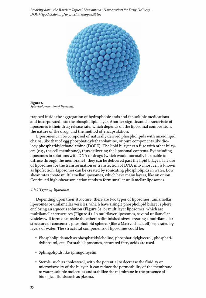

Role of Novel Drug Delivery Vehicles in Nanobiomedicine
36
• Synthetic phospholipids (saturated and unsaturated).
• Polymeric materials. Polymerized liposomes have significantly higher perme-ability barriers to entrap aqueous drugs.
• Polymer-bearing lipids. Coating liposome surfaces with charged polymers results in repulsion interactions with macromolecules.
• Cationic lipids such as dioctadecyldimethylammonium bromide or chloride.
Figure 3. Single-layer liposome.
Figure 4. Multilayer liposome.
37
Breaking down the Barrier: Topical Liposomes as Nanocarriers for Drug Delivery…DOI: http://dx.doi.org/10.5772/intechopen.86601
4.6.2 Size of liposomes
Due to differences in preparation methods and lipid compositions, liposomes may be classified, according to their size, in small (<100 nm), medium (100–250 nm), or large (>250 nm) [121, 122]. Some liposomes may be as small as <0.1 μm or as big as >1 mm in size.
4.6.3 Preparation method (incorporating drugs)
In addition to the ability to entrap drugs with different solubility character-istics, it has been hypothesized that liposomes have different release kinetics. In general, multilamellar liposomes are more easily formed at larger hydrodynamic diameters, and, therefore, they have greater entrapped volumes than unilamellar liposomes. As a result of this, unilamellar liposomes with a hydrodynamic diameter of 130 nm exhibit a much faster release rate than multilamellar liposomes with two to three lamellar bilayers and a hydrodynamic diameter of 250 nm [123, 124]. The difference in the release rate is overall due to the number of phospholipid bilayers that it has to cross before being released [123, 124]. The ongoing interest of researchers in liposomal characteristics such as their stability, pharmacokinetic properties, and therapeutic efficacy has led to second-generation liposomes through the modulation of their lipid composition, size, and the charge of the vesicle. The addition of cholesterol to the lipid bilayer of liposomes reduces their permeability, increases their in vivo and in vitro stability, and can be used to anchor other mol-ecules such as polyethylene glycol (PEG) or deoxyribonucleic acid (DNA) to the liposomes for their application in biosensing or as “stealth” drug carriers [124]. The use of phosphatidylcholine with saturated fatty acyl chains and materials that stretch transition temperatures beyond 37°C offered even greater stabilization [115, 125]. Furthermore, hydrophilic carbohydrates or polymers, such as monosialogan-glioside (GM1) and PEG, were included in the liposomal composition. GM1 can lead to the prolongation of the in vivo liposome viability time [126–129].
There are many different methods to prepare liposomes. According to Bozzuto and Molinari [130], the choice of the method depends on factors such as the physicochemical characteristics of liposomes and/or drug components; the toxicity and concentration of the loaded drug; the type of medium in which liposomes are dispersed; the additional process during the application/delivery of liposomes; the desired size for the application; the half-life desired for successful application; costs, reproducibility, and applicability regarding large-scale production for clinical pur-pose; and good manufacturing practice-relevant issues. In addition, the target organ is a significant issue to be considered when planning the preparation of liposomes.
Every method for preparing liposomes involves four basic stages: drying down lipids from an organic solvent, dispersing the lipid in aqueous media, purifying the resultant liposome, and analyzing the final product. The most common methods for producing liposomes, according to Gabizon et al. [123, 128] and Akbarzadeh et al., [131] are the following.
5. Mechanical dispersion method
5.1 Handshaking and non-handshaking method
In order to produce liposomes, lipid molecules must be introduced into an aque-ous environment. When a dry lipid layer film is hydrated, lamellae swell and grow

Role of Novel Drug Delivery Vehicles in Nanobiomedicine
38
into myelin figures. Mechanical agitation provided by vortexing, shaking, swirling, or pipetting causes myelin figures to break and reseal the exposed hydrophobic edges, resulting in the formation of liposomes, which can be made by using the handshaking method.
5.2 French pressure cell method
It involves the extrusion of multilamellar vesicles (MLV) through a small orifice. The method involves the gentle handling of unstable materials. The resulting liposomes are larger than those generated by using the sonicated method. However, high temperatures are difficult to obtain and working volumes are small.
5.3 Freeze-thawed liposome method
The creation of unilamellar vesicles is a result of a fusion of UV throughout the processes of rapidly freezing and slowly thawing. The encapsulation rates go from 20 to 30%.
5.4 The sonication method
The sonication method is the most used method for the preparation of lipo-somes (unilamellar). Both techniques are probe sonication, in which the tip sonicator is directly immersed into the liposome. Dispersion is high and there is overheating, so the vessel is submerged in an ice bath. With this technique, up to 5% of the lipids can be de-esterified after 1 h. The bath sonicator is the second type of sonication method, and the dispersion of liposomes in a tube is placed into a bath sonicator. The regulation of the temperature is easier, and the lipid bilayer of liposomes can fuse with other bilayers, thus delivering the liposomal contents. By making lipo-somes within drug solutions, they can be delivered past lipid bilayers.
6. Solvent dispersion method
6.1 Ether injection method
Ether injection method is also known as solvent vaporization. A solution of lipids dissolved in diethyl ether or an ether-methanol mixture is injected into an aqueous solution of the drug in order to be encapsulated at 55–65°C or under reduced pressures. The removal of ether under vacuum leads to the formation of liposomes.
6.2 Ethanol injection method
A lipid solution of ethanol is rapidly injected to a huge buffer excess. Multilamellar vesicles are at once formed. Unfortunately, the population is not homogeneous with this method.
6.3 Reverse-phase evaporation method
Reverse-phase evaporation method is based on the formation of inverted micelles. These inverted micelles are formed upon the sonication of a mixture of a buffered aqueous phase, which contains the water-soluble molecules to be encapsu-lated into the liposomes, and an organic phase, in which the amphiphilic molecules
39
Breaking down the Barrier: Topical Liposomes as Nanocarriers for Drug Delivery…DOI: http://dx.doi.org/10.5772/intechopen.86601
are solubilized. The slow removal of organic solvent leads to the transformation of these inverted micelles into a gel-like, viscous state. After the gel-state collapses, some of the inverted micelles disintegrate, but the excess of phospholipids in the environment contributes to the formation of a complete bilayer around the remain-ing micelles, which results in the formation of liposomes.
7. Detergent removal method
7.1 Dialysis method
As the detergent is detached, the micelles increasingly become better-off in phospholipids, and, lastly, they combine to form UVs.
7.2 Detergent removal of mixed micelle method
The absorption method is attained by shaking mixed micelle solution with beaded organic polystyrene absorbers.
7.3 Gel permeation chromatography method
In this method, the detergent is depleted through size special chromatography.
7.4 Dilution method
The spontaneous transition from polydispersed micelles to vesicles occurs due to the dilution of the aqueous micellar solution of detergent and phospholipids with buffer.
7.5 Stealth liposomes and conventional liposomes
Liposomes become known by the mononuclear phagocytic system following contact with plasma proteins. This is solved through the use of synthetic phospho-lipids, particle coated with amphipathic polyethylene glycol, coating liposomes with chitin derivatives, freeze drying, polymerization, and microencapsulation of gangliosides. A stealth liposome is a sphere-shaped vesicle with a membrane that is composed of a phospholipid bilayer used to deliver drugs or genetic material to a cell.
Drug loading can be achieved through passive (if the drug is encapsulated during liposome formation) or active methods (after liposome formation). Freeze-dried (lyophilization) liposomes are formed from preformed liposomes at tremendously low pressures. Very high encapsulation efficiencies, even for macromolecules, can be achieved using this method. During dehydration, the lipid bilayers and the drug to be encapsulated into the liposomes are brought into close contact. Upon reswelling, the chances for the encapsulation of the adhered molecules are much higher. Rehydration is a very important step and it should be very carefully done. The aqueous phase should be added in very small portions with a micropipette to the dried materials. After each addition, the tube should be thoroughly vortexed.
7.5.1 Transportation mechanisms
Liposomes can interact with cells through different mechanisms [129]:

Role of Novel Drug Delivery Vehicles in Nanobiomedicine
40
Endocytosis. Carried out by phagocytic cells of the reticuloendothelial system such as macrophages and neutrophils.
Adsorption to the cell surface either through nonspecific weak hydrophobic or electrostatic forces or specific interactions with cell surface components.
Fusion with the plasmatic cell membrane through the insertion of the lipid bilayer of the liposome into the plasma membrane with the simultaneous release of the liposomal content into the cytoplasm.
Transfer of liposomal lipids to cellular or subcellular membranes, or vice versa, without any association of liposomal contents.
7.5.2 Evaluation
Liposomal formulation and processing for a specific purpose are character-ized for ensuring their predictable in vitro and in vivo performance [132]. Characterization parameters for the purpose of evaluation may be classified into three broad categories that include:
Physical characterization. It evaluates the size, shape, surface features, lamellar-ity, phase behavior, and drug release profile.
Chemical characterization. It includes studies in order to establish the purity and potency of various lipophilic constituents.
Biological characterization. Biological characterization is helpful in establishing the safety and suitability of the formulation for therapeutic applications.
Some parameters are the vesicle’s shape and lamellarity, vesicle size and size distribution, encapsulation efficiency (expressed as percentage (%)), phase response, and transitional behavior and drug release. Zeta potential (ZP) refers to the potential difference between the electric double layer (EDL) (an adsorbed double layer developed on the surface of dispersed charged particles) of movable particles and the layer of dispersant around them at the slipping plane. A stabilized nanosuspension is a suspension that may be affected by several factors such as pH, ionic strength, and the concentration of particles. The phospholipid composition of liposomes is the main content that influences the overall surface charge of liposomes.
7.5.3 Applications of liposomes in medicine
Liposomes are versatile carriers for the delivery of numerous challenging mol-ecules, and they have remarkable advantages compared to other colloidal systems. They have been investigated for a wide range of applications in pharmaceutical technology through topical, transdermal, nasal, and oral routes for efficient and effective drug delivery. Liposome formulations have potential advantages that other drug delivery systems do not have:
i. Suitable for the delivery of hydrophobic, amphipathic, and hydrophilic drugs.
ii. They protect the encapsulated drug from the external environment.
iii. Reduced toxicity and increased stability.
iv. Increased efficacy and therapeutic index of the drug.
v. The sustained-release system of systematically or locally administered liposomes.
41
Breaking down the Barrier: Topical Liposomes as Nanocarriers for Drug Delivery…DOI: http://dx.doi.org/10.5772/intechopen.86601
vi. Improved transfer of hydrophilic, charged molecules.
vii. Improved penetration into the tissues.
viii. Site avoidance mechanism.
ix. They offer site-specific targeting.
x. Nontoxic, flexible, biocompatible, and completely biodegradable.
xi. Size may vary in order to incorporate smaller or larger drug molecules.
xii. They can be administered through various routes.
xiii. The therapeutic activity of chemotherapeutic agents may be improved through liposomal encapsulation. This reduces deleterious effects similar to or lower than those required for maximum therapeutic activity.
xiv. They help reduce exposure of sensitive tissues to toxic drugs.
Some of the disadvantages of liposomes are:
i. Production cost is high.
ii. Leakage and fusion of encapsulated drug molecules with certain preparation methods.
iii. Short half-life.
iv. Less stability.
v. Low solubility.
vi. Phospholipids undergo oxidation and hydrolysis.
vii. Leakage and fusion.
viii. Allergic reactions to liposomal constituents may occur.
Liposomal formulations have several applications in cancer chemotherapy. Due to the nature and behavior of cancer tissues and the large difference between them and regular tissues, cancer tissues are considered an appropriate target for liposome drug delivery systems. For example, the tumor vasculature is character-ized by a leaky vasculature and limited lymphatic drainage; consequently, drug molecules can easily be accumulated in intercellular spaces of a large variety of tumors. Numerous different liposome formulations of several anticancer agents were shown to be less toxic than free drugs [131, 133–135]. Examples of these drugs are doxorubicin and daunorubicin citrate for Kaposi sarcoma; doxorubicin for ovarian cancer and solid tumors; nystatin, vinorelbine, cisplatin and its analog docetaxel, tretinoin, siRNA, topotecan, irinotecan, paclitaxel, and camptothecin for solid tumors; cisplatin and its analog for colorectal neoplasms, Grb-2 in leukemia; Bcl-2 for lymphoma; BikDD for pancreatic cancer; and DOTAP (Chol-Fus I) for the

Role of Novel Drug Delivery Vehicles in Nanobiomedicine
42
treatment of lung cancer [130]. Many other drugs are being researched in order to make them affordable for liposomal drug delivery systems.
In dermatology, liposomes have successfully been used in atopic dermatitis (tax-ifolin glycoside) as antibacterial, antifungal (metronidazole nitrate, amphotericin B), and anti-leishmaniasis treatments (amphotericin B, meglumine antimoniate). Antioxidants such as natural flavonoids (catechin and naringenin) have been used to prevent the oxidation of cutaneous disorders and as photoprotective (quercetin), antipsoriatic, anti-inflammatory agents and anti-acne drugs [136].
In the same context, prostaglandin has been used in peripheral vascular disease, meglumine antimoniate for cutaneous leishmaniasis, fentanyl for pain relief, amikacin in cystic fibrosis, and prilocaine in dental anesthesia, all of them are more examples of liposomal drug products for medical use [136]. Furthermore, antihy-pertensive drugs have been used for the management of cardiovascular disorders (propanol hydrochloride, valsartan, and nifedipine have been developed in liposo-mal formulations) [136]. There are publications regarding local anesthetic drugs [137–139], drug delivery to the brain in anti-migraine and anti-Parkinson drugs [140–143], and the nasal delivery of liposomal formulations (salbutamol).
The application of liposomes in vaccine formulations and toxoids is one of the main achievements of modern medicine. Vaccination activates particular parts of the immune system in order for it to express specific immune responses followed by the induction of long-lasting immunological memories to defend against subse-quent infectious attacks [144]. Most available immunizations are intramuscularly delivered, which is painful and requires an aseptic technique, as well as skilled and trained personnel for their administration. Thus, biological products (vaccines and toxoids) are suitable as a noninvasive approach compared to conventional methods, and they have numerous obvious advantages such as increased patient compliance, reduced systemic side effects, and constant plasma concentrations. Ding et al. reported on antigens such as vaccines and toxoids. Depending on the type of antigens, the dose to be delivered, immunization schedule, the presence of co-stimulatory factors, and liposomal composition, the immunization of antigens loaded in ELs elicits an effective immune response with serum IgG levels compa-rable to those obtained after subcutaneous injections [145–148]. Transcutaneous immunization (TCI) is a novel technique, and it requires the simple introduction of antigens into the host tissue through topical application on the skin [148]. This offers ease of administration and the potential to elicit a robust immune response as compared to conventional painful (needle injections) methods prescribed in equivalent doses [145].
Image-guided delivery is another opportunity area for liposomal formulations. Imaging plays an integral part in modern precision and individualized medi-cine. Wide applications of imaging such as monitoring drug delivery, accurate diagnosis of diseases, determining the response to therapy, and guiding mini-mally invasive procedures are some of the applications of imaging in the clinic. However, traditional imaging modalities, such as computed tomography (CT), positron-emission tomography (PET), magnetic resonance imaging (MRI), and single-photon emission computed tomography (SPECT), all suffer from target specificity, which limits their clinical utility. Nanoparticles, with their versatility in surface functionalization, provide opportunities to enhance target specific-ity and label NPs with various isotopes, which enables them to act as contrast agents. Recent developments in multimodality imaging to better diagnose diseases and monitor treatments have embarked on using liposomes as a diagnostic tool. Conjugating liposomes with different labeling probes enable the precise localiza-tion of these liposomal formulations by using various modalities such as PET, SPECT, and MRI [149].
43
Breaking down the Barrier: Topical Liposomes as Nanocarriers for Drug Delivery…DOI: http://dx.doi.org/10.5772/intechopen.86601
Glucocorticoids (GC) are one of the most popular and versatile classes of drugs available to treat chronic inflammation and cancer, but side effects and resistance constrain their use. In order to overcome these hurdles, which are often related to the uniform tissue distribution of free GC and their short half-life in biological fluids, new delivery vehicles have been developed, including PEGylated liposomes, poly-meric micelles, polymer-drug conjugates, inorganic scaffolds, and hybrid NPs. While each of these nanoformulations has individual drawbacks, they are often superior to free GC in many aspects, including therapeutic efficacy when tested in cell cultures or animal models. The characterization and pharmacokinetics of triamcinolone acetonide-loaded liposomal topical formulations for vitreoretinal drug delivery [150] have been published by Altamirano-Vallejo et al. They showed that a formulation with triamcinolone acetonide-loaded liposomes is feasible and that it has the poten-tial to reach the vitreous cavity with a significant concentration of the drug.
Liposomal vesicles have potential advantages when compared to conventional drug delivery methods. Liposomes in different forms are still one of the most investigated drug delivery carrier systems for the ocular delivery of drugs in both preclinical studies and clinical trials. They seem to be an almost ideal drug-carrier system since their morphology is similar to that of cellular membranes and because of their ability to incorporate various substances. They are valued for their biologi-cal and technological advantages as optimal delivery systems for biologically active substances, both in vitro and in vivo, and are considered to be the most successful drug-carrier system known to date [151].
7.5.4 Liposomes in ophthalmology
Several liposome formulations have been used in ophthalmology to target the anterior segment of the eye. The use of liposomal formulations to enhance the bioavailability of topical applied acyclovir and ganciclovir has been evaluated with promising results [152]. Antibacterial drugs, such as tetracycline, gentamycin, ciprofloxacin, norfloxacin, and chloramphenicol, have been prepared as liposo-mal solutions with higher activity compared to that of standard solutions [153]. Antifungal agents such as amphotericin B and fluconazole have been under research [152], and liposomal formulations were highly effective in treating Candida kerati-tis. Anti-inflammatory drugs and immunomodulatory agents are widely used in the treatment of ocular inflammatory and immunological diseases. In order to enhance ocular bioavailability and reduce the toxic effects following topical or intravitreal administration, liposomal forms of many of these drugs have been evaluated (diclofenac, cyclosporine, tacrolimus) with promising results and better concentra-tions than standard formulations [152, 153]. Similarly, liposomal antiglaucoma agents’ formulations (pilocarpine, latanoprost, acetazolamide) were more effective in reducing intraocular pressure (IOP). In the same context, lubricants and antioxi-dants have also other potential uses for liposomal formulations.
Liposomal drugs that have transitioned from preclinical research to clinical phase trials include latanoprost-loaded conventional liposomes developed for subconjunctival administration [154]. Phase I and II trials on the safety and effi-cacy of latanoprost-loaded liposomes in the treatment of ocular hypertension and primary open-angle glaucoma have been completed (ClinicalTrials.gov identifier, NCT0198732357; ClinicalTrials.gov identifier, NCT02466399).
7.5.5 Topical liposomes for drug delivery into the posterior segment of the eyeball
Different efforts have been performed to deliver drugs into the posterior seg-ment of the eye through the instillation of loaded liposomes (Table 2) [155–163].
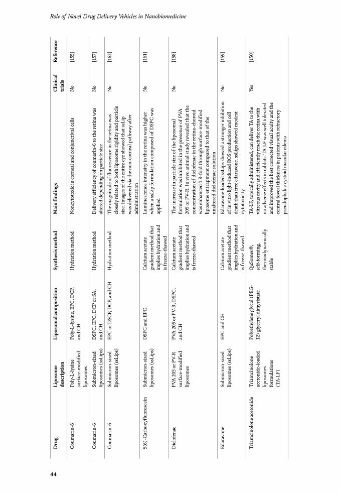
Role of Novel Drug Delivery Vehicles in Nanobiomedicine
44
Dru
gLi
poso
me
desc
ript
ion
Lipo
som
al co
mpo
sitio
nSy
nthe
sis m
etho
dM
ain
find
ings
Clin
ical
tr
ials
Refe
renc
e
Coum
arin
-6Po
ly-L
-lysin
e su
rfac
e-m
odifi
ed
lipos
omes
Poly
-L-ly
sine,
EPC
, DCP
, an
d CH
Hyd
ratio
n m
etho
dN
oncy
toto
xic i
n co
rnea
l and
conj
unct
ival
cells
No
[155
]
Coum
arin
-6Su
bmic
ron-
sized
lip
osom
es (s
sLip
s)D
SPC,
EPC
, DCP
or S
A,
and
CHH
ydra
tion
met
hod
Del
iver
y ef
ficie
ncy
of co
umar
in-6
to th
e ret
ina w
as
alte
red
depe
ndin
g on
par
ticle
size
No
[157
]
Coum
arin
-6Su
bmic
ron-
sized
lip
osom
es (s
sLip
s)EP
C or
DSC
P, D
CP, a
nd C
HH
ydra
tion
met
hod
The m
agni
tude
of f
luor
esce
nce i
n th
e ret
ina w
as
clos
ely
rela
ted
to b
oth
lipos
ome r
igid
ity an
d pa
rtic
le
size.
Imag
es o
f the
entir
e eye
show
ed th
at ss
Lip
was
del
iver
ed v
ia th
e non
-cor
neal
pat
hway
afte
r ad
min
istra
tion
No
[162
]
5(6)
-Car
boxy
fluor
esce
inSu
bmic
ron-
sized
lip
osom
es (s
sLip
s)D
SPC
and
EPC
Cal
cium
acet
ate
grad
ient
met
hod
that
im
plie
s hyd
ratio
n an
d is
free
ze-t
haw
ed
Lum
ines
cenc
e int
ensi
ty in
the r
etin
a was
hig
her
whe
n a s
sLip
form
ulat
ion
com
pose
d of
DSP
C w
as
appl
ied
No
[161
]
Dic
lofe
nac
PVA
205 o
r PV-
R su
rfac
e-m
odifi
ed
lipos
omes
PVA
205 o
r PV-
R, D
SPC,
an
d CH
Cal
cium
acet
ate
grad
ient
met
hod
that
im
plie
s hyd
ratio
n an
d is
free
ze-t
haw
ed
The i
ncre
ase i
n pa
rtic
le si
ze o
f the
lipo
som
al
form
ulat
ion
was
inhi
bite
d in
the p
rese
nce o
f PVA
20
5 or
PV-
R. In
viv
o an
imal
stud
y re
veal
ed th
at th
e co
ncen
trat
ion
of d
iclo
fena
c in
the r
etin
a-ch
oroi
d w
as e
nhan
ced
1.8-
fold
thro
ugh
surf
ace-
mod
ified
lip
osom
e ent
rapm
ent c
ompa
red
to th
at o
f the
un
alte
red
dicl
ofen
ac so
lutio
n
No
[158
]
Edar
avon
eSu
bmic
ron-
sized
lip
osom
es (s
sLip
s)EP
C an
d CH
Cal
cium
acet
ate
grad
ient
met
hod
that
im
plie
s hyd
ratio
n an
d is
free
ze-t
haw
ed
Edar
avon
e-lo
aded
ssLi
ps sh
owed
a st
rong
er in
hibi
tion
of in
vitr
o lig
ht-in
duce
d RO
S pr
oduc
tion
and
cell
deat
h th
an fr
ee ed
arav
one.
ssLi
ps sh
owed
mod
est
cyto
toxi
city
No
[159
]
Tria
mci
nolo
ne ac
eton
ide
Tria
mci
nolo
ne
acet
onid
e-lo
aded
lip
osom
es
form
ulat
ions
(T
A-LF
)
Poly
ethy
lene
gly
col (
PEG
-12
) gly
cery
l dim
yrist
ate
QuS
omes
®;
self-
form
ing,
th
erm
odyn
amic
ally
st
able
TA-L
F, to
pica
lly ad
min
ister
ed, c
an d
eliv
er T
A to
the
vitr
eous
cavi
ty an
d ef
ficie
ntly
reac
h th
e ret
ina w
ith
no ad
vers
e eff
ects
in ra
bbits
. TA-
LF w
as w
ell t
oler
ated
an
d im
prov
ed th
e bes
t cor
rect
ed v
isual
acui
ty an
d th
e ce
ntra
l fov
eal t
hick
ness
in p
atie
nts w
ith re
frac
tory
ps
eudo
phak
ic cy
stoi
d m
acul
ar ed
ema
Yes
[150
]
45
Breaking down the Barrier: Topical Liposomes as Nanocarriers for Drug Delivery…DOI: http://dx.doi.org/10.5772/intechopen.86601
Dru
gLi
poso
me
desc
ript
ion
Lipo
som
al co
mpo
sitio
nSy
nthe
sis m
etho
dM
ain
find
ings
Clin
ical
tr
ials
Refe
renc
e
Tria
mci
nolo
ne ac
eton
ide
Tria
mci
nolo
ne
acet
onid
e ch
itosa
n-co
ated
lip
osom
es
(TA-
CHL)
Chito
san,
ph
osph
atid
ylch
olin
e, an
d CH
Cal
cium
acet
ate
grad
ient
met
hod
that
im
plie
s hyd
ratio
n an
d is
free
ze-t
haw
ed
TA-C
HL
pene
trat
es in
to th
e pos
terio
r seg
men
t of t
he
eye.
Chi
tosa
n-co
ated
lipo
som
es w
ere a
mor
e eff
icie
nt
ocul
ar d
eliv
ery
syst
em o
f tria
mci
nolo
ne ac
eton
ide t
o th
e pos
terio
r seg
men
t of t
he ey
e as e
ye d
rops
than
non
-co
ated
lipo
som
es
No
[164
]
Beva
cizu
mab
Ann
exin
A
5-su
rfac
e-m
odifi
ed
lipos
omes
Phos
phat
idyl
serin
e,
phos
phat
idyl
etha
nola
min
e,
and
anio
nic p
hosp
holip
id-
bind
ing
prot
ein
anne
xin
A5
Cal
cium
acet
ate
grad
ient
met
hod
that
im
plie
s hyd
ratio
n an
d is
free
ze-t
haw
ed
Ann
exin
A5-
surf
ace-
mod
ified
lipo
som
es d
eliv
er
phys
iolo
gica
lly si
gnifi
cant
conc
entr
atio
ns o
f be
vaci
zum
ab to
the p
oste
rior s
egm
ent o
f rat
eyes
(1
27 n
g/g)
and
rabb
it re
tinas
(18
ng/g
). A
nnex
in
A5-
med
iate
d en
docy
tosis
enha
nces
the d
eliv
ery
of
beva
cizu
mab
No
[156
]
pDN
ApD
NA
/PEI
co
mpl
ex-lo
aded
lip
osom
es
mod
ified
with
tr
ansf
errin
EPC
and
CHD
eter
gent
rem
oval
m
etho
dTh
e mod
ifica
tion
of li
gand
(tra
nsfe
rrin
), w
hich
bin
ds
to a
spec
ific r
ecep
tor i
n RP
E ce
lls to
the l
ipos
omes
, im
prov
es g
ene d
eliv
ery
effic
acy
to th
e pos
terio
r se
gmen
t of t
he ey
e
No
[160
]
Plas
mid
DN
A w
ith
β-ga
lact
osid
ase g
ene
TMAG
lipo
som
e an
d D
C-C
H
lipos
ome
TMAG
lipo
som
es, T
MAG
an
d D
OPE
; DC
-CH
lip
osom
e, D
C-c
hole
ster
ol
and
DO
PE
Hyd
ratio
n m
etho
dG
ene e
xpre
ssio
n w
as fo
und
in re
tinal
gan
glio
n ce
ll un
til 1
mon
th af
ter t
he to
pica
l app
licat
ion
of li
poso
mes
No
[163
]
CH
, ch
oles
tero
l; D
C,
dice
tyl
phos
phat
e; D
C-c
holes
tero
l, 3b
eta
[N-(
N′-N
′-dim
ethy
lam
inoe
than
e)-c
arba
myl
] ch
oles
tero
l; D
LPC
, di
laur
oylp
hosp
hatid
ylch
olin
e; D
OPE
, di
oleo
ylph
osph
atid
ylet
hano
lam
ine;
D
SPC
, L-α
-dist
earo
yl p
hosp
hatid
ylch
olin
e; E
PC, e
gg p
hosp
hatid
ylch
olin
e; P
EI, p
olye
thyl
enim
ine;
PVA
205
, pol
yvin
yl a
lcoho
l; PV
A-R
, pol
yvin
yl a
lcoho
l der
ivat
ives
bea
ring
a h
ydro
phob
ic a
ncho
r (C
16H
33 S
) at
the t
erm
inal
of t
he m
olec
ule;
SA
, stea
ryl a
min
e; T
MAG
, N-(
alph
a-tr
imet
hyl a
mm
onia
cety
l)-d
idod
ecyl
-D-g
luta
mat
e.
Tabl
e 2.
Re
port
ed li
poso
mes
for d
rug d
elive
ry to
the p
oste
rior
segm
ent o
f the
eyeb
all.
Role of Novel Drug Delivery Vehicles in Nanobiomedicine
46
For example, fluorescent probes used as drug models, such as coumarin-6 and 5(6)-carboxyfluorescein, have efficiently been released into the posterior segment of the eyeball by liposomes [155, 157, 161, 162]. On the other hand, drugs like edaravone and diclofenac were successfully released into the vitreous and the retina by liposomes [158, 159].
Special mention is reserved for the study performed by Davis BM et al. This group demonstrated that the topical instillation of eye drops containing annexin A5 associated with liposomes loaded with bevacizumab is able to deliver physi-ologically significant concentrations of this large therapeutic protein (monoclonal antibody against vascular endothelial growth factor A) into the posterior segment of the eye in animal models (rats and rabbits) [156]. Moreover, liposomes can release genetic material into the vitreous and the retina [160, 163].
Lastly, a topical triamcinolone acetonide-loaded liposomes formulation (TA-LF) was used to successfully deliver TA into the vitreous and the retina of rabbits. Besides the authors report that TA-LF was well tolerated by the study animals and that no toxicity was observed in cell culture assays and no adverse events like corneal and conjunctival erosions were observed [150]. Recently, Jin Li et al. [164] validated in animal models (mice) that eye drops containing chitosan-coated liposomes carrying TA are an efficient method to deliver this drug into the pos-terior segment of rabbit eyeballs, supporting the previous findings published by Altamirano-Vallejo et al. Even though Li et al. reached superior TA entrapment efficiency in their TA-loaded liposomes prepared through the calcium acetate gradi-ent method, it seems that this characteristic does not compromise the therapeutic activity of TA-LF. TA-LF has been tested in clinical assays where its efficiency and safety profile have satisfactorily been demonstrated. In a recent report, TA-LF was efficient in the management of refractory pseudophakic cystoid macular edema [20], where the use of this formulation was associated with the improvement of best corrected visual acuity and central foveal thickness with no reports of adverse events. At this time, phase II trials are underway to demonstrate the efficacy of TA-LF for macular edema.
8. Conclusion
Although there has been great interest in the development of new topical ocular delivery systems, the topical administration of drugs is not as effective as intravit-real delivery in treating retinal diseases. During topical delivery, drug molecules must cross several anatomic and physiologic barriers before reaching the posterior segment of the eye. Consequently, very low concentrations, with almost no clinical effect, are usually obtained. However, dramatic changes have been observed in the field of ocular drug delivery over the last decade, and nanomedicine is one of the most promising technologies for efficient drug delivery into intraocular tissues. Nanocarriers have a great potential to solve the challenge of releasing drugs into the posterior segment of the eyeball and reaching deeper intraocular tissues. Different nanocarriers, such as NPs, LPs, micelles, dendrimers, cyclodextrin nanoparticles, and liposomes, have been developed for the safe and effective drug delivery to the vitreous and the retina. Nevertheless, only a few of these particles have clinically been approved (liposomes and CDs among them).
Liposomes have lately been of great interest as carriers for advanced drug deliv-ery in medicine and, especially, in ophthalmology due to their potential to avoid sophisticated ocular barriers when they are topically used. Liposomal drug delivery systems have a bright future in ophthalmology and, particularly, for the retina for several reasons. First, features such as the passive-targeting effect may substantially
47
Breaking down the Barrier: Topical Liposomes as Nanocarriers for Drug Delivery…DOI: http://dx.doi.org/10.5772/intechopen.86601
Author details
Arturo Santos1,2*, Juan C. Altamirano-Vallejo1,2, José Navarro-Partida1,2, Alejandro González-De la Rosa1,2 and Jane H. Hsiao3
1 Tecnológico de Monterrey, Escuela de Medicina y Ciencias de la Salud, Zapopan, Jalisco, México
2 Centro de Retina Médica y Quirúrgica S.C., Centro Médico Puerta de Hierro, Zapopan, Jalisco, México
3 OPKO Health Inc., Miami, Florida, United States of America
*Address all correspondence to: [email protected]
enhance the amount of drug in target tissues. Second, liposomal technology allows the therapeutic index of already established drugs, with well-established therapeutic profiles, to be improved. This eliminates some of the considerable risks associated with the development of new pharmaceutical products. Third, many of the potential new pharmaceutical products arising from advances in biotechnology would come from macromolecules such as proteins, peptides, oligonucleotides, and plasmids, which could easily be carried by liposomes so as to access target tissues and cells. Additionally, liposomes possess features of optimal drug delivery systems to get to the ocular surface and intraocular tissues, for instance, sustained and con-trolled drug delivery, ease to instill for the patient, good corneal-scleral penetration, and the fact that they reach appropriate concentrations of the drug in the target tissue, avoid side effects produced by conventional systems (like IVT injections), there is better patient compliance, and they are potentially affordable for patients. A proof of the clinical advantages of liposomes is represented by recent trials where TA-loaded liposomes have successfully been used to treat macular edema.
Considering that liposomes collect the main characteristics of an optimal drug delivery system and that these nanocarriers have proven their capacity not only to deliver drugs but also genetic materials to the posterior segment of the eye, we consider that liposomes are promising nanoparticles for the therapy of multiple intraocular illnesses.
© 2019 The Author(s). Licensee IntechOpen. This chapter is distributed under the terms of the Creative Commons Attribution License (http://creativecommons.org/licenses/by/3.0), which permits unrestricted use, distribution, and reproduction in any medium, provided the original work is properly cited.

48
Role of Novel Drug Delivery Vehicles in Nanobiomedicine
References
[1] Jiang S, Franco YL, Zhou Y, Chen J. Nanotechnology in retinal drug delivery. International Journal of Ophthalmology. 2018;11:1038-1044
[2] Kim SH, Lutz RJ, Wang NS, Robinson MR. Transport barriers in transscleral drug delivery for retinal diseases. Ophthalmic Research. 2007;39:244-254
[3] Raghava S, Hammond M, Kompella UB. Periocular routes for retinal drug delivery. Expert Opinion on Drug Delivery. 2004;1:99-114
[4] Ruponen M, Urtti A. Undefined role of mucus as a barrier in ocular drug delivery. European Journal of Pharmaceutics and Biopharmaceutics. 2015;96:442-446
[5] Leong YY, Tong L. Barrier function in the ocular surface: From conventional paradigms to new opportunities. The Ocular Surface. 2015;13:103-109
[6] Grzybowski A, Told R, Sacu S, Bandello F, Moisseiev E, Loewenstein A, et al. 2018 update on intravitreal injections: Euretina expert consensus recommendations. Ophthalmologica. 2018;239:181-193
[7] Young S, Larkin G, Branley M, Lightman S. Safety and efficacy of intravitreal triamcinolone for cystoid macular oedema in uveitis. Clinical & Experimental Ophthalmology. 2001;29:2-6
[8] Smith JR, Rosenbaum JT, Wilson DJ, Doolittle ND, Siegal T, Neuwelt EA, et al. Role of intravitreal methotrexate in the management of primary central nervous system lymphoma with ocular involvement. Ophthalmology. 2002;109:1709-1716
[9] Davis M. Results of the Endophthalmitis Vitrectomy Study. A randomized trial of immediate
vitrectomy and of intravenous antibiotics for the treatment of postoperative bacterial endophthalmitis. Endophthalmitis Vitrectomy Study group. Archives of Ophthalmology. 1995;113:1479-1496
[10] Eyetech Study G. Anti-vascular endothelial growth factor therapy for subfoveal choroidal neovascularization secondary to age-related macular degeneration: Phase II study results. Ophthalmology. 2003;110:979-986
[11] Wu H, Chen TC. The effects of intravitreal ophthalmic medications on intraocular pressure. Seminars in Ophthalmology. 2009;24:100-105
[12] Lyall DA, Tey A, Foot B, Roxburgh ST, Virdi M, Robertson C, et al. Post-intravitreal anti-VEGF endophthalmitis in the United Kingdom: Incidence, features, risk factors, and outcomes. Eye. 2012;26:1517-1526
[13] Poku E, Rathbone J, Wong R, Everson-Hock E, Essat M, Pandor A, et al. The safety of intravitreal bevacizumab monotherapy in adult ophthalmic conditions: Systematic review. BMJ Open. 2014;4:e005244
[14] Fung AE, Rosenfeld PJ, Reichel E. The international intravitreal bevacizumab safety survey: Using the internet to assess drug safety worldwide. The British Journal of Ophthalmology. 2006;90:1344-1349
[15] Droege KM, Muether PS, Hermann MM, Caramoy A, Viebahn U, Kirchhof B, et al. Adherence to ranibizumab treatment for neovascular age-related macular degeneration in real life. Graefe’s Archive for Clinical and Experimental Ophthalmology. 2013;251:1281-1284
[16] Azad R, Chandra P, Gupta R. The economic implications of the use
49
Breaking down the Barrier: Topical Liposomes as Nanocarriers for Drug Delivery…DOI: http://dx.doi.org/10.5772/intechopen.86601
of anti-vascular endothelial growth factor drugs in age-related macular degeneration. Indian Journal of Ophthalmology. 2007;55:441-443
[17] Bisht R, Mandal A, Jaiswal JK, Rupenthal ID. Nanocarrier mediated retinal drug delivery: Overcoming ocular barriers to treat posterior eye diseases. Wiley Interdisciplinary Reviews Nanomedicine and Nanobiotechnology. 2018;10:1-21
[18] Duncan R, Gaspar R. Nanomedicine(s) under the microscope. Molecular Pharmaceutics. 2011;8:2101-2141
[19] Cho K, Wang X, Nie S, Chen ZG, Shin DM. Therapeutic nanoparticles for drug delivery in cancer. Clinical Cancer Research. 2008;14:1310-1316
[20] Gonzalez-De la Rosa A, Navarro-Partida J, Altamirano-Vallejo JC, Hernandez-Gamez AG, Garcia-Banuelos JJ, Armendariz-Borunda J, et al. Novel triamcinolone acetonide-loaded liposomes topical formulation for the treatment of cystoid macular edema after cataract surgery: A pilot study. Journal of Ocular Pharmacology and Therapeutics. 2019;35:106-115
[21] Ohira A, Hara K, Johannesson G, Tanito M, Asgrimsdottir GM, Lund SH, et al. Topical dexamethasone gamma-cyclodextrin nanoparticle eye drops increase visual acuity and decrease macular thickness in diabetic macular oedema. Acta Ophthalmologica. 2015;93:610-615
[22] Gaudana R, Ananthula HK, Parenky A, Mitra AK. Ocular drug delivery. The AAPS Journal. 2010;12:348-360
[23] Barar J, Javadzadeh AR, Omidi Y. Ocular novel drug delivery: Impacts of membranes and barriers. Expert Opinion on Drug Delivery. 2008;5:567-581
[24] Boubriak OA, Urban JP, Akhtar S, Meek KM, Bron AJ. The effect of
hydration and matrix composition on solute diffusion in rabbit sclera. Experimental Eye Research. 2000;71:503-514
[25] Hamalainen KM, Kananen K, Auriola S, Kontturi K, Urtti A. Characterization of paracellular and aqueous penetration routes in cornea, conjunctiva, and sclera. Investigative Ophthalmology & Visual Science. 1997;38:627-634
[26] Kao JC, Geroski DH, Edelhauser HF. Transscleral permeability of fluorescent-labeled antibiotics. Journal of Ocular Pharmacology and Therapeutics. 2005;21:1-10
[27] Shuler RK Jr, Dioguardi PK, Henjy C, Nickerson JM, Cruysberg LP, Edelhauser HF. Scleral permeability of a small, single-stranded oligonucleotide. Journal of Ocular Pharmacology and Therapeutics. 2004;20:159-168
[28] Cruysberg LP, Franklin AJ, Sanders J, Self C, Yuan P, Csaky KG, et al. Effective transscleral delivery of two retinal anti-angiogenic molecules: Carboxyamido-triazole (CAI) and 2-methoxyestradiol (2ME2). Retina. 2005;25:1022-1031
[29] Cheruvu NP, Kompella UB. Bovine and porcine transscleral solute transport: Influence of lipophilicity and the choroid-Bruch’s layer. Investigative Ophthalmology & Visual Science. 2006;47:4513-4522
[30] Cruysberg LP, Nuijts RM, Geroski DH, Koole LH, Hendrikse F, Edelhauser HF. In vitro human scleral permeability of fluorescein, dexamethasone-fluorescein, methotrexate-fluorescein and rhodamine 6G and the use of a coated coil as a new drug delivery system. Journal of Ocular Pharmacology and Therapeutics. 2002;18:559-569
[31] Prausnitz MR, Noonan JS. Permeability of cornea, sclera, and conjunctiva: A literature analysis

Role of Novel Drug Delivery Vehicles in Nanobiomedicine
50
for drug delivery to the eye. Journal of Pharmaceutical Sciences. 1998;87:1479-1488
[32] Ambati J, Canakis CS, Miller JW, Gragoudas ES, Edwards A, Weissgold DJ, et al. Diffusion of high molecular weight compounds through sclera. Investigative Ophthalmology & Visual Science. 2000;41:1181-1185
[33] Ahmed I, Gokhale RD, Shah MV, Patton TF. Physicochemical determinants of drug diffusion across the conjunctiva, sclera, and cornea. Journal of Pharmaceutical Sciences. 1987;76:583-586
[34] Olsen TW, Edelhauser HF, Lim JI, Geroski DH. Human scleral permeability. Effects of age, cryotherapy, transscleral diode laser, and surgical thinning. Investigative Ophthalmology & Visual Science. 1995;36:1893-1903
[35] Larsson BS. Interaction between chemicals and melanin. Pigment Cell Research. 1993;6:127-133
[36] Schoenwald RD, Tandon V, Wurster DE, Barfknecht CF. Significance of melanin binding and metabolism in the activity of 5-acetoxyacetylimino-4-methyl-delta2-1,3,4,-thiadiazolin e-2-sulfonamide. European Journal of Pharmaceutics and Biopharmaceutics. 1998;46:39-50
[37] Leblanc B, Jezequel S, Davies T, Hanton G, Taradach C. Binding of drugs to eye melanin is not predictive of ocular toxicity. Regulatory Toxicology and Pharmacology. 1998;28:124-132
[38] Pitkanen L, Ranta VP, Moilanen H, Urtti A. Binding of betaxolol, metoprolol and oligonucleotides to synthetic and bovine ocular melanin, and prediction of drug binding to melanin in human choroid-retinal pigment epithelium. Pharmaceutical Research. 2007;24:2063-2070
[39] Salminen L, Imre G, Huupponen R. The effect of ocular pigmentation on intraocular pressure response to timolol. Acta Ophthalmologica Supplement. 1985;173:15-18
[40] Gipson IK, Argueso P. Role of mucins in the function of the corneal and conjunctival epithelia. International Review of Cytology. 2003;231:1-49
[41] Ahmed I, Patton TF. Importance of the noncorneal absorption route in topical ophthalmic drug delivery. Investigative Ophthalmology & Visual Science. 1985;26:584-587
[42] Patel A, Cholkar K, Agrahari V, Mitra AK. Ocular drug delivery systems: An overview. World Journal of Pharmacology. 2013;2:47-64
[43] Urtti A. Challenges and obstacles of ocular pharmacokinetics and drug delivery. Advanced Drug Delivery Reviews. 2006;58:1131-1135
[44] Galloway NR. Common eye diseases and their management. American Journal of Ophthalmology. 1985;100:360
[45] Ng J. Ocular Anatomy and Physiology; Thorofare, NJ: SLACK, Incorporated; 2009
[46] Djebli N, Khier S, Griguer F, Coutant AL, Tavernier A, Fabre G, et al. Ocular drug distribution after topical administration: Population pharmacokinetic model in rabbits. European Journal of Drug Metabolism and Pharmacokinetics. 2017;42:59-68
[47] Agrahari V, Mandal A, Agrahari V, Trinh HM, Joseph M, Ray A, et al. A comprehensive insight on ocular pharmacokinetics. Drug Delivery and Translational Research. 2016;6:735-754
[48] Gruntzig J, Schicha H, Kiem J, Becker V, Feinendegen LE. Studies on the lymph drainage of the eye 5.
51
Breaking down the Barrier: Topical Liposomes as Nanocarriers for Drug Delivery…DOI: http://dx.doi.org/10.5772/intechopen.86601
Quantitative registration of the lymph drainage from the subconjunctival space with a radioactive tracer (author’s transl). Klinische Monatsblätter für Augenheilkunde. 1978;172:872-876
[49] Collin HB. Lymphatic drainage of 131-I-albumin from the vascularized cornea. Investigative Ophthalmology. 1970;9:146-155
[50] Amrite AC, Kompella UB. Size-dependent disposition of nanoparticles and microparticles following subconjunctival administration. The Journal of Pharmacy and Pharmacology. 2005;57:1555-1563
[51] Toris CB, Yablonski ME, Wang YL, Camras CB. Aqueous humor dynamics in the aging human eye. American Journal of Ophthalmology. 1999;127:407-412
[52] Bill A. A method to determine osmotically effective albumin and gammaglobulin concentrations in tissue fluids, its application to the uvea and a note on the effects of capillary “leaks” on tissue fluid dynamics. Acta Physiologica Scandinavica. 1968;73:511-522
[53] Bill A. Blood circulation and fluid dynamics in the eye. Physiological Reviews. 1975;55:383-417
[54] Kennedy BG, Mangini NJ. P-glycoprotein expression in human retinal pigment epithelium. Molecular Vision. 2002;8:422-430
[55] Steuer H, Jaworski A, Elger B, Kaussmann M, Keldenich J, Schneider H, et al. Functional characterization and comparison of the outer blood-retina barrier and the blood-brain barrier. Investigative Ophthalmology & Visual Science. 2005;46:1047-1053
[56] Zhang N, Kannan R, Okamoto CT, Ryan SJ, Lee VH, Hinton DR. Characterization of brimonidine transport in retinal pigment epithelium. Investigative Ophthalmology & Visual Science. 2006;47:287-294
[57] Kumari A, Yadav SK, Yadav SC. Biodegradable polymeric nanoparticles based drug delivery systems. Colloids and Surfaces. B, Biointerfaces. 2010;75:1-18
[58] Dhananjay P. Biology of Ocular Transporters: Efflux and Influx Transporters in the Eye. Sawstone, Cambridge: Woodhead Publishing; 2013
[59] Wrighton SA, Thummel KE. CYP3A. In: Metabolic Drug Interactions. Philadelphia: Lippincott Williams and Wilkins; 2000
[60] Lee VH, Robinson JR. Topical ocular drug delivery: Recent developments and future challenges. Journal of Ocular Pharmacology. 1986;2:67-108
[61] Hughes PM, Olejnik O, Chang-Lin JE, Wilson CG. Topical and systemic drug delivery to the posterior segments. Advanced Drug Delivery Reviews. 2005;57:2010-2032
[62] Duvvuri S, Majumdar S, Mitra AK. Drug delivery to the retina: Challenges and opportunities. Expert Opinion on Biological Therapy. 2003;3:45-56
[63] Ghate D, Edelhauser HF. Ocular drug delivery. Expert Opinion on Drug Delivery. 2006;3:275-287
[64] Ghate D, Brooks W, McCarey BE, Edelhauser HF. Pharmacokinetics of intraocular drug delivery by periocular injections using ocular fluorophotometry. Investigative Ophthalmology & Visual Science. 2007;48:2230-2237
[65] Castellarinand A, Pieramici DJ. Anterior segment complications following periocular and intraocular injections. Ophthalmology Clinics of North America. 2004;17:583-590
[66] Heinemann MH. Long-term intravitreal ganciclovir therapy for cytomegalovirus retinopathy. Archives of Ophthalmology. 1989;107:1767-1772

Role of Novel Drug Delivery Vehicles in Nanobiomedicine
52
[67] Jacobson MA, O’Donnell JJ, Mills J. Foscarnet treatment of cytomegalovirus retinitis in patients with the acquired immunodeficiency syndrome. Antimicrobial Agents and Chemotherapy. 1989;33:736-741
[68] Thrimawithana TR, Young S, Bunt CR, Green C, Alany RG. Drug delivery to the posterior segment of the eye. Drug Discovery Today. 2011;16:270-277
[69] Mitra AK, Anand BS, Duvvuri S. Drug delivery to the eye. In: The Biology of Eye. New York: Academic Press; 2006
[70] Meyer CH, Michels S, Rodrigues EB, Hager A, Mennel S, Schmidt JC, et al. Incidence of rhegmatogenous retinal detachments after intravitreal antivascular endothelial factor injections. Acta Ophthalmologica. 2011;89:70-75
[71] Gaudreault J, Fei D, Rusit J, Suboc P, Shiu V. Preclinical pharmacokinetics of Ranibizumab (rhuFabV2) after a single intravitreal administration. Investigative Ophthalmology & Visual Science. 2005;46:726-733
[72] Stewart MW, Rosenfeld PJ, Penha FM, Wang F, Yehoshua Z, Bueno-Lopez E, et al. Pharmacokinetic rationale for dosing every 2 weeks versus 4 weeks with intravitreal ranibizumab, bevacizumab, and aflibercept (vascular endothelial growth factor trap-eye). Retina. 2012;32:434-457
[73] Zhang K, Zhang L, Weinreb RN. Ophthalmic drug discovery: Novel targets and mechanisms for retinal diseases and glaucoma. Nature Reviews Drug Discovery. 2012;11:541-559
[74] Okabe K, Kimura H, Okabe J, Kato A, Kunou N, Ogura Y. Intraocular tissue distribution of betamethasone after intrascleral administration using a non-biodegradable sustained drug delivery device. Investigative Ophthalmology & Visual Science. 2003;44:2702-2707
[75] Yasukawa T, Ogura Y, Sakurai E, Tabata Y, Kimura H. Intraocular sustained drug delivery using implantable polymeric devices. Advanced Drug Delivery Reviews. 2005;57:2033-2046
[76] Daull P, Paterson CA, Kuppermann BD, Garrigue JS. A preliminary evaluation of dexamethasone palmitate emulsion: A novel intravitreal sustained delivery of corticosteroid for treatment of macular edema. Journal of Ocular Pharmacology and Therapeutics. 2013;29:258-269
[77] Haghjou N, Soheilian M, Abdekhodaie MJ. Sustained release intraocular drug delivery devices for treatment of uveitis. Journal of Ophthalmic Vision Research. 2011;6:317-329
[78] Hunter RS, Lobo AM. Dexamethasone intravitreal implant for the treatment of noninfectious uveitis. Clinical Ophthalmology. 2011;5:1613-1621
[79] London NJ, Chiang A, Haller JA. The dexamethasone drug delivery system: Indications and evidence. Advances in Therapy. 2011;28:351-366
[80] Schwartz SG, Flynn HW Jr. Fluocinolone acetonide implantable device for diabetic retinopathy. Current Pharmaceutical Biotechnology. 2011;12:347-351
[81] Campochiaro PA, Hafiz G, Shah SM, Bloom S, Brown DM, Busquets M, et al. Sustained ocular delivery of fluocinolone acetonide by an intravitreal insert. Ophthalmology. 2010;117:1393-1399
[82] Birch DG, Liang FQ. Age-related macular degeneration: A target for nanotechnology derived medicines. International Journal of Nanomedicine. 2007;2:65-77
[83] Harmia T, Speiser P, Kreuter J. A solid colloidal drug delivery
53
Breaking down the Barrier: Topical Liposomes as Nanocarriers for Drug Delivery…DOI: http://dx.doi.org/10.5772/intechopen.86601
system for the eye: Encapsulation of pilocarpine in nanoparticles. Journal of Microencapsulation. 1986;3:3-12
[84] Harmia TKJ, Speiser P, Boye T, Gurny R, Kubis A. Enhancement of the myotic response of rabbits with pilocarpine-loaded polybutylcyanoacrylate nanoparticles. International Journal of Pharmaceutics. 1986;33:87-193
[85] Kimura H, Ogura Y. Biodegradable polymers for ocular drug delivery. Ophthalmologica. 2001;215:143-155
[86] Kohei Tahara KK, Onodera R, Takeuchi H. Feasibility of drug delivery to the eye’s posterior segment by topical instillation of PLGA nanoparticles. Asian Journal of Pharmaceutical Sciences. 2017;12:394-399
[87] Eljarrat-Binstock E, Orucov F, Aldouby Y, Frucht-Pery J, Domb AJ. Charged nanoparticles delivery to the eye using hydrogel iontophoresis. Journal of Controlled Release. 2008;126:156-161
[88] Almeida AJ, Souto E. Solid lipid nanoparticles as a drug delivery system for peptides and proteins. Advanced Drug Delivery Reviews. 2007;59:478-490
[89] Joshi MD, Muller RH. Lipid nanoparticles for parenteral delivery of actives. European Journal of Pharmaceutics and Biopharmaceutics. 2009;71:161-172
[90] Beloqui A, Solinis MA, Rodriguez-Gascon A, Almeida AJ, Preat V. Nanostructured lipid carriers: Promising drug delivery systems for future clinics. Nanomedicine. 2016;12:143-161
[91] Wissing SA, Kayser O, Muller RH. Solid lipid nanoparticles for parenteral drug delivery. Advanced Drug Delivery Reviews. 2004;56:1257-1272
[92] Mukherjee S, Ray S, Thakur RS. Solid lipid nanoparticles: A modern formulation approach in drug delivery system. Indian Journal of Pharmaceutical Sciences. 2009;71:349-358
[93] Tian B, Luo Q , Song S, Liu D, Pan H, Zhang W, et al. Novel surface-modified nanostructured lipid carriers with partially deacetylated water-soluble chitosan for efficient ocular delivery. Journal of Pharmaceutical Sciences. 2012;101:1040-1049
[94] Teixeira MC, Carbone C, Souto EB. Beyond liposomes: Recent advances on lipid based nanostructures for poorly soluble/poorly permeable drug delivery. Progress in Lipid Research. 2017;68:1-11
[95] Souto EB, Doktorovova S, Gonzalez-Mira E, Egea MA, Garcia ML. Feasibility of lipid nanoparticles for ocular delivery of anti-inflammatory drugs. Current Eye Research. 2010;35:537-552
[96] Araujo J, Nikolic S, Egea MA, Souto EB, Garcia ML. Nanostructured lipid carriers for triamcinolone acetonide delivery to the posterior segment of the eye. Colloids and Surfaces. B, Biointerfaces. 2011;88:150-157
[97] Balguri SP, Adelli GR, Majumdar S. Topical ophthalmic lipid nanoparticle formulations (SLN, NLC) of indomethacin for delivery to the posterior segment ocular tissues. European Journal of Pharmaceutics and Biopharmaceutics. 2016;109:224-235
[98] Kakkar S, Karuppayil SM, Raut JS, Giansanti F, Papucci L, Schiavone N, et al. Lipid-polyethylene glycol based nano-ocular formulation of ketoconazole. International Journal of Pharmaceutics. 2015;495:276-289
[99] Nishiyama N, Kataoka K. Current state, achievements, and future prospects of polymeric micelles as

Role of Novel Drug Delivery Vehicles in Nanobiomedicine
54
nanocarriers for drug and gene delivery. Pharmacology & Therapeutics. 2006;112:630-648
[100] Vadlapudi AD, Mitra AK. Nanomicelles: An emerging platform for drug delivery to the eye. Therapeutic Delivery. 2013;4:1-3
[101] Cholkar K, Patel A, Vadlapudi AD, Mitra AK. Novel nanomicellar formulation approaches for anterior and posterior segment ocular drug delivery. Recent Patents on Nanomedicine. 2012;2:82-95
[102] Vaishya RD, Khurana V, Patel S, Mitra AK. Controlled ocular drug delivery with nanomicelles. Wiley Interdisciplinary Reviews. Nanomedicine and Nanobiotechnology. 2014;6:422-437
[103] Earla R, Boddu SH, Cholkar K, Hariharan S, Jwala J, Mitra AK. Development and validation of a fast and sensitive bioanalytical method for the quantitative determination of glucocorticoids--quantitative measurement of dexamethasone in rabbit ocular matrices by liquid chromatography tandem mass spectrometry. Journal of Pharmaceutical and Biomedical Analysis. 2010;52:525-533
[104] Chopra P, Hao J, Li SK. Sustained release micellar carrier systems for iontophoretic transport of dexamethasone across human sclera. Journal of Controlled Release. 2012;160:96-104
[105] Velagaleti PR, Anglade E, Khan IJ, et al. Topical delivery of hydrophobic drugs using a novel mixed nanomicellar technology to treat diseases of the anterior & posterior segments of the eye. Journal of Drug Delivery Science and Technology. 2010;10:42-47
[106] Cholkar K, Gunda S, Earla R, Pal D, Mitra AK. Nanomicellar topical
aqueous drop formulation of Rapamycin for Back-of-the-eye delivery. AAPS PharmSciTech. 2015;16:610-622
[107] Liaw J, Chang SF, Hsiao FC. In vivo gene delivery into ocular tissues by eye drops of poly(ethylene oxide)-poly(propylene oxide)-poly(ethylene oxide) (PEO-PPO-PEO) polymeric micelles. Gene Therapy. 2001;8:999-1004
[108] Cholkar K, Gilger BC, Mitra AK. Topical delivery of aqueous micellar resolvin E1 analog (RX-10045). International Journal of Pharmaceutics. 2016;498:326-334
[109] Rodriguez Villanueva J, Navarro MG, Rodriguez Villanueva L. Dendrimers as a promising tool in ocular therapeutics: Latest advances and perspectives. International Journal of Pharmaceutics. 2016;511:359-366
[110] Alhalafi AM. Applications of polymers in intraocular drug delivery systems. Oman Journal of Ophthalmology. 2017;10:3-8
[111] Kompella UB, Amrite AC, Pacha Ravi R, Durazo SA. Nanomedicines for back of the eye drug delivery, gene delivery, and imaging. Progress in Retinal and Eye Research. 2013;36:172-198
[112] Chaplot SP, Rupenthal ID. Dendrimers for gene delivery—A potential approach for ocular therapy? The Journal of Pharmacy and Pharmacology. 2014;66:542-556
[113] Yavuz B, Pehlivan SB, Vural I, Unlu N. In vitro/In vivo evaluation of dexamethasone-PAMAM dendrimer complexes for retinal drug delivery. Journal of Pharmaceutical Sciences. 2015;104:3814-3823
[114] Liu C, Jiang K, Tai L, Liu Y, Wei G, Lu W, et al. Facile noninvasive retinal gene delivery enabled by penetratin.
55
Breaking down the Barrier: Topical Liposomes as Nanocarriers for Drug Delivery…DOI: http://dx.doi.org/10.5772/intechopen.86601
ACS Applied Materials & Interfaces. 2016;8:19256-19267
[115] Hosta-Rigau L, Zhang Y, Teo BM, Postma A, Stadler B. Cholesterol—A biological compound as a building block in bionanotechnology. Nanoscale. 2013;5:89-109
[116] Kurkov SV, Loftsson T. Cyclodextrins. International Journal of Pharmaceutics. 2013;453:167-180
[117] Rahim MMF, Nouar L, et al. Driving forces and electronic structure in β-cyclodextrin/3,3′-diaminodiphenylsulphone complex. Journal of Molecular Liquids. 2014;199:501-510
[118] Loftsson T, Stefansson E. Cyclodextrins and topical drug delivery to the anterior and posterior segments of the eye. International Journal of Pharmaceutics. 2017;531:413-423
[119] Abd El-Gawad AEH, Soliman OA, El-Dahan MS, Al-Zuhairy SAS. Improvement of the ocular bioavailability of econazole nitrate upon complexation with cyclodextrins. AAPS PharmSciTech. 2017;18:1795-1809
[120] Loftsson T, Hreinsdottir D, Stefansson E. Cyclodextrin microparticles for drug delivery to the posterior segment of the eye: Aqueous dexamethasone eye drops. The Journal of Pharmacy and Pharmacology. 2007;59:629-635
[121] Jesorka A, Orwar O. Liposomes: Technologies and analytical applications. Annual Review of Analytical Chemistry. 2008;1:801-832
[122] Makino KS. Chapter 2: Surface Properties of Liposomes Depending on their Composition. Netherland: Elsevier Science; 2006
[123] Betageri GV, Parsons DL. Drug encapsulation and release from
multilamellar and unilamellar liposomes. International Journal of Pharmaceutics. 1192;81:235-241
[124] Niven RW, Speer M, Schreier H. Nebulization of liposomes. II. The effects of size and modeling of solute release profiles. Pharmaceutical Research. 1991;8:217-221
[125] Schechter E. Aspects structuraux et fonctionnels. Paris: Dunod; 2002
[126] Bitounis D, Fanciullino R, Iliadis A, Ciccolini J. Optimizing druggability through liposomal formulations: New approaches to an old concept. ISRN Pharmaceutics. 2012;2012:738432
[127] Milla P, Dosio F, Cattel L. PEGylation of proteins and liposomes: A powerful and flexible strategy to improve the drug delivery. Current Drug Metabolism. 2012;13:105-119
[128] Gabizon A, Papahadjopoulos D. Liposome formulations with prolonged circulation time in blood and enhanced uptake by tumors. Proceedings of the National Academy of Sciences of the United States of America. 1988;85:6949-6953
[129] Sharma S, Sharma N, Sandeep K, et al. Liposomes: A review. Journal of Pharmacy Research. 2009;2:1163-1167
[130] Bozzuto G, Molinari A. Liposomes as nanomedical devices. International Journal of Nanomedicine. 2015;10:975-999
[131] Akbarzadeh A, Rezaei-Sadabady R, Davaran S, Joo SW, Zarghami N, Hanifehpour Y, et al. Liposome: Classification, preparation, and applications. Nanoscale Research Letters. 2013;8:102
[132] Kant Shashi KS, Bharat P. A complete review on: Liposomes. International Research Journal of Pharmacy. 2012;3:10-16

Role of Novel Drug Delivery Vehicles in Nanobiomedicine
56
[133] Gabizon A. Liposomes as a drug delivery system in cancer chemotherapy. Horizons in Biochemistry and Biophysics. 1989;9:185-211
[134] Storm G, Roerdink FH, Steerenberg PA, de Jong WH, Crommelin DJ. Influence of lipid composition on the antitumor activity exerted by doxorubicin-containing liposomes in a rat solid tumor model. Cancer Research. 1987;47:3366-3372
[135] Akbarzadeh A, Mikaeili H, Zarghami N, Mohammad R, Barkhordari A, Davaran S. Preparation and in vitro evaluation of doxorubicin-loaded Fe(3)O(4) magnetic nanoparticles modified with biocompatible copolymers. International Journal of Nanomedicine. 2012;7:511-526
[136] Hussain A, Singh S, Sharma D, Webster TJ, Shafaat K, Faruk A. Elastic liposomes as novel carriers: Recent advances in drug delivery. International Journal of Nanomedicine. 2017;12:5087-5108
[137] Palac Z, Hurler J, Skalko-Basnet N, Filipovic-Grcic J, Vanic Z. Elastic liposomes-in-vehicle formulations destined for skin therapy: The synergy between type of liposomes and vehicle. Drug Development and Industrial Pharmacy. 2015;41:1247-1253
[138] Cereda CM, Franz-Montan M, da Silva CM, Casadei BR, Domingues CC, Tofoli GR, et al. Transdermal delivery of butamben using elastic and conventional liposomes. Journal of Liposome Research. 2013;23:228-234
[139] Li L, Zhang Y, Han S, Qu Z, Zhao J, Chen Y, et al. Penetration enhancement of lidocaine hydrochloride by a novel chitosan coated elastic liposome for transdermal drug delivery. Journal of Biomedical Nanotechnology. 2011;7:704-713
[140] Illum L. Transport of drugs from the nasal cavity to the central nervous system. European Journal of Pharmaceutical Sciences. 2000;11:1-18
[141] Thorne RG, Pronk GJ, Padmanabhan V, Frey WH 2nd. Delivery of insulin-like growth factor-I to the rat brain and spinal cord along olfactory and trigeminal pathways following intranasal administration. Neuroscience. 2004;127:481-496
[142] Garg T, Jain S, Singh HP, Sharma A, Tiwary AK. Elastic liposomal formulation for sustained delivery of antimigraine drug: In vitro characterization and biological evaluation. Drug Development and Industrial Pharmacy. 2008;34:1100-1110
[143] Villalon CM, Centurion D, Valdivia LF, de Vries P, Saxena PR. Migraine: Pathophysiology, pharmacology, treatment and future trends. Current Vascular Pharmacology. 2003;1:71-84
[144] Foged C, Sundblad A, Hovgaard L. Targeting vaccines to dendritic cells. Pharmaceutical Research. 2002;19:229-238
[145] Ding Z, Bivas-Benita M, Hirschberg H, Kersten GF, Jiskoot W, Bouwstra JA. Preparation and characterization of diphtheria toxoid-loaded elastic vesicles for transcutaneous immunization. Journal of Drug Targeting. 2008;16:555-563
[146] Shi Z, Curiel DT, Tang DC. DNA-based non-invasive vaccination onto the skin. Vaccine. 1999;17:2136-2141
[147] Singh RPSP, Mishra V, et al. Vesicular systems for non-invasive topical immunization: Rationale and prospects. Indian Journal of Biotechnology. 2002;3:9-21
[148] Gupta PN, Singh P, Mishra V, et al. Topical immunization: Mechanistic insight and novel delivery systems.
57
Breaking down the Barrier: Topical Liposomes as Nanocarriers for Drug Delivery…DOI: http://dx.doi.org/10.5772/intechopen.86601
Indian Journal of Biotechnology. 2004;3:9-21
[149] Lamichhane N, Udayakumar TS, D’Souza WD, Simone CB 2nd, Raghavan SR, Polf J, et al. Liposomes: Clinical applications and potential for image-guided drug delivery. Molecules. 2018;23:1-17
[150] Altamirano-Vallejo JC, Navarro-Partida J, Gonzalez-De la Rosa A, Hsiao JH, Olguin-Gutierrez JS, Gonzalez-Villegas AC, et al. Characterization and pharmacokinetics of triamcinolone acetonide-loaded liposomes topical formulations for vitreoretinal drug delivery. Journal of Ocular Pharmacology and Therapeutics. 2018;34:416-425
[151] Felice B, Prabhakaran MP, Rodriguez AP, Ramakrishna S. Drug delivery vehicles on a nano-engineering perspective. Materials Science & Engineering. C, Materials for Biological Applications. 2014;41:178-195
[152] Agarwal R, Iezhitsa I, Agarwal P, Abdul Nasir NA, Razali N, Alyautdin R, et al. Liposomes in topical ophthalmic drug delivery: An update. Drug Delivery. 2016;23:1075-1091
[153] Ahmed KS, Hussein SA, Ali AH, Korma SA, Lipeng Q , Jinghua C. Liposome: Composition, characterisation, preparation, and recent innovation in clinical applications. Journal of Drug Targeting. 2018;15:1-20
[154] Natarajan JV, Ang M, Darwitan A, Chattopadhyay S, Wong TT, Venkatraman SS. Nanomedicine for glaucoma: Liposomes provide sustained release of latanoprost in the eye. International Journal of Nanomedicine. 2012;7:123-131
[155] Sasaki H, Karasawa K, Hironaka K, Tahara K, Tozuka Y, Takeuchi H.
Retinal drug delivery using eye drop preparations of poly-L-lysine-modified liposomes. European Journal of Pharmaceutics and Biopharmaceutics. 2013;83:364-369
[156] Davis BM, Normando EM, Guo L, Turner LA, Nizari S, O’Shea P, et al. Topical delivery of Avastin to the posterior segment of the eye in vivo using annexin A5-associated liposomes. Small. 2014;10:1575-1584
[157] Inokuchi Y, Hironaka K, Fujisawa T, Tozuka Y, Tsuruma K, Shimazawa M, et al. Physicochemical properties affecting retinal drug/coumarin-6 delivery from nanocarrier systems via eye drop administration. Investigative Ophthalmology & Visual Science. 2010;51:3162-3170
[158] Fujisawa T, Miyai H, Hironaka K, Tsukamoto T, Tahara K, Tozuka Y, et al. Liposomal diclofenac eye drop formulations targeting the retina: Formulation stability improvement using surface modification of liposomes. International Journal of Pharmaceutics. 2012;436:564-567
[159] Shimazaki H, Hironaka K, Fujisawa T, Tsuruma K, Tozuka Y, Shimazawa M, et al. Edaravone-loaded liposome eye drops protect against light-induced retinal damage in mice. Investigative Ophthalmology & Visual Science. 2011;52:7289-7297
[160] Takashima Y, Tsuchiya T, Igarashi Y, Kanazawa T, Okada H, Urtti A. Non-invasive ophthalmic liposomes for nucleic acid delivery to posterior segment of eye. Yakugaku Zasshi. 2012;132:1365-1370
[161] Hironaka K, Fujisawa T, Sasaki H, Tozuka Y, Tsuruma K, Shimazawa M, et al. Fluorescence investigation of the retinal delivery of hydrophilic compounds via liposomal eye drops. Biological & Pharmaceutical Bulletin. 2011;34:894-897

Role of Novel Drug Delivery Vehicles in Nanobiomedicine
58
[162] Hironaka K, Inokuchi Y, Tozuka Y, Shimazawa M, Hara H, Takeuchi H. Design and evaluation of a liposomal delivery system targeting the posterior segment of the eye. Journal of Controlled Release. 2009;136:247-253
[163] Masuda I, Matsuo T, Yasuda T, Matsuo N. Gene transfer with liposomes to the intraocular tissues by different routes of administration. Investigative Ophthalmology & Visual Science. 1996;37:1914-1920
[164] Li J, Cheng T, Tian Q , Cheng Y, Zhao L, Zhang X, et al. A more efficient ocular delivery system of triamcinolone acetonide as eye drop to the posterior segment of the eye. Drug Delivery. 2019;26:188-198
59
Chapter 3
Lipid Polymer Hybrid Nanoparticles: A Novel Approach for Drug DeliveryNayab Tahir, Muhammad Tahir Haseeb, Asadullah Madni, Farzana Parveen, Muhammad Muzamil Khan, Safiullah Khan, Nasrullah Jan and Arshad Khan
Abstract
Applications of nanotechnology and material sciences emerge in the development of various novel drug delivery systems that have been proven as promising clinically. Among these, liposomes, noisome, polymeric carriers and lipid-based delivery sys-tem were extensively explored and enter into clinical trials and clinical applications. However, each system has its own pros and cons in term of different physicochemi-cal, pharmacokinetics and therapeutics aspects. Lipid-polymer hybrid carriers merge the potential benefit of these structural components and can be prepared by dif-ferent approaches to improve the therapeutic outcomes. In this chapter, we provide the useful insight about the lipid-polymer hybrid nanoparticles (LPHNPs) that can be prepared by using the different structural components including the synthetic and natural polymers and lipids. Among these, we also explain the various methods to prepare the LPHNPs with various desired characteristics. Finally, the various therapeutic and clinical applications have been presented briefly.
Keywords: lipid-polymer hybrid, nanoparticles, drug delivery, targeted release
1. Introduction
The advances in the field of nanotechnology and the material sciences have been open a new horizon for the development of various drug delivery systems (DDS) for the effective and efficient delivery of therapeutic and diagnostic agents [1]. During last few decades, many new DDS have been explored in term of their struc-tural components and medical applications. These systems enhance the application of novel approaches toward the translational medicines by improving the prepara-tion techniques and combing the natural and synthetic polymers and materials [2]. Formulation of these DDS helps to encapsulate a variety of chemotherapeutic agents, vaccines, proteins, antibodies, nucleic acids and diagnostic agents [3]. These agents might be encapsulated inside the core of NPs or might be adsorbed on the surface individually or in combination. These formulations enhance the pharmaco-kinetic and pharmacodynamics properties of the NPs by increasing the solubility, dispersion, permeability and overall bioavailability of the formulation. The release of the drug might be controlled passively or actively through various stimuli such as

Role of Novel Drug Delivery Vehicles in Nanobiomedicine
60
temperature, and pH [4]. All these factors results in the higher concentration of the entrapped drug that reached the systemic circulation that helps to attain the mean effective concentration without producing any toxic effects [5].
Among these DDS, polymeric nanoparticles (NPs), liposomes, niosomes, dendrimers and porous silicon NPs have been extensively employed in the pharma-ceutical delivery. Polymeric NPs have versatility in term of their chemical composi-tion and applications. Large type of chemical material based NPs were formulated such as polymeric NPs, Porous silicon NPs, Carbon nanotubes, Graphene NPs and quantum dot. All these DDS have their own pros and cons in term of drug loading, encapsulation, release and applicability [6]. Furthermore, all these DDS were deco-rated with different chemical reagents and ligands to impart the desired character-istics through modifying the physicochemical properties of the NPs including (1) enhanced means residence time and improved stability, (2) external stimuli driven drug release, (3) controlled and targeted delivery of various chemotherapeutics agents and (4) administration of various theranostic agents [7].
These nanocarriers have been explored due to their extensive potential applica-tions and excellent in vitro performances, but these NPs still have poor in vivo properties in term of their poor solubility in various body fluids, rapid uptake and excretion by the body defense system, poor penetration among the various biologi-cal membranes and body tissues, uncontrolled fluctuations in the plasma levels of the active therapeutic components and dose related toxicity issues [8, 9].
The most important domains of these nanocarriers include the polymeric DDS and the lipid based vesicular systems. The polymeric DDS provide the variety in term of their structural materials and chemical composition [6]. Different polymers include from the synthetic and natural sources have been employed for the medical applications. These DDS include the polymeric NPs, mesoporous silicon NPs, metal coated NPs, inorganic NPs, dendrimers and the carbon nanotubes [10]. Vesicular DDS include the liposome and noisome. These were defined as the single and the multilayer lipid vesicles while the niosomes were made up of nonionic surfactants instead of the phospholipids [11]. These novel systems provide excellent compat-ibility with other ingredients, higher and simultaneous encapsulation of the hydro-philic and lyophilic therapeutic moieties and due to lipid nature it provide better pharmacokinetic profiles that might lead to improved therapeutic response of the encapsulated drug. But still this system might suffer from some draw back in term of drug leakage, stability problems and difficulty in the scale up of the process [12].
The above-mentioned problems associated with these DDS including liposomes and polymeric nanoparticles can be overcome by merging their structural components by formulating the lipid-polymer hybrid nanoparticles (LPHNPs). These NPs combine the potential benefits and reduce the different drawbacks of all the individual struc-tural components [13]. These hybrid particles might be produced in different morphol-ogies including the core shell and matrix type LPHNPs. The core or the central material may be encapsulated in single and/or multiple layers of the lipid on the polymeric core materials that also provide the site for the surface modification with different targeting moieties and ligands that help to induce the desired characteristics in the DDS [14].
In this chapter, the different structural components such as lipids and poly-mers were explained along with the different formulation methods to prepare the LPHNPs along with various process parameters and their pros and cons.
2. Structural components and their arrangement mechanism
The core and the shell materials might include different polymeric materials, oils, metal oxides, organic and inorganic compound from the natural and synthetic
61
Lipid Polymer Hybrid Nanoparticles: A Novel Approach for Drug DeliveryDOI: http://dx.doi.org/10.5772/intechopen.88269
sources that successfully employed for the fabrication of the NPs. These systems have been composed of following major layers and components given as fellows
i. The inner most layer consist of different polymers, organic and inorganic materials that act as a core material. These core materials might be coated with other agents or may form a matrix structure that then functionalized by using the different targeting moieties. These cores of the NPs might encapsu-late the hydrophilic or hydrophobic drugs [15].
ii. The second layer of these hybrid NPs is fabricated with the natural or derived lipid that impart the desired pharmacokinetic properties to the DDS. This layer encapsulates the central polymeric core and enhances the compatibility with the biological system. While it also act as a permeability control barrier that limit the release of loaded therapeutic agent as a function of water penetration [16].
iii. The third layer is composed of either lipid or polymer-conjugate that help in the functionalization or surface decoration of the NPs to provide the desired therapeutic of pharmacokinetic effects in term of target specific release and improved retention time of the NPs in the biological system [17].
The mechanism of the arrangement in different layers and their compilation with each other might need further investigation. However, different mechanisms
Figure 1. Schematic diagram of the lipid-polymer hybrid nanoparticles.
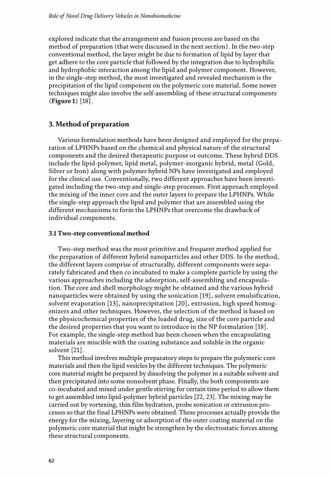
Role of Novel Drug Delivery Vehicles in Nanobiomedicine
62
explored indicate that the arrangement and fusion process are based on the method of preparation (that were discussed in the next section). In the two-step conventional method, the layer might be due to formation of lipid by layer that get adhere to the core particle that followed by the integration due to hydrophilic and hydrophobic interaction among the lipid and polymer component. However, in the single-step method, the most investigated and revealed mechanism is the precipitation of the lipid component on the polymeric core material. Some newer techniques might also involve the self-assembling of these structural components (Figure 1) [18].
3. Method of preparation
Various formulation methods have been designed and employed for the prepa-ration of LPHNPs based on the chemical and physical nature of the structural components and the desired therapeutic purpose or outcome. These hybrid DDS include the lipid-polymer, lipid metal, polymer-inorganic hybrid, metal (Gold, Silver or Iron) along with polymer hybrid NPs have investigated and employed for the clinical use. Conventionally, two different approaches have been investi-gated including the two-step and single-step processes. First approach employed the mixing of the inner core and the outer layers to prepare the LPHNPs. While the single-step approach the lipid and polymer that are assembled using the different mechanisms to form the LPHNPs that overcome the drawback of individual components.
3.1 Two-step conventional method
Two-step method was the most primitive and frequent method applied for the preparation of different hybrid nanoparticles and other DDS. In the method, the different layers comprise of structurally, different components were sepa-rately fabricated and then co incubated to make a complete particle by using the various approaches including the adsorption, self-assembling and encapsula-tion. The core and shell morphology might be obtained and the various hybrid nanoparticles were obtained by using the sonication [19], solvent emulsification, solvent evaporation [13], nanoprecipitation [20], extrusion, high speed homog-enizers and other techniques. However, the selection of the method is based on the physicochemical properties of the loaded drug, size of the core particle and the desired properties that you want to introduce in the NP formulation [18]. For example, the single-step method has been chosen when the encapsulating materials are miscible with the coating substance and soluble in the organic solvent [21].
This method involves multiple preparatory steps to prepare the polymeric core materials and then the lipid vesicles by the different techniques. The polymeric core material might be prepared by dissolving the polymer in a suitable solvent and then precipitated into some nonsolvent phase. Finally, the both components are co-incubated and mixed under gentle stirring for certain time period to allow them to get assembled into lipid-polymer hybrid particles [22, 23]. The mixing may be carried out by vortexing, thin film hydration, probe sonication or extrusion pro-cesses so that the final LPHNPs were obtained. These processes actually provide the energy for the mixing, layering or adsorption of the outer coating material on the polymeric core material that might be strengthen by the electrostatic forces among these structural components.
63
Lipid Polymer Hybrid Nanoparticles: A Novel Approach for Drug DeliveryDOI: http://dx.doi.org/10.5772/intechopen.88269
3.2 Modified two-step method
Different modifications have been suggested in the conventional two-step pro-cess for the fabrication of the LPHNPs. These modifications might include the use spray drying and lithographic molding along with the freeze drying [24]. The inner central core of the NPs have been prepared with the process that further suspended or dispersed in any suitable organic solvent containing the different structural components of the LPHNPs [2]. Different studies indicate the formulation of LPHNPs loaded with various antibiotics agents including levofloxacin, ciprofloxa-cin and isoniazid in the form of freeze dried powders for the inhalation therapy that were entrapped in the mono or multiple layers of the lipid shell. The coating of the lipid might provide the core shell morphology to the NPs. This modification in the preparation of the NPs added advantage in term of better inhalation efficiency and greater control on the overall average particle size of the LPHNPs relative to the conventional method [25]. The nanoparticles and hybrid microfiber fabrication are the some other examples that utilized the polyglutamic acid, poly lysine and various grade of PLG and PLA using the freeze-drying method [26, 27].
3.3 Single-step preparation method
The poor entrapment and easy leakage of the drug from the polymeric core of the LPHNPs prepared by the conventional two-step methods urged the develop-ment of some newer methods that overcome these shortcomings and enhance the therapeutic efficiency of the prepared formulation [14, 28]. The development of single-step method provides the way to control the particle size, PDI, uniformity of the structural components. It also overcomes the variability in different batches and other properties of the LPHNPs in term of their physicochemical properties and stability. The method involves the single preparatory step that considers the mixing of two different phases containing the lipid and polymer in each of the given phase. The mixing process followed by the self-assembling of the structural components that either make the matrix or core shell morphology [29].
Various techniques in the single-step preparation method might include the single or double emulsification method, sonication technique, nanoprecipitation, solvent evaporation method and solvent diffusion method.
4. Biomedical applications
LPNs have been prepared for efficient encapsulation and delivery of the wide range of therapeutic agents either alone or in combinations. LPNs have wide range of applications in cancer therapy and delivery of protein based therapeutic agents, i.e., small interfering RNA, nucleic acid and genes delivery etc. Additionally, LPNs can be used for oral drug delivery of many drugs [46]. LPN has wide range of applications in gene and DNA delivery, vaccines and diagnostic imaging agents as shown in Figure 2 [2].
4.1 Cancer therapy
Doxorubicin loaded polymer-lipid hybrid nanoparticles (Dox-PLN) were designed and injected intratumorally in mice. At a dose of 0.1 and 0.2 mg, 70 and 100% tumor growth delay was observed, respectively. Dox-PLN treated mice have not shown any sign of toxicity and only 2 mice out of 15 exhibited transient fur

Role of Novel Drug Delivery Vehicles in Nanobiomedicine
64
Typ
e of
hybr
id
nano
part
icle
s
Stru
ctur
al co
mpo
nent
sPh
ysic
oche
mic
al p
rope
rtie
sBi
olog
ical
pr
oper
ties
App
licat
ion
Refe
renc
e
Size
(nm
)Ze
ta p
oten
tial
Entr
apm
ent
effic
ienc
y
Lipi
d-po
lym
er
hybr
idPa
clita
xel
PLG
ASo
ybea
n le
cith
inD
SPE-
PEG
(con
juga
ted
with
folic
acid
)
186.
9 ±
8.52
−29
.5 ±
2.0
81.3
4 ±
3.41
T 1/2
18.0
8AU
C 0-
∞ 10
9.21
MRT
30.
06
Brai
n ta
rget
ing
in g
liobl
asto
ma
mul
tifor
m
[1]
Lipi
d-po
lym
er
hybr
idM
elat
onin
PLA
DDA
BC
TAB
180–
218
+15.4
to −
36.1
90.3
5N
/AO
phth
alm
ic
deliv
ery
[2]
PLG
AD
OX in
pol
ymer
ic co
re a
nd S
oraf
enib
in
lipid
ic co
reβ-
cycl
odex
trin
Dip
alm
itoyl
gly
cero
l pho
phoc
holin
e (D
PPC
)D
istea
royl
gly
cero
l ph
osph
oeth
anol
amin
e (D
SPE)
PEG
85.1
PDI 0
.103
Sust
aine
d re
leas
e fo
r 21 d
ays
Colo
n ca
ncer
[4]
Lipi
d-po
lym
er
hybr
idG
emci
tabi
ne, h
ypox
ia-in
duci
ble f
acto
r 1α
, ε-p
olyl
ysin
e co-
poly
mer
, PLG
A,
mPE
G, L
ecith
in, d
oubl
e em
ulsio
n m
etho
d an
d ul
tras
ound
assis
ted
self-
asse
mbl
y
141.8
−34
42Pa
ncre
atic
ca
ncer
[7]
Lipi
d-po
lym
er
hybr
idBu
deso
nide
, PLG
A,
diol
eoyl
trim
ethy
lam
mon
ium
pro
pane
(D
OTA
P), d
oubl
e em
ulsio
n so
lven
t ev
apor
atio
n m
etho
d
PDI 0
.09–
0.14
, 13
6–16
9 nm
−3
to 54
20–3
6 an
d 27
–80
Chro
nic
obst
ruct
ive
pulm
onar
y di
seas
e (CO
PD)
[13]
65
Lipid Polymer Hybrid Nanoparticles: A Novel Approach for Drug DeliveryDOI: http://dx.doi.org/10.5772/intechopen.88269
Typ
e of
hybr
id
nano
part
icle
s
Stru
ctur
al co
mpo
nent
sPh
ysic
oche
mic
al p
rope
rtie
sBi
olog
ical
pr
oper
ties
App
licat
ion
Refe
renc
e
Size
(nm
)Ze
ta p
oten
tial
Entr
apm
ent
effic
ienc
y
Lipi
d-po
lym
er
hybr
idD
extr
an, b
ovin
e ser
um al
bum
in (B
SA),
as
taxa
nthi
n, p
repa
red
thro
ugh
orga
nic
solv
ent f
ree h
omog
eniz
atio
n an
d so
nica
tion
tech
niqu
e, P
reci
rol®
ATO
5
(gly
cery
l pal
mito
stea
rate
)
139–
180
nm, P
DI
0.19
970
%40
% re
leas
e in
SGF
and
50%
in S
IF,
Diff
usio
n ba
sed
rele
ased
Ant
ioxi
dant
ac
tivity
and
sust
aine
d re
leas
e
[14]
Lipi
d-po
lym
er
hybr
idEr
lotin
ib, s
ingl
e-st
ep so
nica
tion
met
hod,
po
lyca
prol
acto
ne (P
CL),
hyd
roge
nate
d so
y ph
osph
atid
ylch
olin
e, 1,
2-di
stea
royl
-sn
-gly
cero
-3-p
hosp
hoet
hano
lam
ine-
N-m
etho
xy(p
olye
thyl
ene g
lyco
l)-2
000
(DSP
E-PE
G20
00)
159.6
–173
nm
, PD
I 0.
09–0
.14−
1.22
to −
47.3
18.1–
66.4
%50
% in
firs
t 3 h
, 10
0% in
24 h
Ant
ican
cer,
lung
canc
er[1
9]
PLG
A, p
aclit
axel
, PVA
200–
300
nm34
.8 ±
1.6
to
62.6
± 7.
9%Fa
st re
leas
e in
first
3 d
ays (
60%
) fo
llow
ed b
y slo
w fi
rst o
rder
re
leas
e for
21 d
ays
(cum
ulat
ive r
elea
se
72%
)
Ant
ican
cer
[22]
Lipi
d-po
lym
er
hybr
idD
oxor
ubic
in, s
tear
ic ac
id, t
riste
arin
, H
PESO
(hyd
roly
zed
poly
mer
of
epox
idiz
ed so
ybea
n oi
l), P
luro
nic-
F68
290
nm5%
Ant
ican
cer
[30]
Lipi
d ve
sicle
sC
arbo
xym
ethy
l chi
tosa
n, p
aclit
axel
, 1,
2-di
palm
itoyl
-sn-
glyc
ero-
3-ph
osph
ocho
line (
DPP
C),
1,2-
dist
earo
yl-
sn-g
lyce
ro-3
-pho
spho
etha
nola
min
e (D
PPC
)
200–
300
nm85
.2 ±
3.3
and
83.8
± 7.
5%Su
stai
ned
rele
ase
form
ulat
ion
Ant
ican
cer
[31]

Role of Novel Drug Delivery Vehicles in Nanobiomedicine
66
Typ
e of
hybr
id
nano
part
icle
s
Stru
ctur
al co
mpo
nent
sPh
ysic
oche
mic
al p
rope
rtie
sBi
olog
ical
pr
oper
ties
App
licat
ion
Refe
renc
e
Size
(nm
)Ze
ta p
oten
tial
Entr
apm
ent
effic
ienc
y
Lipi
d-po
lym
er
hybr
idD
oxor
ubic
in, e
poxi
dize
d so
yabe
an o
il,
Plur
onic
F68
80–3
50 n
m−
19.7
± 0
.65
60–8
0%50
% d
rug
rele
ased
in
firs
t few
hou
rs
and
addi
tiona
l 10
–20%
in 2
wee
ks
Ant
ican
cer
(bre
ast c
ance
r)[3
2]
Lipi
d-po
lym
er
hybr
idM
itoxa
ntro
ne h
ydro
chlo
ride,
dex
tran
su
lfate
, Cre
mop
hor (
poly
etho
xyla
ted
cast
or o
il), e
mul
sific
atio
n-ul
tras
onic
atio
n m
etho
d
130.
3 ±
4.7 t
o 13
6.7 ±
8.6
−19
.9 ±
1.4
to
−31
.6 ±
0.8
97.4
%Su
stai
ned
86.9
%
at 7
2 h,
Cm
ax (n
g/m
L) 4
21.6
± 24
.6,
t 1/2 (
h) 8
.49
± 1.
23,
AUC 0
-t (n
g/m
L.h)
69
0.9
± 83
.5,
AUC 0
-∞ (n
g/m
L.h)
72
2.6 ±
94.
1
Ant
ican
cer
[33]
Sora
feni
b, P
LGA
, Sin
gle-
step
na
nopr
ecip
itatio
n, D
-α-t
ocop
hero
l po
lyet
hyle
ne g
lyco
l 100
0 su
ccin
ate,
TP
GS,
dio
leoy
lpho
spha
tidic
acid
(D
OPA
)
150–
200
nm (a
vera
ge
175.
25 ±
1.82
nm
), P
DI
0.14
8 ±
0.00
4
−19
to −
5585
%H
ighl
y va
scul
ar
hepa
toce
llula
r ca
rcin
oma
[34]
Mito
myc
in C
, PLA
, Soy
bean
ph
osph
atid
ylch
olin
e (SP
C),
PEG
, fol
ate
215.6
± 5.
1 nm
, PD
I 0.
143
−25
.88
± 2.
3995
%A
ntic
ance
r[3
5]
Lipi
d-po
lym
er
hybr
idPa
clita
xel,
PLG
A, 1
,2-d
istea
royl
-sn
-gly
cero
-3-p
hosp
hoet
hano
lam
ine
(DSP
E), 1
,2-d
istea
royl
-sn-
glyc
ero-
3-ph
osph
oeth
anol
amin
e (D
SPE-
PEG
-200
0), f
olic
acid
, soy
bean
leci
thin
74.9
3 ±
3.93
to
81.3
4 ±
3.41%
87.2
± 4
.28
to
96.9
± 4
.93%
in
5 day
s
Ant
ican
cer
[36]
Lipi
d-po
lym
er
hybr
idN
anop
reci
pita
tion
proc
ess,
PLG
A, P
EG,
doce
taxe
l25
nm
−10
to −
5020
%50
% in
firs
t 12
h an
d re
mai
ning
in 7
2 h
Ant
ican
cer
[37]
67
Lipid Polymer Hybrid Nanoparticles: A Novel Approach for Drug DeliveryDOI: http://dx.doi.org/10.5772/intechopen.88269
Typ
e of
hybr
id
nano
part
icle
s
Stru
ctur
al co
mpo
nent
sPh
ysic
oche
mic
al p
rope
rtie
sBi
olog
ical
pr
oper
ties
App
licat
ion
Refe
renc
e
Size
(nm
)Ze
ta p
oten
tial
Entr
apm
ent
effic
ienc
y
PLG
A, C
urcu
min
, 1,2
-dip
alm
itoyl
-sn
-gly
cero
-3-p
hosp
hoch
olin
e (D
PPC
), 1,
2-di
stea
royl
-sn-
glyc
ero-
3-ph
osph
oeth
anol
amin
e-N
-[s
ucci
nyl(
poly
ethy
lene
gly
col)
-200
0]
(DSP
E-PE
G),
emul
sion/
solv
ent
evap
orat
ion
tech
niqu
e
150
nm, 1
71.6
± 8
.2 n
m
in D
I wat
er an
d 17
7.3 ±
6.2
nm
in P
BS,
PDI o
f 0.17
4 ±
0.02
3 an
d 0.
159
± 0.
026
12%
30%
in fi
rst 5
hA
ntic
ance
r[3
8]
Lipi
d-po
lym
er
hybr
idPo
ly(e
thyl
ene g
lyco
l)-d
istea
royl
phos
phat
idyl
etha
nola
min
e (PE
G-D
SPE)
, em
ulsif
ying
-sol
vent
evap
orat
ion
met
hod,
egg
yolk
pho
spha
tidyl
chol
ine,
pl
asm
id D
NA
128
nm+3
5.2
Non
vira
l gen
e de
liver
y[3
9]
Poly
-(β-
amin
o es
ter)
(PBA
E), d
oubl
e em
ulsio
n/so
lven
t eva
pora
tion,
PLG
A,
Phos
phol
ipid
s
230
± 40
to 3
00 ±
50,
PDI 0
.100
± 0.
05 to
0.
182
± 0.
10
32 ±
8 to
42
± 8
mRN
A b
ased
va
ccin
e[4
0]
Lipi
d-po
lym
er
hybr
idsiR
NA
, PLG
A, P
EG, m
odifi
ed d
oubl
e-em
ulsio
n so
lven
t eva
pora
tion
tech
niqu
e,
leci
thin
225 ±
8 n
m−
10 to
0 m
V78
–82%
50%
of s
iRN
A
rele
ased
in 1
2–20
h[4
1]
Lipi
d-po
lym
er
hybr
idPL
GA
, siR
NA
, Par
ticle
Rep
licat
ion
in
Non
wet
ting
Tem
plat
es (P
RIN
T) p
roce
ss19
8 ±
3.45 t
o 20
7 ± 4
.461
, PD
I 0.
045 ±
0.0
09 to
0.
092
± 0.
005
−3.4
5 ± 1.
9 to
5.
29 ±
1.5
32–4
6%Pr
osta
te ca
ncer
[42]
Lipi
d-po
lym
er
hybr
idN
orflo
xaci
n, P
LA, e
mul
sific
atio
n so
lven
t ev
apor
atio
n m
etho
d, P
VA, c
arbo
pol
K-94
0
178.
6 ±
3.7 n
m to
22
0.8
± 0.
66 n
m,
PDI 0
.206
± 0
.36
to
0.38
3 ±
0.66
+23.4
± 1.
5 mV
to
+4
1.5 ±
3.4
mV
53.2
9 ±
0.30
to
72.3
4 ±
0.23
89.7
2% d
rug
rele
ased
in 24
hTo
pica
l an
tibio
tic[4
3]

Role of Novel Drug Delivery Vehicles in Nanobiomedicine
68
Typ
e of
hybr
id
nano
part
icle
s
Stru
ctur
al co
mpo
nent
sPh
ysic
oche
mic
al p
rope
rtie
sBi
olog
ical
pr
oper
ties
App
licat
ion
Refe
renc
e
Size
(nm
)Ze
ta p
oten
tial
Entr
apm
ent
effic
ienc
y
Lipi
d-po
lym
er
hybr
idLi
doca
ine,
Chi
tosa
n, C
hole
ster
ol,
cety
ltrim
ethy
l am
mon
ium
bro
mid
e,
1,2-
dila
uroy
l-sn-
glyc
ero-
3-ph
osph
ocho
line (
DLP
C),
hya
luro
nic a
cid
71.2
± 2
.8 to
14
5.6 ±
5.9
nm,
PDI 0
.09
± 0.
02 to
0.
19 ±
0.0
2
−4.
6 ±
0.7 t
o +3
2.7 ±
4.6
78.6
± 4
.3 to
85
.2 ±
3.1
40%
in 8
h an
d re
mai
ning
in 7
2 h
Loca
l ane
sthe
tic
ther
apy
[44]
Lipi
d-po
lym
er
hybr
idTe
nofo
vir d
isopr
oxil
fum
arat
e, m
elt
emul
sific
atio
n-pr
obe s
onic
atio
n te
chni
que
239
nm−
42.1
± 2.4
6 to
−
55.3
9 ±
3.12
87.14
%, 6
3.83
±3.74
to
90.8
4 ±
5.73%
85.3
4 ±
5% at
the
end
of 1
2 h
Ant
ivira
l[4
5]
69
Lipid Polymer Hybrid Nanoparticles: A Novel Approach for Drug DeliveryDOI: http://dx.doi.org/10.5772/intechopen.88269
roughing. These results indicated that Dox-PLN expressed a good cytotoxic activity against solid tumors and improved the therapeutic efficacy [30].
Paclitaxel loaded LPNs were prepared with a size range of 200–300 nm for oral administration. Theses LPNs were designed to withstand harsh gastrointestinal tract conditions and improve the bioavailability of paclitaxel. On comparison with Taxol, 1.5- and 5.5-fold increase in bioavailability and elimination half-life was observed, respectively. Additionally, reduction in the reticuloendothelial system mediated uptake by liver and spleen was noted due to stealth characteristics of biopolymer blanket of these LPNs [31].
To overcome the multidrug resistance (MDR) of anticancer drugs, a new strat-egy was adopted in which doxorubicin loaded solid lipid nanoparticles (SLNs) were complexed with anionic polymer. Due to high encapsulation efficiency (60–80%) of doxorubicin, the cytotoxicity in tumor cells was increased to 8-fold. Due to physical interaction between drug and polymer and smaller size of nanoparticles (80–350 nm), drug was difficult to clear from target cells by efflux pump [32]. Another strategy to counter MDR is to synthesize lipid-anionic dextran sulfate hybrid carriers loaded with mitoxantrone hydrochloride. The interaction between cationic drug (mitoxantrone hydrochloride) and anionic dextran not only increased drug accumulation but also enhanced the cytotoxicity in breast cancer cell lines. Sustained release of drug (86.9%) was maintained for 72 h with an encapsulation efficiency of 97.4% [33].
Sorafenib is an antiangiogenic agent used in highly vascular hepatocellular car-cinoma (HCC). The development of resistance during HCC therapy is mainly due to activation of CXC receptor type 4 (CXCR4). Gao et al. developed PLGA nanopar-ticles loaded with sorafenib and evaluated the antitumor activity both in vitro and in vivo. On comparing with control group, sorafenib loaded PLGA nanoparticles have shown an improved survival in HCC model, delay in progression of tumor and enhanced antiangiogenic effect [34].
Mitomycin C is a water soluble drug and major disadvantages associated with this drug are poor water stability, rapid elimination and lacking in target specific-ity. A sustained (up to 120 h) and effective delivery of mitomycin C from LPH
Figure 2. Applications of lipid-polymer hybrid nanocarriers [46].

Role of Novel Drug Delivery Vehicles in Nanobiomedicine
70
nanoparticles was observed with improved encapsulation efficiency of 95%. Improved cell uptake and site specific accumulation of drug are the major advan-tages of LPNs [35]. Paclitaxel and folic acid loaded polymer-lipid hybrid nanopar-ticles were prepared to bypass the tight junctions of blood-brain barrier (BBB) and target the glioma cells. The survival time of mice was increased to 42 days as compared to free paclitaxel which last only 18 days. These targeted nanoparticles have shown better pharmacokinetics and biodistributions which result in better therapeutic outcomes [36].
Ultra-small lipid-polymer hybrid nanoparticles were fabricated using modified nanoprecipitation method. The prepared nanoparticles loaded with docetaxel have the size of 25 nm which exhibited a better antitumor activity than Taxotere. It was observed that the survival time of Taxotere treated mice were 44 days whereas more than half of the mice treated with ultra-small nanoparticles survived for 64 days. These ultra-small nanoparticles have better biodistribution properties and enhanced permeation ability [37]. Long circulating PLGA nanoparticles loaded with curcumin were fabricated to counter cancer metastasis. The adhesion of cancer cells onto endothelial cells and vascular deposition were reduced by 70 and 50%, respectively. Therefore, these nanoparticles could improve the therapeutic efficacy by preventing metastasis and impairing circulating tumor cells [38].
Core-shell LPN was fabricated to deliver erlotinib using single-step sonication method. In vitro cellular uptake, colony forming assay and luminescent cell viability assay was performed in human lung adenocarcinoma cell line (Figure 3). The mean particle size of LPN is 170 nm and entrapment efficiency of 66% with excellent storage stability. The enhanced and efficient uptake of these LPN by cancer cells makes these nanoparticles a potential delivery system for erlotinib [19].
Figure 3. (A) Confocal microscopy images of erlotinib loaded CSLPHNPs uptake in A549 cells after 1 and 4 h, (B) in vitro cellular viability result in A549 cells after 72 h, and (C) colony formation assay in A549 cells [19].
71
Lipid Polymer Hybrid Nanoparticles: A Novel Approach for Drug DeliveryDOI: http://dx.doi.org/10.5772/intechopen.88269
4.2 Gene delivery
Plasmid DNA, miRNA and siRNA are now gaining much of the interest of researchers for cancer therapy. Both miRNA and siRNA have different origin and mechanism but similar physicochemical properties. miRNA is endogenous in nature and target the mRNA by developing imperfect pairing and hence act by mRNA deg-radation, mRNA endonucleolytic cleavage or suppression of translation. siRNA is exogenous in nature and primarily act by endonucleolytic cleavage of target mRNA. siRNA has single mRNA target whereas miRNA has multiple targets. Plasmid DNA carries the recombinant gene or gene of interest and can be administered locally of systemically for cancer therapy [47–50].
Lot of challenges is associated with effective gene delivery especially for cancer therapy. Viral vectors are also facing problems such as development of immunity and inflammatory response, limited carrying ability of DNA and short shelf life [51]. Therefore, the research has now been shifted to nonviral vectors due to nonimmunogenicity, nontoxicity, low cost and feasibility in large scale production. Polyethylene glycol (PEG) and its copolymers have widely used for gene delivery because of its low toxicity, increase water solubility and reduced ability to interact with serum proteins [52].
Effective gene delivery through nonviral vectors with reduced toxicity was developed by emulsification solvent evaporation method. The particle size of newly developed positively charged LPN is in the range from 130 to 240 nm. Fluorescent protein was complexed with plasmid DNA by adsorption and transfection efficiencies was recorded as 37.2 and 34% for LPN and commercially available product, respectively [39].
Core shell LPN was fabricated using three different methods for incorporation of DNA and the resulted nanoparticles were in the range from 100 to 400 nm. Surface adsorbed DNA, encapsulated DNA and combination of adsorbed and encapsulated DNA are three important methods for fabrication of these nanoparticles. For sus-tained release of active ingredient, combination method is employed which is neces-sary for booster vaccination followed by decline release. For primary vaccination (strong and short effective delivery), surface adsorbed mechanism is followed. By adjusting the concentration of different ingredients, the drug release properties can be adjusted [53].
SiRNA delivery through cationic complexes such as polyplexes and lipoplexes has many disadvantages, e.g., development of inflammatory responses, instability and toxicity etc. Small size (100 nm) with prolong circulation time nanoparticles containing siRNA was developed using PLGA. These hybrid nanoparticles has 80% encapsulation efficiency of siRNA without any significant degradation until 24 h. Immunofluorescence studies revealed the in vitro apoptosis and >90% knockdown of nonsmall cell lung cancer [54].
LPNs are also used for incorporation of mRNA for mRNA vaccines. mRNA was complexed with LPN through electrostatic adsorption to develop 150–300 nm size nanoparticles. These newly developed nanoparticles have shown successful transfection through intranasal route and taken up by dendritic cells with minimum toxicity [40].
A novel approach, modified double emulsion/solvent evaporation method, was used to fabricate hollow core/shell LPNs in which PLGA core was surrounded by lipid shell attached with PEG chains. The size of nanoparticles was 230 nm, 80% encapsulation efficiency and 50% siRNA was sustained release for 12–20 h. Moreover, enhanced gene silencing ability was also observed with profound inhibi-tion of gene expression in xenograft tumor [41]. PLGA/siRNA nanoparticles coated with lipids are prepared using Particle Replication in Non wetting Templates tech-nique and exhibited 32–46% encapsulation efficiency for the treatment of prostate cancer [42]. siRNA was localized in PLGA core at high concentration by varying the

Role of Novel Drug Delivery Vehicles in Nanobiomedicine
72
concentration of polymer and lipid. This localized siRNA then modifies the release, physicochemical properties and transfection efficiencies [55].
4.3 Theranostic agents
LPNs not only used for delivering of therapeutic agent but also used for diagnostic purpose. Different type of imaging agents such as quantum dots, fluorescent dyes and iron oxide are incorporated in polymer core. Lipid-polymer and lipid-quantum dot nanoparticles were prepared in a single step with narrow size distribution [56]. Physicochemical properties can be controlled by varying the experimental conditions. Such multicomponent nanoparticles can be used therapy and diagnosis simultaneously.
LPNs are used for theranostic purposes in which phospholipids are attached on one side of polymer chain and fluorophores are linked on the other side. LPNs were designed to incorporate MRI contrast agent, gadolinium, chelated with lipid-PEG in PLGA core. These agents have shown effective uptake of nanoparticles within 3 h by J-774 cells [57]. Instead of conjugation with imaging agent, fluorescence dye was uploaded in LPNs core surrounded by lipid-PEG shell. The final size of such nanoparticles was in the range from 20 to 30 nm and bright fluorescence was observed due to lipid tail and polymeric core [58].
4.4 Stimuli responsive LPNs
Stimuli responsive drug delivery systems have the ability to deliver therapeutic agents at target sites in a controlled manner with minimum side effects. LPNs com-prise of magnetic beads. Stimuli responsive drug delivery system has the advantage to deliver the therapeutic agent in a controlled manner at target site. In a core (PLGA) shell (soyabean lecithin) nanoparticle system, magnetic beads were used for stimuli responsive release of camptothecin when exposed to radio frequency magnetic field. The release of drug was increased by 60% using radio frequency which significantly decreased the mouse breast cancer cell growth. Such system can be beneficial in cancer chemotherapy due to easy preparation, bio-stability and site specific drug delivery [59].
4.5 Miscellaneous pharmacological applications
Hepatitis C is a chronic disease which leads to liver cirrhosis and hepatocellular carcinoma. LPNs are used to label HCV viral particles for their detection, possible interaction and entrance pathway into host cells [60].
SLNs are developed for topical administration of norfloxacin using solvent evaporation method. These nanoparticles have shown antimicrobial activity against Staphylococcus aureus and Pseudomonas aeruginosa and could be used for the treat-ment of burn wound and topical infections. SLNs have shown 89.72% drug release after 24 h and passed the skin irritation and stability tests [43].
LPNs have recently used for improving the local anesthetic action of lidocaine. Two different kinds of nanocarriers, i.e., liposomes and LPNs were prepared and evaluated the skin permeation ability, in vitro and in vivo drug release studies, encapsulation efficiency and particle size. Results indicated that LPNs has better and improved efficacy of lidocaine as compared to liposomal delivery system due to smaller size (88.6 nm). The steady state flux of LPNs was found to be 65.4 μg/h/cm2 which showed increased skin permeation capacity [44].
LPNs were used for nasal delivery of an antiviral drug, tenofovir using melt emulsification-probe sonication technique. Intranasal flux of 135.36 μg/cm2/h and enhanced fluidity improved the drug permeation through membrane phospholip-ids, which increase the bioavailability of the drug [45].
73
Lipid Polymer Hybrid Nanoparticles: A Novel Approach for Drug DeliveryDOI: http://dx.doi.org/10.5772/intechopen.88269
Author details
Nayab Tahir1*, Muhammad Tahir Haseeb1, Asadullah Madni2, Farzana Parveen2, Muhammad Muzamil Khan2, Safiullah Khan2, Nasrullah Jan2 and Arshad Khan2
1 College of Pharmacy, University of Sargodha, Sargodha, Pakistan
2 Department of Pharmacy, The Islamia University of Bahawalpur, Bahawalpur, Pakistan
*Address all correspondence to: [email protected]
© 2019 The Author(s). Licensee IntechOpen. This chapter is distributed under the terms of the Creative Commons Attribution License (http://creativecommons.org/licenses/by/3.0), which permits unrestricted use, distribution, and reproduction in any medium, provided the original work is properly cited.

74
Role of Novel Drug Delivery Vehicles in Nanobiomedicine
References
[1] Zhang L et al. Self-assembled lipid−polymer hybrid nanoparticles: A robust drug delivery platform. ACS Nano. 2008;2(8):1696-1702
[2] Hadinoto K, Sundaresan A, Cheow WS. Lipid–polymer hybrid nanoparticles as a new generation therapeutic delivery platform: A review. European Journal of Pharmaceutics and Biopharmaceutics. 2013;85(3):427-443
[3] Vikulina AS, Skirtach AG, Volodkin DV. Hybrids of polymer multilayers, lipids, and nanoparticles. Mimicking the cellular microenvironment. Langmuir. 2019;35(26):8565-8573
[4] Mieszawska AJ et al. Synthesis of polymer–lipid nanoparticles for image-guided delivery of dual modality therapy. Bioconjugate Chemistry. 2013;24(9):1429-1434
[5] Gong J et al. Polymeric micelles drug delivery system in oncology. Journal of Controlled Release. 2012;159(3):312-323
[6] Fan J et al. Targeted anticancer prodrug with mesoporous silica nanoparticles as vehicles. Nanotechnology. 2011;22(45):455102
[7] Zhao X et al. Co-delivery of HIF1α siRNA and gemcitabine via biocompatible lipid-polymer hybrid nanoparticles for effective treatment of pancreatic cancer. Biomaterials. 2015;46:13-25
[8] Madni A et al. Liposomal drug delivery: A versatile platform for challenging clinical applications. Journal of Pharmacy & Pharmaceutical Sciences. 2014;17(3):401-426
[9] Mehnert W, Mäder K. Solid lipid nanoparticles: production, characterization and applications. Advanced Drug Delivery Reviews. 2012;64:83-101
[10] Prabaharan M et al. Gold nanoparticles with a monolayer of doxorubicin-conjugated amphiphilic block copolymer for tumor-targeted drug delivery. Biomaterials. 2009;30(30):6065-6075
[11] Khan MI, Madni A, Peltonen L. Development and in-vitro characterization of sorbitan monolaurate and poloxamer 184 based niosomes for oral delivery of diacerein. European Journal of Pharmaceutical Sciences. 2016;95:88-95
[12] Wakaskar RR. General overview of lipid–polymer hybrid nanoparticles, dendrimers, micelles, liposomes, spongosomes and cubosomes. Journal of Drug Targeting. 2018;26(4):311-318
[13] Leng D et al. Engineering of budesonide-loaded lipid-polymer hybrid nanoparticles using a quality-by-design approach. International Journal of Pharmaceutics. 2018;548(2):740-746
[14] Wang T et al. Solid lipid–polymer hybrid nanoparticles by in situ conjugation for oral delivery of astaxanthin. Journal of Agricultural and Food Chemistry. 2018;66(36):9473-9480
[15] Chaudhary Z et al. Lipid polymer hybrid carrier systems for cancer targeting: A review. International Journal of Polymeric Materials and Polymeric Biomaterials. 2018;67(2):86-100
[16] Pimentel-Moral S et al. Lipid nanocarriers for the loading of polyphenols—A comprehensive review. Advances in Colloid and Interface Science. 2018;260:85-94
[17] Negi JS. Nanolipid materials for drug delivery systems: A comprehensive Review. In: Characterization and Biology of Nanomaterials for Drug Delivery. 2019. pp. 137-163
75
Lipid Polymer Hybrid Nanoparticles: A Novel Approach for Drug DeliveryDOI: http://dx.doi.org/10.5772/intechopen.88269
[18] Mukherjee A et al. Lipid–polymer hybrid nanoparticles as a next-generation drug delivery platform: State of the art, emerging technologies, and perspectives. International Journal of Nanomedicine. 2019;14:1937
[19] Mandal B et al. Development and in vitro evaluation of core–shell type lipid–polymer hybrid nanoparticles for the delivery of erlotinib in non-small cell lung cancer. European Journal of Pharmaceutical Sciences. 2016;81:162-171
[20] Dave V et al. Lipid-polymer hybrid nanoparticles: Synthesis strategies and biomedical applications. Journal of Microbiological Methods. 2019;160:130-142
[21] Chaisri W, Hennink WE, Okonogi S. Preparation and characterization of cephalexin loaded PLGA microspheres. Current Drug Delivery. 2009;6(1):69-75
[22] Dong Y, Feng S-S. Poly (D,L-lactide-co-glycolide)(PLGA) nanoparticles prepared by high pressure homogenization for paclitaxel chemotherapy. International Journal of Pharmaceutics. 2007;342(1-2):208-214
[23] Paillard-Giteau A et al. Effect of various additives and polymers on lysozyme release from PLGA microspheres prepared by an s/o/w emulsion technique. European Journal of Pharmaceutics and Biopharmaceutics. 2010;75(2):128-136
[24] Enlow EM, Luft JC, Napier ME, DeSimone JM. Potent engineered PLGA nanoparticles by virtue of exceptionally high chemotherapeutic loadings. Nano Letters. 2011;11(2):808-813
[25] Wang H, Zhao P, Su W, Wang S, Liao Z, Niu R, et al. PLGA/polymeric liposome for targeted drug and gene co-delivery. Biomaterials. 2010;31(33):8741-8748
[26] Hitzman CJ, Elmquist WF, Wattenberg LW, Wiedmann TS. Development of a respirable, sustained release microcarrier for 5-fluorouracil I: In vitro assessment of liposomes, microspheres, and lipid coated nanoparticles. Journal of Pharmaceutical Sciences. 2006;95(5):1114-1126
[27] Keloglu N, Verrier B, Trimaille T, Sohier J. Controlled association and delivery of nanoparticles from jet-sprayed hybrid microfibrillar matrices. Colloids and Surfaces. B, Biointerfaces. 2016;140:142-149
[28] Cheow WS, Hadinoto K. Factors affecting drug encapsulation and stability of lipid–polymer hybrid nanoparticles. Colloids and Surfaces. B, Biointerfaces. 2011;85(2):214-220
[29] D'Addio SM, Prud'homme RK. Controlling drug nanoparticle formation by rapid precipitation. Advanced Drug Delivery Reviews. 2011;63(6):417-426
[30] Wong HL et al. In vivo evaluation of a new polymer-lipid hybrid nanoparticle (PLN) formulation of doxorubicin in a murine solid tumor model. European Journal of Pharmaceutics and Biopharmaceutics. 2007;65(3):300-308
[31] Joshi N et al. Carboxymethyl-chitosan-tethered lipid vesicles: Hybrid nanoblanket for oral delivery of paclitaxel. Biomacromolecules. 2013;14(7):2272-2282
[32] Wong HL et al. A new polymer–lipid hybrid nanoparticle system increases cytotoxicity of doxorubicin against multidrug-resistant human breast cancer cells. Pharmaceutical Research. 2006;23(7):1574-1585
[33] Zhang P et al. Novel nanostructured lipid-dextran sulfate hybrid carriers overcome tumor

Role of Novel Drug Delivery Vehicles in Nanobiomedicine
76
multidrug resistance of mitoxantrone hydrochloride. Nanomedicine: Nanotechnology, Biology and Medicine. 2012;8(2):185-193
[34] Gao D-Y et al. CXCR4-targeted lipid-coated PLGA nanoparticles deliver sorafenib and overcome acquired drug resistance in liver cancer. Biomaterials. 2015;67:194-203
[35] Li Y et al. Mitomycin C-soybean phosphatidylcholine complex-loaded self-assembled PEG-lipid-PLA hybrid nanoparticles for targeted drug delivery and dual-controlled drug release. Molecular Pharmaceutics. 2014;11(8):2915-2927
[36] Agrawal U et al. Tailored polymer–lipid hybrid nanoparticles for the delivery of drug conjugate: Dual strategy for brain targeting. Colloids and Surfaces B: Biointerfaces. 2015;126:414-425
[37] Dehaini D et al. Ultra-small lipid–polymer hybrid nanoparticles for tumor-penetrating drug delivery. Nanoscale. 2016;8(30):14411-14419
[38] Palange AL et al. Lipid–polymer nanoparticles encapsulating curcumin for modulating the vascular deposition of breast cancer cells. Nanomedicine: Nanotechnology, Biology and Medicine. 2014;10(5):e991-e1002
[39] Li J et al. A novel polymer-lipid hybrid nanoparticle for efficient nonviral gene delivery. Acta Pharmacologica Sinica. 2010;31(4):509
[40] Su X et al. In vitro and in vivo mRNA delivery using lipid-enveloped pH-responsive polymer nanoparticles. Molecular Pharmaceutics. 2011;8(3):774-787
[41] Shi J et al. Differentially charged hollow core/shell lipid–polymer–lipid hybrid nanoparticles for small interfering RNA delivery. Angewandte
Chemie International Edition. 2011;50(31):7027-7031
[42] Hasan W et al. Delivery of multiple siRNAs using lipid-coated PLGA nanoparticles for treatment of prostate cancer. Nano Letters. 2011;12(1):287-292
[43] Dave V et al. Lipid-polymer hybrid nanoparticles: Development & statistical optimization of norfloxacin for topical drug delivery system. Bioactive Materials. 2017;2(4):269-280
[44] Wang J et al. An alternative choice of lidocaine-loaded liposomes: Lidocaine-loaded lipid–polymer hybrid nanoparticles for local anesthetic therapy. Drug Delivery. 2016;23(4):1254-1260
[45] Pokharkar VB, Jolly MR, Kumbhar DD. Engineering of a hybrid polymer–lipid nanocarrier for the nasal delivery of tenofovir disoproxil fumarate: Physicochemical, molecular, microstructural, and stability evaluation. European Journal of Pharmaceutical Sciences. 2015;71:99-111
[46] Hallan SS et al. Lipid polymer hybrid as emerging tool in nanocarriers for oral drug delivery. Artificial Cells, Nanomedicine, and Biotechnology. 2016;44(1):334-349
[47] Chitkara D, Mittal A, Mahato RI. miRNAs in pancreatic cancer: Therapeutic potential, delivery challenges and strategies. Advanced Drug Delivery Reviews. 2015;81:34-52
[48] Chitkara D, Singh S, Mittal A. Nanocarrier-based co-delivery of small molecules and siRNA/miRNA for treatment of cancer. Therapeutic Delivery. 2016;7(4):245-255
[49] Husain S et al. Gene therapy for cancer: Regulatory considerations for approval. Cancer Gene Therapy. 2015;22(12):554
77
Lipid Polymer Hybrid Nanoparticles: A Novel Approach for Drug DeliveryDOI: http://dx.doi.org/10.5772/intechopen.88269
[50] Lam JK et al. siRNA versus miRNA as therapeutics for gene silencing. Molecular Therapy - Nucleic Acids. 2015;4:e252
[51] Oh Y-K, Park TG. siRNA delivery systems for cancer treatment. Advanced Drug Delivery Reviews. 2009;61(10):850-862
[52] Lee M, Kim SW. Polyethylene glycol-conjugated copolymers for plasmid DNA delivery. Pharmaceutical Research. 2005;22(1):1-10
[53] Zhong Q et al. Optimization of DNA delivery by three classes of hybrid nanoparticle/DNA complexes. Journal of Nanobiotechnology. 2010;8(1):6
[54] Zhu X et al. Long-circulating siRNA nanoparticles for validating Prohibitin1-targeted non-small cell lung cancer treatment. Proceedings of the National Academy of Sciences. 2015;112(25):7779-7784
[55] Colombo S et al. Mechanistic profiling of the siRNA delivery dynamics of lipid–polymer hybrid nanoparticles. Journal of Controlled Release. 2015;201:22-31
[56] Valencia PM et al. Single-step assembly of homogenous lipid−polymeric and lipid−quantum dot nanoparticles enabled by microfluidic rapid mixing. ACS Nano. 2010;4(3):1671-1679
[57] Aryal S et al. Engineered magnetic hybrid nanoparticles with enhanced relaxivity for tumor imaging. Biomaterials. 2013;34(31):7725-7732
[58] Kandel PK et al. Incorporating functionalized polyethylene glycol lipids into reprecipitated conjugated polymer nanoparticles for bioconjugation and targeted labeling of cells. Nanoscale. 2011;3(3):1037-1045
[59] Kong SD et al. Magnetic field activated lipid–polymer hybrid nanoparticles for stimuli-responsive drug release. Acta Biomaterialia. 2013;9(3):5447-5452
[60] Bathfield M et al. Synthesis of lipid-α-end-functionalized chains by RAFT polymerization. Stabilization of lipid/polymer particle assemblies. Macromolecules. 2008;41(22):8346-8353

79
Chapter 4
Preparation and Characterisation of Niosomal Emulsions as Novel Drug Delivery Vehicle Derived from Natural SeaweedsReshma Joy, Franklin John and Jinu George
Abstract
Biomedical field uses polysaccharides for the last one half of the century due to its remarkable applications. Nature is an abundant source for natural polysaccha-rides made its use widely in this field. In this present chapter study, the potential of alginate-κ-carrageenan and alginate-gelatin hydrogel beads for encapsulating cur-cumin was investigated. The evaluation of different combination of combinations of alginate/κ-carrageenan and alginate/gelatin hydrogel beads were developed and drug release properties were compared using curcumin as a model drug. Alginate/ κ-carrageenan hydrogel beads with 50:50 weight ratio exhibited higher swelling and better drug release percentage than compared to other beads. Antibacterial activity of curcumin released from hydrogel beads against B. cereus was established by disc assay. This nanosized vehicles with increases the efficacy and to control the delivery of biologically active substances at their specific site is as important as the drug activity itself.
Keywords: κ-carrageenan, alginate, niosome, hydrogel, drug delivery
1. Introduction
Revolution changes and modifies the conservative systems existing in the entire zones of the world. Medicinal chemistry and nanotechnology in that sense practices a revolutionary idea that promises healthy life to every human being. One such important category belongs to drug delivery is the use of natural sources as drug carriers [1]. A number of therapeutic agents show potentiality against cancer, HIV, Tuberculosis, and other kind of diseases, but most of them are not having the poten-tial to overcome the biological barriers inside the body and this causing least effect than actual [2]. This appeal the production of a new system that can able to reduce the typical hitches associated with drugs. We need safe, secure and efficient mea-sures to dose new generation diseases that threatening the life of living beings [3].
Accordingly, among the new discovered diseases, cancer leads as a number one killer disease because of the versatile nature and failure of therapeutic potential against the disease [4]. Recent years witnessed discovery of efficient therapeutic agents but its actual challenge is to overcome the biological barriers inside the body [5]. Most of the drugs are potent to the particular disease but the properties limited
Role of Novel Drug Delivery Vehicles in Nanobiomedicine
80
due to its hydrophobic nature, non-specificity, toxicity to normal cells, less stability, etc. [6]. So the researchers move to deliver these available potent drugs to the patho-logical site by diminishing all these factors through entrapment in a biocompatible polymers as carriers [7]. They were trying to develop smart drug carriers to enhance the efficiency of drugs at its specific site with controlled delivery [8].
Drug delivery is a term that refers the successful delivery of therapeutic agents at the specific site with specific time without any kind of degradation [9]. The problem associated with available drugs in the market includes hydrophobic nature, higher toxic nature, nonspecific release, in vivo degradation and short circulating half-lives [10]. Drug delivery systems emerging from the significance of controlled release of therapeutic agent on the affected part in the effective time with least side effects [1, 11]. Natural polymer based drug carriers is the central focus due to biocompatible nature, nontoxicity, low cost, ease of use and biodegradability [10]. Specificity [12] of drug molecule can be improved using ligands at the surface of delivery systems which in turn termed targeted drug delivery to the affected area of the body [13]. Targeting can be through either passive or active [14]. Major types of drug carriers falling under
• Niosomes,
• Liposomes,
• Polymeric micelles,
• Microspheres,
• Nanostructures,
• Nanofibers,
• Protein-DNA complexes,
• Protein-drug conjugates,
• Erythrocytes,
• Virosomes,
• Dendrimers.
Utilization of drug delivery systems is a promising methodology for creating efficient therapeutic agents. Among these kinds of carrier’s polymeric micelles, dendrimers, liposomes [15] are the growing area because of their outstanding properties and smart drug delivery potential [16]. Polymer based systems must meet some important features to become a good delivery vehicle such as; the backbone should be biocompatible; the term biocompatible in the sense explains capacity of a polymer to act with proper host reaction as well as the polymers are biodegradable without the formation of any kind of harmful by-products inside the body [17–19]. The polymer must soluble in various solvents and versatile in chemi-cal, structural and in application, etc. [3, 9]. The exploitation of micelles prepared from amphiphilic copolymers for solubilization of ineffectively solvent medica-tions has drawn in much consideration. Natural polysaccharide gets much attention towards the preparation of drug delivery systems because of their exceptional
81
Preparation and Characterisation of Niosomal Emulsions as Novel Drug Delivery Vehicle…DOI: http://dx.doi.org/10.5772/intechopen.86942
hydrogel forming ability [20]. These are derived from plants, sea weeds shells of crustaceans and microorganisms [21].
Niosomes are one of the excellent drug carriers, composed of non-ionic surface in addition to cholesterol or its derivatives. Niosomes can overcome the disadvan-tages of the liposomes [22, 23]. Liposomes firstly proposed by Alec Bangham called Bangosomes in 1965, the first generation liposomes the entrapped drugs are leaked out because of the membranes are not much stronger. In the case of niosomes, the hydrophilic and hydrophobic parts influence the entrapment of drug molecules, where as in the liposomes, lipophilic domain influences in it [5, 24, 25]. Non-ionic surfactants Tween, Brij, Span, etc. are mostly used for the noisome preparation. Hydrophilic-lipophilic balance (HLB) is a dimensionless parameter which can direct the entrapment efficiency for the drug and controlled release [26]. These values are varies between the non-ionic surfactant nature [27–30]. Surfactants with a HLB number between 3 and 8 are compatible with preparation bilayer surfaces and refer to water-in-oil (W/O) emulsifier [2, 31]. The additives in the niosomes include cholesterol and charge inducer. The cholesterol reduces the HLB value and charge inducer like dicetyl phosphate (DCP), produce surface charge leads to the stabilization of the bilayer [32, 33]. Applications of niosomes mainly related to the systemic delivery of hydrophobic drug molecules. More clearly niosome is used to improve the stability and physical properties, controlled release of drug molecules and for targeting and retention of drug in blood circulation. Niosomes offer various advantages over other drug delivery devices and have found applicability in phar-maceutical field. Niosomes are exceptionally powerful drug delivery instruments for construction/focusing of different remedially dynamic moieties and the respon-sibility lies on future researchers to viably outfit its potential in assorted application zones to support humanity [34].
Hydrophilic interpenetrating polymeric networks like hydrogels represent excel-lent carrier for hydrophobic drug encapsulation. In comparison to hydrophilic nano systems widely used in drug delivery, hydrogels show better swelling property and biocompatibility [35, 36]. Hydrogels derived from natural polymers like polysac-charides, alginates, gelatin polysaccharide conjugates exhibit a gradual change in swelling properties upon variation in temperature, pH, and ionic strength. 3D polymeric network derived from hydrogels represent flexible tissue-like material for efficient delivery of hydrophobic drugs [37] (Figure 1).
2. Natural polysaccharides
The use of nanotechnology for the delivery of therapeutic agents was put forward by Paul Enrich [6], the proponent of drug loaded magic bullets. Biomedical field uses polysaccharides for the last one half of the century due to its remarkable applications [38]. Nature is an abundant source for natural polysaccharides made its use widely in this field. Most of the drugs used for different types of diseases
Figure 1. (A) Niosome, (B) liposome, (C) polymeric micelles and (D) dendrimers.

Role of Novel Drug Delivery Vehicles in Nanobiomedicine
82
cause dangerous side effects. These problems can be reduced by the use of natural biocompatible polymer/polysaccharides as carriers for the drug. Seaweeds provide a better source for available polysaccharides such as carrageenan (iota, kappa, and lambda), alginate, and agar [39].
Monosaccharides combine to form polysaccharides with number of reactive groups that can be easily modified to get various types of polysaccharides. Typical sources of polysaccharide include algae, microorganisms, animals, etc. The positive features of polysaccharides such as higher molecular weight, changing chemical composition and large number of reactive groups gave diversity in its function. Adaptable chemical composition delivers its variety in structure and also in prop-erty. Medicinal field exploits these best properties of polysaccharides for the better-ment of drug delivery. Drug carriers from polysaccharides make sustainable, cost effective and biodegradable drug carriers. Modification of polysaccharide chains opens a way to target drug to the affected site, altering the solubility, biocompatibil-ity, moisture resistant and particularly reduce the severe side effects from therapeu-tic agents. Polysaccharides can perform bio adhesion through hydrophilic groups such as carboxyl, hydroxyl and amino groups by making non covalent interaction with biological tissues that elongates residing time. By this the absorption of drug at the particular area can be easily made. In this chapter we discuss niosomal emul-sions of alginate, κ-carrageenan and gelatin as drug carrier [38, 40, 41].
Alginates are naturally derived polysaccharide block copolymers composed of β-d-mannuronic acid monomers (M-blocks), α-l-guluronic acids (G-blocks) and regions of interspersed M and G units [38, 42]. Divalent cations such as Ca2+ can induce hydrogel formation by polysaccharide chain association in alginates [20]. Carrageenans represent a family of sulphated linear polysaccharides consist-ing of (1 → 3)-linked β-galactose and (1 → 4)-linked α-d-galactose units mainly extracted from certain red seaweeds of Rhodophyceae family particularly from Chondrus crispus, Eucheuma gigartina, Stellate iridaea, Hypnea, Solieria agardhiella and Sarconema, which are variously substituted and modified to the 3,6-anhydro derivative, depending on the nature of origin and extraction conditions [43]. Three main types of carrageenans are known: kappa-κ, lambda-λ and iota-i, depending on the number and the position of the ionic sulphate groups. The presence of a suitable cation, typically potassium, or calcium is an absolute requirement for gelation of the carrageenans, especially kappa [44]. Gelatin is a protein derived from denatured collagen that contains high levels of hydroxyproline, proline and glycine. It is useful as a thermally reversible gelling agent for encapsulation [45].
However, we go through the development and characterization of alginate/κ-carrageenan and alginate/gelatin hydrogel beads and its application in encapsula-tion of a model hydrophobic potential drug, curcumin. Hydrogel beads were developed via ionotropic gelation. Alginate/κ-carrageenan beads were cross-linked with CaCl2-KCl salt solution and alginate/gelatin beads with CaCl2 solution. Drug release behaviour of alginate/κ-carrageenan and alginate/gelatin hydrogel beads was compared employing curcumin, which is a natural polyphenolic compound isolated from rhizome of turmeric (Curcuma longa) as a model drug [46]. Curcumin is widely known to possess antioxidant [47], antitumor [48], and anti-inflammatory [49] activity. Swelling of the beads and in vitro release of curcumin were studied in phosphate buffered saline (PBS, pH 7.4).
3. Materials and methods
Sodium alginate (RM 7494), κ-carrageenan (22,048, Sigma Aldrich), Gelatin, phosphate buffer saline pH 7.4 (PBS, Sigma Aldrich) and curcumin (C1386, Sigma
83
Preparation and Characterisation of Niosomal Emulsions as Novel Drug Delivery Vehicle…DOI: http://dx.doi.org/10.5772/intechopen.86942
Aldrich) were used without further purification. CaCl2 and KCl were commercial products of analytical grade. Single distilled water (Labsil Water Distiller, Model: OPTI-M4) was used in all experiments.
3.1 Preparation of alginate/κ-carrageenan and alginate/gelatin hydrogel beads and curcumin loading
Alginate, κ-carrageenan and gelatin solutions were prepared separately by dissolving each of the biopolymers in distilled water and heated up at 50°C (for alginate), 60°C (for κ-carrageenan) and 40°C (for gelatin) under constant stirring from 30 min to 1 h until complete dissolution [50]. Ionotropic method was used for the preparation of hydrogel beads. Polysaccharide mixture com-posed of 50:50, 70:30, 80:20 (weight ratio) with concentrations varying from 1 to 2.5% (given in Table 1) were prepared by mixing under constant stirring at 30°C for 30 min. Briefly the hydrogel beads were prepared by dropping the mixture through a plastic syringe (5 ml) into an aqueous salt solutions stirred magnetically. For the preparation of alginate/κ-carrageenan hydrogel beads, the salt solution was composed of 100 ml of 2% (w/v) KCl and 100 ml of 2% (w/v) CaCl2 and for alginate/gelatin hydrogel beads, 200 ml of 2% CaCl2 solu-tion. To complete gelation, beads were maintained in the solution for 30 min then filtered, followed by washing with distilled water and then allowed to dry overnight at 37°C.
For curcumin loading, 10 mg/ml curcumin prepared with ethanol was mixed with each of the alginate/κ-carrageenan and alginate/gelatin mixtures and stirred until complete evaporation of ethanol. Curcumin loaded beads were prepared by dropping the mixture through a 5 ml syringe into a stirred salt solution which was composed of 100 ml of 2% (w/v) KCl and 100 ml of 2% (w/v) CaCl2 in the case of alginate/κ-carrageenan and 200 ml of 2% (w/v) CaCl2 for alginate/gelatin and kept in the solution for 30 min, filtered, washed with distilled water and dried overnight at 37°C.
3.2 Morphological analysis
Scanning electron microscopy (SEM, LEO 1430 V) was used to analyse the morphology of hydrogel beads.
3.3 Swelling behaviour
The swelling degree (SD) was determined gravimetrically as follows: Both the hydrogel beads were immersed in phosphate buffered saline (PBS, pH 7.4) at 37°
Materials Weight ratio (w/w)
Polymeric concentrations (%)
Salt type Salt concentration (%) (w/v)
Alginate/κ-carrageenan 50:5070:3080:20
11.52
2.5
CaCl2·KCl 2
Alginate/gelatin 50:5070:3080:20
11.52
2.5
CaCl2 2
Table 1. Weight ratio percentage concentrations of polymeric solutions.

Role of Novel Drug Delivery Vehicles in Nanobiomedicine
84
C. The swelled samples were taken from the PBS at selected time intervals of 1, 2, 3, 4, and 10 up to 24 h, wiped with tissue paper, weighed and placed again in PBS. The SD, in percentage, was calculated using Eq. (1)
SD = Ws − Wd _______ Wd ∗ 100 (%) (1)
where Ws and Wd are the weights of swollen and dry beads, respectively.
3.4 Release study of curcumin
The cumulative release of curcumin from hydrogel beads was carried out through incubating samples while shaking at 100 rpm in 20 ml of buffer solution (PBS) at pH 7.4 (1, 2, 3, 4, 10 up to 24 h incubation) at 37°C. After incubation, 3 ml sample was taken out from the buffer solution and analysed by UV-Vis spectropho-tometer (Lambda 25-PerkinElmer UV-vis spectrophotometer) at 𝜆𝜆max of 430 nm. The withdrawn volume of the sample was replaced with equal volume of fresh buffer solution to keep the volume of release fluid constant. The percentage drug release was calculated by using the following formula,
Percentage drug release = (Amount of drug released / Amount of drug loaded) ∗ 100 (2)
4. Results and discussion
4.1 Morphological analysis of hydrogel beads
Image of the swollen beads is shown in Figures 2 and 3. Beads obtained has diameter of 0.50 cm. Morphological features of curcumin loaded and unloaded beads were analysed through SEM images shown in Figure 4.
The morphological features of curcumin loaded and unloaded beads were analysed by recording the SEM images as shown in Figure 4.
Hydrogel beads containing alginate were smoother and more spherical com-pared to the beads consisting of alginate and carrageenan. This effect can be attributed to the higher cross linking ability of alginate relative to carrageenan. This cross linking takes place instantaneously upon dropping sodium alginate into the cross linking solution (consisting of Ca2+) leading to geometrically stable particles.
In the case of formulations containing carrageenan, the beads were less spheri-cal, having rough and folded surfaces. This behaviour could be attributed to the low concentration of K+ in the hardening solution, and also to the reduced cross-linking efficiency of carrageenan as compared to alginate, which led to the production of non-spherical and less physically stable beads. The alginate-gelatin hydrogel beads show a smooth and homogeneous morphology, suggesting component miscibility and blend homogeny.
4.2 Swelling degree
4.2.1 Effect of alginate, κ-carrageenan and gelatin ratios on swelling
In the case of alginate-gelatin and alginate/κ-carrageenan hydrogel beads, the effect of sodium alginate on swelling of the hydrogel beads has been studied by varying its amount in the polymer blend in the range 50–80% (w/w). The results shown in Figures 5 and 6, clearly indicates that initially at 50% of alginate
85
Preparation and Characterisation of Niosomal Emulsions as Novel Drug Delivery Vehicle…DOI: http://dx.doi.org/10.5772/intechopen.86942
content in the hydrogel beads, the swelling ratio is high, while beyond this amount (70–80%) a fall in swelling degree is observed. The observed initial increase in the swelling degree could be attributed to the fact that since alginate is a linear anionic hydrophilic polymer, its increasing amount in the hydrogel beads cause an increased hydrophilicity of the polymer network with fixed ionic charges and enhanced repulsion among the polymer chains. This leads to greater swelling. However the decrease in swelling degree observed beyond 50% can be attributed to the fact that increase in alginate content results in a compact polymer network. This will form small pore sizes that slow down the diffusion of water molecules into the hydrogel beads and consequently the swelling degree decreases.
The effect of gelatin on the swelling degree of the alginate/gelatin hydrogel beads has been studied by varying the amount of gelatin in the polymer blend in the range 20–50% (w/w). Figure 5 clearly indicates that, an increase in swelling
Figure 2. Preparation of hydrogel beads and drug loading.
Figure 3. Image of the swollen beads.

Role of Novel Drug Delivery Vehicles in Nanobiomedicine
86
degree is observed with increase in gelatin content. An increase in the amount of gelatin in hydrogel beads enhances the hydrophilicity of polymer network and leads to higher swelling degree. The effect of κ-carrageenan on swelling degree of
Figure 4. SEM micrographs of unloaded (A) alginate/κ-Car, (B) alginate/gelatin hydrogel beads and curcumin loaded, (C) alginate/κ-Car, (D) alginate/gelatin hydrogel beads.
Figure 5. Effect of alginate/gelatin weight ratio’s on the swelling of the alginate/gelatin composite hydrogel beads at different polymer concentrations (a) 1%, (b) 1.5%, (c) 2%, (d) 2.5%.
87
Preparation and Characterisation of Niosomal Emulsions as Novel Drug Delivery Vehicle…DOI: http://dx.doi.org/10.5772/intechopen.86942
alginate/κ-carrageenan hydrogel beads has been studied by varying the amount of κ-carrageenan in the range 20–50% (w/w). The results showed in Figure 6 shows the swelling degree of alginate/κ-carrageenan hydrogel beads increases with increase in carrageenan content. With increase in carrageenan content, the beads become less compact in structure and large pores and surface cavities were observed in the hydrogel beads. The alginate/κ-carrageenan (50:50) hydrogel beads show a higher degree of swelling than the alginate/gelatin (50:50) hydrogel beads. Swelling behaviour of hydrogel beads were examined by varying polymer concentration (1, 1.5, 2, 2.5%). As shown in Figures 5 and 6, increase in polymer concentration leads to enhancement in the swelling degree of both hydrogel beads. In both cases, hydrogel beads with polymer concentration 2.5% show the highest swelling. Comparison of the swelling degree of alginate/κ-carrageenan hydrogel beads with alginate/gelatin hydrogel beads shows that the former one with polymer concentration 2.5% and weight ratio 50:50 has higher degree of swelling than latter alginate/gelatin hydrogel beads with polymer concentration 2.5% and weight ratio 50:50.
4.3 Curcumin release study
4.3.1 Effect of alginate on curcumin release
Curcumin release studies were carried out in PBS (pH 7.4). Upon variation of sodium alginate ratio in the range 50–80% (w/w) in the polymer blend, the amount of curcumin released was found to diminish with time. This observation could be explained on the basis of swelling behaviour of hydrogel beads as discussed earlier. Moreover, by increasing the amount of alginate content, volume fraction of alginate
Figure 6. Effect of alginate/κ-carrageenan weight ratio’s on the swelling of the alginate/κ-carrageenan hydrogel beads with polymer concentrations (a) 1%, (b) 1.5%, (c) 2%, (d) 2.5%.

Role of Novel Drug Delivery Vehicles in Nanobiomedicine
88
in the hydrogel beads increases leading to the fact that curcumin molecules has to travel a long path in order to diffuse out through the swollen beads because of its characteristic size. This causes a slow release of curcumin from both the hydrogel beads.
4.3.2 Effect of κ-Car and gelatin on curcumin release
κ-Car content on drug release has been studied by varying κ-Car in the range 20–50% (w/w). Figure 4 clearly depicts that the amount of released curcumin increases with increasing κ-Car content. With increase in κ-Car content in hydrogel beads, swelling increases resulting in enhanced drug release. Alginate/κ-Car beads with alginate/κ-Car weight ratio 50:50 showed highest curcumin release percentage (95.45%), owing to the fact that the matrices were built on weak entanglements driven by the presence of κ-Car.
4.3.3 Effect of gelatin on curcumin release
In the case of alginate/gelatin hydrogel beads, influence of gelatin content on curcumin release was studied by varying its amount in the range 20–50% (w/w) (Figure 7). Drug release investigations suggests that with increasing gelatin con-tent, release of curcumin from alginate/gelatin hydrogel beads increases and the observed curcumin release is due to the larger swelling. Alginate/gelatin hydrogel beads with 50:50 weight ratios showed 48.24% curcumin release.
4.3.4 Effect of varying percentage composition of κ-Car and gelatin on curcumin release
Drug release from the hydrogel beads was also studied by varying polymer con-centration in the range 1–2.5%. In the case of both hydrogel beads, 2.5% is the best
Figure 7. Effect of alginate/gelatin weight ratio’s on cumulative release of curcumin from the hydrogel beads with polymer concentrations (a) 1%, (b) 1.5%, (c) 2%, (d) 2.5%.
89
Preparation and Characterisation of Niosomal Emulsions as Novel Drug Delivery Vehicle…DOI: http://dx.doi.org/10.5772/intechopen.86942
Author details
Reshma Joy, Franklin John and Jinu George*Biotechnology Laboratory, Department of Chemistry, Sacred Heart College, Thevara, India
*Address all correspondence to: [email protected]
polymer concentration for curcumin release. Curcumin release from alginate/κ-Car hydrogel beads shows a higher release percentage than alginate/gelatin hydrogel beads. It can be concluded that higher swelling degree of the alginate/κ-Car hydro-gel beads leads to greater curcumin release percentage.
5. Conclusions
Present study describes the development and evaluation of natural polymer based hydrogel beads for drug encapsulation. The key role of the carrier was for the dissolution of curcumin in aqueous media. Alginate/κ-Car hydrogel beads with 50:50 weight ratio showed highest swelling degree having a higher of curcumin release percentage (95.45%) in PBS (pH 7.4). Observations of morphological analy-sis show that the amount of carrageenan content has significant content in the pore size of beads. Swelling degree of alginate/gelatin hydrogel beads were lower than compared to the swelling behaviour of alginate/κ-Car hydrogel beads. This reveals the role of Car in improving the swelling as well as release pattern of curcumin. Results obtained reiterate the increased efficacy of polymer blends with incorpora-tion of κ-Car for hydrophobic drug encapsulation. Effective drug release is also established. In fact, the drug release and biological activity of environment friendly hydrogel beads based on natural polymers represent an innovative and adequate alternative for the development of novel therapeutic agents in drug discovery research.
Acknowledgements
Authors acknowledge financial support from Kerala State Council for Science, Technology and Environment (SAN No. 564/2017/KSCSTE). We thank central instrumentation facility, Sacred Heart College for support and Cochin University of Science and Technology-STIC for instrumental analysis.
© 2019 The Author(s). Licensee IntechOpen. This chapter is distributed under the terms of the Creative Commons Attribution License (http://creativecommons.org/licenses/by/3.0), which permits unrestricted use, distribution, and reproduction in any medium, provided the original work is properly cited.
90
Role of Novel Drug Delivery Vehicles in Nanobiomedicine
References
[1] Agarwal S, Mohamed MS, Raveendran S, Rochani AK, Maekawa T, Kumar DS. Formulation, characterization and evaluation of morusin loaded niosomes for potentiation of anticancer therapy. RSC Advances. 2018;8(57):32621-32636. Available from: http://xlink.rsc.org/?DOI=C8RA06362A
[2] Lim E-K, Chung B, Chung S. Recent advances in pH-sensitive polymeric nanoparticles for smart drug delivery in cancer therapy. Current Drug Targets. 2016;17(999):1-1. Available from: http://www.eurekaselect.com/openurl/content.php?genre=article&doi=10.2174/ 1389450117666160602202339
[3] Safari J, Zarnegar Z. Advanced drug delivery systems: Nanotechnology of health design a review. Journal of Saudi Chemical Society. 2014;18(2):85-99. DOI: 10.1016/j.jscs.2012.12.009
[4] John F, George J, Vartak SV, Srivastava M, Hassan PA, Aswal VK, et al. Enhanced efficacy of pluronic copolymer micelle encapsulated SCR7 against cancer cell proliferation. Macromolecular Bioscience. 2015;15(4):521-534
[5] Blanco E, Shen H, Ferrari M. Principles of nanoparticle design for overcoming biological barriers to drug delivery. Nature Biotechnology. 2015;33(9):941-951
[6] Abedini F, Abraham J. Overview on natural hydrophilic polysaccharide polymers in drug delivery. Polymers Advanced Technologies. Wiley 2017;2018:1-10
[7] John F, George J, Srivastava M, Hassan PA, Aswal VK, Karki S, et al. Pluronic copolymer encapsulated SCR7 as a potential anticancer agent. Faraday Discussions. 2015;177:155-161. DOI: 10.1039/C4FD00176A
[8] Parveen S, Misra R, Sahoo SK. Nanoparticles: A boon to drug delivery, therapeutics, diagnostics and imaging. Nanomedicine: Nanotechnology, Biology and Medicine. 2012;8(2):147-166. DOI: 10.1016/j.nano.2011.05.016
[9] Arroyo E, Luque PA, Cosio M, Soto C, Villarreal R, Nava O, et al. Study of a controlled release polymeric system based on Pluronic P123: Spectroscopic characterization and theoretical model approach. Journal of Molecular Structure. 2017;1138:172-176. DOI: 10.1016/j.molstruc.2017.03.018
[10] Senapati S. Controlled drug delivery vehicles for cancer treatment and their performance. Signal Transduction and Targeted Therapy 2017;2018:1-19
[11] Alippilakkotte S, Sreejith L. Pectin mediated synthesis of curcumin loaded poly(lactic acid) nanocapsules for cancer treatment. Journal of Drug Delivery Science and Technology. 2018;48:66-74. DOI: 10.1016/j.jddst.2018.09.001
[12] Zhao D, Zhao X, Zu Y, Li J, Zhang Y, Jiang R, et al. Preparation, characterization, and in vitro targeted delivery of folate-decorated paclitaxel-loaded bovine serum albumin nanoparticles. International Journal of Nanomedicine. 2010;5(1):669-677
[13] Rosenblum D, Joshi N, Tao W, Karp JM, Peer D. Progress and challenges towards targeted delivery of cancer therapeutics. Nature Communications. 2018;9(1). DOI: 10.1038/s41467-018-03705-y
[14] Sykes EA, Chen J, Zheng G, Chan WCW. Investigating the impact of nanoparticle size on active and passive tumor targeting efficiency. ACS Nano. 2014;8(6):5696-5706
[15] Yingchoncharoen P, Kalinowski DS, Richardson DR. Lipid-based drug
91
Preparation and Characterisation of Niosomal Emulsions as Novel Drug Delivery Vehicle…DOI: http://dx.doi.org/10.5772/intechopen.86942
delivery systems in cancer therapy: What is available and what is yet to come. 2016;68:701-787
[16] Patra JK, Das G, Fraceto LF, Vangelie E, Campos R, Rodriguez P, et al. Nano based drug delivery systems: Recent developments and future prospects. Journal of Nanobiotechnology. 2018;16: 1-33. DOI: 10.1186/s12951-018-0392-8
[17] Alshehri A, Grabowska A, Stolnik S. Pathways of cellular internalisation of liposomes delivered siRNA and effects on siRNA engagement with target mRNA and silencing in cancer cells. Scientific Reports. 2018;8(1):1-9
[18] Amjad MW, Kesharwani P, Mohd Amin MCI, Iyer AK. Recent advances in the design, development, and targeting mechanisms of polymeric micelles for delivery of siRNA in cancer therapy. Progress in Polymer Science. 2017;64:154-181. DOI: 10.1016/j.progpolymsci.2016.09.008
[19] Arora R, Kumar R, Agarwal A, Reeta KH, Gupta YK. Comparison of three different extracts of Centella asiatica for anti-amnesic, antioxidant and anticholinergic activities: in vitro and in vivo study. Biomedicine & Pharmacotherapy. 2018;105:1344-1352. DOI: 10.1016/j.biopha.2018.05.156
[20] Balakrishnan B, Jayakrishnan A. Self-cross-linking biopolymers as injectable in situ forming biodegradable scaffolds. Biomaterials. 2005;26(18):3941-3951
[21] Gopinath V, Saravanan S, Al-maleki AR, Ramesh M, Vadivelu J. Biomedicine and pharmacotherapy: A review of natural polysaccharides for drug delivery applications: Special focus on cellulose, starch and glycogen. Biomedicine & Pharmacotherapy. 2018;107:96-108. DOI: 10.1016/j.biopha.2018.07.136
[22] Bartelds R, Nematollahi MH, Pols T, Stuart MCA, Pardakhty A, Asadikaram
G, et al. Niosomes, an alternative for liposomal delivery. PLoS One. 2018;13(4):1-18
[23] Moghassemi S, Hadjizadeh A. Nano-niosomes as nanoscale drug delivery systems: An illustrated review. Journal of Controlled Release. 2014;185(1):22-36. DOI: 10.1016/j.jconrel.2014.04.015
[24] Bhat PA, Chat OA, Zhang Y, Dar AA. An unprecedented dual responsive gelation of Carbopol induced by Pluronic P123 triblock copolymer. Polymer (Guildf). 2016;102:153-166. DOI: 10.1016/j.polymer.2016.09.013
[25] Bhattarai N, Gunn J, Zhang M. Chitosan-based hydrogels for controlled, localized drug delivery. Advanced Drug Delivery Reviews. 2010;62(1):83-99. DOI: 10.1016/j.addr.2009.07.019
[26] Mészáros M, Porkoláb G, Kiss L, Pilbat AM, Kóta Z, Kupihár Z, et al. Niosomes decorated with dual ligands targeting brain endothelial transporters increase cargo penetration across the blood-brain barrier. European Journal of Pharmaceutical Sciences. 2018;123:228-240. DOI: 10.1016/j.ejps.2018.07.042
[27] Elzoghby AO, Freag MS, Elkhodairy KA. Biopolymeric nanoparticles for targeted drug delivery to brain tumors. In: Kesharwani P, Gupta U editor. Nanotechnology-Based Targeted Drug Delivery Systems for Brain Tumors. Elsevier Inc.; 24 Apr 2018. pp. 169-190. Available from: https://linkinghub.elsevier.com/retrieve/pii/B9780128122181000075. ISBN: 9780128122181. eBook ISBN: 9780128122495
[28] Gao N, Lü S, Gao C, Wang X, Xu X, Bai X, et al. Injectable shell-crosslinked F127 micelle/hydrogel composites with pH and redox sensitivity for combined release of anticancer drugs. Chemical Engineering Journal. 2016;287:20-29. DOI: 10.1016/j.cej.2015.11.015

Role of Novel Drug Delivery Vehicles in Nanobiomedicine
92
[29] Gianak O, Pavlidou E, Sarafidis C, Karageorgiou V, Deliyanni E. Silk fibroin nanoparticles for drug delivery: Effect of bovine serum albumin and magnetic nanoparticles addition on drug encapsulation and release. Separations. 2018;5(2):25. Available from: http://www.mdpi.com/2297-8739/5/2/25
[30] Hyun H, Park J, Willis K, Park JE, Lyle LT, Lee W, et al. Surface modification of polymer nanoparticles with native albumin for enhancing drug delivery to solid tumors. Biomaterials. 2018;180:206-224. DOI: 10.1016/j.biomaterials.2018.07.024
[31] Liang H, He L, Zhou B, Li B, Li J. Folate-functionalized assembly of low density lipoprotein/sodium carboxymethyl cellulose nanoparticles for targeted delivery. Colloids and Surfaces. B, Biointerfaces. 2017;156:19-28. DOI: 10.1016/j.colsurfb.2017.05.004
[32] Marianecci C, Di Marzio L, Rinaldi F, Celia C, Paolino D, Alhaique F, et al. Niosomes from 80s to present: The state of the art. Advances in Colloid and Interface Science. 2014;205:187-206. DOI: 10.1016/j.cis.2013.11.018
[33] Qin Y, Tian Y, Liu Y, Li D, Zhang H, Yang Y, et al. Hyaluronic acid-modified cationic niosomes for ocular gene delivery: Improving transfection efficiency in retinal pigment epithelium. The Journal of Pharmacy and Pharmacology. 2018;70(9):1139-1151
[34] Rajera R, Nagpal K, Singh SK, Mishra DN. Niosomes: A controlled and novel drug delivery system. Biological & Pharmaceutical Bulletin. 2011;34(7):945-953. Available from: https://www.jstage.jst.go.jp/article/bpb/34/7/34_7_945/_article
[35] Gu D, O’Connor AJ, Qiao GGH, Ladewig K. Hydrogels with smart systems for delivery of hydrophobic drugs. Expert Opinion on Drug Delivery. 2017;14(7):879-895. DOI: 10.1080/17425247.2017.1245290
[36] Blount RP, Bhattarai N. Natural Polysaccharide-Based Hydrogels for Controlled Localized Drug Delivery. Gels Handbook. 2016;35-59. DOI:10.1142/9789813140417_0002
[37] Chen J, Huang GD, Tan SR, Guo J, Su ZQ. The preparation of capsaicin-chitosan microspheres (CCMS) enteric coated tablets. International Journal of Molecular Sciences. 2013;14(12):24305-24319
[38] Liu Z, Jiao Y, Wang Y, Zhou C, Zhang Z. Polysaccharides-based nanoparticles as drug delivery systems. Advanced Drug Delivery Reviews. 2008;60(15):1650-1662. DOI: 10.1016/j.addr.2008.09.001
[39] Sathuvan M, Thangam R, Gajendiran M, Vivek R, Balasubramanian S, Nagaraj S, et al. κ-Carrageenan: An effective drug carrier to deliver Curcumin in cancer cells and to induce apoptosis. Carbohydrate Polymers. 2016;160:184-193 DOI: 10.1016/j.carbpol.2016.12.049
[40] Malafaya PB, Silva GA, Reis RL. Natural-origin polymers as carriers and scaffolds for biomolecules and cell delivery in tissue engineering applications. Advanced Drug Delivery Reviews.Elsevier 2007;59:207-233
[41] Yadav G, Sharma N, Bansal M. Thakur N, Application of natural polysaccharide for delivery of biopharmaceuticals. International Journal of pharmacy & Life Sciences. 2013;4(6):2756-2765
[42] Boateng J, Burgos-Amador R, Okeke O, Pawar H. Composite alginate and gelatin based bio-polymeric wafers containing silver sulfadiazine for wound healing. International Journal of Biological Macromolecules. 2015;79:63-71. DOI: 10.1016/j.ijbiomac.2015.04.048
[43] Shimizu T, Yamato M, Kikuchi A, Okano T. Cell sheet engineering for myocardial tissue reconstruction. In: Leong K editor. The Biomaterials: Silver
93
Preparation and Characterisation of Niosomal Emulsions as Novel Drug Delivery Vehicle…DOI: http://dx.doi.org/10.5772/intechopen.86942
Jubilee Compendium. Vol. 24. Elsevier. 2006. pp. 211-218. ISSN: 0142-9612
[44] Pankongadisak P, Ruktanonchai UR, Supaphol P, Suwantong O. Development of silver nanoparticles-loaded calcium alginate beads embedded in gelatin scaffolds for use as wound dressings. Polymer International. 2015;64(2):275-283
[45] Cuadros TR, Erices AA, Aguilera JM. Porous matrix of calcium alginate/gelatin with enhanced properties as scaffold for cell culture. Journal of the Mechanical Behavior of Biomedical Materials. 2015;46:331-342
[46] John F, George JUK. Curcumin encapsulated alginate/Pluronic block copolymer micelles as a promising therapeutic agent. UK Journal of Pharmaceutical and Biosciences. 2014;2(3):6-12. Available from: www.ukjpb.com
[47] Mei L, He F, Zhou RQ , De Wu C, Liang R, Xie R, et al. Novel intestinal-targeted Ca-alginate-based carrier for pH-responsive protection and release of lactic acid bacteria. ACS Applied Materials & Interfaces. 2014;6(8):5962-5970
[48] Santoro M, Tatara AM, Mikos AG. Gelatin carriers for drug and cell delivery in tissue engineering. Journal of Controlled Release. 2014;190:210-218. DOI: 10.1016/j.jconrel.2014.04.014
[49] Ganta S, Devalapally H, Shahiwala A, Amiji M. A review of stimuli-responsive nanocarriers for drug and gene delivery. Journal of Controlled Release. 2008;126(3):187-204
[50] Lázaro N, López Sevilla A, Morales S, Marqués AM. Heavy metal biosorption by gellan gum gel beads. Water Research. 2003;37(9):2118-2126

Chapter 5
Solid Lipid Based Nano-particulateFormulations in Drug TargetingBibhash Chandra Mohanta, Subas Chandra Dinda,Narahari Narayan Palei and Jyotirmoy Deb
Abstract
Recently, targeted drug delivery systems have gained much more interest fordelivering varieties of drugs as well as imaging agents specifically to the targeteddisease cells or tissues. These are well known for their increased precision andaccuracy in mode of drug delivery along with reduced side effects. Though numer-ous carriers are being employed for drug targeting, the solid lipid basednanoparticles (SLNs) are preferred over them owing to their ability to encapsulatewide varieties of drugs, biocompatibility, ease of surface modification, scaling upfeasibility, and possibilities of both active as well as passive targeting to variousorgans. Surface of these drug loaded SLNs can be modified by conjugating differentligands to enhance their tissue/organ targeting ability and therapeutic efficacy tomuch higher extent. In this chapter, we have discussed about the SLNs and theirdifferent surface modified forms for passive as well as active targeting to differentorgan such as (colon, breast, lungs, liver, kidney, brain, eyes, etc.) in combatingdifferent diseases.
Keywords: SLNs, targeted drug delivery, imaging, colon, breast, eye, lungs, liver,kidney, brain
1. Introduction
Drug delivery refers to the approaches or methods or technologies of adminis-tering or transporting active pharmaceutical ingredient(s) and other xenobioticsthrough different routes for achieving the desired therapeutic effect in human oranimal safely. The pharmacokinetics and pharmacodynamics issues of drugs are themost important considerations of drug delivery which is profoundly integratedwith dosage form and route of administration. The kinetics of drug release, drugconcentration profile in the plasma, onset of action, duration of action, site ofaction, and side effects of a drug deeply influenced by the DDS.
2. Conventional vs. targeted drug delivery systems
2.1 Conventional drug delivery systems
Conventional drug delivery system is also known as classical drug deliverysystem or traditional drug delivery system, which sometimes unable to maintain the
95
steady-state plasma concentration as desired for a specific time period and may notbe able to deliver the drugs to the specific site of organ or tissues may be because ofbarriers in transportation for which it may be needed to administer with multipledoses at a regular time interval or need to go for targeted drug delivery systems.
2.2 Targeted drug delivery systems
Targeted drug delivery system (TDDS) is popularly known as smart drug deliv-ery system. The aim of the TDDS is to localize, target and to have a protectedinteraction of a drug with the diseased cells/tissues of interest for a prolongedperiod of time. TDDS helps in maintaining the requisite plasma and tissue druglevels in the body and protects the healthy tissues from damage may be some timescaused by the drugs [1]. It offers various benefits over conventional DDS such aslocalization of a drug to the desired or specific site, enhancement of therapeuticefficacy, reduction in the dosing frequency and toxic side effects, controlledbiodistribution of drug, modulated pharmacokinetics, and improved patient com-pliances [2]. The TDDS is a highly integrated DDS which needs the coordinatedeffort from various experts such as biologist, chemist, engineers for its fabricationand optimization.
2.3 Nanotechnology vs. drug delivery systems
Nanotechnology is defined as the technology which allows studying, controlling,manipulating and manufacturing of structures or devices in the nanoscale. It is amulti-disciplinary scientific field applying engineering and manufacturing princi-ples at the molecular level. These nanosized objects/structures/devices, e.g.“nanoparticles” exhibit unique properties and function that distinctly differ fromthose seen from the items made up from the same materials. Nanomaterials possessmany unique characteristics such as mechanical, optical, magnetic, electrical, andbiochemical, which provoke them to intermingle with complex cellular functions inan exceptional manner [3].
Since its introduction in 1959, the nanotechnology brought a great revolution inall areas of sciences and particularly in drug formulation and drug delivery systemdesign. Nanomedicine is the medical application of nanotechnology, which plays animperative role in the medical biology, diagnosis, monitoring, prevention andtreatment of diseases. Since last few decades, owing to the rapid developments innanotechnology and carrier materials, a great advancement in the nanoparticulateDDS has been noticed and they are taking the lead among all types of DDS [4].
The nanoparticulate DDS possesses numerous advantages such as higher intra-cellular uptake (cells and tissues have a greater affinity and acceptability to thenanoparticles as compared to micro/macro molecules), ability to penetrate into sub-mucosal layer (nanometric size), greater suitability for administration through thesystemic circulation (nanometric size), greater feasibility/flexibility to develop intoa targeted DDS for targeting various sites/organs. Thus, the nanometric size, tai-lored surface, and cross functionality of these nanoparticles will continue to exploremany unexplored research areas and may help in designing and developing newbiomedical applications [5].
2.4 Colloidal drug carrier and solid lipid nanoparticles
The term “colloid” is applied to the dispersed system where the dispersed phaseparticles size are very fine and generally below 1 μm. Thus, the biphasic drug carriercontaining very fine dispersed phase particles (<1 μm) which sequester, transport
96
Role of Novel Drug Delivery Vehicles in Nanobiomedicine
and retain the active drug en route, while they deliver the drug within or in thevicinity of a target is popularly known as colloidal drug carrier. These colloidal drugcarriers comprise nanoparticles, liposome, niosome, nanospheres, multiple emul-sion, and nanosuspensions, etc. [6]. Colloidal carriers aid in solubilization of lipo-philic drug, protect the sensitive drug from degradation in biological fluid, reducetoxic side effect, improve patient compliances, prolong the duration of action anddrug targeting potentiality [7].
Though the polymeric nanoparticulate DDS have shown hugely impressive per-formance for providing therapeutic benefits in the case of long term delivery of atherapeutic agent, but still, the number of polymeric nanoparticulate formulationsin the market is still limited. This is because of polymeric toxicity, high cost ofpolymers, and lack of feasibility for scaling up. Lipid based nanoparticulate DDSsare proposed as an alternative to polymeric nanoparticulate DDS and gained tre-mendous attention in the field of nanomedicine. These comprise liposomes,niosomes, nanoemulsions, solid lipid nanoparticles (SLNs) and nanostructured lipidcarrier (NLCs), etc. [5].
SLNs are the second generation lipid nanocarriers that overcome most of thelimitations associated with conventional drug delivery system and other colloidallipid/polymeric nano carriers. It promises to offer numerous benefits includingbiocompatibility and biodegradability, physiochemical stability, lower toxicity,ability to incorporate both hydrophilic and lipophilic drugs, improved bioavailabil-ity, enhanced in vitro and in vivo stability of drugs, controlled-release characteris-tics, site specificity in drug delivery as well as feasibility in pilot scale up along withits suitability in drug delivery through different routes of administration [4].
2.5 Solid lipid nanoparticles and drug targeting
In the emerging field of nanomedicine, SLNs is at the forefront. It is made upfrom biocompatible/physiological lipids (e.g. partial glycerides, triglycerides, fattyacids, wax, and steroids) that remain in solid form at room temperature. Numeroustechniques are being developed for the fabrication of SLNs using the biocompatible/physiological lipid which has records of innocuous use in medicine [8]. Apart fromdrugs, the essential materials for fabricating SLNs are solid lipids as matrix mate-rials, emulsifiers, stabilizers, and water. The nanometric size and larger surface areaof SLNs is suitable to be embedded with some potential functionalized ligands,antibodies, moieties, and other functional groups that help in drug targeting [5].
The real success of lipid nanoparticles relies on the development of dosage formsthat are able to improve the therapeutic index of the drugs by mounting theirconcentration specifically at the targeted site or organs. Drugs can be incorporatedin SLNs which lead to offer a new model in drug delivery that could be applied fordrug targeting. The therapeutic payload of various categories of drugs (such as anti-infective, anticancer drugs, anti-inflammatory, etc.), antigens, proteins, and nucle-otides can be enhanced in specific site and organs by associating with SLNs. Onanother side, SLNs face numerous challenges which include rapid clearance, seruminstability (dependent on the specific formulation) and nonspecific uptake by themononuclear phagocytic system (play a major role for opsonizing the foreign parti-cles and remove SLNs from the circulation) [9]. The above mentioned limitationscan be nullified by conjugating different ligands to the surface of SLNs which couldhelp to increase the circulation time and targeted delivery of the drug to the specificsite. The targeting properties to a specific site can be further enhanced by selectingsurface markers [10]. Thus, in this article, we focused on SLNs and various ligandconjugated SLNs which act as suitable carriers for targeting to different sites such aslungs, brain, liver, breast, eyes, colon, kidney, etc.
97
Solid Lipid Based Nano-particulate Formulations in Drug TargetingDOI: http://dx.doi.org/10.5772/intechopen.88268

3. Solid lipid nanoparticles for lungs targeting
Targeted delivery of a drug to the lungs is gaining much more interest at thepresent time, for the treatment of lungs cancer, tuberculosis, and other airbornediseases where lungs are the primary site of action or site administration ofdrugs [11]. In order to get maximum therapeutic benefits from lungsdelivery, a suitable DDS with appropriate physicochemical properties arenecessary and SLN along with ligand conjugated SLN are the most fitted on thisground.
3.1 Solid lipid nanoparticles for active lungs targeting
The lungs offer a very high surface area for rapid absorption of drugs owing tohigh vascularization and avoidance of the first pass effect. Sometimes, targeteddelivery of certain drugs to the lungs is very important not only for improving thebioavailability and therapeutic activity but also for reducing the systemic sideeffects [5].
To achieve a prolonged hypoglycemic effect, Liu et al. developed insulin-loadednebulized SLNs which were administered through intrapulmonary route. Thehypoglycemic effects, stability of SLN during nebulization, and deposition patternof the drug were evaluated. SLNs exhibited excellent protective effect for insulinagainst degradation or leakage from nanospheres and were relatively stable duringnebulization via jet nebulizer. Nebulized insulin SLNs helped in improving thebioavailability and showed significantly higher hypoglycemic effect as compared toinsulin phosphate buffer solution administered through pulmonary route. Thusnebulized insulin-SLNs could be a promising DDS for the treatment of diabetes[12]. In another study Bi et al. successfully developed freeze dried insulin-SLNssuitable for intra-tracheal administration through a spray. High preservation ofinsulin was noticed in SLNs after spray freeze drying. They had performed in vivostudies on diabetic rat and observed a prolonged hypoglycemic effect [13].
Rifampicin (RIF) were successfully encapsulated into the SLNs that deliveredRIF specifically to the alveolar macrophage (AM) with strong antimycobacterialefficacy (MIC reduced to 1/8 fold than that of the free drug). Generally, mycobac-terium safely multiplies in the AM (acts as an incubator), as the mycobacterium isresistant to the biocidal mechanism of AM. The developed SLNs were more stableand the particle sizes were very much suitable for improving RIF’s uptake by AMswhich are particle size dependent [14]. Similarly, Rifabutin loaded SLNs signifi-cantly improved uptake of the drug by the macrophages which were demonstratedin an in vitro model [15].
Co-administration of RIF and isoniazid through SLN formulation significantlyreduced (60%) degradation of RIF (from 48.81 to 12.35%) from acidic gastric pHowing to the presence of isoniazid. The developed SLNs promoted targeted deliveryof drug to the brain with enhanced bioavailability and lesser side effect that couldbe helpful in the case of cerebral tuberculosis [16].
Significantly higher biodistribution of dexamethasone acetate (DXM) to thelungs was achieved through intravenous administration of DXM loaded SLNs. Thearea under curve (AUC) of DXM- SLNs was increased by 17.8-fold as compared toDXM-solution. The maximum concentration of the DXM in the lungs was observedat 0.5 h post DXM-SLNs injection [17].
Similarly higher biodistribution of amikacin (AMK) in the lungs was achievedby pulmonary administration of AMK-SLNs as compared to the free drug adminis-tered through i.v., which could be helpful in the treatment of cystic fibrosis [18].
98
Role of Novel Drug Delivery Vehicles in Nanobiomedicine
3.2 Ligand conjugated solid lipid nanoparticles for passive lungs targeting
Lungs targeted delivery of drugs is a challenging task due to the mucociliaryclearance. In this regard, the ligand-anchored DDS not only proves its potential inachieving improved site-specific drug delivery, but also it reduces the chances ofdrug uptake by reticulo-endothelial system (RES). It is believed to play a major rolein congenital defense and exhibit diversified biological activities such as antimicro-bial, anticancer, immunomodulation, an exertion to control cell growth, binding,and inhibition of numerous biologically active compounds. However, clinical suc-cess of such approaches relies on the choice of appropriate ligand free from immu-nogenic potential with the potential to provoke cargo internalization by the targetcell [19].
The mechanism of receptor mediated endocytosis of ligand anchored SLNs anddrug release technique has been shown in Figure 1.
3.2.1 Lactoferrin (Lf) conjugated solid lipid nanoparticles
In lung associated diseases, receptors of Lactoferrin (80-kDa iron-binding gly-coprotein) is overexpressed in the lungs. Thus, Lf conjugated DDS may become apromising tool for targeted delivery of drugs to lungs in lung-associated diseases[20]. Rifampicin (RIF) loaded SLNs were successfully prepared and were coupledwith Lf via carbodiimide chemistry i.e., coupling of the Lf carboxylic group with thestearylamine amine group present on the surface of the previously formed RIFloaded SLNs in the presence of N-ethyl-N-(dimethylaminopropyl)-carbodiimide(EDC). An in vivo biodistribution study revealed 3.05 time higher drug uptakes bythe lungs in case of Lf-RIF-SLNs as compared to unconjugated RIF-SLNs. It wasfurther confirmed from fluorescence photomicrographs that clearly showed accessof the Lf-coupled SLNs into the lung. Thus, lactoferrin is an efficient molecule thatcan be used for targeting active agents directly to the lungs [21].
3.2.2 Wheat germ agglutinin (WGA) conjugated solid lipid nanoparticles
Conjugation of bioadhesive ligand molecules with SLNs helps in improving drugabsorption/bioavailability by increasing residence time in the GIT and reducing
Figure 1.Mechanism of receptor mediated endocytosis of ligand anchored SLNs and drug release technique.
99
Solid Lipid Based Nano-particulate Formulations in Drug TargetingDOI: http://dx.doi.org/10.5772/intechopen.88268
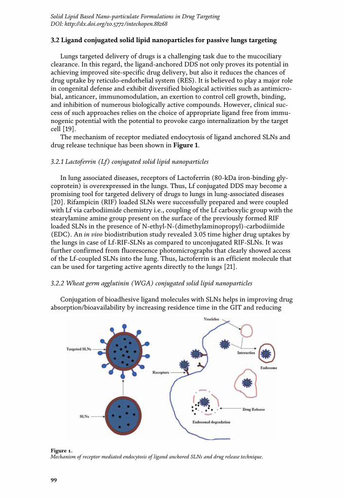
3. Solid lipid nanoparticles for lungs targeting
Targeted delivery of a drug to the lungs is gaining much more interest at thepresent time, for the treatment of lungs cancer, tuberculosis, and other airbornediseases where lungs are the primary site of action or site administration ofdrugs [11]. In order to get maximum therapeutic benefits from lungsdelivery, a suitable DDS with appropriate physicochemical properties arenecessary and SLN along with ligand conjugated SLN are the most fitted on thisground.
3.1 Solid lipid nanoparticles for active lungs targeting
The lungs offer a very high surface area for rapid absorption of drugs owing tohigh vascularization and avoidance of the first pass effect. Sometimes, targeteddelivery of certain drugs to the lungs is very important not only for improving thebioavailability and therapeutic activity but also for reducing the systemic sideeffects [5].
To achieve a prolonged hypoglycemic effect, Liu et al. developed insulin-loadednebulized SLNs which were administered through intrapulmonary route. Thehypoglycemic effects, stability of SLN during nebulization, and deposition patternof the drug were evaluated. SLNs exhibited excellent protective effect for insulinagainst degradation or leakage from nanospheres and were relatively stable duringnebulization via jet nebulizer. Nebulized insulin SLNs helped in improving thebioavailability and showed significantly higher hypoglycemic effect as compared toinsulin phosphate buffer solution administered through pulmonary route. Thusnebulized insulin-SLNs could be a promising DDS for the treatment of diabetes[12]. In another study Bi et al. successfully developed freeze dried insulin-SLNssuitable for intra-tracheal administration through a spray. High preservation ofinsulin was noticed in SLNs after spray freeze drying. They had performed in vivostudies on diabetic rat and observed a prolonged hypoglycemic effect [13].
Rifampicin (RIF) were successfully encapsulated into the SLNs that deliveredRIF specifically to the alveolar macrophage (AM) with strong antimycobacterialefficacy (MIC reduced to 1/8 fold than that of the free drug). Generally, mycobac-terium safely multiplies in the AM (acts as an incubator), as the mycobacterium isresistant to the biocidal mechanism of AM. The developed SLNs were more stableand the particle sizes were very much suitable for improving RIF’s uptake by AMswhich are particle size dependent [14]. Similarly, Rifabutin loaded SLNs signifi-cantly improved uptake of the drug by the macrophages which were demonstratedin an in vitro model [15].
Co-administration of RIF and isoniazid through SLN formulation significantlyreduced (60%) degradation of RIF (from 48.81 to 12.35%) from acidic gastric pHowing to the presence of isoniazid. The developed SLNs promoted targeted deliveryof drug to the brain with enhanced bioavailability and lesser side effect that couldbe helpful in the case of cerebral tuberculosis [16].
Significantly higher biodistribution of dexamethasone acetate (DXM) to thelungs was achieved through intravenous administration of DXM loaded SLNs. Thearea under curve (AUC) of DXM- SLNs was increased by 17.8-fold as compared toDXM-solution. The maximum concentration of the DXM in the lungs was observedat 0.5 h post DXM-SLNs injection [17].
Similarly higher biodistribution of amikacin (AMK) in the lungs was achievedby pulmonary administration of AMK-SLNs as compared to the free drug adminis-tered through i.v., which could be helpful in the treatment of cystic fibrosis [18].
98
Role of Novel Drug Delivery Vehicles in Nanobiomedicine
3.2 Ligand conjugated solid lipid nanoparticles for passive lungs targeting
Lungs targeted delivery of drugs is a challenging task due to the mucociliaryclearance. In this regard, the ligand-anchored DDS not only proves its potential inachieving improved site-specific drug delivery, but also it reduces the chances ofdrug uptake by reticulo-endothelial system (RES). It is believed to play a major rolein congenital defense and exhibit diversified biological activities such as antimicro-bial, anticancer, immunomodulation, an exertion to control cell growth, binding,and inhibition of numerous biologically active compounds. However, clinical suc-cess of such approaches relies on the choice of appropriate ligand free from immu-nogenic potential with the potential to provoke cargo internalization by the targetcell [19].
The mechanism of receptor mediated endocytosis of ligand anchored SLNs anddrug release technique has been shown in Figure 1.
3.2.1 Lactoferrin (Lf) conjugated solid lipid nanoparticles
In lung associated diseases, receptors of Lactoferrin (80-kDa iron-binding gly-coprotein) is overexpressed in the lungs. Thus, Lf conjugated DDS may become apromising tool for targeted delivery of drugs to lungs in lung-associated diseases[20]. Rifampicin (RIF) loaded SLNs were successfully prepared and were coupledwith Lf via carbodiimide chemistry i.e., coupling of the Lf carboxylic group with thestearylamine amine group present on the surface of the previously formed RIFloaded SLNs in the presence of N-ethyl-N-(dimethylaminopropyl)-carbodiimide(EDC). An in vivo biodistribution study revealed 3.05 time higher drug uptakes bythe lungs in case of Lf-RIF-SLNs as compared to unconjugated RIF-SLNs. It wasfurther confirmed from fluorescence photomicrographs that clearly showed accessof the Lf-coupled SLNs into the lung. Thus, lactoferrin is an efficient molecule thatcan be used for targeting active agents directly to the lungs [21].
3.2.2 Wheat germ agglutinin (WGA) conjugated solid lipid nanoparticles
Conjugation of bioadhesive ligand molecules with SLNs helps in improving drugabsorption/bioavailability by increasing residence time in the GIT and reducing
Figure 1.Mechanism of receptor mediated endocytosis of ligand anchored SLNs and drug release technique.
99
Solid Lipid Based Nano-particulate Formulations in Drug TargetingDOI: http://dx.doi.org/10.5772/intechopen.88268

dosing frequency. Lectins (a group of diverse proteins/glycoproteins) are thebioadhesive ligand and have stable structure and receptor binding ability. It offersresistance to enzymatic digestion/degradation, which helps in its in vivo survival fora prolonged time period [22]. Wheat germ agglutinin (WGA) is an example ofnontoxic plant lectin which binds particularly to N-acetyl-D-glucosamine and sialicacid on cell surfaces. Conjugation of WGA with SLNs could be helpful for targeteddelivery of antitubercular drugs to the lungs.
WGA conjugated SLNs loaded with rifampicin (WRSLNs) were successfullydeveloped for lungs specific delivery of rifampicin (RIF). The conjugation of WGAto RIF loaded SLN was carried out by two-steps carbodiimide reaction reported byErtl et al. with slight modification [23]. Even after conjugation with the SLNs, theWGA retained its bio-recognition activity and sugar-binding specificity [24]. Fromthe in vivo study result, the interaction of WRSLNs with porcine mucin was con-firmed. However, non-conjugated SLNs did not show any conjugation with theporcine mucin. The WRSLNs were stable at refrigerated temperature and also inpresence of electrolytes up to 1.0 M Na2SO4 concentration. The WRSLNs were 10times more stable than RIF-SLN which could be owing to the steric stabilization bylectin present on nanoparticle surface. Prepared WRSLNs had shown narrow sizedistribution, controlled release of a drug, retention of biorecognition activity andimproved physical stability of drug against electrolyte induced flocculation [25].
3.2.3 Mannose conjugated solid lipid nanoparticles
Studies on tuberculosis revealed overexpression of the mannose receptors spe-cifically on alveolar macrophages (AM) surfaces [26]. Keeping it in mind attemptswere taken to develop mannosylated SLN to deliver antitubercular drugs targetingto alveolar macrophages which have higher affinity for mannose. Rifabutin loadedmannosylated SLNs were successfully developed. Manosylation was done by ringopening reactions followed by reaction of aldehyde groups of mannose in 0.1 Msodium acetate buffer (pH 4.0) with the amino groups of lipid. This leads toformation of Schiff’s base (–N=CH–), which may then get reduced to secondaryamine (–NH–CH2–) and remain in equilibrium with Schiff’s base at basic pH. In vivostudies were conducted to evaluate for their cytotoxicity, targeting potential, AMuptake, and hematological studies. It was reported that mannosylation improveduptake (up to 6-fold) of rifabutin by the AM. Moreover, mannosylated rifabutin-SLNs were less immunogenic and helped in sustained delivery of drug. Thus, themannosylated SLNs may be employed for AM targeted delivery of rifabutin foreffective management of TB [27].
Overexpression of mannose receptors in case of lungs cancer was reported bynumerous investigators. The mannosylated-distearoyl phosphatidyl-ethanolamineSLNs loaded with paclitaxel (PTX) was developed for lungs targeted delivery ofPTX. Manosylation was done by ring opening reaction followed by reaction of analdehyde group of mannose with the free amine group provided by stearylamineand DSPE in sodium acetate buffer (pH 4.0). The stability testing data indicatedthat SLNs formulations stored at 4 � 2°C were more stable than those stored at27 � 2°C. It was revealed that mannosylated SLNs deliver significantly higherconcentration of drug to the alveolar cell sites and showed improved antiproli-ferative efficacy as compared to PTX solution and PTX-SLNs [28].
3.2.4 Folate conjugated solid lipid nanoparticles
The folate receptors (α- form) are overexpressed on the surface of lung tumorcells. The extents of the overexpression are different in different types of lungtumors (adenocarcinomas—72%, squamous cell carcinomas—51%, small cell
100
Role of Novel Drug Delivery Vehicles in Nanobiomedicine
carcinoma—25%, and lung metastases—30%) [29]. These receptors allow folatederivatives to bind preferentially that permits intracellular incorporation of folatederivative by endocytosis.
Folate-conjugated copolymer of polyethylene glycol (PEG) and N-[(2-hydroxy-3-trimethylammonium) propyl] chitosan chloride (HTCC) SLNs loaded with Pacli-taxel (PTX) were successfully developed. The conjugation of folate- conjugatedPEG and HTCC to RIF-SLN was carried out by carbodiimide mediated couplingchemistry. Pulmonary administration of the developed F-PEG-HTCC–SLNs selec-tively delivered the PTX to the lung’s cancer cells with improved penetrability andprolonged lung residence. Moreover, the developed SLN significantly reduced thein vitro half-maximum inhibitory concentration of PTX in M109-HiFR cells [30].
List of SLNs and their different ligand conjugated forms for lungs targeting havebeen summarized in Table 1.
4. Solid lipid nanoparticles for brain targeting
Targeted delivery of drugs to the brain for is gaining much more interest at thepresent time, not only for the treatment of brain tumor and other neurodegenera-tive disorders but also for their diagnosis. Brain targeted delivery of drugs is themost challenging task because of the presence of strongest physiological barrier, i.e.,
Sl.no
SLN(Type)
Lipids Preparationmethod
Drugs Target Model Comments Ref.
01 SLN SA, PA Reverse micelle-double emulsion
Insulin Lungs Male SDrats
Improved hypoglycemiceffect
[12]
02 SLN SA, PA Modified lipidfilm hydrationmethod
RIF AM SD rats Improved drug uptakeby AMs.
[14]
03 SLN SL,GTS
Ultrasonicationwith highpressurehomogenization
DXM Lungs Mice Improved therapeuticefficacy targeting tolungs
[17]
04 Lf-SLNs
SL Solvent injectionmethod
RIF LfR Albinorats
Targeted delivery of RIFto Lungs
[21]
05 WGA-SLN
SA,GMS
Singleemulsificationfollowed bysolventevaporation
RIF Mucin Porcine Controlled release of RIF [25]
06 Msy-SLN
TSN Modified solventinjection method
RFB MR J774 &albinorats
AM specific drugdelivery. Enhancedtherapeutic efficacy.
[27]
07 Msy-SLN
TSN,DSPE
Modified solventinjection method
PTX MR A549 &Malealbinorat
Alveolar cell sitedelivery of drug withenhanced anti-proliferative efficacy.
[28]
08 Ft-SLN CHO,GS
Nanoprecipitationmethod
PTX FR HeLa &M109-HiFRcells
Prolonged drugresidence time, reducedMIC
[30]
Table 1.List of SLNs and their different ligand conjugated forms for lungs cells targeting.
101
Solid Lipid Based Nano-particulate Formulations in Drug TargetingDOI: http://dx.doi.org/10.5772/intechopen.88268

blood brain barrier (BBB). It is a highly selective semipermeable membrane barrierconstituted by specialized microvascular endothelial cells, basement membrane andglial cells (astrocytes, neurons, and pericytes). As long as the BBB remains integral,the drugs remain ineffective in the brain. Though BBB is a major issue for it, yet itoffers scores of opportunities such as presence of numerous transport proteins andspecialized receptors [5].
Conventional approach for the treatment of brain tumor and other brain-relateddisorders needs a higher dose of the drug that leads to systemic toxicity and sub-stantial adverse effects on CNS and vital normal tissues. However, variousresearchers have reported SLNs to be a suitable DDS targeting the brain as the SLNspossess numerous unique characteristics, such as improved uptake of SLN by thebrain due to lipidic nature, bioacceptability and biodegradability nature, non-toxic,nano sized particles suitable for prolonged circulation time in blood scale up feasi-bility, absence of burst drug release effect [31]. Thus, SLN could be used aspotential as well as promising candidate for brain targeting.
4.1 Solid lipid nanoparticles for active brain targeting
SLNs are the most acceptable brain targeted DDS employed in brain tumor owingto their ability to escape and/or inhibit P-glycoprotein in the blood–brain barrier [32].Camptothecin (CMP) loaded SLN were successfully developed for the treatment ofglioma. Encapsulation of drug in phospholipids and conjugation with the peptideenhances the permeation of drug across BBB, which leads to brain targeted deliveryof the drug. The developed SLN was stable in terms of size and charge within a yearof storage which might be due to appropriated acidic pH (inclusion of behenic acidinto the lipid core of the SLNs). DMPC (1,2-dimyristoyl-sn-glycero-3-phospho-choline) membrane helps in efficient release of the incorporated CMP into the brainparenchyma crossing the BBB. Against glioma, the developed SLNs showed highercell death or antitumor activity using the lowest maximal inhibitory concentration(IC50) values. Biodistribution study revealed higher accumulation of CMP in thebrain in the case of SLNs as compared to other non-encapsulated drugs. However,pharmacokinetics study revealed lower deposition of CMP in peripheral organs indi-cating lesser toxicological effects in the vital organ. Thus, in short, the SLNs exhibitedenhanced accumulation, distribution, and retention of camptothecin in the animalbrain along with superior in vitro antitumor activity against glioma [33].
Riluzole (RLZ), a potent neuroprotective agent is useful in the treatment ofneurodegeneration including traumatic brain injury and amyotrophic lateral sclero-sis. RLZ-SLNs were able to deliver the riluzole successfully to the brain which helpsin preventing acute cell damage induced by glutamate in neurodegenerative dis-eases of motor neurons. Moreover, it also protects dopamine neuron in the case ofParkinson’s disease. Stability studies on RLZ-SLNs were also carried out at pH 1.1,5.5 and 7.4, and in human plasma which revealed the absence of riluzole degrada-tion in all the investigated media [34].
Vinpocetine (VIN), a derivative of vincamine alkaloid, useful against chroniccerebral vascular ischemia. VIN loaded SLN were successfully developed andachieved the objectives of delivering the drugs to the brain. Release kinetics of thedeveloped SLN followed zero-order sustained release profile [35].
4.2 Ligand conjugated solid lipid nanoparticles for passive brain targeting
4.2.1 Lactoferrin conjugated solid lipid nanoparticles
Augmentation of drug uptake into the brain is a subtle mission in the treatmentof brain tumor. Lactoferrin (Lf) receptor present on the BBB in different species
102
Role of Novel Drug Delivery Vehicles in Nanobiomedicine
along with the cell surface of glioblastomas [36]. Keeping it in mind, variousresearchers tried to target lactoferrin receptor through conjugation of lactoferrinwith SLNs for active targeting to brain tumor. Moreover, this conjugated systemshowed higher stability, and drug payload.
Lactoferrin (Lf) conjugated SLNs loaded with docetaxel (DTX) were able toshow effective brain targeting efficiency. Carbodiimide chemistry was employedfor conjugation of Lf on SLN surface (Lf-SLN). Receptor saturation studies anddistribution studies of lipidic nanoparticles in the brain indicated brain targetingmechanism for uptake in brain tumor cell and brain respectively. The Lf-SLNs weremore stable. It not only showed significantly higher DTX concentration in the brainbut also showed superior apoptotic activity when compared with unconjugatedSLNs and DTX. Thus it was confirmed that conjugation of Lf to SLN significantlyimproved the targeting potential of the DTX for brain tumor [37].
4.2.2 Tamoxifen-Lactoferrin cross conjugated solid lipid nanoparticles
P-glycoprotein (P-gp) and multidrug resistance-associated proteins (MRAPs)are localized on brain microvascular endothelial cells (BMEC) which pump out thesubstances/drug out from the central nervous system. Tamoxifen (Tf), a selectiveestrogen receptor modulator possibly reverse the capability of efflux transporterslike MRAPs in cancer cells [38]. Thus, incorporation of Tf could help inpreventing a wide range of medication from efflux loss. Lactoferrin (Lf)receptors are overexpressed in BMEC and glioblastoma multiforme (GBM) cells.It is reported that the Lf cause inhibition to the multiplication of malignantGBM cells.
Tamoxifen (Tx) and lactoferrin (Lf) cross-conjugated carmustine (CRM)-loaded SLNs were developed. For conjugation of Tx on CRM-SLN, the carbonylgroups of the SLN were first activated with 0.1% (w/v) carbodiimide and 0.05%(w/v) N-hydroxysuccinimide. The suspended SLNs were then crosslinked with0.05%, 0.1%, 0.15%, or 0.2% (w/v) Tx at 150 rpm and 25°C for 3 h and centrifuged.Further Lf conjugation was done by reacting Lf (0.02%, 0.04%, 0.06%, or 0.08%)(w/v) with the activated CRM-SLNs and Tx-CRM-SLN. The conjugated SLNs weremore stable which could be due to inclusion of behenic acid into the lipid core.These were efficiently penetrated through a monolayer of human BMEC andhuman astrocytes and to target GBM cells. A 10-fold increase in the permeability ofBBB and improved the sustained release of CRM was achieved with the help of thedeveloped SLNs as compared to unconjugated CRM-SLNs. Thus, TX and Lf cross-conjugated SLNs enhance the BBB permeability of the drug with improved anti-proliferative action against GBM [39].
4.2.3 PEG conjugated solid lipid nanoparticles for gene delivery
Treatment of brain tumor through siRNA is preferable, as it can target specifi-cally to one gene and is able to silence it in a post-transcriptional way. Moreover,siRNA can target several functional proteins available at the BBB [40]. Treatment ofbrain tumor through siRNA, needs a safe, stable, effective carrier which must beable to cross the BBB. The SLNs are mostly preferred as it meets most of the criteriawhich siRNA needs. Targeted delivery of gene by SLNs is a bi-stage system. Conju-gation of angiopep to SLN surface for targeting the low-density lipoproteinreceptor-related protein-1(expressed in BBB) is the initial step. The proteolyticcleavage of PEGylated lipopeptide, which releases PEG, glutamic acid residues andrelease of siRNA for high silencing efficiency is the second stage [5].
siRNA encapsulated SLNs were successfully developed using a combination oftitrable cationic lipids. For effective gene delivery, it was PEGylated after
103
Solid Lipid Based Nano-particulate Formulations in Drug TargetingDOI: http://dx.doi.org/10.5772/intechopen.88268

incorporating MMP-cleavable lipopeptide. The in vitro study indicated that thedeveloped SLNs showed higher uptake and gene knockdown efficacy. SLNs alsoshowed low cytotoxicity which was owing to masking of intrinsic positive charge ofSLNs by PEGylated cleavable lipopeptide. In in vivo studies, angiopep functiona-lization played a vital role as a mediator of transport across the BBB and targetingto glioma [41].
4.2.4 Apolipoprotein E conjugated solid lipid nanoparticles
Low density lipoprotein (LDL) receptors are overexpressed on the BBB andapolipoprotein E (AP-E), a plasmatic protein is easily recognized by these receptors.Resveratrol (RSV), polyphenolic flavonoid promises to offer neuroprotectiveeffects which are helpful in neurological disorders like Alzheimer’s, Parkinson’s,Huntington’s diseases, brain ischemia, and epilepsy. The AP-E conjugated resvera-trol (RSV) SLNs were successfully developed. The binding of ApoE to the SLNssurface was carried out by spontaneous interaction between the previouslybiotinylated ApoE and the covalently attached avidin on the SLNs surface, resulting
Sl.no
SLN(Type)
Lipid(s)
Preparationmethod
Drugs Target Model Comments Ref.
01 SLN CP,DMPC
High shearhomogenizationandultrasonicationtechniques
CMP Brain Humanglioma &Monocyticcell line.Wistar rats
Enhancedaccumulation ofCMP. Superiorin vitro antitumoractivity
[33]
02 SLN COMP,SPC
Warm oil-in-watermicroemulsiontechnique
RLZ Brain Male SD rats Higherbioaccumulationof RLZ in brain.
[34]
03 SLN SA,GMS,PRE,GTP
High shearhomogenizationandultrasonicationtechniques
VIN Brain — Zero-ordersustained drugrelease kinetics
[35]
04 Lf-SLN GMS,SA, SL
Emulsificationand solventevaporationmethod
DTX LfReceptors
U-87 MGcell lines &Swiss albinomice
Improved thebrain targetingpotential
[37]
05 Tf-Lf-SLNs
TPM,DSPE
Homogenizationfollowed bycentrifugation
CRM Tf and LfReceptors
U87MG,HBMECs
Higher BBBpermeability.Superior anti-proliferativeaction
[39]
06 SLN POPC,DSPE,CHO,DM
Detergentdialysistechnique
siRNA Angiopep bEnd.3 cell,U87MGcells,
Higher uptakeand geneknockdownefficacy
[41]
07 AP-E -SLNs
CP High shearhomogenizationfollowed bysonicationtechnique
RSV LDLReceptor
hCMEC/D3Cell line
Brain targeteddelivery of RSV
[42]
Table 2.List of SLNs and their different ligand conjugated forms for brain cell targeting.
104
Role of Novel Drug Delivery Vehicles in Nanobiomedicine
in two different ApoE-functionalized SLNs: SLN-DSPE-ApoE and SLN-Palmitate-ApoE. These conjugated SLNs were sufficiently stable and were able to prevail overthe issues of RSV like low solubility, degradation but also to help its brain targeteddelivery. Brain targeted delivery of RSV by such SLNs follows a bi-stage system.The AP-E-RSV-SLNs mimic lipoprotein particles that are endocytosed into the BBBendothelium via the LDL receptor and then transcytosed to the brain [42].
List of SLNs and their different ligand conjugated forms for brain cell targetinghave been summarized in Table 2.
5. Solid lipid nanoparticles for liver targeting
Though targeted delivery of a drug to the liver is a challenging task, still, it isan interesting approach in the treatment of various liver disorders. In the treatmentof liver disorders, drugs targeting to the liver, face irresistible obstacles fromvarious physiological barriers and processes like uptake by the reticuloendothelialsystem, mechanical entrapment by the pulmonary vascular bed, and opsonizationprocess [43].
Numerous approaches are being proposed to enhance bioaccumulation/biodistribution of drugs to liver and hepatocytes. These approaches include bothactive targeting as well as passive accumulations of nanoparticulate formulation dueto ligand (carbohydrate, peptide, antibodies conjugation) conjugated nanoparticles.Recently, liver targeted deliveries of drugs by the SLNs are gaining much attentionin the treatment of various types of liver disorders. Thus, various liver targetingstrategies using SLNs are enlightened below.
5.1 Solid lipid nanoparticles for active targeting
Baicalin (BCL), a natural product obtained from Scutellaria baicalensis (Family:Labiatae) popularly used in the treatment of Hepatitis-B and liver fibrosis. BCLloaded SLNs were developed. The prepared SLNs were stable and were able toenhance the therapeutic efficacy of BCL by improving its biodistribution in theliver. In vivo biodistribution, targeting evaluation and in vitro anti-oxidant studyreveals that the developed BCL-SLNs have substantial liver targeting, improved antioxidative and hydroxyl radical scavenging abilities [44].
Ficus benjamina (Family: Moraceae) is rich in phenolic (chlorogenic, p-coumaric, ferulic and syringic acids) and total flavonoid content that are effectiveagainst chronic alcoholism induced fatal liver and cardio-renal injury. Ethanolicextract of Ficus benjamina (FB) loaded in SLNs (FBSLNs) helped inbioaccumulation of the phenolic and flavonoid content in the liver due to lipophilicnature of SLNs. In vivo evaluation of FBSLNs against hepatic and cardio-renal injuryrevealed its hepatoprotective activity which was further evident from various bio-logical parameters and histopathological photomicrography. In the liver, accumula-tion of aldehyde level was reduced that validated the detoxifying nature of FBSLN.Moreover, restoration of aberrant cardio-renal biomarkers and histological conse-quences revealed the cardio-renal protective potential of FBSLNs [45].
Berberine (BBR), an active constituent of Coptis chinensis (Family:Ranunculaceae) have potential pharmacological effects on type-2 diabetes. It hasalready been validated that BBR enhances glucose and lipid metabolism through theactivation of adenosine monophosphate-activated protein kinase (AMPK) andimprove insulin sensitivity. BBR loaded SLNs (BBR-SLNs) were developed toimprove the beneficial effect of BBR on hepatosteatosis. The effect of BBR-SLNs onlipid metabolism were studied which revealed gaining of body weight and reduction
105
Solid Lipid Based Nano-particulate Formulations in Drug TargetingDOI: http://dx.doi.org/10.5772/intechopen.88268

in liver weight with simultaneous reduction of serum alanine transaminase and livertriglyceride level. Biodistribution study reported 20-fold increase in the concentra-tion of drug in the liver than that of blood. Moreover, it reduced the accumulationof fat and lipid droplet size. It was also noticed that the expression of lipogenicgenes was down-regulated and lipolytic gene was up-regulated in BBR-SLNs treatedlivers which could be helpful in the treatment of hepatosteatosis [46].
Cisplatin (CSPT) is an anti-cancer drug which is used in the treatment of manymalignancies including hepatocellular carcinoma, lungs carcinoma, etc. The CSPTloaded SLNs (CSPT-SLNs) were successfully developed and were stable in terms ofdrug content after storage for 3 months in different temperature and humid condi-tions. In vivo tissue distribution study revealed that the developed CSPT-SLNs wereable to deliver a higher amount of CSPT particularly to the liver as compared to thebrain, lungs, and kidney [47].
Sorafenib (SFB), a potent multi-kinase inhibitor possess anti-tumor angiogenesiseffect (block vascular endothelial growth factor (VEGF) and platelet-derivedgrowth factor receptor (PDGFR)) and is preferentially used in the treatment ofhepatocellular carcinoma. The SFB loaded SLNs (SFB-SLNs) were developed withan objective of improving bioavailability and reducing adverse effects. The resultsof the stability test showed that SRF-SLNs remained stable for more than 1 month atroom temperature. In vivo study of SFB-SLNs revealed improved bioavailability(increased by 66.7%) with remarkably higher bioaccumulation of drug in the liver(2.20-fold higher drug selectivity index value) when compared with the SFBsuspension [48].
Primaquine phosphate (PP) is an antimalarial drug that acts on the primarytissue forms of the Plasmodium which after growth within the liver, initiate theerythrocytic stage. Thus, PP loaded SLNs (PP-SLNs) were developed with an aim todeliver liver schizonticide PP directly to the hepatocytes. Stability of the PP-SLNs insuspension was tested for a period of 3 months in terms of size, poly-dispersity,ζ-potential, and pH. There were no noteworthy changes in size, poly-dispersity,ζ-potential, or pH occurred over time. In vivo study report revealed that thedeveloped SLNs were highly effective (>20%) against hypnozoites/liver stage ofall malarial species with a reduced dose when compared with the conventionaloral dose [49].
5.2 Solid lipid nanoparticles delivering gene
The fibrous scars occurring in the liver due to the increased production anddeposition of hepatic extracellular matrix (ECM) components are called liver fibro-sis reduce the physiological performance of the liver. Hepatitis viral infection is oneof the major reasons for liver fibrosis and cirrhosis. Administration of antifibrotictherapeutics (e.g. connective tissue growth factor (siRNA) responsible for the cel-lular and molecular basis of fibrogenesis) is one of the most preferable approachesfor the treatment of liver fibrosis. The siRNA loaded cationic SLNs (cSLNs) weredeveloped by gently mixing CSLNs with siRNA at various weight ratios of cSLN tosiRNA in 0.1 M PBS (pH 7.4) and then incubated at room temperature for 15 min.Naturally obtained low-density lipids (LDLs) were used in the preparation. Thedeveloped cSLN were able to silence the targeted gene in the presence of serumwith notably low cytotoxicity. The cSLNs were PEGylated which were hydrody-namically stable and were able to protect their siRNA cargo from nuclease degra-dation during systemic circulation. The developed cSLNs loaded with siRNAadminister through intravenous route delivered siRNA exclusively to the liver andresulted in a considerable reduction in collagen content and pro-fibrogenic factorswith spectacular progress of pathophysiological symptoms in a liver fibrosis rat
106
Role of Novel Drug Delivery Vehicles in Nanobiomedicine
model. Biodistribution study revealed site-specific delivery and accumulation ofsiRNA loaded cSLNs to the liver tissues [50].
5.3 Solid lipid nanoparticles for passive targeting
5.3.1 Polyethylene glycol conjugated (PEGylated) solid lipid nanoparticles for passivetargeting
PEGylated SLNs are reported to be preferentially accumulated in the liver as thekidney is unable to clear the same. Thus, in liver disorders, liver targeting strategyusing PEGylation technique is used for delivering numerous drugs. Paclitaxel (PTX)loaded PEGylated SLNs were successfully developed for targeting the liver, in thecase of hepatic carcinoma. The cellular uptake study revealed that PTX loadedPEGylated SLNs showed prolonged circulation time in plasma and higherbioaccumulation of drug in the liver when compared with the PTX solution [51].
The preferential drug targeting ability of PEGylated SLNs to cancer cells havebeen shown in Figure 2.
5.3.2 Galactosylated lipid [N-hexadecyl lactobionamide] conjugated solid lipidnanoparticles
The parenchymal cells of the liver contain asialoglycoprotein receptors whichrecognize terminal b-D-galactose or N-acetylgalactosamine residues. The N-hexadecyl lactobionamide (N-HLBA) was synthesized via an amide bond betweenthe amine group of hexadecylamine and the carboxyl group of lactobionic acid. The
Figure 2.PEGylated SLN in targeting preferentially to cancer cells.
107
Solid Lipid Based Nano-particulate Formulations in Drug TargetingDOI: http://dx.doi.org/10.5772/intechopen.88268

lactobionic acid was converted to 1,5-lactone that contain more reactive aminegroups. Cucurbitacin B (CurB), a tetracyclic triterpene shows significant pharma-cological activities including anti-tumor, anti-hepatitis, hepatocurative andhepatoprotective. The CurB loaded N-hexadecyl lactobionamide (N-HLBA) conju-gated SLNs were developed for liver-targeted delivery of CurB. The N-HLBA SLNwith anchored galactose moiety via amide bonds might achieve effective liver-targeting delivery in vivo by retaining the surface galactose in blood stream, and byexposing a higher amount of galactose to the liver parenchymal cells. The optimumzeta potential held up the physical stability whereas the optimum particle sizedistribution offered the convenience for intravenous administration and deep pen-etration into targeting area. The Biodistribution study of the CurB loaded N-hexadecyl lactobionamide (N-HLBA) conjugated SLNs revealed a 2.5-fold increase
Sl.no
SLN(Type)
Lipid(s)
Preparationmethod
Drugs Target Model Comments Ref.
01 SLN SL,GMS
Emulsificationultrasonicdispersionmethod
BCL Liver Rats Improvedbiodistribution of BCLin Liver, Superior antioxidative andhydroxyl radicalscavenging abilities
[44]
02 SLN SA Hot-homogenizationfollowed byultra-sonication
FB Liver HepG2 cellline, Rats
Anti-oxidant, anti-inflammatory anddetoxificationpotential
[45]
03 SLN GL,SP, TP
HotEmulsificationTechnique
BBR Liver Male db/dbmice,
Down regulate thelipogenic gene andUp-regulate thelipolytic gene.
[46]
04 SLN SA Warmemulsificationfollowed bysonication
CSPT Hepatocellularcarcinomas
Wistar rats Higherbioaccumulation ofdrug in liver
[47]
05 SLN GB High-speedshearingfollowed byultrasonication
SFB Liver Female SDRats
Improvedbioavailability, higherbioaccumulation inliver
[48]
06 SLN SA Modifiedmultipleemulsionsolventevaporationtechnique
PP Hepatocytes 3D7, Mice Improved antimalarialactivity
[49]
07 C-SLN CO Modifiedemulsificationand solventevaporationmethod
siRNA LDL Receptor Rat, HSCsandhepatocytes
Spectacular progressof pathophysiologicalsymptoms in liverfibrosis
[50]
08 PEG-SLN
GT,CHO
Solvent-emulsificationmethod
PTX Hepatocytes HepG2,MCF7,PANC-1
Higher liverbioaccumulation ofPTX
[51]
09 G-SLN COMP High-pressurehomogenization
CurB ASGPReceptor
Wistar rats Enhanced antitumorand hepatoprotectiveactivity
[52]
Table 3.List of SLNs and their different ligand conjugated forms for liver cell targeting.
108
Role of Novel Drug Delivery Vehicles in Nanobiomedicine
in the amount of CurB in the liver when compared with CurB-SLN. In vitro cyto-toxicity study revealed enhancement of cytotoxicity. The experimental result vali-dated the liver targeting ability of N-HLBA conjugated SLNs [52].
List of SLNs and their different ligand conjugated form for liver targeting havebeen summarized in Table 3.
6. Solid lipid nanoparticles for breast cell targeting
Breast cancer is the most common form of cancer and the second most deadlydisease among the woman around the globe. The breast cancer new incidences andmortality rate has been increased by 20 and 14% since 2008.
Recently, controlled release of the drugs to the targeted site of the disease using ananocarrier vehicle is getting more attention as it enhances the therapeutic efficacyof the drugs. Solid lipid nanoparticulate (SLN) formulations possess an endlesspotential to deliver active chemotherapeutic molecules in a programmed prototypeto improve bioavailability and nullify the side-effects. The bio-compatibility andbio-degradability characteristics of SLNs promise to offer a lesser toxic product ascompared to polymeric nanoparticles which forced to consider it as an idealistictargeted drug delivery system for breast cancer therapy [5].
6.1 Solid lipid nanoparticle for active breast cell targeting
6.1.1 SLNs for photodynamic therapy
Photodynamic therapy is one of the emerging approaches in the treatment ofcancer which comprises application of a photosensitizer followed by laser irradia-tion of tumor lesions. Temoporfin (TP), a photosensitizer loaded in thermore-sponsive SLNs were developed with an objective to improve anticancer activitythrough site specific drug delivery. The copolymer poly(ethylene oxide)-block-poly(ε-caprolactone) copolymers (PEO45-b-PCL7) were synthesized by the mecha-nism of catalyst-free ring opening polymerization of ε-caprolactone which wasinitiated by poly(ethylene oxide) monomethyl ether(MPEO). These copolymerswere acts as a stabilizer in the preparation of thermoresponsive SLN. The stabilitystudy report revealed that the developed SLNs had higher stability in human serumwithin the blood transport to tumor tissue. In vitro phototoxicity study revealedhigher phototoxic activity of TP against breast cancer cells due to its fasterbioaccumulation in the targeted cancer cells. The in vivo anticancer efficacy of TP-SLN was remarkably higher as compared to the commercial TP formulation [53].
6.1.2 Manganese (Mn) II complex solid lipid nanoparticles
Transition metal complex (e.g., Manganese II complex[Mn2(μ(C6H5)2CHCOO)2(bipy)4]
� 2) has been extensively used for cancer therapynowadays due to its potential anticancer activity (interact with the DNA). Mn(II)complex loaded SLNs were developed that showed superior cytotoxicity activity onbreast cancer cells. The zeta potential value of the product was higher indicatinggood physical stability and dispersion quality of the SLNs. Cell proliferation assayrevealed that the normal cell death rate was lower with Mn(II) SLNs which indi-cated lesser toxicity of the product to the normal cell. Moreover, higher earlyapoptosis rate was observed with Mn(II) complex SLN as compared to Mn(II)alone [54].
109
Solid Lipid Based Nano-particulate Formulations in Drug TargetingDOI: http://dx.doi.org/10.5772/intechopen.88268

6.2 Solid lipid nanoparticles for passive breast cell targeting
6.2.1 Fucose conjugated solid lipid nanoparticles
Fucose receptors are overexpressed in the breast cancer cell. Thus, conjugationof fucose to SLNs was proposed to deliver the drug specifically to breast cancerouscells. Fucose conjugated methotrexate (MTX) loaded SLNs were developed toachieve enhanced targeting potential for breast cancer cells. Fucosylation of MTX-SLNs was related with opening of fucose ring and reaction of its aldehyde groupwith free amino functionalities expressed over the surface of MTX-SLNs in sodiumacetate buffer (pH 4.0). The above process led to the formation of Schiff’s base (–N=CH). The Schiff’s base might be reduced to secondary amine (–NHCH2) andestablish equilibrium with Schiff’s base. Physical stability of prepared SLNs washigher which could be due to positive zeta potential value that provides repulsiveinteraction between nanosized lipid particles preventing particle aggregation. Theex vivo study revealed higher cellular uptake as well as higher cytotoxicity at lowerIC50 of MTX. The in vitro study results showed increased rate apoptosis with achange in lysosomal membrane permeability and a higher rate of lysosomalmembrane degradation. The in vivo study revealed maximum bioavailability andtumor targeting efficiency with minimum secondary drug distribution toother organs [55].
6.2.2 Folic acid functionalized solid lipid nanoparticles
The folate receptor (FR) is one of the most widely evaluated receptor for activetargeting of anticancer therapeutics in the case of in breast cancer cells. Folic acidhas many advantages over antibody ligands such as small size, non-immunogenicity, non-toxicity, ease of handling, stability and low cost [56]. Severalresearchers had reported earlier that the FA functionalized SLNs were able todeliver the chemotherapeutic agent, particularly to the cancerous cells. Thus, FAfunctionalized SLNs co-encapsulated with Docetaxel (DTX) and Curcumin (CUR)were successfully developed to enhance its therapeutic efficacy against breast can-cer cells. FA-stearic acid (FA–SA) conjugate was synthesized by classical 1-ethyl-3-(3-dimethylaminopropyl) carbodiimide (EDAC) chemistry and incorporated in theDTX-CUR-SLN. Additionally, PEG–stearic acid (PEG–SA) was incorporated toobtain FA-DTX-CUR-SLN. The DTX-CUR-SLN and FA-DTX-CUR-SLN formula-tions were found to be stable at refrigerated condition for 2 months. Both theformulations showed sign of instability at accelerated condition (25°C/65% RH).The developed FA functionalized SLNs exhibited improved pharmacokineticparameters, superior cancer cell targeting efficiency, and improved therapeuticefficacy of DTX. Folic acid was believed to be responsible for the targeting efficacyof the developed SLNs to the breast cancer cell. This conjugated system showedsignificant increase in area under the curve and mean residence time of the drug.Co-conjugation (FA and PEG) to SLNs co-encapsulated with DTX and CURresponsible for synergistic activity of both DTX and CUR. Moreover,bioaccumulation of DTX in heart and kidney was found very low which signifiedavoidance of vital organ toxicity [57].
6.2.3 Cetamide and trimethylphytosphingosine-iodide co-conjugated solid lipidnanoparticles
The sphingosine-1-phosphate (S1P), phytosphingosine, ceramides, and sphin-gosine are the metabolites of sphingolipids and are reported as essential structural
110
Role of Novel Drug Delivery Vehicles in Nanobiomedicine
components of cell membranes or important mediators of cellular process thatregulate the proliferation, survival, and death of cells. Moreover, sphingosinesincluding N,N,N-trimethylsphingosine (TMP-I) have been reported for their role asa negative modulator of transmembrane signaling through protein kinase C (PKC)as well as an inhibitor of sphingosine kinase-1 (SK-1), controlling the variousmembrane-associated signaling mechanisms associated with cell growth and inhib-itory apoptosis in tumor cells [58]. Ceramide, a kind of sphingosine conjugated withfatty acid residue, is also reported to enhance the sensitivity of MDR-acquiredcancer cell lines to chemotherapeutic agents [59]. Thus, attempts were made toemploy ceramide (CD) and trimethylphytosphingosine-iodide (TMP-I) as atargeting agent for docetaxel (DTX) loaded SLNs. The prepared SLNs were physi-cally stable without any significant change in their physical appearance, drug con-tent, and particle size over a period of 8 weeks at 4°C. CD enhanced the DTXsensitivity in MDR-acquired cancer cell lines. In vivo clearance of drug and tumorgrowth inhibitors were significantly decreased in case of CD and TMP-I conjugatedSLNs when compared with the marketed product. Thus, CD and TMP-I conjugatedDTX-SLN could serve as a potentials alternative parenteral formulations ofDTX [60].
Sl.no
SLN(Type)
Lipid(s) Drugs Preparationmethod
Target Model Comments Ref.
01 SLN TD TMP Modified hothomogenizationandultrasonicationmethod
BreastCancercell
4T1,MDA-MB-231,Female Nu/Numice- MDA-MB-231
Exhibitedimprovedphototoxicity andanticancerefficacy
[53]
02 Mn (II)Complex-SLN
COMP Mn (II)complex
Hothomogenizationmethod
BreastCancercell
MCF-7 andHUVEC cellline
Possessedsuperioranticanceractivity withreduced toxiceffect.
[54]
03 F-SLN PL90NG,PL, STA,GEL
MTX Hotmicroemulsionmethod
FucoseReceptor
MCF-7 Cellline & FemaleSD rat
Improvedbioavailabilityand tumortargetingefficiency withminimumsecondary drugdistribution invarious organs.
[55]
04 FA-PEG-SLN
GMS, SA CUR,DTX
Modifiedethanol injectionmethod
FolicacidReceptor
MCF-7&MDA-MB-231Cell line.Female WistarRat
Synergisticcancer efficacydue tocoencapsulationof DTX and CRMalong withtargeteddelivery of drugs
[57]
05 CD-TMPI-SLN
PC, TMS, DTX High-pressurehomogenizationmethod
CD &TMP-1
MCF-7 cell,MCF-7/ADRcells
Significantincreasedantitumorefficacy withtargeted drugdelivery
[60]
Table 4.List of SLNs and their different ligand conjugated forms for breast cancer cell targeting.
111
Solid Lipid Based Nano-particulate Formulations in Drug TargetingDOI: http://dx.doi.org/10.5772/intechopen.88268

List of SLNs and their different ligand conjugated form for breast cancer celltargeting have been summarized in Table 4.
7. Solid lipid nanoparticles for eye targeting
The eye is one of the delicate organs of human and one of the most delicateroutes of drug delivery. However, the eye poses unique challenges relative to drugdelivery due to the ocular anatomical and physiological constraints. SLNs are one ofthe promising targeted DDS for an eye. Numerous drugs such as antibiotics, plas-mids, anti-inflammatory, and immunosuppressive agents were encapsulated inSLNs for the treatment of ophthalmic disorders.
7.1 Solid lipid nanoparticles for active eye targeting
Corneal neovascularization (CNV), a sight-threatening condition is caused dueto various inflammatory settings including chemical injury. Single-strandedproline-modified short hairpin anti-angiopoietin-like protein 2 (ANGPTL2 RNA)interference molecules acts as potent angiogenic and pro-inflammatory factor and isused for the treatment of CNV [61]. Thus, ANGPTL2 RNA loaded SLNs weredeveloped to deliver the interference molecule specifically to the retina. The single-stranded RNAi (pshRNA) loaded SLNs exhibit high stability in vivo and preliminarydata suggest that pshRNA is less susceptible to degeneration due to RNase activity.The fluorescence-labeled developed SLNs eye drops penetrated all layers of corneaand expression of ANGPTL2mRNA was considerably inhibited in both epitheliumand stroma. Further, the area of angiogenesis was remarkably decreased in thecorneas by the SLNs drops as compared to the control group [62].
X-linked juvenile retinoschisis (XJR), a retinal degenerative disorder caused bymutation in the RS1 gene encoding a retinoschisin [63]. Among non-viral vectors,solid lipid nanoparticles (SLNs) represent one of the most effective lipid-basedcolloidal carriers, and for gene delivery to the posterior segment of the eye [64].Plasmid (human RS1 gene) loaded SLNs were developed for the treatment of XJRdiseases which showed significant improvement of the retinal structure with pho-toreceptor specific expression of the RS1 gene [65].
Tobramycin (TMC) is one of the most preferable drugs to treat vitreoretinaldiseases, such as bacterial infections, endophthalmitis, cytomegalovirus retinitis(CMV), uveitis, proliferative vitreoretinopathy (PVR), diabetic retinopathy, age-related macular degeneration [66]. Thus TMC loaded mucoadhesive SLNs weredeveloped which showed higher bioaccumulation of drugs in most of the ocular tissuesand was able to penetrate into the retina. Moreover, it resulted in enhancedintraphagocytic antibiotic concentration in polymorphonuclear granulocytes andsuperior bactericidal activity against Pseudomonas aeruginosa. The developed SLNsshowed good stability up to 12 months with no aggregation or precipitation phenom-ena, variations in zeta potential values or in amount of the drug encapsulated into thenanoparticles [67].
7.2 Solid lipid nanoparticles for passive eye targeting
7.2.1 Chitosan coated solid lipid nanoparticles
Indomethacin (IMC), a topical non-steroidal anti-inflammatory drug (NSAID)is used for ocular inflammatory disorders such as conjunctivitis, uveitis, cystoidmacular edema, and anterior segment inflammation, including post-operative pain
112
Role of Novel Drug Delivery Vehicles in Nanobiomedicine
following cataract surgery [68]. Chitosan (Cs) coated IMC loaded SLNs (IMC-Cs-SLN) were developed to deliver NSAID to the posterior segment of ocular tissuesand for improving the pre-corneal residence time and transcorneal permeabilitycharacteristics. For surface modification of the developed SLNs, the chitosan wasincorporated into the aqueous phase prior to preparation of the SLNs. The devel-oped SLNs were stable in terms of drug loading, EE, and less drug expulsion duringstorage at 40°C for 90 days. The SLNs showed higher bioaccumulation of IMC inthe ocular tissues. The IMC-Cs-SLN showed superior trans-membrane IMCpermeation characteristics which were due to penetration enhancing propertiesof Cs [69].
7.2.2 Intercalated montmorillonite solid lipid nanoparticles
Betaxolol hydrochloride (BH) is widely used for the treatment of ocular hyper-tension and open-angle glaucoma in clinical therapeutics. However, it faces certainlimitations like low bioavailability and pre-ocular retention, and some side effects.
Sl.no
SLN (Type) Lipid(s) Drugs Preparationmethod
Target Model Comments Ref.
01 SLN DSGPC,CHO
ANGPTL2RNA
Hydrationmethodfollowed byextrusion
Retina C57BL/6mice
Inhibition ofexpression ofANGPTL2mRNA,Reduction inangiogenesis area
[62]
02 SLN PRE Human RS1gene
Solventemulsificationfollowed byevaporation
Retina 661W,RS1h-deficientmouse
Improvement ofthe retinalstructure
[65]
03 SLN SA TMC Warm o/wmicroemulsionmethod
Aqueoushumor
Albinorabbit
Superiorbactericidalactivity
[67]
04 Cs-SLN GB IMC Hothomogenization
Posteriorsegment ofoculartissue
WhitealbinoRabbits
Improvedbiodistribution ofIMC at posteriorsegment
[69]
05 Mt-SLNs PC,GMS
BH Emulsionevaporation-lowtemperaturesolidificationmethod
Cornea andConjunctiva
Rabbit Significantlyreducedinflammation, Noirritation
[70]
06 PEG-SLNs COMP KTZ Emulsificationfollowed byhigh pressurehomogenizer
Upperposterioreye
ARPE-19& RCECell line,Rat
Superiorantifungal activity
[73]
07 MultifunctionalSLNs
CP BAI (Drug)C IR-780(Diagnosticagent)
Modifiedsolvent-diffusionmethod
Colorectalpart
LoVo,CHO-K1
Imaging, Superiorcytotoxicity
[75]
08 SIA-PEGylatedSLN
GMS,OA
DXM Solventdiffusionmethod
E-selectinreceptor
HUVECs,ICR maleMice
Targeted deliveryof DXM forischemia-reperfusion-induced injury-induced AKI
[78]
Table 5.List of SLNs and their different ligand conjugated forms for eye, colon and kidney targeting.
113
Solid Lipid Based Nano-particulate Formulations in Drug TargetingDOI: http://dx.doi.org/10.5772/intechopen.88268

In order to overcome these limitations acid treated montmorillonite (Mt)- BetaxololHydrochloride (BH) nanocomposite encapsulated SLNs (Mt-BH-SLNs) weredeveloped. An acid-treated montmorillonite (acid-Mt) was first intercalated withBH in the interlayers and this nanocomposite was encapsulated by SLNs. Thedeveloped Mt-BH-SLNs possess good stability. Long term irritation test reportedthat the (Mt-BH-SLNs) showed no damage for cornea and conjunctiva. The cornealhydration level of Mt-BH-SLNs was higher (78.25 � 0.63)% indicating higher drugcorneal permeability and absence of irritation to the cornea. Thus, Mt-BH-SLNscould be used for effective management of glaucoma [70].
7.2.3 Polyethylene glycol (PEG) conjugated (PEGylated) solid lipid nanoparticles
Ketoconazole (KTZ) is a broad spectrum antifungal agent, with high lipo-solubility [71] but a short ocular half-life (elimination half-life is 19 min inaqueous humor and 43 min in cornea) [72] and very poor solubility (0.04 mg/ml).Ketoconazole (KTZ) loaded PEGylated SLNs were developed for targeted deliveryof KTZ to the posterior part of the eye for treatment of fungal infection. It showedhigher bioavailability both in the aqueous and vitreous humor with significantantifungal potential. The ex vivo corneal permeation study revealed higher cornealpermeability of the PEGylated KTZ-SLNs. The developed SLN was satisfying vari-ous parameters suitable for ocular delivery such as pH, osmolarity, stability,autoclavability, particle size, preservation against contamination. The SLNs werefound to be stable in terms of entrapment efficiency and total drug content at 2–8°Cfor 12 months. Thus it could be helpful in the treatment of keratitis andendophthalmitis [73].
List of SLNs and their different ligand conjugated forms for eye targeting havebeen summarized in Table 5.
8. Solid lipid nanoparticles for passive colon targeting
Combination of nanocarriers and electroporation techniques is named aselectropermeabilization which is commonly used for enhancing drug transport. TheCyanine–type IR 780 and Baicalein (BAI) co-encapsulated SLNs were developed forboth imaging and therapy of colorectal carcinoma where cyanine–type IR 780 andbaicalein (flavonoid derivative) were acting as a diagnostic agent (photosensitizer)and therapeutic cargo respectively. For preparation of SLNs the organic phase wasprepared by dissolving IR-780, BAI, and melted lipid in dichloromethane. Theorganic phase was then added dropwise to hot aqueous phase containing surfactantunder vigorous stirring. Supplementary material (flavonoids) facilitated in thereduction of dose and reduction in normal cell toxicity in cancer chemotherapy. Theexternal electric field pulses applied in electroporation helped in increased of cellmembrane permeability, either by generating transient pores or membraneelectropermeabilization [74]. Electropermeabilization mediated administration ofthe developed SLNs showed cytoskeletal abnormalities more significantly thenwithout electropermeabilization. The prepared SLNs particles were with goodphysical stability. With electroporation support, the developed SLNs showedincreased p53 and manganese superoxide dismutase expression with significanthigher cytotoxicity, thus validating their suitability for combined therapy andmolecular imaging simultaneously [75].
List of SLNs and their different ligand conjugated form for colon targeting havebeen summarized in Table 5.
114
Role of Novel Drug Delivery Vehicles in Nanobiomedicine
9. Solid lipid nanoparticles for kidney targeting
9.1 Solid lipid nanoparticles for passive kidney targeting
9.1.1 Polyethylene glycol (PEG) surface modified solid lipid nanoparticles
Icariin (IRN) is widely used as traditional Chinese medicine for the treatment ofkidney diseases and reinforce yang. The PEG surface modified Icariin (IRN) loadedSLNs (PEG-IRN-SLNs) was developed for targeted delivery of IRN to the kidneyand to improve the bioavailability. The SLN was prepared by high temperaturemelt-cool solidification method. Upon comparing with IRN solution it was revealedfrom the pharmacokinetic study that the biological half-life (t1/2) and area undercurve (AUC) of PEG-IRN-SLN was 7-fold and 4-fold higher. Biodistribution studyrevealed that IRN concentration in kidney tissues was significantly increased.Moreover, the relative target efficiency to kidney tissues was 79% and relative tissueexposure was 16.95. Thus the develop SLN could be helpful in the treatment ofkidney diseases [76].
9.1.2 Sialic acid conjugated PEGylated solid lipid nanoparticles
E-selectin is a promising target for the site-specific delivery of anti-inflammatory agents. Several researchers have reported that sialic acid (SA)-medi-ated micelles could be specifically internalized by lipopolysaccharide (LPS)-acti-vated human umbilical vein endothelial cells (HUVECs) via the specific bindingbetween SA and E-selectin receptor [77]. Sialic acid (SIA) conjugated PEGylateddexamethasone (DXM) loaded SLNs (SIA-PEGylated-DXM-SLN) were developedto deliver DXM specifically to the kidney and to improve the therapeutic efficacy ofDXM for renal ischemia–reperfusion injury (IRI)-induced acute renal injury. TheSialic acid (SIA) conjugated PEGylated DXM (SIA-PEG-DXM) was synthesized byadding PEG-DXM, dicyclohexylcarbodiimide (DCC), and 4-dimethylamino-pyridine (DMAP) into anhydrous dimethyl formamide (DMF) followed by additionof SIA into the solution. The resulting mixture was stirred for obtaining SIA-PEG-DXM. The crude product was purified by dialysis against deionized water for2 days, followed by lyophilization. The developed SLNs potentially had good colloi-dal stability in human body. The study revealed that the apoptotic human umbilicalvein endothelial cells (HUVECs) were significantly decreased. It indicated the suit-ability of SIA-PEGylated-SLNs for internalization by the inflamed vascular endo-thelial cells. Biodistribution study revealed higher renal accumulation of DXM(range 2.7- to 5.88-fold higher) after 6 h of intravenous administration. The Phar-macodynamic study revealed that higher blood biochemical indexes, histopatho-logical changes, oxidative stress levels, and pro-inflammatory cytokines whichindicated improved renal function by the influence of SIA-PEGylated DXM [64].
List of SLNs and their different ligand conjugated form targeting to the kidneyhave been summarized in Table 5.
10. Conclusion
Though drug targeting to a specific site in the body is an interesting approach, it isa highly challenging task. Despite that a large variety of smart nanocarriers have beendeveloped for drug targeting in recent years, SLN has achieved a special status amongthem and can be employed for both passive as well as active targeting. These can be
115
Solid Lipid Based Nano-particulate Formulations in Drug TargetingDOI: http://dx.doi.org/10.5772/intechopen.88268

employed for delivering not only the drug but also antibody, proteins, genes, imagingagents etc. and bring about increased cellular uptake at the targeted disease sites.Moreover, surface modification of SLNs through conjugation of various ligands onSLNs surfaces helped in enhancing its targeting efficacy in terms of cellular binding,uptake and intracellular transport to different cells as well as organs and ensure itsgreatest potentiality to combat wide ranges of diseases. However, the intrinsic com-plexity of biological environments strongly influences its functionality and oftencomplicates their effective use for therapeutic treatments. Therefore, a deeperknowledge and understanding of the real interactions involved in the diseased tissuesis fundamental for the development of therapeutic protocols of SLNs.
Conflict of interest
The authors confirm that this article content has no conflicts of interest.
Abbreviations
AM alveolar macrophagesANGPTL2 RNA single-stranded proline-modified short hairpin anti-
angiopoietin-like protein 2AP-E apolipoprotein EASGP asialoglycoproteinBAI baicaleinBBR berberineBCL baicalinBH betaxolol hydrochlorideCHO cholesterolCIR-780 cyanine-type IR-780CMP camptothecinCO cholesterol oleateCOMP compritol 888 ATOCP cetyl palmitateCRM carmustinec-SLN cationic SLNCSPT cisplatinCs-SLN chitosan coated SLNsCUR curcuminCurB curcumin BDM dimyristoylDMPC 1,2-dimyristoyl-sn-glycero-3-phospho-cholineDSPE Distearoylphosphatidyl-ethanolamineDSPEG 1,2-distearoyl-snglycero-3-phosphoethanolamine-N-[carboxy
(polyethylene glycol)-2000]DSGPC 1,2-distearoyl-sn-glycero-3-phosphocholineDTX docetaxelDXM dexamethasoneFA-PEG-SLN folic acid-polyethylene glycol cross conjugated SLNFB Ficus benjaminaFR folate receptorF-SLN fucose conjugated SLNFt-SLN folate conjugated SLNGB glyceryl behenate
116
Role of Novel Drug Delivery Vehicles in Nanobiomedicine
GEL Gelucire®50/13GL glycerolGMS glyceryl monostearateGS glyceryl stearateG-SLNs galactosylated lipid conjugated SLNsGT glycerol trioleateGTP glyceryl tripalmitateGTS Glycerol tristearateHSC hepatic stellate cellsIMC indomethacinLDL low density lipidLfR lactoferrin receptorLf-SLN lactoferrin SLNKTZ ketoconazoleMIC minimum inhibitory concentrationMn (II) complex [Mn2(l (C6H5)2CHCOO)2(bipy)4](bipy)(ClO4)2 complexMR mannose receptorMsy-SLN mannosylated SLNMt. intercalated montmorillonite SLNMTX methotrexatePA palmitic acidPC phosphatidylcholinePEG-SLN PEGylated SLNsPL phospholipidPL90NG Phospholipon 90NGPP primaquine phosphatePRE Precirol ATO5POPC 1-palmitoyl-2-oleoyl-sn-glycero-3-phosphocholinePTX paclitaxelRIF rifampicinRFB rifabutinRSV resveratrolRLZ riluzoleSA stearic acidSFB sorafenibSL soya lecithinSIA sialic acidsiRNA small interfering RNASD Sprague-DawleySP soybean phospholipidSPC soya phosphatidylcholineSTA stearyl amineTD 1-tetradecanolTMC tobramycinTMP temoporfinTMP-I trimethylphytosphingosine-iodideTMS trimyristinTP tripalmitateTSN tristearinVIN vinpocetineWGA-SLN wheat germ agglutinin conjugated SLN3D7 asexual intraerythrocytic stage of P. falciparum laboratory
strain (3D7)
117
Solid Lipid Based Nano-particulate Formulations in Drug TargetingDOI: http://dx.doi.org/10.5772/intechopen.88268

Author details
Bibhash Chandra Mohanta1, Subas Chandra Dinda2*, Narahari Narayan Palei3
and Jyotirmoy Deb4
1 Seemanta Institute of Pharmaceutical Sciences, Jharpokharia, Mayurbhanj,Odisha, India
2 College of Pharmacy, Teerthanker Mahaveer University, Moradabad,Uttar Pradesh, India
3 Sree Vidyanikethan College of Pharmacy, Tirupati, Andhra Pradesh, India
4 Department of Pharmacy, HUDA Group of Institution, Nagaon, Assam, India
*Address all correspondence to: [email protected]
©2019 TheAuthor(s). Licensee IntechOpen. This chapter is distributed under the termsof theCreativeCommonsAttribution License (http://creativecommons.org/licenses/by/3.0),which permits unrestricted use, distribution, and reproduction in anymedium,provided the original work is properly cited.
118
Role of Novel Drug Delivery Vehicles in Nanobiomedicine
References
[1] Himri I, Guaadaoui A. Cell and organdrug targeting: Types of drug deliverysystems and advanced targetingstrategies. In: Grumezescu AM, editor.Nanostructures for the Engineering ofCells, Tissues and Organs: From Designto Applications. Oxford, UnitedKingdom:Willium Andrew AppliedScience Publisher (an imprint ofElsevier); 2018. pp. 1-66. DOI: 10.1016/B978-0-12-813665-2.00001-6
[2] Saltzman WM, Torchilin VP. Drugdelivery systems. In: Access Science.16th ed. Chicago: McGraw-HillEducation; 2016. DOI: 10.1036/1097-8542.757275
[3] Singh R, Willard JJW. Nanoparticle-based targeted drug delivery.Experimental and Molecular Pathology.2009;86:215-223. DOI: 10.1016/j.yexmp.2008.12.004
[4] Bibhas CM, Subas CD, Gitanjali M,Narahari NP. Exploring the use of lipidbased nano-formulations for themanagement of tuberculosis. Journal ofNanosciences: Current Research. 2017;2(3):1-15. DOI: 10.4172/2572-0813.1000112
[5] Palei NP, Mohanta BC,Sabapathi ML, Das MK. Lipid-basednanoparticles for cancer diagnosis andtherapy. In: Grumezescu AM, editor.Organic Materials as SmartNanocarriers for Drug Delivery. Oxford,United Kingdom:Willium AndrewApplied Science Publisher (an imprintof Elsevier); 2018. pp. 415-470. DOI:10.1016/B978-0-12-813663-8.00010-5
[6] Sharma J, Kalra S, Sharma A, Rani S.Colloidal drug carriers. The InternetJournal of Family Practice. 2009;9(2):1-6
[7] Maiti S, Sen KK. Drug deliveryconcepts. In: Maiti S, Sen KK, editors.Advanced Technology for Delivering
Therapeutics. Rijeka, Croatia:IntechOpen; 2016. pp. 1-13. DOI:10.5772/65245
[8] Rostami E, Kashanian S,Azandaryani AH, Faramarzi H,Dolatabadi JEN, Omidfar K. Drugtargeting using solid lipid nanoparticles.Chemistry and Physics of Lipids. 2014;181:56-61. DOI: 10.1016/j.chemphyslip.2014.03.006
[9] Deshpande A, Mohamed M,Daftardar SM, Patel M, Boddu SHS,Nesamony J. Solid lipid nanoparticles indrug delivery: Opportunities andchallenges. In: Mitra A, Cholkar K,Mandal A, editors. EmergingNanotechnologies for Diagnostics, DrugDelivery and Medical Devices Micro andNano Technologies. Amsterdam,Netherlands: Elsevier; 2017.pp. 291-330. DOI: 10.1016/B978-0-323-42978-8.00012-7
[10] Dhanasekaran S, Chopra S. Gettinga handle on smart drug deliverysystems—A comprehensive view oftherapeutic targeting strategies. In:Sezer AD, editor. Smart DrugDelivery System. Rijeka, Croatia:IntechOpen; 2015. pp. 31-62. DOI:10.5772/61388
[11] Mansour HM, Rhee YS, Wu X.Nanomedicine in pulmonary delivery.International Journal of Nanomedicine.2009;4:299-319. DOI: 10.2147/IJN.S4937
[12] Liu J, Gong T, Fu H, et al. Solid lipidnanoparticles for pulmonary delivery ofinsulin. International Journal ofPharmaceutics. 2008;356(1–2):333-344.DOI: 10.1016/j.ijpharm.2008.01.008
[13] Bi R, Shao W, Wang Q, Zhang N.Solid lipid nanoparticles as insulininhalation carriers for enhancedpulmonary delivery. Journal ofBiomedical Nanotechnology. 2009;5:84-92. DOI: 10.1166/jbn.2009.036
119
Solid Lipid Based Nano-particulate Formulations in Drug TargetingDOI: http://dx.doi.org/10.5772/intechopen.88268

[14] Chuan J, Li Y, Yang L, Sun X,Zhang Q, Gong T, et al. Enhancedrifampicin delivery to alveolarmacrophages by solid lipidnanoparticles. Journal of NanoparticleResearch. 2013;15:1634-1641
[15] Gospar DP, Faria V, Goncalves LMD,Taboada P, Remuñán-López C,Almeida AJ. Rifabutin-loaded solidlipid nanoparticles for inhaledantitubercular therapy: Physicochemicaland in vitro studies. InternationalJournal of Pharmaceutics. 2016;497:199-209
[16] Singh H, Bhandari R, Kaur IP.Encapsulation of rifampicin in a solidlipid nanoparticulate system to limit itsdegradation and interaction withisoniazid at acidic pH. InternationalJournal of Pharmaceutics. 2013;446:106-111
[17] Xiang QY, Wang MT, Chen F,Gong T, Jian YL, Zhang ZR, et al. Lung-targeting delivery of dexamethasoneacetate loaded solid lipid nanoparticles.Archives of Pharmacal Research. 2007;30(4):519-525
[18] Varshosaz S, Ghaffari SF,Mirshojaei A, Jafarian F, Atyabi F,Kobarfard SA. Biodistribution ofamikacin solid lipid nanoparticles afterpulmonary delivery. BioMed ResearchInternational. 2013;2013:1-8. DOI:10.1155/2013/136859
[19] Muro S. Challenges in design andcharacterization of ligand-targeted drugdelivery systems. Journal of ControlledRelease. 2012;164(2):125-137. DOI:10.1016/j.jconrel.2012.05.052
[20] Elfinger M, Maucksch C,Rudolph C. Characterization oflactoferrin as a targeting ligand fornonviral gene delivery to airwayepithelial cells. Biomaterials. 2007;28:3448-3455. DOI: 10.1016/j.biomaterials.2007.04.011
[21] Shilpi S, Vimal VD, Soni V.Assessment of lactoferrin-conjugatedsolid lipid nanoparticles for efficienttargeting to the lung. Progress inBiomaterials. 2015;4:55-63. DOI:10.1007/s40204-015-0037-z
[22] Gorelik E, Galili U, Raz A. On therole of cell surface carbohydrates andtheir binding proteins (lectins) in tumormetastasis. Cancer Metastasis Reviews.2001;20:245-277. DOI: 10.1023/A:101553542759
[23] Ertl B, Franziska H, Wirth M, et al.Lectin-mediated bioadhesion:Preparation, stability and Caco-2binding of wheat germ agglutinin-functionalized poly(D,L-lactide-co-glycolic acid)-microspheres. Journal ofDrug Targeting. 2000;8:173-184. DOI:10.3109/10611860008996863
[24] Coelho LCBB, Silva PMDS,Lima VLDM, Pontual EV, Paiva PMG,Napoleão TH, et al. Lectins,interconnecting proteins withbiotechnological/pharmacological andtherapeutic applications. Evidence-based Complementary and AlternativeMedicine. 2017;2017:1-22. DOI: 10.1155/2017/1594074
[25] Pooja D, Tunki L, Kulhari H,Reddy BB, Sistla R. Characterization,biorecognitive activity and stability ofWGA grafted lipid nanostructures forthe controlled delivery of Rifampicin.Chemistry and Physics of Lipids. 2015;193:11-17. DOI: 10.1016/j.chemphyslip.2015.09.008
[26] Lane KB, Egan B, Vick S,Abdolrasulnia R, Shepherd VL.Characterization of a rat alveolarmacrophage cell line that expresses afunctional mannose receptor. Journal ofLeukocyte Biology. 1998;64(3):345-350.DOI: 10.1002/jlb.64.3.345
[27] Nimje N, Agarwal A, Saraogi GK,Lariya N, Rai G, Agrawal H, et al.Mannosylated nanoparticulate carriers
120
Role of Novel Drug Delivery Vehicles in Nanobiomedicine
of rifabutin for alveolar targeting.Journal of Drug Targeting. 2009;17(10):777-787. DOI: 10.3109/10611860903115308
[28] Sahu PK, Mishra DK, Jain N,Rajoriya V, Jain AK. Mannosylated solidlipid nanoparticles for lung-targeteddelivery of paclitaxel. DrugDevelopment and Industrial Pharmacy.2015;41(4):640-649. DOI: 10.3109/03639045.2014.891130
[29] Shi H, Guo J, Li C, Wang Z. Acurrent review of folate receptor alphaas a potential tumor target in non-small-cell lung cancer. Drug Design,Development and Therapy. 2015;9:4989-4996. DOI: 10.2147/DDDT.S90670
[30] Rosière R, Van Woensel M,Gelbcke M, Mathieu V, et al. Newfolate-grafted chitosan derivative toimprove delivery of paclitaxel-loadedsolid lipid nanoparticles for lung tumortherapy by inhalation. MolecularPharmaceutics. 2018;15(3):899-910.DOI: 10.1021/acs.molpharmaceut.7b00846
[31] Brioschi AM, Calderoni S, Zara GP,Priano L, Gasco MR, Mauro A. Solidlipid nanoparticles for brain tumorstherapy: State of the art and novelchallenges. In: Sharma HS, editor.Progress in Brain Research. Amsterdam,Netherlands: Elsevier; 2009.pp. 193-223. DOI: 10.1016/S0079-6123(08)80011-8
[32] Kuo YC, Liang CT. Inhibition ofhuman brain malignant glioblastomacells using carmustine-loadedcatanionic solid lipid nanoparticles withsurface antiepithelial growth factorreceptor. Biomaterials. 2011;32:3340-3350
[33] Martins SM, Sarmento B, Nunes C,et al. Brain targeting effect ofcamptothecin-loaded solid lipidnanoparticles in rat after intravenousadministration. European Journal of
Pharmaceutics and Biopharmaceutics.2013;85:488-502. DOI: 10.1016/j.ejpb.2013.08.011
[34] Bondì ML, Craparo EF,Giammona G, Drago F. Brain-targetedsolid lipid nanoparticles containingriluzole: Preparation, characterizationand biodistribution. Nanomedicine.2010;5(1):25-32. DOI: 10.2217/nnm.09.67
[35] Morsi NM, Ghorab DM, Badie HA.Brain targeted solid lipid nanoparticlesfor brain ischemia: Preparation andin vitro characterization.Pharmaceutical Development andTechnology. 2013;18(3):736-744. DOI:10.3109/10837450.2012.734513
[36] Fillebeen C, Descamps L,Dehouck MP, Fenart L, Benaı̈ssa M,Spik G, et al. Receptor-mediatedtranscytosis of lactoferrin through theblood-brain barrier. The Journal ofBiological Chemistry. 1999;274:7011-7017. DOI: 10.1074/jbc.274.11.7011
[37] Singh I, Swami R, Pooja D, et al.Lactoferrin bioconjugated solid lipidnanoparticles: A new drug deliverysystem for potential brain targeting.Journal of Drug Targeting. 2016;24(3):212-223. DOI: 10.3109/1061186X.2015.1068320
[38] Lee WL, Cheng MH, Chao HT,Wang PH. The role of selective estrogenreceptor modulators on breast cancer:From tamoxifen to raloxifene.Taiwanese Journal of Obstetrics &Gynecology. 2008;47(1):24-31. DOI:10.1016/S1028-4559(08)60051-0
[39] Kuo YC, Cheng SJ. Brain targeteddelivery of carmustine using solidlipid nanoparticles modified withtamoxifen and lactoferrin forantitumor proliferation. InternationalJournal of Pharmaceutics. 2016;499:10-19. DOI: 10.1016/j.ijpharm.2015.12.054
121
Solid Lipid Based Nano-particulate Formulations in Drug TargetingDOI: http://dx.doi.org/10.5772/intechopen.88268

[40] Gomes MJ, Martins S, Sarmento B.siRNA as a tool to improve thetreatment of brain diseases: Mechanism,targets and delivery. Ageing ResearchReviews. 2015;21:43-54. DOI: 10.1016/j.arr.2015.03.001
[41] Brunn J, Larsen TB, Jolck RI, et al.Investigation of enzyme-sensitive lipidnanoparticles for delivery of siRNA toblood–brain barrier. InternationalJournal of Nanomedicine. 2015;10:5995-6008. DOI: 10.2147/IJN.S87334
[42] Neves AR, Queiroz JF, Reis S. Brain-targeted delivery of resveratrol usingsolid lipid nanoparticles functionalizedwith apolipoprotein E. Journal ofNanbiotechnology. 2016;14(27):1-11.DOI: 10.1186/s12951-016-0177-x
[43] Mishra N, Yadav NP, Rai VK,Sinha P, Yadav KS, Jain S, et al. Efficienthepatic delivery of drugs: Novelstrategies and their significance. BioMedResearch International. 2013;2013:1-20.DOI: 10.1155/2013/382184
[44] Yang P, Lian YU, Yan-qiu HU, Li-na MA, et al. Study on targeting andin vitro anti-oxidation of baicalin solidlipid nanoparticles. Chinese HerbalMedicines. 2012;4(4):335-339. DOI:10.3969/j.issn.1674-6348.2012.04.012
[45] Sharma AK, Kumar A, Kumar S,Mukherjee S, et al. Preparation andtherapeutic evolution of Ficusbenjamina solid lipid nanoparticlesagainst alcohol abuse/antabuse inducedhepatotoxicity and cardio-renal injury.RSC Advances. 2017;7:35938-359490.DOI: 10.1039/C7RA04866A
[46] Xue M, Zhang L, Yang M-X,Zhang W. Berberine-loaded solid lipidnanoparticles are concentrated in theliver and ameliorate hepatosteatosis indb/db mice. International Journal ofNanomedicine. 2015;10:5049-5057. DOI:10.2147/IJN.S84565
[47] DoijadRC,Manvi FV,GodhwaniDM,Joseph R, DeshmukhNV. Formulation
and targeting efficiency of cisplatinengineered solid lipid nanoparticles.Indian Journal of PharmaceuticalSciences. 2008;70(2):203-207. DOI:10.4103/0250-474X.41456
[48] Wang H, Wang H, Yang W, et al.Improved Oral bioavailability and livertargeting of sorafenib solid lipidnanoparticles in rats. AAPSPharmSciTech. 2018;19(2):761-768.DOI: 10.1208/s12249-017-0901-3
[49] Owuor JJ, Oloo F, Ouma D,Omwoyo WN, et al. Optimization andcharacterization of primaquine-loadedsolid lipid nanoparticles (SLN) for liverschizonticide targeting by freeze drying.MOJ Drug Design Development &Therapy. 2017;1(3):104-112
[50] Kong WH, Park K, Lee MY, et al.Cationic solid lipid nanoparticlesderived from apolipoprotein-free LDLsfor target specific systemic treatment ofliver fibrosis. Biomaterials. 2013;34:542-551. DOI: 10.1016/j.biomaterials.2012.09.067
[51] Bani-Jaber A, Cui H, Elsaid A,Yalcin M, Sudha T, Mousa SA. Pegylatedsolid lipid nanoparticles reconstitutedfrom high-density lipoproteincomponents for hepatic targeting.Science Letters Journal. 2015;4:145
[52] Wang W, Zhao X, Hu H, Chen D,Gu J, Deng Y, et al. Galactosylatedsolid lipid nanoparticles withcucurbitacin B improves the livertargetability. Drug Delivery. 2010;17(3):114-122
[53] Brezaniova I, Hruby M, Kralova J,Kral V, et al. Temoporfin-loaded1-tetradecanol-based thermoresponsivesolid lipid nanoparticles forphotodynamic therapy. Journal ofControlled Release. 2016;241:34-44
[54] Guney EG, Cecener G, Dikmen G,Kani I, Egeli U, Tunca B. A novel[Mn2(μ-(C6H5)2CHCOO)2(bipy)4]
122
Role of Novel Drug Delivery Vehicles in Nanobiomedicine
(bipy)(ClO4)2 complex loaded solidlipid nanoparticles: Synthesis,characterization and in vitrocytotoxicity on MCF-7 breast cancercells. Journal of Microencapsulation.2016;33(6):575-584. DOI: 10.1080/02652048.2016.1228704
[55] Garg NK, Singh B, Jain A, et al.Fucose decorated solid-lipidnanocarriers mediate efficient deliveryof methotrexate in breast cancertherapeutics. Colloids and Surfaces. B,Biointerfaces. 2016;146:114-126
[56] Li W Jr, Szoka FC. Lipid-basednanoparticles for nucleic acid delivery.Pharmaceutical Research. 2007;24:438-449. DOI: 10.1007/s11095-006-9180-5
[57] Pawar H, Surapaneni SK, Tikoo K,Singh C, et al. Folic acid functionalizedlong-circulating co-encapsulateddocetaxel and curcumin solid lipidnanoparticles: In vitro evaluation,pharmacokinetic and biodistribution inrats. Drug Delivery. 2016;23(4):1453-1468
[58] Petremand J, Widmann C. Lipidmetabolism: Sphingolipids- frommembrane constituents to signalingmolecules that control cell-to-cellcommunications. Current Opinion inLipidology. 2008;19(6):620-621
[59] Devalapally H, Duan Z, Seiden MV,Amiji MM. Modulation of drugresistance in ovarian adenocarcinomaby enhancing intracellular ceramideusing tamoxifen-loaded biodegradablepolymeric nanoparticles. Clinical CancerResearch. 2008;14(10):3193-3203
[60] Balakrishnan P, Song CK, Jahu A,Cho HJ. Ceramide and N,N,N trimethylphytosphingosine-iodide (TMP-I)-based lipid nanoparticles for Cancertherapy. Pharmaceutical Research. 2016;33:206-216
[61] Kanda A, Noda K, Oike Y, Ishida S.Angiopoietin-like protein 2 mediates
endotoxin-induced acute inflammationin the eye. Laboratory Investigation.2012;92:1553-1563. DOI: 10.1038/labinvest.2012.111
[62] Taketani Y, Usui T, Toyono T, et al.Topical use of angiopoietin-like protein2 RNAi-loaded lipid nanoparticlessuppresses corneal neovascularization.Molecular Therapy–Nucleic Acids. 2016;5:e292. DOI: 10.1038/mtna.2016.1
[63] Sikkink SK, Biswas S, Parry NR,Stanga PE, Trump D. X-linkedretinoschisis: An update. Journal ofMedical Genetics. 2007;44:225-232.DOI: 10.1136/jmg.2006.047340
[64] Rodríguez-Gascón A, Solinís MA,dei Pozo-Rodríguez A, Delgado D,Pedraz JL. Lipid nanoparticles for genetherapy. Patent No. US 20120183589 A1;2012
[65] Apaolaza PS, Rodríguez AP,Solinís MA, et al. Structural recovery ofthe retina in a retinoschisin-deficientmouse after gene replacement therapyby solid lipid nanoparticles.Biomaterials. 2016;90:40-49. DOI:10.1016/j.biomaterials.2016.03.004
[66] Novack GD, Robin AL. Ocularpharmacology. Journal of ClinicalPharmacology. 2016;56:517-527. DOI:10.1002/jcph.634
[67] Chetonia P, Burgalassia S, Monti D,et al. Solid lipid nanoparticles aspromising tool for intraoculartobramycin delivery: Pharmacokineticstudies on rabbits. European Journal ofPharmaceutics and Biopharmaceutics.2016;109:214-223. DOI: 10.1016/j.ejpb.2016.10.006
[68] Toker MI, Erdem H, Erdogan H,Arici MK, Topalkara A, Arslan OS, et al.The effects of topical ketorolac andindomethacin on measles conjunctivitis:Randomized controlled trial.American Journal of Ophthalmology.2006;141(5):902-905. DOI: 10.1016/j.ajo.2005.12.004
123
Solid Lipid Based Nano-particulate Formulations in Drug TargetingDOI: http://dx.doi.org/10.5772/intechopen.88268

[69] Balguri SP, Adelli GR, Majumdar S.Topical ophthalmic formulations ofindomethacin for delivery to theposterior segment ocular tissues.European Journal of Pharmaceutics andBiopharmaceutics. 2016;109:224-235.DOI: 10.1016/j.ejpb.2016.10.015
[70] Hou D, Hu S, Huang Y, et al.Preparation and in vitro study of lipidnanoparticles encapsulating drug loadedmontmorillonite for ocular delivery.Applied Clay Science. 2016;119(2):277-283. DOI: 10.1016/j.clay.2015.10.028
[71] Logua AD, Faddabs AM,Anchisib C, Maccionib AM, Sinicob C,Schivoa ML, et al. Effects of in-vitroactivity of miconazole and ketoconazolein phospholipid formulations. TheJournal of Antimicrobial Chemotherapy.1997;40:889-893. DOI: 10.1093/jac/40.6.889
[72] Zhang J, Wang L, Gao C, Zhang L,Xia H. Ocular pharmacokinetics oftopically-applied ketoconazole solutioncontaining hydroxypropyl beta-cyclodextrin to rabbits. Journal ofOcular Pharmacology and Therapeutics.2008;24:501-506
[73] Kakkar S, Karuppayil SM, Raut JS,et al. Lipid-polyethylene glycol basednano-ocular formulation ofketoconazole. International Journal ofPharmaceutics. 2015;495(1):276-289.DOI: 10.1016/j.ijpharm.2015.08.088
[74] Kotulska M. Electrochemotherapyin cancer treatment. Advances inClinical and Experimental Medicine.2007;16(5):601-607
[75] Kulbacka J, Pucek A, Kotulsk M,et al. Electroporation and lipidnanoparticles with cyanine IR-780 andflavonoids as efficient vectors toenhanced drug delivery in colon cancer.Bioelectrochemistry. 2016;110:19-31
[76] Liu K-p, Wang L-f, Li Y, Yang B,Du C, Wang Y. Preparation,
Pharmacokinetics, and tissuedistribution properties of icariin-loadedstealth solid lipid nanoparticles in mice.Chinese Herbal Medicines. 2012;4(2):170-174. DOI: 10.3969/j.issn.1674-6384.2012.02.012
[77] Hu JB, Kang XQ, Liang J. E-selectin-targeted sialic acid-PEGdexamethasonemicelles for enhanced anti-inflammatory efficacy for acute kidneyinjury. Theranostics. 2017;7:2204-2219.DOI: 10.7150/thno.19571
[78] Hu JB, Song G-L, Liu D, et al. Sialicacid-modified solid lipid nanoparticlesas vascular endothelium-targetingcarriers for ischemia-reperfusion-induced acute renal injury. DrugDelivery. 2017;24(1):1856-1867. DOI:10.1080/10717544.2017.1410258
124
Role of Novel Drug Delivery Vehicles in Nanobiomedicine
125
Chapter 6
Drug Delivery through Targeted Approach with Special References to PhytosomesMahendra Rana, Aadesh Kumar and Amita J. Rana
Abstract
Novel drug delivery is a great tool to deliver the drug at a specific site of action by the means of specific drug carrier like Solid-lipid Nanoparticles, Nano-structured lipid carriers, lipid vesicles, liposomes, phytosomes and ethosomes. Every carrier used in novel drug delivery system to deliver the drug at targeted site requires some special formulation techniques. These techniques help the drug carrier to deliver the active drug at targeted site, by reducing the side-effect, minimizing the dose, increasing the absorption and increasing the efficacy of the drug. There is a recent upsurge to move “back to nature” for healing body ailments because the report states that conventional treatment cause various side effects to the human body after prolonged used. Various novel drug delivery vesicles/‘somes’ are being used for the delivery of phytoconstitutents to the targeted site of action. Phytosome is one of the more reliable and best option for the delivery of herbal constituent to the targeted site. The combination of Novel drug delivery with the transdermal route may be a good idea having fast and targeted delivery of drug. Many health challenges like skin diseases, skin burns, migraine, allergies, cardiac problems, diabetes and trauma like bone fracture could be easily managed by the combination of novel drug delivery and the transdermal route in future.
Keywords: novel drug delivery, Phytosomes, herbal products, trauma, lipid vesicles, trandermal route
1. Background
With the advent of civilization Human beings have demonstrated their depen-dence on Nature and plant to fulfill all basic needs. In an eventuality of an infirmity, diseased situation or healing of body ailments the use the plant based remedies had been on the anvil. With the passing of time the knowledge of healing from natural resources developed into science of healing and was documented in India as Ayurveda. The other world event over had similar chronological developments that led to the validation of the traditional knowledge of healing, that strength of of herbal formulations for the treatment of disease and body ailments. Most of the herbal formulations are administered via oral route and are poorly absorbed due to hydrophobic behavior which leads to decreased bioavailability and increased systemic clearance. Repeated administration or dose increment is required to maintain the therapeutic efficacy thereby limiting the use of herbal medicines [1]. Commonly, the herbal medicines utilize crude powders, extracts be formulation

Role of Novel Drug Delivery Vehicles in Nanobiomedicine
126
in accordance to the statutory compendia which might not be needed in large doses, limited route of drug administration but also could be less effective than the contemporary therapeutic formulations. The rapid progress of allopathic drug lead to its dominance in the therapeutic field. The various types of dosage form of allo-pathic drugs are available to treat the disease conditions i.e. solid dosage form (tab-lets, capsules, and powders), Liquid dosage form (syrup, suspension, and emulsion given by oral route and injectables given by parental route), and aerosols. Nausea, vomiting, and dizziness are some common side effects associated with these con-ventional dosage forms of allopathic system. Major complications have come forth after prolonged use of these formulations. There has been a continuous research on reducing side effects experienced by the patients resulting in some advancement to such conventional dosage form. Novel Drug delivery System (NDDS) is one such area of drug research that focuses on the target specific site of action and to mini-mize the side effects of the conventional dosage form. Nowadays, there has been a change in global trend from synthetic to natural medicine, which we can say “back to nature.” The efficiency of medicinal plant species, or herbal medicine, depends on the active molecules present since they provide synergistic action and thus enhance the therapeutic effectiveness. Research is being continuously carried out for the amalgamation of Novel drug delivery and phytoconstituents to ward off any prevalent side effects of NDDS in allopathic system of treatment after prolonged used [2].
There are many kinds of herbal medicines and supplements used worldwide. The global market of phytomedicines currently stands at over $60 billion annually. The sale of herbal medicines was expected 6.4% on an average annual growth rate in 2012 [3, 4] (Table 1).
It is widely accepted that 70–80% of people globally rely on natural resources for their treatment. In the year 2017 the American Botanical council accepted that the sale of herbal supplements increased by 7.7% in US (on basis of turnover of herbal market in 2016). Europe leads the chart owing to extensive R&D for herbal medicine owing to considerable increase in funding for research on herbal medicinal plants. America stands on second position in the global herbal medicine market and held 16.2% of market share in 2017. The consumption of herbal supplements in US was majorly used for the conditions such as heart stroke (48.7%), Cancer (43.1%), and arthritis (43%) announced by National center for biotechnology information in 2017. Asia Pacific stands on third position having a growth rate of 5.99% due to the adoption of tradi-tional medicine by pharmaceutical companies, researchers, and policy maker [5].
Global herbal market
Market size (US$ billions) Percentage
European Union 28 45
Rest EU 2.4 4
ASEAN 10.8 19
Japan 9.8 16
North America 6.9 11
Others 4.1 7
Total 60 100
Source: http://www.markertresearchfuture.com/reports/herbal-medicine-market-325.
Table 1. Segmentation of global herbal market in 2012.
127
Drug Delivery through Targeted Approach with Special References to PhytosomesDOI: http://dx.doi.org/10.5772/intechopen.86644
The major players in global herbal medicine market are Arkopharma, Beovita, Hisimo Pharmaceuticals, Bayer AG, Schaper & Brummer Venus Pharma, and Arizona Natural products. Some key players in the industry involved in the produc-tion of herbal medicine and supplements include Bio-Botanica Inc., Dabur Ltd., Guangzhu Pharma Co., Nature’s Answer, Inc., Sanjiu Medical & Pharmaceutical Co. Ltd. The high market demand and an upbeat market trend in favor of natural products are likely to boost the growth in the days to come [5].
Current drug market-mix contains numerous formulations based on medicinal plants and phytoconstituents. However the constant decree that the herbal formula-tions lack proper standardization protocols and is less efficacious or require larger dose to show its efficacy has deprived the impetus to become the alternative treatment regi-men. Application of recent technological advancements such as amalgamation of novel drug delivery with herbal drugs would augment the efficacy of the traditional system.
Various approaches for improving bioavailability of novel herbals formulations followed as under [6–8]:
• Chemical derivatization to improve bioavailability which, however, generates a number of chemical analogs that need to be appropriately screened.
• Combination of the active molecules with other compounds as adjutants promoting the active molecule’s absorption.
• Stabilizing natural molecules and promotion of intestinal absorption.
• Improving pharmacokinetic profile by formulating with dietary ingredients (soy lecithin).
Many herbal formulations are being developed with the help of novel technolo-gies of incorporation the phytoconstituents in the vesicles of lipid bi-layers. These vesicles work as novel carriers to deliver the drug at its targeted site. Some advanced drug delivery techniques use novel vehicles “somes” as nanobiomedicine delivered by different route of drug delivery. The novel carrier should ideally fulfill two pre-requisites. Firstly, it should deliver the drug at predefined period of time or accord-ing to the body requirements. Secondly, it should channel the active constituent to the site of action [9]. There are different types of novel drug vehicle as tabulated, use to deliver the drug molecules on the site of action (Table 2).
The nano-carrier used in novel drug delivery system of herbal drugs has a potential future, improving the activity and overcoming the problem associated with herbal constituents [10]. Any type of medicament could be delivered to its specific site of action with the help of novel drug delivery system. Researchers are
• Liposomes • Nanoparticles and microspheres
• Niosomes • Solid lipid nanoparticles
• Hydrogel nanoparticles • Microparticles
• Resealed erythrocytes • Supramolecular biovectors
• Lipoproteins • Cyclodextrins
• Polymeric micelles • Aquasomes
• Dendrimers • Emulsions
Table 2. Different types of novel drug delivery systems [10].
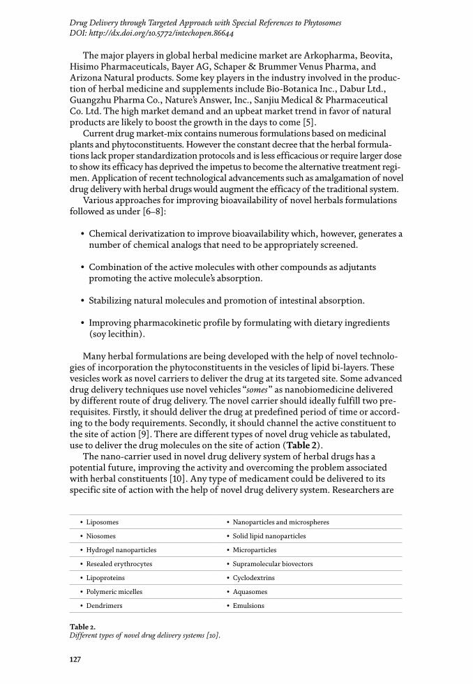

Role of Novel Drug Delivery Vehicles in Nanobiomedicine
128
currently underway to develop an ideal drug delivery system which satisfies the need of targeted site of action. Biological membrane presents a barrier through which a drug must pass before it gets absorbed or excreted. Lipid solubility and molecular size of drug molecule pose two major limiting factors to pass the biologi-cal membrane by which drug can be absorbed systematically following oral or topical administration [11].
The conventional herbal dosage form has a problem of standardization and quality control which had relegated its usage in the in last few decades. When the side effects of allopathic drugs outweigh their risk to usage the focus for treatment shifts towards the natural components and herbal system of treatment. In herbal novel drug development polymeric nanoparticles and nanocapsules like liposomes, solid lipid nanoparticles, phytosomes and nanoemulsion are been formulated. NDDS provides many advantages for herbal drugs including enhancement of solubility and bioavailability, protection from toxicity, enhancement of stability, enhancement of pharmacological action, improving tissue macrophages distribu-tion, sustained delivery, and protection from physical and chemical degradation. The efficacy of herbal product (or medication) is dependent upon effective route of delivery and level of the active compounds [12].
Novel drug delivery systems deliver the drug components at a rate directed by the need of the body during the period of treatment and channelize the active entity onto the site of action. A number of novel drug delivery systems have been used to deliver herbal medicaments encompassing various routes of administration to achieve controlled and targeted drug delivery, for example, encapsulated vesicles can prolong the existence of the drug in systemic circulation and reduce the toxicity if selective uptake can be achieved through drug targeting, sustained or controlled release of conventional medicines [11, 12].
The novel drug delivery vehicles can be classified on the basis of their size, shape, and their composition (Table 3).
1.1 Liposomes
Liposomes are micro-sized spheres in which an aqueous core is surrounded by one or more lipid bi-layer. The lipid bi-layers are separated by water or aqueous buffer compartments, liposomes are simple microscopic vesicles in which aqueous volume is entirely enclosed by a membrane composed of lipid bilayers [13] (Figure 1).
Name of somes Particle size/shape Composition
Liposomes [13] 100–1000 nm(diameter)/spherical
Phospholipids and cholesterol
Resealed erythrocytes [14] 6–9 μm (diameter)/oval Plasma, protein and platelets
Niosomes [15] 100–140 nm (diameter)/spherical
Non-ionic surfactants and cholesterol
Discomes [16] 16–20 μm/disc shaped vesicles Cholesterol and niosomes
Transferosomes [17] 170–200 nm/oval Soya phophatidylcholine surfactant and drug/antigen
Ethosomes [18] 50–100 nm/spherical Phospholipid and ethanol
Phytosomes [19, 20] 10–100 nm/spherical Phospholipid and herbal extract
Aquasome/hydrosomes 60–300 nm/circular Ceramics, carbon [21]
Table 3. Classification of herbal novel drug delivery approach on the basis of their size, shape, and composition.
129
Drug Delivery through Targeted Approach with Special References to PhytosomesDOI: http://dx.doi.org/10.5772/intechopen.86644
1.2 Niosomes
Niosomes are non-ionic surfactant-based liposomes. Niosomes can be obtained by hydration of liposomes. These are microscopic lamellar structures formed upon combining non-ionic surfactant of the alkyl or dialkyl polyglycerol ether class with cholesterol [15]. The stability of niosomes are affected by type of surfactant, nature of encapsulated drug, storage temperature, detergents, use of membrane spanning lipids, the interfacial polymerization of surfactant mono-mers in situ, inclusion of charged molecule. Niosomes have great penetrating capability [23] (Figure 2).
The sizes of Niosomes are microscopic and lie in nanometric scale. The particle size ranges from 10 to 100 nm [24].
Figure 1. Liposome (source: http://healthproductdistributor.com) [22].
Figure 2. Structure of niosome [24].

Role of Novel Drug Delivery Vehicles in Nanobiomedicine
130
1.3 Discomes
Discomes (disc shaped vesicles) are large (16–20 μm) sized vesicles structures capable of entrapping water soluble solutes, formed by solubilization of niosomes with a nonionic surfactant. They act as drug reservoirs in the field of drug delivery [16].
1.4 Transferesome
Transferosomes are complex vesicular aggregate optimized to attain extremely flexible and self-regulating membrane; this makes the vesicles very deformable. Transferosome have the property to cross microporous barrier very efficiently, even when available passage are much smaller than the average aggregate size. Transferosome consists of natural amphipathic compound suspended in water-based solution, something containing biocompatible surfactant. Transferosome also have lipid bilayer that surrounds an aqueous core and contain at least one component that soften the membrane and make skin more flexible. This allows an easy and rapid change in shape of transferosome [17].
1.5 Ethosomes
Ethosomes are lipid vesicles containing high amount of ethanol. They have the efficiency to penetrate the skin and enhance compound delivery both to deep skin strata and systemically. Ethanol fluidizes both lipid bilayers of the stratum corneum and intercellular lipid. The soft malleable vesicles then penetrate the disorganized lipid bilayer. This system consists of phospholipid, ethanol and water [18].
1.6 Resealed erythrocytes
The fluid portion of blood contains a large number of organic and inorganic substances in solution, which may diffusible (electrolyte, anabolic and catabolic substance formed during metabolism) and non-diffusible (proteins). The cellular portion of blood consists of erythrocytes (red blood cells), leukocytes (white blood cells) and thrombocytes (platelets). Erythrocytes have a solid content of about 35% (rest of being water) most of which is hemoglobin, which is remain tightly bound to the stroma of cell membrane [14]. Resealed erythrocytes are drug-loaded carrier erythrocytes, prepared simply by collecting blood samples from the organism of interest, separating erythrocytes from plasma, entrapping drug in the erythrocytes, and resealing the resultant cellular carriers. Hence, these carriers are called resealed erythrocytes [14].
1.7 Aquasomes
Aquasomes are the nano-structured biopharmaceutical vesicles based upon fundamental principle of self-assembling. It contains the particle core composed of nano-crystalline calcium phosphate or ceramic diamond, and is covered by a polyhydroxyl oligomeric film. Aquasomes are also called as “bodies of water”. The drug candidates/molecule delivered by Aquasomes represent better biological activity even in case of conformationally sensitive ones. Aquasome having proper-ties like protection and preservation of fragile biological molecules, conformational integrity, and surface exposure made it as a successful carrier system for bioactive molecules like peptide, protein, hormones, antigens and genes to specific sites [21].
131
Drug Delivery through Targeted Approach with Special References to PhytosomesDOI: http://dx.doi.org/10.5772/intechopen.86644
1.8 Phytosomes
Phytosomes is the combination of two words, the term “PHYTO” means plant while “SOME” means cell-like [25]. The formulation is developed by encapsulat-ing the plant material or plant extract within the spherical cell like structure, which is an advanced nano-sphere or cell forms of herbal products that are better absorbed. Phytosomes produces better pharmacokinetic and pharmacodynamic profile of drug than conventional herbal formulations. It’s a novel emerging technique that is applied to phytopharmaceuticals for the enhancement of bio-availability of natural plant extract for medicinal applications. Phytomedicines, complex chemical mixture prepared from plants, have been used in India and worldwide from the very beginning of human civilization and continue to have widespread popular use [26].
Phytosome is a patented process developed by Indena, a leading supplier of nutraceutical ingredients, to incorporate phospholipids into standardized extract and so vastly improve their absorption and utilization. Phytosome is one kind of herbal extracts delivery system that deliver natural values at target site in such manner. Certain of the water-soluble phytomolecules (mainly flavonoids and other polyphenols) can be converted into lipid-friendly com-plexes, by reacting herbal extract owing to their enhanced capacity to cross the lipid-rich biomembranes and finally reach the blood. They have improved pharmacokinetic and pharmacological parameters which are advantageous in the treatment of acute disease as well as in pharmaceutical and cosmetic compositions [19].
Phytosomes are better able making transition from a hydrophilic environment into the lipid-friendly environment of the enterocyte cell membrane and from there into the cell, finally reaching the blood. The lipid- phase substances employed to make flavonoids lipid- compatible are phospholipids from soy, mainly phospha-tidylcholine (PC). PC, the principal molecular building block of cell membranes, is miscible both in water and in oil/lipid environments and is well absorbed when taken by mouth. Precise chemical analysis indicates that a Phytosome is usually a hydrophilic herbal extract linked with at least one PC molecule. A bond is formed between the two molecules, creating a hybrid molecule. This highly lipid-miscible hybrid bond is better suited to merge into the lipid phase of the enterocyte outer cell membrane [19].
2. Difference between phytosome and liposome
The fundamental difference between liposomes and Phytosomes is that in liposomes the active principle is dissolved in the medium contained in the cavity or in the layers of the membrane, whereas in the Phytosome it is an integral part of the membrane, being the molecules anchored through chemical bonds to the polar head of the phospholipid [20].
There may be hundreds or even thousands of phosphatidylcholine molecules surrounding the water soluble compound in Liposome. In contrast, the Phytosome contain Phosphatidylcholine and the individual plant components actually form a 1:1 or a 2:1 complex depending on the substance. This difference show that Phytosomes being much better absorbed than liposomes. Phytosomes are superior to liposomes in skin care products [27, 28] (Figure 3).
Phytosome (lower segment) liposome (upper segment).

Role of Novel Drug Delivery Vehicles in Nanobiomedicine
132
3. Merits of phytosomes over conventional dosage forms
• The bioavailability of botanical extracts enhanced dramatically due to their complexation with phospholipids by which absorption of the constituent improved in the intestinal tract [27–30].
• Phytosme enhances the permeation of non- lipophilic botanical extract to allow better absorption from the intestinal lumen, which not possible by other carriers.
• The formulation aspect of phytosome is safe and all active components have been approved for pharmaceutical and cosmetics use.
• Due to high Bioavailability, Phytosome is being used to deliver liver protecting Flavonoids.
• This technology offers cost effective delivery of Phytoconstituents to various targeted site for pharmacological action. Phytosomes produce synergistic ben-efits when used as functional cosmetics, to protect the skin against exogenous or endogenous hazards in normal as well as stressful environment.
• They can be also used for enhancing permeation of drug, through skin in transdermal drug delivery system.
• They can be widely used in cosmetics due to their improved skin penetration and have a high lipid profile.
• Phosphatidylcholine, an essential part of the cell membrane and active com-ponents in phytome, act as a carrier and nourish the skin. There is no problem with drug entrapment during formulation development. The drug entrapment efficiency with phytsome is high and more over predetermined; because the drug itself forms vesicles after conjugation with lipid.
• They offer a better stability profile because chemical bonds are formed between the phosphatidylcholine molecules and phytoconstituents.
Figure 3. Difference between phytosome and liposome [29].
133
Drug Delivery through Targeted Approach with Special References to PhytosomesDOI: http://dx.doi.org/10.5772/intechopen.86644
The phytosomal system is passive, non-invasive and can is suitable for immedi-ate commercialization.
• The dose requirement is reduced due to improved absorption of the main constituent. They can also be given in smaller quantities to achieve the desired therapeutic values.
• Low risk profile: This technology has no risk since the toxicological profiles of the phytosomal components are well documented in the scientific literature. Highly attractive market profile for products with proprietary technology. It is easy to formulate because no complicated technical investment required for the production of phytosomes.
4. Pharmaceutical scope of phytosome
• It enhances the absorption of lipid insoluble polar phytoconstituents through oral as well as topical route showing better bioavailability, so it has wide therapeutic benefits [28, 30].
• Appreciable drug entrapment.
• Phytosomes help to improve the absorption of active constituents and reduces the dose requirements.
• Phosphatidylcholine used in preparation of Phytosomes, besides acting as a carrier also acts as a hepatoprotective agent. Hence produce the synergistic effect to the liver targeted drug.
• Chemical bonds are formed between phosphatidylcholine molecule and phytoconstituent, this results better stability in Phytosome.
• Application of phytoconstituents in form of phytosome improves their percutaneous absorption and act as basic carrier for transdemal drug delivery.
5. Phospholipids: creating the structural framework for novel drug delivery systems
These are complex substance having chemical, biochemical and nutritional characteristics by which they can distinguish as placed in a unique nutritional category. They are complex lipid molecules important for life and are found in all human and the other known structure to make cell membranes. The biochemical importance of phospholipids is reflected in their extraordinary clinical benefits as dietary supplements. The phospholipids are readily compatible with the entire range of vitamins, minerals, metabolites and herbal preparations. The dietary phos-pholipids and omega-3 fatty acid works in functional synergy in cell membranes. Phosphatidylcholine is a bi-functional compound miscible both in water and in oil environments, and is well absorbed when taken by mouth. Phosphatidylcholine is not merely a passive “carrier” for the bioactive compounds, but is itself a bioactive nutrient with documented clinical efficacy for liver disease (including alcoholic hepatitis). Phosphatidylcholine is present in egg yolk, brain tissue and a wide

Role of Novel Drug Delivery Vehicles in Nanobiomedicine
134
variety of animal fat and plant oils. It is routinely present in the bile fluid, to help emulsify food ingredient for absorption [31] (Figure 4).
A number of drug delivery system is based entirely on Phosphatidylcholine such as liposomes, ethosomes, phytosomes, transferosomes, and nanocochhelates. The hydrophilic and hydrophobic domain/segment within the molecular geometry of amphiphilic lipids orient and self-organize in ordered supramolecular structure when confronted with solvents. Some commonly used synthetic phospholipids are dioleoyl-phosphatidyl-choline (DOPC), dioleoyl-phosphatidyl-ethanolamine (DOPE), distearoyl-phosphatidyl-choline (DSPC), and distearoyl-phosphatidyl- ethanolamine (DSPE) [31].
6. Conjugation of NDDS with transdermal route
Transdermal drug delivery systems (TDDS) are topically administered medications in the form of patches, which can deliver certain medication to systemic circulation in a more convenient and effective way than is possible with conventional dosage form. The potential of skin as a path of drug administra-tion has been amply demonstrated by the acceptability of marketed therapeutic systems [32]. Administration of systemic drugs using a transdermal patch represents a noninvasive route, with improved patient compliance. This route of administration prevents passage through the gastrointestinal tract and maintains constant plasma levels for prolonged periods of time [33]. A transdermal patch is a medicated adhesive patch placed on the skin to deliver a time-released dose of medication through the skin for treating topical or systemic illness. Since early 1990, this dosage form of transdermal therapeutic system has been available in the pharmaceutical market. Such a system offers a variety of significant clinical benefits over others, such as tablet and injection [32, 33]. Transdermal route is a potential mode of delivery of lipophilic drugs in the systemic circulation. Some factors are responsible for transdermal drug delivery such as area of application, amount applied, release kinetics and prolongation of application time [34].
Figure 4. Cell membranes are largely lipid phase. A double molecular layer consisting of PC and other phospholipids provides a continuous matrix into which the proteins insert [31].
135
Drug Delivery through Targeted Approach with Special References to PhytosomesDOI: http://dx.doi.org/10.5772/intechopen.86644
7. Feasibility of phytosome as novel vehicle for transdermal drug delivery
Lipophilic drugs could be easily transfused by transdemal route which enhance the bioavalibility of phytoconstitutent. Drug would be delivered into systemic circulation by transdermal route transdermal route. Drug has to pass three different skin layers before reaching to systemic circulation. The outermost layer, Stratum corneum provides hindrances to such type of drugs delivery due to its lipophilicity. The Phytosomal form of phytoconstituents can pass this barrier easily and helps to deliver the phytoconstituents in systemic circulation. The research studies from two decades show the phytosome technology has enough potency to be used as novel carrier of Phytoconstituents. Phytosomal carrier has enough potency to be used transdermally for increasing bioavailability of Phytoconstituents [34, 35].
8. Conclusion
Ever since the advent of human civilization, man is dependent on nature to fulfill all his need. Traditional system of healing human aliment was also derived from natural resources. In an era of development, devised various means of treatment primarily based on synthetic chemicals and popularly known as allopathic system of treatment. The inherent side effects of this system led to the shift in focus from contemporary to either conventional system of medication or to evolve the Novel drug delivery system. Novel drug delivery vehicles that can be used for the poorly soluble, pH sensitive and less potent drug to make them safe and target oriented. These novel drug delivery vehicles can also be utilized to deliver herbal formulations to its targeted site with lesser side effect. This phenomenon can be further augmented by conjugation of transdermal drug delivery system with novel drug delivery vehicle containing nanobiomedicine as herbal constituents. Phytosomes have shown the potential to be good carrier of herbal constituents for novel drug delivery systems. The combination of Transdermal route and Phytosomal drug delivery opens up an arena for better treatment for the patient suffering from skin diseases, Burn, allergies and bone fracture.
Acknowledgements
Authors want to acknowledge the Department of Pharmaceutical Sciences, Kumaun University campus bhimtal, for providing support for library and litera-ture search.
Conflict of interest
There is no conflict of interest.

Role of Novel Drug Delivery Vehicles in Nanobiomedicine
136
Author details
Mahendra Rana, Aadesh Kumar* and Amita J. RanaDepartment of Pharmaceutical Sciences, Kumaun University Campus, Bhimtal, Uttarakhand, India
*Address all correspondence to: [email protected]
© 2020 The Author(s). Licensee IntechOpen. This chapter is distributed under the terms of the Creative Commons Attribution License (http://creativecommons.org/licenses/by/3.0), which permits unrestricted use, distribution, and reproduction in any medium, provided the original work is properly cited.
137
Drug Delivery through Targeted Approach with Special References to PhytosomesDOI: http://dx.doi.org/10.5772/intechopen.86644
References
[1] Inamdar NS, Edalat VB, Kotwal VB, Pawar SS. Herbal drugs in milieu of modern drugs. International Journal of Green Pharmacy. 2008;2:2-8
[2] Kalia AN. Text Book of Industrial Pharmacognosy. New Delhi: Oscar Publication; 2005
[3] Calixto JB. Efficacy, safety, quality control, marketing and regulatory guidelines for herbal medicines (phytotherapeutic agents). Brazilian Journal of Medical and Biological Research. 2000;33:179-189
[4] Christie A. Herbs for health, but how safe are they. The Bulletin of the World Health Organization. 2001;79:691-692
[5] Herbal Medicine Market Research Report- Forecast to 2023. Available from: http://www.markertresearchfuture.com/reports/herbal-medicine-market-3250
[6] Mukherjee PK, Wahile A. Integrated approaches towards drug development from ayurveda and other Indian system of medicine. Journal of Ethnopharmacology. 2006;103:25-35
[7] Ajazuddin SS. Application of novel drug delivery system for herbal formulations. Fitoterapia. 2010;81:680-689. DOI: 10.1016/j.fitote.2010.05.001
[8] Kidd P, Head K. A review of the bioavailability and clinical efficacy of milk thistle phytosome: A silybinphos phatidylcholine complex. Alternative Medicine Review. 2005;10(3):193-203
[9] Medina OP, Zhu Y, Kairemo K. Current Pharmaceutical Design. 2004;10:2981-2989
[10] Vyas SP, Khar RK. Targeted and Controlled Drug Delivery. 1st ed. CBS Publisher; 2013. pp. 3-10
[11] Nanoparticulate Drug Delivery. Perspectives on the Transition from Laboratory to Market. Woodhead Publishing Series in Biomedicine; 2012. pp. 29-85. DOI: 10.1533/9781908818195.29
[12] Barragan Montero V, Winum J, Moles J, Juan E, Clavel C, Montero J. The European Journal of Medicinal Chemistry. 2005;40:1022-1029
[13] Chonn A, Cullis PR. Recent advances in liposomal drug delivery system. Current Opinion in Biotechnology. 1995;6:698-708
[14] Balasubramanian J, Narayanan N, Kumar V. Resealed erythrocytes: A novel drug carrier in drug delivery. Drug Discovery. 2012;2(6):30-32
[15] Malhotra M, Jain NK. Niosomes as drug carriers. Indian Drugs. 1994;31:81-86
[16] Kumarn GP, Pogaku R. Nonionic surfactant vesicular systems for effective drug delivery—An overview. Acta Pharmaceutica Sinica B. 2011;1(4):208-219. DOI: 10.1016/j.apsb.2011.09.002
[17] Vyas SP. Theory and Practice in Novel Drug Delivery System. 1st ed. CBS Publisher; 2011. pp. 257-260
[18] Vyas SP. Theory and Practice in Novel Drug Delivery System. 1st ed. Vol. 2011. CBS Publisher. pp. 245-250
[19] Rathore P, Swami G. Planterosome: A potential phyto-phospholipid carrier for the bioavailability enhancement of herbal extracts. International Journal of Pharmaceutical Sciences and Research. 2012;3(3):737-755
[20] Pandey S, Patel K. Phytosomes: Technical revolution in phytomedicine. International Journal of Pharma Tech Research. 2010;2(1):627-631

Role of Novel Drug Delivery Vehicles in Nanobiomedicine
138
[21] Sutariya V, Patel P. Aquasomes: A novel carrier for drug delivery. IJPSR. 2012;3(3):688-694. DOI: 10.13040/ijpsr.0975-8232.3(3).688-94
[22] http://healthproductdistributor.com
[23] Udupa N. Niosomes as drug carriers. In: Jain NK, editor. Controlled and Novel Drug Delivery. 1st ed. New Delhi: CBS Publishers and Distributors; 2002
[24] Baillie AJ, Florence AT, Hume LR, Muirhead GT, Rogerson A. The preparation and properties of niosomes non-ionic surfactant vesicles. The Journal of Pharmacy and Pharmacology. 1985;37:863-868
[25] Jain N, Gupta BP, Thakur N. Phytosome: A novel drug delivery system for herbal medicine. International Journal of Pharmaceutical Science and Drug Research. 2010;2(4):224-228
[26] Sindhumol PG, Thomas M, Mohanachandran PS. Phytosomes: A novel dosage form for enhancement of bioavailability of botanicals and nutraceuticals. International Journal of Pharmacy and Pharmaceutical Science. 2010;2(4):10-14
[27] Sharma A, Sharma US. Review liposome in drug delivery: Progress and limitations. International Journal of Pharmaceutics. 1997;154:123-140
[28] Kidd PM. Bioavailability and activity of phytosome complexes from botanical polyphenols: The silymarin, curcumin, green tea, and Grape Seed Extracts. Alternative Medicine Review. 2005;14(3):226-246
[29] Bhattacharya S. Phytosomes: Emerging strategy in delivery of herbal drugs and nutraceuticals. Pharmacy Times. 2009;41(3):9-12
[30] Awasthi R, Kulkarni G, Pawar V. Phytosome: An approach to
increase the bioavailability of plant extracts. International Journal of Pharmacy and Pharmaceutical Science. 2011;3(2):1-3
[31] Bombardelli E, Spelta M. Phospholipid-polyphenolcomplexes: A new concept in skin care ingredients. Cosmetics and Toiletries. 1991;106:69-76
[32] Modamio P, Lastra CF, Marino EL. A comparative in-vitro study of percutaneous penetration of beta-blockers in human skin. International Journal of Pharmaceutics. 2000;194:249-259
[33] Ke GM, Wang L, Xue HY, Lu WL, Zhang X, Zhang Q , et al. In-vitro and in- vivo characterization of a newly developed clonidine transdermal patch for treatment of attention deficit hyperactivity disorder in children. Biological and Pharmaceutical Bulletin. 2005;28:305-310
[34] Nicoli S, Colombo P, Santi P. Release and permeation kinetics of caffeine from bioadhesive transdermal films. The AAPS Journal. 2005;7:E218-E223
[35] Tymes NW, Shah VP, Skelly JP. In-vitro profile of estradiol transdermal therapeutic systems. Journal of Pharmaceutical Sciences. 1990;79:601-602
139
Chapter 7
Polymer Properties: Functionalization and Surface Modified NanoparticlesChander Amgoth, Chiuyen Phan, Murali Banavoth, Santosh Rompivalasa and Guping Tang
Abstract
Herein, the various polymer properties and the underlying mechanism for the functionalization and surface modification of polymer nanoparticles have been discussed. There are numerous polymer particles designed and developed for vari-ous applications. The synthesis and characterization of different types of polymers followed by the engineering of nanoparticles and capsules depend on various fac-tors. There are too many polymerization methods approached for the development of nanoparticles with desired surface properties. The ring-opening polymerization (ROP), emulsion polymerization (EP), atom transfer radical polymerization (ATRP), and free radical micro initiation are the significant approaches for the polymeriza-tion reactions. The polymer nanoparticle functionalization and modification of their surfaces based on requirements is an essential task. The solvent concentration, pH, temperature, and sonication have played a vital role to tune the morphology of polymer nanoparticles and capsules. Different characterizations such as FTIR, NMR (1H and 13C), HRMS, and MALDI-TOF are used for preliminary structural and confir-mations. Further, SEM, FE-SEM, TEM, AFM, BET, XRD, Raman, EDAX, TGA-DSC, DLS, and zeta potential were used for morphological and thermal properties.
Keywords: polymers, properties, functionalization, surface modification, nanoparticles
1. Introduction
Across the globe, there is a great interest in the field of polymers and polymer-based nanoparticles followed by their nanotechnology-based applications. However, polymeric nanoparticles (PNPs) are under a wide range of utilization in various fields. Especially researchers and scientists are trying to design various nanoparticles (NPs) and capsules with surface modifiable character features [1]. The polymer nanoparticles do obey and endorse the aforementioned sentence with existing literature which shows numerous nanoparticles with modifiable size, shape, and their surface properties. For example, quantum dots (QDs) carbon nanotubes (CNTs), cellulose NPs and fibers [2] iron oxide nanoparticles (FeONPs), gold nanoparticles (AuNPs), silver nanoparticles (AgNPs), cobalt nanoparticles (CNPs), zinc oxide nanoparticles (ZnONPs), SnO2 nanoparticles, protein-based nanoparticles, amino acid-based nanoparticles, silica (SiO2) nanoparticles,

Role of Novel Drug Delivery Vehicles in Nanobiomedicine
140
graphene nanosheets, titanium-based nanoparticles, core-shell nanoparticles, hollow core-shell (hCS) nanoparticles, hollow polymer-polymer nanoparticles (HPPNPs), polymer-metal nanoparticles (PMNPs), polymer-metal blends (PBs), polymer-silica nanocomposites (PSiNCs), polymeric micelles (PMs), solid-lipid nanocapsules (SLPNCs), liposomal nanoparticles, albumin-based nanoparticles, chitosan-based nanoparticles, platinum-based nanoparticles (PtNPs), ceramic nanoparticles, carbohydrate nanoparticles, etc. are reported and corroborated in the literature [2–5]. Furthermore, a significant interest in the synthesis and character-ization of polymers followed by the design and development of polymeric nanopar-ticles (PNPs) with surface tunable properties has become an essential due to the various applications. For example, the amino acid-based polymer is much more suitable for the preparation of nanoparticles because of its biosafety and nontoxic and biodegradable nature. Similarly, thermosensitive polymer, for example, poly-N-isopropyl acrylamide (PNIPAM), shows lower critical solution temperature (LCST) at ~32°C, and it is much more useful to prepare the nanoparticles or capsules with thermally tunable morphology and other futuristic characteristic properties [6]. However, the main advantage to polymer scientists is they can easily synthesize the polymer to engineer the nanoparticles followed by the significant functionalization and surface modifications by changing the solvents, concentration, and stirring methods or by changing the functional groups which are present in the synthesized polymer complex. Similarly, the ionic (cationic and anionic) polymers referred to as polaxomers can help to modify the surface of the particles. Polymeric nanoparticles are under a wide range of utilization in the field of biomedical, nanobiotechnology, nanobiopharma, and other pharmaceutical domains. For example, they are very much used in the field of drug delivery systems (DDSs), gene delivery systems (GDSs), protein delivery systems (PDSs), cell delivery systems (CDSs), and tissue engineering followed by plastic surgery, etc. [7, 8]. However, polymers have numer-ous industrial applications as well, such as cell phone manufacturing companies, soft drink and water bottle industries, electronics and electrical engineering departments, automobile industries, and many other industrial usages based on the requirements. Interestingly, polymeric nanoparticles with porosity have multidirec-tional usages such as drug or nanomedicine loading and its target specific release. The nanoparticles with ~200 nm (in diameter) are the ideal size for the biomedical applications. The pore size with ~100 nm in the polymer nanoparticles or capsules is much more suitable for drug delivery and cancer therapeutics. According to International Union of Pure and Applied Chemistry (IUPAC) definition, the mic-roparticle size should be <2 nm, (in diameter) and meso-particles or mesoporous particle and capsule size ca. 2–50 nm, and nanoparticles (particle size 50–200 nm) have been referred for various applications [6–8]. The effective and potential work-ing capacity of nanoparticles has been defined by their size, shape, morphology, porosity, and other thermal properties. However, the qualitative quantification of use of polymer-based nanoparticles and their potential applications completely depends on the characteristic features like surface modification, size, and shape ten-ability. For example, many properties like color and atomic orientation are differing from the sizes of bulk to nanoscale [9, 10].
2. Classification of polymers
The word polymer derived from Greek has been split into two meaningful words: (1) poly means many, and (2) meros or mers means units or parts. More specifically monomer is the smallest repeating unit for the development of polymer. Simply polymers are made up of monomers. The word monomer is also derived
141
Polymer Properties: Functionalization and Surface Modified NanoparticlesDOI: http://dx.doi.org/10.5772/intechopen.84424
from the Greek. Mono means one or single and mer means part/s. As reported in Table 1, there are varieties of polymers classified based on their biodegrad-ability, biocompatibility, thermal stability, pH-responsive nature, environmental responsive nature, etc. Based on occurrence these are again categorized into three types: (1) natural polymers, for example, starch, chitin, cellulose, etc., (2) synthetic polymers, for example, polystyrene (PS), polyurethane (PU), polyvinyl chloride (PVC), and (3) semisynthetic polymers, for example, cellulose nitrate, guncotton, cellulose acetate, etc. Absorbable synthetic polymers include polyvinyl alcohol (PVA), polylactic acid (PLA), polycaprolactone (PCL), and polyglycolic acid (PGA), and these are homopolymers. The heteropolymers include poly-trimethylene carbonate (PTMC), polyetherimide (PEI), polydioxanone (PDO), polylactide-co-glycolide (PLGA), and the combination of block copolymers like and poly-glycolide-co-trimethylene carbonate (PGTMC) which are best examples for heterogeneous polymers. Apart from these polymers, there are pH-sensitive polymers like poly-L-lysine which is positively charged at lower pH conditions [22, 23]. Liposome and poly-histidine are the systems with pH sensitivity, and this can be easily interacted with negatively charged particles or membranes to promote fusogenic properties. Furthermore, some hydrogel-based nanoparticles show unique characteristics toward various biomedical as well as industrial applications. Liposomal and hydrogel based systems has tendency towards pH-sensitivity and it is a characteristic property to tune-up their morphology for many therapeutic appli-cations. For example, PLGA, MeO-PEG-NH2, and PAMAM-based nanoparticles show pH-dependent destabilization and thus are used for cytoplasmic experiments because of its ease of permeability through the membrane to deliver nanomedicines and nanoparticles [9, 10, 22, 23]. The NIPAM/pNIPAM (N-isopropyl acrylamide/(poly-N-isopropyl acrylamide)) is the best example for the thermal sensitive polymer which shows lower critical solution temperature (LCST) or cloud point at ~32°C Table 1. Because of the thermal behavior, it is termed as magic or smart poly-mer and used for various medical and industrial usages. However, these polymers are mainly prepared based on two polymerizations reactions such as (1) addition polymerization and (2) condensation polymerization methods [24, 25].
S. no.
Name of the polymer Monomer formula
Polymer formula
Polymer structure
Ref.
1 PVC (polyvinyl chloride) C2H3Cl (C2H3Cl)n Figure 1a [11, 12]
2 PVA (polyvinyl alcohol) C2H4O (C2H4O)n Figure 1b [12, 13]
3 PE (polyethylene) C2H4 (C2H4)n Figure 1c [12, 14]
4 PS (polystyrene) C8H8 (C8H8)n Figure 1d [14, 15]
5 PP (polypropylene) C3H6 (C3H6)n Figure 1e [15, 16]
6 NIPAM (isopropylacrylamide)
C6H11NO (C6H11NO)n Figure 1f [16, 17]
7 PTFE (Teflon) C2F4 (C2F4)n Figure 1g [17, 18]
8 PCL (polycaprolactone) C6H10O2 (C6H10O2)n Figure 1h [18, 19]
9 PEI (polyetherimide) C37H24O6N2 (C37H24O6N2)n Figure 1i [19, 20]
10 PSU (polysulfone) C27H22O4S (C27H22O4S)n Figure 1j [20, 21]
Here, “n” represents the number of monomer units involved in the synthesis of the polymers.
Table 1. Show the different types of polymers with their monomer units and their chemical formula followed by the structures in Figure 1a–j.
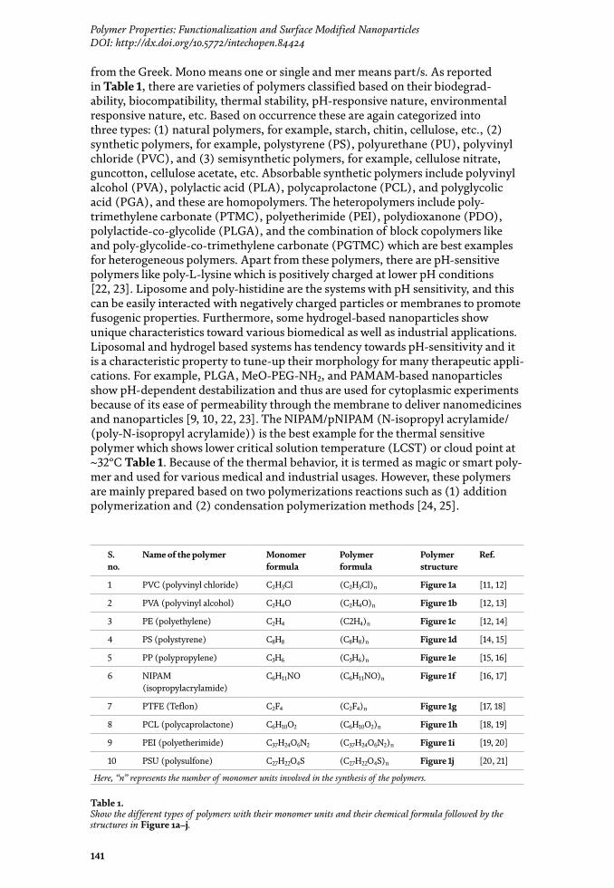
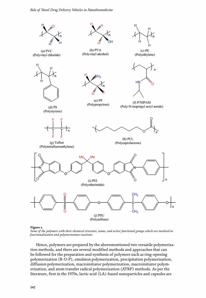
Role of Novel Drug Delivery Vehicles in Nanobiomedicine
142
Hence, polymers are prepared by the abovementioned two versatile polymeriza-tion methods, and there are several modified methods and approaches that can be followed for the preparation and synthesis of polymers such as ring-opening polymerization (R-O-P), emulsion polymerization, precipitation polymerization, diffusion polymerization, macroinitiator polymerization, macroinitiator polym-erization, and atom transfer radical polymerization (ATRP) methods. As per the literature, first in the 1970s, lactic acid (LA)-based nanoparticles and capsules are
Figure 1. Some of the polymers with their chemical structure, name, and active functional groups which are involved in functionalization and polymerization reactions.
143
Polymer Properties: Functionalization and Surface Modified NanoparticlesDOI: http://dx.doi.org/10.5772/intechopen.84424
used for biomedical and other pharmaceutical applications, and later it has been extended for various fields [26]. Polymers when considered for biomedical appli-cations, it needs to refer self-assembly nature and functionality followed by the formation of spherical and sphere-shaped nanocapsules and particles particularly for drug delivery systems. Based on degradability, thermosensitivity, and pH-responsive nature, these are again categorized into various classes and reported in Figure 2 with some examples. However, biodegradable polymers which show a ten-dency to degrade in biological systems such as polylactic acid (PLA), polyglycolic acid (PGA) and poly terephthalic acid (PTA), polycaprolactone (PCL), and amino acid (AA)-based polymers are potential materials to engineer nanoparticles and capsules for nanotechnology and nanobiotechnology applications. The polymers which do not degrade in the biological systems are termed as non-biodegradable polymers such as carboxymethyl, ethyl cellulose, and acetates of cellulose. The poly anhydrides like poly sebacic acid (PSA), poly adipic acid (PAA), and other envi-ronmental-responsive polymers also enlisted in the non-biodegradable polymers. Thermo-responsive (temperature-sensitive) polymers show a significant change in the size and shape of nanoparticles to tune up based on requirements [27]. The thermosensitive behavior of PNIPAM and pH-responsive nature of L-glutamic acid gamma benzyl ester (L-GluA-5-BE), L-aspartic acid beta benzyl ester (L-AspA-4-BE), polyethylene glycol (PEG), poly L-glycolic acid (PLGA), and PAMAM allow changing the morphology of nanoparticles as well surface profile of the NPs and capsules (Figure 1). Apart from these polymers, electrical-responsive polymers show piezoelectric effects which are a characteristic property to mold the nanopar-ticles based on requirements. The supramolecular interactions between the atoms and molecules lead to compression or elongation and stretching of bonds, and it can play a vital role to change the properties of final compounds. However, polyvinyli-dene fluoride (PVDF) polymer has been considered as one of the magic polymers which allow structural and morphological modifications through the appropriate synthesis and preparation methodologies [28].
The chemically responsive polymers allow internal changes through the formation of anionic and cationic and amphiphilic nature through the oxida-tion, reduction, and redox (both oxidation and reduction) reactions and lead to surface modification based on smooth and rough surface. As per the literature, the increase in the size of the particles depends on an increase in monomer units or polymer units. Sometimes branching and hyper-branching nature also lead to the formation of larger nanoparticles and capsules. Polymers prepared with the
Figure 2. Some of the polymers categorized based on their characteristic properties.

Role of Novel Drug Delivery Vehicles in Nanobiomedicine
144
combination of a large number of high molecular mass polymers give micron-sized nanoparticles, and they can be used for bulk phenomenal approaches such as absorption, chemisorption, etc. There is a class of polymers named as block copolymers, and these are again categorized into several such as (a) random block copolymers (BCPs), (b) star block copolymers, (c) grafted block copolymers, and (d) alternating block copolymers, and these are classified based on their synthesis approaches and final structural confirmations, and these are low-density polymers (Figure 2) [20, 21, 26–29].
3. Polymer properties
There are some important physical and chemical properties needed to be considered before selecting polymers for synthesis followed by the design and development of nanoparticles such as (1) structural integrity, (2) low immunogenic nature, (3) biocompatibility (for biomedical applications), (4) biologically active nature, and (5) structural versatility (which can easily allow topographic changes internally as well externally). Polymers with physicochemical properties such as hydrophilic (solvent loving), hydrophobic (solvent repelling), and amphiphilic (affinity to absorb and repel the solvent) nature, surface energy, viscosity, lubricity, density, surface tension, pH sensitivity, smoothness, swelling nature, solubility, amorphous and crystalline nature, polymer dissolution, bioadhesive and sticky nature, polymer-solvent interaction, polymer erosion, flexibility, chain formatting nature, chemical composition, microstructural design, and other surface proper-ties need to be examined carefully for the design of nanoparticles and capsules. By considering these properties, nanoparticle preparation methods can also be modi-fied for the polymerization reactions (Figure 3). For example, (1) natural polymers like (a) chitin, (b) cellulose, and (c) starch can be widely used for nanoparticle development through the modified chemical synthesis methods [29, 30]. Further, (2) semisynthetic polymers like (a) cellulose nitrate, (b) rubber, (c) rayon and (d) silk and fiber-based materials, and (e) guncotton are synthesized based on their flexibility and other stresses and strain rate for various applications, and these are very much suitable to design the ribbon type of fibrils for textile industry-based usages. However, (3) synthetic polymers such as (a) polymethyl methacrylate amide (PMMA); (b) nylon, nylon-6, and nylon-6,6; and (c) polystyrene (PS) are very much useful for plastic industries to manufacture polyvinyl chloride (PVC)-based pipes, tubes, cables, and other plasticwares. The polymer tetrafluoroethylene (Teflon or PTFE) with monomer chemical formula (–C2F4–) is under wide range utilization in the name of Teflon tape as a sealant for pipe linkages and insulation tape. The natural rubbers isoprene and neoprene can be used to develop various products industrially [31, 32].
The natural rubber, vulcanized rubber, or sulfur has been used in tire industries. Similarly, formaldehyde and benzophenol combination is used to develop combs and other plasticwares, and its trade name is Bakelite. The polymer polyethylene (PE) is used for polyethylene covers/carriage bags and much more houseware and industrial manufacturing. The polymer PE is again categorized into two types based on its density for the manufacturing of plastic materials such as low-density polyethylene (LDPE) and high-density polyethylene (HDPE) based on applica-tions [33]. The following properties of polymers are essential to develop potential nanoparticles for drug delivery systems (DDSs), gene delivery systems (GDSs), cell delivery systems (CDSs), tissue engineering (TE), and other nanobiotechnol-ogy applications such as molecular weight, self-assembly nature, density, network
145
Polymer Properties: Functionalization and Surface Modified NanoparticlesDOI: http://dx.doi.org/10.5772/intechopen.84424
forming capacity, and interlocking nature of functional groups followed by the self-assembly. Based on applications (biomedical or industrial), polymeric nanopar-ticles should possess the following characteristics such as biocompatibility and biodegradability with good cell survival rate, and they should obey the properties as mentioned in earlier sections [34]. However, every nanoparticle system has their limitations for their use in medical biotechnology and nanotechnology applications. By changing reaction methods, solvent concentration, temperature conditions, pH levels, its surface profile and another size, shape, and morphology can be modified [35]. Design and development of surfactant-free nanoparticles without using any harmful solvents and catalyst is a novel approach for green synthesis and technol-ogy. The term green synthesis, green technology, green nanoparticles, and green reactions define the methods and approaches followed by engineer nanoparticles and materials without any side effects, toxicity, and harm to mankind applica-tions. The nanoparticles and capsules developed based on amino acid-based block copolymers, and plant extracts are termed as green nanoparticles as they show good biocompatibility for human beings [36].
4. Classification of nanoparticles
Based on core synthesis materials, nanoparticles are categorized into various types such as (a) protein nanoparticles (PrNPs), (b) polymeric nanoparticles (PNPs), (c) organic nanoparticles (ONPs), (d) inorganic nanoparticles (INPs),
Figure 3. Schematic shows the characteristic properties of polymers needed to be considered for the design and development of nanoparticles.

Role of Novel Drug Delivery Vehicles in Nanobiomedicine
146
(e) metal nanoparticles (MNPs), (f) metal oxide nanoparticles (MONPs), (g) amino acid-based nanoparticles (AANPs), (h) ceramic nanoparticles (CNPs), (i) hydrogel nanoparticles (HNPs), (j) solid-lipid nanoparticles (SLNPs), etc. [37–40].
However, these polymer-based nanoparticles and capsules are again classi-fied into several categories as follows: (a) polymer-polymer nanostructures, (b) polymer-metal NPs, (c) polymer-nonmetal NPs, (d) polymer-inorganic NPs, and (e) polymer-organic (proteins, amino acids) NPs. However, polymer-polymer nanoparticles, polymer-silica NPs, amino acid-based polymers, and polymer-zinc oxide NPs have unique characteristic properties for various biomedical applica-tions such as drug delivery systems (DDSs). Based on cytotoxicity, side effects and biocompatibility issue the quantum dots (QDs), and carbon nanotubes (CNTs) are avoided for the usage in the field of biomedical and pharmaceuticals. Aforementioned, there are various polymer-based nanoparticles which are already developed, but they all have their limitations, and for amino acid-based polymer nanoparticles and nanocapsules (NCs) like dendrimers, core-shell nanoparticles (CSNPs), the block copolymer (BCP), and mesoporous SiO2NPs, it is easy to change their surface and other properties [41]. However, Figures 4 and 5 illustrate the clas-sification of polymer and polymer nanoparticles and non-polymeric nanoparticles. The concept self-assembly has an obligatory role to rearrange and orient the active functional groups present in the monomers and polymers through the esthetic structural integrity throughout the surface of the nanoparticles and capsules. The concept self-assembly as well amorphous and crystalline nature of polymer are also one of the key factors which influence the arrangement and organization between functional groups like hydroxyl (–OH), amine (–NH2), amide (–NH), keto (–C=O), ester (R–COO–R), acid (–COOH), aldehyde (–CHO), and saturated and unsaturated hydrocarbons as discussed in earlier sections [38–43]. The forma-tion of porous spheroid-shaped nanoparticles and capsules depends on intra- and intermolecular interactions between functional groups. The molecular imprinting of hydrophilic nanoparticles with denser hydrophobic particles can lead to the formation and creation of pores within the particles and throughout the surface of the particles and capsules [41–44].
Figure 4. Shows the classification of various nanoparticles and their morphology acquired through the different microscopic characterizations.
147
Polymer Properties: Functionalization and Surface Modified NanoparticlesDOI: http://dx.doi.org/10.5772/intechopen.84424
Nanotechnologically there are two important approaches to prepare the nanoparticles such as top-down approach and bottom-up approach. However, top-down approach can be termed as breakdown approach, and it is the core method to prepare nanoparticles. For example, bulk gold cube to gold nanoparticles (AuNPs) can be achieved through the top-down approach, and simply particle segregation takes place during this approach [42–45]. Sonication, ultra-sonication, ball milling, and stirring are the best examples for the top-down approach to preparing nanopar-ticles. Similarly, bottom-up can be referred to as the buildup approach and in this approach nano to macro and macro to bulk materials can be achieved. This bottom-up approach is like aggregation of nanoparticles which leads to the formation of bulk materials. However, monomer to polymer formation is the best example for the bottom-up approach. Sometimes based on potential applications, these nanopar-ticles are called smart materials or smart nanoparticles (Figure 5) [43–46].
5. Advantages with polymer nanoparticle
These polymer-based nanoparticles are developed in principle to protect the guest particles or molecules from degradation and its functional activity, for example, porous polymer nanoparticles or nanocapsules loaded with anticancer drugs or nanomedicines [47]. Because of the soft nature of polymer NPs and nanocapsules, it is very easy to modify the surface of the particles and their mono-dispersion based on its applications. The enhanced absorption and modification of the drug distribution throughout the surface of the particles or capsules lead to the target-specific or tissue- or cell- or organ-specific pharmacokinetics and dynamics in various biomedical and pharmaceutical usages. Polymer-based nanoparticles and capsules allow easy surface functionalization and modification to tune up them with desirous properties. Sometimes smooth surface of the polymer thin films can become rough due to the internalization and incorporation of metal and other inor-ganic nanoparticles such as FeONPs, ZnONPs, AlNPs, AgNPs, AuNPs, SiO2NPs, etc. The surface properties like particle smoothness and roughness or porous nature of particles in composite can give better results, but its surface and size may not fit for
Figure 5. Schematic illustration shows the classification of different types of nanoparticles and their synthesis approach methods.
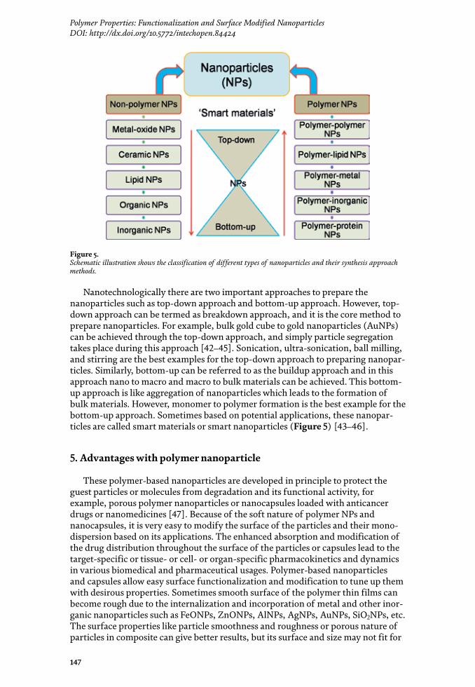

Role of Novel Drug Delivery Vehicles in Nanobiomedicine
148
the usage of biomedical applications [48]. In such cases the composite of polymer thin film embedded or doped with some other metal nanoparticles or organic and inorganic materials needs to be molded without changing its original activity or characteristic properties. However, the functional groups such as amide (–NH), amine (–NH2), acid (–COOH), aldehyde (–CHO), keto (–C=O), ester (R–COO–R), anhydride (R–CO–O–CO–R), acid halides, hydrogen halides, salts, and solvents (organic, inorganic) can also influence the surface properties for the nanoparticles or nanocapsules. Based on the requirement, it can be chosen to modify the particles surface and size, shape, etc. The concentration of the solvent also affects the pat-tern of the nanoparticles and nanocapsules. Sometimes additives like precursors and surfactants can also show an adverse effect on the morphology of polymer nanoparticles [49]. The activity (bioactive nature) of polymer nanoparticles and nanocapsules can disturb the body metabolism. The active functional groups with their potential resonance capabilities can also interact with body fluids and interfere to shore-up the mutations. Apart from the solvent, there are several factors which can affect the patterning of the surface of the nanoparticles such as pH, thermal stability, rubbery nature, solid (amorphous and crystalline) nature, environmental conditions, etc. The stability, biocompatibility, and route or synthesis methodol-ogy are also playing a vital role to tune up the surface properties of the polymeric nanoparticles and capsules. For example, (a) protein nanoparticles have stability issues during storage. If the storage conditions are not followed properly, they may get contaminated and become toxic [50]. But these protein-based NPs are very much compatible with their unique characteristic features such as nontoxic nature and nonantigenic nature. As per the existing literature, it is known that bulk protein in plasma can easily bind to a variety of drug molecules through its trajectory while carrying them to deliver target specifically. And these are very much flexible to ease scale-up manufacturing and other nanobiotechnological applications; (b) albumin-based nanoparticles are very much suitable for the morphological modifications and to tune the surface properties. These are biocompatible and biodegradable without any detrimental activities upon mankind usages. Sometimes these albumin-based nanoparticles are used for drug delivery and other biomedical applications. However, protein albumin contains a huge number of charged amino acid residues, and it binds water-soluble drugs through charge interactions, and it leads to surface modifications and morphological changes [51].
6. Preparation of polymer nanoparticles
There are several approaches to prepare and synthesize the polymeric nanopar-ticles such as (a) desolvation, (b) emulsification, (c) esterification, (d) thermal gelation, (e) nanospray drying, (f) self-assembly, etc. Apart from these approaches, there are mainly two polymerization methods widely followed for the preparation of polymer nanoparticles such as (1) addition polymerization and (2) condensa-tion polymerization. The preparation of polymer nanoparticles is not an easy task. The size and shape of the particle are a very important concern while preparing the polymer-based nanoparticles. As the number of monomers or polymer units increases, the size of the nanoparticle or capsule also increases. It is easy to prepare and reproduce the polymeric nanoparticles with the porous matrix for loading the drug molecules, but its size and shape cannot be formulated easily. Amino and carboxyl functional groups permit functionalization and surface modifications, but patterning them in a fruitful design is a challenging task, and it can overcome through the detailed study of their properties [52]. However, albumin is phase sepa-rated due to reduced water solubility followed by the formation of the tiny particles
149
Polymer Properties: Functionalization and Surface Modified NanoparticlesDOI: http://dx.doi.org/10.5772/intechopen.84424
such as coacervates which has a tendency to dissolve again and again with universal solvent. Albumin functional groups are cross-linked by agents like glutaraldehyde, and this cross-linking is achieved through the lysine and arginine amino groups which are present in the so-called amino acids. The stability of these cross-linked nanoparticles also affects the surface properties of the nanoparticles. Furthermore, separation methods, stirring methods, time, temperature, ultrasound, magnetic effect, sonication time, and bath temperature also show an adverse effect on the surface morphology of albumin nanoparticles.
There are significant advantages with the aforementioned approaches to prepare the polymer nanoparticles, emulsification technique requires removal of organic solvents and surfactants, thermal stabilization method is applicable to heat-stable molecules, chemical stabilization is needed to remove the toxicity of cross-linkers and uncontrolled linking, desolvation leads to the substitution for the emulsifica-tion and reproducibility with safe and suitable properties for the preparation of polymeric nanoparticles, and Figure 6a–f shows the morphology of different types of nanoparticles designed for pictorial representation [54, 55].
7. Characterizations of nanoparticles
Variety of techniques used to find out the chemical formula and structural confirmations of polymers after synthesis, for example, nuclear magnetic reso-nance (NMR) followed by the 1H and 13C for functional groups, chemical formula, and structure of the synthesized compound. Spectrophotometer fourier transform infrared (FTIR) for functional groups identification and it has been acquired through the absorption spectra followed by the analysis of peak positions, high-resolution mass spectrometer (HRMS), and low-resolution mass spectrometer to measure the molecular weight of samples, and this technique give very much
Figure 6. Pictorial representation of different types of polymer nanoparticles. Image (a) core-shell nanoparticle, (b) liposomal nanoparticle, (c) star-shaped NPs, (d) dendritic nanoparticles, (e) coral reef-type nanoparticle, and (f) symmetric pattern of nanoparticles [46–51, 53].

Role of Novel Drug Delivery Vehicles in Nanobiomedicine
150
accurate mass of the synthesized compounds, matrix-assisted laser desorption ionization-time of flight (MALDI-TOF) to measure the molecular mass of heavier compounds such as polymer macromolecules, mass of genetic materials such as deoxyribonucleic acid (DNA), ribonucleic acid (RNA) and other biomolecules, carbon, hydrogen, nitrogen, sulfur (CHNS) analyzer. Once the synthesized molecules are confirmed, then it needs microscopic characterizations for the morphology of polymeric nanoparticles. Morphology of polymer nanoparticles has been characterized by using scanning electron microscope (SEM), field emission scanning electron microscope (FESEM), transmission electron microscopy (TEM), high-resolution transmission electron microscopy (HRTEM) for morphology of nanoparticles, X-ray diffraction (XRD) to know the solid state and nature of mate-rials such as amorphous, crystalline and partially crystalline nature of synthesized nanoparticles, energy dispersive spectroscopy (EDAX) for the presence of elements and elemental composition in the final compound, Brunauer–Emmett–Teller (BET) to measure the BET surface area, porosity, pore width, pore thickness, specific surface area, Langmuir surface area through the nitrogen (N2) gas adsorption and desorption hysteresis curve analysis, Atomic force microscope (AFM) for surface properties like smooth/hard/rough surface of the polymer and non-polymer nanoparticles followed by the pore profile such as pore depth and width in case of porous capsules or particles, apart from this characterization Optical profilometer can also use to evaluate the surface properties of nanoparticles and thin films. To examine the thermal properties and degradation behavior of nanoparticles, the following characterization techniques are used such as differential scanning calorimetry (DSC) for heat flow measurements such as endo- and exothermic nature of particles. Endothermic has been defined as the system which absorbs heat from surrounding and exothermic the system which releases heat to surroundings, thermogravimetric analysis (TGA) for weight loss and degradation temperature of various nanoparticles, Raman to measure the band gap, dynamic light scattering (DLS) for particle size analysis, zeta potential to measure the charge of the poly-meric and non-polymeric nanoparticles.
8. Preparation and functionalization of polymer nanoparticles
However, polymer nanoparticles can be prepared through various approaches through the monomer to polymer formation trajectory which is scientifically called as polymerization reactions followed by the development of nanoparticles. Interestingly, the group transfer polymerization (GTP), ring-opening polymeriza-tion (ROP), radical polymerization (RP), polymer re-precipitation (PRP), emul-sion, micro radical polymerization (MRP), macro polymer initiation reactions (MPIR), addition polymerization reactions (APR), emulsion polymerization (EP), atom transfer radical polymerization (ATRP), addition polymerizations (AP), condensation polymerization (CP), oxidation polymerization (OP), and reduction polymerization (RDP) followed by the redox reactions and self-assembly approach of polymer materials are used to design and develop nanoparticles from the synthe-sized polymers. The hydrophobic and hydrophilic nature of polymers followed by the amphiphilic character has been considered while performing the polymeriza-tion reactions to develop nanoparticles [52–56]. After synthesis of the polymer by using any of the abovementioned polymerization reactions, the final compound can be used for morphological characterizations. Sometimes solid powder form of the sample can be used directly for SEM and TEM characterizations. But for polymeric samples, it needs to be dispersed in the suitable solvent before going
151
Polymer Properties: Functionalization and Surface Modified NanoparticlesDOI: http://dx.doi.org/10.5772/intechopen.84424
for morphological characterizations. Dispersion followed by the sonication, stir-ring, ultra-sonication, shaking, etc. based on particle aggregation and segregation tendency dispersion methods needs to be chosen. During the dispersion followed by the sonication, there are certain precautions needed to be taken care of such as sonication time, bath temperature, etc. Bath temperature and sonication time have to be optimized; otherwise structural disintegration or polymer particles may get collapsed or disrupted with over sonication and temperature conditions. Apart from the abovementioned methods, there are several modified chemical synthesis routes to approach to elucidate the nanoparticle design and development based on their applications. Nanoparticle formulation and functionalization depends on the synthesis methods and desired characteristic features of the polymers chosen [57]. Charged nanoparticles may have the tendency to attract or repel with other nanomaterials. In general, the size of nanoparticles increases upon blending with some other nanomaterials, and it leads to form bulk materials. For example, polystyrene (PS) nanoparticles followed by the sulfonation lead to increase the composite size. The swelling and dispersion and diffusion nature of nanoparticles lead to enhance the size of nanoparticles, and this is because of the additional functionalization. The monodispersion approach helps to get the uniform-sized nanoparticles with desired surface properties [58]. The synthesis approaches, nanoformulations, and functionalization of nanocarrier capsules and particles are very important in the drug delivery systems. And this can be achieved through the synthesis methods like ring-opening polymerization, polymer precipitation, self-assembly of polymer materials, etc. Polymers are having the capability to form the network-like structures due to its functional group entanglement and orientation to get the stability in its structure internally. The chain cross-linking and branching nature hyper-branching also lead to the functionalization of polymer nanoparticles. The synthesis of nanocarriers like nanocapsules, nanospheres, and nanoparticles with the characteristic features such as with the mesoscale porosity (pore diameter is 2–50 nm), nanoporous (pore diameter ~100 nm), microporous (pore diameter ~2 nm) and cavities, voids, and core-shell concept is important to absorb or encap-sulate and entrap the drug molecules [59].
9. Morphology and surface patterning of nanoparticles
Scientists already developed different advanced techniques to examine the meso-, nano-, and micro-, and macroscale porosity within the polymer nanopar-ticles, and this porous surface patterning depends on the self-assembly of chosen materials. Polymer nanoparticles’ preparation is fairly easy, and they show good control over the size and shape with longer clearance time. The extensive use of polymer nanoparticles has led to study the surface properties based on its pharma-ceutical and industrial applications [57–61]. For example, PVA and PLA nanopar-ticles show unique characteristic properties to modify their surface with greater control of size. However, double emulsion techniques can be applied to stabilize the PVA nanoparticles. The protein containing nanoparticles has limitations on the increase of nanoparticle size upon functionalization with other nanomateri-als. The protein bovine serum albumin (BSA) containing PLGA nanoparticles has good control on its size and shape while functionalizing through the solid-in-oil and oil-in-oil microsphere development method with suspended droplet harden-ing using impeller atomization followed by the droplet shearing in a spinning oil film approach. As shown in Figures 7 and 8, the gold nanoparticles have good interactions with polymer micelles and formed a new nanomaterial which is

Role of Novel Drug Delivery Vehicles in Nanobiomedicine
152
the combination of polymer micelle and gold nanoparticles with functionalized features. Similarly, iron and iron oxide containing a core of magnetite had a primary coat of a polymer of dextrans to get microspheres for bio applications [62].
However, liposomes have a spherical vesicular structure formed by the combina-tion of hydrophilic head part and hydrophobic tail or hydrocarbon (fatty acid) chain which are called phospholipid bilayers. The hydrophilic and hydrophobic parts of the phospholipids are the main cause to form the liposome and for its structural integrity. The images in Figure 7 corroborate the monodispersed silica (SiO2) nanoparticles prepared by using modified Stober’s sol–gel method. The formation of spherical-shaped SiO2 nanoparticles depends on the stirring methods, the solvent used for the preparation, rotations per minute (RPM), purity of chemicals used for the synthesis of SiO2 nanoparticles, surfactants, and equipment used for the experi-ments [66]. Figure 7 shows sharp uniform-sized monodispersed silica nanoparticles are penetrating the boundary of polymer thin film and sitting inside the film. Scientifically, it’s a composite of polymer-silica, but specifically, here, in this case, polymer thin film has been inserted with the mesoporous silica nanoparticles. Nucleic acids are another class of biopolymers which are called as polysaccharides, and they can also be involved in the development of nanocarrier capsules or particles for biomedical applications. The chemical linkage between nucleic acid monomers is phosphodiester, and it also helps in the development of nanoparticles through the self-assembly manner. The synthesized PP-based nanoparticles have precise control
Figure 7. Transmission electron microscopic images. (a, b) Silica nanoparticle internalization into polymer thin film, (c) bare silica nanoparticles and separate polymer thin film interactions, (d) pure silica nanoparticles showing mesoscale porosity at the center and uniform size of particles at higher magnifications [62, 63].
153
Polymer Properties: Functionalization and Surface Modified NanoparticlesDOI: http://dx.doi.org/10.5772/intechopen.84424
over size and shape, and it can be controlled through the crystal sequestering of atoms and molecules [67]. Distribution of the guest particles and molecules inside the host matrix can be optimized through the agitation methods like sonication time, temperature, stirring (RPM), incubation time, and encapsulation and entrap-ment. However, numerous possible applications in the biomedical field arise due to the presence of long-range order in the spatial distribution of nanocarriers. Block copolymers show unique and novel characteristic features to the pattern surface of capsules and nanoparticles with porosity. The self-assembly nature between func-tional groups of polymers is highly dependent on solvent interactions (Figure 8). Surface and morphology of particles can be tuned based on using selective solvents. Surface patterning is an ideal as lithography templates to the spontaneous self-assembly in nanometer (nm) to micrometer (μm) size topography. The surface pat-terned by self-assembly of block copolymers thereby reduces the number of process steps involved in the formation of such desired structures [68].
Sometimes pore size and shape throughout the surface of the capsules and nanoparticles may appear irregular and inconsistent because of interatomic and molecular orientations and imprinting methods. However, intermolecular attractions also show the effect on the morphology of particles. There are highly inconsistent pores with smooth and rough surfaces because of the solvent effect and sonication methods. From Figure 9, AFM images of the surface profile for zinc oxide nanoparticles have been measured, and it shows the surface characters. From the image (a), the arrangement of ZnONPs can be seen, and the line scan profile gives a channel type of surface morphology for zinc oxide nanoparticles, whereas wet chemical methods and sample preparation procedures also play a vital role to exhibit a considerable change in shape and surface morphology of polymer NPs. A non-contact mode AFM operation has been carried out to acquire the topography of the ZnONPs. However, tip size and magnification play a crucial role to acquire the topography of the NPs. Furthermore, for the uniform pores and consistent particle size, the shape can be optimized based on several factors such as sonication,
Figure 8. Transmission electron microscopic images. (a, b) Gold nanoparticles functionalized with polymer micelle, image (c) dense polymer micelle, (d) gold nanoparticles arranged in a core-shell morphology with the eternal polymer micelles, (e) bare gold nanoparticles, and (f) shows lattice parameters of AuNPs [64, 65].

Role of Novel Drug Delivery Vehicles in Nanobiomedicine
154
solvent, pH, and concentration of the system (Figure 10). The nanoscale cluster-ing and complexation of the peripheral of nanoparticles and capsules have been done through the modification of active functional groups as potential substrates for subsequent functionalization and surface modifications. The post-insertion method is used for the preparation of dendrimer nanoparticles and lipid nanocap-sules (LNCs) with the chemically reactive surface. Ideally, this method has been designed for the grafting of ligands to the polymer compounds, and later it has been
Figure 10. Schematic shows the factors which cause the surface modification and functionalization in polymer nanoparticles.
Figure 9. Atomic force microscopic images acquired for the ZnO nanoparticles. The image (a) corresponds to phase(s), and it shows the surface properties of ZnO NPs, image (b) elucidates the three-dimensional view of ZnONPs and their morphology, the image (c) illustrates the surface properties of ZnONPs arranged on the substrate, and these are acquired from the line scanning mode AFM operation as represented in the image (a) phase [64, 65, 69, 70].
155
Polymer Properties: Functionalization and Surface Modified NanoparticlesDOI: http://dx.doi.org/10.5772/intechopen.84424
extended to another particle development also. During the preparation of polymer nanoparticles or dendrimers, one can be focused on the experimental conditions such as the ratio between both reactants, quantity of solvent, external stimuli fac-tors such as stirring methods, sonication, magnetic field, electric field applications, pH, temperature, viscosity, density, and molecular weight of reactants [69].
The reaction conditions such as slow, fast, and moderate reactions can be tuned or controlled based on literature. The polymers, i.e., PCL, PLGA, PNIPAM, PS, and PEG, are advantageous to design the nanocapsules because they have the capability to form network structure which helps to load the drug molecules efficiently, and these properties help to sustain release of the medicines. The rigid morphology and close compacted structure of nanoparticles or nanocapsules with covalent bonding cannot allow us to modify its surface even at rigorous conditions also [71]. The loosely bound functional groups allow modifying the surface of the nanoparticles based on our applications. The homogeneous mixture of ingredients present in the polymer nanoparticles leads to form a close-compacted and smooth surface of the particles.
However, development of a vinyl ether-functionalized poly phosphoester as a template for multiple post-polymerization conjugation core-shell type degrad-able polymeric nanoparticles was appended in the various publications, and those help to design and develop polymer NPs with surface-modifiable properties [72]. The confirmation and confinement of two reactants followed by the forma-tion nanoparticles at the interface have a unique advantage to tune their surface properties based on its phase transitions and thermal stability, and this can help to improve and to know the reaction kinetics, higher yields, and selectivity of NPs. However, the presence of a liquid interface can accelerate the reaction through the phase-transfer catalyst which is employed to draw the reaction for the development of NPs based on phase transfer phenomena. Interestingly, the use of immiscible systems in emulsions offers an easy phase separation and formulation of desired nanoparticles. However, a brief overview on low molecular weight and low-density polymer complexes show the significant proximity of the interface in emulsions, and this strategy can be used for the efficient production of nano- and micropar-ticles for various applications [65, 69–72]. However, several mechanisms were proposed for the formation of nanoscale pores within the nanoparticles because of the removal of some of the BCP fragments from the network of the PCL. The size of the BCP-based polymer nanoparticles reduced as blocks were removed from the parent blocks. As PCL is hydrophobic in nature, therefore BCP spheroids may be dissolved out from the PCL network structure and create the pores of the equivalent size of BCP spheroids. Moreover, as per factors appended in Figure 10, temperature change affects a lot on the pore size and morphology of nanoparticles [65, 73–78].
10. Conclusions
Herein, polymer synthesis, design, and development of nanoparticles and their functionalization have been discussed in detail. The various polymerization methods and different types of nanoparticles and capsules were reported in this chapter. The nanoparticle/capsule classification and surface functionalization followed by size, shape, and morphological changes based on industrial and biomedical applications have been corroborated. The polymer properties play a key role in the development of NPs with desired features, and such influencing factors were reported and elabo-rated in the respective sections. Nanoparticles and capsules with significant change-able surface and with a more specific surface area have potential applications in various fields. The porosity on the surface of nanoparticles and capsules is an impor-tant concern to prepare the NPs for both industrial and pharmaceutical applications.
Role of Novel Drug Delivery Vehicles in Nanobiomedicine
156
Author details
Chander Amgoth1*, Chiuyen Phan2, Murali Banavoth3, Santosh Rompivalasa3 and Guping Tang2
1 Department of Sciences and Humanities, MLR Institute of Technology, Hyderabad, TS, India
2 School of Chemistry, Zhejiang University, Hangzhou, China
3 School of Chemistry, University of Hyderabad, Hyderabad, TS, India
*Address all correspondence to: [email protected]
© 2019 The Author(s). Licensee IntechOpen. This chapter is distributed under the terms of the Creative Commons Attribution License (http://creativecommons.org/licenses/by/3.0), which permits unrestricted use, distribution, and reproduction in any medium, provided the original work is properly cited.
157
Polymer Properties: Functionalization and Surface Modified NanoparticlesDOI: http://dx.doi.org/10.5772/intechopen.84424
References
[1] Villanova JC, Ayres E, Carvalho SM, Patricio PS, Pereira FV, Orefice RL. Pharmaceutical acrylic beads obtained by suspension polymerization containing cellulose nano-whiskers as an excipient for drug delivery. European Journal of Pharmaceutical Sciences. 2011;42:406-415. DOI: 10.1016/j.ejps.2011.01.005
[2] Sczech R, Riegler H. Molecularly smooth cellulose surfaces for adhesion studies. Journal of Colloid and Interface Science. 2006;301:376-385. DOI: 10.1016/j.jcis.2006.05.021
[3] Wang Y, Grayson SM. Approaches for the preparation of non-linear amphiphilic polymers and their applications to drug delivery. Advanced Drug Delivery Reviews. 2012;64:852-865. DOI: 10.1016/j.addr.2012.03.011
[4] Yaping Z, Minsheng H, Wei W, Wei J. Synthesis of the cotton cellulose-based Fe (III)-loaded adsorbent for arsenic (V) removal from drinking water. Desalination. 2009;249:1006-1011. DOI: 10.1016/j.desal.2009.09.015
[5] Guo X, Du Y, Chen F, Park HS, Xie Y. Mechanism of removal of arsenic by bead cellulose loaded with iron oxyhydroxide (β-FeOOH): EXAFS study. Journal of Colloid and Interface Science. 2007;314:427-433. DOI: 10.1016/j.jcis.2007.05.071
[6] Jeong B, Bae YH, Lee DS, Kim SW. Biodegradable block copolymers as injectable drug-delivery systems. Letters to Nature. 1997;388:860-862. DOI: 10.1038/42218
[7] Winzenburg G, Schmidt C, Fuchs S, Kissel T. Biodegradable polymers and their potential use in parenteral veterinary drug delivery systems. Advanced Drug Delivery Reviews. 2004;56:1453-1466. DOI: 10.1016/j.addr.2004.02.008
[8] Tan Z, Xu J, Zhu T, Wang AY, Sun Q , Li Y, et al. Bright and color-saturated quantum dot light-emitting diodes, a new star for next-generation displays and solid-state lighting. Proceedings of SPIE;7518:751803. DOI: 10.1117/12.843089
[9] Chen S, Zou Y, Yan Z, Shen W, Shi S, Zhang X, et al. Carboxymethylated-bacterial cellulose for copper and lead ion removal. Journal of Hazardous Materials. 2009;161:1355-1359. DOI: 10.1016/j.jhazmat.2008.04.098
[10] Zhou D, Zhang L, Zhou J, Guo S. Cellulose/chitin beads for adsorption of heavy metals in aqueous solution. Water Research. 2004;38:2643-2650. DOI: 10.1016/j.watres.2004.03.026
[11] Bayramoglu G, Arica MY. Kinetics of mercury ions removal from synthetic aqueous solutions using by novel magnetic p (GMA-MMA-EGDMA) beads. Journal of Hazardous Materials. 2007;144:449-457. DOI: 10.1016/j.jhazmat.2006.10.058
[12] Jin L, Bai R. Mechanisms of Lead adsorption on chitosan/PVA hydrogel beads. Langmuir. 2002;18:9765-9770
[13] Miyajima M, Koshika A, Okada J, Ikeda M. Mechanism of drug release from a poly (L-lactic acid) matrix containing acidic or neutral drugs. Journal of Controlled Release. 1999;60:199-209
[14] Nam K, Nam HY, Kim PH, Kim SW. Paclitaxel-conjugated PEG and Arginine-grafted bioreducible poly (disulfide amine) micelles for co-delivery of drug and gene. Biomaterials. 2012;33:8122-8130. DOI: 10.1016/j.biomaterials.2012.07.031
[15] Lu Y, Chen SC. Micro and nano-fabrication of biodegradable polymers for drug delivery. Advanced Drug

Role of Novel Drug Delivery Vehicles in Nanobiomedicine
158
Delivery Reviews. 2004;56:1621-1633. DOI: 10.1016/j.addr.2004.05.002
[16] Yan H, Tsujii K. Potential application of poly (N-isopropylacrylamide) gel containing polymeric micelles to drug delivery systems. Colloids and Surfaces. B, Biointerfaces. 2005;46:142-146. DOI: 10.1016/j.colsurfb.2005.10.007
[17] Knop K, Hoogenboom R, Fischer D, Schubert US. Poly (ethylene glycol) in drug delivery: Pros and cons as well as potential alternatives. Angewandte Chemie, International Edition. 2010;49:6288-6308. DOI: 10.1002/anie.200902672
[18] Gupta S, Tyagi R, Parmar VS, Sharma SK, Haag R. Polyether-based amphiphiles for delivery of active components. Polymer. 2012;53:3053-3078. DOI: 10.1016/j.polymer.2012.04.047
[19] Yang XZ, Dou S, Wang YC, Long HY, Xiong MH, Mao CQ , et al. Single-step assembly of cationic lipid polymer hybrid nanoparticles for systemic delivery of siRNA. ACS Nano. 2012;6:4955-4965. DOI: 10.1021/nn300500u
[20] Zhang Q , Xie G, Yan H. Compatibility of block copolymers on blends of polysulfone and semiaromatic polyester crystalline polymer. Polymer - Plastics Technology and Engineering. 2003;42:311-326. DOI: 10.1081/PPT-120017936
[21] Twu Y-K, Huang HI, Ying CS, Wang SL. Preparation and sorption activity of chitosan/cellulose blend beads. Carbohydrate Polymers. 2003;54:425-430. DOI: 10.1016/j.carbpol.2003.03.001
[22] Ragnarok LH, Davidson R, Nystrom C, Ragnarsson G. Pore swelling in beads made of cellulose fibers and fiber fragments. International Journal of Pharmaceutics. 1995;122:49-56
[23] Butun S, GulInce F, Erdugan H, Sahiner N. One-step fabrication of biocompatible particles carboxymethyl cellulose polymeric for drug delivery systems. Carbohydrate Polymers. 2011;86:636-643. DOI: 10.1016/j.carbpol.2011.05.001
[24] Xia HF, Lin DQ , Yao SJ. Preparation and characterization of macroporous cellulose–tungsten carbide composite beads for expanded bed applications. Journal of Chromatography. A. 2007;1175:55-62. DOI: 10.1016/j.chroma.2007.10.004
[25] O’Connell DW, Birkinshaw C, O’Dwyer TF. Heavy metal adsorbents prepared from the modification of cellulose: A review. Bioresource Technology. 2008;99:6709-6724. DOI: 10.1016/j.biortech.2008.01.036
[26] Li P, Wang Y, Peng Z, She F, Kong L. Development of chitosan nanoparticles as drug delivery systems for 5-fluorouracil and leucovorin blends. Carbohydrate Polymers. 2011;85:698-704. DOI: 10.1016/j.carbpol.2011.03.045
[27] Takegawa A, Murakami M, Kaneko Y, Kadokawa J. Preparation of chitin/cellulose composite gels and films with ionic liquids. Carbohydrate Polymers. 2010;79:85-90. DOI: 10.1016/j.carbpol.2009.07.030
[28] Wolff B, Schmitz W, Schneider H. Composites of bead cellulose and hydrophilic solubilizers. International Journal of Pharmaceutics. 1996;139:87-94
[29] Bai YX, Li YF. Preparation and characterization of crosslinked porous cellulose beads. Carbohydrate Polymers. 2006;64:402-407. DOI: 10.1016/j.carbpol.2005.12.009
[30] Gurgel LV, Junior OK, Gil RP, Gil LF. Adsorption of Cu(II), Cd(II), and Pb(II) from aqueous single metal solutions by cellulose and mercerized
159
Polymer Properties: Functionalization and Surface Modified NanoparticlesDOI: http://dx.doi.org/10.5772/intechopen.84424
cellulose chemically modified with succinic anhydride. Bioresource Technology. 2008;99:3077-3083. DOI: 10.1016/j.biortech.2007.05.072
[31] Wong AD, DeWit MA, Gillies ER. Amplified release through the stimulus-triggered degradation of self-immolation oligomers, dendrimers, and linear polymers. Advanced Drug Delivery Reviews. 2012;64:1031-1045. DOI: 10.1016/j.addr.2011.09.012
[32] Patri AK, Majoros IJ, Junior JRB. Dendritic polymer macromolecular carriers for drug delivery. Current Opinion in Chemical Biology. 2002;6:466-471. DOI: 10.1016/S1367-5931(02)00347-2
[33] Jeong B, Bae YH, Kim SW. Drug release from biodegradable injectable thermosensitive. Journal of Controlled Release. 2000;63:155-163
[34] Svirskis D, Sejdic JT, Rodgers A, Garg S. Electrochemically controlled drug delivery based on intrinsically conducting polymers—Review. Journal of Controlled Release. 2010;146:6-15
[35] Liu T, Qi X, Huang S, Jiang L, Li J, Tang C, et al. Exploiting hydrophobic borohydride-rich ionic liquids as faster-igniting rocket fuels. Chemical Communications. 2016;52:2031-2034. DOI: 10.1039/c5cc09737a
[36] Tyrrell ZL, Shen Y, Radosz M. Fabrication of micellar nanoparticles for drug delivery through the self-assembly of block copolymers. Progress in Polymer Science. 2010;35:1128-1143. DOI: 10.1016/j.progpolymsci.2010.06.003
[37] Wolf B. Bead cellulose products with ®lm formers and solubilizers for controlled drug release. International Journal of Pharmaceutics. 1997;156:97-107
[38] Kim H, Kim Y, Guk K, Yoo D, Lim H, Kang G, et al. Fully
biodegradable and cationic poly (amino oxalate) particles for the treatment of acetaminophen-induced acute liver failure. International Journal of Pharmaceutics. 2012;434:243-250. DOI: 10.1016/j.ijpharm.2012.05.067
[39] Tencomnao T, Chaleawlert-upon S, Apijaraskul A, Rakkhithawatthana V, Pimpa N, Sajomsang W, et al. Gold/cationic polymer nano-scaffolds mediated transfection for non-viral gene delivery system. Carbohydrate Polymers. 2011;84:216-222. DOI: 10.1016/j.carbpol.2010.12.063
[40] Liu C, Bai R. Preparation of chitosan/cellulose acetate blends hollow fibers for adsorptive performance. Journal of Membrane Science. 2005;267:68-77. DOI: 10.1016/j.memsci.2005.06.001
[41] Siepmann J, Peppas NA. Modeling of drug release from delivery systems based on hydroxypropyl methylcellulose (HPMC). Advanced Drug Delivery Reviews. 2001;48:139-157
[42] Chang C, Duan B, Cai J, Zhang L. Superabsorbent hydrogels based on cellulose for smart swelling and controllable delivery. European Polymer Journal. 2010;46:92-100. DOI: 10.1016/j.eurpolymj.2009.04.033
[43] Ramakrishna P, Mallikarjuna B, Chandra Babu A, Sudhakar P, Rao KC, Subha MCS. Interpenetrating polymer network of crosslinked blend microspheres for controlled release of Acebutolol HCl. Journal of Applied Pharmaceutical Science. 2011;1:212-219
[44] Liang S, Zhang L, Xu J. Morphology and permeability of cellulose/chitin blend membranes. Journal of Membrane Science. 2007;287:19-28. DOI: 10.1016/j.memsci.2006.10.002
[45] Wang Y, Zhao Q , Han N, Bai L, Li J, Liu J, et al. Mesoporous silica nanoparticles in drug delivery and

Role of Novel Drug Delivery Vehicles in Nanobiomedicine
160
biomedical applications. Nanomedicine: Nanotechnology, Biology, and Medicine. 2015;11:313-327. DOI: 10.1016/j.nano.2014.09.014
[46] Husseini GA, Pitt WG. Micelles and nanoparticles for ultrasonic drug and gene delivery. Advanced Drug Delivery Reviews. 2008;60:1137-1152. DOI: 10.1016/j.addr.2008.03.008
[47] Lukyanov AN, Torchilin VP. Micelles from lipid derivatives of water-soluble polymers as delivery systems for poorly soluble drugs. Advanced Drug Delivery Reviews. 2004;56:1273-1289. DOI: 10.1016/j.addr.2003.12.004
[48] Cunliffe D, Kirby A, Alexander C. Molecularly imprinted drug delivery systems. Advanced Drug Delivery Reviews. 2005;57:1836-1853. DOI: 10.1016/j.addr.2005.07.015
[49] Efentakis M, Politis S. Comparative evaluation of various structures in polymer-controlled drug delivery systems and the effect of their morphology and characteristics on drug release. European Polymer Journal. 2006;42:1183-1195. DOI: 10.1016/j.eurpolymj.2005.11.009
[50] Wang T, Li M, Gao H, Wu Y. Nanoparticle carriers based on copolymers of poly (e-caprolactone) and hyperbranched polymers for drug delivery. Journal of Colloid and Interface Science. 2011;353:107-115. DOI: 10.1016/j.jcis.2010.09.053
[51] Yan W, Hsiao VKS, Zheng YB, Shariff YM, Gao T, Huang TJ. Towards nanoporous polymer thin film-based drug delivery systems. Thin Solid Films. 2009;517:1794-1798. DOI: 10.1016/j.tsf.2008.09.080
[52] Vashist A, Gupta YK, Ahmad S. Interpenetrating biopolymer network-based hydrogels for an effective drug delivery
system. Carbohydrate Polymers. 2012;87:1433-1439. DOI: 10.1016/j.carbpol.2011.09.030
[53] Malafaya PB, Silva GA, Reis RL. Natural-origin polymers as carriers and scaffolds for biomolecules and cell delivery in tissue engineering applications. Advanced Drug Delivery Reviews. 2007;59:207-233. DOI: 10.1016/j.addr.2007.03.012
[54] Giavaresi G, Tschon M, Borsari V, Daly JH, Liggat JJ, Fini M, et al. New polymers for drug delivery systems in orthopedics: in vivo biocompatibility evaluation. Biomedicine & Pharmacotherapy. 2004;58:411-417. DOI: 10.1016/j.biopha.2004.08.001
[55] Cypes SH, Saltzman WM, Giannelis EP. Organosilicate-polymer drug delivery systems: Controlled release and enhanced mechanical properties. Journal of Controlled Release. 2003;90:163-169. DOI: 10.1016 / S0168-3659(03)00133-0
[56] Sadekar S, Ghandehari H. Transepithelial transport and toxicity of PAMAM dendrimers: Implications for oral drug delivery. Advanced Drug Delivery Reviews. 2012;64:571-588. DOI: 10.1016/j.addr.2011.09.010
[57] Rackstraw BJ, Stolnik S, Davis SS, Bignotti F, Garnett MC. Development of multicomponent DNA delivery systems based upon poly (amidoamine)–PEG copolymers. Biochimica et Biophysica Acta. 2002;1576:269-286
[58] Nakamura K, Nara E, Akiyama Y. Development of an oral sustained release drug delivery system utilizing pH-dependent swelling of a carboxyvinyl polymer. Journal of Controlled Release. 2006;111:309-315. DOI: 10.1016/j.jconrel.2005.12.018
[59] Crowley MM, Schroeder B, Talarico M, Fredersdorf A, Obara S, Kucera S, et al. Physicochemical
161
Polymer Properties: Functionalization and Surface Modified NanoparticlesDOI: http://dx.doi.org/10.5772/intechopen.84424
properties and mechanism of drug release from ethyl cellulose matrix tablets prepared by direct compression and hot-melt extrusion. International Journal of Pharmaceutics. 2004;269:509-522. DOI: 10.1016/j.ijpharm.2003.09.037
[60] Kabanov AV, Batrakova EV, Alakhov VY. Pluronic block copolymers as novel polymer therapeutics for drug and gene delivery. Journal of Controlled Release. 2002;82:189-212
[61] Li G, Liu J, Pang Y, Wang R, Mao L, Yan D, et al. Polymeric micelles with water-insoluble drug as hydrophobic moiety for drug delivery. Biomacromolecules. 2011;12:2016-2026. DOI: 10.1021/bm200372s
[62] Gong J, Chen M, Zheng Y, Wang S, Wang Y. Polymeric micelles drug delivery system in oncology. Journal of Controlled Release. 2012;159:312-323. DOI: 10.1016/j.jconrel.2011.12.012
[63] Amgoth C, Suman J. Thermosensitive block copolymer [(PNIPAM)-b-(glycine)] thin film as a protective layer for drug-loaded mesoporous silica nanoparticles. Materials Research Express. 2017;4:105306. DOI: 10.1088/2053-1591/aa91eb
[64] Amgoth C, Dharmapuri G, Kalle AM, Paik P. Nanoporous capsules of block copolymers of [(MeO-PEG-NH)-b-(L-GluA)]-PCL for the controlled release of anticancer drugs for therapeutic applications. Nanotechnology. 2016;27:125101. DOI: 10.1088/0957-4484/27/12/125101
[65] Amgoth C, Suman Joshi DSD, Dharmapuri G, Mohan Lakavathu M. Self-assembled block copolymer [(BenzA)-b-(PCL)] micelles to orient randomly distributed AuNPs into hollow core-shell morphology and its role as payload for nanomedicines. Materials Science and Engineering: C.
2018;92:790-799. DOI: 10.1016/j.msec.2018.07.033
[66] Prakash S, Malhotra M, Shao W, Duchesneau CT, Abbasi S. Polymeric nanohybrids and functionalized carbon nanotubes as drug delivery carriers for cancer therapy. Advanced Drug Delivery Reviews. 2011;63:1340-1351. DOI: 10.1016/j.addr.2011.06.013
[67] Patel T, Zhou J, Piepmeier JM, Saltzman WM. Polymeric nanoparticles for drug delivery to the central nervous system. Advanced Drug Delivery Reviews. 2012;64:701-705. DOI: 10.1016/j.addr.2011.12.006
[68] Agarwal RC, Kant S, Singh PN, Maiti P, Pandit JK. Polymeric nanoparticulate system: A potential approach for ocular drug delivery. Journal of Controlled Release. 2009;136:2-13. DOI: 10.1016/j.jconrel.2008.12.018
[69] Oster J, Parker J, Brassard L. Polyvinyl-alcohol-based magnetic beads for rapid and efficient separation of specific or unspecific nucleic acid sequences. Journal of Magnetism and Magnetic Materials. 2001;225:145-150
[70] Sarparanta MP, Bimbo LM, Makila EM, Salonen JJ, Laaksonen PH, Helariutta AMK, et al. The mucoadhesive and gastro-retentive properties of hydrophobin-coated porous silicon nanoparticle oral drug delivery systems. Biomaterials. 2012;33:3353-3362. DOI: 10.1016/j.biomaterials.2012.01.029
[71] Whittlesey KJ, Shea LD. Delivery systems for small molecule drugs, proteins, and DNA: The neuroscience/biomaterial interface. Experimental Neurology. 2004;190:1-16. DOI: 10.1016/j.expneurol.2004.06.020
[72] Yun J, I'm JS, Lee YS, Kim HI. Electro-responsive transdermal drug delivery behavior of PVA/PAA/

Role of Novel Drug Delivery Vehicles in Nanobiomedicine
162
MWCNT nanofibers. European Polymer Journal. 2011;47:1893-1902. DOI: 10.1016/j.eurpolymj.2011.07.024
[73] Muhlen AZ, Schwarz C, Mehnert W. Solid lipid nanoparticles (SLN) for controlled drug deliveryDrug release and release mechanism. European Journal of Pharmaceutics and Biopharmaceutics. 1998;45:149-155
[74] Ramesh G, Dharmapuri G, Suman Joshi D, Amgoth C. Composite BME-AuNPs: Chemopreventive effect on skin carcinoma and inhibition on leukemia blood cancer cells. Advanced Materials Letters. 2017;8:1057-1064. DOI: 10.5185/amlett.2017.1733
[75] Amgoth C, Dharmapuri G. Synthesis and characterization of polymeric nanoparticles and capsules as payload for anticancer drugs and nanomedicines. Materials Today: Proceedings. 2016;3:3833-3837
[76] Paik P, Manda R, Amgoth C, Santhosh KK. Polyaniline nanotubes with rectangular-hollow-core and its self-assembled surface decoration: High conductivity and dielectric properties. RSC Advances. 2014;4:12342. DOI: 10.1039/c3ra47155a
[77] Doddapaneni SJDS, Amgoth C, Kalle AM, Suryadevara SN, Alapati KS. Antimicrobial and anticancer activity of AgNPs coated with Alphonsea sclerocarpa extract. 3 Biotech. 2018;8:156. DOI: 10.1007/s13205-018-1155-9
[78] Liang Y, Deng L, Chen C, Zhang J, Zhou R, Li X, et al. Preparation and properties of thermoreversible hydrogels based on methoxy poly (ethylene glycol)-grafted chitosan nanoparticles for drug delivery systems. Carbohydrate Polymers. 2011;83:1828-1833. DOI: 10.1016/j.carbpol.2010.10.048
Chapter 8
Applications of Chitosan inPulmonary Drug DeliveryXiuwen Guan and Weifen Zhang
Abstract
Pulmonary administration is an effective method for treating lung and otherdiseases. Drugs can be transported directly to the lung by the pulmonary drugdelivery system (PDDS). PDDS has the advantages of maintained local drug con-centration, reduced side effects, controllable drug release, promoted drug absorp-tion, prolonged drug action time, and improved patient compliance. Polymers havebeen extensively utilized to prepare novel PDDS. Among these polymers, chitosan(CS) is a natural cationic polysaccharide which is rich in its source. It has manyunique physicochemical properties, good biocompatibility, and satisfactory biode-gradability. CS has been a popular biomaterial in pharmaceutics for decades and iswidely used in drug delivery. CS contains many amino groups. The containedpositive charges can interact strongly with the negatively charged mucosa mem-branes, thereby facilitating CS adsorption on the mucosal surface, avoiding clear-ance by the cilia, and improving the adhesion and penetration rate on the cellmembrane. Moreover, studies have shown that CS could open cell tight junctions,which would promote drug transportation across the epithelial tissue. Thus, CS is anespecially suitable material for PDDS. In this chapter, we will focus on the researchprogress and the applications of CS in PDDS. Many representative and advancedstudies on CS-based PDDS are reviewed in detail.
Keywords: chitosan, pulmonary drug delivery, microsphere, nanoparticle,liposome, targeted delivery
1. Introduction
In recent decades, with the continuous development of medical technology,people have achieved an in-depth understanding of lung functions and characteris-tics. Pulmonary administration has been recognized as a simple and effectivemethod for treating lung and other diseases [1–3]. The drug delivery system (DDS),which transports drugs directly to the lung to produce local or systemic therapeuticeffects, is known as the pulmonary drug delivery system (PDDS). Oral drugs needto be absorbed by the gastrointestinal tract and further transported through theblood circulation to reach the lesion site. In this series of complex processes, seriousdrug loss happens after confronting acid, alkali, enzyme degradation, and liverelimination. Ultimately, only a small amount of drugs can reach the lesion site,which seriously reduces the drug bioavailability and impairs the therapeutic out-comes [4]. Parenteral administration can cause damage to the tissue and reducepatient compliance. By contrast, PDDS can deliver the drugs directly to the lung
163

through the trachea for the local treatment of lung diseases. Compared with sys-temic administration, pulmonary administration is an ideal way for treating lungdisease. It can significantly reduce the dosage, and decrease the drug distribution innontarget tissues, thereby reducing the toxicity and side effects [5]. After adminis-tration through the lung, the drugs can be transported into the systemic circulationthrough the thin alveolar epithelial cell layer. Thus, PDDS can also be used forsystemic drug delivery. Due to the large absorption area of the lung and the lack ofdegrading enzymes, pulmonary administration can be used as a convenient andeffective noninjection administration method for biomacromolecular drugs, such asproteins and nucleic acids [6–8]. The PDDS can maintain local drug concentration,reduce systemic side effects, control drug release, promote drug absorption, pro-long the drug action time, and improve patient compliance. Therefore, PDDS hasbecome a hotspot to prevent and treat diseases in current research [9, 10].
Chitosan (CS) is a natural and widely available polycationic polysaccharide, andit has many unique physicochemical properties, good biocompatibility, and satis-factory biodegradability [11]. CS has been a popular biomaterial in pharmaceuticalresearch for decades and is widely used in drug delivery [12]. As a drug carrier, CScan control drug release and improve the dissolution of poorly soluble drugs. A largenumber of studies have reported the developments and applications of CS-basedmaterials as drug delivery carriers with versatile functions, such as CS-based beads,films, sponges, hydrogels, microspheres, and nanoparticles (NPs) [13]. Moreover,CS contains a large number of positively charged amino groups. These positivecharges can interact strongly with the negatively charged mucosa membranes andadsorb on the mucosal surface, thereby avoiding the drugs being removed by thecilia, improving the adhesion rate, reducing drug clearance, and providing condi-tions for the drugs to penetrate through the cell membrane. At the same time,studies have shown that CS can open the tight junctions between the cells, whichwill promote drug transportation through the epithelial tissues, and increase thedrug absorption rate and bioavailability [14]. Thus, CS is a suitable material espe-cially for PDDS.
In this chapter, we focus on the applications of CS in pulmonary drug delivery.The research progress of PDDS and the applications of CS in PDDS are reviewed,and many representative and advanced studies on CS-based PDDS are enumeratedin detail.
2. The research progress of PDDS
2.1 The physiological basis of pulmonary administration
Lung is the respiratory organ of the human body. It cooperates with the tracheato constitute the main place for gas exchange. The bronchi and lung can provide alarge absorption area for drugs, and the total number of human alveoli is as high as5.6 � 108, and the total surface absorption area can reach up to 140 m2 [15].Moreover, the distance from the alveoli surface to the capillaries is only about 1 μm.By contrast, the distance is about 40 μm from the microvilli of the small intestinemucosa to the capillaries. Therefore, the transport distance required for the pulmo-nary absorption process is much shorter than that of intestinal absorption, and thedrugs can be rapidly absorbed through pulmonary administration. At the sametime, the lung contains the most abundant capillaries compared with other organsin human, and about 90% of the alveolar area is covered with capillaries. The area ofpulmonary capillaries in adults can be as wide as 80 m2. Moreover, the volume ofblood flowing through the lung is very high, as almost all of the blood discharged
164
Role of Novel Drug Delivery Vehicles in Nanobiomedicine
from the right ventricle will pass through the lung, which is up to 5 L per minute.This blood flow volume is the equivalent of the total blood flow in all other organsand tissues of the body [16]. Besides, the chemical and enzymatic degradationactivity in the lung is relatively low, which can reduce the hydrolysis of the bioac-tive macromolecules, such as proteins, peptides, and nucleic acids. Thus, thesedrugs can maintain a good biological activity after pulmonary administration,which will be beneficial to improve the drug bioavailability.
In summary, the huge absorption area, abundant capillaries, and minimal trans-port distance together contribute to the rapid absorption of pulmonary administra-tion. After being quickly absorbed in the lung, the drugs will directly enter thesystemic blood circulation, avoiding the first-pass effect of the liver, which isbeneficial to improve the bioavailability of the drugs. And due to the low enzymeactivity in the lung, the drug’s adverse reactions during local administration can bereduced, which is especially suitable for the patients who need long-term adminis-tration of the medicine [17–19].
2.2 The characteristic and development of pulmonary administration
Pulmonary administration has been initially used to relax the tracheal smoothmuscles to treat the acute exacerbation of asthma, and the disease is treated byinhaling the drugs through the trachea and the lung in the forms of drug aerosolparticles or dry powder particles [20]. In general, there are two therapeutic pur-poses for pulmonary administration. One is the treatment of diseases in the lung,such as bronchial asthma and chronic obstructive pulmonary disease. The otherpurpose is to achieve systemic treatment through the pulmonary absorption of thedrugs. Pulmonary inhalation administration can deliver the therapeutic agentsdirectly to the lesion site, which can reduce the drug distribution in nontargettissues. Therefore, for the treatment of pulmonary diseases, inhalation administra-tion has a higher therapeutic index and fewer side effects than oral administration.And pulmonary inhalation is the preferred administration method for bronchodila-tors, β2-receptor agonists and corticosteroids [21].
With the deep understanding of lung functions and lung diseases in medicinefield, pulmonary inhalation is recognized as a simple and effective administrationroute for the respiratory tract and other disease treatments. The types of the pulmo-nary inhalation-treated diseases have been gradually increasing, such as the insulinaerosol for treating diabetes, and the salmon calcitonin powder for the treatment ofosteoporosis and osteoarthritis [22]. In recent years, the number of studies on pul-monary inhalation of macromolecular drugs, such as proteins and peptides, has beenincreasing. These macromolecular drugs (such as insulin, growth hormone, vaccines,and cytokines) can be formulated into pulmonary administration preparations forlocal or systemic treatment. However, most of the pulmonary inhalation drugs cur-rently used in clinical treatment is short-acting preparations, which require frequentadministrations (about 3–4 times a day). Therefore, the long-acting preparationsshould be developed, because they can maintain stable blood concentrations and alsoincrease the compliance of the patients [23]. At the same time, the sustained orcontrolled drug release preparations for pulmonary administration can effectivelyregulate drug release behavior and promote drug absorption rate, thereby contribut-ing to the achievement of ideal therapeutic effects [10].
The particle size of the pulmonary inhalation preparations directly affects thedeposition form and deposition site in the lung [24–26]. The particles of sizes>5.0 μm produce inertial impact and are deposited in the pharynx, larynx, andupper respiratory tract. Particles of sizes 1.0–5.0 μm mainly reach the deep part ofthe respiratory tract, trachea, bronchi, and alveolar surface by gravity deposition.
165
Applications of Chitosan in Pulmonary Drug DeliveryDOI: http://dx.doi.org/10.5772/intechopen.87932

The particles of sizes 0.5–1.0 μm are deposited on the respiratory bronchioles andalveolar walls. The particles of sizes <0.5 μm will be discharged out with airflowdue to the Brownian motion, and typically 80% of them will be expelled out of therespiratory tract. Therefore, the particles with a size range of 1.0–5.0 μm have thehighest deposition rate in the bronchioles and alveoli, and they are generallyselected as the main components of the pulmonary inhalation preparations. Studieshave shown that the pulmonary absorption of the drugs is a passive process, a smallmolecular weight contributes to fast drug absorption, and the absorption of themacromolecular drug is relatively slow [16]. The drugs with molecular weightbelow 1000 Da present short absorption half-life and good bioavailability. As thealveolar wall is very thin, macromolecular drugs can also be absorbed through thelarge gap between the cells or be swallowed into the lymphatic system by themacrophages in alveoli, before finally entering the blood circulation [27].
In recent years, with the development of biomaterial science, biotechnology, andmedical technology, the research of new PDDS for drug-loading has focused onpolymer-based microspheres, liposomes, and NPs, which can be inhaled into thelung and deposited in the lung mucosa through atomization and in the form of drypowder or other forms [28]. Compared with the atomized injections currently usedin clinic, the pulmonary administration preparations based on new PDDS have theadvantages of convenience, sustained or controlled drug release, prolonged drugaction time, enhanced bioavailability, and improved therapeutic efficiency. PDDSwith better efficacy will be designed with the development of new materials and theadvancement of pharmaceutical preparation technologies. Pulmonary administra-tion will have broad prospects for disease treatment in the medical field.
3. The applications of CS in PDDS
3.1 The basic properties of CS
CS is a nontoxic natural polymer that is the only basic polysaccharide found innature. It has good physical and chemical properties. CS has many advantages, suchas widely available, nontoxic, biocompatible, biodegradable, and structure modifi-able, and its derivatives also have some unique properties. CS has been widely usedin environmental engineering, textile industry, papermaking, food industry, cos-metics, medicine, biotechnology, pharmacy, and also many other fields [29, 30]. Inrecent years, with the rapid development of the advanced DDS, CS and its deriva-tives have received extensive attention as drug carrier materials, especially forsustained and controlled drug release, and they have become popular researchtopics in this field [31, 32].
As a drug carrier, CS has the following advantages compared with other mate-rials. (1) CS contains a large amount of amino groups, which easily become posi-tively charged via combination with the H+ in solution. Thus, CS has a strongadsorption effect on carrying drugs with different characteristics [33, 34]. (2) Thepolysaccharide chain of the CS and the lipopolysaccharide (LPS) structure can berecognized by the macrophages, thereby activating and triggering local immuneresponses and enhancing the targeting effect to specific tissues or cells [35]. (3) CSand its derivatives have good antibacterial activity and can inhibit the growth andreproduction of some fungi, bacteria, and viruses [36]. (4) CS is a polycation, whichcan easily interact with the negatively charged groups on the cell membrane sur-face, thereby changing the fluidity and permeability of the cell membrane [37]. (5)CS is especially suitable for local administration because of its good adhesion prop-erty and histocompatibility, which can particularly meet the requirements for drug
166
Role of Novel Drug Delivery Vehicles in Nanobiomedicine
retention after mucosal administration [38]. Therefore, CS has a wide range ofapplications in drug delivery. It can increase the stability of the drug, prolong thedrug action time, change the administration route, increase the targeting ability ofthe drug, control the drug release, improve the dissolution of drugs with poorsolubility, and adjust the cell membrane permeability of the hydrophobic drugs. Atthe same time, the positively charged CS can be easily adsorbed on the mucosalsurface and also hard to be removed by the cilium, thereby providing conditions forthe drug to penetrate the cell membrane. Moreover, CS can open the tight junctionsbetween the cells, which will promote drug transportation in the epithelial tissuesand increase the drug absorption rate and bioavailability. Thus, CS is especiallyapplicable for PDDS [39, 40]. CS also has inherent immunogenicity, which is absentin other polymers, and this enables its use as an adjuvant for vaccine delivery intothe lung [5]. Therefore, research on the applications of the CS-based PDDS hasattracted great attention all over the world.
3.2 Novel PDDS based on CS and its derivatives
Traditional pulmonary administration preparations have drawbacks such as rel-atively short drug onset time, high frequency of administration, and poor patientcompliance. In order to overcome these problems, research has been focused on thedevelopment of new PDDS with sustained or controlled drug release properties,also with active targeting abilities, for increasing the drug retention time in thelung, improving the drug concentration in treated areas, reducing the damage tonormal tissues or cells, and enhancing the bioavailability of the drugs. The newformulations for PDDS in recent studies mainly include microspheres, polymericNPs, liposomes, and active targeted systems [41]. In the following content, we willintroduce the above-mentioned formulations one by one.
3.2.1 CS-based microspheres
Microspheres are microparticulate disperse systems formed by drugs dispersedor adsorbed in the polymer matrix. Microspheres have some unique advantages as aDDS for pulmonary administration [42]. They can be deposited in the lung, delaythe drug release, and protect biomacromolecules, such as proteins and peptidesfrom hydrolysis by enzymes. By optimization of the preparation process, a micro-sphere with an aerodynamic diameter of 1–5 μm and with suitable shape andporosity can be obtained, for meeting the requirements of pulmonary administra-tion. In addition, microspheres usually have good stability with high moistureresistance ability. These characteristics have determined the wide applications ofmicrospheres in pulmonary administration formulations [43].
There are many kinds of carrier materials for preparing microspheres. At pre-sent, the use of biodegradable microsphere as controlled release carrier is popular inDDS research [44]. Poly(lactic-co-glycolic acid) copolymer (PLGA) and CS are thecommonly used biodegradable materials for microsphere preparation [45]. Theconventional methods for preparing CS microspheres include emulsioncrosslinking, solvent evaporation, ion induction, and spray drying [46–49]. Amongthese, crosslinking is the most commonly used method in the preparation of drug-loaded CS microspheres with controlled-release property. The reaction can be car-ried out under mild conditions, and it also can be industrially prepared easily [50].Moreover, as CS is positively charged, it can combine with the negatively chargeddrugs by electrostatic binding interaction to form a complex, which can help toimprove the drug loading capacity of the microspheres.
167
Applications of Chitosan in Pulmonary Drug DeliveryDOI: http://dx.doi.org/10.5772/intechopen.87932

Weifen Zhang’s group had developed a series of CS-based microspheres forpulmonary administration. They reported the preparation of CS and β-cyclodextrinmicrospheres as PDDS [51–55]. The microspheres were prepared by the spray dry-ing method, and theophylline was loaded into the microspheres as a model drug.These microspheres possessed spherical shape with smooth or wrinkled surfaces,and had suitable aerodynamic diameters, which were suitable for inhalation. Themicrospheres had high drug entrapment and encapsulation efficiency. They canremain stable under storage conditions. The microspheres could also easily pene-trate the membrane with a high permeation rate. The results showed that thesemicrospheres had good potential as a sustained drug release carrier for pulmonaryadministration.
In Weifen Zhang’s another work, paclitaxel (PTX) and quercetin (QUE) wererespectively loaded in the NPs, which were synthesized with the oleic acid-conjugated CS (OA-CTS). And these drug-loaded NPs were further used in thepreparation of polymeric microspheres (PMs) by the spray-drying method(Figure 1) [56]. The microspheres could help prolong the retention time of PTX inthe presence of QUE, for bypassing the P-glycoprotein drug efflux pumps. Thediameters of the PMs ranged from 1 to 5 μm, and they had a uniform size distribution.The PMs displayed slow-release characteristics at pH levels of 4.5 and 7.4. In vivopharmacokinetic and biodistribution studies suggested that the PMs exhibited aprolonged circulation time and a markedly high accumulation in the lung. The PMscould serve as a promising PDDS for combined therapy using hydrophobic drugs.
Recently, Ludmylla Cunha et al. developed inhalable CS microparticles forsimultaneous delivery of isoniazid and rifabutin in lung tuberculosis treatment [57].Spray-dried CS microparticles were obtained with adequate flow properties fordeep lung delivery (aerodynamic diameter of 4 μm) and high drug associationefficiencies (93% for isoniazid and 99% for rifabutin). No cytotoxicity effect wasfound in human alveolar epithelial (A549) cells. The CS microparticles could acti-vate macrophage-like cells, inducing cytokine secretion well above basal levels.Moreover, the uptake level of macrophages to internalize microparticles was over90%. The microparticles also inhibited bacterial growth by 96%, demonstrating thatthe microencapsulation preserved drug antibacterial activity in vitro. The dualdrug-loaded CS microparticles demonstrated to be potential candidates forinhalable therapy of pulmonary tuberculosis.
3.2.2 CS-based NPs
As a potential DDS, NPs have been widely used in medicine and other fields, andit has already become a research hotspot for decades [58–60]. NPs can improve thesolubility and stability of the drugs, prolong the half-life, and enhance the drugabsorption rate. NPs can also help to realize sustained or controlled drug release,prolong drug acting time, reduce administration frequency, and improve patientcompliance. Moreover, NPs can target the drugs to specific organs and cells by thepassive or active targeting ability of the multiple functionalized NPs. The NPs canbe modified to avoid the phagocytosis of the macrophages or the removal ofmucosal cilia, thereby improving the bioavailability of the drugs.
Drug delivery by NPs is an effective approach for the pulmonary administrationof insoluble drugs [61, 62]. The surface of the NPs can be modified to prolong thedrug residence time in the lung and to achieve appropriate release property forimproving the therapeutic effect. The pulmonary administration route for NPs ismostly by inhalation in the form of aerosolized colloidal solution. However, whenthe NPs are administered directly into the lung, some of them may be dischargedout with the breath due to their small particle size, thereby resulting in low
168
Role of Novel Drug Delivery Vehicles in Nanobiomedicine
deposition in the lung and discounted effect. Studies have shown that coating thesurface of the NPs with biocompatible polymers, such as CS, can prolong theresidence time of NPs in the lung [5]. Sometimes the surface energy of the NPs is
Figure 1.(A) The synthesis and preparation route of the PMs. (B and C) SEM image of the PMs with a scale bar of 5 and2 μm. (D and E) The concentration of PTX and QUE accumulated in different organs measured by HPLC at0.5, 1, 3, and 6 h after pulmonary administration.
169
Applications of Chitosan in Pulmonary Drug DeliveryDOI: http://dx.doi.org/10.5772/intechopen.87932
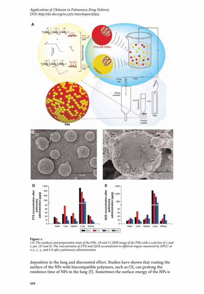
Weifen Zhang’s group had developed a series of CS-based microspheres forpulmonary administration. They reported the preparation of CS and β-cyclodextrinmicrospheres as PDDS [51–55]. The microspheres were prepared by the spray dry-ing method, and theophylline was loaded into the microspheres as a model drug.These microspheres possessed spherical shape with smooth or wrinkled surfaces,and had suitable aerodynamic diameters, which were suitable for inhalation. Themicrospheres had high drug entrapment and encapsulation efficiency. They canremain stable under storage conditions. The microspheres could also easily pene-trate the membrane with a high permeation rate. The results showed that thesemicrospheres had good potential as a sustained drug release carrier for pulmonaryadministration.
In Weifen Zhang’s another work, paclitaxel (PTX) and quercetin (QUE) wererespectively loaded in the NPs, which were synthesized with the oleic acid-conjugated CS (OA-CTS). And these drug-loaded NPs were further used in thepreparation of polymeric microspheres (PMs) by the spray-drying method(Figure 1) [56]. The microspheres could help prolong the retention time of PTX inthe presence of QUE, for bypassing the P-glycoprotein drug efflux pumps. Thediameters of the PMs ranged from 1 to 5 μm, and they had a uniform size distribution.The PMs displayed slow-release characteristics at pH levels of 4.5 and 7.4. In vivopharmacokinetic and biodistribution studies suggested that the PMs exhibited aprolonged circulation time and a markedly high accumulation in the lung. The PMscould serve as a promising PDDS for combined therapy using hydrophobic drugs.
Recently, Ludmylla Cunha et al. developed inhalable CS microparticles forsimultaneous delivery of isoniazid and rifabutin in lung tuberculosis treatment [57].Spray-dried CS microparticles were obtained with adequate flow properties fordeep lung delivery (aerodynamic diameter of 4 μm) and high drug associationefficiencies (93% for isoniazid and 99% for rifabutin). No cytotoxicity effect wasfound in human alveolar epithelial (A549) cells. The CS microparticles could acti-vate macrophage-like cells, inducing cytokine secretion well above basal levels.Moreover, the uptake level of macrophages to internalize microparticles was over90%. The microparticles also inhibited bacterial growth by 96%, demonstrating thatthe microencapsulation preserved drug antibacterial activity in vitro. The dualdrug-loaded CS microparticles demonstrated to be potential candidates forinhalable therapy of pulmonary tuberculosis.
3.2.2 CS-based NPs
As a potential DDS, NPs have been widely used in medicine and other fields, andit has already become a research hotspot for decades [58–60]. NPs can improve thesolubility and stability of the drugs, prolong the half-life, and enhance the drugabsorption rate. NPs can also help to realize sustained or controlled drug release,prolong drug acting time, reduce administration frequency, and improve patientcompliance. Moreover, NPs can target the drugs to specific organs and cells by thepassive or active targeting ability of the multiple functionalized NPs. The NPs canbe modified to avoid the phagocytosis of the macrophages or the removal ofmucosal cilia, thereby improving the bioavailability of the drugs.
Drug delivery by NPs is an effective approach for the pulmonary administrationof insoluble drugs [61, 62]. The surface of the NPs can be modified to prolong thedrug residence time in the lung and to achieve appropriate release property forimproving the therapeutic effect. The pulmonary administration route for NPs ismostly by inhalation in the form of aerosolized colloidal solution. However, whenthe NPs are administered directly into the lung, some of them may be dischargedout with the breath due to their small particle size, thereby resulting in low
168
Role of Novel Drug Delivery Vehicles in Nanobiomedicine
deposition in the lung and discounted effect. Studies have shown that coating thesurface of the NPs with biocompatible polymers, such as CS, can prolong theresidence time of NPs in the lung [5]. Sometimes the surface energy of the NPs is
Figure 1.(A) The synthesis and preparation route of the PMs. (B and C) SEM image of the PMs with a scale bar of 5 and2 μm. (D and E) The concentration of PTX and QUE accumulated in different organs measured by HPLC at0.5, 1, 3, and 6 h after pulmonary administration.
169
Applications of Chitosan in Pulmonary Drug DeliveryDOI: http://dx.doi.org/10.5772/intechopen.87932

relatively high, and the weak stability will lead to aggregations and interactionsbetween the NPs. Thus, special surface modification will also be an effective way toimprove the stability of the NPs.
Kenneth A. Howard et al. had developed CS-based siRNA-loaded NPs for pulmo-nary RNA interference therapy [63]. The negatively charged siRNA was complexedby the positively charged CS to form the polyelectrolyte complex NPs. The particlesize ranged from 40 to 600 nm. These NPs showed a rapid uptake (1 h) into NIH 3T3cells. The NPs could mediate efficient knockdown of endogenous enhanced greenfluorescent protein (EGFP) in both H1299 human lung carcinoma cells and murineperitoneal macrophages in vitro. Moreover, effective in vivo RNA interference wasalso realized in the bronchiole epithelial cells of transgenic EGFP mice after nasaladministration. The results indicated that this kind of CS-based complex NPs hasgreat potential in RNA interference therapy for systemic and mucosal disease.
A series of CS/fucoidan (CS/F) NPs had been designed and prepared as PDDSfor gentamicin (GM) in Yi-Cheng Huang’s study (Figure 2) [64]. The NPspresented a biphasic release feature, with a zero-order release of GM for the first10 h, followed by a sustained release of up to 72 h, and the cumulative release valuereached 99%. The GM-loaded CS/F NPs exhibited multiple antimicrobial capabili-ties against Klebsiella pneumoniae. The intratracheal administration of the GM-loaded CS/F NPs displayed a higher area under the curve (AUC) and lower mini-mum inhibitory concentration ratio than the free GM that was administrated byintravenous injection. These results had showed that the CS/F NPs had superiorantimicrobial efficacy and lower risk of systemic toxicity, and this GM pulmonarydelivery system has exhibited good potential for pneumonia treatment.
Figure 2.(A) The preparation of the GM-loaded CS/F NPs. (B) TEM of the GM-C5F1 NPs. (C–E) Long-term stabilityof the series of CS/F NPs.
170
Role of Novel Drug Delivery Vehicles in Nanobiomedicine
Abdallah Makhlof et al. synthesized a thiomer derivative of glycol CS (GCS),which was coupled with thioglycolic acid (TGA) [65]. The NPs were prepared withGCS and GCS-TGA by ionic gelation for the pulmonary delivery of peptides. TheNPs were positively charged and had sizes in the range of 230–330 nm. They alsoshowed high calcitonin entrapment. After intratracheal administration, themucoadhesion capacity of the GCS-TGA NPs was much better than that ofnonthiolated NPs in rats. The toxic effect of the NPs with lung tissue was confirmedwith negligible epithelial damage and toxicity. The NPs could enhance the pulmo-nary absorption of the delivered peptides, and the calcitonin-loaded GCS and GCS-TGA NPs showed efficient hypocalcemic effect. The GCS and its thiomer derivativecould be used as potential PDDS for delivering peptides.
In another work, Adriana Trapani et al. had prepared CS and GCS NPscontaining Lipoid S100 for the systemic delivery of low molecular weight heparin(LMWH) by pulmonary administration [66]. The NPs were prepared by ionotropicgelation method. The NPs were positively charged and with nanoscale size. Efficientdrug entrapment and good mucoadhesive property could be achieved by usingthese NPs. The LMWH was effectively delivered into the lung by the aerosolizationof the drug-loaded NPs. These results revealed the promising characteristics of theCS-based NPs, which were highly applicable for PDDS.
Tejal Rawal et al. designed a NP-based dry powder formulation of rifampicin(RFM) for achieving local and sustained targeting of anti-tubercular drugs toreduce dose and frequency [67]. The drug-loaded NPs were formulated by the ionicgelation probe sonication method. The optimized formulation had a small particlesize of 124.1 nm with an entrapment efficiency of 72%. No toxicity was found byin vitro and in vivo experiments. The pharmacokinetic assay verified that the NPformulation would serve a better alternative because it could minimize the frequentdosing schedule than conventional dry powder inhalation and market formulation.The RFM-loaded NPs open new avenues for developing therapeutic interventionsfor lung tuberculosis.
3.2.3 CS-modified liposomes
Liposomes are primarily used as PDDS for the treatment of respiratory distresssyndrome and other lung diseases [68]. The drugs carried by liposomes mainly havestable physicochemical properties and strong lipophilicity. Liposome-based drugcarrier has been one of the research hotspots in pharmaceutics [69, 70]. At present,drug-loaded liposome preparations, such as for amphotericin B, daunorubicin,doxorubicin, cytarabine, and morphine, have been successively developed [71]. Theliposome preparations of these drugs have many unique advantages compared withtheir common preparations. Liposome-based pulmonary administration has theadvantages of rapid absorption, little irritation, good tolerance, high safety, con-trollable drug release, and improved bioavailability [72]. The encapsulation of thedrug in liposomes affects the pharmacokinetic property of the drug and prolongsdrug half-life. Moreover, 85% of the components of the alveolar surface are phos-pholipids, which can facilitate the liposomes accumulate and enter the lung tissue.Liposome-loaded macromolecules with low lipophilicity can significantly improvebioavailability, and liposomes can also reduce the side effects of the toxic drugs tonormal tissues by pulmonary administration.
Marco Zaru et al. prepared CS-coated liposomes and used them as drug carriersfor pulmonary delivery by nebulization [73]. Rifampicin (RFM) was loaded in theCS-coated liposomes with different lipid compositions. By coating with CS, theencapsulation efficiency of the liposomes increased slightly, and after nebulization,the stability also remarkably increased. The mucoadhesive capacity of the
171
Applications of Chitosan in Pulmonary Drug DeliveryDOI: http://dx.doi.org/10.5772/intechopen.87932

CS-coated liposomes was much better than that of the noncoated ones. The cyto-toxicity of the RFM-loaded CS-coated liposomes on A549 cells was much lower thanthat of the free drug. The results showed that the CS coating could significantlyenhance the mucoadhesive capacity of the liposomes, and these CS-coatedliposomes could be potential drug carriers for pulmonary administration bynebulization.
In another study, Mitsutaka Murata et al. investigated the surface modificationof liposomes by CS for pulmonary delivery [74]. The surface of the liposomes wasmodified with CS oligosaccharide (oligoCS) and polyvinyl alcohol with a hydro-phobic anchor (PVA-R). The association study showed that the PVA-R modificationdecreased the interaction between liposomes and A549 cells. By contrast, theoligoCS modification could significantly promote the cellular interaction. Afterpulmonary administration to rats, the peptide elcatonin (eCT) loaded oligoCS orPVA-R modified liposomes exhibited significantly enhanced therapeutic efficacy.The oligoCS-modified liposomes could adhere to the lung tissues and open the tightjunctions between cells, which remarkably improved the drug absorption rate. Onthe other hand, the PVA-R-modified liposomes could induce a sustained absorptionthrough the long-term eCT retention in lung fluid. The results verified that thesurface-modification of liposomes with oligoCS and PVA-R could be used as effec-tive PDDS for peptide pulmonary administration.
3.2.4 CS-based active targeting DDS
The pulmonary inhalation preparations are usually directly transported into thelung. Thus, they can passively target to the lung tissues by pulmonary administration.The deposition site of the drugs in the lung can be controlled by regulating thephysicochemical and functional properties of the drug carriers to meet the diseasetreatment requirements. In addition to passive targeting, active targeting systemshave more applications in precise and efficient disease treatment. Active targetingsystems utilize ligands or antibody-modified carriers to deliver the drugs to thespecifically targeted tissues or cells, even intracellular organelles, thereby improvingdrug efficacy and reducing toxicity and side effects [75, 76]. The drug carriers couldbe chemically modified with active ligands, such as sugar moieties (galactose, man-nose, and glucose) or specific ligands, such as folic acid (FA), transferrin (Tf), andArg-Gly-Asp (RGD) peptide [77–79]. Antibody-mediated active targeting is also aprimary strategy for delivering the drugs to the specific parts of the body [80].
Hu-Lin Jiang et al. had prepared a folate-CS-graft-polyethylenimine (FC-g-PEI)copolymer for cancer cell-targeting gene delivery [81]. FC-g-PEI could effectivelyload and protect the shRNA. The copolymer also showed decreased cytotoxicitycompared with the PEI control. Compared with the untargeted polymer, FC-g-PEIwas a more efficient Akt1 shRNA carrier, and it exhibited effective cancer cell-targeting ability. Moreover, aerosol delivery of the FC-g-PEI/Akt1 shRNA NPseffectively inhibited lung tumorigenesis in the urethane-induced lung cancer modelmice via Akt signaling pathway. The results demonstrated that the FC-g-PEI couldbe applied for the shRNA gene therapy via aerosol delivery.
Yongfeng Luo et al. developed an inhalable β2-adrenoceptor ligand-directedguanidinylated-CS (GCS) carrier for targeted lung delivery of siRNA [82]. GCScould effectively condense the siRNA and form complex NPs. Compared withpristine CS, the siRNA-loaded GCS NPs exhibited lower cytotoxicity and highercellular internalization, which finally resulted in promoted gene silence efficiency.Salbutamol (a β2-adrenoceptor agonist) was further chemically coupled to the GCSto enhance the targeting specificity of the siRNA-loaded NPs. The gene silenceefficacy was remarkably increased both in vitro and in vivo. The carrier could
172
Role of Novel Drug Delivery Vehicles in Nanobiomedicine
protect the siRNA against the destructive shear forces generated by mesh-basednebulizers. Aerosol treatment also improved the size distribution of the NPs, whichwas beneficial in promoting the transfection efficiency. This CS-based targetingDDS had potential applications for siRNA delivery in lung disease treatment byaerosol inhalation.
Suhui Ni et al. had prepared γ-aminobutyric acid type B (GABAB) receptorligand-directed NPs for survivin siRNA delivery [83]. The NPs were synthesized bybaclofen (Bac)-functionalized trimethyl CS (Bac-TMC) with tripolyphosphate(TPP) as an ionic crosslinker. GABAB receptor agonist Bac was initially introducedinto TMC as a novel ligand. The Bac-TMC/TPP NPs could promote the cellularuptake of siRNA by interacting with the GABAB receptor, which further inducedeffective gene silence and cell apoptosis. Mannitol microparticles were furtherutilized for the pulmonary delivery of the siRNA-loaded NPs via pressurizedmetered dose inhalers (pMDI). The obtained formulation had good aerodynamicproperties, which benefited the deep lung deposition. The pMDI formulationcontaining Bac-TMC/TPP NPs could be a potential PDDS for siRNA.
Recently, Ting Wu et al. had developed a genipin-crosslinked carboxymethyl CSnanogel for lung-targeted delivery of isoniazid (INH) and rifampin (RMP) [84].The dual drug-loaded nanogel particles (NGPs) had a uniform particle size from 60to 130 nm with spherical morphology. The NGPs had sustained release behavior insimulated lung fluid. The dual drug-loaded NGPs had high antibacterial activityagainst multidrug-resistant Mycobacterium tuberculosis, and also could realize long-term antibacterial activity. Further in vivo evaluation exhibited that alveolar deliv-ery of NGPs had satisfactory deposition of drug within the lung with lower toxicity.The results indicated that the NGPs would be a potential dosage form for treatingagainst multidrug-resistant Mycobacterium tuberculosis.
4. Future perspectives
Pulmonary administration is a promising route for drug delivery to prevent andtreat diseases, especially for delivering the drugs for lung diseases and some smallmolecule drugs with low absorption rate when in oral dosage form, also suitable forsome traditional Chinese medicines and peptide or protein drugs. PDDS can effec-tively deliver the therapeutic drugs to the target sites, and improve the drug bio-availability and therapeutic efficacy. Inhalation is a noninvasive method forpulmonary administration, and the inhaled drugs can directly enter the blood cir-culation through the absorption of the alveolar epithelium. Pulmonary administra-tion can enhance the drug absorption rate, reduce systemic side-effects, andimprove the compliance of long-term medication. By transforming the drugs intodry powder inhalation formulations, drug degradation can be avoided. The thera-peutic effect of the PDDS is mainly influenced by the physicochemical properties ofthe DDS, the dosage forms, and the administration devices, and also some otherfactors. The increase of the amount of drug delivered into the lung and the promo-tion of drug absorption rate are the key factors to improve the therapeutic effi-ciency of pulmonary administration. The application of sustained or controlledrelease preparations is an important method to prolong drug action time, enhancedrug bioavailability, and improve patient compliance. In recent years, many con-trolled release preparations or active targeting preparations for pulmonary drugdelivery have been constructed by drug-loaded microparticles, liposomes, and NPs.These multifunctional drug carriers have gained increasing popularity in pulmonaryadministration. It is really inspiring for us to see that some of the PDDS havealready been applied to treat patients in clinic and become commercially available
173
Applications of Chitosan in Pulmonary Drug DeliveryDOI: http://dx.doi.org/10.5772/intechopen.87932

commodities with promising prospects. And in the coming years, great progress willbe made in PDDS research by the development of drug delivery devices and phar-maceutical preparation technology.
The PDDS constructed with biomaterials has been a hot research direction inmedicine and pharmacy fields for decades. Among these biomaterials, CS, which isthe only natural cationic polysaccharide, has been recognized as one of the mostversatile and promising functional biomaterials. Moreover, CS is also one of the mostabundant regeneration resources, second only to the cellulose. After decades ofresearch, CS has been recognized as a nontoxic, biocompatible (especially with respi-ratory cells) and biodegradable polymer. Moreover, CS can accommodate bothhydrophilic and hydrophobic drugs due to its amphiphilic properties [5]. The excel-lent performances of CS as a building component of DDS have been confirmed bymany studies, and it has been identified as a novel drug delivery carrier with broadapplication prospects, especially for sustained and controlled drug release. Due to theunique features, CS can assist the drugs accomplishing local and systemic delivery,realizing high mucoadhesion, and efficient drug absorption in target tissues, which isespecially applicative for PDDS. However, CS is insoluble in aqueous solutions atneutral and alkaline pH, and it only can be dissolved in aqueous acidic media (pH< 5). This frustrating issue has badly limited CS’s application range, and there is anurgent need for developing convenient and practical modification strategies toimprove its solubility. Up to now, CS has been approved as safe by US-FDA and EUfor dietary use and wound dressing applications [85]. And additionally, CS has onlybeen approved by the European Pharmacopeia as a pharmaceutical excipient for oralpreparations. The safety issues of CS and its derivatives in pulmonary delivery andother administration routes have not been fully understood and still remain to befurther evaluated. Therefore, the safety issue is a noteworthy research gap remainedto be filled in the future work, for figuring out the potential adverse effects of CS andits derivatives in humans. At the same time, appropriate engineering of designing andmodifying CS and its derivatives is highly demanded to optimize the performance ofthe CS-based drug carriers, for meeting the special requirements of in vivo pulmo-nary drug delivery. We hope that in the near future, more advanced synthesis andmodification methods will be developed. And in the meanwhile, the physicochemicalproperty, toxicity, biodistribution, biocompatibility, and biodegradability of CS andits derivatives should also be thoroughly investigated in detail.
5. Conclusions
Despite all this, CS-based PDDS has already achieved considerable success in thepast decades. It is believed that in the near future, with the rapid development ofmaterial science, biotechnology, genetic engineering, medical technology, and otherscientific fields, people will have a more extensive and in-depth understanding ofthe unique properties of CS and its derivatives. We hope that more efficient CS-based PDDS will be designed, by developing novel material preparation strategies,advanced formulation methods, and improved inhalation technology. We believethat CS-based PDDS will play more important roles in the applications of local andsystemic disease treatment in the near future.
174
Role of Novel Drug Delivery Vehicles in Nanobiomedicine
Acknowledgements
The authors are thankful to the generous financial support from the NationalNatural Science Foundation of China (81774125) and National Key TechnologyR&D Program of the Ministry of Science and Technology (2013GA740103).
Conflict of interest
The authors declare no conflicts of interest in this work.
Author details
Xiuwen Guan1,2 and Weifen Zhang1,2*
1 College of Pharmacy, Weifang Medical University, Weifang, Shandong, PR China
2 Shandong Engineering Research Center for Smart Materials and RegenerativeMedicine, Weifang, Shandong, PR China
*Address all correspondence to: [email protected]
©2019 TheAuthor(s). Licensee IntechOpen. This chapter is distributed under the termsof theCreativeCommonsAttribution License (http://creativecommons.org/licenses/by/3.0),which permits unrestricted use, distribution, and reproduction in anymedium,provided the original work is properly cited.
175
Applications of Chitosan in Pulmonary Drug DeliveryDOI: http://dx.doi.org/10.5772/intechopen.87932

References
[1] Patton JS, Byron PR. Inhalingmedicines: Delivering drugs to the bodythrough the lungs. Nature Reviews DrugDiscovery. 2007;6(1):67-74. DOI:10.1038/nrd2153
[2] Patil J, Sarasija S. Pulmonary drugdelivery strategies: A concise,systematic review. Lung India: OfficialOrgan of Indian Chest Society. 2012;29(1):44-49. DOI: 10.4103/0970-2113.92361
[3] Mansour HM, Rhee Y-S, Wu X.Nanomedicine in pulmonary delivery.International Journal of Nanomedicine.2009;4:299-319. DOI: 10.2147/IJN.S4937
[4] Agoram B, Woltosz WS, Bolger MB.Predicting the impact of physiologicaland biochemical processes on oraldrug bioavailability. AdvancedDrug Delivery Reviews. 2001;50:S41-S67. DOI: 10.1016/S0169-409X(01)00179-X
[5] Islam N, Ferro V. Recent advances inchitosan-based nanoparticulatepulmonary drug delivery. Nanoscale.2016;8(30):14341-14358. DOI: 10.1039/C6NR03256G
[6] Agu RU, Ugwoke MI, Armand M,Kinget R, Verbeke N. The lung as aroute for systemic delivery oftherapeutic proteins and peptides.Respiratory Research. 2001;2(4):198-209. DOI: 10.1186/rr58
[7] Qaseem A, Wilt TJ, Weinberger SE,Hanania NA, Criner G, van der MolenT, et al. Diagnosis and management ofstable chronic obstructive pulmonarydisease: A clinical practice guidelineupdate from the American College ofPhysicians, American College of ChestPhysicians, American Thoracic Society,and European Respiratory Society.Annals of Internal Medicine. 2011;155(3):179-191. DOI: 10.7326/0003-4819-155-3-201108020-00008
[8] Hoppentocht M, Hagedoorn P,Frijlink H, De Boer A. Technological andpractical challenges of dry powderinhalers and formulations. AdvancedDrug Delivery Reviews. 2014;75:18-31.DOI: 10.1016/j.addr.2014.04.004
[9] Pham D-D, Fattal E, Tsapis N.Pulmonary drug delivery systems fortuberculosis treatment. InternationalJournal of Pharmaceutics. 2015;478(2):517-529. DOI: 10.1016/j.ijpharm.2014.12.009
[10] Liang Z, Ni R, Zhou J, Mao S. Recentadvances in controlled pulmonary drugdelivery. Drug Discovery Today. 2015;20(3):380-389. DOI: 10.1016/j.drudis.2014.09.020
[11] Manivasagan P, Oh J. Marinepolysaccharide-based nanomaterials as anovel source of nanobiotechnologicalapplications. International Journal ofBiological Macromolecules. 2016;82:315-327. DOI: 10.1016/j.ijbiomac.2015.10.081
[12] Sonia T, Sharma CP. Chitosan andits derivatives for drug deliveryperspective. In: Chitosan forBiomaterials I. Berlin, Germany:Springer; 2011. pp. 23-53. DOI: 10.1007/12_2011_117
[13] Singh Dhillon G, Kaur S, Jyoti SarmaS, Kaur Brar S, Verma M, YadagiriSurampalli R. Recent development inapplications of important biopolymerchitosan in biomedicine,pharmaceuticals and personal careproducts. Current Tissue Engineering.2013;2(1):20-40. DOI: 10.2174/2211542011302010004
[14] Yamamoto H, Kuno Y, Sugimoto S,Takeuchi H, Kawashima Y. Surface-modified PLGA nanosphere withchitosan improved pulmonary deliveryof calcitonin by mucoadhesion andopening of the intercellular tightjunctions. Journal of Controlled Release.
176
Role of Novel Drug Delivery Vehicles in Nanobiomedicine
2005;102(2):373-381. DOI: 10.1016/j.jconrel.2004.10.010
[15] Fröhlich E, Mercuri A, Wu S,Salar-Behzadi S. Measurements ofdeposition, lung surface area and lungfluid for simulation of inhaledcompounds. Frontiers in Pharmacology.2016;7:181. DOI: 10.3389/fphar.2016.00181
[16] Patton JS, Fishburn CS, Weers JG.The lungs as a portal of entry forsystemic drug delivery. Proceedings ofthe American Thoracic Society. 2004;1(4):338-344. DOI: 10.1513/pats.200409-049TA
[17] Patton JS. Mechanisms ofmacromolecule absorption by the lungs.Advanced Drug Delivery Reviews. 1996;19(1):3-36. DOI: 10.1016/0169-409X(95)00113-L
[18] Weibel ER. Morphological basis ofalveolar-capillary gas exchange.Physiological Reviews. 1973;53(2):419-495. DOI: 10.1152/physrev.1973.53.2.419
[19] Stone KC, Mercer RR, Gehr P,Stockstill B, Crapo JD. Allometricrelationships of cell numbers and size inthe mammalian lung. American Journalof Respiratory Cell and MolecularBiology. 1992;6(2):235-243. DOI:10.1165/ajrcmb/6.2.235
[20] Smola M, Vandamme T, SokolowskiA. Nanocarriers as pulmonary drugdelivery systems to treat and to diagnoserespiratory and non respiratory diseases.International Journal of Nanomedicine.2008;3(1):1-19. DOI: 10.1016/S0378-7753(01)01033-3
[21] Ricciardolo FL, Blasi F, Centanni S,Rogliani P. Therapeutic novelties ofinhaled corticosteroids andbronchodilators in asthma.Pulmonary Pharmacology &Therapeutics. 2015;33:1-10. DOI:10.1016/j.pupt.2015.05.006
[22] Chono S, Fukuchi R, Seki T,Morimoto K. Aerosolized liposomeswith dipalmitoyl phosphatidylcholineenhance pulmonary insulin delivery.Journal of Controlled Release. 2009;137(2):104-109. DOI: 10.1016/j.jconrel.2009.03.019
[23] Kwon MJ, Bae JH, Kim JJ, Na K, LeeES. Long acting porous microparticle forpulmonary protein delivery.International Journal of Pharmaceutics.2007;333(1–2):5-9. DOI: 10.1016/j.ijpharm.2007.01.016
[24] Chow AH, Tong HH,Chattopadhyay P, Shekunov BY.Particle engineering for pulmonary drugdelivery. Pharmaceutical Research.2007;24(3):411-437. DOI: 10.1007/s11095-006-9174-3
[25] Frampton MW, Stewart JC,Oberdörster G, Morrow PE, Chalupa D,Pietropaoli AP, et al. Inhalation ofultrafine particles alters blood leukocyteexpression of adhesion molecules inhumans. Environmental HealthPerspectives. 2005;114(1):51-58. DOI:10.1289/ehp.7962
[26] Todoroff J, Vanbever R. Fate ofnanomedicines in the lungs. CurrentOpinion in Colloid & Interface Science.2011;16(3):246-254. DOI: 10.1016/j.cocis.2011.03.001
[27] Scheuch G, Kohlhaeufl MJ, Brand P,Siekmeier R. Clinical perspectives onpulmonary systemic and macromoleculardelivery. Advanced Drug DeliveryReviews. 2006;58(9–10):996-1008. DOI:10.1016/j.addr.2006.07.009
[28] Zhang J, Wu L, Chan H-K,Watanabe W. Formation,characterization, and fate of inhaleddrug nanoparticles. Advanced DrugDelivery Reviews. 2011;63(6):441-455.DOI: 10.1016/j.addr.2010.11.002
[29] Kumar MNR. A review of chitin andchitosan applications. Reactive and
177
Applications of Chitosan in Pulmonary Drug DeliveryDOI: http://dx.doi.org/10.5772/intechopen.87932

Functional Polymers. 2000;46(1):1-27.DOI: 10.1016/S1381-5148(00)00038-9
[30] Dutta PK, Dutta J, Tripathi V.Chitin and chitosan: Chemistry,properties and applications. Journal ofScientific & Industrial Research. 2004;63:20-31. DOI: 10.1016/j.foodhyd.2010.08.008
[31] Yuan Q, Shah J, Hein S, Misra R.Controlled and extended drug releasebehavior of chitosan-based nanoparticlecarrier. Acta Biomaterialia. 2010;6(3):1140-1148. DOI: 10.1016/j.actbio.2009.08.027
[32] Wang JJ, Zeng ZW, Xiao RZ, Xie T,Zhou GL, Zhan XR, et al. Recentadvances of chitosan nanoparticles asdrug carriers. International Journal ofNanomedicine. 2011;6:765-774. DOI:10.2147/IJN.S17296
[33] Boonsongrit Y, Mitrevej A, MuellerBW. Chitosan drug binding by ionicinteraction. European Journal ofPharmaceutics and Biopharmaceutics.2006;62(3):267-274. DOI: 10.1016/j.ejpb.2005.09.002
[34] Shukla SK, Mishra AK, Arotiba OA,Mamba BB. Chitosan-basednanomaterials: A state-of-the-artreview. International Journal ofBiological Macromolecules. 2013;59:46-58. DOI: 10.1016/j.ijbiomac.2013.04.043
[35] Gorzelanny C, Pöppelmann B,Pappelbaum K, Moerschbacher BM,Schneider SW. Human macrophageactivation triggered by chitotriosidase-mediated chitin and chitosandegradation. Biomaterials. 2010;31(33):8556-8563. DOI: 10.1016/j.biomaterials.2010.07.100
[36] Fei Liu X, Lin Guan Y, Zhi Yang D,Li Z, De Yao K. Antibacterial action ofchitosan and carboxymethylatedchitosan. Journal of Applied PolymerScience. 2001;79(7):1324-1335. DOI:
10.1002/1097-4628(20010214)79:7<1324::AID-APP210>3.0.CO;2-L
[37] Chung Y-C, Chen C-Y. Antibacterialcharacteristics and activity of acid-soluble chitosan. BioresourceTechnology. 2008;99(8):2806-2814.DOI: 10.1016/j.biortech.2007.06.044
[38] Bansal V, Sharma PK, Sharma N, PalOP, Malviya R. Applications of chitosanand chitosan derivatives in drugdelivery. Advances in BiologicalResearch. 2011;5(1):28-37
[39] Yeh T-H, Hsu L-W, Tseng MT, LeeP-L, Sonjae K, Ho Y-C, et al. Mechanismand consequence of chitosan-mediatedreversible epithelial tight junctionopening. Biomaterials. 2011;32(26):6164-6173. DOI: 10.1016/j.biomaterials.2011.03.056
[40] Illum L, Jabbal-Gill I, HinchcliffeM, Fisher A, Davis S. Chitosan as a novelnasal delivery system for vaccines.Advanced Drug Delivery Reviews.2001;51(1–3):81-96. DOI: 10.1016/S0169-409X(01)00171-5
[41] Agnihotri SA, Mallikarjuna NN,Aminabhavi TM. Recent advances onchitosan-based micro-and nanoparticlesin drug delivery. Journal of ControlledRelease. 2004;100(1):5-28. DOI:10.1016/j.jconrel.2004.08.010
[42] Cook RO, Pannu RK, Kellaway IW.Novel sustained release microspheresfor pulmonary drug delivery. Journal ofControlled Release. 2005;104(1):79-90.DOI: 10.1016/j.jconrel.2005.01.003
[43] Sakagami M, Byron PR. Respirablemicrospheres for inhalation. ClinicalPharmacokinetics. 2005;44(3):263-277.DOI: 10.2165/00003088-200544030-00004
[44] Edlund U, Albertsson A-C.Degradable polymer microspheres forcontrolled drug delivery. In: DegradableAliphatic Polyesters. Berlin, Germany:
178
Role of Novel Drug Delivery Vehicles in Nanobiomedicine
Springer; 2002. pp. 67-112. DOI:10.1007/3-540-45734-8_3
[45] Mathiowitz E, Jacob JS, Jong YS,Carino GP, Chickering DE, ChaturvediP, et al. Biologically erodablemicrospheres as potential oral drugdelivery systems. Nature. 1997;386(6623):410-414. DOI: 10.1038/386410a0
[46] Liping W, Weihua W, Qian L, QianZ, Yong W, Wei L, et al. Thepreparation of functionalizedcrosslinked macroporous chitosanmicrospheres and their adsorptionproperties for bilirubin. In:Macromolecular Symposia. WileyOnline Library; 2010. pp. 179-187. DOI:10.1002/masy.200900070
[47] Kumar V, Lewis SA, Mutalik S,Shenoy DB, Udupa N. Biodegradablemicrospheres of curcumin for treatmentof inflammation. Indian Journal ofPhysiology and Pharmacology. 2002;46(2):209-217
[48] Mi F-L, Shyu S-S, Chen C-T, Lai J-Y. Adsorption of indomethacin ontochemically modified chitosan beads.Polymer. 2002;43(3):757-765. DOI:10.1016/S0032-3861(01)00580-8
[49] He P, Davis SS, Illum L. Chitosanmicrospheres prepared by spray drying.International Journal of Pharmaceutics.1999;187(1):53-65. DOI: 10.1016/S0378-5173(99)00125-8
[50] Thanoo BC, Sunny M, JayakrishnanA. Cross-linked chitosan microspheres:Preparation and evaluation as a matrixfor the controlled release ofpharmaceuticals. The Journal ofPharmacy and Pharmacology. 1992;44(4):283-286. DOI: 10.1111/j.2042-7158.1992.tb03607.x
[51] Zhang WF, Chen XG, Li PW, HeQZ, Zhou HY. Preparation andcharacterization of theophylline loadedchitosan/β-cyclodextrin microspheres.
Journal of Materials Science: Materials inMedicine. 2008;19(1):305-310. DOI:10.1007/s10856-006-0021-1
[52] Zhang WF, Chen XG, Li PW, LiuCS, He QZ. Preparation andcharacterization of carboxymethylchitosan and β-cyclodextrinmicrospheres by spray drying. DryingTechnology. 2007;26(1):108-115. DOI:10.1080/07373930701781736
[53] Zhang W, Chen X, Li P, He Q, ZhouH, Cha D. Chitosan and β-cyclodextrinmicrospheres as pulmonary sustaineddelivery systems. Journal of WuhanUniversity of Technology-MaterialsScience Edition. 2008;23(4):541-546.DOI: 10.1007/s11595-006-4541-9
[54] Zhang WF, Zhou HY, Chen XG,Tang SH, Zhang JJ. Biocompatibilitystudy of theophylline/chitosan/β-cyclodextrin microspheres aspulmonary delivery carriers. Journal ofMaterials Science: Materials inMedicine. 2009;20(6):1321-1330. DOI:10.1007/s10856-008-3680-2
[55] Zhou H-Y, Zhou D-J, Zhang W-F,Jiang L-J, Li J-B, Chen X-G.Biocompatibility and characteristics ofchitosan/cellulose acetate microspheresfor drug delivery. Frontiers of MaterialsScience. 2011;5(4):367-378. DOI:10.1007/s11706-011-0146-0
[56] Liu K, ChenW, Yang T,Wen B, DingD, KeidarM, et al. Paclitaxel and quercetinnanoparticles co-loaded inmicrospheresto prolong retention time for pulmonarydrug delivery. International Journal ofNanomedicine. 2017;12:8239-8255. DOI:10.2147/IJN.S147028
[57] Cunha L, Rodrigues S, Rosa AM,Faleiro L, Buttini F, Grenha A. Inhalablechitosan microparticles forsimultaneous delivery of isoniazid andrifabutin in lung tuberculosis treatment.Drug Development and IndustrialPharmacy. 2019;45(8):1313-1320. DOI:10.1080/03639045.2019.1608231
179
Applications of Chitosan in Pulmonary Drug DeliveryDOI: http://dx.doi.org/10.5772/intechopen.87932

[58] Cho K, Wang X, Nie S, Shin DM.Therapeutic nanoparticles for drugdelivery in cancer. Clinical CancerResearch. 2008;14(5):1310-1316. DOI:10.1158/1078-0432.CCR-07-1441
[59] Kumari A, Yadav SK, Yadav SC.Biodegradable polymeric nanoparticlesbased drug delivery systems. Colloidsand Surfaces B, Biointerfaces. 2010;75(1):1-18. DOI: 10.1016/j.colsurfb.2009.09.001
[60] Wilczewska AZ, Niemirowicz K,Markiewicz KH, Car H. Nanoparticles asdrug delivery systems. PharmacologicalReports. 2012;64(5):1020-1037. DOI:10.1016/S1734-1140(12)70901-5
[61] Rytting E, Nguyen J, Wang X, KisselT. Biodegradable polymericnanocarriers for pulmonary drugdelivery. Expert Opinion on DrugDelivery. 2008;5(6):629-639. DOI:10.1517/17425247.5.6.629
[62] Bailey MM, Berkland CJ.Nanoparticle formulations inpulmonary drug delivery. MedicinalResearch Reviews. 2009;29(1):196-212.DOI: 10.1002/med.20140
[63] Howard KA, Rahbek UL, Liu X,Damgaard CK, Glud SZ, Andersen MØ,et al. RNA interference in vitro andin vivo using a novel chitosan/siRNAnanoparticle system. MolecularTherapy. 2006;14(4):476-484. DOI:10.1016/j.ymthe.2006.04.010
[64] Huang Y-C, Li R-Y, Chen J-Y, ChenJ-K. Biphasic release of gentamicin fromchitosan/fucoidan nanoparticles forpulmonary delivery. CarbohydratePolymers. 2016;138:114-122. DOI:10.1016/j.carbpol.2015.11.072
[65] Makhlof A, Werle M, Tozuka Y,Takeuchi H. Nanoparticles of glycolchitosan and its thiolated derivativesignificantly improved the pulmonarydelivery of calcitonin. InternationalJournal of Pharmaceutics. 2010;397
(1–2):92-95. DOI: 10.1016/j.ijpharm.2010.07.001
[66] Trapani A, Di Gioia S, Ditaranto N,Cioffi N, Goycoolea FM, Carbone A,et al. Systemic heparin delivery by thepulmonary route using chitosan andglycol chitosan nanoparticles.International Journal of Pharmaceutics.2013;447(1–2):115-123. DOI: 10.1016/j.ijpharm.2013.02.035
[67] Rawal T, Parmar R, Tyagi RK,Butani S. Rifampicin loaded chitosannanoparticle dry powder presents animproved therapeutic approach foralveolar tuberculosis. Colloids andSurfaces B, Biointerfaces. 2017;154:321-330. DOI: 10.1016/j.colsurfb.2017.03.044
[68] Schreier H, Gonzalez-Rothi RJ,Stecenko AA. Pulmonary delivery ofliposomes. Journal of ControlledRelease. 1993;24(1–3):209-223. DOI:10.1016/0168-3659(93)90180-D
[69] Sercombe L, Veerati T, MoheimaniF, Wu SY, Sood AK, Hua S. Advancesand challenges of liposome assisted drugdelivery. Frontiers in Pharmacology.2015;6:286. DOI: 10.3389/fphar.2015.00286
[70] Lian T, Ho RJ. Trends anddevelopments in liposome drug deliverysystems. Journal of PharmaceuticalSciences. 2001;90(6):667-680. DOI:10.1002/jps.1023
[71] Daraee H, Etemadi A, Kouhi M,Alimirzalu S, Akbarzadeh A.Application of liposomes in medicineand drug delivery. Artificial Cells, BloodSubstitutes, and Biotechnology. 2016;44(1):381-391. DOI: 10.3109/21691401.2014.953633
[72] Willis L, Hayes D, Mansour HM.Therapeutic liposomal dry powderinhalation aerosols for targeted lungdelivery. Lung. 2012;190(3):251-262.DOI: 10.1007/s00408-011-9360-x
180
Role of Novel Drug Delivery Vehicles in Nanobiomedicine
[73] Zaru M, Manca M-L, Fadda AM,Antimisiaris SG. Chitosan-coatedliposomes for delivery to lungs bynebulisation. Colloids and Surfaces B,Biointerfaces. 2009;71(1):88-95. DOI:10.1016/j.colsurfb.2009.01.010
[74] Murata M, Nakano K, Tahara K,Tozuka Y, Takeuchi H. Pulmonarydelivery of elcatonin using surface-modified liposomes to improve systemicabsorption: Polyvinyl alcohol with ahydrophobic anchor and chitosanoligosaccharide as effective surfacemodifiers. European Journal ofPharmaceutics and Biopharmaceutics.2012;80(2):340-346. DOI: 10.1016/j.ejpb.2011.10.011
[75] Allen TM. Ligand-targetedtherapeutics in anticancer therapy.Nature Reviews Cancer. 2002;2(10):750-763. DOI: 10.1038/nrc903
[76] Brannon-Peppas L, Blanchette JO.Nanoparticle and targeted systemsfor cancer therapy. AdvancedDrug Delivery Reviews. 2012;64:206-212. DOI: 10.1016/j.addr.2012.09.033
[77] Byrne JD, Betancourt T, Brannon-Peppas L. Active targeting schemes fornanoparticle systems in cancertherapeutics. Advanced Drug DeliveryReviews. 2008;60(15):1615-1626. DOI:10.1016/j.addr.2008.08.005
[78] Vyas SP, Singh A, Sihorkar V.Ligand-receptor-mediated drugdelivery: An emerging paradigm incellular drug targeting. CriticalReviews in Therapeutic DrugCarrier Systems. 2001;18(1):1-76. DOI:10.1615/CritRevTherDrugCarrierSyst.v18.i1.10
[79] Zwicke GL, Ali Mansoori G, JefferyCJ. Utilizing the folate receptor foractive targeting of cancernanotherapeutics. NanotechnologyReviews. 2012;3(1):18496. DOI:10.3402/nano.v3i0.18496
[80] Ansell SM, Harasym TO, Tardi PG,Buchkowsky SS, Bally MB, Cullis PR.Antibody conjugation methods foractive targeting of liposomes. In: DrugTargeting. New Jersey, USA: HumanaPress; 2000. pp. 51-68. DOI: 10.1385/1-59259-075-6:51
[81] Jiang H-L, Xu C-X, Kim Y-K, AroteR, Jere D, Lim H-T, et al. Thesuppression of lung tumorigenesis byaerosol-delivered folate-chitosan-graft-polyethylenimine/Akt1 shRNAcomplexes through the Akt signalingpathway. Biomaterials. 2009;30(29):5844-5852. DOI: 10.1016/j.biomaterials.2009.07.017
[82] Luo Y, Zhai X, Ma C, Sun P, Fu Z,Liu W, et al. An inhalable β2-adrenoceptor ligand-directedguanidinylated chitosan carrier fortargeted delivery of siRNA to lung.Journal of Controlled Release. 2012;162(1):28-36. DOI: 10.1016/j.jconrel.2012.06.005
[83] Ni S, Liu Y, Tang Y, Chen J, Li S, PuJ, et al. GABAB receptor ligand-directedtrimethyl chitosan/tripolyphosphatenanoparticles and their pMDIformulation for survivin siRNApulmonary delivery. CarbohydratePolymers. 2018;179:135-144. DOI:10.1016/j.carbpol.2017.09.075
[84] Wu T, Liao W, Wang W, Zhou J,Tan W, Xiang W, et al. Genipin-crosslinked carboxymethyl chitosannanogel for lung-targeted delivery ofisoniazid and rifampin. CarbohydratePolymers. 2018;197:403-413. DOI:10.1016/j.carbpol.2018.06.034
[85] Mohammed M, Syeda J, Wasan K,Wasan E. An overview of chitosannanoparticles and its application innon-parenteral drug delivery.Pharmaceutics. 2017;9:53. DOI:10.3390/pharmaceutics9040053
181
Applications of Chitosan in Pulmonary Drug DeliveryDOI: http://dx.doi.org/10.5772/intechopen.87932

Role of Novel Drug Delivery Vehicles in Nanobiomedicine
Edited by Rajeev K. Tyagi, Neeraj Garg, Rahul Shukla and Prakash Singh Bisen
Edited by Rajeev K. Tyagi, Neeraj Garg, Rahul Shukla and Prakash Singh Bisen
Medical Nanotechnology and Nanomedicine introduces non-experts to the world of nanomedicine and its evolving organizational infrastructure. Considering the
fluid nature of nano breakthroughs and the delicate balance between benefits and consequences as they apply to medicine, readers at all levels will gain a practical, understandable base of information on these developments so that they may take the greatest advantage of them. This practical reference investigates the impact of
nanotechnology on applications in medicine and biomedical sciences, and the broader societal and economic effects. Eschewing technological details, it focuses on enhancing
awareness of the business, regulatory, and administrative aspects of medical applications. It gives readers a critical, balanced, and realistic evaluation of existing nanomedicine developments and future prospects and provides an ideal foundation
upon which to plan and make decisions.
Published in London, UK
© 2020 IntechOpen © Olique / iStock
ISBN 978-1-78923-985-0
Role of Novel D
rug Delivery Vehicles in N
anobiomedicine
ISBN 978-1-78984-330-9