Role of Intestinal Circadian Genes in Alcohol-Induced Gut Leakiness
21
Role of Intestinal Circadian Genes in Alcohol-induced Gut Leakiness Garth Swanson 6 , Christopher B. Forsyth 2 , Yueming Tang 3 , Maliha Shaikh 4 , Lijuan Zhang 5 , Fred W. Turek 7 , and Ali Keshavarzian 1 Department of Medicine, Division of Digestive Diseases and Nutrition, Rush University Medical Center, Chicago, IL 60612 and Department of Neurobiology and Physiology, Northwestern University, Evanston, IL 60202 Abstract Background—Several studies have indicated that endotoxemia is the required co-factor for alcoholic steatohepatitis (ASH) that is seen in only about 30% of alcoholics. Recent studies have shown that gut leakiness that occurs in a subset of alcoholics is the primary cause of endotoxemia in ASH. The reasons for this differential susceptibility are not known. Since disruption of circadian rhythms occurs in some alcoholics and circadian genes control the expression of several genes that are involved in regulation of intestinal permeability, we hypothesized that alcohol induces intestinal hyperpermeability by stimulating expression of circadian clock gene proteins in the intestinal epithelial cells. Methods—We used Caco-2 monolayers grown on culture inserts as an in vitro model of intestinal permeability and performed western blotting, permeability, and siRNA inhibition studies to examine the role of Clock and Per2 circadian genes in alcohol-induced hyperpermeability. We also measured PER2 protein levels in intestinal mucosa of alcohol fed rats with intestinal hyperpermeability. Results—Alcohol, as low as 0.2%, induced time dependent increases in both Caco-2 cell monolayer permeability and in CLOCK and PER2 proteins. SiRNA specific inhibition of either Clock or Per2 significantly inhibited alcohol-induced monolayer hyperpermeability. Alcohol-fed Correspondence and Reprint requests to: Ali Keshavarzian, M.D., Rush University Medical Center, Section of Gastroenterology and Nutrition, 1725 W. Harrison, Suite 206, Chicago IL 60612, Tel: 312-5633890, Fax: 312-563 3883, [email protected]. 1 M.D., Josephine M. Dyrenforth Chair of Gastroenterology, Professor of Medicine, Pharmacology and Molecular Biophysics & Physiology, Department of Medicine, Division of Digestive Diseases and Nutrition, Rush University Medical Center, Chicago, IL 2 Ph.D., Assistant Professor of Medicine and Biochemistry, Department of Medicine, Division of Digestive Diseases and Nutrition, Rush University Medical Center, Chicago, IL 3 M.D./Ph.D., Assistant Professor of Medicine, Department of Medicine, Division of Digestive Diseases and Nutrition, Rush University Medical Center, Chicago, IL 4 M.S., Research Associate, Department of Medicine, Division of Digestive Diseases and Nutrition, Rush University Medical Center, Chicago, IL 5 M.D., Instructor of Medicine, Department of Medicine, Division of Digestive Diseases and Nutrition,, Rush University Medical Center, Chicago, IL 6 M.D., Assistant Professor of Medicine, Department of Medicine, Division of Digestive Diseases and Nutrition, Rush University, Chicago IL 7 Ph.D., Morrison Professor of Biology, Center for Sleep and Circadian Biology, Department of Neurobiology and Physiology, Northwestern University, Evanston IL Disclosers: the authors have no potential competing interests to declare. Authors contributions: GS participated in developing experimental design, helped in data analysis, experiments and manuscript writing; CBF participated in developing experimental design, helped with data analysis, and writing the manuscript and submitting the manuscript; YT helped with experimental design and data analysis and manuscript writing; MS carried out experiments and made the figures and performed data analysis; LZ carried out experiments and performed data analysis; FWT participated in developing the experimental design, provided the expertise in the field of circadian research, helped with data analysis, and manuscript writing; AK conceived the hypothesis, participated in developing experimental design, and helped with data analysis and manuscript writing. NIH Public Access Author Manuscript Alcohol Clin Exp Res. Author manuscript; available in PMC 2012 July 1. Published in final edited form as: Alcohol Clin Exp Res. 2011 July ; 35(7): 1305–1314. doi:10.1111/j.1530-0277.2011.01466.x. NIH-PA Author Manuscript NIH-PA Author Manuscript NIH-PA Author Manuscript
Transcript of Role of Intestinal Circadian Genes in Alcohol-Induced Gut Leakiness

Role of Intestinal Circadian Genes in Alcohol-induced GutLeakiness
Garth Swanson6, Christopher B. Forsyth2, Yueming Tang3, Maliha Shaikh4, Lijuan Zhang5,Fred W. Turek7, and Ali Keshavarzian1
Department of Medicine, Division of Digestive Diseases and Nutrition, Rush University MedicalCenter, Chicago, IL 60612 and Department of Neurobiology and Physiology, NorthwesternUniversity, Evanston, IL 60202
AbstractBackground—Several studies have indicated that endotoxemia is the required co-factor foralcoholic steatohepatitis (ASH) that is seen in only about 30% of alcoholics. Recent studies haveshown that gut leakiness that occurs in a subset of alcoholics is the primary cause of endotoxemiain ASH. The reasons for this differential susceptibility are not known. Since disruption ofcircadian rhythms occurs in some alcoholics and circadian genes control the expression of severalgenes that are involved in regulation of intestinal permeability, we hypothesized that alcoholinduces intestinal hyperpermeability by stimulating expression of circadian clock gene proteins inthe intestinal epithelial cells.
Methods—We used Caco-2 monolayers grown on culture inserts as an in vitro model ofintestinal permeability and performed western blotting, permeability, and siRNA inhibition studiesto examine the role of Clock and Per2 circadian genes in alcohol-induced hyperpermeability. Wealso measured PER2 protein levels in intestinal mucosa of alcohol fed rats with intestinalhyperpermeability.
Results—Alcohol, as low as 0.2%, induced time dependent increases in both Caco-2 cellmonolayer permeability and in CLOCK and PER2 proteins. SiRNA specific inhibition of eitherClock or Per2 significantly inhibited alcohol-induced monolayer hyperpermeability. Alcohol-fed
Correspondence and Reprint requests to: Ali Keshavarzian, M.D., Rush University Medical Center, Section of Gastroenterology andNutrition, 1725 W. Harrison, Suite 206, Chicago IL 60612, Tel: 312-5633890, Fax: 312-563 3883, [email protected]., Josephine M. Dyrenforth Chair of Gastroenterology, Professor of Medicine, Pharmacology and Molecular Biophysics &Physiology, Department of Medicine, Division of Digestive Diseases and Nutrition, Rush University Medical Center, Chicago, IL2Ph.D., Assistant Professor of Medicine and Biochemistry, Department of Medicine, Division of Digestive Diseases and Nutrition,Rush University Medical Center, Chicago, IL3M.D./Ph.D., Assistant Professor of Medicine, Department of Medicine, Division of Digestive Diseases and Nutrition, RushUniversity Medical Center, Chicago, IL4M.S., Research Associate, Department of Medicine, Division of Digestive Diseases and Nutrition, Rush University Medical Center,Chicago, IL5M.D., Instructor of Medicine, Department of Medicine, Division of Digestive Diseases and Nutrition,, Rush University MedicalCenter, Chicago, IL6M.D., Assistant Professor of Medicine, Department of Medicine, Division of Digestive Diseases and Nutrition, Rush University,Chicago IL7Ph.D., Morrison Professor of Biology, Center for Sleep and Circadian Biology, Department of Neurobiology and Physiology,Northwestern University, Evanston ILDisclosers: the authors have no potential competing interests to declare.Authors contributions: GS participated in developing experimental design, helped in data analysis, experiments and manuscriptwriting; CBF participated in developing experimental design, helped with data analysis, and writing the manuscript and submitting themanuscript; YT helped with experimental design and data analysis and manuscript writing; MS carried out experiments and made thefigures and performed data analysis; LZ carried out experiments and performed data analysis; FWT participated in developing theexperimental design, provided the expertise in the field of circadian research, helped with data analysis, and manuscript writing; AKconceived the hypothesis, participated in developing experimental design, and helped with data analysis and manuscript writing.
NIH Public AccessAuthor ManuscriptAlcohol Clin Exp Res. Author manuscript; available in PMC 2012 July 1.
Published in final edited form as:Alcohol Clin Exp Res. 2011 July ; 35(7): 1305–1314. doi:10.1111/j.1530-0277.2011.01466.x.
NIH
-PA Author Manuscript
NIH
-PA Author Manuscript
NIH
-PA Author Manuscript
rats with increased total gut permeability, assessed by urinary sucralose, also had significantlyhigher levels of PER2 protein in their duodenum and proximal colon than control rats.
Conclusions—Our studies: (1) demonstrate a novel mechanism for alcohol-induced intestinalhyperpermeability through stimulation of intestinal circadian clock gene expression, and (2)provide direct evidence for a central role of circadian genes in regulation of intestinalpermeability.
Keywordsethanol; intestinal permeability; Clock gene; Per2 gene; Circadian rhythm
IntroductionSeveral epidemiological studies have shown that alcoholism causes tissue injury such asalcoholic steatohepatitis (ASH) in only about 30% of alcoholics (Grant et al., 1988; Rao etal., 2004). These data indicate that chronic alcohol (EtOH) consumption is a required but nota sufficient factor for ASH. Although the reasons behind this differential susceptibility arenot fully understood, multiple clinical and experimental studies have provided compellingevidence that gut derived endotoxin is directly involved in the initiation of sustainednecroinflammatory cascades in the liver that are required for alcohol-induced tissue injury,including ASH (Rao et al., 2004; Purohit et al., 2008). A major challenge is to determine thefactors that are responsible for the development of gut leakiness and endotoxemia in only asub-set of alcoholics (or conversely, factors that may protect alcoholics from gut leakiness).
We wished to test the hypothesis that disrupted circadian rhythms may contribute to thedifferential susceptibility for alcohol-induced gut leakiness in a subset of alcoholics. Ourrationale for this hypothesis and the key findings linking circadian rhythms, alcohol, and thegut, are: (1) The core circadian clock molecular machinery is within essentially all thetissues and organs of the body including the central circadian clock in the hypothalamicsuprachiasmatic nucleus (SCN), and intestinal epithelial cells (Hastings et al., 2003; Yoo etal., 2004; Bell-Pedersen et al., 2005; Hoogerwerf et al., 2007); (2) The SCN regulates andcoordinates the expression and timing of multiple peripheral circadian molecular rhythms(Green et al., 2008; Laposky et al., 2008a), possibly including brain-gut interactions of theso called “brain-gut axis,” (BGA); (3) The BGA can regulate intestinal permeability, anddisruption of this communication by pathological stimuli like physical and psychologicalstress that can affect circadian rhythms can also cause gut leakiness in both humans androdents (Gareau et al., 2007; Stasi and Orlandelli, 2008); (4) The circadian modulation of thebrain-gut communication could mediate normal and pathological states of intestinalpermeability since circadian genes can regulate apical junctional complex (AJC) proteingenes that are directly involved in regulation of intestinal permeability (Yamato et al.,2010); (5) Alcohol can disrupt the central SCN circadian rhythm in rodents and affectexpression of clock genes in the brain and these effects may be different in a subset ofalcoholics (Mistlberger and Nadeau, 1992; Chen et al., 2004; Rosenwasser et al., 2005b;Rosenwasser et al., 2005a; Spanagel et al., 2005a; McElroy et al., 2009; Seggio et al., 2009);(6) The Clock gene is important for the circadian regulation of macronutrient absorption byenterocytes in the gut (Pan and Hussain, 2009).
Although these data provide compelling evidence to suggest a role of disrupted circadianrhythms in the etiology of alcohol-induced gut leakiness, there is no direct evidence for arole of the circadian molecular machinery in alcohol-induced intestinal hyperpermeability.Therefore, we carried out studies in a validated in vitro model of intestinal permeability(Caco-2 cell monolayers) as well as on intestinal tissue from an in vivo rodent alcohol model
Swanson et al. Page 2
Alcohol Clin Exp Res. Author manuscript; available in PMC 2012 July 1.
NIH
-PA Author Manuscript
NIH
-PA Author Manuscript
NIH
-PA Author Manuscript

of leaky gut to directly test the hypothesis that the circadian clock molecular machinery candirectly impact alcohol-induced gut leakiness. Our overall objective was to determinewhether alcohol can impact circadian clock gene expression in intestinal epithelial cells invitro and in vivo and also to determine whether modulation of intestinal circadian genes invitro can impact the effects of alcohol on intestinal permeability.
Materials and MethodsIntestinal epithelial cell monolayer barrier function
Barrier permeability was determined using Caco-2 cells grown to confluence on Type 1collagen-coated 12 mm/0.4μm PET tissue culture plate inserts (Transwell, Corning,Corning, NY) as we previously described (Banan et al., 1999; Banan et al., 2000; Forsyth etal., 2007). Permeability of insert Caco-2 monolayers was measured as apical to basolateralflux of the fluorescent marker fluorescein-5-(and-6)-sulfonic acid trisodium salt (FSA, 478d) (Invitrogen, Carlsbad, CA) or as Transepithelial Electrical Resistance (TER). TER isdetermined using a dual electrode system designed for cell culture insert analysis (EVOM,World Precision Instruments, Sarasota, FL). Cell viability is routinely measured by live/deadassay (Invitrogen) or trypan blue staining. Cell viability for all assays was greater than 95%.We have also previously shown that alcohol in concentrations as high as 1% has no effect onCaco-2 viability in this assay for as long as 6h (Banan et al., 1999; Banan et al., 2000).
Treatment of cells with siRNACaco-2 cells were treated with siRNA using a modification of our previously publishedmethods (Forsyth et al., 2010). Briefly, 105 cells are combined with 11 picomoles of siRNAin 50μl Lipofectamine (Invitrogen) and 50μl Optimem (Invitrogen), mixed by gentlyshaking and then plated on Type 1 collagen coated Corning 12mm PET culture inserts(#3460) (Corning). Inserts are used for permeability studies 72-96h after plating, when cellsare confluent. Clock and Per2 siRNA was On Target Smartpool from Dharmacon(Dharmacon Inc., Lafayette, CO). The siRNA sequences for Clock (cat.# L-012977-00)were: CAACUUGCACCUAUAAAUA; CGACAGGACUGGAAACCUA;GAACAACGGACACGCAUGA; CUAGAAAGAUGGACAAAUC; the siRNA sequencesfor Per2 (cat.# L-08212-00) were: GAAUGGAUACGCGGAAUUU;CUUCAGCGAUGCCAAGUUU; GCAGUGGAGCAGAUUCUUU;CGACCAGUCUUCGAAAGUG while control (non-targeting) siRNA was from Santa CruzBiotechnology (Santa Cruz, CA) and the sequence is proprietary (cat.# 37007). Figure 1shows that as a further control for off target effects of siRNA treatment in addition to the useof nontargeting siRNA as a control, we performed western blot analysis for CLOCK proteinexpression in lysates from the Per2 knockdown experiments shown in Fig. 5. As seen in Fig.1, Per2 siRNA did not significantly affect CLOCK protein levels compared to our averageof 70% knockdown of PER2 protein with the Per2 siRNA (see Fig. 5), thus supporting thatthe specific effect of the Per2 siRNA on Caco-2 permeability was not due to effects onCLOCK protein expression but were in fact due specifically to Per2 expression knockdownwith siRNA.
Animal model of alcohol-induced gut leakinessWe have recently published a Rush IACUC animal use committee approved rat model ofalcohol-induced gut leakiness (Keshavarzian et al., 2009). In brief, male Sprague-Dawleyrats (250-300 g at intake) were obtained from Harlan (Indianapolis, IN). Duringexperiments, each rat was given either alcohol or an isocaloric amount of dextrose in liquidrat chow intragastrically (by gavage; 2 cc) 2× daily. Intestinal permeability was measuredtwice: (i) just prior to alcohol administration; and (ii) just before sacrifice. Rats were
Swanson et al. Page 3
Alcohol Clin Exp Res. Author manuscript; available in PMC 2012 July 1.
NIH
-PA Author Manuscript
NIH
-PA Author Manuscript
NIH
-PA Author Manuscript

sacrificed after 2, 4, 8, and 10 weeks of administration of 6 g/kg/day alcohol with N=6 orgreater for each time point.
Intestinal permeability in ratsSprague-Dawley rats (6-8wk, 250-300g) were maintained in a metabolic cage (for 6 hoururine collection) and gut permeability is measured for each rat after a sugar bolus by gavageas we previously described (Farhadi et al., 2006; Keshavarzian et al., 2009). Measurement ofurinary sugars using GC is used to calculate intestinal permeability and is expressed aspercent oral dose excreted in the urine. We have recently revised our method which brieflyinvolves conversion of the relevant sugars to their alditol acetate form rather than ourprevious method of N-Trimethylsilylimidazole (TMSI) derivatization and find it is a moresensitive method to detect the sugars. This is thus a modification of our method that wepreviously published (Farhadi et al., 2006; Keshavarzian et al., 2009). The widely usedurinary L/M (L = lactulose, M=mannitol) ratio represents only small bowel permeability,while the urinary sucralose excretion reported in this study represents total intestinalpermeability [small bowel +colonic permeability].
Measurement of clock genes protein expressionCircadian clock genes (Per2, Clock) protein levels were assayed by western blot. Westernblotting and densitometry analysis with Image J software (NIH) was performed with cell ortissue lysates equalized for total protein and cell number as previously described (Forsyth etal., 2002; Forsyth et al., 2007). CLOCK Ab was from Santa Cruz Biotechnology (SantaCruz, CA) and PER2 Ab was from Abcam (Cambridge, MA) while anti-actin Ab was fromSigma (St. Louis, MO).
Data Analysis and StatisticsThe data are presented as means ± SE. For circadian genes protein levels, group means werecompared by ANOVA and post-hoc tests since the data were normally distributed. Forpermeability data in rats, group means were compared using the nonparametric analyses,Kruskal-Wallis test, because data were not normally distributed. P-values < 0.05 wereconsidered statistically significant. All analyses were done using SPSS (SPSS Inc., Chicago,IL).
ResultsAlcohol at Physiological Concentrations Increases Permeability of Caco-2 IntestinalEpithelial Cell Monolayers
We have previously shown that alcohol at concentrations of 1-15% increases permeability ofCaco-2 cell monolayers, the most widely used in vitro model of intestinalpermeability(Banan et al., 1999; Banan et al., 2000). We now wished to test alcoholconcentrations more relevant to those found in the mucosal layers of the distal intestine andcolon. To do this, we first performed a dose and time course study of alcohol-stimulatedhyperpermeability of Caco-2 monolayers using the fluorescent dye FSA as a marker ofpermeability (Sanders et al., 1995). As seen in Figure 2A we tested concentrations of alcoholranging from 0.05% to 0.5% over a period of 3 hours and found that these physiologicalconcentrations of alcohol dose and time-dependently increased permeability of the intestinalepithelial cell monolayers to FSA probe. Caco-2 monolayer permeability was significantly(p< .05 vs. media control) increased by alcohol by 1 hour and further increased at 2 hr and 3hr for alcohol concentrations as low as 0.1% (22mM, 1-3 drinks). We chose to use a 0.2%alcohol concentration (43mM, 2-4 drinks) for the remainder of the studies because itresulted in a significant increase in permeability at 2 hr and is approximately the average
Swanson et al. Page 4
Alcohol Clin Exp Res. Author manuscript; available in PMC 2012 July 1.
NIH
-PA Author Manuscript
NIH
-PA Author Manuscript
NIH
-PA Author Manuscript

Blood Alcohol Level (BAL) found in our rat alcohol treatment model of alcoholic leaky gutand steatohepatitis (Keshavarzian et al., 2001; Keshavarzian et al., 2009).
As a second measure of Caco-2 permeability, we assessed the effects of alcohol treatment(0.2%) on Transepithelial Resistance (TER) measured as % change in Ωxcm2 (Figure 2B).This is another reliable measure of intestinal monolayer permeability (Shen et al., 2008;Rodriguez-Lagunas et al., 2010). Similar to data for FSA, alcohol significantly and in a timedependent manner led to a disruption in the monolayer barrier. Alcohol-induced decrease inTER was significant (P≤ .001) by 30 min and further dropped to 50% of the original TERlevel by 240 min (Figure 2B).
Alcohol Stimulates Expression of Circadian CLOCK and PER2 Proteins in Caco-2Intestinal Epithelial Cells
We measured protein levels of two key canonical clock genes, Clock and Per2, in theCaco-2 intestinal epithelial cells after alcohol treatment using western blots on lysates fromthe same inserts that were tested for permeability with TER measurements including timepoint-matched control well lysates for each time point. CLOCK protein binds to andupregulates transcription from the Per2 promoter to regulate 24h circadian rhythms (Reppertand Weaver, 2002). The total PER2 protein levels in the Caco-2 cells (mean relative density2611+/- 183SE) is more than CLOCK (mean relative density 1674+/- 117SE) at baseline innon-alcohol treated cells (Figure 3, western blot gel, representative of 9 wells). We foundthat both PER2 and CLOCK gene proteins remained virtually unchanged in control wellsthrough 240 minutes of the experiment. In contrast, both PER2 and CLOCK gene proteins inalcohol treated cells increased after 30 minutes of alcohol treatment and significantlyincreased after 60 and 240 minutes exposure to 0.2% alcohol (Figure 3). Compared to timepoint-matched controls for each protein (set as 100%), alcohol treatment resulted in meanincreases of 225% for CLOCK protein (p=.024) and 240% for PER2 protein (p=.013) inCaco-2 cells after only 60 min of exposure. At the 240 min time point, alcohol treatmentresulted in a mean increase of 237% for CLOCK (p=.036) and 315% for PER2 (p=.039).This alcohol-induced increase in PER2 and CLOCK proteins was associated with analcohol-induced increase in FSA permeability across the monolayer and a drop in TER(Figure 2). The time course of increase in circadian gene proteins and disruption ofmonolayer barrier function was identical.
Thus, alcohol disrupts intestinal monolayer barrier integrity. This disrupting effect ofalcohol included both the “leak pathway” (increased FSA permeability) and “pore pathway”(drop in TER) (Turner, 2009). This disrupting effect of alcohol on barrier function correlatedwith an increase in CLOCK and PER2 proteins in the intestinal epithelial cells.
SiRNA knockdown of Clock or Per2 Circadian Genes Prevents Alcohol-induced IntestinalEpithelial Caco-2 Monolayer Hyperpermeability
We next tested the hypothesis that alcohol-induced stimulation of CLOCK and/or PER2proteins is directly involved in the regulation of intestinal monolayer hyperpermeability inresponse to alcohol by using siRNA specific for either Clock or Per2. The Caco-2 cells weretreated with siRNA for 72-96h prior to treatment with alcohol (N= 3 experiments for each,Clock and Per2 siRNA).
Figure 4 depicts TER and FSA permeability data and representative blots and thesummarized histogram data for CLOCK protein (N=9 data points for each condition), Asseen in Figure 4A and 4C, when compared to control siRNA treated Caco-2 cells to controlfor off target siRNA effects, siRNA specific inhibition of Clock significantly preventedalcohol-induced hyperpermeability (decreased TER and increased FSA) of Caco-2 cell
Swanson et al. Page 5
Alcohol Clin Exp Res. Author manuscript; available in PMC 2012 July 1.
NIH
-PA Author Manuscript
NIH
-PA Author Manuscript
NIH
-PA Author Manuscript

monolayers at 60 min, 120 min, and 240 min after treatment (p≤ .001 TER, p≤ .05 FSA). Toassess the levels of siRNA-mediated knockdown of Clock in our cells, we performedwestern blot analysis of lysates prepared from actual wells that were tested for permeabilityTER measurements and confirmed that we were able to knockdown CLOCK protein levelsby about 70% in Clock siRNA and alcohol-treated cells compared to alcohol-treated controlsiRNA cells (Figure 4B).
Figure 5 depicts TER and FSA permeability data and representative blots and thesummarized histogram data for PER2 protein (N=9 data points for each condition), As seenin Figure 5A and 5C, siRNA inhibition of Per2 also significantly inhibited alcohol-inducedhyperpermeability (decreased TER and increased FSA respectively) in our intestinalepithelial Caco-2 cell model at 60 min, 120 min, and 240 min (p≤ .001 TER, p≤ .05 FSA).As shown in Figure 5B we were able to knockdown PER2 protein levels by about 70% inPer2 siRNA and alcohol-treated cells compared to alcohol-treated control siRNA cells.These data together with the data shown in Fig. 1 further confirm that this effect wasspecific for PER2 protein knockdown with siRNA. Thus, gene specific knockdown ofCLOCK or PER2 circadian proteins significantly inhibits alcohol-induced disruption of theintestinal epithelial cell monolayer barrier function. This finding supports our hypothesisthat intestinal circadian genes are involved in alcohol-induced gut leakiness and that alcoholstimulation of increased CLOCK and PER2 protein expression is required for alcohol-induced intestinal disruption of barrier function.
Alcohol Increases Intestinal Permeability in a Rat Alcohol Model of Leaky GutWe have previously shown that daily alcohol gavage at a dose of 6g/kg/day of alcoholresulted in increased intestinal permeability after 4 weeks of daily alcohol consumption withhistological evidence of steatohepatitis after 8 weeks of treatment in rats (Keshavarzian etal., 2001; Keshavarzian et al., 2009). In those studies, intestinal permeability wasdetermined by measurement of urinary sugar excretion after an oral dose of sugars mixture(Farhadi et al., 2006). In the present study we used a newer modification of those gaschromatography-based methods of analysis (see Methods section) to re-analyze the urinaryconcentration of sucralose (a marker of total gut permeability) in urine samples from asubgroup of randomly selected control (N=9) and alcohol-fed (10wk, N=9) rats. We againfound that chronic alcohol consumption caused gut leakiness. As seen in Figure 6A, dailyalcohol gavage for 10 weeks resulted in significantly increased sucralose urinary excretion(p ≤ .003). We have previously determined (Keshavarzian et al.2001) that Blood AlcoholLevels (BAL) of these alcohol fed rats are in the range of .185-.249%, very comparable toour in vitro testing concentration of 0.2% alcohol indicating that these data are thereforephysiologically relevant and comparable in terms of alcohol dose.
Alcohol Stimulates Increased Expression of the Circadian Clock Gene Protein PER2 in theIntestinal Mucosa of Alcohol Fed Rats
Finally, we sought to determine if chronic alcohol feeding and increased intestinalpermeability coincided in vivo with increased protein levels of the circadian PER2 protein aswe had found in our in vitro alcohol treated Caco-2 cell model. We used western blotting ofrat intestinal tissue from either the duodenum or the proximal colon from the same group ofrats whose permeability data are presented in Fig. 6A. As seen in Figure 6B, alcohol fed ratswith gut leakiness had significantly higher levels of PER2 protein levels in their duodenal(p≤ .008) and colonic (p≤ .012) mucosa compared to dextrose fed rats. Thus chronic alcoholconsumption increased levels of the intestinal mucosa circadian PER2 protein levels in ratsand this increase is associated with an increase in total gut permeability.
Swanson et al. Page 6
Alcohol Clin Exp Res. Author manuscript; available in PMC 2012 July 1.
NIH
-PA Author Manuscript
NIH
-PA Author Manuscript
NIH
-PA Author Manuscript

DiscussionThe finding that a mutation of a canonical circadian clock gene, Clock, leads to obesity andcharacteristics of the metabolic syndrome in mice, opened up a new era for linking centraland peripheral clocks, as well as linking the circadian molecular machinery to metabolismand energy balance (Turek et al., 2005). This salient finding has touched off an explosion ofinterest in both the circadian and metabolic fields as to the intersections at the behavioral,tissue, and molecular levels of the circadian clock and metabolic systems, and has led tomajor mechanistic discoveries of these interactions (Green et al., 2008; Laposky et al.,2008b; Nakahata et al., 2008; Ramsey et al., 2009). In contrast, few attempts have beenmade to examine the importance of circadian dysregulation of the brain-gut axis or intestineat the behavioral, physiological, or molecular levels despite considerable evidence thatcircadian rhythmicity is an important component of normal gut function (Scheving, 2000;Hoogerwerf et al., 2007). Indeed, our recent study (Preuss et al., 2008) demonstrating thatchronic disruption of the central circadian clock in mice can lead to an increasedvulnerability of the intestine to chemically (DSS)-induced colitis (a model where intestinalhyperpermeability is the primary pathogenic mechanism) strongly suggests that circadianclock machinery at the level of the brain-gut axis and/or in the intestine plays a pivotal rolein the regulation of intestinal permeability in physiological and pathological states. Asrecently noted in a review on circadian rhythms and the GI tract, while there are cluessuggestive of links between gastrointestinal disorders and peripheral circadian rhythms, “…understanding these links is still in its infancy”(Bron and Furness, 2009). The presentfindings support such linkages and strongly suggest a key role of intestinal circadian genesin the pathogenesis of alcohol-induced gut leakiness.
In the present study, we found that alcohol treatment of intestinal Caco-2 cells at thephysiological concentration of 0.2% increased monolayer permeability measured by TER orFSA. Our findings are similar to our prior findings (Banan et al., 1999; Banan et al., 2000)that showed alcohol can disrupt intestinal monolayer integrity and thus the Caco-2 cellmonolayer is an appropriate in vitro model for studying the mechanisms of alcohol-inducedgut leakiness. Here we demonstrate for the first time that alcohol not only disruptsmonolayer permeability, it also stimulates protein levels of the canonical circadian clockgenes, Per2 and Clock. Most importantly, we found that the knock down of Clock or Per2gene expression by siRNA prevents this alcohol-induced intestinal epithelial cell monolayerhyperpermeability and that PER2 protein is increased in the intestinal mucosa of alcohol fedrats with gut leakiness.
Our knock down experiments could support the hypothesis that upregulation of circadiangenes by alcohol is a key mechanism of alcohol-induced intestinal hyperpermeability only ifour knock down method is specific and we can rule out the off-target effects of siRNAs. Infact, siRNA transfection is widely acknowledged to have nonspecific off target effects andthus the accepted control is to base comparison not on untransfected cells but on cellstransfected with a non-targeting ‘control’ siRNA. Thus, we have carefully controlled for offtarget effects of siRNA by what we believe is the most widely accepted approach. First, thegene specific siRNA we used is On Target Smartpool siRNA for Clock and Per2. Second,we controlled for general off target effects of siRNA transfection by using a non-targetingsiRNA control (the principal control for off target effects). Third, we show in Fig. 1 thatPer2 knockdown with siRNA resulting in 70% decreased PER2 protein had no significanteffect on CLOCK protein levels. Figures 4 and 5 depict the data for cells treated with thiscontrol siRNA as well as cells treated with siRNA specific for either Clock or Per2demonstrating that the control siRNA had no significant off target effect on CLOCK orPER2 protein expression while siRNA specific for Clock and Per2 significantly knockeddown CLOCK and PER2 protein expression respectively. Together with data in Fig. 1, this
Swanson et al. Page 7
Alcohol Clin Exp Res. Author manuscript; available in PMC 2012 July 1.
NIH
-PA Author Manuscript
NIH
-PA Author Manuscript
NIH
-PA Author Manuscript

supports that our siRNA inhibition of Clock and Per2 is specific and not due to generalsiRNA off target effects. Lastly, to control for off target effects of the specific Clock or Per2siRNA on other proteins expression and for equal loading we also show protein data for β-actin in the representative blot. As shown in Figures 4 &5, β-actin protein level was notaffected differentially by either the control or Clock or Per2 specific siRNA. Taken togetherthese data and controls support the assertion that our siRNA data is specific for knockdownof Clock and Per2 expression and not due to nonspecific off target effects of siRNA.
It is noteworthy that we also found that knock down of Clock or Per2 genes in the non-alcohol exposed control Caco-2 cells did not affect intestinal monolayer permeability. Thisis not surprising because we hypothesized that alcohol stimulation of increased CLOCK andPER2 protein expression is required for alcohol-induced increase in intestinal permeability.Indeed, our data supported this hypothesis. In contrast, we did not propose that decreasedexpression of CLOCK or PER2 protein below baseline values results in a change inpermeability in the normal state. Indeed, our data indicate that normal levels of eitherCLOCK or PER2 in the intestinal cells are not required for normal intestinal epithelial cellbarrier function.
The finding that knockdown of both Clock and Per2 should have similar effects onpermeability is consistent with what is known about the circadian clock gene circuit. Themolecular interactions among clock genes have been partly identified. The CLOCK proteindimerizes with another circadian transcription factor protein BMAL1. This CLOCK-BMAL1dimer binds to sequences called E-box elements in the promoters of the circadianregulatory genes Per1-3, and Cry 1-2. The PER and CRY proteins then accumulate in thecytosol and are phosphorylated and then translocated back to the nucleus where theypromote the degradation/inhibition of the CLOCK and BMAL1 proteins to end the cycle(Ukai and Ueda, 2010). Then the cycle begins again with this feedback transcriptionalmechanism representing the core circadian oscillator. Other clock-controlled genes havebeen added to this network including NPAS2 and BMAL2 transcription factors and DEC1-2proteins as well as kinases regulating phosphorylation of PER and CRY proteins and orphanreceptors such as RevErb-α and ROR (Green et al., 2008; Ukai and Ueda, 2010). In additionto understanding regulation of the circadian timing mechanism itself, much recent attentionhas also been directed at identifying non-circadian targets of clock genes. In the presentstudy, we only measured CLOCK and PER2 circadian proteins because they are products ofkey genes in the circadian clock gene circuit. Further studies are needed to assess the effectsof alcohol on other circadian clock genes and circadian clock-controlled genes.
Our findings suggest that disruption of circadian rhythms in the brain-gut axis and/orintestinal circadian clock genes by alcohol might be central for promoting intestinaldysfunction and thus explain differential susceptibility to gut leakiness in alcoholics. This isa particularly attractive hypothesis in view of the finding that circadian genes directlycontrol the expression of about 10%-20% of the genes in multiple tissues and organs,including those organs strongly affected by alcohol (e.g. brain, intestine, liver) (Duffield etal., 2002; Panda et al., 2002; Turek, 2008; Bozek et al., 2009). Furthermore, EtOH caninduce changes in the central circadian clock in the brain that regulate the periods ofbehavioral and physiological rhythms, as well as at the molecular level where alcohol hasbeen found to induce changes in the expression of canonical circadian clock genes(Chen etal., 2004; Rosenwasser et al., 2005b; Spanagel et al., 2005a; Seggio et al., 2009). Thus, suchan alcohol-induced change in circadian clock gene expression at the intestinal epithelial celllevel could have profound implications for alcohol-induced gut leakiness because themolecular clock in many central and peripheral tissues regulates the diurnal transcription of:(1) key genes in core pathways (Panda et al., 2002; Yoo et al., 2004; Laposky et al., 2008a),including genes such as NF-kB and iNOS that are involved in regulating the redox state of
Swanson et al. Page 8
Alcohol Clin Exp Res. Author manuscript; available in PMC 2012 July 1.
NIH
-PA Author Manuscript
NIH
-PA Author Manuscript
NIH
-PA Author Manuscript

the tissue that is key in alcohol-induced gut leakiness(Banan et al., 2000; Banan et al., 2007;Tang et al., 2009); and (2) genes like tight junctional (TJ) proteins (Occludin, Claudins) andadherens junctional (AJ) proteins (E-cadherin) that make up the apical junctional complex(AJC). These proteins are essential in the regulation of intestinal permeability(Hoogerwerfet al., 2007; Bozek et al., 2009; Hughes et al., 2009; Polidarova et al., 2009; Turner, 2009;Yamato et al., 2010) and their function is regulated by circadian genes.
Particularly intriguing are the recent observations showing extensive polymorphisms in thecircadian clock genes in humans including Period (Per) genes that have been shown to beinvolved in alcohol-induced changes in circadian rhythms in rodents (Spanagel et al., 2005b;Spanagel et al., 2005a; Perreau-Lenz et al., 2009). This observation raises the possibility thatsuch polymorphisms could underlie the differential response of alcoholics to alcoholinduced gut leakage, endotoxemia, and subsequent tissue injury like liver damage. However,in order to substantiate circadian genes as susceptibility factors for gut leakiness, furtherstudies are needed to determine whether the expression of intestinal circadian genes isaltered in alcoholic patients and whether the change in circadian gene levels or function candifferentiate those alcoholics with gut leakiness and endotoxemia from those with normalintestinal permeability.
The discovery that the chronic disruption of the normal diurnal temporal environment canlead to an increased responsiveness of the intestines to alcohol induced gut leakage toendotoxins and associated ALD would have profound implications for addressing why onlya sub-group of alcoholics develops ALD. Such a finding would immediately suggest newapproaches for examining the temporal life style of alcoholics as well as possible therapeuticand preventive strategies for improving the overall temporal organization of alcoholics inorder to prevent gut leakiness, endotoxemia and endotoxin-mediated disorders in alcoholicslike cirrhosis (Adachi et al., 1995; Thurman, 1998; Keshavarzian et al., 1999; Rao et al.,2004) and neuronal damage (Diamond and Messing, 1994; Fadda and Rossetti, 1998; Brust,2010). Furthermore, it would provide an opportunity for optimal risk stratification of organdysfunction in alcoholics. While humans engaged in shift-work represent the obviouspopulation that would be vulnerable to alcohol-induced gut leakage and associated ALD,they may only represent the “tip of the iceberg”(Turek, 2008). That is, temporaldisorganization is a hallmark of the life-style of many alcoholics who have disturbances intheir sleep-wake rhythm/cycle. The degree of such disruption of the diurnal sleep-wakerhythms, and undoubtedly other rhythms associated with the sleep-wake cycle (e.g. feedingrhythm), may be a causative factor for pre-disposing an alcoholic to alcohol-induced gutleakage to endotoxins (Laposky et al., 2008a).
AcknowledgmentsFinancial support. The study was supported by NIH grant AA13745 (to AK) and an unrestricted research gift fromMrs. and Mr. Larry Field (to AK).
ReferencesAdachi Y, Moore LE, Bradford BU, Gao W, Thurman RG. Antibiotics prevent liver injury in rats
following long-term exposure to ethanol. Gastroenterology. 1995; 108:218–224. [PubMed:7806045]
Banan A, Choudhary S, Zhang Y, Fields JZ, Keshavarzian A. Ethanol-induced barrier dysfunction andits prevention by growth factors in human intestinal monolayers: evidence for oxidative andcytoskeletal mechanisms. J Pharmacol Exp Ther. 1999; 291:1075–1085. [PubMed: 10565827]
Banan A, Fields JZ, Decker H, Zhang Y, Keshavarzian A. Nitric oxide and its metabolites mediateethanol-induced microtubule disruption and intestinal barrier dysfunction. J Pharmacol Exp Ther.2000; 294:997–1008. [PubMed: 10945852]
Swanson et al. Page 9
Alcohol Clin Exp Res. Author manuscript; available in PMC 2012 July 1.
NIH
-PA Author Manuscript
NIH
-PA Author Manuscript
NIH
-PA Author Manuscript

Banan A, Keshavarzian A, Zhang L, Shaikh M, Forsyth CB, Tang Y, Fields JZ. NF-kappaB activationas a key mechanism in ethanol-induced disruption of the F-actin cytoskeleton and monolayer barrierintegrity in intestinal epithelium. Alcohol. 2007; 41:447–460. [PubMed: 17869053]
Bell-Pedersen D, Cassone VM, Earnest DJ, Golden SS, Hardin PE, Thomas TL, Zoran MJ. Circadianrhythms from multiple oscillators: lessons from diverse organisms. Nat Rev Genet. 2005; 6:544–556. [PubMed: 15951747]
Bozek K, Relogio A, Kielbasa SM, Heine M, Dame C, Kramer A, Herzel H. Regulation of clock-controlled genes in mammals. PLoS One. 2009; 4:e4882. [PubMed: 19287494]
Bron R, Furness JB. Rhythm of digestion: keeping time in the gastrointestinal tract. Clin ExpPharmacol Physiol. 2009; 36:1041–1048. [PubMed: 19566817]
Brust JC. Ethanol and cognition: indirect effects, neurotoxicity and neuroprotection: a review. Int JEnviron Res Public Health. 2010; 7:1540–1557. [PubMed: 20617045]
Chen CP, Kuhn P, Advis JP, Sarkar DK. Chronic ethanol consumption impairs the circadian rhythm ofpro-opiomelanocortin and period genes mRNA expression in the hypothalamus of the male rat. JNeurochem. 2004; 88:1547–1554. [PubMed: 15009656]
Diamond I, Messing RO. Neurologic effects of alcoholism. West J Med. 1994; 161:279–287.[PubMed: 7975567]
Duffield GE, Best JD, Meurers BH, Bittner A, Loros JJ, Dunlap JC. Circadian programs oftranscriptional activation, signaling, and protein turnover revealed by microarray analysis ofmammalian cells. Curr Biol. 2002; 12:551–557. [PubMed: 11937023]
Fadda F, Rossetti ZL. Chronic ethanol consumption: from neuroadaptation to neurodegeneration. ProgNeurobiol. 1998; 56:385–431. [PubMed: 9775400]
Farhadi A, Keshavarzian A, Fields JZ, Sheikh M, Banan A. Resolution of common dietary sugars fromprobe sugars for test of intestinal permeability using capillary column gas chromatography. JChromatogr B Analyt Technol Biomed Life Sci. 2006; 836:63–68.
Forsyth CB, Pulai J, Loeser RF. Fibronectin fragments and blocking antibodies to alpha2beta1 andalpha5beta1 integrins stimulate mitogen-activated protein kinase signaling and increasecollagenase 3 (matrix metalloproteinase 13) production by human articular chondrocytes. ArthritisRheum. 2002; 46:2368–2376. [PubMed: 12355484]
Forsyth CB, Tang Y, Shaikh M, Zhang L, Keshavarzian A. Alcohol Stimulates Activation of Snail,Epidermal Growth Factor Receptor Signaling, and Biomarkers of Epithelial-MesenchymalTransition in Colon and Breast Cancer Cells. Alcohol Clin Exp Res. 2010; 34:19–31. [PubMed:19860811]
Forsyth CB, Banan A, Farhadi A, Fields JZ, Tang Y, Shaikh M, Zhang LJ, Engen PA, KeshavarzianA. Regulation of oxidant-induced intestinal permeability by metalloprotease-dependent epidermalgrowth factor receptor signaling. J Pharmacol Exp Ther. 2007; 321:84–97. [PubMed: 17220428]
Gareau MG, Jury J, Perdue MH. Neonatal maternal separation of rat pups results in abnormalcholinergic regulation of epithelial permeability. Am J Physiol Gastrointest Liver Physiol. 2007;293:G198–203. [PubMed: 17510196]
Grant BF, Dufour MC, Harford TC. Epidemiology of alcoholic liver disease. Semin Liver Dis. 1988;8:12–25. [PubMed: 3283941]
Green CB, Takahashi JS, Bass J. The meter of metabolism. Cell. 2008; 134:728–742. [PubMed:18775307]
Hastings MH, Reddy AB, Maywood ES. A clockwork web: circadian timing in brain and periphery, inhealth and disease. Nat Rev Neurosci. 2003; 4:649–661. [PubMed: 12894240]
Hoogerwerf WA, Hellmich HL, Cornelissen G, Halberg F, Shahinian VB, Bostwick J, Savidge TC,Cassone VM. Clock gene expression in the murine gastrointestinal tract: endogenous rhythmicityand effects of a feeding regimen. Gastroenterology. 2007; 133:1250–1260. [PubMed: 17919497]
Hughes ME, DiTacchio L, Hayes KR, Vollmers C, Pulivarthy S, Baggs JE, Panda S, Hogenesch JB.Harmonics of circadian gene transcription in mammals. PLoS Genet. 2009; 5:e1000442. [PubMed:19343201]
Keshavarzian A, Holmes EW, Patel M, Iber F, Fields JZ, Pethkar S. Leaky gut in alcoholic cirrhosis: apossible mechanism for alcohol-induced liver damage. Am J Gastroenterol. 1999; 94:200–207.[PubMed: 9934756]
Swanson et al. Page 10
Alcohol Clin Exp Res. Author manuscript; available in PMC 2012 July 1.
NIH
-PA Author Manuscript
NIH
-PA Author Manuscript
NIH
-PA Author Manuscript

Keshavarzian A, Choudhary S, Holmes EW, Yong S, Banan A, Jakate S, Fields JZ. Preventing gutleakiness by oats supplementation ameliorates alcohol-induced liver damage in rats. J PharmacolExp Ther. 2001; 299:442–448. [PubMed: 11602653]
Keshavarzian A, Farhadi A, Forsyth CB, Rangan J, Jakate S, Shaikh M, Banan A, Fields JZ. Evidencethat chronic alcohol exposure promotes intestinal oxidative stress, intestinal hyperpermeability andendotoxemia prior to development of alcoholic steatohepatitis in rats. J Hepatol. 2009; 50:538–547. [PubMed: 19155080]
Laposky AD, Bass J, Kohsaka A, Turek FW. Sleep and circadian rhythms: key components in theregulation of energy metabolism. FEBS Lett. 2008a; 582:142–151. [PubMed: 17707819]
Laposky AD, Bradley MA, Williams DL, Bass J, Turek FW. Sleep-wake regulation is altered in leptin-resistant (db/db) genetically obese and diabetic mice. Am J Physiol Regul Integr Comp Physiol.2008b; 295:R2059–2066. [PubMed: 18843095]
McElroy B, Zakaria A, Glass JD, Prosser RA. Ethanol Modulates Mammalian Circadian Clock PhaseResetting Through Extrasynaptic Gaba Receptor Activation. Neuroscience. 2009
Mistlberger RE, Nadeau J. Ethanol and circadian rhythms in the Syrian hamster: effects on entrainedphase, reentrainment rate, and period. Pharmacol Biochem Behav. 1992; 43:159–165. [PubMed:1409799]
Nakahata Y, Yoshida M, Takano A, Soma H, Yamamoto T, Yasuda A, Nakatsu T, Takumi T. A directrepeat of E-box-like elements is required for cell-autonomous circadian rhythm of clock genes.BMC Mol Biol. 2008; 9:1. [PubMed: 18177499]
Pan X, Hussain MM. Clock is important for food and circadian regulation of macronutrient absorptionin mice. J Lipid Res. 2009; 50:1800–1813. [PubMed: 19387090]
Panda S, Antoch MP, Miller BH, Su AI, Schook AB, Straume M, Schultz PG, Kay SA, Takahashi JS,Hogenesch JB. Coordinated transcription of key pathways in the mouse by the circadian clock.Cell. 2002; 109:307–320. [PubMed: 12015981]
Perreau-Lenz S, Zghoul T, de Fonseca FR, Spanagel R, Bilbao A. Circadian regulation of centralethanol sensitivity by the mPer2 gene. Addict Biol. 2009; 14:253–259. [PubMed: 19523042]
Polidarova L, Sotak M, Sladek M, Pacha J, Sumova A. Temporal gradient in the clock gene and cell-cycle checkpoint kinase Wee1 expression along the gut. Chronobiol Int. 2009; 26:607–620.[PubMed: 19444744]
Preuss F, Tang Y, Laposky AD, Arble D, Keshavarzian A, Turek FW. Adverse effects of chroniccircadian desynchronization in animals in a “challenging” environment. Am J Physiol Regul IntegrComp Physiol. 2008; 295:R2034–2040. [PubMed: 18843092]
Purohit V, Bode JC, Bode C, Brenner DA, Choudhry MA, Hamilton F, Kang YJ, Keshavarzian A, RaoR, Sartor RB, Swanson C, Turner JR. Alcohol, intestinal bacterial growth, intestinal permeabilityto endotoxin, and medical consequences: summary of a symposium. Alcohol. 2008; 42:349–361.[PubMed: 18504085]
Ramsey KM, Yoshino J, Brace CS, Abrassart D, Kobayashi Y, Marcheva B, Hong HK, Chong JL,Buhr ED, Lee C, Takahashi JS, Imai S, Bass J. Circadian clock feedback cycle through NAMPT-mediated NAD+ biosynthesis. Science. 2009; 324:651–654. [PubMed: 19299583]
Rao RK, Seth A, Sheth P. Recent Advances in Alcoholic Liver Disease I. Role of intestinalpermeability and endotoxemia in alcoholic liver disease. Am J Physiol Gastrointest Liver Physiol.2004; 286:G881–884. [PubMed: 15132946]
Reppert SM, Weaver DR. Coordination of circadian timing in mammals. Nature. 2002; 418:935–941.[PubMed: 12198538]
Rodriguez-Lagunas MJ, Martin-Venegas R, Moreno JJ, Ferrer R. PGE2 promotes Ca2+-mediatedepithelial barrier disruption through EP1 and EP4 receptors in Caco-2 cell monolayers. Am JPhysiol Cell Physiol. 2010; 299:C324–334. [PubMed: 20484658]
Rosenwasser AM, Logan RW, Fecteau ME. Chronic ethanol intake alters circadian period-responsesto brief light pulses in rats. Chronobiol Int. 2005a; 22:227–236. [PubMed: 16021840]
Rosenwasser AM, Fecteau ME, Logan RW. Effects of ethanol intake and ethanol withdrawal on free-running circadian activity rhythms in rats. Physiol Behav. 2005b; 84:537–542. [PubMed:15811388]
Swanson et al. Page 11
Alcohol Clin Exp Res. Author manuscript; available in PMC 2012 July 1.
NIH
-PA Author Manuscript
NIH
-PA Author Manuscript
NIH
-PA Author Manuscript

Sanders SE, Madara JL, McGuirk DK, Gelman DS, Colgan SP. Assessment of inflammatory events inepithelial permeability: a rapid screening method using fluorescein dextrans. Epithelial Cell Biol.1995; 4:25–34. [PubMed: 8563793]
Scheving LA. Biological clocks and the digestive system. Gastroenterology. 2000; 119:536–549.[PubMed: 10930389]
Seggio JA, Fixaris MC, Reed JD, Logan RW, Rosenwasser AM. Chronic ethanol intake alterscircadian phase shifting and free-running period in mice. J Biol Rhythms. 2009; 24:304–312.[PubMed: 19625732]
Shen L, Weber CR, Turner JR. The tight junction protein complex undergoes rapid and continuousmolecular remodeling at steady state. J Cell Biol. 2008; 181:683–695. [PubMed: 18474622]
Spanagel R, Rosenwasser AM, Schumann G, Sarkar DK. Alcohol consumption and the body'sbiological clock. Alcohol Clin Exp Res. 2005a; 29:1550–1557. [PubMed: 16156052]
Spanagel R, Pendyala G, Abarca C, Zghoul T, Sanchis-Segura C, Magnone MC, Lascorz J, Depner M,Holzberg D, Soyka M, Schreiber S, Matsuda F, Lathrop M, Schumann G, Albrecht U. The clockgene Per2 influences the glutamatergic system and modulates alcohol consumption. Nat Med.2005b; 11:35–42. [PubMed: 15608650]
Stasi C, Orlandelli E. Role of the brain-gut axis in the pathophysiology of Crohn's disease. Dig Dis.2008; 26:156–166. [PubMed: 18431066]
Tang Y, Forsyth CB, Farhadi A, Rangan J, Jakate S, Shaikh M, Banan A, Fields JZ, Keshavarzian A.Nitric oxide-mediated intestinal injury is required for alcohol-induced gut leakiness and liverdamage. Alcohol Clin Exp Res. 2009; 33:1220–1230. [PubMed: 19389191]
Thurman RG. II. Alcoholic liver injury involves activation of Kupffer cells by endotoxin. Am JPhysiol. 1998; 275:G605–611. [PubMed: 9756487]
Turek FW. Circadian clocks: tips from the tip of the iceberg. Nature. 2008; 456:881–883. [PubMed:19092918]
Turek FW, Joshu C, Kohsaka A, Lin E, Ivanova G, McDearmon E, Laposky A, Losee-Olson S, EastonA, Jensen DR, Eckel RH, Takahashi JS, Bass J. Obesity and metabolic syndrome in circadianClock mutant mice. Science. 2005; 308:1043–1045. [PubMed: 15845877]
Turner JR. Intestinal mucosal barrier function in health and disease. Nat Rev Immunol. 2009; 9:799–809. [PubMed: 19855405]
Ukai H, Ueda HR. Systems biology of mammalian circadian clocks. Annu Rev Physiol. 2010; 72:579–603. [PubMed: 20148689]
Yamato M, Ito T, Iwatani H, Yamato M, Imai E, Rakugi H. E-cadherin and claudin-4 expression hascircadian rhythm in adult rat kidney. J Nephrol. 2010; 23:102–110. [PubMed: 20091493]
Yoo SH, Yamazaki S, Lowrey PL, Shimomura K, Ko CH, Buhr ED, Siepka SM, Hong HK, Oh WJ,Yoo OJ, Menaker M, Takahashi JS. PERIOD2:LUCIFERASE real-time reporting of circadiandynamics reveals persistent circadian oscillations in mouse peripheral tissues. Proc Natl Acad SciU S A. 2004; 101:5339–5346. [PubMed: 14963227]
Abbreviations
ASH alcoholic steatohepatitis
BAL blood alcohol level
BGA Brain-Gut axis
EtOH ethanol
FSA fluorescein-5-(and-6)-sulfonic acid trisodium salt
SCN suprachiasmatic nucleus
TMSI N-Trimethylsilylimidazole
Swanson et al. Page 12
Alcohol Clin Exp Res. Author manuscript; available in PMC 2012 July 1.
NIH
-PA Author Manuscript
NIH
-PA Author Manuscript
NIH
-PA Author Manuscript

Fig. 1.Densitometry data for western blot of CLOCK protein in Per2 siRNA knockdownexperiment. As a further control for off target effects of siRNA treatment we performedwestern blot analysis for CLOCK protein expression in lysates from the Per2 knockdownexperiments shown in Fig. 5. Per2 siRNA did not significantly affect CLOCK protein levelscompared to our average of 70% knockdown of PER2 protein with the Per2 siRNA andsupporting that the specific effect of the Per2 siRNA was not due to effects on CLOCKprotein expression but were in fact due to Per2 knockdown.
Swanson et al. Page 13
Alcohol Clin Exp Res. Author manuscript; available in PMC 2012 July 1.
NIH
-PA Author Manuscript
NIH
-PA Author Manuscript
NIH
-PA Author Manuscript

Fig. 2.Fig. 2A. Alcohol (EtOH) treatment resulted in a dose and time dependent increasedpermeability to FSA in Caco-2 monolayers. Caco-2 cell monolayers were grown on collagencoated culture inserts and assessed for apical to basal flux of the fluorescent marker FSA asan in vitro model for intestinal permeability. The effects of alcohol (0.01%-0.5%; 2 mM-100mM) on Caco-2 cell permeability to FSA probe were tested over time from 0.5-3 hours asdescribed in Methods. Data shown are means for N=3 experiments using triplicate wells (N= 9 each point) and are presented as FSA clearance from the apical to basal chamber as nM/h/cm2 (SE bars of approx. ± 10% have been omitted for graphical clarity).*,+,# p<.05 vs.control for noted concentrations of 0.1%, 0.2%, and 0.5% EtOH respectively.Fig. 2B. Alcohol time dependently increased Caco-2 permeability as measured by a decreasein TER. As a second measure of alcohol effects on Caco-2 permeability, we assessed theeffects of alcohol (0.2%) over time (.25 hr-4 hr) on Transepithelial Resistance (TER) ofCaco-2 cells grown on culture inserts as described in Methods. Data are means of triplicatewells from three experiments (N=9 each time point) and are presented as TER (Ωxcm2) %change from the original baseline TER at time “0”.“*” denotes alcohol treated cells had asignificantly different TER than control cells (p≤.001).
Swanson et al. Page 14
Alcohol Clin Exp Res. Author manuscript; available in PMC 2012 July 1.
NIH
-PA Author Manuscript
NIH
-PA Author Manuscript
NIH
-PA Author Manuscript
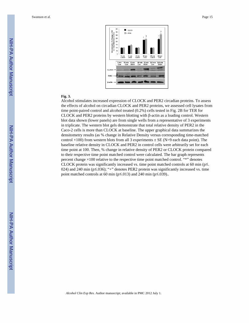
Fig. 3.Alcohol stimulates increased expression of CLOCK and PER2 circadian proteins. To assessthe effects of alcohol on circadian CLOCK and PER2 proteins, we assessed cell lysates fromtime point-paired control and alcohol treated (0.2%) cells tested in Fig. 2B for TER forCLOCK and PER2 proteins by western blotting with β-actin as a loading control. Westernblot data shown (lower panels) are from single wells from a representative of 3 experimentsin triplicate. The western blot gels demonstrate that total relative density of PER2 in theCaco-2 cells is more than CLOCK at baseline. The upper graphical data summarizes thedensitometry results (as % change in Relative Density versus corresponding time-matchedcontrol ×100) from western blots from all 3 experiments ± SE (N=9 each data point). Thebaseline relative density in CLOCK and PER2 in control cells were arbitrarily set for eachtime point at 100. Then, % change in relative density of PER2 or CLOCK protein comparedto their respective time point matched control were calculated. The bar graph representspercent change ×100 relative to the respective time point matched control. “*” denotesCLOCK protein was significantly increased vs. time point matched controls at 60 min (p≤.024) and 240 min (p≤.036); “+” denotes PER2 protein was significantly increased vs. timepoint matched controls at 60 min (p≤.013) and 240 min (p≤.039)..
Swanson et al. Page 15
Alcohol Clin Exp Res. Author manuscript; available in PMC 2012 July 1.
NIH
-PA Author Manuscript
NIH
-PA Author Manuscript
NIH
-PA Author Manuscript

Fig. 4.SiRNA inhibition of CLOCK protein expression prevents alcohol-inducedhyperpermeability of Caco-2 monolayers. To assess the role of alcohol-stimulation ofCLOCK protein expression in the alcohol-induced increased permeability of Caco-2monolayers, we inhibited CLOCK protein expression with siRNA specific for Clock andcompared the effects to cells also treated with alcohol but treated with control nontargetingsiRNA. Fig. 4A (upper panel) depicts permeability data which are presented as % ChangeTER (Ωxcm2) versus the “0” time point control for each condition. Data are means fromtriplicate wells in 3 experiments (total N=9) for each time point ± SE. Thus knock down ofClock gene by Clock specific siRNA prevented alcohol-induced monolayerhyperpermeability (drop in TER). * p≤.0001 vs. control siRNA; +p≤.001 vs. Clock siRNA+EtOH. Fig. 4B middle panel shows western blot data from lysates from 4 representativeinserts for each treatment group confirming that cells treated with Clock siRNA expressedsignificantly lower levels of CLOCK protein. Figure 4B lower histogram shows thesummarized densitometry data for all western blots (N=9 inserts each group, each timepoint) confirming knockdown of CLOCK protein by about 70% in Clock siRNA and
Swanson et al. Page 16
Alcohol Clin Exp Res. Author manuscript; available in PMC 2012 July 1.
NIH
-PA Author Manuscript
NIH
-PA Author Manuscript
NIH
-PA Author Manuscript

alcohol-treated cells compared to alcohol-treated control siRNA cells. Histogram data areexpressed as % Relative Density ± SE versus the control siRNA treated cells not treatedwith alcohol (set as 100%). *p≤ .05 vs. control siRNA; #p≤.001vs. control siRNA + EtOH.Fig. 4C shows Caco-2 monolayer permeability data for FSA permeability for cells treatedwith alcohol ± siRNA for CLOCK as in Fig. 4A above shown as % change in FSA vs.control (control siRNA treated) cells for each data point.*p≤.05 vs. control siRNA + EtOH.Data are means ± SE for 3 experiments in triplicate (9 data points each).
Swanson et al. Page 17
Alcohol Clin Exp Res. Author manuscript; available in PMC 2012 July 1.
NIH
-PA Author Manuscript
NIH
-PA Author Manuscript
NIH
-PA Author Manuscript

Fig. 5.SiRNA inhibition of PER2 protein expression prevents alcohol-induced hyperpermeabilityof Caco-2 monolayers. To assess the role of alcohol-stimulation of PER2 protein expressionin the alcohol-induced increased permeability of Caco-2 monolayers, we inhibited PER2protein expression with siRNA specific for Per2 and compared the effects to cells alsotreated with alcohol but treated with control siRNA. Fig. 5A (upper panel) depictspermeability data which are presented as % Change TER (Ω/cm2). TER data are presentedas % change versus the “0” time point control for each condition. Data are means fromtriplicate wells in 3 experiments (total N=9) for each time point ± SE. *p≤0.001 vs. ControlsiRNA; #p≤0.001 vs. Per2 siRNA+ EtOH Fig. 5B middle panel shows data from lysatesfrom 4 representative inserts for each treatment group confirming that cells treated withPer2 siRNA expressed significantly lower levels of PER2 protein. Figure 5B lowerhistogram shows the summarized densitometry data for all western blots (N=9 inserts eachgroup, each time point) confirming knockdown of PER2 protein by about 70% in Per2siRNA and alcohol-treated cells compared to alcohol-treated control siRNA cells. Data areexpressed as % Relative Density ± SE versus the control siRNA treated cells not treatedwith alcohol (set as 100%). * p≤0.03 vs. Control siRNA; # p≤0.001 vs. Control siRNA+
Swanson et al. Page 18
Alcohol Clin Exp Res. Author manuscript; available in PMC 2012 July 1.
NIH
-PA Author Manuscript
NIH
-PA Author Manuscript
NIH
-PA Author Manuscript

EtOH. Fig. 5C depicts FSA permeability data expressed as % change FSA flux vs. controlsiRNA treated cells for permeability of Caco-2 cells treated as in Fig. 5A above. Allpermeability data are means ± SE from triplicate wells in 3 experiments (9 data points).*p≤ .05 vs. control siRNA treated cells + EtOH.
Swanson et al. Page 19
Alcohol Clin Exp Res. Author manuscript; available in PMC 2012 July 1.
NIH
-PA Author Manuscript
NIH
-PA Author Manuscript
NIH
-PA Author Manuscript

Fig. 6.Fig. 6A. Intestinal permeability measured by sucralose urinary excretion of an oral bolus issignificantly increased in alcohol treated rats. Sprague-Dawley rats were gavaged twicedaily with a control-chow or an alcohol-chow slurry for a total daily dose of 6g/kg/dayalcohol for 10 weeks. Intestinal permeability was assessed at baseline and at 10 weeks byGC measurement of 6h urinary excretion of sucralose (a measure of whole gut permeability)after an oral dose of sucralose sugar mixture as described in Methods. Data are for N=9 rats
Swanson et al. Page 20
Alcohol Clin Exp Res. Author manuscript; available in PMC 2012 July 1.
NIH
-PA Author Manuscript
NIH
-PA Author Manuscript
NIH
-PA Author Manuscript

for both control and alcohol treated groups and are presented as mean % urinary excretion oforal dose of sucralose ± SE. Chronic alcohol consumption resulted in significantly (*p≤.003) increased sucralose urinary excretion.Fig. 6B. PER2 protein levels are significantly increased in the duodenum and proximalcolon of alcohol fed rats. Rats were gavaged daily for 10 weeks with either control-chow oralcohol-chow slurry for a dose of 6g/kg/day alcohol and assessed for intestinal permeabilityas shown in Fig. 6A. After 10 weeks these same rats were sacrificed and lysates of intestinaltissue from either duodenum (left) or proximal colon (right) were prepared for westernblotting with PER2 Ab or reprobed with anti-actin Ab as a control for equal loading. Theupper panels in Figure 6B show representative western blots for PER2 protein (and actincontrol) of tissue lysates from individual representative rats from either the control (Con) oralcohol-fed group. As seen in the summarized densitometry data for all rats (N=9 eachgroup) shown in lower Fig. 6B, PER2 protein was significantly increased in the intestinaltissue of alcohol fed rats compared to controls for both the duodenum (*p≤.008) and theproximal colon (*p≤.012) tissue.
Swanson et al. Page 21
Alcohol Clin Exp Res. Author manuscript; available in PMC 2012 July 1.
NIH
-PA Author Manuscript
NIH
-PA Author Manuscript
NIH
-PA Author Manuscript