Right ventricular failure secondary to chronic overload in congenital heart diseases: Benefits of...
10
Right ventricular failure secondary to chronic overload in congenital heart diseases: Benefits of cell therapy using human embryonic stem cell–derived cardiac progenitors Virginie Lambert, MD, PhD, a,b,c Elodie Gouadon, PhD, a,b Andr e Capderou, MD, PhD, a,b Emmanuel Le Bret, MD, PhD, c Mohamed Ly, MD, c Sylvie Dinanian, MD, d Jean-Francois Renaud, PhD, a,b Michel Puc eat, PhD, e and Catherine R€ ucker-Martin, PhD a,b Objective: Despite the increasing incidence of right ventricular (RV) failure in adult patients with congenital heart disease, current therapeutic options are still limited. By contrast to left-heart diseases, cell-based myocardial regeneration applied to the right ventricle is poorly studied, even though it may be a therapeutic solution. As human embryonic stem cell–derived cardiac progenitors seem to be good candidates owing to their proliferation capacity, our aim was to assess, in a large animal model of overloaded RV dysfunction, the feasibility and effects of such a cell therapy. Methods: Human MesP1 þ /SSEA-1 þ cardiogenic mesodermal cells were administered using multiple intramyocardial injections 4 months after a surgical procedure mimicking the repaired tetralogy of Fallot, and their effects were observed 3 months later on hemodynamic, rhythmic, and histologic parameters. Results: All pigs (sham n ¼ 6, treated n ¼ 6) survived without complication, and cell therapy was clinically well tolerated. Although functional, contractility, and energetics parameters evolved similarly in both groups, benefits regarding arrhythmic susceptibility were observed in the treated group, associated with a significant decrease of peri-myocyte fibrosis (5.71% 2.49% vs 12.12% 1.85%; P <.01) without interstitial fibrosis change (5.18% 0.81% vs 5.49% 1.01%). Such a decrease could be related to paracrine effects, as no human cells could be detected within the myocardium. Conclusions: Cell therapy using intramyocardial injections of human MesP1 þ /SSEA-1 þ cardiogenic mesodermal cells seems to have benefits regarding overloaded RV tissue remodeling and arrhythmic susceptibility, but this mode of administration is not sufficient to obtain a significant improvement in RV function. (J Thorac Cardiovasc Surg 2015;-:1-8) Supplemental material is available online. Right ventricular (RV) failure remains a major problem in the long-term follow-up of patients with congenital heart disease, leading to impairment of functional status, severe arrhythmias, and premature death. 1 This RV dysfunction occurs in young adults as a consequence of chronic pressure and volume overload secondary to procedures that had early success, in patients with either 2 ventricles in which the right ventricle was abnormal (eg, tetralogy of Fallot [TOF]), or was the dominant or systemic ventricle (eg, hypoplastic left heart syndrome). 2 In these patients, more than 50% have a risk of heart failure after age 30 years 1 Moreover, RV function is a major prognostic factor of pulmonary artery hypertension, regardless of etiology. 3 Consequently, management of RV failure was identified as a priority in cardiology research. 4 By contrast to left ventricular failure, clinical manage- ment of patients with RV failure remains challenging: drugs or resynchronization give variable benefits, 5 and surgical procedures such as valve replacement or repair may delay RV failure, but may fail to improve altered RV function. At the end-stage of heart failure, cardiac transplantation is mandated, but its application is restricted by the availability of donor organs. Cell-based myocardial repair may be an alternative approach. Human embryonic stem cell (ESC) engraftments were successfully attempted in left ventricular From the Medical Research Department, a Centre Chirurgical Marie Lannelongue, Inserm UMR_S999, LabEx LERMIT, Le Plessis-Robinson; Universit e Paris Sud, b DHU Thorax Innovation, Le Kremlin-Bic^ etre; Department of Pediatric and Congenital Heart Diseases, c Centre Chirurgical Marie Lannelongue, Le Plessis-Robinson; D epartement de Cardiologie, d Centre Hospitalier Antoine B ecl ere, Clamart; and Inserm UMR_S910, e Facult e de M edecine de la Timone, Marseille, France. Supported by the Association Franc ¸aise contre les Myopathies, grants numbers 12063, 13056, and 13968, Evry, France; the Agence Nationale pour la Recherche, grant number ANR-06-MRAR-041-03 Cardiostem, Paris, France; and Field of Major Interest STEM Pole 2008-2010, IDF Paris, France. Disclosures: Authors have nothing to disclose with regard to commercial support. Received for publication June 27, 2014; revisions received Oct 7, 2014; accepted for publication Nov 16, 2014. Address for reprints: Virginie Lambert, MD, PhD, Centre Chirurgical Marie Lannelongue, 133 Ave de la R esistance, 92350 Le Plessis-Robinson, France (E-mail: [email protected]). 0022-5223/$36.00 Copyright Ó 2015 by The American Association for Thoracic Surgery http://dx.doi.org/10.1016/j.jtcvs.2014.11.033 The Journal of Thoracic and Cardiovascular Surgery c Volume -, Number - 1 Lambert et al Congenital Heart Disease CHD
Transcript of Right ventricular failure secondary to chronic overload in congenital heart diseases: Benefits of...
Lambert et al Congenital Heart Disease
Right ventricular failure secondary to chronic overload in congenitalheart diseases: Benefits of cell therapy using human embryonic stemcell–derived cardiac progenitors
Virginie Lambert, MD, PhD,a,b,c Elodie Gouadon, PhD,a,b Andr�e Capderou, MD, PhD,a,b
Emmanuel Le Bret, MD, PhD,c Mohamed Ly, MD,c Sylvie Dinanian, MD,d
Jean-Francois Renaud, PhD,a,b Michel Puc�eat, PhD,e and Catherine R€ucker-Martin, PhDa,b
From th
Inser
Sud,b
and
Le P
B�ecl�e
Mars
Support
1206
grant
Majo
Disclos
Receive
publi
Address
Lann
(E-m
0022-52
Copyrig
http://dx
CHD
Objective: Despite the increasing incidence of right ventricular (RV) failure in adult patients with congenitalheart disease, current therapeutic options are still limited. By contrast to left-heart diseases, cell-basedmyocardial regeneration applied to the right ventricle is poorly studied, even though it may be a therapeuticsolution. As human embryonic stem cell–derived cardiac progenitors seem to be good candidates owing to theirproliferation capacity, our aim was to assess, in a large animal model of overloaded RV dysfunction, thefeasibility and effects of such a cell therapy.
Methods: Human MesP1þ/SSEA-1þ cardiogenic mesodermal cells were administered using multipleintramyocardial injections 4 months after a surgical procedure mimicking the repaired tetralogy of Fallot,and their effects were observed 3 months later on hemodynamic, rhythmic, and histologic parameters.
Results:All pigs (sham n¼ 6, treated n¼ 6) survived without complication, and cell therapy was clinically welltolerated. Although functional, contractility, and energetics parameters evolved similarly in both groups,benefits regarding arrhythmic susceptibility were observed in the treated group, associated with a significantdecrease of peri-myocyte fibrosis (5.71% � 2.49% vs 12.12% � 1.85%; P<.01) without interstitial fibrosischange (5.18% � 0.81% vs 5.49% � 1.01%). Such a decrease could be related to paracrine effects, as nohuman cells could be detected within the myocardium.
Conclusions: Cell therapy using intramyocardial injections of human MesP1þ/SSEA-1þ cardiogenicmesodermal cells seems to have benefits regarding overloaded RV tissue remodeling and arrhythmicsusceptibility, but this mode of administration is not sufficient to obtain a significant improvement in RVfunction. (J Thorac Cardiovasc Surg 2015;-:1-8)
Supplemental material is available online.
Right ventricular (RV) failure remains a major problem inthe long-term follow-up of patients with congenital heart
e Medical Research Department,a Centre Chirurgical Marie Lannelongue,
m UMR_S999, LabEx LERMIT, Le Plessis-Robinson; Universit�e Paris
DHU Thorax Innovation, Le Kremlin-Bicetre; Department of Pediatric
Congenital Heart Diseases,c Centre Chirurgical Marie Lannelongue,
lessis-Robinson; D�epartement de Cardiologie,d Centre Hospitalier Antoine
re, Clamart; and Inserm UMR_S910,e Facult�e de M�edecine de la Timone,
eille, France.
ed by the Association Francaise contre les Myopathies, grants numbers
3, 13056, and 13968, Evry, France; the Agence Nationale pour la Recherche,
number ANR-06-MRAR-041-03 Cardiostem, Paris, France; and Field of
r Interest STEM Pole 2008-2010, IDF Paris, France.
ures: Authors have nothing to disclose with regard to commercial support.
d for publication June 27, 2014; revisions received Oct 7, 2014; accepted for
cation Nov 16, 2014.
for reprints: Virginie Lambert, MD, PhD, Centre Chirurgical Marie
elongue, 133 Ave de la R�esistance, 92350 Le Plessis-Robinson, France
ail: [email protected]).
23/$36.00
ht � 2015 by The American Association for Thoracic Surgery
.doi.org/10.1016/j.jtcvs.2014.11.033
The Journal of Thoracic and C
disease, leading to impairment of functional status, severearrhythmias, and premature death.1 This RV dysfunctionoccurs in young adults as a consequence of chronic pressureand volume overload secondary to procedures that had earlysuccess, in patients with either 2 ventricles in which theright ventricle was abnormal (eg, tetralogy of Fallot[TOF]), or was the dominant or systemic ventricle(eg, hypoplastic left heart syndrome).2 In these patients,more than 50% have a risk of heart failure after age30 years1 Moreover, RV function is a major prognosticfactor of pulmonary artery hypertension, regardless ofetiology.3 Consequently, management of RV failure wasidentified as a priority in cardiology research.4
By contrast to left ventricular failure, clinical manage-ment of patients with RV failure remains challenging: drugsor resynchronization give variable benefits,5 and surgicalprocedures such as valve replacement or repair may delayRV failure, but may fail to improve altered RV function.At the end-stage of heart failure, cardiac transplantation ismandated, but its application is restricted by the availabilityof donor organs. Cell-based myocardial repair may be analternative approach. Human embryonic stem cell (ESC)engraftments were successfully attempted in left ventricular
ardiovascular Surgery c Volume -, Number - 1

Abbreviations and AcronymsDNA ¼ desoxuribose nucleic acidECG ¼ electrocardiogramESC ¼ embryonic stem cellHD ¼ high-densityPVS ¼ programmed ventricular stimulationRV ¼ right ventricularTOF ¼ tetralogy of Fallot
Congenital Heart Disease Lambert et al
CHD
myocardium after ischemic injuries.6 Regarding the rightventricle, cell therapy using myoblastic7 or cord-bloodstem cells8,9 has been attempted but yielded poor results.
Treating RV failure, in contrast to ischemic left ventricle,by cell therapy, has to take into account not only thepostoperative scarred area, but also the specific geometryand physiologic processes of the right ventricle. Indeed,RV chronic overload alters the entire RV myocardium.10,11
This extensive alteration requires a substantial number ofcells to treat, and potentially induce a functionalimprovement of, the RV myocardium. Among multiplecell types considered as potential sources of cardiacprogenitors, some did establish a functional coupling withthe host myocardium.6,12,13 ESCs derived from the innermass of the blastocyst possess this capacity and are ableto proliferate, differentiate in vivo into mature cardiacmyocytes, and repopulate significant regions of thedamaged myocardium.
A proof of concept has been reported in a nonhuman-primate model of infarcted myocardium.6,13 In one ofthese models,13 the maturation and differentiation ofcardiac progenitors into ventricular myocytes seemed tobe optimal in areas composed of both fibrosis and damagedcardiac fibers. Instances in which such a structuralremodeling constitutes a favorable environment forstem-cell grafting has been observed in patients withoverloaded RV dysfunction,14 suggesting that cardiacprogenitor cell therapy may be applied in this indication.
The purpose of our study was to evaluate the feasibilityand effects of a cell therapy using human ESC-derivedmesodermal cardiogenic cells in our porcine model ofchronic overloaded RV dysfunction.10 The impact of thistreatment on hemodynamic and rhythmic parameters wasevaluated throughout the follow-up; the fate of cardiacprogenitors and the RV structural remodeling wereassessed.
METHODSExperimental Design
Twelve Landrace male piglets were studied in accordance with
European Union regulations (Directive 86/609 EEC). This study
was approved by the French Ministry of Agriculture (approval No.
B92-019-01) and the Committee on Ethics of Animal Experiments
2 The Journal of Thoracic and Cardiovascular Surger
CEEA26 CAPSud. Animals underwent an electrocardiogram (ECG) and
hemodynamic evaluation at 3 points in the experiment: baseline, and 4
and 7 months of follow-up. The surgical procedure mimicking repaired
TOF was performed after the baseline step. Briefly, an enlargement of
the RV outflow tract by a polytetrafluoroethylene patch, excision of 1
pulmonary valve leaflet, and a pulmonary artery banding were
performed.10 The rhythmic risk was evaluated throughout the
cell-therapy period. Histologic analyses were performed on each animal
at the end of the procedure.
Cardiac Progenitor Characteristics andImplantation into the RV Myocardium
Human MesP1þ (mesoderm posterior 1)/SSEA-1þ (stage-specific
embryonic antigen-1; CD15) cardiac progenitor cells derived from the
HUES-24 (human embryonic stem) cell line were used. Briefly,
HUES-24 cells were treated with 100-ng/ml Wnt3a for 24 hours and
then with 10-ng/ml BMP2 for 3 days; cells were sorted with a biotin
anti-CD15 antibody (Exbio, CliniSciences, Nanterre, France) and
antibiotin conjugated beads (Miltenyi Biotec, Paris, France). RNAs
extracted from both CD15þ and CD15� cells were submitted to reverse
transcriptase. Complementary DNAs were run in a real-time polymerase
chain reaction, as described elsewhere.13 Cell characteristics are presented
in Figure 1. The selection excludes undifferentiated or early neural stem
cells, and no teratoma or proliferative cell foci formation was detected
on explanted heart, lung, or liver using scanner multislices imaging
(Somatom Definition Flash, Siemens Healthcare, St. Denis, France).
Cell transplantation was performed 4 months after surgery through a
right thoracotomy approach. Animals received either medium containing
mesodermal cardiogenic cells (treated group: n ¼ 6) or vehicle alone
(sham group: n ¼ 6). The total bolus (107 cells) was injected into the RV
free wall at 20 separate injection sites; 20 other injections were made in
a high-density (HD) area of 1 cm2 identified by nonabsorbable sutures,
using a 28-gauge needle (3 mm deep, 25 ml/site) connected to a
mesotherapy pistol (DHN-2, Techdent, Sallanches, France).
Hydrocortisone (1 mg/kg) was injected intravenously before closing, to
reduce inflammation. All animals were immunosuppressed by tacrolimus
by mouth (0.3 mg/kg/day, plasmatic level: see Table 1).
ECG and Rhythm StudyA 12-lead surface ECG was recorded, and QRS duration was
analyzed.10 An insertable recorder (Reveal, Medtronic France SAS,
Boulogne-Billancourt, France) was implanted subcutaneously under the
left scapula at the time of cell implantation, to record the heart’s rhythm
until the end of follow-up. This cardiac monitor was programmed to record
tachycardia>200/minutes, during at least 6 complexes. As a last step, a
stored ECG was collected by percutaneous interrogation, and a
programmed ventricular stimulation (PVS) was carried out with a
quadripolar catheter inserted into the RV apex through the femoral vein.
Standard clinical PVS protocols were employed, including application
of single, double, and triple extrastimuli of increasing prematurity until
reaching the RV refractory period, after a sequence of 8 conditioning
stimuli. The heart was then challenged 3 times with a sequence of 8,
followed by a single extrastimulus at compulsory rhythms of 100, 120,
and 150/minutes. If no ventricular tachycardia was induced, this procedure
was repeated to apply 3 challenges with double, and if necessary
triple, extrastimuli. The PVS was considered positive if these
challenges produced sustained ventricular tachycardia (>30s) or
ventricular fibrillation.
Hemodynamic StudyQuantification of RV overload and contractile performance of RV
myocardium were assessed by the conductance catheter technique. Briefly,
the conductance catheter was inserted in the right ventricle through the
y c - 2015
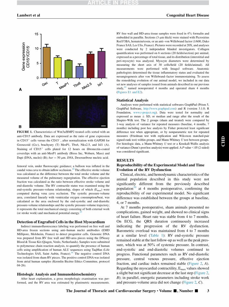
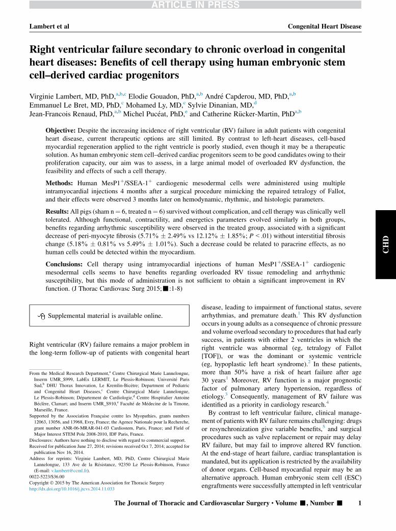
FIGURE 1. Characteristics of Wnt3a/BMP2 treated cells sorted with an
anti-CD15 antibody. Data are expressed as the ratio of gene expression
in CD15þ cells versus the CD15�, after normalization with GAPDH for
Goosecoid (Gsc), brachyury (T) MesP1, Tbx6, Nkx2.5, and Isl1 (A).
Staining of CD15þ cells plated for 12 hours on fibronectin-coated
coverslips with an anti-MesP1 antibody (Bioss Inc, Woburn, Mass) and
Dapi (DNA, nuclei) (B); bar ¼ 50 mm. DNA, Desoxuribose nucleic acid.
Lambert et al Congenital Heart Disease
CHD
femoral vein, under fluoroscopic guidance; a balloon was inflated in the
caudal vena cava to obtain inflow occlusion.10 The effective stroke volume
was calculated as the difference between the total stroke volume and the
measured volume of the pulmonary regurgitation. The effective ejection
fraction was calculated as the ratio between effective stroke volume and
end-diastolic volume. The RV contractile status was examined using the
end-systolic pressure-volume relationship, slopes of which (Emax) were
computed during vena cava occlusion. The systolic pressure-volume
area, correlated linearly with ventricular oxygen consumption/beat, was
calculated as the area enclosed by the end-systolic and end-diastolic
pressure-volume relationships and the systolic pressure-volume trajectory;
it represents the total mechanical energy consisting of both external work
(or stroke work) and mechanical potential energy.15
Detection of Engrafted Cells in the HostMyocardiumIndirect immunofluorescence labeling was performed on free wall and
HD-area frozen sections using anti–human nuclei antibodies (EMD
Millipore, Molsheim, France) to detect progenitor cells. Genomic DNA
was prepared from RV free wall and HD-area pieces using the DNeasy
Blood & Tissue Kit (Qiagen, Venlo, Netherlands). Samples were submitted
to polymerase chain reaction analysis, to quantify the presence of human
cells using amplification of human ALU sequences using TaqMan (Life
Technologies SAS, Saint Aubin, France).16 The negative control DNA
was isolated from sham RV pieces. The positive control DNAwas isolated
from atrial human samples (Kremlin Bicetre Ethics Committee, protocol
04-26).
Histologic Analysis and ImmunohistochemistryAfter heart explantation, a gross morphologic examination was per-
formed, and the RV area was estimated by planimetric measurements.
The Journal of Thoracic and C
RV free wall and HD-area tissue samples were fixed in 4% formalin and
embedded in paraffin. Sections (3-mm thick) were stained with Picrosirius
Red F3BA, hematein/eosin, or an anti–von Willebrand factor (1/600, Dako
France SAS, Les Ulis, France). Pictures were recorded at 20X, and analyses
were conducted by 2 independent blinded investigators. Collagen
quantification was performed on 6 sections (20 fields/section) per animal,
expressed as a percentage of total tissue, and its distribution (interstitial and
peri-myocyte) was analyzed. Myocyte diameters were determined by
measuring the short axis of 30 cells/field (20 fields/animal). All
measurements were performed with ImageJ software. Anatomic
pathologists determined the tissue inflammatory status and evaluated the
neoangiogenesis after von Willebrand–factor immunostaining. To assess
the remodeling evolution of our animal model, we included in our data
set new analyses of samples issued from animals described in our previous
study,10 named nonoperated 4 months and operated sham 4 months
(Figures E1 and E2).
Statistical AnalysisAnalyses were performed with statistical softwares GraphPad (Prism 5,
GraphPad Software, http://www.graphpad.com) and R (version 3.1.0; R
Foundation, www.r-project.org). Data were tested for normality and
expressed as mean � SD, or median and range after the result of the
Shapiro-Wilk test. The 2 groups (sham and treated) were compared by
1-way analysis of variance for repeated measures (baseline, 4 months, 7
months) including post hoc analysis by Fisher protected least significant
difference test when appropriate, or by nonparametric test for repeated
measures (Friedman test with replication and Wilcoxon matched-pair
signed-rank test) within groups, and Mann-Whitney U test between groups.
For histologic data, a Mann-Whitney U test or a Kruskall-Wallis analysis
of variance (Dunn’s post hoc analysis) were applied.AP value<.05 (2-sided)
was considered significant.
RESULTSReproducibility of the Experimental Model and TimeEvolution of the RV DysfunctionClinical, electric, and hemodynamic characteristics of the
animal population described in this study were notsignificantly different from the previously describedpopulation10 at 4 months postoperative, confirming thereproducibility of our experimental model. No significantdifference was established between the groups at baseline,4, or 7 months.At 7 months postoperative, sham animals presented no
complications, gained weight, and showed no clinical signsof heart failure. Heart rate was stable from 4 to 7 months.On ECG, the QRS duration continuously increasedindicating the progression of the RV dysfunction.Barometric overload was maintained from 4 to 7 monthsat a similar level (Table 1): RV end-systolic pressureremained stable at the last follow-up aswell as the peak pres-sure, which was at 50% of systemic pressure. In contrast,end-systolic and end-diastolic volumes continued toprogress. Functional parameters such as RV end-diastolicpressure, central venous pressure, effective ejectionfraction, and cardiac index remained stable (Figure 2, A).Regarding the myocardial contractility, Emax values showeda slight but not significant decrease at the last step (Figure 2,B); in parallel, energetic parameters including stroke workand pressure-volume area did not change (Figure 2, C).
ardiovascular Surgery c Volume -, Number - 3
TABLE 1. Population, ECG, and intracardiac pressures and volumes in sham and treated groups at each step of experimentation
Group
Baseline
Follow-up
At 4 months At 7 months
Sham n ¼ 6 Treated n ¼ 6 Sham n ¼ 6 Treated n ¼ 6 Sham n ¼ 6 Treated n ¼ 6
Population characteristics
Age (d) 69.3 � 11.8 75.0 � 7.9 201.2 � 7.1* 194.8 � 8.1* 303.7 � 16.3*,y 289.2 � 12.3*,yWeight (kg) 21.0 � 3.6 21.0 � 1.5 55.9 � 8.0* 58.9 � 6.2* 73.1 � 8.1*,y 71.5 � 11.9*,yBody length (cm) 67.0 � 4.9 65.5 � 4.6 92.5 � 4.0* 92.2 � 4.9* 102.3 � 4.3*,y 103.5 � 10.3*,yBody surface area (m2) 0.62 � 0.08 0.61 � 0.04 1.20 � 0.11* 1.22 � 0.09* 1.44 � 0.11*,y 1.43 � 0.19*,yTacrolimus plasmatic level (ng/ml) 6.0 � 1.6 5.9 � 3.2 5.7 � 3.8 6.0 � 3.3
ECG
Heart rate (beats/min) 116 � 4 118 � 14 91 � 18* 97 � 11* 84 � 20* 84 � 11*
QRS duration (ms) 59.7 � 7.5 59.7 � 6.9 72.3 � 3.2* 71.0 � 9.9* 85.2 � 9.0*,y 82.0 � 7.5*,yIntracardiac pressures
Mean aortic blood pressure (mm Hg) 58 � 9 66 � 9 85 � 17* 85 � 11* 80 � 11*,y 74 � 8*,ySystolic aortic blood pressure (mm Hg) 77 � 12 85 � 10 103 � 18* 104 � 15* 100 � 11*,y 90 � 9*,yCentral venous pressure (mm Hg) 1.3 � 1.0 1.5 � 0.8 4.8 � 2.0* 4.7 � 1.9* 5.3 � 3.0* 4.8 � 2.1*
RV pressure (mm Hg)
Peak 20.3 � 5.3 22.3 � 1.6 50.5 � 27.2* 59.2 � 14.1* 44.4 � 13.9* 56.8 � 11.7*
End-systolic 11.3 � 3.7 13.2 � 1.2 42.5 � 25.8* 49.2 � 9.3* 37.0 � 14.6* 50.8 � 12.6*
End-diastolic 1.5 � 1.5 1.7 � 0.8 5 � 2.9* 5.0 � 3.2* 6.5 � 3.7* 5 � 2.5*
Ratio systolic pressure RV/Aorta (%) 27 � 8 27 � 5 47 � 17* 57 � 10* 45 � 12* 64 � 15*
Intracardiac volumes
End-diastolic volume (ml) 91 � 26 85 � 15 166 � 46* 148 � 24* 192 � 55*,y 208 � 59*,yEnd-systolic volume (ml) 45 � 19 39 � 9 80 � 39 67 � 20 83 � 37* 103 � 36*
Stroke volume (ml) 44 � 10 42 � 5 69 � 5* 71 � 1* 95 � 25* 87 � 20*
Fraction of regurgitation (%) 22 � 9 18 � 3 15 � 8 18 � 3
ECG, Electrocardiogram; RV, right ventricle. *P<.05 versus corresponding group at baseline. yP<.05, corresponding group at 7 months versus 4 months.
Congenital Heart Disease Lambert et al
CHD
Cell Therapy: Clinical Tolerance, HemodynamicEffects, and Rhythmic Impact
Transmyocardial injections were performed after asimilar length of time after the operation in each group(120 � 9 days in treated, 132 � 5 days in sham). Onlytransient arrhythmias were observed at the time of needlepenetration, and they were fully resolved after injections.No death occurred as a result of the cell injectionprocedure, or during the follow-up, and no severe adverseeffects occurred. Immunosuppression therapy was welltolerated. During the postinjection period, animals gainedweight (Table 1) and showed no clinical signs of either heartfailure or immune rejection. No difference between treatedand sham groups was observed in the RV dysfunctionevolution (Table 1). Functional characteristics, contractilityparameters, and energetics similarly evolved after eitherplacebo or cell injections (Figure 2).
On ECG, the QRS duration continuously increased,in similar fashion in both the sham and treated groups(Table 1). Despite this risk factor of arrhythmia, all treatedanimals showed normal long-duration Holter recordings,and no ventricular arrhythmia could be induced by PVS.In contrast, sinus pauses >2 seconds were detected onlong-duration Holter in 2 sham animals. In one of them, anonsustained ventricular tachycardia was induced twiceafter a single extrastimulus at a compulsory rhythm of
4 The Journal of Thoracic and Cardiovascular Surger
120/min and a ventricular fibrillation had occurred afterPVS. In the other animal, the PVS led to a transitory STdepression that appeared after a sequence of 8, followedby the single extrastimulus at a compulsory rhythm of150/min, suggesting an alteration of the myocardial reserve.This arrhythmic susceptibility supports the evolutioncapacities of our model in accordancewith clinical findings.
Fate of Injected Human Cells and Their Effect on RVTissue Remodeling
All animals were euthanized about 3 months afterinjections (94 � 12 days in treated, 103 � 11 days insham). Human cardiogenic mesodermal cells or cardiaccells have been sought in the RV free wall and HD areausing 2 different techniques. At the cellular level,immunolabelings against human nuclear antigen did notreveal the presence of human cells. Similarly, at the DNAlevel, polymerase chain reaction analyses of treated animalsamples did not establish the presence of human ALUsequences, thus indicating with a more sensitive approach,the lack of human cell survival at 3months (data not shown).
Regarding the histologic analysis, human cell injectionsdid not modify either RV area (115.1 � 17.3 cm2 intreated, 117.6 � 7.9 cm2 in sham) or myocyte diameters(23.2 � 1.2 mm in treated, 22.2 � 2.4 mm in sham). Noinflammatory infiltrates were observed in either group, in
y c - 2015
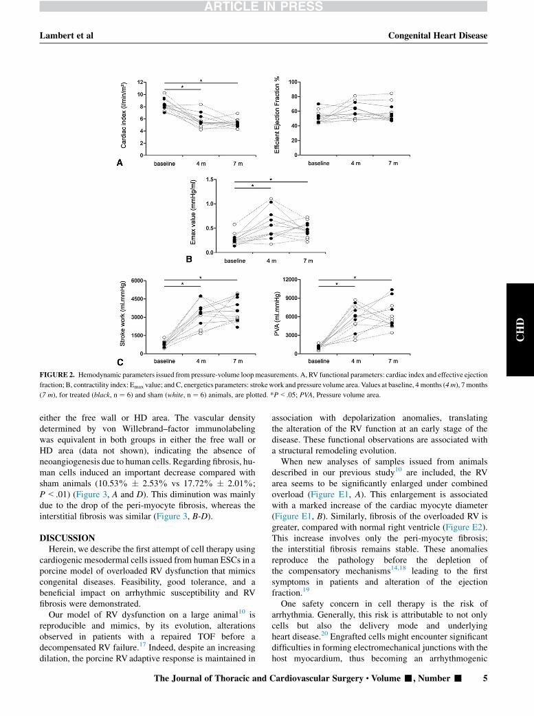
FIGURE 2. Hemodynamic parameters issued from pressure-volume loopmeasurements. A, RV functional parameters: cardiac index and effective ejection
fraction; B, contractility index: Emax value; and C, energetics parameters: strokework and pressure volume area. Values at baseline, 4 months (4 m), 7 months
(7 m), for treated (black, n ¼ 6) and sham (white, n ¼ 6) animals, are plotted. *P<.05; PVA, Pressure volume area.
Lambert et al Congenital Heart Disease
CHD
either the free wall or HD area. The vascular densitydetermined by von Willebrand–factor immunolabelingwas equivalent in both groups in either the free wall orHD area (data not shown), indicating the absence ofneoangiogenesis due to human cells. Regarding fibrosis, hu-man cells induced an important decrease compared withsham animals (10.53% � 2.53% vs 17.72% � 2.01%;P<.01) (Figure 3, A and D). This diminution was mainlydue to the drop of the peri-myocyte fibrosis, whereas theinterstitial fibrosis was similar (Figure 3, B-D).
DISCUSSIONHerein, we describe the first attempt of cell therapy using
cardiogenic mesodermal cells issued from human ESCs in aporcine model of overloaded RV dysfunction that mimicscongenital diseases. Feasibility, good tolerance, and abeneficial impact on arrhythmic susceptibility and RVfibrosis were demonstrated.
Our model of RV dysfunction on a large animal10 isreproducible and mimics, by its evolution, alterationsobserved in patients with a repaired TOF before adecompensated RV failure.17 Indeed, despite an increasingdilation, the porcine RVadaptive response is maintained in
The Journal of Thoracic and C
association with depolarization anomalies, translatingthe alteration of the RV function at an early stage of thedisease. These functional observations are associated witha structural remodeling evolution.When new analyses of samples issued from animals
described in our previous study10 are included, the RVarea seems to be significantly enlarged under combinedoverload (Figure E1, A). This enlargement is associatedwith a marked increase of the cardiac myocyte diameter(Figure E1, B). Similarly, fibrosis of the overloaded RV isgreater, compared with normal right ventricle (Figure E2).This increase involves only the peri-myocyte fibrosis;the interstitial fibrosis remains stable. These anomaliesreproduce the pathology before the depletion ofthe compensatory mechanisms14,18 leading to the firstsymptoms in patients and alteration of the ejectionfraction.19
One safety concern in cell therapy is the risk ofarrhythmia. Generally, this risk is attributable to not onlycells but also the delivery mode and underlyingheart disease.20 Engrafted cells might encounter significantdifficulties in forming electromechanical junctions with thehost myocardium, thus becoming an arrhythmogenic
ardiovascular Surgery c Volume -, Number - 5
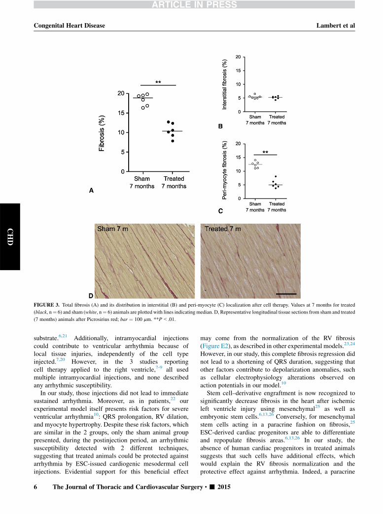
FIGURE 3. Total fibrosis (A) and its distribution in interstitial (B) and peri-myocyte (C) localization after cell therapy. Values at 7 months for treated
(black, n¼ 6) and sham (white, n¼ 6) animals are plotted with lines indicating median. D, Representative longitudinal tissue sections from sham and treated
(7 months) animals after Picrosirius red; bar ¼ 100 mm. **P<.01.
Congenital Heart Disease Lambert et al
CHD
substrate.6,21 Additionally, intramyocardial injectionscould contribute to ventricular arrhythmia because oflocal tissue injuries, independently of the cell typeinjected.7,20 However, in the 3 studies reportingcell therapy applied to the right ventricle,7-9 all usedmultiple intramyocardial injections, and none describedany arrhythmic susceptibility.
In our study, those injections did not lead to immediatesustained arrhythmia. Moreover, as in patients,22 ourexperimental model itself presents risk factors for severeventricular arrhythmia10: QRS prolongation, RV dilation,and myocyte hypertrophy. Despite these risk factors, whichare similar in the 2 groups, only the sham animal grouppresented, during the postinjection period, an arrhythmicsusceptibility detected with 2 different techniques,suggesting that treated animals could be protected againstarrhythmia by ESC-issued cardiogenic mesodermal cellinjections. Evidential support for this beneficial effect
6 The Journal of Thoracic and Cardiovascular Surger
may come from the normalization of the RV fibrosis(Figure E2), as described in other experimental models.23,24
However, in our study, this complete fibrosis regression didnot lead to a shortening of QRS duration, suggesting thatother factors contribute to depolarization anomalies, suchas cellular electrophysiology alterations observed onaction potentials in our model.10
Stem cell–derivative engraftment is now recognized tosignificantly decrease fibrosis in the heart after ischemicleft ventricle injury using mesenchymal25 as well asembryonic stem cells.6,13,26 Conversely, for mesenchymalstem cells acting in a paracrine fashion on fibrosis,25
ESC-derived cardiac progenitors are able to differentiateand repopulate fibrosis areas.6,13,26 In our study, theabsence of human cardiac progenitors in treated animalssuggests that such cells have additional effects, whichwould explain the RV fibrosis normalization and theprotective effect against arrhythmia. Indeed, a paracrine
y c - 2015

Lambert et al Congenital Heart Disease
CHD
effect of MesP1þ/SSEA-1þ–secreting vascular endothelialgrowth factor–A has been shown in a recent model oflimb ischemia.27 In addition, these paracrine effects,mediated through vascular endothelial growth factor, mayresult from a modulation of local inflammation,28 and/or,by analogy with mesenchymal stem cells, the secretion ofsoluble factors acting directly on matrix metalloproteinasesand matrix metalloproteinase endogenous inhibitorproduction by cardiac fibroblasts.25
The reason for the lack of grafted MesP1þ/SSEA-1þ
cardiac progenitors within the myocardium in ourstudy remains unclear, whereas the RV structuralremodeling constituted a favorable environment to cellengraftment.10,13 Blin and colleagues13 indicated successfulengraftments of the same cell type at 2 months aftertransmyocardial injections.13 Our results underline thelong-term survival failure of such cells. One explanationmay be the immune rejection of ESCs.29 Herein, theimmunosuppressive regimen was efficient, as plasmatictacrolimus levels revealed therapeutic values as recommen-ded in human after organ transplantation, and as noinflammatory infiltrates were detected during histologicanalyses. Another reason could be related to a hypoxicenvironment of grafted cells. Since RV outflow–tractdiseases were not corrected, the RV chronic pressureoverload was maintained, leading to a diminishedanatomic capillary vascular reserve30 that limited the bloodsupply to engrafted cells and affected cardiac progenitorsurvival.31
In contrast to the results of cell therapies applied toischemic models, no functional improvement was observed,despite the fibrosis normalization. As RV outlet tractdiseases were not corrected, benefits of the fibrosis decreasemight not have been sufficient to balance the deleteriouseffects of combined overload and lead to a detectablefunctional improvement. Regarding cell therapies appliedto the right ventricle, only Davies and colleagues9 reportedan enhancement of systolic RV function 1 month afterinjections of human cord–blood stem cells into neonatalpressure-loaded RV myocardium. However, thesefindings need to be interpreted cautiously, because asignificant RV function improvement was observed, thoughto a lesser degree, without stem cell transplantation,suggesting that the improvement may reflect homeometricadaptation.32 Others7,8 did not find any RV systolicfunctional improvement. Overall, these results underlinethe difficulties and the specificity of the cell therapytreatment necessitated by RV failure, compared withischemic left ventricular dysfunction.
CONCLUSIONSCell therapy using humanMesP1þ/SSEA-1þ cardiogenic
mesodermal cells applied to an overloaded right ventricle isfeasible in a large animal model, well tolerated, and has
The Journal of Thoracic and C
beneficial effects on fibrosis and arrhythmic susceptibility.Next objectives are, first, to improve the long-term survivalof ESC-derived cardiac progenitors using more-protectivedelivery modes and prosurvival factors33; and second, topromote their migration and differentiation into the RVmyocardium.
The authors thank Alain Chapel for ALU sequence analyses;Nicolas Raymond for his excellent histologic work; Dr Dorfm€ullerand Dr de Montpreville for anatomic-pathology expertise; Dr Stosfor his technical assistance; and the Surgical Research Staff:Benoit Decante, Bruno Baudet, Fr�ed�eric Seccatore, and AnaelleLe Breton for technical assistance and animal care.
We greatly acknowledge the Surgical Research Laboratory andMicroscopy Facility of the Centre Chirurgical Marie Lannelongueand the Bligny Farm.
References1. Norozi K, Wessel A, Alpers V, Arnhold JO, Geyer S, Zoege M, et al.
Incidence and risk distribution of heart failure in adolescents and adults
with congenital heart disease after cardiac surgery. Am J Cardiol. 2006;97:
1238-43.
2. Lopez L, Cohen MS, Anderson RH, Redington AN, Nykanen DG, Penny DJ,
et al. Unnatural history of the right ventricle in patients with congenitally
malformed hearts. Cardiol Young. 2010;20(Suppl 3):107-12.
3. Vonk-Noordegraaf A, Haddad F, Chin KM, Forfia PR, Kawut SM, Lumens J,
et al. Right heart adaptation to pulmonary arterial hypertension: physiology
and pathobiology. J Am Coll Cardiol. 2013;62(25 Suppl):D22-33.
4. Voelkel NF, Quaife RA, Leinwand LA, Barst RJ, McGoon MD, Meldrum DR,
et al. Right ventricular function and failure: report of a National Heart, Lung,
and Blood Institute working group on cellular and molecular mechanisms of
right heart failure. Circulation. 2006;114:1883-91.
5. Winter MM, Bouma BJ, Groenink M, Konings TC, Tijssen JG, van
Veldhuisen DJ, et al. Latest insights in therapeutic options for systemic right
ventricular failure: a comparison with left ventricular failure. Heart. 2009;95:
960-3.
6. Chong JJ, Yang X, Don CW, Minami E, Liu YW, Weyers JJ, et al. Human
embryonic-stem-cell-derived cardiomyocytes regenerate non-human primate
hearts. Nature. 2014;510:273-7.
7. Borenstein N, Jian Z, Fromont G, Bruneval P, Hekmati M, Behr L, et al.
Noncultured cell transplantation in an ovine model of right ventricular
preparation. J Thorac Cardiovasc Surg. 2005;129:1119-27.
8. Yerebakan C, Sandica E, Prietz S, Klopsch C, Ugurlucan M, Kaminski A, et al.
Autologous umbilical cord blood mononuclear cell transplantation preserves
right ventricular function in a novel model of chronic right ventricular volume
overload. Cell Transplant. 2009;18:855-68.
9. Davies B, Elwood NJ, Li S, Cullinane F, Edwards GA, Newgreen DF, et al.
Human cord blood stem cells enhance neonatal right ventricular function in an
ovine model of right ventricular training. Ann Thorac Surg. 2010;89:585-93.
e581-4.
10. Lambert V, Capderou A, Le Bret E, Rucker-Martin C, Deroubaix E, Gouadon E,
et al. Right ventricular failure secondary to chronic overload in congenital heart
disease: an experimental model for therapeutic innovation. J Thorac Cardiovasc
Surg. 2010;139:1197-204. e1191.
11. Kozak MF, Redington A, Yoo SJ, Seed M, Greiser A, Grosse-Wortmann L.
Diffuse myocardial fibrosis following tetralogy of Fallot repair: a t1 mapping
cardiac magnetic resonance study. Pediatr Radiol. 2014;44:403-9.
12. Bolli R, Tang XL, Sanganalmath SK, Rimoldi O, Mosna F, Abdel-Latif A, et al.
Intracoronary delivery of autologous cardiac stem cells improves cardiac
function in a porcine model of chronic ischemic cardiomyopathy. Circulation.
2013;128:122-31.
13. Blin G, Nury D, Stefanovic S, Neri T, Guillevic O, Brinon B, et al. A purified
population of multipotent cardiovascular progenitors derived from primate
pluripotent stem cells engrafts in postmyocardial infarcted nonhuman primates.
J Clin Invest. 2010;120:1125-39.
ardiovascular Surgery c Volume -, Number - 7

Congenital Heart Disease Lambert et al
CHD
14. Richart ALX, Neri T. Ventricular fibrosis suggested by cardiovascular magnetic
resonance in adults with repaired tetralogy of fallot and its relationship to adverse
markers of clinical outcome. Circulation. 2006;113:405-13.
15. Takaoka H, Takeuchi M, Odake M, Yokoyama M. Assessment of myocardial
oxygen consumption (vo2) and systolic pressure-volume area (pva) in human
hearts. Eur Heart J 1992;13 Suppl E:85-90.
16. Francois S, Mouiseddine M, Mathieu N, Semont A, Monti P, Dudoignon N, et al.
Human mesenchymal stem cells favour healing of the cutaneous radiation
syndrome in a xenogenic transplant model. Ann Hematol. 2007;86:1-8.
17. Haddad F, Doyle R, Murphy DJ, Hunt SA. Right ventricular function in
cardiovascular disease, part ii: pathophysiology, clinical importance, and
management of right ventricular failure. Circulation. 2008;117:1717-31.
18. Chowdhury UK, Sathia S, Ray R, Singh R, Pradeep KK, Venugopal P.
Histopathology of the right ventricular outflow tract and its relationship to
clinical outcomes and arrhythmias in patients with tetralogy of Fallot. J Thorac
Cardiovasc Surg. 2006;132:270-7.
19. Geva T. Indications and timing of pulmonary valve replacement after tetralogy of
Fallot repair. Semin Thorac Cardiovasc Surg Pediatr Card Surg Annu.
2006;11-22.
20. Menasche P. Stem cell therapy for heart failure: Are arrhythmias a real safety
concern? Circulation. 2009;119:2735-40.
21. Laflamme MA, Murry CE. Regenerating the heart. Nature Biotechnol. 2005;23:
845-56.
22. Gatzoulis MA, Till JA, Somerville J, Redington AN. Mechanoelectrical
interaction in tetralogy of fallot. Qrs prolongation relates to right ventricular
size and predicts malignant ventricular arrhythmias and sudden death.
Circulation. 1995;92:231-7.
23. Rucker-Martin C, Milliez P, Tan S, Decrouy X, Recouvreur M, Vranckx R, et al.
Chronic hemodynamic overload of the atria is an important factor for
gap junction remodeling in human and rat hearts. Cardiovasc Res. 2006;72:
69-79.
24. Milliez P, Gomes S, Champ-Rigot L, Callebert J, Samuel JL, Delcayre C. Effects
of spironolactone alone and in addition to a beta-blocker on myocardial
8 The Journal of Thoracic and Cardiovascular Surger
histological and electrical remodeling in chronic severe failing rat hearts.
J Cardiovasc Pharmacol. 2012;60:315-21.
25. Mias C, Lairez O, Trouche E, Roncalli J, Calise D, Seguelas MH, et al.
Mesenchymal stem cells promote matrix metalloproteinase secretion by cardiac
fibroblasts and reduce cardiac ventricular fibrosis after myocardial infarction.
Stem Cells. 2009;27:2734-43.
26. Puymirat E, Geha R, Tomescot A, Bellamy V, Larghero J, Trinquart L, et al. Can
mesenchymal stem cells induce tolerance to cotransplanted human embryonic
stem cells? Mol Ther. 2009;17:176-82.
27. Richart ALX, N�eri T, Howangyin K, Gu�erin C, Ngkelo A, Bakker W, et al.
Microrna-21 coordinates human multipotent cardiovascular progenitors:
therapeutic potential and post-ischemic revascularization. Stem Cells. 2014;32:
2908-22.
28. Crisostomo PR, Abarbanell AM, Wang M, Lahm T, Wang Y, Meldrum DR.
Embryonic stem cells attenuate myocardial dysfunction and inflammation after
surgical global ischemia via paracrine actions. Am J Physiol Heart Circ Physiol.
2008;295:H1726-35.
29. Calderon D, Planat-Benard V, Bellamy V, Vanneaux V, Kuhn C, Peyrard S,
et al. Immune response to human embryonic stem cell-derived cardiac
progenitors and adipose-derived stromal cells. J Cell Mol Med. 2012;16:
1544-52.
30. Marino TA, Kent RL, Uboh CE, Fernandez E, Thompson EW, Cooper GT.
Structural analysis of pressure versus volume overload hypertrophy of cat right
ventricle. Am J Physiol. 1985;249(2 Pt 2):H371-9.
31. Wollert KC, Drexler H. Clinical applications of stem cells for the heart. Circ Res.
2005;96:151-63.
32. de VroomenM, Cardozo RH, Steendijk P, van Bel F, Baan J. Improved contractile
performance of right ventricle in response to increased rv afterload in newborn
lamb. Am J Physiol Heart Circ Physiol. 2000;278:H100-5.
33. LaflammeMA, Chen KY, Naumova AV,Muskheli V, Fugate JA, Dupras SK, et al.
Cardiomyocytes derived from human embryonic stem cells in pro-survival
factors enhance function of infarcted rat hearts. Nat Biotechnol. 2007;25:
1015-24.
y c - 2015

FIGURE E1. Evolution of the right ventricle area (A) and cardiac myocyte diameters (B) during right ventricle chronic overload. Representative trans-
versal tissue sections from nonoperated and operated sham (4 months), and operated sham (7 months) animals after Picrosirius red; bar ¼ 100 mm (C).
*P<.05, **P<.01.
FIGURE E2. Evolution of the total fibrosis (A) and its distribution in interstitial (B), and peri-myocyte (C) localization during right ventricle chronic
overload and after cell therapy. The 4months data set shows results of new analyses of samples issued from animals described in our previous study.10 Values
at 4 months (4 m) and 7 months (7 m), for nonoperated and operated sham (black) and treated (gray) animals are plotted with lines indicating median.
Fibrosis was significantly reduced in treated animals, back to the control level, assessing its complete regression. D, Representative longitudinal tissue
sections from nonoperated and operated sham (4 months), and operated sham (7 months) animals after Picrosirius red; bar ¼ 100 mm. Kruskall-Wallis
analysis of variance (Dunn’s post hoc test); *P<.05, **P<.01.
Lambert et al Congenital Heart Disease
The Journal of Thoracic and Cardiovascular Surgery c Volume -, Number - 8.e1
CHD
Congenital Heart Disease Lambert et al
CHD
000 Right ventricular failure secondary to chronic overload in congenital heartdiseases: Benefits of cell therapy using human embryonic stem cell–derivedcardiac progenitorsVirginie Lambert, MD, PhD, Elodie Gouadon, PhD, Andr�e Capderou, MD, PhD, Emmanuel Le
Bret, MD, PhD, Mohamed Ly, MD, Sylvie Dinanian, MD, Jean-Francois Renaud, PhD, Michel
Puc�eat, PhD, and Catherine R€ucker-Martin, PhD, Le Plessis-Robinson, Le Kremlin-Bicetre,
Clamart, and Marseille, France
Cell therapy using human MesP1+/SSEA-1+ cardiogenic mesodermal cells was applied in a
porcine model of chronic overloaded RV dysfunction. Feasibility and good tolerance were
validated. At 3 months postinjection, improvements were observed in arrhythmic susceptibility
and fibrosis.
The Journal of Thoracic and Cardiovascular Surgery c - 2015