Right-hemispheric brain activation correlates to language performance
30
Linköping University Post Print Right-hemispheric brain activation correlates to language performance Helene M van Ettinger-Veenstra, Mattias Ragnehed, Mathias Hällgren, Thomas Karlsson, Anne-Marie Landtblom, Peter Lundberg and Maria Engström N.B.: When citing this work, cite the original article. Original Publication: Helene M van Ettinger-Veenstra, Mattias Ragnehed, Mathias Hällgren, Thomas Karlsson, Anne-Marie Landtblom, Peter Lundberg and Maria Engström, Right-hemispheric brain activation correlates to language performance, 2010, NEUROIMAGE, (49), 4, 3481-3488. http://dx.doi.org/10.1016/j.neuroimage.2009.10.041 Copyright: Elsevier Science B.V., Amsterdam http://www.elsevier.com/ Postprint available at: Linköping University Electronic Press http://urn.kb.se/resolve?urn=urn:nbn:se:liu:diva-53932
-
Upload
independent -
Category
Documents
-
view
7 -
download
0
Transcript of Right-hemispheric brain activation correlates to language performance

Linköping University Post Print
Right-hemispheric brain activation correlates
to language performance
Helene M van Ettinger-Veenstra, Mattias Ragnehed, Mathias Hällgren, Thomas Karlsson,
Anne-Marie Landtblom, Peter Lundberg and Maria Engström
N.B.: When citing this work, cite the original article.
Original Publication:
Helene M van Ettinger-Veenstra, Mattias Ragnehed, Mathias Hällgren, Thomas Karlsson,
Anne-Marie Landtblom, Peter Lundberg and Maria Engström, Right-hemispheric brain
activation correlates to language performance, 2010, NEUROIMAGE, (49), 4, 3481-3488.
http://dx.doi.org/10.1016/j.neuroimage.2009.10.041
Copyright: Elsevier Science B.V., Amsterdam
http://www.elsevier.com/
Postprint available at: Linköping University Electronic Press
http://urn.kb.se/resolve?urn=urn:nbn:se:liu:diva-53932

1
Right-hemispheric brain activation
correlates to language performance
HM van Ettinger-Veenstra1,2
, M Ragnehed1,2
, M Hällgren3, T. Karlsson
1,4,
A-M Landtblom1,5
, P Lundberg1,6
, M Engström1,2
1Center for Medical Image Science and Visualization (CMIV),
2Dep Medical and Health Sciences, Div Radiological Sciences/Radiology,
3Dep Clinical and Experimental Medicine, Div Technical Audiology,
4Dep Behavioral Science and Learning,
5Dep Clinical and Experimental Medicine, Div Neurology, and
6Dep Medical and Health Sciences, Div Radiological Sciences/Radiation Physics,
Linköping University, Linköping, Sweden.
Corresponding author: HM van Ettinger – Veenstra
Corresponding address:
Center for Medical Image Science and Visualization (CMIV)
Department of Medical and Health Sciences (IMH) / Radiological Sciences
Linköping University/US
SE-581 85 Linköping, Sweden
Tel: +46 13 22 89 86
Email: [email protected]
Running title: Brain lateralization, dichotic listening and fMRI

2
Abstract
Language function in the right-hemispheric homologues of Broca‟s and Wernicke‟s areas
does not only correlate with left-handedness or pathology, but occurs naturally in right-
handed healthy subjects as well. In the current study, two non-invasive methods of assessing
language lateralization are correlated with behavioral results in order to link hemispheric
dominance to language ability in healthy subjects.
Functional Magnetic Resonance Imaging (fMRI) together with a sentence-completion
paradigm was used to determine region-specific lateralization indices in the left- and right-
sided Broca‟s and Wernicke‟s areas, the frontal temporal lobe, the anterior cingulate cortex
and the parietal lobe. In addition, dichotic listening results were used to determine overall
language lateralization and to strengthen conclusions by correlating with fMRI indices.
Results showed that fMRI lateralization in the superior parietal, the posterior temporal, and
the anterior cingulate cortices correlated to dichotic listening. A decreased right ear advantage
(REA), which indicates less left-hemispheric dominance in language, correlated with higher
performance in most administered language tasks, including reading, language ability,
fluency, and non-word discrimination. Furthermore, right hemispheric involvement in the
posterior temporal lobe and the homologue of Broca‟s area suggests better performance in
behavioral language tasks. This strongly indicates a supportive role of the right-hemispheric
counterparts of Broca‟s and Wernicke‟s areas in language performance.
3
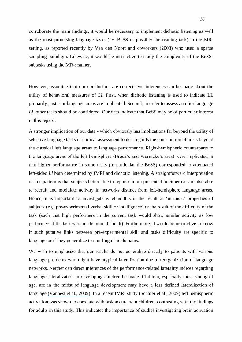
Introduction
Knowledge about the lateralization of brain functions is important for resolving both research
and clinical issues. An increasing interest in lateralization is presently seen, among others, in
research on schizophrenia (Angrilli et al., 2009; Bleich-Cohen et al., 2009) and dyslexia
(Pernet et al., 2009). In language research, the lateralization of language function can be used
to explain subtle differences in behavior and cognitive skills. For example, clinical language
lateralization assessment is necessary in the examination of epilepsy patients prior to resective
surgery of the temporal lobe. The standard method to determine language laterality in the
clinic is the Wada procedure, which is highly invasive, with associated risks for the patient
(Baxendale, 2009).
Non-invasive alternatives to the Wada test have proven to be reliable in determining
lateralization of language function in the brain. Lateralization in healthy subjects is often
determined by a dichotic listening test, where ear dominance is correlated to language
dominance. This is based on the notion that the contralateral (direct) pathways from the ears
to the opposite hemisphere are stronger than the projection to the ipsilateral hemisphere
(Kimura, 1961; Geffen et al., 1978). Typically, language is left-lateralized, resulting in a
better perception of verbal stimuli in the right ear compared to the left, expressed in degree of
right ear advantage (REA) (Hugdahl, 1995).
A more recent, promising method is the use of functional Magnetic Resonance Imaging
(fMRI) to determine language lateralization, enabling language function location within the
hemispheres. With fMRI, the hemispheric asymmetry of the activation pattern reflects
lateralization of language. The fMRI-determined lateralization index (LI) has been shown to
correlate with some versions of dichotic listening (Fernandes et al., 2006; Fontoura et al.,
2008; Van den Noort et al., 2008). More convincingly, both dichotic listening (Strauss et al.,
1987; Hugdahl et al., 1997; Fontoura et al., 2008) and fMRI-LI tests (Desmond et al., 1995;
Yetkin et al., 1995; Spreer et al., 2002, Fontoura et al., 2008) repeatedly show a correlation
with Wada test results. However, fMRI-LIs are influenced by task choice (Lee et al., 2008)
and are more robust when measured in regions of interest (ROIs) rather than when derived
from a whole-brain index calculation (Spreer et al., 2002). In their study on language
lateralization, Szaflarski and colleagues (2002) point out that fMRI-LIs depend on the
locations from which the laterality is derived. They showed that frontal regions are more
strongly linked to handedness and hence show a higher incidence of right-sided lateralization

4
for left-handed people, thus replicating results from Ellis and colleagues (1998). Such results
strongly advocate a hypothesis-driven choice of ROIs in deriving LIs, rather than calculating
whole-brain-laterality.
When fMRI results are used to estimate laterality the most common approach is to generate
an activation map by imposing a pre-defined statistical threshold to declare voxels as active or
inactive. The LI is often determined by simply subtracting and normalizing right-hemispheric
activation from left-hemispheric activation. However, this index can be severely influenced
by many methodological factors, such as activation thresholds and outlier influence. One
possible way to circumvent the threshold problem is to calculate LIs for several different
thresholds and then use a weighted average of the indices to define the resulting LI. In this
study, a method presented by Wilke and colleagues (2006; 2007) was used. This method
combines the weighted average approach with a bootstrap procedure to further increase the
robustness of the LI calculation.
In determining fMRI-LI it is important to employ both a reliable LI-calculation method, as
discussed above, and a language paradigm that activates areas of interest. Semantic sentence
processing generally activates Wernicke‟s area and the angular gyrus whereas word
generation relies on Broca‟s area (Price, 2000). The role of the inferior frontal lobe
(specifically Broca‟s area) and the posterior superior temporal gyrus (Wernicke‟s area) in
language processing is extensively researched and exceeds the straightforward view of a
division between language accessing and production as summarized by Geschwind
(Geschwind, 1965; Bookheimer, 2002; Price, 2000; Hickok and Poeppel, 2004). The frontal
temporal lobe has been shown to be active during sentence reading (Fletcher et al., 1995),
albeit not consistently (Price et al., 2003) and is involved in name retrieval (Damasio et al.,
2004). Conventional word retrieval paradigms also trigger activation in the left temporal
gyrus (Wise et al., 2001; Damasio et al., 2004). The anterior cingulate cortex shows activity
during fluency paradigms (Lurito et al, 2000; Fu et al., 2001). Kircher and colleagues (2001)
used fMRI to study sentence completion and found activation in the left middle frontal and
anterior cingulate gyrus as well as in the superior parietal lobe (precuneus). Activation in the
superior parietal lobe is also found during verb production (Shapiro et al., 2006) and
suggested to be involved in retrieval of associations during semantic processing (Carreiras et
al., 2009). We selected five regions of interest in primary language areas (the posterior
temporal lobe and the inferior frontal lobe) and areas involved in cognitively demanding
linguistic tasks (the frontal temporal lobe; the superior parietal lobe and the anterior cingulate

5
cortex). Next to their involvement in language, the ROIs were chosen so that they were well
apart, in order to detect separate clusters of activation. For exploring language-related
activation in the selected ROIs, a sentence completion paradigm, which involves semantic
processing as well as word generation, was used (McDonald and Tamariz, 2002).
In the present study we wished to investigate language lateralization in correlation to
language performance. Sentence completion has been shown to elicit activation in the right
hemisphere, additional to the described left-hemispheric activation, specifically in the middle
temporal gyrus (Kircher et al., 2001). The classical view on right-hemispheric contributions is
the involvement in decoding ambiguity, metaphors and distant semantic relationships
(Koivisto, 1997; Abdullaev and Posner, 1997; Chiarello, 1998). In addition, the contribution
of the right hemisphere involves decisions about semantic relations between words (Faust,
1998; Beeman and Chiarello, 1998). Just and colleagues (1996) investigated the influence of
the right hemisphere (right-sided counterparts of Wernicke‟s and Broca‟s areas) in language
function employing an increased-demand fMRI paradigm. The authors found increased
activation in right-hemispheric areas when cognitive demands were high. Likewise, Dräger
and colleagues (2004) described increased activation in the right parietal lobe as a result of
increased task difficulty.
The main aim of this study was to assess lateralization of language function using both
dichotic listening and fMRI-based region-specific LIs, in order to clarify the usability of both
methods in clinical lateralization assessment. Secondarily, we aimed to apply these methods
in order to determine the influence of language ability on the amount of (additional) right-
hemispheric activation. We correlated fMRI and dichotic listening derived LIs with
performance measurements on tasks measuring fluency, reading ability, picture naming and
high-level linguistic ability performance.

6
Materials and Methods
Subjects
Sixteen healthy, right-handed subjects of different gender and age were recruited to
participate in a series of behavioral tasks tapping language functions, an fMRI sentence
completion paradigm, and a dichotic listening test. One subject was excluded due to
movement artifacts in fMRI images and another subject was excluded due to hearing
impairment, which could have confounded the dichotic listening result. Results from 14
subjects, 7 females and 7 males, aged 21–55 (mean age = 36.9, SD=11.7) years are presented
in this study. Pre-experimental screening consisted of assessing handedness by means of the
Edinburgh handedness inventory. In addition, MR-safety and health status were recorded. The
subjects had neither a contraindication for MR safety, nor any history of neurological,
cognitive or psychiatric disorders - including alcohol and drug abuse - or pathological
language problems. Examination of the anatomical MRI images did not reveal any individual
brain anomalies. The test procedure encompassed a total of 2 – 3 hours, distributed over 2
days. The study was approved by the regional Ethics Committee of Linköping and the
subjects signed written informed consent.
fMRI acquisition and analysis
Images were acquired using a Philips Achieva 1.5 T MR-scanner. The language paradigm
was presented using high-resolution video goggles (Resonance Technology Inc., CA, USA).
For experimental design and task presentation, Superlab Pro 4 software was used (Cedrus
Corp., San Pedro, USA).
At the start of the fMRI examination the participants were given instructions regarding the
language paradigm and were familiarized with the procedure. Functional images were
acquired using a blood oxygen level dependent (BOLD) sensitive gradient echo sequence,
employing the following acquisition parameters: TR = 2.7 s; TE = 40 ms; FOV = 24 cm; flip
angle = 90°; matrix = 80 x 80 x 32; slice thickness = 3 mm. The slices were aligned between
the floor of sella turcica and the posterior angle of the fourth ventricle.
The sentence completion paradigm was visually presented and consisted of four blocks of 8
sentences each, in which the last word was substituted by “…”. The sentences were presented
for 3 seconds, followed by an asterisk presented for 2 seconds. The subjects were instructed to
think of one or several words that would complete the sentence during the time the asterisk

7
was on screen. Trial blocks were alternated with a total of 5 control blocks consisting of
asterisks, mimicking a short sentence. These were presented for 3 seconds and followed by a
single asterisk presented for 2 seconds. The subjects were instructed not to think of words or
sentences during the control blocks. In addition, the subjects were randomly assigned to one
of two paradigms each containing different sentences of an equal degree of difficulty. The
order of the blocks and sentences within the blocks was random for each subject. The total
duration of the test was 5 minutes.
The sentences were translated into Swedish from a Spanish study by McDonald and Tamariz
(2002) and validated in-house using 29 native Swedish students, 55% female and 45% males,
ranging between 19 and 37 years (mean age 23 years). Sentences with more than 10%
irrelevant answers as well as sentences with marked gender differences were excluded.
SPM5 (http://www.fil.ion.ucl.ac.uk/spm/software/spm5/) was used for the analysis of the
fMRI data. Functional scans were realigned to the first image of the time series, normalized at
2x2x2mm3 to a standard brain atlas (MNI space) and smoothed using an 8 mm FWHM
Gaussian kernel. A general linear model (GLM) was fitted to the data and the estimated
regression coefficients for each voxel were tested against zero with a t-test to generate
individual statistical maps which were entered in the calculation of fMRI-LI. To create figure
I, the group activation was assessed by a 2nd
level one sample t-test. The group statistical map
was thresholded using a false discovery rate (FDR) correction (Benjamini and Hochberg,
1995) to control for multiple comparisons across all voxels (significance threshold of p =
0.05).
Five non-overlapping regions of interest (ROIs) were defined as:
Frontal Temporal Lobe (FTL, middle and superior temporal gyrus)
Posterior Temporal Lobe (PTL, middle and superior temporal gyrus)
Inferior Frontal Gyrus (IFG) – opercular and triangular part corresponding with
Broca‟s area
Anterior Cingulate Cortex (ACC)
Superior Parietal Lobe (SPL)
All ROIs were constructed using WFU_Pickatlas software (version 2.4) (Maldjian et al.,
2003). The ROIs were post-processed to be mirror-symmetrical in the left-right direction.

8
fMRI laterality index
Lateralization was assessed by computing LI from the individual statistic images (T-maps).
The commonly used approach (Eq. 1) takes only the number of activated voxels in the left or
right hemisphere within an ROI in account.
LI =Left Right
Left + Right (1)
In this calculation Left and Right denote the number of activated voxels in each
hemisphere or region of interest within each hemisphere. The LI ranges from -1 (only right-
hemispheric activation) to +1 (only left-hemispheric activation). The main difficulty with this
definition is that the resulting LI depends on the statistical threshold chosen to define
activation. One way to avoid the threshold dependence is to calculate LIs for several different
thresholds (defined by the t-statistic, t) and use a weighted average of the LIs to define the
final LI (Eq. 2).
LI =t LI t
t (2)
In this study, the method introduced by Wilke and colleagues (2006; 2007) was used. The
method combines the weighted average approach with a bootstrap resampling procedure to
further increase the robustness of the LI. The data was first thresholded and masked by the
ROI, generating left and right data sets. From the left and right data a number (n) of
bootstrapped re-samples were generated. LIs were calculated according to Eq. (1). Then, the
LI for all possible combinations (n²) of the re-sampled data sets were calculated, and a
trimmed mean of the n² LIs was calculated. This procedure was repeated for 20 thresholds
equally spaced between zero and the maximum t-value within the ROI according to the
default values (Wilke et al., 2006), and the final LI was calculated as a weighted average (Eq.
2) of the trimmed means.
Dichotic Listening test
Dichotic listening was assessed by means of a version of the consonant-vowel Bergen
Dichotic Listening Test (Hugdahl, 1995). In this task, the subjects were presented with
auditory verbal stimuli in both ears simultaneously. The perceived stimuli had to be reported

9
verbally directly after each presentation, according to directions of the examiner who
instructed to attend to either: 1) one of the ears, 2) both ears or 3) one of the ears according to
the participant‟s best perception. The stimuli used in the dichotic speech tests were the
consonant-vowel combinations of a stop consonant and the letter „a‟: /ba/, /da/, /ga/, /ka/, /pa/
and /ta/. The stimuli were combined in dichotic pairs and used in three different conditions,
executed in the following order (for details see Hällgren et al., 1998):
1. The non-forced condition: the subjects were instructed to recall one stimulus, namely
the syllable they perceived best or most clearly (20 dichotic pairs).
2. The free-report condition: the subjects were encouraged to recall the syllables heard
in both ears (20 dichotic pairs).
3. The directed-report conditions: the subjects were instructed to recall syllables
perceived by one ear, when attention was directed to either the right or left ear (20
dichotic pairs with instructed attention to the left ear and 20 pairs with instructed
attention to the right ear).
Two parameters were calculated; the number of correct answers and right ear advantage
(REA). The REA was scored as a lateralization index, defined by the number of correctly
repeated items presented to the right ear minus the number of correctly repeated items to the
left ear, divided by the total number of correct answers for both ears (Hugdahl, 2003). This
definition holds also for the directed-report condition – the subjects always repeated some of
the items presented to the non-focused ear. A positive lateralization index means a right ear
advantage and thus a positive value for REA.
The dichotic tests were performed in a sound-isolated chamber. The test was delivered
through a CD player, an audiometer and earphones (Telephonics TDH 39). The presentation
level was 70 dB SPL (C-weighted equivalent level). Pure tone thresholds were established for
the frequencies: 500 Hz, 1000 Hz, 2000 Hz and 4000 Hz, in order to ensure that the subjects
did not have asymmetrical hearing losses (an interaural difference of more than 10 dB).
Behavioral tasks
The administered behavioral language tests were selected to ensure a proper assessment of
language performance in subjects with a normal to advanced language ability.
The Test of Language Competence (TLC) is suitable for investigating higher language
abilities, by investigating applied semantics, syntax and pragmatics. The test is translated into

10
Swedish and proposed to be suitable for discovering subtle speech and language disorders
(Testbatteri för Bedömning av Subtila Språkstörningar, TBSS) (Laakso et al., 2000). From
this test-battery we utilized a subset of seven tasks to determine subtle speech and language
disorders (Bedömning av Subtila Språkstörningar [Assessment of Subtle Language Deficits],
BeSS) which do not show ceiling effects when applied to healthy individuals (Laakso et al.,
2000). The BeSS-test investigates the following abilities: repetition of long sentences,
constructing a correct sentence of three words, inference (text understanding), understanding
complex logic-grammatical sentences, understanding dubious sentences, understanding
metaphors, and describing presented words. Each subtask of 10 questions could give a
maximum of 30 points, maximal total 210 points. Two well-known word retrieval tasks from
the TLC were also selected. The Boston Naming Test (BNT), consisting of 60 pictures which
have to be named, detects naming abilities and correlates with education level (Kaplan et al.,
1983; Zec et al., 2007). The Swedish norm established by Tallberg (2005) was used in this
study. Furthermore we used a verbal fluency test. The subject was cued with a letter (F, A, S)
and had to generate as many words as possible within a minute (FAS-test, Friedman et al.,
1998; Loonstra et al., 2001). The Swedish norm, depending on age and gender, is predictive
of intellectual level (Tallberg et al., 2008). Lastly, a Reading task selected from the Swedish
exam for university students was used. The subjects had to read three texts and answer four
questions for each test, resulting in a maximum score of 12.
Correlations between the results from fMRI, dichotic listening and language performance
tasks were tested with a two-tailed significance test using GraphPad Prism (GraphPad
software, Inc. San Diego, USA). p < 0.05 was considered as significant.

11
Results
fMRI results
When contrasted with the non-language control condition, the sentence completion paradigm
elicited clusters of activation in the primary language areas in the left hemisphere, namely in
the posterior temporal lobe (corresponding to Wernicke‟s area) and the left inferior frontal
gyrus (corresponding to Broca‟s area). Also the frontal temporal lobe, the superior parietal
lobe and the anterior cingulate cortex were activated. Furthermore, activation was observed in
the occipital lobe including the fusiform areas. In Fig. 1, the group activity (orange) that
remains after applying an inclusive map of ROIs (blue) as described in Materials and Methods
can be seen. The remaining group activation is superimposed over the ROIs. Note that the
anterior cingulate ROIs are not depicted.
Correlation of dichotic listening with fMRI-LI
The results of the three dichotic listening conditions (non-forced, free-report, directed-report
left/right) were correlated with LIs derived from the fMRI paradigm. The results are found in
Table 1 and Figure 2.
The non-forced condition REA was significantly positively correlated (p < 0.05) with fMRI-
LI in the anterior cingulate cortex.
In the free-report condition, which demands attention to both ears, the REA correlated
positively (p < 0.01) with fMRI-LI in the superior parietal lobe.
In the directed-report condition, a significant positive correlation (p < 0.01) of directed-report-
left REA with the fMRI-LI in the posterior temporal lobe, which includes Wernicke‟s area,
was found.
Nieither the fMRI-LIs, nor the dichotic listening REA LIs correlated with age.
Correlating dichotic listening with language ability
Performance in all behavioral language tasks except the BNT correlated positively with left-
ear reports during the directed left condition of the dichotic listening test (Table 2, Figure III).

12
Only the FAS test correlated with the amount of right ear reports in the free report condition.
Notably, the reading test (LäS) and the BeSS test showed negative correlations with REA
during the directed-report-left condition and the free-report condition, respectively.
Correlating fMRI-LI with language performance
fMRI-LI, determined from the posterior temporal lobe including Wernicke‟s area correlated
negatively (p < 0.05) with reading ability (LäS), r = -0.55 (Table 3). Performance in the
cognitive assessment task, measuring subtle language dysfunctions (BeSS) correlated
negatively (p < 0.05) with LI in the inferior frontal cortex (Broca‟s area) r = -0.56 (Figure IV).

13
Discussion
The purpose of this study was twofold. First, we wished to compare LI derived from ROIs
acquired with fMRI with estimates of LI obtained with Hugdahl‟s implementation of the
classical dichotic listening task (Hugdahl, 1995), in order to evaluate the usability of either
method in clinical settings. Second, we were interested in delineating the relation between
measures of LI and performance on a set of salient language tasks, typically used in the
clinical assessment of language impairment. In particular, we sought to clarify whether LI
varied between subjects performing high or low in the language tasks.
In keeping with the findings in previous investigations (Fernandes, et al., 2006, Fontoura et
al., 2008), fMRI and dichotic listening generated similar measures of laterality. However, in
contrast to previous reports, the correlation of dichotic-listening- with fMRI-derived LIs
varied according to the task demands of the different dichotic listening conditions. In the non-
forced condition (where subjects are asked to repeat the most noticeable stimuli), a less
prominent right ear advantage was associated with more bilateral cingulate cortex activation.
The anterior cingulate, as well as the orbitofrontal cortex, is involved in monitoring
behaviorally motivated stimuli, a process that may be crucial to performance in the non-
forced condition (Luu and Posner, 2003).
In contrast, performance in the condition where subjects were asked to report lateralized
stimuli (directed-report-left) correlated with activity in the posterior temporal region, where
subjects with less right ear advantage show involvement of the right posterior temporal lobe
in addition to the left. This is clearly a finding much in keeping with previous reports of
neuroimaging of dichotic listening (e.g., van den Noort, 2008). The activation observed in the
right-hemispheric counterpart of Wernicke‟s area in the temporal lobe strengthens the
hypothesis of Grodzinsky and Friederici (2006), who linked this area to lexical and syntactic
information integration. Moreover, the temporal lobe is known to be responsible for
processing phonological information, not exclusively, but proven to be vital for perception of
language (Gernsbacher and Kaschak, 2003). The dichotic listening task is very clearly a task
of phonological perception which requires involvement of the left posterior temporal lobe,
which we indeed have found. The right-hemispheric activation might indicate additional
resources required for the process of integrating phonological input. Finally, the free-report
condition, which presumably is more similar to conventional working memory tasks than the
other two conditions, rests more upon recruitment of attentional resources. A less pronounced

14
right ear advantage shows to correlate with more activity in the right-hemispheric superior
parietal lobe in addition to left-hemispheric superior parietal lobe activation. Next to
hypothesized involvement in reading and word classification/production (Ino et al., 2008;
Shapiro et al., 2006), important portions of the parietal cortex are involved in attention
mechanisms (Yantis and Serences, 2003, Behrmann et al., 2004).
Both dichotic listening and fMRI have important functions in the assessment of language
lateralization. Given the employment of a relevant language task, fMRI can provide a
relatively direct account of brain physiology. However, limitations with respect to cost and
availability may make fMRI a less likely choice for routine clinical assessment of
lateralization. Furthermore, fMRI may be compromised by pathological changes (such as
edema or cortical atrophy) or postoperative distortion (e.g., effects of blood material, signal
dropout). Under such circumstances, behavioral measures such as dichotic listening may
provide more reliable clues to specific aspects of brain function. Although the latter point
rarely has been examined in patients with brain damage, it is still important to know how
fMRI results and behavioral measures relate to each other. According to our findings, the
directed-report-left condition of the Bergen Dichotic Listening Test indicated lateralization
with respect to the posterior temporal lobe, which is likely to be the cortical region of choice
when assessment of language lateralization is warranted (Van den Noort et al., 2008).
Secondly, we aimed to clarify the relation between dichotic listening as well as the fMRI-
derived LI and clinically salient language tasks. Interestingly, better performance on reporting
stimuli presented to the left ear correlated with better language ability. Although the level of
significance (p < 0.05) was not strong, findings were consistent for all language tasks in the
directed-report-left condition. Both BeSS and reading test results were reflected in the REA
calculation, for the free-report and the directed-report-left condition respectively.
With respect to fMRI-LI, two of our tasks, BeSS and the reading task, provided clear evidence
for an association. The reading task exhibited a correlation with LI of the posterior temporal
cortex, a finding that we believe reflects the well-known connection between reading, reading
impairment and temporoparietal structures (Price, 2000; Price et al., 2003, Hillis et al., 2008).
In addition, a particularly interesting finding was that the correlation was negative, that is,
better performance was related to an attenuated LI. This outcome suggests the involvement of
right-hemisphere mechanisms in reading. Such mechanisms need not be related to language in
the strict sense in that reading also draws upon perceptual and visuospatial skills, which may
recruit posterior cortical processes. Furthermore, it is intriguing to see that the fMRI results

15
lead to the same conclusion as shown by the results of the dichotic listening test; i.e. increased
right-hemispheric involvement results in increased performance in various language tasks.
When discussing these results it is interesting to note that other researchers have recently
demonstrated results indicating a variation in brain lateralization during various language
tasks. However these researchers have used different techniques (Stroobant et al., 2009) and
observed partially dissociated lateralization patterns within the language network, inconsistent
with an overall “dominance” model (Pinel and Dehaene, 2009).
Considering the BeSS test (Fig. 4) a similar, but non-significant correlation with the posterior
temporal lobe was noted. More conspicuously, BeSS performance was negatively correlated
with Broca LI. Given the complex nature of the BeSS, the outcome may reflect several
functions (perhaps simultaneously) of the anterior language cortices. Broca‟s area is involved
in subtle grammatical decisions (Damasio, 1992; Ullman et al., 2005, Rodd et al., 2005).
Furthermore, Broca‟s area may be recruited in working memory tasks; the BeSS battery
clearly involves such tasks. Finally, BeSS performance may be facilitated by covert and
perhaps even overt vocalizations. Such extra-linguistic behaviors clearly thrive upon anterior
language areas (Huang et al., 2001).
Results were considered statistically significant if p < 0.05. Given the large number of tests
performed, a few of the significant results probably reached this level by chance only. This
fact inevitably cast some doubt on the conclusions drawn. However, our conclusions are
based on collective evidence from several significant correlations. Moreover, all of the
significant results were consistent, which led us to believe that the conclusions drawn likely
will retain their validity. We tested post-hoc for any correlations between gender and ROI
activation using ANOVA with Bonferroni multiple comparison post-tests, however we found
no correlation, ensuring the validity of the presented analyses.
Nevertheless, many of our conclusions must remain tentative. Our findings are based upon
correlations between different measures, and we employed a relatively limited number of
measurements considering the complex nature of the response. In a follow-up study, we aim
to investigate the nature of task demands in comparison with language ability. This study will
shed light on the difference between fMRI activation and laterality indices brought forth by
attention or cognitive demand and activation related to individual language ability. To further
16
corroborate the main findings, it would be necessary to implement dichotic listening as well
as the most promising language tasks (i.e. BeSS or possibly the reading task) in the MR-
setting, as reported recently by Van den Noort and coworkers (2008) who used a sparse
sampling paradigm. Likewise, it would be instructive to study the complexity of the BeSS-
subtasks using the MR-scanner.
However, assuming that our conclusions are correct, two inferences can be made about the
utility of behavioral measures of LI. First, when dichotic listening is used to indicate LI,
primarily posterior language areas are implicated. Second, in order to assess anterior language
LI, other tasks should be considered. Our data indicate that BeSS may be of particular interest
in this regard.
A stronger implication of our data - which obviously has implications far beyond the utility of
selective language tasks or clinical assessment tools - regards the contribution of areas beyond
the classical left language areas to language performance. Right-hemispheric counterparts to
the language areas of the left hemisphere (Broca‟s and Wernicke‟s area) were implicated in
that higher performance in some tasks (in particular the BeSS) corresponded to attenuated
left-sided LI both determined by fMRI and dichotic listening. A straightforward interpretation
of this pattern is that subjects better able to report stimuli presented to either ear are also able
to recruit and modulate activity in networks distinct from left-hemisphere language areas.
Hence, it is important to investigate whether this is the result of „intrinsic‟ properties of
subjects (e.g. pre-experimental verbal skill or intelligence) or the result of the difficulty of the
task (such that high performers in the current task would show similar activity as low
performers if the task were made more difficult). Furthermore, it would be instructive to know
if such putative links between pre-experimental skill and tasks difficulty are specific to
language or if they generalize to non-linguistic domains.
We wish to emphasize that our results do not generalize directly to patients with various
language problems who might have atypical lateralization due to reorganization of language
networks. Neither can direct inferences of the performance-related laterality indices regarding
language lateralization in developing children be made. Children, especially those young of
age, are in the midst of language development may have a less defined lateralization of
language (Vannest et al., 2009). In a recent fMRI study (Schafer et al., 2009) left hemispheric
activation was shown to correlate with task accuracy in children, contrasting with the findings
for adults in this study. This indicates the importance of studies investigating brain activation
17
and connectivity related to language ability of healthy adults.
Conclusions
Laterality indices assessed using fMRI in the anterior cingulate cortex, superior parietal lobe,
and the posterior temporal lobe, correlated with dichotic listening derived laterality
measurements. Compared to dichotic listening, fMRI-LIs are region specific, which is a clear
advantage. Results in this study may direct the development of fMRI-derived LIs for clinical
assessment of language laterality in specific parts of the brain. However, as fMRI has
limitations with respect to cost and availability, behavioral measures such as dichotic listening
will remain important.
A particularly interesting finding was that the right-sided posterior temporal cortex, but not
the superior parietal lobe, showed increased activation in subjects that performed better in
salient clinical language tasks; the same occurred for the right-hemispheric homologue of
Broca‟s area. In concordance, decreased right-ear dominance (and increased reporting of left-
ear presented stimuli when focusing attention on the left ear) correlated with a higher
performance in all presented language tasks. Thus, more bilateral activation in classical
language areas rather than left-hemispheric dominance during language processing indicates
higher language ability.
Our presented results, acquired using (1) dichotic listening, (2) fMRI and (3) behavioral
language tasks indicated a supporting role of non-classical language areas in the brain in
language performance, where mainly right-hemispheric counterparts of Broca‟s and
Wernicke‟s areas are involved.
Acknowledgements
The contributions of research funding from the Cancer Foundation, University Hospital
Research Funds, Ståhl‟s foundation, the County Council of Östergötland, and the University
of Linköping are gratefully acknowledged. In addition, the measurements were performed
with use of equipment that to a large extent was funded by the K and A Wallenberg
Foundation, to which we would also like to express our gratitude. Finally, we would also like
to express our appreciation for the enthusiastic support from our colleagues for our research in

18
this area. Cecilia Nellie and Jennie Pettersson are especially acknowledged for administrating
the behavioral language tasks.

19
References
Abdullaev, Y.G., Posner, M.I., 1997. Time course of activating brain areas in generating
verbal associations. Psychol Sci 8, 56-59.
Angrilli, A., Spironelli, C., Elbert, T., Crow, T.J., Marano, G., Stegagno, L., 2009.
Schizophrenia as failure of left hemispheric dominance for the phonological component of
language. PLoS ONE 4, e4507.
Baxendale, S., 2009- The Wada test. Curr Opin Neurol 22, 185-189.
Beeman, M.J., Chiarello, C., 1998. Complementary right- and left-hemisphere language
comprehension. Curr Dir Psychol Sci 7, 2-8.
Behrmann, M., Geng, J.J., Shomstein, S., 2004. Parietal cortex and attention. Curr Op
Neurobiol 14, 212-217.
Benjamini, Y., Hochberg, Y., 1995. Controlling the False Discovery Rate: A practical and
powerful approach to multiple testing. J R Stat Soc B 57, 289-300.
Bleich-Cohen, M., Hendler, T., Kotler, M., Strous, R.D., 2009. Reduced language
lateralization in first-episode schizophrenia: An fMRI index of functional asymmetry.
Psychiatry Res 171, 82-93.
Bookheimer, S., 2002. Functional MRI of language: new approaches to understanding the
cortical organization of semantic processing. Annu Rev Neurosci 25, 151-188.
Carreiras M., Riba, J., Vergara, M., Heldmann, M., Münte, TF., 2009. Syllable congruency
and word frequency effects on brain activation. (Epub ahead of print) Hum Brain Mapp,
Jan. 26.
Chiarello, C., 1998. On codes of meaning and the meaning of codes: semantic access and
retrieval within and between hemispheres. In M. Beeman & C. Chiarello (Eds.), Right
hemisphere language comprehension: perspectives from cognitive neuroscience. Erlbaum,
Mahwah, pp. 141-160.
Damasio, A.R., 1992. Aphasia. New England J Medic 326, 531-539.
Damasio, H., Tranel, D., Grabowski, T., Adolphs, R., Damasio, A., 2004. Neural systems
behind word and concept retrieval. Cognition 92, 179-229.

20
Desmond, J.E., Sum, J.M., Wagner, A.D., Demb, J.B., Shear, P.K., Glover, G.H., Gabrieli,
J.D., Morrell, M.J., 1995. Functional MRI measurement of language lateralization in
Wada-tested patients. Brain 118, 1411-1419.
Dräger, B., Jansen, A., Bruchmann, S., Förster, A.F., Pleger, B., Zwitserlood, P., Knecht, S.
2004. How does the brain accommodate to increased task difficulty in word finding? A
functional MRI study. NeuroImage 23, 1152-1160.
Ellis, S.J., Ellis, P.J., Marshall, E., Windridge, C., Jones, S., 1998. Is forced dextrality an
explanation for the fall in the prevalence of sinistrality with age? A study in northern
England. J Epidemiol Community Health 52, 41-44.
Faust, M., 1998. Obtaining evidence of language comprehension from sentence priming. In
M. Beeman & C. Chiarello (Eds.), Right hemisphere language comprehension:
perspectives from cognitive neuroscience. Erlbaum, Mahwah, pp. 141-160.
Fernandes, M.A., Smith, M.L., Logan, W., Crawley, A., McAndrews, M.R., 2006. Comparing
language lateralization determined by dichotic listening and fMRI activation in frontal and
temporal lobes in children with epilepsy. Brain Lang 96, 106-114.
Fletcher, P.C., Happé, F., Frith, U., Baker, S.C., Dolan, R.J., Frackowiak, R.S.J., Frith, C.D.
1995. Other minds in the brain: a functional imaging study of “theory of mind” in story
comprehension. Cognition 57, 109-128.
Fontoura, D.R., Branco, D.M., Anés, M., Costa, J.C., Portuguez, M.W., 2008. Language brain
dominance in patients with refractory temporal lobe epilepsy. Arq Neuropsicuiatr 66, 34-
39.
Friedman, L., Kenny, J.T., Wise, A.L., Wu, D., Stuve, T.A., Miller, D.A., Jesberger, J.A.,
Lewin, J.S., 1998. Brain activation during silent word generation evaluated with functional
MRI. Brain Lang 64, 231-256.
Fu, C.H.Y., Morgan, K., Suckling, J., Williams, S.C.R., Andrew, C., Vythelingum, G.N.,
McGuire, P.K., 2001. A functional Magnetic Resonance Imaging study of overt letter
verbal fluency using a clustered acquisition sequence: Greater anterior cingulate activation
with increased task demand. NeuroImage 17, 871-879.
Geffen, G., Traub, E., Stierman, I., 1978. Language assessed by unilateral ECT and dichotic
monitoring. J Neurol Neurosurg Psychiatry 41, 34-360.
Gernsbacher, M.A., Kaschak, M.P., 2003. Neuroimaging studies of language production and

21
comprehension. Annu. Rev. Psychol. 54, 91-114.
Geschwind, N., 1965, Disconnexion syndromes in animals and man. I. Brain 88, 237-294.
Grodzinsky, Y., Friederici, A.D., 2006. Neuroimaging of syntax and syntactic processing.
Neurobiology 16, 240-246.
Hällgren, M., Johansson, M., Larsby, B., Arlinger, S, 1998. Dichotic speech tests. Scand
Audiol Suppl 49, 35-39.
Hickok, G., Poeppel, D., 2004. Dorsal and ventral streams: A framework for understanding
aspects of the functional anatomy of language. Cognition 92, 67-99.
Hillis, A.E., 2008. Cognitive processes underlying reading and writing and their neural
substrates. In G. Goldenberg & B. L. Miller (Eds.) Handbook of Clinical Neurology, Vol.
88 (3rd series): Neuropsychology and behavioral neurology. Elsevier, Amsterdam, pp.
311-322.
Huang, J., Carr, T. H., Cao, Y., 2001. Comparing cortical activations for silent and overt
speech using event-related fMRI. Hum Brain Mapp 15, 39-53.
Hugdahl, K., 1995. Dichotic listening: probing temporal lobe functional integrity. In:
Davidson, R.J., Hugdahl, K. (Eds.), Brain asymmetry. MIT Press, Cambridge, MA, pp.
123-156.
Hugdahl, K., Carlsson, G., Uvebrant, P., Lundervold, A.J., 1997. Dichotic-listening
performance and intracarotid injections of amobarbital in children and adolescents.
Preoperative and postoperative comparisons. Arch Neurol 54, 1494-1500.
Hugdahl, K., 2003. Dichotic listening in the study of auditory laterality. In: Hugdahl, K.,
Davidsson, R.J., (Eds) The asymmetrical brain. MIT Press, Cambridge, MA, pp. 441-466.
Ino, T., Tokumoto, K., Usami, K., Kimura T., Hashimoto, Y., Fukuyama, H., 2008.
Longitudinal fMRI study of reading in a patient with letter-by-letter reading. Cortex 44,
773-781.
Just, M.A., Carpenter, P.A., Keller, T.A., Eddy, W.F., Thulborn, K.R., 1996. Brain activation
modulated by sentence comprehension. Science 274, 114-116.
Kaplan, E., Goodglass, H., Weintraub, S., 1983. The Boston Naming Test. Lea & Febiger,
Erlbaum.
Kimura, D., 1961. Cerebral dominance and the perception of verbal stimuli. Canad J Psychol

22
15, 166-171.
Kircher, T., Brammer, M., Tous Andreu, N., Williams S., McGuire, P., 2001. Engagement of
right temporal cortex during processing of linguistic context. Neuropsychologia 39: 798-
809.
Koivisto, M., 1997. Time course of semantic activation in the cerebral hemispheres.
Neuropsychologia 35, 497-504.
Laakso, K., Brunnegård, K., Hartelius, L., Ahlsén, E., 2000. Assessing high-level language in
individuals with multiple sclerosis: A pilot study. Clin Linguist Phon 14, 329-349.
Lee, D., Swanson, S.J., Sabsevitz, D.S., Hammeke, T.A., Winstanley, F.S., Possing, E.T.,
Binder, J.R., 2008. Functional MRI and Wada studies in patients with interhemispheric
dissociation of language functions. Epilepsy Behav 13, 350-356.
Loonstra, A.S., Tarlow, A.R., Sellers, A.H., 2001. COWAT metanorms across age, education,
and gender. Appl Neuropsychol 8, 161-166.
Luu, P., Posner, M.I. , 2003. Anterior cingulate cortex regulation of sympathetic activity.
Brain 126, 2119-2120.
Lurito, J.T., Kareken, D.A., Lowe, M.J., Chen, S.H.A., Mathews, V.P., 2000. Comparison of
rhyming and word generation with fMRI. Hum Brain Mapp 10, 99-106.
Maldjian, J.A., Laurienti, P.J., Burdette, J.H., Kraft, R.A., 2003. An automated method for
neuroanatomic and cytoarchitectonic atlas-based interrogation of fMRI data sets.
NeuroImage 19, 1233-1239.
McDonald, S.A., Tamariz, M., 2002. Completion norms for 112 Spanish sentences. Behav
Res Methods Instrum Comput 34, 128-137.
Pernet, C., Andersson, J., Paulesu, E., Demonet, J.F., 2009. When all hypotheses are right: A
multifocal account of dyslexia. (Epub ahead of print) Hum Brain Mapp, Feb. 23.
Pinel, P., Dehaene, S., 2009. Beyond hemispheric dominance: Brain regions underlying the
joint lateralization of language and arithmetic to the left hemisphere. (Epub ahead of print)
J Cogn Neurosci, Jan 13.
Price, C.J., 2000. The anatomy of language: contributions from functional neuroimaging. J
Anat 197, 335-359.
Price, C.J., Gorno-Tempini, M.L., Graham, K.S., Biggio, N., Mechelli, A., Patterson, K.,

23
Noppeney, U., 2003. Normal and pathological reading: converging data from lesion and
imaging studies. NeuroImage 20, 30-41.
Rodd, J. M., Davis, M. H., Johnsrude, I. H., 2005. The neural mechanisms of speech
comprehension: fMRI studies of semantic ambiguity. Cer Cor 15, 1261–1269.
Schafer, R.J., Lacadie, C., Vohr, B., Kesler, S.R., Katz, K.H., Schneider, K.C., Pugh, K.R.,
Makuch, R.W., Reiss, A.L., Constable, R.T., Ment, L.R., 2009. Alterations in functional
connectivity.
Shapiro, K.A., Moo, L.R., Caramazza, A., 2006. Cortical signatures of noun and verb
production. Proc Natl Acad Sci U S A 103, 1644-1649.
Spreer, J., Arnold, S., Quiske, A., Wohlfarth, R., Ziyeh, S., Altenmüller, D., Herpers, M.,
Kassubek, J., Klisch, J., Steinhoff, B.J., Honegger, J., Schulze-Bonhage, A., Schumacher,
M., 2002. Determination of hemisphere dominance for language: comparison of frontal
and temporal fMRI activation with intracarotid amytal testing. Neuroradiology 44, 467-
474.
Strauss E., Gaddes W.H., Wada, J., 1987. Performance on a free-recall verbal dichotic
listening task and cerebral dominance determined by the carotid amytal test.
Neuropsychologia 25, 747-753.
Stroobant, N., Buijs, D., Vingerhoets, G., 2009. Variation in brain lateralization during
various language tasks: A functional transcranial Doppler study. Behav Brain Res 199,
190-196.
Szaflarski, J.P., Binder, J.R., Possing, E.T., McKiernan, K.A., Ward, B.D., Hammeke, T.A.,
2002. Language lateralization in left-handed and ambidextrous people: fMRI data.
Neurology 59, 238-244.
Tallberg, I.M., 2005. The Boston Naming Test in Swedish: Normative data. Brain Lang 94,
19-31.
Tallberg, I.M., Ivachova, E., Jones Tinghag K., Östberg, P., 2008. Swedish norms for word
fluency tests: FAS, animals and verbs. Scand J Psych 49, 479-485.
Ullman, M.T., Pancheva, R., Love, T., Yee, E., Swinney, D., Hickok, G., 2005. Neural
correlates of lexicon and grammar: Evidence from the production, reading and judgment of
inflection in aphasia. Brain Lang 93, 185-238.
24
van den Noort, M., Specht, K., Rimol, L.M., Ersland, L., Hugdahl, K., 2008. A new verbal
reports fMRI dichotic listening paradigm for studies of hemispheric asymmetry.
NeuroImage 40, 902-911.
Vannest, J., Karunanayaka, P.R., Schmithorst, V.J., Szaflarski, J.P., Holland, S.K., 2009.
Language networks in children: evidence from functional MRI studies. AJR 192, 1190-
1196.
Wilke, M., Schmithorst, V.J., 2006. A combined bootstrap/histogram analysis approach for
computing a lateralization index from neuroimaging data. NeuroImage 22, 522-530.
Wilke, M., Lidzba, K., 2007. LI-tool: A new toolbox to assess lateralization in functional
MR-data‟. J Neurosci Methods 163, 128-136.
Wise, J.S.R., Scott, S.K., Blank, S.C., Mummery, C.J., Murphy, K., Warburton, E.A., 2001.
Separate neural subsystems within „Wernicke‟s area‟. Brain 124, 83-95.
Yantis, S., Serences, J.T., 2003. Cortical mechanisms of space-based and object-based
attentional control. Curr Opin Neurobiol 13, 187-193.
Yetkin, F.Z., Hammeke, T.A., Swanson, S.J., Morris, G.L., Mueller, W.M., McAuliffe, T.L.,
Haughton, V.M., 1995. A comparison of functional MR activation patterns during silent
and audible language tasks. Am J Neuroradiol 16, 1087-1092.
Zec, R.F., Burkett, N.R., Markwell, S.J., Larsen, D.L., 2007. Normative data stratified for
age, education, and gender on the Boston Naming Test. Clin Neuropsy 21, 617-637.

25
Table 1. Significant correlations (Pearson’s r) between dichotic listening REA and fMRI LI in
the regions of interest. * = p < 0.05, ** = p < 0.01.
Anterior
Temporal
Lobe
Posterior
Temporal
Lobe
Inferior
Frontal Gyrus
/ Broca‟s area
Superior
Parietal Lobe
Anterior
Cingulate
Cortex
Non-forced - - - - 0.56*
Free-report - - - 0.68** -
Directed-
report left
- 0.65** - - -
Directed-
report right
- - - - -

26
Table 2. Significant correlations (Pearson’s r) between dichotic listening and behavioural
language tests. * = p < 0.05, ** = p < 0.01. FAS = fluency test, LäS = reading test, BeSS =
cognitive ability.
FAS LäS BeSS
Non-forced (REA) - - -
Free-report (REA) - - -0.53*
Directed-report-left (REA) - -0.56* -
Directed-report-right (REA) - - -
Non-forced, left ear - - -
Non-forced, right ear - - -
Free-report, left ear - - 0.58*
Free-report, right ear 0.52* - -
Directed-report-left, left ear 0.52* 0.69** 0.62*
Directed-report-left, right ear - - -
Directed-report-right, left ear - - -
Directed-report-right, right ear - - -

27
Table 3. Significant correlations (Pearson‟s r) between language performance tests and fMRI
LI in regions of interest. * = p<0.05, ns = non significant (p=0.07), FAS = fluency test, LäS =
reading test, BeSS = cognitive ability, BNT = Boston Naming Test.
Anterior
Temporal
Lobe
Posterior
Temporal
Lobe
Inferior
Frontal Gyrus
/ Broca‟s Area
Superior
Parietal Lobe
Anterior
Cingulate
Cortex
FAS - - - - -
LäS - -0.55* - - -
BeSS - -0.48ns
-0.56* - -
BNT - - - - -

28
Figure 1. MR image showing the functional ROIs (blue) used for LI calculations and ROI
specific group activations (orange). This group analysis was restricted to the ROIs and
thresholded at p = 0.05 (FDR).

29
Figure 2. Correlation between dichotic listening non-forced, free-report and directed-
report-left conditions and fMRI-LI. Left and right on the axes indicate the hemispheres.
Figure 3. Correlation between correct left ear answers at the dichotic listening directed-
report-left condition and performance on the behavioral language tests (higher score means
higher performance): FAS = fluency test, LäS = reading test, BeSS = language ability
Figure 4. Correlation between LI assessed by fMRI and the Battery for Subtle Language
impairment Score (BeSS).