Modelling, design and analysis of a testing rig for composite materials

JOURNAL OF VIROLOGY, Feb. 2011, p. 1224–1236 Vol. 85, No. 30022-538X/11/$12.00 doi:10.1128/JVI.01635-10Copyright © 2011, American Society for Microbiology. All Rights Reserved.
RIG-I-Mediated Antiviral Signaling Is Inhibited in HIV-1 Infection bya Protease-Mediated Sequestration of RIG-I�
Mayra Solis,1,2 Peyman Nakhaei,1,2 Mohammad Jalalirad,3 Judith Lacoste,4 Renee Douville,1,2
Meztli Arguello,1 Tiejun Zhao,1 Michael Laughrea,3Mark A. Wainberg,2,3 and John Hiscott1,2*
Terry Fox Molecular Oncology Group, Lady Davis Institute, Jewish General Hospital,1 Departments of Microbiology andImmunology and Medicine, McGill University,2 McGill AIDS Center, Lady Davis Institute, Jewish General Hospital,3 and
Department of Biology, McGill University,4 Montreal, Quebec H3T1E2, Canada
Received 5 August 2010/Accepted 10 November 2010
The rapid induction of type I interferon (IFN) is essential for establishing innate antiviral responses. Duringinfection, cytoplasmic viral RNA is sensed by two DExD/H box RNA helicases, RIG-I and MDA5, ultimatelydriving IFN production. Here, we demonstrate that purified genomic RNA from HIV-1 induces a RIG-I-dependent type I IFN response. Both the dimeric and monomeric forms of HIV-1 were sensed by RIG-I, but notMDA5, with monomeric RNA, usually found in defective HIV-1 particles, acting as a better inducer of IFN thandimeric RNA. However, despite the presence of HIV-1 RNA in the de novo infection of monocyte-derivedmacrophages, HIV-1 replication did not lead to a substantial induction of IFN signaling. We demonstrate theexistence of an evasion mechanism based on the inhibition of the RIG-I sensor through the action of the HIV-1protease (PR). Indeed, the ectopic expression of PR resulted in the inhibition of IFN regulatory factor 3 (IRF-3)phosphorylation and decreased expression of IFN and interferon-stimulated genes. A downregulation ofcytoplasmic RIG-I levels occurred in cells undergoing a single-cycle infection with wild-type provirus BH10 butnot in cells transfected with a protease-deficient provirus, BH10-PR�. Cellular fractionation and confocalmicroscopy studies revealed that RIG-I translocated from the cytosol to an insoluble fraction during the de novoHIV-1 infection of monocyte-derived macrophages, in the presence of PR. The loss of cytoplasmic RIG-I wasprevented by the lysosomal inhibitor E64, suggesting that PR targets RIG-I to the lysosomes. This study revealsa novel PR-dependent mechanism employed by HIV-1 to counteract the early IFN response to viral RNA ininfected cells.
Human immunodeficiency virus type 1 (HIV-1) causes pro-found immune deregulation that results in immune hyperacti-vation, CD4� T-cell depletion, and progression to AIDS (re-viewed in reference 5). HIV-1 has also evolved numerousmechanisms to evade various aspects of the innate and adap-tive immune response (reviewed in reference 36), including theability to subvert several host innate immune factors that limitretroviral replication (reviewed in reference 14). Since theimmune system fails to eradicate HIV-1 in infected cells, fur-ther studies are required to evaluate the strategies utilized byHIV-1 to counteract the innate immune response.
The induction of the innate immune response by viral patho-gens is characterized by a rapid production of type I interfer-ons (IFNs) (IFN-�/�), which play a major role in the inhibitionof virus replication. Upon recognition of viral products, anti-viral responses are initiated by either Toll-like receptors(TLRs) or retinoic acid-inducible gene I (RIG-I)-like recep-tors (RLRs), which elicit multiple intracellular signaling cas-cades culminating in the production of immunoregulatory mol-ecules, including type I IFN, proinflammatory cytokines, andIFN-stimulated genes (ISGs). The activation of these pathways
disrupts virus replication and initiates adaptive immune re-sponses (23, 24, 34).
The recognition of viral RNA differs between TLRs andRLRs due to their subcellular localizations and capacities torecognize specific nucleic acid sequences and structures. TLRssense incoming viral nucleic acids present in the extracellularenvironment or in endosomes (reviewed in reference 48). Sev-eral studies have highlighted a critical role for TLRs in theregulation of HIV-1 replication. Indeed, uridine-rich oligonu-cleotides derived from HIV-1 RNA have been shown to inducethe production of IFN-� and proinflammatory cytokines viaTLR7 and TLR8 in dendritic cells and macrophages (20, 43).Furthermore, in human plasmacytoid dendritic cells (pDCs),TLR7 is crucial for the detection of HIV-1 infection (3). Re-cently, TLR8 and DC-SIGN have been shown to promoteHIV-1 replication by enhancing the transcription of full-lengthviral transcripts (19). Although TLR7 and TLR8 play a majorrole in HIV-1 recognition, the persistent activation of TLRs byHIV-1 can often lead to chronic immune activation that en-hances CD4� T-cell loss (1, 7, 43).
In contrast to the TLR-dependent sensing of HIV-1 RNA,the RLRs RIG-I and melanoma differentiation-associatedgene 5 (MDA5) are DExD/H box RNA helicases that detectviral RNA in the cytoplasm of infected cells. RLRs are pivotalfor the recognition of viral infection in almost all cell types,including epithelial, fibroblastic, and conventional dendriticcells (cDCs), as well as macrophages (31, 74). Upon RNA
* Corresponding author. Mailing address: Lady Davis Institute forMedical Research, 3755 Cote Ste. Catherine, Room 526, Montreal,Quebec H3T 1E2, Canada. Phone: (514) 340-8222, ext. 5265. Fax:(514) 340-7576. E-mail: [email protected].
� Published ahead of print on 17 November 2010.
1224

binding through the helicase domain, RIG-I interacts with thedownstream CARD-containing adapter molecule MAVS (mi-tochondrial antiviral signaling protein [also called IPS-1,VISA, or CARDIF]) (35, 45, 64, 78). MAVS in turn activatesthe IKK-related kinases TBK1 and IKKε, which results in thephosphorylation and activation of interferon regulatory factor3 (IRF-3) and IRF-7. The coordinated activation of thesefactors as well as NF-�B and AP-1 results in the induction ofthe IFN response (24, 41, 56).
RIG-I distinguishes viral from cellular RNA in part by rec-ognizing 5�-triphosphorylated (5�-PPP) structures, a modifica-tion that is not found on capped or processed cellular RNA (2,26, 54, 55, 57, 58). RIG-I is required for the detection ofsingle-stranded RNA viruses, including negative-stranded vi-ruses like influenza A virus, measles virus, vesicular stomatitisvirus (VSV), and Sendai virus (SeV), and positive-strandedviruses, such as hepatitis C virus (HCV) or Japanese enceph-alitis virus (JEV). RIG-I was also shown previously to be ac-tivated by the complex double-stranded DNA (dsRNA) struc-tures found in the untranslated regions (UTRs) of the HCVgenome (60).
Retroviral genomic RNAs (gRNAs) are capped and poly-adenylated, as are host mRNAs, yet they contain complexsecondary structures in their 5�- and 3�-untranslated regions,which may be recognized by the cellular innate immune sys-tem. Mature HIV-1 viral particles contain two copies of thefull-length positive-sense viral genomic RNA that are foundtightly associated as a compact dimer. The conformation of thestable dimeric RNA structure appears to be important for theproduction of infectious HIV-1 particles (8, 9; reviewed inreference 47). Indeed, RNA from infectious mature virions ismainly dimeric, while RNA isolated from immature, noninfec-tious viral particles obtained from protease-defective (PR�)mutant viruses is predominantly monomeric (18). Althoughthe significance of HIV-1 genomic RNA dimerization is stillnot fully understood, it appears that HIV-1 protease plays akey role in this event.
Despite numerous reports describing the inhibitory effects oftype I IFN production on retroviral replication (22, 25, 73), theability of lentiviral RNA to trigger RLR signaling has not beendescribed. In the present study, we provide evidence thatRIG-I, but not MDA5, detects both dimeric and monomericforms of HIV-1 viral RNA, resulting in the activation of theRIG-I pathway. Furthermore, we describe a novel evasionmechanism in which HIV-1 uses the viral protease to depleteRIG-I from the cytoplasm, thus inhibiting the initiation of theRIG-I signaling cascade.
MATERIALS AND METHODS
Cell culture, transfections, and luciferase assays. Peripheral blood mononu-clear cells (PBMCs) were obtained from healthy donors at the Royal VictoriaHospital, Montreal, Quebec, Canada, with informed consent, in agreement withRoyal Victoria Hospital, Jewish General Hospital, and McGill University Re-search Ethics Committees. PBMCs were isolated by density centrifugation onFicoll-Paque Plus (Amersham Biosciences, Uppsala, Sweden) from fresh aphere-sis specimens obtained with informed consent from healthy donors. Monocyteswere isolated from PBMCs by magnetic cell sorting using anti-CD14-conjugatedmicrobeads and an Automacs instrument (Miltenyi Biotec, Auburn, CA) andcultured in Iscove medium (Wisent Technologies, Rocklin, CA) supplementedwith 2% human serum A/B (Wisent Technologies), 700 U/ml granulocyte-mac-rophage colony-stimulating factor (GM-CSF) (a generous gift from CangeneCorporation, Mississauga, Canada), 100 U/ml penicillin G, and 100 �g/ml strep-
tomycin in gas-permeable thermoplastic nonadherent culture bags (Origen Bio-medical). On day 7, monocyte-derived macrophages (MDMs) were harvestedand resuspended in complete McCoy’s 5A medium (supplemented with 10%fetal bovine serum [FBS] and antibiotics) (Wisent Technologies). Monocyte-derived macrophage differentiation and purity were analyzed by flow cytometryas described previously (59).
Human hepatoma Huh7.0 cells, HEK293 cells, HeLa cells, and RIG-I knock-out (RIG-I�/�) and MDA5�/� mouse embryonic fibroblasts (MEFs) as well asthe corresponding wild-type (WT) MEFs were used for transient transfectionsand were maintained in Dulbecco’s modified Eagle’s medium (DMEM) supple-mented with 10% heat-inactivated fetal bovine serum and antibiotics. BothRIG-I�/� and MDA5�/� MEFs were obtained from Robert H. Silverman,Lerner Research Institute, Cleveland Clinic, with the permission of ShizuoAkira, Osaka University, who generated the original RIG-I and MDA5 knockoutmice (31, 33). For luciferase assays, subconfluent HeLa cells and WT andMDA5�/� MEFs were transfected by using TransIT-LT1 transfection reagent(Mirus) with 100 ng of the pRL-TK reporter (Renilla luciferase [Luc] for aninternal control) and 100 ng of the respective Luc or pGL3 reporter (fireflyluciferase, experimental reporter). RNA transfection was performed 24 h laterwith 2 �g of HIV-1 RNA dimers and RNA monomers, respectively, and 500 ngof RNA bearing 5�-triphosphate by using Transmessenger transfection reagent(Qiagen). At 24 h post-RNA transfection, reporter gene activity was measured bya dual-luciferase reporter assay, according to the manufacturer’s instructions(Promega). Luciferase activity was measured as the fold activation (relative tothe basal level of the reporter gene of the nontransfected sample after normal-ization with the cotransfected Renilla luciferase activity). In RIG-I�/� and RIG-IWT MEFs, HIV-1 gRNA transfections were performed as described above. Fordose-response experiments, subconfluent HEK293 cells were transfected by thecalcium phosphate coprecipitation method with 100 ng of the pRL-TK reporter(Renilla luciferase for an internal control); 100 ng of the IFNB pGL3 reporter(firefly luciferase, experimental reporter); 200 ng of the �RIG-I, TBK1, andpEGFP-C1 expression plasmids; and 50 to 500 ng of the green fluorescentprotein (GFP)-protease expression plasmid as indicated. IFNB promoter activitywas measured at 24 h posttransfection by use of the dual-luciferase reporter assayas described above. Single-cycle infections were performed with subconfluentHEK293 cells, where cells were transfected by the calcium phosphate coprecipi-tation method with 1 �g of Myc–RIG-I expression plasmid and 1 �g of eitherWT BH10 or the BH10-PR� proviral clone.
Plasmid constructs. HIV-1 protease cDNA was amplified from the pNL4.3HIV-1 expression plasmid and cloned into the pEGFP C1 expression plasmid atthe EcoRV and BamHI cloning sites using the following set of primers: 5�-CCGCGAATTCGATGCCTCAGATCACTCTTTGG-3� (forward) and 5�-CCGCTCTAGACTAAAAATTTAAAGTGCAGCCAATCTG-3� (reverse). Plasmidsencoding �RIG-I, RIG-I, RIG-IC, TBK1, IFN-�–pGL3, interferon-stimulatedresponse element (ISRE)-Luc, NF-�B–pGL3, and pRL-TK were previously de-scribed (35, 38, 65, 80).
Isolation of HIV-1 viral RNA. HIV-1 genomic RNA dimers and monomerswere produced by the transfection of HeLa cells with BH10 WT, BH10-PR�
(protease defective), and FL proviral clones for 48 h (29). In the BH10 FLtransfection, HeLa cells were incubated with the protease inhibitor saquinavir(1 �M) (28). Virus-containing supernatants were collected and centrifuged at40,000 rpm for 1 h onto a 20% (wt/vol) sucrose cushion in phosphate-bufferedsaline (PBS) by using a Beckman SW40 rotor. The virus pellet was dissolved insterile lysis buffer (50 mM Tris [pH 7.4], 50 mM NaCl, 10 mM EDTA, 1%[wt/vol] SDS, 50 �g of tRNA/ml, and 100 �g proteinase K/ml) and extractedtwice at 4°C with an equal volume of buffer-saturated phenol-chloroform-isoamylalcohol (25:24:1) (Invitrogen). The aqueous phase containing the viral RNA wasprecipitated overnight at �80°C with 0.1 volumes of 3 M sodium acetate (pH 5.2)and 2.5 volumes of 95% (vol/vol) ethanol and centrifuged at 4°C for 30 min. ThegRNA pellet was rinsed in 70% (vol/vol) ethanol and dissolved in 20 �l of bufferS (10 mM Tris [pH 7.5], 100 mM NaCl, 10 mM EDTA, 1% SDS). The genomicviral RNA concentration was determined by absorption at 260 nm using aNanodrop ND-1000 spectrophotometer, while the purity of RNA dimers andmonomers was verified by electrophoresis on a nondenaturing 1% (wt/vol) aga-rose gel and subsequently analyzed by Northern blotting as described previously(29, 68). Densitometric analysis was performed by using NIH 1.6.3 software toevaluate the amount of dimers and monomers in each RNA preparation prior toRNA transfection (further described in reference 28). The percentage of mono-meric RNA in the FL mutant was approximately 93% 8%. A total of 2 �g ofHIV-1 RNA dimers and RNA monomers was used for transfections.
Viral stocks and infections. The R5-tropic HIV-1 B primary isolate was pro-vided by the McGill AIDS Center at the Lady Davis Institute (Montreal, Can-ada) and was isolated from a treatment-naïve HIV-1-infected patient from Mon-
VOL. 85, 2011 HIV-1 PROTEASE INHIBITS RIG-I SIGNALING 1225

treal. MDMs and THP-1 cells were infected in a pellet for 2 h at a multiplicityof infection (MOI) of 1 and washed twice with PBS to remove unbound viralparticles. Cells were then plated onto RPMI 1640 medium supplemented asdescribed above. Cells were harvested at 6, 24, and 72 h after infection for RNAextraction. MDMs and THP-1 cells were infected with SeV at 40 hemaggluti-nating units (HAU) per 10E6 cells in serum-free medium for the first 2 h, andcells were harvested for RNA or protein extraction at 6 h. SeV strain Cantell wasobtained from Charles River Laboratories (North Franklin, CT). The HIV-1proviral clones WT BH10, BH10-PR� (protease defective), and pNL4.3 weregenerously provided by Mark Wainberg (McGill AIDS Center, Montreal,Canada).
Reagents. Cells were treated with 5 �M MG132 (Boston Biochem), 50 �MZ-VAD-fmk (R&D systems), 10 �M E64 (Roche Applied Science), 5 �M sa-quinavir (NIH AIDS Research and Reference Reagent Program), and 10 �g/mlof TLR7 ligand ssRNA40/LyoVec (Invivogen). 5�-PPP RNA was synthesized byusing the T7 Megascript kit (Ambion).
Immunoblot analysis. Whole-cell extracts (WCEs) were prepared in NonidetP-40 (NP-40) lysis buffer (50 mM HEPES [pH 7.4], 150 mM NaCl, 1% NP-40, 2mM EDTA, 10% glycerol, 5 mM NaF, 0.1 mM Na3VO4, 10 mM �-glycerophos-phate [pH 7.4], 10 mM p-nitrophenyl phosphate disodium salt hexahydrate[PNPP], 0.1 mM phenylmethylsulfonyl fluoride [PMSF], and 5 �g/ml each ofleupeptin, pepstatin, and aprotinin). Cell debris was removed by centrifugation at15,000 rpm for 30 min. For subcellular fractionation, cell pellets were washedwith PBS and further lysed in a solution containing 1% NP-40, 1% SDS, and0.5% deoxycholate (DOC), followed by sonication. WCEs (50 �g) were sub-jected to electrophoresis on 7.5 to 12% acrylamide gels by SDS-PAGE and weretransferred onto nitrocellulose membranes (Bio-Rad Laboratories, Mississauga,Canada). The membranes were blocked in 5% dried milk in PBS plus 0.1%Tween 20 and then probed with primary antibodies. Anti-Flag (M2) and anti-Myc (9E10), each used at a concentration of 1 �g/ml, were purchased fromSigma-Aldrich (Oakville, Canada), and anti-GFP (Roche) was prepared inblocking solution. Anti-IRF-3 (1:1,000; IBL, Japan) and the phospho-specificantibodies directed against IRF-3 Ser-396 (1:1,000) (63), anti-p24 (1:1,000; in-house) (10), anti-RIG-I (1:1,000; in-house) (42), and �-actin (MAb1501; Chemi-con) were prepared in 5% bovine serum albumin (BSA)-PBS-Tween. Afterwashing in PBS–0.1% Tween 20, the membranes were incubated for 1 h withhorseradish peroxidase-conjugated goat anti-rabbit or anti-mouse IgG (1:8,000).Immunoreactive bands were visualized with an enhanced chemiluminescencedetection system (ECL, Amersham Biosciences).
Real-time PCR. DNase-treated total RNA was prepared by using the RNeasykit (Qiagen Inc.). Total RNA was reverse transcribed with 100 U of SuperscriptII Plus RNase H� reverse transcriptase (RT) using oligonucleotide AnCT prim-ers (Gibco BRL Life Technologies). Quantitative PCR (Q-PCR) assays wereperformed in triplicates using FastStart Universal SYBR green master mix (Rox)(Roche) on the AB 7500 Fast real-time PCR system (Applied Biosystems, FosterCity, CA). The human primer pairs used were as follows: 5�-TTGTGCTTCTCCACTACAGC-3� (forward) and 5�-CTGTAAGTCTGTTAATGAAG-3� (re-verse) for IFN-�, 5�-CCTGATGAAGGAGGACTCCATT-3� (forward) and 5�-AAAAAGGTGAGCTGGCATACG-3� (reverse) for IFN-�2, 5�-ACCCGCAGATGTCCATGAG-3� (forward) and 5�-GTGGCATCATGTAGTTGTGAACCT-3� (reverse) for IRF-7, 5�-AGCTCCATGTCGGTGTCAG-3� (forward) and5�-GAAGGTCAGCCAGAACAGGT-3� (reverse) for ISG15, 5�-CAACCAAGCAAATGTGAGGA-3� (forward) and 5�-AGGGGAAGCAAAGAAAATGG-3� (reverse) for ISG56, 5�-TTCCTGCAAGCCAATTTTGTC-3� (forward)and 5�-TCTTCTCACCCTTCTTTTTCATTGT-3� (reverse) for CXCL10, 5�-ATGCTCCAGAAGGCCAGACA-3� (forward) and 5�-CCTCCACTGTGCTGGTTTTATCT-3� (reverse) for interleukin-12� (IL-12�), 5�-GGAGACTTGCCTGGTGAAAA-3� (forward) and 5�-ATCTGAGGTGCCCATGCTAC-3� (reverse)for IL-6, 5�-GGTCAGAGGACGGCATGAGA-3� (forward) and 5�-GCAGGACCCAGGTGTCATTG-3� (reverse) for APOBEC3G, and 5�-CCTTCCTGGGCATGGAGTCCT-3� (forward) and 5�-AATCTCATCATCTTGTTTTCTGCG-3� (reverse) for �-actin. The HIV-1 Gag primer set was 5�-GGAGCTAGAACGATTCGCAGTTA-3� (forward) and 5�-GGTTGTAGCTGTCCCAGTATTTGTC-3� (reverse). The mouse primers set used were as follows: 5�-CACAGCCCTCTCCATCAACT-3�(forward) and 5�-TCCCACGTCAATCTTTCCTC-3�(reverse) for mouse IFN-� (mIFN-�), 5�-GAAGGACAGGAAGGATTTTGGA-3� (forward) and 5�-TGAGCCTTCTGGATCTGTTGGT-3� (reverse) formIFN-�4, 5�-GGGCCACAGCAACATCTATGA-3� (forward) and 5�-CCGCTGGGACACCTTCTTC-3� (reverse) for mISG15, 5�-CAACCAAGCAAATGTGAGGA-3� (forward) and 5�-AGGGGAAGCAAAGAAAATGG-3� (reverse) formISG56, and 5�-CACCAGTTCGCCATGGAT-3� (forward) and 5�-CCTCGTCACCCACATAGGAG-3� (reverse) for m�-actin. All data are presented as rel-ative quantification (RQ) using the comparative threshold cycle (CT) method as
the expression of the target gene versus the reference gene �-actin. The absenceof genomic DNA contamination was demonstrated routinely by analyses ofPCRs performed with total RNA using each of the primer sets.
RT-PCR. DNase-treated total RNA was prepared by using the RNeasy kit(Qiagen Inc.). Total RNA was reverse transcribed with 100 U of Superscript IIPlus RNase H� reverse transcriptase using oligonucleotide AnCT primers(Gibco BRL Life Technologies). The cDNAs were amplified in 50 �l of PCRbuffer containing deoxynucleoside triphosphate (dNTP), MgCl2, and Taq poly-merase (Amersham Biosciences, England). A total of 20, 25, 30, and 35 cycleswere carried out, and PCR products were analyzed by electrophoresis on a 2%agarose gel. Pictures show ethidium bromide fluorescence of PCR productsobtained at a given number of PCR cycles, i.e., before reaching saturation levels.The primer pairs used were 5�-TGCAAGCTGTGTGCCTCT-3� (forward) and5-CATCTTTGTCTGGCATCTGG-3� (reverse) for RIG-I and 5�-CCTTCCTGGGCATGGAGTCCT-3� (forward) and 5�-AATCTCATCATCTTGTTTTCTGCG-3� (reverse) for �-actin.
Confocal microscopy. A549 cells were seeded onto Ibidi (Munchen, Germany)�-Slides VI0.4 and grown overnight to 50% confluence. A549 cells were trans-fected by using Lipofectamine LTX and Plus reagent (Invitrogen) with GFP orGFP-protease with or without saquinavir (5 �M), respectively. Cells were main-tained in F-12K nutrient mixture (Kaighn’s modified) supplemented with 10%heat-inactivated fetal bovine serum, antibiotics, and 25 mM HEPES to helpmaintain cell viability during the experiment (17). At 15 h posttransfection, A549cells were set up for live imaging at 37°C in a humidified 5% CO2 chamber usinga Chamlide environmental control system (LCI, Seoul, South Korea) installed ona Leica DM16000B microscope. The microscope was equipped with a WaveFXspinning disk confocal head (Quorum Technologies, Ontario, Canada), andimages were acquired with a Hamamatsu ImageEM EM–charge-coupled-device(CCD) camera. After confirming the expression of GFP-positive live cells, sam-ples were fixed with 4% paraformaldehyde for 15 min and washed in PBS. Cellswere permeabilized and blocked in 3% BSA and 0.25% Triton X-100 in PBS.Primary anti-LAMP-1 antibody (DSHB) and anti-RIG-I (1:1,000; in-house) (42)were incubated for 1 h at room temperature. Secondary antibodies, anti-rabbitIgG(H�L)-Alexa Fluor546 (1:1,000) (catalog number A11071; MolecularProbes) and anti-rat IgG(H�L)-Alexa Fluor647 (1:200) (catalog numberA21472; Molecular Probes) conjugates, were applied for 1 h at room tempera-ture. Controls were prepared by immunostaining without the primary antibody.Images were acquired by using a 63/1.4-plan-apochromat oil immersion objec-tive. The images were acquired by using Volocity Imaging software (version4.3.2; Improvision, Perkin-Elmer, Waltham, MA) and then processed by usingthe Fiji package of ImageJA (version 1.44f).
Statistical analysis. Data were analyzed as means standard errors of themeans (SEM). Statistical significance was assessed by a paired Student’s t test.Analyses were performed by using Prism 5 software (GraphPad). Statisticalsignificance was evaluated by using the following P values: a P values of �0.05,a P value of �0.01, or a P value of �0.001.
RESULTS
HIV-1 genomic RNA induces IFN signaling. HIV-1 genomicRNA (gRNA) was obtained from an infectious HIV-1 molec-ular clone, BH10, that generates predominantly HIV-1 RNAdimers. RNA monomers were virion isolated from a previouslycharacterized noninfectious BH10 HIV-1 strain (29). This mu-tant strain also contains a mutation in the amino-terminal zincfinger and linker region of the nucleocapsid, resulting in theformation of monomeric RNA (29). The mutant virus wasgrown in the presence of the HIV-1 protease inhibitor saquina-vir for 48 h to ensure the complete formation of monomericviral RNA (28). RNA extracted from the respective virions waselectrophoresed in a nondenaturing agarose gel and visualizedby autoradiography (Fig. 1A). Only two types of migratingbands were observed: a slower-migrating band correspondingto RNA dimers and a faster-migrating band corresponding toHIV-1 monomers.
While RIG-I is an important cytoplasmic sensor of RNAviruses (reviewed in reference 48), no reports have examinedRIG-I activation during HIV-1 infection. To explore the role
1226 SOLIS ET AL. J. VIROL.

of RLR cytoplasmic sensors in the recognition of HIV-1, theability of transfected monomeric and dimeric HIV-1 gRNA toactivate the IFN response was initially evaluated in HeLa cellsby a reporter gene assay (Fig. 1B). A luciferase reporter underthe control of either the IFNB promoter or consensus ISRE orNF-�B elements was introduced together with HIV-1 dimericor monomeric RNA into HeLa cells, and promoter activity wasassessed at 24 h. Both forms of HIV-1 RNA exhibited signif-icant IFN-inducing activity, although the monomeric form wasconsistently a more potent ligand. Indeed, the IFNB promoterwas stimulated 7.5-fold with HIV-1 dimers (P � 0.001) and15-fold with HIV-1 monomers (P � 0.05), while the ISRE wasactivated 3.5-fold (P � 0.001) and 7-fold (P � 0.05), respec-tively. Similarly, NF-�B-responsive elements were stimulated6-fold (P � 0.001) by the HIV-1 RNA dimers and 9-fold (P �0.05) with the RNA monomers.
The HIV-1 viral RNA structure is well known to be sensedby TLR7 (3, 20, 43). To investigate if HIV-1 monomers and
dimers have differential stimulatory activities through TLR7,an overlay of HIV-1 RNA dimers and monomers was alsoperformed with Huh7.0 cells, which uniquely express TLR7 butnot TLR3 (37). IFN-�, IFN-�2, and CXCL10 mRNA levelswere induced by both HIV-1 RNA dimers and monomers at24 h post-RNA overlay, but again, HIV-1 RNA monomersstrongly stimulated IFN-�2 mRNA compared to HIV-1 RNAdimers (22-fold versus 2.5-fold, respectively) (data not shown).These results indicate that HIV-1 RNA dimers and monomersare bona fide ligands of TLR7.
RIG-I recognizes both dimeric and monomeric forms ofHIV-1 gRNA. To determine which RLR mediated HIV-1 RNArecognition, RIG-I knockout (RIG-I�/�) and the correspond-ing wild-type mouse embryo fibroblasts (MEFs) were trans-fected with either HIV-1 RNA dimers, monomers, or 5�-PPPcontrol RNA, and the induction of IFN and ISG gene expres-sion was assessed by Q-PCR. mIFN-�, mIFN-�4, mISG56, andmISG15 mRNAs were induced by HIV-1 RNA in RIG-I wild-
FIG. 1. HIV-1 genomic RNA induces IFN signaling. (A) HIV-1 viral RNA dimers were obtained from WT BH10 virions, whereas HIV-1 RNAmonomers were isolated from BH10 virions grown in the presence of the protease inhibitor saquinavir (S) (1 �M) for 48 h. Also, a joint mutationin the amino-terminal finger and linker region of the nucleocapsid was included in these virions. Genomic RNAs extracted from the respectivevirions were electrophoresed on a nondenaturing 1% (wt/vol) agarose gel and analyzed by Northern blotting. (B) Activity of type I IFN promotersby HIV-1 RNA. HeLa cells were transfected with the respective reporter plasmids for IFNB-pGL3, ISRE-Luc, and NF-�B–pGL3. At 24 hposttransfection, HIV-1 gRNA dimers, monomers, or control 5�-PPP RNA was transfected. Luciferase activity was analyzed at 24 h posttrans-fection. Values are as means SEM. Statistical significance was evaluated by using P values of �0.05 (*), �0.01 (**), and �0.001 (***). NT, nottreated.
VOL. 85, 2011 HIV-1 PROTEASE INHIBITS RIG-I SIGNALING 1227

type but not in RIG-I-deficient MEFs (Fig. 2A). As expected,the positive control 5�-PPP RNA also failed to induce detect-able IFN levels in RIG-I knockout MEFs. HIV-1 RNA mono-mers elicited a 20- to 60-fold increase in mIFN-�, mIFN-�4,mISG56, and mISG15 gene expression levels, while a 7- to
20-fold induction was detected with HIV-1 dimers. A positive-feedback induction of the RIG-I pathway was also evident, asboth forms of HIV-1 RNA induced RIG-I protein levels, aknown ISG (30) (Fig. 2B). In contrast, in MDA5�/� andMDA5�/� MEFs expressing the IFNB promoter, no significant
FIG. 2. RIG-I recognizes both dimeric and monomeric forms of full-length HIV-1 viral RNA. (A) RIG-I�/� and RIG-I�/� mouse embryofibroblasts (MEFs) were transfected with HIV-1 gRNA dimers or monomers along with control 5�-PPP RNA, respectively. Total RNA was isolatedat 24 h posttransfection and subjected to real-time PCR analysis for the quantification of type I IFN gene expression, such as mIFN-�, mIFN-�4,mISG15, mISG56, and m�-actin. Results are presented as relative quantification (RQs). Values are means SEM. (B) Whole-cell extracts fromRIG-I�/� (WT) and RIG-I�/� (KO) MEFs transfected with HIV-1 gRNA dimers or monomers along with control 5�-PPP RNA, respectively, weresubjected to Western blot analysis and probed with anti-RIG-I and anti-�-actin antibodies. (C) The IFNB-pGL3 promoter construct wastransfected into MDA5�/� and MDA5�/� MEFs. Subsequently, HIV-1 gRNA dimers, monomers, or control 5�-PPP RNA was transfected. IFNBpromoter activity was analyzed at 24 h posttransfection. Values represent means SEM. IB, immunoblot.
1228 SOLIS ET AL. J. VIROL.

difference in activation was observed with either dimeric ormonomeric HIV-1 RNA (Fig. 2C). Thus, HIV-1 genomicRNA induces a strong IFN response through the specific ac-tivation of the RIG-I but not the MDA5 pathway. Further-more, monomeric gRNA possessed a higher level of stimula-tory activity than did dimeric gRNA, consistent with thepremise that RNA conformation is important for RIG-I rec-ognition (61, 62).
RIG-I expression inhibits HIV-1 replication. HIV-1 replica-tion was inhibited during a single-cycle infection in the pres-ence of different RIG-I expression constructs (Fig. 3). Theactive form of RIG-I (�RIG-I) (amino acids [aa] 1 to 229) orthe adaptor protein MAVS (data not shown) blocked HIV-1BH10 proviral DNA replication in the presence of both induc-ers of antiviral signaling (35, 80). The expression of full-lengthRIG-I or RIG-I C (helicase domain alone), which does nottrigger significant IFN production (80), resulted in 2-fold (P �0.05) and 10-fold (P � 0.05) decreases in HIV-1 replication,respectively, suggesting that the helicase domain alone possessesdirect antiretroviral activity. When the proviral clone BH10 wastransfected prior to RIG-I, an inhibition of HIV-1 replication wasno longer noted. This suggests that HIV-1 products generatedduring viral replication target RIG-I activity.
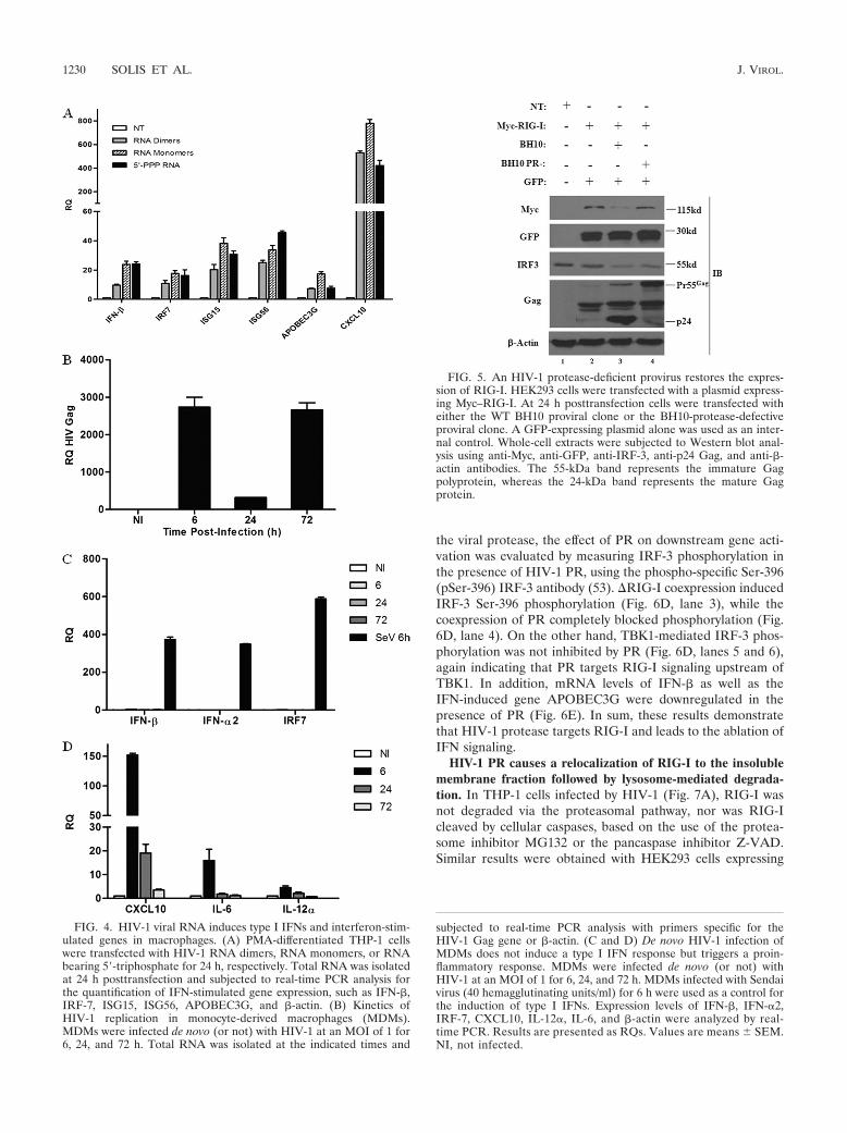
HIV-1 infection inhibits the type I IFN response. Severalreports, including our own, demonstrate that the induction ofthe type I IFN response is not detected during the early stagesof HIV-1 infection in CD4� T cells, macrophages, and imma-ture dendritic cells (51, 67, 70, 72, 77). To demonstrate thatHIV-1 monomeric and dimeric gRNAs act as functional RIG-Iligands in natural cellular targets of HIV-1 infection, the pro-files of type I IFN and IFN-stimulated gene expression weredetermined in phorbol myristate acetate (PMA)-differentiatedmonocytic THP-1 cells (Fig. 4A). HIV-1 RNA dimers andmonomers led to 10- to 24-fold increases in IFN-� gene ex-pression, respectively. The classic antiviral ISGs IRF-7, ISG15,
and ISG56 were induced 17-, 38-, and 34-fold, respectively, bythe monomeric gRNA and 11-, 20-, and 25-fold, respectively,by the dimeric gRNA. The antiviral ISG APOBEC3G wasinduced 17-fold and 7-fold by the monomers and dimers, re-spectively. Levels of CXCL10 mRNA, a gene dually regulatedby NF-�B and IRFs, were induced 500- to 800-fold in PMA-differentiated THP-1 cells. These findings demonstrate thatHIV-1 viral RNA can induce a type I IFN response in macro-phages.
However, de novo HIV-1 infection of primary monocyte-derived macrophages (MDMs) did not lead to a noticeabletype I IFN response (Fig. 4C). MDMs were permissive toHIV-1 replication, as demonstrated by the detection of HIV-1Gag transcripts as early as 6 h postinfection; a sharp increasein Gag mRNA levels at 72 h corresponded to the production ofnewly synthesized HIV-1 Gag mRNA (Fig. 4B). mRNA levelsof IFN-�, IFN-�2, and the IFN-stimulated gene IRF-7 wereunchanged after infection, whereas Sendai virus infection in-duced a strong antiviral response in MDMs (Fig. 4C). In con-trast, an upregulation of the proinflammatory NF-�B-stimu-lated genes CXCL10, IL-6, and IL-12� was detected at 6 h butdecreased to near-basal levels by 72 h (Fig. 4D). These resultsstrongly suggest the existence of an evasion mechanism thattargets the RIG-I signaling pathway during HIV-1 infection.
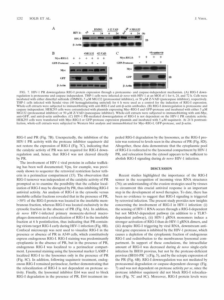
HIV-1 protease targets RIG-I. Many viruses circumvent theinnate antiviral response by using viral proteins to antagonizethe RIG-I/MAVS pathway at different levels (6). Viral pro-teases, in particular, are known to abrogate the RIG-I/MAVSpathway through the direct degradation of RIG-I (52) or thecleavage of the adaptor MAVS (38, 45). To investigate a po-tential role for the HIV-1 protease (PR) in the inhibition ofRIG-I signaling, a sequential transfection of RIG-I followed24 h later by the WT BH10 proviral clone or the BH10-pro-tease-defective proviral clone was performed (Fig. 5). Thetransduction of BH10 decreased RIG-I protein levels by �3-fold, whereas RIG-I protein expression was unchanged in thepresence of a PR-defective BH10 provirus (Fig. 5, lanes 3 and4). Analysis of endogenous IRF-3 confirmed that IRF-3 deg-radation was not protease dependent, consistent with previousreports demonstrating that Vif and Vpr target IRF-3 for deg-radation (13, 51). This finding thus argues for a specific role forHIV-1 PR in the disruption of RIG-I signaling.
To further examine the role of PR in the inhibition of RIG-Isignaling, increasing amounts of HIV-1 PR were coexpressedwith constitutively active RIG-I (�RIG-I; aa 1 to 229); theactivation of the IFNB promoter was dramatically decreased byPR in a dose-dependent manner (Fig. 6A). In contrast, theactivation of the downstream kinase TBK1 was not affected,arguing that the protease targets RIG-I signaling upstream ofTBK1. Concomitant with the inhibition of the RIG-I-mediatedactivation of the IFNB promoter, the level of RIG-I proteinexpression was also reduced in the presence of increasingamounts of the HIV-1 PR (Fig. 6B). To verify that RIG-I isspecifically targeted by PR, the downregulation of other sig-naling proteins by PR was also examined (Fig. 6C). Regardlessof the presence of PR, no changes in the expression of MDA5or IRF-3 were observed. Also, RIG-I transcript levels were notaffected by PR expression, indicating that RIG-I is specificallytargeted by the protease at the protein level (data not shown).
To further characterize the inhibition of the IFN response by
FIG. 3. RIG-I overexpression inhibits HIV-1 replication in single-cycle infections. Total RNA was extracted from HEK293 cells 24 hafter cotransfection of pcDNA3, Myc–RIG-I, and Myc–RIG-I C alongwith the BH10 proviral clone. BH10 transfection prior to Myc–RIG-Itransfection is shown by the gray bar. cDNA samples were subjected toreal-time PCR analysis with primers specific for the HIV-1 Gag geneor �-actin. HIV-1 replication is represented as an RQ based on therelative expression of the HIV-1 Gag gene versus �-actin as a refer-ence gene. Values are means SEM. Statistical relevance was evalu-ated using a P value of �0.05 (*).
VOL. 85, 2011 HIV-1 PROTEASE INHIBITS RIG-I SIGNALING 1229
the viral protease, the effect of PR on downstream gene acti-vation was evaluated by measuring IRF-3 phosphorylation inthe presence of HIV-1 PR, using the phospho-specific Ser-396(pSer-396) IRF-3 antibody (53). �RIG-I coexpression inducedIRF-3 Ser-396 phosphorylation (Fig. 6D, lane 3), while thecoexpression of PR completely blocked phosphorylation (Fig.6D, lane 4). On the other hand, TBK1-mediated IRF-3 phos-phorylation was not inhibited by PR (Fig. 6D, lanes 5 and 6),again indicating that PR targets RIG-I signaling upstream ofTBK1. In addition, mRNA levels of IFN-� as well as theIFN-induced gene APOBEC3G were downregulated in thepresence of PR (Fig. 6E). In sum, these results demonstratethat HIV-1 protease targets RIG-I and leads to the ablation ofIFN signaling.
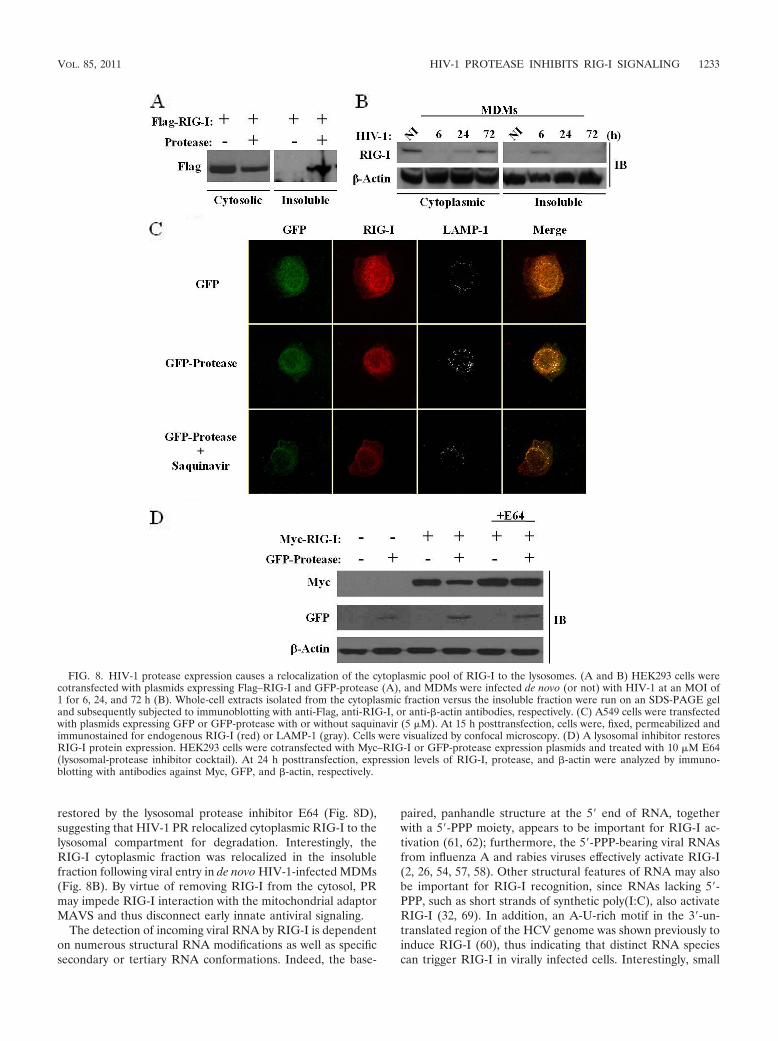
HIV-1 PR causes a relocalization of RIG-I to the insolublemembrane fraction followed by lysosome-mediated degrada-tion. In THP-1 cells infected by HIV-1 (Fig. 7A), RIG-I wasnot degraded via the proteasomal pathway, nor was RIG-Icleaved by cellular caspases, based on the use of the protea-some inhibitor MG132 or the pancaspase inhibitor Z-VAD.Similar results were obtained with HEK293 cells expressing
FIG. 4. HIV-1 viral RNA induces type I IFNs and interferon-stim-ulated genes in macrophages. (A) PMA-differentiated THP-1 cellswere transfected with HIV-1 RNA dimers, RNA monomers, or RNAbearing 5�-triphosphate for 24 h, respectively. Total RNA was isolatedat 24 h posttransfection and subjected to real-time PCR analysis forthe quantification of IFN-stimulated gene expression, such as IFN-�,IRF-7, ISG15, ISG56, APOBEC3G, and �-actin. (B) Kinetics ofHIV-1 replication in monocyte-derived macrophages (MDMs).MDMs were infected de novo (or not) with HIV-1 at an MOI of 1 for6, 24, and 72 h. Total RNA was isolated at the indicated times and
subjected to real-time PCR analysis with primers specific for theHIV-1 Gag gene or �-actin. (C and D) De novo HIV-1 infection ofMDMs does not induce a type I IFN response but triggers a proin-flammatory response. MDMs were infected de novo (or not) withHIV-1 at an MOI of 1 for 6, 24, and 72 h. MDMs infected with Sendaivirus (40 hemagglutinating units/ml) for 6 h were used as a control forthe induction of type I IFNs. Expression levels of IFN-�, IFN-�2,IRF-7, CXCL10, IL-12�, IL-6, and �-actin were analyzed by real-time PCR. Results are presented as RQs. Values are means SEM.NI, not infected.
FIG. 5. An HIV-1 protease-deficient provirus restores the expres-sion of RIG-I. HEK293 cells were transfected with a plasmid express-ing Myc–RIG-I. At 24 h posttransfection cells were transfected witheither the WT BH10 proviral clone or the BH10-protease-defectiveproviral clone. A GFP-expressing plasmid alone was used as an inter-nal control. Whole-cell extracts were subjected to Western blot anal-ysis using anti-Myc, anti-GFP, anti-IRF-3, anti-p24 Gag, and anti-�-actin antibodies. The 55-kDa band represents the immature Gagpolyprotein, whereas the 24-kDa band represents the mature Gagprotein.
1230 SOLIS ET AL. J. VIROL.

FIG. 6. HIV-1 PR targets RIG-I and interferes with IFN-� activation. (A) HIV-1 PR downregulates the IFNB promoter. HEK293 cells weretransfected with an IFNB-pGL3 reporter plasmid, the pEGFP-C1 vector, or expression plasmids encoding �RIG-I and TBK1 along with increasingamounts (50 ng, 100 ng, 200 ng, and 500 ng) of the GFP-protease expression construct. IFNB promoter activity was measured at 24 hposttransfection. Values represent means SEM. (B) RIG-I is downregulated in the presence of HIV-I PR. HEK293 cells were cotransfected withexpression vectors for Myc–RIG-I (2 �g) and increasing amounts (2 �g, 5 �g, and 10 �g) of GFP-protease as indicated. Cell lysates were subjectedto Western blot analysis, and expression levels of RIG-I, protease, and �-actin were analyzed by immunoblotting with antibodies against Myc, GFP,and �-actin, respectively. (C) HIV-1 PR specifically targets RIG-I. HEK293 cells were cotransfected with plasmids expressing Myc–IRF-3,Flag-MDA5, and GFP-protease. Cell lysates were subsequently subjected to immunoblotting with anti-Myc, anti-Flag, anti-GFP, and anti-�-actinantibodies. (D) HIV-1 PR inhibits �RIG-I-mediated activation of IRF-3. HEK293 cells were transfected with 500 ng of Myc–IRF-3, 1 �gMyc-�RIG-I or GFP-TBK1, and 1 �g of GFP-protease expression plasmids as indicated. Whole-cell extracts were analyzed by immunoblotting forIRF-3 pSer-396, RIG-I, GFP-TBK1, GFP-protease, and �-actin. (E) HIV-1 PR downregulates IFN-stimulated gene expression. HEK293 cells weretransfected with Myc-�RIG-I along with the GFP-protease expression plasmid. cDNA samples were subjected to real-time PCR analysis withprimers specific for IFN-�, APOBEC3G, and �-actin. Results are presented as RQs. Values are means SEM.
1231
RIG-I and PR (Fig. 7B). Unexpectedly, the inhibition of theHIV-1 PR activity with the protease inhibitor saquinavir didnot restore the expression of RIG-I (Fig. 7C), indicating thatthe catalytic activity of PR was not required for RIG-I down-regulation and, hence, that RIG-I was not cleaved directlyby PR.
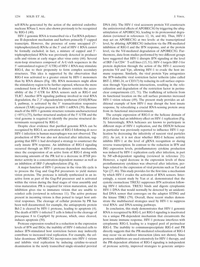
The involvement of HIV-1 viral proteins in cellular traffick-ing has been well documented; Vpu, for example, was previ-ously shown to sequester the retroviral restriction factor teth-erin in a perinuclear compartment (15). The observation thatRIG-I turnover was independent of the catalytic activity of PRprompted us to examine the possibility that the cellular local-ization of RIG-I may be disrupted by PR, thus inhibiting RIG-Iantiviral activity. An analysis of RIG-I in the cytosolic versusinsoluble cellular fractions revealed that in the presence of PR, 50% of the RIG-I protein was located in the insoluble mem-branous fraction, whereas RIG-I was located exclusively in thecytosolic fraction in the absence of PR (Fig. 8A). In addition,de novo HIV-1-infected primary monocyte-derived macro-phages demonstrated a relocalization of RIG-I in the insolublefraction at 6 h postinfection, thus indicating that PR-contain-ing virions target RIG-I early during HIV-1 infection (Fig. 8B).Confocal microscopy was next used to visualize RIG-I in thepresence or absence of PR in A549 cells, which constitutivelyexpress endogenous RIG-I. RIG-I staining was predominantlycytoplasmic in the absence of PR, but in the presence of PR,endogenous RIG-I was localized to a perinuclear compart-ment. Lysosomal staining using the lysosomal marker LAMP-1localized RIG-I to the lysosomes only in the presence of PR(Fig. 8C). In addition, following saquinavir treatment, endog-enous RIG-I remained perinuclear, further demonstrating thatthe relocalization of RIG-I is not dependent on protease ac-tivity. Finally, the lysosomal inhibitor E64 was used to blockRIG-I degradation in the presence of PR. E64 treatment im-
peded RIG-I degradation by the lysosomes, as the RIG-I pro-tein was restored to levels seen in the absence of PR (Fig. 8D).Altogether, these data demonstrate that the cytoplasmic poolof RIG-I is redirected to the lysosomal compartment by HIV-1PR, and relocation from the cytosol appears to be sufficient toabolish RIG-I signaling during de novo HIV-1 infection.
DISCUSSION
Recent studies highlighted the importance of the RIG-Isensor in the recognition of incoming virus RNA structures(76); an understanding of the evasion strategies used by virusesto circumvent this crucial antiviral response is an importantstep in the development of novel therapies. To date, there hasbeen no evidence to suggest that RIG-I signaling is triggeredby retroviral infection. The present study provides new insightsconcerning the involvement of RIG-I in HIV-1 infection: (i)the sensing of HIV-1 RNA occurs through a RIG-I-dependentbut not MDA5-dependent pathway (in addition to a TLR7-dependent pathway), (ii) HIV-1 gRNA monomers induce astronger activation of RIG-I than do HIV-1 gRNA dimers, and(iii) despite RIG-I triggering by viral RNA, downstream anti-viral gene expression is inhibited by the HIV-1 protease, whichcauses a depletion of the cytoplasmic fraction of endogenousRIG-I and redistribution to the membranous lysosomal com-partment. In support of these conclusions, the intracellularamount of RIG-I was decreased during de novo single-cycleinfection by BH10 provirus, but not by the protease-deficientprovirus (BH10-PR�) (Fig. 5), and by the ectopic expression ofthe PR (Fig. 6B). RIG-I downregulation was not mediated byproteasomal degradation or caspase-dependent cleavage (Fig.7) and was not dependent on protease activity per se, since theprotease inhibitor saquinavir did not block RIG-I relocaliza-tion (Fig. 7C and 8C). Moreover, RIG-I protein levels were
FIG. 7. HIV-1 PR downregulates RIG-I protein expression through a proteasome- and caspase-independent mechanism. (A) RIG-I down-regulation is proteasome and caspase independent. THP-1 cells were infected de novo with HIV-1 at an MOI of 1 for 6, 24, and 72 h. Cells wereincubated with either dimethyl sulfoxide (DMSO), 5 �M MG132 (proteasomal inhibitor), or 50 �M Z-VAD (pancaspase inhibitor), respectively.THP-1 cells infected with Sendai virus (40 hemagglutinating units/ml) for 6 h were used as a control for the induction of RIG-I expression.Whole-cell extracts were subjected to immunoblotting with anti-RIG-I and anti-�-actin antibodies. (B) RIG-I downregulation is proteasome andcaspase independent. HEK293 cells were cotransfected with plasmids expressing Myc–RIG-I and GFP-protease and incubated with either 5 �MMG132 (proteasomal inhibitor) or 50 �M Z-VAD (pancaspase inhibitor). Whole-cell extracts were subjected to immunoblotting with anti-Myc,anti-GFP, and anti-�-actin antibodies. (C) HIV-1 PR-mediated downregulation of RIG-I is not dependent on the HIV-1 PR catalytic activity.HEK293 cells were transfected with Myc–RIG-I or GFP-protease expression plasmids and incubated with 5 �M saquinavir. At 24 h posttrans-fection, whole-cell extracts were subjected to Western blot analysis and immunoblotted for Myc–RIG-I, GFP-protease, and �-actin.
1232 SOLIS ET AL. J. VIROL.
restored by the lysosomal protease inhibitor E64 (Fig. 8D),suggesting that HIV-1 PR relocalized cytoplasmic RIG-I to thelysosomal compartment for degradation. Interestingly, theRIG-I cytoplasmic fraction was relocalized in the insolublefraction following viral entry in de novo HIV-1-infected MDMs(Fig. 8B). By virtue of removing RIG-I from the cytosol, PRmay impede RIG-I interaction with the mitochondrial adaptorMAVS and thus disconnect early innate antiviral signaling.
The detection of incoming viral RNA by RIG-I is dependenton numerous structural RNA modifications as well as specificsecondary or tertiary RNA conformations. Indeed, the base-
paired, panhandle structure at the 5� end of RNA, togetherwith a 5�-PPP moiety, appears to be important for RIG-I ac-tivation (61, 62); furthermore, the 5�-PPP-bearing viral RNAsfrom influenza A and rabies viruses effectively activate RIG-I(2, 26, 54, 57, 58). Other structural features of RNA may alsobe important for RIG-I recognition, since RNAs lacking 5�-PPP, such as short strands of synthetic poly(I:C), also activateRIG-I (32, 69). In addition, an A-U-rich motif in the 3�-un-translated region of the HCV genome was shown previously toinduce RIG-I (60), thus indicating that distinct RNA speciescan trigger RIG-I in virally infected cells. Interestingly, small
FIG. 8. HIV-1 protease expression causes a relocalization of the cytoplasmic pool of RIG-I to the lysosomes. (A and B) HEK293 cells werecotransfected with plasmids expressing Flag–RIG-I and GFP-protease (A), and MDMs were infected de novo (or not) with HIV-1 at an MOI of1 for 6, 24, and 72 h (B). Whole-cell extracts isolated from the cytoplasmic fraction versus the insoluble fraction were run on an SDS-PAGE geland subsequently subjected to immunoblotting with anti-Flag, anti-RIG-I, or anti-�-actin antibodies, respectively. (C) A549 cells were transfectedwith plasmids expressing GFP or GFP-protease with or without saquinavir (5 �M). At 15 h posttransfection, cells were, fixed, permeabilized andimmunostained for endogenous RIG-I (red) or LAMP-1 (gray). Cells were visualized by confocal microscopy. (D) A lysosomal inhibitor restoresRIG-I protein expression. HEK293 cells were cotransfected with Myc–RIG-I or GFP-protease expression plasmids and treated with 10 �M E64(lysosomal-protease inhibitor cocktail). At 24 h posttransfection, expression levels of RIG-I, protease, and �-actin were analyzed by immuno-blotting with antibodies against Myc, GFP, and �-actin, respectively.
VOL. 85, 2011 HIV-1 PROTEASE INHIBITS RIG-I SIGNALING 1233
self-RNAs generated by the action of the antiviral endoribo-nuclease RNase L were also shown previously to be recognizedby RIG-I (40).
HIV-1 genomic RNA is transcribed via a Tat/RNA polymer-ase II-dependent mechanism and harbors primarily 5�-cappedtranscripts. However, the presence of some uncapped (5�-triphosphorylated) RNAs at the 5� end of HIV-1 RNA cannotbe formally excluded; in fact, a mixture of capped and 5�-triphosphorylated RNAs was previously detected in producercells and virions at early stages after virus entry (44). Severalstem-loop structures composed of A-U-rich sequences in the5�-untranslated region (5�-UTR) of HIV-1 RNA may stimulateRIG-I, due to their propensity to form complex secondarystructures. This idea is supported by the observation thatRIG-I was activated to a greater extent by HIV-1 monomersthan by RNA dimers (Fig. 1B). RNA monomers might allowthe stimulatory region to be further exposed, whereas the morecondensed form of RNA found in dimers restricts the acces-sibility of the 5�-UTR for RNA sensors such as RIG-I andTLR7. Another IFN signaling pathway involved in viral RNAdegradation, the 2�,5�-oligoadenylate synthetase (OAS)/RNaseL pathway, is activated by the 5� transactivation responsiveelement (TAR) region present in HIV-1 mRNA (39). Becausemuch of the HIV-1 genomic structure remains uncharacterized( 85%) (75), further structural analysis of the 5�-UTR and theviral genome is required to identify the precise structural de-terminants recognized by RIG-I.
Despite clear evidence that purified genomic RNA can berecognized by RIG-I, an activation of RIG-I following de novoHIV-1 infection in human macrophages was not observed. Theproduction of IFN was also not detected after infection, sug-gesting that HIV-1 had developed a strategy to counteract theearly innate IFN response. An inhibition of RIG-I signalingoccurred through an HIV-1 protease-dependent mechanism,since the coexpression of an active form of RIG-I along withincreasing amounts of the PR led to a decrease in IFNB pro-moter activity in a concentration-dependent manner as well asan inhibition of IRF-3 phosphorylation (Fig. 6).
A major function of HIV-1 protease in the virus life cycle isto process the Gag and Gag-Pol precursors to yield maturevirion proteins. The protease is initially synthesized in an in-active form as part of the Gag-Pol precursor and is activatedwithin the virion during the final stages of virus assembly andvirus maturation. PR is required for virion maturation, and itsinhibition gives rise to immature virions that are unable toreinfect cells (reviewed in reference 11). The active proteasepresent in incoming virions is thus poised to target host anti-viral responses. The cleavage of cellular proteins by PR hasbeen well documented; for example, the antiapoptotic proteinBcl-2 is cleaved by HIV-1 protease, leading to apoptosis (4).The death of HIV-1-infected T cells is linked to the cleavage ofprocaspase 8 to Casp8p41 by protease, which, once cleaved,induces apoptosis (50).
Protease expression resulted in a reduction in the expressionlevels of IFN and ISGs; the inability of HIV-1-infected cells toinduce IFN-stimulated host restriction factors may indirectlycontribute to increased viral replication. For example, the cel-lular deaminase APOBEC3G incorporates into HIV-1 virionsand inhibits viral replication by inducing cytidine-to-uracildeamination in the newly transcribed single-stranded proviral
DNA (66). The HIV-1 viral accessory protein Vif counteractsthe antiretroviral effects of APOBEC3G by triggering the ubiq-uitinylation of APOBEC3G, leading to its proteasomal degra-dation (reviewed in references 12, 16, and 66). Thus, HIV-1may act on APOBEC3G at two levels: at the transcriptionallevel, by ablating APOBEC3G induction via the PR-mediatedinhibition of RIG-I and the IFN response, and at the proteinlevel, via the Vif-mediated degradation of APOBEC3G. Fur-thermore, data from studies performed by two different groupshave suggested that HIV-1 disrupts IFN signaling at the levelof IRF-3 in CD4� T-cell lines (13, 51). HIV-1 targets IRF-3 forprotein depletion through the action of the HIV-1 accessoryproteins Vif and Vpr, allowing HIV-1 to evade the host im-mune response. Similarly, the viral protein Vpu antagonizesthe IFN-inducible viral restriction factor tetherin (also calledBST-2, HM1.24, or CD317) by reducing its cell surface expres-sion through Vpu-tetherin interactions, resulting in the relo-calization and degradation of the restriction factor in perinu-clear compartments (15, 71). The trafficking of tetherin fromits functional location on the cell surface promotes increasedHIV-1 virion release (49). The present study provides an ad-ditional example of how HIV-1 may disrupt the host innateresponse, by relocalizing a crucial RNA-sensing protein awayfrom its functional microenvironment.
The ectopic expression of RIG-I or the helicase domain ofRIG-I alone had an inhibitory effect on HIV-1 replication (Fig.3). Interestingly, RNA helicases are known to participate indifferent steps of HIV-1 replication, and the DDX30 helicasein particular was previously reported to influence HIV-1 rep-lication by decreasing the infectivity of nascent viral particles(81). As yet, it is not clear whether RIG-I helicase activityinhibits HIV-1 at the level of gRNA recognition or duringreverse transcription. In contrast to the reduction in IFN andISG expression levels, proinflammatory cytokine productionwas induced by HIV-1 replication early after infection, in partby NF-�B-dependent signaling (reviewed in reference 21).However, a rapid decrease in the expression levels of theseproinflammatory cytokines was observed after infection, per-haps related to the expression of viral proteins such as Tat andVpr (27, 46). This study provides for the first time a mechanismby which HIV-1 evades the activation of RNA sensors. Inter-estingly, a recent study by Yan et al. demonstrated that thecystolic exonuclease TREX1 suppresses IFN activation follow-ing HIV-1 infection. TREX1 binds and digests cytoplasmicHIV-1 DNA that would normally be detected by an unidenti-fied DNA sensor that converges on the adaptor STING andthe kinase TBK1 (79). Overall, these recent studies demon-strate the multifaceted strategies used by HIV-1 to suppressviral RNA- and DNA-sensing pathways.
In conclusion, this study demonstrates that HIV-1 genomicRNA is recognized by RIG-I, yet RIG-I signaling is inactivatedvia a unique PR-dependent mechanism that circumvents thehost innate immune response. HIV-1 protease interferes withcytoplasmic RIG-I, leading to a trapped pool of perinuclearRIG-I. The inability to coimmunoprecipitate RIG-I and PRdirectly suggests that the PR-mediated relocalization of RIG-Imay occur via an indirect interaction (data not shown). Sinceprotease inhibitors are used extensively for HIV-1 therapy, andthe PR-dependent ablation of RIG-I signaling is independentof protease activity, improved strategies to generate antipro-
1234 SOLIS ET AL. J. VIROL.
tease drugs will be required to combat this specific mode ofhost immune response evasion.
ACKNOWLEDGMENTS
This research was supported by grants from the Canadian Instituteof Health Research (CIHR) and the Canadian Foundation for AIDSResearch (CANFAR). M.S. was supported by a CIHR doctoral fel-lowship. P.N. was supported by an FRSQ doctoral fellowship.
We thank the members of the Molecular Oncology Group and theMcGill AIDS Center, especially Jorge Martinez-Cajas and MaureenOliveira, for helpful discussions and reagents. We also thank RobertSilverman and Shizuo Akira for reagents used in this study.
REFERENCES
1. Baenziger, S., et al. 2009. Triggering TLR7 in mice induces immune activa-tion and lymphoid system disruption, resembling HIV-mediated pathology.Blood 113:377–388.
2. Baum, A., R. Sachidanandam, and A. Garcia-Sastre. 2010. Preference ofRIG-I for short viral RNA molecules in infected cells revealed by next-generation sequencing. Proc. Natl. Acad. Sci. U. S. A. 107:16303–16308.
3. Beignon, A. S., et al. 2005. Endocytosis of HIV-1 activates plasmacytoiddendritic cells via Toll-like receptor-viral RNA interactions. J. Clin. Invest.115:3265–3275.
4. Blanco, R., L. Carrasco, and I. Ventoso. 2003. Cell killing by HIV-1 protease.J. Biol. Chem. 278:1086–1093.
5. Boasso, A., and G. M. Shearer. 2008. Chronic innate immune activation as acause of HIV-1 immunopathogenesis. Clin. Immunol. 126:235–242.
6. Bowie, A. G., and L. Unterholzner. 2008. Viral evasion and subversion ofpattern-recognition receptor signalling. Nat. Rev. Immunol. 8:911–922.
7. Brenchley, J. M., et al. 2006. Microbial translocation is a cause of systemicimmune activation in chronic HIV infection. Nat. Med. 12:1365–1371.
8. Chin, M. P., J. Chen, O. A. Nikolaitchik, and W. S. Hu. 2007. Moleculardeterminants of HIV-1 intersubtype recombination potential. Virology 363:437–446.
9. Chin, M. P., T. D. Rhodes, J. Chen, W. Fu, and W. S. Hu. 2005. Identificationof a major restriction in HIV-1 intersubtype recombination. Proc. Natl.Acad. Sci. U. S. A. 102:9002–9007.
10. Clerzius, G., et al. 2009. ADAR1 interacts with PKR during human immu-nodeficiency virus infection of lymphocytes and contributes to viral replica-tion. J. Virol. 83:10119–10128.
11. Coffin, J. M. 1997. Retroviruses. Cold Spring Harbor Laboratory Press,Plainview, NY.
12. Cullen, B. R. 2006. Role and mechanism of action of the APOBEC3 familyof antiretroviral resistance factors. J. Virol. 80:1067–1076.
13. Doehle, B. P., F. Hladik, J. P. McNevin, M. J. McElrath, and M. Gale, Jr.2009. Human immunodeficiency virus type 1 mediates global disruption ofinnate antiviral signaling and immune defenses within infected cells. J. Virol.83:10395–10405.
14. Douville, R. N., and J. Hiscott. 2010. The interface between the innateinterferon response and expression of host retroviral restriction factors.Cytokine 52:108–115.
15. Dube, M., et al. 2010. Antagonism of tetherin restriction of HIV-1 release byVpu involves binding and sequestration of the restriction factor in a perinu-clear compartment. PLoS Pathog. 6:e1000856.
16. Ehrlich, E. S., and X. F. Yu. 2006. Lentiviral Vif: viral hijacker of theubiquitin-proteasome system. Int. J. Hematol. 83:208–212.
17. Frigault, M. M., J. Lacoste, J. L. Swift, and C. M. Brown. 2009. Live-cellmicroscopy—tips and tools. J. Cell Sci. 122:753–767.
18. Fu, W., R. J. Gorelick, and A. Rein. 1994. Characterization of human im-munodeficiency virus type 1 dimeric RNA from wild-type and protease-defective virions. J. Virol. 68:5013–5018.
19. Gringhuis, S. I., et al. 2010. HIV-1 exploits innate signaling by TLR8 andDC-SIGN for productive infection of dendritic cells. Nat. Immunol. 11:419–426.
20. Heil, F., et al. 2004. Species-specific recognition of single-stranded RNA viaToll-like receptor 7 and 8. Science 303:1526–1529.
21. Herbein, G., and A. Varin. 2010. The macrophage in HIV-1 infection: fromactivation to deactivation? Retrovirology 7:33.
22. Herbeuval, J. P., and G. M. Shearer. 2007. HIV-1 immunopathogenesis: howgood interferon turns bad. Clin. Immunol. 123:121–128.
23. Hiscott, J., R. Lin, P. Nakhaei, and S. Paz. 2006. MasterCARD: a pricelesslink to innate immunity. Trends Mol. Med. 12:53–56.
24. Honda, K., and T. Taniguchi. 2006. IRFs: master regulators of signalling byToll-like receptors and cytosolic pattern-recognition receptors. Nat. Rev.Immunol. 6:644–658.
25. Hong, S., J. Cao, and Y. T. Tu. 2009. Evolution of HIV-1 in a patientpopulation failing multiple-drug therapy. Microbiol. Immunol. 53:535–539.
26. Hornung, V., et al. 2006. 5�-triphosphate RNA is the ligand for RIG-I.Science 314:994–997.
27. Ito, M., et al. 1998. HIV type 1 Tat protein inhibits interleukin 12 productionby human peripheral blood mononuclear cells. AIDS Res. Hum. Retrovi-ruses 14:845–849.
28. Jalalirad, M., and M. Laughrea. 2010. Formation of immature and maturegenomic RNA dimers in wild-type and protease-inactive HIV-1: differentialroles of the Gag polyprotein, nucleocapsid proteins NCp15, NCp9, NCp7,and the dimerization initiation site. Virology 407:225–236.
29. Kafaie, J., R. Song, L. Abrahamyan, A. J. Mouland, and M. Laughrea. 2008.Mapping of nucleocapsid residues important for HIV-1 genomic RNAdimerization and packaging. Virology 375:592–610.
30. Kang, D. C., et al. 2002. mda-5: an interferon-inducible putative RNA heli-case with double-stranded RNA-dependent ATPase activity and melanomagrowth-suppressive properties. Proc. Natl. Acad. Sci. U. S. A. 99:637–642.
31. Kato, H., et al. 2005. Cell type-specific involvement of RIG-I in antiviralresponse. Immunity 23:19–28.
32. Kato, H., et al. 2008. Length-dependent recognition of double-strandedribonucleic acids by retinoic acid-inducible gene-I and melanoma differen-tiation-associated gene 5. J. Exp. Med. 205:1601–1610.
33. Kato, H., et al. 2006. Differential roles of MDA5 and RIG-I helicases in therecognition of RNA viruses. Nature 441:101–105.
34. Kawai, T., and S. Akira. 2006. Innate immune recognition of viral infection.Nat. Immunol. 7:131–137.
35. Kawai, T., et al. 2005. IPS-1, an adaptor triggering RIG-I- and Mda5-mediated type I interferon induction. Nat. Immunol. 6:981–988.
36. Kirchhoff, F. 2010. Immune evasion and counteraction of restriction factorsby HIV-1 and other primate lentiviruses. Cell Host Microbe 8:55–67.
37. Li, K., Z. Chen, N. Kato, M. Gale, Jr., and S. M. Lemon. 2005. Distinctpoly(I-C) and virus-activated signaling pathways leading to interferon-betaproduction in hepatocytes. J. Biol. Chem. 280:16739–16747.
38. Lin, R., et al. 2006. Dissociation of a MAVS/IPS-1/VISA/Cardif-IKKepsilonmolecular complex from the mitochondrial outer membrane by hepatitis Cvirus NS3-4A proteolytic cleavage. J. Virol. 80:6072–6083.
39. Maitra, R. K., et al. 1994. HIV-1 TAR RNA has an intrinsic ability toactivate interferon-inducible enzymes. Virology 204:823–827.
40. Malathi, K., B. Dong, M. Gale, Jr., and R. H. Silverman. 2007. Smallself-RNA generated by RNase L amplifies antiviral innate immunity. Nature448:816–819.
41. Maniatis, T., et al. 1998. Structure and function of the interferon-betaenhanceosome. Cold Spring Harb. Symp. Quant. Biol. 63:609–620.
42. Matikainen, S., et al. 2006. Tumor necrosis factor alpha enhances influenzaA virus-induced expression of antiviral cytokines by activating RIG-I geneexpression. J. Virol. 80:3515–3522.
43. Meier, A., et al. 2007. MyD88-dependent immune activation mediated byhuman immunodeficiency virus type 1-encoded Toll-like receptor ligands.J. Virol. 81:8180–8191.
44. Menees, T. M., B. Muller, and H. G. Krausslich. 2007. The major 5� end ofHIV type 1 RNA corresponds to G456. AIDS Res. Hum. Retroviruses23:1042–1048.
45. Meylan, E., et al. 2005. Cardif is an adaptor protein in the RIG-I antiviralpathway and is targeted by hepatitis C virus. Nature 437:1167–1172.
46. Mirani, M., et al. 2002. HIV-1 protein Vpr suppresses IL-12 production fromhuman monocytes by enhancing glucocorticoid action: potential implicationsof Vpr coactivator activity for the innate and cellular immunity deficitsobserved in HIV-1 infection. J. Immunol. 169:6361–6368.
47. Moore, M. D., and W. S. Hu. 2009. HIV-1 RNA dimerization: it takes two totango. AIDS Rev. 11:91–102.
48. Nakhaei, P., P. Genin, A. Civas, and J. Hiscott. 2009. RIG-I-like receptors:sensing and responding to RNA virus infection. Semin. Immunol. 21:215–222.
49. Neil, S. J., T. Zang, and P. D. Bieniasz. 2008. Tetherin inhibits retrovirusrelease and is antagonized by HIV-1 Vpu. Nature 451:425–430.
50. Nie, Z., et al. 2002. HIV-1 protease processes procaspase 8 to cause mito-chondrial release of cytochrome c, caspase cleavage and nuclear fragmenta-tion. Cell Death Differ. 9:1172–1184.
51. Okumura, A., et al. 2008. HIV-1 accessory proteins VPR and Vif modulateantiviral response by targeting IRF-3 for degradation. Virology 373:85–97.
52. Papon, L., et al. 2009. The viral RNA recognition sensor RIG-I is degradedduring encephalomyocarditis virus (EMCV) infection. Virology 393:311–318.
53. Paz, S., et al. 2006. Induction of IRF-3 and IRF-7 phosphorylation followingactivation of the RIG-I pathway. Cell. Mol. Biol. (Noisy-le-Grand) 52:17–28.
54. Pichlmair, A., et al. 2006. RIG-I-mediated antiviral responses to single-stranded RNA bearing 5�-phosphates. Science 314:997–1001.
55. Plumet, S., et al. 2007. Cytosolic 5�-triphosphate ended viral leader transcriptof measles virus as activator of the RIG I-mediated interferon response.PLoS One 2:e279.
56. Randall, R. E., and S. Goodbourn. 2008. Interferons and viruses: an interplaybetween induction, signalling, antiviral responses and virus countermeasures.J. Gen. Virol. 89:1–47.
57. Rehwinkel, J. 2010. Exposing viruses: RNA patterns sensed by RIG-I-likereceptors. J. Clin. Immunol. 30:491–495.
VOL. 85, 2011 HIV-1 PROTEASE INHIBITS RIG-I SIGNALING 1235
58. Rehwinkel, J., et al. 2010. RIG-I detects viral genomic RNA during negative-strand RNA virus infection. Cell 140:397–408.
59. Romieu-Mourez, R., et al. 2006. Distinct roles for IFN regulatory factor(IRF)-3 and IRF-7 in the activation of antitumor properties of humanmacrophages. Cancer Res. 66:10576–10585.
60. Saito, T., D. M. Owen, F. Jiang, J. Marcotrigiano, and M. Gale, Jr. 2008.Innate immunity induced by composition-dependent RIG-I recognition ofhepatitis C virus RNA. Nature 454:523–527.
61. Schlee, M., et al. 2009. Recognition of 5� triphosphate by RIG-I helicaserequires short blunt double-stranded RNA as contained in panhandle ofnegative-strand virus. Immunity 31:25–34.
62. Schmidt, A., et al. 2009. 5�-triphosphate RNA requires base-paired struc-tures to activate antiviral signaling via RIG-I. Proc. Natl. Acad. Sci. U. S. A.106:12067–12072.
63. Servant, M. J., N. Grandvaux, B. R. tenOever, D. Duguay, R. Lin, and J.Hiscott. 2003. Identification of the minimal phosphoacceptor site requiredfor in vivo activation of interferon regulatory factor 3 in response to virus anddouble-stranded RNA. J. Biol. Chem. 278:9441–9447.
64. Seth, R. B., L. Sun, C. K. Ea, and Z. J. Chen. 2005. Identification andcharacterization of MAVS, a mitochondrial antiviral signaling protein thatactivates NF-kappaB and IRF 3. Cell 122:669–682.
65. Sharma, S., B. R. tenOever, N. Grandvaux, G. P. Zhou, R. Lin, and J.Hiscott. 2003. Triggering the interferon antiviral response through an IKK-related pathway. Science 300:1148–1151.
66. Sheehy, A. M., N. C. Gaddis, J. D. Choi, and M. H. Malim. 2002. Isolationof a human gene that inhibits HIV-1 infection and is suppressed by the viralVif protein. Nature 418:646–650.
67. Solis, M., et al. 2006. Gene expression profiling of the host response toHIV-1 B, C, or A/E infection in monocyte-derived dendritic cells. Virology352:86–99.
68. Song, R., J. Kafaie, L. Yang, and M. Laughrea. 2007. HIV-1 viral RNA isselected in the form of monomers that dimerize in a three-step protease-dependent process; the DIS of stem-loop 1 initiates viral RNA dimerization.J. Mol. Biol. 371:1084–1098.
69. Takahasi, K., et al. 2008. Nonself RNA-sensing mechanism of RIG-I heli-case and activation of antiviral immune responses. Mol. Cell 29:428–440.
70. Vahey, M. T., et al. 2002. Impact of viral infection on the gene expressionprofiles of proliferating normal human peripheral blood mononuclear cellsinfected with HIV type 1 RF. AIDS Res. Hum. Retroviruses 18:179–192.
71. Van Damme, N., et al. 2008. The interferon-induced protein BST-2 restrictsHIV-1 release and is downregulated from the cell surface by the viral Vpuprotein. Cell Host Microbe 3:245–252.
72. Vazquez, N., et al. 2005. Human immunodeficiency virus type 1-inducedmacrophage gene expression includes the p21 gene, a target for viral regu-lation. J. Virol. 79:4479–4491.
73. Vendrame, D., M. Sourisseau, V. Perrin, O. Schwartz, and F. Mammano.2009. Partial inhibition of human immunodeficiency virus replication by typeI interferons: impact of cell-to-cell viral transfer. J. Virol. 83:10527–10537.
74. Wang, F., et al. 2008. RIG-I mediates the co-induction of tumor necrosisfactor and type I interferon elicited by myxoma virus in primary humanmacrophages. PLoS Pathog. 4:e1000099.
75. Watts, J. M., et al. 2009. Architecture and secondary structure of an entireHIV-1 RNA genome. Nature 460:711–716.
76. Wilkins, C., and M. Gale, Jr. 2010. Recognition of viruses by cytoplasmicsensors. Curr. Opin. Immunol. 22:41–47.
77. Woelk, C. H., et al. 2004. Interferon gene expression following HIV type 1infection of monocyte-derived macrophages. AIDS Res. Hum. Retroviruses20:1210–1222.
78. Xu, L. G., et al. 2005. VISA is an adapter protein required for virus-triggeredIFN-beta signaling. Mol. Cell 19:727–740.
79. Yan, N., A. D. Regalado-Magdos, B. Stiggelbout, M. A. Lee-Kirsch, and J.Lieberman. 2010. The cytosolic exonuclease TREX1 inhibits the innateimmune response to human immunodeficiency virus type 1. Nat. Immunol.11:1005–1013.
80. Yoneyama, M., et al. 2004. The RNA helicase RIG-I has an essential func-tion in double-stranded RNA-induced innate antiviral responses. Nat. Im-munol. 5:730–737.
81. Zhou, Y., et al. 2008. The packaging of human immunodeficiency virus type1 RNA is restricted by overexpression of an RNA helicase DHX30. Virology372:97–106.
1236 SOLIS ET AL. J. VIROL.
Copyright © 2022 FDOKUMEN