A stem-loop RNA RIG-I agonist protects against acute and ...
22
ARTICLE A stem-loop RNA RIG-I agonist protects against acute and chronic SARS-CoV-2 infection in mice Tianyang Mao 1 , Benjamin Israelow 1,2 , Carolina Lucas 1 , Chantal B.F. Vogels 3 , Maria Luisa Gomez-Calvo 1 , Olga Fedorova 4,8 , Mallery I. Breban 3 , Bridget L. Menasche 5 , Huiping Dong 1 , Melissa Linehan 1 , Yale SARS-CoV-2 Genome Surveillance Initiative, Craig B. Wilen 1,5 , Marie L. Landry 2,5 , Nathan D. Grubaugh 3,6 , Anna M. Pyle 4,7,8 , and Akiko Iwasaki 1,3,4,8 As SARS-CoV-2 continues to cause morbidity and mortality around the world, there is an urgent need for the development of effective medical countermeasures. Here, we assessed the antiviral capacity of a minimal RIG-I agonist, stem-loop RNA 14 (SLR14), in viral control, disease prevention, post-infection therapy, and cross-variant protection in mouse models of SARS- CoV-2 infection. A single dose of SLR14 prevented viral infection in the lower respiratory tract and development of severe disease in a type I interferon (IFN-I)–dependent manner. SLR14 demonstrated remarkable prophylactic protective capacity against lethal SARS-CoV-2 infection and retained considerable efficacy as a therapeutic agent. In immunodeficient mice carrying chronic SARS-CoV-2 infection, SLR14 elicited near-sterilizing innate immunity in the absence of the adaptive immune system. In the context of infection with variants of concern (VOCs), SLR14 conferred broad protection against emerging VOCs. These findings demonstrate the therapeutic potential of SLR14 as a host-directed, broad-spectrum antiviral for early post- exposure treatment and treatment of chronically infected immunosuppressed patients. Introduction SARS-CoV-2 is an enveloped, positive-strand RNA virus that causes both upper and lower respiratory infection in humans and other animals (V’Kovski et al., 2021). As of October 26, 2021, the ongoing global COVID-19 pandemic caused by SARS-CoV- 2 has led to 243.86 million confirmed cases and 4.95 million deaths worldwide, inflicting widespread economic, sociological, and psychological damage. The clinical spectrum of SARS-CoV- 2 infection is wide. While most infections are asymptomatic or mild, older patients, particularly those with underlying medical comorbidities and male sex, are more likely to develop severe diseases involving acute respiratory distress syndrome, multi- organ failure, and death (Hu et al., 2021). Currently, there is a paucity of effective antivirals to treat COVID-19, with remdesivir and monoclonal antibodies demonstrating modest efficacy in a select subset of patients (Beigel et al., 2020; Taylor et al., 2021). To halt substantial morbidity and mortality from COVID-19 around the globe, in addition to the use of vaccines in preventing the disease, efforts are required to develop efficacious thera- peutics against SARS-CoV-2. Great strides made in the understanding of COVID-19 im- munology have provided crucial insights into the central role of IFN-I in host immune responses against SARS-CoV-2 infection (Lowery et al., 2021; Park and Iwasaki, 2020). The innate im- mune system utilizes host-encoded nucleic acid sensors, known as the pattern recognition receptors (PRRs), to surveil viral pathogens by detecting their pathogen-associated molecular patterns (Iwasaki and Medzhitov, 2015). Following SARS-CoV- 2 infection, multiple cytosolic PRRs, including RIG-I, MDA-5, and LGP2, mediate viral RNA recognition in infected lung epi- thelial cells and initiate front-line antiviral defense through IFN- I–dependent and independent mechanisms (Yamada et al., 2021; Yin et al., 2021). Upon secretion, IFN-I engages with its uni- versally expressed receptor in autocrine and paracrine fashions, stimulating the expression of a large network of IFN-stimulated genes (ISGs) to inhibit viral replication (Schneider et al., 2014) and cytokines and chemokines to recruit specialized immune cells to sites of infection. In the context of infection with SARS- CoV-2, IFNs appear to play dichotomous roles. While delayed ............................................................................................................................................................................. 1 Department of Immunobiology, Yale School of Medicine, New Haven, CT; 2 Department of Internal Medicine, Section of Infectious Diseases, Yale School of Medicine, New Haven, CT; 3 Department of Epidemiology of Microbial Diseases, Yale School of Public Health, New Haven, CT; 4 Department of Molecular, Cellular and Developmental Biology, Yale University, New Haven, CT; 5 Department of Laboratory Medicine, Yale School of Medicine, New Haven, CT; 6 Department of Ecology and Evolutionary Biology, Yale University, New Haven, CT; 7 Department of Chemistry, Yale University, New Haven, CT; 8 Howard Hughes Medical Institute, Chevy Chase, MD. Correspondence to Akiko Iwasaki: [email protected] Yale SARS-CoV-2 Genome Surveillance Initiative members: Tara Alpert, Anderson F. Brito, Rebecca Earnest, Joseph R. Fauver, Chaney C. Kalinich, Ketty Munyenyembe, Isabel M. Ott, Mary E. Petrone, Jessica Rothman, and Anne E. Watkins. © 2021 Mao et al. This article is available under a Creative Commons License (Attribution 4.0 International, as described at https://creativecommons.org/licenses/by/4.0/ ). Rockefeller University Press https://doi.org/10.1084/jem.20211818 1 of 17 J. Exp. Med. 2021 Vol. 219 No. 1 e20211818 Downloaded from http://rupress.org/jem/article-pdf/219/1/e20211818/1425498/jem_20211818.pdf by guest on 02 March 2022
-
Upload
khangminh22 -
Category
Documents
-
view
0 -
download
0
Transcript of A stem-loop RNA RIG-I agonist protects against acute and ...

ARTICLE
A stem-loop RNA RIG-I agonist protects againstacute and chronic SARS-CoV-2 infection in miceTianyang Mao1, Benjamin Israelow1,2, Carolina Lucas1, Chantal B.F. Vogels3, Maria Luisa Gomez-Calvo1, Olga Fedorova4,8,Mallery I. Breban3, Bridget L. Menasche5, Huiping Dong1, Melissa Linehan1, Yale SARS-CoV-2 Genome Surveillance Initiative, Craig B. Wilen1,5,Marie L. Landry2,5, Nathan D. Grubaugh3,6, Anna M. Pyle4,7,8, and Akiko Iwasaki1,3,4,8
As SARS-CoV-2 continues to cause morbidity and mortality around the world, there is an urgent need for the development ofeffective medical countermeasures. Here, we assessed the antiviral capacity of a minimal RIG-I agonist, stem-loop RNA 14(SLR14), in viral control, disease prevention, post-infection therapy, and cross-variant protection in mouse models of SARS-CoV-2 infection. A single dose of SLR14 prevented viral infection in the lower respiratory tract and development of severedisease in a type I interferon (IFN-I)–dependent manner. SLR14 demonstrated remarkable prophylactic protective capacityagainst lethal SARS-CoV-2 infection and retained considerable efficacy as a therapeutic agent. In immunodeficient micecarrying chronic SARS-CoV-2 infection, SLR14 elicited near-sterilizing innate immunity in the absence of the adaptive immunesystem. In the context of infection with variants of concern (VOCs), SLR14 conferred broad protection against emerging VOCs.These findings demonstrate the therapeutic potential of SLR14 as a host-directed, broad-spectrum antiviral for early post-exposure treatment and treatment of chronically infected immunosuppressed patients.
IntroductionSARS-CoV-2 is an enveloped, positive-strand RNA virus thatcauses both upper and lower respiratory infection in humansand other animals (V’Kovski et al., 2021). As of October 26, 2021,the ongoing global COVID-19 pandemic caused by SARS-CoV-2 has led to 243.86 million confirmed cases and 4.95 milliondeaths worldwide, inflicting widespread economic, sociological,and psychological damage. The clinical spectrum of SARS-CoV-2 infection is wide. While most infections are asymptomatic ormild, older patients, particularly those with underlying medicalcomorbidities and male sex, are more likely to develop severediseases involving acute respiratory distress syndrome, multi-organ failure, and death (Hu et al., 2021). Currently, there is apaucity of effective antivirals to treat COVID-19, with remdesivirand monoclonal antibodies demonstrating modest efficacy in aselect subset of patients (Beigel et al., 2020; Taylor et al., 2021).To halt substantial morbidity and mortality from COVID-19around the globe, in addition to the use of vaccines in preventingthe disease, efforts are required to develop efficacious thera-peutics against SARS-CoV-2.
Great strides made in the understanding of COVID-19 im-munology have provided crucial insights into the central role ofIFN-I in host immune responses against SARS-CoV-2 infection(Lowery et al., 2021; Park and Iwasaki, 2020). The innate im-mune system utilizes host-encoded nucleic acid sensors, knownas the pattern recognition receptors (PRRs), to surveil viralpathogens by detecting their pathogen-associated molecularpatterns (Iwasaki and Medzhitov, 2015). Following SARS-CoV-2 infection, multiple cytosolic PRRs, including RIG-I, MDA-5,and LGP2, mediate viral RNA recognition in infected lung epi-thelial cells and initiate front-line antiviral defense through IFN-I–dependent and independent mechanisms (Yamada et al., 2021;Yin et al., 2021). Upon secretion, IFN-I engages with its uni-versally expressed receptor in autocrine and paracrine fashions,stimulating the expression of a large network of IFN-stimulatedgenes (ISGs) to inhibit viral replication (Schneider et al., 2014)and cytokines and chemokines to recruit specialized immunecells to sites of infection. In the context of infection with SARS-CoV-2, IFNs appear to play dichotomous roles. While delayed
.............................................................................................................................................................................1Department of Immunobiology, Yale School of Medicine, New Haven, CT; 2Department of Internal Medicine, Section of Infectious Diseases, Yale School of Medicine, NewHaven, CT; 3Department of Epidemiology of Microbial Diseases, Yale School of Public Health, New Haven, CT; 4Department of Molecular, Cellular and DevelopmentalBiology, Yale University, New Haven, CT; 5Department of Laboratory Medicine, Yale School of Medicine, New Haven, CT; 6Department of Ecology and EvolutionaryBiology, Yale University, New Haven, CT; 7Department of Chemistry, Yale University, New Haven, CT; 8Howard Hughes Medical Institute, Chevy Chase, MD.
Correspondence to Akiko Iwasaki: [email protected]
Yale SARS-CoV-2 Genome Surveillance Initiative members: Tara Alpert, Anderson F. Brito, Rebecca Earnest, Joseph R. Fauver, Chaney C. Kalinich, Ketty Munyenyembe, IsabelM. Ott, Mary E. Petrone, Jessica Rothman, and Anne E. Watkins.
© 2021 Mao et al. This article is available under a Creative Commons License (Attribution 4.0 International, as described at https://creativecommons.org/licenses/by/4.0/).
Rockefeller University Press https://doi.org/10.1084/jem.20211818 1 of 17
J. Exp. Med. 2021 Vol. 219 No. 1 e20211818
Dow
nloaded from http://rupress.org/jem
/article-pdf/219/1/e20211818/1425498/jem_20211818.pdf by guest on 02 M
arch 2022

and prolonged IFN-I and type-III IFN (IFN-III) are associatedwith severe disease, an early, robust, and regulated productionof IFN is protective against COVID-19 (Carvalho et al., 2021;Lucas et al., 2020). This is well exemplified by the susceptibilityto life-threatening disease of SARS-CoV-2–infected individualswith inborn defects in IFN-I production and signaling or neu-tralizing autoantibodies against IFN-I (Bastard et al., 2020;Zhang et al., 2020). COVID-19 patients found with anti–IFN-Iautoantibodies demonstrate significantly delayed virologicalclearance relative to patients without such autoantibodies(Wang et al., 2021). In a mouse model of SARS-CoV-2 infection,early IFN-I blockade leads to exacerbation of disease severity(Wang et al., 2021). Collectively, these studies highlight thebeneficial role of IFN-I in SARS-CoV-2 infection and suggestinnate immune sensors as promising therapeutic targets to beharnessed for prevention and treatment of COVID-19.
The innate immune system can be pharmacologically mod-ulated to elicit tailored effector outputs with desired immuno-logical outcomes (Demaria et al., 2019; Vanpouille-Box et al.,2019). Given the importance of timely induction of IFN-I inSARS-CoV-2 infection, PRRs can be activated in a targetedmanner to induce antiviral protection (Goulet et al., 2013). Ourapproach in leveraging a synthetic activator of antiviral immu-nity to combat SARS-CoV-2 builds on our previous work dem-onstrating that short, tri-, or di-phosphorylated stem-loop RNAs(SLRs) act as specific and potent agonists for the cytosolic RNAsensor RIG-I (Linehan et al., 2018; Luo et al., 2011). SLRs aredesigned to mimic physiological double-stranded RNA ligandsfor RIG-I by stably folding into a minimal ligand containing 14-bp RNA duplex (hence the name SLR14) and a tri- or di-phosphorylated 59 terminus. Each SLR14 presents a single du-plex terminus and productively binds one RIG-I molecule. Theopposite end of the duplex is blocked with a stable RNA tetraloopto ensure that the RIG-I–SLR14 interaction is structurally de-fined and resistant to nucleases and strand dissociation. Unlikepolyinosinic/polycytidylic acid (poly(I:C)), which is a widelyused double-stranded RNA ligand of unknown structure recog-nized by a handful of PRRs, SLR14 specifically activates RIG-Iand triggers an IFN-I–dominant innate immune response (overan IFN-III response) characterized by the induction of multipleIFN-I members within 2 h of i.v. injection inmice (Linehan et al.,2018). Growing evidence suggests that recombinant IFN(rIFN)–based intervention during the early stage of COVID-19could provide desired clinical benefits in humans. However,rIFN therapy is costly (Nguyen et al., 2020) and can be renderedineffective by the induction of binding and/or neutralizing anti-drug antibodies (Giovannoni et al., 2002; Matsuda et al., 2012).In contrast, SLR14 is highly manufacturable and can elicit abroad and diverse IFN response. Altogether, with its syntheticsimplicity, chemically defined composition, targeted receptorbinding, breadth of downstream effector responses, and in vivopotency, SLR14 holds great promise as a new class of RNAtherapeutics that can be applied as antivirals against SARS-CoV-2.
While most individuals effectively clear SARS-CoV-2 infec-tion, growing evidence suggests that infection in immunocom-promised patients, such as those with severe forms of B cell and
antibody deficiency, can become chronic (Aydillo et al., 2020;Choi et al., 2020). In these patients, persistent infection can alsofoster continuous intrahost viral evolution and lead to furtheremergence of immune-evasive variants, likely as a result of se-lective pressure driven by insufficient natural or transferredantibodies. While some patient case reports have used conva-lescent plasma (CP) to treat chronic SARS-CoV-2 infection,currently there are no approved therapeutic options (Carvelliet al., 2020). Although vaccines currently approved againstSARS-CoV-2 are effective at preventing severe disease and deathin individuals with an intact immune system, their immuno-genicity is significantly attenuated in immunocompromisedpatients, eliciting suboptimal humoral immune responses(Deepak et al., 2021). Therefore, therapeutic strategies that exertstrong antiviral effect independent of adaptive immunity in thesetting of immunosuppression are in dire need.
Since the initial outbreak, multiple SARS-CoV-2 variantshave emerged with increased transmissibility and altered im-munogenicity. The B.1.1.7 lineage (Alpha) was first detected inSeptember 2020 and estimated to be more transmissible thanother lineages (Alpert et al., 2021; Washington et al., 2021).While mutations accumulated by B.1.1.7 seem to have negligibleimpact on infection- and vaccine-induced antibody immunity(Collier et al., 2021; Planas et al., 2021a), other variants havebeen found to acquire mutations on their spike proteins that canevade antibody targeting. Notably, variant B.1.351 (Beta) and P.1(Gamma) have both demonstrated considerable resistance toantibody binding and neutralization (Hoffmann et al., 2021;Zhou et al., 2021a). In addition, B.1.526 (Iota) has also exhibitedsome level of antibody evasion (Zhou et al., 2021b Preprint).More recently, B.1.617.2 (Delta) emerged with significantly en-hanced transmissibility (40% to 60% increase compared withB.1.1.7), a substantially higher viral replication rate, andheightened neutralization resistance to CP, monoclonal anti-bodies, and sera from vaccinated individuals (Li et al., 2021aPreprint; Lucas et al., 2021; Planas et al., 2021b). In addition,these variants harbor mutations outside the spike protein thatmay enable strong antagonism of the host antiviral innate im-munity. In this context, the containment of COVID-19 will re-quire prophylactic and therapeutic antiviral strategies thatafford cross-variant protection.
ResultsA single dose of SLR14 confers potent antiviral protectionagainst lethal SARS-CoV-2 infectionTo examine the antiviral activity of SLR14 in vivo, we used amouse model of SARS-CoV-2 infection that transgenically ex-presses human angiotensin-converting enzyme 2 (ACE2) underthe keratin 18 gene promoter, also known as the K18-hACE2mice (McCray et al., 2007). Intranasal infection with SARS-CoV-2 in K18-hACE2 mice leads to viral replication, pulmonaryinflammation, and respiratory dysfunction, recapitulating keyaspects of infection and pathogenesis seen in patients withCOVID-19 (Winkler et al., 2020; Zheng et al., 2021). We havepreviously shown that i.v. injection of SLR14 complexed withpolyethyleneimine results in a rapid, short-lived, and systemic
Mao et al. Journal of Experimental Medicine 2 of 17
RIG-I agonist as COVID antiviral agent https://doi.org/10.1084/jem.20211818
Dow
nloaded from http://rupress.org/jem
/article-pdf/219/1/e20211818/1425498/jem_20211818.pdf by guest on 02 M
arch 2022

IFN-I response that peaks as early as 2 h after injection anddeclines to undetectable levels within 24 h of injection (Linehanet al., 2018). Based on this, we intranasally infected K18-hACE2mice with the ancestral strain of SARS-CoV-2 (2019n-CoV/USA_WA1/2020), administered SLR14 i.v. 4 h after infection,and monitored survival and weight loss daily thereafter (Fig. 1A). SLR14 treatment considerably prevented weight loss anddramatically improved survival following the infection (Fig. 1, Band C). In contrast, vehicle-treated mice uniformly lost weightand developed apparent signs of sickness behaviors such as re-duced motility and hyporesponsiveness, rapidly succumbing toinfection by 8 d post-infection (DPI). These results showed thatSLR14 effectively alleviates morbidity and reduces mortality,affording protection against lethal SARS-CoV-2 infection in vivo.
To investigate the mechanisms by which SLR14 mediatesprotection, we collected lung tissues from naive as well as in-fected mice treated with SLR14 or vehicle 5 DPI. Given thecrucial role for RIG-I activation in the initiation of antiviralimmunity, we first assessed the impact of SLR14 treatment onlung viral burden. We observed striking reduction in the level ofviral genomic RNA (vRNA; by RT-qPCR) and complete clearanceof infectious virus (by plaque assay) in lung tissues from SLR14-treated mice compared with vehicle control (Fig. 1, D and E).These results confirm that SLR14 affords protection againstSARS-CoV-2 by efficiently mediating viral clearance in the lungtissue. Consistent with the absence of infectious virus, we foundsignificantly attenuated expression of ISGs, including Cxcl9,Isg15, and Usp18, in lung tissues from SLR14-treated mice at thistime point (Fig. 1, F–H). In contrast, abundant ISG expressionwas detected in lungs from vehicle-treated mice, likely resultingfrom high viral burden.
To further probe the impact of SLR14 on lung immunopa-thology, we assessed lung immune infiltrates by flow cytometry.We observed markedly decreased CD11b+Ly6C+ monocyte-derived proinflammatory macrophages in SLR14-treated mice5 DPI (Fig. 1 I). Additionally, SLR14 treatment led to a significantreduction in the surface expression of MHC class II molecules onLy6Chigh monocytes (Fig. 1 J). To directly assess the impact ofSLR14 on immunopathology, we performed histological analyseson H&E-stained lung sections from SARS-CoV-2–infected K18-hACE2mice treated with vehicle or SLR14 5 DPI. Consistent withprevious studies (Winkler et al., 2020), we found widespreadviral pneumonia associated with immune infiltration at alveolarand interstitial locations in lung sections from SARS-CoV-2–infected vehicle-treated mice (Fig. 1 K and Fig. S1). In contrast,we found minimal inflammatory infiltrates in lung tissues fromSLR14-treated mice. Together, these results indicated that inaddition to providing viral control, SLR14 protects lung tissuesfrom SARS-CoV-2 infection–induced viral pneumonia.
SLR14-mediated protection against SARS-CoV-2 depends onIFN-I signalingTo determine the molecular pathway required for SLR14-mediated respiratory protection against SARS-CoV-2, we firstinvestigated whether SLR14 affects IFN-I and IFN-III responsesin the respiratory tract (Fig. S2 A). Shortly following a single i.v.injection of SLR14, we detected robust levels of IFN-α and IFN-β
in the bronchoalveolar lavage fluid (BALF; Fig. 2 A). Consis-tently, we found substantially elevated expression levels ofmultiple IFN-I genes, including Ifna1, Ifna2, Ifna4, Ifna5, Ifna7,Ifna16, and Ifnb1, in lung tissues of SLR14-treated mice (Fig. 2 B).In contrast, we found no induction of BALF IFN-λ comparedwith vehicle controls by ELISA and only a mild elevation ofIfnl2,3 gene expression in the lungs of SLR14-treated mice (Fig.S2, B and C). These results demonstrate that in addition tosystemic IFN-I responses as previously reported (Linehan et al.,2018), i.v.-delivered SLR14 rapidly induces local IFN-I produc-tion at the respiratory mucosa.
Next, we assessed the effect of IFN-I signaling blockade onSLR14-mediated protection using neutralizing antibodies againstthe receptor for IFN-I, IFN-α/β receptor (IFNAR). Similar toSLR14 treatment at 4 h after infection, K18-hACE2 mice werecompletely protected from morbidity and mortality when trea-ted with SLR14 2 h before SARS-CoV-2 infection (Fig. 2, C–E).However, mice that were additionally pretreated with anti-IFNAR antibodies lost the protection provided by SLR14, andall succumbed to the infection by 8 DPI. These results indicatedthat SLR14-mediated disease protection depends on IFN-Isignaling.
Viral infections of the lower respiratory tract are a leadingcause of mortality in this disease context, whereas upper res-piratory infection primarily contributes to viral transmission.To characterize the tissue sites that are protected by SLR14 andthe contribution of IFN-I signaling to SLR14-mediated protec-tion, we collected the lung parenchyma and the trachea to assessviral burden in the lower respiratory tract at 3, 6, and 8 DPI. Theability of SLR14 to suppress lung viral replication was prominentas early as 3 DPI and maintained throughout the course of in-fection up to 8 DPI. However, the reduction in the level of vRNAin lung tissues was completely abolished when mice were alsopretreated with anti-IFNAR antibodies (Fig. 2, F–H). Thesefindings were largely recapitulated in the trachea, although theoverall viral titer was lower than that of the lung (Fig. 2, I–K).We also observed a significant decrease in the level of vRNA innasal washes (Fig. 2 L) and brain tissues (Fig. 2 M) from SLR14-treated mice at 8 DPI. Consistent with reduced vRNA, we foundthat SLR14-treated mice developed much lower titers of anti-bodies against SARS-CoV-2 spike protein compared with vehi-cle- or SLR14 + αIFNAR–treated mice (Fig. 2 N). These resultsindicated that SLR14 utilizes IFN-I signaling to suppress respi-ratory and extrapulmonary infection by SARS-CoV-2.
We additionally assessed the role of IFN-I signaling in SLR14-mediated viral control using IFNAR-deficient (Ifnar−/−) mice.Laboratory mice are not susceptible to SARS-CoV-2 infectiondue to the inability of the virus to use the mouse orthologue ofhuman ACE2 for viral entry. Therefore, we first transducedC57BL/6J (B6J) or Ifnar−/− mice with hACE2-expressing adeno-associated viruses (AAV-hACE2) through intratracheal deliveryto sensitize them for SARS-CoV-2 infection (Fig. S2 D; Israelowet al., 2020). Both AAV-hACE2 and K18-hACE2 mice allow forproductive replication of SARS-CoV-2 in the lung. K18-hACE2mice rapidly succumb to intranasal SARS-CoV-2 infection,whereas AAV-hACE2 mice do not manifest apparent disease.2 wk after transduction, AAV-hACE2 B6J or Ifnar−/− mice were
Mao et al. Journal of Experimental Medicine 3 of 17
RIG-I agonist as COVID antiviral agent https://doi.org/10.1084/jem.20211818
Dow
nloaded from http://rupress.org/jem
/article-pdf/219/1/e20211818/1425498/jem_20211818.pdf by guest on 02 M
arch 2022

Figure 1. Single-dose SLR14 induces protective antiviral immunity against SARS-CoV-2 infection. (A) Experimental scheme. K18-hACE2 mice wereintranasally infected with 103 PFU SARS-CoV-2 (2019n-CoV/USA_WA1/2020). 4 h after infection, 15 µg SLR14 or vehicle was i.v. administered. Weight loss andsurvival were monitored daily up to 14 DPI. Death was recorded when mice were found dead in the cage, moribund, or at 80% of original body weight. In aseparate cohort, lung tissues were collected for virological, immunological, and histological analysis 5 DPI. (B and C) Weight loss and survival of SLR14- andvehicle-treated K18-hACE2 mice from 1 to 14 DPI. (D) Measurement of vRNA in the lung at 5 DPI by RT-qPCR against SARS-CoV-2 N gene using CDCN1 orCDCN2 primer-probe sets. (E) Measurement of infectious virus titer in the lung at 5 DPI by plaque assay. Limit of detection (LOD): 102 PFU/ml. (F–H)Measurement of expression of the ISGs Cxcl9, Isg15, and Usp18 in the lung at 5 DPI by RT-qPCR. (I and J) Frequency of CD11b+CD64+ macrophages of CD45+
cells and mean fluorescence intensity of MHC class II on Ly6Chigh monocytes in the lung at 5 DPI by flow cytometry. (K) H&E staining of lung sections fromvehicle-treated (left) or SLR14-treated (right) K18-hACE2 mice at 5 DPI. Mean ± SEM; statistical significance was calculated by log-rank Mantel–Cox test (C) orone-way ANOVA followed by Tukey correction (D–J). Scale bars, 250 µm. *, P ≤ 0.05; **, P ≤ 0.01; ***, P ≤ 0.001; ****, P ≤ 0.0001. Data are representative oftwo independent experiments. Images are representative of n = 5 per group.
Mao et al. Journal of Experimental Medicine 4 of 17
RIG-I agonist as COVID antiviral agent https://doi.org/10.1084/jem.20211818
Dow
nloaded from http://rupress.org/jem
/article-pdf/219/1/e20211818/1425498/jem_20211818.pdf by guest on 02 M
arch 2022

Figure 2. SLR14-mediated disease prevention and antiviral control rely on IFN-I signaling. (A and B) Experimental scheme. K18-hACE2 mice were i.v.administered with 15 µg SLR14 or vehicle. 3 h after injection, BALF and lung tissues were collected for IFN-I ELISA (A) and RT-qPCR (B), respectively. (C–M)Experimental scheme. K18-hACE2mice were intranasally infected with 103 PFU SARS-CoV-2 (2019n-CoV/USA_WA1/2020). 2 h before infection, 15 µg SLR14 orvehicle was i.v. administered. 24 h before SLR14 injection, half of the SLR14-treated mice were additionally given 2 mg anti-IFNAR antibodies. Weight loss andsurvival were monitored daily up to 14 DPI. In a separate cohort, lung and trachea tissues were collected for virological analysis 3, 6, and 8 DPI. Nasal washesand brain tissues were collected for virological analysis at 8 DPI. (C–E)Weight loss and survival of K18-hACE2 mice treated with vehicle + PBS, SLR14 + PBS, or
Mao et al. Journal of Experimental Medicine 5 of 17
RIG-I agonist as COVID antiviral agent https://doi.org/10.1084/jem.20211818
Dow
nloaded from http://rupress.org/jem
/article-pdf/219/1/e20211818/1425498/jem_20211818.pdf by guest on 02 M
arch 2022
infected with SARS-CoV-2 and treated with SLR14 4 h after in-fection. Consistent with experiments using anti-IFNAR anti-bodies, AAV-hACE2 Ifnar−/− mice did not respond to SLR14 andmaintained high levels of vRNA similar to that of untreatedcontrols 4 DPI (Fig. S2 E). In contrast, SLR14-treated AAV-hACE2B6J mice had significantly reduced level of vRNA compared withuntreated controls. Together, these results showed, by twoseparate approaches, that the SLR14-mediated antiviral resis-tance against SARS-CoV-2 requires IFNAR.
SLR14 is taken up by various cell types in the lungRIG-I is ubiquitously expressed in all cell types (Rehwinkel andGack, 2020). To determine the cell type that is being targeted bypolyethyleneimine-complexed SLR14 following i.v. injection andresponsible for producing an early source of IFN-I to mediateprotection, we injected Alexa Flour 647–conjugated SLR14 intonaive K18-hACE2 mice and collected lung tissues 4 h after in-jection to assess cellular uptake of SLR14 by flow cytometry (Fig.S3 A). Of the total SLR14+ cells, we found SLR14 to be broadlydistributed across multiple immune and nonimmune cellularcompartments (Fig. S3 B). In particular, EpCAM+ epithelial cellsand CD64+ macrophages accounted for the majority of SLR14uptake (∼70% of SLR14+ cells). We further analyzed the com-position of SLR14+ macrophages and found this population to bemainly CD11b+Ly6C+ monocyte-derived proinflammatory mac-rophages, although some SLR14+ interstitial and alveolar mac-rophages were also found (Fig. S3 C). We additionally derived adistribution index to account for cell-type abundance and ob-served similar patterns of SLR14 uptake by epithelial cells andmacrophages (Fig. S3, D and E). Together, these results indicatedthat i.v.-injected SLR14 ismainly taken up by lung epithelial cellsand inflammatory macrophages, contributing to the rapid pro-duction of IFN-I and elicitation of local ISG response againstSARS-CoV-2 infection.
SLR14 confers superior protection compared with other IFN-I–based antiviral strategiesTo more thoroughly characterize the antiviral potency of SLR14against SARS-CoV-2, we also benchmarked our approach againstrecombinant IFN-I as well as IFN-I-inducing agents in vivo. Wefocused on a recombinant universal IFN-I (rIFN-αA/D) and asmall-molecule agonist, diABZI, that activates STING (a criticalcomponent of the cytosolic DNA–sensing pathway) given theirpromising antiviral activities in preclinical studies (Hoaglandet al., 2021; Humphries et al., 2021; Li et al., 2021b). Similar toSLR14 treatment, we treated SARS-CoV-2–infected K18-hACE2mice i.v. with low-dose rIFN-αA/D, high-dose rIFN-αA/D, ordiABZI 4 h after infection and monitored their disease pro-gression (Fig. 3 A). Consistent with our initial observations,
SLR14 largely prevented SARS-CoV-2 infection–induced weightloss and lethality (Fig. 3, B–D). rIFN-αA/D treatment resulted invariable but dose-dependent protective effects (Fig. 3, B–D).While high-dose rIFN-αA/D partially alleviated weight loss andlethality in treated K18-hACE2 mice, low-dose rIFN-αA/D failedto confer any protection. The protective capacity of systemicdiABZI in preventing lethality was comparable to that of SLR14,although it did not prevent weight loss caused by the infection(Fig. 3, B–D). This was consistent with recent studies reportingdiABZI as a highly protective antiviral agent against SARS-CoV-2 infection in mice, especially when given intranasally(Humphries et al., 2021; Li et al., 2021b). Together, these resultsdemonstrate that SLR14 represents a superior antiviral strategythat protects against weight loss and death induced by SARS-CoV-2 infection in vivo.
SLR14 treatment timing relative to SARS-CoV-2 infectiondetermines protectionEarly and robust IFN-I production in response to infection withSARS-CoV-2 is essential for rapid control of viral replication,whereas IFN-I induced late during the infection may contributeto immunopathology and drive severe disease. Thus, we nextexamined the effect of treatment timing on the protective ca-pacity of SLR14. We treated K18-hACE2 mice with SLR14 atdifferent time points relative to SARS-CoV-2 challenge (Fig. 4 A).Prophylactic treatment of SLR14 either at 16 or 2 h before in-fection protected mice fromweight loss and clinical disease afterSARS-CoV-2 infection (Fig. 4, B–D). Similarly, treatment ofSLR14 4 h after infection as a post-exposure prophylaxis was alsohighly protective and largely prevented disease development.However, the efficacy of SLR14 became more dependent ontreatment timing when administered therapeutically. Treat-ment at 24 or 48 h after infection resulted in an intermediatelevel of protection (40% survival), with some level of morbidityand mortality being observed, while SLR14 lost its protectivecapacity when administered 72 h after infection (Fig. 4, E–G).These results corroborated the protective role of early IFN-I and,importantly, demonstrated that SLR14-based treatment can bebroadly used as prophylaxis and early post-exposure prophy-laxis against COVID-19.
Therapeutic SLR14 cures persistent SARS-CoV-2 infection inimmunodeficient mice through induction of IFN-IThere is a clinically unmet need for the development of an ef-fective therapy to treat chronic SARS-CoV-2 infection in im-munodeficient individuals and prevent further emergence ofviral variants. We have previously demonstrated that AAV-hACE2–transduced Rag1−/− or Rag2−/− mice (which completelylack mature T and B cells, collectively referred to as Rag−/− mice)
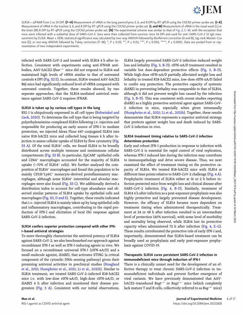
SLR14 + αIFNAR from 1 to 14 DPI. (F–H)Measurement of vRNA in the lung parenchyma 3, 6, and 8 DPI by RT-qPCR using the CDCN2 primer-probe set. (I–K)Measurement of vRNA in the trachea 3, 6, and 8 DPI by RT-qPCR using the CDCN2 primer-probe set. (L and M)Measurement of vRNA in the nasal wash (L) orthe brain (M) 8 DPI by RT-qPCR using the CDCN2 primer-probe set. (N) The experimental scheme was similar to that of Fig. 2, C–M, with the exception thatmice were infected with a sublethal dose of SARS-CoV-2. Sera were then collected from survivor mice 14 DPI and used for anti–SARS-CoV-2 S1 IgG mea-surement by ELISA. Mean ± SEM; statistical significance was calculated by two-way ANOVA followed by Bonferroni correction (A and B), log-rank Mantel–Coxtest (E), or one-way ANOVA followed by Tukey correction (F–M); *, P ≤ 0.05; **, P ≤ 0.01; ***, P ≤ 0.001; ****, P ≤ 0.0001. Data are pooled from or rep-resentative of two independent experiments.
Mao et al. Journal of Experimental Medicine 6 of 17
RIG-I agonist as COVID antiviral agent https://doi.org/10.1084/jem.20211818
Dow
nloaded from http://rupress.org/jem
/article-pdf/219/1/e20211818/1425498/jem_20211818.pdf by guest on 02 M
arch 2022

become chronically infected following SARS-CoV-2 infection,similar to what is seen in immunodeficient patients (Israelowet al., 2021). These mice maintain stable levels of viral RNA andinfectious virus for at least 14 DPI. This is in stark contrast to B6Jmice, which clear the infection by 7 DPI and remain virus-freethereafter. Given that CP therapy has been implemented to treatimmunocompromised patients with COVID-19 (Hueso et al.,2020), we first validated whether persistently infected Rag−/−
mice are a clinically relevant model in their response to CPtherapy. To this end, we adoptively transferred sera from con-valescent AAV-hACE2 B6J mice into persistently infected re-cipient AAV-hACE2 Rag−/− mice 7 DPI and measured lung viraltiter 14 DPI (Fig. 5 A). We found that CP transfer resulted insignificant reduction in vRNA and complete clearance of infec-tious virus in the lung compared with PBS-treated SARS-CoV-2 infected AAV-hACE2 Rag−/− controls (Fig. 5, B and C). Theseresults suggest that Rag−/− mice are a suitable in vivo model ofimmunocompromised patients for preclinical testing of antiviraltherapeutics, as they support persistent SARS-CoV-2 infectionand derive benefits from CP therapy.
We next examined whether SLR14 can be used as a thera-peutic modality to treat persistent infection in Rag−/− mice. Weinfected AAV-hACE2 Rag−/− mice with SARS-CoV-2, treatedthemwith SLR14 7 DPI, and collected lung tissues 14 DPI to assessthe viral burden (Fig. 5 D). We injected SLR14 at 7 DPI instead ofadministering immediately following viral exposure so thatchronic infection could be established in AAV-hACE2 Rag−/−
mice first, before any intervention was provided. Additionally,unlike K18-hACE2 mice, which we have shown are only
effectively protected from preexposure or early postexposureintervention due to their rapid disease progression (Fig. 4),AAV-hACE2 mice do not die from intranasal SARS-CoV-2 infec-tion, allowing treatments to be given at much later time points.1 d before SLR14 treatment, a subset of SLR14-treated AAV-hACE2 mice also received anti-IFNAR antibodies. SLR14 treat-ment led to a significant reduction in the level of lung vRNA(Fig. 5 E). The ability of SLR14 in decreasing vRNAwas abolishedwhen anti-IFNAR blocking antibody was given, suggestingSLR14 similarly utilizes IFN-I signaling to promote viral clear-ance in mice lacking the adaptive immune system. We addi-tionally observed a striking difference in the infectious viralload in lung tissues from SLR14-treated mice compared withvehicle controls. Treatment with SLR14, but not vehicle, sig-nificantly reduced viral burden and resulted in completeclearance of infectious virus in five out of seven AAV-hACE2Rag−/− mice and reduction of viral titer in the remaining two(Fig. 5 F). Moreover, SLR14-mediated protection required IF-NAR signaling. These results show that in the setting ofcomplete T and B cell deficiency, a single therapeutic SLR14treatment, through the induction of IFN-I, is sufficient to curepersistent infection.
SLR14 affords broad protection against immunologicallyevasive SARS-CoV-2 variantsAs SARS-CoV-2 variants continue to emerge and spread, anti-viral therapeutics that confer broadly cross-reactive protectionare urgently needed. Emerging evidence suggests that severalvariants have acquiredmutations that confer elevated resistance
Figure 3. SLR14 affords superior protection compared to recombinant IFN-I or a STING agonist. (A) Experimental scheme. K18-hACE2 mice were in-tranasally infected with 5 × 10² PFU SARS-CoV-2 (2019n-CoV/USA_WA1/2020). 4 h after infection, infected K18-hACE2 mice were i.v. treated with 15 µgSLR14, 2 × 10⁴ U rIFN-αA/D (low-dose), 2 × 10⁵ U rIFN-αA/D (high-dose), 20 µg diABZI, or vehicle. Weight loss and survival were monitored daily up to 14 DPI.Death was recorded when mice were found dead in the cage, moribund, or at 80% of original body weight. (B–D)Weight loss and survival of K18-hACE2 micefrom 1 to 14 DPI. Mean ± SEM; statistical significance was calculated by log-rank Mantel–Cox test (D); *, P ≤ 0.05. Data are pooled from two independentexperiments.
Mao et al. Journal of Experimental Medicine 7 of 17
RIG-I agonist as COVID antiviral agent https://doi.org/10.1084/jem.20211818
Dow
nloaded from http://rupress.org/jem
/article-pdf/219/1/e20211818/1425498/jem_20211818.pdf by guest on 02 M
arch 2022

to IFN-I treatment in cell culture (Guo et al., 2021 Preprint;Thorne et al., 2021 Preprint). However, whether such alteredproperties in vitro translate into evasion of IFN-based therapyin vivo remains unclear. To this end, we obtained five clinicallyrelevant SARS-CoV-2 variants of concern (VOCs) or variants ofinterest, including B.1.1.7, B.1.351, P.1, B.1.526, and B.1.617.2, andused them to infect K18-hACE2mice. The P.1, B.1.526, B.1.1.7, andB.1.617.2 variants were identified and isolated as a part of theYale SARS-CoV-2 Genomic Surveillance Initiatives (Kalinichet al., 2020), and the B.1.351 variant was obtained from BEIResources Repository. All variants were confirmed to harborsignature mutations characteristic of their respective lineagesand show correct placement in the phylogenetic tree built with
public SARS-CoV-2 genomic sequences (Table S1; Lucas et al.,2021).
To examine whether SLR14 is protective against SARS-CoV-2 variants, we infected mice with variants P.1, B.1.526, B.1.617.2,B.1.351, or B.1.1.7 and treated them with SLR14 4 h after infection(Fig. 6 A). SLR14 afforded potent protection against the P.1variant, almost completely preventing morbidity and mortalityin the face of highly lethal infection, even when treated post-exposure prophylactically (Fig. 6, B and C; and Fig. S4 A). SLR14also fully prevented weight loss or any discernable disease fol-lowing infectionwith B.1.526, which, in untreatedmice, caused aless pathogenic infection compared with that of the ancestralstrain or circulating variants (Fig. 6, D and E; and Fig. S4 B). In
Figure 4. Protective activities of SLR14 is determined by treatment timing relative to SARS-CoV-2 exposure. (A) Experimental scheme. K18-hACE2mice were intranasally infected with 103 PFU SARS-CoV-2 (2019n-CoV/USA_WA1/2020). 15 µg SLR14 was i.v. administered at 16 h before, 2 h before, 4 h after,24 h after, 48 h after, or 72 h after infection. Weight loss and survival weremonitored daily up to 14 DPI. Death was recordedwhenmice were found dead in thecage, moribund, or at 80% of original body weight. (B–D)Weight loss and survival of prophylactically SLR14- and vehicle-treated K18-hACE2 mice from 1 to 14DPI. (E–G)Weight loss and survival of therapeutically SLR14- and vehicle-treated K18-hACE2 mice from 1 to 14 DPI. Mean ± SEM; statistical significance wascalculated by log-rank Mantel–Cox test (D and G); *, P ≤ 0.05; ****, P ≤ 0.0001. Data are pooled from of three independent experiments.
Mao et al. Journal of Experimental Medicine 8 of 17
RIG-I agonist as COVID antiviral agent https://doi.org/10.1084/jem.20211818
Dow
nloaded from http://rupress.org/jem
/article-pdf/219/1/e20211818/1425498/jem_20211818.pdf by guest on 02 M
arch 2022

addition, we also found that SLR14 was highly effective againstB.1.617.2, protecting against weight loss in K18-hACE2 mice in-fected with a relatively low dose of virus (Fig. 6, F and G; and Fig.S4 C). In contrast, vehicle-treated mice uniformly lost 10–20% oftheir starting body weight. Remarkably, even in the face of ahigh-dose infection, SLR14-treated mice were protected fromclinical disease or death, whereas vehicle controls rapidly suc-cumbed to the infection (Fig. 6, H and I; and Fig. S4 D).
Consistent with the reported resistance to IFN-I signalingin vitro (Guo et al., 2021 Preprint; Thorne et al., 2021 Preprint),SLR14 treatment was less effective against infection with B.1.351or B.1.1.7 in vivo, conferring ∼40–50% net protection in K18-hACE2 mice (60% survival in SLR14-treated mice comparedwith 10–20% survival in vehicle controls; Fig. 6, J–M; Fig. S4, Eand F). Additional experiments with the B.1.1.7 variant con-firmed its partial resistance to SLR14 treatment, irrespective of
Figure 5. Therapeutic SLR14 treatment effectively cures persistent SARS-CoV-2 infection in Rag−/− mice in an IFN-I–dependent manner. (A) Ex-perimental scheme. Rag−/− mice were intratracheally administered with 1011 genome copies of AAV9-hACE2 and allowed to rest for 2 wk before intranasalinfection with 106 PFU SARS-CoV-2 (2019n-CoV/USA_WA1/2020). 200 µl convalescent sera or PBS was i.v. administered at 7 DPI. Lung tissues were collectedfor virological analysis at 14 DPI. (B)Measurement of vRNA in the lung at 14 DPI by RT-qPCR. (C)Measurement of infectious virus titer in the lung at 14 DPI byplaque assay. (D) Experimental scheme. Rag−/− mice were intratracheally administered with 1011 genome copies of AAV9-hACE2 and allowed to rest for 2 wkbefore intranasal infection with 106 PFU SARS-CoV-2 (2019n-CoV/USA_WA1/2020). 15 µg SLR14 or vehicle was i.v. administered at 7 DPI. 24 h before SLR14injection, half of the SLR14-treated mice were additionally given 2 mg anti-IFNAR antibodies. Lung tissues were collected for virological analysis at 14 DPI. (E)Measurement of vRNA in the lung at 14 DPI by RT-qPCR. (F)Measurement of infectious virus in the lung 14 at DPI by plaque assay. Limit of detection (LOD), 102
PFU/ml. Mean ± SEM; statistical significance was calculated by one-way ANOVA followed by Tukey correction (B, C, E, and F); **, P ≤ 0.01; ***, P ≤ 0.001; ****,P ≤ 0.0001. Data are pooled from two independent experiments.
Mao et al. Journal of Experimental Medicine 9 of 17
RIG-I agonist as COVID antiviral agent https://doi.org/10.1084/jem.20211818
Dow
nloaded from http://rupress.org/jem
/article-pdf/219/1/e20211818/1425498/jem_20211818.pdf by guest on 02 M
arch 2022

Figure 6. SLR14 is broadly protective against emerging immunologically evasive SARS-CoV-2 variants. (A) Experimental scheme. K18-hACE2 mice wereintranasally infected with P.1 (Gamma), B.1.526 (Iota), B.1.617.2 (Delta), B.1.351 (Beta), or B.1.1.7 (Alpha) variant. 15 µg SLR14 or vehicle was i.v. administered at4 h after infection. Weight loss and survival were monitored daily up to 14 DPI. Death was recorded when mice were found dead in the cage, moribund, or at80% of original body weight. (B and C)Weight loss and survival of SLR14- and vehicle-treated K18-hACE2mice from 1 to 14 DPI following 104 PFU P.1 infection.(D and E)Weight loss and survival of SLR14- and vehicle-treated K18-hACE2 mice from 1 to 14 DPI following 104 PFU B.1.526 infection. (F and G)Weight lossand survival of SLR14- and vehicle-treated K18-hACE2 mice from 1 to 14 DPI following 5 × 10⁵ PFU (low-dose) B.1.617.2 infection. (H and I) Weight loss andsurvival of SLR14- and vehicle-treated K18-hACE2mice from 1 to 14 DPI following 5 × 10⁷ PFU (high-dose) B.1.617.2 infection. (J and K)Weight loss and survivalof SLR14- and vehicle-treated K18-hACE2 mice from 1 to 14 DPI following 104 PFU B.1.351 infection. (L and M)Weight loss and survival of SLR14- and vehicle-treated K18-hACE2 mice from 1 to 14 DPI following 104 PFU B.1.1.7 infection. Mean ± SEM; statistical significance was calculated by log-rank Mantel–Cox test(C, E, G, I, K, and M); *, P ≤ 0.05; **, P ≤ 0.01; ***, P ≤ 0.001. Data are pooled from two independent experiments.
Mao et al. Journal of Experimental Medicine 10 of 17
RIG-I agonist as COVID antiviral agent https://doi.org/10.1084/jem.20211818
Dow
nloaded from http://rupress.org/jem
/article-pdf/219/1/e20211818/1425498/jem_20211818.pdf by guest on 02 M
arch 2022

initial sizes of viral inoculum (Fig. S4, G–O). Nevertheless, clearbenefits were seen with prophylactic treatment of SLR14. Theseresults suggest that SLR14 confers broad-coverage protectionagainst antibody- and IFN-I–evasive variants.
DiscussionThe sudden arrival and devastating spread of COVID-19 haveemphasized the importance of continuous efforts to developbroad-spectrum antiviral agents. Here, we examined the in vivoefficacy of SLR14 against viral replication throughout the res-piratory tract and disease development in a mouse model ofsevere SARS-CoV-2 infection. We first showed that SLR14 rap-idly triggers local production of IFN-I in the respiratory tract.Consistent with the induction of airway IFN-I responses, wefound that SLR14 conferred considerable antiviral resistance inthe lower respiratory tract and effectively prevented morbidityand mortality following infection with the ancestral virus. Wealso examined the effect of host factor, tissue compartment, andtreatment timing in the protective capacity of SLR14, and wefound that the protective efficacy of SLR14 depends on intactIFNAR signaling and that early SLR14 administration providedsuperior protection, while treatment as late as 48 h after in-fection still afforded partial protection. We further tested thetherapeutic potential for SLR14 in chronically infected immu-nodeficient mice and demonstrated that a single dose of SLR14conferred near-sterilizing immunity by the innate immunesystem alone, even in the absence of T and B cells. Finally, wefound that SLR14 confers broad protection against all emergingSARS-CoV-2 variants.
The apparent protective role of early and regulated IFN-Isuggests IFN-based therapies can be used for prevention andtreatment of COVID-19. In a golden hamster model of SARS-CoV-2 infection, intranasal administration of commercially availableuniversal IFN (rIFN-αA/D) reduced viral burden and attenuatedpathology in the lung (Hoagland et al., 2021). In a retrospectivemulticenter cohort study of 446 COVID-19 patients, early ad-ministration of inhaled IFNα2b producedmore favorable clinicalresponses compared with lopinavir/ritonavir treatment aloneand was associated with reduced in-hospital mortality (Wanget al., 2020). While results from rIFN-based clinical trials arepromising, one of the major disadvantages of this approach is itshigh cost, with direct medical costs ranging between $1,120 and$1,962 for the IFN treatment regimen and $2,156 and $5,887 forthe PEG-IFN treatment regimen (Nguyen et al., 2020). In addi-tion, administration of rIFN has been shown to induce neu-tralizing antibody response against IFN that could render thetherapy ineffective (Giovannoni et al., 2002; Matsuda et al.,2012). SLR14 addresses these challenges with 1) increased af-fordability due to its synthetic simplicity, small size, and man-ufacturability, and 2) its ability to induce different members ofthe IFN-I family, including 10 IFN-α subtypes and an IFN-β,which maximizes the likelihood of downstream responses to befunctional (Linehan et al., 2018). In particular, the ability ofSLR14 to elicit IFN-β is ideal, as it enables early medical inter-vention for COVID-19 patients with preexisting autoantibodiesagainst one or multiple subtypes of IFN-α, who are particularly
susceptible to prolonged viral replication and severe diseaseafter infection with SARS-CoV-2 (Meffre and Iwasaki, 2020).Besides rIFN, innate modulators that induce IFN production,such as poly(I:C) and diABZI, may also serve as a strategy tocombat COVID-19. We have previously found that both SLRs andpoly(I:C) induce a diverse array of genes associated with anti-viral immunity. Importantly, SLRs induced an IFN-I–dominantresponse, both systemically (Linehan et al., 2018) and inmucosaltissues (this study). When administered i.v. in mice, SLR14 in-duced a much stronger systemic IFN-α response compared withthat of poly(I:C) (Linehan et al., 2018). Given that SLR14-mediated protection critically depends on IFN-I, these data in-dicate that SLR14 will be similarly, if not more, effective againstSARS-CoV-2 than poly(I:C). With their potent antiviral activi-ties, however, the safety profile related to inflammation asso-ciated with systemic administration of rIFN-I– and IFN-I–inducing agents (including SLR14), particularly in the con-text of SARS-CoV-2 infection, needs to be carefully examined infuture studies.
The clinical efficacy of CP in patients with severe COVID-19has not been thoroughly demonstrated, and its use in differentstages of infection and disease remains experimental (Li et al.,2020; Simonovich et al., 2021). Emergence of immune-evadingvariants from patients with immunosuppression of T cell andB cell arms indicate caution should be used for CP therapy(Kemp et al., 2021). In these patients, the administered anti-bodies have little support from cytotoxic CD8 T cells or helperCD4 T cells, thereby reducing the chances of clearance andtheoretically allowing for SARS-CoV-2 escape. Therefore, anovel therapeutic paradigm that treats persistent viral infectionregardless of its effect on adaptive immunity will hold immensepotential for this patient population. We have shown in thisstudy that a single dose of SLR14 in mice lacking the adaptiveimmune system in the setting of chronic SARS-CoV-2 infectioncan induce near-sterilizing immunity. These results demon-strated that SLR14’s utility extends beyond prophylactic anti-virals, but also therapeutics that can be given to patients withimmunocompromised conditions, providing an immediate so-lution to simultaneously cure chronic infection and suppressfuture emergence of immune-evasive variants. From an evolu-tionary perspective, such sterilizing protection induced exclu-sively through innate immune activation is analogous toantiviral mechanisms in metazoan organisms lacking adaptiveimmunity, which provides a basic, yet crucial, protective strat-egy against viral pathogens (Wang et al., 2015).
Vaccines remain the best approach to thwart the COVID-19pandemic. However, with many countries lacking access to ad-equate vaccine doses, alternative strategies need to be developedand rapidly distributed to parts of the world severely impactedby these variants. Here, we showed that SLR14 potently pre-vented morbidity and mortality following infection with clini-cally relevant VOC, which have vastly different signaturemutations and immune-evading capacity. Consistent with a re-cent study examining IFN-I potency against different variantsin vitro (Guo et al., 2021 Preprint; Thorne et al., 2021 Preprint),the protective capacity of SLR14 was less impressive when ad-ministered against B.1.351 or B.1.1.7. Importantly, SLR14 still
Mao et al. Journal of Experimental Medicine 11 of 17
RIG-I agonist as COVID antiviral agent https://doi.org/10.1084/jem.20211818
Dow
nloaded from http://rupress.org/jem
/article-pdf/219/1/e20211818/1425498/jem_20211818.pdf by guest on 02 M
arch 2022
retained considerable residual antiviral capacity, which may beattributed by the speed, magnitude, and diversity of IFN-I re-sponses induced by SLR14 that could collectively overcome viralresistance (Linehan et al., 2018). Up until this point, B.1.1.7 hasbeen recognized as a minimally immune-evasive variant basedon both antibodies and T cell recognition. Here, we provide thefirst set of in vivo evidence to suggest that B.1.1.7 exhibits signsof IFN-I evasion and responds only moderately to IFN-basedtherapy. Such innate immune evasion may underlie the rapidglobal spread of B.1.1.7. These results showcase SLR14’s ability tobe used not only as a therapeutic agent but also as an investi-gative tool for functional assessment of basic SARS-CoV-2 biology.
Several drugs have been approved by the US Food and DrugAdministration under Emergency Use Authorization, includingdexamethasone, remdesivir, monoclonal antibodies, tocilizu-mab, and baricitinib, to treat COVID-19 (Beigel et al., 2020;Calabrese and Calabrese, 2021; Horby et al., 2021; Kalil et al.,2021; Rosas et al., 2021; Taylor et al., 2021). However, thesetherapeutics typically provide modest benefits at best and arelimited to a subset of patients. While currently licensed vaccinesdemonstrate astounding protective efficacy against COVID-19, anew variant may develop in the future to significantly reduceefficacy. Further, there is a global shortage in vaccines withinequitable access in many lower income countries. The devel-opment, characterization, and ultimate deployment of an ef-fective antiviral against SARS-CoV-2 could prevent substantialmorbidity and mortality associated with COVID-19. In additionto its therapeutic potential, SLR14 can be used as an invaluableinvestigative tool to advance our understanding of protectiveantiviral immunity against respiratory viruses, which will en-able the rational design of next-generation antiviral therapeu-tics. Lastly, with the prevalence of prepandemic zoonotic virusesand the unpredictable overlap of human and wild animal ecol-ogies, the potential for a novel viral emergence from its naturalreservoir into humans is a matter of time. Through the creationof a simple and versatile RNA-based therapeutics, our studieswill facilitate pandemic preparedness and response against fu-ture respiratory pathogens.
Materials and methodsEthicsThe institutional review board of the Yale University HumanResearch Protection Program determined that the RT-qPCRtesting and sequencing of deidentified remnant COVID-19 clin-ical samples conducted in this study is not research involvinghuman subjects (institutional review board protocol ID2000028599).
MiceB6.Cg-Tg(K18-ACE2)2Prlmn/J (K18-hACE2), B6(Cg)-Ifnar1t-m1.2Ees/J (Ifnar1−/−), B6.129S7-Rag1tm1Mom/J (B6J Rag1−/−), andC.129S7(B6)-Rag1tm1Mom/J (BALB/c Rag1−/−) mice were purchasedfrom The Jackson Laboratory and subsequently bred and housedat Yale University. Rag2−/− mice were generously gifted from R.Flavell (Yale University, New Haven, CT). 6- to 10-wk-old
mixed-sex mice were used throughout the study. All mice werehoused as groups of five or six individuals per cage and main-tained on a 12-h light/dark cycle (lights on at 7:00 a.m.) at22–25°C temperature and 30–70% relative humidity under spe-cific pathogen–free conditions. All mice were fed with regularrodent’s chow and sterilized water ad libitum. All proceduresused in this study (sex-matched and age-matched) compliedwith federal guidelines and the institutional policies of the YaleSchool of Medicine Animal Care and Use Committee.
Virus sequencingNucleic acid was extracted from 300 µl viral transport mediumfrom nasopharyngeal swabs and eluted in 75 µl using the Mag-MAX viral/pathogen nucleic acid isolation kit. Extracted nucleicacid was tested by our multiplexed RT-qPCR variant assay(Vogels et al., 2021a; Vogels et al., 2021b), and then libraries wereprepared using the Illumina COVIDSeq Test RUO version. Theprotocol was slightly modified by lowering the annealing tem-perature of the amplicon PCR step to 63°C and reducing tag-mentation to 3 min. Pooled libraries were sequenced on theIllumina NovaSeq (paired-end 150). Data were processed andconsensus sequences were generated using iVar (version 1.3.1)with the minimum depth threshold (-m) at 20 and minimumfrequency threshold (-t) at 0.6 (Grubaugh et al., 2019). Genomesequences were uploaded to GISAID. Samples belonging to theB.1.1.7 (EPI_ISL_1038987), P.1 (EPI_ISL_1293215), B.1.526 (EPI_-ISL_944591), and B.1.617.2 (EPI_ISL_2035068) lineages wereselected for virus isolation from the original sample. Virus be-longing to the B.1.351 lineage was obtained from BEI Resources.
Virus isolationSamples selected for virus isolation were diluted 1:10 in DMEMand then filtered through a 45-µM filter. The samples weretenfold serially diluted from 1:50 to 1:19,531,250. The dilutionwas subsequently incubated with TMPRSS2-Vero E6 in a 96-well plate and adsorbed for 1 h at 37°C. After adsorption, re-placement medium was added, and cells were incubated at 37°Cfor up to 5 d. Supernatants from cell cultures with cytopathiceffect were collected, frozen, thawed, and subjected to RT-qPCR.Fresh cultures were inoculated with the lysates as describedabove for viral expansion. Viral infection was subsequentlyconfirmed through reduction of cycle threshold values in the cellcultures with the multiplex variant qPCR assay. Expanded vi-ruses were resequenced following the samemethod as describedabove and were identical to the original clinical sample se-quence. Genome sequences of cultured viruses B.1.1.7 (SARS-CoV-2/human/USA/Yale-3363/2021; GenBank accession:MZ202178), B.1.351 (SARS-CoV-2/human/ZAF/Yale-3366/2020; GenBank accession: MZ202314), P.1 (SARS-CoV-2/hu-man/USA/Yale-3365/2021; GenBank accession: MZ202306),B.1.526 (SARS-CoV-2/human/USA/Yale-3362/2021; GenBankaccession: MZ201303), and B.1.617.2 (SARS-CoV-2/human/USA/Yale-5641/2021; GenBank accession: MZ468047) wereuploaded to GenBank. We used Nextclade v0.14.2 (https://clades.nextstrain.org/) to generate a phylogenetic tree and tocompile a list of amino acid changes in the virus isolates ascompared with the Wuhan-Hu-1 reference strain (Table S1).
Mao et al. Journal of Experimental Medicine 12 of 17
RIG-I agonist as COVID antiviral agent https://doi.org/10.1084/jem.20211818
Dow
nloaded from http://rupress.org/jem
/article-pdf/219/1/e20211818/1425498/jem_20211818.pdf by guest on 02 M
arch 2022

Synthesis, purification, and labeling of theSLR14 oligonucleotideThe triphosphorylated RNA oligonucleotides SLR14 (59-pppGGAUCGAUCGAUCGUUCGCGAUCGAUCGAUCC-39) and SLR14-amino(59-pppGGAUCGAUCGAUCGUXCGCGAUCGAUCGAUCC-39, where X= aminomodifier C6dT; Glen Research) were prepared as describedpreviously (Jiang et al., 2019). Briefly, for every 1 mg of startingmaterial, removal of the oligonucleotide from the polymer supportand base deprotection was performed in a 1:1 mixture of 40% me-thylamine (Sigma-Aldrich) and 30% ammonium hydroxide (JTBaker) at 65°C for 15 min. The solution was cooled on ice for 10 min,transferred to a new vial, and evaporated to dryness. 500 µl of ab-solute ethanolwas added, and themixturewas evaporated to drynessagain. To deprotect the 29-OH groups, the dry oligonucleotide wasincubated with 500 µl of a 1 M solution of tetrabutylammoniumfluoride in tetrahydrofuran (Sigma-Aldrich) at room temperature for36 h. 500 µl of 2 M sodium acetate (pH 6.0) was added, and thesolution was evaporated to a 500–600 µl volume, extracted with 3 ×800 µl ethyl acetate, and ethanol precipitated. The RNA oligonucle-otide was then purified on a 16% denaturing polyacrylamide gel. Forfluorescent labeling, for every 1 mg of starting material, the purifiedSLR14-amino oligonucleotide was dissolved in 200 µl of 0.25 M so-dium bicarbonate buffer (pH 9.2). Then, a solution containing 0.5mgAlexa Fluor 647 NHS ester (Life Technologies) in 200 µl N,N-dime-thylformamidewas added, and the reactionmixturewas incubated atroom temperature for 2 h. The labeled oligonucleotide (AF647-SLR14)was ethanol precipitated and purified on a 20% denaturingpolyacrylamide gel.
In vivo SARS-CoV-2 infectionBefore infection, mice were anesthetized using 30% (vol/vol)isoflurane diluted in propylene glycol. For K18-hACE2 mice,50 µl of SARS-CoV-2 was delivered intranasally at 103 PFU permouse, unless specified otherwise. Following infection, weightloss and survival were monitored daily up to 14 DPI. For AAV-hACE2mice, 50 µl SARS-CoV-2 was delivered intranasally at 106
PFU per mouse. Experiments involving SARS-CoV-2 infectionwere performed in a biosafety level 3 facility with approval fromthe Yale Institutional Animal Care and Use Committee and YaleEnvironmental Health and Safety.
Antibody and drug treatment in miceFor IFNAR blockade, mice were treated once with 2 mg blockingantibodies diluted in 200 µl PBS 1 d before infection (CloneMAR1-5A3; BioXCell). Universal IFN-I (rIFN-αA/D, no. 11200;PBL Assay Science) was supplied frozen in PBS containing 0.1%BSA. Cross-species activity of rIFN-αA/D on mouse cells wasconfirmed by the manufacturer and a previous study (Uccelliniand Garcıa-Sastre, 2018). 4 h after infection, 2 × 10⁴ U rIFN-αA/D(low-dose; 106 U/kg) or 2 × 10⁵ U rIFN-αA/D (high-dose; 107
U/kg) were diluted in 100 µl PBS and i.v. administered to mice.STING agonist diABZI (Compound 3; Selleckchem) was firstreconstituted in DMSO at 50mg/ml. 20 µg diABZI (1 mg/kg) wasdiluted in 100 µl PBS and i.v. administered to mice 4 h afterinfection. The dosage for systemic diABZI treatment was de-termined based on previous publications (Humphries et al.,2021; Ramanjulu et al., 2018). The dosing solution was
prepared fresh and confirmed to be clear at the time ofadministration.
i.v. injection of SLR14 in miceAt indicated time points, 15 µg SLR14 was i.v. injected. Briefly,15 µg (∼0.75 mg/kg body weight) SLR14 and 4 µl jetPEI (PolyplusTransfection) were diluted and mixed with 5% glucose solution toa total of 100 µl injection solution per mouse. After 15 min ofincubation at room temperature, the 100-µl complexwas carefullyinjected into the retro-orbital sinus with a 0.5-ml BD insulin sy-ringe. Before injection, mice were anesthetized using 30% (vol/vol) isoflurane diluted in propylene glycol. H2O and jetPEI weremixed with 5% glucose solution and used as a vehicle control.
AAV-hACE2 transductionAAV-hACE2 transduction was performed as previously de-scribed (Israelow et al., 2020). AAV9 vector encoding hACE2was purchased from Vector Biolabs (AAV9-CMV-hACE2). Ani-mals were anaesthetized using a mixture of ketamine(50 mg/kg) and xylazine (5 mg/kg) injected intraperitoneally.The rostral neck was shaved and disinfected. A 5-mm incisionwas made, the salivary glands were retracted, and the tracheawas visualized. Using a 32-G insulin syringe, a 50-µl bolus in-jection of 1011 genomic copies AAV-CMV-hACE2 was injectedinto the trachea. The incision was closed with 4–0 Vicryl suture.Following intramuscular administration of analgesic (melox-icam and buprenorphine, 1 mg/kg), animals were placed on aheating pad and closely monitored until full recovery.
Measurements of genomic RNA and infectious virusViral RNA and titer from mouse lung tissues were measured aspreviously described (Israelow et al., 2020). Briefly, at indicatedtime points, mice were euthanized with 100% isoflurane. Thewhole lung was placed in a Lysing Matrix D tube (MP Bio-medicals) with 1 ml PBS and homogenized using a table-tophomogenizer at medium speed for 2 min. For RNA analysis,250 µl of the lung homogenates was added to 750 µl Trizol LS(Invitrogen), and RNA was extracted with the RNeasy Mini Kit(Qiagen) according to the manufacturer’s instructions. SARS-CoV-2 RNA levels were quantifiedwith 250 ng RNA inputs usingthe Luna Universal Probe One-Step RT-qPCR Kit (New EnglandBiolabs) using real-time RT-PCR primer/probe sets 2019-nCoV_N1 (CDCN1) and 2019-nCoV_N2 (CDCN2). For determi-nation of infectious titer, plaque assays were performed usinglung homogenates in Vero E6 cells cultured with MEM supple-mented with NaHCO3, 4% FBS, and 0.6% Avicel RC-581. 48 hafter infection, plaques were resolved by 1-h fixation with 10%formaldehyde and sequential 1-h staining with 0.5% crystal vi-olet in 20% ethanol. Finally, plates were rinsed in water forbetter visualization of plaques.
Measurements of cellular gene expression by RT-qPCRFollowing RNA extraction, total cDNA was prepared with 2 µgRNA inputs using the iScript cDNA Synthesis Kit following themanufacturer’s instruction (Bio-Rad). RT-PCR was then performedusing the SYBR Green PCR Master Mix (Applied Biosystems). Foreach RT-qPCR reaction, 50 ng cDNA input was used.
Mao et al. Journal of Experimental Medicine 13 of 17
RIG-I agonist as COVID antiviral agent https://doi.org/10.1084/jem.20211818
Dow
nloaded from http://rupress.org/jem
/article-pdf/219/1/e20211818/1425498/jem_20211818.pdf by guest on 02 M
arch 2022

ImmunohistochemistryMice were first perfused by intracardiac injection 20ml PBS intothe right ventricle until the lung appeared opaque. After perfusion,the lung was slowly inflated with 1 ml 4% paraformaldehyde (PFA;Electron Microscopy Sciences) through intratracheal instillation.Following inflation, the tracheawas quickly tiedwith suture. Tissuewas collected and fixed in 4% PFA overnight. Yale Pathology kindlyassisted with embedding, sectioning, and H&E staining of lungtissues. H&E-stained lung sections were then imaged by a fluores-cence microscope (BX51; Olympus) with a 10× lens.
BALF collectionMice were euthanized with 100% isoflurane. After euthanasia,the trachea was exposed and the lung was slowly inflated with1 ml PBS through intratracheal instillation. Lung tissues wereflushed three times. Following lavage, samples were centrifugedat 3,900 rpm for 5 min at 4°C; the supernatant (i.e., BALF) wasaliquoted in 100 µl aliquots and stored at −80°C.
Antibodies for flow cytometryAnti-mouse antibodies used in this study, together with vendorsand dilutions, are listed as follows: FITC anti-mCD11c (N418; 1:400; BioLegend), PerCP-Cy5.5 anti-mLy6C (HK1.4; 1:400; BioL-egend), Alexa Fluor 700 anti-mLy6G (1A8; 1:400; BioLegend),Brilliant Violet 786 anti-mCD11b (M1/70; 1:400; BioLegend),APC-Cy7 anti-mTCRb (H57-597; 1:400; BioLegend), APC-Cy7anti-mCD3 (H57-597; 1:400; BioLegend), APC-Cy7 anti-mCD19(6D5; 1:400; BioLegend), APC-Cy7 anti-mNK1.1 (PK136; 1:400;BioLegend), PE anti-mCD64 (X54-5/7.1; 1:200; BioLegend), Bril-liant Violet 711 anti-mSiglecF (E50-2440; 1:200; BD Biosciences),and Pacific Blue anti-mI-A/I-E (M5/114.15.2; 1:400; BioLegend).
Flow cytometryMouse lung tissues were collected at experimental end point,digested with 1 mg/ml collagenase A (Roche) and 30 µg/mlDNase I (Sigma-Aldrich) in complete RPMI-1640 media for30 min at 37°C, and mechanically minced. Digested tissues werethen passed through a 70-µm strainer (Thermo Fisher Scientific)to single-cell suspension and treated with ACK Lysing Buffer(Thermo Fisher Scientific) to remove red blood cells. Cells wereresuspended in Live/Dead Fixable Aqua (Thermo Fisher Scien-tific) for 20 min at 4°C. Following a wash, cells were blockedwith anti-mouse CD16/32 antibodies (BioXCell) for 30 min at4°C. Cocktails of staining antibodies were added directly to thismixture for 30 min at 4°C. Prior to analysis, mouse cells werewashed and resuspended in 100 µl 4% PFA for 30–45 min at 4°C.Following this incubation, cells were washed and prepared foranalysis on an Attune NXT (Thermo Fisher Scientific). Datawere analyzed using FlowJo software version 10.6 software(Tree Star). The specific sets of markers used to identify eachsubset of cells are summarized in Fig. S5.
SARS-CoV-2–specific antibody ELISA measurementSARS-CoV-2–specific antibodies were measured as previouslydescribed (Israelow et al., 2021). In brief, sera were treated with0.5% Triton X-100 and 0.5 mg/ml RNase A to inactivate poten-tially infectious viruses. Recombinant SARS-CoV-2 S1 protein
(S1N-C52H3; ACRO Biosystems) was used to coat 96-well Max-iSorp plates (Thermo Fisher Scientific) overnight. The coatingbuffer was removed, and plates were treated with blocking so-lution followed by incubation with diluted serum. Plates werewashed with PBS-T and HRP anti-mouse IgG antibody wereadded to each well. After incubation plates were washed withPBS-T and developed with TMB Substrate Reagent Set (BD Bi-osciences 555214). The reaction was stopped by 2 N sulfuric acid.Plates were then read at a wavelength of 450 nm and 570 nm.
Determination of IFN-I and IFN-III concentrationConcentration of IFN-I in BAL fluid was determined by ELISA(42120 and 42400; PBL Assay Science) following the manu-facturer’s instructions. IFN-α–precoated plates were incubatedfor 24 h with diluted or undiluted samples and target antibody,followed by a wash with PBS-T. Similarly, IFN-β–precoatedplates were incubated with undiluted samples for 1 h andwashed with PBS-T. Detection antibody was added and incu-bated for 1 h, followed by a second wash. Both plates were thentreated with HRP solution and washed before addition of TMBsubstrate solution. The reaction was left to develop for 10 and15 min, respectively, and stopped with 2 N sulfuric acid. Ab-sorbance was recorded at 450 nm, and background noise wassubtracted from the negative control in the experiment. Con-centration of IFN-λ in BAL fluid was determined by ELISA(DY1789B; R&D Systems) according to manufacturer’s in-structions. 96-well MaxiSorp plates (Thermo Fisher Scientific)were coated overnight with 1 µg/ml coating antibody. After awash with PBS-T, the plates were blocked with 1% BSA in PBSfor 1 h and subsequently incubated for 2 h with diluted or un-diluted samples. The plate was washed and incubated for 2 hwith detection antibody followed by a third wash. Streptavidin-HRP treatment was performed for 20 min and then washed outbefore the addition of TMB substrate solution. The reaction wasleft to develop for 8 min and stopped with 2 N sulfuric acid.Absorbance was recorded at 450 nm, and background noise wassubtracted from the negative control in the experiment.
Adoptive transfer of seraWT AAV-hACE2 mice were infected with SARS-CoV-2 as indi-cated above. At 14 DPI, animals were euthanized for blood col-lection. Blood was allowed to coagulate at room temperature for30min and then was centrifuged at 3,900 rpm for 20min at 4°C.Serum was collected, and anesthetized mice (30% vol/vol iso-flurane diluted in propylene glycol) were injected with 200 µlserum with a 32-G 8-mm syringe via the retro-orbital route.
Statistical analysisData were analyzed by log-rank Mantel–Cox test or one-wayANOVA followed by Tukey correction. All statistical tests werecalculated using GraphPad Prism. A P value of <0.05 was con-sidered statistically significant.
Online supplemental materialFig. S1 illustrates additional histological analyses of lung sectionsfrom vehicle- or SLR14-treated mice following SARS-CoV-2 in-fection. Fig. S2 demonstrates that SLR14 treatment does not
Mao et al. Journal of Experimental Medicine 14 of 17
RIG-I agonist as COVID antiviral agent https://doi.org/10.1084/jem.20211818
Dow
nloaded from http://rupress.org/jem
/article-pdf/219/1/e20211818/1425498/jem_20211818.pdf by guest on 02 M
arch 2022

induce significant induction of IFN-III response and that SLR14-mediated antiviral effects and its dependency on intact IFN-Isignaling can also be seen in the AAV-hACE2 mouse model. Fig.S3 shows that i.v.-injected SLR14 can be taken up by various celltypes in the lung, in particular epithelial cells and macrophages.Fig. S4 depicts weight loss of individual mice treated with ve-hicle or SLR14 following infection with different SARS-CoV-2 variants and partial SLR14 resistance by the Alpha variant. Fig.S5 shows the gating strategy for flow cytometry experimentsconducted in this study. Table S1 lists changes in amino acidsthat were identified by sequencing in clinical isolates of SARS-CoV-2 compared with the reference genome.
AcknowledgmentsThe authors gratefully acknowledge all members of the Iwasakilaboratory for invaluable feedback, especially Annsea Park forcritical reading of the manuscript and Xiaodong Jiang for helpfuldiscussions. We thank Barney Graham (National Institutes ofHealth Vaccine Research Center, Bethesda, MD) for kindlyproviding VeroE6 cells overexpressing ACE2 and TMPRSS2. Wegive special recognition of the services of Ben Fontes and theYale Environmental Health and Safety Department for theirongoing assistance in safely conducting biosafety level 3 re-search. We also thank the Yale IMPACT Research Team, Yale–New Haven Hospital, and Connecticut Department of PublicHealth for their partnerships in our SARS-CoV-2 GenomicSurveillance Initiative. Graphical illustrations were made withBiorender.com.
This work was supported by Women’s Health Research atYale Pilot Project Program (A. Iwasaki), the Mathers FamilyFoundation (A. Iwasaki and C.B. Wilen), the Ludwig FamilyFoundation (A. Iwasaki and C.B. Wilen), National Institute ofAllergy and Infectious Diseases grants 1R01AI157488-01 and5R01AI127429-04 (A. Iwasaki), the Burroughs Wellcome Fund(C.B. Wilen), and an Emergent Ventures Fast Grant (A. Iwasakiand C.B. Wilen). T. Mao is supported by National Institute ofAllergy and Infectious Diseases grant T32AI007019. B. Israelowis supported by National Institute of Allergy and InfectiousDiseases grants 2T32AI007517-16 and K08AI163493. C. Lucas is aPew Charitable Trusts Latin American Fellow. C.B.F. Vogels issupported by Netherlands Organization for Scientific ResearchRubicon 019.181EN.004. C.B. Wilen is supported by NationalInstitute of Allergy and Infectious Diseases grant K08AI128043.A. Iwasaki and A.M. Pyle are investigators of the HowardHughes Medical Institute.
Author contributions: T. Mao and A. Iwasaki designed theexperiments; T. Mao, B. Israelow, C. Lucas, C.B.F. Vogels, M.L.Gomez-Calvo, M.I. Breban, B.L. Menasche, and H. Dong per-formed experiments; T. Mao, B. Israelow, C. Lucas, C.B.F. Vogels,M.L. Gomez-Calvo, N.D. Grubaugh, and A. Iwasaki analyzed andinterpreted results; O. Fedorova, B.L. Menasche, M.L. Landry,C.B. Wilen, N.D. Grubaugh, and A.M. Pyle provided critical re-sources and reagents; T. Mao, B. Israelow, and A. Iwasaki pre-pared the manuscript; and A. Iwasaki secured funds andsupervised the research. Authors from the Yale SARS-CoV-2 Genomic Surveillance Initiative contributed to sample
screening, sample processing, SARS-CoV-2 genome sequencing,and data analysis.
Disclosures: T. Mao reported a patent to US20210102209A1pending. C.B. Wilen reported a patent pending related to thiswork entitled "Compounds and Compositions for Treating,Ameliorating, and/or Preventing SARS-CoV-2 Infection and/orComplications Thereof"; and reported, "This work was sup-ported by K08 AI128043, Burroughs Wellcome Fund, LudwigFamily Foundation, and Mathers Charitable Foundation." N.D.Grubaugh reported personal fees from Tempus Labs and theNational Basketball Association, and grants from the CDC andFast Grants during the conduct of the study. A.M. Pyle reported apatent to SLR14 patent pending. A.M. Pyle and A. Iwasaki are co-founders of RIGImmune, a company that is developing agonistsfor the RIG-I receptor, including SLR compounds. A. Iwasakireported a patent number 10947543 issued (Yale UniversitySchool ofMedicine), served as a consultant for Spring Discovery,Boehringer Ingelheim, and Adaptive Biotechnologies, and is amember of the scientific advisory board of 4BIO Capital. Noother disclosures were reported.
Submitted: 26 August 2021Revised: 19 October 2021Accepted: 28 October 2021
ReferencesAlpert, T., A.F. Brito, E. Lasek-Nesselquist, J. Rothman, A.L. Valesano, M.J.
MacKay, M.E. Petrone, M.I. Breban, A.E. Watkins, C.B.F. Vogels, et al.2021. Early introductions and transmission of SARS-CoV-2 variantB.1.1.7 in the United States. Cell. 184:2595–2604.e13. https://doi.org/10.1016/j.cell.2021.03.061
Aydillo, T., A.S. Gonzalez-Reiche, S. Aslam, A. van de Guchte, Z. Khan, A.Obla, J. Dutta, H. van Bakel, J. Aberg, A. Garcıa-Sastre, et al. 2020.Shedding of Viable SARS-CoV-2 after Immunosuppressive Therapy forCancer. N. Engl. J. Med. 383:2586–2588. https://doi.org/10.1056/NEJMc2031670
Bastard, P., L.B. Rosen, Q. Zhang, E. Michailidis, H.H. Hoffmann, Y. Zhang, K.Dorgham, Q. Philippot, J. Rosain, V. Beziat, et al. COVID Human GeneticEffort. 2020. Autoantibodies against type I IFNs in patients with life-threatening COVID-19. Science. 370:eabd4585. https://doi.org/10.1126/science.abd4585
Beigel, J.H., K.M. Tomashek, L.E. Dodd, A.K. Mehta, B.S. Zingman, A.C. Kalil,E. Hohmann, H.Y. Chu, A. Luetkemeyer, S. Kline, et al. ACTT-1 StudyGroup Members. 2020. Remdesivir for the Treatment of Covid-19 -Final Report. N. Engl. J. Med. 383:1813–1826. https://doi.org/10.1056/NEJMoa2007764
Calabrese, L.H., and C. Calabrese. 2021. Baricitinib and dexamethasone forhospitalized patients with COVID-19. Cleve. Clin. J. Med. https://doi.org/10.3949/ccjm.88a.ccc073
Carvalho, T., F. Krammer, and A. Iwasaki. 2021. The first 12 months ofCOVID-19: a timeline of immunological insights. Nat. Rev. Immunol. 21:245–256. https://doi.org/10.1038/s41577-021-00522-1
Carvelli, J., O. Demaria, F. Vely, L. Batista, N. Chouaki Benmansour, J. Fares, S.Carpentier, M.L. Thibult, A. Morel, R. Remark, et al. Explore COVID-19Marseille Immunopole group. 2020. Association of COVID-19 inflam-mation with activation of the C5a-C5aR1 axis. Nature. 588:146–150.https://doi.org/10.1038/s41586-020-2600-6
Choi, B., M.C. Choudhary, J. Regan, J.A. Sparks, R.F. Padera, X. Qiu, I.H.Solomon, H.H. Kuo, J. Boucau, K. Bowman, et al. 2020. Persistence andEvolution of SARS-CoV-2 in an Immunocompromised Host. N. Engl.J. Med. 383:2291–2293. https://doi.org/10.1056/NEJMc2031364
Collier, D.A., A. De Marco, I.A.T.M. Ferreira, B. Meng, R.P. Datir, A.C. Walls,S.A. Kemp, J. Bassi, D. Pinto, C. Silacci-Fregni, et al. COVID-19 GenomicsUK (COG-UK) Consortium. 2021. Sensitivity of SARS-CoV-2 B.1.1.7 to
Mao et al. Journal of Experimental Medicine 15 of 17
RIG-I agonist as COVID antiviral agent https://doi.org/10.1084/jem.20211818
Dow
nloaded from http://rupress.org/jem
/article-pdf/219/1/e20211818/1425498/jem_20211818.pdf by guest on 02 M
arch 2022

mRNA vaccine-elicited antibodies. Nature. 593:136–141. https://doi.org/10.1038/s41586-021-03412-7
Deepak, P., W. Kim, M.A. Paley, M. Yang, A.B. Carvidi, E.G. Demissie, A.A. El-Qunni, A. Haile, K. Huang, and B. Kinnett. 2021. Effect of Immuno-suppression on the Immunogenicity of mRNA Vaccines to SARS-CoV-2:A Prospective Cohort Study. Ann Intern Med. https://doi.org/10.7326/M21-1757
Demaria, O., S. Cornen, M. Daeron, Y. Morel, R. Medzhitov, and E. Vivier.2019. Harnessing innate immunity in cancer therapy. Nature. 574:45–56. https://doi.org/10.1038/s41586-019-1593-5
Giovannoni, G., F.E. Munschauer III, and F. Deisenhammer. 2002. Neutral-ising antibodies to interferon beta during the treatment of multiplesclerosis. J. Neurol. Neurosurg. Psychiatry. 73:465–469. https://doi.org/10.1136/jnnp.73.5.465
Goulet, M.L., D. Olagnier, Z. Xu, S. Paz, S.M. Belgnaoui, E.I. Lafferty, V. Ja-nelle, M. Arguello, M. Paquet, K. Ghneim, et al. 2013. Systems analysisof a RIG-I agonist inducing broad spectrum inhibition of virus infec-tivity. PLoS Pathog. 9:e1003298. https://doi.org/10.1371/journal.ppat.1003298
Grubaugh, N.D., K. Gangavarapu, J. Quick, N.L. Matteson, J.G. De Jesus, B.J.Main, A.L. Tan, L.M. Paul, D.E. Brackney, S. Grewal, et al. 2019. Anamplicon-based sequencing framework for accurately measuring in-trahost virus diversity using PrimalSeq and iVar. Genome Biol. 20:8.https://doi.org/10.1186/s13059-018-1618-7
Guo, K., B.S. Barrett, K.L. Mickens, K.J. Hasenkrug, and M.L. Santiago. 2021.Interferon Resistance of Emerging SARS-CoV-2 Variants. bioRxiv.https://doi.org/10.1101/2021.03.20.436257 (Preprint posted March 21,2021)
Hoagland, D.A., R. Møller, S.A. Uhl, K. Oishi, J. Frere, I. Golynker, S. Horiuchi,M. Panis, D. Blanco-Melo, D. Sachs, et al. 2021. Leveraging the antiviraltype I interferon system as a first line of defense against SARS-CoV-2 pathogenicity. Immunity. 54:557–570.e5. https://doi.org/10.1016/j.immuni.2021.01.017
Hoffmann, M., P. Arora, R. Groß, A. Seidel, B.F. Hornich, A.S. Hahn, N.Krüger, L. Graichen, H. Hofmann-Winkler, A. Kempf, et al. 2021. SARS-CoV-2 variants B.1.351 and P.1 escape from neutralizing antibodies. Cell.184:2384–2393.e12. https://doi.org/10.1016/j.cell.2021.03.036
Horby, P., W.S. Lim, J.R. Emberson, M. Mafham, J.L. Bell, L. Linsell, N. Sta-plin, C. Brightling, A. Ustianowski, E. Elmahi, et al. RECOVERY Col-laborative Group. 2021. Dexamethasone in Hospitalized Patients withCovid-19. N. Engl. J. Med. 384:693–704. https://doi.org/10.1056/NEJMoa2021436
Hu, B., H. Guo, P. Zhou, and Z.L. Shi. 2021. Characteristics of SARS-CoV-2 andCOVID-19. Nat. Rev. Microbiol. 19:141–154. https://doi.org/10.1038/s41579-020-00459-7
Hueso, T., C. Pouderoux, H. Pere, A.L. Beaumont, L.A. Raillon, F. Ader, L.Chatenoud, D. Eshagh, T.A. Szwebel, M. Martinot, et al. 2020. Conva-lescent plasma therapy for B-cell-depleted patients with protractedCOVID-19. Blood. 136:2290–2295. https://doi.org/10.1182/blood.2020008423
Humphries, F., L. Shmuel-Galia, Z.Z. Jiang, R. Wilson, P. Landis, S.L. Ng, K.M.Parsi, R. Maehr, J. Cruz, A. Morales-Ramos, et al. 2021. A dia-midobenzimidazole STING agonist protects against SARS-CoV-2 infec-tion. Sci. Immunol. 6:eabi9002. https://doi.org/10.1126/sciimmunol.abi9002
Israelow, B., T.Mao, J. Klein, E. Song, B.Menasche, S.B. Omer, and A. Iwasaki.2021. Adaptive immune determinants of viral clearance and protectionin mouse models of SARS-CoV-2. Sci. Immunol. eabl4509. https://doi.org/10.1126/sciimmunol.abl4509
Israelow, B., E. Song, T. Mao, P. Lu, A. Meir, F. Liu, M.M. Alfajaro, J. Wei, H.Dong, R.J. Homer, et al. 2020. Mouse model of SARS-CoV-2 revealsinflammatory role of type I interferon signaling. J. Exp. Med. 217:e20201241. https://doi.org/10.1084/jem.20201241
Iwasaki, A., and R. Medzhitov. 2015. Control of adaptive immunity by theinnate immune system. Nat. Immunol. 16:343–353. https://doi.org/10.1038/ni.3123
Jiang, X., V. Muthusamy, O. Fedorova, Y. Kong, D.J. Kim, M. Bosenberg, A.M.Pyle, and A. Iwasaki. 2019. Intratumoral delivery of RIG-I agonist SLR14induces robust antitumor responses. J. Exp. Med. 216:2854–2868.https://doi.org/10.1084/jem.20190801
Kalil, A.C., T.F. Patterson, A.K. Mehta, K.M. Tomashek, C.R. Wolfe, V. Gha-zaryan, V.C. Marconi, G.M. Ruiz-Palacios, L. Hsieh, S. Kline, et al.ACTT-2 Study Group Members. 2021. Baricitinib plus Remdesivir forHospitalized Adults with Covid-19.N. Engl. J. Med. 384:795–807. https://doi.org/10.1056/NEJMoa2031994
Kalinich, C.C., C.G. Jensen, P. Neugebauer,M.E. Petrone,M. Peña-Hernandez,I.M. Ott, A.L. Wyllie, T. Alpert, C.B.F. Vogels, J.R. Fauver, et al. 2020.Real-time public health communication of local SARS-CoV-2 genomicepidemiology. PLoS Biol. 18:e3000869. https://doi.org/10.1371/journal.pbio.3000869
Kemp, S.A., D.A. Collier, R.P. Datir, I.A.T.M. Ferreira, S. Gayed, A. Jahun, M.Hosmillo, C. Rees-Spear, P. Mlcochova, I.U. Lumb, et al. COVID-19 Ge-nomics UK (COG-UK) Consortium. 2021. SARS-CoV-2 evolution duringtreatment of chronic infection. Nature. 592:277–282. https://doi.org/10.1038/s41586-021-03291-y
Li, L., W. Zhang, Y. Hu, X. Tong, S. Zheng, J. Yang, Y. Kong, L. Ren, Q. Wei, H.Mei, et al. 2020. Effect of Convalescent Plasma Therapy on Time toClinical Improvement in Patients With Severe and Life-threateningCOVID-19: A Randomized Clinical Trial. JAMA. 324:460–470. https://doi.org/10.1001/jama.2020.10044
Li, B., A. Deng, K. Li, Y. Hu, Z. Li, and J. Lu. 2021a. Viral infection andtransmission in a large, well-traced outbreak caused by the SARS-CoV-2 Delta variant. medRxiv. https://doi.org/10.1101/2021.07.07.21260122(Preprint posted July 23, 2021)
Li, M.H., M. Ferretti, B.L. Ying, H. Descamps, E. Lee, M. Dittmar, J.S. Lee, K.Whig, B. Kamalia, L. Dohnalova, et al. 2021b. Pharmacological activationof STING blocks SARS-CoV-2 infection. Sci. Immunol. 6:eabi9007.https://doi.org/10.1126/sciimmunol.abi9007
Linehan, M.M., T.H. Dickey, E.S. Molinari, M.E. Fitzgerald, O. Potapova, A.Iwasaki, and A.M. Pyle. 2018. A minimal RNA ligand for potent RIG-Iactivation in living mice. Sci. Adv. 4:e1701854. https://doi.org/10.1126/sciadv.1701854
Lowery, S.A., A. Sariol, and S. Perlman. 2021. Innate immune and inflam-matory responses to SARS-CoV-2: Implications for COVID-19. Cell HostMicrobe. 29:1052–1062. https://doi.org/10.1016/j.chom.2021.05.004
Lucas, C., C.B.F. Vogels, I. Yildirim, J.E. Rothman, P Lu, V. Monteiro, J.R.Gelhausen, M. Campbell, J. Silva, and A. Tabachikova. 2021. Impact ofcirculating SARS-CoV-2 variants on mRNA vaccine-induced immunity.Nature. https://doi.org/10.1038/s41586-021-04085-y.
Lucas, C., P. Wong, J. Klein, T.B.R. Castro, J. Silva, M. Sundaram, M.K. El-lingson, T. Mao, J.E. Oh, B. Israelow, et al. Yale IMPACT Team. 2020.Longitudinal analyses reveal immunological misfiring in severe COVID-19. Nature. 584:463–469. https://doi.org/10.1038/s41586-020-2588-y
Luo, D., S.C. Ding, A. Vela, A. Kohlway, B.D. Lindenbach, and A.M. Pyle. 2011.Structural insights into RNA recognition by RIG-I. Cell. 147:409–422.https://doi.org/10.1016/j.cell.2011.09.023
Matsuda, F., Y. Torii, H. Enomoto, C. Kuga, N. Aizawa, Y. Iwata, M. Saito, H.Imanishi, S. Shimomura, H. Nakamura, et al. 2012. Anti-interferon-αneutralizing antibody is associated with nonresponse to pegylatedinterferon-α plus ribavirin in chronic hepatitis C. J. Viral Hepat. 19:694–703. https://doi.org/10.1111/j.1365-2893.2012.01598.x
McCray, P.B. Jr., L. Pewe, C.Wohlford-Lenane, M. Hickey, L. Manzel, L. Shi, J.Netland, H.P. Jia, C. Halabi, C.D. Sigmund, et al. 2007. Lethal infectionof K18-hACE2 mice infected with severe acute respiratory syndromecoronavirus. J. Virol. 81:813–821. https://doi.org/10.1128/JVI.02012-06
Meffre, E., and A. Iwasaki. 2020. Interferon deficiency can lead to severeCOVID. Nature. 587:374–376. https://doi.org/10.1038/d41586-020-03070-1
Nguyen, H.A., G.S. Cooke, J.N. Day, B. Flower, L.T. Phuong, T.M. Hung, N.T.Dung, D.B. Khoa, L.M. Hung, E. Kestelyn, et al. SEARCH Investigators.2020. The direct-medical costs associated with interferon-based treat-ment for Hepatitis C in Vietnam. Wellcome Open Res. 4:129. https://doi.org/10.12688/wellcomeopenres.15408.2
Park, A., and A. Iwasaki. 2020. Type I and Type III Interferons - Induction,Signaling, Evasion, and Application to Combat COVID-19. Cell Host Mi-crobe. 27:870–878. https://doi.org/10.1016/j.chom.2020.05.008
Planas, D., T. Bruel, L. Grzelak, F. Guivel-Benhassine, I. Staropoli, F. Porrot, C.Planchais, J. Buchrieser, M.M. Rajah, E. Bishop, et al. 2021a. Sensitivityof infectious SARS-CoV-2 B.1.1.7 and B.1.351 variants to neutralizingantibodies. Nat. Med. 27:917–924. https://doi.org/10.1038/s41591-021-01318-5
Planas, D., D. Veyer, A. Baidaliuk, I. Staropoli, F. Guivel-Benhassine, M.M.Rajah, C. Planchais, F. Porrot, N. Robillard, J. Puech, et al. 2021b.Reduced sensitivity of SARS-CoV-2 variant Delta to antibody neu-tralization. Nature. 596:276–280. https://doi.org/10.1038/s41586-021-03777-9
Ramanjulu, J.M., G.S. Pesiridis, J. Yang, N. Concha, R. Singhaus, S.Y. Zhang,J.L. Tran, P. Moore, S. Lehmann, H.C. Eberl, et al. 2018. Design ofamidobenzimidazole STING receptor agonists with systemic activity.Nature. 564:439–443. https://doi.org/10.1038/s41586-018-0705-y
Mao et al. Journal of Experimental Medicine 16 of 17
RIG-I agonist as COVID antiviral agent https://doi.org/10.1084/jem.20211818
Dow
nloaded from http://rupress.org/jem
/article-pdf/219/1/e20211818/1425498/jem_20211818.pdf by guest on 02 M
arch 2022

Rehwinkel, J., andM.U. Gack. 2020. RIG-I-like receptors: their regulation androles in RNA sensing. Nat. Rev. Immunol. 20:537–551. https://doi.org/10.1038/s41577-020-0288-3
Rosas, I.O., N. Brau, M. Waters, R.C. Go, B.D. Hunter, S. Bhagani, D. Skiest,M.S. Aziz, N. Cooper, I.S. Douglas, et al. 2021. Tocilizumab in Hospi-talized Patients with Severe Covid-19 Pneumonia. N. Engl. J. Med. 384:1503–1516. https://doi.org/10.1056/NEJMoa2028700
Schneider, W.M., M.D. Chevillotte, and C.M. Rice. 2014. Interferon-stimulatedgenes: a complex web of host defenses. Annu. Rev. Immunol. 32:513–545.https://doi.org/10.1146/annurev-immunol-032713-120231
Simonovich, V.A., L.D. Burgos Pratx, P. Scibona, M.V. Beruto, M.G. Vallone,C. Vazquez, N. Savoy, D.H. Giunta, L.G. Perez, M.D.L. Sanchez, et al.PlasmAr Study Group. 2021. A Randomized Trial of ConvalescentPlasma in Covid-19 Severe Pneumonia. N. Engl. J. Med. 384:619–629.https://doi.org/10.1056/NEJMoa2031304
Taylor, P.C., A.C. Adams, M.M. Hufford, I. de la Torre, K. Winthrop, and R.L.Gottlieb. 2021. Neutralizing monoclonal antibodies for treatment ofCOVID-19. Nat. Rev. Immunol. 21:382–393. https://doi.org/10.1038/s41577-021-00542-x
Thorne, L.G., M. Bouhaddou, A.K. Reuschl, L. Zuliani-Alvarez, B. Polacco, A.Pelin, J. Batra, M.V.X. Whelan, M. Ummadi, A. Rojc, et al. 2021. Evo-lution of enhanced innate immune evasion by the SARS-CoV-2 B.1.1.7UK variant. bioRxiv. https://doi.org/10.1101/2021.06.06.446826 (Pre-print posted June 7, 2021)
Uccellini, M.B., and A. Garcıa-Sastre. 2018. ISRE-Reporter Mouse RevealsHigh Basal and Induced Type I IFN Responses in Inflammatory Mon-ocytes. Cell Rep. 25:2784–2796.e3. https://doi.org/10.1016/j.celrep.2018.11.030
V’kovski, P., A. Kratzel, S. Steiner, H. Stalder, and V. Thiel. 2021. Coronavirusbiology and replication: implications for SARS-CoV-2. Nat. Rev. Micro-biol. 19:155–170. https://doi.org/10.1038/s41579-020-00468-6
Vanpouille-Box, C., J.A. Hoffmann, and L. Galluzzi. 2019. Pharmacologicalmodulation of nucleic acid sensors - therapeutic potential and persist-ing obstacles.Nat. Rev. Drug Discov. 18:845–867. https://doi.org/10.1038/s41573-019-0043-2
Vogels, C.B.F., M.I. Breban, I.M. Ott, T. Alpert, M.E. Petrone, A.E. Watkins,C.C. Kalinich, R. Earnest, J.E. Rothman, J. Goes de Jesus, et al. Networkfor Genomic Surveillance in South Africa. 2021a. Multiplex qPCR dis-criminates variants of concern to enhance global surveillance of SARS-CoV-2. PLoS Biol. 19:e3001236. https://doi.org/10.1371/journal.pbio.3001236
Vogels, C.B.F., J.R. Fauver, and N.D. Grubaugh. 2021b. Multiplexed RT-qPCRto screen for SARS-COV-2 B.1.1.7, B.1.351, and P.1 variants of concernV.3. protocols.io. https://dx.doi.org/10.17504/protocols.io.br9vm966
Wang, P.H., S.P. Weng, and J.G. He. 2015. Nucleic acid-induced antiviralimmunity in invertebrates: an evolutionary perspective. Dev. Comp.Immunol. 48:291–296. https://doi.org/10.1016/j.dci.2014.03.013
Wang, E.Y., T. Mao, J. Klein, Y. Dai, J.D. Huck, J.R. Jaycox, F. Liu, T. Zhou, B.Israelow, P. Wong, et al. 2021. Diverse functional autoantibodies inpatients with COVID-19. Nature. 595:283–288. https://doi.org/10.1038/s41586-021-03631-y
Wang, N., Y. Zhan, L. Zhu, Z. Hou, F. Liu, P. Song, F. Qiu, X. Wang, X. Zou, D.Wan, et al. 2020. Retrospective Multicenter Cohort Study Shows EarlyInterferon Therapy Is Associated with Favorable Clinical Responses inCOVID-19 Patients. Cell Host Microbe. 28:455–464.e2. https://doi.org/10.1016/j.chom.2020.07.005
Washington, N.L., K. Gangavarapu, M. Zeller, A. Bolze, E.T. Cirulli, K.M.Schiabor Barrett, B.B. Larsen, C. Anderson, S. White, T. Cassens, et al.2021. Emergence and rapid transmission of SARS-CoV-2 B.1.1.7 in theUnited States. Cell. 184:2587–2594.e7. https://doi.org/10.1016/j.cell.2021.03.052
Winkler, E.S., A.L. Bailey, N.M. Kafai, S. Nair, B.T. McCune, J. Yu, J.M. Fox,R.E. Chen, J.T. Earnest, S.P. Keeler, et al. 2020. SARS-CoV-2 infection ofhuman ACE2-transgenic mice causes severe lung inflammation andimpaired function. Nat. Immunol. 21:1327–1335. https://doi.org/10.1038/s41590-020-0778-2
Yamada, T., S. Sato, Y. Sotoyama, Y. Orba, H. Sawa, H. Yamauchi, M. Sasaki,and A. Takaoka. 2021. RIG-I triggers a signaling-abortive anti-SARS-CoV-2 defense in human lung cells. Nat. Immunol. 22:820–828. https://doi.org/10.1038/s41590-021-00942-0
Yin, X., L. Riva, Y. Pu, L. Martin-Sancho, J. Kanamune, Y. Yamamoto, K.Sakai, S. Gotoh, L. Miorin, P.D. De Jesus, et al. 2021. MDA5 Governs theInnate Immune Response to SARS-CoV-2 in Lung Epithelial Cells. CellRep. 34:108628. https://doi.org/10.1016/j.celrep.2020.108628
Zhang, Q., P. Bastard, Z. Liu, J. Le Pen, M. Moncada-Velez, J. Chen, M. Ogishi,I.K.D. Sabli, S. Hodeib, C. Korol, et al. NIAID-USUHS/TAGC COVIDImmunity Group. 2020. Inborn errors of type I IFN immunity in pa-tients with life-threatening COVID-19. Science. 370:eabd4570. https://doi.org/10.1126/science.abd4570
Zheng, J., L.R. Wong, K. Li, A.K. Verma,M.E. Ortiz, C. Wohlford-Lenane, M.R.Leidinger, C.M. Knudson, D.K. Meyerholz, P.B. McCray Jr., and S.Perlman. 2021. COVID-19 treatments and pathogenesis including an-osmia in K18-hACE2 mice. Nature. 589:603–607. https://doi.org/10.1038/s41586-020-2943-z
Zhou, D.,W. Dejnirattisai, P. Supasa, C. Liu, A.J. Mentzer, H.M. Ginn, Y. Zhao,H.M.E. Duyvesteyn, A. Tuekprakhon, R. Nutalai, et al. 2021a. Evidenceof escape of SARS-CoV-2 variant B.1.351 from natural and vaccine-induced sera. Cell. 184:2348–2361.e6. https://doi.org/10.1016/j.cell.2021.02.037
Zhou, H., B.M. Dcosta, M.I. Samanovic, M.J. Mulligan, N.R. Landau, and T.Tada. 2021b. B.1.526 SARS-CoV-2 variants identified in New York Cityare neutralized by vaccine-elicited and therapeutic monoclonal anti-bodies. bioRxiv. https://doi.org/10.1101/2021.03.24.436620 (Preprintposted March 24, 2021)
Mao et al. Journal of Experimental Medicine 17 of 17
RIG-I agonist as COVID antiviral agent https://doi.org/10.1084/jem.20211818
Dow
nloaded from http://rupress.org/jem
/article-pdf/219/1/e20211818/1425498/jem_20211818.pdf by guest on 02 M
arch 2022

Supplemental material
Figure S1. SLR14 protects against infection-induced viral pneumonia. H&E staining of lung sections from vehicle- (left) or SLR14-treated (right) K18-hACE2 mice 5 DPI. Images show low- or high-power magnification. Images are representative of n = 5 per group. Scale bars, 500 µm.
Mao et al. Journal of Experimental Medicine S1
RIG-I agonist as COVID antiviral agent https://doi.org/10.1084/jem.20211818
Dow
nloaded from http://rupress.org/jem
/article-pdf/219/1/e20211818/1425498/jem_20211818.pdf by guest on 02 M
arch 2022

Figure S2. SLR14 does not significantly elicit IFN-III responses in the respiratory tract. (A–C) Experimental scheme. K18-hACE2 mice were i.v. ad-ministered with 15 µg SLR14 or vehicle. 3 h after injection, BALF and lung tissues were collected for IFN-λ ELISA (B) and RT-qPCR (C), respectively. (D and E)Experimental scheme. Ifnar−/− mice were intratracheally administered with 1011 genome copies of AAV9-hACE2 and allowed to rest for 2 wk before intranasalinfection with 106 PFU SARS-CoV-2 (2019n-CoV/USA_WA1/2020). 15 µg SLR14 or vehicle were i.v. administered at 4 h after infection. Lung tissues werecollected for virological analysis at 4 DPI. Measurement of vRNA at 4 DPI by RT-qPCR using the CDCN2 primer-probe set (E). Mean ± SEM; statistical sig-nificance was calculated by two-way ANOVA followed by Bonferroni correction (B and C) or one-way ANOVA followed by Tukey correction (E); ****, P ≤0.0001. Data are representative of two independent experiments.
Mao et al. Journal of Experimental Medicine S2
RIG-I agonist as COVID antiviral agent https://doi.org/10.1084/jem.20211818
Dow
nloaded from http://rupress.org/jem
/article-pdf/219/1/e20211818/1425498/jem_20211818.pdf by guest on 02 M
arch 2022

Figure S3. Intravenously injected SLR14 targets a diverse array of cell types in the lung. (A) Experimental scheme. K18-hACE2 mice were i.v. injectedwith 15 µg Alexa Flour 647–conjugated SLR14 or vehicle. Lung tissues were collected for SLR14 uptake analysis by flow cytometry 4 h after injection. Lungtissues from vehicle-injected controls were also collected as negative controls. (B) Frequency of indicated immune and nonimmune cell types among SLR14+
cells versus total lung cells. (C) Frequency of indicated macrophage populations among SLR14+ cells or total lung cells. (D) Distribution index (frequency of agiven cell type in the SLR14+ compartment/frequency of all cells) of indicated immune and nonimmune cell types. (E) Distribution index of indicated mac-rophage populations. The specific sets of markers used to identify each subset of cells and assess SLR14 uptake are summarized in Fig. S5. Data are rep-resentative of two independent experiments.
Mao et al. Journal of Experimental Medicine S3
RIG-I agonist as COVID antiviral agent https://doi.org/10.1084/jem.20211818
Dow
nloaded from http://rupress.org/jem
/article-pdf/219/1/e20211818/1425498/jem_20211818.pdf by guest on 02 M
arch 2022

Figure S4. Alpha variant exhibits partial therapeutic resistance to SLR14 treatment. (A–F) Experimental scheme. K18-hACE2 mice were intranasallyinfected with P.1 (Gamma), B.1.526 (Iota), B.1.617.2 (Delta), B.1.351 (Beta), or B.1.1.7 (Alpha) variant. 15 µg SLR14 or vehicle was i.v. administered at 4 h afterinfection. Weight loss by individual SLR14- and vehicle-treated K18-hACE2 mice from 1 to 14 DPI following infection with P.1 (A), B.1.526 (B), low-dose B.1.617.2(C), high-dose B.1.617.2 (D), B.1.351 (E), or B.1.1.7 (F). Data are representative of two independent experiments. (G–O) Experimental scheme. K18-hACE2 micewere intranasally infected with 104 PFU, 3.3 × 10³ PFU, or 103 PFU B.1.1.7 variant. 15 µg SLR14 or vehicle were i.v. administered at 4 h after infection. Weightloss and survival were monitored daily up to 14 DPI. Death was recorded when mice were found dead in the cage, moribund, or at 80% of original body weight.(G–I)Weight loss and survival of SLR14- and vehicle-treated K18-hACE2 mice from 1 to 14 DPI following infection with 104 PFU B.1.1.7. (J–L)Weight loss andsurvival of SLR14- and vehicle-treated K18-hACE2 mice from 1 to 14 DPI following infection with 3.3 × 10³ PFU B.1.1.7. (M–O) Weight loss and survival ofSLR14- and vehicle-treated K18-hACE2 mice from 1 to 14 DPI following infection with 103 PFU B.1.1.7. Mean ± SEM; statistical significance was calculated bylog-rank Mantel–Cox test (I, L, and O); *, P ≤ 0.05. Data are representative of or pooled from two independent experiments.
Mao et al. Journal of Experimental Medicine S4
RIG-I agonist as COVID antiviral agent https://doi.org/10.1084/jem.20211818
Dow
nloaded from http://rupress.org/jem
/article-pdf/219/1/e20211818/1425498/jem_20211818.pdf by guest on 02 M
arch 2022

Table S1 is provided online and lists amino acid changes identified in SARS-CoV-2 resequenced after virus isolation as compared tothe reference genome (GenBank accession no. MN908947).
Figure S5. Gating strategies for identification of various cellular subsets using flow cytometry. (A) Gating strategies for identification of various immuneand nonimmune cell populations in the lung were used to generate Fig. 1, I and J and Fig. S3, B–E. (B) Histogram examples of SLR14 uptake by epithelial cellsand different macrophage subsets. Lung tissues from vehicle-treated mice were included as negative controls.
Mao et al. Journal of Experimental Medicine S5
RIG-I agonist as COVID antiviral agent https://doi.org/10.1084/jem.20211818
Dow
nloaded from http://rupress.org/jem
/article-pdf/219/1/e20211818/1425498/jem_20211818.pdf by guest on 02 M
arch 2022