On Unilateral Constraints, Friction and Plasticity - Archive ...
Upload
independentCategory
view
3download
0
BRAINA JOURNAL OF NEUROLOGY
Rewiring of the corticospinal tract in the adult ratafter unilateral stroke and anti-Nogo-A therapyNicolas T. Lindau, Balthasar J. Banninger, Miriam Gullo, Nicolas A. Good, Lukas C. Bachmann,Michelle L. Starkey* and Martin E. Schwab
Brain Research Institute, University of Zurich and Department of Health Science and Technology Swiss Federal Institute of Technology Zurich,
Switzerland
*Present address: Paraplegic Centre, Balgrist University Hospital Zurich, Switzerland
Correspondence to: Nicolas Thomas Lindau
Brain Research Institute,
University and ETH Zurich,
Winterthurerstrasse 190
8057 Zurich,
Switzerland
E-mail: [email protected]
Adult Long Evans rats received a photothrombotic stroke that destroyed 490% of the sensorimotor cortex unilaterally; they
were subsequently treated intrathecally for 2 weeks with a function blocking antibody against the neurite growth inhibitory
central nervous system protein Nogo-A. Fine motor control of skilled forelimb grasping improved to 65% of intact baseline
performance in the anti-Nogo-A treated rats, whereas control antibody treated animals recovered to only 20% of baseline
scores. Bilateral retrograde tract tracing with two different tracers from the intact and the denervated side of the cervical
spinal cord, at different time points post-lesion, indicated that the intact corticospinal tract had extensively sprouted across
the midline into the denervated spinal hemicord. The original axonal arbours of corticospinal tract fibres that had recrossed the
midline were subsequently withdrawn, leading to a complete side-switch in the projection of a subpopulation of contralesional
corticospinal tract axons. Anterograde tracing from the contralesional cortex showed a 2–3-fold increase of midline crossing
fibres and additionally a massive sprouting of the pre-existing ipsilateral ventral corticospinal tract fibres throughout the entire
cervical enlargement of the anti-Nogo-A antibody-treated rats compared to the control group. The laminar distribution pattern of
the ipsilaterally projecting corticospinal tract fibres was similar to that in the intact spinal cord. These plastic changes were
paralleled by a somatotopic reorganization of the contralesional motor cortex where the formation of an ipsilaterally projecting
forelimb area was observed. Intracortical microstimulation of the contralesional motor cortex revealed that low threshold
currents evoked ipsilateral movements and electromyography responses at frequent cortical sites in the anti-Nogo-A, but not
in the control antibody-treated animals. Subsequent transection of the spared corticospinal tract in chronically recovered ani-
mals, treated with anti-Nogo-A, led to a reappearance of the initial lesion deficit observed after the stroke lesion. These results
demonstrate a somatotopic side switch anatomically and functionally in the projection of adult corticospinal neurons, induced by
the destruction of one sensorimotor cortex and the neutralization of the CNS growth inhibitory protein Nogo-A.
Keywords: stroke; anti-Nogo-A; somatotopic reorganization; plasticity; midline crossing fibres
Abbreviations: BrdU = bromodeoxyuridine; CST = corticospinal tract
doi:10.1093/brain/awt336 Brain 2013: Page 1 of 18 | 1
Received July 16, 2013. Revised October 16, 2013. Accepted October 18, 2013.
� The Author (2013). Published by Oxford University Press on behalf of the Guarantors of Brain. All rights reserved.
For Permissions, please email: [email protected]
Brain Advance Access published December 18, 2013 at U
niversitaet Zuerich on D
ecember 19, 2013
http://brain.oxfordjournals.org/D
ownloaded from

IntroductionStroke is the leading cause of persistent neurological disability in
the elderly population worldwide (Bakhai, 2004; Donnan et al.,
2008). Small stroke lesions, where 510–15% of the cortex is
destroyed unilaterally, often show good spontaneous recovery
within days and weeks (Liepert et al., 2000; Cramer, 2008;
Starkey et al., 2012), accompanied with a plastic reorganization
of nearby areas, larger stroke lesions, where 460% of the cortex
is destroyed unilaterally, often result in permanent neurological
deficits (Biernaskie et al., 2005). Such larger lesions have been
shown to be accompanied by a somatotopic reorganization of
more remote areas, such as the contralesional motor- and pre-
motor cortex in humans and in experimental animals (Johansen-
Berg et al., 2002; Biernaskie et al., 2005; Bestmann et al., 2010;
Rehme et al., 2011). However, if such a lesion happens early in
life, in the plastic developmental CNS, as observed in children with
a congenital hemiparesis or hemispherectomy, reorganization of
the contralesional hemisphere is often correlated with good recov-
ery of function (Choi et al., 2009; Lotze et al., 2009). For these
patients a functional shift, so that the unlesioned hemisphere con-
trolled both sides of the body, was observed. Similar observations
were made in neonatally operated animals, accompanied by an
anatomical reorganization of the descending, spared corticospinal
tract (CST) which involved fibres recrossing the midline, as well as
the sprouting of pre-existing ipsilateral CST fibres (Kartje-Tillotson
et al., 1987; Rouiller et al., 1991; Fujimura et al., 2003; Umeda
and Isa, 2011). In adult stroke patients, abnormal activation of
contralesional cortical areas was sometimes correlated with poor
outcomes (Murase et al., 2004; Nowak et al., 2009), but due to
recent high resolution imaging and stimulation techniques,
more and more studies describe a correlation between functional
recovery and somatotopic reorganization of the unlesioned
hemisphere (Chollet et al., 1991; Johansen-Berg et al., 2002;
Schaechter and Perdue, 2008; Bestmann et al., 2010; Stoeckel
and Binkofski, 2010; Rehme et al., 2011; Grefkes and Ward,
2013). However, this is still a matter of debate and the size and
precise location of the lesion play crucial roles that require more
detailed investigation.
Exercise-driven rehabilitation training, like constraint-induced
movement therapy, has been shown to enhance plastic rearrange-
ments and increase functional recovery even in adult stroke
patients and animals (Taub and Uswatt, 2006; Maier et al.,
2008). Furthermore, in experimental animal models several
growth-promoting treatments have been shown to increase the
number of midline crossing CST fibres in the spinal cord and to
enhance behavioural recovery after stroke. These treatments
include function blocking antibodies against the neurite growth
inhibitory CNS protein Nogo-A (Emerick et al., 2003; Seymour
et al., 2005; Hamadjida et al., 2012), Nogo (now known as
RTN4) knockout mice (Kim et al., 2003; Simonen et al., 2003),
reagents blocking the Nogo receptor NgR1 (now known as
RTN4R) (Lee et al., 2004; Kubo et al., 2008), digestion of chon-
droitin sulphate proteoglycans with chondroitinase ABC (Soleman
et al., 2012; Harris et al., 2013), and high doses of the sugar
inosine (Chen et al., 2002). An important aspect of the
anti-Nogo/Nogo-receptor treatments concerns their temporal
window of opportunity. In contrast with the narrow time
window of a few hours for the conventional recombinant tissue
plasminogen activator (rt-PA) treatment, anti-Nogo-A antibodies
were shown to be fully efficacious even when applied 9 weeks
after the stroke in adult rats (Wiessner et al., 2003; Tsai et al.,
2010; Kumar and Moon, 2013). However, many open questions
regarding a correlation between improved recovery and plastic
reorganization, especially the location of ipsilaterally projecting
cells in the cortex, possible bilaterally projecting cells and the func-
tionality of new projections after stroke remain. In this study, we
used the highly controllable photothrombotic stroke model in adult
rats, in combination with anterograde and retrograde tracing tech-
niques, electrophysiology and behavioural analyses to investigate
spontaneous and anti-Nogo-A antibody-induced functional recov-
ery and to correlate this with the anatomical reorganization of the
contralesional hemisphere and its descending CST over the course
of 1 to 12 weeks after the injury.
Materials and methods
AnimalsAdult, female Long Evans rats (n = 129; 230–400 g, 3–7 months
of age, Charles River) were used in this study. Female animals
were used as they are less aggressive and learn the single pellet
grasping task more easily. Animals were housed in groups of three
to four under a constant 12 h light/dark cycle with food and water
ad libitum. All experimental procedures (Supplementary Figs 1–3)
were approved by the veterinary office of the canton of Zurich,
Switzerland.
Anaesthesia and postoperative careFor all surgical procedures, except intracortical microstimulation, ani-
mals were anaesthetized with 3% isoflurane followed by a subcuta-
neous injection of a mixture of Hypnorm� (600 ml/kg body weight,
Janssen Pharmaceutics) and Dormicum� (3.75 mg/kg body weight,
Roche Pharmaceuticals). For intracortical microstimulation a mixture
of ketamine (50 mg/ml, 7 mg/kg body weight, Streuli Pharma) and
xylazine (20 mg/ml, 5 mg/kg body weight, Streuli Pharma) was in-
jected intramuscularly with additional top-up of ketamine when
needed. After the surgery all animals received postoperative care
including analgesics (Rimadyl�, 2.5 mg/kg body weight, Pfizer) and
antibiotics (Baytril�, 5 mg/kg body weight, Bayer) for 3 days. After a
stroke lesion animals additionally received a single subcutaneous injec-
tion of mannitol (20%, 17 ml/kg, B. Braun) to reduce swelling of the
cortex. After surgery, animals were warmed on a heating pad for 24 h
before being transferred back to the animal facility.
Photothrombotic stroke and antibodyapplicationA photothrombotic stroke was induced in 116 animals (n = 59 control
antibody treatment, n = 57 anti-Nogo-A antibody treatment) to uni-
laterally lesion the sensorimotor cortex as previously described
(Watson et al., 1985; Wiessner et al., 2003). Briefly the animal was
fixed in a stereotactic frame and the skull was exposed and cleaned.
2 | Brain 2013: Page 2 of 18 N. T. Lindau et al.
at Universitaet Z
uerich on Decem
ber 19, 2013http://brain.oxfordjournals.org/
Dow
nloaded from
An opaque template with an opening for the light source (10 � 5 mm)
was positioned on the side of the cortex contralateral to the preferred
paw (see ‘Behaviour’ section). The opening of the template was pos-
itioned 5 mm to �5 mm anterior and 0.5 mm to 5.5 mm lateral relative
to bregma. A fibreglass light with 10 mm diameter was placed directly
over the exposed skull. A mixture of Rose Bengal in saline (13 mg/kg
body weight, 10 mg/ml Rose Bengal in 0.9% NaCl solution) was in-
jected through a catheter in the femoral vein, and after 2 min the skull
was illuminated (Olympus KL1500LCD, 150 W, 3000 K) for 10 min at
the maximum output of the light source. The catheter was then care-
fully removed and the animal was detached from the frame and
sutured.
For the antibody application (Weinmann et al., 2006) a laminec-
tomy was performed at the lumbar level L2 during the same anaes-
thesia as for the stroke. A fine intrathecal catheter (32 gauge) was
placed in the subarachnoid space and connected to an osmotic mini-
pump (Alzet� 2ML2; 5 ml/h, 3.1 mg/ml) for constant delivery of the
function blocking IgG1 mouse monoclonal anti-Nogo-A antibody
11C7 (3 mg/ml, Novartis) (Oertle et al., 2003; Liebscher et al.,
2005) or the monoclonal control antibody, anti-bromodeoxyuridine
(BrdU; 3 mg/ml, AbD Serotec). After 2 weeks the pump and catheter
were removed and the animal was sutured. Animals were number-
coded and investigators were always blinded to the treatment
groups until the end of the data analysis.
BehaviourNineteen animals (n = 10 control antibody treatment, n = 9 anti-
Nogo-A antibody treatment) were used in the single pellet grasping
test to assess the recovery of fine motor skills of the forelimb after
photothrombotic stroke and treatment (Whishaw and Pellis, 1990).
Animals were food deprived the day before testing to increase their
motivation. The rat was placed in a Plexiglas box (34 � 14 cm) with
two openings on opposite sides. The animal was trained to reach for a
sugar pellets (45 mg dustless precision pellet, TSE Systems Int. Group),
alternately at both openings of the box, and bring them to their
mouth. The first training sessions were used to identify the paw pref-
erence of the animal, and once established, testing was always per-
formed on this side. The grasping success of the animals was scored
during a maximum time of 10 min where the animals could grasp for
20 pellets. A score of 1 was given when the animal was able to reach
out for the pellet and bring it to the mouth. If the animal dropped the
pellet inside the box before bringing it to its mouth, a score of 0.5
was given. If the rat knocked the pellet off the shelf without retriev-
ing it, a score of 0 was given. The success rate was calculated for
each animal as the sum of the pellet scores (eaten and dropped)
divided by the pre-lesion baseline score (eaten and dropped) multi-
plied by 100; normalizing the success rate of each animal to the
baseline performance. Animals were trained daily for �3 weeks and
only animals able to successfully grasp 410 of 20 pellets were
included in the study. Post-operation, the animals were tested at
Days 3 and 7, and from then on, once a week until Day 84. In
addition, each testing session was filmed (Panasonic HDC-SD800
High Definition Camcorder) and evaluated using frame-by-frame
video analysis at a later date (VideoReDo TV Suite, H.264, DRD
Systems Inc.). Briefly, three grasps were evaluated after the 10-
point evaluation skilled reaching score: digits to midline, digits semi
flexed, elbow to midline, advance, digits extend, pronation, grasp,
supination I and II and release (Farr and Whishaw, 2002; Metz
et al., 2005). A score of 0 was given for a normal movement, a
score of 1 was given for an impaired movement and a score of 0.5
was given for a slightly impaired movement.
Retrograde tracingRetrograde tracing was performed in 70 animals, of which four ani-
mals were excluded from the study because of a spread of tracer over
the midline (n = 2 anti-Nogo-A treated animals and n = 2 naive ani-
mals). With the remaining animals, multiple time points after stroke
were investigated: six naive animals, 31 anti-BrdU treated animals
(10 days postoperative n = 7, 21 days postoperative n = 7, 30 days
postoperative n = 5, 42 days postoperative n = 6, 84 days postopera-
tive n = 6) and 29 anti-Nogo-A treated animals (10 days postoperative
n = 7, 21 days postoperative n = 6, 30 days postoperative n = 5,
42 days postoperative n = 5, 84 days postoperative n = 6). Briefly, a
laminectomy was performed at the cervical spinal level C7 to C8 and
the animal was fixed to a stereotactic frame by holding the dorsal
processes, rostral and caudal of the laminectomy. The dura was
opened carefully and folded aside. Ten injections of Diamidino
Yellow (2% suspension in 0.1 M phosphate buffer with 2% dimethyl
sulphoxide, Sigma) were made ipsilateral to the lesion and afterwards
10 injections of Fast Blue (2% suspension in 0.1 M phosphate buffer
with 2% dimethylsulphoxide, EMS-Chemie) were made contralateral
to the lesion. This injection pattern allowed us to use the slightly
more efficient tracer Fast Blue to label the rare ipsilateral projecting
fibres (Reinoso and Castro, 1989; Puigdellivol-Sanchez et al., 2000).
A 28-gauge, 10 ml syringe (Hamilton, BGB Analytik) driven by an elec-
trical pump (World Precision Instruments) with a flow rate of 170 nl/s
was used for each injection consisting of 120 nl tracer. The first injec-
tion was made most caudally at C8 and was positioned 0.8 mm from
the midline in a depth of 1.2 mm. Two minutes after the injection the
needle was retracted and positioned 0.5 mm rostral for the next injec-
tion. This procedure was repeated for all injections. The tracer was
transported for 4 days before the animal was perfused. All injection
sites were carefully checked on coronal cross-sections of the spinal
cord for a possible spread of tracer over the midline. Animals were
strictly excluded whenever a spread of tracer was observed.
Anterograde tracingAnterograde tracing was performed 70 days after stroke in 16 animals
(n = 8 anti-BrdU and n = 8 anti-Nogo-A treated animals). Briefly, the
animals were fixed in a stereotactic frame, the scalp was opened and a
craniotomy was performed over the intact motor cortex of the con-
tralesional side (0–5 mm anterior and 0–4 mm lateral relative to
bregma) exposing the forelimb area. Two stereotactic injections of
the anterograde tracer tetramethylrhodamine-dextran (10% in H2O,
10 000 MW, Fluoro Ruby, Invitrogen) were made into the rostral
forelimb area (3.5 mm anterior/2 mm lateral; 3.5 mm anterior/2.5 mm
lateral relative to bregma) and four injections of fluorescein-biotin-
dextran (10% in H2O, 10 000 MW, Mini Emerald, Invitrogen) were
made into the caudal forelimb area (1.5 mm anterior/2.5 mm lateral;
1.5 mm anterior/3 mm lateral; 1 mm anterior/ 2.5 mm lateral and
1 mm anterior/3 mm lateral; all coordinates relative to bregma). All
injections were made at a depth of 1.5 mm relative to the surface of
the cortex. The injections were made through the intact dura with a
35-gauge, 10ml syringe (World Precision instruments) controlled by an
electrical pump (World Precision Instruments). Each injection consisted
of 200 nl of tracer at a flow rate of 6 nl/s. The needle remained in
place for 2 min after each injection to avoid backflow of the tracer.
After the last injection the animal was sutured and removed from the
stereotactic frame. The tracer was transported for 2 weeks (chronic
time point 84 days after lesion) before the animal was perfused.
Rewiring the CST after stroke Brain 2013: Page 3 of 18 | 3
at Universitaet Z
uerich on Decem
ber 19, 2013http://brain.oxfordjournals.org/
Dow
nloaded from

Intracortical microstimulationTwenty-four animals were used for intracortical microstimulation:
five naive animals, 10 anti-BrdU-treated animals (21 days postopera-
tive n = 4, 84 days postoperative n = 6) and nine anti-Nogo-A treated
animals (21 days postoperative n = 4, 84 days postoperative n = 5).
Briefly, animals were anaesthetized with ketamine/xylasine (see
above), the forelimbs were shaved for better visibility of muscles and
the rat was mounted in a stereotactic frame (Neafsey et al., 1986;
Emerick et al., 2003). The scalp was opened and a craniotomy was
performed on the contralesional side exposing the entire motor cortex
(5 mm to � 4 mm anterior and 5 mm lateral relative to bregma).
Immediately afterwards a single subcutaneous injection of mannitol
(20%, 17 ml/kg, B. Braun) was given to reduce swelling of the
cortex. The dura was kept intact and warm Ringer solution
(Fresenius) was used to keep the brain moist. Using bregma as a cra-
nial landmark, randomized electrode penetrations were made perpen-
dicular to the pial surface (depth 1.3–1.9 mm). The exploration grid
covered the entire area of the motor cortex (100–120 stimulation
points with around 40 forelimb responses per animal) with a minimum
distance of 400mm for each stimulation point. Forty-five-millisecond
trains of 0.2 ms biphasic pulses at 333 Hz were delivered through a
glass isolated platinum/tungsten stimulation electrode with an imped-
ance of 1–2 M� (Thomas Recording). EMG recordings from the ipsi-
lateral and contralateral forelimb (M. trapezius for shoulder, M. biceps
and triceps brachii for elbow and M. extensor digitorum for wrist and
finger movements) were used as readout. Stimulation was considered
successful if a movement/EMG response was evoked with a current of
480mA (Emerick et al., 2003; Brus-Ramer et al., 2007). If a move-
ment/EMG was detected, the current was reduced to define the
lowest threshold inducing a response. Threshold was always defined
from two ways; coming from a higher current down to a lower current
and additionally coming from a lower current upwards. One investi-
gator (blinded to the treatment group and the EMG response) applied
the stimulation current, while the other experimenter (blinded to the
treatment and the position of the electrode) assessed EMG response.
For each stimulation site the lowest current threshold where a forelimb
response was observed was determined. The side of the movement
(ipsi- and/or contralateral), the type of movement (shoulder/elbow/
wrist) and the current was analysed for each coordinate. If a bilateral
EMG response was observed at high current strengths, the current
thresholds for contra- and ipsilateral responses were recorded
independently. The EMG signal was amplified, filtered, digitized and
visualized via PowerLab (AD Instruments).
PyramidotomyThe 19 animals used for behavioural testing (n = 10 anti-BrdU, n = 9
anti-Nogo-A) received, additionally, a unilateral transection of the con-
tralesional CST, which had been spared from the photothrombotic
stroke, as described previously (Thallmair et al., 1998; Starkey et al.,
2011). Briefly, the medullary pyramids were surgically exposed using a
ventral approach. The trachea was exposed and together with the
oesophagus carefully displaced laterally. The midline surface of the
occipital bone was uncovered by blunt dissection and a small hole
was drilled just lateral to the midline exposing the medullary pyramids
and the basilar artery. The dura was opened and a fine tungsten
needle was lowered to 1 mm below the surface of the pyramids.
The needle was displaced laterally and towards the midline and
gently lifted to avoid rupture of blood vessels. The opening was
closed by suturing superficial muscles and the skin.
Perfusion and tissue preparationAll animals were transcardially perfused with 100 ml Ringer solution
[containing 100 000 IU/l heparin (Roche) and 0.25% NaNO2] followed
by 300 ml of a 4% phosphate-buffered paraformaldehyde solution
containing 5% sucrose. The brain and spinal cord were dissected
and post-fixed overnight in the same solution at 4�C, before being
cryoprotected in a phosphate-buffered 30% sucrose solution for 7
days at 4�C. The brain and spinal cord were embedded in Tissu-
Tek� O.C.T.TM and frozen in isopentane (Sigma) at �40�C. Brains
were cut coronally on a cryostat in 40 mm thick sections, whereas
spinal cords were cut coronally into 50mm thick sections. Sections
were collected on slides (SuperFrost�) before being coverslipped with
Mowiol� mounting medium (Calbiochem).
PKC-� stainingFor animals which received a pyramidotomy (n = 10 anti-BrdU, n = 9
anti-Nogo-A) an immunostaining with an antibody raised against
PKC-� was used to assess the completeness of the pyramidotomy
lesion, as described previously (Mori et al., 1990). Briefly, 50mm sec-
tions were incubated with a rabbit anti-PKC-� at 1:200 (Santa Cruz
Biotechnology) overnight. After rinsing, the primary antibody was
detected with a secondary goat anti-rabbit Cy3 coupled antibody at
1:300 (Molecular Probes, Invitrogen) for 2 h. Sections were cover-
slipped in Mowiol� mounting medium (Calbiochem) and visualized
under a Zeiss fluorescent microscope. For quantification of the
lesion, the staining intensity of the superficial dorsal horn (unaffected
by lesion) was compared with the staining intensity of the dorsal CST.
Quantification of anterograde tracingMosaic pictures of every ninth spinal cord cross section were obtained
through the entire cervical spinal cord (C2–T1) (Zeiss Axioskop 2MOT
with motorized stage, �200) for further analysis. For both anterograde
tracers, fluorescein and tetramethylrhodamine, the amount of midline
crossing fibres and white/grey matter crossings of pre-existing ipsilat-
eral/ventral CST fibres were counted (ImageJ) and normalized against
the number of fibres in the intact dorsal CST at the cervical level C2.
For midline crossing fibres a virtual line was placed at a tangent to the
border of the central canal contralateral to the traced dorsal CST. It
was aligned with the anterior median fissure of the spinal cord and the
boundary of traced and untraced dorsal CST. Fibres were counted as
midline crossing fibres when they crossed this virtual line. Ventral CST
fibres were counted at the transition of the white matter forming the
anterior median fissure and the grey matter of the ventral horn. A
virtual line was aligned with the centre of the central canal. Around
this point the line was rotated and aligned with the boundary between
grey and white matter. Fibres crossing this line were counted as ven-
tral CST fibres innervating the grey matter. To investigate the spatial
distribution of contra- and ipsilateral projecting CST fibres throughout
the spinal cord laminae, the position of each fibre within the spinal
cord grey matter at the cervical level C4 was manually drawn using
Photoshop (CS4) (two sections per animal). Each section, including the
fibre drawings, was adapted to a standard spinal cord template
(Paxinos and Watson, 2005) of the cervical level C4. The fibre density
and distribution was analysed as grey-scale densitometry of fibre
drawings, and Matlab (R2011b) was used to create false colour
coded heat-maps for anti-Nogo-A and control antibody treated ani-
mals. This intermediate step of manual fibre drawing was necessary, as
the fluorescent tracers induced too much unspecific background for an
automated image analysis.
4 | Brain 2013: Page 4 of 18 N. T. Lindau et al.
at Universitaet Z
uerich on Decem
ber 19, 2013http://brain.oxfordjournals.org/
Dow
nloaded from
Quantification of retrograde tracingThe number of Fast Blue, Diamidino Yellow and double-labelled cells
was counted on every third section for the entire cortex of each animal
(magnification �100). For the chronic time points (84 days post-
lesion), 3D reconstructions of the brain surface, the lesion size and
the location of the labelled neurons were obtained from every ninth
section using Neurolucida 8.0 (MicroBrightField) and Matlab (R2011b).
StatisticsFor comparing the behavioural recovery of the two treatment groups,
a two-way ANOVA followed by Bonferroni’s post hoc test was
used (Figs 1 and 7). The same test was also applied when the distri-
bution of cells (retrograde tracing) or fibres (anterograde tracing) was
investigated over a distance/time (Figs 3 and 5). Whenever the two
treatments were compared at one time point (bar-plot), Student’s
t-test (two-tailed, unpaired) was used (Figs 2, 5 and 6). For parts of
the intracortical microstimulation analysis it was necessary to use a
one sample t-test against a theoretical mean, as the anti-BrdU group
did not have one positive ipsilateral EMG response (Fig. 6). Data are
presented as mean � standard error of the mean (SEM) with asterisks
indicating significance: *P4 0.05; **P4 0.01 and ***P4 0.001.
Results
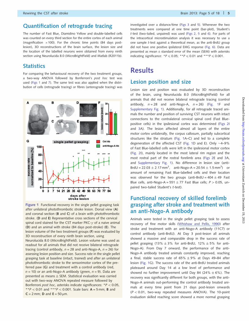
Lesion position and sizeLesion size and position was evaluated by 3D reconstruction
of the brain, using Neurolucida 8.0 (MicroBrightField) for all
animals that did not receive bilateral retrograde tracing (control
antibody, n = 28 and anti-Nogo-A, n = 26) (Fig. 1F and
Supplementary Fig. 1). Additionally, for all retrograde traced ani-
mals the number and position of surviving CST neurons with intact
connections to the contralateral cervical spinal cord (Fast Blue-
positive cells) in the ipsilesional cortex was determined (Figs 2I
and 3A). The lesion affected almost all layers of the entire
motor cortex unilaterally, the corpus callosum, partially subcortical
structures like the striatum (Fig. 1A–C) and led to a complete
degeneration of the affected CST (Fig. 1D and E). Only �4–9%
of Fast Blue-labelled cells were left in the ipsilesional motor cortex
(Fig. 2I), mainly located in the most lateral rim region and the
most rostral part of the rostral forelimb area (Figs 2E and 3A,
and Supplementary Fig. 1). No difference in lesion size (anti-
BrdU = 22.03 � 2.17 mm3, anti-Nogo-A = 20.54 � 1.5 mm3) or
amount of remaining Fast Blue-labelled cells and their location
was observed for the two groups (anti-BrdU = 404 � 49 Fast
Blue cells, anti-Nogo-A = 551 � 77 Fast Blue cells; P40.05, un-
paired two-tailed Student’s t-test).
Functional recovery of skilled forelimbgrasping after stroke and treatment withan anti-Nogo-A antibodyAnimals were tested in the single pellet grasping task to assess
recovery of fine motor skills (Whishaw and Pellis, 1990) after
stroke and treatment with an anti-Nogo-A antibody (11C7) or
control antibody (anti-BrdU). At Day 3 post-lesion all animals
showed a massive and comparable drop in the success rate of
pellet grasping (13% � 3% for anti-BrdU, 12% � 5% for anti-
Nogo-A). From Day 7 onward, the performance of the anti-
Nogo-A antibody treated animals constantly improved, reaching
a final, stable success rate of 65% � 9% at Days 49–84 after
lesion (Fig. 1G). The success rate of the anti-BrdU treated animals
plateaued around Day 14 at a low level of performance and
showed no further improvement until Day 84 (24% � 6%). The
recovery was significantly different for both groups, with the anti-
Nogo-A animals out-performing the control antibody treated ani-
mals at every time point from 21 days post-lesion onwards
(P5 0.01, two-way repeated measures ANOVA). The 10-point
evaluation skilled reaching score showed a more normal grasping
Figure 1 Functional recovery in the single pellet grasping task
after unilateral photothrombotic stroke lesion. Dorsal view (A)
and coronal section (B and C) of a brain with photothrombotic
stroke. (D and E) Representative cross sections of the cervical
spinal cord stained for the CST marker PKC-� of a naive animal
(D) and an animal with stroke (84 days post-stroke) (E). The
lesion volume of the two treatment groups (F) was evaluated by
3D reconstruction of every ninth brain section, using
Neurolucida 8.0 (MicroBrightField). Lesion volume was used as
readout for all animals that did not receive bilateral retrograde
tracing (control antibody, n = 28 and anti-Nogo-A, n = 26) for
assessing lesion position and size. Success rate in the single pellet
grasping task at baseline (intact, trained) and after an unilateral
photothrombotic stroke to the sensorimotor cortex of the pre-
ferred paw (G) and treatment with a control antibody (red,
n = 10) or an anti-Nogo-A antibody (green, n = 9). Data are
presented as means � SEM. Statistical evaluation was carried
out with two-way ANOVA repeated measure followed by
Bonferroni post hoc, asterisks indicate significances: *P50.05,
**P50.01 and ***P5 0.001. Scale bars: A = 5 mm; B and
C = 2 mm; D and E = 50 mm.
Rewiring the CST after stroke Brain 2013: Page 5 of 18 | 5
at Universitaet Z
uerich on Decem
ber 19, 2013http://brain.oxfordjournals.org/
Dow
nloaded from

performance of anti-Nogo-A treated animals compared with
control antibody treated animals (Supplementary Fig. 2). Control
antibody-treated animals showed impairment in almost every
aspect of the grasping movement, whereas anti-Nogo-A treated
animals only had impairments in the supination of the grasping
movement.
Somatotopic reorganization of thecontralesional hemisphere caused bya ‘side switch’ of the descendingcorticospinal projections after strokeTwo different retrogradely transported dyes were injected into the
denervated (Fast Blue) and intact (Diamidino Yellow) side of the
spinal cord in intact (naive) animals, and at 10, 21, 30, 42 and 84
days after the stroke. Numbers of single- and double-labelled cells
on the contralesional and ipsilesional side of the sensorimotor
cortex were determined (Fig. 2A–E). For naive animals,
9198 � 410 Fast Blue-labelled cells were detected on the side
contralateral to the injection. This number declined massively
after the stroke lesion when only �4–9% of Fast Blue-labelled
cells were left in the ipsilesional motor cortex, mainly located in
the most lateral rim region and the most rostral part of the rostral
forelimb area (Fig. 2E and I). No change over time was
detected for either of the two treatment groups (anti-BrdU and
anti-Nogo-A) (P40.05, unpaired two-tailed Student’s t-test).
For the second tracer, 4621 � 752.3 Diamidino Yellow retro-
gradely-labelled cortical cells were detected in naive animals on
the side contralateral to the injection, making Diamidino Yellow
roughly half as sensitive as Fast Blue. After a photothrombotic
stroke, both treatment groups showed no significant changes of
Diamidino Yellow-labelled cells in the contralesional cortex until
Day 42 post-lesion. For the chronic time point (Day 84) a signifi-
cant increase was observed for both lesion groups (8099 � 563.7
anti-BrdU, 7821 � 764 anti-Nogo-A), but no significant difference
existed between the groups (Fig. 2J).
To study the existence of bilaterally projecting neurons, we
evaluated double-labelled cells that had taken up both retro-
grade tracers, Fast Blue and Diamidino Yellow (Fig. 2B and
F–H). In naive animals only 18 � 2 double-labelled cells were
observed on the side of the contralaterally projecting Diamidino
Yellow-labelled cells (Fig. 2K). After stroke the control antibody-
treated group showed a decrease of double-labelled cells which
returned to naive levels at the chronic time point (12 � 2 double-
labelled cells). For the anti-Nogo-A antibody-treated animals a
marked increase of double-labelled cells was observed which
peaked at Day 21 post-lesion (Fig. 2K), indicating a massive
sprouting of intact CST fibres over the spinal cord midline. The
increase of double-labelled cells was 2.4-fold and significantly
different from anti-BrdU treated animals (P50.001, unpaired
two-tailed Student’s t-test). Interestingly, the time point of the
peak (21 days post-lesion) correlated with the time point where
the anti-Nogo-A treated animals started to outperform the anti-
BrdU treated animals in the single pellet grasping task (Fig. 1G).
After the initial peak of double-labelled cells at Day 21 post-lesion
a rapid decrease of the number of double-labelled cells occurred,
reaching baseline levels at Day 84 (Fig. 2K). To investigate which
of the two axonal arbours of these cells was pruned, it was
necessary to analyse the amount of ipsilaterally projecting cells
over time.
As the CST is mainly a crossed fibre tract with only �5% of all
CST fibres projecting ipsilaterally in naive adult animals (Brosamle
and Schwab, 1997), a persistent increase of ipsilaterally projecting
CST fibres could be an important source for functional recovery
after a unilateral lesion of the motor cortex. For naive animals
393 � 50 ipsilaterally projecting Fast Blue-labelled cells were
observed in the contralesional cortex, corresponding to �4% of
all Fast Blue-labelled cells (Fig. 2I and L). After stroke and treat-
ment with the control antibody no significant increase of ipsilat-
erally projecting Fast Blue cells was observed at any time point,
whereas the anti-Nogo-A treated animals showed an almost 3-fold
increase of ipsilaterally projecting cells in the contralesional cortex
at Day 84 post lesion (1094 � 110.1, P5 0.001, unpaired two-
tailed Student’s t-test). The amount of ipsilaterally projecting cells,
projecting to the denervated side of the spinal cord, reached sig-
nificance after 21 days post-lesion (P5 0.05, unpaired two-tailed
Student’s t-test) and continued to increase until 84 days post-
lesion (Fig. 2L).
To study the position of the cervically projecting CST cells, in
particular the ipsilaterally and the double-projecting cells of the
sensorimotor cortex, we mapped their position along the rostro-
caudal axis (Fig. 3). Naive animals showed three distinguishable
peaks of contralaterally projecting Fast Blue cells, corresponding to
the rostral forelimb area, the caudal forelimb area and the second-
ary somatosensory cortex (Fig. 3A). The photothrombotic stroke
lesion affected the hemisphere massively, and only �4–9% of all
contralaterally projecting Fast Blue cells were spared by the lesion.
These cells were mainly located in the most rostral part of the
rostral forelimb area and at the lateral rim of the caudal forelimb
area and the secondary somatosensory cortex. No significant dif-
ference between the two treatment groups was observed at any
time point (Fig. 3A). The tracing efficiency for contralaterally pro-
jecting Diamidino Yellow cells was slightly lower as for Fast Blue
cells (high efficiency tracer Fast Blue for ipsilaterally projecting
cells), but the same three distinguishable peaks were observed.
In naive animals the rostral forelimb area consisted of �5% of
all Diamidino Yellow cells, whereas the caudal forelimb area con-
sisted of �79% of all Diamidino Yellow cells and the secondary
somatosensory cortex of �16% of all Diamidino Yellow cells
(Fig. 3B). After stroke, both treatment groups showed plastic
rearrangements over time, with a massive increase of cells in the
caudal and rostral forelimb area (Fig. 3B). This increase of contra-
lateral projecting cells was around 2.5-fold for the rostral forelimb
and 2-fold for the caudal forelimb area (84 days post-lesion).
Despite the general increase for both groups, the caudal forelimb
area was still comprised of �79% of all contralateral projecting
cells (anti-BrdU: 79%, anti-Nogo-A: 78%), whereas the rostral
forelimb region increased proportionally (anti-BrdU: 10%, anti-
Nogo-A: 14%) at 84 days after lesion.
The distribution of double-labelled cells for naive animals was
almost uniform over the entire forelimb area and did not show
peaks at specific locations (Fig. 3C). After stroke and treatment
with an anti-BrdU antibody a similar distribution as for naive
6 | Brain 2013: Page 6 of 18 N. T. Lindau et al.
at Universitaet Z
uerich on Decem
ber 19, 2013http://brain.oxfordjournals.org/
Dow
nloaded from
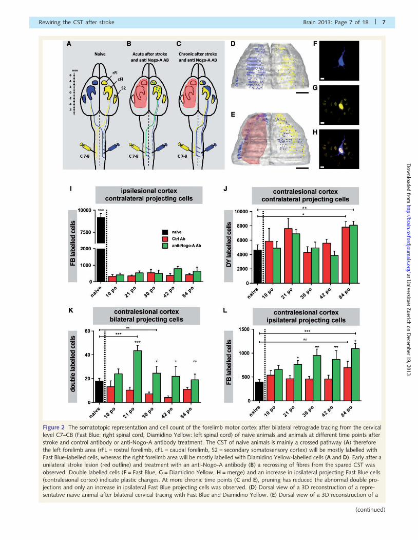
Figure 2 The somatotopic representation and cell count of the forelimb motor cortex after bilateral retrograde tracing from the cervical
level C7–C8 (Fast Blue: right spinal cord, Diamidino Yellow: left spinal cord) of naive animals and animals at different time points after
stroke and control antibody or anti-Nogo-A antibody treatment. The CST of naive animals is mainly a crossed pathway (A) therefore
the left forelimb area (rFL = rostral forelimb, cFL = caudal forelimb, S2 = secondary somatosensory cortex) will be mostly labelled with
Fast Blue-labelled cells, whereas the right forelimb area will be mostly labelled with Diamidino Yellow-labelled cells (A and D). Early after a
unilateral stroke lesion (red outline) and treatment with an anti-Nogo-A antibody (B) a recrossing of fibres from the spared CST was
observed. Double labelled cells (F = Fast Blue, G = Diamidino Yellow, H = merge) and an increase in ipsilateral projecting Fast Blue cells
(contralesional cortex) indicate plastic changes. At more chronic time points (C and E), pruning has reduced the abnormal double pro-
jections and only an increase in ipsilateral Fast Blue projecting cells was observed. (D) Dorsal view of a 3D reconstruction of a repre-
sentative naive animal after bilateral cervical tracing with Fast Blue and Diamidino Yellow. (E) Dorsal view of a 3D reconstruction of a
Rewiring the CST after stroke Brain 2013: Page 7 of 18 | 7
(continued)
at Universitaet Z
uerich on Decem
ber 19, 2013http://brain.oxfordjournals.org/
Dow
nloaded from

Figure 3 The anterior-posterior distribution and the amount of contralaterally projecting, spared Fast Blue (FB) cells in the ipsilesional
cortex (A), contralaterally projecting Diamidino Yellow cells in the contralesional cortex (B), bilaterally projecting double-labelled cells in
the contralesional cortex (C) and ipsilaterally projecting Fast Blue-labelled cells in the contralesional cortex (D) in relation to bregma (0 mm
anterior-posterior). Black group = naive animals (n = 6), red group = stroke lesion and anti-BrdU treatment [10 days postoperative (po)
n = 7, 21 days postoperative n = 7, 30 days postoperative n = 5, 42 days postoperative n = 6, 84 days postoperative n = 6], green
group = stroke lesion and anti-Nogo-A treatment (10 days postoperative n = 7, 21 days postoperative n = 6, 30 days postoperative n = 5,
42 days postoperative n = 5, 84 days postoperative n = 6). Red outline indicates area affected by photothrombotic stroke. rFL = rostral
forelimb, cFL = caudal forelimb and S2 = secondary somatosensory cortex. Data are presented as sliding window average of mean � SEM.
Statistical evaluation was carried out with two-way repeated measures ANOVA followed by Bonferroni post hoc, asterisks indicate
significances: *P50.05, ***P50.001.
Figure 2 Continuedrepresentative animal after stroke and treatment with anti-Nogo-A (red outline, 84 days post-lesion) and bilateral cervical tracing with Fast
Blue and Diamidino Yellow. This increase of ipsilateral projections is a result of midline crossing fibres and strengthening of the pre-existing
ipsilateral projections and induces a somatotopic reorganization of the contralesional cortex. (I) Amount of contralateral projecting Fast
Blue-labelled cells spared by the stroke lesion in the ipsilesional cortex at different time points after stroke and for the naive situation.
(J) Amount of contralateral projecting Diamidino Yellow cells in the contralesional cortex at different time points after stroke and for the
naive situation. (K) Amount of double-labelled cells (projecting to both sides of the spinal cord) in the contralesional cortex at different
time points after stroke and for the naive situation. (L) Amount of ipsilaterally projecting Fast Blue cells in the contralesional cortex at
different time points after stroke and for the naive situation. Black group = naive animals (n = 6), red group = stroke lesion and anti-BrdU
treatment [10 days postoperative (po) n = 7, 21 days postoperative n = 7, 30 days postoperative n = 5, 42 days postoperative n = 6, 84
days postoperative n = 6], green group = stroke lesion and anti-Nogo-A treatment (10 days postoperative n = 7, 21 days postoperative
n = 6, 30 days postoperative n = 5, 42 days postoperative n = 5, 84 days postoperative n = 6). Data are presented as means � SEM.
Statistical evaluation was carried out with unpaired two-tailed Student’s t-test, asterisks indicate significances: *P50.05, **P50.01,
***P50.001. Scale bars: D and E = 2 mm; F–H = 10 mm.
8 | Brain 2013: Page 8 of 18 N. T. Lindau et al.
at Universitaet Z
uerich on Decem
ber 19, 2013http://brain.oxfordjournals.org/
Dow
nloaded from

animals was observed, except for Day 84 after lesion when an
almost 3-fold increase in double-labelled cells was detected in
the rostral forelimb area. For anti-Nogo-A treated animals a mas-
sive increase in double-labelled cells, with peaks in the rostral and
caudal forelimb area, was observed at Day 21 post-lesion. For the
rostral forelimb area this increase was 43-fold, compared with the
naive situation, whereas for the caudal forelimb area it was 42-
fold. This was followed by a decrease of double-labelled cells until
Day 42, where a difference between the groups could no longer
be detected (Fig. 3C).
For naive animals a small amount of pre-existing ipsilaterally
projecting Fast Blue cells was detected in the contralesional
cortex with a peak in the caudal forelimb area (Fig. 3D), where
�84% of all ipsilaterally projecting cells were located (rostral fore-
limb: �5% and secondary somatosensory cortex: �11%). After
stroke and treatment with the control anti-BrdU antibody a 4-fold
increase in ipsilaterally projecting Fast Blue cells was observed
for the rostral forelimb area at Day 84. The caudal forelimb and
the secondary somatosensory cortex were almost unchanged
(Fig. 3D). Animals that were treated with an anti-Nogo-A anti-
body showed a continuous increase of ipsilaterally projecting cells
over time in all forelimb regions. At Day 84 after lesion a 5.5-fold
increase of cells in the rostral forelimb area, a 2-fold increase in
the caudal forelimb area and a 1.5-fold increase in the secondary
somatosensory area were detected. At this time point the rostral
forelimb contained �20% of all ipsilateral cells, whereas the
caudal forelimb comprised �74% of the cells and the secondary
somatosensory cortex contained �6% of all ipsilateral cells.
Average 2D false colour-coded projection heat maps of the
position and density of contralaterally and ipsilaterally projecting
cells were obtained for all naive animals and for all animals from
the chronic time point (84 Days post lesion) (Fig. 4). For contra-
lateral projections the majority of CST fibres originate in the caudal
forelimb area with smaller proportions coming from the rostral
forelimb area and the secondary somatosensory area (Fig. 4A).
In naive rats only a small fraction of fibres project to the ipsilateral
side and their origin is almost exclusively in the caudal forelimb
region (Fig. 4B). The increase in ipsilateral projections observed
Figure 4 Average false colour coded heat map of the spatial distribution (dorsal 2D view) and cell density of contralateral projecting
Diamidino Yellow cells (A) and ipsilateral projecting Fast Blue cells (B), for naive animals (n = 6) and animals 84 days post-lesion
(control antibody n = 6 and anti-Nogo-A antibody n = 6), in the contralesional cortex in relation to bregma (0 mm anterior-posterior and
medio-lateral). Straight lines indicate the boundaries of the rostral forelimb (rFL), the caudal forelimb area (cFL) and the secondary
somatosensory cortex = S2.
Rewiring the CST after stroke Brain 2013: Page 9 of 18 | 9
at Universitaet Z
uerich on Decem
ber 19, 2013http://brain.oxfordjournals.org/
Dow
nloaded from

after a unilateral stroke lesion originates, to a large extent, from
the rostral forelimb area (Fig. 4B). In the caudal forelimb region,
the ipsilaterally projecting neurons are located more medially than
the bulk of the contralaterally projecting cells, suggesting that the
more medially located motor CST component might be more plas-
tic than the lateral, mostly sensory part (Fig. 4B). Anti-Nogo-A
treated animals showed the highest density of ipsilaterally and
contralaterally projecting CST neurons in all analysed cortical
regions.
These results demonstrate an enhanced outgrowth/sprouting of
the contralesional CST in anti-Nogo-A treated animals with fibres
crossing the midline and innervating the denervated half of the
spinal cord. These plastic changes are followed by a somatotopic
reorganization of the unlesioned hemisphere and the formation of
an ipsilateral forelimb area.
Cortical reorganization is caused bymidline crossing fibres and sproutingventral corticospinal tract fibres in thecervical spinal cordTo study the course of the increased ipsilateral CST fibres, we
injected the anterograde tracers biotin-dextran-tetramethylrhoda-
mine (Fluoro Ruby) into the contralesional rostral forelimb area
and biotin-dextran-fluorescein (Mini Emerald) into the contrale-
sional caudal forelimb area of chronically recovered animals that
were either treated with anti-Nogo-A or control antibody. CST
fibres can reach the denervated cervical spinal cord in two ways:
as originally contralateral fibres that recross the spinal cord midline
segmentally, or as uncrossed, pre-existing ipsilaterally projecting
axons. The latter are predominantly found in the medial part of
the ventral funiculus (Brosamle and Schwab, 1997), and we
counted the number of intersections of collaterals of these fibres
at the grey/white matter boundary of the ventral funiculus as a
measure of innervation of the cervical grey matter by ventral CST
fibres. Our anterograde tracing experiments showed that both,
midline crossing fibres and ventral fibre branches increased in
number in response to the anti-Nogo-A treatment. Anti-Nogo-A
treated animals had significantly more midline crossing fibres pro-
jecting from the rostral forelimb area to the cervical spinal cord
than control antibody treated animals (P5 0.01, two-way
repeated measures ANOVA) (Fig. 5A and E). For pre-existing ip-
silateral fibres the main difference between the two groups was
observed in more rostral cervical areas (C2–C5), with a constant
decline in more caudal regions (Fig. 5B). For midline crossing fibres
projecting from the caudal forelimb area a similar observation was
made (Fig. 5C and G), with the main difference between the anti-
Nogo-A and control antibody treated groups at the cervical level
C4 (P50.05, two-way repeated measure ANOVA followed by
Bonferroni post hoc), which innervates mostly shoulder and
upper arm muscles. Pre-existing ipsilateral CST fibres from the
rostral forelimb area increased their cervical grey matter projec-
tions 42-fold in anti-Nogo-A treated animals (Fig. 5B and F)
compared to control antibody-treated animals (P50.001, two-
way repeated measure ANOVA followed by Bonferroni post
hoc). The sprouting was again more prominent in the rostral
cervical region from C2–C4, but was also observed at caudal
cervical areas around C7, where fibres innervate distal arm and
forepaw muscles (Fig. 5B). The anterograde tracing from the
caudal forelimb area revealed an almost identical picture
with more pre-existing ipsilateral, ventral fibres crossing the
ventral funiculus grey/white matter border in the anti-Nogo-A
treated animals compared with control antibody-treated animals
(Fig. 5D and H).
To investigate the dorso-ventral projection pattern of CST fibres
from the rostral and caudal forelimb area, we analysed the aver-
age CST fibre density (as optical density grey values) on spinal
cord cross-sections at the spinal cord level C4 for all animals.
The contralateral projection from the intact cortex (left side of
the spinal cord) terminated in a dense plexus covering the dorsal
layers 3–6, the premotor layer 7 and the ventral horn (Fig. 5I–L).
Consistent with the retrograde tracing data, anti-Nogo-A treated
animals had the highest density of CST fibres projecting from the
caudal forelimb cortex to the contralateral spinal cord (Fig. 5K and
L) and showed an increased ipsilateral fibre density (right side of
the spinal cord) which closely mimicked the pattern of the contra-
lateral innervation. Typically, the dorsal horn projections were
less dense from the rostral forelimb area than from the caudal,
sensorimotor forelimb field, where anti-Nogo-A treated rats again
had higher fibre densities than control antibody treated animals
(Fig. 5I and J).
The combined data from the retrograde and anterograde
tracing show increased plasticity of the spared CST from the unle-
sioned hemisphere with an increase of ipsilaterally projections
through midline crossing fibres as well as sprouting of pre-existing
ipsilateral fibres after stroke and treatment with anti-Nogo-A
antibodies.
Physiological analysis of theipsilaterally projecting forelimb area byintracortical microstimulation mappingTo test if the ipsilateral projections from the contralesional hemi-
sphere are functionally relevant, we used intracortical microstimula-
tion in combination with EMG recordings of the contra- and
ipsilateral forelimb. We analysed the coordinates, the current
and the side of the forelimb response for each stimulation point
(100–120 stimulation points with �40 forelimb responses per
animal). The EMG response was classified as a pure contralateral
response (when the lowest current induced only a contralateral
forelimb response) or as pure ipsilateral response (when the lowest
current induced only an ipsilateral forelimb response). These were
the two main categories and the combined sum of pure contralateral
and pure ipsilateral responses made up 100% of all forelimb
responses. However, we wanted to gain additional information
about changes of supra-threshold ipsilateral responses, which were
not strong enough to induce a pure ipsilateral response, but could still
contribute to an ipsilateral movement. Therefore, we investigated a
third category, the total ipsilateral response (any ipsilateral forelimb
response that was detected additionally to the stronger contralateral
response, up to a maximum current of 80 mA). For naive animals
the majority of all forelimb responses (from caudal and rostral
10 | Brain 2013: Page 10 of 18 N. T. Lindau et al.
at Universitaet Z
uerich on Decem
ber 19, 2013http://brain.oxfordjournals.org/
Dow
nloaded from
forelimb area) were pure contralateral (94% � 4%), and only a few
pure ipsilateral responses could be evoked (6% � 4%, Fig. 6A).
However, for �25% of all forelimb stimulation points a total
ipsilateral response could be evoked additionally to the stronger
contralateral response (23% � 9%). Eighty-four days after stroke,
animals treated with anti-BrdU showed a similar distribution of
forelimb responses, with 98% � 1% pure contralateral, 2% � 1%
pure ipsilateral and 14% � 6% total ipsilateral responses. After
stroke and treatment with anti-Nogo-A, a decrease of pure contra-
lateral responses (86% � 4%) due to an �6-fold increase of pure
ipsilateral responses (14% � 4%) and a 4-fold increase of total
ipsilateral responses (57% � 3%) compared with anti-BrdU treated
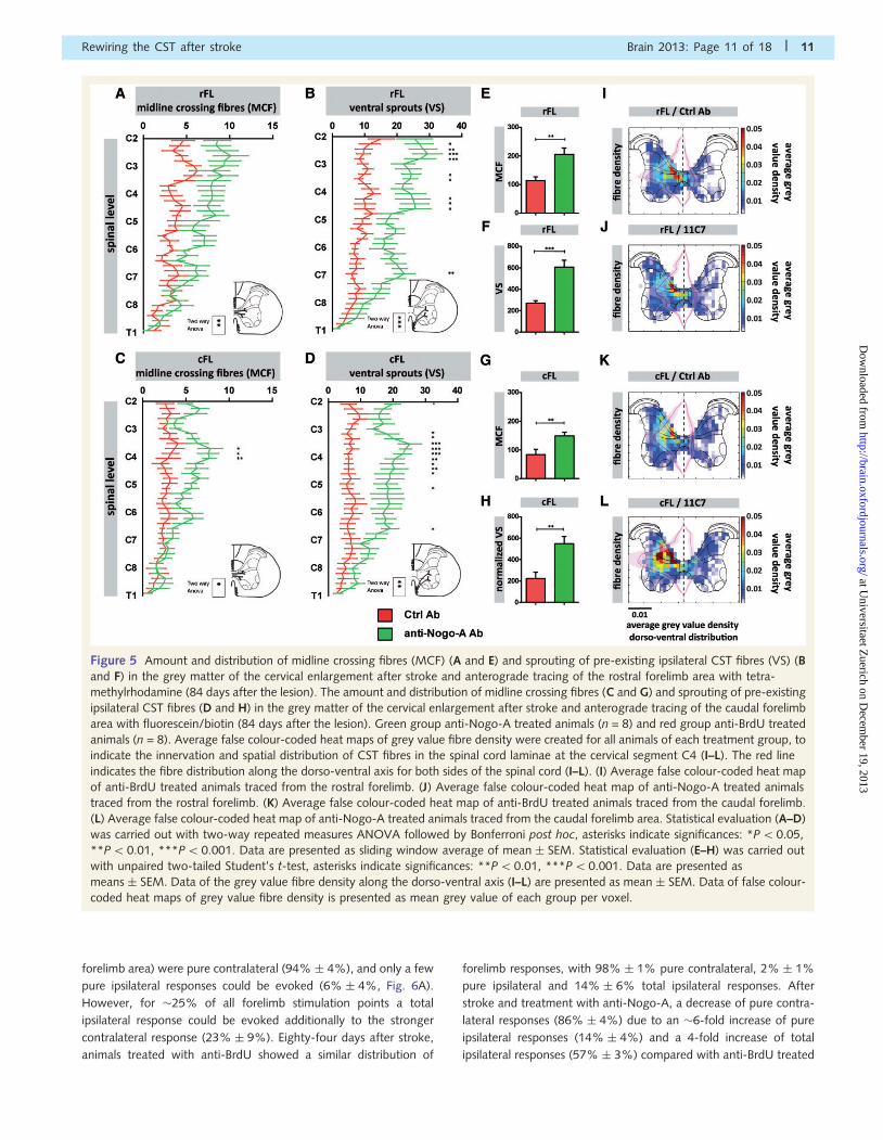
Figure 5 Amount and distribution of midline crossing fibres (MCF) (A and E) and sprouting of pre-existing ipsilateral CST fibres (VS) (B
and F) in the grey matter of the cervical enlargement after stroke and anterograde tracing of the rostral forelimb area with tetra-
methylrhodamine (84 days after the lesion). The amount and distribution of midline crossing fibres (C and G) and sprouting of pre-existing
ipsilateral CST fibres (D and H) in the grey matter of the cervical enlargement after stroke and anterograde tracing of the caudal forelimb
area with fluorescein/biotin (84 days after the lesion). Green group anti-Nogo-A treated animals (n = 8) and red group anti-BrdU treated
animals (n = 8). Average false colour-coded heat maps of grey value fibre density were created for all animals of each treatment group, to
indicate the innervation and spatial distribution of CST fibres in the spinal cord laminae at the cervical segment C4 (I–L). The red line
indicates the fibre distribution along the dorso-ventral axis for both sides of the spinal cord (I–L). (I) Average false colour-coded heat map
of anti-BrdU treated animals traced from the rostral forelimb. (J) Average false colour-coded heat map of anti-Nogo-A treated animals
traced from the rostral forelimb. (K) Average false colour-coded heat map of anti-BrdU treated animals traced from the caudal forelimb.
(L) Average false colour-coded heat map of anti-Nogo-A treated animals traced from the caudal forelimb area. Statistical evaluation (A–D)
was carried out with two-way repeated measures ANOVA followed by Bonferroni post hoc, asterisks indicate significances: *P50.05,
**P50.01, ***P50.001. Data are presented as sliding window average of mean � SEM. Statistical evaluation (E–H) was carried out
with unpaired two-tailed Student’s t-test, asterisks indicate significances: **P50.01, ***P50.001. Data are presented as
means � SEM. Data of the grey value fibre density along the dorso-ventral axis (I–L) are presented as mean � SEM. Data of false colour-
coded heat maps of grey value fibre density is presented as mean grey value of each group per voxel.
Rewiring the CST after stroke Brain 2013: Page 11 of 18 | 11
at Universitaet Z
uerich on Decem
ber 19, 2013http://brain.oxfordjournals.org/
Dow
nloaded from

animals was detected (P50.001, unpaired two-tailed Student’s
t-test, Fig. 6A). This increase in ipsilateral muscle responses
suggests that the newly formed ipsilateral projections developed
large numbers of functional spinal connections as more than half
of the cortical stimulation points induced an ipsilateral forelimb
muscle response.
The total forelimb response (100%) was further subdivided into
responses induced from the rostral (Fig. 6B) and the caudal
(Fig. 6C) forelimb areas. For naive animals, as for both treatment
groups, the majority of all forelimb responses from the caudal
forelimb area were pure contralateral muscle twitches (naive:
81% � 7%, anti-BrdU: 85% � 2%, anti-Nogo-A: 77% � 3%).
Figure 6 Percentage of forelimb response (rostral and caudal forelimb) evoked during intracortical microstimulation (ICMS) and recorded
by EMG for naive animals (black, n = 5), anti-BrdU (red, n = 6) and anti-Nogo-A treated animals (green, n = 5) 84 days after stroke (A).
The forelimb EMG response was classified as a pure contralateral response (the lowest current induced a contralateral response), a pure
ipsilateral response (the lowest current induced an ipsilateral response) and a total ipsilateral response (any ipsilateral response which was
detected independent of the current to a maximum of 80 mA). The total forelimb response was further subdivided into responses from the
rostral forelimb area (B) and the caudal forelimb area (C). The minimum current needed to evoke a movement was analysed for the rostral
(D) and caudal forelimb area (E) and the latency for the induction of a contralateral (F) and ipsilateral EMG response (G) was detected.
(H and I) Overlay of typical contralateral (H) and ipsilateral (I) EMG responses of one animal. For (n = 4) anti-BrdU and (n = 4) anti-Nogo-
A treated animals the EMG forelimb response of the rostral forelimb area (J) and caudal forelimb area (K) 21 days after lesion was
evaluated. An example of a full wave rectified bilateral EMG response (mirror movement) of contralateral (L) and ipsilateral (M) proximal
muscle movement is illustrated for an anti-Nogo-A antibody treated animal 21 days after stroke. Data are presented as means � SEM.
Statistical evaluation was carried out with unpaired two-tailed Student’s t-test, asterisks indicate significances: *P50.05, **P50.01,
***P50.001. For the comparison of the pure ipsilateral response (B) and the ipsilateral current of the rostral forelimb area (D) between
anti-Nogo-A and anti-BrdU treated animals, a one sample t-test against a theoretical mean was used, as the anti-BrdU animals had no
pure ipsilateral response. Asterisks indicate significances: *P5 0.05. All data are presented as means � SEM.
12 | Brain 2013: Page 12 of 18 N. T. Lindau et al.
at Universitaet Z
uerich on Decem
ber 19, 2013http://brain.oxfordjournals.org/
Dow
nloaded from

No difference between the groups was detected when the pure
ipsilateral response was evaluated (naive: 5% � 3%, anti-BrdU:
2% � 1%, anti-Nogo-A: 6% � 2%). However, for the total ipsi-
lateral responses seen at higher current strengths, the anti-Nogo-A
treated group showed a significant increase (28% � 4.9%)
compared with anti-BrdU treated animals (6% � 3%, P50.01,
unpaired two-tailed Student’s t-test) (Fig.6C). Interestingly the
rostral forelimb area (Fig.6B) of anti-Nogo-A treated animals
showed the same amount of pure contralateral responses
(9% � 3%) as pure ipsilateral responses (9% � 3%). This was
significantly different from anti-BrdU treated animals (P50.05,
one sampled Student’s t-test against hypothetical mean), where
no pure ipsilateral response could be detected and naive animals
(P50.05, unpaired two-tailed Student’s t-test) where only one
animal showed a pure ipsilateral response (Fig. 6B). Furthermore,
the total ipsilateral response of anti-Nogo-A treated animals
increased to 29% � 4%, compared with 8% � 5% for anti-
BrdU and 3% � 2% for naive animals.
The current required for inducing a pure contralateral response
from the rostral (Fig. 6D) and caudal forelimb area (Fig. 6E) was
reduced 84 days after a unilateral stroke lesion compared with
naive animals. Pure ipsilateral responses had the same low stimu-
lation threshold as the pure contralateral responses, whereas the
thresholds of the total ipsilateral responses in the rostral or caudal
forelimb area were generally higher (Fig. 6D and E). The latency of
the stimulation to the first peak of a contralateral EMG response
was 29.9 � 1 ms for naive animals, 32.7 � 0.8 ms for anti-BrdU
treated animals and 32.4 � 0.1 ms for anti-Nogo-A treated ani-
mals (Fig. 6F and H). A similar latency with no differences
among the treatment groups was observed for ipsilateral EMG
responses (34.5 � 3.2 ms for naive, 33.5 � 3 ms for anti-BrdU,
33.3 � 1.2 ms for anti-Nogo-A, Fig. 6G and I).
For chronically recovered animals (84 days after lesion) no fore-
limb mirror movements were detected. However, when intracor-
tical microstimulation was performed 21 days after the lesion,
forelimb mirror movements could be observed for some animals
treated with anti-Nogo-A (Fig. 6L and M). These mirror move-
ments were only detected for proximal muscles and only with
higher currents. For this early time point after stroke, no significant
increase of ipsilateral responses was observed for animals treated
with anti-Nogo-A, even if a tendency was visible for the rostral
forelimb area (Fig. 6J and K).
Reappearance of the initial lesion deficitin chronically recovered anti-Nogo-Atreated animals after transection of thespared corticospinal tract from theunlesioned hemisphereTo assess the functional role of the spared CST from the
unlesioned hemisphere in behaviourally recovered chronic stroke
animals, the tract was transected at the pyramidal decussation
(Fig. 7A–D) and animals were tested for skilled forelimb reaching.
A massive drop in success rate was observed in the anti-Nogo-A
treated animals at Day 3 after the lesion, which did not recover
(Fig. 7E). Control antibody-treated animals showed no decrease in
their (low) success rate after pyramidotomy, even if the more
Figure 7 Success rate in the single pellet grasping task normalized to pre-lesion baseline performance after unilateral photothrombotic
stroke lesion and unilateral transection of the spared CST. (A–C) Cross sections of the cervical spinal cord stained for the CST marker
PKC-� of a naive animal (A and B) and an animal with stroke followed by a contralesional, unilateral pyramidotomy (98 days post-stroke,
14 days post-pyramidotomy) (C). The PKC-� staining labels the dorsal CST and the superficial dorsal horn (A), allowing to compare the
completeness of the pyramidotomy lesion between the groups using the staining intensities of these two regions (D). Success rate in
the single pellet grasping task 84 days post-stroke and after stroke followed by pyramidotomy (dashed line) for control antibody-treated
animals (red, n = 10) and anti-Nogo-A antibody treated animals (green, n = 9) (E). Data are presented as means � SEM. Statistical
evaluation was carried out with two-way ANOVA repeated measure followed by Bonferroni post hoc, asterisks indicate significances:
**P50.01. Scale bars: A = 150 mm; B and C = 50 mm.
Rewiring the CST after stroke Brain 2013: Page 13 of 18 | 13
at Universitaet Z
uerich on Decem
ber 19, 2013http://brain.oxfordjournals.org/
Dow
nloaded from

detailed 10-point evaluation skilled reaching score showed a sig-
nificant change in the movement performance (Supplementary
Fig. 2). The anti-Nogo-A treated animals dropped to exactly
the level of anti-BrdU treated animals and showed a deficit in
the 10-point evaluation skilled reaching score comparable to the
movement performance observed 3 days after the photothrombo-
tic stroke lesion (Supplementary Fig. 2).
DiscussionUsing a growth enhancing treatment, intrathecal application of
anti-Nogo-A antibodies for 2 weeks, after a unilateral subtotal
photothrombotic stroke to the sensorimotor cortex, we observed
a high degree of functional recovery of fine forelimb movements
in adult rats. Such a large amount of recovery after such a severe
injury, which destroyed 490% of the forelimb motor cortex, is
astonishing and is, without further intervention, usually only
observed in neonatally lesioned animals (Whishaw and Kolb,
1988; Kolb et al., 2000). The anti-Nogo-A treated animals con-
tinuously improved their success rate at skilled grasping over time,
reaching a maximal performance �6–7 weeks after stroke.
Performance then remained stable up to the latest time point
tested at 12 weeks. The spontaneous recovery of the control anti-
body treated group was minimal. To what extent a functional
recovery reflects a true recovery of a movement and to what
extent compensatory movement patterns contribute to the
observed improvement after stroke is a matter of great debate
and interest (Krakauer, 2006). Our detailed 10-point grasping
analysis indicated a more normal grasping movement for anti-
Nogo-A treated animals than for control antibody-treated animals.
This observed functional recovery reflects at least partially a true
recovery, probably with additional compensatory mechanisms.
Movement categories involved with the alignment, advance, aim
and grasp of the paw recovered almost fully, whereas other cate-
gories such as the supination remained affected. These affected
categories were most likely compensated by proximal limb move-
ments in the anti-Nogo-A treated animals, which showed a denser
innervation of ipsilaterally projecting fibres into high cervical re-
gions, controlling proximal muscles (Whishaw and Pellis, 1990).
Interestingly, the functional recovery of anti-Nogo-A treated ani-
mals was paralleled by the enhanced outgrowth of ipsilaterally
projecting CST fibres, originating in the contralesional hemisphere.
These plastic changes in the contralesional hemisphere were seen
as a massive increase of bilaterally projecting double-labelled cells,
representing new axonal branches growing over the spinal cord
midline. No plastic changes could be observed in the ipsilesional
sensory motor cortex, indicating a specific sprouting of the
unlesioned CST after very large hemispheric stroke lesions and
anti-Nogo-A treatment. Similar results were observed in untreated
neonatally lesioned animals (Yoshikawa et al., 2012), an age
where the level of growth inhibitory proteins in the CNS, including
Nogo-A, are much lower than in the adult CNS (Qiu et al., 2000).
Intracortical microstimulation of the contralesional hemisphere 21
days after lesion showed mirror movements in the anti-Nogo-A
treated animals. Normally, mirror movements are only found in
young children, where they are associated with immature
bilateral/ipsilateral projections of CST neurons and a lack of callo-
sal inhibition. In adults mirror movements are unusual and often
reflect abnormalities in the projection of CST fibres (Farmer, 2005;
Rocca et al., 2005; Srour et al., 2010; Tsuboi et al., 2010).
Our detailed time course study, using retrograde tracing,
showed that the initial 2–3-fold increase in double-labelled CST
neurons projecting to both sides of the spinal cord in the anti-
Nogo-A treated stroke animals was followed by a reduction of this
number and an increase in ipsilaterally projecting cells. The in-
crease of ipsilaterally-projecting fibres was partially induced
through midline crossing fibres, which were still increased at the
chronic time point (84 days post-lesion) for anti-Nogo-A treated
animals, as well as a sprouting of pre-existing ipsilateral fibres. This
result indicates that the original axonal arbours in the contralateral
spinal cord of the sprouting CST fibres were withdrawn to the
extent that no tracer uptake was detectable anymore when the
fibres innervated the opposite, originally denervated side of
the spinal cord. Such a sorting of bilateral projections was
observed during developmental maturation of the CST system,
where it is regulated by competition dependent mechanisms
(Martin, 2005; Friel and Martin, 2007). We speculate that once
outgrowth of fibres was induced after stroke and anti-Nogo-A
treatment, the spared cortex gained access to the formerly dener-
vated side of the body which led to activity-dependent competi-
tion of fibres. Electrical stimulation, constraint induced movement
therapy or task-specific training (Brus-Ramer et al., 2007; Maier
et al., 2008; Carmel et al., 2010; Starkey et al., 2011; Carmel
et al., 2013) had been used to increase plasticity of the spared
CST fibres and strengthen functionally important/meaningful con-
nections. As rehabilitation therapy is crucial for recovery after
stroke, the combination of a growth and plasticity enhancing
anti-Nogo-A antibody treatment, followed by intense rehabilita-
tion, could be a promising future approach for stroke patients to
enhance the formation of new neuronal connections and circuits
and to shape and stabilize meaningful connections (Starkey and
Schwab, 2011). In addition to CST fibres growing across the spinal
cord midline, our results also show enhanced branching of the
small population of original ipsilateral fibres, in line with earlier
observations (Weidner et al., 2001).
The impressive compensatory sprouting of CST fibres from
the unlesioned hemisphere that was observed in animals treated
with an anti-Nogo-A antibody led to the formation of a greatly
expanded ipsilateral forelimb representation in the rostral and
caudal forelimb areas (premotor cortex and area 4 in humans;
Rouiller et al., 1993) as well as the secondary somatosensory
cortex. The newly connected, ipsilaterally projecting cells seemed
to be intermixed with the original, contralateral CST neurons.
Similar observations were made for animals which received a
hemidecortication as neonates and which were bilaterally retro-
gradely traced as adults (Umeda and Isa, 2011; Yoshikawa
et al., 2012). Intracortical microstimulation of the rostral and
caudal motor field 84 days after stroke and treatment with an
anti-Nogo-A antibody demonstrated their influence on ipsilateral
muscle activation, as 57% of all forelimb stimulation points lead to
an ipsilateral response (14% in control antibody-treated rats). In
particular the rostral forelimb area reacted in a highly plastic
manner after lesion and growth enhancing treatment and
14 | Brain 2013: Page 14 of 18 N. T. Lindau et al.
at Universitaet Z
uerich on Decem
ber 19, 2013http://brain.oxfordjournals.org/
Dow
nloaded from
showed a predominant ipsilateral innervation. The influence of this
hierarchically higher motor structure on functional recovery was
investigated in several recent studies, showing that it is important
for the planning of a movement, being densely connected to the
caudal forelimb area as well as the spinal cord and being at least
partially a bilaterally/ipsilaterally projecting system through the
cortico-bulbo-spinal tract, making it a particularly interesting
region for plastic reorganization after stroke (Chollet et al.,
1991; Frost et al., 2003; Davidson and Buford, 2006; Horenstein
et al., 2009; Bestmann et al., 2010; Bashir et al., 2012; Glover
et al., 2012; Kantak et al., 2012). This indirect projecting pathway
of the cortico-bulbo-spinal tract is known to be involved in the
positioning of the body, trunk stability, but also distal wrist and
digit movements (Riddle and Baker, 2010; Soteropoulos et al.,
2012). These observations together with the increased direct ipsi-
lateral projections from the rostral forelimb area to higher cervical
regions, innervating mostly proximal muscles crucial for successful
pellet grasping (Whishaw and Pellis, 1990), make the rostral
forelimb area a promising region for plasticity based functional
recovery. However, further more detailed studies are necessary
to analyse the precise contribution of the rostral and caudal fore-
limb area on functional recovery after stroke and treatment and
to understand the interplay between these areas, including the
sensory system (Harrison et al., 2013).
Interestingly, the latencies for contralateral EMG responses did
not differ from those of ipsilateral responses, neither in naive nor
in stroke and anti-Nogo-A treated animals suggesting a similar
connectivity pattern for both sides. Such short ipsilateral latencies
were observed for neonatally lesioned animals and human patients
with lesions early in life, whereas adult lesioned subjects tend
to show a prolonged latency for ipsilateral muscle responses,
indicating the involvement of indirect pathways like the cortico-
reticulo-spinal tract (Benecke et al., 1991; Muller et al., 1997;
Kastrup et al., 2000; Eyre et al., 2001). To test the influence of
the direct CST pathway, which was spared by the stroke lesion,
we applied a unilateral pyramidotomy lesion 12 weeks after the
stroke. The well recovered anti-Nogo-A treated animals showed a
massive drop in their skilled forelimb grasping success rate 3 days
after the pyramidotomy whereas the control antibody treated ani-
mals were unaffected in their low success rate. No further
recovery was observed until Day 14 after pyramidotomy. The de-
tailed 10-point evaluation reaching score revealed a comparable
deficit for anti-Nogo-A treated animals after pyramidotomy as 3
days after the initial stroke lesion. These results show that the
enhanced functional recovery of anti-Nogo-A treated animals
was crucially dependent on fibres running in the contralesional
CST. For control antibody-treated animals this fibre tract played
only a minor role in functional recovery, even if the general move-
ment pattern, evaluated by 10-point evaluation skilled reaching
score, was changed after the pyramidotomy lesion (Bashir et al.,
2012). Spontaneous plasticity of the spared CST was insufficient to
contribute to behavioural recovery in the single pellet reaching
task. These observations indicate that an anti-Nogo-A treatment
can increase functional recovery by increasing plasticity of the
spared CST and furthermore, that indirect descending pathways,
like the cortico-bulbo-spinal pathway, were not involved in the
recovery of skilled movement after stroke and anti-Nogo-A
treatment. However, further investigations would be necessary
to distinguish between the influence of midline crossing fibres
and the contribution of sprouting pre-existing ipsilateral projec-
tions on functional recovery. Similar observations were made in
neonatally operated animals after the transection of the spared
CST (Kartje-Tillotson et al., 1987) and in transcranial magnetic
stimulation studies in human patients with congenital hemiparesis
and good functional recovery (Lotze et al., 2009). These patients
showed an increased ipsilateral activation during paretic hand
movements and no difference of motor evoked potentials for
the affected or unaffected side. A functional blockade, induced
through repetitive transcranial magnetic stimulation of the ipsilat-
eral primary motor cortex or dorsal premotor cortex, led to a
decrease in temporal precision of finger movements, showing
the importance of the contralesional hemisphere. These results
indicate a strong influence of direct CST connections on functional
recovery after stroke. Nevertheless, phylogenetically older
brainstem pathways like the rubro-, vestibulo- and reticulo-spinal
pathway are known to influence postural control, proximal limb
movements and flexion-biased movements, but their contribution
to stroke recovery is less understood (Lemon, 2008). Additional
studies are necessary to understand the spontaneous and treat-
ment induced plasticity of these descending pathways and to in-
vestigate their influence on functional recovery (Wenk et al.,
1999; Papadopoulos et al., 2002).
Recent studies showed that BDNF, Gap-43, ephrin-B3 and other
factors are involved with midline crossing fibres, sprouting and
functional recovery after a brain injury (Omoto et al., 2011;
Ueno et al., 2012), and anti-Nogo-A has been shown to influence
the upregulation of different endogen growth factors (Bareyre
et al., 2002). Other growth/sprouting enhancing interventions
like chondroitinase ABC treatment, intense rehabilitation, electrical
stimulation or the manipulation of the intrinsic neuronal growth
regulation, e.g. by PTEN or SOCS3 deletion, are also known to
induce a massive stimulation of sprouting and regeneration of
axons after injury. However, the precise molecular mechanisms
that allow fibres to recross over the adult spinal cord midline,
e.g. up- and downregulation of particular factors at the spinal
cord midline or possible gradients in adjacent areas, are far from
being understood. Especially the question, if different growth/
sprouting enhancing treatments induce changes of the same
molecular factors, or if each treatment has a unique ‘fingerprint’
is not solved. Further studies including the combination of
different treatments would help to deepen our understanding of
the mechanisms of recovery.
In conclusion, this study shows substantial functional recovery of
forelimb precision movements for adult animals after near com-
plete unilateral stroke to the sensorimotor cortex and anti-Nogo-A
antibody treatment which coincides with an anatomical and
functional reorganization of the unlesioned hemisphere and its
descending CST. Fibres from the unlesioned hemisphere recrossed
the spinal cord midline and changed their projection pattern ana-
tomically and functionally from contra- to ipsilateral. Additionally,
an increase in pre-existing ipsilateral fibre projections was
detected. Newly ipsilaterally projecting neurons are found in the
rostral and caudal forelimb areas as well as the secondary
somatosensory cortex, with a particularly high contribution of
Rewiring the CST after stroke Brain 2013: Page 15 of 18 | 15
at Universitaet Z
uerich on Decem
ber 19, 2013http://brain.oxfordjournals.org/
Dow
nloaded from

the rostral, premotor area. These results in rodents guide our
understanding of functional recovery based on anatomical plasti-
city of the CNS after large strokes and show that growth enhan-
cing interventions like the suppression of the growth inhibitory
CNS protein Nogo-A can greatly enhance structural rewiring and
functional recovery of the injured adult CNS. Human anti-Nogo-A
antibodies have entered clinical trials for spinal cord injury and mul-
tiple sclerosis, opening the door for new therapeutic approaches
also for stroke and brain injury.
AcknowledgementsThe authors thank Christiane Bleul for surgery assistance, Regular
Schneider for her help with tissue processing and Hansjorg Kasper
and Stefan Giger for technical support.
FundingThis work was funded by the Swiss National Science Foundation
(SNSF) (Grant number 3100AO-122527/1), the National Centre
for Competence in Research ‘Neural Plasticity and Repair’ of the
SNSF (Grant number 502213), and the following grants from the
7th Framework Programme FP7/2007-2013 of the European
Union: the European Stroke Network (Grant number 201024
and 202213) and the HEALTH – Collaborative Project
PLASTICISE (Grant number 223524).
Supplementary materialSupplementary material is available at Brain online.
ReferencesBakhai A. The burden of coronary, cerebrovascular and peripheral arterial
disease. Pharmacoeconomics 2004; 22 (Suppl 4): 11–18.
Bareyre FM, Haudenschild B, Schwab ME. Long-lasting sprouting and
gene expression changes induced by the monoclonal antibody IN-1
in the adult spinal cord. J Neurosci 2002; 22: 7097–110.
Bashir S, Kaeser M, Wyss A, Hamadjida A, Liu Y, Bloch J, et al. Short-
term effects of unilateral lesion of the primary motor cortex (M1) on
ipsilesional hand dexterity in adult macaque monkeys. Brain Struct
Funct 2012; 217: 63–79.
Benecke R, Meyer BU, Freund HJ. Reorganisation of descending motor
pathways in patients after hemispherectomy and severe hemispheric
lesions demonstrated by magnetic brain stimulation. Exp Brain Res
1991; 83: 419–26.Bestmann S, Swayne O, Blankenburg F, Ruff CC, Teo J, Weiskopf N,
et al. The role of contralesional dorsal premotor cortex after stroke as
studied with concurrent TMS-fMRI. J Neurosci 2010; 30: 11926–37.Biernaskie J, Szymanska A, Windle V, Corbett D. Bi-hemispheric contri-
bution to functional motor recovery of the affected forelimb following
focal ischemic brain injury in rats. Eur J Neurosci 2005; 21: 989–99.Brosamle C, Schwab ME. Cells of origin, course, and termination patterns
of the ventral, uncrossed component of the mature rat corticospinal
tract. J Comp Neurol 1997; 386: 293–303.
Brus-Ramer M, Carmel JB, Chakrabarty S, Martin JH. Electrical stimula-
tion of spared corticospinal axons augments connections with ipsilat-
eral spinal motor circuits after injury. J Neurosci 2007; 27: 13793–801.
Carmel JB, Berrol LJ, Brus-Ramer M, Martin JH. Chronic electrical stimu-
lation of the intact corticospinal system after unilateral injury restores
skilled locomotor control and promotes spinal axon outgrowth.
J Neurosci 2010; 30: 10918–26.Carmel JB, Kimura H, Berrol LJ, Martin JH. Motor cortex electrical
stimulation promotes axon outgrowth to brain stem and spinal targets
that control the forelimb impaired by unilateral corticospinal injury.
Eur J Neurosci 2013; 37: 1090–102.
Chen P, Goldberg DE, Kolb B, Lanser M, Benowitz LI. Inosine induces
axonal rewiring and improves behavioral outcome after stroke. Proc
Natl Acad Sci USA 2002; 99: 9031–6.Choi JT, Vining EP, Mori S, Bastian AJ. Sensorimotor function and
sensorimotor tracts after hemispherectomy. Neuropsychologia 2009;
48: 1192–9.
Chollet F, DiPiero V, Wise RJ, Brooks DJ, Dolan RJ, Frackowiak RS. The
functional anatomy of motor recovery after stroke in humans: a study
with positron emission tomography. Ann Neurol 1991; 29: 63–71.
Cramer SC. Repairing the human brain after stroke: I. Mechanisms of
spontaneous recovery. Ann Neurol 2008; 63: 272–87.
Davidson AG, Buford JA. Bilateral actions of the reticulospinal tract
on arm and shoulder muscles in the monkey: stimulus triggered
averaging. Exp Brain Res 2006; 173: 25–39.
Donnan GA, Fisher M, Macleod M, Davis SM. Stroke. Lancet 2008; 371:
1612–23.
Emerick AJ, Neafsey EJ, Schwab ME, Kartje GL. Functional reorganization
of the motor cortex in adult rats after cortical lesion and treatment
with monoclonal antibody IN-1. J Neurosci 2003; 23: 4826–30.
Eyre JA, Taylor JP, Villagra F, Smith M, Miller S. Evidence of activity-
dependent withdrawal of corticospinal projections during human
development. Neurology 2001; 57: 1543–54.
Farmer SF. Mirror movements in neurology. J Neurol Neurosurg
Psychiatry 2005; 76: 1330.
Farr TD, Whishaw IQ. Quantitative and qualitative impairments in skilled
reaching in the mouse (Mus musculus) after a focal motor cortex
stroke. Stroke 2002; 33: 1869–75.
Friel KM, Martin JH. Bilateral activity-dependent interactions in the de-
veloping corticospinal system. J Neurosci 2007; 27: 11083–90.
Frost SB, Barbay S, Friel KM, Plautz EJ, Nudo RJ. Reorganization of
remote cortical regions after ischemic brain injury: a potential substrate
for stroke recovery. J Neurophysiol 2003; 89: 3205–14.Fujimura K, Koga E, Baba S. Neonatal frontal lesion in unilateral hemi-
sphere enhances the development of the intact higher motor cortex in
the rat. Brain Res 2003; 965: 51–6.
Glover S, Wall MB, Smith AT. Distinct cortical networks support the
planning and online control of reaching-to-grasp in humans. Eur J
Neurosci 2012; 35: 909–15.
Grefkes C, Ward NS. Cortical reorganization after stroke: how much
and how functional? Neuroscientist 2013; doi: 10.1177/
1073858413491147.Hamadjida A, Wyss AF, Mir A, Schwab ME, Belhaj-Saif A, Rouiller EM.
Influence of anti-Nogo-A antibody treatment on the reorganization of
callosal connectivity of the premotor cortical areas following unilateral
lesion of primary motor cortex (M1) in adult macaque monkeys. Exp
Brain Res 2012; 223: 321–40.Harris NG, Nogueira MS, Verley DR, Sutton RL. Chondroitinase enhances
cortical map plasticity and increases functionally active sprouting axons
after brain injury. J Neurotrauma 2013; 15: 1257–69.
Harrison TC, Silasi G, Boyd JD, Murphy TH. Displacement of sensory
maps and disorganization of motor cortex after targeted stroke in
mice. Stroke 2013; 44: 2300–6.
Horenstein C, Lowe MJ, Koenig KA, Phillips MD. Comparison of unilat-
eral and bilateral complex finger tapping-related activation in premotor
and primary motor cortex. Hum Brain Mapp 2009; 30: 1397–412.Johansen-Berg H, Rushworth MF, Bogdanovic MD, Kischka U,
Wimalaratna S, Matthews PM. The role of ipsilateral premotor cortex in
hand movement after stroke. Proc Natl Acad Sci USA 2002; 99: 14518–23.
Kantak SS, Stinear JW, Buch ER, Cohen LG. Rewiring the brain: potential
role of the premotor cortex in motor control, learning, and recovery of
16 | Brain 2013: Page 16 of 18 N. T. Lindau et al.
at Universitaet Z
uerich on Decem
ber 19, 2013http://brain.oxfordjournals.org/
Dow
nloaded from

function following brain injury. Neurorehabil Neural Repair 2012; 26:
282–92.
Kartje-Tillotson G, O’Donoghue DL, Dauzvardis MF, Castro AJ.
Pyramidotomy abolishes the abnormal movements evoked by intracor-
tical microstimulation in adult rats that sustained neonatal cortical
lesions. Brain Res 1987; 415: 172–7.Kastrup O, Leonhardt G, Kurthen M, Hufnagel A. Cortical motor
reorganization following early brain damage and hemispherectomy
demonstrated by transcranial magnetic stimulation. Clin Neurophysiol
2000; 111: 1346–52.
Kim JE, Li S, GrandPre T, Qiu D, Strittmatter SM. Axon regeneration in
young adult mice lacking Nogo-A/B. Neuron 2003; 38: 187–99.Kolb B, Cioe J, Whishaw IQ. Is there an optimal age for recovery from
motor cortex lesions? II. behavioural and anatomical consequences of
unilateral motor cortex lesions in perinatal, infant, and adult rats.
Restor Neurol Neurosci 2000; 17: 61–70.
Krakauer JW. Motor learning: its relevance to stroke recovery and
neurorehabilitation. Curr Opin Neurol 2006; 19: 84–90.
Kubo T, Yamaguchi A, Iwata N, Yamashita T. The therapeutic effects of
Rho-ROCK inhibitors on CNS disorders. Ther Clin Risk Manag 2008; 4:
605–15.Kumar P, Moon LD. Therapeutics targeting nogo-a hold promise for
stroke restoration. CNS Neurol Disord Drug Targets 2013; 12: 200–8.Lee JK, Kim JE, Sivula M, Strittmatter SM. Nogo receptor antagonism
promotes stroke recovery by enhancing axonal plasticity. J Neurosci
2004; 24: 6209–17.
Lemon RN. Descending pathways in motor control. Annu Rev Neurosci
2008; 31: 195–218.
Liebscher T, Schnell L, Schnell D, Scholl J, Schneider R, Gullo M, et al.
Nogo-A antibody improves regeneration and locomotion of spinal
cord-injured rats. Ann Neurol 2005; 58: 706–19.Liepert J, Bauder H, Wolfgang HR, Miltner WH, Taub E, Weiller C.
Treatment-induced cortical reorganization after stroke in humans.
Stroke 2000; 31: 1210–16.
Lotze M, Sauseng P, Staudt M. Functional relevance of ipsilateral motor
activation in congenital hemiparesis as tested by fMRI-navigated TMS.
Exp Neurol 2009; 217: 440–3.
Maier IC, Baumann K, Thallmair M, Weinmann O, Scholl J, Schwab ME.
Constraint-induced movement therapy in the adult rat after unilateral
corticospinal tract injury. J Neurosci 2008; 28: 9386–9403.Martin JH. The corticospinal system: from development to motor control.
Neuroscientist 2005; 11: 161–73.Metz GA, Antonow-Schlorke I, Witte OW. Motor improvements after
focal cortical ischemia in adult rats are mediated by compensatory
mechanisms. Behav Brain Res 2005; 162: 71–82.
Mori M, Kose A, Tsujino T, Tanaka C. Immunocytochemical localization
of protein kinase C subspecies in the rat spinal cord: light and electron
microscopic study. J Comp Neurol 1990; 299: 167–77.
Muller K, Kass-Iliyya F, Reitz M. Ontogeny of ipsilateral corticospinal
projections: a developmental study with transcranial magnetic stimu-
lation. Ann Neurol 1997; 42: 705–711.
Murase N, Duque J, Mazzocchio R, Cohen LG. Influence of interhemi-
spheric interactions on motor function in chronic stroke. Ann Neurol
2004; 55: 400–9.
Neafsey EJ, Bold EL, Haas G, Hurley-Gius KM, Quirk G, Sievert CF, et al.
The organization of the rat motor cortex: a microstimulation mapping
study. Brain Res 1986; 396: 77–96.Nowak DA, Grefkes C, Ameli M, Fink GR. Interhemispheric competition
after stroke: brain stimulation to enhance recovery of function of the
affected hand. Neurorehabil Neural Repair 2009; 23: 641–56.
Oertle T, van der Haar ME, Bandtlow CE, Robeva A, Burfeind P, Buss A,
et al. Nogo-A inhibits neurite outgrowth and cell spreading with three
discrete regions. J Neurosci 2003; 23: 5393–406.
Omoto S, Ueno M, Mochio S, Yamashita T. Corticospinal tract fibers
cross the ephrin-B3-negative part of the midline of the spinal cord
after brain injury. Neurosci Res 2011; 69: 187–95.Papadopoulos CM, Tsai SY, Alsbiei T, O’Brien TE, Schwab ME, Kartje GL.
Functional recovery and neuroanatomical plasticity following middle
cerebral artery occlusion and IN-1 antibody treatment in the adult
rat. Ann Neurol 2002; 51: 433–41.
Paxinos G, Watson C. The Rat Brain in Stereotactic Coordinates. 5th edn.
San Diego: Elsevier Academic Press; 2005.
Puigdellivol-Sanchez A, Prats-Galino A, Ruano-Gil D, Molander C. Fast
blue and diamidino yellow as retrograde tracers in peripheral nerves:
efficacy of combined nerve injection and capsule application to
transected nerves in the adult rat. J Neurosci Methods 2000; 95:
103–10.Qiu J, Cai D, Filbin MT. Glial inhibition of nerve regeneration in the
mature mammalian CNS. Glia 2000; 29: 166–74.
Rehme AK, Fink GR, von Cramon DY, Grefkes C. The role of the
contralesional motor cortex for motor recovery in the early days
after stroke assessed with longitudinal FMRI. Cereb Cortex 2011;
21: 756–68.
Reinoso BS, Castro AJ. A study of corticospinal remodelling
using retrograde fluorescent tracers in rats. Exp Brain Res 1989; 74:
387–94.
Riddle CN, Baker SN. Convergence of pyramidal and medial brain
stem descending pathways onto macaque cervical spinal interneurons.
J Neurophysiol 2010; 103: 2821–32.
Rocca MA, Mezzapesa DM, Comola M, Leocani L, Falini A, Gatti R,
et al. Persistence of congenital mirror movements after hemiplegic
stroke. AJNR Am J Neuroradiol 2005; 26: 831–4.
Rouiller EM, Liang FY, Moret V, Wiesendanger M. Trajectory of redir-
ected corticospinal axons after unilateral lesion of the sensorimotor
cortex in neonatal rat; a phaseolus vulgaris-leucoagglutinin (PHA-L)
tracing study. Exp Neurol 1991; 114: 53–65.
Rouiller EM, Moret V, Liang F. Comparison of the connectional proper-
ties of the two forelimb areas of the rat sensorimotor cortex: support
for the presence of a premotor or supplementary motor cortical area.
Somatosens Mot Res 1993; 10: 269–89.
Schaechter JD, Perdue KL. Enhanced cortical activation in the contrale-
sional hemisphere of chronic stroke patients in response to motor skill
challenge. Cereb Cortex 2008; 18: 638–47.
Seymour AB, Andrews EM, Tsai SY, Markus TM, Bollnow MR,
Brenneman MM, et al. Delayed treatment with monoclonal antibody
IN-1 1 week after stroke results in recovery of function and corticor-
ubral plasticity in adult rats. J Cereb Blood Flow Metab 2005; 25:
1366–75.
Simonen M, Pedersen V, Weinmann O, Schnell L, Buss A, Ledermann B,
et al. Systemic deletion of the myelin-associated outgrowth inhibitor
Nogo-A improves regenerative and plastic responses after spinal cord
injury. Neuron 2003; 38: 201–11.
Soleman S, Yip PK, Duricki DA, Moon LD. Delayed treatment with
chondroitinase ABC promotes sensorimotor recovery and plasticity
after stroke in aged rats. Brain 2012; 135: 1210–23.Soteropoulos DS, Williams ER, Baker SN. Cells in the monkey ponto-
medullary reticular formation modulate their activity with slow finger
movements. J Physiol 2012; 590: 4011–27.
Srour M, Riviere JB, Pham JM, Dube MP, Girard S, Morin S, et al.
Mutations in DCC cause congenital mirror movements. Science
2010; 328: 592.
Starkey ML, Bleul C, Maier IC, Schwab ME. Rehabilitative
training following unilateral pyramidotomy in adult rats improves
forelimb function in a non-task-specific way. Exp Neurol 2011; 232:
81–89.
Starkey ML, Schwab ME. Anti-Nogo-A and training: can one plus one
equal three? Exp Neurol 2011; 235: 53–61.
Starkey ML, Bleul C, Zorner B, Lindau NT, Mueggler T, Rudin M, et al.
Back seat driving: hindlimb corticospinal neurons assume forelimb
control following ischaemic stroke. Brain 2012; 135: 3265–81.Stoeckel MC, Binkofski F. The role of ipsilateral primary motor cortex in
movement control and recovery from brain damage. Exp Neurol 2010;
221: 13–17.Taub E, Uswatt G. Constraint-Induced Movement therapy: answers and
questions after two decades of research. NeuroRehabilitation 2006;
21: 93–5.
Rewiring the CST after stroke Brain 2013: Page 17 of 18 | 17
at Universitaet Z
uerich on Decem
ber 19, 2013http://brain.oxfordjournals.org/
Dow
nloaded from
Thallmair M, Metz GA, Z’Graggen WJ, Raineteau O, Kartje GL,Schwab ME. Neurite growth inhibitors restrict plasticity and functional
recovery following corticospinal tract lesions. Nat Neurosci 1998; 1:
124–31.
Tsai SY, Papadopoulos CM, Schwab ME, Kartje GL. Delayed Anti-Nogo-A therapy improves function after chronic stroke in adult rats. Stroke
2010; 42: 186–90.
Tsuboi F, Nishimura Y, Yoshino-Saito K, Isa T. Neuronal mechanism of
mirror movements caused by dysfunction of the motor cortex. Eur JNeurosci 2010; 32: 1397–406.
Ueno M, Hayano Y, Nakagawa H, Yamashita T. Intraspinal rewiring of
the corticospinal tract requires target-derived brain-derived neuro-trophic factor and compensates lost function after brain injury. Brain
2012; 135: 1253–67.
Umeda T, Isa T. Differential contributions of rostral and caudal frontal
forelimb areas to compensatory process after neonatal hemidecortica-tion in rats. Eur J Neurosci 2011; 34: 1453–60.
Watson BD, Dietrich WD, Busto R, Wachtel MS, Ginsberg MD. Induction
of reproducible brain infarction by photochemically initiated throm-
bosis. Ann Neurol 1985; 17: 497–504.Weidner N, Ner A, Salimi N, Tuszynski MH. Spontaneous corticospinal
axonal plasticity and functional recovery after adult central nervous
system injury. Proc Natl Acad Sci USA 2001; 98: 3513–18.
Weinmann O, Schnell L, Ghosh A, Montani L, Wiessner C, Wannier T,et al. Intrathecally infused antibodies against Nogo-A penetrate the
CNS and downregulate the endogenous neurite growth inhibitor
Nogo-A. Mol Cell Neurosci 2006; 32: 161–73.
Wenk CA, Thallmair M, Kartje GL, Schwab ME. Increased cortico-fugal plasticity after unilateral cortical lesions combined with neutral-
ization of the IN-1 antigen in adult rats. J Comp Neurol 1999; 410:
143–57.
Whishaw IQ, Kolb B. Sparing of skilled forelimb reaching andcorticospinal projections after neonatal motor cortex removal or hemi-
decortication in the rat: support for the Kennard doctrine. Brain Res
1988; 451: 97–114.Whishaw IQ, Pellis SM. The structure of skilled forelimb reaching in
the rat: a proximally driven movement with a single distal rotatory
component. Behav Brain Res 1990; 41: 49–59.
Wiessner C, Bareyre FM, Allegrini PR, Mir AK, Frentzel S, Zurini M, et al.Anti-Nogo-A antibody infusion 24 hours after experimental stroke
improved behavioral outcome and corticospinal plasticity in normoten-
sive and spontaneously hypertensive rats. J Cereb Blood Flow Metab
2003; 23: 154–65.Yoshikawa A, Atobe Y, Takeda A, Kamiya Y, Takiguchi M, Funakoshi K.
A retrograde tracing study of compensatory corticospinal projections in
rats with neonatal hemidecortication. Dev Neurosci 2012; 33: 439–47.
18 | Brain 2013: Page 18 of 18 N. T. Lindau et al.
at Universitaet Z
uerich on Decem
ber 19, 2013http://brain.oxfordjournals.org/
Dow
nloaded from
Copyright © 2022 FDOKUMEN