Mental transformation abilities in patients with unilateral and bilateral vestibular loss
10
RESEARCH ARTICLE Mental transformation abilities in patients with unilateral and bilateral vestibular loss Luzia Grabherr • Cyril Cuffel • Jean-Philippe Guyot • Fred W. Mast Received: 10 December 2009 / Accepted: 29 December 2010 / Published online: 2 February 2011 Ó Springer-Verlag 2011 Abstract Vestibular information helps to establish a reliable gravitational frame of reference and contributes to the adequate perception of the location of one’s own body in space. This information is likely to be required in spatial cognitive tasks. Indeed, previous studies suggest that the processing of vestibular information is involved in mental transformation tasks in healthy participants. In this study, we investigate whether patients with bilateral or unilateral vestibular loss show impaired ability to mentally transform images of bodies and body parts compared to a healthy, age-matched control group. An egocentric and an object- based mental transformation task were used. Moreover, spatial perception was assessed using a computerized ver- sion of the subjective visual vertical and the rod and frame test. Participants with bilateral vestibular loss showed impaired performance in mental transformation, especially in egocentric mental transformation, compared to partici- pants with unilateral vestibular lesions and the control group. Performance of participants with unilateral vestib- ular lesions and the control group are comparable, and no differences were found between right- and left-sided labyrinthectomized patients. A control task showed no differences between the three groups. The findings from this study substantiate that central vestibular processes are involved in imagined spatial body transformations; but interestingly, only participants with bilateral vestibular loss are affected, whereas unilateral vestibular loss does not lead to a decline in spatial imagery. Keywords Vestibular Á Spatial cognition Á Mental rotation Á Subjective visual vertical Á Rod and frame test Introduction Patients with vestibular loss show impaired performance in the control of posture and gait (e.g., Mamoto et al. 2002; Peterka 2002), oculomotor responses (e.g., Halmagyi et al. 1990), and perceptual tasks. The last point is illustrated by patients with unilateral vestibular loss that adjust the sub- jective visual vertical tilted toward their lesioned side (e.g., Bohmer and Mast 1999a). Moreover, in vestibular patients the perception of verticality strongly depends on visual cues (Guerraz et al. 2001; Lopez et al. 2006). Much less is known about the effects vestibular loss can have on performance in cognitive tasks. Relatively few previous studies have investigated vestibular–cognitive interactions in patients with vestibular disorders, yet rele- vant literature is growing (for a review see Smith et al. 2005; Hanes and McCollum 2006; Borel et al. 2008). In this context, it is noteworthy that vestibular sensory information is processed in the central vestibular system including the vestibular nuclei, parts of the cerebellum and the thalamus (Chen-Huang and McCrea 1999; McCrea and Luan 2003; Dieterich and Brandt 2008), along with various cortical regions including the insula and parts of the Electronic supplementary material The online version of this article (doi:10.1007/s00221-011-2535-0) contains supplementary material, which is available to authorized users. L. Grabherr Á F. W. Mast (&) Department of Psychology, University of Bern, Muesmattstr. 45, 3009 Bern, Switzerland e-mail: [email protected] C. Cuffel Á J.-P. Guyot Department of Otorhinolaryngology, Head and Neck Surgery, University Hospital of Geneva, Geneva, Switzerland 123 Exp Brain Res (2011) 209:205–214 DOI 10.1007/s00221-011-2535-0
Transcript of Mental transformation abilities in patients with unilateral and bilateral vestibular loss

RESEARCH ARTICLE
Mental transformation abilities in patients with unilateraland bilateral vestibular loss
Luzia Grabherr • Cyril Cuffel • Jean-Philippe Guyot •
Fred W. Mast
Received: 10 December 2009 / Accepted: 29 December 2010 / Published online: 2 February 2011
� Springer-Verlag 2011
Abstract Vestibular information helps to establish a
reliable gravitational frame of reference and contributes to
the adequate perception of the location of one’s own body
in space. This information is likely to be required in spatial
cognitive tasks. Indeed, previous studies suggest that the
processing of vestibular information is involved in mental
transformation tasks in healthy participants. In this study,
we investigate whether patients with bilateral or unilateral
vestibular loss show impaired ability to mentally transform
images of bodies and body parts compared to a healthy,
age-matched control group. An egocentric and an object-
based mental transformation task were used. Moreover,
spatial perception was assessed using a computerized ver-
sion of the subjective visual vertical and the rod and frame
test. Participants with bilateral vestibular loss showed
impaired performance in mental transformation, especially
in egocentric mental transformation, compared to partici-
pants with unilateral vestibular lesions and the control
group. Performance of participants with unilateral vestib-
ular lesions and the control group are comparable, and
no differences were found between right- and left-sided
labyrinthectomized patients. A control task showed no
differences between the three groups. The findings from
this study substantiate that central vestibular processes are
involved in imagined spatial body transformations; but
interestingly, only participants with bilateral vestibular loss
are affected, whereas unilateral vestibular loss does not
lead to a decline in spatial imagery.
Keywords Vestibular � Spatial cognition �Mental rotation � Subjective visual vertical �Rod and frame test
Introduction
Patients with vestibular loss show impaired performance in
the control of posture and gait (e.g., Mamoto et al. 2002;
Peterka 2002), oculomotor responses (e.g., Halmagyi et al.
1990), and perceptual tasks. The last point is illustrated by
patients with unilateral vestibular loss that adjust the sub-
jective visual vertical tilted toward their lesioned side (e.g.,
Bohmer and Mast 1999a). Moreover, in vestibular patients
the perception of verticality strongly depends on visual
cues (Guerraz et al. 2001; Lopez et al. 2006).
Much less is known about the effects vestibular loss can
have on performance in cognitive tasks. Relatively few
previous studies have investigated vestibular–cognitive
interactions in patients with vestibular disorders, yet rele-
vant literature is growing (for a review see Smith et al.
2005; Hanes and McCollum 2006; Borel et al. 2008). In
this context, it is noteworthy that vestibular sensory
information is processed in the central vestibular system
including the vestibular nuclei, parts of the cerebellum and
the thalamus (Chen-Huang and McCrea 1999; McCrea and
Luan 2003; Dieterich and Brandt 2008), along with various
cortical regions including the insula and parts of the
Electronic supplementary material The online version of thisarticle (doi:10.1007/s00221-011-2535-0) contains supplementarymaterial, which is available to authorized users.
L. Grabherr � F. W. Mast (&)
Department of Psychology, University of Bern,
Muesmattstr. 45, 3009 Bern, Switzerland
e-mail: [email protected]
C. Cuffel � J.-P. Guyot
Department of Otorhinolaryngology, Head and Neck Surgery,
University Hospital of Geneva, Geneva, Switzerland
123
Exp Brain Res (2011) 209:205–214
DOI 10.1007/s00221-011-2535-0
temporal, parietal, and frontal lobes (Brandt and Dieterich
1999; de Waele et al. 2001; Emri et al. 2003), and the
hippocampus (Vitte et al. 1996). Clinical studies revealed
impaired performance of vestibular patients in spatial tasks
such as spatial perception (Bohmer and Mast 1999a, b;
Clement et al. 2009), spatial memory, and navigation
(Schautzer et al. 2003; Brandt et al. 2005; Peruch et al.
2005). Moreover, research in healthy participants suggests
that the processing of vestibular information is also
involved in mental spatial transformations (Grabherr et al.
2007; Lenggenhager et al. 2008), but these cognitive tasks
have not yet been tested in patients with vestibular loss.
Thus, this study aims to investigate whether peripheral
vestibular loss can cause deficits in mental transformation
abilities. At least two types of mental transformations can
be distinguished: object-based mental transformations
(OMTs) and egocentric mental transformations (EMTs).
OMTs concern a representation of an external object,
which is mentally transformed (i.e., rotated or translated) in
space. Behaviorally, performance typically decreases with
increasing angle of mental rotation (Shepard and Metzler
1971). During an EMT, a mental representation of one’s
own body or body part is imagined to be moving relative to
the environment (Parsons 1987a, b). Results from neuro-
imaging, behavioral, and clinical studies suggest that
OMTs and EMTs rely in part on distinct brain areas
(Ratcliff 1979; Zacks et al. 2002, 2003; Parsons 2003;
Tomasino and Rumiati 2004; Blanke et al. 2005). A recent
study investigated the influence of vestibular information
on OMTs and EMTs using galvanic vestibular stimulation.
Performance was impaired during right anodal stimula-
tion but interestingly only when participants were
engaged in an egocentric mental transformation strategy
(Lenggenhager et al. 2008). Moreover, an OMT task
using cubic stimuli has been tested in microgravity, but
no detrimental or upgrading performance was found
when compared to results from the ground (Leone et al.
1995). In contrast, another study (Grabherr et al. 2007)
performed under microgravity conditions investigated
EMTs using body and hand stimuli. It was hypothesized
that an imagined body or body-part transformation is
likely to use some reference information regarding the
actual body position. Therefore, a missing update about
the direction of gravity could interfere with EMTs.
Response times and error rates were in fact increased
during microgravity compared to normal gravity, thus
suggesting that performance in EMTs is impaired when
the gravitointertial force is no longer perceived by oto-
lithic sensory information. Moreover, body-part stimuli
were affected more strongly in microgravity than the
body stimuli (Grabherr et al. 2007).
Whole-body movements stimulate the vestibular sys-
tem, including its various central regions. We hypothesize
that EMTs engage—at least partly—the same body
representation, which is associated with the processing of
real body movements. Previous research has shown that
imagined whole-body movements can induce eye move-
ments similar to those when the vestibular input is per-
ceptually present (Rodionov et al. 2004). Taken together,
we expect EMTs to be more affected by vestibular loss
than OMTs.
The aim of this study is to provide further evidence for
the involvement of central vestibular processes in spatial
cognition. Knowing more about the cognitive conse-
quences that vestibular loss can have will provide a more
profound understanding of such patients and may help to
establish rehabilitation procedures along those lines.
Methods
Participants
Eight patients with bilateral vestibular loss (bilateral ves-
tibular patients, BVPs) and 15 patients with unilateral
vestibular loss (unilateral vestibular patients, UVPs) were
recruited for this study. Bilateral vestibular conditions were
caused by congenital disorders (3 patients), ototoxicity
(2 patients), Meniere’s disease (2 patients) and meningitis
(1 patient). All BVPs showed absence of response to
caloric stimulation measured with videonystagmography.
All UVPs had undergone a labyrinthectomy: 10 UVPs on
the right side and 5 UVPs on the left side. In 10 out of 15
UVPs, this surgery was performed because of severe forms
of Meniere’s disease. Other reasons were intralabyrinthic
neurinoma, delayed endolymphatic hydrops, fistula, and in
two cases chronic otitis media. On average, patients were
tested 8 (±5) years post-surgery. Medical exams show
positive results on the Halmagyi test for the three canals on
the side of the operated ear in all unilateral patients. No
patients in the acute state were included. Moreover, 14 age-
matched healthy control participants (CPs) took part in this
study. Age-matched CPs were recruited to control for a
potential age-related decline in performance because
previous studies reported performance decreases in older
participants when compared to younger participants
(Inagaki et al. 2002; Saimpont et al. 2009). The CPs were
screened with a vestibular diagnostic questionnaire to
verify the absence of a vestibular medical condition.
Table 1 provides group information about age, gender, and
handedness. Participants were paid for their participation,
and informed consent was obtained before commencing the
tests. The study has been performed in accordance with the
Declaration of Helsinki and was approved by the respon-
sible ethics committee of the University Hospital of
Geneva.
206 Exp Brain Res (2011) 209:205–214
123

Tasks and stimuli
Mental transformation tasks and control task
One task was designed to elicit an egocentric mental
transformation (EMT); the other task was designed to elicit
an object-based mental transformation (OMT). Addition-
ally, a control task requiring no mental transformation was
administered. The same stimuli were used in all three tasks:
line drawings of human bodies with one arm outstretched
and line drawings of human hands. Bodies and hands were
presented in separate blocks.
In the EMT task, participants were asked to make lat-
eralization judgements (left or right). They had to indicate
which arm is outstretched (if the stimulus is a body) or
whether a left or a right hand is depicted (if the stimulus is
a hand). Previous research suggests that participants use a
mental representation of their own body or body part and
rotate it mentally until it is aligned with the depicted
stimuli (Parsons 1987a, b). The body stimuli were pre-
sented in 16 (4 9 2 9 2) different variations—in four
different orientations in the picture plane: 0� (upright), 90�(clockwise), 180� (upside down), and 270� (90� counter-
clockwise), left or right arm outstretched, and in front
(body figure facing the participant) or in back view (back
of the body facing the participant). Moreover, two different
body postures (outstretched arm extended straight away
from the body’s midline or the arm crossed over the chest)
were used to render the task more difficult and to dis-
courage participants from rote learning, which could have
prevented them from relying on an EMT strategy. There
was an equal amount of hand stimuli: four different ori-
entations in the picture plane (0�, 90�, 180�, 270�), left or
right hand, and front (palm facing the participant) or back
view (back of the hand facing the participant). Also, two
different hand postures were used. Examples of the stimuli
are shown in Fig. 1.
In the OMT task, the stimuli appeared in pairs (either
simultaneously two body or two hand stimuli). One stim-
ulus appeared in the upright orientation (0�), while the
second stimulus was presented in the same orientation or
differed by a 90�, 180�, or 270� rotation in the picture
plane. The participants had to indicate whether the two
stimuli are identical or mirror reversed. Research on mental
transformation suggests that participants mentally rotate
one of the two stimuli in order to match it with the second
stimulus (Shepard and Metzler 1971; Zacks et al. 2002).
As in the EMT task, only one stimulus was presented in
the control task (Blanke et al. 2005). The stimuli were
shown either upright (0�) or upside down (180�). The
participants made lateralization judgements about whether
the outstretched arm (when the stimulus is a body) or the
thumb (when the stimulus is a hand) is on the left or on the
right side of the computer screen. No mental transforma-
tion is required to solve this task. The control task aims to
separate processes associated with visual perception,
decision making (lateralization judgment), and the motor
response from mental transformation processes.
Subjective visual vertical and rod and frame test
In addition to the spatial transformation tasks, spatial per-
ception was assessed using the subjective visual vertical
Table 1 Group characteristics
BVPs UVPs CPs
Participants (n) 8 15 14
Age (mean ± standard deviation) 54 ± 16 53 ± 13 55 ± 5
Gender (women, men) 8 m 9 w, 6 m 8 w, 6 m
Handedness (right-handed,
left-handed)
8 r 12 r, 3 l 12 r, 2 l
This table indicates number of participants, age, gender, and hand-
edness per group
Fig. 1 Examples of stimuli.
Examples of the body and hand
stimuli used in the mental
transformation and control tasks
Exp Brain Res (2011) 209:205–214 207
123
(SVV) and the rod and frame test (RFT). The test was
performed in order to investigate whether a bias in visual
orientation perception can also be found in mental trans-
formation tasks. For example, a patient with a right-sided
unilateral vestibular lesion is expected to tilt the rod toward
the right side (the lesioned side), and his or her perfor-
mance in mental rotation for stimuli tilted to the right (90�clockwise) compared to stimuli tilted to the left (90�counterclockwise) could be subject to a similar directional
bias. The computerized test consisted of four different
conditions: the rod (subtending a visual angle of 3.1�)
presented without a frame on the screen for the adjustment
of the SVV, the rod presented within an upright frame
(subtending a visual angle of 6.1�), and the frame either
tilted 20� to left (counterclockwise) or 20� to the right
(clockwise) with respect to the gravitational vertical. Nine
trials were completed per condition. The participants could
adjust the orientation of the rod in clockwise and coun-
terclockwise direction by operating two keys on the key-
board. After each trial, a visual mask appeared for 800 ms.
The SVV and the RFT test were carried out in the dark and
a black cardboard with a circular aperture was mounted
over the computer screen to cover its edges. The rod was
slightly low-pass filtered to prevent aliasing effects.
Questionnaires
A French version (author’s translation) of the Vividness of
Movement Imagery Questionnaire (VMIQ) was used (Isaac
et al. 1986). The VMIQ measures one’s ability to imagine
different actions like how vividly, on a scale from one to
five, can you imagine yourself walking, climbing a wall, or
riding a bike. Only the second part of the questionnaire was
administered in which participants were instructed to
imagine performing the movement themselves.
Participants were also asked to report about the strate-
gies they used to solve the four mental transformation tasks
(EMT bodies, EMT hands, OMT bodies, OMT hands). For
each task, they had to report on a scale from one to five
(1 = never, 5 = always) whether they used an egocentric
mental transformation strategy (I used my own body as
reference/I imagined myself rotating), an object-based
mental transformation strategy (I rotated one of the stimuli)
or whether they used a different strategy.
Experimental procedures
Participants were tested in a single test session that lasted
about 1.5 h. Half of the participants started with the EMT
task (one block of body stimuli and one block of hand
stimuli, counterbalanced in order across participants),
while the other half started with the OMT tasks. Practice
trials including feedback (correct or incorrect) preceded
each task. Participants were instructed to respond as fast
and as accurately as possible. They responded with their
left index finger when the answer was ‘‘left’’ (or ‘‘same’’
for OMT), and they responded with their right index finger
when the answer was ‘‘right’’ (or ‘‘mirror-reversed’’ for the
OMT). The stimuli were presented until the participants
responded by pressing a button. This response triggered a
fixation cross (1,000 ms) before presenting the next stim-
ulus. All tasks were computerized (Flash animation) and
response times and errors were recorded. In each block, the
same stimulus was presented four times, and thus 128
stimuli per block were presented (4 orientations 9 2 pos-
sible answers 9 2 views 9 2 postures 9 4 repetitions).
The SVV and the RFT were conducted in-between the
OMT and EMT tasks. The control task was administered
last and consisted of 64 stimuli. Throughout the test ses-
sion, the participants were comfortably seated on a chair.
Data analysis
Mean response times (RTs) and error rates (ERs) were
computed for each task (EMT, OMT, control), type of
stimuli (bodies, hands), and orientation (0�, 90�, 180�,
270�). For the analysis of RTs, only correct answers were
taken into account. RTs longer than 8 s were excluded, and
RTs 2.5 times the standard deviation above or under the
mean were discarded as outliers and excluded from further
analysis. For each of the four conditions tested in the SVV
and the RFT, mean deviation from the gravitational vertical
was determined by averaging the last eight trials (first trial
not counted). RTs, ERs, and mean deviations were ana-
lyzed with analyses of variance (ANOVAs) using SPSS
17.0. Huynh–Feldt correction was used when sphericity
was not assumed. Significant effects were further analyzed
using post hoc tests (Bonferroni).
Results
Subjective visual vertical and rod and frame test
The SVV was analyzed using a univariate ANOVA with
the between-subjects factor group (right-sided UVPs, left-
sided UVPs, BVPs, CPs), revealing a significant difference
between groups [F (3,33) = 5.70, P \ .01, gp2 = .34]. Post
hoc tests show a significant difference between UVPs with
right-sided lesions compared to UVPs with left-sided
lesions (P \ .01). UVPs adjusted the SVV so that the rod
was tilted toward the lesioned side. That is, right-sided
UVPs tilted the rod toward their right ear, while the left-
sided UVPs set the rod toward their left ear. No significant
differences were found between the BVPs and the CPs.
Both groups were able to adjust the rod with little deviation
208 Exp Brain Res (2011) 209:205–214
123
from the gravitational vertical. The results of the SVV are
summarized in Table 2.
The RFT was analyzed using a repeated measures
ANOVA with the between-subjects factor group (right-
sided UVPs, left-sided UVPs, BVPs, CPs) and the within-
subjects factor condition (frame tilted to the left, frame
tilted to the right). Absolute values were used and results
were bias corrected, meaning that frame tilted to the left
and frame tilted to the right conditions were each corrected
for a possible bias in the frame upright condition. The
analysis of the RFT revealed no significant main effects,
nor a significant interaction. The results of the RFT are
summarized in Table 2. Two groups, the UVPs left and the
BVPs, show relatively high standard errors. Three partici-
pants (one UVP left and two BVPs) had deviations greater
than 10� from the upright and thus mainly contributed to
this effect (please refer to Table 4 in the electronic sup-
plementary material for individual data in the SVV and the
RFT).
Mental transformation and control tasks: error rates
In contrast to the SVV, statistical analysis using repeated
measures ANOVAs revealed no significant differences
between UVPs with vestibular lesions on the right side and
UVPs with vestibular lesions on the left side in any of the
three tasks. Thus, for further analysis, the data from all
UVPs were pooled. A repeated measures ANOVA with the
between-subjects factor group (UVPs, BVPs, CPs) and the
within-subjects factors stimulus (bodies, hands) and ori-
entation was performed for each task separately. The factor
orientation was computed with four levels (0�, 90�, 180�,
270�) in the EMT and the OMT task; the control task was
computed with two levels (0�, 180�).
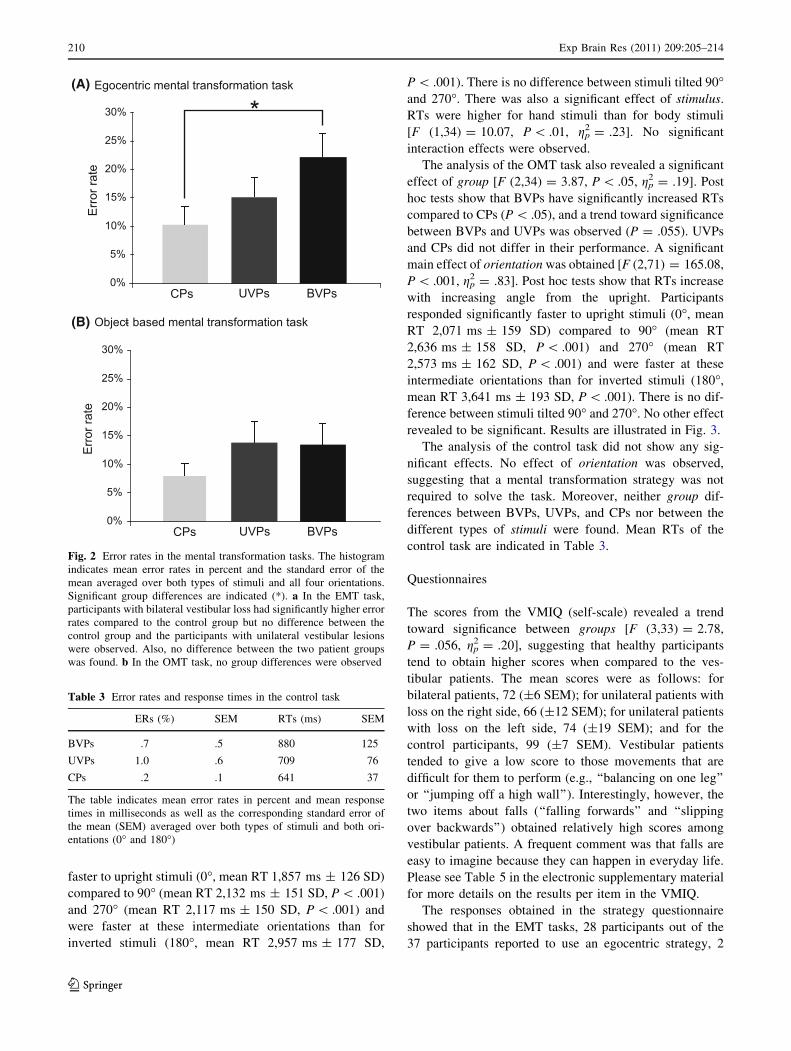
The analysis of the EMT task revealed as hypothesized a
significant effect of group [F (2,34) = 3.29, P \ .05,
gp2 = .16]. Post hoc tests show that BVPs had significantly
higher ERs when compared to the CPs (P \ .05). There
was no significant difference between BVPs and UVPs
(P = .485) and between UVPs and CPs (P = .552). As
expected in a mental transformation task, the effect of
orientation was significant [F (3,55) = 29.31, P \ .001,
gp2 = .46]. Post hoc tests show that error rates (ERs) were
higher when the stimuli were inverted (180�, mean ER
21.5% ± 2.5 SD) compared to when they were upright (0�,
mean ER 10.2% ± 1.7 SD, P \ .001). There was no dif-
ference between stimuli tilted 90� (mean ER 10.6% ± 1.7
SD) and 270� (mean ER 9.2% ± 1.6 SD). These interme-
diate orientations did not differ from upright stimuli but
were significantly different from inverted stimuli
(P \ .001). Moreover, orientation interacted with the fac-
tor stimulus [F (3,53) = 5.03, P \ .05, gp2 = .13]. The
increase in ERs for inverted stimuli was more pronounced
when the stimulus depicted a body compared to a hand.
The analysis of the OMT task revealed no significant
effect of group [F (2,34) = 1.57, P = .223, gp2 = .09].
Therefore, the OMT did not discriminate between patients
and CPs. The factor orientation was significant
[F (3,53) = 31.15, P \ .001, gp2 = .478]. Post hoc tests
show that ERs increase with increasing angle from the
upright. Participants made significantly fewer errors to
upright stimuli (0�, mean ER 4.5% ± 1.2 SD) compared to
90� (mean ER 7.6% ± 1.2 SD, P \ .01) and 270� (mean
ER 8.3 ± 1.3 SD, P \ .001) and performed better at these
intermediate orientations than for inverted stimuli (180�,
mean ER 19.1 ± 2.7 SD, P \ .001). There was no differ-
ence between stimuli tilted 90� and 270�. No other effect
was significant. The results are illustrated in Fig. 2.
The analysis of the control task revealed no significant
effects. The participants’ performance did not depend on
the orientation of the stimuli, and thus it is unlikely that
they applied a mental transformation strategy in the control
task. Also, no differences between groups or stimuli were
found. Mean ERs of the control task are shown in Table 3.
Mental transformation and control tasks: response times
As for ERs, there were no differences between UVPs with
vestibular loss on the right side and UVPs with vestibular
loss on the left side in any of the three tasks. Thus, the data
from all UVPs were pooled for further analysis. As for
ERs, repeated measures ANOVAs with the between-subjects
factor group and the within-subjects factors stimulus and
orientation were computed for each task separately.
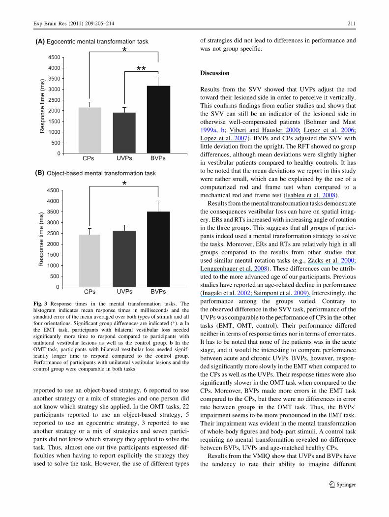
The analysis of the EMT task revealed as hypothesized a
significant effect of group [F (2,34) = 5.43, P \ .01,
gp2 = .24]. Post hoc tests show impaired performance in
BVPs when compared to UVPs (P \ .01) and CPs
(P \ .05). No difference was found between UVPs and
CPs. We also observed a significant effect of orientation
[F (2,74) = 117.60, P \ .001, gp2 = .78]. Post hoc tests
indicate that response times (RTs) increase with increasing
angle from the upright. Participants responded significantly
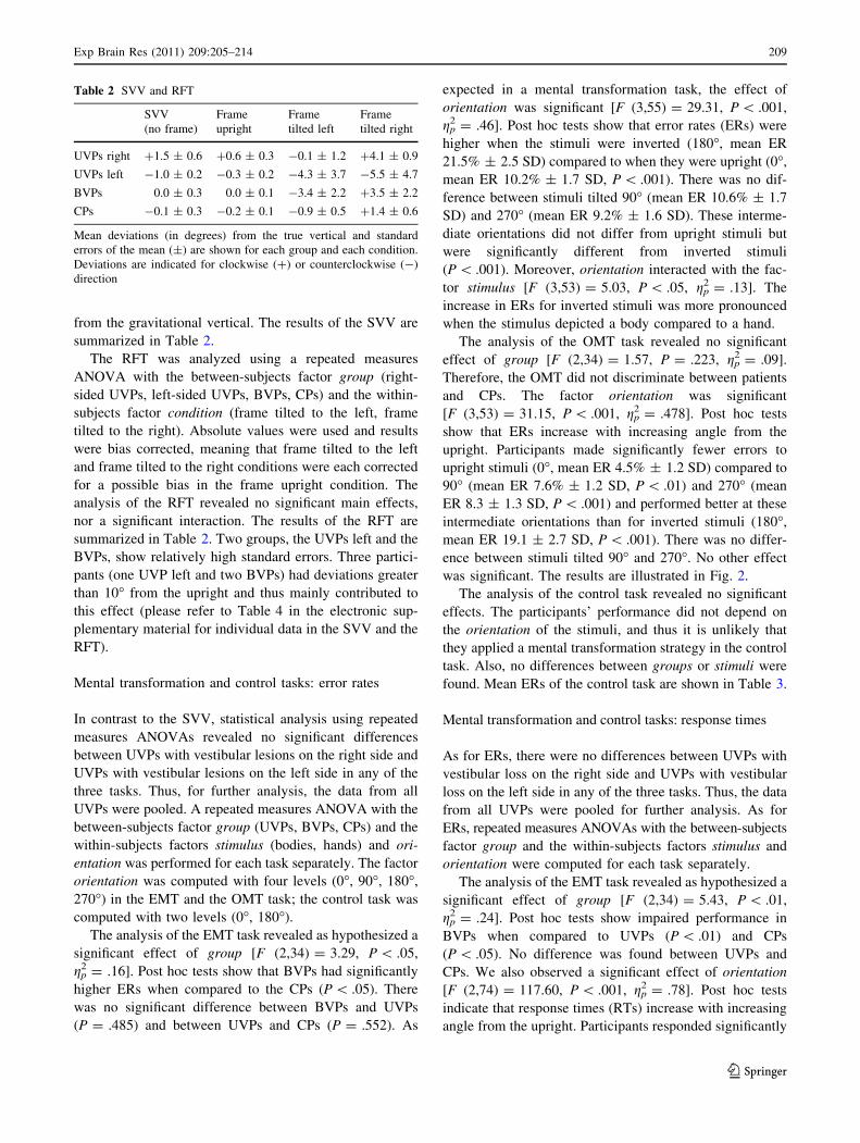
Table 2 SVV and RFT
SVV
(no frame)
Frame
upright
Frame
tilted left
Frame
tilted right
UVPs right ?1.5 ± 0.6 ?0.6 ± 0.3 -0.1 ± 1.2 ?4.1 ± 0.9
UVPs left -1.0 ± 0.2 -0.3 ± 0.2 -4.3 ± 3.7 -5.5 ± 4.7
BVPs 0.0 ± 0.3 0.0 ± 0.1 -3.4 ± 2.2 ?3.5 ± 2.2
CPs -0.1 ± 0.3 -0.2 ± 0.1 -0.9 ± 0.5 ?1.4 ± 0.6
Mean deviations (in degrees) from the true vertical and standard
errors of the mean (±) are shown for each group and each condition.
Deviations are indicated for clockwise (?) or counterclockwise (-)
direction
Exp Brain Res (2011) 209:205–214 209
123
faster to upright stimuli (0�, mean RT 1,857 ms ± 126 SD)
compared to 90� (mean RT 2,132 ms ± 151 SD, P \ .001)
and 270� (mean RT 2,117 ms ± 150 SD, P \ .001) and
were faster at these intermediate orientations than for
inverted stimuli (180�, mean RT 2,957 ms ± 177 SD,
P \ .001). There is no difference between stimuli tilted 90�and 270�. There was also a significant effect of stimulus.
RTs were higher for hand stimuli than for body stimuli
[F (1,34) = 10.07, P \ .01, gp2 = .23]. No significant
interaction effects were observed.
The analysis of the OMT task also revealed a significant
effect of group [F (2,34) = 3.87, P \ .05, gp2 = .19]. Post
hoc tests show that BVPs have significantly increased RTs
compared to CPs (P \ .05), and a trend toward significance
between BVPs and UVPs was observed (P = .055). UVPs
and CPs did not differ in their performance. A significant
main effect of orientation was obtained [F (2,71) = 165.08,
P \ .001, gp2 = .83]. Post hoc tests show that RTs increase
with increasing angle from the upright. Participants
responded significantly faster to upright stimuli (0�, mean
RT 2,071 ms ± 159 SD) compared to 90� (mean RT
2,636 ms ± 158 SD, P \ .001) and 270� (mean RT
2,573 ms ± 162 SD, P \ .001) and were faster at these
intermediate orientations than for inverted stimuli (180�,
mean RT 3,641 ms ± 193 SD, P \ .001). There is no dif-
ference between stimuli tilted 90� and 270�. No other effect
revealed to be significant. Results are illustrated in Fig. 3.
The analysis of the control task did not show any sig-
nificant effects. No effect of orientation was observed,
suggesting that a mental transformation strategy was not
required to solve the task. Moreover, neither group dif-
ferences between BVPs, UVPs, and CPs nor between the
different types of stimuli were found. Mean RTs of the
control task are indicated in Table 3.
Questionnaires
The scores from the VMIQ (self-scale) revealed a trend
toward significance between groups [F (3,33) = 2.78,
P = .056, gp2 = .20], suggesting that healthy participants
tend to obtain higher scores when compared to the ves-
tibular patients. The mean scores were as follows: for
bilateral patients, 72 (±6 SEM); for unilateral patients with
loss on the right side, 66 (±12 SEM); for unilateral patients
with loss on the left side, 74 (±19 SEM); and for the
control participants, 99 (±7 SEM). Vestibular patients
tended to give a low score to those movements that are
difficult for them to perform (e.g., ‘‘balancing on one leg’’
or ‘‘jumping off a high wall’’). Interestingly, however, the
two items about falls (‘‘falling forwards’’ and ‘‘slipping
over backwards’’) obtained relatively high scores among
vestibular patients. A frequent comment was that falls are
easy to imagine because they can happen in everyday life.
Please see Table 5 in the electronic supplementary material
for more details on the results per item in the VMIQ.
The responses obtained in the strategy questionnaire
showed that in the EMT tasks, 28 participants out of the
37 participants reported to use an egocentric strategy, 2
(A)
(B)
Fig. 2 Error rates in the mental transformation tasks. The histogram
indicates mean error rates in percent and the standard error of the
mean averaged over both types of stimuli and all four orientations.
Significant group differences are indicated (*). a In the EMT task,
participants with bilateral vestibular loss had significantly higher error
rates compared to the control group but no difference between the
control group and the participants with unilateral vestibular lesions
were observed. Also, no difference between the two patient groups
was found. b In the OMT task, no group differences were observed
Table 3 Error rates and response times in the control task
ERs (%) SEM RTs (ms) SEM
BVPs .7 .5 880 125
UVPs 1.0 .6 709 76
CPs .2 .1 641 37
The table indicates mean error rates in percent and mean response
times in milliseconds as well as the corresponding standard error of
the mean (SEM) averaged over both types of stimuli and both ori-
entations (0� and 180�)
210 Exp Brain Res (2011) 209:205–214
123
reported to use an object-based strategy, 6 reported to use
another strategy or a mix of strategies and one person did
not know which strategy she applied. In the OMT tasks, 22
participants reported to use an object-based strategy, 5
reported to use an egocentric strategy, 3 reported to use
another strategy or a mix of strategies and seven partici-
pants did not know which strategy they applied to solve the
task. Thus, almost one out five participants expressed dif-
ficulties when having to report explicitly the strategy they
used to solve the task. However, the use of different types
of strategies did not lead to differences in performance and
was not group specific.
Discussion
Results from the SVV showed that UVPs adjust the rod
toward their lesioned side in order to perceive it vertically.
This confirms findings from earlier studies and shows that
the SVV can still be an indicator of the lesioned side in
otherwise well-compensated patients (Bohmer and Mast
1999a, b; Vibert and Hausler 2000; Lopez et al. 2006;
Lopez et al. 2007). BVPs and CPs adjusted the SVV with
little deviation from the upright. The RFT showed no group
differences, although mean deviations were slightly higher
in vestibular patients compared to healthy controls. It has
to be noted that the mean deviations we report in this study
were rather small, which can be explained by the use of a
computerized rod and frame test when compared to a
mechanical rod and frame test (Isableu et al. 2008).
Results from the mental transformation tasks demonstrate
the consequences vestibular loss can have on spatial imag-
ery. ERs and RTs increased with increasing angle of rotation
in the three groups. This suggests that all groups of partici-
pants indeed used a mental transformation strategy to solve
the tasks. Moreover, ERs and RTs are relatively high in all
groups compared to the results from other studies that
used similar mental rotation tasks (e.g., Zacks et al. 2000;
Lenggenhager et al. 2008). These differences can be attrib-
uted to the more advanced age of our participants. Previous
studies have reported an age-related decline in performance
(Inagaki et al. 2002; Saimpont et al. 2009). Interestingly, the
performance among the groups varied. Contrary to
the observed difference in the SVV task, performance of the
UVPs was comparable to the performance of CPs in the other
tasks (EMT, OMT, control). Their performance differed
neither in terms of response times nor in terms of error rates.
It has to be noted that none of the patients was in the acute
stage, and it would be interesting to compare performance
between acute and chronic UVPs. BVPs, however, respon-
ded significantly more slowly in the EMT when compared to
the CPs as well as the UVPs. Their response times were also
significantly slower in the OMT task when compared to the
CPs. Moreover, BVPs made more errors in the EMT task
compared to the CPs, but there were no differences in error
rate between groups in the OMT task. Thus, the BVPs’
impairment seems to be more pronounced in the EMT task.
Their impairment was evident in the mental transformation
of whole-body figures and body-part stimuli. A control task
requiring no mental transformation revealed no difference
between BVPs, UVPs and age-matched healthy CPs.
Results from the VMIQ show that UVPs and BVPs have
the tendency to rate their ability to imagine different
(A)
(B)
Fig. 3 Response times in the mental transformation tasks. The
histogram indicates mean response times in milliseconds and the
standard error of the mean averaged over both types of stimuli and all
four orientations. Significant group differences are indicated (*). a In
the EMT task, participants with bilateral vestibular loss needed
significantly more time to respond compared to participants with
unilateral vestibular lesions as well as the control group. b In the
OMT task, participants with bilateral vestibular loss needed signif-
icantly longer time to respond compared to the control group.
Performance of participants with unilateral vestibular lesions and the
control group were comparable in both tasks
Exp Brain Res (2011) 209:205–214 211
123
movements lower compared to healthy CPs. This seems
especially true for movements that can be difficult for them
to perform. Please note that although UVPs seem to obtain
lower scores in the VMIQ, they do not show impaired
performance in the mental transformation tasks.
The impairment due to bilateral vestibular loss in the
OMT task was unexpected insofar as previous studies have
shown in healthy participants that performance in this task
remains unchanged when tested in the absence of gravity-
related vestibular input in microgravity (Leone et al. 1995)
or a supine body position (Mast et al. 2003). These findings
suggest that the OMT task can be solved by means of
purely visuospatial processes, and thus changes in central
vestibular processing due to vestibular loss do not interfere
with task performance. In contrast, there is empirical evi-
dence for the involvement of vestibular information in the
EMT task (Grabherr et al. 2007; Grabherr and Mast 2010).
Clearly, however, comparing performance of BVPs with
healthy participants in microgravity (no otolithic input
besides the resting discharge level) or under body tilt
(complete vestibular input but the direction of the gravi-
tational force is not aligned with the body axis) has its
limits. How can we explain the finding that RTs were
increased in BVPs in the EMT and the OMT task? We used
identical stimuli in the mental transformation tasks (apart
from presenting pairs of stimuli in the OMT task and one
stimulus only in the EMT task) in order to rule out any
influence of factors unrelated to mental transformation
such as object recognition or stimulus complexity. This is
important to control for since—at least in animal studies—
deficits in object recognition were found after peripheral
vestibular lesions (Zheng et al. 2004). However, the fact
that whole-body and body-part stimuli were used for the
object-based mental transformation strategy could have
given the participants the possibility to project a mental
representation of their own body onto the depicted stimuli.
Amorim et al. (2006) reconfigured the classical Shepard
and Metzler cubes (abstract 3D objects) with human fea-
tures. Making the stimuli more human like improved the
performance. The authors argued that participants were
able to map their own bodily coordinates onto one of the
stimuli, which led to a more holistic instead of a ‘‘piece-
meal like’’ transformation process. Therefore, we cannot
completely rule out the possibility that our participants also
used such a process in order to improve their performance.
However, this would suggest that there is no clear-cut
distinction between the OMT and EMT task we applied
and that the OMT task is no longer a purely visual–spatial
task. Studies that did not reveal influences of vestibular
cues in OMT tasks used objects like the Shepard and
Metzler cubes, letters and plants as stimuli (Leone et al.
1995; Mast et al. 2003; Lenggenhager et al. 2008). Despite
the disadvantage of using different types of stimuli, future
studies may want to include such object stimuli. Last but
not least, there is also the possibility that BVPs showed
increased RTs in both mental transformation tasks due to a
general decrease in attention as other studies have found
decreased attention in vestibular patients (for a discussion
see Smith et al. 2005; Hanes and McCollum 2006). How-
ever, if more general effects were the cause, one would also
expect—at least in part—impairment in unilateral patients.
But this was not the case; UVPs and CPs showed similar
performances. In addition to this, there were no group
differences in the control task. It is therefore unlikely that
the decrease in performance observed in BVPs can be
explained by a general lack of attention.
BVPs showed impaired performance to mentally trans-
form own-body and body-part representations. The role of
vestibular information in constituting a body representation
has been shown previously. For example, Bisiach et al.
(1991) described a patient with somatoparaphrenia, whose
abnormal ownership for the left arm was normalized by
means of caloric vestibular stimulation. This type of ves-
tibular stimulation also led to body schema changes in
amputees (Andre et al. 2001) and paraplegic participants
(Le Chapelain et al. 2001). More research needs to be done
to better explore how cortical vestibular processing is
nested and intertwined with the brain areas associated with
EMTs. In BVPs, the absence of vestibular input is com-
plete and chronic. Interestingly, decreased spatial memory
and navigation abilities were found in bilateral vestibular
patients (Brandt et al. 2005) but not in unilateral vestibular
patients (Hufner et al. 2007). Along with these behavioral
findings, neuroanatomical findings revealed hippocampal
atrophy in bilateral vestibular patients (Brandt et al. 2005)
but not in unilateral vestibular patients (Hufner et al. 2007).
However, the effect complete vestibular loss can have on
other brain areas is less understood, and the involvement of
yet other brain areas is likely for the types of tasks we used.
For example, neuroimaging and clinical studies have
shown that different areas such as the temporo-parietal
junction and the inferior and the superior parietal cortex are
involved in EMT tasks (Zacks et al. 1999; Blanke et al.
2005; Creem-Regehr et al. 2007). Interestingly, these areas
are also found to receive vestibular input (e.g., Lobel et al.
1999; Bense et al. 2001; de Waele et al. 2001; Dieterich
et al. 2003). The results from this study provide further
evidence for impaired spatial cognitive abilities in bilateral
but not unilateral vestibular patients.
Conclusion
Tasks involving mental transformations of bodies and
body parts were more challenging for patients with bilat-
eral vestibular loss when compared to healthy controls.
212 Exp Brain Res (2011) 209:205–214
123
We conclude that central vestibular processes are involved
in imagined spatial body and body-part transformations.
Participants with unilateral vestibular loss, however,
showed no impairment in mental transformation perfor-
mance, suggesting that they can fully compensate for a
potential decline in cognitive performance. This study adds
to the growing body of evidence that persons with vestib-
ular disorders can experience cognitive deficits. In the
future, a more profound understanding of the cognitive
effects as a consequence of vestibular disorders may help
to design more specific rehabilitation procedures.
Acknowledgments We thank the participants for volunteering to
participate, Nikola Sanz and Aurelie Manuel for assistance with data
collection, Michael Vogeli for his support in programming and
Claudia Blum for designing the stimuli. We would also like to thank
two anonymous reviewers for helpful comments on an earlier version
of the manuscript. This study was funded by a grant from the Swiss
National Science Foundation (Sinergia project ‘‘Balancing Self and
Body’’).
References
Amorim MA, Isableu B, Jarraya M (2006) Embodied spatial
transformations: ‘‘body analogy’’ for the mental rotation of
objects. J Exp Psychol Gen 135:327–347
Andre JM, Martinet N, Paysant J, Beis JM, Le Chapelain L (2001)
Temporary phantom limbs evoked by vestibular caloric stimu-
lation in amputees. Neuropsychiatry Neuropsychol Behav Neu-
rol 14:190–196
Bense S, Stephan T, Yousry TA, Brandt T, Dieterich M (2001)
Multisensory cortical signal increases and decreases during vestib-
ular galvanic stimulation (fMRI). J Neurophysiol 85:886–899
Bisiach E, Rusconi ML, Vallar G (1991) Remission of somatopara-
phrenic delusion through vestibular stimulation. Neuropsycho-
logia 29:1029–1031
Blanke O, Mohr C, Michel CM, Pascual-Leone A, Brugger P, Seeck
M, Landis T, Thut G (2005) Linking out-of-body experience and
self processing to mental own-body imagery at the temporopa-
rietal junction. J Neurosci 25:550–557
Bohmer A, Mast F (1999a) Assessing otolith function by the
subjective visual vertical. Ann N Y Acad Sci 871:221–231
Bohmer A, Mast F (1999b) Chronic unilateral loss of otolith function
revealed by the subjective visual vertical during off center yaw
rotation. J Vestib Res 9:413–422
Borel L, Lopez C, Peruch P, Lacour M (2008) Vestibular syndrome: a
change in internal spatial representation. Neurophysiol Clin
38:375–389
Brandt T, Dieterich M (1999) The vestibular cortex. Its locations,
functions, and disorders. Ann N Y Acad Sci 871:293–312
Brandt T, Schautzer F, Hamilton DA, Bruning R, Markowitsch HJ,
Kalla R, Darlington C, Smith P, Strupp M (2005) Vestibular loss
causes hippocampal atrophy and impaired spatial memory in
humans. Brain 128:2732–2741
Chen-Huang C, McCrea RA (1999) Effects of viewing distance on the
responses of vestibular neurons to combined angular and linear
vestibular stimulation. J Neurophysiol 81:2538–2557
Clement G, Fraysse MJ, Deguine O (2009) Mental representation of
space in vestibular patients with otolithic or rotatory vertigo.
Neuroreport 20:457–461
Creem-Regehr SH, Neil JA, Yeh HJ (2007) Neural correlates of two
imagined egocentric transformations. Neuroimage 35:916–927
de Waele C, Baudonniere PM, Lepecq JC, Tran Ba Huy P, Vidal PP
(2001) Vestibular projections in the human cortex. Exp Brain
Res 141:541–551
Dieterich M, Brandt T (2008) Functional brain imaging of peripheral
and central vestibular disorders. Brain 131:2538–2552
Dieterich M, Bense S, Lutz S, Drzezga A, Stephan T, Bartenstein P,
Brandt T (2003) Dominance for vestibular cortical function in
the non-dominant hemisphere. Cereb Cortex 13:994–1007
Emri M, Kisely M, Lengyel Z, Balkay L, Marian T, Miko L, Berenyi
E, Sziklai I, Tron L, Toth A (2003) Cortical projection of
peripheral vestibular signaling. J Neurophysiol 89:2639–2646
Grabherr L, Mast FW (2010) Effects of microgravity on cognition:
The case of mental imagery. J Vestib Res 20:53–60
Grabherr L, Karmali F, Bach S, Indermaur K, Metzler S, Mast FW
(2007) Mental own-body and body-part transformations in
microgravity. J Vestib Res 17:279–287
Guerraz M, Yardley L, Bertholon P, Pollak L, Rudge P, Gresty MA,
Bronstein AM (2001) Visual vertigo: symptom assessment,
spatial orientation and postural control. Brain 124:1646–1656
Halmagyi GM, Curthoys IS, Cremer PD, Henderson CJ, Todd MJ,
Staples MJ, D’Cruz DM (1990) The human horizontal vestibulo-
ocular reflex in response to high-acceleration stimulation before and
after unilateral vestibular neurectomy. Exp Brain Res 81:479–490
Hanes DA, McCollum G (2006) Cognitive-vestibular interactions: a
review of patient difficulties and possible mechanisms. J Vestib
Res 16:75–91
Hufner K, Hamilton DA, Kalla R, Stephan T, Glasauer S, Ma J,
Bruning R, Markowitsch HJ, Labudda K, Schichor C, Strupp M,
Brandt T (2007) Spatial memory and hippocampal volume in
humans with unilateral vestibular deafferentation. Hippocampus
17:471–485
Inagaki H, Meguro K, Shimada M, Ishizaki J, Okuzumi H, Yamadori
A (2002) Discrepancy between mental rotation and perspective-
taking abilities in normal aging assessed by Piaget’s three-
mountain task. J Clin Exp Neuropsychol 24:18–25
Isaac A, Marks DF, Russell DG (1986) An instrument for assessing
imagery of movement: the vividness of movement imagery
questionnaire (VMIQ). J Mental Imag 10:23–30
Isableu B, Gueguen M, Fourre B, Giraudet G, Amorim MA (2008)
Assessment of visual field dependence: comparison between the
mechanical 3D rod-and-frame test developed by Oltman in 1968
with a 2D computer-based version. J Vestib Res 18:239–247
Le Chapelain L, Beis JM, Paysant J, Andre JM (2001) Vestibular
caloric stimulation evokes phantom limb illusions in patients
with paraplegia. Spinal Cord 39:85–87
Lenggenhager B, Lopez C, Blanke O (2008) Influence of galvanic
vestibular stimulation on egocentric and object-based mental
transformations. Exp Brain Res 184:211–221
Leone G, Lipshits M, Gurfinkel V, Berthoz A (1995) Is there an effect
of weightlessness on mental rotation of three-dimensional
objects? Brain Res Cogn Brain Res 2:255–267
Lobel E, Kleine JF, Leroy-Willig A, Van de Moortele PF, Le Bihan
D, Grusser OJ, Berthoz A (1999) Cortical areas activated by
bilateral galvanic vestibular stimulation. Ann N Y Acad Sci
871:313–323
Lopez C, Lacour M, Magnan J, Borel L (2006) Visual field
dependence-independence before and after unilateral vestibular
loss. Neuroreport 17:797–803
Exp Brain Res (2011) 209:205–214 213
123

Lopez C, Lacour M, Ahmadi AE, Magnan J, Borel L (2007) Changes
of visual vertical perception: a long-term sign of unilateral and
bilateral vestibular loss. Neuropsychologia 45:2025–2037
Mamoto Y, Yamamoto K, Imai T, Tamura M, Kubo T (2002) Three-
dimensional analysis of human locomotion in normal subjects and
patients with vestibular deficiency. Acta Otolaryngol 122:495–500
Mast FW, Ganis G, Christie S, Kosslyn SM (2003) Four types of
visual mental imagery processing in upright and tilted observers.
Brain Res Cogn Brain Res 17:238–247
McCrea RA, Luan H (2003) Signal processing of semicircular canal
and otolith signals in the vestibular nuclei during passive and
active head movements. Ann N Y Acad Sci 1004:169–182
Parsons LM (1987a) Imagined spatial transformation of one’s body.
J Exp Psychol Gen 116:172–191
Parsons LM (1987b) Imagined spatial transformations of one’s hands
and feet. Cogn Psychol 19:178–241
Parsons LM(2003) Superior parietal cortices and varieties of mental
rotation. Trends Cogn Sci 7:515–517
Peruch P, Borel L, Magnan J, Lacour M (2005) Direction and distance
deficits in path integration after unilateral vestibular loss depend
on task complexity. Brain Res Cogn Brain Res 25:862–872
Peterka RJ (2002) Sensorimotor integration in human postural
control. J Neurophysiol 88:1097–1118
Ratcliff G (1979) Spatial thought, mental rotation and the right
cerebral hemisphere. Neuropsychologia 17:49–54
Rodionov V, Zislin J, Elidan J (2004) Imagination of body rotation
can induce eye movements. Acta Otolaryngol 124:684–689
Saimpont A, Pozzo T, Papaxanthis C (2009) Aging affects the mental
rotation of left and right hands. PLoS One 4:e6714
Schautzer F, Hamilton D, Kalla R, Strupp M, Brandt T (2003) Spatial
memory deficits in patients with chronic bilateral vestibular
failure. Ann N Y Acad Sci 1004:316–324
Shepard RN, Metzler J (1971) Mental rotation of three-dimensional
objects. Science 171:701–703
Smith PF, Zheng Y, Horii A, Darlington CL (2005) Does vestibular
damage cause cognitive dysfunction in humans? J Vestib Res
15:1–9
Tomasino B, Rumiati RI (2004) Effects of strategies on mental
rotation and hemispheric lateralization: neuropsychological
evidence. J Cogn Neurosci 16:878–888
Vibert D, Hausler R (2000) Long-term evolution of subjective visual
vertical after vestibular neurectomy and labyrinthectomy. Acta
Otolaryngol 120:620–622
Vitte E, Derosier C, Caritu Y, Berthoz A, Hasboun D, Soulie D
(1996) Activation of the hippocampal formation by vestibular
stimulation: a functional magnetic resonance imaging study. Exp
Brain Res 112:523–526
Zacks J, Rypma B, Gabrieli JD, Tversky B, Glover GH (1999)
Imagined transformations of bodies: an fMRI investigation.
Neuropsychologia 37:1029–1040
Zacks JM, Mires J, Tversky B, Hazeltine E (2000) Mental spatial
transformations of objects and perspective. Spatial Cognit
Comput 2:315–332
Zacks JM, Ollinger JM, Sheridan MA, Tversky B (2002) A
parametric study of mental spatial transformations of bodies.
Neuroimage 16:857–872
Zacks JM, Gilliam F, Ojemann JG (2003) Selective disturbance of
mental rotation by cortical stimulation. Neuropsychologia
41:1659–1667
Zheng Y, Darlington CL, Smith PF (2004) Bilateral labyrinthectomy
causes long-term deficit in object recognition in rat. Neuroreport
15:1913–1916
214 Exp Brain Res (2011) 209:205–214
123