Characterization of Elasticity-Tensor Symmetries Using SU(2)
Upload
independentCategory
view
0download
0
Biol Cybern (2007) 96:439–453DOI 10.1007/s00422-006-0136-y
ORIGINAL PAPER
Spatial symmetries in vestibular projections to the uvula-nodulus
Isaac Z. Foster · Douglas A. Hanes ·Neal H. Barmack · Gin McCollum
Received: 23 January 2006 / Accepted: 20 November 2006 / Published online: 5 January 2007© Springer-Verlag 2007
Abstract The discharge of secondary vestibular neu-rons relays the activity of the vestibular endorgans, occa-sioned by movements in three-dimensional physicalspace. At a slightly higher level of analysis, the dischargeof each secondary vestibular neuron participates in amultifiber projection or pathway from primary affer-ents via the secondary neurons to another neuronalpopulation. The logical organization of this projectiondetermines whether characteristics of physical space areretained or lost.
The logical structure of physical space is standardlyexpressed in terms of the mathematics of group the-ory. The logical organization of a projection can becompared to that of physical space by evaluating itssymmetry group. The direct projection from the semi-circular canal nerves via the vestibular nuclei to neckmotor neurons has a full three-dimensional symmetrygroup, allowing it to maintain a three-dimensional coor-dinate frame. However, a projection may embed onlya subgroup of the symmetry group of physical space,which incompletely mirrors the properties of physicalspace. The major visual and vestibular projections inthe rabbit via the inferior olive to the uvula-noduluscarry three degrees of freedom—rotations about onevertical and two horizontal axes—but do not have full
I. Z. FosterWheaton College, Norton, MA, USA
D. A. Hanes (B) · G. McCollumNeuro-otology Department, Legacy Research Center,1225 NE 2nd Avenue, Portland, OR 97232, USAe-mail: [email protected]
N. H. BarmackNeurol. Sci. Inst., Oregon Health and Sciences University,Beaverton, OR, USA
three dimensional symmetry. Instead, the vestibulo-oli-vo-nodular projection has symmetries corresponding toa product of two-dimensional vestibular and one-dimen-sional optokinetic spaces. This combination of projectionsymmetries provides the foundation for distinguishinghorizontal from vertical rotations within a three dimen-sional space.
In this study, we evaluate the symmetry group givenby the physiological organization of the vestibulo-olivo-nodular projection. Although it acts on the same sets ofelements and mirrors the rotations that occur in physi-cal space, the physiological transformation group is dis-tinct from the spatial group. We identify symmetries asproducts of physiological and spatial transformations.The symmetry group shapes the information the pro-jection conveys to the uvula-nodulus; this shaping maydepend on a physiological choice of generators, in thesame way that function depends on the physiologicalchoice of coordinates. We discuss the implications of thesymmetry group for uvula-nodulus function, evolution,and functions of the vestibular system in general.
1 Introduction
Properties of three-dimensional space are constructedby nervous systems. For example, the perceptual three-dimensionality of the Necker cube (Fig. 1) is fabricatedby nervous systems. There is no information inthe two-dimensional Necker cube drawing about three-dimensionality, nor is there information about whichvertical face is in front. Therefore, a perceptual choiceof the lower or the upper vertical face being in front is acue-free perception.
Color perception is not cue-free but is reorganizedfrom ranges of light frequency to construct a three-

440 Biol Cybern (2007) 96:439–453
Fig. 1 The Necker cube. This two-dimensional figure is seen as athree-dimensional cube. A front-back reflection along the (imagi-nary) depth axis allows for two symmetric interpretations of depth
dimensional color space. Neural organization constructscolor space.
1.1 Neural organization projects vestibular space
Eye movements, head movements, postural adjustments,and locomotion depend on the construction of a threedimensional space by secondary vestibular nuclear neu-rons (Goldberg and Fernández 1984). Rotational andgravitoinertial information is separately transduced insingle dimensions by the vestibular endorgans. The ves-tibular primary afferent convergence onto secondaryvestibular neurons establishes a range of possibilitiesfor the construction of neural spaces. At one extreme,no convergence, only one-dimensional spaces can beconstructed. At the other extreme, complete conver-gence, spatial sensitivity is abolished, without a meansto distinguish degrees of freedom. Two or three dimen-sional space can only be constructed with intermediatepatterns of convergence to form the logical structure ofthose spaces.
A standard way to formalize this logical structure usesthe mathematics of symmetry groups. (See Sect. 3.1 fora formal presentation of symmetry group mathematics.)Evaluation of the logical structure of the direct projec-tion from the semicircular canals onto vestibular sec-ondary neurons and then onto neck motor neurons hasshown that this projection has an embedded symmetrygroup of a rotating cube (McCollum and Boyle 2004).The symmetry group of the cube includes not only thereflections and rotations of physical space, but also theright and the left of the vertebrate body.
In constructing spaces, vertebrate nervous systemsnecessarily begin from the intrinsic organization of thevertebrate body. However, maintenance of the reflectionand rotation symmetry of semicircular canal planes is
important because self-motion can reorient these canalplanes with respect to external objects and to rest of thebody. To see the importance of structural symmetriesin this respect, consider a hypothetical reflex mediatedby a projection from the semicircular canals to lowersegments of the body. To be effective, the projectionmust accommodate altered positions of the head withrespect to the trunk. However, when the head rotates,so do necessary weightings of the projection; for exam-ple, if a certain muscle synergy is normally appropri-ate in reaction to stimulation of the horizontal canal,then with an altered head position, this same synergymay be appropriate for stimulation of the left anteriorcanal, which is now oriented in the horizontal plane. Itfollows that weightings of the projections need to beswitched in order to maintain a functional reflex; thiscan only happen, though, if the structure of the projec-tion has symmetry consistent with the head rotation thatinterchanges the canal planes. Thus, in addition to therequirement that there be an appropriate muscle acti-vation pattern corresponding to each semicircular canalstimulation pattern (i.e., each rotation of the head), thepreservation of spatial symmetries allows the system toautomatically adapt to situations in which the head isnot in its usual orientation (McCollum and Boyle 2004).
An example of such symmetric reweighting appearsin the work of Ivanenko et al. (1999), who showed thatpostural reflex responses to vibration of the dorsal neckmuscles or galvanic stimulation of the vestibular canalsare modulated by horizontal rotations of the head and/oreyes. Their results show that reflex pathways can be re-weighted so that the postural response to the same stim-ulus utilizes different movements of the torso and lowerlimbs to achieve compensation in the pointing directionof the head or gaze. An oculomotor example of symmet-ric reweighting appears in the modulation of optokineticnystagmus by pitch rotation of the head (Barmack et al.2002). In these experiments it was shown that horizontaloptokinetic stimulation generates horizontal nystagmusin the rabbit, regardless of the pitch orientation of theanimal’s head. When the head is in the initial orienta-tion, the nystagmus is produced by the horizontal andmedial rectus muscles. When the head is pitched, nys-tagmus in the same plane, and generated by the samestimulus, is produced by the vertical rectus and obliquemuscles (Barmack et al. 2002). This efficient reweigh-ting indicates an important symmetry in the optokineticsystem.
This finding expands the potential role of the ves-tibular nuclei in providing spatial information to var-ious sensorimotor systems, perception, and cognition(Hanes and McCollum 2006). The spatial symmetries ofvestibular secondary neuronal projections may provide

Biol Cybern (2007) 96:439–453 441
spatial subgroups for all of these functions, whetheror not the spatial subgroups provide complete three-dimensional spatial symmetry.
To a large extent, nervous systems are successful atinternalizing physical space, as witnessed by the accu-racy of gaze and other movements (Guitton and Volle1987; Goossens and van Opstal 1999; Stahl 2001). Motionsickness, spatial illusions, and cue-free perceptions mayarise from the construction of multiple, incompletespaces in separate projections, rather than a complete,unified three-dimensional space. Extending the evalu-ation of vestibular projection symmetry groups to pro-jections besides the direct semicircular canal-vestibularnuclei-neck motor neurons projection will clarify therole of the vestibular system in constructing spaces.
1.2 Evaluation of the organization of projections viathe inferior olive to the uvula-nodulus
In this study we evaluate the projection symmetries ofthe visual and vestibular climbing fiber projections tothe uvula-nodulus. This climbing fiber projection car-ries three rotational degrees of freedom: two from thevestibular system, in the planes of the vertical semicir-cular canals, and one from the optokinetic system, in thehorizontal plane (Barmack 2003). A priori, these pro-jections could be organized to provide full three-dimen-sional symmetry like that of the direct projection fromthe vestibular primary afferent to the neck motor neuronvia the vestibular nuclei. (McCollum and Boyle 2004).First, we ask whether the olivary projection providesthree dimensional symmetry. Next, we investigate whatsymmetries are present and discuss their implications.
In Sect. 2 we review data presented by Barmack(2003), describe the anatomical and physiological struc-ture of the arcs and outline several assumptions that willfacilitate our analysis. Section 3 sets up the mathemati-cal structure. Section 3.1 gives a brief introduction to theaspects of group theory to be used and Sect. 3.2 presentsthe data in a form that can be analyzed mathematically.Section 4 presents our evaluation of the transformationsand symmetry group of the vestibulo-olivo-nodularprojection.
2 Anatomy and physiology of nodular projections
In a region extending 2 mm laterally from the mid-line of the rabbit uvula-nodulus (folia 9 and 10)Purkinje cells receive synaptic projections from climbingfibers that originate from the contralateral inferior olive.These climbing fibers convey signals from the contra-lateral otoliths, vertical semicircular canals, and ipsilat-
eral horizontal direction-selective retinal ganglion cells(Fushiki and Barmack 1997; Barmack 2003; Barmackand Yakhnitsa 2000; Voogd and Barmack 2006). Climb-ing fiber signals are organized into discrete zonescharacterized by the axis of vestibular or optokineticstimulation that optimally modulates CFRs. In themedial zone, the discharge of CFRs is modulated byvestibular roll-tilt in the anatomical plane of the ipsi-lateral posterior semicircular canal. In the lateral zone,CFRs are modulated by roll-tilt in the anatomical planeof the ipsilateral anterior semicircular canal. Interposedbetween these two vestibular zones is an optokineticzone in which the CFRs of Purkinje cells are optimallyexcited by horizontal optokinetic stimulation of the ipsi-lateral eye in the temporal to nasal direction. Thus, theuvula-nodulus receives climbing fibers responding tothree degrees of freedom of rotation. Two zones receivevestibular climbing fiber inputs that align with the ipsi-lateral vertical semicircular canals. One zone receiveshorizontal optokinetic climbing fiber afferents that orig-inate from the ipsilateral eye.
In addition to the climbing fiber projection evalu-ated in this study, the uvula-nodulus receives a vestib-ular primary afferent mossy fiber projection includinginformation from all vestibular endorgans. The utricularotoliths contribute a projection that is complementaryto the projection under investigation here, so that thePurkinje cells in the medial and lateral zones have opti-mal response planes in either the anatomical plane ofone of the two vertical canals or an otolithic response inthe same quadrant (Barmack 2003).
The purpose of this study is to evaluate the symmetrygroup of the projection or set of connectivity arcs bywhich signals representing three degrees of freedom ofrotation are transmitted via climbing fibers to the uvula-nodulus. For such an evaluation, it is helpful to haveclear and concise notation of the relevant data.
2.1 Notation
The recorded responses are evoked by sinusoidal oscil-lations (Fushiki and Barmack 1997). An essential pieceof information to display is the direction of modula-tion. Increased activity is indicated by a dashed line, anddecreased activity by a solid line (Fig. 2a). The angleof the line represents the optimal plane: a line slicingdown from left to right on the page represents optimalalignment with the left anterior–right posterior (LARP)plane. A line slicing down from right to left representsoptimal alignment with the right anterior–left poster-ior (RALP) plane. A horizontal line indicates optimalalignment with the horizontal plane. Thus, the profile in

442 Biol Cybern (2007) 96:439–453
LA
L.P.L
L.y.L
R.Rβ.RL.Cβ.R
RP
R.P.L
R.y.L
LP
L.y.R
L.P.R
L.Rβ.L
R.y.R
R.P.R
RA
R.NOT.HL.NOT.H
L.EYE R.EYE
olives
excitatory, positive, and complex spikes
angle of lines indicates canal plane
inhibitory, negative, and simple spikes
response to left tilt response to right tilt
labelhere: left posterior semicircular canal,in the form SIDE.ORGAN.OPTIMAL-PLANE,where the side is left (L), the organ is the left posterior semicircular canal (LP),and the canal plane of optimal response isRALP (R).
A
L.Z2.L L.Z1.R
L.OKZ.H
R.Z1.L R.Z2.R
R.OKZ.Hnodulusleft endorgans right endorgans
L.DC.H R.DC.H
B
L.LP.R
R.Cβ.L
Fig. 2 Diagram of the vestibular and optokinetic arcs underinvestigation. a Explanation of the profile used to diagrameach neural center or endorgan. b Anatomy and physiologyof arcs, as discussed in the text. Labels are on the formSIDE.ORGAN.OPTIMAL-PLANE, where the side may be left
(L) or right (R). The organs (neural centers or endorgans) areleft posterior (LP), left anterior (LA), eye, y group (y), Psol (P),nucleus of the optic tract (NOT), beta nucleus (β), dorsal cap(DC), and nodulus zones 1 (Z1), 2 (Z2), and optokinetic (OKZ).The optimal planes are RALP (R) and LARP (L)
Fig. 2a characterizes the left posterior semicircular canaland is labeled "LP".
Pathways are depicted using a labeled profile for eachneural center, in order to present anatomical and physio-logical information concisely. Most labels are of the formSIDE.NEURALCENTER.OPTIMAL-PLANE. Forinstance, the zones of the left uvula-nodulus are Left.Nodulus Zone 1.RALP or L.Z1.R, Left.Nodulus Zone2.LARP or L.Z2.L, and Left.Nodulus Optokinetic Zone.Horizontal or L.OKZ.H. Within the uvula-nodulus, thedashed and solid lines take on a different meaning, withthe dashed lines representing complex spikes and solidlines indicating simple spikes.
2.2 Optokinetic and vestibular arcsto the uvula-nodulus
The anatomy and physiology of the arcs from the endor-gans through the inferior olive, the source of climbing
fiber signals to the contralateral cerebellum, has beeninvestigated in a range of species (rabbits, mice, rats,monkeys) and using various methods relevant to thequestion at issue (Maklad and Fritzsch 2003; Barmack2003, 2006; Voogd and Barmack 2006). The resultingarcs are diagrammed as viewed from the back, usingmodulation profiles (Fig. 2a) and dashed and solid linesshowing connectivity (Fig. 2b).
The vestibular arcs originate in the semicircular canals(right anterior, RA; right posterior RP; left anteriorLA; left posterior LP). Neural centers along each arcare optimally responsive to either the RALP or theLARP plane, as dictated by the semicircular canal. Eachcanal has an excitatory projection to two sub-nuclei ofthe ipsilateral vestibular nuclei, the Parasolitary nucleus(Psol) and Y group (Barmack 2003). All Psol neuronsare GABAergic. Consequently, they relay an inhibi-tory signal to two subdivisions of the ipsilateral infe-rior olive (IO), the beta nucleus, and the dorsal medial

Biol Cybern (2007) 96:439–453 443
cell column (DMCC). Only the beta nucleus is shownin Fig. 2b because the connections are the same as tothe DMCC. Because of the inhibitory connection, theresponses originating from the Psol are reversed (solidand dashed lines). Each Y-group is connected with thesame subdivisions of the IO, but contralaterally andwith an excitatory connection. Excitatory connectionsfrom the two subdivisions of the IO cross the midlineto synapse as climbing fibers in the contralateral uvula-nodulus.
All of the connections along the optokinetic arcs areexcitatory (Fig. 2b). Signals from the retina project con-tralaterally to the nucleus of the optic tract (NOT), asmall sub-nucleus of the accessory optic system (AOS),sensitive to temporal-to-nasal horizontal optokineticstimulation of the contralateral eye (Alley et al. 1975;Simpson 1984; Barmack 2003, 2006). Each NOT thenprojects to a third subdivision of the IO, the dorsal capof Kooy (DC). The arc finishes with a projection fromthe DC to the contralateral uvula-nodulus and flocculus.
3 Symmetry group evaluation
We will evaluate the arcs presented in the previous sec-tion to find the spatial properties embedded in them.This section presents the mathematics of symmetrygroups (Sect. 3.1) and refines the diagram of the arcsfor evaluation (Sect. 3.2).
3.1 Mathematics of symmetry groups
A symmetry of a geometric figure in the real space �n
is a rigid map of �n that preserves the figure (though itlikely interchanges the points that make up the figure).For example, consider a square lying in the real plane�2. All symmetries of the square are either rotations ofthe plane about the center of the square by an angle thatis a multiple of 90◦, or else reflections of the plane aboutone of the four lines which either bisect two oppositesides of the square or pass through two opposite cornersof the square. All of these maps interchange sides andvertices of the square, but result in a square of the samesize and occupying the same location in the plane: inother words, the same (or at least an indistinguishable)square.
Rigid transformations of the real plane form a group,since they can be composed to form new rigid transfor-mations and have inverses that are also rigid transfor-mations. For example, the map rotate the plane 90◦ aboutthe origin has the inverse rotate the plane 270◦ about theorigin. Formally, a group is a non-empty set G with abinary operation * satisfying:
1. For all elements a, b, c in G, (a*b)*c = a*(b*c).2. For all elements a, b in G, a*b is in G.3. There exists an identity element e in G, such that for
every element a in G, a*e = a = e*a.4. For every element a in G, there is an element b in G
such that a*b = e = b*a.
In other words, (1) the associative law holds, (2) the setof elements is closed under the operation *, (3) there isan identity element (in fact, only one), and (4) for everyelement in the set there is an inverse (again, only one).The number of elements in a group is the order of thegroup. The composition of an element a with itself, a∗a,is written a2. The order of an element a is the smallestnumber n such that an = e. Two groups G and H aresaid to be isomorphic when there is a bijective map-ping ϕ : G → H such that ϕ(g1
∗g2) = ϕ(g1)∗ϕ(g2) for
all g1 and g2 in G. Two groups that are isomorphic areconsidered mathematically identical.
The symmetries of a geometric figure in �n also forma group, since (1) the identity map is a symmetry of anyobject; (2) the composition of two symmetries still pre-serves the figure, since each does in sequence, so thecomposition is likewise a symmetry; and (3) the inverseof a symmetry map takes the outcome of the originalsymmetry (which is the figure) back to the figure, and sois a symmetry! As a result, each geometric figure in �n
has an associated symmetry group that is a subgroup ofthe group of rigid transformations of �n. The symmetrygroup encodes a great deal of the spatial structure of theobject.
An external direct product is a combination of twogroups into one. The external direct product GxH oftwo groups G and H is the set of all ordered pairs(g, h), where g is in G and h is in H, and where theproduct (g1, h1)
∗(g2, h2) is defined component-wise as(g1
∗g2, h1∗h2). An external direct product of two groups
is itself a group. The order of the group GxH is theproduct of the order of G and the order of H.
3.2 Diagram with straight arcs
For the purpose of testing transformations of the arcs,Fig. 2 is dauntingly complicated. Although it means los-ing the anatomical arrangement of the organs in thehead, the connectivity of the arcs can be better exhibitedif we arrange them linearly (Fig. 3), with the optokineticarcs separated from the vestibular arcs. The presentationof arcs in Fig. 3 preserves all of the information thatis required for evaluation of the spatial symmetrygroup.

444 Biol Cybern (2007) 96:439–453
L.Z2.L
LA
L.Z1.R R.Z2.R
L.P.LL.y.L
R.Rβ.R
R.Z1.L
L.Cβ.R
RP
R.P.L
R.y.L
LP
R.Cβ.L
L.y.R
L.P.R
L.Rβ.L
R.y.RR.P.R
RA
L.OKZ.H
R.NOT.H L.NOT.H
L.DC.H
L.EYE
R.DC.H
R.EYE
R.OKZ.H
canals andeyes
y-groups
parasolitariiand NOT’s
olives
noduli
Fig. 3 The vestibular and optokinetic arcs, disentangled so thatall organs of an arc are together and connections are not crossed.The two optokinetic arcs are on the outside. The four vestibular
arcs are split into two joined pairs: each arc of each pair has con-nections to the other arc in the pair, but the two pairs share noconnections
4 Results: transformations and symmetry groups
The vestibular arcs cross the midline only once, andeach arc contains one inhibitory connection (Fig. 3). Theoptokinetic arcs cross the midline twice and have onlyexcitatory connections. In terms of midline crossings andinhibitory/excitatory connections, the vestibular arcs aresimilar to each other and not to the optokinetic arcs.Using only transformations that preserve the arc as aunit, the vestibular and optokinetic arcs do not trans-form into each other. Therefore, if the arcs were repre-sented on the faces of a cube (viz. McCollum and Boyle2004), faces representing the optokinetic arcs wouldneed to be distinguished (e.g., by shading) to show thatthey are not symmetric with vestibular arcs (Fig. 4).
The vestibular arcs, which can be transformed intoeach other as a unit, can be drawn on a square, if thedirection along the arc is collapsed onto the side of thesquare (Fig. 5). The only disadvantage of this depiction isthat, in displaying the arcs along the sides of the square,we are forced to choose an artificial orientation for eacharc, which may not be preserved by rotations and reflec-tions of the figure. This apparent asymmetry disappears,however, if we think of the square representation asshorthand for an open-ended cube with all the profilesdisplayed vertically. We will denote the vestibulo-olivo-nodular projection in this form (Fig. 5b) as V .
In presenting the results, we first display transforma-tions of V and separate them into physiological andspatial transformations (Sect. 4.1). Next, we analyzethe group of physiological transformations more deeply
Fig. 4 A cube exploded so as to separate its faces. On the top andbottom faces are the optokinetic arcs and on the side faces are thevestibular arcs. The arc originating in the left eye is on top; the arcoriginating in the right eye is on the bottom; the arc originating inthe left anterior canal is on the far left face; the arc originating inthe left posterior canal is on the near left face; the arc originatingin the right anterior canal is on the far right face; the arc originat-ing in the right posterior canal is on the near right face. The cubeintegrates V with the optokinetic arcs along separate dimensions

Biol Cybern (2007) 96:439–453 445
L.Z2
LAL.Z1
R.Z2
L.P.L
L.y.L
R.Rβ
R.Z1
L.Cβ
RP
R.P.L
R.y.L
LP
R.Cβ
L.y
.R
L.P.R
L.Rβ
R.y.R
R.P.R
RA
A B
Fig. 5 The cross-connected squares. The four vestibular arcsarranged around the sides of a square. The profiles of the Y-group are in the middle of the square and connect to the oppositeedge. The connections of each of the canals to the contralateralPsol are left out. a The arcs in labeled profile form. Here, the arcoriginating in the left anterior canal is placed on the top left sideof the square, and similarly for the other arcs. b Here, the arcsare in unlabeled profile form. The side of the square an arc is onrepresents the position in the head the arc originates: the arc on
the top left side of the square represents the arc originating fromthe left anterior position of the head; the arc on the top right sideof the square represents the arc originating from the right anteriorposition of the head; the arc on the bottom right side of the squarerepresents the arc originating from the right posterior position ofthe head; the arc on the bottom left side of the square representsthe arc originating from the left posterior position of the head.This is the vestibular projection V
(Sect. 4.2). This allows us to derive the symmetry groupof the vestibulo-olivo-nodular projection V (Sect. 4.3).In the final subsection, we examine the way in whichother transformations of V fail to yield symmetries(Sect. 4.4).
4.1 Transformations of the vestibulo-olivo-nodularprojection
Several spatially-related transformations of the vestibu-lo-olivo-nodular projection have inverses that are natu-ral physiological transformations (Fig. 6). For example,the inverse of ρ, a 90◦ rotation of V (counterclockwisefrom above) is an exchange of excitation and inhibitionin the LARP plane, σL , and a reversal of all planes, ϕ:
(ϕσL)(ρ)V = e.
These two physiological transformations, σL and ϕ,along with σR, the exchange of inhibition and excitationin the RALP plane, generate the group GVP of physio-logical transformations. Rotations can be performed onany physical object, so they are not essentially physio-logical transformations. On the other hand, excitationand inhibition are physiological, as are the canal planes.Both exist in organisms rather than in physical space.All three of these physiological transformations, σL, σR,and ϕ, are of order two, so that the arrows represent-
ing the transformations have arrowheads at both ends.Figure 6 separates transformations into a set of spatialand a set of physiological transformations that act onthe vestibulo-olivo-nodular projection.
The relevant spatial transformations are generatedby ρ, along with one reflection. Let the reflection be µ,the front right to back left reflection. These transforma-tions generate a subgroup of the group GVS of spatialtransformations of V . While µ is of order two, like thephysiological transformations, ρ is of order four. Thiscan be seen in Fig. 6, where ρ4 = e. This difference inorder raises the question to what extent the physiologi-cal system can mirror the properties of the spatial group.Since the spatial transformations permute the locationsof arcs without changing their content and the physio-logical transformations change physiological propertiesof the arcs without changing their location or even refer-ring to their location, spatial and physiological transfor-mations commute with one another (i.e., it does notmatter which is applied first).
These transformations will be used to find the sym-metry group of V (Sect. 4.3). Note that, in Figs. 5 and 6,we have diagrammed V with canals in the same planeon opposite sides of the square. This arrangement cor-responds to the anatomical arrangement of the verticalsemicircular canals in the head, as viewed from above(Fig. 7, no. 1). To the upper right of each square in Fig. 6

446 Biol Cybern (2007) 96:439–453
µ
ϕ
ϕ
V
ρ
ρ
ρ
ρ
ρ ρ
ρρ
σR
Lσ
ρV
ρ2V3ρ V
σR
µ
Lσ
µ
Lσ
µ
Lσ
σR
Vµ
ρ Vµ
ρ2 Vµ
3ρ Vµ
σR
ϕ
ϕ
Fig. 6 Actions on the vestibular projection V of physiologicaltransformations (dark arrows) and the spatial subgroup isomor-phic to D8 (light arrows). Only generators of the two groups areshown; actions of composite transformations, as well as determina-
tion of spatial inverses, can be read off by following the compositepaths of multiple arrows. Between any two squares there is exactlyone spatial and exactly one physiological transformation
is a diagram of the transformed canal positions (Fig. 7,1–8). In Sect. 4.2 we present the transformations in termsof the arrangement of the canals rather than the arcs. Inthis way, we ask: how does permuting the positions ofthe four canals (and hence the corresponding positionsof the arcs around the square) affect the symmetries ofthe system? This provides a larger set of transformationsand insight into the symmetry group of the projection V .
4.2 Transformations of vertical canal positionsand excitation/inhibition
Since there are four canals in four positions, the com-plete group GVS of possible spatial transformations isisomorphic to S4, the symmetric group of permutationsof four elements. There are 24 such permutations, shownin Fig. 7. The semicircular canals are formalized as excit-atory–inhibitory pairs, such that if one of the pair is
excitatory, then the other is inhibitory. This is consis-tent with the experimental paradigm, in which the ani-mal is tilted sinusoidally (Barmack 2003). Excitation (orfacilitation) in the RALP plane will be written RALP,whereas inhibition (or disfacilitation) is denoted ralp;similarly, LARP is excitatory and larp is inhibitory. Notall of the transformations in S4 are consistent with headmovements. For example, there is one that switchesthe positions of the anterior canals without moving theposterior canals (Fig. 7). We will see that these extratransformations are not included as symmetries in thephysiology. However, we include all possible transfor-mations in order to better understand the symmetrygroup of V .
Actions of the physiological generators σL, σR, and ϕ
on the canal profiles are shown in Fig. 8. These genera-tors produce the permutations shown in Fig. 7, nos. 1–8.The element ϕσR in Fig. 8 has order 4, as does the rota-

Biol Cybern (2007) 96:439–453 447
1 2 3 4
5Subg
roup
1Su
bgro
up 2
Subg
roup
3
6 7 8
9 10 11 12
13 14 15 16
21 22 23 24
17 18 19 20
Fig. 7 Twenty-four canal arrangements obtained by the actionof the symmetric group S4. Arrangement no. 1 shows an over-head view of the vertical vestibular canals as they are actuallypositioned in the rabbit’s head. Each of the three subgroups issymmetric under the action of the dihedral group D8. However,physiological and anatomical properties allow for greater symme-try of the projection to the uvula-nodulus when the canals arearranged as in subgoup 1
tion ρ in Fig. 6. GVP is “non abelian”, which means thatit contains non-commuting elements. For example, thesequence in which ϕ and σR are performed changes theresult: ϕσR(RALP) = larp, whereas σR ϕ(RALP) = LARP(Fig. 8).
Understanding the orders and relationships of thegenerators provides insight into the group. To gain it, itis useful to prove certain identities among the genera-tors:
Lemma 1 The following relations on the generators hold:
1. σLσR = σRσL.2. ϕσL = σRϕ.3. ϕσR = σLϕ.
Proof (1) is clear, since σL acts only on LARP/larp,and σR acts only on RALP/ralp. For (2), there are three
ϕϕσ L
σ R
σ L
σ Lσ R
σ R
ϕσ R
ϕσ R
ϕσ R
ϕσ R
Fig. 8 Actions of the generators of GVP on the four element setgiven by canal plane and excitation/inhibition. With the canalsarranged in this way, ϕ is given by vertical reflection of the plane,while ϕσR is given by rotation of the plane by 90◦
cases. If x is RALP or ralp, then ϕσL(x) = ϕ(x), andsince ϕ(x) is LARP or larp, σRϕ(x) = ϕ(x). For theremaining cases, note that ϕσL(LARP) = ϕ(larp) =ralp and σRϕ(LARP) = σR(RALP) = ralp. Similarly,ϕσL(larp) = ϕ(LARP) = RALP and σRϕ(larp) =σR(ralp) = RALP. Statement (3) can be proved inexactly the same way as (2).
Since the group GVP permutes elements of a four-element set (Fig. 8), it is isomorphic to a subgroup ofthe symmetric group S4. Because S4 is a group with 24elements (Fig. 7) the number of transformations in GVPdivides 24. Moreover, again because the group GVP actson a four-element set (Fig. 8), every transformation inGVP must have order 1, 2, or 4. These relationshipsamong the orders of groups and elements have beenproven for groups in general (Artin 1991).
Lemma 1 has shown that the generators can be rear-ranged in certain ways, and each generator is of order2, so every element of GVP can be written in the formσ a
Lσ bRϕc with a, b, c equal to 0 or 1. This implies that
the number of transformations in GVP is at most 8.Moreover, it follows from Lemma 1 that GVP is gen-erated by the elements ϕσR of order 4 and ϕ of order2, since between them they generate both σR and σL:σR = ϕ−1(ϕσR) and, by equation (3) of Lemma 1, σL =(σLϕ)ϕ−1 = (ϕσR)ϕ−1.
In fact, we will now show that GVP contains exactlyeight transformations by proving that it is isomorphicto the dihedral group D8, the symmetry group of thesquare. Consider the four canals in Fig. 8 to be on thesides of a square. Then the transformation ϕσR actson the square as rotation by 90◦, while the action ofϕ reflects the square about a horizontal axis. This identi-fication proves the isomorphism between GVP and D8.

448 Biol Cybern (2007) 96:439–453
The elements of GVP are {e, ϕ, σL, σR, ∆, σ , λ, Σ}, withthe latter elements defined as follows:
∆ = σLϕ = ϕσR (switch all planes, then switch exci-tation/inhibition in LARP plane).
σ = σLσR = σRσL (switch excitation/inhibition in allpositions).
λ = σRϕ = ϕσL (switch all planes, then switch excita-tion/inhibition in RALP plane).
Σ = σLσRϕ (switch all planes and switch excita-tion/inhibition in all positions).
We are now in a position to specify the symmetrygroup of V . We will use the elements, just listed, ofGVP, along with the elements of GVS.
4.3 The symmetry group of the vestibulo-olivo-nodularprojection V
Recall that a symmetry transformation of V must leavethe profile responses looking exactly the same, eventhough the ‘hidden’ profile labels (Fig. 5a) are allowedto change. The full group of transformations on V is thegroup TV generated by all elements of GVS and GVP.Since elements of GVS and GVP commute, any elementof TV can be written in the form ab, where a is a spatialtransformation and b is a physiological transformation.Our goal is to determine which such compositions aresymmetry transformations of the projection V .
Figure 6 shows V and the transformed projectionunder each of the elements of GVP, isomorphic to D8.Notice that none of the elements of GVP are symme-try transformations on their own. Placing the vestibulararcs on the sides of a square (as in Fig. 5) identifiesthe elements of GVP with rotations or reflections of thesquare.
Since the spatial transformations GVS include all per-mutations of the arcs (and hence also of the sides of thesquare), for each physiological transformation b, thereis exactly one spatial transformation ab such that ab
∗b isa symmetry of V . For instance, ∆ = ϕσR is accompaniedby ρ, the rotation by 90◦, and when these act in succes-sion on V , yield V again. In other words, ρ · ∆(V) = V .Therefore, the composition ρ∆ is a symmetry transfor-mation. It should be noted that the reverse compositionis a symmetry transformation as well: ∆ · ρ(V) = V .
The group SV is the symmetry group of transforma-tions on V . By the argument above, SV is a group oforder eight, and one can deduce from Fig. 6 that itselements are
e = identity, ρ • ∆, ρ2 • σ , ρ3 • λ, µρ3 • ϕ, µρ • Σ ,
µρ2 • σL, and µ • σR,
where µρ3, µρ, µρ2, and µ are reflections about horizon-tal, vertical, and two diagonal axes, respectively.
One can either (1) check directly from the elementsthat SV is isomorphic to D8, (2) note that SV is a non-abelian group of order eight, and so must be isomorphicto D8, or (3) use the homomorphism GVP → GVS : b →ab to construct an isomorphism GVP → SV : b → abb.By any of the arguments, we see that the space of vestib-ular arcs has symmetry group isomorphic to the groupD8 of symmetries of the square. Remarkably, the sym-metries of this projection correspond nicely to the hori-zontal rotations and reflections of the head, in which thefour vertical canals are embedded.
4.4 What about the other transformations?
Each of the arrows pointing away from V in Fig. 6, whichshows the symmetries of the pathways, points to thecross-connected square that would result if the canalswere arranged as in one of the instances of Fig. 7, group1. As a result, any of these arrangements must produceexactly the same group of symmetries! Thus, any one ofthese could have been the actual biological arrangementof the canals in the head, without changing the sym-metry group of the projection. The arrangement wouldhave functioned exactly the same, had it been square-like (nos. 2, 4, 5, 6), rather than X-shaped (nos. 1, 3,7, 8).
What about the other possible canal arrangements(shown in groups 2 and 3 of Fig. 7)? Do thesearrangements yield a projection space with the samesymmetries? Figure 9 shows V ′, what the vestibulo-oli-vo-nodular projection would look like if the canals werearranged as in arrangement 9 of Fig. 7, as well as theeffects of the elements of D8 on V ′. The figure showsthat the action of these spatial transformations on thecross-connected square representing V ′ does not leadto full planar symmetry. Because of the Y group con-nections, only four elements of D8 (the identity oper-ation, vertical reflection ρµ, horizontal reflection ρ3µ
and rotation by 180◦ρ2) can be ‘undone’ by an elementof GVP operating on V ′. The other four elements of D8change the fundamental structure of V ′: the Y groupconnection no longer links the left anterior position withthe right anterior position, but instead with the left pos-terior position. This is a spatial transformation that can-not be reversed by any physiological transformation inGVP (since these change only the contents of profiles,not their connections or locations).
Using the full spatial transformation group isomor-phic to S4, it may be possible to generate spatial inversesfor the actions of GVP on V ′, leading to an abstract

Biol Cybern (2007) 96:439–453 449
ρV’ ρ2V’ ρ3V’V’
ρ3µV’ ρµV’ ρ2µV’ µV’
Fig. 9 V ′, what the projection would look like under Arrangement 9 of Fig. 7 (top left diagram) along with its images under thetransformations of D8. Only four of the altered diagrams produced by an element of D8 can be undone by an element of GVP
symmetry group likewise isomorphic to D8. As shownby the argument above, though, symmetries of V ′ arereduced if one restricts to spatial maps identified withrigid transformations of the head in physical space. Itcan be checked that horizontal and vertical rotationscommute, and that rotation by 180◦ is the compositionof horizontal and vertical reflections. As a result, thesymmetries of V ′ using spatial transformations in D8 isa subgroup of D8 isomorphic to the group Z2 × Z2 oforder 4. An identical result holds for canal arrangementsas in group three of Fig. 7. Thus, evaluation of sym-metries in the vestibulo-nodular projection shows thatthe actual anatomical arrangement of the vertical canalsis consistent with the generation of maximal symmetrycorresponding to rigid transformations of the horizon-tal plane in which the canals lie, while many possiblearrangements are not.
5 Discussion
There are three major results of this paper. First, there isa two-dimensional symmetry among the vestibular pro-jections arriving via climbing fibers in the uvula-nodulus,but no three dimensional symmetry among the visualand vestibular projections, even though they cover allthree rotational degrees of freedom. The focus of thisstudy is on spatially-related transformations. Transfor-mations within the arcs were not tested: excitatory andinhibitory links were not exchanged, nor were midlinecrossings switched. Second, the relationship of spatialto physiological transformations is presented in termsof group inverses, where each spatial transformation has
an inverse that is physiological and/or anatomical. Dem-onstration of this relationship clarifies the way spatialproperties are embedded in the vestibulo-olivo-nodularprojection. Furthermore, it raises the question to whatextent the spatial properties are completely mirroredin the physiological and anatomical ones. Third, thephysiological transformation group is shown to yield analternative set of generators for the vestibulo-olivo-nod-ular symmetry group, which has mathematical proper-ties identical with those of the spatial generators.
The third result raises scientific questions about therole of symmetry generators in the function of a pro-jection. A dynamical system displays behaviors that arepreserved by the generators of its symmetry group(Golubitsky and Stewart 2002). For example, gaits arepredicted on the basis of the symmetry group of aspecies’s set of legs (Golubitsky et al. 1998). In the caseof alternative sets of generators, the group is equivalent.Similarly, the use of a different set of coordinates leavesa physical object or event unchanged. Nevertheless, thecoordinates used by neural centers strongly affect theirfunction (Soechting et al. 1986; Soechting and Flanders1989; Sparks 1989; Duhamel et al. 1997; Goossens andvan Opstal 1999). It may be that the identification ofbiologically appropriate generators of a projection hasa similar strong effect on the projection’s function. Inparticular, in the biological setting composition of sym-metry transformations may involve a nontrivial process,and as a result, the intrinsic generators may gain especialprominence in the structuring of function. For exam-ple, a projection may favor rotations if it has a rotationas a generator, and not if the generator set includesnone.

450 Biol Cybern (2007) 96:439–453
5.1 The symmetry group and functionsof the uvula-nodulus
The results of this study are in keeping with the func-tions known for the uvula-nodulus. Not surprisingly,the need to reconcile two-dimensional symmetry with athree-dimensional world is associated with illusions andmotion sickness (Cohen et al. 2003; Dai et al. 2003). Theuvula-nodulus is thought to play a critical role in the rela-tionship of eye and head velocity (Barmack 2003) and invelocity storage (Solomon and Cohen 1994; Hasegawaet al. 1994; Cohen et al. 1999; Wiest et al. 1999). Inmonkeys, the uvula-nodulus orients eye movements tothe gravito-inertial vector (Igarashi et al. 1992; Angelakiand Hess 1995b; Cohen et al. 2002). However, verticaland horizontal eye movements are controlled separately(Wearne et al. 1998), as are torsional VOR and optoki-netic reflexes (Angelaki and Hess 1994). Removal ofthe uvula and nodulus affects the spatial properties ofthe vertical but not of the horizontal angular vestibuloocular reflex (AVOR) (Angelaki and Hess 1995a).
MRI measurements indicate that the uvula-nodulusis activated during illusory roll vection; it was the onlybrain area more active during perceived self-motion(Kleinschmidt et al. 2002). It is likely therefore to beinvolved in the vection illusions caused by a tilted rotat-ing disk (DiZio and Lackner 1986; Hanes 2006). Subjectsexperienced illusions of body position, including headtilt and gaze direction, which reoriented the rotatingdisk to a horizontal position (Fig. 10). The results of thispaper suggest that the nervous system perceives two-dimensional rotation separately and then relates thatrotation to three-dimensional space, locating a horizon-tal plane in physical three-space. In that case, it wouldnot be surprising for horizontal rotation to be perceivedas a primary entity in itself, then for conflicting sensa-tions such as neck position to be perceptually rearrangedto yield illusions of just the type reported in the tiltedrotating disk experiments (DiZio and Lackner 1986). In
A BActual Perceived
Fig. 10 A perceptual illusion observed by DiZio and Lackner(1986), when supine subjects viewed a rotating optokinetic disk.a Actual orientation of subjects in one condition. The orientationin b represents the perception of a significant number of subjects(all subjects perceived the disk to be horizontal)
this case, the illusions arise from the nervous system’sefforts to reconcile to a whole perception the results ofa system following intrinsic geometric guidelines.
For the vestibular system to have such geometricguidelines embedded in its fundamental projectionsmeans that spatial and physical invariants are automati-cally available to sensorimotor behavior and perception.The symmetries of the direct semicircular canal–neckprojection assure a fundamental three-dimensionalityin that projection; those of the vestibulo-olivo-nodularprojection maintain the two-dimensionality of the hori-zontal plane. Other aspects of space may be determinedat higher levels of analysis. For example, the designa-tion of up and down may only occur at the behaviorallevel of analysis, when the evidence of all sensory modal-ities is available. This would explain the sudden up/downreversals experienced in altered gravitational levels(Lackner and DiZio 1993).
5.2 Possible evolution of the embedding of spatialproperties in the vestibular system
Mammalian nervous systems provide striking spatialaccuracy in gaze and limb movement and in percep-tion. However, we are subject to spatial illusions, motionsickness, and cue-free perceptions. Perhaps this com-bination of accuracy with spatial incongruencies arisesfrom a nervous system that has evolved spatial prop-erties piecemeal, separately, and for specific purposes,rather than once and for all, in a unified way.
The vertebrate spinal cord and spinal column, evolu-tionarily early features, provide a rostral–caudal direc-tion. In addition, early vertebrates also had paired senseorgans arranged in such a way that they provided aright and a left side (Butler and Hodos 1996). Bilat-eral symmetry distinguishes the right–left direction andtherefore the remaining dorsal–ventral direction. Thesethree directions provide a bodily coordinate system ofthree dimensions, the same dimensionality as physicalspace but moving independently within it. In addition,the paired sensory organs and bilateral symmetry of con-nectivity automatically endow the animal with a unity ofmovement style, as is illustrated by the first few Brait-enberg (1984) vehicles.
The vestibular endorgans, in particular, evolved frompaired statocysts and later differentiated into an array ofspecies-specific canal and otolithic maculae (Lewis et al.1985). The vertebrate bilateral symmetry leads naturallyto right–left homologous endorgans, such as the rightand left anterior canals and to commissural inhibitionbetween them. However, in the course of evolutionary

Biol Cybern (2007) 96:439–453 451
experiments in spatial arrangements, the right anteriorand left posterior canals came to be in nearly the sameplane; the right and left anterior canals did not.
This anatomical near-conjunction leads to a simplifi-cation of afferent neural activity not captured byright–left symmetric inhibition. Although commissuralinhibition favors right–left homologous endorgans, it isnot restricted to homologous pairs (Uchino et al. 2001).This inhibitory overlap may have left the door open to aspecialization of projections that neuroanatomically rec-ognized the RALP and LARP canal planes. The ves-tibulo-olivo-nodular projection and uvula-nodulus areclearly organized in terms of canal planes and not inright–left homologous pairs (Barmack 2003).
The vestibulo-olivo-nodular projection carries onlyvertical canal activity (Barmack 2003). This projectionmay have evolved when vertebrates had only two setsof vertical canals as in the lamprey (Lowenstein 1971;Fritzsch et al. 2002). The lamprey’s eyes are on top ofits head, so that the two vertical canal planes provide anappropriate sensorimotor space, once projection sym-metries evolve that forge responses into a unified twodimensional continuum with spatial symmetries. Thistwo dimensional continuum may be the one provided bythe projection symmetries of the vestibulo-olivo-nodu-lar projection; mammals may use it in different ways, inparticular as a horizontal plane.
The head and neck were later developments in evolu-tion. It is therefore plausible that the three-dimensionalsymmetry group of the canal–neck projection (McCol-lum and Boyle 2004) evolved only when movements ofthe head relative to the body were necessary. For animalswith supple necks, the head rotates in three degrees offreedom with respect to the body. The three-dimensionalbody coordinates, although independent of spatial coor-dinates, are sufficient as a base set of coordinates rela-tive to which the head is moved. Lizard heads continuethe line of the spinal column forward, rostrally, so thatthe compensatory response to a horizontal rotation is theexcitation of all neck muscles on one side of the neckand the inhibition of all those on the other (Vidal etal. 1986). Thus, the disynaptic canal–neck innervationpatterns found experimentally may have been exactlycompensatory in early animals equipped with heads andnecks (Wilson and Maeda 1974; Shinoda et al. 1994,1996, 1997). However, as necks became S-shaped andheads came to sit vertically upon them (Vidal et al.1986), projections required modifications. As guidelinesfor these modifications, three-dimensional cubical sym-metry would have maintained the ability of canal–neckprojections to maintain three-dimensional coordinateframes and, therefore, accurate head movements.
5.3 Vestibular symmetries and physical laws
Further spatial properties are required of nervoussystems in order to establish and maintain posture andperform movements. Postural and movement disordersoccur in patients with damaged vestibular systems. Thesedisorders include some that are visually-centered, suchas oscillopsia and changed sensitivity to optokineticstimuli, and some that are more somatosensory, such asunsteadiness going up and especially down stairs, stand-ing up from sitting, and turning.
These difficulties, like vestibular functions in gen-eral, involve physical space and physical laws of motion.Turning without falling, for example, typically involvesthe ability to perceive and execute a symmetric turnabout a vertical axis (Fig. 11a). A patient without thisability may twist off-axis and fall (Fig. 11b). Preliminarypatient trials of the Bach-y-Rita (2003) vestibular pros-thetic tongue device included a patient who regainedthe ability to twist (Fig. 11a) as a result of training withthe device (Black et al. 2005). The rotational symme-try involved in this vertical twist is exactly that foundin the nodulus. In particular, the vertical semicircularcanals and/or utricular otoliths would need to registerany deviation from a vertical twist.
Turning or twisting in this way can be understood asrespecting the law of conservation of angular momen-tum, that is, being aware of its consequences and act-ing in accord with it. Of course, as physical beings wehave no choice but to follow all the physical conserva-tion laws. However, we have choices about the way wealign our bodies to physical forces; this is a major differ-ence between active and passive movement. Having thesymmetry groups embedded in vestibular projections
A B
Fig. 11 a Subject twisting while maintaining rotational symme-try about the vertical body axis. b Patient unable to maintainrotational symmetry and risking a fall
452 Biol Cybern (2007) 96:439–453
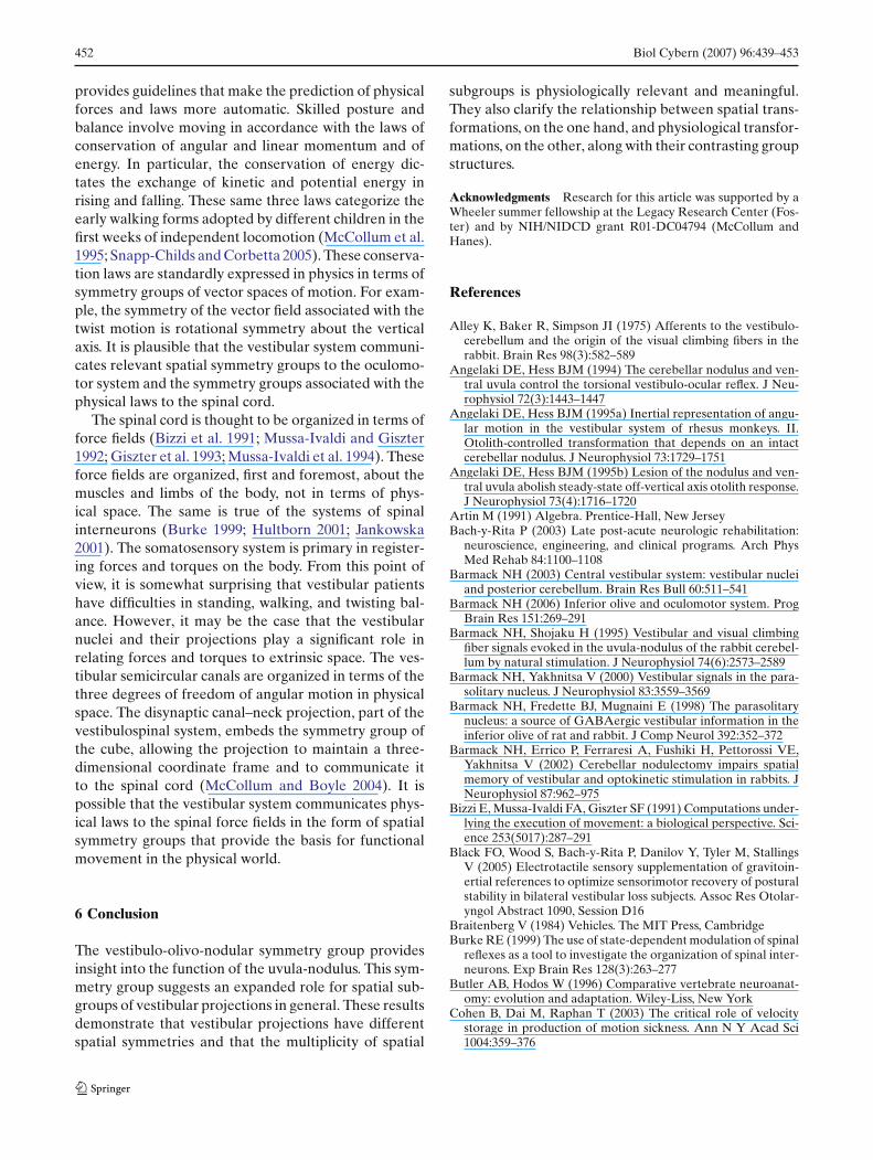
provides guidelines that make the prediction of physicalforces and laws more automatic. Skilled posture andbalance involve moving in accordance with the laws ofconservation of angular and linear momentum and ofenergy. In particular, the conservation of energy dic-tates the exchange of kinetic and potential energy inrising and falling. These same three laws categorize theearly walking forms adopted by different children in thefirst weeks of independent locomotion (McCollum et al.1995; Snapp-Childs and Corbetta 2005). These conserva-tion laws are standardly expressed in physics in terms ofsymmetry groups of vector spaces of motion. For exam-ple, the symmetry of the vector field associated with thetwist motion is rotational symmetry about the verticalaxis. It is plausible that the vestibular system communi-cates relevant spatial symmetry groups to the oculomo-tor system and the symmetry groups associated with thephysical laws to the spinal cord.
The spinal cord is thought to be organized in terms offorce fields (Bizzi et al. 1991; Mussa-Ivaldi and Giszter1992; Giszter et al. 1993; Mussa-Ivaldi et al. 1994). Theseforce fields are organized, first and foremost, about themuscles and limbs of the body, not in terms of phys-ical space. The same is true of the systems of spinalinterneurons (Burke 1999; Hultborn 2001; Jankowska2001). The somatosensory system is primary in register-ing forces and torques on the body. From this point ofview, it is somewhat surprising that vestibular patientshave difficulties in standing, walking, and twisting bal-ance. However, it may be the case that the vestibularnuclei and their projections play a significant role inrelating forces and torques to extrinsic space. The ves-tibular semicircular canals are organized in terms of thethree degrees of freedom of angular motion in physicalspace. The disynaptic canal–neck projection, part of thevestibulospinal system, embeds the symmetry group ofthe cube, allowing the projection to maintain a three-dimensional coordinate frame and to communicate itto the spinal cord (McCollum and Boyle 2004). It ispossible that the vestibular system communicates phys-ical laws to the spinal force fields in the form of spatialsymmetry groups that provide the basis for functionalmovement in the physical world.
6 Conclusion
The vestibulo-olivo-nodular symmetry group providesinsight into the function of the uvula-nodulus. This sym-metry group suggests an expanded role for spatial sub-groups of vestibular projections in general. These resultsdemonstrate that vestibular projections have differentspatial symmetries and that the multiplicity of spatial
subgroups is physiologically relevant and meaningful.They also clarify the relationship between spatial trans-formations, on the one hand, and physiological transfor-mations, on the other, along with their contrasting groupstructures.
Acknowledgments Research for this article was supported by aWheeler summer fellowship at the Legacy Research Center (Fos-ter) and by NIH/NIDCD grant R01-DC04794 (McCollum andHanes).
References
Alley K, Baker R, Simpson JI (1975) Afferents to the vestibulo-cerebellum and the origin of the visual climbing fibers in therabbit. Brain Res 98(3):582–589
Angelaki DE, Hess BJM (1994) The cerebellar nodulus and ven-tral uvula control the torsional vestibulo-ocular reflex. J Neu-rophysiol 72(3):1443–1447
Angelaki DE, Hess BJM (1995a) Inertial representation of angu-lar motion in the vestibular system of rhesus monkeys. II.Otolith-controlled transformation that depends on an intactcerebellar nodulus. J Neurophysiol 73:1729–1751
Angelaki DE, Hess BJM (1995b) Lesion of the nodulus and ven-tral uvula abolish steady-state off-vertical axis otolith response.J Neurophysiol 73(4):1716–1720
Artin M (1991) Algebra. Prentice-Hall, New JerseyBach-y-Rita P (2003) Late post-acute neurologic rehabilitation:
neuroscience, engineering, and clinical programs. Arch PhysMed Rehab 84:1100–1108
Barmack NH (2003) Central vestibular system: vestibular nucleiand posterior cerebellum. Brain Res Bull 60:511–541
Barmack NH (2006) Inferior olive and oculomotor system. ProgBrain Res 151:269–291
Barmack NH, Shojaku H (1995) Vestibular and visual climbingfiber signals evoked in the uvula-nodulus of the rabbit cerebel-lum by natural stimulation. J Neurophysiol 74(6):2573–2589
Barmack NH, Yakhnitsa V (2000) Vestibular signals in the para-solitary nucleus. J Neurophysiol 83:3559–3569
Barmack NH, Fredette BJ, Mugnaini E (1998) The parasolitarynucleus: a source of GABAergic vestibular information in theinferior olive of rat and rabbit. J Comp Neurol 392:352–372
Barmack NH, Errico P, Ferraresi A, Fushiki H, Pettorossi VE,Yakhnitsa V (2002) Cerebellar nodulectomy impairs spatialmemory of vestibular and optokinetic stimulation in rabbits. JNeurophysiol 87:962–975
Bizzi E, Mussa-Ivaldi FA, Giszter SF (1991) Computations under-lying the execution of movement: a biological perspective. Sci-ence 253(5017):287–291
Black FO, Wood S, Bach-y-Rita P, Danilov Y, Tyler M, StallingsV (2005) Electrotactile sensory supplementation of gravitoin-ertial references to optimize sensorimotor recovery of posturalstability in bilateral vestibular loss subjects. Assoc Res Otolar-yngol Abstract 1090, Session D16
Braitenberg V (1984) Vehicles. The MIT Press, CambridgeBurke RE (1999) The use of state-dependent modulation of spinal
reflexes as a tool to investigate the organization of spinal inter-neurons. Exp Brain Res 128(3):263–277
Butler AB, Hodos W (1996) Comparative vertebrate neuroanat-omy: evolution and adaptation. Wiley-Liss, New York
Cohen B, Dai M, Raphan T (2003) The critical role of velocitystorage in production of motion sickness. Ann N Y Acad Sci1004:359–376

Biol Cybern (2007) 96:439–453 453
Cohen B, Wearne S, Dai M, Raphan T (1999) Spatial orientationof the angular vestibulo ocular reflex. J Vestib Res 9(3):163–172
Cohen B, John P, Yakushin SB, Buettner-Ennever J, Raphan T(2002) The nodulus and uvula: source of cerebellar control ofspatial orientation of the angular vestibulo-ocular reflex. AnnN Y Acad Sci 978:28–45
Dai M, Kunin M, Raphan T, Cohen B (2003) The relation ofmotion sickness to the spatial temporal properties of velocitystorage. Exp Brain Res 151(2):173–189
DiZio P, Lackner JR (1986) Perceived orientation, motion, andconfiguration of the body during viewing of an off-vertical,rotating surface. Percept Psychophys 39(1):39–46
Duhamel JR, Bremmer F, BenHamed S, Graf W (1997) Spatialinvariance of visual receptive fields in parietal cortex neurons.Nature 389(6653):845–848
Fritzsch B, Beisel KW, Jones K, Farinas I, Maklad A, Lee J, Reic-hardt LF (2002) Development and evolution of inner ear sen-sory epithelia and their innervation. J Neurobiol 53(2):143–156
Fushiki H, Barmack NH (1997) Topography and reciprocal activ-ity of cerebellar Purkinje cells in the uvula-nodulus modulatedby vestibular stimulation. J Neurophysiol 78:3083–3094
Giszter SF, Mussa-Ivaldi FA, Bizzi E (1993) Convergent forcefields organized in the frog’s spinal cord. J Neurosci 13:467–491
Goldberg JM, Fernández C (1984) The Vestibular System. In:Brookhart JM, Mountcastle VB, Darian-Smith I (eds) Hand-book of physiology — the nervous system III. pp 977–1022
Golubitsky M, Stewart I (2002) The symmetry perspective: fromequilibrium to chaos in phase space and physical space. Birkha-user Verlag, Basel
Golubitsky M, Stewart I, Buono P-L, Collins JJ (1998) A modularnetwork for legged locomotion. Physica D 115:56–72
Goossens HHLM, van Opstal AJ (1999) Influence of head positionon the spatial representation of acoustic targets. J Neurophysiol81:2720–2736
Guitton D, Volle M (1987) Gaze control in humans: eye–headcoordination during orienting movements to targets withinand beyond the oculomotor range. J Neurophysiol 58(3):427–459
Hanes DA (2006) Perceptual centering effects in body orientation.Biol Cybern 9(4):288–299
Hanes DA, McCollum G (2006) Cognitive–vestibular interactions:a review of cognitive difficulties of vestibular patients and pos-sible mechanisms (in press)
Hasegawa T, Kato I, Harada K, Ikarashi T, Yoshida M, Koike Y(1994) The effect of uvulonodular lesions on horizontal optoki-netic nystagmus and optokinetic after-nystagmus in cats. ActaOtolaryngol 511:126–130
Hultborn H (2001) State-dependent modulation of sensory feed-back. J Physiol 533(1):5–13
Ivanenko YP, Grasso R, Lacquaniti F (1999) Effect of gaze onpostural responses to neck proprioceptive and vestibular stim-ulation in humans. J Physiol 519(1):301–314
Igarashi M, Takeda N, Chae S (1992) Uvula-nodulus and gravitydirection (A study on vertical optokinetic–oculomotor func-tions). Acta Astronau 27:25–30
Jankowska E (2001) Spinal interneuronal systems: identification,multifunctional character and reconfigurations in mammals. JPhysiol 533(1):31–40
Kleinschmidt A, Thilo KV, Buchel C, Gresty MA, Bronstein AM,Frackowiak RS (2002) Neural correlates of visual–motion per-ception as object- or self-motion. Neuroimage 16(4):873–882
Lackner JR, DiZio P (1993) Multisensory, cognitive, and motorinfluences on human spatial orientation in weightlessness. JVestib Res 3:361–372
Lewis ER, Leverenz EL, Bialek WS (1985) The vertebrate innerear. CRC Press, Boca Raton
Lowenstein O (1971) The labyrinth in fish physiology, vol V.In: Hoar WS, Randall DJ (eds) Sensory systems and electricorgans. Academic, New York pp 207–240
Maklad A, Fritzsch B (2003) Partial segregation of posterior cristaand saccular fibers to the nodulus and uvula of the cerebellumin mice, and its development. Dev Brain Res 140(2):223–236
McCollum G, Boyle R (2004) Rotations in a vertebrate setting:evaluation of the symmetry group of the disynaptic canal–neckprojection. Biol Cybern 90:203–217
McCollum G, Holroyd C, Castelfranco AM (1995) Forms of earlywalking. J Theor Biol 176:373–390
Mussa-Ivaldi FA, Giszter SF (1992) Vector field approximation: acomputational paradigm for motor control and learning. BiolCybern 67:491–500
Mussa-Ivaldi FA, Giszter SF, Bizzi E (1994) Linear combinationsof primitives in vertebrate motor control. Proc Natil Acad Sci91:7534–7538
Shinoda Y, Sugiuchi Y, Futami T, Ando N, Kawasaki T (1994)Input patterns and pathways from six semicircular canals tomotoneurons of neck muscles. I. The Multifidus Muscle Group.J Neurophysiol 72:2691–702
Shinoda Y, Sugiuchi Y, Futami T, Kakei S, Izawa Y, Na J (1996)Four convergent patterns of input from the six semicircularcanals to motoneurons of different neck muscles in the uppercervical cord. Ann N Y Acad Sci 781:264–275
Shinoda Y, Sugiuchi Y, Futami T, Ando N, Yagi J (1997) Inputpatterns and pathways from six semicircular canals to moto-neurons of neck muscles. II. The Longissimus and SemispinalisMuscle Groups. J Neurophysiol 72:2691–2702
Simpson JI (1984) The accessory optic system. Ann Revi Neurosci7:13–14
Snapp-Childs W, Corbetta D (2005) Learning to walk: individualdifferences and early strategies. J Sport Exerc Psychol 27:S143–S144
Soechting JF, Flanders M (1989) Sensorimotor representations forpointing to targets in three-dimensional space. J Neurophysiol62(2):582–594
Soechting JF, Lacquaniti F, Terzuolo CA (1986) Coordinationof arm movements in three-dimensional space. Sensorimotormapping during drawing movement. Neuroscience 17(2):295–311
Solomon D, Cohen B (1994) Stimulation of the nodulus and uvuladischarges velocity storage in the vestibulo-ocular reflex. ExpBrain Res 102(1):57–68
Sparks DL (1989) The neural encoding of the location of targetsfor saccadic eye movements. J Exp Biol 146:195–207
Stahl JS (2001) Adaptive plasticity of head movement propensity.Exp Brain Res 139:201–208
Uchino Y, Sato H, Zakir M, Kushiro K, Imagawa M, OgawaY, Ono S, Meng H, Zhang X, Katsuta M, Isu N, Wilson VJ(2001) Commissural effects in the otolith system. Exp BrainRes 126:421–430
Vidal P-P, Graf W, Berthoz A (1986) The orientation of the cervi-cal vertebral column in unrestrained awake animals. Exp BrainRes 61:549–559
Voogd J, Barmack NH (2006) Oculomotor cerebellum. Prog BrainRes 151:231–268
Wearne S, Raphan T, Cohen B (1998) Control of spatial orienta-tion of the angular vestibuloocular reflex by the nodulus anduvula. J Neurophysiol 79(5):2690–2715
Wiest G, Deecke L, Trattnig S, Mueller C (1999) Abolished tiltsuppression of the vestibulo ocular reflex caused by a selectiveuvulo-nodular lesion. Neurology 52(2):417–419
Wilson VJ, Maeda M (1974) Connection between semicircularcanals and neck motoneurons in the cat. J Neurophysiol 37:346–357
Copyright © 2022 FDOKUMEN