The unilateral declaration of rescission - an extrajudicial mean ...
Upload
independentCategory
view
0download
0
Kidney International, Vol. 55 (1999), pp. 793–807
GENETIC DISORDERS – DEVELOPMENT
Recovery following relief of unilateral ureteral obstruction inthe neonatal rat
ROBERT L. CHEVALIER, ANDREW KIM, BARBARA A. THORNHILL,and JENNIFER T. WOLSTENHOLME
Department of Pediatrics, University of Virginia, Charlottesville, Virginia, USA
Conclusions. The relief of obstruction in the neonatal ratRecovery following relief of unilateral ureteral obstruction inattenuates, but does not reverse, renal vascular, glomerular,the neonatal rat.tubular, and interstitial injury resulting from five days of UUO.Background. Obstructive nephropathy is a primary causeHyperfiltration by remaining nephrons and residual tubuloin-of renal insufficiency in infants and children. This study was
designed to distinguish the reversible and irreversible cellular terstitial injury in the postobstructed kidney are likely to leadconsequences of temporary unilateral ureteral obstruction to deterioration of renal function later in life.(UUO) on the developing kidney.
Methods. Rats were subjected to UUO or sham operationin the first 48 hours of life, and the obstruction was removed
Obstructive nephropathy is the most important identi-five days later (or was left in place). Kidneys were removedfor study 14 or 28 days later. In additional groups, kidneys were fiable cause of chronic renal insufficiency in infants andremoved at the end of five days of obstruction. Immunoreactive children [1], and ureteropelvic junction obstruction ac-distribution of renin was determined in arterioles, and the
counts for the majority of congenital urinary tract ob-distribution of epidermal growth factor, transforming growthstruction in the fetus, infant, and child. Currently, how-factor-b1, clusterin, vimentin, and a-smooth muscle actin was
determined in tubules and/or interstitium. The number of glo- ever, the indications for relief of ureteral obstruction inmeruli, glomerular maturation, tubular atrophy, and interstitial the infant remain controversial. Early pyeloplasty hascollagen deposition was determined by morphometry. Renal been advocated by some [2–4] but opposed by otherscellular proliferation and apoptosis were measured by prolifer-
[5–7].ating cell nuclear antigen and the TdT uridine-nick-end-labelWe have shown previously that ureteral obstructiontechnique, respectively. The glomerular filtration rate was mea-
sured by inulin clearance. in early development impairs the growth and develop-Results. Renal microvascular renin maintained a fetal distri- ment of the kidney [8, 9]. A number of animal models
bution with persistent UUO; this was partially reversed by thehave been developed to study the effects of ureteralrelief of obstruction. Although glomerular maturation was alsoobstruction during the period of nephrogenesis, includ-delayed and glomerular volume was reduced by UUO, the
relief of obstruction prevented the reduction in glomerular ing the fetal sheep, the neonatal opossum, and the neona-volume. Although relief of obstruction did not reverse a 40% tal rodent [8, 10–12]. Although the sheep probably mostreduction in the number of nephrons, the glomerular filtration closely approximate human renal development, the largerate of the postobstructed kidney was normal. The relief of
size and expense of this species limit the number ofobstruction did not improve tubular cell proliferation and onlysequential observations that can be made. The opossumpartially reduced apoptosis induced by UUO. This was associ-
ated with a persistent reduction in the tubular epidermal has been studied as an “extrauterine fetus” that cangrowth factor. In addition, the relief of obstruction reduced but be more conveniently manipulated postnatally; however,did not normalize tubular expression of transforming growth
the correlation of renal structure and function is poorlyfactor-b1, clusterin, and vimentin, all of which are evidence ofdefined, and reagents for cellular and molecular investi-persistent tubular injury. The relief of obstruction significantly
reduced interstitial fibrosis and expression of a-smooth muscle gation are not generally available for this species. Theactin by interstitial fibroblasts, but not to normal levels. rat, in contrast, has been well studied, and most nephro-
genesis proceeds during the first two weeks of postnatallife. Although maturation of the rat kidney during theKey words: obstructive nephropathy, renal insufficiency, unilateral ure-
teral obstruction, developing kidney, tubulointerstitial disease. first month of life is thus analogous to that of the humanfetus during the second and third trimesters, the intra-
Received for publication August 4, 1998uterine environment of the latter may alter the renaland in revised form October 15, 1998
Accepted for publication October 15, 1998 response to ureteral obstruction compared with the post-natal rat model. This potential limitation is outweighed 1999 by the International Society of Nephrology
793
Chevalier et al: Recovery following neonatal ureteral obstruction794
by the advantages of postnatal surgical intervention inthe rat.
We have developed a model of unilateral ureteral ob-struction (UUO) in the neonatal rat that has allowed usto characterize the cellular response of the microvascula-ture, glomerulus, tubule, and interstitium from 1 to 28days following UUO within the first 48 hours of life[8]. Chronic UUO in the neonatal rodent activates theintrarenal renin-angiotensin system [13] and causes glo-merular contraction [14]. In addition, UUO induces thephenotypic transformation of renal tubular epithelialcells that acquire mesenchymal characteristics, such as
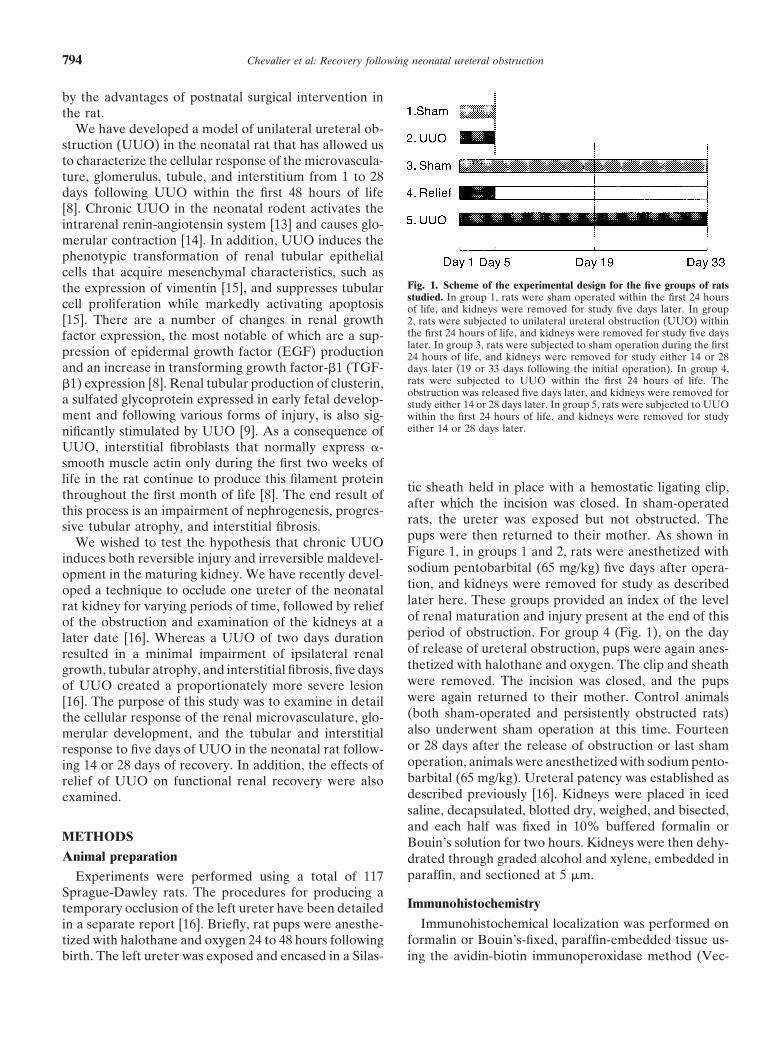
Fig. 1. Scheme of the experimental design for the five groups of ratsthe expression of vimentin [15], and suppresses tubularstudied. In group 1, rats were sham operated within the first 24 hourscell proliferation while markedly activating apoptosis of life, and kidneys were removed for study five days later. In group
[15]. There are a number of changes in renal growth 2, rats were subjected to unilateral ureteral obstruction (UUO) withinthe first 24 hours of life, and kidneys were removed for study five daysfactor expression, the most notable of which are a sup-later. In group 3, rats were subjected to sham operation during the firstpression of epidermal growth factor (EGF) production 24 hours of life, and kidneys were removed for study either 14 or 28
and an increase in transforming growth factor-b1 (TGF- days later (19 or 33 days following the initial operation). In group 4,rats were subjected to UUO within the first 24 hours of life. Theb1) expression [8]. Renal tubular production of clusterin,obstruction was released five days later, and kidneys were removed fora sulfated glycoprotein expressed in early fetal develop- study either 14 or 28 days later. In group 5, rats were subjected to UUO
ment and following various forms of injury, is also sig- within the first 24 hours of life, and kidneys were removed for studyeither 14 or 28 days later.nificantly stimulated by UUO [9]. As a consequence of
UUO, interstitial fibroblasts that normally express a-smooth muscle actin only during the first two weeks oflife in the rat continue to produce this filament protein
tic sheath held in place with a hemostatic ligating clip,throughout the first month of life [8]. The end result ofafter which the incision was closed. In sham-operatedthis process is an impairment of nephrogenesis, progres-rats, the ureter was exposed but not obstructed. Thesive tubular atrophy, and interstitial fibrosis.pups were then returned to their mother. As shown inWe wished to test the hypothesis that chronic UUOFigure 1, in groups 1 and 2, rats were anesthetized withinduces both reversible injury and irreversible maldevel-sodium pentobarbital (65 mg/kg) five days after opera-opment in the maturing kidney. We have recently devel-tion, and kidneys were removed for study as describedoped a technique to occlude one ureter of the neonatallater here. These groups provided an index of the levelrat kidney for varying periods of time, followed by reliefof renal maturation and injury present at the end of thisof the obstruction and examination of the kidneys at aperiod of obstruction. For group 4 (Fig. 1), on the daylater date [16]. Whereas a UUO of two days durationof release of ureteral obstruction, pups were again anes-resulted in a minimal impairment of ipsilateral renalthetized with halothane and oxygen. The clip and sheathgrowth, tubular atrophy, and interstitial fibrosis, five dayswere removed. The incision was closed, and the pupsof UUO created a proportionately more severe lesionwere again returned to their mother. Control animals[16]. The purpose of this study was to examine in detail(both sham-operated and persistently obstructed rats)the cellular response of the renal microvasculature, glo-also underwent sham operation at this time. Fourteenmerular development, and the tubular and interstitialor 28 days after the release of obstruction or last shamresponse to five days of UUO in the neonatal rat follow-operation, animals were anesthetized with sodium pento-ing 14 or 28 days of recovery. In addition, the effects ofbarbital (65 mg/kg). Ureteral patency was established asrelief of UUO on functional renal recovery were alsodescribed previously [16]. Kidneys were placed in icedexamined.saline, decapsulated, blotted dry, weighed, and bisected,and each half was fixed in 10% buffered formalin or
METHODS Bouin’s solution for two hours. Kidneys were then dehy-Animal preparation drated through graded alcohol and xylene, embedded in
paraffin, and sectioned at 5 mm.Experiments were performed using a total of 117Sprague-Dawley rats. The procedures for producing a
Immunohistochemistrytemporary occlusion of the left ureter have been detailedImmunohistochemical localization was performed onin a separate report [16]. Briefly, rat pups were anesthe-
formalin or Bouin’s-fixed, paraffin-embedded tissue us-tized with halothane and oxygen 24 to 48 hours followingbirth. The left ureter was exposed and encased in a Silas- ing the avidin-biotin immunoperoxidase method (Vec-
Chevalier et al: Recovery following neonatal ureteral obstruction 795
tastain ABC Kit; Vector Laboratories, Burlingame, CA, number of glomeruli per section was counted in longitu-dinal sections, including both poles and the renal papilla.USA). The primary antibodies used were polyclonal goat
antirat renin (1:10,000; gift of Tadashi Inagami), EGF Because there is no nephrogenic zone in one month-oldrats, all glomeruli were counted without attempting to(1:200; Biomedical Technologies, Inc., Stoughton, MA,
USA), monoclonal mouse antirat proliferating cell nu- separate cortical and nephrogenic zones.clear antigen diluted 1:200 in phosphate buffered saline
Tubules(Vector Laboratories), monoclonal mouse antihuman vi-mentin (1:500, Sigma Chemical Co., St. Louis, MO, For measurement of cellular proliferation by prolifer-
ating cell nuclear antigen and for determination of apo-USA), monoclonal rabbit antirat clusterin (1:50; UpstateBiotechnology, Lake Placid, NY, USA), monoclonal ptosis by the TdT uridine-nick-end-label technique, the
number of labeled tubular and interstitial cells wasanti-a-smooth muscle-specific actin (1:400; Sigma Chem-ical), and polyclonal rabbit antihuman TGF-b1 (1:50; counted in each field in sections from kidneys harvested
on day 19. The number of vimentin-staining tubular cellsPromega, Madison, WI, USA). Apoptotic nuclei werelabeled using the TdT uridine-nick-end-label technique per field was similarly counted in kidneys harvested on
day 33. For EGF, the fraction of tubules in the field with(Apoptag; Oncor, Gaithersburg, MD, USA) [15]. Sec-tions were counterstained with Gill’s #2 hematoxylin, positive immunostaining was compared with the total
number of tubules per field in kidneys on day 5 and daydehydrated, and mounted for examination by light mi-croscopy. Representative results of immunostaining are 33. For TGF-b1, tubular staining was scored using a
semiquantitative scale from 1 (no staining) to 5 (maximalshown in Figure 2. As a negative control, the primaryantiserum was replaced by nonimmune rabbit serum, staining) in kidneys on day 5 and day 33. For clusterin
staining, the number of positive-staining tubules per fieldand no staining occurred. Also, no primary antibody wasused as a negative control. was determined in kidneys on day 5 and day 33. The
number of atrophic tubules per field was determined asHistomorphometry those tubules with thickened tubular basement mem-
branes identified by periodic acid-Schiff staining in entireThe cellular effects of temporary UUO in the devel-oping rat kidney were quantitated by examining histo- sections of kidneys on day 5 and 50 nonoverlapping fields
on day 33.morphometric parameters in the renal microvasculature,glomeruli, tubules, and interstitium. Using image analy-
Interstitiumsis software (Mocha 1.2; Jandel Scientific, San Rafael,CA, USA), kidneys were examined by scanning 10 non- Interstitial collagen deposition was measured in Mas-
son trichrome-stained sections in kidneys on day 33 usingoverlapping corticomedullary fields by light microscopyat a magnification of 3400 with the observer blinded to image analysis software (Mocha 1.2; Jandel Scientific).
Digital images of the sections were superimposed on athe treatment group.grid, and the number of grid points overlapping intersti-
Microvascular renin distribution tial blue-staining collagen was recorded for each field.The same method was used to quantitate immunoreac-In each of the 10 fields examined per kidney section
for day 5 and day 33, the length of renin immunostaining tive interstitial a-smooth muscle actin and vimentin dis-tribution in kidneys on day 5 and day 33. Representativewas measured for each afferent arteriole from its junc-
tion with the glomerulus. photomicrographs of interstitial collagen, a-smooth mus-cle actin, and vimentin from kidneys on day 33 are shown
Glomerular maturation and glomerular area in Figure 9.The maturation index of each glomerulus per field was
Renal functiondetermined in kidneys from day 5 using the criteria ofNiimura et al [17]. Briefly, glomeruli were scored as In day 33 rats (N 5 5, group 3, and N 5 6, group 4),
the animal was anesthetized with intraperitoneal sodiumimmature, intermediate, or mature based on the numberof capillary loops and the phenotype of podocytes (cu- pentobarbital (50 mg/kg) and was placed on a thermo-
statically controlled heating table. After placement of aboidal or flattened). The glomerular tuft area was mea-sured in kidneys from day 33 as the mean area enclosed tracheotomy, a carotid artery was cannulated for blood
collection. A jugular vein was cannulated for infusion ofby the visceral epithelial cells.0.85% sodium chloride containing 3H inulin (15 mCi/ml;
Number of glomeruli New England Nuclear, Boston, MA, USA) at a rateof 3 ml/kg/hr. The right ureter was ligated above theBoth periodic acid-Schiff stain and Masson trichrome-
stained sections were used to count the number of glo- ureterovesical junction and was transected above theligature to allow free drainage of urine. A suprapubicmeruli in rats on day 5 and day 33. Using a modification
of the method of McVary and Maizels [18], the total catheter was inserted in the bladder for collection of

Chevalier et al: Recovery following neonatal ureteral obstruction796
Fig. 2. Immunochemical distribution of cytoplasmic proteins or markers of cellular proliferation or apoptosis in rat kidneys. (A–E) Distributionof immunoreactive renin in kidneys of rats from groups 1 through 5. Renin-containing vascular cells are brown. In sham-operated groups (A,group 1, and C, group 3, 33 days), renin is localized to the juxtaglomerular region of the afferent arteriole. In UUO groups (B, group 2, and E,group 5, 33 days), renin extends along the length of the afferent arteriole. In UUO followed by relief at five days (D, group 4, 33 days), renindistribution is intermediate between that of groups 3 and 5. (F) The localization of proliferating cells by proliferating cell nuclear antigen (PCNA)in obstructed kidney from rat in group 2 (19 days). Tubular cell nuclei stain brown (arrowheads). (G) The localization of apoptotic cells by TdTuridine-nick-end-label (TUNEL) technique from rat in group 5 (19 days): fragmented DNA in apoptotic nuclei stain brown (arrowheads). (H )The distribution of epidermal growth factor (EGF) in tubules from rat in group 3 (33 days; *). (I) The distribution of transforming growth factor-b1 (TGF-b1) in tubules from rat in group 5 (33 days; *). (J) The distribution of clusterin in tubules from rat in group 5 (33 days; *). (K) Thedistribution of vimentin in tubules from rat in group 5 (33 days; *).
Chevalier et al: Recovery following neonatal ureteral obstruction 797
Table 1. Somatic and kidney growth of rats
Left (obstructed)Body weight g kidney/body weight % Right (contralateral)
Group 1 (N 5 5) 9.560.7 NA NASham 5 day study
Group 2 (N 5 10) 7.861.1 0.7260.03 0.69 60.02UUO 5 day study
Group 3 (N 5 35) 99.062.1 0.47 60.01 NASham 33 day study
Group 4 (N 5 17) 92.463.6 0.38 60.02 0.6060.02a
Relief 33 day studyGroup 5 (N 5 50) 95.361.8 0.2160.01ab 0.7360.02ac
NA is not available.a P , 0.05 vs. Group 3 Shamb P , 0.05 vs. Group 4 Reliefc P , 0.05 vs contralateral
urine from the left kidney only. Following a 60-minuteequilibration period, three 20-minute urine collectionswere obtained. At the beginning of each urine collectionperiod, 100 ml of blood was withdrawn and replacedwith 4% bovine serum albumin. Following the last urinecollection, an additional blood sample was withdrawn,after which the rat was sacrificed with an overdose ofsodium pentobarbital. Radioactivity in urine and plasmasamples were counted in a LS6500 scintillation counter(Beckman Instruments, Inc., Schaumburg, IL, USA).The urine sodium concentration was measured by flamephotometry. The glomerular filtration rate for the leftkidney was calculated as the mean inulin clearance foreach animal.
Statistical analysis
Data are presented as mean 6 standard error. Com-parisons between groups were made using one-way anal-ysis of variance followed by the Student-Newman-Kuelstest. Comparisons between left and right kidneys wereperformed using the Student t-test for paired data. Statis-tical significance was defined as P , 0.05.
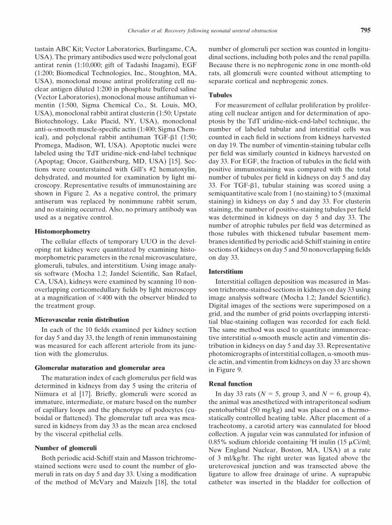
Fig. 3. Length of afferent arteriole adjacent to glomerulus containingRESULTS immunoreactive renin for rats in groups 1 and 2 (A) and in groups 3
through 5 (B). Data are mean 6 sem. Symbols are: (h) left kidney, fiveSomatic and renal growth days (groups 1 and 2); ( ) left kidney, 33 days; ( ) right (contralateral)kidney. *P , 0.05 vs. sham; #P , 0.05 vs. contralateral kidney; 1P ,As shown in Table 1, there was no significant effect0.05 vs. 5 days of relief.
of UUO on somatic growth at either 5 or 33 days. Therewas no difference between left and right kidney weightfive days following UUO (groups 1 and 2). However, 28days following UUO, the weight of the obstructed kidney Microvasculaturewas reduced by more than 50%, and that of the contralat- As shown in Figures 2 and 3, immunoreactive renineral kidney had increased by over 50% compared with was normally localized to the juxtaglomerular region atsham-operated rats (groups 3 and 5). The relief of UUO either 5 or 33 days of age. After five days of UUO, renin(group 4) attenuated both the reduction in mass of the was localized along the length of the afferent arteriole,postobstructed kidney and the compensatory growth of a pattern that persisted to 33 days during continuous
UUO. Following the relief of obstruction, however, therethe contralateral kidney.
Chevalier et al: Recovery following neonatal ureteral obstruction798
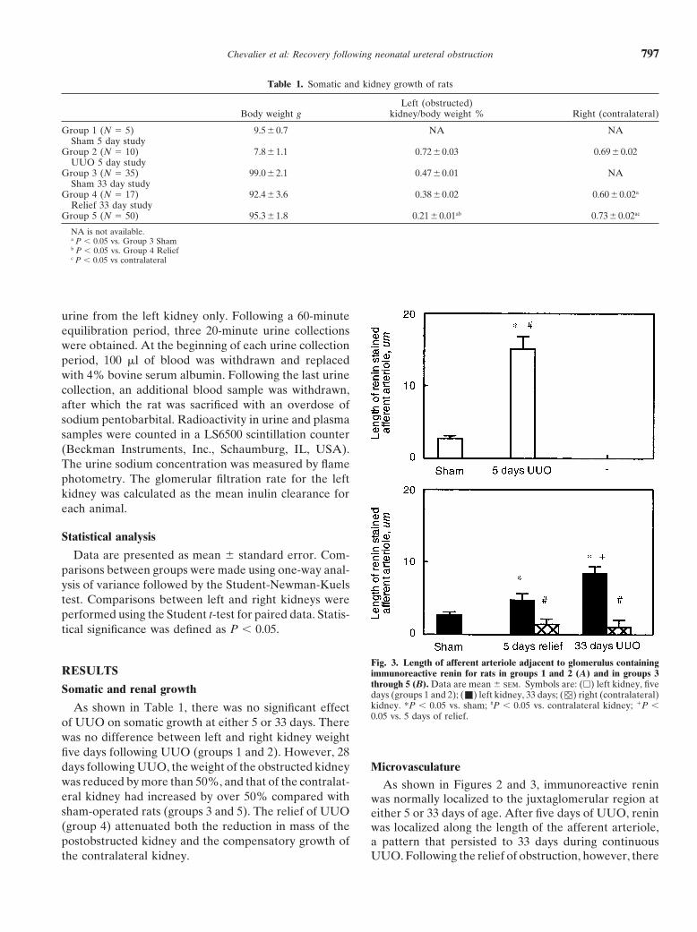
Fig. 4. (A) Weighted glomerular maturation score in sham-operated and UUO rats five days after operation. (B) Glomerular tuft area 28 daysafter operation. (C) Relative number of glomeruli five days after operation. (D) The relative number of glomeruli 28 days after operation. Symbolsare: (h) left kidney, five days (groups 1 and 2); ( ) right (contralateral) kidney; ( ) left kidney, 33 days; ( ) right (contralateral) kidney.
was a significant reduction in the extension of renin along in the postobstructed kidney, but the difference was notsignificant (Table 2).the afferent arteriole.
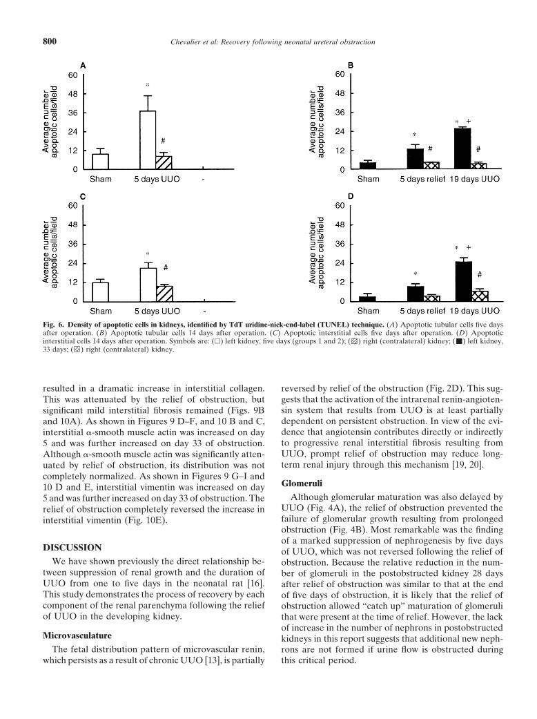
TubulesGlomeruliAs shown in Figure 5 A and B, UUO induced a 46%As shown in Figure 4A, glomerular maturation was
reduction in tubular cell proliferation, which persistedsignificantly delayed in the kidney subjected to five daysfor 14 days following relief of obstruction. Tubular cellof UUO. Whereas the glomerular tuft area was reducedapoptosis was increased threefold after five days of UUOby 35% following 28 days of ipsilateral UUO and in-(Fig. 6A). Although tubular apoptosis remained three-creased 35% in the contralateral kidney, these glomeru-fold higher than sham and contralateral kidneys 14 dayslar effects were prevented by relief of obstruction (Fig.following relief of obstruction, this was 50% less than4B). There were no immature glomeruli at 19 or 33 dayskidneys with persistent obstruction at 19 days (Fig. 6B).
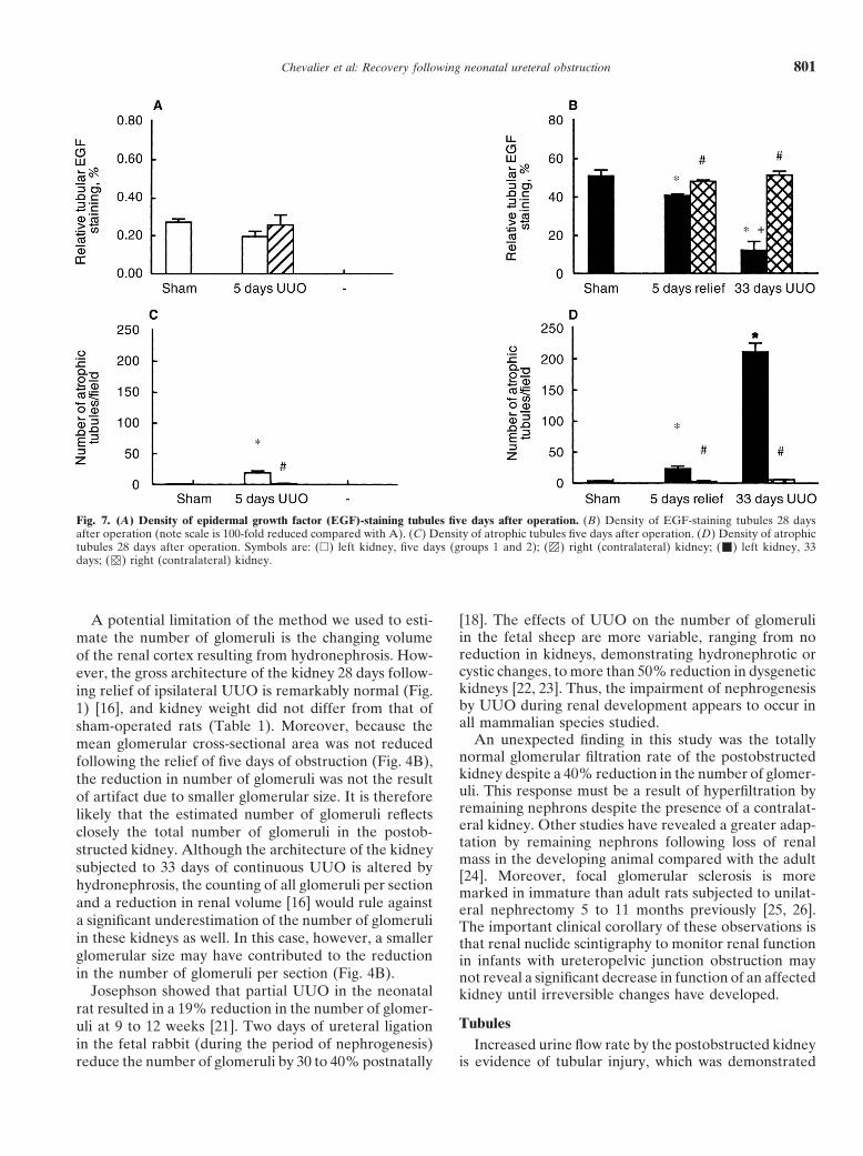
regardless of UUO (data not shown).As shown in Figure 7A, immunoreactive tubular EGF
The number of glomeruli was reduced by 40% after was nearly absent at five days, and there was no differ-five days of UUO, and this reduction persisted 28 days ence between UUO and sham-operated kidneys. Follow-following the relief of obstruction (Fig. 4 C, D). Contin- ing 33 days of UUO, tubular EGF expression was re-ual UUO throughout 33 days reduced the number of duced by 75% compared with contralateral or sham-glomeruli further by 60% (Fig. 4D). There was no effect operated kidneys. Although the relief of obstruction sig-of UUO on the number of glomeruli in the intact oppo- nificantly attenuated the inhibition of EGF expression,site kidney (Fig. 4 C, D). there was a persistent significant reduction in tubular
Despite the significant reduction in the number of EGF in the postobstructed kidney (Fig. 7B).glomeruli in the postobstructed kidney, there was no Tubular atrophy was detectable after only five daysreduction in glomerular filtration rate 28 days after relief of UUO (Fig. 7C), and this level of tubular atrophyof five days of UUO (Table 2). Urine flow rate for the persisted for 28 days following the relief of obstructionpostobstructed kidney was double that of the sham-oper- (Fig. 7D). However, as a result of 33 days of continuous
obstruction, tubular atrophy had increased 60-fold com-ated animals, and sodium excretion tended to increase
Chevalier et al: Recovery following neonatal ureteral obstruction 799
Table 2. Function of sham-operated and postobstructed kidney of 33-day-old rats
Glomerular Urine sodiumfiltration rate Urine flow rate excretion
ml/min/g kidney wt ll/min/g kidney wt lEq/min/g kidney wt
Group 3 0.32 60.07 0.97 60.13 0.1360.02Sham (N 5 5)
Group 4 0.35 60.05 2.22 60.42a 0.2460.06Relief (N 5 6)a P , 0.05 vs. Sham
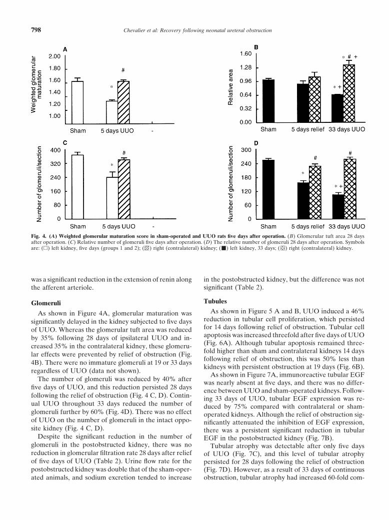
Fig. 5. Density of proliferating cells in kidneys, identified by proliferating cell nuclear antigen (PCNA). (A) PCNA-staining tubular cells five daysafter operation. (B) PCNA-staining tubular cells 14 days after operation. (C) PCNA-staining interstitial cells five days after operation. (D) PCNA-staining interstitial cells 14 days after operation. Symbols are: (h) left kidney, five days (groups 1 and 2); ( ) right (contralateral) kidney; ( )left kidney, 33 days; ( ) right (contralateral) kidney.
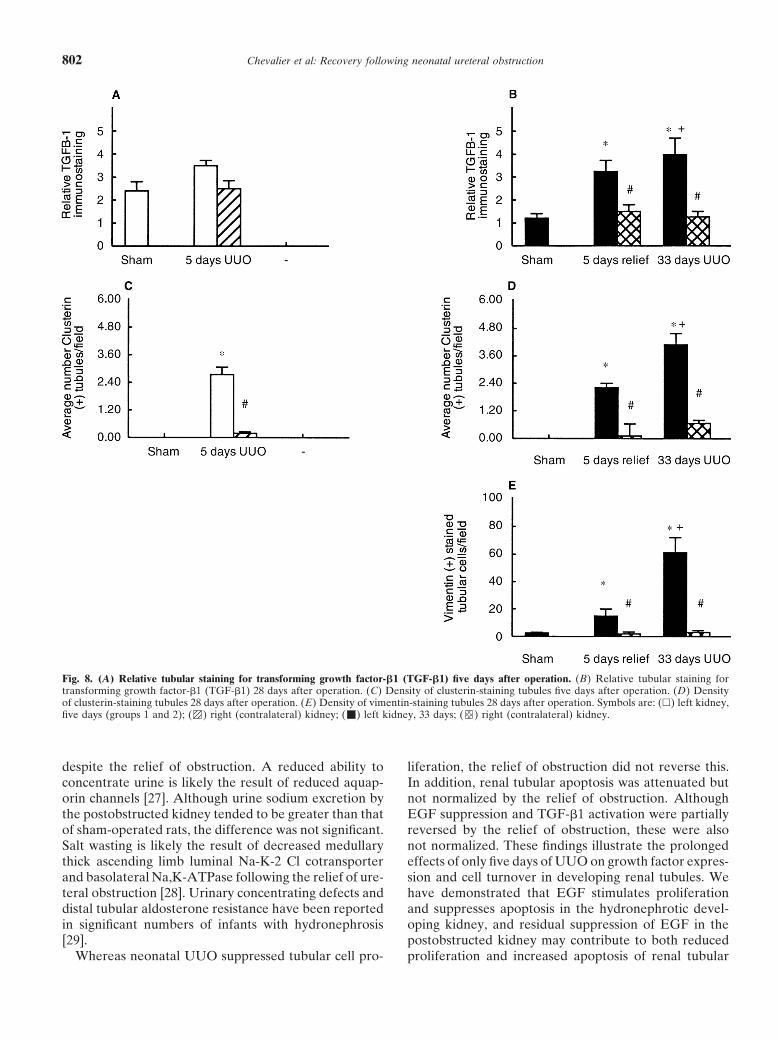
pared with the contralateral kidney or sham-operated bules five days following UUO (data not shown). Follow-ing 33 days of continuous UUO, there was a markedcontrols (Fig. 7D).
As shown in Figure 8A, there were no differences increase in vimentin-positive tubules (Fig. 8E). The reliefof obstruction decreased the distribution of vimentin-between UUO and sham-operated kidneys in TGF-b1
expression after five days of UUO. However, there was staining tubules, although not to normal levels (Fig. 8E).a threefold increase in tubular TGF-b1 expression fol-
Interstitiumlowing 33 days of continuous UUO, which was onlyslightly (but significantly) reduced by the relief of UUO As shown in Figure 5 C and D, there were no signifi-
cant differences in the number of proliferating cells inat five days (Fig. 8B). Tubular clusterin expression wasmarkedly increased after five days of UUO and increased the interstitium of rats in any of the groups. In contrast,
there was an increase in interstitial apoptosis after fivefurther after 33 days of UUO (Fig. 8 C, D). Althoughthe relief of UUO significantly reduced the number of days of UUO (Fig. 6C), which persisted through 19 days
of UUO (Fig. 6D). The relief of obstruction reducedclusterin-staining tubules, these remained well above thelevel in the contralateral or sham-operated kidneys (Fig. interstitial apoptosis, but not to normal levels (Fig. 6D).
As shown in Figures 9 A–C and 10A, persistent UUO8D). There were no identifiable vimentin-staining tu-
Chevalier et al: Recovery following neonatal ureteral obstruction800
Fig. 6. Density of apoptotic cells in kidneys, identified by TdT uridine-nick-end-label (TUNEL) technique. (A) Apoptotic tubular cells five daysafter operation. (B) Apoptotic tubular cells 14 days after operation. (C) Apoptotic interstitial cells five days after operation. (D) Apoptoticinterstitial cells 14 days after operation. Symbols are: (h) left kidney, five days (groups 1 and 2); ( ) right (contralateral) kidney; ( ) left kidney,33 days; ( ) right (contralateral) kidney.
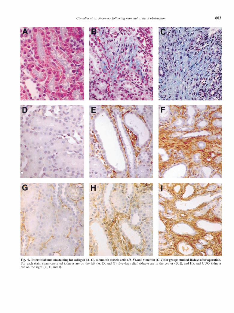
resulted in a dramatic increase in interstitial collagen. reversed by relief of the obstruction (Fig. 2D). This sug-gests that the activation of the intrarenal renin-angioten-This was attenuated by the relief of obstruction, butsin system that results from UUO is at least partiallysignificant mild interstitial fibrosis remained (Figs. 9Bdependent on persistent obstruction. In view of the evi-and 10A). As shown in Figures 9 D–F, and 10 B and C,dence that angiotensin contributes directly or indirectlyinterstitial a-smooth muscle actin was increased on dayto progressive renal interstitial fibrosis resulting from5 and was further increased on day 33 of obstruction.UUO, prompt relief of obstruction may reduce long-Although a-smooth muscle actin was significantly atten-term renal injury through this mechanism [19, 20].uated by relief of obstruction, its distribution was not
completely normalized. As shown in Figures 9 G–I andGlomeruli10 D and E, interstitial vimentin was increased on day
Although glomerular maturation was also delayed by5 and was further increased on day 33 of obstruction. TheUUO (Fig. 4A), the relief of obstruction prevented therelief of obstruction completely reversed the increase infailure of glomerular growth resulting from prolongedinterstitial vimentin (Fig. 10E).obstruction (Fig. 4B). Most remarkable was the findingof a marked suppression of nephrogenesis by five days
DISCUSSION of UUO, which was not reversed following the relief ofWe have shown previously the direct relationship be- obstruction. Because the relative reduction in the num-
tween suppression of renal growth and the duration of ber of glomeruli in the postobstructed kidney 28 daysUUO from one to five days in the neonatal rat [16]. after relief of obstruction was similar to that at the endThis study demonstrates the process of recovery by each of five days of obstruction, it is likely that the relief ofcomponent of the renal parenchyma following the relief obstruction allowed “catch up” maturation of glomeruliof UUO in the developing kidney. that were present at the time of relief. However, the lack
of increase in the number of nephrons in postobstructedMicrovasculature kidneys in this report suggests that additional new neph-
The fetal distribution pattern of microvascular renin, rons are not formed if urine flow is obstructed duringthis critical period.which persists as a result of chronic UUO [13], is partially
Chevalier et al: Recovery following neonatal ureteral obstruction 801
Fig. 7. (A) Density of epidermal growth factor (EGF)-staining tubules five days after operation. (B) Density of EGF-staining tubules 28 daysafter operation (note scale is 100-fold reduced compared with A). (C) Density of atrophic tubules five days after operation. (D) Density of atrophictubules 28 days after operation. Symbols are: (h) left kidney, five days (groups 1 and 2); ( ) right (contralateral) kidney; ( ) left kidney, 33days; ( ) right (contralateral) kidney.
A potential limitation of the method we used to esti- [18]. The effects of UUO on the number of glomeruliin the fetal sheep are more variable, ranging from nomate the number of glomeruli is the changing volumereduction in kidneys, demonstrating hydronephrotic orof the renal cortex resulting from hydronephrosis. How-cystic changes, to more than 50% reduction in dysgeneticever, the gross architecture of the kidney 28 days follow-kidneys [22, 23]. Thus, the impairment of nephrogenesising relief of ipsilateral UUO is remarkably normal (Fig.by UUO during renal development appears to occur in1) [16], and kidney weight did not differ from that ofall mammalian species studied.sham-operated rats (Table 1). Moreover, because the
An unexpected finding in this study was the totallymean glomerular cross-sectional area was not reducednormal glomerular filtration rate of the postobstructedfollowing the relief of five days of obstruction (Fig. 4B),kidney despite a 40% reduction in the number of glomer-the reduction in number of glomeruli was not the resultuli. This response must be a result of hyperfiltration byof artifact due to smaller glomerular size. It is thereforeremaining nephrons despite the presence of a contralat-likely that the estimated number of glomeruli reflectseral kidney. Other studies have revealed a greater adap-closely the total number of glomeruli in the postob-tation by remaining nephrons following loss of renalstructed kidney. Although the architecture of the kidneymass in the developing animal compared with the adultsubjected to 33 days of continuous UUO is altered by[24]. Moreover, focal glomerular sclerosis is more
hydronephrosis, the counting of all glomeruli per section marked in immature than adult rats subjected to unilat-and a reduction in renal volume [16] would rule against eral nephrectomy 5 to 11 months previously [25, 26].a significant underestimation of the number of glomeruli The important clinical corollary of these observations isin these kidneys as well. In this case, however, a smaller that renal nuclide scintigraphy to monitor renal functionglomerular size may have contributed to the reduction in infants with ureteropelvic junction obstruction mayin the number of glomeruli per section (Fig. 4B). not reveal a significant decrease in function of an affected
Josephson showed that partial UUO in the neonatal kidney until irreversible changes have developed.rat resulted in a 19% reduction in the number of glomer-
Tubulesuli at 9 to 12 weeks [21]. Two days of ureteral ligationin the fetal rabbit (during the period of nephrogenesis) Increased urine flow rate by the postobstructed kidney
is evidence of tubular injury, which was demonstratedreduce the number of glomeruli by 30 to 40% postnatally
Chevalier et al: Recovery following neonatal ureteral obstruction802
Fig. 8. (A) Relative tubular staining for transforming growth factor-b1 (TGF-b1) five days after operation. (B) Relative tubular staining fortransforming growth factor-b1 (TGF-b1) 28 days after operation. (C) Density of clusterin-staining tubules five days after operation. (D) Densityof clusterin-staining tubules 28 days after operation. (E) Density of vimentin-staining tubules 28 days after operation. Symbols are: (h) left kidney,five days (groups 1 and 2); ( ) right (contralateral) kidney; ( ) left kidney, 33 days; ( ) right (contralateral) kidney.
despite the relief of obstruction. A reduced ability to liferation, the relief of obstruction did not reverse this.In addition, renal tubular apoptosis was attenuated butconcentrate urine is likely the result of reduced aquap-
orin channels [27]. Although urine sodium excretion by not normalized by the relief of obstruction. AlthoughEGF suppression and TGF-b1 activation were partiallythe postobstructed kidney tended to be greater than that
of sham-operated rats, the difference was not significant. reversed by the relief of obstruction, these were alsonot normalized. These findings illustrate the prolongedSalt wasting is likely the result of decreased medullary
thick ascending limb luminal Na-K-2 Cl cotransporter effects of only five days of UUO on growth factor expres-sion and cell turnover in developing renal tubules. Weand basolateral Na,K-ATPase following the relief of ure-
teral obstruction [28]. Urinary concentrating defects and have demonstrated that EGF stimulates proliferationand suppresses apoptosis in the hydronephrotic devel-distal tubular aldosterone resistance have been reported
in significant numbers of infants with hydronephrosis oping kidney, and residual suppression of EGF in thepostobstructed kidney may contribute to both reduced[29].
Whereas neonatal UUO suppressed tubular cell pro- proliferation and increased apoptosis of renal tubular
Chevalier et al: Recovery following neonatal ureteral obstruction 803
Fig. 9. Interstitial immunostaining for collagen (A–C), a-smooth muscle actin (D–F), and vimentin (G–I) for groups studied 28 days after operation.For each stain, sham-operated kidneys are on the left (A, D, and G); five-day relief kidneys are in the center (B, E, and H); and UUO kidneysare on the right (C, F, and I).
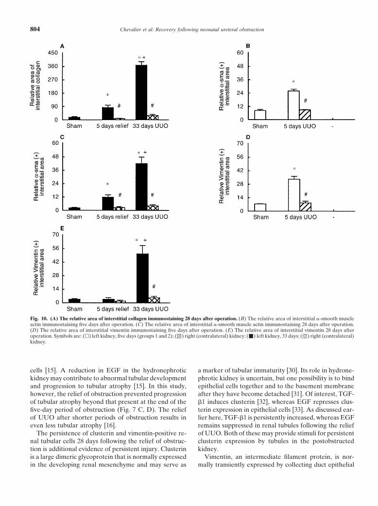
Chevalier et al: Recovery following neonatal ureteral obstruction804
Fig. 10. (A) The relative area of interstitial collagen immunostaining 28 days after operation. (B) The relative area of interstitial a-smooth muscleactin immunostaining five days after operation. (C) The relative area of interstitial a-smooth muscle actin immunostaining 28 days after operation.(D) The relative area of interstitial vimentin immunostaining five days after operation. (E) The relative area of interstitial vimentin 28 days afteroperation. Symbols are: (h) left kidney, five days (groups 1 and 2); ( ) right (contralateral) kidney; ( ) left kidney, 33 days; ( ) right (contralateral)kidney.
cells [15]. A reduction in EGF in the hydronephrotic a marker of tubular immaturity [30]. Its role in hydrone-phrotic kidney is uncertain, but one possibility is to bindkidney may contribute to abnormal tubular development
and progression to tubular atrophy [15]. In this study, epithelial cells together and to the basement membraneafter they have become detached [31]. Of interest, TGF-however, the relief of obstruction prevented progression
of tubular atrophy beyond that present at the end of the b1 induces clusterin [32], whereas EGF represses clus-terin expression in epithelial cells [33]. As discussed ear-five-day period of obstruction (Fig. 7 C, D). The relief
of UUO after shorter periods of obstruction results in lier here, TGF-b1 is persistently increased, whereas EGFremains suppressed in renal tubules following the reliefeven less tubular atrophy [16].
The persistence of clusterin and vimentin-positive re- of UUO. Both of these may provide stimuli for persistentclusterin expression by tubules in the postobstructednal tubular cells 28 days following the relief of obstruc-
tion is additional evidence of persistent injury. Clusterin kidney.Vimentin, an intermediate filament protein, is nor-is a large dimeric glycoprotein that is normally expressed
in the developing renal mesenchyme and may serve as mally transiently expressed by collecting duct epithelial
Chevalier et al: Recovery following neonatal ureteral obstruction 805
cells in fetal kidneys [34]. Whereas postnatal polarized formation of fibroblasts to myofibroblasts (expressinga-smooth muscle actin) may play a significant role in therenal tubular epithelial cells do not normally express
vimentin, UUO induces phenotypic transformation of deposition of extracellular matrix in the progression ofinterstitial fibrosis [39, 40]. It is likely that angiotensintubular cells that acquire mesenchymal characteristics
[15]. It is likely that such transformed cells lack normal plays a role in stimulation of a-smooth muscle actinexpression by myofibroblasts [41, 42].polarity and are more susceptible to apoptosis as a result
of disrupted desmosomal contacts [35]. This process,Clinical correlatestermed anoikis, may represent a significant stimulus for
ongoing apoptosis in the postobstructed kidney [36]. In a retrospective analysis of the renal biopsy obtainedduring pyeloplasty in 55 children (mean age of 4.8 years),
Interstitium histologic changes were compared with the differentialrenal function determined by the preoperative renal scanIn this study, the interstitial deposition of collagen was
attenuated, but not normalized, by the relief of obstruc- [43]. The mean differential function correlated inverselywith the severity of the histologic grade, but in 25% oftion. The progression of interstitial fibrosis following
UUO has been related to the increased expression of cases, there was a disparity between the preoperativedifferential renal function and the biopsy severity scorefibrogenic cytokines in the obstructed kidney. Renal ex-
pression of TGF-b1, a major fibrogenic cytokine, nor- [43]. However, in a prospective study of 17 patients (me-dian age of 15 months), renal biopsy results were corre-mally decreases during the first month of life in the rat
[8]. However, following UUO at birth, the abundance of lated with preoperative and postoperative differentialfunction on scan. The biopsy revealed tubular atrophysteady-state messenger RNA for renal TGF-b1 increases
linearly with age [8]. In this study, tubular TGF-b1 was and interstitial fibrosis in six kidneys, of which five hada preoperative differential function below 33%, andreduced, but not to normal levels, by the relief of UUO.
We have shown previously that TGF-b1 expression by none of these kidneys showed postoperative improve-ment in renal function despite a technically successfulthe neonatal rat kidney subjected to UUO is attenuated
by the inhibition of angiotensin AT1 receptors [37]. The result [44]. The remaining 11 kidneys had normal renalbiopsies and a preoperative differential function of moreresidual TGF-b1 production by the postobstructed kid-
ney may therefore be due to ongoing activation of the than 44% [44]. Although these studies confirm the corre-lation of histologic changes with renal function and out-renin-angiotensin system as evidenced by the extension
of renin along afferent arterioles (Figs. 2D and 3B). come in severe cases of urinary tract obstruction, thecriteria for surgical intervention in less severe cases areBilateral ureteral obstruction in the fetal sheep also sig-
nificantly increases renal TGF-b1 expression, indicating less clear.In summary, the renal morphologic responses werethat the expression of this cytokine may play an impor-
tant role in the fetal kidney as well [10]. examined during recovery following the relief of UUOin the neonatal rat. A five-day period of UUO causedMuch of the progressive interstitial fibrosis resulting
from UUO is related to proliferation of interstitial fi- delayed maturation of the microvasculature, glomeruli,tubules, and interstitium. Nephrogenesis was impairedbroblasts. Whereas tubular cell proliferation was re-
duced by UUO in the neonate, we found a tendency for by temporary UUO, and there was no catch up of addi-tional nephron formation following the relief of obstruc-interstitial cell proliferation to increase following 5 and
33 days of UUO (Fig. 5 C, D). Interstitial cell apoptosis tion. The lack of reduced glomerular filtration rate of thepostobstructed kidney indicates that remaining nephronswas significantly increased after either 5 or 33 days of
UUO and was attenuated (but not normalized) by the are hyperfiltering. Recovery from tubular injury was in-complete: The balance of tubular cell dynamics wasrelief of obstruction (Fig. 6 C, D). It is therefore likely
that interstitial cell turnover is increased as a result of shifted toward reduced proliferation and increased apo-ptosis, and tubular EGF expression remained depressed,UUO. Whereas during normal renal maturation, intersti-
tial fibroblast a-smooth muscle actin, and vimentin ex- whereas tubular TGF-b1, clusterin, and vimentin re-mained increased. Tubular atrophy was increased at thepression disappear by two weeks of age in the rat [38],
we found that expression of these filament proteins was end of five days of UUO, but there was no additionaltubular atrophy 28 days following the relief of ob-increased after five days of UUO and persisted to 33 days
(Figs. 9 and 10). These results indicate that maturation of struction. Although renal interstitial fibrosis was attenu-ated by relief of obstruction, interstitial fibroblasts nointerstitial cells, as with vascular, glomerular, and tubular
cells, is delayed by UUO. The relief of obstruction sig- longer expressed vimentin, but they continued to expressa-smooth muscle actin. These findings indicate that thenificantly reduced interstitial a-smooth muscle actin dis-
tribution and normalized interstitial vimentin, which delicate interplay of growth factors and cytokines thatcharacterize normal renal development is disrupted bycorresponds closely with the reduction in interstitial
collagen distribution (Fig. 10 A, C). Phenotypic trans- UUO and is improved, but not normalized, by the relief
Chevalier et al: Recovery following neonatal ureteral obstruction806
15. Chevalier RL, Goyal S, Wolstenholme JT, Thornhill BA: Ob-of obstruction. The clinical implications of the study sug-structive nephropathy in the neonate is attenuated by epidermal
gest that although early pyeloplasty in infants with ure- growth factor. Kidney Int 54:38–47, 1998teropelvic junction obstruction may attenuate long-term 16. Chevalier RL, Thornhill BA, Wolstenholme JT, Kim A: Unilat-
eral ureteral obstruction in early development alters renal growth:progression of renal injury, catch up of renal maturationDependence on the duration of obstruction. J Urol 161:309–313,lost during the period of obstruction is not likely. To 1999
reduce the incidence of renal insufficiency in infants and 17. Niimura F, Labosky PA, Kakuchi J, Okubo S, Yoshida H, OikawaT, Ichiki T, Naftilan AJ, Fogo A, Inagami T, Hogan BLM,children, new strategies for the prevention and treatmentIchikawa I: Gene targeting in mice reveals a requirement forof congenital obstructive nephropathy will be necessary. angiotensin in the development and maintenance of kidney mor-
These may include targeted delivery of growth factors phology and growth factor regulation. J Clin Invest 96:2947–2954,1995to the developing kidney [15].
18. McVary KT, Maizels M: Urinary obstruction reduces glomerulo-genesis in the developing kidney: A model in the rabbit. J Urol
ACKNOWLEDGMENTS 142:646–651, 198919. Kaneto H, Morrissey J, McCracken R, Reyes A, Klahr S: Enala-This research was supported in part by the National Institutes of pril reduces collagen type IV synthesis and expansion of the inter-Health Research Center of Excellence in Pediatric Nephrology and
stitium in the obstructed rat kidney. Kidney Int 45:1637–1647, 1994Urology, DK44756 and DK52612; the National Institutes of Health20. Fern R, Yesko CM, Thornhill BA, Kim HS, Smithies O, Cheva-O’Brien Center of Excellence in Nephrology and Urology, DK45179;
lier RL: Interstitial fibrosis in chronic hydronephrosis is modulatedand the National Institutes of Health Child Health Research Center,by angiotensinogen expression. (abstract) Pediatr Res 43:306A,HD28810.1998
21. Josephson S: Experimental obstructive hydronephrosis in newbornReprint requests to Robert L. Chevalier, M.D., Department of Pediat-rats. III. Long-term effects on renal function. J Urol 129:396–400,rics Box 386, University of Virginia, Health Sciences Center, Charlottes-1983ville, Virginia 22908, USA.
22. Peters CA, Carr MC, Lais A, Retik AB, Mandell J: The re-E-mail: [email protected] of the fetal kidney to obstruction. J Urol 148:503–509, 1992
23. Peters CA, Gaertner RC, Carr MC, Mandell J: Fetal compensa-REFERENCES tory renal growth due to unilateral ureteral obstruction. J Urol
150:597–600, 19931. Fivush BA, Jabs K, Neu AM, Sullivan EK, Feld L, Kohaut E, 24. Aschinberg LC, Koskimies O, Bernstein J, Nash M, Edelmann
Fine R: Chronic renal insufficiency in children and adolescents: CM, Spitzer A: The influence of age on the response to renalThe 1996 annual report of NAPRTCS. Pediatr Nephrol 12:328–337, parenchymal loss. Yale J Biol Med 51:341–345, 19781998 25. Okuda S, Motomura K, Sanai T, Tsuruda H, Oh Y, Onoyama2. Roth DR, Gonzales ET Jr: Management of ureteropelvic junc-
K, Fujishima M: Influence of age on deterioration of the remnanttion obstruction in infants. J Urol 129:108–110, 1983kidney in uninephrectomized rats. Clin Sci 72:571–576, 19873. King LR, Coughlin PWF, Bloch EC, Bowie JD, Ansong K,
26. O’Donnell MP, Kasiske BL, Raij L, Keane WF: Age is a determi-Hanna MK: The case for immediate pyeloplasty in the neonatenant of the glomerular morphologic and functional responses towith ureteropelvic junction obstruction. J Urol 132:725–728, 1984chronic nephron loss. J Lab Clin Med 106:308–313, 19854. Dowling KJ, Harmon EP, Ortenberg J, Polanco E, Evans BB:
27. Frokiaer J, Marples D, Knepper MA, Nielsen S: Bilateral ure-Ureteropelvic junction obstruction: The effect of pyeloplasty onteral obstruction downregulates expression of vasopressin-sensi-renal function. J Urol 140:1227–1230, 1988tive AQP-2 water channel in rat kidney. Am J Physiol 270:F657–5. Koff SA, Campbell K: Nonoperative management of unilateralF668, 1996neonatal hydronephrosis. J Urol 148:525–531, 1992
28. Hwang SJ, Haas M, Harris HW Jr, Silva P, Yalla S, Sullivan6. MacNeily AE, Maizels M, Kaplan WE, Firlit CF, ConwayMR, Otuechere G, Kashgarian M, Zeidel ML: Transport defectsJJ: Does early pyeloplasty really avert loss of renal function? Aof rabbit medullary thick ascending limb cells in obstructive ne-retrospective review. J Urol 150:769–773, 1993phropathy. J Clin Invest 91:21–28, 19937. Salem YH, Majd M, Rushton HG, Belman AB: Outcome analysis
29. Chandar J, Abitbol C, Zilleruelo G, Gosalbez R, Montaneof pediatric pyeloplasty as a function of patient age, presentationB, Strauss J: Renal tubular abnormalities in infants with hydrone-and differential renal function. J Urol 154:1889–1893, 1995phrosis. J Urol 155:660–663, 19968. Chung KH, Chevalier RL: Arrested development of the neonatal
30. French LE, Chonn A, Ducrest D, Baumann B, Belin D, Wohl-kidney following chronic ureteral obstruction. J Urol 155:1139–wend A, Kiss JZ, Sappino A-P, Tschopp J, Schifferli JA: Murine1144, 1996clusterin: Molecular cloning and mRNA localization of a gene9. Chevalier RL, Chung KH, Smith CD, Ficenec M, Gomez RA:associated with epithelial differentiation processes during em-Renal apoptosis and clusterin following ureteral obstruction: Thebryogenesis. J Cell Biol 122:1119–1130, 1993role of maturation. J Urol 156:1474–1479, 1996
31. Silkensen JR, Skubitz KM, Skubitz APN, Chmielewski DH,10. Medjebeur AA, Bussieres L, Gasser B, Gimonet V, LabordeManivel JC, Dvergsten JA, Rosenberg ME: Clusterin promotesK: Experimental bilateral urinary obstruction in fetal sheep: Trans-the aggregation and adhesion of renal porcine epithelial cells. Jforming growth factor-beta1 expression. Am J Physiol 273:F372–Clin Invest 96:2646–2653, 1995F379, 1997
32. Reddy KB, Jin G, Karode MC, Harmony JAK, Howe PH: Trans-11. Steinhardt GF, Salinas-Madrigal L, Demello D, Farber R,forming growth factor beta (TGFbeta)-induced localization ofPhillips B, Vogler G: Experimental ureteral obstruction in theapolipoprotein J/clusterin in epithelial cells. Biochemistry 35:6157–fetal opossum: Histologic assessment. J Urol 152:2133–2138, 19946163, 199612. Chevalier RL, Gomez RA, Jones CA: Developmental determi-
33. Gutacker C, Flach R, Diel P, Klock G, Koch-Brandt C: Multi-nants of recovery after relief of partial ureteral obstruction. Kidneyple signal transduction path ways regulate clusterin (gp 80) geneInt 33:775–781, 1988expression in MDCK cells. J Mol Endocrinol 17:109–119, 199613. El-Dahr SS, Gomez RA, Gray MS, Peach MJ, Carey RM, Cheva-
34. Holthofer H, Miettinen A, Lehto V-P, Lehtonen E, Virtanenlier RL: In situ localization of renin and its mRNA in neonatalI: Expression of vimentin and cytokeratin types of intermediateureteral obstruction. Am J Physiol 258:F854–F862, 1990filament proteins in developing and adult human kidneys. Lab14. Chevalier RL, Sturgill BC, Jones CE, Kaiser DL: MorphologicInvest 50:552–559, 1984correlates of renal growth arrest in neonatal partial ureteral ob-
struction. Pediatr Res 21:338–346, 1987 35. Ben-Ze’ev A: Differential control of cytokeratins and vimentin
Chevalier et al: Recovery following neonatal ureteral obstruction 807
synthesis by cell-cell contact and cell spreading in cultured epithe- Muller GA: Transformation of rat inner medullary fibroblasts tomyofibroblasts in vitro. Kidney Int 52:1279–1290, 1998lial cells. J Cell Biol 99:1424–1433, 1984
41. Johnson MC, Aguilera G: Angiotensin-II receptor subtypes and36. Frisch SM, Francis H: Disruption of epithelial cell-matrix interac-coupling to signaling systems in cultured fetal fibroblasts. Endocri-tions induces apoptosis. J Cell Biol 124:619–626, 1994nology 129:1266–1274, 199137. Chung KH, Gomez RA, Chevalier RL: Regulation of renal
42. Ruiz-Ortega M, Egido J: Angiotensin II modulates cell growth-growth factors and clusterin by angiotensin AT1 receptors duringrelated events and synthesis of matrix proteins in renal interstitialneonatal ureteral obstruction. Am J Physiol 268:F1117–F1123, 1995fibroblasts. Kidney Int 52:1497–1510, 199738. Marxer-Meier A, Hegyi I, Loffing J, Kaissling B: Postnatal 43. Elder JS, Stansbrey R, Dahms BB, Selzman AA: Renal histologi-
maturation of renal cortical peritubular fibroblasts in the rat. Anat cal changes secondary to ureteropelvic junction obstruction. J UrolEmbryol (Berlin) 197:143–153, 1998 154:719–722, 1995
39. Alpers CE, Pichler R, Johnson RJ: Phenotypic features of corti- 44. Stock JA, Krous HF, Heffernan J, Packer M, Kaplan GW:cal interstitial cells potentially important in fibrosis. Kidney Int Correlation of renal biopsy and radionuclide renal scan differential49(Suppl):S28–S31, 1996 function in patients with unilateral ureteropelvic junction obstruc-
tion. J Urol 154:716–718, 199540. Grupp C, Lottermoser J, Cohen DI, Begher M, Franz H-E,
Copyright © 2022 FDOKUMEN