FUNDICIÓ N - Escuela Colombiana de Ingeniería Julio Garavito
Upload
khangminh22Category
view
2download
0
RC
G R
evista Colom
biana de Gastroenterología Vol. 34 N
o. 3 July-September 2019 Pags. 231-317
Volume 34 No. 3July-September
2019
Revista Colombiana deRevista Colombiana deGastroenterologíaGastroenterología
· www.gastrocol.com · www.scielo.org.co ·· www.revistagastrocol.com ·
ISSN 0120-9957ISSN 2500-7440 (Online) DOI: https://doi.org/10.22516/issn.2500-7440
Original articles• The role of endoscopic ultrasound in evaluating
patients with dyspepsia in a Colombian population• Effectiveness of vitamins C and E adjuvant to
standard triple therapy for Helicobacter pylori in a cohort from the Peruvian Amazon
• Prevalence of gastroesophageal reflux disease found by pH measurements in preterm infants with suggestive symptoms
• Characterization of patients with chronic hepatitis C treated in a high complexity hospital in Medellín
• Clinical effectiveness of two esomeprazole presentations in a pilot trial
Review articles• The role of antispasmodics in managing irritable
bowel síndrome
• Pathophysiology of Hepatitis C and Diabetes Mellitus: Towards the cure of two epidemics in the 21st century
Case report• A case report of eosinophilic esophagitis• A case of heterotopic pancreas in a gastric
polyp• Case report and literature review of Budd-
Chiari syndrome during the puerperium• Ancylostomiasis: a rare cause of
gastrointestinal bleeding and severe anemia• Transcatheter venous coil embolization of
gastric varices• An endoscopic videocapsule finding of
heterotopia of the gastric mucosa of the small intestine

Contents
Revista Colombiana deGastroenterología
Volume 34 No. 3July-September2019
Original articles
The role of endoscopic ultrasound in evaluating patients with dyspepsia in a Colombian population ...............Martín A. Gómez Zuleta, William Otero Regino, Óscar Ruíz Morales.
231
Effectiveness of vitamins C and E adjuvant to standard triple therapy for Helicobacter pylori in a cohort from the Peruvian Amazon ..................................................................................................................................................Wildor Samir Cubas, Rómulo Reyes Cahuila, Heriberto Arévalo Ramírez, Antonio M. Quispe.
236
Prevalence of gastroesophageal reflux disease found by pH measurements in preterm infants with suggestive symptoms ................................................................................................................................................................................Carlos Alberto Velasco Benítez, Ernesto León Vallejo Mondragón, Mauricio Alberto Arévalo Sanabria.
242
Characterization of patients with chronic hepatitis C treated in a high complexity hospital in Medellín ...........Mónica Ledezma-Morales, Juan Carlos Restrepo G., Pedro Amariles, María Camila Trillos A., Rubén Darío Vargas R.
247
Clinical effectiveness of two esomeprazole presentations in a pilot trial ....................................................................Carlos Arturo Rojas, Mario Sepúlveda Copete, Jairo Alberto García Abadía, Héctor Raúl Echavarría Abad, Fernando Rosso Suárez, Andrés Fernando Jiménez, Andrés Mauricio Castro Llanos.
259
Review articles
The role of antispasmodics in managing irritable bowel síndrome ..............................................................................Valeria Atenea Costa Barney, Alan Felipe Ovalle Hernández.
267
Pathophysiology of Hepatitis C and Diabetes Mellitus: Towards the cure of two epidemics in the 21st century ...Harold Muñoz Díaz, Adán Lúquez Mindiola, Andrés Gómez Aldana.
274

Case report
A case report of eosinophilic esophagitis ..........................................................................................................................Pedro Rosales Torres, Rafael Pila Pérez, Rafael Pila Peláez, Pedro León Acosta, Yudenia Toledo Cabarcos.
284
A case of heterotopic pancreas in a gastric polyp ............................................................................................................Pedro Rosales Torres, Rafael Pila Pérez, Pedro León Acosta, Rafael Pila Peláez.
289
Case report and literature review of Budd-Chiari syndrome during the puerperium .............................................Gabriel Sebastián Díaz, Carlos Alberto Salgado, Adolfo Zuluaga, Estefanía Orozco, Juan Ignacio Marín.
293
Ancylostomiasis: a rare cause of gastrointestinal bleeding and severe anemia ..........................................................Edwar Jassir Rozo O., Ledmar Jovanny Vargas R., Jessica Paola Jiménez W., Erika Tatiana Szemmelveisz N.
298
Transcatheter venous coil embolization of gastric varices .............................................................................................Oscar Rivero Rapalino, Camilo Barragán Leal, Diego Salcedo Miranda, Laura Quintero Rojas.
302
An endoscopic videocapsule finding of heterotopia of the gastric mucosa of the small intestine .........................Santiago Castaño Quintero, Natalia Calvache, Mauricio Sepúlveda, Catalina Maldonado, Pedro Tomás Argüello, Juliana Escobar, Carlos Arturo Rojas.
308
Letter to the Editor
Letter to the editor .................................................................................................................................................................Valeria Atenea Costa Barney, Alexander Castañeda Ladino.
313
Response to the letter to the editor ....................................................................................................................................Gabriel Mosquera-Klinger, Kenny Gálvez Cárdenas, Alejandro Ocampo Hincapié.
316
Cover: Endoscopic videocapsule. Concentric ulcerated stenosis, surrounded by mucosal edema.Courtesy by the authors: Santiago Castaño Quintero, Natalia Calvache, Mauricio Sepúlveda, Catalina Maldonado, Pedro Tomás Argüello, Juliana Escobar, Carlos Arturo Rojas.Article: An endoscopic videocapsule finding of heterotopia of the gastric mucosa of the small intestine

© 2019 Asociaciones Colombianas de Gastroenterología, Endoscopia digestiva, Coloproctología y Hepatología 231
Martín A. Gómez Zuleta,1* William Otero Regino,2 Óscar Ruíz Morales.3
The role of endoscopic ultrasound in evaluating patients with dyspepsia in a Colombian population
1 Specialist in gastroenterology and internal medicine at the National University Hospital of Colombia, Hospital Occidente de Kennedy and the Gastroenterology and Endoscopy Unit in Bogotá D.C., Colombia
2 Professor of medicine and coordinator of gastroenterology at the National University of Colombia, Gastroenterologist at the National University Hospital of Colombia and Clínica Fundadores in Bogotá, D.C., Colombia
3 Specialist in gastroenterology and internal medicine at Universidad San Martín, Kennedy West Hospital and the National University Hospital of Colombia in Bogotá D.C., Colombia
*Correspondence: [email protected].
.........................................Received: 25/03/18 Accepted: 29/08/18
AbstractDyspepsia is defined as upper abdominal pain or discomfort that is considered to originate in the upper gastrointestinal tract. Many diseases and clinical conditions can cause dyspepsia. Among others, they include peptic ulcers, gastric and esophageal cancer, medications, biliary lithiasis, pancreatitis, and pancreatic cancer. Traditionally, dyspepsia is only evaluated with digestive endoscopy whose diagnostic yield is only 27%. On the other hand, endoscopic ultrasound combines an endoscopic image and an ultrasound image thereby po-tentially broadening diagnostic range to detect more of the causes of dyspepsia allowing treatment of patients in a timelier manner. Objective: To evaluate whether endoscopic ultrasound increases the diagnostic yield of endoscopy (27% in our environment) in the initial approach to previously unstudied dyspepsia. Materials and methods: This is a prospective study of analytical prevalence in adult patients with previously unstudied dyspepsia who were examined at a university institution in Colombia. The patients included were seen in the gastroenterology unit from January to October 2016 and underwent upper digestive endoscopy and endos-copic ultrasound. Under anesthesiologist-guided sedation, the stomach and duodenal esophagus were first evaluated endoscopically. Then retrograde endoscopic ultrasound was used to evaluate the pancreas in its entirety, the extra hepatic bile duct, the gallbladder, the celiac trunk, the left lobe of the liver and the medias-tinal region. All abnormalities were noted on the patient’s admission form. Results: In total we included 60 patients of whom 65% were female and whose average age of was 40.8 years (SD: 12.5). The findings in the endoscopic phase of the endoscopic ultrasound were mainly chronic Gastritis 43 patients (71.6%), the rest had a structural lesion (17 patients): esophagitis 5 (8.3%), gastric ulcer 2 (3.3%), duodenal ulcer 5 (8.3%), gastric cancer, 4 (6.6%), gastric subepithelial lesion (GIST) 1 (1.6%). In the endoscopy phase, we found 11 cases of cholelithiasis (18.3%), one case of choledocholithiasis (1.6%), and five cases of chronic pancreatitis (8.3%). Only 17 patients of these patients (28.3%) had a structural finding in the endoscopy phase, but 18 additional patients (30%) had some positive finding in the ultrasound phase. In other words, the diagnostic yield rose to 58.3% (p < 0.001). Conclusion: Although this study’s sample size is small, it suggests that using endoscopic ultrasound in the initial evaluation of dyspepsia could be useful since it increased diagnostic yield in this group of patients from 28.3 to 58.3%. This is very significant because patients with dyspepsia and negative endoscopy are usually classified as functional and only treated with medications. However, in recognition of the methodological limitations of this study, it should be considered an initial exploration. Larger, controlled studies should be considered to confirm this work. Another factor that should be considered is the cost of endoscopic ultrasound which is much higher than the upper digestive endoscopy.
KeywordsEndoscopic ultrasound, evaluation, dyspepsia, gastric cáncer.
Original articlesDOI: https://doi.org/10.22516/25007440.449

Rev Colomb Gastroenterol / 34 (3) 2019232 Original articles
INTRODUCTION
Dyspepsia is a clinical syndrome characterized by pain or discomfort in the upper abdomen. It affects at least 20% of the world’s population. (1, 2) It has multiple causes inclu-ding benign and malignant pathologies, negatively affects the quality of life, (8) and is often incorrectly called chro-nic gastritis or acid peptic disease. (3, 4) In some countries the annual incidence of dyspepsia has been found to be approximately 1%, and has been estimated that one in two people will consult a physician for dyspeptic symptoms at some time in their lives. Dyspepsia accounts for 5% of the people seen by general practitioners and for approximately 20% to 30% of those seen by gastroenterologists. (5-7) When a patient has dyspepsia whose cause has not been determined, it is called uninvestigated dyspepsia (UD). (8) The approach to adult patients includes upper gastrointesti-nal endoscopy and occasionally includes upper abdominal ultrasound. (1, 3) Dyspepsia can be secondary or organic if there are obvious alterations, it is called functional dyspep-sia (FD) if examinations and images show no alterations that explain symptoms. (2, 3)
Early use of endoscopy is cost-effective, (9, 10) but the timing of upper endoscopy varies from place to place. In developed countries, it is recommended after age 55, but in developing countries with high incidences of gastric can-cer, it is recommended at age 35. (3) In Colombia, no stu-dies of cost-effectiveness have been carried out for UD, but studies of the prevalence of endoscopic lesions in patients with UD have been performed. More than 10 years ago, a prospective study of patients with UD found that 73% had FD. (11). Another Colombian study of patients with UD found cholelithiasis in 21%. (12)
Overall, the diagnostic yield of upper endoscopy alone for identification of organic causes does not exceed 30% , (1, 11) so other methods that have greater sensitivity need to be used. As understanding of these patients deepens, unknown causes may be found. In this regard, dyspepsia secondary to helicobacter pylori, a new entity that was previously included in the FD category, has recently been described. (13) It has now been shown that approximately 5% of patients with H. pylori can be cured of dyspepsia if the infection is eradicated. (13, 14) This new finding demonstrates the need to continue investigating unknown etiologies that could explain dyspeptic symptoms. When upper endoscopy is negative in the traditional diagnostic approach, the diagnosis is FD. If no further studies are per-formed, the patient is treated with proton pump inhibitors (PPIs), prokinetic agents or antidepressants for several months. (8) This can mask unidentified pathologies that may lead to probable complications.
Endoscopic ultrasound (EUS) combines the image from conventional upper endoscopy with an ultrasound image. When radial equipment is used, the ultrasound image has a range of about 6 cm which can cover the entire esopha-gus, stomach or duodenum. (15) Since Pentax 360-Degree Radial-Array Ultrasound Gastroscopes are frontal, unlike Olympus Radial Ultrasound Endoscopes which are obli-que, endoscopic views identical to those of conventional endoscopy can be obtained prior to the ultrasound phase. Visibility beyond the upper gastrointestinal lumen allows diagnoses of additional pathologies that may be associated with dyspepsia. Taking into account the high frequency of dyspepsia, as well as the lack of research other than the upper endoscopy in our environment, we decided to per-form this study of EUS for diagnosis of patients with UD. The objective is to evaluate whether EUS increases the diagnostic yield from the 27% yield of conventional endos-copy here in Colombia.
MATERIALS AND METHODS
This is a prospective study of the prevalence of UD found in adult patients at a university institution in Colombia. The patients included came to the gastroenterology unit from January to October 2016 where they underwent upper endoscopy. After patients had agreed and signed consent forms, the Pentax echoendoscope was used instead of a conventional endoscope. On the basis of the ROME IV criteria, we defined dyspepsia as the presence of upper GI tract symptoms of postprandial fullness, early satiety and epigastric pain or burning. If no structural lesion is found endoscopically, it is defined as functional dyspep-sia (FD). (8) Endoscopy indications used were those of the dyspepsia guide of the Colombian Association of Gastroenterology which recommends performing upper endoscopy in all patients with UD who are over 35 years old regardless of whether or not they have alarm symptoms. (16) We excluded patients who had any contraindication for performance of endoscopy with sedation, patients who had previously undergone upper endoscopy, patients with histories of gastrointestinal surgery including cholecystec-tomies, and patients who had had endoscopic retrograde cholangiopancreatography (ERCP), and patients who had had abdominal ultrasound images, magnetic resonance imaging (MRI) or computed tomography (CT). EUS was performed in the usual way. (17)
Procedures were performed under sedation administered by an anesthesiologist. A Pentax echoendoscope, described above, was used. First, the endoscope was used to examine the esophagus, stomach and duodenum according to the protocol described by Yao. (18) This consists of the eva-luation of 21 areas in the proximal, middle and distal third

233Papel de la ecoendoscopia en la evaluación de la dispepsia no investigada en una población colombiana
nic pancreatitis defined according to by Rosemont’s crite-ria in five (8.3%), a pancreas cyst suggestive of mucinous cystadenoma in one (1.6%), stage T2 pancreatic cancer in one (1.6%) and mediastinal adenopathies in one patient who also had gastric cancer (1.6%). (19) Overall, 17/60 patients (28.3%) had structural alterations found in the endoscopy phase and 18/60 (30%) had some positive finding that had not been found in the endoscopic phase. In other words, these cases would have been technically considered to have FD. Only 25 patients (41.6%) had no findings in both the endoscopy phase and the EUS phase. In all cases, the gallbladder, bile duct, head, body and tail of the pancreas were properly visualized. Overall findings are shown in Figure 1.
DISCUSSION
This study found that 28.3% of the patients with UD had alterations found during the endoscopic phase which is similar to the 27% found in a previous investigation by the National University and which corroborates the poor per-formance of upper endoscopy in our environment. (11) In the ultrasound phase using EUS equipment with fron-tal vision, additional lesions that could explain dyspeptic symptoms were detected in 30% of the patients for a total of 58.3% (p <0.001). In other words, only 25 (41.6%) of the 60 patients had normal results.
of the stomach using both direct view and rearview. Two biopsies each were taken from the antrum and the corpus to check for H. pylori infections. Additional biopsies were taken when patients had apparent structural lesions. After the ultrasound phase, EUS was used to the major papilla. A retrograde evaluated the entire pancreas, the extrahepatic bile duct, the gallbladder, the celiac trunk, the left lobe of the liver and the mediastinal region. All abnormalities were noted on the patient’s admission form. The protocol, inves-tigation and informed consent form were approved by the ethics committee of the participating institution.
RESULTS
In total, sixty patients with an average age of 40.8 years + 12.5 years were included. Sixty-five percent of them were women. In the first phase, forty-three of the patients (71.6%) were endoscopically diagnosed with chronic gastritis while 17 had structural lesions including five cases of esophagitis in 5 (8.3%), two cases of gastric ulcers (3.3%), five cases of duo-denal ulcers (8.3%), four cases of histologically proven gas-tric cancer (6.6%), and one case of a gastrointestinal stromal tumor (GIST) (1.6%). H. pylori was found in 42 patients (70%), atrophic gastritis was found in 8 patients (13%), and intestinal metaplasia was found in six (10%).
In the second phase, EUS found cholelithiasis in 11 patients (18.3%), choledocolithiasis in one (1.6%), chro-
Figure 1. Overall results of the study.
Patients included: 60Age: 40.8 years, 65% women
Alterations in the endoscopic phase Alterations in the sonographic phase
Chronic gastritis: 71,6 %H. pylori positive: 70 %
Atrophic gastritis: 8 (13,3 %)Intestinal metaplasia: 6 (10%)
Cholelithiasis: 11 (18,3 %)Choledocholithiasis: 1 (1,6 %)Chronic pancreatitis: 5 (8,3 %)
Pancreatic cysts: 1(1,6 %)Pancreatic cancer: 1 (1,6 %)
Mediastinal adenopathy: 1 (1,6 %)
Structural lesionsEsophagitis: 5 (8,3 %)
Gastric ulcers: 2 (3,3 %)Duodenal ulcers: 5 (8,3 %)Gastric cancer: 4 (6,6 %)
GIST: 1 (1,6 %)

Rev Colomb Gastroenterol / 34 (3) 2019234 Original articles
tion of ERCP in Colombia more than 50 years ago,. It was considered to be a very complex procedure that could only be performed by a very few gastroenterologists in very few medical centers. Currently, ERCPs are routine proce-dures performed by many gastroenterologists the world over, and ERCP is a much more complex and dangerous procedure than is EUS. (24) The real complexity of EUS lies in therapeutic and invasive procedures such as staging of pancreatic cancer, punctures and drainage. It is time to demystify non-therapeutic EUS and understand reality. It is not difficult to imagine that EUS could be used for ini-tial evaluation of UD in the near future. This is a complex pathology that, curiously, many consider to be an insignifi-cant or easy-to-solve problem when it is called gastritis or acid peptic disease. In fact, addressing it is complex due to its recurrent nature. In addition, it causes deterioration of patients’ quality of life, has high costs for the world’s health care systems, and can have serious unsuspected etiologies, such as those found in this study.
To our knowledge, this is the first study of this type on the initial evaluations of patients with dyspepsia using EUS with frontal equipment done in Latin America. A study with a similar design done more than 15 years ago used an Olympus echoendoscope which only provides oblique views. (25) That study did not detect as many endolumi-nal lesions as did endoscopy. Unlike the Fuji and Pentax equipment, Olympus echoendoscopes do not have frontal vision, which makes it impossible to use for conventional endoscopy. (15) Because it limits the endoscopic phase, many areas cannot be evaluated which can leave lesions such as ulcers and tumors undiscovered.
It is also important to keep in mind that defensive medi-cine is frequently exercised. (26) Many patients press their doctors to reach a rapid diagnosis because they are anxious about their conditions and do not accept that their pain is only explained by gastritis. When the doctor is influenced, multiple tests that may even be more expensive than an EUS can be the result.
The limitations of this study include its sample size (not calculated) and the fact that it was performed at only one center. It would be convenient to conduct studies with lar-ger samples to confirm the findings of this study.
CONCLUSION
This study was found that the diagnostic yield of EUS exceeds that of upper endoscopy by 30% in patients with UD. EUS identified pathologies in 58.3% of the patients whereas upper endoscopy alone could only identify them in 28.3% of the patients ( p <0.001). When upper endos-copy is negative for a patient with UD, current recom-mendations call for a diagnosis of FD and indefinite pres-
The finding of cholelithiasis in 18% of patients coincides with the prevalence of this pathology in the west. (20, 21) However, it cannot be concluded that gallbladder stones cause dyspeptic symptoms since the symptom most fre-quently associated with them is colic. To determine the association of the two, a study with a greater number of patients, probably a case and control study, would be requi-red. However, all gastroenterologists know that patients with cholelithiasis do not necessarily manifest the typical colic in the right hypochondrium but may manifest pos-tprandial symptoms in the epigastrium which are very suggestive of dyspepsia. If the finding of cholelithiasis is eli-minated, the diagnostic yield of EUS exceeds that of upper endoscopy by 12%. Given the types of pancreatic patholo-gies found, it can be inferred that when only upper endos-copy is taken into account for UD patients, negative fin-dings can lead to the erroneous conclusion that the patient has FD which should be managed with prolonged adminis-tration of prokinetics, PPIs or antidepressants. (22) These medications would not achieve significant improvement but could generate anxiety in patients. For the safety of patients and doctors, in many cases research into the cause of dyspepsia should continue with additional tests such as abdominal ultrasound, CT scans and other studies. (8, 22)
A new approach for patients with FD and persistent symptoms is posed by the findings that more than 11% of UD patients have pancreatic pathologies while others suffer from choledocholithiasis. For these patients, the next step should be biliopancreatic EUS rather than another upper endoscopy. EUS is even preferable to conventional ultra-sound, CT scans and abdominal MRI. A study conducted more than 15 years ago suggested that the EUS could also be more cost-effective. (23) A more recent study from the United States by Chang et al. showed that, in the presence of symptoms, using EUS as the first exam is more cost-effective than upper endoscopy followed by abdominal ultrasound. (24) Compared to abdominal CT scans and MRI, the advantages of EUS are even more evident. Taking into account our results and those of international studies, we consider that it is necessary and timely to determine the place of biliopancreatic EUS in patients with UD in our environment. We consider that it would be premature to recommend this test for all patients with UD given the high cost of EUS in our setting. Nevertheless, the preva-lence of biliopancreatic pathologies demonstrates that it should become the test of choice for FD patients who do not improve with conventional treatments.
A very important issue is that complexity of EUS has been exaggerated here in Colombia. Performing gastroe-sophageal or pancreatic EUS is relatively simple, does not take more than 5 or 10 minutes, and training is not very complex. This recalls what happened with the introduc-

235Papel de la ecoendoscopia en la evaluación de la dispepsia no investigada en una población colombiana
13. Sugano K, Tack J, Kuipers EJ. Kyoto global consensus report on Helicobacter pylori gastritis. Gut. 2015;64:1353-67. doi: 10.1136/gutjnl-2015-309252.
14. Malfertheiner P, Megraud F, O’Morain CA, Gisbert JP, Kuipers EJ, Axon AT, et al. Management of Helicobacter pylori infection: The Maastricht V/Florence Consensus Report. Gut. 2017;66:6-30. doi: 10.1136/gutjnl-2016-312288.
15. Bamber J, Cosgrove D, Dietrich CF, Fromageau J, Bojunga J, Calliada F, et al. EFSUMB guidelines and recommen-dations on the clinical use of ultrasound elastography. Part 1: Basic principles and technology. Ultraschall Med. 2013;34(2):169-84. doi: 10.1055/s-0033-1335205.
16. Pineda LF, Rosas MC, Amaya M, Rodríguez A, Luque A, Agudelo F, et al. Guía de Práctica Clínica para el diagnós-tico y tratamiento de la dispepsia en adultos. Rev Colomb Gastroenterol. 2015;30(Suppl 1):9-16.
17. Godfrey EM, Rushbrook SM, Carroll NR. Endoscopic ultrasound: a review of current diagnostic and therapeu-tic applications. Postgrad Med J. 2010;16(4):111-22. doi: 10.1136/pgmj.2009.096065.
18. Yao K. Endoscopic diagnosis of early gastric cancer. Ann Gastroenterol. 2013;26(1):11-22.
19. Catalano MF, Sahai A, Levy M, Romagnuolo J, Wiersema M, Brugge W, et al. EUS-based criteria for the diagno-sis of chronic pancreatitis: the Rosemont classification. Gastrointest Endosc. 2009;69(7):1251-61. doi: 10.1016/j.gie.2008.07.043.
20. Russo MW, Wei JT, Thiny MT, Gangarosa LM, Brown A, Ringel Y, et al. Digestive and liver diseases statistics, 2004. Gastroenterology. 2004;126(5):1448-53. doi: 10.1053/j.gastro.2004.01.025.
21. Sakorafas GH, Milingos D, Peros G. Asymptomatic chole-lithiasis: is cholecystectomy really needed? A critical reap-praisal 15 year after the introduction of laparoscopic chole-cystectomy. Dig Dis Sci. 2007,52:1313-25. doi: 10.1007/s10620-006-9107-3.
22. Enck P, Azpiroz F, Boeckxstaens G, Elsenbruch S, Feinle-Bisset C, Holtmann G, et al. Functional dyspepsia. Nat Rev Dis Primers. 2017;3:17081. doi: 10.1038/nrdp.2017.81.
23. Sahai AV, Penman ID, Mishra G, Williams D, Pearson A, Wallace MB, et al. An assessment of the potential value of endoscopic ultrasound as a cost-minimizing tool in dys-peptic patients with persistent symptoms. Endoscopy. 2001;33(8):662-7. doi: 10.1055/s-2001-16223.
24. Chang KJ, Erickson RA, Chak A, Lightdale C, Chen YK, Binmoeller KF, et al. EUS compared with endoscopy plus transabdominal US in the initial diagnostic evaluation of patients with upper abdominal pain. Gastrointest Endosc. 2010;72(5):967-74. doi: 10.1016/j.gie.2010.04.007.
25. Lee YT, Lai AC, Hui Y, Wu JC, Leung VK, Chan FK, et al. EUS in the management of uninvestigated dyspepsia. Gastrointest Endosc. 2002;56(6):842-8.
26. Studdert DM, Mello MM, Sage WM. Defensive medicine among high-risk specialist physicians in a volatile mal-practice environment. JAMA. 2005;293:2609-261. doi: 10.1001/jama.293.21.2609.
cription of medications whose efficacy is moderate. Our findings indicate the potential for using EUS as the initial evaluation examination as well as the potential for using it as a follow-up examination instead of a CT scan or MRI when symptoms persist. Studies of the cost-effectiveness of EUS for these purposes in our environment should be undertaken to determine whether this test should become the first choice instead of conventional endoscopy.
Conflicts of Interest
None.
REFERENCES
1. Graham DY, Rugge M. Clinical practice: diagnosis and eva-luation of dyspepsia. J Clin Gastroenterol. 2010;44(3):167-72. doi: 10.1097/MCG.0b013e3181c64c69.
2. Tack J, Talley NJ, Camilleri M. Functional gastroduode-nal disorders. Gastroenterology 2006;130:1466-79. doi: 10.1053/j.gastro.2005.11.059.
3. Otero W, Gómez M, Otero L. Enfoque del paciente con dispepsia y dispepsia funcional. Rev Colomb Gastroenterol. 2014;29:132-8.
4. Talley NJ, Ford AC. Functional dyspepsia. N Engl J Med. 2015;373:1853-63. doi: 10.1056/NEJMra1501505.
5. Halder SLS, Talley NJ. Functional dyspepsia: A New Rome III Paradigm. Curr Treat Options Gastroenterol. 2007;10(4):259-72. doi: 10.1007/s11938-007-0069-0.
6. Talley NJ, Vakil NB, Moayyedi P. American gastroentero-logical association technical review on the evaluation of dyspepsia. Gastroenterology. 2005;129(5):1756-80. doi: 10.1053/j.gastro.2005.09.020.
7. Talley NJ, Vakil N. Practice Parameters Committee of the American College of Gastroenterology. Guidelines for the mana-gement of dyspepsia. Am J Gastroenterol. 2005;100(10):2324-37. doi: 10.1111/j.1572-0241.2005.00225.x.
8. Stanghellini V, Chan FK, Hasler WL, Malagelada JR, Suzuki H, Tack J, et al. Gastroduodenal disorders. Gastroenterology. 2016;150:1380-92. doi: 10.1053/j.gastro.2016.02.011.
9. Delaney BC, Wilson S, Roalfe A, Roberts L, Redman V, Wearn A, et al. Cost effectiveness of initial endoscopy for dyspepsia in patients over age 50 years: a randomised controlled trial in primary care. Lancet. 2000;356(9246):1965-9.
10. Bytzer P. Diagnostic approach to dyspepsia. Best Pract Res Clin Gastroenterol. 2004;18:681-93. doi: 10.1016/j.bpg.2004.04.005.
11. Pineda LF, Otero W, Gómez M, Arbeláez V. Enfermedad estructural y valor predictivo de la Historia Clínica en pacien-tes con dispepsia no investigada. Rev Col Gastroenterol. 2004;19:13-25.
12. Gómez M, Otero W, Rincón J. Frecuencia de colelitiasis en dispepsia funcional, enfermedad por reflujo gastro-esofágico y en pacientes asintomáticos. Rev Colomb Gastroenterol. 2007;22(3):64-172.

© 2019 Asociaciones Colombianas de Gastroenterología, Endoscopia digestiva, Coloproctología y Hepatología 236
Wildor Samir Cubas,1* Rómulo Reyes Cahuila,2 Heriberto Arévalo Ramírez,3 Antonio M. Quispe.4
Effectiveness of vitamins C and E adjuvant to standard triple therapy for Helicobacter pylori in a cohort from the Peruvian Amazon
1 Faculty of Human Medicine at the Universidad Nacional de San Martín in San Martín, Peru
2 Gastroenterology Service of the Hospital Essalud II-Tarapoto in San Martín, Peru
3 Microbiology and Parasitology Area of the Universidad Nacional de San Martín in San Martín, Peru
4 Universidad Continental in Lima, Perú
*Correspondence: [email protected]
.........................................Received: 17/09/18 Accepted: 18/12/18
AbstractIntroduction and Objectives: Adjuvant therapy with vitamins C and E has been proposed to increase standard triple therapy’s Helicobacter pylori eradication rate. This study tested this hypothesis in a cohort of patients from the Peruvian Amazon. Material and Methods: We retrospectively evaluated a cohort of 50 patients at Tarapoto Hospital who were treated for H. pylori infections from July to December 2016. Of these, 25 were randomly selected to receive standard triple therapy of 1 g amoxicillin, 500mg clarithromycin and 20mg omeprazole twice a day for 14 days plus adjuvant vitamins C and E. The other 25 only received stan-dard triple therapy. The effectiveness of both treatments was estimated and compared using a general linear regression model using the Poisson family of distributions and log link with H. pylori eradication confirmed by histopathology as the outcome of interest. Results: A comparison of the two groups found no significant differences in their baseline symptoms, histopathological diagnoses, ages (38 ± 11 years vs. 36 ± 10 years) or genders (65% male vs. 63% male). A comparison of the effectiveness both treatments found a non-significant increase in eradication rates of 9.5% (91% vs. 82%, incidence rate ratio = 1.11; 95% confidence interval: 0.92 to 1.36). Conclusions: Adjuvant therapy with vitamins C and E may help increase the effectiveness of standard triple therapy for H. pylori in patients in the Peruvian Amazon, although this hypothesis needs to be confirmed in a clinical trial.
KeywordsAscorbic acid; Vitamin E; drug therapy; Helicobacter pylori; Peru (source: Decs BIREME).
Original articlesDOI: https://doi.org/10.22516/25007440.292
INTRODUCTION
Worldwide, H. pylori infections are some of the most com-mon bacterial infections in adults. (1) In developing coun-tries, prevalence is estimated to be above 70%, while in developed countries it is close to 35%. (2) This bacterium colonizes the stomach and usually produces symptoms in 32% of cases. The most frequent are abdominal pain, regur-gitation, heartburn, nausea and hyporexia. (3) Currently, the World Health Organization (WHO) lists H. pylori as a type 1 carcinogen due to its close relationship with gastric cancer. (4) For this reason, and because it is a highly patho-genic microorganism, initiation of eradication therapy is recommended at the time of diagnosis. (5)
Currently, first-line H. pylori eradication treatment is standard triple therapy (STT) which consists of adminis-tration of two antibiotics, 2 g of amoxicillin twice daily and 500 mg of clarithromycin twice daily, and 20 mg of omeprazole, a proton pump inhibitor, twice daily for two weeks. (5) Until the last decade, the best H. pylori eradica-tion rates achieved were in the range of 77% to 82%. (6, 7) Since then, various modifications have been proposed for increasing these rates. Among them are quadruple therapy, sequential therapy, hybrid therapy and adjuvant therapy with vitamins C and E. (8)
Adjuvant therapy with vitamins C and E has generated great expectations because it is relatively safe and accessible and because some reports have indicated that it can raise era-
237Effectiveness of vitamins C and E adjuvant to standard triple therapy for Helicobacter pylori in a cohort from the Peruvian Amazon
dication rates to above 90%. (9) Vitamin C’s adjuvant effect has been shown to be due to its ability to inhibit the forma-tion of N-nitrous compounds and reactive oxygen metaboli-tes in the gastric mucosa. (10) Both of these are considered vital for growth and carcinogenicity of H. pylori. (11) The adjuvant effect of vitamin E is due to its ability to inhibit lipid peroxidation and oxidative stress in the microenvironment created by H. pylori in the gastric mucosa. (12) These two factors are also considered to be essential factors for growth and pathogenesis of H. pylori. (13)
The first studies that evaluated the effects of adjuvant therapy with vitamins C and E yielded unfavorable results, (14) and the first metaanalysis published by Li in 2011 stated that there were insufficient significant findings evidence to recommend the therapy (relative risk = 0.93; 95% confidence interval (CI): 0.56 to 1.53) (15) However, newer studies show satisfactory and significant results with eradication rates for adjuvant therapy with vitamins C and E exceed those of standard triple therapy by 20%. (16) According to the latest published reports, the difference could be even larger for patients with low levels of antio-xidant capacity and iron deficiency anemia. (17,18) This is very important for Peru where the prevalence of iron deficiency anemia exceeds 40%. (19) In the Peruvian jun-gle regions including the department of San Martín, pre-valence of iron deficiency anemia is around 24%. (20) In most cases, this pathology is associated with malabsorption of iron related to low concentrations of vitamin C. (21) For this reason, our study attempts to determine the efficacy of the use of vitamins C and E, two antioxidants, as treatment adjuvant to STT for H. pylori in a cohort of patients from the Peruvian Amazon.
MATERIALS AND METHODS
Study Design
A retrospective cohort study was conducted in the district of Tarapoto in the province of San Martín (6 ° 29′00 ″ S, 76 ° 22′00 ″ W, population ~ 118,000) in the department of San Martín, in the northeastern region of Peru. The entire cohort consisted of patients with H. pylori infections diagnosed at the Social Security Hospital (EsSalud) of Tarapoto between July and December 2016. Patients who received STT plus adjuvant treatment with vitamins C and E were labeled the “exposed” cohort in this study. In order to estimate the effects attributable to adjuvant treatment with vitamins C and E, the exposed cohort was compa-red with a control cohort that was labelled “unexposed”. For this, a random sample (1:1) was taken from hospital records by simple random sampling from the total number of patients who were diagnosed with H. pylori infections
and treated with STT during the same period of time. The control cohort was not exposed to adjuvant therapy with vitamins C and E. To compare the effectiveness of the treatment, both cohorts (exposed and unexposed) were followed retrospectively to measure and compare the eradi-cation rates of H. pylori. In order to avoid information bias, eradication of H. pylori was defined a priori as a confirmed histopathological and upper endoscopy diagnosis. Population and Sample
The exposed cohort and the unexposed cohort consisted of patients who were adults aged 18 to 60 years old, who had symptoms of abdominal pain, regurgitation, heartburn, nausea and/or hyporexia, and who had been diagnosed histopathologically with H. pylori. The exposed cohort consisted of those patients treated during the study period while the unexposed cohort was chosen randomly (sample 1: 1) from among all patients who had been diagnosed and treated previously and who met the criteria. Both cohorts received STT, but only the exposed cohort also received adjuvant treatment with vitamins C and E. Patients were excluded if they had had prior treatment for H. pylori, his-tories of gastric or duodenal ulcers, neoplasms of any kind, had been diagnosed as carriers of any metabolic disease, if they were pregnant or lactating women, if they had been treated with antibiotics in the six months prior to the study, if they were allergic to penicillins or other antibiotics, and if they had histories of previous gastric surgery. The sam-ple size was estimated on the basis that a minimum of 50 patients (25 exposed and 25 unexposed) would be requi-red to find differences in eradication rates over 25%, a 70% eradication rate was assumed in the unexposed, and an exploratory 90% CI and a study power of 80% were set. To maximize the power of study, from the beginning we plan-ned to include all patients who met the selection criteria during the study period
Standard Triple Therapy for H. pylori
STT was provided for free to all patients by the Social Security hospital (EsSalud) of Tarapoto. STT, considered the first line H. pylori treatment, (8) consists of twice daily oral administration of 1 g of amoxicillin, 500 mg of cla-rithromycin, and 20 mg of omeprazole for 14 days.
Standard Triple Therapy Plus Adjuvant Treatment with Vitamins C and E
The use of STT plus adjuvant treatment with vitamins C and E is a potential new alternative therapy for H. pylori. It consists of twice daily administration of 500 mg of vitamin

Rev Colomb Gastroenterol / 34 (3) 2019238 Original articles
RESULTS
Study Population Characteristics
Between July and December 2016, a total of 50 patients with H. pylori infections were evaluated and received eradi-cation therapy. Of these, five patients (10%) were excluded from the analysis because they did not complete treatment. Of these five, two belonged to the exposed group and three belonged to the unexposed group. Among the 45 patients analyzed, no differences were found between exposed and unexposed patients (23 versus 22) in terms of male predo-minance (65% vs. 64%) and average age (38 ± 11 versus 36 ± 10 years ), which was 37 ± 11 years (range: 19-59 years). The majority of patients came from the city of Tarapoto (74% versus 64%), and the rest came from rural areas of the town’s periphery (26% versus 36%). The majority worked in the public sector (52% vs. 63%), and the rest were private sector workers (39% vs. 31%) or unemplo-yed (9% vs. 5%). The main reason these patients came to the Gastroenterology Department of the Social Security Hospital (EsSalud) in Tarapoto was abdominal pain (43% versus 40%) which was followed by regurgitation (22% versus 27%), heartburn (17% vs. 18%), hyporexia (13% vs. 5%) and nausea (4% vs. 9%). Clinical manifestations had presented more than seven days prior to coming to the hospital in most cases (74% versus 77%). According to endoscopic findings prior to eradication therapy, the most frequent gastric lesions were antral gastritis (43% vs. 55%), pangastritis (43% vs. 27%) and mild intestinal metaplasia (13% versus 18%)
Efficacy of H. Pylori Eradication Therapies
The overall efficacy of H. pylori eradication therapies, the era-dication rate, was estimated at 87% (95% CI: 76% to 97%). That for the exposed group was 91% (95% CI: 79-100%) while that for the unexposed group was 82% (95% CI: 64-99%). Based on these eradication rates, a ratio of inci-dence rates of 1.12 (95% CI 0.88 to 1.41) was calculated.
DISCUSSION
Adjuvant therapy with vitamins C and E has been reported to be an effective alternative for increasing the eradication rate of H. pylori attributable to STT. Although this may be the case for H. pylori patients in the Peruvian Amazon, the results of our study do not allow us to make this conclusion even though our data indicate that adjuvant therapy increa-sed the eradication rate by 9.5% (91% vs. 82%; incidence rate ratio = 1.11; 95% CI: 0.92 to 1.36). Nevertheless, this effect was statistically not significant (Figure 1). This could
C and 200 IU of vitamin E until 30 days after the comple-tion of STT. (9)
Data Collection
The medical records from the Social Security hospital (EsSalud) in Tarapoto of all of these patients were used as the primary source of information. In the case of the exposed cohort, all data of interest were identified by the principal investigator (Wildor Samir Cubas, WSC) and taken from patients’ medical records. For the unexposed patients, a form was first elaborated with patients’ medical record numbers in chronological order. Next, the sample was taken, and then the data was collected. In both cases, age, gender, work environment, origin, main symptoms, disease duration and endoscopic and histopathological findings before and after therapy were recorded. All the variables of interest were measured in the standard way and collected retrospectively from the medical records of each study subject. To facilitate data collection, a chec-klist with ranges of values and pre-established categories was developed to ensure reliable data collection. Once this process was finished, the data were typed in duplicate and any discrepancies were resolved by another review of the medical records.
Data Analysis
Our descriptive analysis of baseline clinical-epidemio-logical characteristics of the study population included standard deviations (SD) of quantitative variables and absolute and relative frequencies of qualitative variables. Fisher’s exact test and the χ² test were used for compari-son of proportions of the baseline characteristics of the exposed and unexposed. A multivariate Poisson regres-sion analysis was performed to estimate the efficacy attri-butable to adjuvant therapy with vitamins C and E. The nested model method controlled by baseline characteris-tics was used to isolate the effect of adjuvant treatment. In this analysis, age, gender, adherence to treatment, disease time and number of symptoms were taken as potential confounding factors. In all cases, data analysis was perfor-med using the STATA MP v13 statistical package consi-dering a 95% CI.
Ethical Considerations
The ethics committee of the Hospital Nacional Docente Madre Niño “San Bartolomé” in Lima, Peru reviewed and approved the protocol of this study. The confidentiality of information was respected although informed consent was not necessary because the data was obtained retrospectively.

239Effectiveness of vitamins C and E adjuvant to standard triple therapy for Helicobacter pylori in a cohort from the Peruvian Amazon
Table 1. Eradication rate of H. Pylori according to exposure to therapy with vitamin C and E
Variables Exposed Not exposedn % n %
EradicationYes 21 91 18 82No 2 9 4 18
Total 23 100 22 100
Source: clinical records of patients infected with H. pylori from the Gastroenterology Service of the Social Security Hospital (EsSalud) of Tarapoto between July and December 2016.
The findings of our work indicate that STT plus adju-vant vitamins C and E has therapeutic results that are superior to those of STT alone (91% vs. 82%). Given the close relationship between H. pylori infections, anemia, and low levels of antioxidants, we can infer that supple-menting eradication therapies with vitamins C and E may have indirectly improved antioxidant levels in this study’s subjects which could have contributed to raising the H. pylori eradication rate above that of STT alone. Although some studies done in the past decade suggest that the evi-dence for recommending this adjuvant therapy is insuffi-cient, (15) a number of other studies have demonstrated its therapeutic effectiveness. (25-27)
As in other studies, H. pylori infections were observed most frequently in male adult patients (24, 28, 29, 30, 31) However, contrary to expectations, the majority of patients infected with H. pylori (68%) came from urban areas of Tarapoto while a minority came from rural areas (Table 2). This may be due to a design effect, since H. pylori infections are commonly reported to be associa-ted with poor socioeconomic conditions and poor basic services with limited access to drinking water. However, in Peru potable water does not seem to prevent new H. pylori infections. In fact, according to a recent study carried out in Lima, where the majority of the population has access to drinking water, it is common to find remains of H. pylori genetic material in drinking water. (32) What is even more worrisome is that strains of H. pylori that are resistant to standard levels of sodium hypochlorite (chlo-rine) are not uncommon, either. (33)
In conclusion, adjuvant treatment with vitamins C and E may increase the effectiveness of standard triple therapy for H. pylori in patients in the Peruvian Amazon. Nevertheless, to demonstrate this conclusively more experimental research is needed.
be because the effect does not exist or because it exists, but its magnitude is too small to have been detected with a power of study as small as ours.
Figure 1. Effectiveness attributable to adjuvant use of vitamins C and E with standard triple therapy for patients infected with H. pylori in the Peruvian Amazon. Adjuvant treatment with vitamins C and E may increase eradication rates obtained with STT in patients infected with H. pylori (91% vs. 82%, incidence rate ratio = 1.11; 95% CI : 0.92 to 1.36).
According to several previous studies, the eradication rates attributable to STT plus adjuvant vitamins C and E can be as high as 94%. (9) This eradication rate is very similar to the 91% found in our study (Table 1). These rates may be explained by deficiencies of its antioxidant capacities of the populations studied. This was evidenced in an experimental study carried out in Asia where the patients in the sample infected with H. pylori also had low levels of antioxidant capacity. After administration of eradication therapy plus adjuvant vitamins C and E, eradication rates were higher than with standard triple therapy. (16) Subsequent studies have found that low levels of antioxidants are related to increased virulence and persistence of H. pylori, and that increasing antioxidant levels can affect survival of the bacte-ria. (22, 23) Another factor involved in H. pylori pathoge-nesis is its direct relationship with iron deficiency anemia in infected individuals resulting from poor absorption of iron when concentrations of antioxidants are low. This is the case with vitamin C in patients with H. pylori infections. (17, 21) This may add to the problem in South American countries like Peru where the latest reports of H. pylori prevalence and anemia exceed 60% and 40%, respectively. (19, 24). In the Peruvian Amazon, including the department of San Martín, anemia’s prevalence is approximately 24%. (20)
STT STT + vitamins C and E
1
0.9
0.8
0.7
0.6
0.5
Erad
icatio
n rate

Rev Colomb Gastroenterol / 34 (3) 2019240 Original articles
pylori Infection: Systematic Review and Meta-Analysis. Gastroenterology. 2017 Aug;153(2):420-429. doi: 10.1053/j.gastro.2017.04.022.
2. Zamani M, Ebrahimtabar F, Zamani V, Miller WH, Alizadeh-Navaei R, Shokri-Shirvani J, et al. Systematic review with meta-analysis: the worldwide prevalence of Helicobacter pylori infection. Aliment Pharmacol Ther. 2018 Apr;47(7):868-876. doi: 10.1111/apt.14561.
3. Suzuki H. Helicobacter pylori-Associated Upper Gastrointestinal Symptoms: FD or HpD? Dig Dis Sci. 2017 Jun;62(6):1391-1393. doi: 10.1007/s10620-017-4556-4.
4. Schistosomes, liver flukes and Helicobacter pylori. IARC Working Group on the Evaluation of Carcinogenic Risks to Humans. Lyon, 7-14 June 1994. IARC Monogr Eval Carcinog Risks Hum. 1994;61:1-241.
5. Fallone CA, Chiba N, van Zanten SV, Fischbach L, Gisbert JP, Hunt RH, et al. The Toronto Consensus for the Treatment of Helicobacter pylori Infection in Adults. Gastroenterology. 2016 Jul;151(1):51-69.e14. doi: 10.1053/j.gastro.2016.04.006.
6. Gisbert JP, Calvet X. Review article: the effectiveness of standard triple therapy for Helicobacter pylori has not changed over the last decade, but it is not good enough. Aliment Pharmacol Ther. 2011 Dec;34(11-12):1255-68. doi: 10.1111/j.1365-2036.2011.04887.x.
7. Novoa Reyes I, Caravedo Martínez M, Huerta-Mercado TJ, De los Ríos Senmache R, Pinto Valdivia J, Bussalleu Rivera A. Recurrencia de la infección gástrica con Helicobacter pylori en adultos peruanos con distrés postprandial dos años después de la erradicación exitosa. Rev Gastroenterol del Perú. 2014;34(1):15-21.
8. Malfertheiner P, Megraud F, O’Morain CA, Gisbert JP, Kuipers EJ, Axon AT, et al. Management of Helicobacter pylori infection-the Maastricht V/Florence Consensus Report. Gut. 2017 Jan;66(1):6-30. doi: 10.1136/gutjnl-2016-312288.
9. Sezikli M, Cetinkaya ZA, Sezikli H, Güzelbulut F, Tiftikçi A, Ince AT, et al. Oxidative stress in Helicobacter pylori infec-tion: does supplementation with vitamins C and E increase the eradication rate? Helicobacter. 2009 Aug;14(4):280-5. doi: 10.1111/j.1523-5378.2009.00686.x.
10. Zhang ZW, Abdullahi M, Farthing MJ. Effect of physiolo-gical concentrations of vitamin C on gastric cancer cells and Helicobacter pylori. Gut. 2002 Feb;50(2):165-9. doi: 10.1136/gut.50.2.165.
11. Zhang ZW, Farthing MJ. The roles of vitamin C in Helicobacter pylori associated gastric carcinogenesis. Chin J Dig Dis. 2005;6(2):53-8. doi: 10.1111/j.1443-9573.2005.00194.x.
12. Traber MG, Stevens JF. Vitamins C and E: beneficial effects from a mechanistic perspective. Free Radic Biol Med. 2011 Sep 1;51(5):1000-13. doi: 10.1016/j.freeradbio-med.2011.05.017.
13. Sugimoto N, Yoshida N, Nakamura Y, Ichikawa H, Naito Y, Okanoue T, et al. Influence of vitamin E on gastric mucosal
Table 2. Population characteristics
Variables Exposed Unexposed n % n %
SexMale 15 65 % 14 64 %Female 8 35 % 8 36 %
Place of residenceTarapoto 17 74 % 14 64 %Periphery 6 26 % 8 36 %
Labor spherePublic 12 52 % 14 63 %Private 9 39 % 7 32 %Unemployed 2 9 % 1 5 %
Reason for consultationAbdominal pain 10 43 % 9 41 %Regurgitation 5 22 % 6 27 %Heartburn 4 17 % 4 18 %Hyporexia 3 13 % 1 5 %Nausea 1 4 % 2 9 %
Disease duration> 7 days 17 74 % 17 77 %< 7 days 6 26 % 5 23 %
Initial histopathologyAntral Gastritis 10 43 % 12 55 %Pangastritis 10 43 % 6 27 %Intestinal metaplasia 3 14 % 4 18 %
Source: clinical records of patients infected with H. pylori from the Gastroenterology Service of the Social Security Hospital (EsSalud) of Tarapoto between July and December 2016.
Authorship Contributions
WSC, RRC, HAR and AMQ participated in the design of the study, the interpretation of the results and the writing of the manuscript. In addition, WSC, RRC and HAR par-ticipated in data collection; and AMQ participated in data analysis.
Conflicts of Interest
The authors declare that they have no conflicts of interest related to this article.
REFERENCES
1. Hooi JKY, Lai WY, Ng WK, Suen MMY, Underwood FE, Tanyingoh D, et al. Global Prevalence of Helicobacter
241Effectiveness of vitamins C and E adjuvant to standard triple therapy for Helicobacter pylori in a cohort from the Peruvian Amazon
población adulta de Lima, Perú 2017. Horizonte Médico. 2017;17(2):55-8. doi: 10.24265/horizmed.2017.v17n2.08.
25. Tümgör G, Baran M, Çakır M, Yüksekkaya HA, Aydoğdu S. Comparison of standard and standard plus vitamin E the-rapy for Helicobacter pylori eradications in children. Turk J Gastroenterol. 2014 Dec;25 Suppl 1:99-103. doi: 10.5152/tjg.2014.5592.
26. Demirci H, Uygun İlikhan S, Öztürk K, Üstündağ Y, Kurt Ö, Bilici M, et al. Influence of vitamin C and E supplemen-tation on the eradication rates of triple and quadruple era-dication regimens for Helicobacter pylori infection. Turk J Gastroenterol. 2015 Nov;26(6):456-60. doi: 10.5152/tjg.2015.0233.
27. Sezikli M, Çetinkaya ZA, Güzelbulut F, Yeşil A, Coşgun S, Kurdaş OÖ. Supplementing vitamins C and E to standard tri-ple therapy for the eradication of Helicobacter pylori. J Clin Pharm Ther. 2012 Jun;37(3):282-5. doi: 10.1111/j.1365-2710.2011.01286.x.
28. Bui D, Brown HE, Harris RB, Oren E. Serologic Evidence for Fecal-Oral Transmission of Helicobacter pylori. Am J Trop Med Hyg. 2016 Jan;94(1):82-8. doi: 10.4269/ajtmh.15-0297.
29. Castillo Contreras O, Maguiña Quispe J, Benites Goñi H, Chacaltana Mendoza A, Guzmán Calderón E, Dávalos Moscol M, et al. Prevalencia de Helicobacter pylori en pacientes sintomáticos de consulta externa de la Red Rebagliati (EsSalud), Lima, Perú, en el período 2010-2013. Rev Gastroenterol Perú. 2016;36(1):49-55.
30. Wex T, Venerito M, Kreutzer J, Götze T, Kandulski A, Malfertheiner P. Serological prevalence of Helicobacter pylori infection in Saxony-Anhalt, Germany, in 2010. Clin Vaccine Immunol. 2011 Dec;18(12):2109-12. doi: 10.1128/CVI.05308-11.
31. Zhang M, Zhou YZ, Li XY, Tang Z, Zhu HM, Yang Y, Chhetri JK. Seroepidemiology of Helicobacter pylori infec-tion in elderly people in the Beijing region, China. World J Gastroenterol. 2014 Apr 7;20(13):3635-9. doi: 10.3748/wjg.v20.i13.3635.
32. Boehnke KF, Brewster RK, Sánchez BN, Valdivieso M, Bussalleu A, Guevara M, et al. An assessment of drinking water contamination with Helicobacter pylori in Lima, Peru. Helicobacter. 2018 Apr;23(2):e12462. doi: 10.1111/hel.12462.
33. Ramírez A, Chinga E, Mendoza D. Variación de la preva-lencia del H. Pylori y su relación con los niveles de cloro en el agua de la Atarjea, Lima, Perú: Período 1985-2002. Rev Gastroenterol Perú. 2004;24(3):223-9.
injury induced by Helicobacter pylori infection. Biofactors. 2006;28(1):9-19. doi: 10.1002/biof.5520280102.
14. Everett SM, Drake IM, White KL, Mapstone NP, Chalmers DM, Schorah CJ, et al. Antioxidant vitamin supplements do not reduce reactive oxygen species activity in Helicobacter pylori gastritis in the short term. Br J Nutr. 2002 Jan;87(1):3-11. doi: 10.1079/BJN2001477.
15. Li G, Li L, Yu C, Chen L. Effect of vitamins C and E supple-mentation on Helicobacter pylori eradication: a meta-analy-sis. Br J Nutr. 2011 Dec;106(11):1632-7. doi: 10.1017/S0007114511003813.
16. Sezikli M, Cetinkaya ZA, Güzelbulut F, Sezikli H, Özkara S, Coşgun S, et al. Efficacy of vitamins supplementation to the-rapy on Helicobacter pylori eradication in patients with low antioxidant capacity. Clin Res Hepatol Gastroenterol. 2011 Nov;35(11):745-9. doi: 10.1016/j.clinre.2011.07.001.
17. Hudak L, Jaraisy A, Haj S, Muhsen K. An updated syste-matic review and meta-analysis on the association between Helicobacter pylori infection and iron deficiency anemia. Helicobacter. 2017 Feb;22(1). doi: 10.1111/hel.12330.
18. Franceschi F, Annalisa T, Teresa DR, Giovanna D, Ianiro G, Franco S, et al. Role of Helicobacter pylori infection on nutrition and metabolism. World J Gastroenterol. 2014 Sep 28;20(36):12809-17. doi: 10.3748/wjg.v20.i36.12809.
19. Alcázar L. Impacto económico de la anemia en el Perú. Lima: GRADE; Acción contra el Hambre; 2012. p. 19-24.
20. Tarqui-Mamani C, Sanchez-Abanto J, Alvarez-Dongo D, Espinoza-Oriundo P, Jordan-Lechuga T. Prevalencia de anemia y factores asociados en adultos mayores peruanos. Rev Peru Med Exp Salud Pública. 2015;32(4):687-92. doi: 10.17843/rpmesp.2015.324.1759
21. Lane DJ, Jansson PJ, Richardson DR. Bonnie and Clyde: Vitamin C and iron are partners in crime in iron deficiency anaemia and its potential role in the elderly. Aging (Albany NY). 2016 May;8(5):1150-2. doi: 10.18632/aging.100966.
22. Sezikli M, Çetinkaya ZA, Güzelbulut F, Çimen B, Özcan Ö, Özkara S, et al. Effects of alpha tocopherol and ascorbic acid on Helicobacter pylori colonization and the severity of gas-tric inflammation. Helicobacter. 2012 Apr;17(2):127-32. doi: 10.1111/j.1523-5378.2011.00925.x.
23. Hagag AA, Amin SM, El-Fiky RB, El-Sayad ME. Study of Serum Levels of Some Oxidative Stress Markers in Children with Helicobacter pylori Infection. Infect Disord Drug Targets. 2018;18(1):52-59. doi: 10.2174/1871526517666170102115116.
24. Pareja Cruz A, Navarrete Mejía PJ, Parodi García JF. Seroprevalencia de infección por Helicobacter pylori en

© 2019 Asociaciones Colombianas de Gastroenterología, Endoscopia digestiva, Coloproctología y Hepatología242
Carlos Alberto Velasco Benítez,1* Ernesto León Vallejo Mondragón,2 Mauricio Alberto Arévalo Sanabria.3
Prevalence of gastroesophageal reflux disease found by pH measurements in preterm infants with suggestive symptoms
1 Pediatrician, Gastroenterologist, Nutritionist, and Senior Professor at Universidad del Valle in Cali, Colombia
2 Pediatrician, Cardiologist and Professor at Universidad Libre, Seccional Cali in Cali Colombia
3 Pediatrician, Neonatologist, and Assistant Professor at Universidad del Valle in Cali, Colombia
*Correspondence: [email protected].
.........................................Received: 12/10/18 Accepted: 04/02/19
AbstractIntroduction: Gastroesophageal reflux is a common physiological phenomenon in preterm infants and is fre-quently diagnosed in neonates for whom it is an important clinical phenomenon. Objective: To determine the prevalence and symptoms of gastroesophageal reflux disease (GERD) by 24-hour ambulatory esophageal pH monitoring of preterm neonates. Methodology: This is a study of the prevalence of GERD among patients in the Neonatal Intensive Care Unit of Cali, Colombia. Esophageal pH of infants was monitored when GERD was suspected. In addition, sociodemographic and clinical variables were recorded and taken into account. Univariate analysis by means of measures of central tendency and bivariate analysis were preformed using the chi-squared test and Student’s T test with p <0.05 established as significant. Results: Twenty preterm newborns whose ages from birth ranged from 27.6 days to 36.5 days and whose gestational ages ranged from 3.8 weeks to 31.6 weeks were included. Twelve were male, and eleven (55.0%) had abnormal pH. Gastric waste and heart disease were associated with abnormal pH. Conclusion: The prevalence of GERD found through pH monitoring was relatively high in this group of infants compared findings in the world literature although no clear associations were found between the symptoms analyzed and other factors except for heart disease and gastric waste.
KeywordsNewborn, prevalence, gastroesophageal reflux, esophageal monitoring.
Original articlesDOI: https://doi.org/10.22516/25007440.300
INTRODUCTION
Gastroesophageal reflux (GER) is defined as the return of the gastric contents to the esophagus, with or without regurgitation or vomiting. It must be distinguished from gastroesophageal reflux disease (GERD) which includes a series of GER symptoms. These can affect a child’s quality of life and cause pathological complications such as failure to grow, problems with eating and/or sleeping, chronic respiratory problems, esophagitis, hematemesis, apnea and life-threatening events. (1, 2) GER is a common physiolo-gical phenomenon in preterm infants which is frequently diagnosed in neonatal intensive care units (NICU). It can lead to prolonged hospital stays and high in-hospital
costs which makes it an important clinical phenomenon in NICUs. (3, 4)
GER in preterm infants is often diagnosed and mana-ged based on clinical manifestations rather than specific paraclinical tests. In addition, there is little evidence of the damage caused by GER in preterm infants, so questionable anti-GER drugs are routinely used in this age group. (4)
Nonspecific signs and symptoms attributable to GER include food intolerance or aversion, poor weight gain, fre-quent regurgitation, apnea, oxygen desaturation, bradycar-dia, arching and irritability. (1, 3, 4)
Classically, pH monitoring (pHm) in the lower esopha-gus has been used to diagnose GER in children and adults. Two methods are in use: 24-hour outpatient esophageal pH

243Prevalence of gastroesophageal reflux disease found by pH measurements in preterm infants with suggestive symptoms
monitoring and multichannel intraluminal impedance tes-ting (MII). MII measures esophageal movements of liquids, solids and air by electrical impedance, and it shows whether movements are antegrade or retrograde while it simulta-neously measures pH. (5) The normal reference values for pH monitoring in preterm infants are those of Ng et al. (6)
The objective of this study is to determine the prevalence and symptoms of GERD by pHm in preterm infants at the Hospital Universitario del Valle (HUV) Evaristo García in Cali, Colombia.
MATERIAL AND METHODS
This observational, descriptive, non-experimental, cross-sectional and comparative study was conducted in preterm infants who were hospitalized in the Newborn Intensive Care Unit of the Hospital Universitario del Valle (HUV) Evaristo García in Cali, Colombia between January and June 2013. Infants diagnosed as premature who had patho-logies typical of GER such as respiratory distress syndrome, sepsis and metabolic disorders were included.
Premature newborns with symptoms suggestive of GERD such as desaturation during feeding, bloating, vomiting, unexplained apnea, residue, coughing, crying, irritability, life-threatening events and lack of weight gain not attributa-ble to another pathology were included. Preterm infants who had already received anti-reflux medications were excluded. Before starting the study, members of the families of the pre-term infants were told the nature and purpose of the investi-gation and had opportunities to request more information and discuss any questions they may have had. All infants were being fed enterally at the time of the study. Some of those with nasogastric feeding in situ were also in their nor-mal sleeping position, whether supine or prone.
Each preterm infant underwent outpatient esophageal pH monitoring for a minimum of 20 continuous hours. Monitoring was preceded by a 4-hour fast. A disposable 1-channel pediatric catheter with antimony tip was atta-ched to a Mark III pH monitor from Synetics Medical that had been calibrated before the study with buffer solutions at pHs of 4.0 and 7.0. The length of catheter to be intro-duced through one of the infant’s nostrils was calculated on the basis of the formula of Strobel et al. (size x 0.252 + 5 x 0.86). (7) It was corroborated by a portable chest x-ray which helped locate the tip of the catheter between the sixth and eighth thoracic vertebrae. Once in place, the catheter and monitor were secured with surgical tape to prevent displacement for duration of pHm. Symptoms and signs present during the procedure were registered.
Synetics Medical software was used to analyze pH plots which were then interpreted according to the standard normal values developed by Ng et al. The reflux index,
the number of acid episodes, the number of acid episodes longer than 5 minutes and the duration of the longest acid episode were all included in the analysis. (6)
The statistical analysis performed with the Stata 15 soft-ware included measures of central tendency such as per-centages, averages and standard deviations and univariate analysis using the chi-square test (χ2) and Student’s t test with p <0.05 considered to be significant.
The risk of this study was classified as minimal according to Resolution 8430 of 1993 of the Ministry of Health of Colombia which established ethical standards of research with human beings. The rights and welfare of each participant were guaranteed in accordance with the Helsinki Declaration. Before a patient was included in the study, parents or guar-dians signed an informed consent document allowing data to be used in research and ensuring confidentiality and profes-sional handling of the study’s data and results.
RESULTS
General Characteristics
An observational, descriptive, non-experimental, cross-sectional, comparative study of 20 preterm infants was per-formed. Average post-birth age was 36.5 + 27.6 days, average gestational age was 31.6 + 3.8 weeks, average weight was 1493.3 + 579.8 grams, and the average Ballard Maturational Assessment Score was 31.6 + 3.7 weeks. Twelve of the infants were male, 18 were mixed race, and 17 were from Cali, Colombia. The mothers’ average age was 25.0 + 7.4 years old. Ten had had no prenatal monitoring, ten infants were born as part of multiple births two of which were vaginal and eight of which were caesarian. Infants were hospitalized because they were premature and had pathologies such as respiratory distress syndrome, sepsis and metabolic disorders.
Outpatient Esophageal pH Monitoring
Eleven of the 20 preterm infants (55.0%) had abnormal esophageal pH measurements (Table 1).
Table 1. Measurements of esophageal pH for eleven outpatient preterm infants at the Hospital Universitario del Valle (HUV) Evaristo García in Cali, Colombia
Variable Average (n = 11)
Normal values according to Ng et al. (6)
Study duration (hours) 21.4Reflux Index (%) 18.1 0.7 + 1.1Number of acid episodes 157 7.6 + 11.2Number of acid episodes> 5 minutes
13.7 0.5 + 1.1
Longest episode (minutes) 70 4.6 + 6.1

Rev Colomb Gastroenterol / 34 (3) 2019244 Original articles
Nutrition (NASPGHAN) and the European Society for Pediatric Gastroenterology Hepatology and Nutrition (ESPGHAN), when pHm cannot be done with MII, other pHm methods should be used to correlate persistent symp-toms with acid gastroesophageal reflux events. (2) This study uses pHm, which detects more episodes of reflux than does MII according to Rossor et al. (13)
Prevalence of GERD in Preterm Infants
More than half of the preterm infants studied in the Newborn Intensive Care Unit of the HUV of Cali, Colombia had abnormal pH. These data are very similar to those des-cribed by Di Fiore et al. (14) who reported that 59.0% of their study participants had GERD diagnosed by pHm. The percentage diagnosed with GERD in our study was greater than the percentages identified Sivalingam et al., Rossor et al. and of Funderburk et al. who found of 30.0%, 21.0%, and 10.0% respectively using MII. (13, 15 16)
Prevalence variability could be the result of different techniques and different interpretations of results used to diagnose GERD. Other factors that could account for these variations include sociodemographic characteristics and differences in postnatal and/or gestational ages from one study to another. This implies a need for greater standar-dization in subsequent analyses. Similarly, results may be influenced by the total number of preterm infants studied. A small sample is likely to have a higher prevalence since the population analyzed is suspected of having GERD.
Signs and Symptoms Associated with GERD in Preterm Infants
Few studies mention associations between symptoms and GERD which is why indices have been used to inter-pret MII/pHm for diagnosis of GERD in neonates. Nevertheless, a study by Barriga Rivera et al. of neonates with cardio-respiratory symptoms has failed to demons-trate that the symptom index (IS), the symptom sensitivity index (ISS) and the probability of association of symptoms (PAS) are useful for this purpose. (17) The relationship between GERD and cardiorespiratory events in neonates is controversial. (18) In our results, children with heart disease without respiratory symptoms had abnormal pH more often than did children without heart disease (p <0.05). These data are similar to those found by Qureshi et al. (19). They found that 6.1% of cardiorespiratory symp-toms during sleep were associated with events diagnosed as GERD using MII/pHm and concurrent polysomnography.
In this study, there were no preterm infants with appa-rently life-threatening events, but Macchini et al. reported them in 80.0% of the children diagnosed with GERD by
Signs and Symptoms Found in Outpatient Esophageal pH Monitoring
Residues and heart disease in these infants was associated with abnormal pH-measurements (Table 2).
Table 2. Signs and symptoms and outpatient esophageal pH measurements in 20 preterm infants at the Hospital Universitario del Valle (HUV) Evaristo García in Cali, Colombia
pH measurement p
pH measurement pNormal Abnormal Normal Abnormal
Cyanosis CoughYes 5 4
0.203Yes 6 5
0.085No 15 7 No 14 6
Apnea Respiratory distress syndromeYes 11 8
0.086Yes 11 6
0.996No 9 3 No 9 5
Vomiting RegurgitationYes 13 6
0.303Yes 8 4
0.731No 7 5 No 12 7
Gastric residuals Abdominal distensionYes 10 3
0.024Yes 3 1
0.85No 10 8 No 17 10
Drooling Necrotizing enterocolitisYes 1 0
0.281Yes 1 1
0.38No 19 11 No 19 10
Failure to gain weight IrritabilityYes 1 1
1Yes 1 1
0.38No 19 10 No 19 10
Cardiopathy SepsisYes 3 3
0.001Yes 10 5
0.673No 17 8 No 10 6
Use of aminophylline Use of antibioticsYes 8 5
0.605Yes 18 9
0.196No 12 6 No 2 2
DISCUSSION
The use of pH-measurement including multichannel intra-luminal impedance is relatively common for investigation of gastroesophageal reflux disease (GERD) in preterm infants (preterm infants). It is performed on between 24.0% (8) and 32.0% of the patients in neonatal care units. (9) Some authors believe that the validity of GERD diagnostic tests, including pH monitoring, cannot be estimated on the basis of current knowledge. They argue for clinical trials to deter-mine the usefulness of these and other tests. (10-12).
According to the current Practical Clinical Guidelines for Pediatric Gastroesophageal Reflux of the North American Society of Pediatric Gastroenterology, Hepatology and

245Prevalence of gastroesophageal reflux disease found by pH measurements in preterm infants with suggestive symptoms
2. Rosen R, Vandenplas Y, Singendonk M, Cabana M, DiLorenzo C, Gottrand F, et al. Pediatric Gastroesophageal Reflux Clinical Practice Guidelines: Joint Recommendations of the North American Society for Pediatric Gastroenterology, Hepatology, and Nutrition and the European Society for Pediatric Gastroenterology, Hepatology, and Nutrition. J Pediatr Gastroenterol Nutr. 2018;66(3):516-554. doi: 10.1097/MPG.0000000000001889.
3. Kültürsay N. Gastroesophageal reflux (GER) in preterms: Current dilemmas and unresolved problems in diagnosis and treatment. Turk J Pediatr. 2012;54:561-9.
4. Eichenwald EC. Diagnosis and Management of Gastroesophageal Reflux in Preterm Infants. Pediatrics. 2018;142(1). pii: e20181061. doi: 10.1542/peds.2018-1061.
5. Velasco Benítez CA. GERD in children: An update | Actualización sobre enfermedad por reflujo gastroesofágico en niños. Rev Colomb Gastroenterol. 2014;29:55-62.
6. Ng SCY, Quak SH. Gastroesophageal reflux in preterm infants: Norms for extended distal esophageal pH moni-toring. J Pediatr Gastroenterol Nutr. 1998;27:411-4. doi: 10.1097/00005176-199810000-00009.
7. Strobel CT, Byrne WJ, Ament ME, Euler AR. Correlation of esophageal lengths in children with height: Application to the Tuttle test without prior esophageal manometry. J Pediatr. 1979;94:81-4. doi: 10.1016/S0022-3476(79)80361-3.
8. Rossor T, Andradi G, Bhat R, Greenough A. Investigation and management of gastro-oesophageal reflux in United Kingdom neonatal intensive care units. Acta Paediatr. 2018;107:48-51. doi: 10.1111/apa.14073.
9. Dhillon AS, Ewer AK. Diagnosis and management of gastro-oesophageal reflux in preterm infants in neonatal intensive care units. Acta Paediatr Int J Paediatr. 2004;93:88-93. doi: 10.1080/08035250310007934.
10. Díaz JJ. ¿Podemos diagnosticar adecuadamente el reflujo gastroesofágico en niños? Evid Pediatr. 2013;9:59.
11. Ochoa C, de Llano A. La validez de las pruebas diagnósticas de la enfermedad por reflujo gastroesofágico en la infancia es dudosa. Evid Pediatr. 2013;9:63.
12. van der Pol RJ, Smits MJ, Venmans L, Boluyt N, Benninga MA, Tabbers MM. Diagnostic accuracy of tests in pediatric gas-troesophageal reflux disease. J Pediatr. 2013;162(5):983-7.e1-4. doi: 10.1016/j.jpeds.2012.10.041.
13. Rossor T, Lingam I, Douiri A, Bhat R, Greenough A. Detection of gastro-oesophageal reflux in the neonatal unit. Acta Paediatr Int J Paediatr. 2018;107:1535-40. doi: 10.1111/apa.14315.
14. Di Fiore J, Arko M, Churbock K, Hibbs A, Martin R. Technical limitations in detection of gastroesophageal reflux (GER) in neonates. J Pediatr Gastroenterol Nutr. 2009;49:177-82. doi: 10.1097/MPG.0b013e318195d7b3.
15. Sivalingam M, Sitaram S, Hasenstab K, Wei L, Woodley F, Jadcheria S. Effects of esophageal acidification on trou-blesome symptoms: an approach to characterize true acid GERD in dysphagic neonates. Dysphagia. 2017;32:509-19. doi: 10.1007/s00455-017-9792-4.
pHm. (20) Although GERD and apnea occur frequently in preterm infants as shown by Rossor et al., (21) we found no association between GERD and apnea. Gastric residue was found in a statistically significant percentage of the infants with abnormal pH in our study, but more observa-tion of the feeding of these infants is required to identify other possible confounders. Although we did not take into account whether preterm infants were stimulated by non-nutritive sucking, a cross-sectional study of preterm infants by Corvaglia et al. has suggested that it is reasonable to use pacifiers with these neonates since their use had no effects on acid and non-acid GER evaluated by MII. (22)
Causality between reflux events and abnormal signs in preterm infants is limited. Given clinical suspicion of GERD in preterm infants, diagnostic procedures such as MII/pHm should be performed. (23) Nevertheless, Salvatore et al. have found poor correlation between parental reports of GERD symptoms, pHm results, and endoscopic evidence of GERD. (24) According to a study by De Rose et al., (25) MII/pHm not only plays a diagnostic role for preterm infants, it also offers prognostic value in terms of duration of drug treatment. In this therapeutic sense, Loots et al. (26) mana-ged to demonstrate that administration of proton pump inhi-bitors, either omeprazole at 1 mg/kg/day or esomeprazole at 0.5 mg/kg/day for 2 weeks, improves the integrity of the esophageal mucosa in infants between 0 and 6 months of age who have been diagnosed with GERD by MII/pHm.
Weaknesses of this study include those associated with the type of feeding and with gastric residue of preterm infants fed by nasogastric tube in terms of quantification, volume, children with or without nasogastric tube, whether gastric residues were present or absent, and whether the infants were prone or supine. The use of a nasogastric tube can be a confusing factor as well as a risk factor for increa-sed episodes of GERD while episodes of GERD in prone position are minor.
In conclusion, the prevalence of GERD diagnosed by pH monitoring in preterm infants in this study was relatively high compared to results found in the world’s literature. No clear associations with the symptoms analyzed, except for heart disease and gastric waste, were found. Finally, we were unable to analyze these associations due to lack of knowledge in terms of quantification, volume, and pre-sence or absence of nasogastric tubes and positions. This emphasizes the need for further studies to determine pos-sible associations and appropriate management of GERD.
REFERENCES
1. Czinn SJ, Blanchard S. Gastroesophageal reflux disease in neonates and infants: When and how to treat. Pediatr Drugs. 2013;15:19-27. doi: 10.1007/s40272-012-0004-2.

Rev Colomb Gastroenterol / 34 (3) 2019246 Original articles
21. Rossor T, Andradi G, Ali K, Bhat R, Greenough A. Gastro-Oesophageal Reflux and Apnoea: Is There a Temporal Relationship? Neonatology. 2018;113:206-11. doi: 10.1159/000485173.
22. Corvaglia L, Martini S, Corrado MF, Mariani E, Legnani E, Bosi I, et al. Does the Use of Paci fi er Affect Gastro-Esophageal Re fl ux in Preterm Infants? J Pediatr. 2016;172:205-8. doi: 10.1016/j.jpeds.2016.01.022.
23. Corvaglia L, Mariani E, Aceti A, Capretti MG, Ancora G, Faldella G. Combined oesophageal impedance-pH moni-toring in preterm newborn: Comparison of two options for layout analysis. Neurogastroenterol Motil. 2009;21:1027-32. doi: 10.1111/j.1365-2982.2009.01301.x.
24. Salvatore S, Hauser B, Vandemaele K, Novario R, Vandenplas Y. Gastroesophageal Reflux Disease in Infants: How Much is Predictable with Questionnaires, pH-metry, Endoscopy and Histology? J Pediatr Gastroenterol Nutr. 2005;40:210-5. doi: 10.1097/00005176-200502000-00024.
25. De Rose DU, Cresi F, Romano V, Barone G, Fundarò C, Filoni S, et al. Can MII-pH values predict the duration of treatment for GERD in preterm infants? Early Hum Dev. 2014;90:501-5. doi: 10.1016/j.earlhumdev.2014.07.003.
26. Loots CM, Wijnakker R, van Wijk MP, Davidson G, Benninga MA, Omari TI. Esophageal impedance baselines in infants before and after placebo and proton pump inhibi-tor therapy. Neurogastroenterol Motil. 2012;24(8):758-62, e351-2. doi: 10.1111/j.1365-2982.2012.01922.x.
16. Funderburk A, Nawab U, Abraham S, DiPalma J, Epstein M, Aldridge H, et al. Temporal association between reflux-like behaviors and gastroesophageal reflux in preterm and term infants. J Pediatr Gastroenterol Nutr. 2016;62:556-61. doi: 10.1097/MPG.0000000000000968.
17. Barriga-Rivera A, Moya MJ, Lopez-Alonso M. El índice de síntomas binomial para la evaluación de la asociación temporal entre síntomas cardiorrespiratorios y reflujo gas-troesofágico en neonatos. An Pediatr. 2016;85:232-9. doi: 10.1016/j.anpedi.2015.09.024.
18. Lopez-Alonso M, Moya MJ, Cabo JA, Ribas J, del Carmen Macias M, Silny J, et al. Twenty-Four-Hour Esophageal Impedance-pH Monitoring in Healthy Preterm Neonates: Rate and Characteristics of Acid, Weakly Acidic, and Weakly Alkaline Gastroesophageal Reflux. Pediatrics. 2006;118:e299-308. doi: 10.1542/peds.2005-3140.
19. Qureshi A, Malkar M, Splaingard M, Khuhro A, Jadcheria S. The role of sleep in the modulation of gastroesophageal reflux and symptoms in NICU neonates. Pediatr Neurol. 2015;53:226-32. doi: 10.1016/j.pediatrneurol.2015.05.012.
20. Macchini F, Morandi A, Cognizzoli P, Farris G, Gentilino V, Zanini A, et al. Acid Gastroesophageal Reflux Disease and Apparent Life-Threatening Events: Simultaneous pH-metry and Cardiorespiratory Monitoring. Pediatr Neonatol. 2017;58:43-7. doi: 10.1016/j.pedneo.2015.12.005.

© 2019 Asociaciones Colombianas de Gastroenterología, Endoscopia digestiva, Coloproctología y Hepatología 247
Mónica Ledezma-Morales,1* Juan Carlos Restrepo G.,2 Pedro Amariles,1 María Camila Trillos A.,3 Rubén Darío Vargas R.3
Characterization of patients with chronic hepatitis C treated in a high complexity hospital in Medellín
1 Pharmaceutical Promotion and Prevention Group and the University of Antioquia in Medellín, Colombia
2 Hepatology and Liver Transplant Unit at the Hospital Pablo Tobón Uribe and the Gastrohepatology Group at the University of Antioquia in Medellín, Colombia
3 Faculty of Medicine of the University of Antioquia in Medellín, Colombia
*Correspondence: [email protected]
.........................................Received: 10/12/18 Accepted: 04/03/19
AbstractIntroduction: Throughout the world hepatitis C (HepC) is a public health problem. Estimates for its preva-lence in Colombia range from 0.5% to 1% but 2.1 % for patients over 50 years of age. The Hepatology Unit at the Hospital Pablo Tobón Uribe (HPTU) has been a benchmark for management of HepC in Medellín and Colombia for years. Objective: To describe sociodemographic and clinical characteristics together with health outcomes of patients with chronic HepC who were treated at the HPTU between 2013 and 2018. Materials and methods: This is an observational, descriptive and retrospective study of patients with chronic HepC, treated between January 1, 2013 and March 31, 2018. Results: One hundred and eight patients were analy-zed. The average age was 55.8 years (SD 13.7), 51.9% were men, and 78.7% belonged to the contributory health care scheme. Most frequently, the disease was transmitted by blood, and genotype 1 predominated in the group of patients analyzed. The effectiveness of interferon schemes was 46.9% while that of Direct-Acting Antivirals (DAA) was 94.6%. Adverse drug reactions were found in 68.2% of patients treated with interferon/ribavirin schemes but in only 25.9% of the patients treated with DAA. Conclusions: In this group of patients treated at HPTU, DAA were safer and more effective than interferon/ribavirin schemes.
KeywordsHepatitis C, Colombia, antivirals, interferons, direct action antivirals.
Original articlesDOI: https://doi.org/10.22516/25007440.328
INTRODUCTION
Hepatitis C (HepC) is a global public health problem whose prevalence is between 2% and 3%. It progresses to chronic diseases, and 70% to 90% of patients develop chro-nic liver diseases including cirrhosis and hepatocellular car-cinoma (HCC). (1 , 2) Especially vulnerable populations including injectable drug users and people with inadequate healthcare. In Colombia, it is estimated that the prevalence of HepC in the overall population is between 0.5% but that it is 2.1% in people over 50 years of age. (3)
The goal of treatment is to reduce adverse health conse-quences such as terminal liver disease and HCC and reduce mortality from any cause by achieving sustained virological
response (SVR). (4) This is defined as an unmeasurably small viral load at 12 weeks after the end of interferon-free therapies or at or 24 weeks interferon-based therapies. (5)
Treatment of HepC has evolved considerably. Treatment with pegylated interferon (peg-IFN) and ribavirin (RBV) are not tolerated well by many patients and which only achieve SVR in 6% to 56% of patients. (2, 6) Consequently, they are being replaced by direct action antivirals (DAAs) which achieve SVR in more than 90% of patients, have shorter treatment times and reduce the number of adverse events. (5, 7, 8) In the United States, second generation DAAs were approved in 2013. In Colombian they were considered to be vital drugs that were not available until simeprevir (SMV), daclatasvir (DCV) and asunaprevir

Rev Colomb Gastroenterol / 34 (3) 2019248 Original articles
went on the market in 2015. These were followed by the treatment regimen of paritaprevir/ombitasvir/ritonavir/dasabuvir (PrOD) in 2016 and by sofosbuvir (SOF) and ledipasvir (LDV) in 2017. (9)
Hospital Pablo Tobón Uribe (HPTU) is a referral cen-ter for hepatology patients which is responsible for pro-viding care for chronic HepC patients from various parts of Colombia. Nevertheless, systematic information on the characteristics of the diseases and patient population had not been published before now. Therefore, the objective of this work was to describe the sociodemographic, clinical characteristics and health outcomes of patients with HepC treated at the HPTU between 2013 and 2018.
MATERIAL AND METHODS
Type of Study
This is an observational, descriptive and retrospective study.
Study Population
Patients with chronic HepC whose diagnoses were confir-med by detection of hepatitis C virus RNA and who were treated in the HPTU between January 1, 2013 and March 31, 2018 were included in this study. Patients who were not treated pharmacologically, patients who were treated before 2013, and patients whose treatment information was incomplete were excluded.
Variables
Sociodemographic information collected included patient sex, age, schooling, insurance, affiliation regime, and resi-dence department.
Clinical information collected included HCV transmis-sion mechanism, HCV genotype/subtype, fibrosis/cirr-hosis status, co-infection with human immunodeficiency virus (HIV) and/or hepatitis B virus (HBV), previous treatment schemes, treatment with DAA, variants associa-ted with resistance (VAR), adverse drug reactions (ADR), number of non-anti-HCV medications used by the patient, hospitalization in the HPTU related to HepC, and SVR.
Information Gathering Process
Consolidation of the medical records of patients with ICD-10 codes B182 and B171 was obtained from the investiga-ting hepatologist. Sociodemographic and clinical variables were extracted from the electronic medical record and recorded on a form in Microsoft Access® 2010.
Statistical Analysis
Absolute frequencies and relative frequencies were used for qualitative variables, and variables, means and standard deviations were used for quantitative variables. Statistical analysis of the data was performed with SPSS 23®.
Ethical Considerations
The Committee on Research and Research Ethics of the HPTU approved this study (Protocol 2018.033).
RESULTS
One hundred eight patients were included in the analysis (Figure 1). Of these, 51.9% were men, and the average age was 55.8 years (standard deviation [SD] 13.7) (Table 1). The most frequent transmission mechanism was a blood transfusion (25%), genotype 1 had the highest prevalence (77.8%), 39.8% of patients had advanced fibrosis/cirrhosis (F3-F4), 77.5% of patients in F4 had compensated cirrho-sis, 4.6% had HCC, 90.7% had no coinfections, and 31.5% were hospitalized in the HPTU for causes related to HepC. Other clinical features can be seen in Table 2.
Treatment of HCV Infections
Thirty-sever percent of the patients were treated solely with peg-INF, 24.1% were treated with peg-INF followed by res-cue therapy with DAA, and 38.9% were treated only with DAA (Table 3).
Of those treated with peg-INF (61.1%), 59.1% received boceprevir or telaprevir. Of these, 46.9% reached SVR (Figure 2). There were no SVR reports for five patients, three patients were waiting for interferon-free therapies, and one patient died due to a septic shock of urinary origin and severe hepatic encephalopathy. DAAs were prescribed for twenty-six patients who did not reach SVR.
Of the patients treated with peg-IFN, 68.2% had reports of ADRs in the EMH. ADRs occurred most frequently with boceprevir schemes. A total of 216 ADRs were recor-ded with asthenia and neutropenia each accounting for 8.8%, anemia for 7.9%, and leukopenia and adynamia for 6.9% each.
Use of Direct-Acting Antivirals
The most frequently prescribed DAAs were SOF/LDV and SOF/DCV/RBV (Table 3). Of the patients for whom DAAs were prescribed, 79.4% reportedly began treatment, and 88.9% of these completed treatment

249Characterization of patients with chronic hepatitis C treated in a high complexity hospital in Medellín
Figure 1. General diagram of the investigation. VR: viral load; EMH: electronic medical history; HPTU: Hospital Pablo Tobón Uribe.
HCV patients attended at HPTU between January 1, 2013 and March 31, 2018
Patients after elimination of duplications: 295
Patients who met inclusion criteria: 227
Patients included in analysis: 108
Patients not included: 68- Undetectable VR: 33- Unconfirmed diagnoses: 28- Without diagnoses of either acute or chronic HCV: 7
Patients excluded: 119- Information in EMH incomplete: 3- Treated before January 2013: 38- Did not receive pharmaceutical prescription: 78
Patients referred by hepatology: 22
Patients referred for clinical care (291)B18.2 Chronic HCV: 273 patientsB17.1 Acute HCV: 18 patients
Table 1. Sociodemographic characteristics of patients with chronic hepatitis C
Characteristics Frequency % (n = 108)Sex
Men 56 51.9Women 52 48.1
Age≤30 5 4.631-40 12 11.141-50 15 13.951-60 32 29.661-70 30 27.871-80 12 11.1>81 2 1.9
EducationBasic primary 12 11.1Basic secondary 23 21.3Technical 5 4.6Professional 2 1.9Graduate school 24 22.2No report 3 2.8Basic primary 39 36.1
Characteristics Frequency % (n = 108)Health care system regimen
Subsidized 11 10.2Contributive 85 78.7Exception 6 5.6Individual 2 1.9No report 4 3.7
Health care benefit plansSURA EPS 24 22.2Nueva EPS 19 17.6Coomeva EPS 14 13.0Others 50 46.3No report 1 0.9
Residence Department Antioquia 86 79.6Atlántico 5 4.6Risaralda 5 4.6Quindío 3 2.8Cundinamarca 2 1.9Others 7 6.5
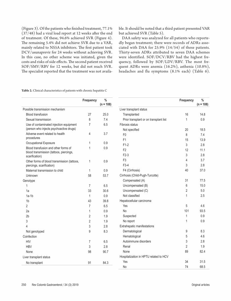
Rev Colomb Gastroenterol / 34 (3) 2019250 Original articles
ble. It should be noted that a third patient presented VAR but achieved SVR (Table 5).
DAA safety was analyzed for all patients who reporte-dly began treatment; there were records of ADRs asso-ciated with DAA for 25.9% (14/54) of these patients. Thirty-seven ADRs attributed to seven DAA schemes were identified. SOF/DCV/RBV had the highest fre-quency, followed by SOF/LDV/RBV. The most fre-quent ADRs were anemia (16.2%), asthenia (10.8%), headaches and flu symptoms (8.1% each) (Table 6).
(Figure 3). Of the patients who finished treatment, 77.1% (37/48) had a viral load report at 12 weeks after the end of treatment. Of these, 94.6% achieved SVR (Figure 4). The remaining 5.4% did not achieve SVR due to a VAR, mainly related to NS5A inhibitors. The first patient took DCV/asunaprevir for 24 weeks without achieving SVR. In this case, no other scheme was initiated, given the costs and risks of side effects. The second patient received SOF/SMV/RBV for 12 weeks, but did not reach SVR. The specialist reported that the treatment was not availa-
Table 2. Clinical characteristics of patients with chronic hepatitis C
Frequency % (n = 108)
Possible transmission mechanismBlood transfusion 27 25.0Sexual transmission 8 7.4Use of contaminated injection equipment (person who injects psychoactive drugs)
7 6.5
Adverse event related to health procedures
4 3.7
Occupational Exposure 1 0.9Blood transfusion and other forms of blood transmission (tattoos, piercings, scarification)
1 0.9
Other forms of blood transmission (tattoos, piercings, scarification)
1 0.9
Maternal transmission to child 1 0.9Unknown 58 53.7
Genotype1 7 6.51a 33 30.61a-1b 1 0.91b 43 39.82 7 6.52a 1 0.92b 2 1.93 2 1.94 3 2.8Not genotyped 9 8.3
CoinfectionHIV 7 6.5HBV 3 2.8None 98 90.7
Liver transplant statusNo transplant 91 84.3
Frequency % (n = 108)
Liver transplant statusTransplanted 16 14.8Prior transplant or on transplant list 1 0.9
Fibrosis statusNot specified 20 18.5F0 8 7.4F1 15 13.9F1-2 3 2.8F2 12 11.1F2-3 3 2.8F3 4 3.7F3-4 3 2.8F4 (Cirrhosis) 40 37.0
Cirrhosis (Child-Pugh-Turcotte)Compensated (A) 31 77.5Uncompensated (B) 6 15.0Uncompensated (C) 2 5.0Not classified 1 2.5
Hepatocellular carcinomaYes 5 4.6No 101 93.5Suspected 1 0.9No report 1 0.9
Extrahepatic manifestationsDermatological 9 8.3Hematological 5 4.6Autoimmune disorders 3 2.8Renal 2 1.9None 89 82.4
Hospitalization in HPTU related to HCVYes 34 31.5No 74 68.5

251Characterization of patients with chronic hepatitis C treated in a high complexity hospital in Medellín
Table 3. Prescribed hepatitis C treatment schemes
Direct action antiviral schemes Interferon based schemesTotalBOC/peg-INF/
RBVpeg-INF/RBV TPV/peg-INF/
RBVpeg-INF None
None 24 10 5 1 40SOF/LDV 12 12SOF/DCV/RBV 3 4 1 4 12SOF/DCV 1 1 7 9PTV/OBV/R/Dasabuvir /RBV 1 8 9PTV/OBV/R/Dasabuvir 3 1 5 9DCV/Asunaprevir 1 2 1 4 8SOF/LDV/RBV 1 3 1 2 7SOF/SMV/RBV 1 1SOF/RBV 1 1Total 30 24 9 3 42 108
BOC: boceprevir; DCV: daclatasvir; LDV: ledipasvir; OBV: ombitasvir; peg-IFN: pegylated interferon; PTV: paritaprevir; R: ritonavir; RBV: ribavirin; SMV: simeprevir; SOF: sofosbuvir; POS: Telaprevir.
0 5 10 15 20 25 30
PEG-INF
Telaprevir/PEG-INF/RBV
PEG-INF/RBV
Boceprevir/PEG-INF/RBV
1 2
5 4
8 16
17 13
Yes
No
Figure 2. Sustained virological response range with interferon (n = 66). peg-INF: Pegylated interferon; RBV: ribavirin.
Figure 3. Status of treatment with direct-acting antivirals (n = 68).
21%
Initiation of treatment not reported
Treatment started
Treatment completed
Treatment uncompleted
No final report
79% 48
51

Rev Colomb Gastroenterol / 34 (3) 2019252 Original articles
Table 4. Adverse reactions to schemes with interferon, ribavirin and protease inhibitors (boceprevir or telaprevir)
ADRMedication
n %BOC/peg-INF/RBV peg-INF/RBV TPV/peg-INF/RBV
Systemic 37 42 7 86 39.8Asthenia 8 9 2 19 8.8Adynamia 7 7 1 15 6.9Fever 5 5 1 11 5.1Muscle pains 2 6 8 3.7Headaches 4 2 6 2.8General malaise 2 4 6 2.8Hyporexia 3 2 5 2.3Chills 2 1 1 4 1.9Coughing 1 2 3 1.4Flu symptoms 1 1 2 0.9Odynophagia 1 1 0.5Polymyositis with elevated CK 1 1 0.5Rhinorrhea 1 1 0.5Dyspnea 1 1 0.5Respiratory symptoms 1 1 0.5Sinusitis 1 1 0.5Weakness 1 1 0.5
Hematological 36 9 12 57 26.4Neutropenia 12 4 3 19 8.8Anemia 13 1 3 17 7.9Leukopenia 8 3 4 15 6.9Thrombocytopenia 2 1 3 1.4Pancytopenia 1 1 2 0.9Hematological alterations 1 1 0.5
Gastrointestinal 10 9 2 21 9.7Nausea 3 2 5 2.3Epigastralgia 3 3 1.4Gastroesophageal reflux 1 2 3 1.4Vomiting 2 1 3 1.4Diarrhea 1 1 2 0.9Loss of appetite 2 2 0.9Dyspepsia 1 1 0.5Belching 1 1 0.5Other gastrointestinal symptoms 1 1 0.5
Neuropsychiatric 11 8 19 8.8Depression 6 4 10 4.6Insomnia 1 1 2 0.9Dysgeusia 1 1 0.5Hypersomnia 1 1 0.5Hypomania 1 1 0.5Suicidal ideation 1 1 0.5Anxiety 1 1 0.5Irritability 1 1 0.5Vertigo 1 1 0.5

253Characterization of patients with chronic hepatitis C treated in a high complexity hospital in Medellín
ADRMedication
n %BOC/peg-INF/RBV peg-INF/RBV TPV/peg-INF/RBV
Dermatological 9 2 6 17 7.9Itching 4 2 6 2.8Rash 2 1 3 6 2.8Alopecia 3 3 1.4Skin lesions 1 1 2 0.9
Misceláneos 4 4 4 12 5.6Anal pain 3 3 1.4Canker sores 1 1 0.5Weight gain 1 1 0.5Dysphonia 1 1 0.5Weight loss 1 1 0.5Pleural pain 1 1 0.5Phosphenes 1 1 0.5Hemoptysis 1 1 0.5Hyperbilirubinemia 1 1 0.5Bleeding hemorrhoid 1 1 0.5Endocrines 1 2 1 4 1.9Thyroid disorders 2 1 3 1.4Increase in blood glucose 1 1 0.5
Total 108 76 32 216 100.0
BOC: boceprevir; peg-INF: pegylated interferon; ADR: adverse drug reaction; RBV: ribavirin; POS: Telaprevir.
Table 4. Adverse reactions to schemes with interferon, ribavirin and protease inhibitors (boceprevir or telaprevir) (continued)
0 1 2 3 4 5 6 7 8 9
SOF/SMV/RBV
SOF/RBV
SOF/DCV
SOF/LDV/RBV
DCV/Asunaprevir
SOF/DCV/RBV
PTV/OBV/dasabuvir/RBV
PTV/OBV/dasabuvir
SOF/LDV
Figure 4. Scope of sustained viral response with Direct Action Antiviral schemes (n = 48). DCV: daclatasvir; LDV: ledipasvir; OBV: ombitasvir; PTV: paritaprevir; r: ritonavir; RBV: ribavirin; SMV: simeprevir; SOF: sofosbuvir.
Yes
No
Unreported
1
1
4
4
4 1
1
1
2
2
2
3
5
5
5
7

Rev Colomb Gastroenterol / 34 (3) 2019254 Original articles
Table 6. Recorded adverse reactions to direct-acting antiviral treatment schemes
Medication SOF/DCV/RBV
SOF/LDV/RBV
DCV/asunaprevir
PTV/OBV/r/dasabuvir/
RBV
SOF/DCV
SOF/LDV
PTV/OBV/r/
dasabuvir
n %
Systemic 7 3 4 1 1 16 43.2Asthenia 2 1 1 4 10.8Flu symptoms 1 1 1 3 8.1Headaches 2 1 3 8.1Lower limb pain 1 1 2 5.4Constitutional nonspecific symptoms 1 1 2.7Arthralgia 1 1 2.7Adynamia 1 1 2.7Dizziness 1 1 2.7
Hematological 4 2 6 16.2Anemia 4 2 6 16.2
Neuropsychiatric 2 1 1 1 1 6 16.2Insomnia 1 1 2 5.4Depression 1 1 2 5.4Irritability 1 1 2.7Alteration of immediate memory 1 1 2.7
Gastrointestinal 1 2 2 5 13.5Diarrhea 1 1 2 5.4Nausea 1 1 2.7Gastrointestinal symptoms 1 1 2.7Dyspepsia 1 1 2.7
Miscellaneous 1 1 1 3 8.1Hypotension 1 1 2.7Weight loss 1 1 2.7Mild indirect hyperbilirubinemia 1 1 2.7
Dermatological 1 1 2.7Itching 1 1 2.7
Total 15 8 6 4 2 1 1 37 100.0
DCV: daclatasvir; LDV: ledipasvir; OBV: ombitasvir; PTV: paritaprevir; r: ritonavir; ADR: adverse drug reaction; RBV: ribavirin; SMV: simeprevir; SOF: sofosbuvir.
ADRs
Table 5. Patients with variants associated with resistance
VAR Sex Age GT Scheme Weeks SVR1 L31V and Y93H: resistance to DCV, EBV, LDV, OBV, VEL Female 78 1b DCV/asunaprevir 24 No2 Y93N: resistance to DCV, ELB, LDV, OBV Male 78 1a SOF/SMV/RBV 12 No3 L31V: resistance to: DCV, EBV, LDV, OBV, reduced susceptibility to VEL
Q80K: SMV resistance.Female 35 1a SOF/LDV/RBV 24 Yes
DCV: daclatasvir; EBV: elbasvir; GT: genotype; LDV: ledipasvir; OBV: ombitasvir; RBV: ribavirin; SVR: sustained virological response; SMV: simeprevir; SOF: sofosbuvir; VAR: variant associated with resistance; VEL: velpatasvir.
255Characterization of patients with chronic hepatitis C treated in a high complexity hospital in Medellín
None of the reported ADRs caused treatment discon-tinuation.
Polypharmacy in Patients with Hepatitis C
Four or more medications in addition to anti-HCV sche-mes were used by 46.3% of the patients used (Table 7). No records of outpatient medications were found for 17.6% of the patients.
Table 7. Polypharmacy in patients with chronic hepatitis C (n = 108)
Number of non-HCV medications Frequency %<4 39 36.14-7 35 32.48-11 13 12.0>12 2 1.9No report 19 17.6
DISCUSSION
This is the first study of patients with HepC at the HPTU to look at the effectiveness and safety of DAAs. The distri-bution of HepC by sex and age was similar to that reported by Santos et al. Based on 1538 samples collected by referral laboratories in Colombia, they found an average patient age of 53 years (SD 14) with approximately 70% of patients between 40 and 70 years. (10) Genotype 1 and subtype 1b were found in 77.8% and 39.8% of the patients analyzed, respectively. According to Santos et al., they are the predo-minant genotype and subtype in Colombia. (10)
Advanced fibrosis/cirrhosis (F3-F4) was found in 39.8% of the patients with compensated cirrhosis in 77.5% of the cases in stage F4. An analytical cross-sectio-nal study conducted in Cartagena for three months found that 50% of 41 patients had advanced cirrhosis/fibrosis, and 68% had compensated cirrhosis. (11) These differen-ces may be due to the number of patients analyzed and to the short time within which information was collected in that study. The proportion of patients with cirrhosis was higher than that described by Hajarizadeh et al. (4-24%). (1) This can be explained by the level of complexity of the HPTU where patients with more advanced stages of disease are generally treated.
As in reports by other authors in Colombia and Latin America, blood transfusions constituted the main risk fac-tor for contracting HCV. (12, 13) This result was expected since screening of blood donations for HCV in Colombia only began in 1993 and only reached 99% coverage in 1995. (14) Considering that the onset of cirrhosis begins 20 years after HCV exposure, the number of HepC diagnoses could
increase over the next few years as the result of transfusions from before 1993. (1)
Effectiveness of Antiviral Therapy
SVR was achieved by 46.9% of patients given peg-IFN which is within the range of 6% to 56% reported in the lite-rature. (8) For genotype 1, the most common genotype in this group, the response rate can reach 50%. (15)
An SVR of 94.6% was found in the group of patients who finished treatment with DAA. The cause of therapeutic failure in the other 5.4% was found to be VAR to NS5A inhibitors. This is consistent with Buti et al. who found that 1% to 7% of patients treated with DAAs do not reach SVR. (16) Causes could be attributable to the patient, the treatment regimen and/or the virus. (17)
VARs are changes in the nucleotide sequence responsible for synthesis of the proteins that are molecular targets of DAAs. This ability to generate resistance, typical of viruses, is greater in HCV than in other viruses such as HBV and HIV. (17) The VARs found in this study were L31V and Y93H which target NS5A inhibitors. VARs related to the nucleo-tide analog NS5B sofosbuvir were not reported. This can be explained by its high genetic resistance barrier. (18)
Similar to reports by other authors, the SVR rates of patients with VAR to NS5A and without VAR to NS5A were similar in this study. (19) Some researchers disagree about the relationship of VAR and SVR, so they recom-mend determining these variants at baseline especially in cases that involve a null response prior to therapy. (17, 18) Current Colombian guidelines for managing HepC recom-mend analyses of resistance to NS3 and/or NS5A only for patients who have not achieved SVR. (20)
The most frequent VARs in genotype 1b are reported to be L31V/M and Y93H/N. Y93H results in high levels of resistance to drugs that act on NS5A. It is important to note that VARs to NS5A continue to be present as long as two years after the end of treatment, so it is essential to consider them before administering rescue therapy. (17)
Safety of Antiviral Therapy
The availability of DAA has led to an improvement in the tolerability of treatment as in this study in which 25.9% of patients who received DAAs presented some type of ADR compared to 68.2% of those who received peg- INF. Although the analysis of the severity of ADRs was not the subject of this study, it was observed that patients with peg-INF/RBV had more severe ADRs especially hospitalizations due to anemia in which patients required blood products and infections associated with leukopenia or neutropenia.

Rev Colomb Gastroenterol / 34 (3) 2019256 Original articles
CONCLUSIONS
Characterization of patients with HepC treated at the HPTU during the study period found a similar distribution among men and women with higher prevalences between 40 and 70 years of age and with transfusions as the most fre-quent transmission mechanism. DAAs were safer and more effective than schemes with peg-IFN/RBV, but RBV is still necessary in cirrhotic patients with previous exposure to treatment, and this increases the risk of ADR.
There is a need to implement comprehensive patient-centered care with access to health services and medica-tions throughout the course of treatment and appropriate pharmacotherapeutic follow-up. Similarly, prospective studies evaluating the safety and effectiveness of DAAs in patients with chronic HepC are needed.
LIMITATIONS
This study has several limitations. Given its retrospective nature, it is directly dependent on the quality of informa-tion recorded in the electronic medical records. During data collection, incomplete records were detected which could diminish the quality of the study. Similarly, medi-cal notes lacked uniformity indicating that the hospital’s electronic medical records need to be standardized from the start to the end dates of treatment. Reports of viral loads, concomitant treatment and possible mechanisms of transmission all need to be recorded for adequate patient follow-up as well as for the national epidemiolo-gical report.
Acknowledgements
We would like to thank the Pharmaceutical Promotion and Prevention research group of the University of Antioquia for academic support and to the HPTU Hepatology Unit for permission for this research.
Conflicts of Interests
The authors declare that they have no conflicts of interest.
Financing
The Pharmaceutical Promotion and Prevention group received funding from the 2018-2019 sustainability call of the Research Development Committee of the University of Antioquia.
We found that 39.8% of the patients who received peg-INF/RBV had systemic ADRs, especially asthenia, adyna-mia, fevers, myalgia and headaches. This is similar to reports the literature which show that these symptoms develop in 11% to 50% of cases, appear within a few hours following administration of medication, and have spontaneous remission from 24 hours to several days later. (21-25) Hematological ADRs are the most common of those due to peg-INF/RBV and are the main cause of low adherence rates, dose reductions and treatment discontinuation. (21, 22, 26) In this study, they occurred in 26.4% of the patients. They developed neutropenia and anemia which could be associated with bone marrow suppression by peg-IFN and RBV-induced extravascular hemolysis. (23, 27)
Systemic ADRs accounted for the largest portion (43.2%) of those that occurred in patients who received DAAs. They were followed by neuropsychiatric ADRs (16.2%), hemato-logical ADRs (16.2%) and gastrointestinal ADRs (13.5%). Barrajón et. Al. reported very similar results in a retrospec-tive analysis of 355 patients treated with DAAs. They found that 43.7% of their study population developed ADRs, mostly systemic (37.1%), gastrointestinal (18%) and neuro-logical (15.8 %). (28) It can be inferred that the appearance of hematological and neuropsychiatric ADR swas related to the use of RBV combined with SOF/DCV or SOF/LDV. Development of anemia and depression occurred more fre-quently in these patiens than in those who did not receive RBV. Calleja et al. have also reported a high incidence of ane-mia (91%) in patients who received SOF/LDV/RBV. (7)
These results show that there are still cases in which the addition of RBV or peg-INF is necessary even though the use of DAA increases tolerability to antiviral treatment. This is especially true for patients previously exposed to interferon who present with cirrhosis which increases the risk of ADRs. (4, 20 , 28)
Polypharmacy in Patients with HepC
Polypharmacy can be defined as the use of five or more daily medications. (29) Our study found that 46.3% of the patients were polymedicated. This can be explained by age (> 50 years) and HepC patients with coexisting diseases which required additional medications.
Polypharmacy can increase susceptibility to medication-related problems such as ADR, falls, hospital readmissions, and drug interactions. (29) This makes establishment of comprehensive health care programs that include pharma-cotherapeutic follow-up imperative to prevent and resolve these medication-related problems.

257Characterization of patients with chronic hepatitis C treated in a high complexity hospital in Medellín
14. Beltrán M. Riesgo de infección transfusional de hepatitis C en Colombia. Iatreia. 2004;17(3-S):305.
15. Saludes V, Ausina V, Martró E. Posibilidades actuales para predecir la respuesta a la terapia en pacientes con hepati-tis C crónica por el genotipo 1 del virus de la hepatitis C. Enfermedades Infecc Microbiol Clínica. 2011;29:51-8. doi: 10.1016/S0213-005X(11)70044-1.
16. Buti M, Riveiro-Barciela M, Esteban R. Management of direct-acting antiviral agent failures. J Hepatol. 2015 Dec;63(6):1511-22. doi: 10.1016/j.jhep.2015.08.010.
17. Llerena S, Cabezas J, Iruzubieta P, Crespo J. Resistencias al virus de la hepatitis C. Implicaciones y posibilidades terapéuticas. Gastroenterol Hepatol. 2017;484-94. doi: 10.1016/j.gastrohep.2017.04.007.
18. Dietz J, Susser S, Berkowski C, Perner D, Zeuzem S, Sarrazin C. Consideration of Viral Resistance for Optimization of Direct Antiviral Therapy of Hepatitis C Virus Genotype 1-Infected Patients. PLoS One. 2015 Aug 28;10(8):e0134395. doi: 10.1371/journal.pone.0134395.
19. Sarrazin C, Dvory-Sobol H, Svarovskaia ES, Doehle B, Martin R, Zeuzem S, et al. The prevalence and the effect of HCV NS5A resistance associated variants in subjects with compensated cirrhosis treated with ledipasvir/sofosbuvir +/- RBV. J Hepatol. 2015;62:S620. doi: 10.1016/S0168-8278(15)30976-4.
20. Ministerio de Salud y Protección Social, Instituto de Evaluación Tecnológica en Salud. Vía clínica para el trata-miento de hepatitis C. Bogotá, Colombia: Ministerio de Salud y Protección Social; 2017. p. 41.
21. Santos OM, Orrego M. Tratamiento: Efectos adversos del tratamiento de hepatitis C. Rev Colomb Gastroenterol. 2012;27:37-40.
22. Mulet Pérez A, Pullés Labadié M, Gámez Escalona M, Mulet Gámez A, Díaz Santos O, Infante Velázquez M. Efectos adversos del tratamiento con interferón alfa-2b humano recombinante y rivabirina en pacientes con hepatitis crónica C. Rev Cuba Med Mil. 2011;40(1):76-84.
23. Sulkowski MS, Cooper C, Hunyady B, Jia J, Ogurtsov P, Peck-Radosavljevic M, Shiffman ML, Yurdaydin C, Dalgard O. Management of adverse effects of Peg-IFN and ribavirin therapy for hepatitis C. Nat Rev Gastroenterol Hepatol. 2011 Apr;8(4):212-23. doi: 10.1038/nrgastro.2011.21.
24. Huang YM, Wang H, Wang C, Chen M, Zhao MH. Promotion of hypercoagulability in antineutrophil cytoplas-mic antibody-associated vasculitis by C5a-induced tissue factor-expressing microparticles and neutrophil extracellu-lar traps. Arthritis Rheumatol. 2015 Oct;67(10):2780-90. doi: 10.1002/art.39239.
25. Hadziyannis SJ, Sette H Jr, Morgan TR, Balan V, Diago M, Marcellin P, Ramadori G, Bodenheimer H Jr, Bernstein D, Rizzetto M, Zeuzem S, Pockros PJ, Lin A, Ackrill AM; PEGASYS International Study Group. Peginterferon-alpha2a and ribavirin combination therapy in chronic hepa-titis C: a randomized study of treatment duration and ribavi-rin dose. Ann Intern Med. 2004 Mar 2;140(5):346-55. doi: 10.7326/0003-4819-140-5-200403020-00010.
REFERENCES
1. Hajarizadeh B, Grebely J, Dore GJ. Epidemiology and natural history of HCV infection. Nat Rev Gastroenterol Hepatol. 2013 Sep;10(9):553-62. doi: 10.1038/nrgas-tro.2013.107.
2. Kohli A, Shaffer A, Sherman A, Kottilil S. Treatment of hepatitis C: a systematic review. JAMA. 2014 Aug 13;312(6):631-40. doi: 10.1001/jama.2014.7085.
3. Center for Disease Analysis. Hepatitis C prevalence [Internet]. 2012 [acceso 19 de febrero de 2017]. Disponible en: http://www.centerforda.com/HepC/HepMap.html
4. American Association for the Study of Liver Diseases and the Infectious Diseases Society of America. HCV Guidance: Recommendations for Testing, Managing, and Treating Hepatitis C [Internet]. 2016 [acceso 23 de marzo de 2017]. Disponible en: http://www.hcvguidelines.org/full-report-view
5. Ministerio de Salud y Protección Social, Instituto de Evaluación Tecnológica en Salud. Guía de Práctica Clínica para la tamización, diagnóstico y tratamiento de perso-nas con infección por el virus de la hepatitis C. Bogotá, Colombia: Ministerio de Salud y Protección Social; 2016.
6. Strader DB, Seeff LB. A brief history of the treatment of viral hepatitis C. Clin Liver Dis (Hoboken). 2012 Mar 6;1(1):6-11. doi: 10.1002/cld.1.
7. Calleja JL, Crespo J, Rincón D, Ruiz-Antorán B, Fernandez I, Perelló C, et al. Effectiveness, safety and clinical outcomes of direct-acting antiviral therapy in HCV genotype 1 infection: Results from a Spanish real-world cohort. J Hepatol. 2017 Jun;66(6):1138-1148. doi: 10.1016/j.jhep.2017.01.028.
8. Yang S, Britt RB, Hashem MG, Brown JN. Outcomes of Pharmacy-Led Hepatitis C Direct-Acting Antiviral Utilization Management at a Veterans Affairs Medical Center. J Manag Care Spec Pharm. 2017 Mar;23(3):364-369. doi: 10.18553/jmcp.2017.23.3.364.
9. Sistema de Trámites en Línea - Consultas Públicas [Internet]. [acceso 30 de julio de 2018]. Disponible en: http://consultaregistro.invima.gov.co:8082/Consultas/consultas/consreg_encabcum.jsp
10. Santos Ó, Gómez A, Vizcaíno V, Casas MC, Ramírez MDP, Olaya P. [Hepatitis C virus genotypes circulating in Colombia]. Biomedica. 2017 Jan 24;37(1):22-27. doi: 10.7705/biomedica.v37i1.3173.
11. Yepes I de J, Carmona ZA, Múnera MN. Calidad de vida en pacientes con hepatitis C crónica en Colombia. Rev Colomb Gastroenterol. 2017;32(2):112. doi: 10.22516/25007440.139.
12. Yepes I de J, Lince B, Caez C, Vuono G de. Factores de riesgo para la infección por el virus de la hepatitis C en la Costa Caribe colombiana: un estudio de casos y controles. Biomédica. 2016;36(4):564-71. doi: 10.7705/biomédica.v36i4.3105.
13. Claudino Botero R, Tagle M. Los nuevos tratamiento de hepatitis C: Perspectivas latinoamericanas. Clin Liver Dis (Hoboken). 2015 Mar 4;5(1):11-13. doi: 10.1002/cld.466.

Rev Colomb Gastroenterol / 34 (3) 2019258 Original articles
28. Barrajón L, Soler E, Lorente L, Pérez J. Efectividad y segu-ridad de los antivirales de acción directa frente al virus de la hepatitis C. Rev OFIL. 2016;26(4):243-50.
29. Masnoon N, Shakib S, Kalisch-Ellett L, Caughey GE. What is polypharmacy? A systematic review of definitions. BMC Geriatr. 2017 Oct 10;17(1):230. doi: 10.1186/s12877-017-0621-2.
26. Nachnani JS, Rao GA, Bulchandani D, Pandya PK, Alba LM. Predictors of hematological abnormalities in patients with chronic hepatitis C treated with interferon and riba-virin. Ann Hematol. 2010 Feb;89(2):121-5. doi: 10.1007/s00277-009-0774-y.
27. UpToDate Inc. Ribavirin (systemic): Drug information [Internet]. [acceso 10 de agosto de 2018]. Disponible en: http://www.uptodate.com

© 2019 Asociaciones Colombianas de Gastroenterología, Endoscopia digestiva, Coloproctología y Hepatología 259
Carlos Arturo Rojas,1* Mario Sepúlveda Copete,1 Jairo Alberto García Abadía,1 Héctor Raúl Echavarría Abad,2 Fernando Rosso Suárez,3 Andrés Fernando Jiménez,4 Andrés Mauricio Castro Llanos.5
Clinical effectiveness of two esomeprazole presentations in a pilot trial
1 Internist and Gastroenterologist in the Endoscopy Unit of Fundación Valle del Lili in Cali, Colombia
2 Surgeon and Gastroenterologist in the Endoscopy Unit of Fundación Valle del Lili in Cali, Colombia
3 Internist, infectologist, epidemiologist. Clinical Studies Coordinator at the Clinical Research Center of the Fundación Valle del Lili in Cali, Colombia
4 Director of Clinical Research, internist, epidemiologist and infectious disease specialist at the Clinical Research Center of the Fundación Valle del Lili in Cali, Colombia
5 Statistician at the Clinical Research Center of the Fundación Valle del Lili in Cali, Colombia
*Correspondence: [email protected].
.........................................Received: 18/12/18 Accepted: 30/05/19
AbstractIntroduction: This pilot studied the clinical effectiveness of two presentations of esomeprazole in patients with dyspepsia with undiagnosed causes. Methods: We conducted a pilot clinical trial of two 40 mg Esomeprazole presentations. Patients with dyspepsia of unknown cause at a gastroenterology clinic in a referral hospital were included. They received one or the other presentation daily for 28 days. Patients were initially evaluated with endoscopy and biopsy and received follow-up examinations at two and four weeks. Adverse events were recor-ded, and clinical symptom scales and quality of life questionnaires validated in Spanish (SODA and QoL-PEI) were used. In addition, gastric pH levels were measured continuously for 24 hours on day 14 of treatment. Serum levels of the medication administered were also measured on day 14 of treatment. A two-way repeated measures ANOVA was used to compare mean differences between the two groups. When significant differences in times were found, a Bonferroni correction was made. Results: A total of 33 patients were randomized into two groups: 16 patients in one group and 17 in the other. There were no differences in the percentages of gastric pH inhibition at day 14 of treatment (p = 0.9795). There were no differences in serum level concentrations on day 14 (p = 0.2199). No significant differences were found in severity and quality of life scales in the first two weeks of treatment. However, in the last two weeks of treatment the test product showed a larger decrease in pain (p = 0.0048) and superiority in compliance (p = 0.01) on the SODA subscale. There were no serious adver-se events, and there were no statistical differences between the presentations of non-serious adverse events. Conclusions: The Test product and the Reference product showed similar effects on clinically relevant variables.
KeywordsEsomeprazole, SODA; QoL-PEI, proton pump inhibitors, dispepsia.
Original articlesDOI: https://doi.org/10.22516/25007440.335
INTRODUCTION
Dyspepsia is defined as chronic and recurring pain or discomfort in the central part of the upper abdomen. (1) According to the criteria of the ROMA IV consensus, there are two types. The first has a defined organic cause while the second has no specific cause and is called functional dyspepsia (FD). FD is considered to be due to physiolo-gical alterations, immunological alterations, hypersensiti-vity and/or brain-intestine interactions. In addition, it is associated with daily eating and life style habits and may be related to Helicobacter pylori infections which are of
great importance in our environment due their prevalence of approximately 60%. (2-4)
Fifteen to forty percent of the world’s population are thought to have dyspeptic symptoms, and of these, 70% are idiopathic. The annual incidence of dyspepsia is approxi-mately 1%, and it is estimated that 50% of people will con-sult a physician because of these symptoms at some point in their lives. (1-3)
The negative impact of dyspepsia on the quality of life has encouraged the development of assessment scales for measuring severity, disability and alterations of daily life. There are two instruments that have been validated in

Rev Colomb Gastroenterol / 34 (3) 2019260 Original articles
Spanish: the SODA (Severity of Dyspepsia Assessment) scale which assesses the intensity of pain, associated symp-toms and level of compliance, and the Dyspepsia Related Health Scale (DRHS) which determines the impact that this disease has on daily life. (4-11)
Part of standard dyspepsia management focuses on con-trol of gastric acid. Proton pump inhibitors (PPIs), one of the main classes of drugs used for this purpose, have been widely used to treat both FD and organic dyspepsia. (1, 5-8) Esomeprazole, a PPI, is indicated for relief of gastroin-testinal symptoms, healing of gastric lesions, and mainte-nance of healing. (5) Esomeprazole improves dyspeptic symptoms through several mechanisms. First, patients with dyspepsia are hypersensitive to duodenal acid. Second, patients with dyspepsia have low-grade inflammation that is worsened by acid secretion. Inhibition of gastric acid secretion by esomeprazole modifies these effects.
Controlled release forms of esomeprazole have been developed to improve absorption and bioavailability thus avoiding early chemical degradation which limits effec-tiveness. NEXIUM-MUPS®, developed by AstraZeneca, includes a system that uses a Multiple-Unit Pellet System (MUPS) that releases micropellets of the PPI as the tablet disintegrates. In Colombia, an esomeprazole formulation has been developed that uses a polymer coating that resists acid as the tablets pass through the gastric acid medium. They disintegrate when they reach the less acidic pH (pH> 4.5) of the proximal portion of the duodenum.
The objective of this study was to compare clinical res-ponses to these two formulations of esomeprazole by eva-luating serum concentrations in the first hour after intake and measuring efficacy due to the increase in gastric pH at 24-hour follow-ups. The SODA and DRHS scales were used to assess patients’ clinical evolution and clinical safety related to adverse events of special-release esomeprazole therapy and reference esomeprazole therapy. Doses of 40 mg/day of all medications were used in dyspeptic patients in whom organic causes of dyspepsia had not been proven.
MATERIALS AND METHODS
A blinded two-arm, randomized, controlled trial was conducted to compare 40 mg/day doses of two different presentations of esomeprazole for 28 days. The study popu-lation consisted of the patients with abdominal and diges-tive symptoms suggestive of dyspepsia who came to the gastroenterology clinic at Fundación Valle del Lili between July 2016 and March 2017, and who had not undergone previous diagnostic studies. Patients who were older than 18 years with a final diagnosis of previously unstudied dys-pepsia were included. Exclusion criteria included unexplai-ned weight loss; dysphagia; anemia; bleeding; jaundice;
history of gastric surgery; neoplasms; erosive esophagitis; pregnancy; lactation; known allergy to esomeprazole; use of nonsteroidal anti-inflammatory drugs (NSAIDs), PPIs or drugs with potential interactions within the two weeks prior to coming to the clinic, and prior endoscopic diagno-sis of alkaline pH, digestive ulcers or malignancy.
This study was carried out at the Fundación Valle del Lili in accordance with the 2013 Helsinki Declaration, the International Conference on Harmonization (ICH) Good Clinical Practice (GCP) Guideline, Colombian Resolution 8430 of 1993 and Resolution 2378 of 2008, and the Guidelines for good clinical practice. The study was appro-ved by the biomedical research ethics committee, and each participant consented to the study. The subjects were not compensated for their participation. This study has been included in the International Clinical Trials registry.
We sought to include at least 30 volunteers in each group in this pilot clinical trial to test normal distribution. However, due to recruitment problems, only 16 patients were included in one group and 17 patients in the other group.
The study began with screening upper endoscopies of volunteers which included routine biopsies and measure-ment of gastric pH an Inolab 7110® pH meter and staining with Congo red and Litmus paper to exclude patients with pH> 4 which is suggestive of hypochlorhydria. After scree-ning, patients were randomly assigned to the two groups. One group received modified-release esomeprazole from Technochemicals (test esomeprazole), and the other group received NEXIUM-MUPS® from AstraZeneca (reference esomeprazole). Both products are registered and marketed in Colombia for the indication used in this study. The ran-domization sequence was performed with Randomization® software which created blocks of six participants. (12) Security envelopes relating the participant’s code and the group to which s/he was assigned were then created.
The attending physician and the research team were blind to the randomized drug throughout the study. Since the presentations of the medications were different, it was not possible to ensure the blinding of the study subjects. The treatment provided on the day of recruitment consis-ted of twenty-eight 40 mg tablets of esomeprazole to be taken daily at least 30 minutes before breakfast for 28 day. Patients were required to keep a daily log. Follow-up visits were scheduled at 2 and 4 weeks to evaluate study outco-mes (Figure 1).
The two week follow-up also evaluated the impact on gastric pH of treatment with esomeprazole by means of 24 hour ambulatory pH monitoring with Versaflex® Z dual pH sensor catheters with 8 impedance rings. Data was was stored on a digitraper pH Z Given® Imagin device and sub-sequently downloaded and analyzed with the Accuview® pH-Z 5.2 program.

261Clinical effectiveness of two esomeprazole presentations in a pilot trial
continuous variables as means and standard deviations (SD) or medians and interquartile ranges (IQR). They were compared with a Student’s T test or the or Mann-Whitney test depending on whether the assumption of normality was fulfilled. Categorical variables were presen-ted in proportions and correlated with the chi square (χ2) test or Fisher’s exact test depending on the observations.
Subsequently, analysis of variance (ANOVA) of two fac-tors (drug-time) with 99 repeated measurements was per-formed with the clinical scales to establish differences. Upon finding significant differences in the times for the main effect of ANOVA, pairwise comparison was performed using Bonferroni correction for multiple comparisons. Analyses were performed with STATA statistical package 12.1.
RESULTS
A total of 205 patients were screened, 55 were recruited, and the final overall sample included 33 patients. The main cause of exclusion was gastric pH suggestive of hypochlor-hydria (Figure 2). (13) During follow-up, two participants who could not perform pH-metrics were excluded from the analysis.
The comparison of baseline sociodemographic and clini-cal characteristics, summarized in Table 1 found no signi-ficant differences between the groups. The average serum concentration of the test esomeprazole was 0.67 µmol/L (0.18-1.61) and that of the reference esomeprazole was 0.28 µmol/L (0.16-0.53) but the difference was not sta-tistically significant (p = 0.219). Similarly, the comparison of inhibition of acid secretion to a constant gastric pH> 4 showed an average time for the test esomeprazole of 19.98 h (SD ± 3.87 [83.3%]) and 19.95 h (SD ± 3.55 [83.2%]) for reference esomeprazole, p = 0.986.
Statistically significant differences were found for both survey scales at four weeks of treatment, and the pain inten-
We decided to take drug serum levels on day 14 of treatment as a measure that could be correlated to pH measurement and clinical outcomes. Two weeks after each patient started treatment, a five ml sample of venous blood was taken within the first hour after the patient took her/his daily dose of esomeprazole. This was sufficient time to guarantee esomeprazole concentrations in the blood at equilibrium. Plasma was frozen at -20° C. Subsequently, the sample was analyzed with a UHPLC Lachrom Ultra-VWR liquid chromatograph whose diode array detector was used to determine serum esomeprazole levels.
The SOSA and DRHS surveys and assessment scales were used for clinical evaluations on the day of recruitment and at subsequent follow-up visits at two and four weeks after the start of treatment. The version of SODA valida-ted by the Benites et al. was used together with the score adjustment for balancing subscales suggested and applied by Rabeneck et al. (10, 11) For analysis, it was divided into subscales of pain intensity, associated non-painful symp-toms such as belching, heartburn, and swelling, and level of compliance. The first two subscales express greater severity at higher scores while the third expresses a higher level of perceived well-being at higher scores. DRHS was used with the same methodology and at the same times as the SODA scale, but a single global score in which greater severity of symptoms is expressed at higher scores was used. Unlike SODA, one of the components of DRHS scores reflects disability associated with pain. (9)
Clinical safety was monitored at each visit through analy-sis of patient’s daily logs in which they recorded any asso-ciated symptoms and adverse events during treatment.
Statistical Analysis
All participant information was uploaded into a database on the BD Clinic® platform. The descriptive analysis expressed
First visit
- Initial medical consultation
- Informed consent
Randomization of groups
- Quality of life surveys- SODA- DRHSI
- Esomeprazole treatment begins
- Reference tests
Second visit
- Digestive Endoscopy- pH measurement
- Litmus- Congo red
Third Visit: Two-week follow-up
- Quality of life surveys- SODA- DRHS
- Medical examination- 24 pH measurement- Serum levels of
esomeprazole
Fourth visit: Four-week follow-up
- Quality of life surveys- SODA- DRHS
Figure 1. Distribution of clinical trial activities

Rev Colomb Gastroenterol / 34 (3) 2019262 Original articles
Evaluated for selection (n = 205)Excluded (n = 150)- Do not meet the selection criteria
(n = 20)- Renounce to participate (n = 130)
Randomized (n = 35)
Assigned to esomeprazole Test (n = 17)- Received the assigned medication
(n = 17)
Assigned to esomeprazoleReference (n = 18)- Received the assigned medication
(n = 18)
Follow-up losses (n = 1) Follow-up losses (n = 1)
Analized (n = 16)- Excluded from analysis (n = 1)
Analized (n = 17)- Excluded from analysis (n = 1)
Recruitment
Assignment
Follow-up
Analysis
Figure 2. Flowchart of participants from recruitment between July 2016 and April 2017 through to the end of the clinical trial.
sity subscale scores decreased more for the test esomepra-zole than they did for the reference esomeprazole. Similarly, improvement in compliance was more noticeable in the test product than in the reference product (Table 2). These trends were repeated the two factor ANOVA which found differences in the time variable, but only for interaction in the pain subscale. Differences in the compliance subscale were found between treatments (p = 0.012) (Table 3). The Bonferroni adjustment found that there are significant differences (p <0.05) between the start and end time of the treatment at 4 weeks in the SODA subscales and the DRHS scale independently for each type of esomeprazole. However, the SODA pain subscale found no significant differences in this time (p = 0.018).
The proportion of patients who experienced adverse events during the study was similar for both groups. At four weeks, it was 6.3% for the group who received the test drug (Tecnoquímicas esomeprazole) and 5.9% for the group who took the reference drug (AstraZeneca esomeprazole) (p = 0.999). There were no serious adverse effects and all adverse effects had resolved before the end of the study. All of these adverse effects are among those referred to in the technical sheets as being associated with the use of esomeprazole and include nausea, dry mouth, belching and flatulence.
DISCUSSION
Serum concentrations measured in our patients varied con-siderably, similar to measurements of serum concentrations
reported in the literature. These variations may be due to the designs of special release presentations, time of admi-nistration, physiological conditions at the time of drug use, and to varying abilities of individuals to metabolize PPI according to polymorphisms in their enzymes, especially CYP2C19. (6, 14-20)
Both drugs effectively increased gastric pH by inhibiting 83% of acid production which is at the upper limits of figu-res reported in similar studies which range from 50% to 85%. The minimum inhibition value in our study was 50% which is within the established range for clinical impact. (7, 8, 14, 15). There were no differences in esomeprazole serum levels at day 14 which may partly explain this fin-ding. However, serum levels do not necessarily correlate with intracellular levels.
Significant decreases were observed in the SODA scores of both drugs. The pain subscale for Tecnoquímicas eso-meprazole fell 20.8 points while that of the AstraZeneca esomeprazole fell 12.8 points. These decreases were slightly higher than those reported by Benites et al., who described decreases of 7 points. However, measurement of symptoms not associated with pain decreased only around 2.5 points, similar to the findings of Benites et al. Similar behavior to our findings and those of Benites et al., another similar study by Rabeneck et al. found that the greatest impact was on the pain subscale. They concluded that this result was possible because of effective control of gastric acid, which is the main cause of the sensation of pain, and that questions about compliance are associated with pain control. In rela-

263Clinical effectiveness of two esomeprazole presentations in a pilot trial
much lower than those found by Shin et al., who reported 40% in a sample of 36 patients, but the two studies are in agreement in that neither found any serious adverse events. This has also been described in various publications which have found that, when use is not prolonged, these drugs’ have good pharmacological safety profiles. (14-21).
One limitation of this present study was that diagnostic means other than clinical and endoscopic examinations were not used even though ultrasound and laboratory tests could have ruled out other causes of dyspepsia. Although
tion to compliance, we observed that the test esomeprazole had a higher final score for clinical improvement than did the reference esomeprazole. On average, scores improved nine points at the first follow-up and an 5.2 points at the second follow-up (p = 0.0035). These increases were also higher than those reported by Benites et al., which were 2.5 points in their study population. (10, 11)
The general frequencies of adverse events at four weeks in this study was 6.3% for the test esomeprazole and 5.9% for the reference esomeprazole. These frequencies are
Table 1. Comparison of baseline sociodemographic and clinical characteristics between groups: test esomeprazole (n = 16) and reference esomeprazole (n = 17)
Variable Overall Test Esomeprazole Reference Esomeprazole pSex n = 33 n = 16 n = 17
Male, n (%) 8 (24.2) 4 (25) 4 (23.5)1
Female, n (%) 25 (75.8) 12 (75) 13 (76.5)Age* 38.2 ± 12.7 37.6 ± 11.4 38.8 ± 14.2 0.7919pH meter** 1.81 (1.61-1.94) 1.7 (1.5-2.04) 1.85 (1.65-1.9) 0.6266Litmus pH measurement* 1.9 ± 0.68 1.8 ± 0.72 1.9 ± 0.66 0.7844Congo red pH measurement pH <4 33 (100) 16 (100) 17 (100) -Presence of H. pylori n = 33 n = 16 n = 17
Yes, n (%) 22 (66.7) 8 (50) 14 (82.4)0.071
No, n (%) 11 (33.3) 8 (50) 3 (17.6)Antral distribution of H. pylori (n = 22) n = 22 n = 8 n = 14
Abundant, n (%) 13 (39.4) 4 (25) 9 (52.9)0.831Moderate, n (%) 2 (6.1) 1 (6.25) 1 (5.9)
Low, n (%) 7 (21.2) 3 (18.75) 4 (23.5)Distribution of H. pylori in the corpus (n = 22) n = 22 n = 8 n = 14
Abundant, n (%) 9 (27.3) 3 (18.75) 6 (35.3)
0.858Moderate, n (%) 4 (12.1) 1 (6.25) 3 (17.6)Low, n (%) 6 (18.2) 3 (18.75) 3 (17.6)Absent, n 3 (9.1) 1 (6.25) 2 (11.8)
Diagnosis from antral biopsy n = 33 n = 16 n = 17Chronic non-atrophic chronic gastritis 28 (84.8) 13 (81.25) 15 (88.2)
0.794Severe chronic gastritis without atrophy 2 (6.1) 1 (6.25) 1 (5.9)Chronic gastritis with atrophy 3 (9.1) 2 (12.5) 1 (5.9)Diagnosis from biopsy of corpus n = 33 n = 16 n = 17Chronic non-atrophic chronic gastritis 31 (93.9) 15 (93.75) 16 (94.1)
1Severe chronic gastritis without atrophy 1 (3) 0 (0) 1 (5.9)Chronic gastritis with atrophy 1 (3) 1 (6.25) 0 (0)
Percentage of time pH> 4 (n = 33) 88.2 (71.2-95.6) 88.3 (71.3-97.05) 87.3 (75.7-91.7) 0.4594Adverse events
T1, two weeks 5 (15.2) 1 (6.3) 4 (23.5) 0.335T2, four weeks 2 (6.1) 1 (6.3) 1 (5.9) 1
* Mean ± SD. ** Median (IQR)

Rev Colomb Gastroenterol / 34 (3) 2019264 Original articles
could not be blinded to the medication they were taking which may have introduced selection bias into our study.
We conclude that the two presentations of esomeprazole had very similar outcomes of interest in terms of increasing pH and answers to symptom questionnaires. No significant differences were observed in the evolution of the clinical assessment scales for the two treatments, but the effect of the test esomeprazole was better sustained over time than was that of the reference esomeprazole. This difference was statistically significance. It could be that this difference was
dyspepsia is very prevalent, diagnosis and screening to exclude patients with organic dyspepsia is complex.
On the other hand, the statistical power of this pilot study at the time of closure was low. A representative sample for this pathology with a prevalence of 10% to 30% of the gene-ral population would have required at least 1500 patients to attain a statistical power of 80%. (22, 23) Also, the follow-up time and effectiveness in the study was four weeks, so our data cannot be extrapolated to clinical situations invol-ving longer use of these medications. In addition, patients
Table 2. Comparison of drugs between independent times: time 0 = time of recruitment, time 1= 2 week follow-up, time 2 = 4 week follow-up.
SODA Scale** and DRHS Scale*** Overall General n = 33
Esomeprazole TEST DRUG
n = 16
Esomeprazole AZ n = 17
p
SODA Pain intensity * (Range 2-47)Time 0 (without treatment) 25.8 ± 5.5 24.4 ± 6.4 27.2 ± 4.4 0.1603Time 1 (2 weeks of treatment) 19.5 ± 6.2 19.75 ± 7.15 19.3 ± 5.4 0.8376Time 2 (4 weeks of treatment) 16.6 ± 9.8 12.18 ± 9.3 20.8 ± 8.5 0.0087
SODA Intensity associated symptoms* (Range 7-35)Time 0 (without treatment) 17.8 ± 2.7 17.6 ± 1.8 18 ± 3.3 0.6911Time 1 (2 weeks of treatment) 14.1 ± 2.9 13.9 ± 2.9 14.3 ± 2.9 0.7273Time 2 (4 weeks of treatment) 13.9 ± 4.04 12.7 ± 3.9 15 ± 3.9 0.1118
SODA Compliance Level * (Range 2-23)Time 0 (without treatment) 7.4 ± 3.2 8.2 ± 3.1 6.7 ± 3.3 0.2113Time 1 (2 weeks of treatment) 12.1 ± 5.1 12.7 ± 5.6 11.5 ± 4.7 0.5046Time 2 (4 weeks of treatment) 14.5 ± 5.5 17.2 ± 5.2 11.9 ± 4.4 0.0035
DRHS* (range 12-120)Time 0 (without treatment) 65.5 ± 17.3 64 ± 17.3 66.8 ± 17.69 0.6401Time 1 (2 weeks of treatment) 38.6 ± 13.4 37.5 ± 13.54 39.58 ± 13.58 0.6616Time 2 (4 weeks of treatment) 39.7 ± 18.5 32.68 ± 14.2 46.29 ± 19.32 0.029
* Average ± SD Two-sample t test. ** Severity of dyspepsia assessment. *** Quality of life associated with intestinal problems questionnaire.
Table 3. Comparison between drugs by means of two factor ANOVA of repeated measures: time 0 = time of recruitment, time 1= 2 week follow-up, time 2 = 4 week follow-up.
SODA** and DRHS*** two factor ANOVA of repeated measuresScale Drug Time 0 Time 1 Time 2 trt time trt#time
SODA pain Test Drug* 24.4 ± 6.4 19.75 ± 7.15 12.18 ± 9.30.0623 0 0.0048
Reference Drug* 27.2 ± 4.4 19.3 ± 5.4 20.8 ± 8.5SODA associated symptoms
Test Drug* 17.6 ± 1.8 13.9 ± 2.9 12.7 ± 3.90.2215 0 0.2973
Reference Drug* 18 ± 3.3 14.3 ± 2.9 15 ± 3.9SODA compliance Test Drug* 8.2 ± 3.1 12.7 ± 5.6 17.2 ± 5.2
0.012 0 0.0968Reference Drug* 6.7 ± 3.3 11.5 ± 4.7 11.9 ± 4.4
DRHS Test Drug* 64 ± 17.3 37.5 ± 13.54 32.68 ± 14.20.1572 0 0.1357
Reference Drug* 66.8 ± 17.69 39.58 ± 13.58 46.29 ± 19.32
* Average ± SD. ** Severity of dyspepsia assessment. *** Quality of life questionnaire associated with intestinal problems.

265Clinical effectiveness of two esomeprazole presentations in a pilot trial
asociada a dispepsia. Adaptación española y validación del cuestionario Dyspepsia Related Health Scale. Medicina Clínica. 2001;117(15):567-73. doi: 10.1016/S0025-7753(01)72182-3.
10. Benites Goñi H, Cabrera Cabrejos S, Chungui Bravo J, Prochazka Zarate R, Bernabe Ortiz A, De los Ríos Senmache R, et al. Modificación y validación del instru-mento SODA (severity of dyspepsia assessment) adaptada al Perú para evaluar la evolución de la severidad de los sín-tomas en pacientes con dispepsia. Rev Gastroenterol Perú. 2013;33(1):9-27.
11. Rabeneck L, Cook KF, Wristers K, Souchek J, Menke T, Wray NP. SODA (severity of dyspepsia assessment): a new effective outcome measure for dyspepsia-related health. Journal of clinical epidemiology. 2001;54(8):755-65. doi: 10.1016/S0895-4356(00)00365-6.
12. Randomization.com. [internet] 2007 [acceso el 15 de marzo de 2017]. Disponible en: http://www.randomiza-tion.com/2007.
13. Moher D, Hopewell S, Schulz KF, Montori V, Gøtzsche PC, Devereaux PJ, et al. CONSORT 2010 Explanation and Elaboration: updated guidelines for reporting para-llel group randomised trials. BMJ. 2010;340:c869. doi: 10.1136/bmj.c869.
14. Ullah MA, Shams Ud D, Maruf AA, Azad MAK, Shohag MH, Sultana R, et al. Relative bioavailability and pharma-cokinetic properties of two different enteric formulations of esomeprazole in healthy bangladeshi male volunteers: An open-label, single-dose, randomized-sequence, two-way crossover study. Clinical Therapeutics. 2010;32(7):1419-26. doi: 10.1016/j.clinthera.2010.07.007.
15. Shin JS, Lee JY, Cho KH, Park HL, Kukulka M, Wu JT, et al. The pharmacokinetics, pharmacodynamics and safety of oral doses of ilaprazole 10, 20 and 40 mg and esomeprazole 40 mg in healthy subjects: a randomised, open-label cros-sover study. Alimentary Pharmacology & Therapeutics. 2014;40(5):548-61. doi: 10.1111/apt.12860.
16. Furuta T, Ohashi K, Kosuge K, Zhao XJ, Takashima M, Kimura M, et al. CYP2C19 genotype status and effect of omeprazole on intragastric pH in humans. Clinical Pharmacology & Therapeutics. 1999;65(5):552-61. doi: 10.1016/S0009-9236(99)70075-5.
17. Hunfeld NG, Touw DJ, Mathot RA, van Schaik RH, Kuipers EJ. A comparison of the acid-inhibitory effects of esome-prazole and rabeprazole in relation to pharmacokinetics and CYP2C19 polymorphism. Alimentary Pharmacology & Therapeutics. 2012;35(7):810-8. doi: 10.1111/j.1365-2036.2012.05014.x.
18. Klotz U. Impact of CYP2C19 polymorphisms on the cli-nical action of proton pump inhibitors (PPIs). Eur J Clin Pharmacol. 2009;65(1):1-2. doi: 10.1007/s00228-008-0571-x.
19. Dean L. Esomeprazole Therapy and CYP2C19 Genotype. 2012 [updated 2016 Mar 8]. In: Pratt V, McLeod H, Rubinstein W, Dean L, Kattman B, Malheiro A, editors. Medical Genetics Summaries [Internet]. Bethesda (MD):
secondary to the greater number of positive cases for H. pylori in the reference esomeprazole group, although this difference was not statistically significant difference (p = 0.071). Both presentations of esomeprazole demonstrated that risks of use are very low for a four week course of admi-nistration in the population studied.
Acknowledgements
We would like to thank the endoscopy unit and the entire team of the clinical research center for their contributions to this study. We would also like to thank the laboratory of chemical instrumentation of ICESI University for analyses of serum concentrations.
Conflicts of Interest
The authors declare that they have no conflicts of interest.
Funding Sources
Tecnoquímicas.
REFERENCES
1. Talley NJ, Vakil N. Guidelines for the management of dys-pepsia. Am J Gastroenterol. 2005;100(10):2324-37. doi: 10.1111/j.1572-0241.2005.00225.x.
2. Stanghellini V, Chan FK, Hasler WL, Malagelada JR, Suzuki H, Tack J, et al. Gastroduodenal disorders. Gastroenterology. 2016;150(6):1380-92. doi: 10.1053/j.gastro.2016.02.011.
3. Talley NJ. Functional dyspepsia: new insights into pathoge-nesis and therapy. Korean J Intern Med. 2016;31(3):444-56. doi: 10.3904/kjim.2016.091.
4. Bravo LE, Cortés A, Carrascal E, Jaramillo R, García LS, Bravo PE, et al. Helicobacter pylori: patología y preva-lencia en biopsias gástricas en Colombia. Colomb Med. 2003;34(3):124-31.
5. Otero W, Zuleta MG, Otero L. Enfoque del paciente con dispepsia y dispepsia funcional: actualización. Rev Col Gastroenterol. 2014;29(2):132-8.
6. Shin JM, Kim N. Pharmacokinetics and pharmacodynamics of the proton pump inhibitors. J Neurogastroenterol Motil. 2013;19(1):25-35. doi: 10.5056/jnm.2013.19.1.25.
7. Hatlebakk JG. Review article: gastric acidity − compari-son of esomeprazole with other proton pump inhibitors. Alimentary Pharmacology & Therapeutics. 2003;17:10-5. doi: 10.1046/j.1365-2036.17.s1.3.x.
8. Kirchheiner J, Glatt S, Fuhr U, Klotz U, Meineke I, Seufferlein T, et al. Relative potency of proton-pump inhibitors-com-parison of effects on intragastric pH. Eur J Clin Pharmacol. 2009;65(1):19-31. doi: 10.1007/s00228-008-0576-5.
9. Ruiz M, Villasante F, León F, González-Lara V, González C, Crespo M, et al. Cuestionario sobre calidad de vida

Rev Colomb Gastroenterol / 34 (3) 2019266 Original articles
21. Menéndez JLT. Farmacología de esomeprazol. Emergencias: Revista de la Sociedad Española de Medicina de Urgencias y Emergencias. 2005;17(4):1059-66.
22. Mahadeva S, Goh KL. Epidemiology of functional dys-pepsia: a global perspective. World J Gastroenterol. 2006;12(17):2661-6. doi: 10.3748/wjg.v12.i17.2661.
23. Talley NJ, Vakil NB, Moayyedi P. American gastroentero-logical association technical review on the evaluation of dyspepsia. Gastroenterology. 2005;129(5):1756-80. doi: 10.1053/j.gastro.2005.09.020.
National Center for Biotechnology Information (US); 2012-. Available from http://www.ncbi.nlm.nih.gov/books/NBK100896/.
20. Lou H-Y, Chang C-C, Sheu M-T, Chen Y-C, Ho H-O. Optimal dose regimens of esomeprazole for gastric acid suppression with minimal influence of the CYP2C19 poly-morphism. Eur J Clin Pharmacol. 2008;65(1):55-64. doi: 10.1007/s00228-008-0552-0.

© 2019 Asociaciones Colombianas de Gastroenterología, Endoscopia digestiva, Coloproctología y Hepatología 267
Valeria Atenea Costa Barney,1* Alan Felipe Ovalle Hernández.1
The role of antispasmodics in managing irritable bowel syndrome
1 Internal Medicine and Gastroenterology specialist in San Ignacio University Hospital, Pontificia Universidad Javeriana, Bogotá, Colombia.
*Correspondence: [email protected]
.........................................Received: 26/10/18 Accepted: 11/02/19
AbstractAlthough antispasmodics are the cornerstone of treating irritable bowel syndrome, there are a number of an-tispasmodic medications currently available in Colombia. Since they are frequently used to treat this disease, we consider an evaluation of them to be important.
KeywordsAntispasmodic, irritable bowel syndrome, pinaverium bromide, otilonium bromide, Mebeverin, trimebutine.
Review articlesDOI: https://doi.org/10.22516/25007440.309
INTRODUCTION
Irritable bowel syndrome (IBS) is one of the most fre-quent chronic gastrointestinal functional disorders. It is characterized by recurrent abdominal pain associated with changes in the rhythm of bowel movements with either or both constipation and diarrhea. Swelling and bloating are frequent occurrences. (1)
IBS is divided into two subtypes: predominance of cons-tipation (20-30% of patients) and predominance of dia-rrhea (20-30% of patients). When both constipation and diarrhea are combined, it is called mixed IBS (up to 45% of patients) and IBS of undetermined type when the pattern of bowel movements is intermediate and cannot be classified as diarrhea or constipation. It is noteworthy that abdominal pain occurring more than once a week plus the temporal relationship of pain with defecation are what theoretically differentiates IBS from functional constipation. (2, 3)
According to the Rome IV criteria, IBS is diagnosed by abdominal pain that recurs at least one day a week plus two or more of the following: pain is associated with defeca-tion; pain is related to a change in the frequency of bowel movements; and/or pain is related to a change of stool
consistency. The criteria must be met for three consecutive months prior to diagnosis and symptoms must have started a minimum of six months before diagnosis. (3, 4)
There are no known structural or anatomical explanations of the pathophysiology of IBS and its exact cause remains unknown. Nevertheless, several mechanisms have been proposed. Altered gastrointestinal motility may contribute to changes in bowel habits reported by some patients, and a combination of smooth muscle spasms, visceral hypersen-sitivity and abnormalities of central pain processing may explain abdominal pain, which is an essential part of the complex of symptoms. (5)
It is estimated that IBS affects 11% of the world’s popu-lation. In Europe, Asia and the United States its prevalence varies from 10% to 20%. The lowest prevalence in South Asia (7%) while the highest is in South America (21%). In Western countries it is twice as frequent in women. (6) IBS has a significant impact on health-related quality of life, results in lower labor productivity, higher absenteeism and increased use of health care with its attendant costs. In 2005, direct medical costs attributed to IBS in the United States were estimated at USD 1.5 to 10 billion per year. (1) IBS can also affect the doctor-patient relationship since

Rev Colomb Gastroenterol / 34 (3) 2019268 Review articles
ineffective control of symptoms can decrease the credibi-lity of doctors and encourage the patient to seek additional opinions. (7)
A heterogeneous group of medications called antispas-modic or spasmolytic drugs has been used in IBS therapy for decades. They act as direct smooth muscle relaxants (papaverine, mebeverin, peppermint oil), anticholinergic agents (butylscopolamine, hioscin, cimetropium bromide, pyrenzepine) or calcium channels blockers (alverine citrate, ethyl bromide, pinaverium bromide). Their goal is to reduce symptoms caused by defecation through increasing colonic transit time, improving stool consistency and/or reducing stool frequency. (2) The pharmacological action of these agents is not always clear, and their mechanisms are often mixed. However, metaanalyses of studies com-paring antispasmodics with placebos or other treatments have consistently confirmed the positive effects of these drugs, and their side effect profiles have been excellent. (8)
PATHOPHYSIOLOGY
The pathogenesis of IBS is considered to be multifactorial. A history of gastrointestinal infections, colonic or bacterial flora of an altered small intestine, increased intestinal per-meability and immune activation may all play roles in the development of the disease. (9) Signals from the gastroin-testinal tract are processed in the brain and can influence motility, secretion and immune function. The brain-gut axis is essential for regulation of the gastrointestinal sys-tem, so structural or functional alteration can lead to the development of disorders such as IBS. (10) Consequently, psychological factors and chronic stress may also be invol-ved in triggering symptoms. (11)
Abnormal intestinal motility and visceral hypersensitivity remain the main factors in the pathogenesis of the disease. (12) Intraluminal factors such as serine proteases may increase colonic permeability of IBS patients by activating the protease-activated receptor-2. This results in visceral hypersensitivity. (13) Similarly, luminal cysteine proteases have been shown to increase colonic permeability through degradation of binding proteins resulting in visceral hyper-sensitivity possibly secondary to local microinflammation. (14) Immune activation of the colonic mucosa has been found to be significantly greater in IBS patients than in healthy controls. (15) In addition, mast cells have been implicated in the development of IBS. One study found that the number of mast cells in the colonic mucosa and the amounts of trypsin and histamine they released were markedly higher in IBS patients than in controls. (16) Mast cells in the vicinity of nerve endings has been significantly correlated with the severity and frequency of abdominal pain and discomfort in IBS patients. Enterochromaffin cells
also play an important role in the development of visceral hypersensitivity because they produce and release sero-tonin which activates 5-HT3 receptors located in afferent sensory neurons. (17)
Abnormalities in colonic motility patterns are characteri-zed by hyperreactivity due to prolonged increase of colonic motor activity after meals, increased motor activity in res-ponse to stressors or cholecystokinin (CCK), and increa-sed motor response to abdominal distention. (18)
Visceral hypersensitivity alone is not painful, but it can cause abdominal pain in IBS patients due to the effect of any intense stimulus such as an exaggerated contraction of the colon. (19) Nevertheless, it has not been possible to establish a clear relationship between visceral hypersen-sitivity and motility disorders, and these two factors have generally been considered to be independent, and both require effective treatment. (20)
Voltage Dependent Calcium Channels
Voltage-dependent calcium channels play a fundamental role in the intestine and in pharmacological management of IBS. These ionic channels mediate calcium influx in res-ponse to membrane depolarization, and they regulate intra-cellular processes such as contraction, secretion and neu-rotransmission in a variety of cells. (21) Calcium channels are classified by their properties and pharmacology. They include L-type calcium channels (long duration). They are high conductance channels that produce long-lasting depolarization and which are inhibited by dihydropyridine derivatives (DHP). (22, 23) Currents associated with this type of channel are important for muscle and endocrine cells in which contraction and secretion of substances are mediated. (21)
Type N (neuronal) currents are also durable, but require strongly negative potentials for complete elimination of inactivation and strong depolarization for activation. (23) Three other channels have been identified in Purkinje cells. Type P currents are blocked by low concentrations of ω-agatoxin, while type Q only responds to high concentra-tions. Residual currents, which were resistant to all known calcium blockers at the time of their discovery, were called type R (resistant). Type T (transient) voltage-dependent calcium channels are characterized by small and transient conductance activated by weak depolarization. (23) These currents are responsible for modulating the action poten-tial and the performance of pacemakers.
In the 1980s DHP antagonists became the first calcium antagonists to be used medically. They block L channels and are used to treat hypertension by exploiting their pro-perties as vasodilators. (22) Calcium antagonists have no effect on skeletal muscle, but they can have some influence
269The role of antispasmodics in managing irritable bowel syndrome
of the days to less than one day per week while the patients who took placebos continued to have one to three episodes per week. During the 10-week follow-up period after the end of treatment, the likelihood of recurrence of symptoms was significantly higher in the placebo group than in the otilonium bromide group. (28, 29) This finding may be explained by the prolonged persistence of ethyl bromide in the colon wall due to its lipophilic properties. (25)
Among the most commonly reported side effects asso-ciated with the use of ethyl bromide are dry mouth, nausea, and dizziness. These may be caused by peripheral and central muscarinic antagonism and may be explained by the known ability of otilonium to bind muscarinic receptors. (27)
Otilonium bromide has been evaluated in 5 randomized controlled studies which included a total of 791 patients. (29-33) A metaanalysis found evidence of a beneficial effect [risk ratio (RR) = 0.70, 95% confidence interval (CI): 0.54-0.90; number needed to treat (NNT): 5, 44% CI, p = 0.13], but there was borderline heterogeneity among study results (I2 = 44%, p = 0.13). The Colombian Association of Gastroenterology (Asociación Colombiana de Gastroenterología - ACG) clinical practice guidelines strongly recommends the use of ethyl bromide for increas-ing the frequency of overall improvement of symptoms in IBS patients but the quality of evidence is still low.
Pinaverium Bromide
Pinaverium bromide, a derivative of quaternary ammo-nium, is poorly absorbed and has pronounced pharmaco-logical effects in the gastrointestinal tract rather than in car-diovascular system. (34) Its gastrointestinal absorption rate is low and is characterized by hepatobiliary excretion. (35) Its effects are very similar to those of established L-type calcium channel blockers (nitrendipine, diltiazem) since it reduces the plateau phase of slow waves which inhibits calcium influx and prevents subsequent contractions. (36)
It has also been shown to inhibit the acetylcholine (ACh)-induced contractile response of smooth muscle in dog and rat colons. Acetylcholine is a neurotransmitter of the intrinsic cholinergic nerves. (36) Similarly, in smooth muscle cells of the colon isolated from normal or inflamed human colons, it inhibits contraction induced by other agonists (CCK). (37) The involvement of sensory afferent neurons in IBS has been demonstrated, and this could also explain the efficacy of pinaverium bromide for treating motility disorders and intestinal hypersensitivity, two key IBS symptoms.
A pilot study of 12 IBS patients has used surface electrom-yography to study how treatment with pinaverium bromide affects colonic motility. Surface electromyography was used during a two hour fasting period and a postprandial
on heart muscle through reduction of activity and con-duction of pacemakers. Because IBS includes abnormal gastrointestinal motility, calcium antagonists used for car-diovascular disease appear to have potential for relieving symptoms by relaxing the smooth muscles of the colon. Nicardipine, which has spasmolytic properties, was pro-posed as a possible IBS treatment in the late 1980s. (24) However, cardiovascular side effects have seriously limited the application these calcium antagonists. This led resear-chers to search for other substances that act selectively within the gastrointestinal tract.
ANTI-SPASMODIC DRUGS
These medications act by inhibiting the action of acetyl-choline on muscarinic receptors or by blocking calcium channels in the gastrointestinal smooth muscle. As a class, antispasmodics have been used in the treatment of IBS for many years. They treat the subgroup of IBS patients who have abnormal contractility of the gastrointestinal smooth muscle and altered gastrointestinal transit which contri-bute to pain and altered bowel habits. (25)
Otilonium Bromide
Otilonium bromide’s structure consists mainly of qua-ternary ammonium, so it is weakly absorbable from the gastrointestinal tract. Experimental studies show that it accumulates in the walls of the gastrointestinal tract after oral administration and is almost completely excreted in feces. (25) Otilonium bromide not only blocks L-type and T-type calcium channels, but also the M1, M2, M4 and M5 muscarinic receptors. The antagonistic effects of otilonium bromide on the M3 coupled calcium signaling pathway in human colonic crypt cells suggests antisecretory action in patients who have the diarrhea type of IBS. Antagonism of neuroquinine-2 receptors (NK-2) also causes spasmolysis while reducing peripheral sensory afferent transmission to the central nervous system, possibly contributing to greater efficacy. (26, 27) These effects suggest that otilonium bro-mide may be effective at reducing spasms and abdominal pain, the two main symptoms of IBS. (2)
Otilonium bromide has been evaluated for management of abdominal pain in IBS patients in a clinical trial. Patients diagnosed according to the Rome II criteria were randomly assigned to case and control groups. They received either 40 mg of ethyl bromide or a placebo three times a day for 15 weeks. Patients who took otilonium bromide had less frequent pain, bloating and bowel movements than did the control patients who took placebos. The outstanding result of this study was that otilonium bromide significantly redu-ced the frequency of abdominal pain from more than half

Rev Colomb Gastroenterol / 34 (3) 2019270 Review articles
A multicenter, randomized, double-blind, non-inferio-rity clinical study of 197 patients has compared fenoverine with trimebutine. Subjects were randomized to receive 100 mg of fenoverine three times a day or 150 mg of trimebu-tine three times a day for 8 weeks. The primary evaluation criterion was the proportion of patients who experienced a 30% reduction of baseline abdominal pain by week eight as measured by the scale of intestinal symptoms. (44)
Assessment criteria were changes in abdominal disten-sion, diarrhea, constipation, and general satisfaction scores. Fenoverine was found to be not inferior to trimebutine at week eight (treatment difference, 1.76%; 90% CI: 10.30 to 13.82; p = 0.81). Fifty-four of seventy-eight patients (69.23%) who took fenoverine and 56 of 83 patients (67.47%) who took trimebutine had 30% reductions in abdominal pain or discomfort compared to the baseline. (44)
There have been two systematic reviews that compared trimebutine to placebos for IBS patients. Both systematic reviews showed greater improvement of abdominal pain with trimebutine treatment than with placebos, but the difference was statistically significant in only one of the reviews. That systematic review was based on three rando-mized controlled trials. It found an RR of 1.32 with a 95% CI of 1.07 to 1.64. The difference in the other systematic review was not statistically significant. Its odds ratio (OR) was 1.28 with a 95% CI of 0.53 to 3.14. (45)
A systematic review based on two randomized contro-lled trials found that trimebutine’s overall evaluation was not significantly better statistically than was the overall eva-luation of placebos (RR: 0.97; 95% CI: 0.68 to 1.38; OR: 1.27; 95% CI: 0.58 to 2.79). Another systematic review based on a randomized controlled trial reported that there was no statistically significant difference in adverse events between trimebutine and placebos (OR: 0.62; 95% CI: 0.20 to 1.88). (45)
Another randomized controlled trial reported that clini-cal recovery was observed in 94.9% of patients treated with trimebutine. Spontaneous recovery was observed in 20.5% of untreated patients. These findings were based on respon-ses of parents who were asked if their child had adequate relief of pain and discomfort related to IBS in the previous seven days. (45)
Another randomized controlled trial that compared tri-mebutine with mebeverine found that there was a statistica-lly significant improvement of abdominal pain, consistency and frequency of feces and flatulence compared to the refe-rence values for each drug after six weeks of treatment (p varies between <0.01 and <0.05). However, there were no statistically significant differences in the improvement of symptoms between the two drugs (p values range between <0.23 and <0.71). Compared to baseline, statistically signi-ficant symptom improvement was reported for both drugs
period of two hours after a standard meal prior to and after 10 days of treatment with 50 mg of pinaverium bromide taken three times a day. Principal IBS symptoms including abdominal pain, bloating and impaired bowel habits began to improve on day 4 of treatment. Abnormal patterns of colonic motility including greater frequency, greater extent of contractions and impaired rhythm in motor activity decreased after 10 days of treatment. A continuation of that study included 22 IBS patients and 7 healthy controls. (38) Healthy controls received no treatment, but served as controls for electromyographic measurements. The study protocol was as described above, except the duration of pinaverium bromide therapy was extended to 14 days. The results showed that fasting and postprandial colonic motility parameters in IBS patients improved in relation to controls. These symptoms were effectively reduced in 14 days of pinaverium bromide therapy. Abdominal pain and bloating also improved significantly with treatment.
Adverse effects that have been described include hyper-sensitivity, angioedema, constipation, drowsiness, dyspha-gia, epigastric pain, erythema, headache, nausea, pruritus, vertigo, vomiting and xerostomia. The use of pinaverium bromide has generally been considered safe although its use is contraindicated in pregnant women. Although there are insufficient animal reproduction studies and no infor-mation on human pregnancies is available, there is a theo-retical risk of sedation and hypotonia in newborns if pina-verium bromide is used at the end of pregnancy. However, no cases have been reported.
Evaluation of pinaverium bromide by four studies with a total of 615 patients found a statistically significant improve-ment of IBS symptoms (RR = 0.56; 95% CI 0, 38-0.82) with a NNT of 4 (95% CI 3-6) although the studies heterogeneity was statistically significant (I2 = 61%, p = 0.05). (39-42) The ACG’s clinical practice guidelines strongly recommends pinaverium bromide for reducing abdominal pain in IBS patients although the quality of evidence is still low.
Trimebutine
Trimebutine [3,4,5-trimethoxybenzoic acid 2 (dimethy-lamino)-2-phenylbutyl ester] has multifaceted modes of action. Its spasmolytic activity is unique, and it has signifi-cant non-selective agonist activity for the μ, κ and δ intes-tinal opioid receptors. Trimebutine has been reported to prematurely induce phase III of the migratory motor com-plex of the intestine, and it has also been shown to modu-late visceral sensitivity. It probably acts on smooth muscles, enteric nerves, and the interstitial cells of Cajal which are key for initiation and regulation of gastrointestinal motility. Some studies have reported that trimebutine acts as a regu-lator of the Ca2 + and K + channel in the intestine. (43)

271The role of antispasmodics in managing irritable bowel syndrome
discomfort and urgency, with improvement of Bristol stool scale scores and frequency of bowel movements compared to the baseline. (2)
Nevertheless, results have been controversial in compari-sons of the effects of mebeverine with placebos and another medication and results have been measured by self-control. A recent systematic review which included eight randomi-zed trials found that clinical improvement and abdominal pain relief with mebeverine were not statistically better than the results from placebos. No differences were found between the effectiveness of 200 mg and 135 mg doses of mebeverine. Tolerability was excellent, and there were no significant adverse effects. Similarly, mebeverine was not found to have better results than placebos in a study in 135 IBS patients recruited from general practice who met the Rome III criteria. Mebeverine, methylcellulose and place-bos were compared with or without combination with cog-nitive behavioral therapy. (2) However, a study conducted in London found that cognitive behavioral therapy sessions plus mebeverine were beneficial and that symptom relief and reduction of social and labor disability persisted for up to 6 months after therapy. Depression and anxiety predict poor outcomes for IBS patients treated with mebeverine, but in cases of patients with behavioral disturbances such as avoidance the combination of mebeverine with cogni-tive behavioral therapy may be useful. (2)
The clinical practice guidelines published by the ACG give a weak recommendation in favor of the use of mebeve-rine for treating IBS due to low quality of evidence.
In conclusion, the individual effect of antispasmodics has been difficult to interpret since there are only a small num-ber of studies evaluating each medication. Nevertheless, these studies have found that antispasmodics are more effective treatments of IBS than are placebos. Of the medi-cations studied, otilonium and pinaverium are quaternary derivatives of ammonium which are poorly absorbed in the gastrointestinal tract. They act primarily at the local level by reducing adverse effects of this group of medications and reduce the risk of persistent symptoms significantly more than do placebos.
REFERENCES
1. Ford AC, Moayyedi P, Chey WD, Harris LA, Lacy BE, Saito YA, et al. American College of Gastroenterology Monograph on Management of Irritable Bowel Syndrome. Am J Gastroenterol. 2018 Jun;113(Suppl 2):1-18. doi: 10.1038/s41395-018-0084-x.
2. Annaházi A, Róka R, Rosztóczy A, Wittmann T. Role of antispasmodics in the treatment of irritable bowel syn-drome. World J Gastroenterol. 2014 May 28;20(20):6031-43. doi: 10.3748/wjg.v20.i20.6031.
after one week of treatment. Quality of life was evaluated with the IBS-QoL questionnaire. It improved statistically significantly after treatment with trimebutine or mebeve-rine (p <0.05). In addition, patients’ improvement was sta-tistically greater with trimebutine than with mebeverine (p <0.05). The authors stated that there were no differences in adverse events between the two drugs, however, no quanti-tative data were presented. (45)
The ACG’s clinical practice guidelines give a weak recom-mendation for trimebutine for improving abdominal pain in IBS patients although the quality of evidence is still low.
Mebeverine
Mebeverine, a beta-phenylethylamine derived from reser-pine, has relatively specific effects on smooth muscle cells without having atropine-like effects in humans. It directly blocks sodium channels and inhibits the accumulation of intracellular calcium, and in experiments with porcine models it was three times more potent than papaverine at inhibiting the ileal peristaltic reflex. Nevertheless, other animal studies of its pharmacology have failed to demons-trate this effect. (2)
An early study by Connell found that intravenous mebe-verine decreased all sigmoid colonic motility, especially in hyperactive subjects but had less or no effect on hypoactive subjects. Mebeverin was superior to placebos in a 12-week study of treatment in IBS patients in terms of symptom improvement and overall well-being. Mebeverin prolonged ambulatory manometry in 12 IBS patients and 6 healthy controls, compared to placebos, and mebeverine had no sig-nificant effects on the motor complex of the small intestine. In contrast, a higher phase 2 motility index was observed in both diarrhea-type IBS and in constipation-type IBS. The phase 3 motility index was also affected. These alterations in the motor activity of the small intestine by mebeverine suggest possible spasmolytic and prokinetic effects in IBS patients. (2)
Positive results have also been obtained in studies of control of symptoms in IBS that did not compare the drug to placebos. Significant improvement with a minimum number of adverse events was observed after 6 weeks of treatment with simple and sustained forms of mebeverine. A comparison of pinaverium bromide to mebeverine in 91 patients with diarrhea-type IBS found that improvements in overall well-being were similar in both groups: frequency of defecation decreased markedly, consistency of bowel movements improved in both groups, and no significant side effects were observed. A clinical trial that compared the effects on diarrhea type IBS of ramosetron, a 5-HT3 antagonist receptor, with those of mebeverine found that both treatments were equally effective in reducing pain,

Rev Colomb Gastroenterol / 34 (3) 2019272 Review articles
15. Ahn JY, Lee KH, Choi CH, Kim JW, Lee HW, Kim JW, et al. Colonic mucosal immune activity in irritable bowel syn-drome: comparison with healthy controls and patients with ulcerative colitis. Dig Dis Sci. 2014 May;59(5):1001-11. doi: 10.1007/s10620-013-2930-4.
16. Barbara G, Stanghellini V, De Giorgio R, Cremon C, Cottrell GS, Santini D, et al. Activated mast cells in proximity to colonic nerves correlate with abdominal pain in irritable bowel syndrome. Gastroenterology. 2004 Mar;126(3):693-702. doi: 10.1053/j.gastro.2003.11.055.
17. Dunlop SP, Jenkins D, Neal KR, Spiller RC. Relative impor-tance of enterochromaffin cell hyperplasia, anxiety, and depression in postinfectious IBS. Gastroenterology. 2003 Dec;125(6):1651-9. doi: 10.1053/j.gastro.2003.09.028.
18. Lind CD. Motility disorders in the irritable bowel syndrome. Gastroenterol Clin North Am. 1991 Jun;20(2):279-95.
19. Clavé P. Treatment of IBS-D with 5-HT3 receptor antago-nists vs spasmolytic agents: similar therapeutical effects from heterogeneous pharmacological targets. Neurogastroenterol Motil. 2011 Dec;23(12):1051-5. doi: 10.1111/j.1365-2982.2011.01808.x.
20. Kanazawa M, Palsson OS, Thiwan SI, Turner MJ, van Tilburg MA, Gangarosa LM, et al. Contributions of pain sensitivity and colonic motility to IBS symptom seve-rity and predominant bowel habits. Am J Gastroenterol. 2008 Oct;103(10):2550-61. doi: 10.1111/j.1572-0241.2008.02066.x.
21. Catterall WA, Perez-Reyes E, Snutch TP, Striessnig J. International Union of Pharmacology. XLVIII. Nomenclature and structure-function relationships of voltage-gated calcium channels. Pharmacol Rev. 2005 Dec;57(4):411-25. doi: 10.1124/pr.57.4.5.
22. Kochegarov AA. Pharmacological modulators of voltage-gated calcium channels and their therapeutical application. Cell Calcium. 2003 Mar;33(3):145-62. doi: 10.1016/S0143-4160(02)00239-7.
23. Nowycky MC, Fox AP, Tsien RW. Three types of neuro-nal calcium channel with different calcium agonist sen-sitivity. Nature. 1985 Aug 1-7;316(6027):440-3. doi: 10.1038/316440a0.
24. Prior A, Harris SR, Whorwell PJ. Reduction of colonic motility by intravenous nicardipine in irritable bowel syn-drome. Gut. 1987 Dec;28(12):1609-12. doi: 10.1136/gut.28.12.1609.
25. Camilleri M, Boeckxstaens G. Dietary and pharmaco-logical treatment of abdominal pain in IBS. Gut. 2017 May;66(5):966-974. doi: 10.1136/gutjnl-2016-313425.
26. Evangelista S, Cochet P, Bromet N, Criscuoli M, Maggi CA. A distribution study with (14)C-otilonium bromide in the rat: evidence for selective tropism for large intestine after oral administration. Drug Metab Dispos. 2000 Jun;28(6):643-7.
27. Evangelista S, Giachetti A, Chapelain B, Neliat G, Maggi CA. Receptor binding profile of Otilonium bromide. Pharmacol Res. 1998 Aug;38(2):111-7. doi: 10.1006/phrs.1998.0340.
3. Lacy BE, Mearin F, Chang L, Chey WD, Lembo AJ, Simren M, et al. Bowel Disorders. Gastroenterology. 2016 May 1;150(6):1393-1407.e5. doi: 10.1053/j.gastro.2016.02.031.
4. Longstreth GF, Thompson WG, Chey WD, Houghton LA, Mearin F, Spiller RC. Functional bowel disorders. Gastroenterology. 2006 Apr;130(5):1480-91. doi: 10.1053/j.gastro.2005.11.061.
5. Martínez-Vázquez MA, Vázquez-Elizondo G, González-González JA, Gutiérrez-Udave R, Maldonado-Garza HJ, Bosques-Padilla FJ. Effect of antispasmodic agents, alone or in combination, in the treatment of Irritable Bowel Syndrome: systematic review and meta-analysis. Rev Gastroenterol Mex. 2012 Apr-Jun;77(2):82-90. doi: 10.1016/j.rgmx.2012.04.002.
6. Janssen HA, Borghouts JA, Muris JW, Metsemakers JF, Koes BW, Knottnerus JA. Health status and management of chronic non-specific abdominal complaints in general prac-tice. Br J Gen Pract. 2000 May;50(454):375-9.
7. Ford AC, Talley NJ, Spiegel BM, Foxx-Orenstein AE, Schiller L, Quigley EM, et al. Effect of fibre, antispasmo-dics, and peppermint oil in the treatment of irritable bowel syndrome: systematic review and meta-analysis. BMJ. 2008 Nov 13;337:a2313. doi: 10.1136/bmj.a2313.
8. Poynard T, Regimbeau C, Benhamou Y. Meta-analysis of smooth muscle relaxants in the treatment of irritable bowel syndrome. Aliment Pharmacol Ther. 2001 Mar;15(3):355-61. doi: 10.1046/j.1365-2036.2001.00937.x.
9. Neal KR, Hebden J, Spiller R. Prevalence of gastrointestinal symptoms six months after bacterial gastroenteritis and risk factors for development of the irritable bowel syndrome: postal survey of patients. BMJ. 1997 Mar 15;314(7083):779-82. doi: 10.1136/bmj.314.7083.779.
10. Fichna J, Storr MA. Brain-Gut Interactions in IBS. Front Pharmacol. 2012 Jul 5;3:127. doi: 10.3389/fphar.2012.00127.
11. Levy RL, Olden KW, Naliboff BD, Bradley LA, Francisconi C, Drossman DA, Creed F. Psychosocial aspects of the functio-nal gastrointestinal disorders. Gastroenterology. 2006 Apr;130(5):1447-58. doi: 10.1053/j.gastro.2005.11.057.
12. Bouin M, Plourde V, Boivin M, Riberdy M, Lupien F, Laganière M, et al. Rectal distention testing in patients with irritable bowel syndrome: sensitivity, specificity, and pre-dictive values of pain sensory thresholds. Gastroenterology. 2002 Jun;122(7):1771-7. doi: 10.1053/gast.2002.33601.
13. Gecse K, Róka R, Ferrier L, Leveque M, Eutamene H, Cartier C, et al. Increased faecal serine protease acti-vity in diarrhoeic IBS patients: a colonic lumenal factor impairing colonic permeability and sensitivity. Gut. 2008 May;57(5):591-9. doi: 10.1136/gut.2007.140210.
14. Annaházi A, Ferrier L, Bézirard V, Lévêque M, Eutamène H, Ait-Belgnaoui A, et al. Luminal cysteine-proteases degrade colonic tight junction structure and are responsible for abdominal pain in constipation-predominant IBS. Am J Gastroenterol. 2013 Aug;108(8):1322-31. doi: 10.1038/ajg.2013.152.
273The role of antispasmodics in managing irritable bowel syndrome
is inhibited by the calcium channel blocker pinaverium bro-mide. Cell Calcium. 2001 Jun;29(6):429-38. doi: 10.1054/ceca.2001.0205.
38. Wittmann T, Fehér A, Rosztóczy A, Jánosi J. [Effectiveness of pinaverium bromide therapy on colonic motility disor-ders in irritable bowel syndrome]. Orv Hetil. 1999 Feb 28;140(9):469-73.
39. Zheng L, Lai Y, Lu W, Li B, Fan H, Yan Z, et al. Pinaverium Reduces Symptoms of Irritable Bowel Syndrome in a Multicenter, Randomized, Controlled Trial. Clin Gastroenterol Hepatol. 2015 Jul;13(7):1285-1292.e1. doi: 10.1016/j.cgh.2015.01.015.
40. Delmont J. Interet de l’adjonction d’un antispasmodique musculotrope au traitement des constipations douloureuses des colopathies fonctionnelles par le son. Med Chir Dig. 1981;10:365-70.
41. Levy C, Charbonnier A, Cachin M. [Pinaverium bromide and functional colonic disease (double-blind study]. Sem Hop Ther. 1977 Sep-Oct;53(7-8):372-4.
42. Virat J, Hueber D. Colopathy pain and dicetel. Prat Med. 1987;43:32-34. doi: 10.1097/00007890-198701000-00008.
43. Hussain Z, Jung DH, Lee YJ, Park H. The Effect of Trimebutine on the Overlap Syndrome Model of Guinea Pigs. J Neurogastroenterol Motil. 2018 Oct 1;24(4):669-675. doi: 10.5056/jnm18049.
44. Kang SH, Jeen YT, Koo JS, Koo YS, Kim KO, Kim YS, et al. [Efficacy of fenoverine and trimebutine in the manage-ment of irritable bowel syndrome: multicenter randomi-zed double-blind non-inferiority clinical study]. Korean J Gastroenterol. 2013 Nov;62(5):278-87. doi: 10.4166/kjg.2013.62.5.278.
45. Trimebutine Maleate and Pinaverium Bromide for Irritable Bowel Syndrome: A Review of the Clinical Effectiveness, Safety and Guidelines [Internet]. Ottawa (ON): Canadian Agency for Drugs and Technologies in Health; 2015 Nov 30.
28. Boeckxstaens G, Corazziari ES, Mearin F, Tack J. IBS and the role of otilonium bromide. Int J Colorectal Dis. 2013 Mar;28(3):295-304. doi: 10.1007/s00384-012-1598-0.
29. Clavé P, Acalovschi M, Triantafillidis JK, Uspensky YP, Kalayci C, Shee V, et al. Randomised clinical trial: otilonium bromide improves frequency of abdominal pain, severity of distention and time to relapse in patients with irritable bowel syndrome. Aliment Pharmacol Ther. 2011 Aug;34(4):432-42. doi: 10.1111/j.1365-2036.2011.04730.x.
30. D’Arienzo A. D’Agostino L. L’ottilonio bromuro nel tratta-mento della sindrome del colon irritabile. Rass Int Clin Ter. 1980;60:649-56.
31. Glende M, Morselli-Labate AM, Battaglia G, Evangelista S. Extended analysis of a double-blind, placebo-controlled, 15-week study with otilonium bromide in irritable bowel syn-drome. Eur J Gastroenterol Hepatol. 2002 Dec;14(12):1331-8. doi: 10.1097/00042737-200212000-00008.
32. Baldi F, Corinaldesi R, Ferrarini F, et al. Clinical and functio-nal evaluation of octilonium bromide in the treatment of irritable bowel syndrome: a double-blind controlled trial. Clin Trials J. 1983;20:77-88.
33. Castiglione F, Daniele B, Mazzacca G. Therapeutic strategy for the irritable bowel syndrome. Ital J Gastroenterol. 1991 Nov;23(8 Suppl 1):53-5.
34. Christen MM-O, Tassignon J-P. Pinaverium bromide: A calcium channel blocker acting selectively on the gastroin-testinal tract. Drug Dev Res. 1989 Jan 1;18(2):101-12. doi: 10.1002/ddr.430180202.
35. Evangelista S. Quaternary ammonium derivatives as spas-molytics for irritable bowel syndrome. Curr Pharm Des. 2004;10(28):3561-8. doi: 10.2174/1381612043382972.
36. Malysz J, Farraway LA, Christen MO, Huizinga JD. Pinaverium acts as L-type calcium channel blocker on smooth muscle of colon. Can J Physiol Pharmacol. 1997 Aug;75(8):969-75. doi: 10.1139/cjpp-75-8-969.
37. Boyer JC, Magous R, Christen MO, Balmes JL, Bali JP. Contraction of human colonic circular smooth muscle cells
© 2019 Asociaciones Colombianas de Gastroenterología, Endoscopia digestiva, Coloproctología y Hepatología 274
Harold Muñoz Díaz,1 Adán Lúquez Mindiola,2* Andrés Gómez Aldana.3
Pathophysiology of Hepatitis C and Diabetes Mellitus: Towards the cure of two epidemics in the 21st century
1 Internist at the National University of Colombia and the National University Hospital of Colombia in Bogotá, Colombia
2 Internist and Gastroenterologist at the National University of Colombia and GutMédica Center of Digestive Diseases in Bogotá, Colombia
3 Internist from Javeriana University and Gastroenterologist from the National University of Colombia, currently at Fundación Santa Fe de Bogotá and GutMédica Center of Digestive Diseases in Bogotá, Colombia
*Correspondence: [email protected].
.........................................Received: 21/11/18 Accepted: 28/03/19
AbstractChronic hepatitis C virus (HCV) and diabetes mellitus (DM) are two public health problems that impact health care systems with overall high costs. HCV infections cause liver manifestations such as hepatitis, cirrhosis and hepatocellular carcinoma. They have also been involved in the pathogenesis of extrahepatic manifes-tations among which are metabolic disorders such as DM. Longitudinal and cross-sectional studies have reported a higher incidence and prevalence of DM in patients with chronic HCV infections. DM accelerates histological and clinical progression of chronic HCV infections and leads to cardiovascular complications. Recently, progress has been made in treatment with the introduction of new medications such as direct-acting antiviral drugs that improve glycemic control in these patients.
KeywordsHepatitis C, chronic hepatitis C, diabetes mellitus.
Review articlesDOI: https://doi.org/10.22516/25007440.322
INTRODUCTION
Worldwide, hepatitis C virus (HCV) infections and diabe-tes mellitus (DM) are two of the main public health pro-blems facing health care systems. They place a heavy overall burden on health care finance, especially in developing countries. (1, 2)
Currently, DM is defined as a group of metabolic disea-ses whose common characteristic is hyperglycemia caused by a deficit in the secretion and/or action of insulin. (3) Chronic hyperglycemia in DM has been associated with long term damage to, and dysfunction and failure of eyes, kidneys, nerves, heart and blood vessels. This affects the quality of life and increases mortality rates. (3, 4)
DM is classified as either type 1 or type 2 (TYPE 2). Type 1 accounts for 5% to 10% of DM cases and is cha-racterized by the destruction β cells in the pancreas due to an autoimmune mechanism associated with the HLA-DR/
DQ haplotype. It leads to absolute insulin deficits. Islet cell cytoplasmic autoantibodies (ICA) are predictive and diagnostic markers for T1D. Other autoantibodies found include anti-glutamic acid decarboxylase (anti-GAD), anti-insulin antibodies (anti-IA) and anti-protein tyrosine phosphatase (PTP) antibodies (IA-2, IA-2 beta). (5, 6) Type 2 diabetes accounts for 90% to 95% of the patients with DM. It is characterized by insulin resistance and varying degrees of insulin deficit. It can also lead to increa-sed glucose production in the liver. There are probably several causes of this type of DM, but its specific etiology is not known exactly. (7)
The prevalence of chronic HCV infections ranges bet-ween 1.2% and 3.8% depending on geographic region. (8) Approximately 130 to 175 million people are currently infected: 3 to 4 million more are infected each year, and 350,000 people die from the disease every year. (9) It is the main cause of liver transplantation in developed countries

275Pathophysiology of Hepatitis C and Diabetes Mellitus: Towards the cure of two epidemics in the 21st century
and is the main cause of morbidity and mortality related to the liver. (10, 11)
HCV is a common cause of chronic liver diseases including hepatitis, cirrhosis and hepatocellular carcinoma. It also invol-ved in the pathogenesis of several autoimmune and rheuma-tologic diseases including arthritis, vasculitis, sicca syndrome, late cutaneous porphyria, lichen planus, kidney disease, thyroid diseases and pulmonary fibrosis. It is also involved in the development of lymphoproliferative disorders of β cells and has been associated with extrahepatic manifestations including metabolic alterations including DM. (14)
The objective of this work is to review the evidence of association of chronic HCV infections and DM related to epidemiology, pathogenesis, clinical symptoms, treatment and prevention.
METHODOLOGY
We conducted a search of the web using the following MeSH terms and keywords: hepatitis C, chronic hepatitis C, diabetes mellitus, epidemiology, physiopathology, diag-nosis, therapeutics, and antiviral agents.
The search was limited to studies conducted in humans that were published in English or Spanish from the initial description of the association in 1994 to November 2018. The Cochrane, Central Controlled Trials, Medline, Embase and Science Citation Index electronic databases were used and then supplemented with manual searches. Publications considered to be most relevant by the authors were chosen.
OVERVIEW
The liver plays an important role in carbohydrate metabo-lism. Chronic liver diseases frequently involve alterations in glucose homeostasis, carbohydrate intolerance and insulin resistance which may eventually lead to DM (Figure 1). (15-17) Moderate asymptomatic elevation of aminotrans-ferases is also common in DM patients, especially those with TYPE 2, and has been associated with fat infiltration of the liver. (18)
Progression of liver fibrosis is responsible for the develop-ment of insulin resistance and type 2 diabetes, but DM can occur in early stages of liver disease. (19, 20) Initially, long-term hepatocyte damage was considered to be a cause of alterations in glucose homeostasis, but various studies have shown that patients with other chronic liver diseases such as hepatitis B virus (HBV) infections have lower prevalences of DM than do patients with chronic hepatitis C. (21-24)
The prevalence of HCV antibodies in the population with type 2 diabetes varies between 1.78% and 12.1%. (25, 26) Cross-sectional studies with a control groups of non-dia-betic individuals have established that patients with type 2
diabetes have a higher prevalence of HCV antibodies than does the general population. (27-30) In contrast, the pre-valence of HCV antibodies in patients with type 1 diabetes does not exceed the prevalence in the general population. (31) The NHANES III study found that people over 40 years of age with HCV infections have three times the pro-bability of having type 2 as do those who are not infected. Non association with TYPE 1 was detected. (32) Another large community-based study has established that people between 39 and 49 years of age who have type 2 are very likely to test positive for the HCV antibody. (33)
Evaluations of patients with HCV and cirrhosis show that the prevalence of type 2 varies from 19.6% to 50%, a higher range than that reported for patients with chronic hepa-titis. (34) A prospective study that compared 50 patients with cirrhosis due to HCV and 50 patients with cirrhosis without HCV found a higher prevalence of DM and hig-her baseline blood insulin levels in patients with HCV. It would appear that the presence of advanced liver disease is a strong diabetogenic factor, like HCV itself. (34).
The clinical consequence of the high prevalence of HCV infections in type 2 patients is that mild elevations of serum aminotransferases should not automatically be attributed to nonalcoholic fatty liver disease, and tests for HCV should be mandatory for diabetic patients with altered liver profiles. (35)
There is insufficient information about the duration of HCV in patients with DM to allow evaluation of their tem-poral relationship. One study has reported that all patients with HCV and type 2 had histories of blood transfusions 10 to 20 years prior to the onset of DM. (31) Another study found that type 2 was diagnosed 18 years after HCV infections. (36) A study of the temporal sequence in dia-betic HCV positive patients found that HCV diagnoses preceded those of DM in 73% of cases. (37)
TYPE 2
Insulin Resistance
Obesity
Metabolic syndrome
Figure 1. Relationship between HCV and metabolic abnormalities.

Rev Colomb Gastroenterol / 34 (3) 2019276 Review articles
The high prevalence of impaired glucose metabolism found in patients with HCV infections suggests that they should be considered a high-risk group who should be screened for DM and IFG. Lecube et al. performed oral glucose tolerance tests (OGTT) on 50 patients with chro-nic hepatitis C and 50 HCV negative patients in whom DM had not been diagnosed. Both groups were matched by age, body mass indexes (BMI) and sex. OGTT diagnosed new cases of DM in 18% of the HCV patients and diagnosed new cases of IFG in 30 % of these patients while only 4% and 18% of the HCV negative patients were found to have DM and IFG, respectively. (44)
Table 1 defines criteria for diagnosis of DM according to the American Diabetes Association (ADA). (45)
Table 1. Criteria for diagnosis of DM
Criteria QuantityRandom blood glucose* ≥ 200 mg/dLFasting blood glucose* ≥ 126 mg/dLOGTT* ≥ 200 mg/dLHBA1C ≥ 6.5 %
* Diagnosis requires two abnormal results from the same sample or from two separate samples. HBA1C: glycosylated hemoglobin; OGTT: oral glucose tolerance test.
PATHOGENIC MECHANISMS INVOLVED IN HCV DIABETOGENIC ACTION
HCV Effects and Insulin Resistance
Hepatitis C is hepatotropic non-cytopathic virus whose genome has been identified in tissues beyond the liver including pancreatic acinar cells and epithelial cells of the pancreatic duct. (46) Post-mortem studies reveal that HCV replicates in the pancreas, and animal models suggest a direct effect of infection on insulin resistance in the liver. (47)
The virus has a 9.6 Kb ribonucleic acid (RNA) genome which encodes approximately 3,010 amino acids and is transported in structural proteins (core, E1, E2) and non-structural proteins (NS3-NS5B). These proteins play important roles in the development of insulin resistance and oxidative stress at the cellular level through reactive oxygen species. (48) The core protein, alone or in combi-nation with other viral proteins, increases phosphorylation of the insulin receptor substrate-1 (IRS1) which is the basis of insulin resistance. (49) Phosphorylated IRS1 activates phosphatidylinositol 3 kinase (PI3K). Activation of PI3K and Akt is essential for many of the metabolic effects of insulin (Figure 2). (50) Therefore, defective association of PI3K with IRS1 and loss of activation of PI3K may contri-
There are risk factors for HCV before the onset of DM in 52% of people who have both HCV and type 2, while none had risk factors for HCV after the onset of DM. (15) The absence of any epidemiological factor for HCV infections among diabetic patients and the evidence suggesting that the infection precedes DM supports the idea that the virus can cause or predispose HCV patients to develop DM. Nevertheless, definitive conclusions should be made on the basis of prospective studies. Age, obesity, family his-tories of DM, African-American origins, and coinfections of HCV and human immunodeficiency virus (HIV) are all risk factors associated with the development of DM. (38, 39) In contrast, the hypothesis that some specific HCV genotype predisposes those infected to the development of DM, or somehow protects the development of DM, remains controversial. (40)
Post-transplant diabetes mellitus (PTDM) is a medical condition that can arise following kidney and liver trans-plantation. Its incidence has increased in recent decades. (41) Independent risk factors for PTDM include adminis-tration of immunosuppressive agents to prevent and treat rejection, donor origin, and factors related to the recipient. (42) Chronic HCV infection is one current indication for orthotopic liver transplantation. The prevalence of PTDM in liver transplant recipients infected with HCV is between 40% and 64% which is significantly higher than the preva-lence reported in patients transplanted for other causes of liver failure. In addition, it has been established that HCV is an independent risk factor for development of PTDM. (42) The incidence of HCV can reach 50% in patients with end-stage renal disease and has been identified as an independent risk factor for development of PTDM after kidney transplantation. (43) All these data reinforce the hypothesis that HCV is more likely to be a cause than a consequence of DM. In addition, the relationship between HCV and DM can contribute substantially to the harmful effects of the virus on patient and graft survival after liver or kidney transplantation. (43)
The link between HCV infections and DM has been explored both by assessing the prevalence DM and by stu-dying impaired fasting glucose (IFG). A cohort of patients with chronic HCV hepatitis was observed to have almost three times more glucose abnormalities than did HCV negative patients including those with other liver diseases (32% versus 12%). Among the patients with HCV infec-tions, a higher prevalence of DM and IFG was found (17% versus 7%, and 15% versus 5%, respectively). No differen-ces were observed in cirrhotic patients with or without HCV infections. These findings suggest that the genuine connection between HCV infections and DM begins in the early stages of liver disease. (40)

277Pathophysiology of Hepatitis C and Diabetes Mellitus: Towards the cure of two epidemics in the 21st century
In patients with chronic HCV, inflammation induced by the virus causes hypersecretion of insulin-resistant inflam-matory cytokines such as interleukin 6 (IL-6) and TNF-α. (52, 54, 55) Inflammatory cytokines also regulate protein suppressors of the cytokine signal as part of a negative feedback circuit by attenuating their signal. (56) This phe-nomenon may contribute to increases in gluconeogenesis due to the loss of Akt-mediated inhibition of the expres-sion of the phosphoenolpyruvate carboxykinase (PEPCK) gene. In this context, it is interesting to note that leptin can modulate the action of insulin in liver cells by antagonizing phosphorylation of insulin-stimulated IRS1. This increases expression of the FEPCK gene and decreases expression of glucokinase resulting in increased gluconeogenesis. (57) Increased gluconeogenesis after HCV infection results in increased production and accumulation of lipids media-ted by inhibition of AMPK. (58) The lipolysis stimulating effect of TNF-α leads to elevated serum levels of free fatty acids which reduce insulin sensitivity. (59, 60)
Cytokines are intercellular mediators involved in viral control and HCV-induced liver damage. The complex cytokine network that operates during an initial infection allows coordinated and effective development of innate and adaptive immune responses, but the virus interferes with cytokines at various levels and escapes the immune response by inducing a Th2 profile. Inability to control infection leads to recruitment of inflammatory infiltrates in the hepatic parenchyma by interferon gamma (IFN-γ) and induces chemokines CXCL9, CXCL10 and CXCL11 which results in sustained liver damage and eventually cirr-hosis. (61, 62) Eslam et al. have found polymorphisms in the IFNL3 region (IL-28B) that are associated with spon-taneous recovery and which were induced by treatment of infection. (63)
The most important extrahepatic systemic diseases rela-ted to HCV (mixed cryoglobulinemia, lymphoproliferative disorders, autoimmune thyroid diseases, and Type 2 DM) are associated with alterations in the complex regulation of the cytokine/chemokine network involving inflammatory chemokines and the Th1 response. (61, 62)
HCV and Type 1 DM
Various mechanisms have been postulated. Although HCV can infect extrahepatic tissues, its participation in the onset of type 1 DM has not yet been clarified. (64) Aside from direct mechanisms which have not been demonstrated, HCV infection initiates or accelerates an immune reaction against β cells. HCV β cell infection can regulate the expres-sion and secretion of the CXCL10 gene and recruitment of Th1 lymphocytes which secrete IFN-γ and TNF-α. They induce secretion of CXCL10 by β cells and thus perpe-
bute to insulin resistance and increased prevalence of DM in subjects infected with HCV. Finally, this mechanism promotes translocation of glucose transporter 4 (GLUT4) to the plasma membrane to improve glucose uptake. (51) In addition, the core protein can directly activate insulin signaling inhibitors such as the mammalian target of rapa-mycin (mTOR), the suppressor of cytokine signaling 3 (SOCS3), and c-Jun N-terminal kinases ( JNKs). (52) In addition, HCV increases stress on the endoplasmic reticu-lum which activates protein phosphatase 2 (PP2) which inhibits two adenosine monophosphate activated protein kinase (AMPK) and Akt which are key gluconeogenesis regulators. (52)
Figure 2. Mechanism by which HCV interferes with insulin signaling. TNF-α: tumor necrosis factor alpha.
Recent evidence supports the existence of a virus indu-ced extrahepatic component of insulin resistance, an indi-cation that the molecular pathogenesis of glucose metabo-lism abnormalities observed in HCV infections is much more complex than previously thought. (51)
Inflammatory Cytokines
Innate viral evasion strategies and human genetic determi-nants are the basis of the transition from acute infections to viral persistence and chronic infection. Host genetic factors can influence infection outcomes and responses to antiviral therapy. Recent studies reveal a complex interaction bet-ween each patient’s genetic context, viral factors, and host factors related to the innate immune trigger which dictates control of HCV infections and immunity. (53)
Beyond the direct effects of HCV on IRS1 and PI3K, the core protein can induce insulin resistance indirectly through stimulating secretion of inflammatory cytokines.
Insulin receptor
IRS1
PI3K
Akt
Active TNF-α HCV core proteins and nonstructural proteins

Rev Colomb Gastroenterol / 34 (3) 2019278 Review articles
fering with the ability of insulin to suppress hepatic glucose production. (70)
Hepatic Steatosis
Hepatic steatosis occurs more commonly in HCV patients than in HBV patients: it occurs in more than 50% of chronic HCV patients. (71) Mild steatosis is associated with ele-vated BMI and visceral obesity while moderate to severe steatosis is directly caused by the virus. (52) Genotypic dif-ferences related to progression of liver disorders have also been described. Steatosis in HCV genotype 4 infection is an expression of metabolic syndrome caused by activa-tion of inflammatory mechanisms as well as obesity and insulin resistance. The degree of steatosis in this genotype is independent of viral load and antiviral therapy does not improve it. (72) Genotype 3a is mainly related to severe steatosis. (52)
Hepatic steatosis may contribute to DM associated with HCV due to damage of the ability of insulin to decrease gluconeogenesis and promote liver fibrosis. (71)
DM in HCV Infected Patients Treated with Interferon Alfa (IFN-α)
Some studies have shown a high prevalence of pancreatic autoimmune markers in patients with HCV during or after IFN-α therapy which is probably due to the immunostimu-latory effects of these cytokines. (73) IFN-α has antiviral, antiproliferative and immunomodulatory activity. In pre-disposed individuals, it can induce a diabetogenic process. For this reason, islet cell and anti-GAD autoantibodies should be investigated before and during treatment to iden-tify individuals who are at high risk of developing type 1 DM. (73) Some patients may develop de novo pancreatic autoimmunity and be at risk of developing DM. Patients who are initially positive for organ specific autoantibodies, especially specific pancreas and thyroid autoantibodies, and those who undergo seroconversion, are at high risk of developing clinical autoimmune disease after treatment. (74) IFN-α increases the expression of HLA class I anti-gens, activates natural killer cells and T lymphocytes and can be an important cofactor in the development of a Th1 immune reactions. (75) Timely suspension of therapy is rarely accompanied by clinical regression of DM. (74)
Cancer in HCV and DM infection
The main characteristic of diabetic patients is insulin resis-tance which is crucial for progression of fibrosis and which negatively impacts responses to antiviral treatment in sub-
tuate the immune cascade. This cascade can lead to β cell dysfunction in genetically predisposed subjects (Figure 3). (65) In addition, molecular mimicry with an HCV-related autoimmune trigger involving glutamic acid decarboxylase (GAD), which shares a similar 65 amino acid sequence with antigenic regions of the virus polyprotein, has been sugges-ted. (66) Another possibility is induction of antibodies that react against GAD and the development of DM mediated by interleukin 18 (IL-18) and other inflammatory cytokines. (67) IL-18 plays a pathogenic role in type 1 DM because it is involved in acceleration of the development of manifest disease. It can induce Th1 and/or Th2 responses depending on the surrounding cytokines. In addition, it plays a patho-genic role in various diseases including acute liver failure. (67, 68) Other inflammatory cytokines such as TNF-α and IL-1B, which are elevated in patients with acute hepatitis, can also induce autoimmune diabetes. (69)
Figure 3. Potential regulation of endocrine manifestations of HCV in pancreatic islet β cells
Iron Overload
High concentrations of ferritin are associated with insulin resistance and with increased risk of type 2 DM in healthy people, and increased ferritin levels have been reported in HCV patients. (70) Ferritin is an acute phase reactant that may be altered by inflammatory processes, and evidence suggests that there is a low degree of inflammation in type 2 DM. Patients with HCV infections without DM do not have high levels of ferritin, so it has been suggested that DM, and not the HCV infection, is a risk factor for high iron concentrations. However, more prospective studies are needed to evaluate this relationship. (70) In addition, hepatic iron deposits can cause insulin resistance by inter-
IFN-γ and TNF-α
β cells of the isolate
Stimulation of CXCL10 secretion
HCV infection
Th1 lymphocytes
CXCL10 activates
chemotaxis of Th1 lymphocytes
perpetuating immune process

279Pathophysiology of Hepatitis C and Diabetes Mellitus: Towards the cure of two epidemics in the 21st century
with HCV and DM. (89) In addition, the treatment of HCV in patients with DM could decrease the prevalence of compli-cations including chronic nephrology. (90)
Various factors including viral genotype, host genetic fac-tors and comorbidities can alter this response. (91) Some research has reported that obesity and hypercholesterole-mia may interfere with the sustained viral response sugges-ting additional therapeutic options for HCV. These include dietary changes, antidiabetic medications and statins although it is not yet clear if biguanides such as metformin are the best oral diabetes treatments while statins have not been able to inhibit HCV replication in vivo even though they can in vitro. (92, 93, 94)
The potential relationship between HCV infections and the development of DM increases the need to implement prevention measures. These should be directed to lifestyle changes that can reduce the risk of developing DM and HCV infections, and should include regular screening for DM in patients with HCV infections plus analysis of other risk factors such as obesity, dyslipidemia and alcohol con-sumption that can accelerate the progression of both. (95)
Additional studies are necessary to improve prevention policies and promote adequate and cost-effective programs for monitoring and treating diabetic patients with chronic HCV. Multifactorial treatment should be implemented to cure two diseases: DM and chronic HCV.
CONCLUSIONS
HCV and DM infection are two disorders which have large high impacts on health and health care throughout the world. The high prevalence of type 2 DM in HCV patients with chronic hepatitis correlates with the increa-sing amount of evidence that this infection is a risk factor for developing DM and other alterations in carbohydrate metabolism. The specific mechanisms through which HCV is associated with DM appear to involve direct viral effects, insulin resistance, inflammatory cytokines, chemokines, cytokine signaling suppressors and other immune media-ted mechanisms. These mechanisms are initiated in the early stages of liver disease.
Age, obesity, family histories of DM, African-American origins and HCV-HIV coinfections are risk factors associa-ted with the development of DM among people infected with HCV. Studies should be carried out to evaluate alte-rations in the carbohydrate metabolism of these patients. Data regarding associations of chronic HCV and type 1 DM are scarce, but it has been reported that IFN-α the-rapy can stimulate pancreatic autoimmunity and, in certain cases, lead to development of type 1 DM. Diabetic patients with chronic HCV have greater risks of developing cirrho-sis and HCC than do non-diabetic patients with chronic
jects with chronic HCV. (76) Reduced insulin sensitivity is the basis of compensatory hyperinsulinemia and elevated levels of insulin-like growth factor type 1 (IGF-1) which stimulates cell proliferation and inhibits apoptosis. This phenomenon has strong mitogenic effects on a wide variety of cancer cell lines. (77) At the same time, insulin activates the IGF-1 receptor which promotes growth and modulates cell cycle progression. Excess insulin can indirectly lead to the development of cancer by increasing the amount and bioavailability of circulating IGF-1. Obesity and physical inactivity also cause hyperinsulinemia and therefore are also associated with accelerated cancer progression. (78)
Chronic HCV is a progressive form of liver disease that leads to cirrhosis and HCC. (13) Patients with diabetes and HCV infections have higher risks of HCC than do non-diabetic individuals, so DM seems to have a selective impact on the development of HCC. (79) Patients with type 2 DM who achieve good glycemic control by reaching HbA1c levels of less than 7% can reduce the risk of HCC. (80)
TREATMENT AND PREVENTION
Clinical studies of HCV patients have reported improve-ment in glycemic control and insulin resistance with direct-acting antiviral agents (DAA) in patients who have DM as well as in those who do not. (81-83) Diabetic patients who receive DAA should be strictly monitored so that diabetes medications, mainly insulin and sulfonylureas, can be redu-ced to avoid hypoglycemia. (84)
The advent of HCV therapies has helped us learn that the first interferon-based treatments acted as facilitators in the development of DM. (85) However, oral antiviral therapies have been accompanied by decreasing incidence of DM. A study of 5,127 patients treated for HCV found an inci-dence of 6.2% among those who achieved sustained viro-logical response in contrast to 21.7 % of the patients who had therapeutic failure after 3.7 years of follow-up (adjus-ted hazard ratio: 0.79; 95% CI: 0.65-0.96). (86) Li et al. followed 1,395 HCV patients who also had type 2 DM who maintained sustained virological responses for an average period of 2.7 years. They found significant less complica-tions such as acute coronary syndrome (Hazard ratio: 0.36; p <0.001), terminal chronic kidney disease (Hazard ratio: 0.46; p <0.001), cerebrovascular events (sub-hazard ratio: 0.34; p <0.001), and retinopathy (sub-hazard ratio: 0.24; p <0.001), when than in untreated patients. (87)
Other variables such as HbA1c that are related to DM have also been identified. Recent studies have shown that HbA1c decreased more in patients who achieved sustained virological response (0.6% to 0.98%) than in those for whom treatment failed. (88, 89) These endocrine benefits provide additional justification for considering antiviral treatment for all patients

Rev Colomb Gastroenterol / 34 (3) 2019280 Review articles
10. Alter MJ. Epidemiology of hepatitis C virus infection. World J Gastroenterol. 2007;13:2436-41. doi: 10.3748/wjg.v13.i17.2436.
11. Mohd Hanafiah K, Groeger J, Flaxman AD, Wiersma ST. Global epidemiology of hepatitis C virus infection: new estimates of age-specific antibody to HCV seroprevalence. Hepatology. 2013;57:1333-42. doi: 10.1002/hep.26141.
12. Antonelli A, Ferri C, Galeazzi M, Giannitti C, Manno D, Mieli-Vergani G, et al. HCV infection: pathogenesis, clinical manifestations and therapy. Clin Exp Rheumatol. 2008;26(1 Suppl 48): S39-47.
13. Ferri C, Antonelli A, Mascia MT, Sebastiani M, Fallahi P, Ferrari D, et al. HCV-related autoimmune and neoplastic disorders: the HCV syndrome. Dig Liver Dis. 2007;39(Suppl 1):S13-21. doi: 10.1016/S1590-8658(07)80005-3.
14. Nocente R, Ceccanti M, Bertazzoni G, Cammarota G, Silveri NG, Gasbarrini G. HCV infection and extrahepatic manifestations. Hepatogastroenterology. 2003;50:1149-54.
15. Mason AL, Lau JY, Hoang N, Qian K, Alexander GJ, Xu L, et al. Association of diabetes mellitus and chronic hepatitis C virus infection. Hepatology. 1999;29:328-33. doi: 10.1002/hep.510290235.
16. Weinman SA, Belalcazar LM. Hepatitis C: a metabolic liver disease. Gastroenterology 2004; 126: 917-9. doi: 10.1053/j.gastro.2003.01.001.
17. Bugianesi E, McCullough AJ, Marchesini G. Insulin resistance: a metabolic pathway to chronic liver disease. Hepatology. 2005;42:987-1000. doi: 10.1002/hep.20920.
18. Vozarova B, Stefan N, Lindsay RS, Saremi A, Pratley RE, Bogardus C, et al. High alanine aminotransferase is associated with decreased hepatic insulin sensitivity and predicts the development of type 2 diabetes. Diabetes. 2002;51(6):1889-95. doi: 10.2337/diabetes.51.6.1889.
19. Romero-Gomez M. Insulin resistance and hepatitis C. World J Gastroenterol. 2006;12:7075-80. doi: 10.3748/wjg.v12.i44.7075.
20. Petit JM, Bour JB, Galland-Jos C, Minello A, Verges B, Guiguet M, et al. Risk factors for diabetes mellitus and early insulin resistance in chronic hepatitis C. J Hepatol. 2001;35:279-83. doi: 10.1016/S0168-8278(01)00143-X.
21. Mehta SH, Brancati FL, Strathdee SA, Pankow JS, Netski D, Coresh J, et al. Hepatitis C virus infection and incident type 2 diabetes. Hepatology. 2003;38:50-6. doi: 10.1053/jhep.2003.50291.
22. Huang JF, Dai CY, Hwang SJ, Ho CK, Hsiao PJ, Hsieh MY, et al. Hepatitis C viremia increases the association with type 2 diabetes mellitus in a hepatitis B and C endemic area: an epidemiological link with virological implication. Am J Gastroenterol. 2007;102:1237-43. doi: 10.1111/j.1572-0241.2007.01181.x.
23. Boluda Monzo S, Mesa Manteca J, Obiols Alfonso G, Simo Canonge R. Surface antigen of hepatitis B in diabetes melli-tus. Med Clin (Barc). 1989;92:397.
24. Mangia A, Schiavone G, Lezzi G, Marmo R, Bruno F, Villani MR, et al. HCV and diabetes mellitus: evidence for a nega-
HCV. DAA treatment improves glycemic control and insu-lin resistance. Additional studies are needed to improve prevention policies and promote adequate and cost-effec-tive programs for diagnosis, treatment and monitoring of diabetic patients with chronic HCV.
Acknowledgements
None declared by the authors.
Conflicts of Interest
None declared by the authors.
Funding Source
None declared by the authors
REFERENCES
1. Stepanova M, Younossi ZM. Economic burden of hepa-titis C. Clin Liver Dis. 2017;21:579-94. doi: 10.1016/j.cld.2017.03.012.
2. Shaw JE, Sicree RA, Zimmet PZ. Global estimates of the prevalence of diabetes for 2010 and 2030. Diabetes Res Clin Pract. 2010;87:4-14. doi: 10.1016/j.diabres.2009.10.007.
3. Fox CS, Golden SH, Anderson C, Bray GA, Burke LE, de Boer IH, et al. Update on prevention of cardiovascular disease in adults with type 2 diabetes mellitus in light of recent evidence: a scientific statement from the American Heart Association and the American Diabetes Association. Diabetes Care. 2015;38:1777-803. doi: 10.2337/dci15-0012.
4. Vaidya V, Gangan N, Sheehan J. Impact of cardiovascular complications among patients with type 2 diabetes mellitus: a systematic review. Expert Rev Pharmacoecon Outcomes Res. 2015;15:487-97. doi: 10.1586/14737167.2015.1024661.
5. Barbeau WE. What is the key environmental trigger in type 1 diabetes--is it viruses, or wheat gluten, or both? Autoimmun Rev. 2012;12:295-9. doi: 10.1016/j.autrev.2012.05.003.
6. Askenasy EM, Askenasy N. Is autoimmune diabetes caused by aberrant immune activity or defective suppression of physiological self-reactivity? Autoimmun Rev 2013;12:633-7. doi: 10.1016/j.autrev.2012.12.004.
7. Ferrannini E. Physiology of glucose homeostasis and insulin therapy in type 1 and type 2 diabetes. Endocrinol Metab Clin North Am. 2012;41:25-39. doi: 10.1016/j.ecl.2012.01.003.
8. Thrift AP, El-Serag HB, Kanwal F. Global epidemiology and burden of HCV infection and HCV-related disease. Nat Rev Gastroenterol Hepatol. 2017;14:122-32. doi: 10.1038/nrgastro.2016.176.
9. Mohamed AA, Elbedewy TA, El-Serafy M, El-Toukhy N, Ahmed W, Ali El Din Z. Hepatitis C virus: A global view. World J Hepatol. 2015;7:2676-80. doi: 10.4254/wjh.v7.i26.2676.

281Pathophysiology of Hepatitis C and Diabetes Mellitus: Towards the cure of two epidemics in the 21st century
37. Knobler H, Schihmanter R, Zifroni A, Fenakel G, Schattner A. Increased risk of type 2 diabetes in noncirrhotic patients with chronic hepatitis C virus infection. Mayo Clin Proc. 2000;75:355-9. doi: 10.4065/75.4.355.
38. Thuluvath PJ, John PR. Association between hepatitis C, diabetes mellitus, and race: a case-control study. Am J Gastroenterol. 2003;98:438-41.
39. Mehta SH, Moore RD, Thomas DL, Chaisson RE, Sulkowski MS. The effect of HAART and HCV infection on the development of hyperglycemia among HIV-infected persons. J Acquir Immune Defic Syndr. 2003;33:577-84. doi: 10.1097/00126334-200308150-00005.
40. Hui JM, Sud A, Farrell GC, Bandara P, Byth K, Kench JG, et al. Insulin resistance is associated with chronic hepatitis C virus infection and fibrosis progression. Gastroenterology. 2003;125:1695-704. doi: 10.1053/j.gastro.2003.08.032.
41. Cosio FG, Pesavento TE, Osei K, Henry ML, Ferguson RM. Post-transplant diabetes mellitus: increasing inci-dence in renal allograft recipients transplanted in recent years. Kidney Int. 2001;59:732-7. doi: 10.1046/j.1523-1755.2001.059002732.x.
42. Baid S, Cosimi AB, Farrell ML, Schoenfeld DA, Feng S, Chung RT, et al. Posttransplant diabetes mellitus in liver transplant recipients: risk factors, temporal relations-hip with hepatitis C virus allograft hepatitis, and impact on mortality. Transplantation. 2001;72:1066-72. doi: 10.1097/00007890-200109270-00015.
43. Abbott KC, Lentine KL, Bucci JR, Agodoa LY, Koff JM, Holtzmuller KC, et al. Impact of diabetes and hepatitis after kidney transplantation on patients who are affected by hepatitis C virus. J Am Soc Nephrol. 2004;15:3166-74. doi: 10.1097/01.ASN.0000145439.48387.BF.
44. Lecube A, Hernandez C, Genesca J, Esteban JI, Jardi R, Simo R. High prevalence of glucose abnormalities in patients with hepatitis C virus infection: a multivariate analysis conside-ring the liver injury. Diabetes Care. 2004;27:1171-5. doi: 10.2337/diacare.27.5.1171.
45. American Diabetes Association. 2. Classification and Diagnosis of Diabetes: Standards of Medical Care in Diabetes-2019. Diabetes Care. 2019;42(Suppl 1):S13-S28. doi: 10.2337/dc19-S002.
46. Masini M, Campani D, Boggi U, Menicagli M, Funel N, Pollera M, et al. Hepatitis C virus infection and human pan-creatic beta-cell dysfunction. Diabetes Care. 2005;28:940-1. doi: 10.2337/diacare.28.4.940.
47. Shintani Y, Fujie H, Miyoshi H, Tsutsumi T, Tsukamoto K, Kimura S, et al. Hepatitis C virus infection and diabe-tes: direct involvement of the virus in the development of insulin resistance. Gastroenterology. 2004;126:840-8. doi: 10.1053/j.gastro.2003.11.056.
48. Bureau C, Bernad J, Chaouche N, Orfila C, Béraud M, Gonindard C, et al. Nonstructural 3 protein of hepatitis C virus triggers an oxidative burst in human monocytes via acti-vation of NADPH oxidase. J Biol Chem. 2001;276:23077-83. doi: 10.1074/jbc.M100698200.
tive association. Am J Gastroenterol. 1998;93:2363-7. doi: 10.1111/j.1572-0241.1998.00688.x.
25. Ozyilkan E, Erbas T, Simsek H, Telatar F, Kayhan B, Telatar H. Increased prevalence of hepatitis C virus antibodies in patients with diabetes mellitus. J Intern Med. 1994;235:283-4. doi: 10.1111/j.1365-2796.1994.tb01075.x.
26. Gray H, Wreghitt T, Stratton IM, Alexander GJ, Turner RC, O’Rahilly S. High prevalence of hepatitis C infection in Afro-Caribbean patients with type 2 diabetes and abnor-mal liver function tests. Diabet Med. 1995;12:244-9. doi: 10.1111/j.1464-5491.1995.tb00466.x.
27. Fabiani S, Fallahi P, Ferrari SM, Miccoli M, Antonelli A. Hepatitis C virus infection and development of type 2 dia-betes mellitus: Systematic review and meta-analysis of the literature. Rev Endocr Metab Disord. 2018;19(4):405-20. doi: 10.1007/s11154-017-9440-1.
28. Sotiropoulos A, Peppas TA, Skliros E, Apostolou O, Kotsini V, Pappa SI. Low prevalence of hepatitis C virus infection in Greek diabetic patients. Diabet Med. 1999;16:250-2. doi: 10.1046/j.1464-5491.1999.00009.x.
29. Ryu JK, Lee SB, Hong SJ, Lee S. Association of chro-nic hepatitis C virus infection and diabetes mellitus in Korean patients. Korean J Intern Med. 2001;16:18-23. doi: 10.3904/kjim.2001.16.1.18.
30. Okan V, Araz M, Aktaran S, Karsligil T, Meram I, Bayraktaroglu Z, et al. Increased frequency of HCV but not HBV infection in type 2 diabetic patients in Turkey. Int J Clin Pract. 2002;56:175-7.
31. Cerutti F, Palomba E, Sacchetti C, Gay V, Versace A, Tovo PA. Anti-HCV antibodies in a population of insulin-depen-dent diabetic children and adolescents. Diabetes Care. 1999;22:1587-8. doi: 10.2337/diacare.22.9.1587.
32. Mehta SH, Brancati FL, Sulkowski MS, Strathdee SA, Szklo M, Thomas DL. Prevalence of type 2 diabetes melli-tus among persons with hepatitis C virus infection in the United States. Ann Intern Med. 2000;133:592-9. doi: 10.7326/0003-4819-133-8-200010170-00009.
33. Wang CS, Wang ST, Yao WJ, Chang TT, Chou P. Community-based study of hepatitis C virus infection and type 2 dia-betes: an association affected by age and hepatitis severity status. Am J Epidemiol. 2003;158:1154-60. doi: 10.1093/aje/kwg259.
34. Garrido Serrano A, Guerrero Igea FJ, Lepe Jimenez JA, Palomo Gil S, Grilo Reina A. Hyperinsulinemia in cirr-hotic patients infected with hepatitis C virus infection. Gastroenterol Hepatol. 2001;24:127-31. doi: 10.1016/S0210-5705(01)70138-0.
35. Simo R, Hernandez C, Genesca J, Jardi R, Mesa J. High pre-valence of hepatitis C virus infection in diabetic patients. Diabetes Care 1996;19:998-1000. doi: 10.2337/dia-care.19.9.998.
36. Grimbert S, Valensi P, Levy-Marchal C, Perret G, Richardet JP, Raffoux C, et al. High prevalence of diabetes mellitus in patients with chronic hepatitis C: a case-control study. Gastroenterol Clin Biol. 1996;20:544-8.

Rev Colomb Gastroenterol / 34 (3) 2019282 Review articles
62. Antonelli A, Ferri C, Fallahi P, Ferrari SM, Sebastiani M, Ferrari D, et al. High values of CXCL10 serum levels in mixed cryoglobulinemia associated with hepatitis C infection. Am J Gastroenterol. 2008;103:2488-94. doi: 10.1111/j.1572-0241.2008.02040.x.
63. Eslam M, Booth DR, George J, Ahlenstiel G. Interaction of IFNL3 with insulin resistance, steatosis and lipid metabolism in chronic hepatitis C virus infection. World J Gastroenterol. 2013;19:7055-61. doi: 10.3748/wjg.v19.i41.7055.
64. Yan FM, Chen AS, Hao F, Zhao XP, Gu CH, Zhao LB, et al. Hepatitis C virus may infect extrahepatic tissues in patients with hepatitis C. World J Gastroenterol. 2000;6:805-11. doi: 10.3748/wjg.v6.i6.805.
65. Antonelli A, Ferri C, Ferrari SM, Colaci M, Sansonno D, Fallahi P. Endocrine manifestations of hepatitis C virus infection. Nat Clin Pract Endocrinol Metab. 2009;5:26-34. doi: 10.1038/ncpendmet1027.
66. Bogdanos DP, Rigopoulou EI. Viral/self-mimicry and immunological cross-reactivity as a trigger of hepatic C virus associated autoimmune diabetes. Diabetes Res Clin Pract. 2007;77:155-6. doi: 10.1016/j.diabres.2006.10.012.
67. Nakanishi K, Yoshimoto T, Tsutsui H, Okamura H. Interleukin-18 is a unique cytokine that stimulates both Th1 and Th2 responses depending on its cytokine milieu. Cytokine Growth Factor Rev. 2001;12:53-72. doi: 10.1016/S1359-6101(00)00015-0.
68. Yumoto E, Higashi T, Nouso K, Nakatsukasa H, Fujiwara K, Hanafusa T, et al. Serum gamma-interferon-inducing factor (IL-18) and IL-10 levels in patients with acute hepa-titis and fulminant hepatic failure. J Gastroenterol Hepatol. 2002;17:285-94. doi: 10.1046/j.1440-1746.2002.02690.x.
69. Lee LF, Xu B, Michie SA, Beilhack GF, Warganich T, Turley S, et al. The role of TNF-alpha in the pathogenesis of type 1 diabetes in the nonobese diabetic mouse: analy-sis of dendritic cell maturation. Proc Natl Acad Sci USA. 2005;102:15995-6000. doi: 10.1073/pnas.0508122102.
70. Lecube A, Hernandez C, Genesca J, Esteban JI, Jardi R, Garcia L, et al. Diabetes is the main factor accounting for the high ferritin levels detected in chronic hepatitis C virus infection. Diabetes Care. 2004;27:2669-75. doi: 10.2337/diacare.27.11.2669.
71. Ramalho F. Hepatitis C virus infection and liver steato-sis. Antiviral Res. 2003;60:125-7. doi: 10.1016/j.antivi-ral.2003.08.007.
72. Tsochatzis E, Papatheodoridis GV, Manesis EK, Chrysanthos N, Kafiri G, Petraki K, et al. Hepatic steatosis in genotype 4 chronic hepatitis C is mainly because of meta-bolic factors. Am J Gastroenterol. 2007;102:634-41. doi: 10.1111/j.1572-0241.2006.01025.x.
73. Schreuder TC, Gelderblom HC, Weegink CJ, Hamann D, Reesink HW, Devries JH, et al. High incidence of type 1 diabetes mellitus during or shortly after treatment with pegylated interferon alpha for chronic hepatitis C virus infection. Liver Int. 2008;28:39-46. doi: 10.1111/j.1478-3231.2007.01610.x.
49. Banerjee S, Saito K, Ait-Goughoulte M, Meyer K, Ray RB, Ray R. Hepatitis C virus core protein upregulates serine phosphorylation of insulin receptor substrate-1 and impairs the downstream akt/protein kinase B signaling pathway for insulin resistance. J Virol. 2008;82:2606-12. doi: 10.1128/JVI.01672-07.
50. Burén J, Liu HX, Jensen J, Eriksson JW. Dexamethasone impairs insulin signalling and glucose transport by deple-tion of insulin receptor substrate-1, phosphatidylinositol 3-kinase and protein kinase B in primary cultured rat adi-pocytes. Eur J Endocrinol. 2002;146:419-29. doi: 10.1530/eje.0.1460419.
51. Negro F. Mechanisms of hepatitis C virus-related insulin resistance. Clin Res Hepatol Gastroenterol. 2011;35:358-63. doi: 10.1016/j.clinre.2011.01.011.
52. Gastaldi G, Goossens N, Clément S, Negro F. Current level of evidence on causal association between hepatitis C virus and type 2 diabetes: A review. J Adv Res. 2017;8:149-59. doi: 10.1016/j.jare.2016.11.003.
53. Horner SM, Gale M Jr. Regulation of hepatic innate immu-nity by hepatitis C virus. Nat Med. 2013;19:879-88. doi: 10.1038/nm.3253.
54. Bastard JP, Maachi M, Van Nhieu JT, Jardel C, Bruckert E, Grimaldi A, et al. Adipose tissue IL-6 content correlates with resistance to insulin activation of glucose uptake both in vivo and in vitro. J Clin Endocrinol Metab. 2002;87:2084-9. doi: 10.1210/jcem.87.5.8450.
55. Nelson DR, Lim HL, Marousis CG, Fang JW, Davis GL, Shen L, et al. Activation of tumor necrosis factor-alpha system in chronic hepatitis C virus infection. Dig Dis Sci. 1997;42:2487-94. doi: 10.1023/A:1018804426724.
56. Krebs DL, Hilton DJ. SOCS proteins: negative regulators of cytokine signaling. Stem Cells 2001;19:378-87. doi: 10.1634/stemcells.19-5-378.
57. Oncül O, Top C, Cavuplu T. Correlation of serum leptin levels with insulin sensitivity in patients with chronic hepati-tis-C infection. Diabetes Care 2002; 25: 937. doi: 10.2337/diacare.25.5.937.
58. Mankouri J, Tedbury PR, Gretton S, Hughes ME, Griffin SD, Dallas ML, et al. Enhanced hepatitis C virus genome replication and lipid accumulation mediated by inhibition of AMP-activated protein kinase. Proc Natl Acad Sci USA. 2010;107:11549-11554. doi: 10.1073/pnas.0912426107.
59. Cheung AT, Wang J, Ree D, Kolls JK, Bryer-Ash M. Tumor necrosis factor-alpha induces hepatic insulin resistance in obese Zucker (fa/fa) rats via interaction of leukocyte anti-gen-related tyrosine phosphatase with focal adhesion kinase. Diabetes 2000; 49: 810-9. doi: 10.2337/diabetes.49.5.810.
60. Ruan H, Lodish HF. Insulin resistance in adipose tissue: direct and indirect effects of tumor necrosis factor-alpha. Cytokine Growth Factor Rev. 2003;14:447-55. doi: 10.1016/S1359-6101(03)00052-2.
61. Fallahi P, Ferri C, Ferrari SM, Corrado A, Sansonno D, Antonelli A. Cytokines and HCV-related disorders. Clin Dev Immunol. 2012;2012:468107. doi: 10.1155/2012/468107.

283Pathophysiology of Hepatitis C and Diabetes Mellitus: Towards the cure of two epidemics in the 21st century
patients with chronic hepatitis C genotype 4. Diabetes Metab J. 2017;41:316-21. doi: 10.4093/dmj.2017.41.4.316.
85. Abdel-Hamid N, Jubori TA, Farhan A, Mahrous M, Gouri A, Awad E, et al. Underlying pathways for interferon risk to type II diabetes mellitus. Curr Diabetes Rev. 2013;9(6):472-7.
86. Li J, Zhang T, Gordon SC, Rupp LB, Trudeau S, Holmberg SD, et al. Impact of sustained virologic response on risk of type 2 diabetes among hepatitis C patients in the United States. J Viral Hepat. 2018;25(8):952-958. doi: 10.1111/jvh.12887.
87. Li J, Gordon SC, Rupp LB, Zhang T, Trudeau S, Holmberg SD, et al. Sustained virological response to hepatitis C treatment decreases the incidence of complications asso-ciated with type 2 diabetes. Aliment Pharmacol Ther. 2019;49:599-608. doi: 10.1111/apt.15102.
88. Gilad A, Fricker ZP, Hsieh A, Thomas DD, Zahorian T, Nunes DP. Sustained Improvement in Type 2 Diabetes Mellitus is Common After Treatment of Hepatitis C Virus With Direct-acting Antiviral Therapy. J Clin Gastroenterol. 2019;53(8):616-20. doi: 10.1097/MCG.0000000000001168.
89. Hum J, Jou JH, Green PK, Berry K, Lundblad J, Hettinger BD, et al. Improvement in glycemic control of type 2 diabe-tes after successful treatment of hepatitis C virus. Diabetes Care. 2017;40:1173-80. doi: 10.2337/dc17-0485.
90. Crook ED, Penumalee S, Gavini B, Filippova K. Hepatitis C is a predictor of poorer renal survival in diabetic patients. Diabetes Care 2005;28:2187-91. doi: 10.2337/dia-care.28.9.2187.
91. Walsh MJ, Jonsson JR, Richardson MM, Lipka GM, Purdie DM, Clouston AD, et al. Non-response to antiviral therapy is associated with obesity and increased hepatic expres-sion of suppressor of cytokine signalling 3 (SOCS-3) in patients with chronic hepatitis C, viral genotype 1. Gut. 2006;55:529-35. doi: 10.1136/gut.2005.069674.
92. Sanyal AJ. Role of insulin resistance and hepatic steatosis in the progression of fibrosis and response to treatment in hepatitis C. Liver Int. 2011;31(Suppl 1):23-8. doi: 10.1111/j.1478-3231.2010.02397.x.
93. Shaw RJ, Lamia KA, Vasquez D, Koo SH, Bardeesy N, Depinho RA, et al. The kinase LKB1 mediates glucose homeostasis in liver and therapeutic effects of metformin. Science. 2005;310:1642-6. doi: 10.1126/science.1120781.
94. O’Leary JG, Chan JL, McMahon CM, Chung RT. Atorvastatin does not exhibit antiviral activity against HCV at conventional doses: a pilot clinical trial. Hepatology. 2007;45:895-8. doi: 10.1002/hep.21554.
95. Wang CS, Yao WJ, Chang TT, Wang ST, Chou P. The impact of type 2 diabetes on the development of hepatocellular car-cinoma in different viral hepatitis statuses. Cancer Epidemiol Biomarkers Prev. 2009;18:2054-60. doi: 10.1158/1055-9965.EPI-08-1131.
74. Betterle C, Fabris P, Zanchetta R, Pedini B, Tositti G, Bosi E, et al. Autoimmunity against pancreatic islets and other tissues before and after interferon-alpha therapy in patients with hepatitis C virus chronic infection. Diabetes Care. 2000;23:1177-181. doi: 10.2337/diacare.23.8.1177.
75. Chakrabarti D, Hultgren B, Stewart TA. IFN-alpha induces autoimmune T cells through the induction of intracellular adhesion molecule-1 and B7.2. J Immunol. 1996;157:522-8.
76. Romero-Gómez M, Del Mar Viloria M, Andrade RJ, Salmerón J, Diago M, Fernández-Rodríguez CM, et al. Insulin resistance impairs sustained response rate to pegin-terferon plus ribavirin in chronic hepatitis C patients. Gastroenterology. 2005;128:636-41. doi: 10.1053/j.gas-tro.2004.12.049.
77. Alexia C, Fallot G, Lasfer M, Schweizer-Groyer G, Groyer A. An evaluation of the role of insulin-like growth factors (IGF) and of type-I IGF receptor signalling in hepato-carcinogenesis and in the resistance of hepatocarcinoma cells against drug induced apoptosis. Biochem Pharmacol. 2004;68:1003-15. doi: 10.1016/j.bcp.2004.05.029.
78. Stuver SO, Kuper H, Tzonou A, Lagiou P, Spanos E, Hsieh CC, et al. Insulin-like growth factor 1 in hepatocellular carcinoma and metastatic liver cancer in men. Int J Cancer. 2000;87:118-21. doi: 10.1002/1097-0215(20000701)87:1<118::aid-ijc17>3.0.co;2-w.
79. Dyal KH, Aguilar M, Bartos G, Holt WE, Bhuket T, Liu B, et al. Diabetes Mellitus increases risk of hepatocellular car-cinoma in chronic hepatitis C virus patients: a systematic review. Dig Dis Sci. 2016;61:636-45. doi: 10.1007/s10620-015-3983-3.
80. Arase Y, Kobayashi M, Suzuki F, Suzuki Y, Kawamura Y, Akuta N, et al. Effect of type 2 diabetes on risk for malignan-cies includes hepatocellular carcinoma in chronic hepatitis C. Hepatology. 2013;57:964-73. doi: 10.1002/hep.26087.
81. Ikeda A, Ikeda K, Takai A, Takahashi K, Ueda Y, Marusawa H, et al. Hepatitis C treatment with sofosbuvir and ledipas-vir accompanied by immediate improvement in hemoglobin A1C. Digestion. 2017;96:228-30. doi: 10.1159/000484237.
82. Adinolfi LE, Nevola R, Guerrera B, D’Alterio G, Marrone A, Giordano M, et al. Hepatitis C virus clearance by direct-acting antiviral treatments and impact on insulin resistance in chronic hepatitis C patients. J Gastroenterol Hepatol. 2018;33:1379-82. doi: 10.1111/jgh.14067.
83. Ciancio A, Bosio R, Bo S, Pellegrini M, Sacco M, Vogliotti E, et al. Significant improvement of glycemic control in diabe-tes patients with HCV infection responding to direct-acting antiviral agents. J Med Virol. 2018;90:320-7. doi: 10.1002/jmv.24954.
84. Dawood AA, Nooh MZ, Elgamal AA. Factors associated with improved glycemic control by direct-acting antivi-ral agent treatment in Egyptian type 2 diabetes mellitus

© 2019 Asociaciones Colombianas de Gastroenterología, Endoscopia digestiva, Coloproctología y Hepatología284
Pedro Rosales Torres,1 Rafael Pila Pérez,2 Rafael Pila Peláez,3 Pedro León Acosta,4* Yudenia Toledo Cabarcos.5
A case report of eosinophilic esophagitis
1 First degree specialist in pathological anatomy and Instructor at the Manuel Ascunce Domenech Provincial Teaching Hospital in Camaguey, Cuba
2 Second degree specialist in internal medicine and tenured Professor at the Manuel Ascunce Domenech Provincial Teaching Hospital in Camaguey, Cuba
3 Second degree specialist in internal medicine and Assistant Professor at the Manuel Ascunce Domenech Provincial Teaching Hospital in Camaguey, Cuba
4 First degree specialist in internal medicine and Instructor at the Manuel Ascunce Domenech Provincial Teaching Hospital in Camaguey, Cuba
5 First degree specialist in pathological anatomy and Instructor at the Manuel Ascunce Domenech Provincial Teaching Hospital in Camaguey, Cuba
*Correspondence: [email protected]
.........................................Received: 22/02/18 Accepted: 23/04/18
AbstractObjective: We present the first case of eosinophilic esophagitis (EE) reported in our country in fifty-three years. Clinical Case: The patient was 28-year-old white woman with a personal history of atopy, allergic rhinitis, dermatitis and occasional diarrhea and asthma. She had relatives with bronchial asthma and had been admitted to hospitals several times previously. According to the patient, these alterations began at age 17, and she had seen several specialists since that time. Ten months prior to this admission, increasingly se-vere dysphagia accompanied by chest and abdominal pain, heartburn and weight loss began. Upon physical examination, wheezing was evident in both lung fields. The complete analytical study and imaging tests were all normal. Endoscopy and esophageal biopsy showed elements compatible with eosinophilic esophagitis. Excellent results were obtained from treatment with oral steroids, montelukast and proton pump inhibitors. Conclusions: EE is still poorly understood. It is more common in children and young white men, and there is usually a marked atopic component. In adulthood it is manifested by dysphagia and impaction of food. To diagnose EE there must be symptoms of esophageal dysfunction, more than 15 eosinophils per field, lack of response to proton pump inhibitors and exclusion of gastroesophageal reflux. Depending on the case, patients may require multidisciplinary treatment by gastroenterologists, allergists, immunologists and nutritionists.
KeywordsEosinophilic esophagitis.
Case reportDOI: https://doi.org/10.22516/25007440.217
INTRODUCTION
Eosinophilic esophagitis is the result of inflammation of the esophagus due to eosinophils, a type of leukocyte involved in immunity against certain infections , especially of parasi-tes, and in many allergic diseases such as bronchial asthma. (1) Although it is a rare disease, the frequency of eosino-philic esophagitis is increasing in many developed coun-tries for reasons that remain unclear although it is known that all types of allergies now occur more frequently than in past generations. (2) In addition, there have been studies of other pathologies that have eosinophilic infiltration such as infectious esophagitis, Crohn’s disease, hypereosinophilic syndrome, adverse drug reactions and connective tissue
disease. (1) Various terms have been used to refer to this entity, but the most commonly accepted term is eosinophi-lic esophagitis. (3) The first case, an adult with a history of atopy who presented esophageal spasms, was reported in the literature by Dobbins et al. in 1977.
Eosinophilic esophagitis’ impact is global since cases have been described on every continent except Africa. (5) It is estimated that its prevalence currently varies between 0.4% and 1.0% in adult patients and between 0.04% and 0.09% in children with an annual incidence of 1:100,000 in adults and 1:10,000 in children. Affected adults are most often young men between 20 and 50 years of age (average age of 38 years). The male to female ratio of cases is 3:1, but reported symptoms are similar for both sexes. (5)

285A case report of eosinophilic esophagitis
The objective of this study is to present the first case of a patient with eosinophilic esophagitis in Colombia in 53 years.
CLINICAL CASE
The patient was a 28 year old white woman who worked as a teacher, did not smoker, and had a family history of allergy and asthma. She said that she had suffered from allergic rhinitis, recurrent atopic dermatitis, occasional dia-rrhea and bronchial asthma from the age 17 and had been hospitalized several times. She had been examined by the pulmonology, immunology and allergy services. A skin prick test had shown sensitivity for lacticin, bread, beans, rice and tomatoes. An allergen-specific immunoglobulin E (IgE) test found elevated levels for these foods, so allergists treated her with multiple antihistamines and a diet. She improved for several months, but at 10 months she began to experience progressively increasing dysphagia, heart-burn, chest pain and epigastric abdominal pain for which reason she came to the hospital.
Physical Examination
The patient was depressed but not in poor general condition. She was hydrated, her skin and mucous membranes were normally colored, her hair and nails were normal, and she had no peripheral lymphadenopathy, edema or skin lesions. Her respiratory rate was 16 breaths per minute (rpm). She had a mild vesicular murmur with scattered wheezing in both lung fields, and her heartbeats were rhythmic and without murmurs. Her blood pressure was 110∕70 mm Hg.
Her abdomen was soft and depressible to touch and without pain. There were no signs of visceromegaly. The rest of the physical examination, including the gynecologi-cal exam, found no alterations. The fundi of her eyes were normal.
Tests
All biochemical and hematological studies were within normal limits and her chest, esophageal, stomach and duo-denal x-rays and her electrocardiogram were normal. No abnormalities were found with abdominal and gynecologi-cal ultrasound or a CT scan.
Esophagogastroduodenoscopy found focal stenosis, super-ficial punctate papules with evident inflammation, and cir-cumferential rings (feline esophagus) with abundant punctate exudates. Her stomach and duodenum were normal.
Histological study showed inflammation with numerous intraepithelial eosinophils with extensive granulation plus microabcesses on the surface (Figures 1-4). The intrae-pithelial eosinophils were most numerous in the upper half
of the epithelium. The histological study of the stomach and duodenum was normal.
Figure 1. Esophagogastroduodenoscopy showing whitish punctate papules.
Figure 2. Photomicrograph showing the epithelium of the esophageal mucous with diffuse distribution eosinophils. H ∕ E 20 x.
A soft diet of purees, broths, and soups, that elimina-ted all foods that had triggered her crises was initiated. Omeprazole, montelukast and prednisone were adminis-tered for eight weeks. Her response was favorable, and a follow-up esophagogastroduodenoscopy was normal.
DISCUSSION
Under normal conditions, there are no eosinophils in the esophagus, so their presence is considered pathological.

Rev Colomb Gastroenterol / 34 (3) 2019286 Case report
onset of symptoms until the time of diagnosis. (1) In our case, the delay was even longer. In 50% to 80% of both adult and pediatric patients, one or more of the following atopic conditions occur: asthma, sinusitis, dermal lesions, food allergies and/or eczema. (1, 3, 5) They are often observed with seasonal changes as was the case with our patient, taking into account her genetic background. Complications include esophageal structural abnormalities, Boerhaave syndrome and nutritional deficiencies. (8)
The diagnosis is confirmed with endoscopy and biopsy when there are more than 15 eosinophils observed per field similar to findings for our patient. (1, 2) The most common endoscopic findings are esophageal grooves, esophageal rings, whitish granulations, esophageal strictures, whitish plaques associated with eosinophilic microabscesses, and areas of high density of eosinophil infiltrate. (2) Thirty per-cent of endoscopies may appear normal, but care should be taken due to the fragility of the mucosa. Five biopsies inclu-ding one from the stomach and one from the duodenum are recommended in order to rule out other entities. (2) This was done in the case presented here.
Treatment of eosinophilic esophagitis is controversial, so no optimal therapy or even expected goals have been defi-ned. (1, 5, 7). Liacouras et al. observed that the combina-tion of steroids and diet significantly improved symptoms and histological findings, as in the case of our patient. (9) Teltelbaum et al. (10) and Konikoff et al. (11) recommend using steroids systemically or topically for four to six weeks. (10, 11) We administered steroids orally for eight weeks, and we progressively decreased them over six months.
The diet eliminates all solid foods and replaces them with appropriate nutritional formulas that avoid foods identified as allergens. (1-3, 6-10). Due to the relationship between
Until a few years ago, eosinophils in the esophageal mucosa was considered a marker of reflux. (1) In recent years, a large number of patients with symptoms of esophageal dysfunction and large eosinophilic infiltrations of the esophagus who do not respond to anti-reflux measures but have normal esophageal pH have been described. This combination corresponds to eosinophilic esophagitis. (3) Noel et al. conducted a study of 103 patients, 0 to 19 years of age, who had eosinophilic esophagitis. It reported a pre-dominance of the male gender (71%). Half of them had histories of atopy, and 70% of them had family histories of atopy. Our patient had a personal and family history of atopy. Personal histories of atopy are found in 60% of cases while family histories are found in 20% to 40% of cases. (6) The pathophysiology of eosinophilic esophagitis inclu-des atopy, eotaxin-3, and interleukins. Some foods and aeroallergens are indicated in atopy while eotaxin-3 implies a genetic component and interleukins are mediators of inflammation. Gastroesophageal reflux is a component that is under discussion. (3) It has been noted that the presence of eosinophils in the esophagus is pathological and prone to chronicity, but the cause of eosinophilic infiltration of the esophagus is currently ignored. (1) Manifestations vary greatly from patient to patient according to their ages. Some patients’ symptoms are constant while other patients’ symptoms appear intermittently or seasonally. (2) Dysphagia and food impaction occur more frequently in adults, followed by symptoms of gastroesophageal reflux, chest pain and abdominal pain. (1-3) A study by Desai et al. has found that eosinophilic esophagitis was responsible for 50% of esophageal food impacts in outpatient visits. (7) A significant delay in the diagnosis of the disease in the adult population is noted, with an average of 4.5 years from the
Figure 3. Histological field of whitish punctate papules. Note the formation of an eosinophilic abscess in the upper third of the epithelium. H ∕ E 40 x.
Figure 4. Histology shows distribution of eosinophilic cells is deeper and less regular toward the middle and lower third of the esophagus. H / E 40 x.

287A case report of eosinophilic esophagitis
RevMed Chile 2009;137:666-671. doi: 10.4067/S0034-98872009000500010.
3. Beltrán C, García R, Espino A, Silva C. Esofagitis eosinofi-lica: una enfermedad emergente. Rev Otorrinolaringol Cir Cabeza y cuello. 2009;69:287-298. doi: 10.4067/S0718-48162009000300013.
4. Dobbins JW, Sheahan DG, Behar J. Eosinophilic gastroente-ritis with esophageal involvement. Gastroenterology. 1977 Jun;72(6):1312-6.
5. Buckmeier BK, Rothenberg ME, Collins NH. The inci-dence and prevalence of eosinophilicesophagitis. J AllergyclinInmunol 2008; 121(2 Suppl 1):S71. doi: 10.1016/j.jaci.2007.12.281.
6. Noel RJ, Putnam PE, Rothenberg ME. Eosinophilic esopha-gitis. N Engl J Med. 2004 Aug 26;351(9):940-1. doi: 10.1056/NEJM200408263510924.
7. Desai TK, Stecevic V, Chang CH, Goldstein NS, Badizadegan K, Furuta GT. Association of eosinophilic inflammation with esophageal food impaction in adults. Gastrointest Endosc. 2005 Jun;61(7):795-801. doi: 10.1016/s0016-5107(05)00313-5.
8. Gupte AR, Draganov PV. Eosinophilic esophagitis. World J Gastroenterol. 2009 Jan 7;15(1):17-24. doi: 10.3748/wjg.15.17.
9. Liacouras CA, Wenner WJ, Brown K, Ruchelli E. Primary eosinophilic esophagitis in children: successful treatment with oral corticosteroids. J Pediatr Gastroenterol Nutr. 1998 Apr;26(4):380-5. doi: 10.1097/00005176-199804000-00004.
10. Teitelbaum JE, Fox VL, Twarog FJ, Nurko S, Antonioli D, Gleich G, et al. Eosinophilic esophagitis in children: immu-nopathological analysis and response to fluticasone propio-nate. Gastroenterology. 2002 May;122(5):1216-25. doi: 10.1053/gast.2002.32998.
11. Konikoff MR, Noel RJ, Blanchard C, Kirby C, Jameson SC, Buckmeier BK, et al. A randomized, double-blind, placebo-controlled trial of fluticasone propionate for pediatric eosinophilic esophagitis. Gastroenterology. 2006 Nov;131(5):1381-91. doi: 10.1053/j.gastro.2006.08.033.
12. Ferré-Ybarz L, Nevot Falcó S, Plaza-Martín AM. Eosinophilic oesophagitis: clinical manifestations and treatment options. The role of the allergologist. Allergol Immunopathol (Madr). 2008 Nov-Dec;36(6):358-65. doi: 10.1016/S0301-0546(08)75869-5.
13. Stein ML, Collins MH, Villanueva JM, Kushner JP, Putnam PE, Buckmeier BK, Filipovich AH, Assa’ad AH, Rothenberg ME. Anti-IL-5 (mepolizumab) therapy for eosinophilic esophagitis. J Allergy Clin Immunol. 2006 Dec;118(6):1312-9. doi: 10.1016/j.jaci.2006.09.007.
eosinophilic esophagitis and gastroesophageal reflux, the use of proton pump inhibitors has been considered as pri-mary or adjuvant treatment, and we administered them in this case. The current recommendation is that proton pump inhibitors should not be considered primary therapy. (3) Leukotrienes are chemoattractants of eosinophils, and they have been postulated as another therapeutic option for improving symptoms. Nevertheless, since they do not improve histology, these medications are not recommen-ded for eosinophilic esophagitis. (12)
More recently, therapies that act against specific subs-tances identified within the inflammatory cascade have been proposed. These include mepolizumab, a humanized monoclonal antibody that blocks interleukin 5 (IL-5). (13) We used steroids together with a proton pump inhibitor and a leukotriene antagonist and obtained favorable results.
CONCLUSIONS
Eosinophilic esophagitis consists of inflammation of the esophagus that is characterized by a large numbers of eosi-nophils in histological studies. It is an allergic disorder whose frequency is increasing in the population because knowledge about it has improved. Its pathophysiology is not known with certainty, and there is no definitive treatment. The diagnosis is confirmed with endoscopy including five biopsy samples including one from the sto-mach and one from the duodenum. Differential diagnosis should consider more frequent diseases that feature eosi-nophilic infiltration of the esophagus.
Conflicts of Interest
The authors declare that this study has not been funded or influenced in any other way that could lead to a conflict of interest.
REFERENCES
1. Furuta GT, Liacouras CA, Collins MH, Gupta SK, Justinich C, Putnam PE, et al. Eosinophilic esophagitis in children and adults: a systematic review and consensus recommen-dations for diagnosis and treatment. Gastroenterology. 2007 Oct;133(4):1342-63. doi: 10.1053/j.gastro.2007.08.017.
2. González G, Torres J, Molina R, Harris P. Esofagitis eosi-nofílica en niños: características clínicas y endoscópicas.

Rev Colomb Gastroenterol / 34 (3) 2019288 Case report

© 2019 Asociaciones Colombianas de Gastroenterología, Endoscopia digestiva, Coloproctología y Hepatología 289
Pedro Rosales Torres,1* Rafael Pila Pérez,2 Pedro León Acosta,3 Rafael Pila Peláez.4
A case of heterotopic pancreas in a gastric polyp
1 First degree specialist in pathological anatomy and Instructor at Manuel Ascunce Domenech Provincial Teaching Hospital in Camaguey, Cuba
2 Second degree specialist in internal medicine and Associate Professor at Manuel Ascunce Domenech Provincial Teaching Hospital in Camaguey, Cuba
3 First degree specialist in internal medicine and instructor at Manuel Ascunce Domenech Provincial Teaching Hospital in Camaguey, Cuba
4 Second degree specialist in internal medicine and Assistant Professor at Manuel Ascunce Domenech Provincial Teaching Hospital in Camaguey, Cuba
*Correspondence: [email protected].
.........................................Received: 22/02/18 Accepted: 09/05/18
AbstractPancreatic heterotopia is rare and is sometimes found accidentally. It can occur anywhere in the digestive tract and even outside of it. Heterotopic pancreas is congenital, but its pathogenesis is under discussion. Although it develops slowly and progressively, its behavior is benign and asymptomatic. Nevertheless, it can manifest in obstructions, hemorrhaging, inflammation and neoplasia. In the latter case, histopathological diagnosis is essential. We present the case of a symptomatic patient with pancreatic heterotopia at the gastric pylorus, a very uncommon location.
KeywordsHeterotopic pancreas, gastric polyp.
Case reportDOI: https://doi.org/10.22516/25007440.214
INTRODUCTION
Pancreatic heterotopia (PH), also known as heterotopic pancreas and ectopic pancreas, is characterized by normal pancreatic tissue in a different anatomical structure or site without vascular, neuronal or anatomical continuity with the pancreas. (1) The authors consulted point to Shultz as the person responsible for the first description of PH in 1727. They also agree that, in 1859, Klob was the first to offer histopathological confirmation while in 1916 Otschkin first reported PH in the gallbladder. (2) The frequency of this finding is calculated at between 0.11% and 13.7%. It is usually an incidental finding of autopsies or surgical pro-cedures and is rarely found intentionally. (3) Its most fre-quent sites are the duodenum (30.3%), stomach (26.5%), jejunum (16.3%), ileum (5.8%) and Meckel’s diverticulum (5.3%). It also occurs in the gallbladder, extrahepatic bile
ducts, the Ampulla of Vater, the mesentery, bladder, lung, esophagus, thyroid, colon and spleen. (3)
Von Heinrich (4) classified PH morphologically into three varieties: • Type I has all the elements of a normal pancreas.• Type II has no pancreatic islets.• Type III has only ducts surrounded by entangled fas-
cicles of smooth muscle. These lesions are known as adenomyomata or mucoepithelial hamartomata. (3)
Other classifications such as the 2010 effort of Bromberg et al. (5) have multiple adherents.
Although the typical endoscopic ultrasound features of the PH include heterogeneous echogenicity, indistinct borders and a location within two or more layers, it can also exhibit hypoechoic homogenous echogenicity and a distinct border within the fourth ultrasound (muscularis

Rev Colomb Gastroenterol / 34 (3) 2019290 Case report
propria) layer similar to the endoscopic ultrasound charac-teristics of gastrointestinal stromal tumors. (6)
Despite the development of modern diagnostic procedu-res such as endoscopic ultrasound, computed tomography (CT) and gastroduodenoscopy, diagnosis remains difficult.
Asymptomatic patients diagnosed with PH should remain under medical supervision with periodic follow-ups. Patients without symptomatic complications should have their lesions removed, preferably by local resection. In sites accessible to endoscopy, endoscopic excision can be performed successfully in experienced hands in selected patients at appropriate institutions. The use of endoscopic ultrasound in this type of intervention is essential. (7)
Complications in ectopic pancreatic tissue are similar to those that occur in a normal pancreas and include acute pancreatitis, pancreatic cancer, insulinomas, gastrinomas and cystic degeneration (2, 3, 8, 9).
The objective of this work is to present the extremely rare case of a patient with pancreatic heterotopia. After presenting upper digestive manifestations refractory to conventional treatment, digestive endoscopy found gastric polypoid thickening and histopathological studies confir-med pancreatic heterotopia.
CLINICAL CASE
The patient was a 66-year-old retired man who had a history of degenerative osteoarthritis and was being treated with non-steroid anti-inflammatory drugs (NSAIDs) at maximum dosage. He came to the hospital after two months of burning abdominal pain in the epigastrium that was exacerbated by food consumption and was accompanied by retrosternal bur-ning and gastroesophageal regurgitation. A gastroenterologi-cal assessment concluded with a diagnosis of NSAID gastro-pathy, so treatment with proton pump inhibitors (PPIs) and dopamine agonists was started. Twenty days later he returned because of persistent symptoms which had not improved.
Physical Examination
Physical examination showed that the patient was obese, his skin and mucous membranes were without alterations, and subcutaneous cellular tissue had not been infiltrated.
Cardiorespiratory SystemHis respiratory rate was 19 breaths per minute. He had a normal vesicular murmur without rales, a central heart rate of 96 beats per minute (bpm) and blood pressure of 125∕85.
Digestive systemHis abdomen was globular, soft, and depressible. It was painful upon deep palpation in the epigastrium and meso-
gastrium, but there were no visceromegaly, abdominal mas-ses, or abdominal murmurs.
Otherwise, the physical exam found no alterations.
Blood TestsBlood tests found hemoglobin of 125 g/L, leucocytes of 9 × 10⁹ g/L with normal differential formula, and a globu-lar sedimentation rate of 55 mm/first hour. Cholesterol, triglycerides, blood sugar, uric acid, urea, creatinine, total and fractionated proteins, and liver functions were nor-mal. In addition, hospital policies require a test for syphilis (VDRL) which was not reactive and ELISA for HIV which was negative.
Chest radiography was normal. Abdominal ultrasound found a diffuse, echogenic liver without dilated bile ducts. The pancreas had no alterations. The kidneys were normal size, without lithiasis or dilation. The bladder and prostate had no abnormalities. The x-ray of the esophagus, stomach and duodenum was inconclusive for the diagnosis.
Esophagogastroduodenoscopy Stomach: A polypoid thickening of approximately 1.5 cm of the same color as the surrounding mucosa was found in the gastric antrum. It was exophytic, did not contain any erosions, and had a central depression through the duct exit (Figure 1). A biopsy of showed that it was pancreatic hete-rotopia type I (Figures 2 and 3).
Figure 1. Endoscopic image of the antrum. Elevated submucosal lesion simulating a tumor in the antrum. Note the small depression at the exit of the duct.
Esophagus: The first and second portion of the duode-num were without alterations.

291A case of heterotopic pancreas in a gastric polyp
in female patients is gallbladder PH. (3, 5) This patient was a 66 year old man.
Several hypotheses have emerged to try to explain the pathogenesis of PH. They include origin as a hamartoma such as focal glandular hyperplasia, reactive changes in res-ponse to a persistent harmful stimulus, diverticular invagi-nation and fibro-adenomatous involution associated with old age. (10)
Cytokeratins detected by immunohistochemistry in ade-nomyomata are similar to those exhibited by the normal biliary and pancreatic duct systems. They test positive for cytokeratin 7 and negative for cytokeratin 20 (CK 7 +/CK 20‒). For this reason, comparisons of this expression pro-file with the expression profile of the intestinal epithelium show an inverted pattern (CK 7 -/CK 20+), as demonstra-ted by Takahashi and Fukusato based on previous work by Babal, Handra-Luca and Yao. (8-11) Macroscopically, PH can be found either as a unique, firm, well-defined nodule ranging in size from 1 mm to 5 cm, or in clusters. In our case, it was a unique mass measuring 1.5 cm. They can be located anywhere on visceral wall in the submucosa. They are either solid or cystic, and at their edges they are whitish to yellow and often have a duct (or ducts) leading to the lumen, as in our patient. This is more likely when PH is found in or near Meckel’s diverticulum. (11) Histologically, the components of the exocrine and endocrine duct sys-tem are distinguished in different proportions and combi-nations, as in our patient.
Most asymptomatic cases of PH are not recognized prior to surgery. They are discovered during surgery, histological study of the anatomical piece, abdominal examination or during an autopsy. (12) In cases of confirmed diagnosis in asymptomatic patients, the recommendation depends on the lesion’s location and resectability. Potential for malignancy must always be taken into account. (11) Sometimes, monito-red waiting is indicated, but if an incidental surgical finding is treated, it is advisable to perform complete resection to avoid late complications and the need for reoperation. (12)
About 30% to 40% of PH cases have gastrointestinal symptoms, as in this case, or complications such as acute pancreatitis, pancreatic cancer, insulinoma, gastrinomas and cystic degeneration. (9) Differential diagnosis should be made for leiomyomas, lymphomas, neuroendocrine tumor, gastrointestinal stromal tumor and metastatic lesions. (2, 3)
Principal diagnostic studies include gastroduodenos-copy, CT scans and endoscopic ultrasound. The latter was not available to us at the time of the study, but it is usually a very useful resource for detecting tumors smaller than 2 cm even though it is not specific and cannot exclude other pathologies such as carcinoids, fibroids, eosinophilic granulomas and leiomyomas. In symptomatic patients, sur-gical examination is usually required to make a definitive
Figure 2. Panoramic image. Note the well-defined lesion in the submucosa and muscularis mucosae. H/E, 20 X.
Figure 3. Image with higher magnification shows heterotopia type I. Note the ducts, acini and small islet nests to the right. H/E, 40 X.
After the diagnosis made, doses of the medications were modified, NSAIDs were suspended, and a date for surgery was scheduled. Given the histopathological result and the persistence of symptoms, the area of the gastric wall in which the ectopic tissue was implanted was excised. Three cm of healthy tissue surrounding the lesion were resected.
DISCUSSION
Although PH is linked to organogenesis, it is usually dis-covered in adulthood. It is normally present as a benign and asymptomatic entity from birth. It is most common in males. Among females, it is found more often in girls and adolescents than in adult women. The most common form

Rev Colomb Gastroenterol / 34 (3) 2019292 Case report
4. Von Heinrich H. Beitrag Zur Histologie des Sogen: akzessorischen Pankreas. Virchows Arch A Pathol Anat Histhopathol. 1909;198:392-401. doi: 10.1007/BF02085327.
5. Bromberg S, Neto C, Borges A. Heterotopia pancreá-tica: análisis clínico patológico de 18 pacientes. Rev Col Bras Cir. 2010;37(6):413-9. doi: 10.1590/S0100-69912010000600007.
6. Chou JW, Cheng KS, Ting CF, Feng CL, Lin YT, Huang WH. Endosonographic features of histologically pro-ven gastric ectopic pancreas. Gastroenterol Res Pract. 2014;2014:160601. doi: 10.1155/2014/160601.
7. Bromberg SH, Camilo Neto C, Borges AF, Franco MI, França LC, Yamaguchi N. Pancreatic heterotopias: clini-copathological analysis of 18 patients. Rev Col Bras Cir. 2010;37(6):413-9.
8. Takahashi Y, Fukusato T. Adenomyoma of the small intes-tine. World J Gastrointest Pathophysiol. 2011;2(6):88-92. doi: 10.4291/wjgp.v2.i6.88.
9. Babál P, Zaviacic M, Danihel L. Evidence that ade-nomyoma of the duodenum is ectopic pancreas. Histopathology. 1998;33(5):487-8. doi: 10.1046/j.1365-2559.1998.0491d.x.
10. Handra-Luca A, Terris B, Couvelard A, Bonte H, Flejou JF. Adenomyoma and adenomyomatous hyperplasia of the Vaterian system: clinical, pathological, and new immunohis-tochemical features of 13 cases. Mod Pathol. 2003;16(6):530-6. doi: 10.1097/01.MP.0000073525.71096.8F.
11. Yao JL, Zhou H, Roche K, Bangaru BS, Ginsburg H, Greco MA. Adenomyoma arising in a meckel diverticulum: case report and review of the literature. Pediatr Dev Pathol. 2000;3(5):497-500. doi: 10.1007/s100240010097.
12. Lasky Davio M, Melgoza C, Benbassat M, Rescala E, Baquera J. Páncreas heterotópico de la vesícula biliar. AMCE. 2004,5(2):107-9.
diagnosis and to exclude other types of lesions including neoplasia.
CONCLUSIONS
PH is infrequently diagnosed but is most often discovered in the duodenum, stomach, jejunum, ileum and Meckel’s diverticulum. It may also be found in locations outside the digestive tract. A definitive diagnosis is often difficult. In this case, it was confirmed by histopathological studies.
Conflicts of Interest
The authors declare that they have not received any finan-cial assistance of any kind for this work.
REFERENCES
1. Yuan Z, Chen J, Zheng Q, Huang XY, Yang Z, Tang J. Heterotopic pancreas in the gastrointestinal tract. World J Gastroenterol. 2009;15(29):3701-3. doi: 10.3748/wjg.15.3701.
2. Watanabe M, Shiozawa K, Kishimoto Y, Arai T, Nakano S, Kikuchi Y, et al. Heterotopic Pancreas of the Jejunum Incidentally Detected by Preoperative Abdominal CT: Report of Two Cases and Review of the Literature. Case Rep Gastroenterol. 2012;6(3):576-82. doi: 10.1159/000343093.
3. Karpińska MS, Nienartowicz M, Markowska-Woyciechowska A, Budrewicz-Czapska K. Heterotopic pancreas in the stomach (type II according to Heinrich) - literature review and case report. Pol Przegl Chir. 2011;83(3):171-4. doi: 10.2478/v10035-011-0026-4.

© 2019 Asociaciones Colombianas de Gastroenterología, Endoscopia digestiva, Coloproctología y Hepatología 293
Gabriel Sebastián Díaz,1 Carlos Alberto Salgado,2 Adolfo Zuluaga,3
Estefanía Orozco,3 Juan Ignacio Marín.4*
Case report and literature review of Budd-Chiari syndrome during the puerperium
1 Internist and Clinical Hepatology Fellow at the University of Antioquia in Medellín, Colombia
2 Physician, surgeon and Internal Medicine Resident at the Universidad Libre in Cali, Colombia
3 Undergraduate student at the Universidad Pontificia Bolivariana in Medellín, Colombia
4 Hepatology and liver transplant unit at Hospital Pablo Tobón Uribe and Professor at the Universidad Pontificia Bolivariana in Medellín, Colombia
*Correspondence: [email protected]
.........................................Received: 19/04/18 Accepted: 16/05/18
AbstractBudd-Chiari syndrome is defined as obstruction of hepatic blood outflow. This obstruction can be located anywhere from the small hepatic veins at the entrance of the inferior vena cava to the right atrium. Most cases are primary Budd-Chiari syndrome which is caused by endoluminal thrombosis. Secondary Budd-Chiari syndrome occurs as the result of extrinsic compression associated with space-occupying lesions such as malignant tumors. Hereditary thrombophilic states are the main risk factors, but since pregnancy and the puerperium are hypercoagulable states, they can be associated with Budd-Chiari syndrome. Nevertheless, the prevalence of this type of case in the literature varies according to the population studied. There have been no studies on the incidence or prevalence of this disease in Colombia. The small number of case reports here have not been related to pregnancy.
We report the case of a patient who developed Budd-Chiari syndrome 12 weeks postpartum. Our report includes management and clinical evolution as well as a review of the literature of cases associated with pregnancy.
KeywordsBudd-Chiari syndrome, postpartum period, venous thrombosis.
Case reportDOI: https://doi.org/10.22516/25007440.451
INTRODUCTION
Budd-Chiari syndrome is a condition in which hepatic venous flow is obstructed anywhere from the hepatic veins to the site of attachment to the inferior vena cava and the right atrium. (1) It can be divided into primary and secondary. Primary Budd-Chiari syndrome is intralumi-nal vascular compromise usually due to thrombosis while secondary Budd-Chiari syndrome is due to extrinsic com-pression of the venous bed. Accepted causes of secondary obstruction include liver transplantation, liver resection, cardiac surgery, extrinsic compression and tumor invasion. (2) Epidemiology varies greatly throughout the world: in Denmark there is an incidence of 0.5 cases/million peo-ple/year while in Japan the prevalence reaches 2.4/million people with approximately 20 new cases each year. (3, 4)
Established risk factors are include myeloproliferative syn-dromes, antiphospholipid syndrome, nocturnal paroxys-mal hemoglobinuria, hyperhomocysteinemia, mutations of Factor V Leiden, mutations of the prothrombin gene (G20210A), deficiencies of C and S proteins, pregnancy, the puerperium, poverty and family history. (2) Pregnancy, a particularly hypercoagulable physiological state in prepa-ration for childbirth, increases the risk of thromboembolic events with an incidence that is 7-10 times higher than in controls of the same age. (5, 6)
Similarly, there is clinical evidence that the risk conti-nues during postpartum with a much higher incidence than among non-pregnant controls. (7) The literature on Budd-Chiari syndrome’s relation to pregnancy and the puerperium reports rates of prevalence that vary greatly. (8) This article reports a case of postpartum Budd-Chiari

Rev Colomb Gastroenterol / 34 (3) 2019294 Case report
syndrome. We highlight the importance of taking into account pregnancy and the puerperium as risk factors in this group of patients.
CLINICAL CASE DESCRIPTION
Our patient was a 14-year-old Afro-Colombian patient from the urban area of Quibdó, Chocó who had had a spontaneous term delivery without complications followed by postpar-tum contraception with depot medroxyprogesterone ace-tate. She had no other relevant medical history. In the second postpartum month, she developed generalized abdominal pain, increased abdominal perimeter and jaundice, but she did not consult until one month after the onset of symptoms at which time she had an episode of hematemesis.
A B
C D
Figure 1. CT scan of the abdomen and pelvis. A. Phase without contrast shows hepatomegaly. B. Contrasted phase shows absence of suprahepatic vein flow. C. Contrasted phase shows compression of the inferior vena cava. D. Contrasted phase shows ascites.
Physical examination found that the whites of her eyes were jaundiced, and she had ascites and a palpable hepa-tomegaly four centimeters from the right costal edge. Digestive endoscopy showed hypertensive gastropathy and esophageal varices grade II. They were ligated endoscopica-lly. She required paracentesis twice to evacuate ascitic fluid (5,000 and 7,000 mL) which had a high albumin gradient. The Doppler study of hepatic circulation showed an abs-ence of flow in the suprahepatic veins. An abdominal CT scan showed portal hypertension (collateral circulation and ascites) with extensive thrombosis of the suprahepatic veins and hepatomegaly compressing the vein cava (Figure 1). This confirmed the diagnosis of subacute postpartum Budd-Chiari syndrome. Laboratory studies are described in Table 1. The Rotterdam score, a prognostic index for

295Case report and literature review of Budd-Chiari syndrome during the puerperium
The patient’s hepatic hemodynamics were measured, and angiography of the vena cava was performed. Total chronic occlusion of hepatic outflow was found. Blood flow had been diverted through a collateral and hepatic venous pres-sure gradient (GPVH) of 22 mm Hg with no possibility of management through stenting. Consequently, a 10 x 70 mm Viatorr transjugular intrahepatic portosystemic shunt (TIPS) was placed. This required a 10 x 60 mm proximal stent extension. Angiographic monitoring revealed disap-pearance of varicose veins, and post-TIPS GPVH was 8 mm Hg (Figure 2). After the procedure, the patient began to progressively improve. Abdominal pain diminished, asci-tes was brought under control with diuretics, and there was no need to perform paracentesis again. In addition, amino-transferase and bilirubin levels decreased. The patient was discharged with a prescription for anticoagulation with low molecular weight heparins. Two subsequent follow-up exa-minations found that the patient had no ascites, her hepa-tomegaly had resolved, and her liver profile was normal. Doppler studies during follow-ups confirmed permeability of the TIPS.
DISCUSSION
The postpartum period is associated with increased risks of thrombotic events. (9) Although the puerperium is currently definition as the six weeks following delivery, some studies indicate that high risk of thrombosis conti-nues as long as 12 weeks after giving birth. (9) Various stu-dies have reported thrombotic complications such as myo-cardial infarction, stroke and venous thromboembolism during the puerperium. (10-12) Nevertheless, reports of Budd-Chiari syndrome related to pregnancy and the puer-perium are very variable. (8) A recent systematic review that brought together 120 patients with Budd-Chiari syndrome related to pregnancy found the prevalence of this syndrome to be 6.8%. (6) This places pregnancy as a hypercoagulable state with an associated prevalence similar to those of other known risk factors such as the Leiden V mutation, the prothrombin gene G20210A and other types of thrombophilia. Therefore, when the etiology of a case of Budd-Chiari syndrome is evaluated, pregnancy should be considered a risk factor.
The systematic review described brings together patients from 20 countries, predominantly from Asia and Europe. (6) Although the report of this case is important, infor-mation about the real prevalence of risk factors for Budd-Chiari in Colombia requires a collaborative study of the referral centers where we patients with hepatic vascular pathologies are evaluated. There are even cultural differen-ces in the management of the puerperium. In India, where reported prevalence of Budd-Chiari syndrome related
Budd-Chiari syndrome, was 1.16 which placed her at inter-mediate risk. Studies were extended in search of throm-bophilia to explain the etiology of the clinical picture, but none of these tests were positive. This confirmed the causal relationship between the hypercoagulable state of the puer-perium as and Budd-Chiari syndrome. We considered that the use of medroxyprogesterone had probably enhanced the thrombotic condition.
Table 1. Laboratory Test Results
Tests Results Reference valuesALT 11 0 to 35 U/LAST 27 0 to 35 U/LTotal bilirubin 2.82 0.3 to 1 mg/dLDirect bilirubin 1.92 0.1 to 0.3 mg/dLAlkaline Phosphatase 53 30 to 120 U/LGGT 72 Up to 40 U/LCreatinine 0.79 <1.5 mg/dLSodium 140 136 to 145 mEq/LPotassium 3.47 3.5 to 5 mEq/LAlbumin 6.1 3.5 to 5.5 g/dLLeukocytes 7700 4,500 to 11,000/mm3
Hemoglobin 9 12 to 16 g/dLHematocrit 27 36 to 46 %Neutrophils 50 % 40 to 70 %Lymphocytes 34 % 22 to 44 %Platelets 238,000 150,000 to 350,000/mm3
PT 21.1 11.1 to 13.1 secPTT 42 22.1 to 35.1 secINR 1.99 0.9 to 1.2AgsHB Negative NRHCV antibody Non- reactive NRANA Negative NegativeIgG anticardiolipin antibody 1.5 0 to 15 UIgM anticardiolipin antibody 13.5 0 to 15 UDRVVT <1.10 <1.10Anti β2 glycoprotein antibody
<20 U <20 U
IgM-IgG-IgA Negative NegativeLeyden Factor V Negative NegativeProthrombin gene mutation Negative NegativeJAK-2 Mutation 0.9 IU/mL 0.8 to 1.2 IU/mL
HCV: hepatitis C virus; AgsHB: hepatitis B virus surface antigen; ALT: alanine aminotransferase; ANA: antinuclear antibodies; AST: aspartate aminotransferase; dRVVT: dilute Russell’s viper venom time; GGT: gamma glutamyl transpeptidase; IgA: immunoglobulin A; IgG: immunoglobulin G; IgM: immunoglobulin M; INR: International Normalized Ratio; NR: not reactive; PT: prothrombin time; PTT: partial thromboplastin time.

Rev Colomb Gastroenterol / 34 (3) 2019296 Case report
Figure 2. TIPS implant. A. Complete obstruction of suprahepatic veins with collateral flow bypass. B. Permeability of the portal vein and its branches, esophageal varices. C. Balloon dilation of the TIPS path. D. GORE® VIATORR® TIPS Endoprosthesis with proximal extension (10 x 60 mm self-expanding stent), disappearance of esophageal varices.
A B
C D
to pregnancy is as high as 13.1%, occurrence seems to be related both to the puerperal hypercoagulable state and to postpartum rest with limited access to good hydration of between 30 and 40 days. (13) A similar belief still exists in Colombia, especially in rural areas.
Most commonly, the clinical presentation of Budd-Chiari syndrome associated with pregnancy is acute and is due to thrombosis of the suprahepatic veins with obstruction of the outflow tract. (14) This was the case in our patient although the patient consulted late due to social difficulties.
Better understanding of this disease combined with deve-lopment of new treatments has made it possible to modify the natural history of patients with Budd-Chiari syndrome. Depending on the situation of each particular patient
and the hepatic hemodynamics findings, the step-by-step treatment algorithm validated in a number of cohorts pro-poses initial management with anticoagulation, followed by angioplasty or placement of a stent with thrombolysis, followed by placement of a TIPS. Performance of liver transplantation is necessary in cases of acute liver failure, chronic liver failure or when the Rotterdam score is adverse. (15, 16) In the case we have reported, the hemodynamic findings of chronic thrombosis with complete occlusion of the outflow tract indicated placement of a TIPS. Thanks to the availability of this device, decompression of the hepatic outflow tract and clinical recovery were achieved.
Finally, with this case we want to highlight that pregnancy and the puerperium should be understood as hypercoagu-

297Case report and literature review of Budd-Chiari syndrome during the puerperium
Systematic Review and Meta-Analysis. Gastroenterol Res Pract. 2015;2015:839875. doi: 10.1155/2015/839875.
9. Kamel H, Navi BB, Sriram N, Hovsepian DA, Devereux RB, Elkind MS. Risk of a thrombotic event after the 6-week pos-tpartum period. N Engl J Med. 2014 Apr 3;370(14):1307-15. doi: 10.1056/NEJMoa1311485.
10. Sultan AA, West J, Tata LJ, Fleming KM, Nelson-Piercy C, Grainge MJ. Risk of first venous thromboembolism in and around pregnancy: a population-based cohort study. Br J Haematol. 2012 Feb;156(3):366-73. doi: 10.1111/j.1365-2141.2011.08956.x.
11. James AH, Bushnell CD, Jamison MG, Myers ER. Incidence and risk factors for stroke in pregnancy and the puerpe-rium. Obstet Gynecol. 2005 Sep;106(3):509-16. doi: 10.1097/01.AOG.0000172428.78411.b0.
12. Heit JA, Kobbervig CE, James AH, Petterson TM, Bailey KR, Melton LJ 3rd. Trends in the incidence of venous thromboembolism during pregnancy or postpartum: a 30-year population-based study. Ann Intern Med. 2005 Nov 15;143(10):697-706. doi: 10.7326/0003-4819-143-10-200511150-00006.
13. Dilawari JB, Bambery P, Chawla Y, Kaur U, Bhusnurmath SR, Malhotra HS, et al. Hepatic outflow obstruction (Budd-Chiari syndrome). Experience with 177 patients and a review of the literature. Medicine (Baltimore). 1994 Jan;73(1):21-36. doi: 10.1097/00005792-199401000-00003.
14. Rautou PE, Plessier A, Bernuau J, Denninger MH, Moucari R, Valla D. Pregnancy: a risk factor for Budd-Chiari syndrome? Gut. 2009 Apr;58(4):606-8. doi: 10.1136/gut.2008.167577.
15. Plessier A, Sibert A, Consigny Y, Hakime A, Zappa M, Denninger MH, et al. Aiming at minimal invasiveness as a therapeutic strategy for Budd-Chiari syndrome. Hepatology. 2006 Nov;44(5):1308-16. doi: 10.1002/hep.21354.
16. Seijo S, Plessier A, Hoekstra J, Dell’era A, Mandair D, Rifai K, et al. Good long-term outcome of Budd-Chiari syn-drome with a step-wise management. Hepatology. 2013 May;57(5):1962-8. doi: 10.1002/hep.26306.
lable states that are risk factors for hepatic vascular events in this specific population group.
REFERENCES
1. DeLeve LD, Valla DC, Garcia-Tsao G; American Association for the Study Liver Diseases. Vascular disorders of the liver. Hepatology. 2009 May;49(5):1729-64. doi: 10.1002/hep.22772.
2. Qi X, Han G, Guo X, De Stefano V, Xu K, Lu Z, et al. Review article: the aetiology of primary Budd-Chiari syn-drome - differences between the West and China. Aliment Pharmacol Ther. 2016 Dec;44(11-12):1152-1167. doi: 10.1111/apt.13815.
3. Almdal TP, Sørensen TI. Incidence of parenchymal liver diseases in Denmark, 1981 to 1985: analysis of hospitali-zation registry data. The Danish Association for the Study of the Liver. Hepatology. 1991 Apr;13(4):650-5. doi: 10.1016/0270-9139(91)92559-Q.
4. Okuda H, Yamagata H, Obata H, Iwata H, Sasaki R, Imai F, et al. Epidemiological and clinical features of Budd-Chiari syndrome in Japan. J Hepatol. 1995 Jan;22(1):1-9. doi: 10.1016/0168-8278(95)80252-5.
5. Falter HJ. Deep vein thrombosis in pregnancy and the puerperium: a comprehensive review. J Vasc Nurs. 1997 Jun;15(2):58-62. doi: 10.1016/S1062-0303(97)90002-9.
6. Ray JG, Chan WS. Deep vein thrombosis during pregnancy and the puerperium: a meta-analysis of the period of risk and the leg of presentation. Obstet Gynecol Surv. 1999 Apr;54(4):265-71.
7. Mahmoodi BK, Brouwer JL, Ten Kate MK, Lijfering WM, Veeger NJ, Mulder AB, et al. A prospective cohort study on the absolute risks of venous thromboembolism and predictive value of screening asymptomatic relatives of patients with hereditary deficiencies of protein S, protein C or antithrombin. J Thromb Haemost. 2010 Jun;8(6):1193-200. doi: 10.1111/j.1538-7836.2010.03840.x.
8. Ren W, Li X, Jia J, Xia Y, Hu F, Xu Z. Prevalence of Budd-Chiari Syndrome during Pregnancy or Puerperium: A

© 2019 Asociaciones Colombianas de Gastroenterología, Endoscopia digestiva, Coloproctología y Hepatología 298
Edwar Jassir Rozo O.,1 Ledmar Jovanny Vargas R.,2* Jessica Paola Jiménez W.,2 Erika Tatiana Szemmelveisz N.3
Ancylostomiasis: a rare cause of gastrointestinal bleeding and severe anemia
1 Specialist in internal medicine in the department of internal medicine at the Regional Hospital of Chiquinquirá in Chiquinquirá, Boyacá, Colombia
2 General Practitioner at the University of Boyacá3 Specialist in internal medicine in the department
of internal medicine at the Regional Hospital of Chiquinquirá and 12th semester at the Universidad Juan N. Corpas in Chiquinquirá, Boyacá, Colombia
*Correspondence: [email protected].
.........................................Received: 10/05/18 Accepted: 20/08/18
AbstractAnemia is characterized by low levels of hemoglobin. In Colombia, anemia affects 27.7% of the population. The most seriously affected populations are preschool children, women of reproductive age, pregnant women and the elderly. Clinical case: Upon admission, the 58-year-old patient was found to have a condition compa-tible with cardiomegaly and redistribution of blood flow and required a blood transfusion. Additional studies su-ggested that digestive hemorrhaging due to ancylostomiasis could be the cause. Treatment with anthelmintics was begun and had good clinical and paraclinical results. Discussion: Acquisition of this parasite, considered to be a forgotten cause of digestive bleeding, is associated with multiple risk factors. In some cases, there are severe consequences such as cardiomegaly and redistribution of blood flow. The effectiveness of treatment with anthelmintics ranges between 62% and 92%.
KeywordsAncylostomiasis, anemia, iron deficiency, helminths, intestinal parasitosis.
Case reportDOI: https://doi.org/10.22516/25007440.250
INTRODUCTION
Anemia is characterized by low levels of hemoglobin, the protein that is the main component in erythrocytes. It is essential for their proper functioning and is synthesized in the bone marrow. (1)
Epidemiologically, it is estimated that 1.6 billion peo-ple in the world have anemia. (2) In Colombia, it affects 27.7% of the population: about 15 million people out of the country’s population of 48,700,000 have some degree of anemia. (3)
The most severely affected populations are preschool children, women of reproductive age, pregnant women and elderly people. (1, 3, 4) Anemia’s multiple causes include malnutrition, micro nutrient and macronutrient deficien-cies, iron deficiency, plant-based diets, hemoglobin patho-logies, (5) malaria, intestinal parasitosis, and gastrointesti-nal bleeding. (6)
In this article, we present the clinical case of a patient who presented severe anemia secondary to gastrointestinal bleeding due to ancylostomiasis.
CLINICAL CASE
A 58-year-old farmer came to the hospital after five days of moderate to severe abdominal pain located in the mesogas-trium associated with hyporexia, asthenia and adynamia but which did not radiate. He also reported that he had suffered trauma to his lower right leg which had caused pain and limited his ability to walk.
Vital signs found during physical examination were as follows: heart rate (HR): 125 beats per minute, respiratory rate (RR): 24 breaths per minute (BPM), blood pressure: 130/87, temperature: 36.7, and body mass index (BMI): 18.3 kg/m2. The patient was pale and had intercostal retrac-tions and rales in his lung segments. On superficial palpa-

299Ancylostomiasis: a rare cause of gastrointestinal bleeding and severe anemia
tion of the mesogastrium, he experienced pain with volun-tary muscular defense. Patient had no signs of peritoneal irritation, erythema, or edema. He also had no sign of heat in the internal malleolus of his lower right leg.
Paraclinical tests performed at admission (Table 1) showed hypochromic microcytic anemia with anisocyto-sis. His clinical picture suggested cardiomegaly and redis-tribution of blood flow. After reviewing the results, it was decided to transfuse two units of red blood cells.
Table 1. Paraclinical tests performed at admission
Diagnostic test Result ReferenceLeukocytes 10.4 × 10⁹/L 4.5 to 11.0 × 10⁹/LNeutrophils 80 % Up to 85 %Eosinophils 3 % Up to 5 %Hemoglobin 6.0 g/dL 13 to16 g/dLHematocrit 17.5 % 36% to 52 %MCV 60 fL 80 to 100 fLMCHC 23.3 g/dL 27 to 32 g/dLRDW 20.30 % 0% to 16 %Platelets 356 × 109/L 150 to 450 × 109/LCreatinine 0.93 mg/dL Up to 1.2 mg/dLPT 16 s 9 to 16 sPTT 33.4 s 25 to 35 s
MCHC: mean corpuscular hemoglobin concentration; PT: prothrombin time; PTT: partial thromboplastin time; RDW: red blood cell distribution width; MCV: mean corpuscular volume.
The next day the patient’s condition improved with vital signs within the normal range: HR: 90 bpm, RR: 18 bpm, and BP: 122/72. His post-transfusion blood count showed an increase in serum hemoglobin values. Complementary studies were then done to determine the cause of anemia (Table 2).
Table 2. Follow-up paraclinical tests and extension studies
Diagnostic tests Results ReferenceHemoglobin 8.4 g/dL 13 to 16 g/dLHematocrit 25% 36% to 52%MCV 62.2 fL 80 to 100 fLMCHC 26.4 g/dL 27 to 32 g/dLRDW 22.30% 0% to 16%Peripheral blood smear Normal NormalFerritin 82 ng/mL 12 a 150 ng/mLTransferrin 400 mg/dL 170 a 370 mg/dLFecal occult blood Positive NegativeDirect Coombs test Negative NegativeReticulocyte count 3.36% 0.3% to 4.5%
A colonoscopy was normal, and the only finding from a total abdominal ultrasound was a moderate splenomegaly (160 x 92 mm). Upper endoscopy found a hiatal hernia without esophagitis and duodenal ancylostomiasis ( Figure 1).
Figure 1. Upper digestive endoscopy shows duodenal ancylostomiasis.
When the patient was questioned about risk factors, he mentioned that his drinking water came from a deep well, he did not have adequate hand hygiene, and his socioeco-nomic condition was irregular because he was living in a rural area.
We considered that the patient was suffering from iron deficiency anemia secondary to hemorrhaging of his diges-tive tract due to ancylostomiasis. We decided to treat him on an outpatient basis with two 400 mg doses of albenda-zole supplemented with 300 mg of ferrous sulfate every 12 hours and 500 mg of cephalexin every 6 hours to treat the infection in his lower right leg.
At his one month follow up the patient’s conditi0on had improved. Physical examination showed adequate general conditions with vital signs within the normal range, no signs of anemia, and normal hemoglobin values in the con-trol blood count.
DISCUSSION
Ancylostomiasis can be caused by either ancylostoa duo-denale (old world hookworm) or necator americanus (new world hookworm), the latter of which is typical of tropical areas. Approximately 31% of the people who live in these areas have intestinal parasitosis. (7) This tropical disease’s economic burden ranges from 7.5 to 138.9 billion pesos per year. (8)
Risk factors associated with the acquisition of these para-sites include personal hygiene, consumption of contamina-

Rev Colomb Gastroenterol / 34 (3) 2019300 Case report
lence of helminthiasis. (20) Mebendazole in 100 mg doses twice a day for 3 days can also be used. The success of the treatment varies between 69% and 92% depending on the regimen used. (21)
CONCLUSIONS
Ancylostomiasis is an important cause of gastrointestinal bleeding that has been forgotten. One fifth of these patients have iron deficiency anemia, but only 2% have severe clini-cal pictures. The diagnosis is made by viewing the parasite either through microscopic analysis of stool samples or through endoscopy. Treatment uses anthelmintics which are highly effective for deworming.
Acknowledgements
We would like to thank Dr. Luis Manuel Limas L., a gas-troenterologist at the Regional Hospital of Chiquinquirá, for his collaboration with endoscopic imaging and review of the final manuscript. The authors certify that he is aware of this mention and has agreed to it.
Conflicts of Interest
The authors have no conflicts of interest.
REFERENCES
1. Wieringa FT, Dahl M, Chamnan C, Poirot E, Kuong K, Sophonneary P, et al. The High Prevalence of Anemia in Cambodian Children and Women Cannot Be Satisfactorily Explained by Nutritional Deficiencies or Hemoglobin Disorders. Nutrients. 2016;8(6). pii: E348. doi: 10.3390/nu8060348.
2. World Health Organization (WHO). The Global Prevalence of Anemia in 2011. Génova: WHO; 2015.
3. Alfonso L, Arango D, Argoty D, Ramírez L, Rodríguez J. Anemia ferropénica en la población escolar de Colombia. Una revisión de la literatura. Rev Biociencias. 2017;3:1-9.
4. Mugisha JO, Baisley K, Asiki G, Seeley J, Kuper H. Prevalence, Types, Risk Factors and Clinical Correlates of Anaemia in Older People in a Rural Ugandan Population. PLoS ONE. 2013;8(10):78-84. doi: 10.1371/journal.pone.0078394.
5. Lopez A, Cacoub P, Macdougall IC, Peyrin-Biroulet L. Iron deficiency anaemia. Lancet. 2015;387:907-16. doi: 10.1016/S0140-6736(15)60865-0.
6. Ghoshal UC, Venkitaramanan A, Verma A, Misra A, Saraswat VA. Hookworm infestation is not an uncommon cause of obscure occult and overt gastrointestinal bleeding in an endemic area: A study using capsule endoscopy. Indian J Gastroenterol. 2015;34(6):463-7. doi: 10.1007/s12664-015-0611-2.
ted water, housing in rural areas, being barefoot, engaging in agricultural work, malnutrition and blood type A. (9, 10) The patient in the case presented here had several of these risk factors.
The parasite can enter the body in either of two ways. Most often its larvae penetrate the skin, migrate through the circulatory system to the lungs, and ascend through the respiratory tract until they reach the digestive tract. However, they can also be acquired orally. When they are, there is no associated pulmonary cycle so they affect only the small intestine. (11) Once the parasites are in the small intestine, they cause blood loss due to suction or intestinal ulcers. (6) These losses amount to 0.3 to 60 mL per day which is why ancylostomiasis is considered to be a cause of digestive bleeding. (12, 13)
Twenty-two percent of ancylostomiasis patients may have anemia. Of these, only 1.9% develop severe anemia (<7 mg/dL) which is why it is considered infrequent. (14)
The clinical picture of ancylostomiasis can include abdo-minal pain, hyporexia, asthenia, adynamia, malnutrition, drum stick fingers and anemia. (15) Anemia decreases oxygen transport capacity which results in activation of compensatory mechanisms such as increase production of 2,3-Bisphosphoglyceric acid and overproduction of erythropoietin to conserve tissue oxygenation. When levels fall below 10 g/dL, increased sympathetic activity produces an increase in cardiac output, which can result in tachycardia and hypertension, and a decrease in blood viscosity which can cause pleural effusions and eventually produce respiratory distress. In the long term, this leads to a remodeling of the left ventricle, and it can trigger heart failure. These changes are included in the concept of car-diomegaly and redistribution of blood flow. (16)
Splenomegaly can occur as a result of hyperplasia of cells of the mononuclear phagocyte system (MPS) associated with the infectious process or secondary to cardiomegaly and redistribution of blood flow. (17)
This entity should be suspected in patients from rural areas, especially if they have digestive symptoms, eosino-philia (present in 30% to 50% of cases) or anemia. (18) The latter is characterized by microcytosis, hypochromia, anisocytosis and elevated reticulocytes, (15) all of which were found in the tests performed on our patient.
Diagnosis requires clinical suspicion and is made by identifying the parasites’ eggs in a stool test. Nevertheless, a direct stool exam has low sensitivity, so analysis of three samples for three consecutive days is recommended. On rare occasions including this case, the parasite can be visua-lized in the small intestine during the performance of stan-dard endoscopic studies. (19)
The use of albendazole in doses of 400 mg/day is effec-tive for community deworming which reduces the preva-

301Ancylostomiasis: a rare cause of gastrointestinal bleeding and severe anemia
15. Botero D, Restrepo M. Parasitosis intestinales por helmintos. Parasitosis Humanas. 5.a edición. Medellín: Corporación de investigaciones biológicas; 2012. p. 121-214.
16. Cho IJ, Mun YC, Kwon KH, Shin GJ. Effect of anemia correction on left ventricular structure and fi lling pres-sure in anemic patients without overt heart disease. Korean J Intern Med. 2014;29(5):445-53. doi: 10.3904/kjim.2014.29.4.445.
17. Vargas P, Hurtado R, Villalobos JA. Esplenomegalia. Rev Facul Medic UNAM. 2013;56(2):36-45.
18. Kassebaum NJ, Jasrasaria R, Naghavi M, Wulf SK, Johns N, Lozano R, et al. A systematic analysis of global anemia bur-den from 1990 to 2010. J Blood. 2014;123(5):615-24. doi: 10.1182/blood-2013-06-508325.
19. Levecke B, Behnke JM, Ajjampur SSR, Albonico M, Ame SM, Charlier J, et al. A comparison of the sensitivity and fecal egg counts of the McMaster egg counting and Kato-Katz thick smear methods for soiltransmitted helminths. PLoS Negl Trop Dis. 2011;5:12-9. doi: 10.1371/journal.pntd.0001201.
20. Echazua A, Juarez M, Vargas PA, Cajal SP, Cimino RO, Heredia V, et al. Albendazole and ivermectin for the control of soil-transmitted helminths in an area with high prevalence of Strongyloides stercoralis and hookworm in northwestern Argentina: A community-based pragmatic study. PLoS Negl Trop Dis. 2017;11(10):1-20. doi: 10.1371/journal.pntd.0006003.
21. Steinmann P, Utzinger J, Du ZW, Jiang JY, Chen JX, Hattendorf J, et al. Efficacy of single-dose and triple-dose albendazole and mebendazole against soil-transmitted helminths and Taenia spp.: a randomized controlled trial. PLoS One. 2011;6(9):e25003. doi: 10.1371/journal.pone.0025003.
7. Casmo V, Augusto G, Nala R, Sabonete A, Carvalho-Costa FA. The effect of hookworm infection and urinary schisto-somiasis on blood hemoglobin concentration of school-children living in northern Mozambique. Rev Inst Med Trop Sao Paulo. 2014;56(3):219-24. doi: 10.1590/S0036-46652014000300007.
8. Bartsch SM, Hotez PJ, Asti L, Zapf KM, Bottazzi ME, Diemert DJ, et al. The Global Economic and Health Burden of Human Hookworm Infection. PLoS Negl Trop Dis. 2016;10(9):24-9. doi: 10.1371/journal.pntd.0004922.
9. Alemu M, Kinfe B, Tadesse D, Mulu W, Hailu T, Yizengaw E. Intestinal parasitosis and anaemia among patients in a Health Center, North Ethiopia. BMC Res Notes. 2017;10(1):632. doi: 10.1186/s13104-017-2957-2.
10. Degarege A, Yimam Y, Madhivanan P, Erko B. The relation-ship between helminth infections and low haemoglobin levels in Ethiopian children with blood type A. J Helminthol. 2017;91(3):278-283. doi: 10.1017/S0022149X16000286.
11. Hotez PJ, Brooker S, Bethony JM, Bottazzi ME, Loukas A, Xiao S, et al. Hookworm infection, current concepts. N Engl J Med. 2014;351:799-807. doi: 10.1056/NEJMra032492.
12. Srygley FD, Gerardo CJ, Tran T, Fisher DA. Does this patient have a severe upper Gastrointestinal bleed? JAMA. 2012;307:1072-9. doi: 10.1001/jama.2012.253.
13. Liao Z, Gao R, Li F, Xu C, Zhou Y, Wang JS, et al. Fields of applications, diagnostic yields and findings of OMOM capsule endoscopy in 2400 Chinese patients. World J Gastroenterol. 2010;16:2669-76. doi: 10.3748/wjg.v16.i21.2669.
14. Grimes JET, Tadesse G, Gardiner IA, Yard E, Wuletaw Y, Templeton MR, et al. Sanitation, hookworm, anemia, stun-ting, and wasting in primary school children in southern Ethiopia: Baseline results from a study in 30 schools. PLoS Negl Trop Dis. 2017;11(10):1-18. doi: 10.1371/journal.pntd.0005948.

© 2019 Asociaciones Colombianas de Gastroenterología, Endoscopia digestiva, Coloproctología y Hepatología 302
Oscar Rivero Rapalino,1 Camilo Barragán Leal,2 Diego Salcedo Miranda,3* Laura Quintero Rojas.3
Transcatheter venous coil embolization of gastric varices
1 Interventional radiologist at Hospital Universitario fundación Santa Fe and Universidad El Bosque in Bogotá, Colombia
2 Radiologist at Hospital Universitario fundación Santa Fe and Universidad El Bosque in Bogotá, Colombia
3 General Surgery Resident at Hospital Universitario fundación Santa Fe and Universidad El Bosque in Bogotá, Colombia
*Correspondence: [email protected]
.........................................Received: 11/05/18 Accepted: 29/06/18
AbstractCoils and microcoils, the most commonly used embolization agents, have reported technical success rates ranging from 81% to 100% of cases. The spirals or coils are available in a wide variety of configurations and sizes which fit into vessels of different calibers. They have good radiopacity allowing for accurate release. Coils are the analogue of a surgical arterial ligation, because they produce mechanical occlusion due to their positioning in the vascular lumen. This decreases blood flow while their synthetic fibers have an additional thrombogenic effect. Case: We present four cases of coil embolization treatment of gastric varicose veins at our institution. All procedures were successful and had good technical results. We also present a review of the literature.
KeywordsDigestive bleeding, gastric varices, embolization, coils.
Case reportDOI: https://doi.org/10.22516/25007440.251
INTRODUCTION
Embolization is the endovascular treatment of choice for acute bleeding of the upper gastrointestinal tract, hepa-tobiliary bleeding and lower digestive tract bleeding. Superselective arterial catheterization or venous cathe-terization using a microcatheter with a caliber less than 3 French (F) is generally required. The catheter is advanced coaxially through a diagnostic catheter (usually 5 F) to those distal vascular segments or as close as possible to the site that is bleeding. This technique allows more selective embolization and avoids other vascular territories which decreases the risk of intestinal ischemia. (1, 2)
Arterial embolization is a feasible treatment of upper gas-trointestinal bleeding because it carries no significant risk of ischemia due to the wide network of collateral arteries that supply the upper gastrointestinal tract. The arterial segments proximal and distal to the hemorrhage should be embolized to prevent retrograde irrigation of the artery and
new episodes of bleeding. This is known as closing the front and back doors of the bleeding site. (2)
Even though some cases of intermittent gastrointestinal bleeding cannot be identified during arteriography, blind embolization of the bleeding vessels is possible. Due to the rich vascular irrigation of the upper gastrointestinal tract, the segment where the bleeding is probably located can be guessed and targeted. It has been reported that this type of empirical embolization can be performed in up to 46% of all cases, and it has been shown that there are no statistica-lly significant differences in the clinical outcomes of these patients. (2-4). It is even possible to embolize branches of the hepatic artery without a significant ischemic risk due to dual irrigation of the liver (75% through the portal vein and 25% through the hepatic artery). (2)
For 25 years gastric varices have also been managed with balloon-occluded retrograde transvenous obliteration (BRTO) which requires a sclerosing agent and a perma-nent balloon to retain the sclerosing agent in the gastric

303Transcatheter venous coil embolization of gastric varices
varice, to prevent it from going into systemic circulation, and to optimize the sclerosis of the vessels causing local early thrombosis. Numerous variables involved in this technique have been described. In fact, variations of it do not require balloon occlusion which can be replaced with a wide variety of embolization materials. Nevertheless, there is no conclusive evidence showing that any single emboliza-tion agent is better than the others. In practice, use of these techniques depends on the experience and preference of the operator and the availability of the product. The most commonly used embolic agents are spirals or coils. Others include absorbable gelatin sponges (Gelfoam®), particles and liquid embolic agents. (2, 5)
Embolization spirals (coils or microcoils) have reported technical success rates ranging from 81% to 100% of cases. Coils are available in a wide variety of configurations and sizes that can be used in different calibers vessels. Because they have good radiopacity, they can be released accurately. These coils are the analogues of surgical arterial ligation because they produce mechanical occlusion due to posi-tioning in the vascular lumen which decreases blood flow. In addition, their synthetic fibers provide a thrombogenic effect. (1, 5, 6 )
Gelfoam® sponges, polyvinyl alcohol particles, and trisa-cryl gelatin microspheres have all been used successfully, but controlling their release is more difficult and less pre-cise than controlling coil release. Another disadvantage of embolization particles is that they can reach the blood cir-culation of the distal intestinal wall which increases the risk of ischemia potentially compromising circulation in vascu-lar territory. To avoid this risk, larger particles measuring more than 500 microns (µ) should be used. Gelfoam® spon-ges produce temporary occlusion which has the theoretical advantage of allowing recanalization of embolized vessels within two to six weeks. Nevertheless, intestinal ischemia occurs in the first eight to twelve hours after interruption of blood flow, so these sponges have this risk. (1, 5, 7)
Technical success of embolization is close to 100%, and it can satisfactorily control digestive hemorrhaging in 80% to 90% of patients. Recurrent bleeding is uncommon except in cases of angiodysplasia, arteriovenous malformations and inflammatory lesions. It has been reported in approximately 15% of patients. New bleeding episodes may require a second performance of arteriography and embolization, but this does not increase the risk of intestinal ischemia. (1, 5, 8, 9)
Major complications related to arterial embolization include intestinal ischemia and contrast-induced nephro-pathy. Today, clinically relevant ischemic complications occur in less than 2% of patients thanks to the development of microcatheter technology and improvement of emboli-zation materials. Most complications are insignificant and do not require additional treatment. (2, 5)
In addition, gastric varices’ pathological and hemody-namic characteristics are slightly different from those of esophageal varices. Most gastric varices are secondary to portal hypertension. Other causes include varicose veins secondary to thrombosis of the splenic vein. BRTO is another highly effective minimally invasive procedure for treating isolated gastric varices, especially in patients with poor functional hepatic reserve who are not candidates for TIPS. This technique uses a catheter with a distal balloon that obstructs blood flow during injection of the sclerosant through portosystemic veins (usually a gastrorenal shunt) to adequately fill the varicose veins with enough time for sclerotherapy to be effective while preventing reflux of the sclerosant into the systemic or portal circulation. (2)
We replaced the balloon used in BRTO with coils and obtained good results and good bleeding control. We pre-sent a series of four cases of control of upper gastrointes-tinal hemorrhaging due to gastric varices with gastrorenal shunt in our institution that we treated with transcatheter venous embolization with coils. We obtained good techni-cal results and good bleeding control in all four patients.
CASE SERIES
First case
This patient was a 74-year-old man who had a history of metastatic hepatocellular carcinoma, cirrhosis, and tumor invasion of the portal vein who came to the hospital after two days of bloody stools. Upper digestive tract endoscopy found small esophageal varices with no signs of bleeding. A subcardial varice was found. It extended approximately 15 mm towards the gastric fundus and had signs of bleeding with red signs at the level. In addition, an ulcerated area on the surface without active bleeding was found. We conclu-ded that the patient had upper digestive bleeding from a large varicose vein in the fundus that was secondary to portal hypertension. Because he was not a candidate for endosco-pic management, interventional radiological evaluation was requested. They performed an abdominal CT scan which found tortuous varices adjacent to the lower curvature of the stomach (Figure 1A). Selective venography was used to make the tortuous varices in the lesser curvature, some paraspinal drainage veins, and a gastrorenal shunt opaque. (Figure 1B) Coil embolization (Figure 1C) was used to occlude the flow in the varicose veins. The procedure was performed without complications, and the patient was discharged two days later.
Second Case
This patient was a 60-year-old man who had previously undergone the Puestow procedure to treat pancreatic neo-

Rev Colomb Gastroenterol / 34 (3) 2019304 Case report
gastrorenal shunt (Figure 2B). Coil embolization (Figure 2C) was used to occlude the flow of blood in the varicose veins. The procedure was performed without complications, and the patient was discharged the next day.
Third Case
This 66-year-old woman had a history of primary biliary cirrhosis and portal hypertension. She came to the hospi-tal after two episodes of hematemesis. Upper endoscopy found four dilated varices in the distal third of the esopha-gus occupying 25% of the esophageal lumen. There were no signs or stigmas of recent or old acute bleeding. Remains of
plasia. He came to the hospital because of melena and ane-mia with a hemoglobin level of 7 g/dL. Upper endoscopy found blood in the gastric cavity. A large clot was found in the posterior wall of greater curvature of the stomach, but it was not removed because bleeding was observed from the tissue below it. Sclerotherapy with adrenaline solution was perfor-med. An endoscopic follow up examination found two large groups of varices that were not actively bleeding in the fun-dus of the stomach. Because of their sizes, it was not possible to ligate them, so embolization was required. An abdominal CT scan showed the tortuous varices in the lower curvature of the stomach (Figure 2A). Selective venography was used to opacify the tortuous varices in the lesser curvature and a
Figure 1. 74 year old man. Cirrhosis, gastric varices with gastrorenal shunt. A. Contrast abdominal CT scan shows coronal cut with soft tissue window showing tortuous varices adjacent to the lower curvature of the stomach (arrow). B. Selective venography opacifying tortuous varices in lesser curvature, with some paraspinal drainage veins and gastrorenal shunt (arrow). C. Coil embolization (arrow) occluding the flow in varicose veins.
A B C
Figure 2. A 60-year-old man who had undergone pancreatoduodenectomy had developed a hepatic artery pseudoaneurysm with gastric varices and gastrorrenal shunt which were embolized with coils (triangles A, B and C). A. Contrast abdominal tomography shows coronal cut with soft tissue window showing tortuous varices in the lesser curvature of the stomach (arrow). B. Selective venography opacifying tortuous varices in the lesser curvature and gastrorenal shunt (arrow). C. Coil embolization(arrow) occluding the flow in varicose veins.
A B C
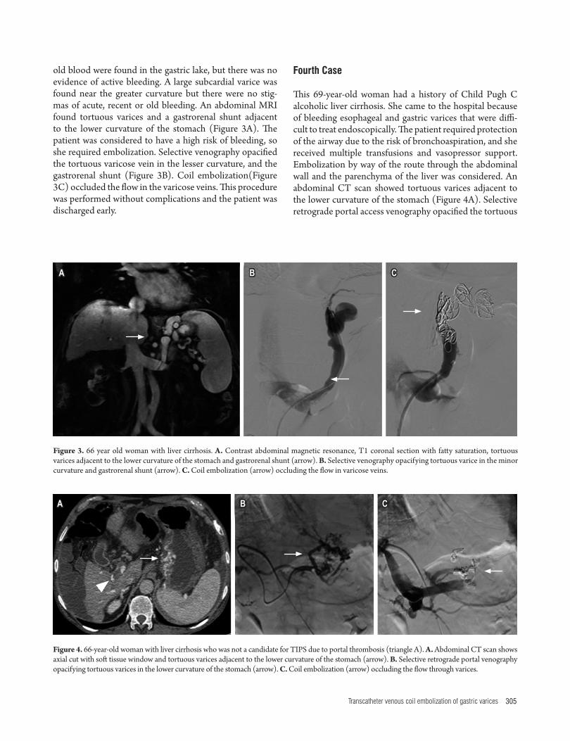
305Transcatheter venous coil embolization of gastric varices
Fourth Case
This 69-year-old woman had a history of Child Pugh C alcoholic liver cirrhosis. She came to the hospital because of bleeding esophageal and gastric varices that were diffi-cult to treat endoscopically. The patient required protection of the airway due to the risk of bronchoaspiration, and she received multiple transfusions and vasopressor support. Embolization by way of the route through the abdominal wall and the parenchyma of the liver was considered. An abdominal CT scan showed tortuous varices adjacent to the lower curvature of the stomach (Figure 4A). Selective retrograde portal access venography opacified the tortuous
old blood were found in the gastric lake, but there was no evidence of active bleeding. A large subcardial varice was found near the greater curvature but there were no stig-mas of acute, recent or old bleeding. An abdominal MRI found tortuous varices and a gastrorenal shunt adjacent to the lower curvature of the stomach (Figure 3A). The patient was considered to have a high risk of bleeding, so she required embolization. Selective venography opacified the tortuous varicose vein in the lesser curvature, and the gastrorenal shunt (Figure 3B). Coil embolization(Figure 3C) occluded the flow in the varicose veins. This procedure was performed without complications and the patient was discharged early.
Figure 4. 66-year-old woman with liver cirrhosis who was not a candidate for TIPS due to portal thrombosis (triangle A). A. Abdominal CT scan shows axial cut with soft tissue window and tortuous varices adjacent to the lower curvature of the stomach (arrow). B. Selective retrograde portal venography opacifying tortuous varices in the lower curvature of the stomach (arrow). C. Coil embolization (arrow) occluding the flow through varices.
A B C
Figure 3. 66 year old woman with liver cirrhosis. A. Contrast abdominal magnetic resonance, T1 coronal section with fatty saturation, tortuous varices adjacent to the lower curvature of the stomach and gastrorenal shunt (arrow). B. Selective venography opacifying tortuous varice in the minor curvature and gastrorenal shunt (arrow). C. Coil embolization (arrow) occluding the flow in varicose veins.
A B C
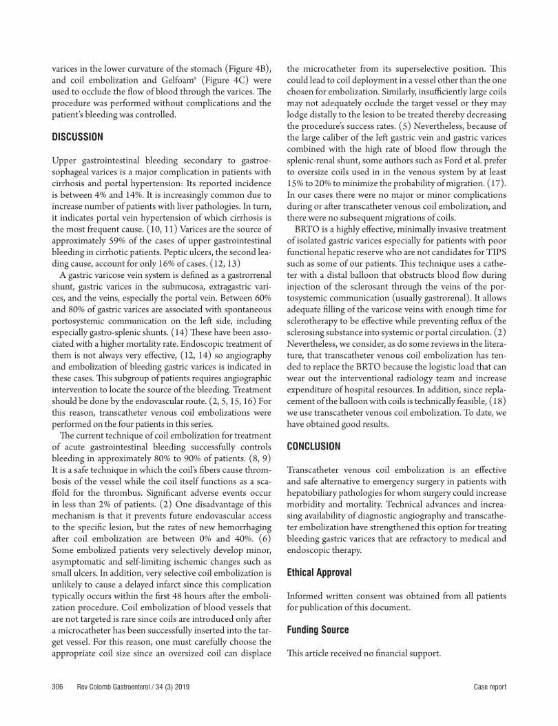
Rev Colomb Gastroenterol / 34 (3) 2019306 Case report
the microcatheter from its superselective position. This could lead to coil deployment in a vessel other than the one chosen for embolization. Similarly, insufficiently large coils may not adequately occlude the target vessel or they may lodge distally to the lesion to be treated thereby decreasing the procedure’s success rates. (5) Nevertheless, because of the large caliber of the left gastric vein and gastric varices combined with the high rate of blood flow through the splenic-renal shunt, some authors such as Ford et al. prefer to oversize coils used in in the venous system by at least 15% to 20% to minimize the probability of migration. (17). In our cases there were no major or minor complications during or after transcatheter venous coil embolization, and there were no subsequent migrations of coils.
BRTO is a highly effective, minimally invasive treatment of isolated gastric varices especially for patients with poor functional hepatic reserve who are not candidates for TIPS such as some of our patients. This technique uses a cathe-ter with a distal balloon that obstructs blood flow during injection of the sclerosant through the veins of the por-tosystemic communication (usually gastrorenal). It allows adequate filling of the varicose veins with enough time for sclerotherapy to be effective while preventing reflux of the sclerosing substance into systemic or portal circulation. (2) Nevertheless, we consider, as do some reviews in the litera-ture, that transcatheter venous coil embolization has ten-ded to replace the BRTO because the logistic load that can wear out the interventional radiology team and increase expenditure of hospital resources. In addition, since repla-cement of the balloon with coils is technically feasible, (18) we use transcatheter venous coil embolization. To date, we have obtained good results.
CONCLUSION
Transcatheter venous coil embolization is an effective and safe alternative to emergency surgery in patients with hepatobiliary pathologies for whom surgery could increase morbidity and mortality. Technical advances and increa-sing availability of diagnostic angiography and transcathe-ter embolization have strengthened this option for treating bleeding gastric varices that are refractory to medical and endoscopic therapy.
Ethical Approval
Informed written consent was obtained from all patients for publication of this document.
Funding Source
This article received no financial support.
varices in the lower curvature of the stomach (Figure 4B), and coil embolization and Gelfoam® (Figure 4C) were used to occlude the flow of blood through the varices. The procedure was performed without complications and the patient’s bleeding was controlled.
DISCUSSION
Upper gastrointestinal bleeding secondary to gastroe-sophageal varices is a major complication in patients with cirrhosis and portal hypertension: Its reported incidence is between 4% and 14%. It is increasingly common due to increase number of patients with liver pathologies. In turn, it indicates portal vein hypertension of which cirrhosis is the most frequent cause. (10, 11) Varices are the source of approximately 59% of the cases of upper gastrointestinal bleeding in cirrhotic patients. Peptic ulcers, the second lea-ding cause, account for only 16% of cases. (12, 13)
A gastric varicose vein system is defined as a gastrorrenal shunt, gastric varices in the submucosa, extragastric vari-ces, and the veins, especially the portal vein. Between 60% and 80% of gastric varices are associated with spontaneous portosystemic communication on the left side, including especially gastro-splenic shunts. (14) These have been asso-ciated with a higher mortality rate. Endoscopic treatment of them is not always very effective, (12, 14) so angiography and embolization of bleeding gastric varices is indicated in these cases. This subgroup of patients requires angiographic intervention to locate the source of the bleeding. Treatment should be done by the endovascular route. (2, 5, 15, 16) For this reason, transcatheter venous coil embolizations were performed on the four patients in this series.
The current technique of coil embolization for treatment of acute gastrointestinal bleeding successfully controls bleeding in approximately 80% to 90% of patients. (8, 9) It is a safe technique in which the coil’s fibers cause throm-bosis of the vessel while the coil itself functions as a sca-ffold for the thrombus. Significant adverse events occur in less than 2% of patients. (2) One disadvantage of this mechanism is that it prevents future endovascular access to the specific lesion, but the rates of new hemorrhaging after coil embolization are between 0% and 40%. (6) Some embolized patients very selectively develop minor, asymptomatic and self-limiting ischemic changes such as small ulcers. In addition, very selective coil embolization is unlikely to cause a delayed infarct since this complication typically occurs within the first 48 hours after the emboli-zation procedure. Coil embolization of blood vessels that are not targeted is rare since coils are introduced only after a microcatheter has been successfully inserted into the tar-get vessel. For this reason, one must carefully choose the appropriate coil size since an oversized coil can displace

307Transcatheter venous coil embolization of gastric varices
Radiol. 2001 Nov;12(11):1263-71. doi: 10.1016/S1051-0443(07)61549-8.
10. Sarin SK, Lahoti D, Saxena SP, Murthy NS, Makwana UK. Prevalence, classification and natural history of gastric vari-ces: a long-term follow-up study in 568 portal hyperten-sion patients. Hepatology. 1992 Dec;16(6):1343-9. doi: 10.1002/hep.1840160607.
11. Castillo O, Palacios F, Yoza M, Contardo C, Soriano CC. Uso de Cianoacrilato en la Terapia Endoscópica de várices Gástricas: Experiencia en el Hospital Nacional Edgardo Rebagliati Martins del 2006 al 2010. Rev Gastroenterol Perú. 2011;31(3):208-215.
12. Gibson JA, Odze RD. Pathology of diseases that cause upper gastrointestinal tract bleeding. Gastrointest Endosc Clin N Am. 2011 Oct;21(4):583-96. doi: 10.1016/j.giec.2011.07.006.
13. Sostres C, Lanas A. Epidemiology and demographics of upper gastrointestinal bleeding: prevalence, incidence, and mortality. Gastrointest Endosc Clin N Am. 2011 Oct;21(4):567-81. doi: 10.1016/j.giec.2011.07.004.
14. Saad WE. Endovascular management of gastric varices. Clin Liver Dis. 2014 Nov;18(4):829-51. doi: 10.1016/j.cld.2014.07.005.
15. Wee E. Management of nonvariceal upper gastrointestinal bleeding. J Postgrad Med. 2011 Apr-Jun;57(2):161-7. doi: 10.4103/0022-3859.81868.
16. Edelman DA, Sugawa C. Lower gastrointestinal bleeding: a review. Surg Endosc. 2007 Apr;21(4):514-20. doi: 10.1007/s00464-006-9191-7.
17. Ford JM, Shah H, Stecker MS, Namyslowski J. Embolization of large gastric varices using vena cava filter and coils. Cardiovasc Intervent Radiol. 2004 Jul-Aug;27(4):366-9. doi: 10.1007/s00270-004-0071-z.
18. Saad WE, Nicholson DB. Optimizing logistics for balloon-occluded retrograde transvenous obliteration (BRTO) of gastric varices by doing away with the indwelling balloon: concept and techniques. Tech Vasc Interv Radiol. 2013 Jun;16(2):152-7. doi: 10.1053/j.tvir.2013.02.006.
REFERENCES
1. Navuluri R, Kang L, Patel J, Van Ha T. Acute lower gas-trointestinal bleeding. Semin Intervent Radiol. 2012 Sep;29(3):178-86. doi: 10.1055/s-0032-1326926.
2. Navuluri R, Patel J, Kang L. Role of interventional radiology in the emergent management of acute upper gastrointestinal bleeding. Semin Intervent Radiol. 2012 Sep;29(3):169-77. doi: 10.1055/s-0032-1326925.
3. Aina R, Oliva VL, Therasse E, Perreault P, Bui BT, Dufresne MP, et al. Arterial embolotherapy for upper gastrointes-tinal hemorrhage: outcome assessment. J Vasc Interv Radiol. 2001 Feb;12(2):195-200. doi: 10.1016/S1051-0443(07)61825-9.
4. Padia SA, Geisinger MA, Newman JS, Pierce G, Obuchowski NA, Sands MJ. Effectiveness of coil embolization in angio-graphically detectable versus non-detectable sources of upper gastrointestinal hemorrhage. J Vasc Interv Radiol. 2009 Apr;20(4):461-6. doi: 10.1016/j.jvir.2009.01.006.
5. Walker TG, Salazar GM, Waltman AC. Angiographic evalua-tion and management of acute gastrointestinal hemorrhage. World J Gastroenterol. 2012 Mar 21;18(11):1191-201. doi: 10.3748/wjg.v18.i11.1191.
6. d’Othée BJ, Surapaneni P, Rabkin D, Nasser I, Clouse M. Microcoil embolization for acute lower gastrointes-tinal bleeding. Cardiovasc Intervent Radiol. 2006 Jan-Feb;29(1):49-58. doi: 10.1007/s00270-004-0301-4.
7. Abdel-Aal AK, Bag AK, Saddekni S, Hamed MF, Ahmed FY. Endovascular management of nonvariceal upper gastroin-testinal hemorrhage. Eur J Gastroenterol Hepatol. 2013 Jul;25(7):755-63. doi: 10.1097/MEG.0b013e32835fb9a9.
8. Patel TH, Cordts PR, Abcarian P, Sawyer MA. Will transcathe-ter embolotherapy replace surgery in the treatment of gastroin-testinal bleeding? (2)(2). Curr Surg. 2001 May;58(3):323-327. doi: 10.1016/S0149-7944(01)00417-2.
9. Schenker MP, Duszak R Jr, Soulen MC, Smith KP, Baum RA, Cope C, et al. Upper gastrointestinal hemorrhage and transcatheter embolotherapy: clinical and techni-cal factors impacting success and survival. J Vasc Interv

© 2019 Asociaciones Colombianas de Gastroenterología, Endoscopia digestiva, Coloproctología y Hepatología308
Santiago Castaño Quintero,1* Natalia Calvache,2 Mauricio Sepúlveda,3 Catalina Maldonado,3 Pedro Tomás Argüello,4 Juliana Escobar,5 Carlos Arturo Rojas.3
An endoscopic videocapsule finding of heterotopia of the gastric mucosa of the small intestine
1 Resident in internal medicine at Universidad CES and Fundación Valle del Lili in Cali, Colombia
2 Physician in the Gastroenterology and Endoscopy Service of Fundación Valle del Lili in Cali, Colombia
3 Gastroenterologist in the Gastroenterology and Endoscopy Service of Fundación Valle del Lili in Cali, Colombia
4 Gastrointestinal surgeon in the Gastroenterology and Endoscopy Service of Fundación Valle del Lili in Cali, Colombia
5 Pathologist in the Pathology Service of Fundación Valle del Lili in Cali, Colombia
*Correspondence: [email protected].
.........................................Received: 05/06/18 Accepted: 13/08/18
AbstractIntroduction: Heterotopia of the gastric mucosa refers to an ectopic location of gastric mucosa in any part of the gastrointestinal tract. It is a rare cause of gastrointestinal ulcers and occult digestive bleeding. Endoscopic videocapsules have become fundamental tools for study of the small intestine. Methods: This is a descriptive case study based on information from the medical history, pathology report and endoscopic studies extracted from the databases of the Fundación Valle del Lili in Cali, Colombia. Results: An endoscopic videocapsule was used to examine a 71-year-old male patient who suffered from occult digestive bleeding. Segments of stenosis and ulcers were found in the jejunum and histopathology revealed heterotopic gastric mucosa. Conclusion: Heterotopy of the gastric mucosa should be thought of as a possible cause of bleeding in the small intestine.
KeywordsGastric heterotopy, occult digestive bleeding, endoscopic videocapsule.
Case reportDOI: https://doi.org/10.22516/25007440.271
INTRODUCTION
Ulcers rarely occur in the small intestine, but endoscopic techniques such as videocapsules and enteroscopy have allowed this type of lesions to be identified more frequently. Intestinal ulcers can manifest clinically with anemia, episodes of abdominal pain, bleeding, obstruction or perforation. (1) Heterotopic gastric mucosa (HGM), the presence of gastric mucosa in an abnormal location, can cause intestinal ulcers although it is rarely found below the Ligament of Treitz. It is difficult to differentiate heterotopia from gastric metaplasia which is associated with Crohn’s disease. (2)
We present the case of the endoscopic videocapsule study of a male patient who had been suffering from digestive blee-ding. Multiple ulcers plus stenosis in were found in several of his small intestine. Histological study revealed the presence of pyloric glands with gastric foveolar epithelium.
CLINICAL CASE
The patient was a 71-year-old man who had a history of hypertension and diverticular disease. He had been referred to the endoscopy unit of the Fundación Valle del Lili as an outpatient for endoscopic videocapsule diagnosis following two years of abdominal pain, colic and the sensation of a mass in the right iliac fossa that were associated with abdo-minal distension and episodic constipation alternating with occasional aqueous diarrhea. Paraclinical studies had found fecal occult blood and iron deficiency anemia refractory to administration of ferrous sulfate. He said that he did not use non-steroidal anti-inflammatory drugs (NSAIDs) and said that he was allergic to them. Reports of upper gastrointesti-nal endoscopy and total colonoscopy conducted elsewhere were normal. He was considered to have digestive bleeding, probably originating in the small intestine.

309An endoscopic videocapsule finding of heterotopia of the gastric mucosa of the small intestine
The patient provided and informed consent and was properly prepared prior to placement of the videocapsule. Erosions and small isolated ulcers were observed in the proximal jejunum. Erosions and deep ulcers with fibrin, retraction of folds, and mucosal edema which appeared to be chronic were found in the middle jejunum together with segments with partial stenoses. The capsule was retained by an ulcerated stenotic lesion in the distal jejunum (Figure 1).
One week after the study, the patient returned with symptoms of an intestinal obstruction. Fluoroscopy docu-mented the capsule in the lower left quadrant of the small intestine. Exploratory laparoscopy found a flange of the omentum causing partial obstruction in the distal ileum of the small intestine. The loop was dilated with thickened and congestive serosa distal to the obstruction. The capsule was trapped in the stenotic segment which required a 6.5 cm resection of the intestine (Figure 2).
The surgical piece contained two ulcerated lesions (1.5 cm x 1 cm and 1 cm x 1 cm) which were reported macros-copically. A microscopic examination showed that the small intestine had areas of mucosal ulceration with granu-lated tissue and fibrin deposits. Foci were found in which foveolar gastric epithelium was observed as well as pylo-ric glands without cytological atypia in the mucosal and submucosal regions. Inflammatory lymphoplasmocytic infiltrate with formation of hyperplastic lymphoid nodu-les was observed. Surgical margins had adequate viability. Immunohistochemical PAS (periodic acid-Schiff) staining was used to check for the MUC-5AC mucin marker. The interpretation of the results indicated that neutral mucin was present in the gastric mucosa represented by the foveo-lar epithelium and the pyloric glands (Figure 3).
Omeprazole was prescribed for the patient during a posto-perative follow-up appointment. Subsequent appointments documented improvement of gastrointestinal symptoms and resolution of anemia. Complementary studies of anti-neutrophil cytoplasmic antibodies (ANCA), anti-Saccha-
romyces cerevisiae antibodies (ASCA), rheumatoid factor, the erythrocyte sedimentation rate (ESR) and qualitative C-reactive protein (PCR) tests were done. The rheumatoid factor test was negative while the C-reactive protein (PCR) test was positive. It was decided to continue clinical follow-up with quarterly laboratory tests.
DISCUSSION
The development and use of the endoscopic videocapsule has enabled diagnosis of various lesions in the small intestine that were previously difficult to identify. (3) This study has become a primary tool for evaluating hemorrhagic lesions, ulcers and tumors in this organ. (4) It is currently considered a first-line study for what was previously called occult diges-tive bleeding, a term that has changed to possible bleeding of the small intestine. This accounts for 5% to 10% of patients who consult for gastrointestinal bleeding. (5-7)
A study by Pandey et al. of patients who were diagnosed by endoscopic videocapsule has shown that the most fre-quent causes of bleeding in the small intestine were angio-dysplasia in up to 23.5% of these patients, Crohn’s disease in 14.7%, and NSAID enteropathy in 11.76%. Ulcers do not commonly cause bleeding in the small intestine although they have been reported in up to 5.8% of the cases. (8) In general, these ulcers are secondary to Crohn’s disease or NSAID enteropathy and are less frequently associated with entities such as Celiac’s disease, Behcet’s disease, unspeci-fied jejunoileitis, vasculitis, gastrointestinal stromal tumors, intestinal adenocarcinoma, lymphoma, multiple myeloma, Meckel’s diverticulum, infections and HGM. (9)
HGM is gastric mucosa in an ectopic location in any part of the gastrointestinal tract. HGM is identified most frequently in the esophagus, followed by the duodenum. (10-12) With the exception of Meckel’s diverticulum, locations below the Ligament of Treitz are extremely rare. There are only a few cases of HGM in the jejunum, ileum, colon and rectum
Figure 1. Endoscopic videocapsule. Concentric ulcerated stenosis, surrounded by mucosal edema.

Rev Colomb Gastroenterol / 34 (3) 2019310 Case report
Figure 2. Surgical piece. Stenotic segment of 6.5 cm of small intestine (ileum). Endoscopic videocapsule (arrow).
Figure 3. Histopathological study. A. Areas covered by the foveolar gastric epithelium (H-E x 10). B. Pyloric glands in the mucosa and submucosa (H-E x 40). C and D. PAS staining with immunohistochemical study with MUC-5AC showing the presence of neutral mucin of the gastric mucosa represented by the foveolar epithelium and the pyloric glands.
in the medical literature. (13, 14) Most reports involving the small intestine present asymptomatic patients whose HGMs were identified by histopathological studies of sam-ples obtained surgically. Nevertheless, associated symptoms of gastrointestinal bleeding, (15, 16) intussusception, (17) ulceration, perforation and intestinal obstruction have been described. In addition, there are cases in which HGMs have become malignant. (14, 18-20) In our patient, the symptoms were abdominal pain and anemia with a bowel obstruction after the endoscopic videocapsule was retained. Findings of multifocal compromises of the small intestine due to several ulcers resulting in various segments of stenosis are very rare situations. There are only two similar cases reported in the literature. One was a 42-year-old patient who had episodes of recurrent abdominal pain, and the other was a 24-year-old
A B
C D

311An endoscopic videocapsule finding of heterotopia of the gastric mucosa of the small intestine
Fordran’s gastrointestinal and liver disease. 10.a edición. Filadelfia: Saunders; 2016. p. 2102-11.
2. Vani M, Nambiar A, Geetha K, Kundil B. Jejunal Gastric Heterotopia causing Multiple Strictures and Perforation Peritonitis- A Case Report with Review of Literature. J Clin Diagn Res. 2017;11(3):ED11-ED12. https://doi.org/10.7860/JCDR/2017/25585.9590.
3. Micic D, Semrad CE. Small Bowel Endoscopy. Curr Treat Options Gastroenterol. 2016;14(2):220-35. https://doi.org/10.1007/s11938-016-0095-x.
4. Ching HL, McAlindon ME, Sidhu R. An update on small bowel endoscopy. Curr Opin Gastroenterol. 2017;33(3):181-8. https://doi.org/10.1097/MOG. 0000000000000346.
5. Gerson LB, Fidler JL, Cave DR, Leighton JA. ACG Clinical Guideline: Diagnosis and Management of Small Bowel Bleeding. Am J Gastroenterol. 2015;110(9):1265-87. https://doi.org/10.1038/ajg.2015.246.
6. Pennazio M, Spada C, Eliakim R, Keuchel M, May A, Mulder CJ, et al. Small-bowel capsule endoscopy and device-assis-ted enteroscopy for diagnosis and treatment of small-bowel disorders: European Society of Gastrointestinal Endoscopy (ESGE) Clinical Guideline. Endoscopy. 2015;47(4):352-76. https://doi.org/10.1055/s-0034-1391855.
7. Gerson LB. Small Bowel Bleeding: Updated Algorithm and Outcomes. Gastrointest Endoscopy Clin N Am. 2017;27(1):171-80. https://doi.org/10.1016/j.giec.2016. 08.010.
8. Pandey V, Ingle M, Pandav N, Parikh P, Patel J, Phadke A, et al. The role of capsule endoscopy in etiological diagno-sis and management of obscure gastrointestinal bleeding. Intest Res. 2016;14(1):69-74. https://doi.org/10.5217/ir.2016.14.1.69.
9. Ersoy O, Harmanci O, Aydinli M, Sivri B, Bayraktar Y. Capability of capsule endoscopy in detecting small bowel ulcers. Dig Dis Sci. 2009;54(1):136-41. https://doi.org/10.1007/s10620-008-0320-0.
10. Cooper JE, Roberts-Thomson IC. Gastrointestinal: heterotopic gastric mucosa. J Gastroenterol Hepatol. 2001;16(4):475.
11. Terada T. Heterotopic gastric mucosa of the gastrointesti-nal tract: a histopathologic study of 158 cases. Pathol Res Pract. 2011;207(3):148-50. https://doi.org/10.1016/j.prp.2010.12.004.
12. Yu L, Yang Y, Cui L, Peng L, Sun G. Heterotopic gastric mucosa of the gastrointestinal tract: prevalence, histological features, and clinical characteristics. Scand J Gastroenterol. 2014;49(2):138-44. https://doi.org/10.3109/00365521.2013.860558.
13. Lee SM, Mosenthal WT, Weismann RE. Tumorous hete-rotopic gastric mucosa in the small intestine. Arch sur-gery. 1970;100(5):619-22. https://doi.org/10.1001/archsurg.1970.01340230085022.
14. Qiao WG, Zhang LZ, Zhi FC. Tumor-like heterotopic gas-tric mucosa discovered by wireless capsule endoscopy. J Dig
patient who had abdominal pain and intestinal perforation. (2, 21) Lesions were identified by endoscopic videocapsule in only one case. (14)
Two types of HGM have been characterized in relation to histological findings. The first has both gastric glands and gastric foveolar epithelium as in our patient. The second has only gastric foveolar epithelium. It is believed that the former has its origin in a congenital abnormality of the gastrointestinal tract, while the latter could be acqui-red through a metaplastic process the mechanism of which remains unknown. (11, 15, 22) There is a possibility that these types of histological lesions are related to Crohn’s disease since outbreaks of pyloric metaplasia have been identified in patients with ileum ulcers and this disease. (23) Koukoulis et al. evaluated 45 biopsies of the termi-nal ileum of different patients and identified pyloric gland metaplasia in 10 (22.2%) specimens. These findings were observed in patients with terminal ileitis and were located below regeneration crypts and close to granulation and ulceration tissue. No granulomas were observed in any of the pieces. The meaning of this type of lesions is not enti-rely clear and has been considered a non-specific finding. (24) Agarwal et al. conducted a retrospective study which found that pyloric metaplasia in patients with inflam-matory bowel disease who had ileoanal anastomoses with ileal pouches were more likely to have Crohn’s disease than ulcerative colitis. In addition, pyloric metaplasia was asso-ciated with postoperative complications such as chronic pouchitis. (25) Although there was not enough evidence to suggest Crohn’s disease in our patient, this possibility cannot be completely ruled out, which makes clinical, para-clinical and imaging follow-up necessary.
CONCLUSION
Endoscopic videocapsules have made it possible to obtain images of the small intestine that were previously not possi-ble. Heterotopic gastric mucosa is a condition that had tra-ditionally been considered to be congenital, one that very rarely compromised the gastrointestinal tract below the Ligament of Treitz. It is very rare to have a multifocal con-dition in the small intestine, as in the case of this patient. Distinguishing HGM from pyloric metaplasia is difficult and still remains a point of discussion. This situation requi-res that patients diagnosed with this condition be strictly monitored clinically, paraclinically and with imaging.
REFERENCES
1. Fisher L, Scheiman JM. Intestinal Ulcerations. En: Feldman M, Friedman LS, Brandt LJ (editores). Sleisenger and

Rev Colomb Gastroenterol / 34 (3) 2019312 Case report
21. Houissa-Vuong S, Martin B, Lascar G, Vuong PN. Multiple jejunal strictures caused by gastric heterotopia. Ann Chir. 2001;126(1):70-4. https://doi.org/10.1016/S0003-3944 (00)00461-2.
22. Genta RM, Kinsey RS, Singhal A, Suterwala S. Gastric foveo-lar metaplasia and gastric heterotopia in the duodenum: no evidence of an etiologic role for Helicobacter pylori. Hum Pathol. 2010;41(11):1593-600. https://doi.org/10.1016/j.humpath.2010.04.010.
23. Kushima R, Borchard F, Hattori T. A new aspect of gastric metaplasia in Crohn’s disease: bidirectional (foveolar and pyloric) differentiation in so-called ‘pyloric metaplasia’ in the ileum. Pathol Int. 1997;47(6):416-9. https://doi.org/10.1111/j.1440-1827.1997.tb04517.x.
24. Koukoulis GK, Ke Y, Henley JD, Cummings OW. Detection of pyloric metaplasia may improve the biopsy diagnosis of Crohn’s ileitis. J Clin Gastroenterol. 2002;34(2):141-3. https://doi.org/10.1097/00004836-200202000-00007.
25. Agarwal S, Stucchi AF, Dendrinos K, Cerda S, O’Brien MJ, Becker JM, et al. Is pyloric gland metaplasia in ileal pouch biopsies a marker for Crohn’s disease? Dig Dis Sci. 2013;58(10):2918-25. https://doi.org/10.1007/s10620-013-2655-4.
Dis. 2017;18(9):543-4. https://doi.org/10.1111/1751-2980.12464.
15. Tai CM, Chang IW, Wang HP. Heterotopic gastric mucosa of the ileum. Endoscopy. 2015;47(Suppl 1 UCTN):E423. https://doi.org/10.1055/s-0034-1392666.
16. Nawaz K, Graham DY, Fechner RE, Eiband JM. Gastric heterotopia in the ileum with ulceration and chronic blee-ding. Gastroenterology. 1974;66(1):113-7. https://doi.org/10.1016/S0016-5085(74)80086-7.
17. Boybeyi O, Karnak I, Güçer S, Orhan D, Senocak ME. Common characteristics of jejunal heterotopic gastric tissue in children: a case report with review of the lite-rature. J Pediatr Surg. 2008;43(7):e19-22. https://doi.org/10.1016/j.jpedsurg.2008.02.072.
18. Martínez A, Decanini-Terán O, Soria-Céspedes D. Polypoid and hyperplastic heterotopic gastric mucosa in the jeju-num as a cause of recurrent subocclusive episodes. Ann Gastroenterol. 2013;26(2):184.
19. Abu-Zidan FM, El-Batrawy TM, Khan NH. Ectopic gastric mucosal ulcer of the jejunum without congenital anomaly causing intestinal obstruction. ANZ J Surg. 2018;88(1-2):E99-E100. https://doi.org/10.1111/ans.13318.
20. Chinnery GE, Bernon MM, Banderker MA, Roberts R, Krige JE. Gastric heterotopia causing jejunal ulceration and obstruction. S Afr J Surg. 2013;51(4):146-7. https://doi.org/10.7196/sajs.1735.

© 2019 Asociaciones Colombianas de Gastroenterología, Endoscopia digestiva, Coloproctología y Hepatología 313
Valeria Atenea Costa Barney,1* Alexander Castañeda Ladino.2
Letter to the Editor
1 Gastroenterologist at the Hospital Universitario San Ignacio and Honorary Professor at the Pontificia Universidad Javeriana in Bogotá, Colombia
2 Second year Gastroenterology and Digestive Endoscopy Fellow at the Hospital Universitario San Ignacio in Bogotá, Colombia
*Correspondence: [email protected]
.........................................Received: 24/07/19Accepted: 26/07/19
Letter to the Editor
Dear Editor:
We would like to respectfully make a series of contributions regarding the original article “Diagnosis and treatment of patients with hereditary hemorrhagic telangiectasia (Osler-Weber-Rendú syndrome) at a university hospital in Colombia” by Dr. Mosquera et al.. Our contributions are on genetic diagnosis, clinical manifestations, medical manage-ment and endoscopic management.
Osler-Weber-Rendú syndrome, also known as hereditary hemorrhagic telangiectasia (HHT), is a disease with an autosomal dominant inheritance pattern characterized by telangiectasias and arteriovenous malformations. (1)
To date, the Curaçao criteria remain the parameters to be considered for diagnosis. It has been reported that these criteria are particularly useful in two situations: unaffec-ted older adults, and young adults and children. This is where genetic testing plays an important role even though it is not widely available and is expensive. It should not be underestimated for making an accurate diagnosis. The alterations described related to endoglin (ENG for type 1 HHT) and the activin type A receptor gene (ACVRL1 for type 2 HHT) genes that account for the majority of HHT cases. They generate protein products that influence signaling TGF-β in vascular endothelial cells. The reported data show detection rates with sensitivity of up to 75% for mutations of ENG and ACVRL1 sequences. (1, 2) We also believe that these tests are relevant because of the different degrees of severity associated with different genotype alterations. Patients with HHT type 1 genotype are more serious and have a higher prevalence of pulmonary arterio-venous malformations and more severe episodes of gastrointestinal bleeding than do patients with HHT type 2. However, no significant changes in the severity of epistaxis, age of presentation and mortality rates have been demonstrated. (3)
On the other hand, more detailed descriptions of clinical manifestations and their frequencies seem relevant since they are data that can help a clinician suspect this disease which otherwise might initially be classified as an orphan disease. At least 90% of patients present nosebleeds, and 80% of HHT patients have gastric or small intestine telangiectasias although only 25% to 30% develop overt bleeding which tends to occur in the fifth to sixth decade of life (rarely before 40 years). (1, 4) These data are similar to those found in the study of Dr. Mosquera et al. It is also important to take into account other manifestations. Cardiac manifestations (acute myocardial infarcts and arrhyth-mia) have low prevalences. Arrhythmia is the most frequent cardiac manifestation. (5)
Rev Colomb Gastroenterol / 34 (3) 2019314 Letter to the Editor
dification, nasal lubrication and saline treatments could be beneficial for controllin epistaxis associated with HHT. They also suggested that modifying the intake of foods high in salicylates and those with natural anti-platelet activity (including red wine, spices, chocolate, coffee, certain types of fruit, garlic, ginger, ginseng, and ginkgo biloba) could be beneficial. (14)
REFERENCES
1. Faughnan ME, Palda VA, Garcia-Tsao G, Geisthoff UW, McDonald J, Proctor DD, et al. International guidelines for the diagnosis and management of hereditary haemorrhagic telangiectasia. J Med Genet. 2011 Feb;48(2):73-87. doi: 10.1136/jmg.2009.069013.
2. Shovlin CL. Hereditary haemorrhagic telangiectasia: patho-physiology, diagnosis and treatment. Blood Rev. 2010 Nov;24(6):203-19. doi: 10.1016/j.blre.2010.07.001.
3. Kjeldsen AD, Møller TR, Brusgaard K, Vase P, Andersen PE. Clinical symptoms according to genotype amongst patients with hereditary haemorrhagic telangiectasia. J Intern Med. 2005 Oct;258(4):349-55. doi: 10.1111/j.1365-2796.2005.01555.x.
4. Iyer VN, Apala DR, Pannu BS, Kotecha A, Brinjikji W, Leise MD, et al. Intravenous Bevacizumab for Refractory Hereditary Hemorrhagic Telangiectasia-Related Epistaxis and Gastrointestinal Bleeding. Mayo Clin Proc. 2018 Feb;93(2):155-166. doi: 10.1016/j.mayocp.2017.11.013.
5. Shovlin CL, Awan I, Cahilog Z, Abdulla FN, Guttmacher AE. Reported cardiac phenotypes in hereditary hemorrha-gic telangiectasia emphasize burdens from arrhythmias, ane-mia and its treatments, but suggest reduced rates of myocar-dial infarction. Int J Cardiol. 2016 Jul 15;215:179-85. doi: 10.1016/j.ijcard.2016.04.006.
6. Buscarini E, Danesino C, Plauchu H, de Fazio C, Olivieri C, Brambilla G, et al. High prevalence of hepatic focal nodular hyperplasia in subjects with hereditary hemorrhagic telan-giectasia. Ultrasound Med Biol. 2004 Sep;30(9):1089-97. doi: 10.1016/j.ultrasmedbio.2004.08.004.
7. van Cutsem E, Rutgeerts P, Vantrappen G. Treatment of bleeding gastrointestinal vascular malformations with oes-trogen-progesterone. Lancet. 1990 Apr 21;335(8695):953-5. doi: 10.1016/0140-6736(90)91010-8.
8. Gaillard S, Dupuis-Girod S, Boutitie F, Rivière S, Morinière S, Hatron PY, et al. Tranexamic acid for epistaxis in heredi-tary hemorrhagic telangiectasia patients: a European cross-over controlled trial in a rare disease. J Thromb Haemost. 2014 Sep;12(9):1494-502. doi: 10.1111/jth.12654.
9. Skaro AI, Marotta PJ, McAlister VC. Regression of cuta-neous and gastrointestinal telangiectasia with sirolimus and aspirin in a patient with hereditary hemorrhagic telan-giectasia. Ann Intern Med. 2006 Feb 7;144(3):226-7. doi: 10.7326/0003-4819-144-3-200602070-00030.
10. Shovlin CL, Sodhi V, McCarthy A, Lasjaunias P, Jackson JE, Sheppard MN. Estimates of maternal risks of pregnancy
There is also evidence that these patients have a higher pre-valence of hepatic focal nodular hyperplasia. (6) We point out these clinical data to complement the article since they were not discussed in the patients of the published series.
As part of the review of the available literature, we would also like to complement the article in regard to clinical and endoscopic management. According to reports in the literature, oral or parenteral iron supplements may be suffi-cient treatment for mild anemia and chronic bleeding of patients with HHT and could even be defined as the first-line. (1) Among the pharmacological treatments described is hormonal therapy (estrogen/progesterone or danacrine preparations). (7) As a second line, antifibrinolytics (ami-nocaproic acid or tranexamic acid) have been used, (8) and there are also reports of the use of tamoxifen, interferon, thalidomide and sirolimus. (9)
In endoscopic therapy, Nd:YAG (neodymium-doped yttrium aluminum garnet; Nd:Y3Al5O12) lasers and argon plasma coagulation (APC) have been described. The latter is considered to be the most effective method avai-lable today. Multiple attempts at local endoscopic therapy are not recommended due to the additive risks of adverse events without additional benefit. (1, 7, 10) There are also data in favor of the use of N-acetylcysteine as an antioxi-dant. Although prospective controlled studies of its effi-cacy have yet to be done, it is considered to be a promising management possibility. (11)
We should not put aside recent guidelines which suggest an approach based on five specific measures to optimize care and reduce morbidity and mortality rates: detection of pulmonary arteriovenous malformations (AVM), advice regarding nasal bleeding, evaluation of iron deficiency, anti-biotic prophylaxis before dental and surgical procedures, and pregnancy advice. It is known that most pregnancies in women with HHT develop normally. Major complica-tions are rare, but survival is better if HHT is recognized and addressed prior to pregnancy. (10, 12)
The Mayo Clinic currently has the most experience mana-ging HHT-related bleeding with intravenous bevacizumab. In general, it is well tolerated, but a relevant adverse effect is arterial hypertension. It is usually benign course and res-ponds well to medical management. From a cost-benefit perspective that considers transfusions, hospitalization time and iron infusions, in the future biological therapy could become an earlier therapeutic approach. (4) There are reports of the use of pazopanib as an alternative for patients who are refractory to bevacizumab, but more controlled and prospective studies of its efficacy are still needed. (13)
Until 2011, there were no data that favored nutritional measures or lifestyle changes for managing this disease. (1) Despite this, in 2013, Silva et al. suggested that room humi-

315Letter to the Editor
Hereditary Haemorrhagic Telangiectasia (HHT). Orphanet J Rare Dis. 2018 Aug 15;13(1):136. doi: 10.1186/s13023-018-0850-2.
13. Parambil JG, Woodard TD, Koc ON. Pazopanib effec-tive for bevacizumab-unresponsive epistaxis in heredi-tary hemorrhagic telangiectasia. Laryngoscope. 2018 Oct;128(10):2234-2236. doi: 10.1002/lary.27129.
14. Silva BM, Hosman AE, Devlin HL, Shovlin CL. Lifestyle and dietary influences on nosebleed severity in heredi-tary hemorrhagic telangiectasia. Laryngoscope. 2013 May;123(5):1092-9. doi: 10.1002/lary.23893.
for women with hereditary haemorrhagic telangiectasia (Osler-Weber-Rendu syndrome): suggested approach for obstetric services. BJOG. 2008 Aug;115(9):1108-15. doi: 10.1111/j.1471-0528.2008.01786.x.
11. de Gussem EM, Snijder RJ, Disch FJ, Zanen P, Westermann CJ, Mager JJ. The effect of N-acetylcysteine on epistaxis and quality of life in patients with HHT: a pilot study. Rhinology. 2009 Mar;47(1):85-8.
12. Shovlin CL, Buscarini E, Kjeldsen AD, Mager HJ, Sabba C, Droege F, et al. European Reference Network For Rare Vascular Diseases (VASCERN) Outcome Measures For

© 2019 Asociaciones Colombianas de Gastroenterología, Endoscopia digestiva, Coloproctología y Hepatología316
Gabriel Mosquera-Klinger,1* Kenny Gálvez Cárdenas,2 Alejandro Ocampo Hincapié.3
Reply to the letter to the Editor
1 Internist and gastroenterologist in the Gastroenterology and Digestive Endoscopy Unit at the Hospital Pablo Tobón Uribe in Medellín, Colombia
2 Internist and hematologist at the Hospital Pablo Tobón Uribe in Medellín, Colombia
3 Coordinator of Non-medical Plan Services and Support in the Admissions Department of the Hospital Pablo Tobón Uribe in Medellín, Colombia
*Correspondence: [email protected]
.........................................Received: 29/07/19Accepted: 30/07/19
Letter to the Editor
Dear Editor:
We thank you in advance for the contributions received by from Dr. Costa Barney and Dr. Castañeda. We are pleased to know that our case series has been reviewed and analy-zed by this honorable working group. Their valuable comments can certainly contribute to managing and monitoring these patients who suffer from such a complex condition, and perhaps interesting guidelines can be established for future studies in this field. For us, it is a pleasure that our work has raised concerns that can be discussed in the acade-mic environment of this type of publication.
The Curacao criteria remain the diagnostic gold standard, especially when three or more of its criteria are present (defined diagnosis). (1, 2) Confirmation of a defined diag-nosis is not required because current management recommendations remain unchan-ged except in the rare situation of the SMAD4 mutation with the theoretical risk of its association with juvenile polyposis. (3) Incidentally, this is the rarest of the genetic mutations identified in this disease.
We agree that this disease is uncommon in our environment. In fact, it is one of the 2,271 diseases that are officially listed as orphan-rare diseases in Colombia. Because it is an orphan disease, it is mandatory to report all cases to SIVIGILA (Sistema de Información para la Vigilancia en Salud Pública - Public Health Surveillance Information System). This disease’s code number is 844 while its ICD-10 code number is I780. Based on the provisions of current regulations regarding the national registry and notification of patients with orphan diseases, we want to clarify that when no confirmatory diagnos-tic test has been determined for an orphan disease, or when no such test is available in Colombia, notification will be made on the basis of the clinical diagnosis declared by one or more of the treating doctors. The declaration of a clinically confirmed orphan disease will be made based on scientifically accepted classifications, medical history and other patient records that confirm the presence of the orphan disease (Ministry of Social Protection, Resolution 946 of April 22, 2019). We make this clarification because, as mentioned in our study, all patients included therein were diagnosed on the basis of the Curacao criteria. Also, genetic tests are not often available in our environment even though they are part of the procedures of the health benefits plan.
Currently there are only 15 sites in Colombia that can do genetic and metabolic tests for this disease. They are mostly based in Bogotá, and none are in Medellín. In addition, although these sites collect the samples, they are processed in laboratories in the United

317Reply to the letter to the Editor
A retrospective review found no data in the medical records that suggest complications during endoscopic procedures despite multiple interventions.
Regarding the general recommendations provided by the literature review by Dr. Costa Barney and Dr. Castañeda, we can only comment that patients with Rendu-Osler-Weber syndrome are complex and usually require multidiscipli-nary management. For this reason, any intervention aimed at improving the quality of life or increasing the life expec-tancy of these patients might be useful, but interventions should be individualized and should weigh risks against benefits. Of course, the context must be analyzed to be fair in terms of the costs and benefits of each intervention, since it is possible that one patient may require multiple inter-ventions during the natural evolution of the disease.
REFERENCES
1. Faughnan ME, Palda VA, Garcia-Tsao G, Geistho UW, McDonald J, Proctor DD, et al. International guidelines for the diagnosis and management of hereditary haemorr-hagic telangiectasia. J Med Genet. 2011;48(2):73-87. doi: 10.1136/jmg.2009.069013.
2. Sharathkumar AA, Shapiro A. Hereditary haemorrhagic telangiectasia. Haemophilia 2008;14(6):1269-80. doi: 10.1111/j.1365-2516.2008.01774.x.
3. Shovlin CL. Hereditary haemorrhagic telangiectasia: Pathophysiology, diagnosis and treatment. Blood Rev. 2010 Nov;24(6):203-19. doi: 10.1016/j.blre.2010.07.001.
4. Kjeldsen AD, Møller TR, Brusgaard K, Vase P, Andersen PE. Clinical symptoms according to genotype amongst patients with hereditary haemorrhagic telangiectasia. J Intern Med. 2005;258(4):349-55. doi: 10.1111/j.1365-2796.2005.01555.x.
5. Mosquera-Klinger G, Gálvez-Cárdenas K, Valencia AM. Diagnóstico y tratamiento de pacientes con telan-giectasia hemorrágica hereditaria (síndrome de Rendu-Osler-Weber) en un hospital universitario en Colombia. Rev Colomb Gastroenterol 2019;34(2):152-158. doi: 10.22516/25007440.280.
States. The confirmatory genetic tests for this disease avai-lable in these centers and suggested by the Ministry are for ACVRL1, ENG, SMAD4, MADH4, GDF2. Each test has an estimated cost of 4 million pesos and average delivery time is 50 days. We reiterate that, in the absence of genetic confirmatory tests, a defined clinical diagnosis of Rendu-Osler-Weber syndrome on the basis of the Curaçao criteria is sufficient. Hopefully, a diagnosis will be endorsed by a multidisciplinary group. For this reason, our discussion cites Kjeldsen et al. At follow-ups of more than 7 years, they found no significant differences in the mortality rates of patients related to genetic diagnoses and/or establishment of disease subtype. (4) With the data described, we confirm that our position is only to request these tests in selected patients who do not meet the defined criteria of Curaçao. We believe that genetic tests can be useful and may also be requested for asymptomatic first-degree relatives without stigmata of the disease as an initial screening method.
We would like to take advantage of this space to suggest development of a multicenter study that can bring together the majority of patients with this disease in Colombia. Perhaps with more data we can propose a follow-up stra-tegy based on the best currently available evidence.
Regarding clinical manifestations, we agree that exten-sion of this description could be of interest to clinicians. The fact that the patients described in our series had no car-diac manifestations can be explained by the low prevalen-ces described in other series. Four of the six patients with hepatic manifestations had vascular malformations while two had hepatic focal nodular hyperplasia.
We agree with what has been described about medical treatment. The data in our work reflect only the actions performed on the patients treated at our center. (5) For patients with very extensive disease who frequently con-sulted the emergency department due to bleeding, the approach was almost always to initially stabilize the patient. In cases of severe anemia, blood products were transfused. If endoscopy documented high-risk stigmas or recent blee-ding, argon plasma therapy was administered, as described.
Copyright © 2022 FDOKUMEN