REVISTA ESPAÑOLA DE
130
REVISTA ESPAÑOLA DE REVISTA ESPAÑOLA DE Q uimioterapia Q uimioterapia ISSN: 0214-3429 Volumen 32 Número 4 Agosto 2019 Páginas: 288-409 SPANISH JOURNAL OF CHEMOTHERAPY Imagen portada: María Teresa Corcuera Publicación Oficial de la Sociedad Española de Quimioterapia
-
Upload
khangminh22 -
Category
Documents
-
view
0 -
download
0
Transcript of REVISTA ESPAÑOLA DE

R E V I S T A E S P A Ñ O L A D ER E V I S T A E S P A Ñ O L A D EQuimioterapiaQuimioterapiaISSN: 0214-3429Volumen 32Número 4Agosto 2019Páginas: 288-409
SPANISH JOURNALOF CHEMOTHERAPY
Imagen portada: María Teresa Corcuera
Publicación Oficialde la Sociedad Españolade Quimioterapia

QuimioterapiaR E V I S T A E S P A Ñ O L A D E
Revista Española de Quimioterapia tiene un carácter multidisciplinar y está dirigida a todos aquellos profesionales involucrados en la epidemiología, diagnóstico, clínica y tratamiento de las enfermedades infecciosas
Fundada en 1988 por la Sociedad Española de Quimioterapia
Indexada enScience Citation IndexExpanded (SCI),Index Medicus (MEDLINE), Excerpta Medica/EMBASE,Índice Médico Español (IME), Índice Bibliográfico en Ciencias de la Salud (IBECS)
Secretaría técnica Dpto. de MicrobiologíaFacultad de MedicinaAvda. Complutense, s/n28040 [email protected] en Internet:www.seq.es
Publicidad y SuscripcionesSociedad Española de QuimioterapiaDpto. de MicrobiologíaFacultad de MedicinaAvda. Complutense, s/n28040 Madrid
Atención al clienteTeléfono 91 394 15 12Correo electró[email protected]
Consulte nuestra página webwww.seq.es
© Copyright 2019Sociedad Española deQuimioterapia
Reservados todos los derechos.Queda rigurosamente prohibida,sin la autorización escrita deleditor, la reproducción parcialo total de esta publicaciónpor cualquier medio oprocedimiento, comprendidos lareprografía y el tratamientoinformático, y la distribución deejemplares mediante alquiler opréstamo públicos, bajo lassanciones establecidas por la ley
Sociedad Española de Quimioterapia
Publicación que cumple los requisitos desoporte válido
ISSN0214-3429
e-ISSN1988-9518
Depósito LegalM-32320-2012
MaquetaciónKumisai
ImpresiónEspaña
Esta publicación se imprime en papel no ácido. This publication is printed in acid free paper.
LOPDInformamos a los lectores que, según lo previsto en el Reglamento General de Protección de Datos (RGPD) 2016/679 del Parlamento Europeo, sus datos personales forman parte de la base de datos de la Sociedad Española de Quimioterapia (si es usted socio)
Si desea realizar cualquier rectificación o cancelación de los mismos, deberá enviar una solicitud por e-mail a la Sociedad Española de Quimioterapia ([email protected])

QuimioterapiaR E V I S T A E S P A Ñ O L A D E
DirectorJ. Barberán López
Comité Editorial
F. Álvarez Lerma (Barcelona)F. Baquero Mochales (Madrid)
E. Bouza Santiago (Madrid)J. A. García Rodríguez (Salamanca)M. Gobernado Serrano (Valencia)
Consejo Editorial
L. Aguilar (Madrid)J. I. Alós (Madrid)J. R. Azanza (Pamplona)J. Aragón (Las Palmas de Gran Canaria) A. Artero (Valencia)V. Asensi (Oviedo)G. Barbeito (Santiago de Compostela)J. M. Barbero (Madrid)J. Campos (Madrid)F.J. Candel (Madrid)E. Cantón (Valencia)R. Cantón (Madrid)J. A. Capdevila Morell(Barcelona)M. Casal (Córdoba)J. Castillo (Zaragoza) F. Cobo (Granada)J. Cobo Reinoso (Madrid)N. Cobos (Madrid)J. L. del Pozo (Navarra)R. De la Cámara (Madrid)C. De la Calle (Barcelona)M. Dominguez-Gil (Valladolid)J. Eiros (Valladolid)P. Escribano (Madrid)A. Estella (Cádiz)M. C. Fariñas Álvarez (Santander)C. Fariñas (Santander)
J. Fortún (Madrid)J. J. Gamazo (Vizcaya)E. García Sánchez (Salamanca)I. García García (Salamanca)J. E. García Sánchez (Salamanca)E. García Vázquez (Murcia)J. Gómez Gómez (Murcia)M. L. Gómez-Lus (Madrid)J. González del Castillo (Madrid)F. González Romo (Madrid)J. J. Granizo (Madrid)S. Grau (Barcelona)J.M. Guardiola (Barcelona)J. Guinea (Madrid)X. Guirao (Barcelona)J. Gutiérrez (Granada)J. B. Gutiérrez (Córdoba)B. Isidoro (Madrid)P. Llinares (La Coruña)J. E. Losa Garcia (Madrid)J. R. Maestre Vera (Madrid)L. Martínez Martínez (Córdoba)E. Maseda (Madrid)R. Menéndez (Valencia)P. Merino (Madrid))P. Muñoz (Madrid)J. L. Muñoz Bellido (Salamanca)V. Navarro (Alicante)
M. Ortega (Barcelona) J. Oteo (Madrid)J. A. Oteo (Logroño)E. Palencia Herrejón (Madrid) A. Pascual Hernández (Sevilla)J. Pasquau (Sevilla)J. Pemán (Valencia)J. L Pérez-Arellano (Las Palmas)B. Pérez-Gorricho (Madrid)A. Ramos (Madrid)J. M. Ramos (Alicante)J. Reina (Palma de Mallorca)M. A. Ripoll (Ávila)I. Rodriguez-Avial (Madrid)M. Ruiz (Alicante)M. Sabriá (Barcelona)M. Salavert (Valencia)B. Sánchez Artola (Madrid)M. Segovia (Murcia)R. Serrano (Madrid)D. Sevillano (Madrid)A. Suárez (Madrid)A. Tenorio (Huelva)A. Torres (Murcia)C. Vallejo (Oviedo)J. Vila (Barcelona)J. Yuste (Madrid)
J. Mensa Pueyo (Barcelona)J. J. Picazo de la Garza (Madrid)
J. Prieto Prieto (Madrid)B. Regueiro García (Santiago de Compostela)
A. Torres Martí (Barcelona)
Secretario de RedacciónLuis Alou Cervera

Sumario
Síndrome de Guillain-Barré y vacuna antigripal: evidencia actual 288Rosario Sanz Fadrique, Luis Martín Arias, Juan Arcadio Molina-Guarneros, Natalia Jimeno Bulnes, Pilar García Ortega
Efectividad de ledipasvir/sofosbuvir durante 12 semanas de tratamiento y factores 296 predictivos de fracaso del tratamiento en pacientes con hepatitis CJuan Carlos del Rio-Valencia, Rocío Asensi-Diez, Rocío Tamayo-Bermejo, Isabel Muñoz-Castillo
Bacterias resistentes en las manos de trabajadores sanitarios y en el área de 303 paciente: un estudio ambiental en un hospital del sur de ItaliaVincenza La Fauci, Gaetano Bruno Costa, Cristina Genovese, Maria Angela Rita Palamara, Valeria Alessi, Raffaele Squeri
Factores de riesgo en bacteriemias nosocomiales secundarias a ITU en un hospital 311 terciarioLaura Sante, María Lecuona, Armando Aguirre Jaime, Ángeles Arias
Diagnóstico al alta y causas de mortalidad de pacientes VIH+ ingresados en un 317 hospital de tercer nivelRocío Asensi-Diez, Cristina Fernández-Cuerva, Juan José Alcaraz Sánchez, Isabel Muñoz-Castillo
Prevalencia de micoplasmas genitales y respuesta al tratamiento de descolonización 327 en pacientes de reproducción humana asistidaErnesto Veiga, Mercedes Treviño, Ana Belén Romay, Daniel Navarro, Rocío Trastoy, Manuel Macía
Vacunas para la prevención de infecciones en adultos: artículo de opinión sobre la 333 situación en EspañaEmilio Bouza, Julio Ancochea-Bermúdez, Magda Campins, José María Eirós-Bouza, Jesús Fargas, Amós José García Rojas, Diego Gracia, Alipio Gutiérrez Sánchez, Aurora Limia, José Antonio López, María del Carmen Magro, Gloria Mirada, Patricia Muñoz, Eduardo Olier, Raúl Ortiz de Lejarazu, Luis Urbiztondo, Esteban Palomo
Comparación de dos métodos que eluden la lisis celular y la extracción de proteínas 365 para la identificación de bacterias crecidas en hemocultivos mediante espectrometría de masas MALDI-TOF Ignacio Torres, Estela Giménez, Dixie Huntley, Mireia Martínez, Javier Colomina, David Navarro
Evaluación de una prueba rápida para la detección de PBP2a en 370 Staphylococcus aureusRocío Sáinz-Rodríguez, Inmaculada de Toro-Peinado, Miriam Valverde-Troya, Mª Pilar Bermúdez Ruíz, Begoña Palop-Borrás
¿Es posible extrapolar las tasas de resistencia de Escherichia coli de bacteriurias 375 asintomáticas en gestantes a las de E. coli en ITU no complicada adquirida en la comunidad?Alejandra Asenjo, Martín C. Grados, Jesús Oteo-Iglesias, Juan-Ignacio Alós
Revisión Sistemática
Originales
Original Breve
QuimioterapiaR E V I S T A E S P A Ñ O L A D E
Volumen 32Número 4Agosto 2019

Sumario
Reactivación del síndrome periódico asociado a criopirinas tras la vacunación 379 en una paciente candidata a inmunosupresiónMaría Fernández-Prada, Jessica Rugeles-Niño, Lucía Suárez-Pérez, Miguel Ruiz-Álvarez, José Bernardino López-Díaz, Juan Fernández-Madera, Carmen Martínez-Ortega, Ismael Huerta-González
Meningitis por Nocardia farcinica en un paciente con enfermedad de Behçet: 381 caso y revisión de la literaturaKiana Shirani, Fatemeh Mohajeri
Desbridamiento y tratamiento antibiótico en un caso de infección 384 protésica de rodillla por Pasteurella multocidaMaria Pilar Ortega Lafont, Luis Buzón Martín, Ledicia Álvarez Paredes, María Mora Fernández, Antonio Rodríguez Pérez, Miguel Ángel Morán Rodríguez
Uso de dolutegravir en infección aguda por VIH-1: primer caso reportado 387Franco Garibaldi, Marcia Pozzati, Claudia Rodriguez, Diego Cecchini
Shock séptico secundario a Elizabethkingia meningoseptica: descripción de un caso 390David Brandariz Núñez, Paula Rodríguez Pedreira, Virginia Hernández Corredoira, Joaquin Puig Forcada, Francisco J Gurri Hernand, Arturo S Boix Boix
Bacteriemia por Actynomices oris 393Javier Miguel Martín Guerra, Miguel Martín Asenjo, Claudia Iglesias Pérez, Carlos Jesús Dueñas Gutiérrez
Endocarditis infecciosa por Candida glabrata: evidencia del desarrollo in vivo de 395 resistencia a equinocandinasCaroline Agnelli, Jesus Guinea, Maricela Valerio, Pilar Escribano, Emilio Bouza, Patricia Muñoz
Absceso periamigdalino producido por Actinomyces marseillensis 398Sara Gómez de Frutos, Marta Soledad Rodríguez Anzules, Arturo Manuel Fraile Torres, Laura Cardeñoso Domingo, Diego Domingo García
Documento de consenso para la implantación y desarrollo del Código Sepsis en la 400 Comunidad de MadridEduardo Palencia Herrejón, Juan González del Castillo, Fernando Ramasco Rueda, Francisco Javier Candel González, Beatriz Sánchez Artola, Andrés von Wernitz Teleki, Federico Gordo Vidal, Patricia Roces Iglesias, Guillermina Bejarano Redondo, Diego Aníbal Rodríguez Serrano, Francisco Javier Cobo Reinoso, Ervigio Corral Torres, Milagros Martí de Gracia, Ana Ruiz Álvarez y el Grupo Multidisciplinar Código Sepsis Madrid
QuimioterapiaR E V I S T A E S P A Ñ O L A D E
Volumen 32Número 4Agosto 2019
Cartas al Director
Documento de Consenso


Contents
QuimioterapiaR E V I S T A E S P A Ñ O L A D E
Volume 32Number 4August 2019
Guillain-Barré syndrome and influenza vaccines: current evidence 288Rosario Sanz Fadrique, Luis Martín Arias, Juan Arcadio Molina-Guarneros, Natalia Jimeno Bulnes, Pilar García Ortega
Effectiveness of 12 week ledipasvir/sofosbuvir and predictors of treatment failure in 296 patients with hepatitis CJuan Carlos del Rio-Valencia, Rocío Asensi-Diez, Rocío Tamayo-Bermejo, Isabel Muñoz-Castillo
Drug-resistant bacteria on hands of healthcare workers and in the patient area: 303 an environmental survey in Southern Italy’s hospitalVincenza La Fauci, Gaetano Bruno Costa, Cristina Genovese, Maria Angela Rita Palamara, Valeria Alessi, Raffaele Squeri
Risk factors to secondary nosocomial bacteremia to UTI in a tertiary hospital 311Laura Sante, María Lecuona, Armando Aguirre Jaime, Ángeles Arias
Hospital admission and mortality causes of HIV patients in a third level hospital 317Rocío Asensi-Diez, Cristina Fernández-Cuerva, Juan José Alcaraz Sánchez, Isabel Muñoz-Castillo
Prevalence of genital Mycoplasma and response to eradication treatment in patients 327 undergoing assisted reproductive techniquesErnesto Veiga, Mercedes Treviño, Ana Belén Romay, Daniel Navarro, Rocío Trastoy, Manuel Macía
The situation of vaccines for the prevention of infections in adults: An opinion 333 paper on the situation in SpainEmilio Bouza, Julio Ancochea-Bermúdez, Magda Campins, José María Eirós-Bouza, Jesús Fargas, Amós José García Rojas, Diego Gracia, Alipio Gutiérrez Sánchez, Aurora Limia, José Antonio López, María del Carmen Magro, Gloria Mirada, Patricia Muñoz, Eduardo Olier, Raúl Ortiz de Lejarazu, Luis Urbiztondo, Esteban Palomo
Comparison of two methods skipping cell lysis and protein extraction for 365 identification of bacteria from blood cultures by matrix-assisted laser desorption/ionization time-of-flight mass-spectrometryIgnacio Torres, Estela Giménez, Dixie Huntley, Mireia Martínez, Javier Colomina, David Navarro
Evaluation of a rapid assay for detection of PBP2a Staphylococcus aureus 370Rocío Sáinz-Rodríguez, Inmaculada de Toro-Peinado, Miriam Valverde-Troya, Mª Pilar Bermúdez Ruíz, Begoña Palop-Borrás
Is it possible to extrapolate the rates of resistance of Escherichia coli from 375 asymptomatic bacteriuria in pregnant women to those of E. coli in uncomplicated community-acquired UTI?Alejandra Asenjo, Martín C. Grados, Jesús Oteo-Iglesias, Juan-Ignacio Alós
Systematic Review
Originals
Brief Report

Contents
QuimioterapiaR E V I S T A E S P A Ñ O L A D E
Reactivation of the cryopyrin-associated periodic syndrome after vaccination 379 in a patient who is a candidate for immunosuppressionMaría Fernández-Prada, Jessica Rugeles-Niño, Lucía Suárez-Pérez, Miguel Ruiz-Álvarez, José Bernardino López-Díaz, Juan Fernández-Madera, Carmen Martínez-Ortega, Ismael Huerta-González
Nocardia farcinica isolated meningitis in a patient with Behçet’s disease: 381 case report and literature reviewKiana Shirani, Fatemeh Mohajeri
Acute zoonotic total knee prosthetic joint infection due to Pasteurella multocida 384 treated successfully with debridement, irrigation and antibiotics without prosthesis removalMaria Pilar Ortega Lafont, Luis Buzón Martín, Ledicia Álvarez Paredes, María Mora Fernández, Antonio Rodríguez Pérez, Miguel Ángel Morán Rodríguez
Dolutegravir in acute HIV-1 infection: first reported case 387Franco Garibaldi, Marcia Pozzati, Claudia Rodriguez, Diego Cecchini
Septic shock secondary to Elizabethkingia meningoseptica: A case report. 390David Brandariz Núñez, Paula Rodríguez Pedreira, Virginia Hernández Corredoira, Joaquin Puig Forcada, Francisco J Gurri Hernand, Arturo S Boix Boix
Bacteremia by Actynomices oris 393Javier Miguel Martín Guerra, Miguel Martín Asenjo, Claudia Iglesias Pérez, Carlos Jesús Dueñas Gutiérrez
Infectious endocarditis caused by Candida glabrata: evidence of in vivo development 395 of echinocandin resistanceCaroline Agnelli, Jesus Guinea, Maricela Valerio, Pilar Escribano, Emilio Bouza, Patricia Muñoz
Peritonsillar abscess due to Actinomyces marseillensis 398Sara Gómez de Frutos, Marta Soledad Rodríguez Anzules, Arturo Manuel Fraile Torres, Laura Cardeñoso Domingo, Diego Domingo García
Consensus document for sepsis code implementation and development in 400 the Community of Madrid Eduardo Palencia Herrejón, Juan González del Castillo, Fernando Ramasco Rueda, Francisco Javier Candel González, Beatriz Sánchez Artola, Andrés von Wernitz Teleki, Federico Gordo Vidal, Patricia Roces Iglesias, Guillermina Bejarano Redondo, Diego Aníbal Rodríguez Serrano, Francisco Javier Cobo Reinoso, Ervigio Corral Torres, Milagros Martí de Gracia, Ana Ruiz Álvarez y el Grupo Multidisciplinar Código Sepsis Madrid
Volume 32Number 4August 2019
Letters to the editor
Consensus Document

Rev Esp Quimioter 2019;32(4): 288-295 288
©The Author 2019. Published by Sociedad Española de Quimioterapia. This article is distributed under the terms of the Creative Commons Attribution-NonCommercial 4.0 International (CC BY-NC 4.0)(https://creativecommons.org/licenses/by-nc/4.0/).
Síndrome de Guillain-Barré y vacuna antigripal: evidencia actual
RESUMEN
Introducción. El síndrome de Guillain-Barré (GBS) después de la administración de la vacuna frente a la gripe es un tema actual que sigue causando preocupación tanto en el personal sanitario como en la población y que permanece sin esclarecer. El objetivo del presente trabajo es investigar la publicación de nuevos datos desde la realización de nuestro metaanálisis so-bre el GBS y las vacunas frente a la gripe (publicado en 2015).
Métodos. Se ha realizado una revisión sistemática en las bases de datos PubMed, Embase y Web of Science (WOS) de estudios observacionales que evaluarán el riesgo de GBS des-pués de la administración de vacunas influenza, desde mayo de 2014 hasta el 20 de julio de 2017.
Resultados. El resultado de las búsquedas fue de 107 artí-culos. Finalmente, solo 3 estudios cumplían con los criterios de inclusión establecidos y referían una estimación del riesgo de GBS después de alguna de las vacunas antigripales. Dos estu-dios investigaron el riesgo de GBS con la vacuna pandémica A/H1N1 y un estudio investigó las vacunas estacionales.
Conclusiones. Esta revisión sistemática parece confirmar los hallazgos obtenidos en nuestro metaanálisis. El SGB se po-dría considerar como un posible efecto adverso poco frecuente de las vacuna antigripales, lo cual no debería afectar negati-vamente en su aceptación. Desafortunadamente, en nuestra revisión sistemática, hemos encontrado muy pocos estudios que cumplieran los criterios de inclusión, este hecho resulta llamativo ya que el consenso actual señala la necesidad de una monitorización continua sobre la seguridad de las vacunas an-tigripales.
Palabras clave: vacuna A/H1N1, Síndrome de Guillain-Barré, gripe.
ABSTRACT
Purpose. Guillain-Barré Syndrome (GBS) as a consequence of influenza vaccination is a relevant topic, yet to be clarified, which raises concern both amongst health care personnel and the general population. Every study and pharmacovigilance system point to need of further research and the importance of continuous monitoring of safety regarding influenza vaccines. The aim of the present study is to investigate the publication of new data since the realisation of our meta-analysis of GBS and influenza vaccines (published in 2015).
Methods. A systematic revision of PubMed, Embase, and Web of Knowledge (WOS) databases has been carried out. These report observational studies assessing GBS risk after the administration of influenza vaccines from May 2014 up to July 20th, 2017.
Results. The research yielded 107 articles. Only three studies met established inclusion criteria and referred to an estimation GBS risk after some influenza vaccine. Two studies investigated GBS risk by the pandemic A/H1N1 vaccine, while only one looked into season vaccines.
Conclusions. The present systematic review, conducted after the publication of our previous meta-analysis, seems to confirm its previous results. Therefore, GBS should be considered an infrequent adverse effect of influenza vaccination, which should not negatively influence its acceptance. Unfortunately, very few of the systematically surveyed studies meeting inclusion criteria. This fact sharply contrasts with the current consensus as to the need of continuously monitoring the safety of influenza vaccines.
Keywords: A/H1N1 vaccine, Guillain-Barré syndrome, influenza vaccines.
Guillain-Barré syndrome and influenza vaccines: current evidence
1Centre for Drug Surveillance (CESME). Valladolid University, Spain.2Department of Pharmacology, Faculty of Medicine. National Autonomous University of Mexico.3Faculty of Medicine, Valladolid University Spain.
Rosario Sanz Fadrique1
Luis Martín Arias1
Juan Arcadio Molina-Guarneros2
Natalia Jimeno Bulnes3
Pilar García Ortega3
Correspondence:Juan A. Molina Guarneros.School of MedicineC/Ramón y Cajal, 7 - 45005 Valladolid (Spain)Phone: 983 263021 / 983 186456Fax.: 983 254924E-mail: [email protected]
Systematic review
Article historyReceived: 11 March 2019; Revision Requested: 14 April 2019; Revision Received: 1 May 2019; Accepted: 7 May 2019

Guillain-Barré syndrome and influenza vaccines: current evidenceR. Sanz, et al.
Rev Esp Quimioter 2019;32(4): 288-295 289
RESULTS
The bibliographic review enabled us to identify 107 studies (figure 1): PubMed (n=39), EMBASE (n=44), and WoS (n=24). First, we eliminated the articles lacking an abstract (n=15), those containing lectures and oral comments (n=7), those no-tifying isolated cases (n=3), duplicated articles (n=30), and ar-ticles previously reviewed or included in our earlier meta-anal-ysis (n=10) [16]. Then, we read the article abstracts and/or the whole texts of the potentially eligible studies. Some of the exclusion criteria were the following: a) studies based on the results from surveillance system notifications of serious and non-serious adverse effects supposedly related to any of the influenza vaccines that did not report any estimates of GBS risk following influenza vaccination [17-22], b) pharmacovigi-lance studies on GBS that included any types of paediatric im-munisation [23], c) studies addressing the potential triggering agents for GBS [24], c) studies based on risk simulation of GBS and other autoimmune diseases associated with either infec-tions or influenza vaccination [25, 26], d) studies whose design or analysis of data on vaccine safety was considered not to be suitable [27, 28, 29], and e) studies focusing on other influenza vaccination adverse effects, such as polyneuropathies, neuro-logical events, Zika virus or surgery
Only 3 studies met all the eligibility criteria (table 1), as follows: Kim et al.; 2015 [30], in which an increased risk of GBS was observed in South Korea following the administration of the adjuvanted and non-adjuvanted pandemic vaccine A/H1N1, with the risk expressed as a rate ratio (RR=1.46, 95% CI: 1.26-1.68); Ghaderi et al.; 2015 [31], in which the authors found an increased risk of GBS in Norway within 42 days after the administration of the pandemic vaccine A/H1N1 (Pandem-rix®), with the risk expressed as a hazard ratio (HR= 1.1, 95% CI: 0.51-2.43); and Sandhu et al.; 2017 [7], which focused on the outcomes for four influenza vaccination campaigns (from 2010/11 to 2013/14). In this latter study, the authors reported a statistically non-significant increased risk of GBS (RR=1.25, 95% CI: 0.96-1.63) for the season 2010/1011 among a Medi-care population (USA); with the risk being much lower than that observed in the season 2009/10 (RR=1,98, 95% CI: 1.42-2.76), while an increased risk was not found in the remaining three vaccination campaigns (i. e. from 2011/12 to 2013/14).
DISCUSSION
Our systematic review focused on the new data about influenza vaccination and its potential association with GBS published after the publication of our prior meta-analysis [16]. The review of the published articles enabled us to identify only three epidemiological studies that fulfilled the eligibili-ty criteria [7, 30, 31]. It is striking the shortage of published studies estimating the magnitude of the GBS risk linked to or following the administration of the influenza vaccines when taking into account that the relevant health authorities have been emphasizing the necessity for continuously monitoring the potential adverse effects of influenza vaccination [21, 28,
INTRODUCTION
Guillain-Barré syndrome (GBS) consists of an acute de-myelinating neuropathy involving the peripheral nervous sys-tem. It causes weakness, paralysis, and, in some cases, leads to death [1-3]. GBS is regarded to be a rare autoimmune disease, in which the body is attacked by its own immune system [3-5].
GBS incidence ranges from 0.8 to 1.9 per 100,000 persons/year, being more frequent among males, and the incidence increase with age [1, 2, 6, 7]. So far, the precise causes that trigger the disease are not well known. It has been reported that GBS is preceded by an infection of the gastrointestinal or respiratory tract in 2/3 of cases. It has been also linked to some viral infections, and even to influenza vaccination [5, 8-11].
The association of GBS with influenza vaccination was first reported in 1976, when the seasonal vaccination cam-paign was stopped in the United States due to an excess of GBS cases (relative risk [RR]: 7-8) [12]. However, few stud-ies addressing the potential relationship of GBS to influenza vaccination were published between 1976 and 2009. Since the pandemic outbreak of influenza A in 2009, the vaccine A/H1N1/2009 were rapidly developed, manufactured and com-mercialised, and surveillance systems were reinforced, adapted or set up with the aim of identifying as early as possible any incidence excess of GBS, notably in the United States, wherein an increased risk of GBS associated to influenza vaccine was found [13, 14].
While isolated studies on vaccination campaigns and ac-tive and passive notification of GBS cases by surveillance sys-tems have been conducted; so far, little research has been de-voted to synthesising the results from epidemiological studies [14, 15]. In 2015, we carried out a meta-analysis with the aim of studying the possible relationship between GBS and influ-enza vaccination. Now, in this article, we present the results of a systematic review of the literature, whose objective was to analyze the new data that has been appearing since the publi-cation of our meta-analysis [16].
METHODS
We reviewed the databases PubMed, EMBASE, and Web of Knowledge (WoS) covering the period 1-May-2014 / 20-Ju-ly-2017. We used the same search terms and study selection criteria as in our prior meta-analysis [16]. The search was con-ducted by combining the terms Influenza vaccine* AND Guil-lain-Barré syndrome, with no restrictions. Criteria for the in-clusion of studies were the following: (a) observational studies evaluating the risk of GBS associated with any of the influen-za vaccines, and (b) studies reporting risk measures expressed as relative risk (RR), odds ratio (OR), relative incidence (RI), or incidence rate ratio (IRR); though, in the present review, we included a new risk measure as well, namely the hazard ratio (HR). In all cases, we considered the respective 95% confidence intervals (95% CI).

Guillain-Barré syndrome and influenza vaccines: current evidenceR. Sanz, et al.
Rev Esp Quimioter 2019;32(4): 288-295 290
ly published meta-analysis (table 2), it should be pointed out that, in one of the studies selected for our review, Kim et al. [30] concluded that the pandemic vaccine (pH1N1) was as-sociated with an increased risk of GBS expressed as a relative risk of 1.46; (95% CI, 1.26-1.68). This finding is in keeping with the results from two meta-analyses [15, 16], which reported a GBS risk estimate expressed as a relative incidence of 2.09 (95% CI: 1.28-3.42) and a relative risk of 1.84 (95% CI: 1.36-2.50), respectively. The finding by Kim et al. is also in line with the results from other individual studies [36-40]. Another meta-analysis [14] reported a statistically non-significant in-creased risk of GBS for the vaccine H1N1 2009 expressed as an incidence rate ratio of 2.35 (95% CI: 1.42-4.01), which con-curs with the results from several other individual studies [33, 41]. The second study identified by our systematic review was that by Ghaderi et al. [31], who reported that the pandemic vaccine (pH1N1) was not associated with an increased risk of GBS. This finding is in keeping with the results from some pre-
29, 32] .Furthermore, it is well known the importance of hav-ing post-commercialisation studies on the influenza vaccines [17-20, 22, 33], as well as near real-time pharmacovigilance studies aimed at rapidly identifying any risk excess of GBS following influenza vaccination [7, 13]. Despite these studies have the disadvantage that it is difficult to quantify any caus-al associations; they present the advantage of enabling us to rapidly detect the signals for potential adverse events follow-ing immunization (AEFI). In addition, such studies may consti-tute the starting basis for further investigation on potential causal associations. Therefore, it should be emphasised the im-portance of conducting and reporting multicentre, collabora-tive, epidemiological studies with prolonged follow-up periods aimed to identifying and quantifying potential unexpected or rare adverse effects (e. g. GBS) following influenza vaccination [7, 15, 34, 35].
Concerning the risk reported in the observational studies selected for our systematic review (table 1) and our previous-
Figure 1 Flow diagram showing identification of studies meeting inclusion criteria.

Guillain-Barré syndrome and influenza vaccines: current evidenceR. Sanz, et al.
Rev Esp Quimioter 2019;32(4): 288-295 291
8-11, 46-47]. One of the studies selected for our systematic review supported the hypothesis that the influenza infection is a potential triggering agent among the Norwegian population. These authors found a post-influenza infection risk expressed as a hazard ratio (HR) of 4.89 (95% CI: 1.17-20.36), this risk magnitude being much higher than that observed after influ-enza vaccination (HR=1.11 95%CI: 0.51-2.43) [31]. Another study we selected for our review reported that in 82.5% of GBS cases there had been a previous respiratory or gastroin-testinal infection [30]. This finding is in agreement with the results from a study reporting a strong association of GBS with either previous respiratory or gastrointestinal infections or previous unspecific viral infections (odds ratio=7.73, 95% CI: 3.60-16.61) [46]. The results from other studies also sup-port the aforementioned hypothesis, since the authors noted an important increment in the risk of GBS following a previous infection. Thus, Tam et al. reported an odds ratio (OR) of 18.6, (95% CI: 7.5-46.4), whilst Stowe et al. spoke of a relative risk (RR) of 7.35, (95% CI: 4.36-12.38) within the first 60 and 90 days of an influenza-like illness (ILI), respectively [8, 10].
Another potential triggering factor for GBS reported in the studies we have reviewed is surgery. According to the re-sults of a study conducted in Finland, the relative risk of devel-oping GBS within the first 6 weeks after surgery is 13.1-fold higher (95% CI: 5.68-30.3, P ≤ 0.0001) [24]. This finding con-curs with the results of other studies [48, 49]. Also, it has been reported an increase in the notified GBS cases related to the ZiKa virus infection [50].
With regard to the type of study, in our systematic review we found that some of the published studies were based on
vious studies [34, 42]. Finally, the third study we selected, that is, that by Sandhu et al. [7] , focused on the outcomes of four influenza vaccination campaigns. The authors reported a sta-tistically non-significant increased risk expressed as a relative risk (RR=1.25, 95% CI: 0.96-1.63) in the season 2010/11. How-ever, they failed to observe any risk excess in the remaining three seasons. In this latter study, the authors found an in-creased risk of GBS (RR=1.98 95% CI: 1.42-2.76) for the season 2009/10. In contrast, an investigation based on both simula-tion models and previously published risk estimates [26] sup-ported the hypothesis posed in an earlier study that influenza immunisation was protective against GBS, and, therefore, re-sulted in a decreased risk [43] . At any rate, it should be borne in mind that the differences in the risk magnitudes reported in each study are small and they only approximated to the value with statistical significance by either excess or defect. On the other hand, the coverage of influenza vaccination programmes is broader every year, and this is not positively correlated with the number of hospitalisations for GBS [44]. In addition, it is worth reminding that the financial burden associated with the complications derived from the infections caused by the in-fluenza virus largely exceeds the financial costs of influenza vaccination [45].
Likewise, other suspected triggering agents of GBS, apart from influenza vaccination, should be taken into considera-tion. The current evidence indicates that previous infections are likely to play an important role in the development of GBS, notably the infections involving the upper respiratory tract or the gastrointestinal tract, those caused by the influ-enza virus and the so-called influenza-like infections (ILI) [5,
Authors/Year Study location GBS cases Design Influenza vaccine Risk
Ghaderi et al. 2016 Norwey 410 Cohort A(H1N1) 2009 HR=1.11 (95% CI 0.51-2.43)
Kim et al. 2016 Korea 245 Cohort A(H1N1) 2009 RR=1.46 (95% CI: 1.26-1.68)
Sandhu et al. 2017 USA SCRI 2010/11 seasonal RR=1.25 (95% CI: 0.96-1.63)
Table 1 Characteristics of selected observational studies.
SCRI: Self-controlled risk interval (SCRI) design; HR: Hazard ratio; RR=risk relative.
Authors/Year Study location Design Influenza vaccine Risk
Salmon et al. 2013 USA Meta-analysis A (H1N1) 2009 IRR=2.35 (95% CI 1.2-4.01)
Dodd et al. 2013 International* Meta-analysis A (H1N1) 2009 RI= 2.09 (95%CI 1.28-3.42)
Martín Arias et al. 2015 International Meta-analysis A(H1N1) 2009
Seasonal
RR= 1.84 (95%CI 1.36-2.50)
RR= 1.22 (95%CI 1.01-1.48)
Table 2 Characteristics of previous published meta-analysis.
*Australia, Canada, China, Denmark, Finland, The Netherlands, Singapore, Spain, The United Kingdom and The United States Databases.IRR: Incidence rate ratio. RI: relative incidence. RR: relative risk.

Guillain-Barré syndrome and influenza vaccines: current evidenceR. Sanz, et al.
Rev Esp Quimioter 2019;32(4): 288-295 292
dren. Further investigation is needed to provide information re-garding influenza vaccination during pregnancy. This issue was addressed by only three of the studies we found in our initial search [22, 52, 53]. Thus, in a large cohort of pregnant women to whom the vaccine A/H1N1 was given, the authors did not find any cases of GBS within the first 42 days after vaccination [52]. Nevertheless, it was notified a case of GBS that occurred 24 days after the administration of a trivalent influenza vac-cine (TIV) to a pregnant woman during the 2013/14 campaign [53]. The pharmacovigilance system deployed in Latin America and the Caribbean to monitor the potential adverse effects of the vaccine A/H1N1 reported increased adverse effect rates among pregnant women as compared to those among health-care personnel and patients with chronic diseases [22], which might be explained by differences in the influenza virus strains that circulate in both hemispheres.
Few studies initially screened for our systematic review addressed the potential relationship between influenza vacci-nation and GBS in paediatric populations [47, 54-56]. A pro-spective epidemiological study conducted in the UK based on the UK pharmacovigilance system (British Paediatric Surveil-lance Unit System) [47] showed that GBS and “Miller Fisher syndrome” associated with the pandemic influenza vaccine were more prevalent among boys compared to girls. In ad-dition, this study revealed that most of the affected children had suffered a previous infection, and that the cases associat-ed with the pandemic vaccine were more frequent than those associated with the seasonal vaccine, though this difference did not reach statistical significance. This finding is in keeping with the results of another study [7], and coincides with those of our previous meta-analysis [16]. In a safety study in children aged 6-12 months conducted in Taiwan, the authors failed to find serious adverse events within the first 7 days after the administration of a killed TIV during the 2010/11 campaign [54]. In a USA pharmacovigilance study with the live quad-rivalent influenza vaccine (LAIV4), it was found that the most frequently notified non-fatal serious adverse reactions were those involving the nervous and respiratory systems. Neuro-logical adverse reactions were more frequent in children than among adults, and GBS was the second most frequently noti-fied neurological event [56]. This children’s susceptibility may be related either to genetic heredity or to the type of vaccine, since live vaccines have not been sufficiently investigated in children aged under 2 years, and the established safety of a given influenza vaccine cannot be extrapolated to the remain-der of vaccines [55].
The advent of inmunogenomics, proteomics, genetic en-gineering, biostatistics, and computational studies may help to find biomarkers that allow us to identify the differences in both individual and group pathophysiologic mechanisms. In turn, these differences might explain why certain individual or groups present a greater susceptibility to autoimmune diseas-es following vaccination. Some genetic polymorphisms (e. g. HLA-DQB1*) are known to render carriers more susceptible to develop GBS [57, 58], while computational simulation studies (in silico) have identified some genes involved in 4 types of au-
the notifications of supposed adverse reactions related to the different influenza vaccines, such as the notifications by the Vaccine Adverse Reporting System (VAERS). However, it should be kept in mind that, while these notifications are very useful for the quick detection of safety concerns, they are not of val-ue for establishing potential cause-effect relationships.
Epidemiological studies with self-controlled design, like self-controlled case series, self-controlled risk-interval, case-centred and case-population studies, represent the most suitable approach in the field of observational studies, since, in this type of investigation, each case acts as its own control. In addition, these studies are adjusted for all confounding factors that may vary with time. In our earlier meta-analysis, 24 of the 39 weighted studies used the self-controlled design [16]. Nev-ertheless, few studies have reported the risk estimates adjusted for either seasonality or subjects’ previous infections [15, 34]. In the three studies selected for the present systematic review, the authors referred to seasonality as an important factor. In one of these studies, a larger number of cases was found in November, January and February [30], whereas another study showed a significant increased risk of GBS during the pandem-ic period compared to other time intervals [31].
The studies selected for our systematic review (i.e. 3/107) estimated the risk of GBS following influenza vaccination, showing a very small risk excess magnitude when taking into account the financial and health benefits obtained from im-munisation. Indeed, the risk excess is very small; however, oth-er potential explanatory factors should be considered, such as the low incidence rate of GBS, the temporal coincidence with the periods with the largest circulation of the influenza virus, the occurrence of respiratory and gastrointestinal infections, or the administration of the vaccine for either seasonal (Oc-tober-November) or pandemic influenza. All the above factors may make it difficult to interpret the results from the epidemi-ological studies on the potential relationship between influen-za vaccination and GBS.
In relation to the differences in the estimates of the risk as reported in the different studies, they can be combined or rec-onciled, because post-vaccination GBS is a rare condition [16]. According to two studies, the risk estimates obtained with the case-population approach (CPA) are consistent with the odds ratios in the case-control studies, and discrepancies were ob-served only with the vaccine A (H1N1) in Sweden and UK [27, 28]. In a review of the statistical methods used in vaccine sur-veillance studies, the authors indicated that up to 37 different methods can be used depending on the kind of analysis [29].
The studies selected for our systematic review reported that GBS was more prevalent among males and elderly peo-ple, which is in agreement with the results from earlier inves-tigation [1-2, 14]. Thus, in 1983 a study was published, whose authors stated that GBS was more frequent in males, the in-cidence increased with age, and incidence rates were more heightened among white race individuals [51].
Another issue of concern for particularly sensitive popula-tions is influenza vaccine safety for pregnant women and chil-

Guillain-Barré syndrome and influenza vaccines: current evidenceR. Sanz, et al.
Rev Esp Quimioter 2019;32(4): 288-295 293
Mycoplasma infections, and hospital admissions for Guillain-Barre syndrome, England. Emerg Infect Dis. 2006;12(12):1880-7. DOI: 10.3201/eid1212.051032
9. Tam CC, O’Brien SJ, Petersen I, Islam A, Hayward A, Rodrigues LC. Guillain-Barre syndrome and preceding infection with campy-lobacter, influenza and Epstein-Barr virus in the general practice research database. PLoS One. 2007;2(4):e344. DOI: 10.1371/journal.pone.0000344
10. Stowe J, Andrews N, Wise L, Miller E. Investigation of the temporal association of Guillain-Barre syndrome with influenza vaccine and influenzalike illness using the United Kingdom General Practice Re-search Database. Am J Epidemiol. 2009;169(3):382-8. DOI: 10.1093/aje/kwn310
11. Lehmann HC, Hartung HP, Kieseier BC, Hughes RA. Guillain-Barre syndrome after exposure to influenza virus. Lancet Infect Dis. 2010;10(9):643-51. DOI: 10.1016/S1473-3099(10)70140-7
12. Schonberger LB, Bregman DJ, Sullivan-Bolyai JZ, Keenlyside RA, Ziegler DW, Retailliau HF, et al. Guillain-Barre syndrome follow-ing vaccination in the National Influenza Immunization Program, United States, 1976--1977. Am J Epidemiol. 1979;110(2):105-23. PMID: 463869
13. Centers for Disease C, Prevention. Preliminary results: surveillance for Guillain-Barre syndrome after receipt of influenza A (H1N1) 2009 monovalent vaccine - United States, 2009-2010. MMWR Morb Mortal Wkly Rep. 2010;59(21):657-61. PMID: 20520590
14. Salmon DA, Proschan M, Forshee R, Gargiullo P, Bleser W, Burwen DR, et al. Association between Guillain-Barre syndrome and influ-enza A (H1N1) 2009 monovalent inactivated vaccines in the USA: a meta-analysis. Lancet. 2013;381(9876):1461-8. DOI: 10.1016/S0140-6736(12)62189-8
15. Dodd CN, Romio SA, Black S, Vellozzi C, Andrews N, Sturkenboom M, et al. International collaboration to assess the risk of Guillain Barre Syndrome following Influenza A (H1N1) 2009 monovalent vaccines. Vaccine. 2013;31(40):4448-58. DOI: 10.1016/j.vac-cine.2013.06.032
16. Martin Arias LH, Sanz R, Sainz M, Treceno C, Carvajal A. Guil-lain-Barre syndrome and influenza vaccines: A meta-analysis. Vac-cine. 2015;33(31):3773-8. DOI: 10.1016/j.vaccine.2015.05.013
17. Haber P, Moro PL, McNeil MM, Lewis P, Woo EJ, Hughes H, et al. Post-licensure surveillance of trivalent live attenuated influenza vaccine in adults, United States, Vaccine Adverse Event Reporting System (VAERS), July 2005-June 2013. Vaccine. 2014;32(48):6499-504. DOI: 10.1016/j.vaccine.2014.09.018
18. Moro PL, Winiecki S, Lewis P, Shimabukuro TT, Cano M. Surveillance of adverse events after the first trivalent inactivated influenza vac-cine produced in mammalian cell culture (Flucelvax((R))) reported to the Vaccine Adverse Event Reporting System (VAERS), United States, 2013-2015. Vaccine. 2015;33(48):6684-8. DOI: 10.1016/j.vaccine.2015.10.084
19. Haber P, Moro PL, Cano M, Lewis P, Stewart B, Shimabukuro TT. Post-licensure surveillance of quadrivalent live attenuated influen-za vaccine United States, Vaccine Adverse Event Reporting System (VAERS), July 2013-June 2014. Vaccine. 2015;33(16):1987-92. DOI:
toimmune diseases, including GBS, a disease in which as many as 73 genes may be implicated [25]. Nonetheless, a meta-anal-ysis based on Asian and Caucasian populations did not find any relationships between alleles and risk of GBS [59].
The present systematic review we conducted after the publication of our previous meta-analysis seems to confirm the results of such a meta-analysis. Therefore, GBS should be con-sidered an infrequent adverse effect of influenza vaccination, which should not negatively influence the vaccination accept-ance. However, unfortunately, very few of the studies meeting inclusion criteria that we found in our systematic review pre-sented sufficient quality. This fact sharply contrasts with the current consensus as to the need of continuously monitoring the safety of influenza vaccines. Therefore, one would expect to find a larger number of competent studies that allow us to detect near real-time signals. However, today it is not easy to find such studies in the medical literature.
FUNDING
None to declare
CONFLICT OF INTEREST
The authors declare that they have not conflict of interest.
REFERENCES
1. Sejvar JJ, Baughman AL, Wise M, Morgan OW. Population incidence of Guillain-Barre syndrome: a systematic review and meta-analysis. Neuroepidemiology. 2011;36(2):123-33. DOI: 10.1159/000324710
2. Shui IM, Rett MD, Weintraub E, Marcy M, Amato AA, Sheikh SI, et al. Guillain-Barre syndrome incidence in a large United States cohort (2000-2009). Neuroepidemiology. 2012;39(2):109-15. DOI: 10.1159/000339248
3. Yuki N, Hartung HP. Guillain-Barre syndrome. N Engl J Med. 2012;366(24):2294-304. DOI: 10.1056/NEJMra1114525
4. Hardy TA, Blum S, McCombe PA, Reddel SW. Guillain-barre syn-drome: modern theories of etiology. Curr Allergy Asthma Rep. 2011;11(3):197-204. DOI: 10.1007/s11882-011-0190-y
5. Vellozzi C, Iqbal S, Broder K. Guillain-Barre syndrome, influenza, and influenza vaccination: the epidemiologic evidence. Clin Infect Dis. 2014;58(8):1149-55. DOI: 10.1093/cid/ciu005
6. Benedetti MD, Pugliatti M, D’Alessandro R, Beghi E, Chio A, Logro-scino G, et al. A Multicentric Prospective Incidence Study of Guil-lain-Barre Syndrome in Italy. The ITANG Study. Neuroepidemiology. 2015;45(2):90-9. DOI: 10.1159/000438752
7. Sandhu SK, Hua W, MaCurdy TE, Franks RL, Avagyan A, Kelman J, et al. Near real-time surveillance for Guillain-Barre syndrome after influenza vaccination among the Medicare population, 2010/11 to 2013/14. Vaccine. 2017;35(22):2986-92. DOI: 10.1016/j.vac-cine.2017.03.087
8. Tam CC, O’Brien SJ, Rodrigues LC. Influenza, Campylobacter and

Guillain-Barré syndrome and influenza vaccines: current evidenceR. Sanz, et al.
Rev Esp Quimioter 2019;32(4): 288-295 294
33. Yih WK, Lee GM, Lieu TA, Ball R, Kulldorff M, Rett M, et al. Sur-veillance for adverse events following receipt of pandemic 2009 H1N1 vaccine in the Post-Licensure Rapid Immunization Safe-ty Monitoring (PRISM) System, 2009-2010. Am J Epidemiol. 2012;175(11):1120-8. DOI: 10.1093/aje/kws197
34. Dieleman J, Romio S, Johansen K, Weibel D, Bonhoeffer J, Sturken-boom M, et al. Guillain-Barre syndrome and adjuvanted pandemic influenza A (H1N1) 2009 vaccine: multinational case-control study in Europe. BMJ. 2011;343:d3908. DOI: 10.1136/bmj.d3908
35. Romio S, Weibel D, Dieleman JP, Olberg HK, de Vries CS, Sammon C, et al. Guillain-Barre syndrome and adjuvanted pandemic influ-enza A (H1N1) 2009 vaccines: a multinational self-controlled case series in Europe. PLoS One. 2014;9(1):e82222. DOI: 10.1371/journal.pone.0082222
36. Greene SK, Rett M, Weintraub ES, Li L, Yin R, Amato AA, et al. Risk of confirmed Guillain-Barre syndrome following receipt of mon-ovalent inactivated influenza A (H1N1) and seasonal influenza vaccines in the Vaccine Safety Datalink Project, 2009-2010. Am J Epidemiol. 2012;175(11):1100-9. DOI: 10.1093/aje/kws195
37. Wise ME, Viray M, Sejvar JJ, Lewis P, Baughman AL, Connor W, et al. Guillain-Barre syndrome during the 2009-2010 H1N1 influen-za vaccination campaign: population-based surveillance among 45 million Americans. Am J Epidemiol. 2012;175(11):1110-9. DOI: 10.1093/aje/kws196
38. De Wals P, Deceuninck G, Toth E, Boulianne N, Brunet D, Bouch-er RM, et al. Risk of Guillain-Barre syndrome following H1N1 in-fluenza vaccination in Quebec. JAMA. 2012;308(2):175-81. DOI: 10.1001/jama.2012.7342
39. Crawford NW, Cheng A, Andrews N, Charles PG, Clothier HJ, Day B, et al. Guillain-Barre syndrome following pandemic (H1N1) 2009 influenza A immunisation in Victoria: a self-controlled case series. Med J Aust. 2012;197(10):574-8. PMID: 23163689
40. Prestel J, Volkers P, Mentzer D, Lehmann HC, Hartung HP, Keller-Stanislawski B, et al. Risk of Guillain-Barre syndrome fol-lowing pandemic influenza A(H1N1) 2009 vaccination in Germa-ny. Pharmacoepidemiol Drug Saf. 2014;23(11):1192-204. DOI: 10.1002/pds.3638
41. Huang WT, Yang HW, Liao TL, Wu WJ, Yang SE, Chih YC, et al. Safe-ty of pandemic (H1N1) 2009 monovalent vaccines in taiwan: a self-controlled case series study. PLoS One. 2013;8(3):e58827. DOI: 10.1371/journal.pone.0058827
42. Andrews N, Stowe J, Al-Shahi Salman R, Miller E. Guillain-Barre syndrome and H1N1 (2009) pandemic influenza vaccination using an AS03 adjuvanted vaccine in the United Kingdom: self-con-trolled case series. Vaccine. 2011;29(45):7878-82. DOI: 10.1016/j.vaccine.2011.08.069
43. Vellozzi C, Iqbal S, Stewart B, Tokars J, DeStefano F. Cumulative risk of Guillain-Barre syndrome among vaccinated and unvaccinated populations during the 2009 H1N1 influenza pandemic. Am J Pub-lic Health. 2014;104(4):696-701. DOI: 10.2105/AJPH.2013.301651
44. Iqbal S, Li R, Gargiullo P, Vellozzi C. Relationship between Guil-lain-Barre syndrome, influenza-related hospitalizations, and in-fluenza vaccine coverage. Vaccine. 2015;33(17):2045-9. DOI:
10.1016/j.vaccine.2015.01.080
20. Haber P, Moro PL, Lewis P, Woo EJ, Jankosky C, Cano M. Post-licen-sure surveillance of quadrivalent inactivated influenza (IIV4) vac-cine in the United States, Vaccine Adverse Event Reporting System (VAERS), July 1, 2013-May 31, 2015. Vaccine. 2016;34(22):2507-12. DOI: 10.1016/j.vaccine.2016.03.048
21. Mayet A, Duron S, Meynard JB, Koeck JL, Deparis X, Migliani R. Surveillance of adverse events following vaccination in the French armed forces, 2011-2012. Public Health. 2015;129(6):763-8. DOI: 10.1016/j.puhe.2015.03.003
22. Ropero-Alvarez AM, Whittembury A, Bravo-Alcantara P, Kurtis HJ, Danovaro-Holliday MC, Velandia-Gonzalez M. Events supposed-ly attributable to vaccination or immunization during pandemic influenza A (H1N1) vaccination campaigns in Latin America and the Caribbean. Vaccine. 2015;33(1):187-92. DOI: 10.1016/j.vac-cine.2014.10.070
23. Top KA, Desai S, Moore D, Law BJ, Vaudry W, Halperin SA, et al. Guillain-BarrE Syndrome After Immunization in Canadian Chil-dren (1996-2012). Pediatr Infect Dis J. 2015;34(12):1411-3. DOI: 10.1097/INF.0000000000000903
24. Sipila JO, Soilu-Hanninen M. The incidence and triggers of adult-on-set Guillain-Barre syndrome in southwestern Finland 2004-2013. Eur J Neurol. 2015;22(2):292-8. DOI: 10.1111/ene.12565
25. McGarvey PB, Suzek BE, Baraniuk JN, Rao S, Conkright B, Lababidi S, et al. In silico analysis of autoimmune diseases and genetic rela-tionships to vaccination against infectious diseases. BMC Immunol. 2014;15:61. DOI: 10.1186/s12865-014-0061-0
26. Hawken S, Kwong JC, Deeks SL, Crowcroft NS, McGeer AJ, Ducharme R, et al. Simulation study of the effect of influenza and influenza vaccination on risk of acquiring Guillain-Barre syndrome. Emerg Infect Dis. 2015;21(2):224-31. DOI: 10.3201/eid2102.131879
27. Théophile H, Moore N, Bégaud B, Pariente A. Application of the case-population approach to vaccine safety surveillance. Drug Saf. 2015;38(10):1014. DOI: 10.1007/s40264-015-0346-0
28. Théophile H, Pariente A, Moore N, Bégaud B. Is the case-population approach useful for vaccine safety surveillance? Pharmacoepide-miol and Drug Saf. 2015;24(S1):187. DOI: 10.1002/pds.3838
29. Leite A, Andrews NJ, Thomas SL. Near real-time vaccine safety sur-veillance using electronic health records-a systematic review of the application of statistical methods. Pharmacoepidemiol Drug Saf. 2016;25(3):225-37. DOI: 10.1002/pds.3966
30. Kim C, Rhie S, Suh M, Kang DR, Choi YJ, Bae GR, et al. Pandemic influenza A vaccination and incidence of Guillain-Barre syndrome in Korea. Vaccine. 2015;33(15):1815-23. DOI: 10.1016/j.vac-cine.2015.02.035
31. Ghaderi S, Gunnes N, Bakken IJ, Magnus P, Trogstad L, Haberg SE. Risk of Guillain-Barre syndrome after exposure to pandemic influ-enza A(H1N1)pdm09 vaccination or infection: a Norwegian popu-lation-based cohort study. Eur J Epidemiol. 2016;31(1):67-72. DOI: 10.1007/s10654-015-0047-0
32. Santuccio C, Trotta F, Felicetti P. Ongoing pharmacovigi-lance on vaccines. Pharmacol Res. 2015;92:2-5. DOI: 10.1016/j.phrs.2014.10.011

Guillain-Barré syndrome and influenza vaccines: current evidenceR. Sanz, et al.
Rev Esp Quimioter 2019;32(4): 288-295 295
10.1016/j.vaccine.2015.02.064
45. Carias C, Reed C, Kim IK, Foppa IM, Biggerstaff M, Meltzer MI, et al. Net Costs Due to Seasonal Influenza Vaccination--United States, 2005-2009. PLoS One. 2015;10(7):e0132922. DOI: 10.1371/journal.pone.0132922
46. Greene SK, Rett MD, Vellozzi C, Li L, Kulldorff M, Marcy SM, et al. Guillain-Barre Syndrome, Influenza Vaccination, and Anteced-ent Respiratory and Gastrointestinal Infections: A Case-Centered Analysis in the Vaccine Safety Datalink, 2009-2011. PLoS One. 2013;8(6):e67185. DOI: 10.1371/journal.pone.0067185
47. Verity C, Stellitano L, Winstone AM, Stowe J, Andrews N, Miller E. Pandemic A/H1N1 2009 influenza vaccination, preceding in-fections and clinical findings in UK children with Guillain-Barre syndrome. Arch Dis Child. 2014;99(6):532-8. DOI: 10.1136/archdis-child-2013-304475
48. Gensicke H, Datta AN, Dill P, Schindler C, Fischer D. Increased in-cidence of Guillain-Barre syndrome after surgery. Eur J Neurol. 2012;19(9):1239-44. DOI: 10.1111/j.1468-1331.2012.03730.x
49. Yang B, Lian Y, Liu Y, Wu BY, Duan RS. A retrospective analysis of possible triggers of Guillain-Barre syndrome. J Neuroimmunol. 2016;293:17-21. DOI: 10.1016/j.jneuroim.2016.02.003
50. Saiz JC, Vazquez-Calvo A, Blazquez AB, Merino-Ramos T, Escrib-ano-Romero E, Martin-Acebes MA. Zika Virus: the Latest Newcom-er. Front Microbiol. 2016;7:496. DOI: 10.3389/fmicb.2016.00496
51. Hurwitz ES, Schonberger LB, Nelson DB, Holman RC. Guillain-Barre syndrome and the 1978-1979 influenza vaccine. N Engl J Med. 1981;304(26):1557-61. DOI: 10.1056/NEJM198106253042601
52. Nordin JD, Kharbanda EO, Vazquez-Benitez G, Lipkind H, Lee GM, Naleway AL. Monovalent H1N1 influenza vaccine safety in pregnant women, risks for acute adverse events. Vaccine. 2014;32(39):4985-92. DOI: 10.1016/j.vaccine.2014.07.017
53. Tomimatsu T, Sugihara M, Nagai T, Sunada Y, Kimura T, Shimoya K. Guillain-Barre syndrome after trivalent influenza vaccination dur-ing pregnancy. Eur J Obstet Gynecol Reprod Biol. 2016;201:225-6. DOI: 10.1016/j.ejogrb.2016.03.031
54. Hwang KP, Hsu YL, Hsieh TH, Lin HC, Yen TY, Wei HM, et al. Im-munogenicity and safety of a trivalent inactivated 2010-2011 influenza vaccine in Taiwan infants aged 6-12 months. Vaccine. 2014;32(21):2469-73. DOI: 10.1016/j.vaccine.2014.02.078
55. Halsey NA, Talaat KR, Greenbaum A, Mensah E, Dudley MZ, Proveaux T, et al. The safety of influenza vaccines in children: An Institute for Vaccine Safety white paper. Vaccine. 2015;33 Suppl 5:F1-F67. DOI: 10.1016/j.vaccine.2015.10.080
56. Haber P, Moro PL, Cano M, Vellozzi C, Lewis P, Woo EJ, et al. Post-Li-censure Surveillance of Trivalent Live-Attenuated Influenza Vac-cine in Children Aged 2-18 Years, Vaccine Adverse Event Reporting System, United States, July 2005-June 2012. J Pediatric Infect Dis Soc. 2015;4(3):205-13. DOI: 10.1093/jpids/piu034
57. Blum S, McCombe PA. Genetics of Guillain-Barre syndrome (GBS) and chronic inflammatory demyelinating polyradiculoneuropathy (CIDP): current knowledge and future directions. J Peripher Nerv Syst. 2014;19(2):88-103. DOI: 10.1111/jns5.12074
58. Waddington CS, Walker WT, Oeser C, Reiner A, John T, Wilkins S,
et al. Safety and immunogenicity of AS03B adjuvanted split virion versus non-adjuvanted whole virion H1N1 influenza vaccine in UK children aged 6 months-12 years: open label, randomised, parallel group, multicentre study. BMJ. 2010;340:c2649. DOI: 10.1136/bmj.c2649
59. Jin PP, Sun LL, Ding BJ, Qin N, Zhou B, Xia F, et al. Human Leukocyte Antigen DQB1 (HLA-DQB1) Polymorphisms and the Risk for Guil-lain-Barre Syndrome: A Systematic Review and Meta-Analysis. PLoS One. 2015;10(7):e0131374. DOI: 10.1371/journal.pone.0131374

Rev Esp Quimioter 2019;32(4): 296-302 296
©The Author 2019. Published by Sociedad Española de Quimioterapia. This article is distributed under the terms of the Creative Commons Attribution-NonCommercial 4.0 International (CC BY-NC 4.0)(https://creativecommons.org/licenses/by-nc/4.0/).
Efectividad de ledipasvir/sofosbuvir durante 12 semanas de tratamiento y factores predictivos de fracaso del tratamiento en pacientes con hepatitis C
RESUMEN
Introducción. La eficacia de ledipasvir/sofosbuvir (LDV/SOF) se ha demostrado en ensayos clínicos, sin embargo, son necesarios más estudios sobre su eficacia en la práctica clínica. Además es importante estudiar los posibles factores predictivos de fracaso de tratamiento con LDV/SOF. Los factores predicti-vos de respuesta viral sostenida (RVS) a antivirales de acción directa pueden informar sobre decisiones de tratamiento. Los objetivos de este estudio fueron evaluar la efectividad de LDV/SOF, RVS12 como variable principal y RVS24 como secundaria, e identificar los factores predictivos de fracaso del tratamiento.
Material y métodos. Estudio retrospectivo y observacio-nal realizado desde abril de 2015 a enero de 2016. Criterios de inclusión: pacientes con infección por VHC tratados con LDV/SOF durante 12 semanas. Se excluyeron los pacientes tratados durante 24 semanas y los tratados con peg-interferón. Apli-camos el método estadístico denominado regresión logística binaria para predecir qué variable estaba relacionada con el fracaso del tratamiento.
Resultados. Se analizaron 122 pacientes logrando el 91,80% (112/122) RVS12. Los pacientes infectados con genoti-po (GT) 1a o GT1b o GT4 lograron RVS12. Solo un paciente, no cirrótico y previamente tratado, infectado con GT1 no alcanzó RVS12. Las tasas más bajas de RVS12 se obtuvieron para GT3, 43.75%, (7/16). Todos los pacientes que obtuvieron RVS12 lo-graron RVS24. Ninguna de las variables analizadas influyó sig-nificativamente en la RVS12, excepto GT (p=0.001). Casi todas las recaídas ocurrieron en GT3.
Conclusiones. La combinación LDV/SOF ha sido muy efec-tiva para tratar a los pacientes infectados con GT1 y GT4, sin
SUMMARY
Introduction. The efficacy of ledipasvir/sofosbuvir (LDV/SOF) have been demonstrated in randomized controlled trials, howev-er, there is an unmet need for real-world effectiveness data. It is important to gather data regarding potential predictors of treat-ment failure with (LDV/SOF). Predictors of sustained virologic re-sponse (SVR) to all-oral HCV regimens can inform nuanced treat-ment decisions. The objectives of this study were to evaluate the effectiveness of LDV/SOF, SVR12 as main endpoint and SVR24 as second endpoint, and identify predictors of treatment failure.
Material and methods. Retrospective and observational study carried out from April 2015 to January 2016. Inclusion criteria: patients with HCV infection treated with LDV/SOF for 12 weeks during study period. The patients that were treated during 24 weeks were excluded as well as those treated with peg-interferon. Binary logistic regression was used to predict what variable was associated with treatment failure.
Results. A total of 122 patients were analyzed achieving SVR12 91.80% (112/122) of them. The patients with HCV geno-type (GT) 1a or GT1b or GT4 achieved SVR12. Only one pre-treated non-cirrhotic HCV GT1 patients relapsed to treatment. The lowest SVR12 were obtained for GT3, 43.75%, (7/16). Everybody that got SVR12 achieved SVR24. None of the variables analyzed significantly influenced the SVR12, except GT (p=0.001). Almost all the relapses occurred in GT3.
Conclusion. LDV/SOF combination has been very effective to treat GT1 and GT4 infected patients, however, has constitut-ed a suboptimal therapeutic option for those patients infected with GT3, regardless of the rest of the variables analyzed.
Keywords: Hepatitis C, ledipasvir/sofosbuvir, effectiveness, treatment fai-lure, predictors.
Effectiveness of 12 week ledipasvir/sofosbuvir and predictors of treatment failure in patients with hepatitis C
Hospital Regional Universitario de Málaga
Juan Carlos del Rio-ValenciaRocío Asensi-DiezRocío Tamayo-BermejoIsabel Muñoz-Castillo
Correspondence:Juan Carlos del Rio-Valencia. Hospital Regional Universitario de Málaga. Avenida de Carlos Haya s/n. CP.29010. Málaga. Phone: 951291435.Fax: 951291493. E-mail: mailto:[email protected].
Original
Article historyReceived: 30 November 2018; Revision Requested: 4 February 2019; Revision Received: 14 April 2019; Accepted: 6 June 2019

Effectiveness of 12 week ledipasvir/sofosbuvir and predictors of treatment failure in patients with hepatitis CJ. C. del Rio-Valencia, et al.
Rev Esp Quimioter 2019;32(4): 296-302 297
ications that reduce the concentrations of LDV or SOF, and hu-man immunodeficiency virus (HIV) coinfection [7].
Predictors of sustained virologic response (SVR) to all-oral HCV regimens can inform nuanced treatment decisions [8]. The objectives of this study were to evaluate the effectiveness of LDV/SOF treatment in HCV genotype 1, 3 and 4 as measured by the rate of SVR12 as main endpoint and SVR24 as second endpoint and to identify predictors of treatment failure in the patients.
MATERIAL AND METHODS
Retrospective and observational study carried out in a third level hospital. Study period: April 2015-February 2016. Inclusion criteria: Patients with HCV infection treated with LDV/SOF for 12 weeks during study period.
Exclusion Criteria: patients from whom adequate clinical and/or analytical information was not available for further analysis. The patients that were treated during 24 weeks were excluded as well as those treated with peg-interferon.
The information was obtained from the electronic clinical/medical records and dispensing records from outpatient soft-ware (Cafydim® and ATHOS-Prisma®) Pharmacy Service.
Outcomes collected: Demographic variables: age and sex. Clinical data: basal viral load (viral RNA content before starting therapy) (VL), SVR at week 12 (SVR12), defined as HCV RNA ti-tres lower than 15 IU/mL 12 weeks after the final of treatment, SVR at week 24 (SVR24), defined as HCV RNA titres lower than 15 IU/mL 24 weeks after the final of treatment. HCV-RNA levels were measured by the COBAS TaqMan HCV Test v2.0 (RCTM) (Roche Molecular Diagnostics) with a lower limit quantifi-cation (LLOQ) of 15 IU/ml. Respect to fibrosis grade, patients were categorized depending on the fibrosis grade according to METAVIR scale (F0-F4). Fibrosis stage was determined by non-invasive device: Fibroscan®. F4 patients were considered as cirrhotic. Other variables picked up were: platelet levels (cel/µl), albumin concentration (g/dl), transaminases hepatic levels (IU/L): aspartate transaminase (AST) and alanine transaminase (ALT) and bilirubin concentration (mg/dl).
We also have assessed whether patients had had liver transplant, HIV co-infection or had been treated previously for HCV and adherence.
The main endpoint measured was the SVR12 and the sec-ond endpoint was: SVR24.
Adherence variable: Adherence was measured according to pharmacy dispensing records.
In the event that one of the patients was admitted to our hospital, the Pharmacy Service provided the DAA agents during the entire hospitalization period. According to this, the adher-ence calculation also took into account the registration of dis-pensed medication by unit dose to hospitalized patients.
Statistical analysis. The variables collected were ex-
embargo, ha constituido una opción terapéutica subóptima para los infectados con GT3, independientemente del resto de las variables analizadas.
Palabras claves: Hepatitis C, ledipasvir/sofosbuvir, efectividad, no respues-ta tratamiento, factores predictivos
INTRODUCTION
Chronic hepatitis C (CHC) is a worldwide cause of liver-re-lated morbidity and mortality. It affects over 185 million peo-ple, approximately 2–3% of the world’s population. Although this prevalence may be relatively low overall, it varies by age group and is typically much higher in cohorts between the ages of 45 and 75. For example, in Central and East Asia, the prevalence peaks at 8.8–8.9% for those aged 55–64 [1].
Over the last several years, the management of CHC has been revolutionized by the development of cell-mediated tar-geted therapies [direct-acting antiviral agents (DAAs)] against hepatitis C virus (HCV). Indeed, we are at the beginning of a new era of HCV management, which is beneficial to patients and clinicians alike. Treatment regimen that have left behind was fraught with side effects, quality of life (QOL) impairment and high treatment failure rates. The new regimens are simple, safe, effective regimens of short duration with minimal side effects [2].
Six different genotypes of hepatitis C virus HCV (geno-types 1, 2, 3, 4, 5, 6) have been identified [3]. Genotype 1, spe-cifically 1b, is the most common subtype worldwide affecting 42% of HCV-infected individuals [3]. This is followed by geno-type 3 (26%), most commonly found in Pakistan and India, and genotype 4 (14%) which is most common in North Africa and the Middle East. In the US, genotype 1a is the most common, accounting for 58% of HCV infected individuals; genotype 1b accounts for 21%, genotype 2 accounts for 15% and genotype 3 accounts for 5% [3]. In Spain, different studies [4-6] have re-vealed that the most frequent genotype is genotype 1 (69.6%-78.4%), predominating the subtype 1b 35.1% and the second most prevalent subtype is 1a, 23.1% [6]; genotype 3 is the second in frequency (12.03-19.5%) [4, 6]; genotype 4 explains between 9.1-12.54% [4-6] and finally genotype 2 constitutes about 1.5-2.4% [4-6]. The genotype is clinically relevant given that some of current DAAs do not have pangenotypic efficacy. In addition, each genotype is associated with a different sus-tained virologic response (SVR) rate [2].
Although the efficacy of ledipasvir/sofosbuvir (LDV/SOF), a fixed-dose combination, have been demonstrated in rand-omized controlled trials, there is an unmet need for real-world effectiveness data and studies that assess the association of rates of SVR with specific clinical and demographic factors in the population. It is important to gather data regarding po-tential predictors of treatment failure with LDV/SOF. Studies have assessed the association between the rate of SVR12 with LDV/SOF in HCV genotype 1 infection and specific clinical and demographic factors, such as sex, history of treatment failure, presence of cirrhosis, basal viral load, concomitant use of med-

Effectiveness of 12 week ledipasvir/sofosbuvir and predictors of treatment failure in patients with hepatitis CJ. C. del Rio-Valencia, et al.
Rev Esp Quimioter 2019;32(4): 296-302 298
(AST), alanine aminotransferase (ALT) and also bilirubin [9]. The median platelet count was 176,000 (27,000-375,000) cel/µl, the mean albumin was 4.00 ± 0 .45 g/dL. The median AST, ALT and total bilirubin were 50 (18-244) IU/L, 64 (12-346) IU/L and 0.66 (0.18-3.15) mg/dL, respectively. Baseline demographics, analytical and clinical characteristics of enrolled patients are summarized in table 2.
pressed as median (range) or mean and standard deviation. Bi-nary logistic regression was used to identify independent clini-cal and demographic factors associated with treatment failure. All analyses were performed by using SPSS v.17. Here p values < 0.05 were considered statistically significant.
RESULTS
In the study period, in our hospital, 124 HCV patients were treated with LDV/SOF. Two patients were excluded due to in-sufficient clinical or analytical information. The genotypic dis-tribution of all patients is summarized in table 1.
Baseline characteristics. Of the 122 patients included in the study, 78 (63.93%) were male, with mean age of 56.23 ± 9.14 years. Cirrhosis was present in 33.60% (n=41) of the co-hort. Also, 15 patients (12.29%) had received liver transplant, and 48 patients (39.34%) were pre-treated patients for HCV. In addition, 32 patients (26.23%) were HIV co-infected and 78 (63.93%) had VL higher than 800,000 IU/mL. We measured oth-er serum biomarkers related to stage of liver fibrosis and liver function such as platelet, albumin, aspartate aminotransferase
Genotypic distribution Number of patients (%)
GT 1 non-a non-b 12 (9.68%)
GT 1a 29 (23.38%)
GT 1b 46 (37.10%)
GT 3 16 (12.90%)
GT 4 21 (16.94%)
Total 124 (100.00%)
Table 1 Genotypic distribution of different patients treated from April 2015 to February 2016.
GT1
(n=10)
GT1a
(n=29)
GT1b
(n=46)
GT3
(n=16)
GT4
(n=21)
TOTAL
(n=122)
P
value
Age (years); mean ± SD 59.09 ± 9.56 58.43 ± 10.33 58.59 ± 10.18 53.05 ± 9.05 52.00 ± 6.60 56.23 ± 9.14 0.398
Sex
Male
Female
5
5
25
4
21
25
12
4
15
6
78
44
0.09
Stage of fibrosis
F4
F3
F2
F1
3
5
2
-
10
8
8
3
14
15
16
1
7
6
2
1
7
7
4
3
41
41
32
8
0.682
Liver transplant 2 3 9 1 0 15 0.625
Previously treated 3 11 20 4 10 48 0.528
HIV co-infected 2 12 3 1 14 32 0.243
Basal VL > 800,000 U/ml 8 24 34 6 6 78 0.338
Platelet; median
(range)
147,000 cel/µl
(51,000-214,000)
192,000 cel/µl (27,000-307,000)
176,000 cel/µl (32,000-375,0000)
152,000 cel/µl (96,000-326,000)
185,000 cel/µl (50,000-243,000)
176,000 cel/µl (27,000-375,000)
0.226
Albumin; mean ± SD 4.02 ± 0.51 g/dL 4.04 ± 0.50 g/dL 4.05 ± 0.48 g/dL 3.91 ± 0.47 g/dL 4.00 ± 0.29 g/dL 4.00 ± 0.45 g/dL 0.439
AST; median
(range)
65 IU/L
(37-127)
50 IU/L
(20-210)
48 IU/L
(18-244)
57 IU/L
(23-170)
49 IU/L
(24-108)
50 IU/L
(18-244)
0.324
ALT; median
(range)
65 IU/L
(31-115)
64 IU/L
(12-253)
56 IU/L
(13-346)
70 IU/L
(26-162)
59 IU/L
(33-217)
64 IU/L
(12-346)
0.125
Bilirubin; median
(range)
0.80 mg/dL
(0.39-3.15)
0.65 mg/dL
(0.18-2.34)
0.66 mg/dL
(0.21-1.63)
0.74 mg/dL
(0.36-2.06)
0.54 mg/dL
(0.28-0.97)
0.66 mg/dL
(0.18-3.15)
0.268
Table 2 Baseline characteristics of all enrolled patients.

Effectiveness of 12 week ledipasvir/sofosbuvir and predictors of treatment failure in patients with hepatitis CJ. C. del Rio-Valencia, et al.
Rev Esp Quimioter 2019;32(4): 296-302 299
Finally, we analyzed the treatment adherence and it was of 100% in all patients. Therefore, this variable was not included in the binary logistic regression analysis.
Sustained virologic response (SVR). Of the 122 patients included in the study, 112 patients (91.80%) achieved SVR12. If we analyze the different genotypes, we observe that 98.82% (84/85) of patients with GT1 achieved SVR12, however; only 43.75% (7/16) of all GT3-infected patients treated with LDV/SOF reached SVR12 and 100% (21/21) of the GT4-infected pa-tients treated got SVR12 (figure 1).
A binary logistic regression analysis was carried out to de-
Figure 1 Percentage of patients who have achieved Sustained Virologic Response at 12 weeks (SVR12) depending on the genotypes. GT1= genotype 1, GT3= genotype 3, GT4= genotype 4.
Figure 3 Percentage of different subgroups of genotype 3-infected patients (n=16) who have achieved Sustained Virologic Response at 12 weeks (SVR12) with ledipasvir/sofosbuvir.
Figure 4 Percentage of different subgroups of genotype 4-infected patients (n=21) who have achieved Sustained Virologic Response at 12 weeks (SVR12) with ledipasvir/sofosbuvir.
Figure 2 Sustained Virologic Response at 12 weeks (SVR12) rates of hepatitis C virus (HCV) genotype 1-infected patients (n=85). SVR12 of all patients with chronic HCV genotype 1 infection treated with ledipasvir/sofosbuvir.
termine if there were factors associated with treatment failure, and it was found that none of the baseline variables analyz-ed in table 2 had a significant influence on SVR12 (p> 0.05), except GT (p = 0.001). In fact, almost all relapses occurred in patients GT3-infected patients.
a) Genotype 1
If we analyze the different subgroups of patients, we observe that all patients achieved SVR12 except one pre-treated non-cir-rhotic HCV GT1. Everybody that reached SVR12 achieved SVR24 (figure 2).

Effectiveness of 12 week ledipasvir/sofosbuvir and predictors of treatment failure in patients with hepatitis CJ. C. del Rio-Valencia, et al.
Rev Esp Quimioter 2019;32(4): 296-302 300
ed and seven of the totals were cirrhotic. In ELECTRON-2 study, we do not know if any patients were cirrhotic or not.
These results are aligned with the treatment regimens as val-uable options for genotype 3 recommended by European Associa-tion for the Study of the Liver (EASL) (guideline 2016), moment in which the study was carried out. EASL determines that in patients infected with HCV genotype 3, the combination of LDV/SOF is not recommended because LDV is considerably less potent against genotype 3 than velpatasvir (VEL) or daclatasvir (DCV) [15].
c) Genotype 4
Patients with HCV GT4 [15] infection are poorly repre-sented in pivotal clinical trials of second-generation DAAs and in most real world studies. In our cohort, 100% (21/21) of all patients with HCV GT4 infection achieved SVR12, that is to say, a similar SVR12 rate to other real world studies such as Ramos et al. 2017 [5] where 100% (n=11) of the patients got SVR12, respectively. Likewise, the SVR12 rates achieved in this study with the treatment SOF/LDV match the results ob-tained in published clinical trials, ION-4 [16] with SVR12=96% (n=322/335).
On the other hand, we have found that every subject who achieved SVR12 subsequently got SVR24, however accord-ing to other studies between 0.4%-2% of the subjects who achieved a SVR12 subsequently relapsed at week 24 (did not achieve SVR24) [5, 7, 18]. These studies demonstrated that in DAAs regimens, both with or without interferon, SVR12 and SVR24 are closely correlated.
According to results obtained and the logistic regression analysis made to identify independent clinical and demograph-ic factors associated with treatment failure, we can affirm that LDV/SOF combination is very effective to treat GT-1 and GT-4 infected patients but not for those with GT-3. These outcomes match the results achieved by Kouris G et al. [7], in which ana-lyzed the effectiveness of LDV/SOF and predictors of treatment failure in patients with HCV GT-1 infection. None of the in-cluded variables were found to be associated with statistical-ly significant differences in odds treatment failure. The same result we got in our cohort, however, we also assessed if the genotype variable could be an important factor of treatment failure observing that GT-3 is a decisive predictor of SVR12 failure. According to the study of Serfaty L. et al. [19] observed that baseline NS5A resistance-associated substitutions (RASs) were more important than the baseline viral load for predict-ing the efficacy of elbasvir/grazoprevir in participants with HCV GT-1 infection.
SOF (NS4B) is a pangenotypic nucleotide polymerase inhibi-tor with potent activity against all 6 HCV genotypes in both in vit-ro replicon assays and extensive clinical use. LDV is a potent and well-tolerated NS5A inhibitor with activity against replicons of gen-otypes 1a,1b, 4, 5 and 6, with 50% effective concentration (EC50) values ranging from 0.006 nM (genotype 1b) to 1.1 nM (genotype 6a) [14]. However, LDV is much less active against genotype 3a HCV in vitro, with an average EC50 of 168 nM against wild-type virus.
b) Genotype 3
If we analyze the different subgroups of patients we ob-serve that: 50% (N=3) of naive-non cirrhotic patients achieved SVR12; 33.33% (N=2) of naive cirrhotic got SVR12; 66.66% (N=2) of pre-treated-non cirrhotic patients reached SVR12 and nobody of pre-treated cirrhotic patients achieved SVR12. Everybody that achieved SVR12 achieved SVR24 (figure 3).
c) Genotype 4
As to GT4, the different subgroups reached SVR12 and like GT1 and GT3 everyone, that achieved SVR12, got SVR24 (figure 4).
DISCUSSION
In this study, we have investigated the real-world effec-tiveness of the regimen LDV/SOF administered for 12 weeks in patients infected with hepatitis C virus (HCV) genotype 1, 3 and 4 who met inclusion criteria explained in materials and methods. Also, we have identified factors associated with SVR in these patients treated in routine clinical practice.
Our population was 122 patients, whose genotypic distri-bution was similar to that published in other studies in Spain [5, 6], regarding to GT1, concretely, the percentage of genotype 1 of our patients was 70.16% vs. 69.6%-78.4%.) As to genotype 3 was 12.90% vs. 12.03%-19.5% and genotype 4 was 16.94% vs. 9.1-12.54%, genotypic distribution a little bit different and it may be explained by the fact that a lot of GT3-4 infected patients were treated with other treatment regimens.
a) Genotype 1
LDV/SOF has showed high rates of SVR12 in our study: 98.82% (n=84/85). This rate was similar to SVR12 rate (95%) derived from the study Ramos et al. (2017). We analyzed different subgroups of patients treated with LDV/SOF and we observed that all naive patients achieved SVR12 (100%, n=50/50), same result as ION-1 study (99%, 211/213) [10]. As to pre-treated patients, 97.17% (34/35) obtained SVR12, it is similar to ION-2 study [11] where SVR12 rate was of 94% (202/215). However, it is important to underline that the sam-ple size in the ION-1 and ION-2 studies was bigger than our study and basal conditions of the patients could differ.
b) Genotype 3
Patients with HCV genotype 3 are at a higher risk of liver disease progression and hepatocellular carcinoma develop-ment [12, 13]. However, compared with other HCV genotypes, DAAs combinations have lower efficacy against genotype 3 in patients with liver cirrhosis. In our study, the global SVR12 in patients with genotype 3 HCV infection was 43.75% (7/16), however, in ELECTRON-2 clinical trial [14] was 16/25 (64%). This difference could be explained because in ELECTORN-2 study, only naive patients were treated with this treatment regimen, conversely, in our study, four patients were pre-treat-

Effectiveness of 12 week ledipasvir/sofosbuvir and predictors of treatment failure in patients with hepatitis CJ. C. del Rio-Valencia, et al.
Rev Esp Quimioter 2019;32(4): 296-302 301
In addition to EC50, another important factor that we should keep in mind is the Resistance-Associated Substitution (RAS). Howev-er, the genotypic presence of a RAS does not necessarily translate to a phenotypic treatment failure. Like advanced cirrhosis or prior treatment experience, the presence of RAS represent an important factor in overall treatment outcomes, and when combined with other negative predictors may result in treatment failure. The clin-ical relevance of resistance testing has been limited to RASs in the NS5A gene. Two RASs in particular, Y93H and A30K, have emerged as the most clinically relevant polymorphisms in HCV-3 with the currently approved regimens, and are present at baseline in up to 8.3 and 6.3% of all HCV-3-infected patients, respectively [14]. To put this in perspective, the 1000-fold shift seen with the signature Y93H resistance-associated substitution in a genotype 1a virus re-sults in an EC50 of approximately 6 nM with a clinically significant reduction in activity. Hence, one might expect that even at baseline the genotype 3 virus is effectively resistant to LDV [20]. However, in the ELECTRON-2 study, 26 patients randomized to receive LDV/SOF+RBV and everybody achieved SVR12, including 6 patients with compensated cirrhosis. These results clearly show that RBV is im-portant but also suggest that LDV is more active against genotype 3 HCV than predicted based on the replicon data alone [20]. Cell culture assays that can assess all stages of the life cycle are limit-ed, particularly for genotype 3 HCV; however, recent advances that allow replication of serum-derived virus may allow for a deeper in-vestigation into the activity of LDV against genotype 3 HCV. It is also possible that RBV and/or SOF increase the sensitivity of genotype 3 HCV to LDV [20].
This study has the usual limitations related to its observa-tional and retrospective design, electronic data collection and the small number of patients included in each arm of treat-ment. In addition, resistance testing was not performed; thus, we were unable to assess the impact of this factor. On the oth-er hand, we have not analyzed concomitant drugs, except HIV drugs, therefore, we do not know the influence of this factor on the effectiveness and it could be analyzed in future studies.
In addition, it is important to note that this study was carried out between 2015-2016 and the EASL HCV treatment guidelines of those years recommended LDV/SOF to treat pre-treated patients with GT1a and GT4 [21]. However, the EASL HCV treatment guidelines (2018) do not recommend its use for these patients [15]. With respect to genotype 3, both guidelines do not recommend LDV/SOF to treat this genotype infection, but this fixed-dose combination was used to treat GT3 infection according to the therapeutic strategy estab-lished by the Ministry of Health, Consumption and Social Wel-fare in Spain in 2015 [22].
In conclusion in our patient cohort, LDV/SOF combination is very effective to treat GT-1 and GT-4 infected patients, however, constitutes a suboptimal therapeutic option for those patients in-fected with GT-3, regardless of the rest of the variables analyzed.
FUNDING
None to declare.
CONFLICT OF INTEREST
The authors declare that they have not conflict of interest.
REFERENCES
1. Mohd Hanafiah K, Groeger J, Flaxman AD, Wiersma ST. Glob-al epidemiology of hepatitis C virus infection: new estimates of age-specific antibody to HCV seroprevalence. Hepatology. 2013;57(4):1333–42. DOI: 10.1002/hep.26141
2. Lam BP, Jeffers T, Younoszai Z, Fazel Y, Younossi ZM. The chang-ing landscape of hepatitis C virus therapy: focus on interferon-free treatment. Therap Adv Gastroenterol. 2015;8(5):298–312. DOI: 10.1177/1756283X15587481
3. Messina JP, Humphreys I, Flaxman A, Brown A, Cooke GS, Pybus OG, et al. Global distribution and prevalence of hepatitis C virus genotypes. Hepatology. 2015;61(1):77–87. DOI:10.1002/hep.27259
4. Del Rio-Valencia JC, Asensi-Diez R, Villalobos-Torres L, Munoz Cas-tillo I. Direct-acting antiviral agents in patients with hepatitis C genotype 1-4 infections in a tertiary hospital. Rev Esp Quimioter. 2018;31(3):226–36. PMID: 29771105
5. Ramos H, Linares P, Badia E, Martin I, Gomez J, Almohalla C, et al. Interferon-free treatments in patients with hepatitis C genotype 1-4 infections in a real-world setting. World J Gastrointest Phar-macol Ther. 2017;8(2):137–46. DOI: 10.4292/wjgpt.v8.i2.
6. Gutierrez-Aroca JB, Ruiz-Martinez P, Rodriguez-Ruiz L, Casal-Ro-man M. Distribution of the genotype of the Hepatitis C Virus in Cordoba-Spain. Rev Esp Quimioter. 2018;31(2):164-165. PMID: 29616511
7. Kouris G, Hydery T, Greenwood BC, Lavitas P, Price M, Clements K, et al. Effectiveness of Ledipasvir/Sofosbuvir and Predictors of Treatment Failure in Members with Hepatitis C Genotype 1 In-fection: A Retrospective Cohort Study in a Medicaid Population. J Manag care Spec Pharm. 2018;24(7):591–7. DOI: 10.18553/jm-cp.2018.24.7.591
8. Backus LI, Belperio PS, Shahoumian TA, Loomis TP, Mole LA. Re-al-world effectiveness and predictors of sustained virological re-sponse with all-oral therapy in 21,242 hepatitis C genotype-1 pa-tients. Antivir Ther. 2017;22(6):481–93. DOI: 10.3851/IMP3117
9. Bang CS, Kang HY, Choi GH, Kim SB, Lee W, Song IH. The Perfor-mance of Serum Biomarkers for Predicting Fibrosis in Patients with Chronic Viral Hepatitis. Korean J Gastroenterol. 2017;69(5):298–307. DOI: 10.4166/kjg.2017.69.5.298
10. Afdhal N, Zeuzem S, Kwo P, Chojkier M, Gitlin N, Puoti M, et al. Le-dipasvir and sofosbuvir for untreated HCV genotype 1 infection. N Engl J Med. 2014;370(20):1889–98. DOI: 10.1056/NEJMoa1402454
11. Afdhal N, Reddy KR, Nelson DR, Lawitz E, Gordon SC, Schiff E, Na-hass R, Ghalib R, Gitlin N, Herring R, Lalezari J, Younes ZH, Pockros PJ, Di Bisceglie AM, Arora S, Subramanian GM, Zhu Y, Dvory-Sobol H, Yang JC, Pang PS, Symonds WT, McHutchison JG, Muir A KP. Ledipasvir and sofosbuvir for previously treated HCV genotype 1 infection. N Engl J Med. 2014;370(16):1483–93. DOI: 10.1056/NEJ-Moa1316366

Effectiveness of 12 week ledipasvir/sofosbuvir and predictors of treatment failure in patients with hepatitis CJ. C. del Rio-Valencia, et al.
Rev Esp Quimioter 2019;32(4): 296-302 302
12. Bochud P-Y, Cai T, Overbeck K, Bochud M, Dufour J-F, Mullhaupt B, et al. Genotype 3 is associated with accelerated fibrosis progres-sion in chronic hepatitis C. J Hepatol. 2009;51(4):655–66. DOI: 10.1016/j.jhep.2009.05.016
13. Nkontchou G, Ziol M, Aout M, Lhabadie M, Baazia Y, Mahmoudi A, et al. HCV genotype 3 is associated with a higher hepatocel-lular carcinoma incidence in patients with ongoing viral C cir-rhosis. J Viral Hepat. 2011;18(10):e516-22. DOI: 10.1111/j.1365-2893.2011.01441
14. Gane EJ, Hyland RH, An D, Svarovskaia E, Pang PS, Brainard D, et al. Efficacy of ledipasvir and sofosbuvir, with or without ribavi-rin, for 12 weeks in patients with HCV genotype 3 or 6 infection. Gastroenterology. 2015;149(6):1454–1461.e1. DOI: 10.1053/j.gas-tro.2015.07.063
15. European Association for the Study of the Liver. EASL Recom-mendations on Treatment of Hepatitis C 2018. J Hepatol. 2018; 69(2):461-511. DOI:10.1016/j.jhep.2018.03.026
16. Naggie S, Cooper C, Saag M, Workowski K, Ruane P, Towner WJ, et al. Ledipasvir and Sofosbuvir for HCV in Patients Coinfected with HIV-1. N Engl J Med. 2015;373(8):705–13. DOI: 10.1056/NEJ-Moa1501315
17. Chen J, Florian J, Carter W, Fleischer RD, Hammerstrom TS, Jadhav PR, et al. Earlier sustained virologic response end points for regu-latory approval and dose selection of hepatitis C therapies. Gas-troenterology. 2013;144(7):1450–1455.e2. DOI: 10.1053/j.gas-tro.2013.02.039
18. Yoshida EM, Sulkowski MS, Gane EJ, Herring RWJ, Ratziu V, Ding X, et al. Concordance of sustained virological response 4, 12, and 24 weeks post-treatment with sofosbuvir-containing regimens for hepatitis C virus. Hepatology. 2015;61(1):41–5. DOI: 10.1002/hep.27366
19. Serfaty L, Jacobson I, Rockstroh J, Altice FL, Hwang P, Barr E, et al. The accuracy of baseline viral load for predicting the efficacy of elbasvir/grazoprevir in participants with hepatitis C virus genotype 1a infection: an integrated analysis. J Viral Hepat. 2019;26(3):329-336 . DOI: 10.1111/jvh.13037
20. Feld JJ, Ramji A, Shafran SD, Willems B, Marotta P, Huchet E, et al. Ledipasvir-Sofosbuvir Plus Ribavirin in Treatment-Naive Patients With Hepatitis C Virus Genotype 3 Infection: An Open-Label Study. Clin Infect Dis. 2017; 65(1):13-19. DOI: 10.1093/cid/cix289
21. European Association for the Study of the Liver. EASL Rec-ommendations on Treatment of Hepatitis C 2016. J Hepatol. 2017;66(1):153-94. DOI:10.1016/j.jhep.2016.09.001
22. Ministerio de Sanidad, Consumo y Bienestar Social. Estrategia ter-apéutica para la hepatitis crónica causada por el virus de hepatitis C en el Sistema Nacional de Salud. Recomendaciones generales y pautas actuales de tratamiento [web page]. Madrid 2015. [Ac-cessed 2019 February 10]. Available at: http://aeeh.es/wp-content/uploads/2015/04/a77478e2a1147600cb9979b5992281cb.pdf

Rev Esp Quimioter 2019;32(4): 303-310 303
©The Author 2019. Published by Sociedad Española de Quimioterapia. This article is distributed under the terms of the Creative Commons Attribution-NonCommercial 4.0 International (CC BY-NC 4.0)(https://creativecommons.org/licenses/by-nc/4.0/).
Bacterias resistentes en las manos de trabajadores sanitarios y en el área del paciente: un estudio ambiental en un hospital del sur de Italia
ABSTRACT
Introducción. La OMS reconoce la resistencia a los anti-microbianos como una creciente amenaza para la salud mun-dial con una amplia variabilidad en toda Europa: en Italia estas tasas son más altas que en otros países. El objetivo de nuestro estudio fue detectar la resistencia a los antimicrobianos en las manos de trabajadores sanitarios y en las superficies alrededor del paciente así como evaluar la variabilidad entre los niveles de contaminación bacteriana en estas superficies y los resulta-dos obtenidos hace seis años.
Material y métodos. El estudio se realizó entre junio de 2017 y mayo de 2018 utilizando dispositivos de contacto para superficies y muestreo activo de aire. Se empleó métodos bio-químicos automatizados para identificar microorganismos y la sensibilidad antimicrobiana fue realizada de acuerdo con las normas del EUCAST.
Resultados. Se analizaron 3.760 muestras, de las cuales el 16,17% fueron positivas y el 34% de ellas fueron resistentes a antibióticos. Al analizar los estafilococos, el 39% fueron multirresistentes y el 5% extremadamente resistentes. Un 30% de las cepas de Enterococcus faecalis fueron resistentes a gentamicina y vancomicina. Se aislaron cepas de Klebsiella pneumoniae resistentes a ceftrixona, cefoxitina, mecillinam e imipenem. Un 7% de las cepas de Acinetobacter baumannii y un 8% de las cepas de Pseudomonas aeruginosa fueron resistentes a gentamicina, imipenem y ceftazidima.
Conclusiones. Estos hallazgos están en línea con los estu-dios publicados en otros países, lo que confirma que la resisten-cia a los antibióticos también está creciendo constantemente en Italia con tasas variadas para los diferentes patógenos
Palabras clave: resistencia a antimicrobianos, bacterias, pacientes, superfi-cies de hospitales, trabajadores sanitarios.
ABSTRACT
Background. The WHO recognized antimicrobial resist-ance as a growing global health threat with a wide variabili-ty across Europe: in Italy these rates are higher than in other countries. The aim of our study was to detect antimicrobial resistance on the hands of healthcare workers and on surfaces around the patient, to assess the variability between levels of bacterial contamination on these surfaces and to compare the results with those achieved six years ago.
Material and methods. The study was conducted from June 2017 to May 2018 using contact slides for surfaces and active sampling for air. We used automated biochemical meth-ods to identify microorganisms; antibiograms were performed in compliance with the EUCAST expert rules.
Results. We analyzed 3,760 samples, 16.17% were found positive and 34 % of these were antimicrobial-resistant. On analyzing the isolated Staphylococci, 39% were multidrug-resistant and 5% extensively drug-resistant. A 30% of the Enterococcus faecalis isolates were resistant to gentamycin and vancomycin. We found Klebsiella pneumoniae isolates resistant to ceftriaxone, cefoxitin, mecillinam and imipenem. A 7% and 8% of the Acinetobacter baumannii and Pseudomonas aeruginosa isolates, respectively, were resistant to gentamicin, imipenem, and ceftazidime
Conclusions. These findings are in line with the international literature, confirming that antimicrobial resistance is also steadily growing in Italy with rates varied for the different pathogens.
Key words: Antimicrobial Drug Resistance, Bacteria, Patients, Hospital sur-faces, Healthcare workers
Drug-resistant bacteria on hands of healthcare workers and in the patient area: an environmental survey in Southern Italy’s hospital
1Department of Biomedical and Dental Sciences and Morphofunctional Imaging, University of Messina, A.O.U. Policlinico “G. Martino”, Biological tour 1° Floor, Messina, Italy2Postgraduate Medical School in Hygiene and Preventive Medicine, University of Messina, Messina, Italy
Vincenza La Fauci1
Gaetano Bruno Costa1
Cristina Genovese2
Maria Angela Rita Palamara2
Valeria Alessi2 Raffaele Squeri1
Correspondence:Vincenza La FauciDepartment of Biomedical and Dental Sciences and Morphofunctional Imaging, University of Messina, A.O.U. Policlinico “G. Martino”, Biological tour 1° Floor, via Consolare Valeria, 98125 Messina, ItalyTel.: +39 090 2213620 - Fax.: +39 090 2213351E-mail: [email protected]
Original
Article historyReceived: 11 January 2019; Revision Requested: 18 February 2019; Revision Received: 19 March 2019; Accepted: 8 May 2019

Drug-resistant bacteria on hands of healthcare workers and in the patient area: an environmental survey in Southern Italy’s hospital
V. La Fauci, et al.
Rev Esp Quimioter 2019;32(4): 303-310 304
of bacterial contamination on different surfaces examined (Universitary Hospital of Messina, Gaetano Martino) and to compare the results with those achieved six years ago from a previous study.
MATERIAL AND METHODS
Samples were collected from the hands of HCWs and from surfaces considered at risk, namely ones near the patient and ones touched by HCWs (bed and headboard, sink, floor, med trays). A longitudinal study was conducted from June 2017 to May 2018. Samples were collected from the following wards: clinical (Cardiology, Internal Medicine, and Geriatrics), surgical (Thoracic Surgery, Orthopaedics and Vascular Surgery) and in-tensive care.
Contact slides (Liofilchem) were used to collect samples both for surfaces and hands of healthcare workers with a contact time of 10 seconds for the following types of culture medium used: PCA for bacterial charge, Vogel-Johnson Agar for Staphylococcus spp, Cetrimide Agar for Pseudomonas spp, Rose Bengal-CAF Yeast and Mold Agar, VRBG Agar for Entero-bacteriaceae and Bile-Esculin Agar for Enterococcus spp.
All samples were taken directly to the laboratory and in-cubated at 37 °C for 48-72 hours.
Samples were classified as positive in accordance with the manufacturer’s instructions for the contact slides (> 14 colo-nies on slide corresponding to 117 CFU/100 cm2) [25, 26].
Test-positive samples were used to grow subcultures in selective agar culture media: Mannitol Salt Agar (Oxoid) was used for the isolation of Staphylococcus spp; MacConkey Agar (bioMérieux) was used for the isolation of Gram-negative bac-teria; Enterococcosel Agar (bioMérieux) for Enterococcus spp; Cetrimide Agar (bioMérieux) for Pseudomonas aeruginosa.
Samples to assess microbial air contamination (expressed as CFU/m3) were collected from the center of the room using a semi-automatic sampler (SAS Super100, Sampler Air System, PBI), which aspirated a volume of 180 l/min. The SAS held one 55 mm diameter plate containing the different selective agar culture media (Mannitol Salt Agar, MacConkey Agar, Entero-coccosel Agar and Cetrimide Agar).
Subsequently, automated biochemical methods (VITEK® 2, Bio-Mérieux, France) were used to identify microorganisms grown in subcultures.
Antibiograms were performed on the same isolated strains using MIC (minimum inhibitory concentration) and zone diam-eter breakpoints in compliance with the European Committee on Antimicrobial Susceptibility Testing (EUCAST) expert rules. Results were read after 24 hours by reference to EUCAST Clin-ical Breakpoint Tables.
MIC values were determined by spot inoculation of 1-2µL of the inoculums (~0.5 Mc Farland) on Mueller Hinton agar plates, containing different concentrations of the antimicro-bial and incubated at 37°C for 18 hours. Antimicrobial sus-ceptibility was tested according to the different bacteria: for
INTRODUCTION
Nosocomial infections are the leading cause of morbidity and mortality worldwide. According to the European Centre for Disease Prevention and Control (ECDC), the impact of six health care-associated infections (HCAIs) (pneumonia, urinary tract infections, surgical site infections, Clostridium difficile in-fections, neonatal sepsis and blood infection) is higher than the combined impact of influenza, HIV/AIDS infections and tuberculosis [1]. In Europe, HCAIs account for 37,000 deaths annually in 2014 [2]. Their economic impact is also significant amounting to approximately 9.8 billion US dollars/year for the five main infections [3]. Antimicrobial resistance is one of the main problems associated with HCAIs [4]. The Centers for Disease Prevention and Control (CDC) estimates that over two million people/year acquire antimicrobial-resistant infections, and 23,000 die as a result [5]. In Europe, 25,000 people/year die from drug-resistant infections [6]. Several studies have been published describing links between contaminated patient environments to an increased risk of HCAIs [7]. Although it is well-[established that pathogens can survive in healthcare en-vironments for long periods of time, the exact survival times of different pathogens vary depending on certain conditions, for example temperature. The ECDC point prevalence survey of HCAIs and antimicrobial use in acute care hospitals (2011-2012) [8] ranked the most frequently isolated microorganisms from HCAIs as follows: Escherichia coli (15.9%), Staphylococ-cus aureus (12.3%), Enterococcus spp. (9.6%), Pseudomonas aeruginosa (8.9%) Klebsiella spp. (8.7%), coagulase-negative staphylococci (7.5%), Candida spp. (6.1%), Clostridium diffi-cile (5.4%), Enterobacter spp. (4.2%), Proteus spp. (3.8%) and Acinetobacter spp. (3.6%). These pathogens are associated with HCAIs causing increases in mortality and morbidity [9].
In Italy, HCAI rates range from 5 to 10%, and infections caused by antimicrobial-resistant microorganisms are becom-ing more and more common, with a mortality rate of 20-30% [10]. Some studies have surveyed the incidence of HCAIs in southern Italy, including our hospital [11, 12]. The HCAI rate detected for our hospital was 4.3% (each HCAI identified in accordance with ECDC criteria) [12, 13]. Many microorganisms are involved in these infections, but multidrug resistance or-ganism (MDROs) play a fundamental role, even in our hospital reality [14-16].
Many studies have reported the isolation of these microorganisms on hands of healthcare workers (HCWs); for example methicillin-resistant Staphylococcus aureus (MRSA), Serratia marcescens and other Gram-negative microorganisms [17-21]. On hospital surfaces some studies reported isolation of MRSA, Escherichia coli and Klebsiella pneumoniae with extended-spectrum beta-lactamases, and carbapenem-resistant Acinetobacter baumannii [22-23]. In light of these findings, careful monitoring of environmental contamination and analysis of the resistance profile of isolated germs is essential [24]. The aim of our study was to detect antimicrobial resistant bacteria on the hands of HCWs and on surfaces around the patient; to assess the variability between levels

Drug-resistant bacteria on hands of healthcare workers and in the patient area: an environmental survey in Southern Italy’s hospital
V. La Fauci, et al.
Rev Esp Quimioter 2019;32(4): 303-310 305
the analysed samples were found to be resistant to at least one agent in three or more antimicrobial categories (MDR).
Gram-positive bacteria
Staphylococcus spp. Isolated staphylococci accounted for 51.97% of the sample (316/608), 81% of these were co-agulase-negative staphylococci (CoNS) belonging to the fol-lowing species: S. auricularis (3%), S. capitis (8%), S. caprae (1%), S. cohnii (4%), S. epidermidis (6%), S. haemolyticus (9%), S. hominis (19%), S. lugdunensis (1%), S. pasteuri (1%), S. saprophyticus (4%), S. simulans (6%), S. warnerii (5%), S. xy-losus (14%).
The remaining 19% were coagulase-positive, of which 10% were S. aureus. Analysis of isolated Staphylococci showed 54% to be resistant to at least 1 antimicrobial, 39% MDR and 5% XDR (75% S. aureus and 25% S. capitis) isolated on bed-room patient (25%) and on floor (75%) (table 2).
Other Gram-positives. Kocuria rosea was detected in 0.7% of samples, Kytococcus sedentarius was also isolated in 0.7% of samples and Enterococcus faecalis was isolated in 1.3% of samples. For the latter, we analysed antimicrobial re-sistance to ampicillin, gentamycin and vancomycin and found 30% of microorganisms to be antimicrobial-resistant.
Gram-negative bacteria
Enterobacteriaceae. An 17.77% (108/608) of the ana-lysed samples were Enterobacteriaceae with 67% MDR, and 22% resistant to all tested antimicrobials isolated on floor (66.7%), bedroom patients (16.7%) and light switch (16.7%); 41% of Enterobacteriaceae were resistant to ceftriaxone, 44% to cefoxitin and ampicillin, 52% to mecillinam and 33% to im-ipenem (table 2).
Staphylococcus spp. we used ampicillin, ceftriaxone, cefoxitin, oxacillin, vancomycin, imipenem, and penicillin, for Enterococ-cus spp. we employed ampicillin, gentamicin, and vancomycin, for Enterobacteriaceae we tested ceftriaxone, cefoxitin, ampi-cillin, mecillinam, and imipenem and finally for Pseudomonas spp and Acinetobacter spp we used imipenem, ceftazidime, and gentamicin. In our study, we used multidrug-resistant (MDR) and extensively drug-resistant (XDR) definitions of Ma-giorakos [27].
Statistical analysis. The sample was determined consid-ering a percentage of MDR for Staphylococcus of 15% in our hospital estimating a 99% Confidence Interval (CI) and abso-lute precision of 5%. For others organisms we did not know the percentage of MDR in our hospital facilities and so we as-sumed the percentage of 50%. So, the minim sample size was 1,058 surfaces examined of which 49.15% (n= 520) hands of HCWs and 50.85% (n=538) environmental surfaces.
We evaluated whether antimicrobial resistance varied for the different surfaces examined, i.e. in proximity to the patient and those touched by healthcare staff. We therefore compared antimicrobial susceptibility of Staphylococci, Enterobacteriace-ae, Pseudomonas and Acinetobacter on the hands of HCWs and on surfaces of the ‘patient zone’ (as defined by WHO). There-fore, 2x2 contingency tables were built and assumptions tested by the chi square method, while degrees of freedom were used to partition r x k tables. Also we evaluated statistical differenc-es between the results recorded between 2012 and 2016 only for Staphylococcus spp. P-values of <0.05 were considered to indicate significance. Software R was used for statistical as-sessment [28].
RESULTS
We analysed 3,760 samples, of which 50.85% (n=1,912) were environmental and 49.15% (n=1,848) from hands of HCWs, and on total 16.17% (n=608) were positive. The positive samples for environmental surfaces were 26.57% (508/1,912) and for hands of HCWS were 5.41% (100/1,848). Table 1 shows microorganisms recovered from hands of HCWs and environ-mental surfaces. The percentage of isolated microorganisms with relative resistance profiles was reported in table 2.
Antimicrobial-resistant bacteria (at least resistant to one antimicrobial) were found in 33.55% (204/608) of the analysed environmental samples. These originated from the following surfaces: 40% from “frequent touch” surfaces (bed bar, wash-basin, bedside table and food tray, light switch, door handle); 38% floor; 10% air; 9% medical devices and 3% HCWs’ hands.
Of the 608 microorganisms identified, 55.3% were Gram-positive and 44.7% were Gram-negative, belonging to the following genera: Staphylococcus, Enterobacteria, Pseu-domonas, Acinetobacter, Rhizobium, Sphingomonas, Ochro-bactrum, Streptococcus spp., Aerococci, Burkholderia, Roseo-monas and Kytococcus. We only analysed the first four genera for antimicrobial susceptibility because others only cause in-fections in immunocompromised patients. 32. 2% (196/608) of
n Totala Hands of HCWsa
Environmental surfacesa
Staphylococcus spp. 316 51.97 5.92% 46.05%
S. aureus 32 5.27 0.66% 4.61%
CoNS 284 46.71 5.26% 41.45%
Other Gram-positive 16 2.63 2.63% 0.00%
Enterobacteriaceae 108 17.77 3.95% 13.82%
Pseudomonas spp. 52 8.55 1.97% 6.58%
Acinetobacter spp. 44 9.20 1.97% 5.26%
Other Gram-negative 40 6.58 0.00% 6.58%
Rhizobium spp. 32 5.26 0.00% 5.26%
Table 1 Microorganisms for hands of HCWs and environmental surfaces
aThe percentage was calculated on the total samples (n=608). HCWs = Healthcare workers. CoNS: coagulase-negative staphylococci

Drug-resistant bacteria on hands of healthcare workers and in the patient area: an environmental survey in Southern Italy’s hospital
V. La Fauci, et al.
Rev Esp Quimioter 2019;32(4): 303-310 306
‘patient zone’ (as defined by WHO). No statistically significant differences emerged (table 3).
We evaluated whether antimicrobial resistance of the mi-croorganisms investigated had changed compared with results obtained six years earlier. We observed increased antimicrobial resistance for S. aureus and for CoNS while a decrease in an-timicrobial resistance was detected for S. epidermidis (table 4).
DISCUSSION
There are numerous studies demonstrating the presence of MDROs in the patient care environment. [29]. These stud-ies typically focus on MRSA, vancomycin-resistant enterococci (VRE), Clostridium difficile, Acinetobacter spp. Other studies have evaluated the presence of other MDROs on environmen-tal surfaces. Our research also confirmed the existence of a substantial percentage of Gram-positive and Gram-negative MDROs our hospital [30].
Contaminated inanimate surfaces and health care pro-viders can be involved in the transmission of nosocomial in-fections and have been often described as the source for such outbreaks [31-34].
In our study, we noticed a high percentage of MDR for S. aureus and CoNS similar to previous studies [35]. In one study, MRSA was cultured from 43% of beds of individuals not known to be MRSA positive [29]. In a previous study, we found met-icillin-resistant Staphylococcus strains in 14.7% (20/136) of samples taken from the hands of HCWs and in 35.7% (15/42) of those from hospital surfaces [30]. When we compared both studies, we found an increase in the antimicrobial resistance
Klebsiella pneumoniae was isolated in 9.9% of the sam-ples and resistance rates were as follows: 33% for ceftriaxone, 44% for cefoxitin, 56% for mecillinam and 25% for imipenem.
Proteus mirabilis was isolated in 0.7% of the samples and found to be resistant to all tested antimicrobials, with the ex-ception of imipenem.
Pseudomonas aeruginosa and Acinetobacter bau-mannii. P. aeruginosa was isolated in 8.55% of cases and 21% of the isolated microorganisms were resistant to gentamicin and 31% to imipenem and ceftazidime. A. baumannii was iso-lated in 9.2% of cases and 36% of the isolated microorganisms were resistant to imipenem, 20% to gentamicin and 22% to ceftazidime.
Other Gram-negative bacteria. The Rhizobium radio-bacter species was isolated in 5.26% of cases. We found the following other Gram-negative species in 6.58% (40/608) of cases: Citrobacter spp. (15%), Pantoea agglomerans (3%), Sphingomonas paucimobilis (2%), Ochrobactrum anthropi (2%), Enterobacter spp. (1%), Vibrio spp. (1%), Sphingobacte-rium thalpophilum (0.7%), Achromobacter denitricans (0.7%), Roseomonas gilardii (0.7%) and Aerococcus viridans (0.7%). The antimicrobial susceptibility of these microorganisms was not tested because they rarely cause infection and affect mainly immunocompromised patients.
Statistical analysis. We evaluated whether antimicro-bial resistance varied for the different surfaces examined, i.e. in proximity to the patient and those touched by healthcare staff. We therefore compared antimicrobial susceptibility of Staphylococcus spp., Enterobacteriaceae, Pseudomonas and Acinetobacter on the hands of HCWs and on surfaces of the
Resistant to
Isolated microorganism AMP CTX FOX OX MEL VaN IMP PEN GEN CAZ
Gram-positive
Staphylococcus spp. 37% 35% 24% 39% 0% 24% 0%
S. aureus 50% 63% 50% 25% 0% 0% 38%
S. epidermidis 40% 40% 0% 0% 0% 0% 20%
Other CoNS 36% 48% 36% 28% 0% 9% 27%
Enterococcus faecalis 30% 30% 30%
Gram-negative
Enterobacteriaceae 41% 44% 52% 33%
Klebsiella spp. 33% 44% 56% 25%
Proteus spp. 0% 100% 100% 0%
Pseudomonas spp. 31% 21% 31%
Acinetobacter spp. 36% 20% 22%
Table 2 Percentage of resistance of the isolated microorganisms.
AMP: ampicillin; CTX: ceftriaxone; FOX: cefoxitin; OX: oxacillin; MEL: mecillinam; VAN: vancomycin; IMP: imipenem; PEN: penicillin; GEN: gentamicin; CAZ: ceftazidime. CoNS: coagulase-negative staphylococci.
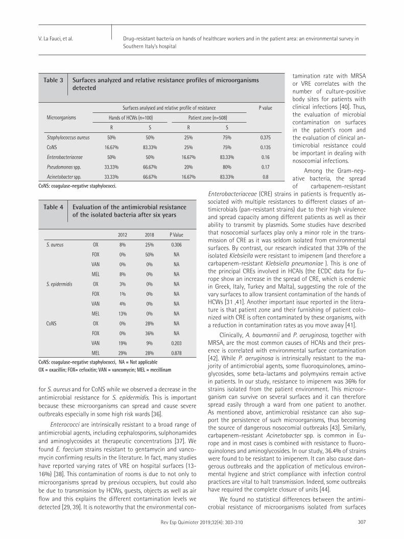
Drug-resistant bacteria on hands of healthcare workers and in the patient area: an environmental survey in Southern Italy’s hospital
V. La Fauci, et al.
Rev Esp Quimioter 2019;32(4): 303-310 307
tamination rate with MRSA or VRE correlates with the number of culture-positive body sites for patients with clinical infections [40]. Thus, the evaluation of microbial contamination on surfaces in the patient’s room and the evaluation of clinical an-timicrobial resistance could be important in dealing with nosocomial infections.
Among the Gram-neg-ative bacteria, the spread of carbapenem-resistant
Enterobacteriaceae (CRE) strains in patients is frequently as-sociated with multiple resistances to different classes of an-timicrobials (pan-resistant strains) due to their high virulence and spread capacity among different patients as well as their ability to transmit by plasmids. Some studies have described that nosocomial surfaces play only a minor role in the trans-mission of CRE as it was seldom isolated from environmental surfaces. By contrast, our research indicated that 33% of the isolated Klebsiella were resistant to imipenem (and therefore a carbapenem-resistant Klebsiella pneumoniae ). This is one of the principal CREs involved in HCAIs (the ECDC data for Eu-rope show an increase in the spread of CRE, which is endemic in Greek, Italy, Turkey and Malta), suggesting the role of the vary surfaces to allow transient contamination of the hands of HCWs [31 ,41]. Another important issue reported in the litera-ture is that patient zone and their furnishing of patient colo-nized with CRE is often contaminated by these organisms, with a reduction in contamination rates as you move away [41].
Clinically, A. baumannii and P. aeruginosa, together with MRSA, are the most common causes of HCAIs and their pres-ence is correlated with environmental surface contamination [42]. While P. aeruginosa is intrinsically resistant to the ma-jority of antimicrobial agents, some fluoroquinolones, amino-glycosides, some beta-lactams and polymyxins remain active in patients. In our study, resistance to imipenem was 36% for strains isolated from the patient environment. This microor-ganism can survive on several surfaces and it can therefore spread easily through a ward from one patient to another. As mentioned above, antimicrobial resistance can also sup-port the persistence of such microorganisms, thus becoming the source of dangerous nosocomial outbreaks [43]. Similarly, carbapenem-resistant Acinetobacter spp. is common in Eu-rope and in most cases is combined with resistance to fluoro-quinolones and aminoglycosides. In our study, 36.4% of strains were found to be resistant to imipenem. It can also cause dan-gerous outbreaks and the application of meticulous environ-mental hygiene and strict compliance with infection control practices are vital to halt transmission. Indeed, some outbreaks have required the complete closure of units [44].
We found no statistical differences between the antimi-crobial resistance of microorganisms isolated from surfaces
for S. aureus and for CoNS while we observed a decrease in the antimicrobial resistance for S. epidermidis. This is important because these microorganisms can spread and cause severe outbreaks especially in some high risk wards [36].
Enterococci are intrinsically resistant to a broad range of antimicrobial agents, including cephalosporins, sulphonamides and aminoglycosides at therapeutic concentrations [37]. We found E. faecium strains resistant to gentamycin and vanco-mycin confirming results in the literature. In fact, many studies have reported varying rates of VRE on hospital surfaces (13-16%) [38]. This contamination of rooms is due to not only to microorganisms spread by previous occupiers, but could also be due to transmission by HCWs, guests, objects as well as air flow and this explains the different contamination levels we detected [29, 39]. It is noteworthy that the environmental con-
Microorganisms
Surfaces analysed and relative profile of resistance P value
Hands of HCWs (n=100) Patient zone (n=508)
R S R S
Staphylococcus aureus 50% 50% 25% 75% 0.375
CoNS 16.67% 83.33% 25% 75% 0.135
Enterobacteriaceae 50% 50% 16.67% 83.33% 0.16
Pseudomonas spp. 33.33% 66.67% 20% 80% 0.17
Acinetobacter spp. 33.33% 66.67% 16.67% 83.33% 0.8
Table 3 Surfaces analyzed and relative resistance profiles of microorganisms detected
CoNS: coagulase-negative staphylococci.
2012 2018 P Value
S. aureus OX 8% 25% 0.306
FOX 0% 50% NA
VAN 0% 0% NA
MEL 8% 0% NA
S. epidermidis OX 3% 0% NA
FOX 1% 0% NA
VAN 4% 0% NA
MEL 13% 0% NA
CoNS OX 0% 28% NA
FOX 0% 36% NA
VAN 19% 9% 0.203
MEL 29% 28% 0.878
Table 4 Evaluation of the antimicrobial resistance of the isolated bacteria after six years
CoNS: coagulase-negative staphylococci, NA = Not applicableOX = oxacillin; FOX= cefoxitin; VAN = vancomycin; MEL = mecillinam

Drug-resistant bacteria on hands of healthcare workers and in the patient area: an environmental survey in Southern Italy’s hospital
V. La Fauci, et al.
Rev Esp Quimioter 2019;32(4): 303-310 308
In fact, today we are faced with increasingly resistant germs and the weapons at our disposal are increasingly lim-ited. Consequently, if we do not take urgent steps to remedy this problem, we will find ourselves faced with “killer germs” [49, 50] The prevention of HCAIs is the most widely used meas-ure to keep them under control and also the most cost-effec-tive; the savings from prevention can be as high as 5.5 billion dollars [3]. Strengthening infection control strategies such as hand washing, environmental sanitizing practices, the contin-uous training of physicians and specialists, correct use of an-timicrobials and vaccine are all measures implemented world-wide to control HCAIs [47, 51-60].
REFERENCES
1. Cassini A, Plachouras D, Eckmanns T, Abu Sin M, Blank HP, Ducom-ble T et al. Burden of Six Healthcare-Associated Infections on Euro-pean Population Health: Estimating Incidence-Based Disability-Ad-justed Life Years through a Population Prevalence-Based Modelling Study. PLoS Med 2016; 13(10): e1002150.
2. Magill SS, Edwards JR, Bamberg W, Beldavs ZG, Dumyati G, Kainer MA et al. Multistate point-prevalence survey of health care-as-sociated infections. N Engl J Med 2014; 370(13): 1198-208. doi:10.1056/NEJMoa1306801.
3. Zimlichman E, Henderson D, Tamir O, Franz C, Song P, Yamin CK et al. Health care-associated infections: a meta-analysis of costs and financial impact on the US health care system. JAMA Intern Med 2013; 173(22): 2039-46. doi: 10.1001/jamainternmed.2013.9763.
4. Glied S, Cohen B, Liu J, Neidell M, Larson E. Trends in mortali-ty, length of stay, and hospital charges associated with health care-associated infections, 2006-2012. Am J Infect Control 2016; 44(9): 983-9. doi: 10.1016/j.ajic.2016.03.010.
5. Centers for Disease Control and Prevention (CDC). Antibiotic/Anti-microbial Resistance (AR/AMR). Available on: https://www.cdc.gov/drugresistance/index.html [Last accessed: 2018, 22 Oct].
6. The European Union summary report on antimicrobial resistance in zoonotic and indicator bacteria from humans, animals and food in 2015. European Food Safety Authority European Centre for Disease Prevention and Control. ADOPTED: 26 January 2017. doi: 10.2903/j.efsa.2017.4694
7. Chemaly RF, Simmons S, Dale C Jr, Ghantoji SS, Rodriguez M, Gubb J, et al. The role of the healthcare environment in the spread of multidrug-resistant organisms: update on current best practic-es for containment. Ther Adv Infect Dis 2014; 2(3-4): 79-90. doi: 10.1177/2049936114543287.
8. European Center for Disease Prevention and Control (ECDC). Stock-holm: ECDC, 2013. Point prevalence survey of healthcare-associat-ed infections and antimicrobial use in acute care hospitals (2011-2012). Available on: https://ecdc.europa.eu/sites/portal/files/media/en/publications/Publications/healthcare-associated-infections-an-timicrobial-use-PPS.pdf [Last accessed: 2018, Oct 22].
9. Harris AD. How Important Is the Environment in the Emergence of Nosocomial Antimicrobial-Resistant Bacteria? Clin Infect Dis 2008; 46(5): 686-8. doi: 10.1086/527395.
around the patients and those from the hands of HCWs, con-firming literature data that the bacteria on hospital surfaces are transferred by the hands of healthcare staff. The role of handwashing is thus vital to prevent the spread of these re-sistant microorganisms [45].
In literature many studies described that the healthcare area may be contaminated by bacteria from different patient zones in two ways: direct shedding from patients and via HCWs’ hands. High-touch surfaces in the area of patients are contaminated with a higher rate of contamination in infected patients than from colonized ones [40].
In a cohort study, the authors investigated how frequent-ly HCWs contaminated gloves and gowns after contact with patients [46]. For example, after one of every three interactions with a patient carrying A. baumannii (present in 80% of rooms from colonized patients) HCWs contaminated their gloves and gowns. In general, independent risk factors for HCWs contami-nation by MDROs were positive environmental cultures, stay in room for more than 5 minutes, performing physical examina-tion and contact with the ventilator [46].
Limitations of our study are that we did not perform a molecular analysis and we did not evaluate the actions carried out by HCWs first of sampling.
In our study, after finding the positivity of the examined surfaces we made a signal to the health management and to the interested operative unit giving prescriptions on the en-vironmental sanitation and on the behaviour of the HCWs. In particular, we have prescribed a more thorough cleaning of the patients zone, cleaning the surfaces with GIOALCOL 70® (hydroalcoholic solution, of 70% ethyl alcohol), and sanitizing with RELY ON VIRKON® (powdered product based on potassi-um peroximonosulfate to be diluted according to the indica-tions in the technical data sheet (1g/L) alternating with STER-X 2000® (sodium hypochlorite 2.5%) or ANIOSPRAY QUICK® (hy-dro-alcoholic solution 55%, quaternary ammonium propion-ate, perfume).
For the behaviour of the operators it was prescribed the respect of the correct technique of washing and disinfection of the hands with different methods and products, according to the activities that must be carried out, by performing the 5 fundamental moments for hand hygiene present on the guide-lines issued by the Ministry of Health with the use of hydro-al-coholic gel for the clutch and chlorhexidine for washing.
After prescriptions, we revaluated the contaminations both of HCWs hands that surfaces and we did not found any positive sample, confirming the role of monitoring and infec-tion control strategies. [47]
Moreover, it was observed that the awareness of being evaluated can improve the adhesion of the HCWs (“Hawthorne effect”) and of the clean staff, although this effect can still be reduced in time [48].
We hope, therefore, to reassess the results obtained over time and evaluate the reduction of MDROs and HCAI in our hospital facilities to fight antimicrobial resistance.

Drug-resistant bacteria on hands of healthcare workers and in the patient area: an environmental survey in Southern Italy’s hospital
V. La Fauci, et al.
Rev Esp Quimioter 2019;32(4): 303-310 309
role of the hospital environment in the healthcare-associated in-fections: a general review of the literature. Eur Rev Med Pharmacol Sci. 2019;23(3):1266-1278. doi: 10.26355/eurrev_201902_17020.
24. World Health Organization (WHO). Antimicrobial resistance Glob-al Report on Surveillance 2014. Available on: http://www.who.int/drugresistance/documents/surveillancereport/en/ . [Last accessed: 2018, Oct 22].
25. ISO 18593:2004. Microbiology of food and animal feeding stuffs -- Horizontal methods for sampling techniques from surfaces using contact plates and swabs
26. ISO 18593:2018 . Microbiology of the food chain -- Horizontal methods for surface sampling
27. Magiorakos AP, Srinivasan A, Carey RB, Carmeli Y, Falagas ME, Giske CG. Multidrug-resistant, extensively drug-resistant and pan-drug-resistant bacteria: an international expert proposal for in-terim standard definitions for acquired resistance. Clin Microbiol Infect. 2012;18(3):268-81. doi: 10.1111/j.1469-0691.2011.03570.x.
28. The R Project for Statistical Computing (https://www.r-project.org/)
29. French GL, Otter JA, Shannon KP, Adams NM, Watling D, Parks MJ. Tackling contamination of the hospital environment by me-thicillin-resistant Staphylococcus aureus (MRSA): a comparison between conventional terminal cleaning and hydrogen peroxide vapour decontamination. J Hosp Infect 2004; 57(1): 31-7.
30. Squeri R, Grillo OC, La Fauci V. Surveillance and evidence of con-tamination in hospital environment from meticillin and vancomy-cin-resistant microbial agents. J Prev Med Hyg 2012; 53(3): 143-5.
31. Lemmen SW, Häfner H, Zolldann D, Stanzel S, Lütticken R. Distribu-tion of multi-resistant Gram-negative versus Gram-positive bac-teria in the hospital inanimate environment. J Hosp Infect 2004; 56(3): 191-7.
32. Weber DJ, Rutala WA, Kanamori H, Gergen MF, Sickbert-Bennett EE. Carbapenem-resistant Enterobacteriaceae: frequency of hospital room contamination and survival on various inoculated surfaces. Infect Con-trol Hosp Epidemiol 2015; 36(5): 590-3. doi: 10.1017/ice.2015.17.
33. La Fauci V, Costa GB, Facciolà A, Conti A, Riso, R. Squeri.Humidifiers for oxygen therapy: what risk for reusable and disposable devices? J Prev Med Hyg 2017; 58(2): E161-E165. PMC: 5584085.
34. Deshpande A, Cadnum JL, Fertelli D, Sitzlar B, Thota P, Mana TS. Are hospital floors an underappreciated reservoir for transmission of health care-associated pathogens? Am J Infect Control 2017; 45(3): 336-8. doi: 10.1016/j.ajic.2016.11.005.
35. Koganti S, Alhmidi H, Tomas ME, Cadnum JL, Jencson A, Donskey CJ. Evaluation of Hospital Floors as a Potential Source of Pathogen Dissemination Using a Nonpathogenic Virus as a Surrogate Marker. Infect Control Hosp Epidemiol 2016; 37(11): 1374-7.
36. De Oliveira AC, Damasceno QS. Surfaces of the hospital environ-ment as possible deposits of resistant bacteria: a review. Rev Esc Enferm USP 2010; 44(4): 1118-23.
37. Miller WR, Munita JM, Arias CA. Mechanisms of antibiotic resistance in enterococci. Expert Rev Anti Infect Ther 2014; 12(10): 1221-36.
38. Trick WE, Temple RS, Chen D, Wright MO, Solomon SL, Peterson LR. Patient colonization and environmental contamination by vanco-
10. Messineo A, Marsella LT. Biological hazards and healthcare-associ-ated infections in Italian healthcare facilities: some considerations on inspections and accountability. Ann Ig 2015; 27(6): 799-807. doi: 10.7416/ai.2015.2073.
11. Agodi A, Auxilia F, Barchitta M and SPIN-UTI network of the GISIO Working Group of the Italian Society of Hygiene, Preventive Med-icine and Public Health (SItI). Antibiotic consumption and resist-ance: results of the SPIN-UTI project of the GISIO-SItI. Epidemiol Prev 2015; 39(4 Suppl 1): 94-8.
12. Caselli E, Berloco F, Tognon L and Study Group SAN-ICA. Influ-ence of Sanitizing Methods on Healthcare-Associated Infections Onset: A Multicentre, Randomized, Controlled Pre-Post Interven-tional Study. J Clin Trials 2016; 6(5): 1000285. doi:10.4172/2167-0870.1000285.
13. Caselli E, Brusaferro S, Coccagna M, Arnoldo L, Berloco F, Antonioli P and the SAN-ICA Study Group. Reducing healthcare-associat-ed infections incidence by a probiotic-based sanitation system: A multicentre, prospective, intervention study. PLoS One 2018;13(7): e0199616. doi: 10.1371/journal.pone.0199616.
14. La Fauci V, Genovese C, Facciolà A, Palamara MAR, Squeri R. Five-year microbiological monitoring of wards and operating theatres in Southern Italy. J Prev Med Hyg 2017; 58(2): 166-72.
15. La Fauci V, Riso R, Facciolà A, Merlina V, Squeri R. Surveillance of microbiological contamination and correct use of protective lead garments. Ann Ig 2016; 28(5): 360-6. doi: 10.7416/ai.2016.2116.
16. La Fauci V, Costa GB, Arena A, et al. Trend of MDR-microorganisms isolated from the biological samples of patients with HAI and from the surfaces around that patient. New Microbiol 2018;41(1): 42-6.
17. Creamer E, Dorrian S, Dolan A, Sherlock O, Fitzgerald-Hughes D, Thomas T. When are the hands of healthcare workers positive for methicillin-resistant Staphylococcus aureus? J Hosp Infect. 2010;75(2):107-11. doi: 10.1016/j.jhin.2009.12.005.
18. Cristina ML, Sartini M, Spagnolo AM. Serratia marcescens Infec-tions in Neonatal Intensive Care Units (NICUs). Int J Environ Res Public Health. 2019;16(4). pii: E610. doi: 10.3390/ijerph16040610.
19. Agodi A, Barchitta M, Auxilia F, Brusaferro S, D’Errico MM, Mon-tagna MT. Epidemiology of intensive care unit-acquired sepsis in Italy: results of the SPIN-UTI network. Ann Ig. 2018;30(5 Supple 2):15-21. doi: 10.7416/ai.2018.2247.
20. Spagnolo AM, Sartini M, Battistella A, Casini B, Lo Pinto G, Schinca E and Hospital Infection Control Operating Group Galliera Hospital. A Clostridium difficile outbreak in an Italian hospital: the efficacy of the multi-disciplinary and multifaceted approach.J Prev Med Hyg. 2018;59(2):E132-E138.
21. Pasquarella C, Ciorba V, Arnoldo L, Auxilia F, Baldovin T, Barchit-ta M et al.Hospital hygiene in Italy: the GISIO-SItI survey. Ann Ig. 2018;30(5 Supple 2):7-14. doi: 10.7416/ai.2018.2246.
22. Tan TY, Tan JS, Tay H, Chua GH, Ng LS, Syahidah N. Multidrug-re-sistant organisms in a routine ward environment: differential propensity for environmental dissemination and implications for infection control. J Med Microbiol. 2013;62(Pt 5):766-72. doi: 10.1099/jmm.0.052860-0.
23. Facciolà A, Pellicanò GF, Visalli G, Paolucci IA, Venanzi Rullo E. The

Drug-resistant bacteria on hands of healthcare workers and in the patient area: an environmental survey in Southern Italy’s hospital
V. La Fauci, et al.
Rev Esp Quimioter 2019;32(4): 303-310 310
mycin-resistant enterococci in a rehabilitation facility. Arch Phys Med Rehabil 2002; 83(7): 899-902.
39. Drees M, Snydman DR, Schmid CH, Barefoot L, Hansjosten K, Vue PM et al. Prior environmental contamination increases the risk of acquisition of vancomycin-resistant enterococci. Clin Infect Dis 2008; 46: 678-85.
40. Rohr U, Kaminski A, Wilhelm M, Jurzik L, Gatermann S, Muhr G. Colonization of patients and contamination of the patients’ envi-ronment by MRSA under conditions of single-room isolation. Int J Hyg Environ Health 2009; 12(2): 209-15.
41. Morgan DJ, Rogawski E, Thom KA, Johnson JK, Perencevich EN, Shardell M et al. Transfer of multidrug-resistant bacteria to healthcare workers’ gloves and gowns after patient contact in-creases with environmental contamination. Critical Care Medicine. 2012;40(4):1045–1051. doi: 10.1097/CCM.0b013e31823bc7c8.
42. Lerner A, Adler A, Abu-Hanna J, Meitus I, Navon-Venezia S, Carmeli Y. Environmental contamination by carbapenem-resistant Entero-bacteriaceae. J Clin Microbiol 2013; 51(1): 177-81. doi: 10.1128/JCM.01992-12.
43. Sekiguchi J, Teruya K, Horii K, Kuroda E, Konosaki H, Mizuguchi Y,et al. Molecular epidemiology of outbreaks and containment of drug-resistant Pseudomonas aeruginosa in a Tokyo hospital. J In-fect Chemother 2007; 13(6): 418-22.
44. Kohlenberg A, Weitzel-Kage D, van der Linden P, Sohr D, Vögeler S, Kola A et al. Outbreak of carbapenem-resistant Pseudomonas aeruginosa infection in a surgical intensive care unit. J Hosp Infect 2010; 74(4): 350-7. doi: 10.1016/j.jhin.2009.10.024.
45. Simor AE, Lee M, Vearncombe M, Jones-Paul L, Barry C, Gomez M et al. An outbreak due to multiresistant Acinetobacter baumannii in a burn unit: risk factors for acquisition and management. Infect Control Hosp Epidemiol 2002; 23: 261-7.
46. World Health Organization (WHO). WHO Guidelines on Hand Hy-giene in Health Care: First Global Patient Safety Challenge Clear Care is Safer Care. Cap. 7: Transmission of Pathogens by Hands. Geneva: WHO, 2009. Available on: https://www.ncbi.nlm.nih.gov/books/NBK144014/ [Last accessed: 2018, Oct 22].
47. Infection control standard precautions in health care. Epidemic and pandemic alert and response. World Health Organization (2007). Available on: https://www.who.int/csr/resources/publications/EPR_AM2_E7.pdf
48. Hagel S, Reischke J, Kesselmeier M, Winning J, Gastmeier P, Brunk-horst FM et al. Quantifying the Hawthorne effect in hand hygiene compliance through comparing direct observation with automat-ed hand hygiene monitoring. Infect Control Hosp Epidemiol2015; 36(8): 957-62.
49. Courvalin P. Why is antibiotic resistance a deadly emerging dis-ease? Clin Microbiol Infect 2016; 22(5): 405-7. doi: 10.1016/j.cmi.2016.01.012.
50. La Fauci V, Alessi V. Antibiotic resistance: where are we going? Ann Ig 2018; 30(4 Suppl 1): 52-7. doi: 10.7416/ai.2018.2235.
51. Squeri R, Genovese C, Palamara MA, Trimarchi G, La Fauci V. “Clean care is safer care”: correct handwashing in the prevention of healthcare associated infections. Ann Ig 2016; 28(6): 409-15. doi:
10.7416/ai.2016.2123.
52. La Fauci V, Riso R, Facciolà A, Ceccio C, Lo Giudice D, Calimeri S et al. Response to anti-HBV vaccine and 10-year follow-up of antibody levels in healthcare workers. Public Health. 2016;139:198-202. doi: 10.1016/j.puhe.2016.08.007.
53. Genovese C, La Fauci V, Buda A, Nucera S, Alessi V, Carnuccio S et al.A potential outbreak of Measles and chickenpox among health-care workers of a university hospital. EMBJ 2019; 14(10): 45-48.
54. Squeri R, Genovese C, Trimarchi G, Palamara MAR, La Fauci V. An evaluation of attitude toward vaccines among healthcare workers of a University Hospital in Southern Italy. Ann Ig. 2017;29(6):595-606. doi: 10.7416/ai.2017.2188.
55. Squeri R, Riso R, Facciolà A, Genovese C, Palamara MA, Ceccio C et al. Management of two influenza vaccination campaign in health care workers of a university hospital in the south Italy. Ann Ig. 2017;29(3):223-231. doi: 10.7416/ai.2017.2150.
56. Squeri R, La Fauci V, Picerno IAM, TrimarchiG, Cannavò G, Egitto G. et al. Evaluation of vaccine coverages in the health care workers of a university hospital in southern Italy. Ann Ig. 2019; 31(2 Supple 1):13-24. doi: 10.7416/ai.2019.2273.
57. Genovese C, Picerno IAM, TrimarchiG, Cannavò G, Egitto G, Cosen-za B. et al. Vaccination coverage in healthcare workers: a multi-center cross-sectional study in Italy. JMPH. J Prev Med Hyg. 2019; 60(1):E12-E17. doi: 10.15167/2421-4248/jpmh2019.60.1.1097. eCollection 2019 Mar.
58. Lo Giudice D, Capua A, La Fauci V, Squeri R, Grillo OC, Calimeri S. Congenital rubella syndrome and immunity status of immigrant women living in southern Italy: a cross-sectional, seroepidemio-logical investigation.Travel Med Infect Dis. 2014;12(3):253-7. doi: 10.1016/j.tmaid.2014.01.003.
59. Genovese C, LA Fauci V, Squeri A, Trimarchi G, Squeri R.HPV vac-cine and autoimmune diseases: systematic review and meta-anal-ysis of the literature. J Prev Med Hyg. 2018;59(3):E194-E199. doi: 10.15167/2421-4248/jpmh2018.59.3.998.
60. Squeri R, La Fauci V, Sindoni L, Cannavò G, Ventura Spagnolo E. Study on hepatitis B and C serologic status among municipal solid waste workers in Messina (Italy). J Prev Med Hyg. 2006 47(3):110-3.

Rev Esp Quimioter 2019;32(4): 311-316 311
©The Author 2019. Published by Sociedad Española de Quimioterapia. This article is distributed under the terms of the Creative Commons Attribution-NonCommercial 4.0 International (CC BY-NC 4.0)(https://creativecommons.org/licenses/by-nc/4.0/).
su aparición y mejorar la seguridad de los pacientes hospitali-zados.
Palabras clave: infección tracto urinario, factores de riesgo, bacteriemia nosocomial secundaria, epidemiología
Risk factors to secondary nosocomial bacteremia to UTI in a tertiary hospital
ABSTRACT
Introduction. Nosocomial bacteremia secondary to uri-nary tract infections (NBS-UTI) occur in 1-4% of episodes and the associated mortality can increase up to 33%. However, very little is known about the epidemiology of these infections. The determination of modifiable risk factors to develop this ty-pe of bacteremia could help to control the infection and redu-ce health costs.
Material and methods. Cases-control study of NBS-UTI diagnosed at the University Hospital of Canary Islands bet-ween 2010-2014. The clinical-epidemiological variables and the intrinsic and extrinsic potential risk factors were collected. Logistic regression was used to study the variables associated with the development of NBS-UTI.
Results. A total of 178 episodes were studied, 85 cases and 93 controls. The average stay was significantly greater in the cases; from admission to bacteremia (p <0.003), as well as from discharge to discharge (p <0.005). Hepatic insufficiency (p <0.091), the use of mechanical ventilation (p <0.001), the central venous catheter (p <0.043) and surgery in the episode (p <0.001) behaved as risk factors for the acquisition of NBS-ITU.
Conclusion. Invasive devices, such as central venous catheter and mechanical ventilation, that had not previously been studied; as well as the surgery in the episode, which had not been studied either, suppose risk factors. In addition, NBS-ITU causes a significant increase in hospital stay. Therefore, it is
RESUMEN
Introducción. Las bacteriemias nosocomiales secunda-rias a infecciones del tracto urinario (BNS-ITU) ocurren en un 1-4% de los episodios y la mortalidad asociada puede aumen-tar hasta el 33%. Sin embargo, se conoce muy poco sobre la epidemiología de estas infecciones. La determinación de los factores de riesgo modificables para desarrollar este tipo de bacteriemias podría ayudar al control de la infección y reducir el gasto sanitario.
Material y métodos. Estudio de casos y controles de las BNS-ITU diagnosticadas en el Hospital Universitario de Cana-rias entre 2010-2014. Se recogieron las variables clínico-epi-demiológicas y los factores de riesgo potenciales intrínsecos y extrínsecos. Se utilizó la regresión logística para estudiar las variables asociadas al desarrollo de BNS-ITU.
Resultados. Se estudiaron 178 episodios, 85 casos y 93 controles. La estancia media fue significativamente mayor en los casos; desde el ingreso hasta la bacteriemia (p< 0,003), co-mo desde ésta hasta el alta (p< 0,005). La insuficiencia hepáti-ca (p< 0,091), el uso de ventilación mecánica (p<0,001), de el catéter venoso central (p<0,043) y la cirugía en el episodio (p< 0,001) se comportaron como factores de riesgo para la adqui-sición de BNS-ITU.
Conclusiones. Los dispositivos invasivos, como el catéter venoso central y la ventilación mecánica, que no había sido estudiada previamente; así como la cirugía en el episodio, que tampoco había sido estudiada, suponen factores de riesgo. Además, la BNS-ITU causa un aumento significativo de la es-tancia hospitalaria. Por ello, es necesario conocer los factores de riesgo para la aparición de estas infecciones, y así, prevenir
Factores de riesgo en bacteriemias nosocomiales secundarias a ITU en un hospital terciario
1Servicio de Microbiología y Control de la Infección, Hospital Universitario de Canarias, Tenerife, España2Servicio de apoyo a la investigación. Colegio Oficial de Enfermería de Santa Cruz de Tenerife, España.3Departamento de Medicina Preventiva y Salud Pública de la Universidad de La Laguna, Tenerife, España
Laura Sante1
María Lecuona1 Armando Aguirre Jaime2 Ángeles Arias3
Correspondencia:Laura Sante Servicio de Microbiología y Control de la Infección, Hospital Universitario de CanariasCtra. Ofra S/N La Cuesta 38320 La Laguna, Tenerife, EspañaE-mail: [email protected]
Original
Article historyReceived: 26 February 2019; Revision Requested: 13 April 2019; Revision Received: 23 April 2019; Accepted: 25 April 2019

Factores de riesgo en bacteriemias nosocomiales secundarias a ITU en un hospital terciarioL. Sante, et al.
Rev Esp Quimioter 2019;32(4): 311-316 312
Los controles se obtuvieron de la base de datos de los uri-nocultivos procesados en el Servicio de Microbiología y Con-trol de la Infección, procedentes de pacientes ingresados en el hospital. Se comprobó que correspondían a un episodio de ITU durante ese periodo, pero que no habían desarrollado una BNS, siguiendo la definición de los CDC [8] para su diagnóstico:
· El paciente con o sin sondaje urinario presentaba al menos uno de los siguientes signos o síntomas:
· fiebre (>38º C)
· dolor en el ángulo costovertebral
· frecuencia urinaria*
· disuria*
· y además, el paciente tuviese un cultivo de orina con no más de dos especies de organismos identificados, al menos uno de los cuales fuese una bacteria con una recuento ≥ 105 UFC/mL.
*Estos dos síntomas no pueden considerarse cuando el pacientes es portador de sondaje urinario.
Los controles se aparearon a los casos por fecha de ingreso, sexo, edad, servicio de ingreso y microorganismo causal de la infección.
Se revisaron las historias clínicas de cada uno de los episo-dios de los casos y de los controles. Se recogieron las variables clínico-epidemiológicas (sexo, edad, servicio, microorganismo, días de hospitalización previos a la bacteriemia, días de hospi-talización desde la bacteriemia hasta el alta, ingreso previo en los último 6 meses); así como los factores de riesgo potencia-les intrínsecos (Neoplasia, Insuficiencia hepática, Insuficiencia renal, Úlcera por Presión, Hipertensión Arterial, Diabetes Me-llitus, Inmunosupresión, Índice de comorbilidad de Charlson) y los factores de riesgo potenciales extrínsecos (tratamiento antibiótico previo a la infección, Sondaje vesical, Ventilación mecánica, Catéter venoso central y Cirugía durante el ingreso).
En cuanto a la prescripción de antibióticos previa a la fecha de infección se consultó en las historias clínicas de los pacientes si éstos habían recibido terapia antimicrobiana la se-mana anterior a la fecha de infección y si el microorganismo aislado era sensible a la misma.
Análisis estadístico. Para dotar al estudio de una poten-cia del 80% en pruebas bilaterales de contraste de hipótesis a un nivel de significación p≤0.05, se consideró que la preva-lencia del factor con sospecha de asociación a la bacteriemia secundaria era de un 30% entre los controles y en un 20% mayor, como diferencia sustancial, entre los casos, se estimó necesario disponer de 93 sujetos por grupo. Ese tamaño de muestra permitiría el empleo de modelos de regresión logís-tica binaria con un máximo de 8 factores con posibilidad de asociación a la bacteriemia secundaria como efecto, según los requisitos de adecuación de Hosmer-Lemeshov para el empleo de un modelo de regresión logística.
La muestra se describió resumiendo las variables nomi-nales con la frecuencia relativa de sus categorías componen-
necessary to know the risk factors for the appearance of these infections, and thus prevent their appearance and improve the safety of hospitalized patients.
Key words: urinary tract infection, risk factors, secondary nosocomial bac-teremia, epidemiology.
INTRODUCCIÓN
Las infecciones del tracto urinario (ITU) son una de las causas más frecuentes de infección nosocomial en Europa [1] y en EEUU [2]. Aproximadamente un 80% de los casos, se relacionan con la presencia de catéter urinario [3]. Las bacte-riemias nosocomiales secundarias a infecciones del tracto uri-nario (BNS-ITU) ocurren en un 1-4% [4] de los episodios y la mortalidad asociada a estos procesos puede aumentar hasta el 33% [2,5]. No obstante, en EEUU se estima que un 17% de las de las bacteriemias nosocomiales son secundarias a ITU [6] y en España, según los datos obtenidos del Estudio de Prevalencia de las Infecciones Nosocomiales en España (EPINE) suponen un 37,3% del total de BNS y un 11,8% del total de BN, ocupando el primer puesto de las bacteriemias nosocomiales secundarias (BNS) [7]. Además, las consecuencias económicas de este tipo de infecciones son también importantes y cuantiosas. En un estudio realizado por Saint et al. [4] describieron que cada epi-sodio relacionado con BNS-ITU suponía un incremento en el coste sanitario de 3.000 dólares.
A pesar de estas cifras, se conoce muy poco sobre la epi-demiología de las bacteriemias nosocomiales relacionadas con las ITU. La determinación de los factores de riesgo modificables para desarrollar este tipo de bacteriemias podría ayudar a de-finir prácticas apropiadas para el control de la infección y a mejorar la seguridad de los pacientes hospitalizados, al mismo tiempo que se reduciría el gasto sanitario.
El objetivo del presente estudio es identificar factores de riesgo potenciales de padecer una bacteriemia secundaria a in-fección urinaria en los pacientes hospitalizados.
MATERIAL Y MÉTODOS
Se realizó un estudio de casos y controles de las BNS-ITU diagnosticadas en el Hospital Universitario de Canarias entre el 1 de enero de 2010 y el 31 de diciembre de 2014. Se trata de un hospital de tercer nivel de 660 camas, que dispone de una Unidad de Cuidados Intensivos médico-quirúrgica y se reali-zan trasplantes renales. Los casos se obtuvieron de las bases de datos las bacteriemias nosocomiales (BN) que fueron cla-sificadas por la Sección de Control de la Infección siguiendo los criterios de los CDC [8] en: Bacteriemias Primarias, Bacte-riemias Relacionadas con Catéter y Bacteriemias secundarias a otros procesos infecciosos. Se consideró que la bacteriemia era secundaria a una ITU cuando se diagnosticaba una infección con un hemocultivo positivo para al menos un mismo microor-ganismo aislado y causante de la infección de la ITU, y que hubiese sido recogido durante el periodo en que se atribuía la bacteriemia secundaria.

Factores de riesgo en bacteriemias nosocomiales secundarias a ITU en un hospital terciarioL. Sante, et al.
Rev Esp Quimioter 2019;32(4): 311-316 313
Whitney si son ordinales o de escala no-normal. Los factores que alcanzaron en estas comparaciones una diferencia a un nivel p≤0,20 fueron introducidas como variables explicativas de la bacteriemia se-cundaria a ITU en modelos de regresión lo-gística binaria multivariable empleando la estrategia de modelo lleno por pasos hacia atrás y criterio de Wald. Para la retención del factor se estableció un valor p≤0,05.
Todos los cálculos se realizaron con ayuda del paquete de procesamiento esta-dístico informatizado SPSS™ 24.0 de IBM-SPSS Inc, Armonk, NY©.
RESULTADOS
Se estudiaron un total de 178 episo-dios de infecciones del tracto urinario, de los cuales 85 correspondían a casos, que desarrollaron bacteriemia nosocomial pos-terior y 93 a controles, que no la desarro-llaron.
En la tabla 1 se observan los poten-ciales factores de riesgo estudiados, para los casos y para los controles. En los ca-sos de BNS-ITU recogidos, observamos un predominio de hombres (54,1%) con una edad media de 67 años. La mayoría de es-tas BNS se diagnosticaron en los servicios médicos (40%), seguidos de los servicios quirúrgicos (25,8%). El uso de dispositivos invasivos, como la sonda vesical (77,6%), la ventilación mecánica (VM) (40%) o el catéter venoso central (CVC) (63,5%) fue-ron utilizados en con frecuencia en estos pacientes. Además, un 51,7% de estos pa-cientes habían sido sometidos a cirugía en el ingreso. Un 18,8% fueron exitus duran-te el episodio.
Entre los casos, los microorganismos más frecuentemente aislados fueron los bacilos gramnegativos (74,4%), principal-mente Enterobacterias (54,9%), seguidos de los cocos grampositivos (18,2%), ma-yoritariamente Enterococcus spp. (14,2%) y en tercer lugar las levaduras del género
Candida (6,5%) (tabla 1 y 2).
La estancia media fue significativamente mayor en los casos que en los controles; tanto desde la fecha de ingreso hasta el desarrollo de la bacteriemia (p< 0,003), como desde ésta hasta el alta (p< 0,005). Además, este grupo presentó con mayor frecuencia insuficiencia hepática (p< 0,091). Asimismo, el uso de dispositivos invasivos, como la VM o el CVC fue sig-nificativamente mayor entre los casos (p<0,001 y p<0,043). El
tes, las ordinales y de escala no-normal con mediana(P5−P95) o mediana(rango) y las de escala normal con media (DT). La nor-malidad se probó con histogramas y test de Kolmogorov-Smir-nov. Los resultados se ofrecen en intervalos de confianza al 95%.
La comparación de los potenciales factores de riesgo en-tre casos y controles se efectuó con la prueba chi2 de Pearson cuando los factores eran variables nominales y U de Mann-
POTENCIALES FACTORES DE RIESGO CASOS (n=85)
CONTROLES (n=93)
Valor-p
Edad en años, mediana (P5-P95) 67 (25-85) 70 (25-86) 0,383
Sexo masculino, n (%) 46 (54,1) 48 (51,6) 0,738
Servicios, n (%)
Médicos
Quirúrgicos
Onco-hematológicos
Intensivos
UCI- Pediátrica
Pediatría
34 (40)
22 (25,8)
15 (17,6)
12 (14,1)
0 (0)
2 (2,3)
30 (32,2)
35 (37,6)
12 (12,9)
12 (12,9)
2 (2,1)
2 (2,1)
0,392
Microorganismos, n(%)
Cocos grampositivos
Bacilos gramnegativos
Levaduras
17 (18,2)
68 (74,7)
6 (6,5)
13 (13,9)
72 (77,4)
8 (8,6)
0,668
Estancia ingreso-infección, mediana(rango) 18 (2-233) 12 (0-338) 0,003
Estancia infección-alta,(mediana (rango) 21 (2-278) 12 (0-175) 0,005
Neoplasia, n (%) 29 (34,1) 26 (27,9) 0,400
Insuficiencia, renal n (%) 27 (31,7) 23 (24,7) 0,297
Insuficiencia hepatica, n (%) 12 (14,1) 6 (6,4) 0,091
Úlcera por Presión, n (%) 12 (14,1) 19 (20,4) 0,267
Hipertensión, n (%) 51 (60) 58 (62,3) 0,746
Diabetes Mellitus, n (%) 32 (37,6) 40 (43) 0,466
Dislipidemia, n (%) 31 (36,4) 31 (33,3) 0,661
Inmunosupresión, n (%) 27 (31,7) 29 (31,1) 0,933
Sonda vesical, n (%) 66 (77,6) 67 (72) 0,390
Ventilación mecánica, n (%) 34(40) 11(11,8) <0,001
Catéter Venoso Central, n (%) 54 (63,5) 45 (48,3) 0,043
Hospitalización previa en 6 meses, n (%) 32 (37,6) 32 (34,4) 0,653
Cirugía en el ingreso, n (%) 44 (51,7) 26 (27,9) 0,001
Índice Charlson, mediana (P5-P95) 4 (0-7) 4 (0-6) 0,173
Consumo previo de antibióticos, (n,%) 49 (57,6) 46 (49,4) 0,274
Sensibles, n (%) 11 (22,4) 15 (32,6) 0,243
Exitus, n (%) 16 (18,8) 13 (13,9) 0,382
Tabla 1 Resultado de las comparaciones de los potenciales factores de riesgo a una bacteriemia secundaria a una infección del tracto urinario entre casos y controles.

Factores de riesgo en bacteriemias nosocomiales secundarias a ITU en un hospital terciarioL. Sante, et al.
Rev Esp Quimioter 2019;32(4): 311-316 314
calización Quirúrgica y las Infecciones del Tracto Respiratorio, las BNS más frecuentes y prevalentes en los hospitales [9, 10]. Estos 85 casos, supusieron un 24% del total de BNS del pe-riodo a estudio, siendo las segundas BNS más prevalentes en nuestro hospital, por detrás de las bacteriemias nosocomiales secundarias a infecciones de localización quirúrgica (ILQ) que representaron el 28,5% [11]. En España, según el estudio EPINE del año 2017, estas bacteriemias fueron un 11,83% del total de BN, siendo las más prevalentes [7]. En Europa, según el in-forme European Center for Disease and Control (E-CDC) [12] las BNS-ITU supusieron un 8% del total de BN (28,8%), siendo las BNS más prevalentes. En nuestro hospital, el porcentaje de prevalencia es menor que en España y Europa [11].
Con estos datos podemos afirmar que las BNS-ITU supo-nen un porcentaje no despreciable con respecto a las BN, sin embargo, constituyen un grupo de infecciones poco estudiado y la bibliografía referente a ellas es muy escasa. Por ello, hemos realizado un estudio de casos y controles para determinar los factores predisponentes para que un paciente con infección del tracto urinario desarrolle una bacteriemia nosocomial. Para ello, hemos recogido todos los casos de BNS-ITU en el perío-do de estudio y hemos apareado nuestros controles según el año de infección y el servicio, además de los factores de riesgo no modificables como son la edad y el sexo. La edad ha sido descrita como factor de riesgo para la adquisición de BNS-ITU [4, 13, 14]. Según un estudio realizado por Griebling et al. [15] las mujeres presentan un mayor riesgo de padecer una ITU, sin embargo, a la hora de desarrollar una bacteriemia secundaria en nuestro estudio no existieron diferencias de sexos, al igual que en el trabajo de Greene et al. [16].
En nuestro estudio, los microorganismos predominantes fueron los bacilos gramnegativos, seguidos de los cocos gram-positivos (especialmente de los Enterococcus spp.) y levaduras. Este porcentaje es similar al encontrado por otros autores [13, 14, 17]. En el estudio presentado por de Saint et al. [4] aunque los bacilos gramnegativos fueron globalmente los microorga-nismos más frecuentes, si se desglosa por especies, los mayo-ritarios fueron los Enterococcus spp. tanto en la serie de casos como en la serie de controles, siendo más frecuentemente ais-lados entre los casos (30,2% vs. 22,3%, p= 0,011). Chang et al. [2] obtuvieron un predominio de los Enterococcus spp. (29%) seguidos de Candida spp., (20%), teniendo los pacientes con bacteriemias por Candida spp. un riesgo 3,4 veces mayor de sufrir exitus que los pacientes con bacteriemia por Escherichia coli.
En nuestra serie se produjo un 18,8% de exitus, de los cuales 10 (62,5%) tenían infección causada por bacilos gram-negativos. Diversos autores refieren porcentajes de mortalidad asociados a una infección BNS-ITU que varía entre el 7,5% [13] y el 16,2% [18].
Se ha identificado que el uso de dispositivos invasivos, co-mo la sonda vesical, como factor de riesgo para el desarrollo de bacteriemia [4, 18] y sepsis [19]. En nuestro estudio, en el aná-lisis univariante obtuvimos que el uso de sonda no se asociaba a mayor riesgo de desarrollo de bacteriemia; pero sin embargo,
51,7% de los casos habían sometido a cirugía en el ingreso, frente al 27,9% del grupo control (p< 0,001).
No se encontraron diferencias significativas en cuanto al consumo previo de antibióticos ni se obtuvieron diferencias en cuanto a la sensibilidad de los tratamientos empíricos aplica-dos en ambos grupos de pacientes.
En la tabla 3 se muestran los resultados del ajuste del modelo de regresión logística en el análisis multivariante. Los pacientes con ventilación mecánica y los que tuvieron un pro-ceso quirúrgico durante el ingreso presentaron un riesgo supe-rior de desarrollar BNS.
DISCUSIÓN
Las BNS-ITU son, junto con las BNS a Infecciones de Lo-
Microorganismo n (%)
Escherichia coli 29 (31,8)
Klebsiella spp. 18 (19,7)
Pseudomonas aeruginosa 16 (17,5)
Enterococcus spp. 13 (14,2)
Candida spp. 6 (6,5)
Staphylococcus coagulasa negativos 3 (3,2)
Stenotrophomonas maltophilia 2 (2,1)
Staphylococcus aureus 1 (1)
Enterobacter cloacae 1 (1)
Fusobacterium nucleatum 1 (1)
Proteus mirabilis 1 (1)
Serratia marcescens 1 (1)
Tabla 2 Distribución de los microorganismos en los pacientes con BNS-ITU 2010-2014
El modelo arranca lleno con los potenciales factores: Días pre-bacteriemia, Insufi-ciencia hepática, Ventilación mecánica, Catéter venoso central, Cirugía al ingreso, e Índice Charlson, como potencialmente explicativos de la bacteriemia secundaria. El modelo converge a los 6 pasos reteniendo sólo a la Ventilación mecánica y la Cirugía al ingreso.
Tabla 3 Resultados del ajuste del modelo de regresión logística multivariante a los potenciales factores de riesgo considerados en el estudio para el desarrollo de BSI- ITU que alcanzan significación en su comparación univariante entre casos y controles
Factor OR(IC95%) p-Valor
Ventilación mecánica 4,6 (2,1-10,3) <0,001
Cirugía en el ingreso 2,8 (1,4- 5,5) 0,003

Factores de riesgo en bacteriemias nosocomiales secundarias a ITU en un hospital terciarioL. Sante, et al.
Rev Esp Quimioter 2019;32(4): 311-316 315
retrospectivo, no pudimos determinar si a todos los pacientes que padecieron un episodio de bacteriemia se le solicitó cultivo microbiológico de otras muestras clínicas para poder descar-tar infección en otra localización, pudiendo tener un exceso de clasificación de bacteriemias primarias quedando así las BNS-ITU infradiagnosticadas. Otra limitación, es que el estu-dio se realizó en un único centro hospitalario, y para conocer mejor la epidemiología de las BNS-ITU en los hospitales de ni-vel terciario sería necesario realizar un estudio multicéntrico. La tercera limitación es el escaso número de controles. Esto es debido a que en nuestro planteamiento decidimos aparear los controles según el tipo de microorganismo, ya que al ser un factor no modificable podríamos observar si encontrábamos otros factores de riesgo modificables para la prevención de las BNS-ITU.
En conclusión, y a pesar de estas limitaciones, creemos que nuestro estudio tiene importantes implicaciones clínicas. Observamos que los dispositivos invasivos, como el catéter venoso central y la ventilación mecánica, que no había sido estudiada previamente, se comporta como un factor de riesgo para la adquisición de una BNS-ITU. La cirugía en el episodio, que tampoco había sido estudiada previamente, supone tam-bién un factor de riesgo. Además, la bacteriemia asociada a ITU causa un aumento significativo de la estancia hospitalaria. Por ello, consideramos que es necesario conocer los factores de riesgo para la aparición de estas infecciones, para poder prevenir su aparición y mejorar la seguridad de los pacientes hospitalizados.
FINANCIACIÓN
Los autores declaran que no han recibido financiación pa-ra la realización de este estudio.
CONFLICTO DE INTERESES
Los autores declaran no tener conflicto de intereses.
BIBLIOGRAFÍA
1. Cassini A, Plachouras D, Eckmanns T, Abu Sin M, Blank HP, Du-comble T et al. Burden of Six Healthcare-Associated Infections on European Population Health: Estimating Incidence-Based Disabi-lity-Adjusted Life Years through a Population Prevalence-Based Modelling Study. PLoS Med. 2016;13(10):e1002150. doi: 10.1371/journal.pmed.1002150.
2. Chang R, Greene MT, Chenoweth CE, Kuhn L, Shuman E, Rogers MA et al. Epidemiology of hospital-acquired urinary tract-related bloodstream infection at a university hospital. Infect Control Hosp Epidemiol. 2011;32(11):1127-9. doi: 10.1086/662378.
3. World Health Organization. 2003. Prevención de las infecciones nosocomiales: guía práctica / revisores : G. Ducel, J. Fabry y L. Ni-colle, 2a ed. Ginebra : Organización Mundial de la Salud. [cited 20 December 2018]. Avaliable from: http://www.who.int/iris/hand-le/10665/67877
el uso de otros dispositivos invasivos, como la ventilación me-cánica y el catéter venoso central si eran factores de riesgo significativos para el desarrollo de una BNS-ITU, lo que podría estar relacionado, ya que se trata de pacientes ingresados en las unidades de cuidados intensivos, con la patología de base y gravedad de estos pacientes. La insuficiencia hepática, al igual que en el trabajo de Greene et al. [16] se comportó como un factor de riesgo.
Encontramos diferencias significativas respecto a la es-tancia hospitalaria. Aquellos pacientes que desarrollaron bac-teriemia permanecieron más días de estancia desde el ingreso hasta el desarrollo de la misma y posteriormente desde ésta última hasta el alta. En el estudio de Greene et al. [16] la estan-cia hospitalaria media también fue significativamente superior en los casos que en los controles. Esto puede significar un au-mento del gasto sanitario, tal y como describieron Riu et al. [21] en un estudio sobre el impacto económico del desarrollo de bacteriemia según el foco de infección y la sensibilidad an-tibiótica. En dicho estudio, observaron que las BNS-ITU por mi-croorganismo sensibles suponían un coste adicional de 6.786€ y hasta 13.299€ en el caso de bacteriemia por microorganis-mos multirresistentes.
No encontramos diferencias significativas en cuanto al tratamiento previo con antimicrobianos a diferencia de otros estudios, como el de Saint et al. [4], Greene et al. [16] y Ro-gers et al. [22]. en los cuales describieron que el uso de an-tibióticos era un factor de riesgo protector para el desarrollo de bacteriemia. Por ello, la importancia de aislar e identificar el microorganismo productor de la infección para adecuar el tratamiento antibiótico y no aumentar el desarrollo de resis-tencias bacterianas.
Padecer diabetes no se comportó como un factor de ries-go para desarrollar BNS-ITU, similar a lo descrito por Greene et al. [16] a diferencia del trabajo de Saint et al. [4], que obser-varon que aquellos pacientes con diabetes tenían mayor pro-babilidad de desarrollar BNS-ITU. Tampoco encontramos dife-rencias en los pacientes con insuficiencia renal, a diferencia de otro estudio [16], que si encontraron diferencias significativas. En nuestra serie la terapia con inmunosupresores no fue esta-dísticamente significativa, a diferencia de otros autores que la describen como un factor de riesgo asociado para desarrollar BNS-ITU [4,14].
Según el análisis multivariante, sólo dos factores se com-portaron como factores independientes asociados a padecer un episodio de bacteriemia nosocomial, el haber sido sometido a un proceso quirúrgico en el episodio y llevar un dispositivo de ventilación mecánica. En el trabajo publicado por Chang et al. [2] la cirugía en el episodio había sido descrita en el 84% de los pacientes con BNS-ITU y Bishara et al. [18] describen que un 12% de la población a estudio procedía de un servicio quirúrgico; sin embargo no ha sido estudiada como factor de riesgo en otros trabajos de casos-control [5, 18]. La ventilación mecánica como factor de riesgo no había sido estudiada pre-viamente [5, 16 18].
Una de las limitaciones de nuestro estudio es que, al ser

Factores de riesgo en bacteriemias nosocomiales secundarias a ITU en un hospital terciarioL. Sante, et al.
Rev Esp Quimioter 2019;32(4): 311-316 316
4. Saint S, Kaufman SR, Rogers MA, Baker PD, Boyko EJ, Lipsky BA. Risk factors for nosocomial urinary tract-related bacteremia: a case-control study. Am J Infect Control. 2006;34(7):401-07. doi: 10.1016/j.ajic.2006.03.001
5. Chenoweth CE, Saint S. Urinary tract infection. Infect Dis Clinic N Amer. 2011; 25 (1):103–115. doi: 10.1016/j.idc.2010.11.005.
6. Weinstein MP, Towns ML, Quartey SM, Mirrett S, Reimer LG, Parmi- giani G, et al. The clinical significance of positive blood cultures in the 1990s: a prospective comprehensive evaluation of the micro-biology, epidemiology, and outcome of bacteremia and fungemia in adults. Clin Infect Dis 1997;24:584-602. PMID: 9145732.
7. EPINE Workgroup. EPINE-EPPS 2017 resultados: Informe global de España. Sociedad Española de Medicina Preventiva, Salud Pública e Higiene, 2017. [cited 21 December 2018]. Avaliable from: http://hws.vhebron.net/epine/Global/EPINEEPPS%202017%20Infor-me%20Global%20de%20España%20Resumen.pdf
8. Centers for Diseases Control and Prevention. CDC/NHSN Survei-llance Definitions for Specific Types of Infections, 2018. [cited 21 December 2018]. Avaliable from: https://www.cdc.gov/nhsn/pdfs/pscmanual/17pscnosinfdef_current.pdf
9. Javaloyas M, Garcia-Somoza D, Gudiol F. Epidemiology and prognosis of bacteremia: a 10-y study in a community hospital. Scand J Infect Dis. 2002;34(6):436-41. PMID: 12160171
10. Endimiani A, Tambirina A, Luzzaro F, Lombardi G, Toniolo A. A two year analysis of risk factors and outcome in patients with bloods-tream infection. Jpn J Infect Dis. 2003; 56: 1-7. PMID: 12711818
11. Sante L, Aguirre-Jaime A, Miguel MA, Ramos MJ, Pedroso Y, Lecuo-na M. Epidemiological study of secondary bloodstream infections: The forgotten issue. J Infect Public Health. 2018;12(1):37-42. doi: 10.1016/j.jiph.2018.08.011.
12. European Centre for Disease Prevention and Control. Point preva-lence survey of healthcare- associated infections and antimicro-bial use in European acute care hospitals. Stockholm: ECDC; 2013. [cited 23 December 2018]. Avaliabre from: https://ecdc.europa.eu/sites/portal/files/media/en/publications/Publications/healthcare-associated-infections-antimicrobial-use-PPS.pdf
13. Jerkeman M, Braconier JH. Bacteremic and non-bacteremic febrile urinary tract infection--a review of 168 hospital-treated patients. Infection. 1992; 20(3):143–145. PMID: 1644489.
14. Krieger JN, Kaiser DL, Wenzel RP. Urinary tract etiology of bloods-tream infections in hospitalized patients. J Infect Dis. 1983; 148(1):57–62. PMID: 6350488.
15. Griebling TL. Urologic diseases in America project: trends in resource use for urinary tract infections in women. J Urol. 2005;173(4):1281-7. PMID: 15758783.
16. Greene MT, Chang R, Kuhn L, Rogers MA, Chenoweth CE, Shuman E et al. Predictors of hospital-acquired urinary tract-related bloods-tream infection. Infect Control Hosp Epidemiol. 2012;33(10):1001-7. doi: 10.1086/667731.
17. Bouza E, San Juan R, Munoz P, Voss A, Kluytmans J. on behalf of the Co-operative Group of the European Study Group on Nosocomial Infections (ESGNI). A European perspective on nosocomial urinary tract infections I. Report on the microbiology workload, etiolo-
gy and antimicrobial susceptibility. Clin Microbiol Infect. 2001; 7(10):523–531. PMID: 11683792.
18. Bishara J, Leibovici L, Huminer D, Drucker M, Samra Z, Konisberger H, et al. Five-year prospective study of bacteraemic urinary tract in-fection in a single institution. Eur J Clin Microbiol Infect Dis. 1997; 16:563–567. PMID: 9323466.
19. Conway LJ, Carter EJ, Larson EL. Risk Factors for Nosocomial Bac-teremia Secondary to Urinary Catheter-Associated Bacteriuria: A Systematic Review. Urologic nursing. 2015;35(4):191-203. PMID: 26402994
20. Melzer M, Welch C. Does the presence of a urinary catheter pre-dict severe sepsis in a bacteraemic cohort? J Hosp Infect. 2017 Apr;95(4):376-382. doi: 10.1016/j.jhin.2017.01.003.
21. Riu M, Chiarello P, Terradas R, Sala M, Garcia-Alzorriz E, Castells X et al. Incremental cost of nosocomial bacteremia according to the focus of infection and antibiotic sensitivity of the causati-ve microorganism in a university hospital. Medicine (Baltimore). 2017;96(17):e6645. doi: 10.1097/MD.0000000000006645.
22. Rogers MA, Blumberg N, Heal JM, Kuhn L, Greene MT, Shuman E et al. Role of transfusion in the development of urinary tract-re-lated bloodstream infection. Arch Intern Med. 2011;171(17):1. doi: 10.1001/archinternmed.2011.423.

Rev Esp Quimioter 2019;32(4): 317-326 317
©The Author 2019. Published by Sociedad Española de Quimioterapia. This article is distributed under the terms of the Creative Commons Attribution-NonCommercial 4.0 International (CC BY-NC 4.0)(https://creativecommons.org/licenses/by-nc/4.0/).
en la prescripción del TAR durante el ingreso hospitalario así como solicitar analíticas de CV y linfocitos CD4 a todos los pa-cientes VIH ingresados en el Hospital.
Palabras Clave: VIH, hospitalización, mortalidad.
Hospital admission and mortality causes of HIV patients in a third level hospital
ABSTRACT
Introduction. The aim of this study is to describe the HIV population admitted to a tertiary level hospital and analyze hospital admission and mortality causes during hospitalization.
Material and methods. Observational, retrospective study carried out in a third level Hospital. Inclusion criteria: Patients ≥18 years with a prescription of ART and diagnosis of HIV known or discovered during admission. It was accepted hospital ward discharge diagnose as hospitalization causes. Cli-nical, analytical outcomes as well as causes of mortality were collected.
Results. Among 162 hospitalized HIV infected, 128 met the inclusion criteria, 8 of those were diagnosed as naive HIV patients. 79.7% were male; Age 50.29 ± 9.81 years. The main reasons for hospital admissions (38.3%) were certain infectious and parasitic diseases (ICD-10 Classification) and more specifi-cally human immunodeficiency virus [HIV] disease represented 24,1% of whole hospitalizations. Mortality rates of ≥18 years HIV patients that were admitted to hospital during 2016-2017 were the 13.52%. The main causes of death were certain infec-tious and parasitic diseases followed by malignancies.
Conclusions. Our results emphasize the need of intensi-fying the HIV early diagnosis as well as Pneumocystis jirovecii primary prophylaxis. Insist on ART adherence from infectology follow-up appointment and pharmacy care consultations, edu-cate clinics on ART treatment prescription during hospital ad-
RESUMEN
Introducción. El objetivo del presente trabajo es describir el perfil de los pacientes VIH que ingresan en un hospital de tercer nivel así como analizar las causas de ingreso hospitalario y de mortalidad durante el mismo.
Material y métodos. Estudio observacional, retrospecti-vo llevado a cabo en un Hospital de tercer nivel. Criterios de inclusión: Pacientes ≥18 años con prescripción de tratamiento antirretroviral (TAR) y diagnóstico de VIH conocido o descu-bierto durante el ingreso. Como motivo de ingreso hospitalario se utilizó el diagnóstico al alta de cada paciente. Se recogieron variables clínicas, analíticas así como las causas de exitus.
Resultados. En el periodo de estudio ingresaron un to-tal de 162 pacientes VIH. Cumplieron los criterios de inclusión 128, de los cuales 8 fueron diagnosticados como VIH de novo. Un 79,7% fueron varones; edad 50,29±9,81 años. El principal diagnóstico al alta (38,3%) fueron ciertas enfermedades in-fecciosas y parasitarias (Clasificación CIE-10) y dentro de esta clasificación, las infecciones directamente relacionadas con el VIH fueron las mayoritarias (24,1%). Las tasas de mortalidad de los pacientes VIH ≥18 años que ingresaron en el Hospital (años 2016 y 2017) fue del 13,52%. Las causas de muerte más fre-cuentes fueron ciertas enfermedades infecciosas y parasitarias seguidas de neoplasias.
Conclusiones. Nuestros resultados enfatizan la necesidad de seguir reforzando el diagnóstico precoz de VIH así como la profilaxis primaria de Pneumocysitis jirovecii en los pacientes VIH. Insistir en la adherencia al TAR desde las consultas de se-guimiento de especialista en enfermedades infecciosas y en las consultas de atención farmacéutica, concienciar a los clínicos
Diagnóstico al alta y causas de mortalidad de pacientes VIH+ ingresados en un hospital de tercer nivel
Hospital Regional Universitario de Málaga
Rocío Asensi-DiezCristina Fernández-Cuerva Juan José Alcaraz SánchezIsabel Muñoz-Castillo
Correspondencia:Rocío Asensi-DiezHospital Regional Universitario de Málaga. Avenida de Carlos Haya s/n. CP.29010. Málaga. Teléfono: 951291435. Fax: 951291493. E-mail: [email protected].
Original
Article historyReceived: 12 March 2019; Accepted: 13 April 2019

Diagnóstico al alta y causas de mortalidad de pacientes VIH+ ingresados en un hospital de tercer nivelR. Asensi-Diez, et al.
Rev Esp Quimioter 2019;32(4): 317-326 318
más elevadas en este grupo de pacientes que en la población general y parece ser causada por infecciones no relacionadas con el VIH [10].
En los pacientes con TAR, las principales causas de morta-lidad no son infecciosas, si no enfermedades cardiovasculares (CV), enfermedad renal avanzada, enfermedad hepática des-compensada y cáncer no relacionado con VIH [11]. Respecto a las enfermedades CV, destacan en incidencia el infarto agudo de miocardio y el ictus [12] que son más prevalentes que en individuos no portadores de VIH. Esto se debe a que la infección por VIH produce una inflamación crónica que incrementa la edad biológica y el desarrollo de enfermedades CV [13] y pese a que el TAR reduce la inflamación, los marcadores siguen siendo más elevados que en pacientes no infectados [12]. En pacientes sin TAR, las principales causas de muerte son las complicacio-nes infecciosas debido a la inmunodeficiencia, siendo la tuber-culosis la más frecuente. En países de Europa del oeste se está produciendo un incremento de la incidencia de tuberculosis por microorganismos multirresistentes [14].
La complejidad del paciente VIH con y sin TAR provoca que las causas de ingreso hospitalario sean variadas. Destacan las causas infecciosas, relacionadas con VIH como la tuberculosis o no relacionadas, como neumonía y bacteriemia. También se producen ingresos por causas no infecciosas: desnutrición, al-teraciones respiratorias, digestivas, cardiovasculares, renales, psiquiátricas, hepáticas, neurológicas y hematológicas [10].
El objetivo del presente trabajo es describir el perfil de los pacientes VIH que ingresan en un hospital de tercer nivel así como analizar las causas de ingreso hospitalario y de mortali-dad durante el mismo.
MATERIAL Y MÉTODOS
Estudio observacional, retrospectivo llevado a cabo en un Hospital de tercer nivel. Criterios de inclusión: Pacientes ≥18 años con prescripción de TAR y diagnóstico de VIH conocido o descubierto durante el ingreso y que ingresaron en cualquier unidad clínica del hospital excepto ginecología y obstetricia. Periodo de estudio: Enero 2016 a Diciembre 2017. Criterios de exclusión: Aquellos pacientes VIH cuya duración de ingreso fue <24 horas debido a la imposibilidad de recoger suficientes datos clínicos y/o analíticos durante su estancia hospitalaria o bien, aquellos fugados antes de aclarar el motivo de su estancia hospitalaria.
La información se recogió de las historias clínicas electró-nicas y del registro de dispensación a pacientes externos del Servicio de Farmacia: ATHOS-Prisma®
Variables empleadas:
Para la recogida de datos se elaboró una base de datos en Excel que incluía:
1. Variables demográficas: edad y sexo
2. Otras variables:
● - Variables clínicas: número de ingresos en el periodo de estudio, número de ingresos previos al periodo de estudio en
mission as well as requesting viral and CD4 lymphocytes loads to every HIV admitted patients.
Keywords: HIV, Mortality, Hospitalization
INTRODUCCIÓN
Según los datos de la Organización Mundial de la Salud de 2017, el VIH afectaba a 36,9 millones de personas de todo el mundo de los cuales 35,1 millones eran adultos. Actualmente nos encontramos en una época en que el acceso generaliza-do a combinaciones efectivas de tratamientos antirretrovirales (TAR) ha permitido disminuir la mortalidad asociada al VIH en un 59% a nivel mundial. A pesar de esto, desde el inicio de la epidemia, 35,4 millones [25,0 millones – 49,9 millones] de personas han muerto a causa de enfermedades relacionadas con el SIDA [1].
Según el informe de vigilancia epidemiológica del VIH/SIDA en España (situación hasta el 30/06/2018), hasta el 30 de junio de 2017 se habían recibido, desde las 17 comunida-des autónomas (CCAA), Ceuta y Melilla, la notificación de 3.381 nuevos diagnósticos de VIH en el año 2017, lo que representa una tasa de 7,26 por 100.000 habitantes sin ajustar por retraso en la notificación [2].
En el periodo 2011-2017, la neumonía por Pneumocystis jirovecii ha sido la enfermedad definitoria de SIDA más fre-cuente (28,4%), seguida de la tuberculosis de cualquier locali-zación (20,6%) y de la candidiasis esofágica (12,6%) [2].
En el año 2016 se produjeron en España un total de 410.611 fallecimientos, de los cuales 498 (1,2 por 1.000) fueron por VIH y SIDA. De éstos, 385 (77,3 %) se produjeron en hom-bres y 113 (22,7%) en mujeres. La tasa de mortalidad global por VIH y SIDA fue de 1,1 por 100.000 habitantes [3].
El TAR debe iniciarse en todos los pacientes con infección por VIH-1, con o sin sintomatología, y con independencia del número de linfocitos CD4+ [4]. Los primeros meses del TAR se consideran clave ya que las tasas de mortalidad son más altas durante el primer año de tratamiento [5].
En países desarrollados, se calcula que la mitad de pacien-tes con VIH tienen ≥ 50 años, por lo que cada vez tienen mayor número de comorbilidades, entre las que destacan depresión, hipertensión arterial, dislipemia, insuficiencia renal, fractura ósea y diabetes mellitus que aparecen hasta diez años antes que en personas no infectadas y en mayor número [6,7,8]..
Sobre estos pacientes también influyen factores del estilo de vida, la obesidad, el tabaco y alcohol. En ciertos casos se producen coinfecciones por otros microorganismos, como el caso de virus de hepatitis B y hepatitis C. A esto, hay que sumarle que los pacientes con TAR se ven expuestos a la toxicidad a largo plazo de los fárma-cos utilizados, lo cual puede enmascarar otras comorbilidades. Las toxicidades más frecuentes del TAR son dislipemia, resistencia a la insulina, y diabetes mellitus, y en el caso del tenofovir disoproxilo problemas renales y pérdida de masa ósea [9].
Pese a que la esperanza de vida actual en pacientes VIH con TAR ha aumentado, las cifras de mortalidad siguen siendo

Diagnóstico al alta y causas de mortalidad de pacientes VIH+ ingresados en un hospital de tercer nivelR. Asensi-Diez, et al.
Rev Esp Quimioter 2019;32(4): 317-326 319
Se recogió solamente el principal diagnóstico al alta, ad-mitiéndose más de uno en los casos en los que el curso de la hospitalización no se entendiera sin la suma de diferentes factores. No se incluyeron en esta clasificación aquellos que fallecieron durante el ingreso, incluyéndose así en una clasi-ficación a parte, la cual analizaba la principal causa de mor-talidad del episodio de hospitalización. Para clasificar la causa de mortalidad se utilizó la Estadística de Defunciones según la Causa de Muerte. Esta estadística se realiza siguiendo los crite-rios establecidos por la OMS en la CIE-10. Este dato se recogió del informe de defunción registrado en la historia clínica de cada paciente. A la hora de clasificar la causa de mortalidad únicamente se recogió el motivo principal que llevó al mismo, a diferencia de los diagnósticos al alta, en los que se admitieron varios por ingreso hospitalario.
Análisis estadístico. El análisis de los datos fue descriptivo. Los valores de las variables fueron expresados como medias ± desviación estándar (DE), medianas y rangos o porcentajes. Se consideró que no existían problemas éticos en la realización del estudio, ya que se trató de un trabajo observacional, en el que la información recopilada se consideró confidencial y se utilizó única y exclusivamente en el ámbito profesional.
RESULTADOS
En el año 2016 ingresaron 21.755 pacientes ≥18 años en nuestro hospital excluyendo ginecología y obstetricia, de los cuales 85 pacientes fueron VIH. En 2017 ingresaron 20.832 pa-cientes, de los que 77 fueron VIH. Las tasas de ingreso por tanto fueron 0,39 y 0,37 respectivamente.
En el periodo de estudio ingresaron un total de 162 pa-cientes con registro desde admisión como VIH excluyendo gi-necología y obstetricia. Cumplieron los criterios de inclusión 128, de los cuales 8 fueron diagnosticados como VIH de novo. Los 120 restantes ya estaban diagnosticados con anterioridad. Por lo que el número total de pacientes reales ingresados fue de 170. La distribución de pacientes en función al tiempo que llevaban diagnosticados de VIH viene reflejada en la tabla 1.
Un 79,7% fueron varones; edad 50,29±9,81 años. Todos fueron ingresados al menos una vez por un periodo de tiempo ≥24h en el periodo de estudio (años 2016-2017).
Se analizaron en total 250 ingresos hospitalarios de los cuales 11 fueron excluidos. La mediana de la estancia hospita-laria fue de 8 días (2-86), presentando una mediana de ingre-so por paciente de 1 (1-15) durante los 2 años que abarcó el estudio.
La mediana de ingresos/paciente en nuestro centro ante-rior al periodo de estudio fue de 3 (0-32) ingresos.
De los 120 ya diagnosticados de VIH, no se dispone de da-tos acerca de los años que habían pasado desde el diagnóstico en 6 pacientes. La media de años diagnosticados de los 114 restantes fue de 13,85±6,52 años. La tabla 1 muestra la distri-bución de pacientes en función al tiempo que llevaban diag-nosticados de VIH. Se considera “diagnóstico en el último año”
nuestro centro, unidad clínica de ingreso, días de hospitali-zación, diagnóstico previo de VIH y en caso afirmativo, años diagnosticado de VIH, ingreso a la unidad clínica del hospital por urgencias o programado desde consulta, diagnóstico al alta como motivo de ingreso, profilaxis con trimetoprim-sulfame-toxazol prescrita en receta electrónica y causa de mortalidad en caso de producirse.
● - Variables analíticas: carga viral (copias/ml) aprox. 6 me-ses antes del ingreso y durante el/los ingreso/s, cifra de lin-focitos CD4 (células/µL) aproximadamente 6 meses antes del ingreso y durante el/los ingreso/s y coinfección con virus de la hepatitis C (VHC).
● En aquellos pacientes que tuvieran más de un ingreso hos-pitalario durante el periodo de estudio y por tanto pudieran tener más de una determinación de niveles de CV y de linfocitos CD4, se consideró una única medida de CV y CD4. En estos pa-cientes se consideró la media de sus niveles de CV y de linfoci-tos CD4 resultante de todos los ingresos.
● - Número pacientes ≥ 18 años y número de pacientes ≥ 18 años VIH y número de exitus en el hospital en los año 2016 y 2017 exceptuando el servicio de ginecología y obstetricia. Datos extraídos de la unidad de documentación y archivo del Hospital.
● - Variable de adherencia. El cálculo se realizó con la si-guiente fórmula: % adherencia = número de unidades de me-dicación ARV total dispensada/Número de unidades de medi-cación ARV prevista*100.
Se consideraron unidades previstas las necesarias para cumplir el tratamiento en los días incluidos desde la primera dispensación hasta la última en el periodo de tiempo conside-rado para el cálculo.
Definimos como buena adherencia un % ≥95 en los 6 me-ses previos al ingreso hospitalario en el periodo de estudio. Una adherencia por encima del 95% es necesaria para mantener una supresión virológica óptima [15-18]. Los datos se recogie-ron de los episodios de dispensación de la consulta de atención farmacéutica a pacientes externos de nuestro hospital. En el caso que un paciente tuviera múltiples ingresos hospitalarios, lo cual podría traducirse en una infraestimación del porcentaje de adherencia al no acudir a la consulta a recoger su medica-ción durante el tiempo de hospitalización, se determinó, que en aquellos pacientes cuyos ingresos hospitalarios distaban menos de 6 meses el uno del otro, se aceptaría como % de adherencia el correspondiente al promedio de los 6 meses previos a la pri-mera de las estancias hospitalarias.
Como motivo de ingreso hospitalario se utilizó el diagnós-tico al alta de cada paciente. Asumimos el diagnóstico al alta de cada ingreso hospitalario como el diagnóstico primordial de toda la estancia del paciente en la unidad de hospitalización, pues nos proporcionaba mayor información que aquel que se recogía en el primer día de ingreso como motivo de ingreso hospitalario. El diagnóstico al alta se identificó a través de un código recogido en Clasificación internacional de enfermeda-des, CIE-10 dividido a su vez en 20 categorías [19].

Diagnóstico al alta y causas de mortalidad de pacientes VIH+ ingresados en un hospital de tercer nivelR. Asensi-Diez, et al.
Rev Esp Quimioter 2019;32(4): 317-326 320
aquellos pacientes que llevan diagnosticados menos de un año en el momento del ingreso hospitalario.
La distribución de los ingresos en las distintas unidades del Hospital viene reflejada en la tabla 2.
En el 66,5% de los casos los pacientes ingresaron a través del servicio de urgencias del hospital, mientras que el 33,5% restante lo hizo de forma programada por su consulta de se-guimiento de atención hospitalaria.
El diagnóstico al alta de aquellos pacientes VIH ingresados durante ≥ 24 horas en nuestro hospital, y no fallecidos durante el mismo, se recogió en la tabla 3. Recogimos como subclasifi-cación dentro del grupo de ciertas enfermedades infecciosas y parasitarias aquellas asociadas a la enfermedad por el virus de la inmunodeficiencia humana (tabla 4).
El principal diagnóstico al alta con un 38,3% de los diag-nósticos fueron ciertas enfermedades infecciosas y parasitarias y dentro de esta clasificación, las infecciones directamente re-lacionadas con el VIH fueron las mayoritarias con un 24,1%. De los diagnósticos al alta clasificados como enfermedades del sis-tema respiratorio, encontramos 22 diagnósticos de neumonías de origen infeccioso de un total de 39 que no incluimos dentro de la categoría B20-B24 de enfermedad por el virus del VIH ya que no se trataba de neumonías causadas por Pneumocystis o tuberculosis.
Cuando analizamos la subclasificación enfermedad por virus de la inmunodeficiencia humana del diagnóstico al alta, encontramos que el 48,4% de los diagnósticos fueron por tu-mores onco-hematológicos seguido de enfermedad por VIH resultante en otras infecciones bacterianas (tabla 4).
En total un 47,66% (N=61) de los pacientes incluidos ha-bían padecido o padecían infección por el virus de hepatitis C. De ellos, 42 (68,85% estaban aún infectados durante el periodo de estudio) y 19 (31,14%) habían sido ya tratados y estaban curados.
Los datos de CV y linfocitos CD4 en los 6 meses previos al ingreso y durante el ingreso en el periodo de estudio vienen reflejados en la tabla 5.
Cabe destacar que un 6,25% pacientes (N=8/128) fueron diagnosticados de novo. La mediana de CV de los pacientes diagnosticados de novo fue 219.009,48 (697,64 - 1.000.000) copias/ml. Respecto a los niveles de linfocitos CD4 durante el ingreso, los 8 pacientes diagnosticados de novo tenían una me-diana de linfocitos CD4 de 45 (22-483) células/µL.
De los 98 pacientes de los que teníamos datos de linfo-citos CD4 en los 6 meses previos al ingreso, 25 pacientes te-nían niveles de CD4 <200 cel/µL y de estos, solo el 40% (N=10) habían recibido profilaxis para la infección por P. jirovecii con trimetoprim-sulfametoxazol.
En cuanto a la adherencia al TAR, de los 120 pacientes con diagnóstico previo de VIH conocido al ingreso, 22,5% (N=27) de ellos recogían fuera de nuestro centro por lo que no dis-ponemos de datos de su adherencia; el 13,66% (N=16) habían abandonado el tratamiento en los 6 meses previos al ingreso. La mediana de adherencia de los pacientes restantes: 81,67%
Años desde el diagnóstico de VIH Número de pacientes %
Diagnóstico > 1 año y < 20 años 62 48,44
Diagnóstico ≥ 20 años 47 36,72
Diagnóstico de novo 8 6,25
Sin datos del tiempo desde el diagnóstico 6 4,69
Diagnóstico en el último año 5 3,91
TOTAL 128 100,00
Tabla 1 Distribución de pacientes en función al tiempo que llevaban diagnosticados de VIH
Unidad de ingreso Número ingresos
N=239 %
Enfermedades Infecciosas 134 56,07
Hematología y Hemoterapia 28 11,72
Cardiología 12 5,02
Aparato Digestivo 10 4,18
Cirugía Digestiva 9 3,77
Oncología 7 2,93
Neurología 6 2,51
Cirugía Cardiovascular 5 2,09
Traumatología 4 1,67
Cuidados Paliativos 4 1,67
Cirugía Plástica 4 1,67
Neurocirugía 4 1,67
Nefrología 3 1,26
Medicina Interna 2 0,84
Cirugía General 2 0,84
Salud Mental 2 0,84
Neumología 2 0,84
Otorrinolaringología 1 0,42
TOTAL 239 100
Tabla 2 Distribución de los ingresos en las diferentes unidades del Hospital.
(N=98) fue del 98% (43-100). De ellos, 42,85% (N=42) pacien-tes eran buenos adherentes (≥95%).
Las tasas de mortalidad de los pacientes ≥ 18 años ingresa-dos en el hospital excluyendo ginecología y obstetricia, en los años 2016 y 2017, fueron 5,93% y 6,46%, respectivamente.
Las tasas de mortalidad de los pacientes VIH ≥18 años que ingresaron en el Hospital excluyendo ginecología y obstetricia en los años 2016 y 2017 fue del 13,52% (23/170). En el año 2016 la tasa de mortalidad fue de 10,22% y en el año 2017 fue

Diagnóstico al alta y causas de mortalidad de pacientes VIH+ ingresados en un hospital de tercer nivelR. Asensi-Diez, et al.
Rev Esp Quimioter 2019;32(4): 317-326 321
Respecto a la adherencia de estos pacientes, 21,74% (N=5) habían abandonado el TAR previo al ingreso en que fallecen, 17,39% (N=4) eran pacientes que fueron diagnosticados de novo de VIH durante el ingreso, del 13% (N=3) no tenemos da-tos de la adherencia al ser de otros centros. De los 11 restantes, 26,10% (N=6) eran buenos adherentes (≥95%) y 21,74% (N=5) tenían una adherencia <95%.
DISCUSIÓN
El acceso global al TAR en la última década ha llevado a disminuciones sustanciales en la morbilidad y mortalidad re-lacionadas con el VIH, especialmente en los países de ingresos medios-bajos en los que la prevalencia es mayor. Sin embargo, se estima que el VIH fue la sexta causa más común de perdida de vida a nivel mundial [20].
Las tasas de ingreso de los años 2016 y 2017 fueron 0,39 y 0,37 respectivamente, tasas más bajas que la reportada en otros estudios como el de Álvarez Barreneche MF et al (2017)
[21]. La media de edad de nuestro estudio es más alta, 50,29 ± 9,81 años, que la reportada en ese trabajo que fue de 37 años (30-49).
En nuestro estudio, observamos que la unidad mayoritaria de ingreso de los pacientes VIH fue Enfermedades Infecciosas, seguida de la unidad de Hematología y Hemoterapia. El moti-vo de ingreso mayoritario, clasificado en nuestro estudio como
de 17,94%. Un 86,95% eran hombres (N=20) con una media de edad ± desviación estándar de de 52 años ±8,15.
Estos 23 pacientes supusieron un total de 58 ingresos (23,20% de los ingresos totales) durante los dos años que abar-có el estudio, con una mediana de ingresos por paciente de 1 (1-8). 4 de estos pacientes fallecidos fueron diagnosticados de VIH durante el tiempo de estudio, 3 de ellos muriendo durante su primer y único ingreso hospitalario y 1 durante su segundo, distanciado 15 días del ingreso en el que fue diagnosticado por primera vez.
Las unidades principales de ingreso fueron: Infecciosos: 55,17% (N=32), Hematología 27,59% (N=16), Cirugía Plástica 5,17% (N=3), Oncología 3,45% (N=2), Cirugía cardiovascular, Cirugía digestiva, Neurología. Cuidados Paliativos y Cirugía Or-topédica y Traumatología cada uno con 1,72% (N=1).
Las causas principales de mortalidad clasificadas por ca-pítulos de la clasificación CIE-10 se encuentran en la tabla 6.
Los datos de CV y linfocitos CD4 en los 6 meses previos al ingreso y durante el ingreso en el periodo de estudio de los pacientes que fallecieron están reflejados en la tabla 5.
Un 17,39% (N=4/23) de los pacientes que fallecieron durante el ingreso fueron diagnosticados de VIH de novo. La mediana de CV de estos pacientes durante el ingreso fue de 81.633,25 copias/ml (39.507 - 1.000.000) y la de linfocitos CD4 fue de 68 células/µL (12-130).
Nº diagnósticos %
Ciertas enfermedades infecciosas y parasitarias 98 38,1
Enfermedad por virus de la inmunodeficiencia humana 62 24,1
Enfermedades del sistema respiratorio 39 15,1
Enfermedades del sistema circulatorio 30 11,7
Enfermedades del sistema digestivo 22 8,6
Traumatismos, envenenamientos y algunas otras consecuencias de causas externas 13 5,1
Tumores (neoplasias) 11 4,3
Síntomas, signos y hallazgos anormales clínicos y de laboratorio, no clasificados en otra parte 11 4,3
Enfermedades del sistema genitourinario 8 3,1
Enfermedades de la sangre y de los órganos hematopoyéticos, y ciertos trastornos que afectan el mecanismo de la inmunidad 7 2,7
Enfermedades de la piel y del tejido subcutáneo 5 1,8
Enfermedades del sistema osteo-muscular y del tejido conjuntivo 4 1,6
Factores que influyen en el estado de salud y contacto con los servicios de salud 3 1,2
Enfermedades endocrinas, nutricionales y metabólicas 2 0,8
Trastornos mentales y del comportamiento 2 0,8
Enfermedades del oído y de la apófisis mastoides 1 0,4
Causas externas de morbilidad y de mortalidad 1 0,4
TOTAL 257 100
Tabla 3 Diagnóstico al alta. Clasificación CIE-10.

Diagnóstico al alta y causas de mortalidad de pacientes VIH+ ingresados en un hospital de tercer nivelR. Asensi-Diez, et al.
Rev Esp Quimioter 2019;32(4): 317-326 322
las principales causas de hospitalización fueron tuberculosis pulmonar, candidiasis oral, neumonía por P. jirovecii y encefa-litis por Toxoplasma. La elevada contribución de ingresos hos-pitalarios y mortalidad observada en nuestro centro debidos a enfermedades infecciosas y parasitarias pone de manifiesto la importancia de la profilaxis con trimetoprim-sulfametoxazol y la vacunación frente a neumococo y meningococo para pa-cientes portadores de VIH. En nuestro estudio resulta llamativo que sólo el 40% (10/25) de pacientes con niveles de linfocitos CD4<200 células/µL tuvieran prescripción de trimetoprim-sul-fametoxazol como profilaxis contra la infección por P. jirovecii a pesar de que las Guías [23] recomiendan profilaxis primaria en pacientes con linfocitos CD4+ <200 cél/µl (AI). Esto nos lleva a concluir la necesidad de reforzar, desde las consultas clínicas y de atención farmacéutica, la profilaxis primaria contra P. ji-rovecii en pacientes con niveles de linfocitos <200 células/µL.
Respecto a los datos analíticos de los 120 pacientes conoce-dores de su status de portadores antes del ingreso llama la aten-ción que 69 pacientes tenían la CV indetectable 6 meses antes de la estancia hospitalaria, permaneciendo solo 47 indetectables en el periodo de estudio. De los 128 pacientes VIH ingresados no se determinó la CV en el 27,50% de los casos. Respecto a los niveles de CD4 en los 6 meses previos al ingreso, la mediana de CD4 de los pacientes de los que se disponía de información disminuye. Estos resultados ponen de relieve la importancia de conocer el estatus de ser portador de VIH en admisión y por parte de los clínicos para pedir determinaciones de CV y linfocitos CD4 sea cual sea la unidad clínica de ingreso hospitalario.
Solo 8 pacientes (6,25%) fueron diagnosticados de novo durante el ingreso hospitalario en los dos años que abarcó el
diagnóstico al alta, según la clasificación CIE-10 (año 2016) fue ciertas enfermedades infecciosas y parasitarias (38,3%) inclu-yéndose dentro de esta clasificación la enfermedad por el virus de la inmunodeficiencia humana (24,1%) como diagnóstico al alta mayoritario.
Estos datos coinciden con los reportados del metaanálisis de Ford N et al (2015) [22] donde se analizaron las causas de hospitalización y de mortalidad según la clasificación CIE-10 (año 2010) de 313.006 adultos y 6.182 niños correspondientes a 99 estudios y concluye que las principales causas de hos-pitalización fueron la enfermedad relacionada con VIH e in-fecciones bacterianas en todas las regiones geográficas, siendo también las causas más frecuentes de mortalidad.
Este estudio refiere que en Europa, tras las enfermeda-des relacionadas con VIH e infecciones bacterianas, las causas más frecuentes de ingreso fueron enfermedades respiratorias (14%), desorden psiquiátrico (13%), enfermedad cardiovascular (12%), renal (11%) y hepática (10%), asemejándose en parte a nuestros datos, donde el 15,18% de los ingresos fue debido a enfermedades del sistema respiratorio, seguido de enfermeda-des del sistema circulatorio (11,67%) y enfermedades del siste-ma digestivo (8,56%).
En nuestro estudio, en lo referente al motivo de ingreso (diagnóstico al alta) en la categoría de enfermedades relacio-nadas con el VIH, destacan los tumores onco-hematológicos seguidos por otras infecciones bacterianas, neumonía por P. ji-rovecii, síndrome caquéctico, enfermedad por citomegalovirus, infección por micobacterias y candidiasis. Estos datos coinciden en parte con el metaanálisis de Ford N et al (2015) [22] donde
Tabla 4 Sub-clasificación del diagnóstico al alta: enfermedad por virus de la inmunodeficiencia humana.
Sub-clasificación del diagnóstico al alta: enfermedad por virus de la inmunodeficiencia humana. Nº ingresos %
Enfermedad por VIH, resultante en linfoma de Burkitt 12 19,3
Enfermedad por VIH, resultante en otros tipos de linfoma no Hodgkin 9 14,5
Enfermedad por VIH, resultante en otros tumores malignos 9 14,5
Enfermedad por VIH, resultante en otras infecciones bacterianas 6 9,7
Enfermedad por VIH, resultante en neumonía por Pneumocystis jirovecii 4 6,5
Enfermedad por VIH, resultante en otras enfermedades infecciosas o parasitarias 4 6,5
Enfermedad por VIH, resultante en síndrome caquéctico 4 6,5
Enfermedad por VIH, resultante en enfermedad por citomegalovirus 4 6,5
Enfermedad por VIH, resultante en infección por micobacterias 3 4,8
Enfermedad por VIH, resultante en candidiasis 3 4,8
Enfermedad por VIH, resultante en otras infecciones virales 1 1,6
Enfermedad por VIH, resultante en encefalopatía 1 1,6
Enfermedad por VIH, resultante en sarcoma de Kapossi 1 1,6
Enfermedad por virus de la inmunodeficiencia humana [VIH], sin otra especificación 1 1,6
Total 62 100

Diagnóstico al alta y causas de mortalidad de pacientes VIH+ ingresados en un hospital de tercer nivelR. Asensi-Diez, et al.
Rev Esp Quimioter 2019;32(4): 317-326 323
de 45 (22-483) células/µL y la mediana de CV de 219.009,48 (697,64 - 1.000.000) copias/mL. Tal como ocurre con la mor-talidad, se ha reportado que las tasas de ingreso hospitalario descienden con el aumento del recuento de CD4 y que se no-tifican mayores tasas de ingreso hospitalario en pacientes con recuento de CD4 <100 células/µL. Los bajos niveles de CD4 ade-más predicen el riesgo de reingreso hospitalario, mientras que el uso del TAR se ha asociado con mejoras en la supervivencia, incrementando los niveles de CD4 y reduciendo el número de
estudio, dato más bajo que el reportado en el estudio de Álva-rez Barreneche MF et al (2017) [21] con un 22% y en del de Ford et al (2015) [22] con un 30% de los ingresos hospitalarios. Esto es debido probablemente a que nuestro estudio se circunscribe a un solo hospital regional de tercer nivel dentro de un país desarrollado donde el acceso a las pruebas de determinación y el acceso al TAR son gratuitos.
De hecho, de los 8 pacientes de los que no se conocía el status de portador de VIH la mediana de los niveles de CD4 fue
Sin datos Diagnosticados con datos (N=100)
Indetectable
CV (mediana, rango)
6 meses previos
(120 pacientes diagnosticados)
20 pacientes (16,67%) 69 pacientes (57,50%)
31 pacientes (25,83%)
345 copias/ml
(37 - 1.183,34)
Sin datos Diagnosticados con datos (N=95)
Durante ingreso
(128 pacientes)33 pacientes (25,78%) 47 pacientes (36,71%)
40 pacientes (31,25%)
9.980 copias/ml
(23 - 239.265)
CD4 (mediana, rango)
Sin datos Diagnosticados con datos (N=98)
6 meses previos
(120 pacientes diagnosticados)
22 pacientes (18,33%)442,69 cel./µL
(1-1.994)
Sin datos Diagnosticados con datos (N=82)
Durante ingreso
(128 pacientes)38 pacientes (31,66%)
257,50 cel/µL
(1-1.687)
EXITUS (N=23)
Sin datos Diagnosticados con datos (N=15)
Indetectable
CV (mediana, rango)
6 meses previos
(19 pacientes diagnosticados)
4 pacientes
(21,05%)10 pacientes (52,63%)
5 pacientes (26,31%)
626,50 copias/ml
(161-1.183,34)
Sin datos Diagnosticados con datos (N=20)
Durante ingreso
(23 pacientes)
3 pacientes
(13,04%)10 pacientes (43,48%)
10 pacientes (43,48%)
91.324 copias/ml
(4.481,33-1.697.605)
CD4 (mediana, rango)
Sin datos Diagnosticados con datos (N=15)
6 meses previos
(19 pacientes diagnosticados)
4 pacientes
(21,05%)
353 cel/µL
(27-1.085)
Sin datos Diagnosticados con datos (N=20)
Durante ingreso
(23 pacientes)
3 pacientes
(13,04%)
130 cel/µL
(12-608)
Tabla 5 Niveles de CD4 y carga viral de los pacientes y exitus 6 meses antes del ingreso y durante el mismo.

Diagnóstico al alta y causas de mortalidad de pacientes VIH+ ingresados en un hospital de tercer nivelR. Asensi-Diez, et al.
Rev Esp Quimioter 2019;32(4): 317-326 324
Tabla 6 Causas de mortalidad.
Nº de pacientes %
Ciertas enfermedades infecciosas y parasitarias 7 30,43
Neoplasias 5 21,74
Enfermedades del sistema respiratorio 4 17,39
Síntomas, signos y hallazgos anormales clínicos y de laboratorio, no clasificados en otra parte 3 13,04
Enfermedades del sistema circulatorio 2 8,70
Enfermedades del aparato digestivo 2 8,70
Total 23 100,00
ingresos hospitalarios por causas debida al VIH [22]. La cifra de linfocitos CD4+ es el indicador fundamental del estado inmu-nológico. Sirve para estadificar la infección por el VIH, evaluar el riesgo de comorbilidad, la vulnerabilidad a determinadas in-fecciones oportunistas, la necesidad de su profilaxis y su even-tual discontinuación [24].
Respecto a las tasas de mortalidad observadas en nuestro estudio, un 13,52% de los pacientes fallecieron en el hospital en el periodo de estudio. Estos % de mortalidad son más altos que los observados en otros estudios [21,25] con tasas del 5,4% o del 2,6%, sin embargo se aproximan a los datos reportados por Ford et al (2015) [22] con tasas globales de mortalidad en adultos del 20%. Esta última tasa de mortalidad procede de un metaanálisis que engloba múltiples estudios realizados a nivel mundial, incluyendo países sub-desarrollados como África.
Podríamos justificar estas tasas de mortalidad mayores que las de otros estudios debido a la situación geográfica de nuestro Hospital, siendo un hospital de referencia de tercer ni-vel que recibe pacientes no sólo de la provincia y Melilla, si no que a la población de derecho se suma también la de la pobla-ción flotante originada por el turismo, que aumenta especial-mente en los meses de verano y la de la población inmigrante. Además, 4 de los pacientes que fallecieron, fueron diagnósticos de novo, 2 pacientes fueron diagnósticos recientes (<6 meses) y otros 2 fueron pacientes que no estaban en seguimiento en nuestro Hospital. De los 15 pacientes restantes, que eran se-guidos por la unidad de enfermedades infecciosas de nuestro hospital, 4 habían abandonado el TAR.
De los pacientes que fallecieron, en los seis meses previos al ingreso la CV fue indetectable solo en el 43,48%, observando como la mediana de CV aumenta debido fundamentalmente a los 4 diagnósticos de novo que se produjeron durante el ingre-so hospitalario. Respecto a los niveles de linfocitos CD4 en los 6 meses previos al ingreso, la mediana disminuye durante el ingreso hospitalario por el mismo motivo. Los resultados refle-jan que los pacientes diagnosticados de novo y que fallecieron, ingresaron en el hospital con un sistema inmunológico muy deteriorado y con un avanzado estado de la enfermedad. Esto nos lleva a reflexionar que en España habría que reforzar las
políticas de diagnóstico precoz ya que el diagnóstico tardío y la presentación tardía dan como resultado una alta morbilidad y mortalidad [20].
La mayor causa de mortalidad fue la de ciertas enferme-dades infecciosas y parasitarias lo cual coincide con las princi-pales causas de mortalidad encontradas en otros estudios [21, 22, 25].
En cuanto a la adherencia al TAR, de los pacientes con diag-nóstico previo de VIH conocido al ingreso y que se disponía de datos, sólo un 42,85% eran buenos adherentes. De los que mu-rieron, sólo un 26,10% tenían buena adherencia y un 21,74% habían abandonado el tratamiento, lo que nos lleva a pensar que hay que reforzar desde el diagnóstico la importancia de la adherencia al TAR. De hecho, en las Guías GESIDA [24] subrayan que la adherencia inadecuada es la primera causa de fracaso terapéutico. Entre los factores asociados con una adherencia imperfecta destacan: mala relación médico-paciente, consumo de drogas, enfermedad mental, deterioro neurocognitivo, bajo nivel educativo, barrera idiomática, falta de apoyo social, com-plejidad terapéutica y efectos secundarios del tratamiento. Por el contrario, el apoyo emocional, la capacidad para incluir la medicación en las actividades de la vida diaria y la comprensión de la importancia del cumplimiento terapéutico son factores que predicen una adherencia correcta. El cumplimiento descri-be la calidad en la ejecución del tratamiento prescrito; aspectos vinculados al TAR, como el acceso y la persistencia en el mismo son esenciales para su éxito.
Nuestro estudio tiene como limitación el carácter retros-pectivo y limitado a pacientes VIH con prescripción de TAR, de hecho, cabe destacar que 34 pacientes VIH no tuvieron pres-cripción de TAR en sus ingresos en el Hospital o bien se trató de pacientes fugados. Por tanto, no se han podido analizar las causas de ingreso hospitalario de todos los pacientes VIH que ingresaron en el hospital en el periodo de estudio.
En conclusión, nuestros resultados enfatizan la necesidad de seguir reforzando el diagnóstico precoz de VIH, reforzar la profilaxis primaria de P. jirovecii en los pacientes VIH con nive-les de linfocitos CD4<200 cel/µL tanto en consulta como ingre-sados y al alta hospitalaria. Reforzar la adherencia al TAR desde

Diagnóstico al alta y causas de mortalidad de pacientes VIH+ ingresados en un hospital de tercer nivelR. Asensi-Diez, et al.
Rev Esp Quimioter 2019;32(4): 317-326 325
las consultas de seguimiento de especialista en enfermedades infecciosas y en las consultas de atención farmacéutica.
Hay que concienciar a los médicos para prescribir el TAR en los ingresos hospitalarios de los pacientes VIH así como so-licitar analíticas de CV y linfocitos CD4 a todos los pacientes VIH ingresados en el Hospital, sea cual sea la unidad clínica de ingreso.
FINANCIACIÓN
Los autores declaran que no han recibido financiación pa-ra la realización de este estudio.
CONFLICTO DE INTERESES
Los autores declaran no tener conflicto de intereses.
BIBLIOGRAFÍA
1. Estadísticas mundiales sobre el VIH y el SIDA - Hoja informativa de 2018. Consulta [Enero 2019]. Disponible en: http://www.unaids.org/en/resources/fact-sheet.
2. Vigilancia epidemiológica de VIH/SIDA en España. Situación a 30/06/2018. Consulta [Enero 2019]. Disponible en: http://www.isciii.es/ISCIII/es/contenidos/fd-servicios-cientifico-tecnicos/fd-vigilancias-alertas/fd-enfermedades/fd-sida/sistemas-de-informa-cion-poblacionales-sobre-vih.shtml.
3. Mortalidad por VIH/SIDA en España. Año 2016. Evolución 1981-2016. Consulta [Enero 2019]. Disponible en: http://www.isciii.es/ISCIII/es/contenidos/fd-servicios-cientifico-tecnicos/fd-vigilan-cias-alertas/fd-enfermedades/fd-sida/informacion-sobre-mortali-dad.shtml.
4. Documento de consenso de Gesida/plan nacional sobre el sida respecto al tratamiento antirretroviral en adultos infectados por el virus de la inmunodeficiencia humana (Enero 2018). Consulta [Enero 2019]. Disponible en: http://gesida-seimc.org/category/guias-clinicas/antirretroviral-vigentes/.
5. Brennan AT, Maskew M, Sanne I, Fox MP. The interplay between CD 4 cell count, viral load suppression and duration of antiretroviral therapy on mortality in a resource-limited setting. Trop Med Int Health. 2013. 18(5): 619-631. DOI: 10.1111/tmi.12079. Epub 2013 Feb 18
6. Maciel RA, Klück HM, Durand M, Sprinz E. Comorbidity is more common and occurs earlier in persons living with HIV than in HIV-uninfected matched controls, aged 50 years and older: A cross-sectional study. International Journal of Infectious Diseases, 2018. : 30-35. DOI: 10.1016/j.ijid.2018.02.009. Epub 2018 Feb 21.
7. Guaraldi G, Orlando G, Zona S, Menozzi M, Carli F, Garlassi E et al. Premature Age-Related Comorbidities Among HIV-Infected Per-sons Compared With the General Population. Clin Infect Dis. 2011. 53(11):1120-6. DOI: 10.1093/cid/cir627. Epub 2011 Oct 13.
8. Cuzin L, Katlama C, Cotte L, Pugliese P, Cheret A, Bernaud C et al. Ageing with HIV: do comorbidities and polymedication dri-
ve treatment optimization?. HIV Med. 2017; 18(6):395-401. DOI: 10.1111/hiv.12441. Epub 2016 Oct 7.
9. Seyler L, Lacor P, Allard SD. Current challenges in the treatment of HIV. Pol Arch Intern Med. 2018; 128(10):609-616. DOI: 10.20452/pamw.4357. Epub 2018 Oct 31.
10. Guaraldi G, Orlando G, Zona S, Menozzi M, Carli F, Garlassi E et al. Premature age-related comorbidities among HIV-infected persons compared with the general population. Clin Infect Dis. 2011. Dec; 53(11):1120-6. DOI: 10.1093/cid/cir627. Epub 2011 Oct 13.
11. Miller CJ, Baker JV, Bormann AM, Erlandson KM, Huppler Hullsiek K, Justice AC et al. Adjudicated Morbidity and Mortality Outcomes by Age among Individuals with HIV Infection on Suppressive Anti-retroviral Therapy. PLoS One. 2014; 1; 9(4):e95061. DOI: 10.1371/journal.pone.0095061. eCollection 2014.
12. Chow FC, Regan S, Feske S, Meigs JB, Grinspoon SK, Triant VA. Com-parison of ischemic stroke incidence in HIV-infected and non-HIV-infected patients in a US healthcare system. J Acquir Immune Defic Syndr. 2012; 1; 60(4):351-8. DOI: 10.1097/QAI.0b013e31825c7f24.
13. Miller CJ, Baker JV, Bormann AM, Erlandson KM, HupplerHullsiek K, Justice AC et al. Adjudicated morbidity and mortality outcomes by age among individuals with HIV infection on suppressive anti-retroviral therapy. PLoS One. 2014; 11; 9(4):e95061. DOI: 10.1371/journal.pone.0095061. eCollection 2014.
14. Podlekareva DN, Schultze A, Panteleev A, Skrahina AM, Miro JM, Rakhmanova A et al. One-year mortality of HIV-positive patients treated for rifampicin- and isoniazid-susceptible tuberculosis in Eastern Europe, Western Europe, and Latin America. AIDS. 2017; 31(3):375-384. DOI: 10.1097/QAD.0000000000001333.
15. Bartlett J. A. Addressing the challenges of adherence. J. Acquir. Im-mune. Defic. Syndr. 2002; 29 (Suppl. 1), S2–S10. PMID:11832696
16. Miller LG, Hays RD. Adherence to combination antiretroviral thera-py: synthesis of the literature and clinical implications, AIDS Read, 2000; 10(3): 177-85. PMID:10758022
17. Turner BJ. Adherence to antiretroviral therapy by human immu-nodeficiency virus-infected patients. J Infect Dis. 2002; 185 (Suppl 2):S143–51. DOI: 10.1086/340197.
18. Nieuwkerk PT, Oort FJ. Self-reported adherence to antiretroviral therapy for HIV-1 infection and virologic treatment response: A meta-analysis. Journal of Acquired Immune Deficiency Syndromes. 2005; 38(4):445–448. DOI: 10.1097/01.qai.0000147522.34369.12.
19. WHO. International statistical classification of diseases and related health problems. 10th revision. World Health Organization. versión: 2016. Consulta [Noviembre 2018]. Disponible en: https://icd.who.int/browse10/2016/en.
20. GBD 2013 Mortality and Causes of Death Collaborators. Global, re-gional, and national age-sex specific all-cause and cause-specific mortality for 240 causes of death, 1990–2013: a systematic analy-sis for the Global Burden of Disease Study 2013. Lancet 2015; 385: 117–71. DOI:10.1016/S0140-6736(14)61682-2
21. Álvarez Barreneche MF, Restrepo Castro CA, Hidrón Botero A, Villa Franco JP, Trompa Romero IM, Restrepo Carvajal L et al. Hospitalization causes and outcomes in HIV patients in the late antiretroviral era in Colombia. AIDS Res Ther. 2017 Nov 13; 14(1):60. DOI: 10.1186/s12981-017-0186-3.

Diagnóstico al alta y causas de mortalidad de pacientes VIH+ ingresados en un hospital de tercer nivelR. Asensi-Diez, et al.
Rev Esp Quimioter 2019;32(4): 317-326 326
22. Ford N, Shubber Z, Meintjes G, Grinsztejn B, Eholie S, Mills EJ et al. Causes of hospital admission among people living with HIV world-wide: a systematic review and meta-analysis. Lancet HIV. 2015. 2(10):e438-44. DOI: 10.1016/S2352-3018(15)00137-X. Epub 2015 Aug 11.
23. Ministerio de Sanidad, servicios sociales e Igualdad. Documento de prevención y tratamiento de infecciones oportunistas y otras coin-fecciones en pacientes con infección por VIH. Actualización Mayo 2015. Consulta [Febrero 2019]. Disponible en: http://gesida-seimc.org/wp-content/uploads/2017/02/gesida-guiasclinicas-2015-In-feccionesOportunistasyCoinfeccionesVIH.pdf.
24. Documento de consenso de Gesida/plan nacional sobre el sida res-pecto al tratamiento antirretroviral en adultos infectados por el virus de la inmunodeficiencia humana (Actualización Enero 2019). Consulta [Febrero 2019]. Disponible en: http://gesida-seimc.org/wp-content/uploads/2019/01/gesida_DC_TAR_2019_v_final.pdf
25. Kim JH, Psevdos G Jr, Gonzalez E, Singh S, Kilayko MC, Sharp V. All-cause mortality in hospitalized HIV-infected patients at an acute tertiary care hospital with a comprehensive outpatient HIV care program in New York City in the era of highly active antiretrovi-ral therapy (HAART). Infection. 2013; 41(2):545-51. DOI: 10.1007/s15010-012-0386-7. Epub 2012 Dec 21

Rev Esp Quimioter 2019;32(4): 327-332 327
©The Author 2019. Published by Sociedad Española de Quimioterapia. This article is distributed under the terms of the Creative Commons Attribution-NonCommercial 4.0 International (CC BY-NC 4.0)(https://creativecommons.org/licenses/by-nc/4.0/).
Prevalencia de micoplasmas genitales y respuesta al tratamiento de descolonización en pacientes de reproducción humana asistida
RESUMEN
Introducción. Se han publicado estudios que demues-tran mayores tasas de éxito en las técnicas reproducción asistida (TRA) en parejas no infectadas por micoplasmas. El objetivo de este estudio fue determinar la prevalencia de los micoplasmas genitales en muestras de orina del miem-bro masculino de las parejas incluidas en el Programa de Reproducción Asistida en nuestro Área Sanitaria realizando un tratamiento descolonizador con el fin de incrementar la calidad del semen, mejorar las tasas éxito de la embriotrans-ferencia y minimizar los efectos adversos sobre la gestación.
Material y métodos. Participaron parejas incluidas en el Programa de Reproducción Asistida durante 2016. La de-tección de los micoplasmas se realizó por PCR en tiempo real. En los casos positivos, la pareja fue tratada con anti-bióticos hasta la erradicación del microorganismo. Los anti-bióticos usados fueron: azitromicina, doxiciclina, levofloxa-cino, moxifloxacino y clindamicina.
Resultados. De los 205 hombres estudiados, 33 fueron positivos: Ureaplasma urealyticum 15,1%, Mycoplasma hom-inis 3,9%. Azitromicina fracasó en el 50% de los casos y doxiciclina en el 10,2%. Con levofloxacino solo en 2 de 5 se consiguió la erradicación de U. urealyticum.
Conclusiones. El cribado de rutina de los micoplas-mas genitales puede ser útil para mejorar la calidad del semen. Esto permitiría simplificar los procedimientos de fertilización in vitro e incrementar las tasas de éxito en la implantación de los embriones y en la gestación, especial-mente con la aplicación de técnicas diagnósticas rápidas y específicas como la PCR en tiempo real.
Palabras clave: Mycoplasma; Ureaplasma;; reproducción asistida; doxicicli-na; azitromicina
ABSTRACT
Introduction. Several studies have reported greater suc-cess of fertilisation by ART in couples who were not infected by Ureaplasma. Increased semen quality and better results have also been observed in couples who were treated with antibiot-ics to eradicate the infection. The aim of this study was to de-termine the prevalence of genital mycoplasmas in urine sam-ples from male partners enrolled in the Assisted Reproduction Program (ARP) in our healthcare area so that, positive cases can be treated prior to the use of ART in order to increase the quality of semen, improve the embryo implantation rates and minimize the risk of adverse effects during pregnancy.
Material and methods. This study included couples enrolled in the ARP during 2016. Mycoplasma detection was made using real-time PCR. In positive cases, both members of the couple were treated with antibiotics until eradication of the microorganism. The antibiotics used were: azithromycin, doxycycline, levofloxacin, moxifloxacin, and clindamycin.
Results. Of the 205 men studied, 33 were positive: Ureaplasma urealyticum 15.1%, Mycoplasma hominis 3.9%. Eradication treatment with azithromycin failed in 50% com-pared to 10.2% for doxycycline. Of the 5 cases treated with levofloxacin, only 2 achieved elimination of U. urealyticum.
Conclusions. We consider that genital mycoplasma rou-tine screening could be useful in order to increase the quality of semen which could simplify the in vitro fertilisation proce-dures and raise the success rate of embryo implantation and pregnancy, especially when fast, sensitive and specific technics as real time PCR are used.Keywords: Mycoplasma; Ureaplasma; Assisted reproduction; doxycycline; azithromycin
Prevalence of genital Mycoplasma and response to eradication treatment in patients undergoing assisted reproductive techniques
1Human Assisted Reproduction Unit. University Clinical Hospital of Santiago de Compostela, Spain2Clinical Microbiology Unit. University Clinical Hospital of Santiago de Compostela, Spain.
Ernesto Veiga1
Mercedes Treviño2
Ana Belén Romay1
Daniel Navarro2
Rocío Trastoy2
Manuel Macía1
Correspondence:Mercedes TreviñoClinical Microbiology Unit. University Clinical Hospital of Santiago de Compostela. c/ Ramón Baltar s/n. 15702- Santiago de Compostela, SpainPhone: (+34)981 951 703E-mail: [email protected]
Original
Article historyReceived: 17 March 2019; Revision Requested: 14 April 2019; Revision Received: 19 April 2019; Accepted: 14 May 2019

Prevalence of genital Mycoplasma and response to eradication treatment in patients undergoing assisted reproductive techniques
E. Veiga, et al.
Rev Esp Quimioter 2019;32(4): 327-332 328
Samples. The first fraction of urine (10-15 mL) was collected from males after at least 2 hours without urinat-ing. Urine was collected under the same conditions for the post-treatment tests in female patients.
Determination of M. genitalium, M. hominis, U. ure-alyticum, and U. parvum. Genital mycoplasma screening was performed using real-time PCR with LightMix Mycoplasma gen/hom Ureaplasma (Roche, Switzerland) in accordance with the manufacturer’s instructions. Briefly, a 224 or 129 bp frag-ment of the glyceraldehyde-3-phosphate dehydrogenase (gap) gene for M. genitalium or M. hominis, respectively, was ampli-fied with specific primers detected with labelled probes, identi-fied by melting curve analysis. In the same way, a 187 bp long fragment of 16S RNA gene was applied for U. urealyticum. This kit detects also U. urealyticum Biotype 1 (U. parvum) accord-ing to manufacturer´s information. The sensitivity of the test is 10 copies/ mL.
Nucleic acids were extracted previously using the Mag-naPure Compact system (Roche, Switzerland) after concen-trating the sample by centrifuging 2 mL of urine for 20 min-utes at 14,000g [14]. After discarding the supernatant, 200 µL of the re-suspended sediment were used to load the extraction system.
Antibiotic treatment to eradicate genital colonisation. Any positive PCR finding was treated with azithromycin or dox-ycycline. Quinolones were used when first line treatment failed.
Statistical analysis. All statistical analyses were carried out using SPSS v. 20.0 (IBM Corp., Armonk, NY). Statistical sig-nificance was set at p< 0.05.
RESULTS
A total of 205 couples were enrolled in the study. The age range was 24 to 54 years in men and 23 to 39 in women, with a median age of 36 years for men and 35 years for women. Most men were Caucasian (n= 201). Other ethnic groups were American (n= 2) and Gypsy (n= 2).
A total of 33 (16.1%) males tested positive by real-time PCR for one or more mycoplasmas: U. urealyticum 15.1% (n= 31) and M. hominis 3.9% (n= 8), with 6 (2.9%) cases of coin-fection with both microorganisms. No sample proved positive for M. genitalium.
Initial treatment for Ureaplasma spp. was azithromycin in the first 12 couples studied and doxycycline in the remain-
INTRODUCTION
Genital mycoplasmas (Ureaplasma parvum, Ureaplasma urealyticum, Mycoplasma genitalium and Mycoplasma hom-inis) are bacteria found in the placenta and/or embryo in preg-nant women. They can cause ascending infection after rupture of membranes and severe problems such as premature mem-brane rupture, preterm labour, miscarriage, postpartum fever, chorioamnionitis, and infection transmission to the newborn infant [1-5]. Several studies have reported greater success of fertilisation by assisted reproductive techniques (ART) in couples who were not infected by Ureaplasma spp. [6, 7]. In-creased semen quality and better ART results have also been observed in couples infected by mycoplasmas who were treat-ed with specific antibiotics to eradicate the infection [8, 9]. Moreover, the impact on female reproduction and the health of the fetus and newborn that can have genital mycoplasma suggests a role for routine screening and treatment before un-dergoing infertility treatment [10-12]. It therefore seems rec-ommendable to avoid the use of semen contaminated/infected with mycoplasmas in ART procedures. However, currently used procedures for selecting and washing spermatozoa prior to fertilisation do not ensure eradication of these bacteria [13].
With the advent of highly sensitive and specific nucle-ic acid amplification technics, more accurate estimate of the prevalence of genital mycoplasma as well as the control of the efficacy of the antibiotic treatment can be determined.
This study presents the results obtained after screening for genital mycoplasmas in urine using amplification by real-time polymerase chain reaction (RT-PCR) in patients enrolled in the assisted human reproduction program at University Hospi-tal Complex of Santiago (CHUS), in Santiago de Compostela, Spain.
MATERIALS AND METHODS
Patients. This study included couples enrolled in the as-sisted human reproduction program carried out by the Assist-ed Human Reproduction Unit at CHUS during 2016. Couples provided their informed consent after the study was approved by the competent Research Ethics Committee and by the hos-pital’s management (reference number 2015/493). The screen-ing for genital mycoplasmas was performed in male urine. In positive cases, both members of the couple received specific antibiotic therapy and had a post-treatment microbiological test. Couples were asked to use protection (condoms) during sexual intercourse for 7-14 days after initiating the treatment. Both members of the couple had a microbiological test 3-4 weeks after completing the treatment. Couples failing anti-biotic treatment were prescribed alternative antibiotics un-til both members had negative post-therapy microbiological tests. No patient was on antibiotic treatment at least, three months prior to collection of the sample. As part of the study protocol prior to in vitro fecundation, all male patients were screened for Chlamydia trachomatis and Neisseria gonorrhoe-ae in the same urine sample used for mycoplasma detection.
Men Women Couples
Azithromycin 1 8 3
Doxycycline 0 3 1
Table 1 Number of therapeutic failures depending on the antibiotic used for initial treatment in patients positive for U. urealyticum

Prevalence of genital Mycoplasma and response to eradication treatment in patients undergoing assisted reproductive techniques
E. Veiga, et al.
Rev Esp Quimioter 2019;32(4): 327-332 329
One of the couples who were successfully treated with levofloxacin initially for U. urealyticum required further treat-ment with moxifloxacin 400 mg p.o. for 7 days as M. hominis was detected in the post-treatment test (couple # 104).
The cases of M. hominis infection (6/7 in coinfection, 86%) were treated with azithromycin (first 2 couples with coinfec-tion with Ureaplasma spp.) and doxycycline (the remaining couples). Treatments failed in 3 patients both them (of whom 2 were members of the same couple) (table 3).
The statistical analysis showed that the response to treat-ment was poorer among women (p< 0.01). In addition, azith-romycin in single-dose regime was significantly less efficient as compared to doxycycline (p< 0.01) in both men and women.
Treatment efficacy was independent of age. However, the age range was too small to observe significant differences.
DISCUSSION
M. hominis and Ureaplasma spp. infections are associat-ed with serious problems during pregnancy (chorioamnionitis, premature rupture of membranes, preterm labour, miscarriage during the last trimester of pregnancy) and/or severe infection in the newborn (bronchopulmonary dysplasia, chronic lung in-fection, intraventricular cerebral haemorrhage) [15, 16]. There is evidence that the use of semen infected with Ureaplasma spp. for in vitro fertilisation (IVF) reduces the rate of pregnan-
ing 19 couples. Table 1 shows post-treatment test results for the cases positive for U. urealyticum. Treatment failed in 16 patients (11 females, 1 male, and 2 couples), including 12 pa-tients treated with a single oral dose of azithromycin 1 g and 4 patients treated with doxycycline 100 mg/12h p.o. for 7 days. One of the couples (couple # 61) who tested negative for U. urealyticum after initial treatment with doxycycline remained colonised by M. hominis and was treated with moxifloxacin (moxifloxacin 400 mg p.o. for 7 days).
Table 2 shows the results of subsequent treatments where real-time PCR tested positive for U. urealyticum following a second antibiotic cycle administered after failure of the initial treatment. One of the 12 couples who failed treatment with azithromycin withdrew from the program. The remaining cou-ples were treated with doxycycline 100 mg/12h p.o. for 7 days. There was only one case of therapeutic failure, which was treated alternatively with levofloxacin (500 mg/day, 7 days). As the woman remained positive, she was prescribed vaginal clindamycin 100 mg/day for 3 days. Her post-treatment test was negative.
Doxycycline proved ineffective in 4 of the 19 couples treated initially. In these cases, levofloxacin (500 mg/day, 7 days) was used as an alternative. Only one patient remained PCR-positive for U. urealyticum. This patient was then treated with vaginal clindamycin 100 mg/day for 3 days; she required a second course combined with a vaginal probiotic, but she still tested positive.
Table 2 Antibiotics used against Ureaplasma spp. and post-treatment real-time PCR success test
1st treatment 2nd treatment Couple Id.RT-PCR
post-treatment3rd treatment
RT-PCR post-treatment
4th treatmentRT-PCR
post-treatment
Azithromycin No 9
Azithromycin Doxycyclin
10 Positive Levofloxacin Positive Clindamycin Negative
12 Negative
25 Negative
28 Negative
29 Negative
31 Negative
36 Negative
39 Negative
43 Negative
46 Negative
48 Negative
Doxycyclin Levofloxacin
104 Negative Moxifloxacina Negative
106 Negative
130 Negative
159 Positive Clindamycin Positive Clindamycin + probiotic PositiveaFor M. hominis eradication.

Prevalence of genital Mycoplasma and response to eradication treatment in patients undergoing assisted reproductive techniques
E. Veiga, et al.
Rev Esp Quimioter 2019;32(4): 327-332 330
less efficacy at low pH. Interestingly, we have also observed a higher rate of antibiotic failure against Ureaplasma spp. in women, as compared to men, and this difference was statisti-cally significant (p< 0.01) for azithromycin. Fluoroquinolones (levofloxacin and moxifloxacin) were used as the therapeutic alternative where doxycycline did not eradicate colonisation by genital mycoplasmas. Levofloxacin was ineffective in one couple colonised by Ureaplasma spp. and in another couple with M. hominis coinfection. Levofloxacin has been suggested as an effective antimicrobial agent against Ureaplasma spp. and M. hominis [35], but our results do not support this, as therapeutic failure occurred in 40% of patients treated with levofloxacin. Nevertheless, in view of the small number of cas-es, we cannot draw any firm conclusions in this regard. Moxi-floxacin has been reported to be a powerful antibiotic against M. hominis [35] and indeed, it was a successful alternative in our series for one couple who had failed levofloxacin.
Clindamycin represent a potential well tolerated treatment for unresolved mycoplasma infections after treatment with tet-racyclines and macrolides [4, 5]. In fact, this antibiotic is com-monly used to treat bacterial vaginosis involving M. hominis. Although Ureaplasma spp. are considered intrinsically resistant to lincosamides, given the low percentage of sensitive strains [35, 36], we decided to use clindamycin as a third-line treatment in women showing therapeutic failure with azithromycin, doxy-cycline and/or levofloxacin, as it can be administered in vaginal suppositories, resulting in high local drug concentrations. In-deed, eradication of Ureaplasma spp. was achieved in one case. More assays are necessary to know the effect of probiotics on the success of the antibiotic treatments.
Treatment efficacy was independent of age, even though the age range was too small to observe significant differences.
Using mycoplasma-free semen we could replace intracy-toplasmic microinjection by conventional “in vitro” fecunda-tion in 7,6% of the cases (from 3% to 10,6%) which means cost savings of 7% for each case.
In summary, we consider that genital mycoplasma routine screening could be useful in order to increase the quality of se-men which could simplify the in vitro fertilisation procedures and raise the success rate of embryo implantation. When an-tibiotic treatment is necessary, real-time PCR significantly re-duces time to initiation of treatment and can be used to assess its efficacy. Doxycycline was more efficacious than azithromy-cin single-dose regime in eliminate genital mycoplasma colo-nization. On the other hand, eradication of genital mycoplas-mas prior to gestation can avoid possible adverse effects on pregnancy, fetus or the newborn, so, according to the opinion of other authors, as genital mycoplasma colonization of cervix is strongly associated with adverse effects as placenta previa, before a planned pregnancy, eradication of this microorgan-isms is necessary to prevent pregnancy negative outcomes. In addition, eradication of genital mycoplasma allows, in most cases, to use conventional in vitro fecundation technics in-stead of intracytoplasmic microinjection which is more labori-ous, complex and expensive.
cies by embryo transfer [6] and increases no-pregnancy [5] and miscarriage rates [6, 7, 10, 14, 17]. On the other hand, the washing procedures used for semen preparation prior to IVF are not always efficient to eliminate mycoplasmas, as these may remain adhered to the surface of spermatozoa, negatively affecting their quality [13, 18, 19]. In this context, mycoplasma screening in urine samples and subsequent eradication with antibiotic treatment may provide considerable benefits in the ART protocol. Molecular techniques such as real-time PCR are highly valuable diagnostic tools as they provide significant time reductions compared to conventional cultures, are not affect-ed by previous antibiotic use and have much higher sensitivity [20]. Our study found that 15.1% of the males were colonised by Ureaplasma spp. and 3.9% by M. hominis (practically all of whom were coinfected with Ureaplasma spp.). These figures are somewhat lower than reported in earlier studies, in which colonisation by Ureaplasma spp. also prevailed over M. homin-is [21-24]. Nevertheless, a high variability on genital mycoplas-ma prevalence has been reported in part due to geographical and methodological differences [25]. Unlike other publications which report rates of up to 5% [21, 26], there were no positive cases of M. genitalium in our patients. This could be explained because it’s a population with a median age above 35-year-old (some studies report decreasing rates of colonisation by M. hominis with age [27], asymptomatic and, possibly, more con-servative sexual behaviour.
Besides the benefits that the eradication of genital my-coplasma can have on the success of assisted reproduction, as already mentioned previously, it was also been shown that pre-emptive antibiotic therapy of genital mycoplasma in col-onized pregnant women might represent a beneficial strategy to reduce premature labour and neonatal complications [28].
According to the recommendations of therapeutic guide-lines, macrolides are the treatment of choice for Ureaplasma spp. infections, with doxycycline or levofloxacin as alterna-tives. For M. hominis, the treatment of choice is doxycycline, with quinolones as an alternative [29, 30]. As recommended in these guidelines and taking into account the treatment sched-ules used in studies in colonized women [21], azithromycin 1g/day single-dose was used to eradicate Ureaplasma spp. in both members of the couple when the male was positive. In addition to being a first-line option, the azithromycin regime simpli-fies treatment compliance. For the same reason, azithromycin was also used in cases of Ureaplasma/M. hominis coinfection. In several studies, azithromycin is reported to be a highly ef-fective antibiotic against Ureaplasma spp., with a very low percentage of resistant strains [31-33]. However, the in vivo results obtained in our series using the treatment recommend-ed for sexually transmitted infections (1 g single dose; www.vademecum.es) revealed the low efficacy of this macrolide. For this reason, after the initial results, doxycycline was used as the first choice (100 mg b.i.d. p.o., 7 days). Some reports have demonstrated that a single dose of azithromycin is less effi-cient than azithromycin used over longer periods [34], and this could be the reason behind its low efficacy in our series. Pos-sibly, also could contribute its bacteriostatic mechanisms and

Prevalence of genital Mycoplasma and response to eradication treatment in patients undergoing assisted reproductive techniques
E. Veiga, et al.
Rev Esp Quimioter 2019;32(4): 327-332 331
tion en assistance medicale a la procréation. Gynecol Obstet Fertil. 2004; 32 (2): 135-39. doi: 10.1016/j.gyobfe.2003.11.017.
8. Swenson CE, Toth A, O’Leary WM. Ureaplasma urealyticum and human infertility: the effect of antibiotic therapy on semen quality. Fertil Steril. 1979; 31 (6): 660-65. doi: 10.1016/S0015-0282(16)44057-4.
9. Toth A, Lesser ML, Brooks C, Labriola D. Subsequent pregnancies among 161 couples treated for T-mycoplasma genital-tract in-fection. New Engl J Med. 1983; 308 (9): 505-507. doi: 10.1056/NEJM198303033080907.
10. Kanakas N, Mantzavinos T, Boufidou F, Koumentakou I, Creatsas G. Ureaplasma urealyticum in semen: is there any effect on in vitro fertilization outcome? Fertil Steril. 1999; 71 (3): 523-27. doi: 10.1016/S0015-0282(98)00472-5.
11. Imudia AN, Detti L, Pusacheck EE, Yelian FD, Diamond MP. The prevalence of Ureaplasma urealyticum, Mycoplasma hominis, Chlamydia trachomatis and Neisseria gonorrhoeae infections, and rubella status on patients undergoing an initial infertility eval-uation. J Assist Reprod Genet. 2008; 25 (1): 43-6. doi: 10.1007/s10815-007-9192-z.
12. Aydogan P, Hahyaoglu S, Sayqan S, Kaymak O, Mollamahmutoglu L, Danisman N. Does cervical Ureaplasma/Mycoplasma colonization increase the lower uterine segment bleeding risk during caesarean section among patients with placenta previa? A cross-sectional study. Eur Rev Med Pharmacol Sci; 2014; 18 (16); 2243-7. PMID 25219820.
13. Knox CL, Cave DG, Farrell DJ, Eastment HT, Timms P. The role of Ureaplasma urealyticum in adverse pregnancy outcome. Aust N Z J Obstet Gynaecol. 1997; 37 (1): 45-51. doi: 10.1111/j.1479-828X.1997.tb02216.x
14. Gdoura R, Kchaou W, Ammar-Keskes L, Chakroun N, Sellemi A, Zna-zen A, et al. Assessment of Chlamydia trachomatis, Ureaplasma urealyticum, Ureaplasma parvum, Mycoplasma hominis and My-coplasma genitalium in semen and first voided urine specimens of asymptomatic male partners of sterile couples. J Androl. 2008; 29 (2): 198-206. doi: 10.2164/jandrol.107.003566.
15. Donders GGG, Ruban K, Bellen G and Petricevic L. Mycoplasma/Ureaplasma infection in pregnancy: to screen or not to screen. J Perinat Med. 2017; 45 (5): 505-15. doi: 10.1515/jpm-2016-0111.
16. Knox CL, Allan JA, Allan JM, Edirisinghe WR, Stenzel D, Lawrence FA, et al. Ureaplasma parvum and Ureaplasma urealyticum are detect-ed in semen after washing before assisted reproductive technology procedures. Fertil Steril. 2003; 80 (4): 921-29. doi: 10.1016/S0015-0282(03)01125-7.
17. Shalika S, Dugan K, Smith RD, Padilla SL. The effecct of positive semen bacterial and Ureaplasma cultures on in-vitro fertilization success. Human Reprod. 1996; 11 (12): 2789-92. doi: 10.1093/ox-fordjournals.humrep.a019211.
18. Zeyad A, Amor H, Hammadeh ME. The Impact of Bacterial Infec-tions on Human Spermatozoa. International Journal of Women’s Health and Reproduction Sciences. 2017; 5 (4): 243–252. doi: 10.15296/ijwhr.2017.43.
19. Hannachi H, Elloumi H, Hamdoun M, Kacem K, Zhioua A, Bahri O.
In the case of patients who do not eliminate colonization in the first treatment regime, the benefit of initiating succes-sive alternative antibiotic treatments is debatable due to de-layed onset of fertilization and the possible undesirable effects on the vaginal microbiota that seems to play an important role in relation to female fertility [37, 38].
FUNDING
None to declare
CONFLICT OF INTEREST
Authors declare that they have no conflicts of interest.
INFORMED CONSENT
All patients included in the study previously signed the in-formed consent document.
REFERENCES
1. Domingues D, Noguera F, and Tavira L. Papel que desempeñan mi-coplasmas en las infecciones humanas. Act Med Port; 2005; 18: 377-384. PMID 16611542.
2. Breugelmans M, Vancutsem E, Naessens A, Laubach M, Folulon W. Association of abnormal vaginal flora and Ureaplasma species as risk factors for preterm birth: a cohort study. Acta Obstet Gynecol Scand. 2010; 89 (2): 256-260. doi: 10.3109/00016340903418769.
3. Tae K, Hye K, Soon MN. Detection of nanobacterianin patients with chronic prostatitis and vaginitis by reverse transcriptase pol-ymerase chain reaction. Korean J. Urol. 2011; 52 (3): 194-9. doi: 10.4111/kju.2011.52.3.194.
4. Capoccia R, Greub G, Baud D. Ureaplasma urealyticum, Mycoplas-ma hominis and adverse pregnancy outcomes. Curr Opin Infect Dis. 2013; 26 (3): 231-240. doi: 10.1097/QCO.0b013e328360db58.
5. Murtha AP, Edwards JM. The role of Mycoplasma and Ureaplasma in adverse pregnancy outcomes. Obstet Gynecol Clin N Am. 2014; 41 (4): 615-27. doi: 10.1016/j.ogc.2014.08.010.
6. Montagut JM, Lapretre S, Degoy J, Rousseau M. Ureaplasma in se-men and IVF. Human Reprod. 1991; 6 (5): 727-29. doi: 10.1093/oxfordjournals.humrep.a137416.
7. Wittemer C, Beattahar-Lebugle K, Ohl J, Rongières C, Viville S, Nisand I. Colonisation bacterienne vaginale anormale et implanta-
Men Women Couples
Azithromycin 0 1 1
Doxycycline 0 1 1
Table 3 Number of therapeutic failures depending on the antibiotic used for initial treatment in patients positive for M. hominis

Prevalence of genital Mycoplasma and response to eradication treatment in patients undergoing assisted reproductive techniques
E. Veiga, et al.
Rev Esp Quimioter 2019;32(4): 327-332 332
Mycoplasma hominis in pregnant women. BMC Infect Dis. 2014; 14: 171-177. doi: 10.1186/1471-2334-14-171.
32. Schneider SC, Tinguely R, Droz S, Hilty M, Dona V, Bodmer T, et al. Antibiotic susceptibility and sequence type distribution of Ureaplasma species isolated from genital samples in Switzer-land. Antimicrob Agents Chemother. 2015; 59 (10): 6026-31. doi: 10.1128/AAC.00895-15.
33. Lee MY, Kim MH, Lee WI, Kang SY , Jeon YL. Prevalence and Antibi-otic Susceptibility of Mycoplasma hominis and Ureaplasma urea-lyticum in Pregnant Women. Yonsei Med J. 2016; 57 (5): 1271-75. doi: 10.3349/ymj.2016.57.5.1271.
34. Skerk V, Schönwald S, Krhen I, Rusinovic M, Strapac Z, Vukovic J. Azithromycin and doxycycline in the treatment of female patients with acute urethral syndrome caused by Ureaplasma urealyticum: significance of duration of clinical symptoms. Drugs Exp Clin Res. 2001; 27 (4): 135-39. PMID 11822223.
35. Krausse R, Schubert S. In-Vitro activities of tetracyclines, mac-rolides, fluoroquinolone and clindadamycin against Mycoplasma hominis and Ureaplasma spp isolated in Germany over 20 years. Clin Microbiol Infect. 2010; 16 (11): 1649-55. doi: 10.1111/j.1469-0691.2009.03155.x.
36. Pónyai K, Ostorházi E, Farkas B, Párducz L, Marschalkó M, Kárpáti S, et al. Incidence and antibiotic susceptibility of genital mycoplasmas in sexually active individuals in Hungary. Eur J Clin Microbiol Infect Dis. 2013; 32 (11): 1423-26. doi: 10.1007/s10096-013-1892-y.
37. Sirota I, Zarek SM, Segars JH. Potential influence of the microbiome on infertility and assisted reproductive technology. Semin Reprod Med. 2014; 32 (1): 35-42. doi: 10.1055/s-0033-1361821.
38. Kroon SJ, Ravel J, Huston WM. Cervicovaginal microbiota, women’s health, and reproductive outcomes. Fertil Steril. 2018, 110 (3): 327-336. doi: 10.1016/j.fertnstert.2018.06.036.
Bacteriospermia: Effects on semen parameters. Gynecol Obstet Fer-til Senol. 2018; 46 (6): 518-523. doi: 10.1016/j.gofs.2018.03.014.
20. Acosta Boga B, Codina Grau MG, Matas Andreu L, Meseguer Peina-do MA. Diagnóstico microbiológico de las infecciones por Myco-plasma spp. y Ureaplasma spp. Procedimientos en Microbiología Clínica Cap 40. Recomendaciones de la Sociedad Española de En-fermedades Infecciosas y Microbiología Clínica. 2011.
21. Andrade-Rocha FT. Ureaplasma urealyticum and Mycoplasma hominis in men attending for routine semen analysis. Prevalence, incidence by age and clinical settings, influence on sperm charac-teristics, relationship with the leukocyte count and clinical value. Urol Int. 2003; 71 (4): 377-81. doi: 10.1159/000074089.
22. Daxboeck F, Zitta S, Stadler M, Iro E, Krause R. Mycoplasma hominis and Ureaplasma urealyticum in patients with sterile pyuria. J In-fect. 2001; 51 (1): 54-8. doi: 10.1016/j.jinf.2004.06.010.
23. Horner P, Donders G, Cusini M, Gomberg M, Jensen JS, Unemo M. Should we be testing for urogenital Mycoplasma hominis, Ureaplasma parvum and Ureaplasma urealyticum in men and women? - a position statement from the European STI Guidelines Editorial Board. J Eur Acad Dermatol Venereol. 2018; 32 (11):1845-51. doi: 10.1111/jdv.15146.
24. Kasprzykowska U, Sobieszczańska B, Duda-Madej A, Secewicz A, Nowicka J, Gościniak G. A twelve-year retrospective analysis of prevalence and antimicrobial susceptibility patterns of Ureaplasma spp. and Mycoplasma hominis in the province of Lower Silesia in Poland. Eur J Obstet Gynecol Reprod Biol. 2018; 220:44-49. doi: 10.1016/j.ejogrb.2017.11.010.
25. Seifoleslami M, Safari A, Khayyat Khameneie M. Prevalence of Ureaplasma urealyticum and Mycoplasma hominis in High Vaginal Swab Samples of Infertile Females. Iran Red Crescent Med J. 2015; 17 (12): e16823. doi: 10.5812/ircmj.16823.
26. Asenjo A, Kusters JG, Severs TT, JL. Mycoplasma genitalium in Spain: prevalence of genital infection and frequency of resistance to macrolides. Enferm Infecc Microbiol Clin. 2018; 36 (3): 169-171. doi: 10.1016/j.eimc.2017.01.006.
27. Pignanelli S, Pulcrano G, Schiavone P, Lula VD, Catania MR. In vitro antimicrobial susceptibility of Mycoplasma hominis genital iso-lates. Indian J Dermatol Venerol. Leprol. 2015; 81 (3): 286-88. doi: 10.4103/0378-6323.153520.
28. Vouga M, Greub G, Prod´hom G, Durussel C, Roth-Kleiner M, Vasile-vsky, et al. Treatment of genital mycoplasma in colonized pregnant women in late pregnancy is associated with a lower rate of pre-mature labour and neonatal complications. Clin Microbiol Infect. 2014; 20 (10): 1074-79. doi: 10.1111/1469-0691.12686.
29. Horner P, Blee K, O´Mahoney C, Muir P, Evans C, Radkliffe K. On behalf of the clinical effectiveness group of the British Association for Sexual Health and HIV. 2015 UK national Guideline on the man-agement of non-gonococal urethritis. Int J STD & AIDS. 2016; 27 (2): 85-96. doi: 10.1177/0956462415586675.
30. Mensa J, Gatell JM, García JE. Guía de terapéutica antimicrobiana. Barcelona: Ed Antares, 2017.
31. Redelinghuys MJ, Ehlers MM, Dreyer AW, Lombaard HA, Kock MM. Antimicrobial susceptibility patterns of Ureaplasma species and

Rev Esp Quimioter 2019;32(4): 333-364 333
©The Author 2019. Published by Sociedad Española de Quimioterapia. This article is distributed under the terms of the Creative Commons Attribution-NonCommercial 4.0 International (CC BY-NC 4.0)(https://creativecommons.org/licenses/by-nc/4.0/).
ommendations, but only to give a multidisciplinary opinion on topics that could be particularly debatable or controversial.
The paper reviews the main vaccine-preventable adult diseas-es, their clinical and economic impact, the possibilities of reducing them with vaccination programmes and the difficulties in carrying them out. The role of nursing, pharmacy services, patient associa-tions and the health administration itself in changing the current situation was discussed. Prospects for new vaccines were discussed and we speculated on the future in this field. Finally, particularly relevant ethical aspects in decision-making regarding vaccination were discussed, which must be faced by both individuals and states.
We have tried to summarize, at the end of the presenta-tion of each question, the environment of opinion that was agreed with all the members of the table.
Key words: adult vaccines, vaccination, Influenza, Hepatitis B, Hepatitis A, Human Papillomavirus, Pneumococcus, Streptococcus pneumoniae, Hae-mophilus influenzae, Meningococcus, Ethics.
Vacunas para la prevención de infecciones en adultos: artículo de opinión sobre la situación en España
RESUMEN
El calendario de vacunación infantil es bien conocido y generalmente bien implementado en los países desarrollados.
ABSTRACT
The childhood immunization schedule is well known and generally well implemented in developed countries. For various reasons, the same is not true of vaccines aimed at preventing infections in adults, in which vaccination coverage is incom-plete and generally very deficient.
In order to assess the situation of adult vaccination in Spain, the Fundación de Ciencias de la Salud has brought to-gether a series of experts in different fields, including doctors, nurses, representatives of patient associations, health managers and economists, health authorities and journalists to deal with this issue. The format was that of a round table in which a se-ries of questions previously formulated by the coordinators were to be answered and debated. The document presented is not an exhaustive review of the topic, nor is it intended to make rec-
Vaccines for the prevention of infections in adults: an opinion paper on the situation in Spain
1Department of Clinical Microbiology and Infectious Diseases, Hospital General Universitario Gregorio Marañón, Medicine Department, School of Medicine, Universidad Complutense de Madrid (UCM), Instituto de Investigación Sanitaria Grego-rio Marañón, and CIBER de Enfermedades Respiratorias (CIBERES CB06/06/0058), Madrid, Spain.2Pneumology Service. Hospital de la Princesa. Coordinator COPD strategy of the National Health Service. Madrid. Spain3Service of Preventive Medicine and Epidemiology. University Hospital Vall d’Hebron. Barcelona. Spain.4Department of Microbiology. University of Valladolid. Department of Microbiology. University Hospital Río Hortega, Valladolid. Spain5National Federation of Liver Transplant Patients (FNETH)6Public Health of the Canary Islands Health Service. Spanish Association of Vaccinology. Spain7Ciencias de la Salud Foundation. Madrid Spain.8National Association of Health Reporters (ANIS)9Vaccination Programs. Subdirección de Promoción de la Salud y Vigilancia en Salud Pública. Ministry of Health. Madrid. Spain10Spanish Society of Geriatry and Gerontology. (SEGG). Spain11Working group in Immunology. Spanish Society of Family and Community Pharmacists (SEFAC). Madrid Spain.12Faculty of Nursing and Physiotherapy. Lleida University. Lérida. Spain13Complutense University. Clinical Microbiology and Infectious Disease Department. Hospital Gregorio Marañon. Madrid. Spain.14Institute Choiseul. Madrid. Spain.15Department of Microbiology. University of Valladolid. Department of Microbiology. University Hospital, Valladolid.Spain16Health Preservation and Prevention Department. Catalonia Health Service. Barcelona, Spain.17Ciencias de la Salud Foundation. Madrid. Spain
Emilio Bouza1
Julio Ancochea-Bermúdez2 Magda Campins3 José María Eirós-Bouza4 Jesús Fargas6 Amós José García Rojas6 Diego Gracia7 Alipio Gutiérrez Sánchez8 Aurora Limia9 José Antonio López10 María del Carmen Magro11 Gloria Mirada12 Patricia Muñoz13 Eduardo Olier14 Raúl Ortiz de Lejarazu15 Luis Urbiztondo16 Esteban Palomo17
Correspondence:Emilio Bouza Instituto de Investigación Sanitaria Gregorio Marañón.C/ Dr. Esquerdo, 4628007 Madrid, SpainPhone: +34- 91- 586 84 53/Fax: +34- 91- 504 49 06 E-mail: [email protected]
Esteban PalomoDirector. Fundación de Ciencias de la Salud.C/ Severo Ochoa, 2, 28760 Tres Cantos. MadridPhone +34 91 3530150
Original
Article historyReceived: 22 February 2019; Revision Requested: 23 March 2019; Revision Received: 15 April 2019; Accepted: 7 May 2019

Vaccines for the prevention of infections in adults: an opinion paper on the situation in SpainE. Bouza, et al.
Rev Esp Quimioter 2019;32(4): 333-364 334
belong. This document is not intended to provide recommen-dations or guidelines, but simply to collect opinions.
The meeting was held in Madrid on April the 18th, 2018 and this document reflects the main questions, answers and conclusions of the meeting updated by the literature available up to May 2018.
METHODS
Before the meeting, the different participants were sent some questions related to the situation of vaccines for adults in Spain, in general terms or in relation to some vaccines in particular. Some questions especially pulsed the vision of these problems on the part of particular groups as the nurses, the associations of transplanted patients, the health economists or the position and attitudes of the press. Each accepted question was introduced and presented by one of the panel members and then discussed by all the participants trying to reach a common opinion or consensus.
The original document, conveniently edited and referenced, was sent to all panel members for correction and final approval.
QUESTION 1.- What are the most prevalent vac-cine-preventable diseases in adults?
Exposure:
The World Health Organization (W.H.O.) has listed the most important diseases preventable by vaccines in adults (ta-ble 1) [1]. Among them are several that do not currently rep-resent a problem in the developed world, precisely because of the widespread use of vaccines among the population, so we will not address them in this text. Examples are measles, rubel-la, mumps, tetanus, diphtheria, polio or rabies. It is important to note that, as recent experiences have shown [2], if a popu-lation’s vaccination coverage is reduced, there is a clear risk of outbreaks of these diseases. WHO does not include in its list other diseases, also preventable by vaccines, but which either
Por varias razones,no ocurre lo mismo en el caso de las va-cunas destinadas a prevenir las infecciones en adultos, en los que la cobertura vacunal es incompleta y generalmente muy deficiente.
Con el fin de evaluar la situación de la vacunación de adul-tos en España, la Fundación de Ciencias de la Salud ha reunido a una serie de expertos en diferentes campos, incluyendo méd-icos, enfermeras, representantes de asociaciones de pacientes, gestores sanitarios, economistas, autoridades sanitarias y periodistas para discutir este asunto. El formato fue el de una mesa redonda en la que una serie de preguntas, formuladas previamente por los coordinadores, debían ser contestadas y debatidas. El documento presentado no es una revisión ex-haustiva del tema, ni tiene por objeto hacer recomendaciones, simplemente pretende dar una opinión multidisciplinar sobre aspectos que pueden ser debatibles o controvertidos. El doc-umento revisa las principales enfermedades de los adultos que pueden prevenirse con vacunas, su impacto clínico y económ-ico, las posibilidades de reducirlos con los programas de vacu-nación y las dificultades para llevarlos a cabo. Se discutió el papel de la enfermería, la farmacia, los servicios de salud, las asociaciones de pacientes y la propia administración sanitaria para cambiar la situación actual. Se evaluaron las perspecti-vas para nuevas vacunas y se especuló sobre el futuro en este campo. Por último, se discutieron los aspectos éticos especial-mente relevantes en la toma de decisiones con respecto a la vacunación, que deben ser afrontados tanto por los individuos como por los estados.
Hemos intentado resumir, al final de la presentación de cada pregunta, la opinión que representaba el consenso de to-dos los miembros de la mesa.
Palabras clave: Vacunas adultos, Vacunación, Gripe, Hepatitis B, Hepatitis A, Papilomavirus, Neumococo, Streptococcus pneumoniae, Haemophilus influenzae, Meningococo, Etica.
INTRODUCTION
The benefits that a child vaccination calendar has had in reducing Infectious Diseases during paediatric age and for the rest of life do not need to be highlighted. However, despite the large number of infectious diseases with significant mor-bidity and mortality that can affect adults, and the availabil-ity of vaccines for many of them, adult vaccination is often neglected. This negligence may be attributable to the patients themselves, as well as to health-care professionals, and the ad-ministration.
In order to examine the situation of this problem in Spain, the Fundación de Ciencias de la Salud has brought together, at a round table, both experts in different aspects of the sub-ject as well as representatives of affected communities and the media. All participants were asked a series of previously agreed questions to review the state of the art of each subject, with particular emphasis on the situation in Spain and searching for opportunities for improvement. The opinions expressed by each of the speakers are their own and do not necessarily rep-resent those of the Institution or Institutions to which they
Measles
Rubella
Mumps
Poliomyelitis
Hepatitis A
Hepatitis B
Influenza
Varicella
Herpes zoster
Rotavirus
Human Papillomavirus
Rabies
Tick-mediated encephalitis
Japanese encephalitis
Hepatitis E
Pneumococcus
Haemophilus influenzae type b
Meningococcus
Diphtheria
Whooping cough
Cholera
Typhoid fever
Tetanus
Tuberculosis
Malaria
Dengue
Table 1 Most prevalent diseases in adults preventable with vaccines

Vaccines for the prevention of infections in adults: an opinion paper on the situation in SpainE. Bouza, et al.
Rev Esp Quimioter 2019;32(4): 333-364 335
caused by the Influenza virus, Varicella Zoster, Human Papillo-mavirus, pneumococcus (Streptococcus pneumoniae), hepati-tis B virus (HBV), meningococcus (Neisseria meningitidis) and Haemophilus influenzae type b [3].
Table 2 shows their incidence, some risk factors and the morbidity and mortality associated with them. Whenever pos-sible, we have used figures provided by the CDC in order to harmonize them and recognizing that incidence and mortality figures are estimates [4-16].
no longer represent a threat (smallpox), or because a fully es-tablished and accepted vaccination system (Bacillus anthracis, plague, Q fever) is not yet available.
We will mention here, by way of example, seven infections that are vaccine-preventable but remain a challenge for adults in the developed world. The selection is based on the recent recommendations of the North American Advisory Committee on Immunization Practices (ACIP). These infections are, follow-ing the same order of the mentioned recommendations, those
Estimation of cases per yearEstimation of
deaths per yearMain Risk factors Morbidity Sequelae Source
Influenza 3-5,000,000 severe cases 650.000 Age, Chronic diseases,
Pregnancy
Health-Care-Worker
Fever, malaise, pneumonia
Infrequent [4]
Herpes Zoster 972,580 in USA (30% life risk) 60/100,000 in adults >65 years
Age, trauma, immunosuppression,
neoplasia, chronic medical conditions
Rash, pain Neuralgia, meningoencephalitis, myelitis, vasculopathy,
retinal necrosis
[5]
HPV 529,000 cervix neoplasia
10-22% with normal Pap-smear
274,000 Sexual intercourse, immune compromised
Asymptomatic
Genital and non-genital warts
Cervix carcinoma, vagina, vulva,
penis, anus and oropharyngeal
recurrent papillomatosis
[6, 7]
Hepatitis B 257-350 million persons with chronic infection
887,000 (2015) Mother-to-child transmission, Drug users,
health-care workers, sexual transmission
Hepatitis, Fulminant hepatitis
Chronic hepatitis, cirrhosis, hepatoma,
extra hepatic manifestations
[8, 9]
S. pneumoniae ≥65: 36.4/ 100.000
<1 year: 34.2 /100,000
hematol. malignancies 186/100,000
HIV 173/ 100,000
500,000 children < 5 years
Age, chronic heart and lung disease, smoking and
asplenia
Pneumonia, otitis, meningitis, sepsis, endocarditis, other
infections
Severe disabilities after meningitis and
endocarditis
[10-12]
Meningococcus 1.2 million 135,000 Age, closed communities, certain medical conditions
(asplenia, deficiency of complement components, HIV infection) and travel
Meningitis, sepsis, pneumonia, and other localized
infections
Cognitive impairment, deafness, motor
impairment, seizures, visual impairment, hydrocephalus, and
limb loss
[13, 14]
Haemophilus influenzae type b
8.13 million severe diseases 371,000 (2000)
199,000 (2008)
Age, immunosuppression (complement deficit,
hypogammaglobulinemia, sickle cell anemia, asplenia,
malignancy, HIV), COPD, smoking, alcoholism.
Meningitis, epiglottitis, pneumonia, empyema,
pericarditis, bacteraemia, septic arthritis, and other
infections
Cognitive deficits and other serious sequelae in cases of meningitis.
[15, 16]
Table 2 Incidence, risk factors, morbidity and mortality of some infections preventable with vaccines.

Vaccines for the prevention of infections in adults: an opinion paper on the situation in SpainE. Bouza, et al.
Rev Esp Quimioter 2019;32(4): 333-364 336
ease and others, stand out for their magni-tude. The number of deaths attributable to notifiable infectious diseases in Spain in 2015 is estimated to be close to 28,000.
The recommendations of vaccines for adults are provided by the Health Department of the United States of America [3].
Despite evidence of a drastic reduction in the incidence, morbidity and mortality of vac-cine-preventable diseases since the late 19th century [23-25], American adults, as an exam-ple of a developed nation, remain inadequate-ly vaccinated [25-27].
The reasons include poor information, fear of undesirable effects, reluctance to use vaccines, low priority on the list of individual concerns, cost, problems of access to vaccines and other [26-28].
It is estimated that since 1924, more than 100 million cases of smallpox, measles, polio, mumps, ru-bella, hepatitis A, diphtheria and pertussis have been prevent-ed.
Considering the period from 1980 to the present day, the reductions in incidence and mortality caused by some of the aforementioned diseases exceed 90% and in some cases 99%. and vaccination is considered an efficient and cost-effective procedure as a public health strategy [27, 28].
Following the line of argument of the previous question, the potential for substantial reductions that could be achieved in the different diseases could be summarised as follows:
Influenza
A study of 18 elderly cohorts in the United States of America that collected 713,872 observational persons/-sea-sons estimated the effectiveness of Influenza vaccines as fol-lows: There was a 27% reduction in the risk of hospitalization for pneumonia or Influenza and a 48% [29] reduction in the risk of death for those vaccinated. These figures were main-tained for different age groups and risk subgroups.
A historical cohort of England and Wales in people over 64 years of age compares the rates of acute respiratory in-fection admissions and death from acute respiratory infec-tions in people vaccinated against Influenza (692,819 per-son-years) and in unvaccinated people (1,534,280 person/years). The reduction in hospitalization was 21% and the death rate was reduced by 12% [30].
Gross et al [31], in a meta-analysis of 20 cohort studies, estimate the effectiveness of influenza vaccination at 56% in preventing respiratory infections, 53% in preventing pneumo-nia, 50% in preventing hospitalizations, and 68% in preventing deaths. In case-control studies, the prevention of hospitaliza-tion for pneumonia ranged from 32% to 45%; between 31% and 65% in preventing hospital deaths caused by pneumonia
We must also mention the immense health expenditure involved in these preventable diseases. A few examples are worth mentioning. It is estimated that “the flu” in the Unit-ed States represents an annual expenditure of 10,400 million US dollars only as direct costs for hospitalization and medical visits of adults [17]. European sources refer to an expenditure attributable to influenza of 56.7 million euros per million in-habitants [18].
In the case of pneumococcus, the cost of an episode of pneumonia, meningitis and bacteraemia has been estimated at 6,283, 3,886 and 4,768 US dollars respectively [19]. Globally, pneumococcal pneumonia costs the United States US$ 4.9 bil-lion annually, which increases by an additional US$ 324 million in the case of antimicrobial resistance [20]. Finally, the cost of bacterial meningitis in the event of sequelae has been estimat-ed at £160,000-£200,000 in the first year after the episode and £590,000 - £1,090,000 per person for the rest of his life, assum-ing that the patient survives to the age of 70 years [21].
Conclusion:
At present, in the developed world, the 7 adult in-fections that could benefit most from a strict vaccina-tion schedule are those caused by: Influenza virus, Herpes Zoster, Human Papillomavirus, Hepatitis B Virus, Pneu-mococcus, Meningococcus and Haemophilus influenzae type b.
QUESTION 2.- What proportion of vaccine-prevent-able diseases in adults could be reduced with currently available vaccines?
Exposure:
Figure 1 shows the incidence of vaccine-preventable dis-eases in Spain published in 2015 [22]. Episodes of influenza, followed by chickenpox, whooping cough, mumps, invasive pneumococcal disease, hepatitis A and B, meningococcal dis-
Figure 1 Incidence of vaccine-preventable diseases in Spain (2015)

Vaccines for the prevention of infections in adults: an opinion paper on the situation in SpainE. Bouza, et al.
Rev Esp Quimioter 2019;32(4): 333-364 337
11, 16 and 18 [40-43]. These results were maintained after 5 years of follow-up [44].
A vaccination with tetravalent vaccine, recommended then only for girls between 11 and 12 years of age, with a res-cue for women between 13 and 26 years of age, was intro-duced in 2006 in the United States [45].
Other recommendations have been added to this, that take into account that there is an older population that can benefit from this vaccine and that men are not excluded from these benefits. However, information is being collected sug-gesting that the immunogenic capacity of these vaccines de-creases when applied to populations over 19 years of age. To the 2 and 4 serotypes vaccines, a 9-serotype vaccine has been added more recently.
For all the above reasons, the current recommendations for vaccination against HPV in the US ideally indicate it be-tween 11 and 12 years old, in both boys and girls, with a po-tential rescue until the age of 26 for those who did not receive it previously and particularly for groups at risk such as men who have sex with men or immunocompromised men [45]. In Spain, the current recommendations of vaccination against HPV indicate 12 years old girls (vaccinate only girls, with 2 doses) and some risk groups adult women (HIV, ICs (non-HIV) & conizated), in 13 autonomous regions [46].
The CDC of the United States of America estimates, with data from 2008-2012, that about 30,700 episodes of cancer per year, 19,100 in women and 11,600 in men, can be attribut-ed to HPV and that a correct vaccination could prevent 24,600 cancers in the U.S. population each year whether vaccinated with bivalent or tetravalent vaccine, to which an additional 3,800 cases could be added if vaccinated with new serotypes, which would add up to a potential prevention of 28,500 tu-mours if HPV vaccination is properly implemented, only in the United States of America [47]. Population studies in Denmark and Australia seem to confirm these assumptions [48-52].
Hepatitis B virus
In most European nations, the prevalence of chronic HBV is estimated at 0.5-0.7% of the general population. It is estimat-ed that cirrhosis will develop in 20-30% of those infected with HBV, with another 25% developing hepatocarcinoma [8, 9, 53].
Hepatitis B vaccine is not strictly considered an adult vac-cine since it must be administered at paediatric age. It is rec-ommended in adults only for those not previously vaccinated in which there is a medical, occupational or behavioural risk factor or in non-immunized adults who lack these conditions and wish to be protected. The incidence of Hepatitis B in devel-oped societies is already very low since the beginning of child-hood vaccination in 1991. It is estimated that the decrease in incidence has been 82%. Despite this, in 2015, the incidence of acute hepatitis B was 2.6 cases per 100,000 people aged 30-39 in the USA [54, 55].
Medical indications for vaccination against HBV in the adult, not previously vaccinated, are primarily chronic renal
or Influenza, between 43% and 50% in preventing deaths from any respiratory cause and between 27% and 30% in pre-venting deaths from any cause.
Herpes Zoster
In Western countries, the incidence of shingles is approx-imately 11 cases per 1,000 inhabitants over 80 years of age/year, compared to 1 to 3 episodes in people under 50 years of age [27]. A study carried out in the United States shows that approximately 1 million cases of Zoster episodes occur in adults per year and that a high proportion of them developed post-herpetic neuralgia [5].
Lal and colleagues [32] conducted a clinical trial in 18 nations on patients ≥ 50 years with two doses of VZV vac-cine two months apart, the results of which were stratified by decades of age (50 to 59, 60 to 69, and ≥70 years). Of a total of 15,411 participants, 7,698 and 7,713 participants received the vaccine or placebo, respectively. During a fol-low-up time of 3.2 years, herpes zoster was confirmed in 6 and 210 participants in the respective groups (incidence, 0.3 vs. 9.1 per 1,000 people/year). Overall, vaccine efficacy was 97.2%. Adverse effects were minimal and there were no dif-ferences between the two groups. In another prospective and comparative study, that enrolled 13,900 evaluable partici-pants (mean age, 75.6 years), observed over an average peri-od of 3.7 years, the efficacy of the vaccine in protecting from episodes of zoster in patients over 70 years was 91.3% and against postherpetic neuralgia, 88.8% [33]. This data offers enormous possibilities for controlling the problem.
Human Papillomavirus
No example more clearly demonstrates the paradigm of infection as a cause of cancer as in the case of the Human Papillomavirus (HPV). Infectious agents are estimated to cause 17.8% of all cancers in the world and their main agents are Helicobacter pylori (5.5% of the total), Hepatitis B and C virus-es (4.9%), EBV (1%), HIV along with Herpes viruses (0.9%) and HPV (5.2%) [6].
The last decade of the last century and the first decade of the present one have served to demonstrate the relation-ship between HPV and cervical cancer in women, a cancer that constitutes the second cause of death by neoplasia for women in the world [6, 34-38].
In a paper published in 2015, looking for HPV on tissues archived with different forms of human cancer, HPV was pres-ent in 91% of cervical cancers, in 69% of vulva cancers, 75% of vaginal cancers, 63% of penile cancers, 89% and 93% of anal cancers in men and women respectively and 72 and 63%, respectively, of oropharyngeal cancers in men and women [39].
After 4 multicentric and multinational, similar design, clinical trials, a tetravalent vaccine against HPV was intro-duced. After a follow-up of 40 months, it showed a protection of almost 100% against genital warts, cervical cancer and the persistence of HPV types contained in the vaccine that were 6,

Vaccines for the prevention of infections in adults: an opinion paper on the situation in SpainE. Bouza, et al.
Rev Esp Quimioter 2019;32(4): 333-364 338
sodes of infection. Of the 12 existing capsular groups, A, B, C, W, X and Y are the cause of most IMI episodes. IMI episodes are usually grouped into three life stages: childhood, adoles-cence, and a third group in people over the age of 65. The classic quadrivalent vaccines include polysaccharide antigens from serogroups A, C, W and Y and induce specific antibody responses in more than 90% of receptors [14].
None of these vaccines, however, offers protection against infection by N. meningitidis serogroup B, which is nev-ertheless the cause of more than 50% of meningococcal in-fections in different parts of the world, today. There are two vaccines against N. meningitidis type B on the market that are recommended not only for children but also for adults with anatomic or functional asplenia, for those who have deficien-cies of complement components, people being treated with eculizumab, microbiologists, and people exposed to epidemic infection situations caused by this bacterium [61]. They con-tain protein antigens from the external membrane that have been incorporated with different techniques [62].
Given its recent introduction, the long-term impact ex-perience of this vaccine is still scarce. In outbreak situations there has been a 42% reduction in expected cases. In the UK, the efficacy of 4CMenB has been estimated at 83% after the administration of the two doses [63, 64].
In a recent systematic review, the proportion of children and adolescents in whom seroconversion occurs at 30 days versus the original 4 strains was, respectively: 92% for strain 44/76-SL, 91% for 5/99n, 84% for NZ98-254 and 87% for M10713. The incidence of serious adverse events in patients receiving the 4CMenB vaccine was low (5.4 per 1,000 indi-viduals), although higher than other routine vaccines (1.2 per 1,000 individuals)[65].
Haemophilus influenzae type b
H. influenzae type b is a well-known cause of meningitis and other invasive infections, usually accompanied by bacte-raemia. Most of them occur in children in whom the vaccine is recommended. In 2012, the rate of invasive Hib disease in Europe in children under 5 years of age was 0.19/100,000 chil-dren. In the United States, after the introduction of the vac-cine, the incidence of the disease has been reduced by 99% [15, 66] and remains below 0.27 cases/100,000 in children un-der 5 years estimated by the Healthy People project for 2020 [67, 68]. This has diverted the current focus of incidence to older adults [69-71].
In adults, the H. influenzae type b vaccine is recommend-ed only in immunosuppressed patients at high risk of acquir-ing this infection, including those with anatomic or function-al asplenia or who are scheduled for splenectomy, as well as patients with bone marrow transplants, including those pre-viously vaccinated, beginning 6-12 months after transplan-tation. This vaccine is not recommended for HIV-positive pa-tients at this time.
In conclusion, with the data summarized above, it is pos-
failure (including haemodialysis), patients with chronic liver diseases, diabetes mellitus and HIV infection. Professional indi-cations focus on health-care workers and security forces who may be exposed to blood or body fluids and people with risky behaviours such as parenteral drug users, those who have had more than one different sexual contact in the last 6 months, men who have sex with men and those who have had a recent Sexually Transmitted Infection (STI).
The WHO aims to eliminate Hepatitis B by 2030, reducing chronic Hepatitis B infections by 90% and associated mortality by 65% [56].
Streptococcus pneumoniaeThe importance of Invasive Pneumococcal Disease (IPD)
does not need to be highlighted and constitutes a very impor-tant cause of morbidity and mortality, mainly in the popula-tions of children and adults over 50 years of age.
The impact that conjugate pneumococcal vaccines have had on the evolution of IPD in children is well known, with clear decreases in the overall incidence of episodes and par-ticularly those caused by serotypes included in them [11].
The impact that has been achieved in the reduction of IPDs in the adult population is not so well known. On this as-pect, a recent systematic review assesses the evolution of IPD between 2000 and 2016 using only articles written in English and collected in PubMed, finding 49 valid papers that met the selection criteria. Most of them came from Canada, the United Kingdom or the United States of America and showed statistically significant decreases in episodes of IPD after the introduction of childhood vaccination. This indirect effect on older populations was associated with coverage rates that had been achieved in different situations and particularly benefited those over 65 years of age [12].
IPD incidence reductions ranged from 61% as a combined effect of PCV7, PCV10 and PCV13 use in people over 65 years of age in Canada [12], with up to 21% reduction as an effect of the use of PCV7 and PCV13 in Israel [57].
An Alaskan study reported a significant reduction in IPD following the introduction of PCV13 [58] but reduction did not reach statistical significance in other studies [59], one of them from Barcelona (Spain) [60]. In the latter case, mortality from IPD in people over 65 years of age did not change significant-ly (24 vs. 22%); but mortality dependent on specific serotypes included in PCV7 did, which in three successive periods were 4.94 vs. 3.58 vs. 2.45 deaths/100,000 population/year.
Neisseria meningitidisN. meningitidis (meningococcus) are Gram-negative, en-
capsulated bacteria that are grouped into pairs that cause invasive meningococcal disease (IMI), characterized primarily by meningitis but also by other extrameningeal manifestations such as disseminated meningococcaemia. Mortality, in one form or another, can vary between 10 and 40% of the epi-

Vaccines for the prevention of infections in adults: an opinion paper on the situation in SpainE. Bouza, et al.
Rev Esp Quimioter 2019;32(4): 333-364 339
1930s. No serious effect associated with it has been demon-strated but a hypothetical relationship between this product and autism or other neuropsychiatric diseases has caused a great damage to confidence in vaccines. Such an effect, we insist, was never demonstrated and the work in question was withdrawn for fraud [75-79].
Other risks such as febrile seizures or immune thrombo-cytopenia are known but extremely rare. The FDA and the CDC maintain a Vaccine Adverse Event Reporting System (VAERS) in the United States of America where manufacturers and phy-sicians report about 30,000 adverse effects annually [80-82].
In this section we will try to respond specifically to the question posed in the adult population and in the vaccines that we have selected as the most relevant and most discussed at the present time.
Influenza vaccine
Influenza vaccination in adults, particularly in peo-ple over 65 years, has a somewhat higher incidence of local manifestations (30%) than in the younger population. There is no evidence that the presence of systemic manifestations after influenza vaccination is greater than in a population re-ceiving placebo. A special mention is deserved for the risk of developing Guillain-Barré syndrome, whose incidence in the general population is about 10-20 cases per million inhabit-ants. With some contradictory data, it is not clear that this rate is increased in the influenza vaccinated population nor that there is a causal relationship between these two problems [83].
A recent systematic review compares the results of influ-enza vaccination carried out with normal doses in young peo-ple or with high doses in the elderly. Although the volume of information is scarce, high-dose vaccine would reduce the risk of influenza by 24%, without clearly being associated with a risk of higher adverse effects [84].
Older patients receiving tetravalent influenza vaccines had neither significant serious adverse effects nor a higher incidence of common adverse effects than trivalent vaccine recipients [85].
Human Papillomavirus vaccines
Serious adverse effects of HPV vaccines are minimal and refer, in the vast majority of cases, to local manifestations of pain or erythema. Occasionally, febrile episodes may occur that rarely exceed a temperature of 39°C. In a safety study, 6 girls had potentially immunomediated reactions (0.8%) such as reactive arthritis, idiopathic juvenile arthritis, erythema no-dosum, alopecia areata, ulcerative colitis and celiac disease, of which only one was possibly considered as related to the vac-cine [86].
This safety profile is maintained in women who are vacci-nated between the ages of 15 and 55 years in which no serious adverse events attributable to the vaccine were detected with-in an observation period of 10 years [87].
sible to imagine the added protection that would result from adequate immunization coverage. American adults have par-ticularly poor immunization coverage against Influenza, hep-atitis B, tetanus, and diphtheria/pertussis, which means that millions of infections in the U.S. [26, 29] could be avoided with the corresponding vaccines. One of the greatest risks is the association between influenza and pneumonia [28], for which vaccination coverage rates among adults did not reach 50%.
The consequences of all this is that some 50,000 Ameri-cans die annually from diseases that could have been prevent-ed by vaccination and 99% of the deaths are in adults [27, 28].
In 2008, an estimated 4,500 people died in the U.S. from invasive pneumococcal disease, the vast majority of whom were adults over 35 years of age [72]
In terms of the reasons for this low coverage, in a recent survey, vaccines are perceived as a low health priority for both doctors and patients and to be vaccinated is not required for the vast majority of employment situations. Many adults are not even aware that they need vaccines or the benefits of vac-cines, nor do they understand that booster doses of vaccines they have received in the past may be necessary. In general, adults are aware that there are vaccines for influenza or tet-anus, however, only 36% of those vaccinated for tetanus re-ceived a booster dose every 10 years. In the same survey, 56% of patients who knew there was a pneumococcal vaccine had not had it because “the doctor did not recommend it”. Added to this is the fear of vaccines, punctures and their effects, and in some cases the high cost of vaccines not covered by public services or health insurers.
Conclusion:
The possibilities of reducing the problem with ade-quate vaccination in adults are always estimated to be above 50% and often more than 90%. The savings in morbidity, mortality and money would be immense if the vaccines were applied in all their indications and with an adequate vaccination calendar in adults.
QUESTION 3.- What data is available on vaccine tol-erance in adults?
Exposure:
Local reactions at the injection site of parenterally admin-istered vaccines are common and may include pain, swelling, and erythema, usually of a moderate nature and of short dura-tion. Systemic manifestations such as fever, irritability, or rash may also occur but are also rare and unimportant [73].
Some vaccines contain traces of antibiotics such as ne-omycin or gelatin as in the case of the MMR vaccine, or egg proteins and can produce an allergic reaction in people with hypersensitivity to these substances. Anaphylactic reactions are estimated to occur in one out of every million doses ad-ministered [74].
Thimerosal is a mercuric compound used to prevent bacterial and viral contamination of vaccines, used since the

Vaccines for the prevention of infections in adults: an opinion paper on the situation in SpainE. Bouza, et al.
Rev Esp Quimioter 2019;32(4): 333-364 340
is pain at the injection site that occurs in less than 25% of vaccines. In much lower percentages there may be fever, ma-laise, headaches, arthralgias and myalgias, generally mild and of short duration.
Suspicions of a link between vaccination against hepatitis B and multiple sclerosis, raised in France, have not been con-firmed in studies carried out in the United States of America [104-109].
In the case of the recombinant HBV vaccine using a new adjuvant (HepB-CpG), the adverse effects are similar to those for the other vaccines [110], but suspicion has recently been raised that it may be associated with a higher incidence of my-ocardial infarction in one of the three major clinical trials, as well as new-onset autoimmune diseases [111].
Pneumococcal vaccines
In many developed countries, vaccination with the 23-va-lent pneumococcal vaccine is recommended to prevent IPD in adults over 50 years of age or with underlying diseases that justify the fact that it has been available for decades. Immuni-ty declines with age and the revaccination recommendation is under discussion. In a meta-analysis that includes 14 studies in vaccinated and revaccinated patients [112], most of them have significant biases, but local and general adverse effects during vaccination and revaccination were few and limited in time, although they were more frequent during the second vaccina-tion than during the first.
For conjugate vaccines, tolerance is also very good and serious adverse effects are minimal [113]. Most studies have found no adverse reactions of particular interest, with the doubt of an increase in asthmatic reactions in some of the studies. The application of these vaccines to patients who have previously received unconjugated vaccines does not increase their intolerance [114, 115].
Meningococcal vaccines
Vaccines to prevent invasive meningococcal disease are usually given before adulthood and are only given in adults if there is a particular risk of contracting this disease. This risk is particularly important in travellers to hyperendemic areas of meningococcal disease, in military personnel working in these areas, and for people frequently in contact with Neisseria meningitidis, such as microbiologists. They are also indicated in individuals with functional or anatomical asplenia, patients with complement deficiency, patients treated with eculizumab, men that have sex with men and patients in some areas where there is an epidemic outbreak of this disease in this population group.
In addition to the classic quadrivalent vaccines, there are two vaccines against Neisseria meningitidis serotype B (Tru-memba® and Bexsero®) that can be used in adults with risk factors such as those mentioned above.
The most common adverse effects with tetravalent vaccines
Serious adverse effects were also not detected in other groups of adults who received the vaccine because they be-longed to high-risk groups [88, 89] or during pregnancy [90]. There is no evidence of increased risk of Guillain-Barré syn-drome in the HPV vaccinated population [91].
Nonavalent vaccines are as harmless as tetravalent vac-cines and there is no difference between them in the incidence of headache, dizziness or tiredness [92].
Zoster vaccine
There are two vaccines available for the prevention of Zoster in adults over 50 years of age: an older live attenuated virus (ZVL) vaccine on the market, and a recombinant vaccine, produced primarily with more recently introduced glycopro-tein E (RVZ) [93-99]. Although the two vaccines have not been compared face-to-face in clinical trials, the efficacy of RZV seen in two clinical trials appears superior to that of ZVL. The protection of ZVL Zoster is estimated at 70% [100], whereas in the case of RVZ the protection was 90 to 97% in two rand-omized clinical trials [33, 101].
Vaccines are preferably indicated for non-immunosup-pressed individuals over the age of 50 and data on immuno-suppressed individuals is limited. Safety data does not allow these vaccines to be indicated in individuals with multiple sclerosis, rheumatoid arthritis and other autoimmune diseases because of the risk of the vaccines inducing flare-ups. RZV is preferred for vaccinating people who have immunosuppressed home contacts. There are no contraindications to RZV vacci-nation for people who have had a previous Zoster more than three years ago or who have previously received ZVL.
The incidence of local reactions is higher with RZV and consists primarily of local pain at the injection site that on-ly limits routine activities in 9% of recipients [33, 101]. The most common systemic reactions to RZV are myalgia, tired-ness, headaches, chills and fever that only limit daily activities in 10.8% of cases. The duration of these side effects is usually less than 3 days and do not prevent the vast majority of recip-ients from receiving the second dose.
The ZVL vaccine is administered in single doses and its local and systemic effects are qualitatively similar to those of RVZ. However, 6 cases of acute retinal necrosis, uveitis or keratitis with ZVL have been reported between 6 days and 2 months after vaccination. Contraindications to VZL include allergy to gelatin or neomycin, immunosuppression that may facilitate dissemination of the vaccine strain, and pregnancy [102, 103].
Hepatitis B vaccine
There are several recombinant hepatitis B vaccines cur-rently available on the market and all of them are considered extraordinarily safe although the protection rate drops sub-stantially as administration takes place later in life.
The most important adverse effect with classic vaccines

Vaccines for the prevention of infections in adults: an opinion paper on the situation in SpainE. Bouza, et al.
Rev Esp Quimioter 2019;32(4): 333-364 341
48,277 cases in 2012 [128] [129]. In the United Kingdom there has also been a significant rebound in recent cases, which reached 12 episodes per 100,000 people aged 15 and over in 2012 [130]. There have also been major outbreaks of the dis-ease in South America, Asia, Africa, Australia and New Zealand with thousands of episodes published since 2008 and finally a severe epidemic that continues since 2008 in West Darfur [131-136].
In Spain, vaccination with whole cell vaccine began in 1975 and acellular vaccine was introduced in 2005. From 1998 to 2009, the numbers of whooping cough cases remained be-low 1.5 episodes/100,000 population. But, those numbers have risen dramatically in recent years and across all age groups [137]. The evolution of whooping cough in Spain between 1982 and 2016 shows a recent upturn in the number of cas-es reaching 17.9 episodes / 100,000 inhabitants in 2016 and growing since 2010. The upturn affects all age groups.
Fernandez-Cano et al. analized the hospitalizated cases in Spain by whooping cough between 1997 and 2011, which amounts to 8,331, of which 92% were children under 1 year [138]. The overall mortality was 0.56%, the vast majority of which occurs in infants who acquire the disease transmitted from their parents or siblings. The causes for this resurgence are the loss of natural and vaccinal immunity over time, the lower antigenic potency of acellular vaccines (DTaP), the scar-city of mucosal immunity induction, greater clinical suspicion, improvements in the use and precision of the techniques and the genetic changes of Bordetella pertussis that facilitate the escape from vaccine protection, along with the existence of strains with higher toxin production. Whole cell vaccines differ from acellular vaccines in different aspects. They have a protective efficacy ranging from 38-92% [139], prevent the transmission of disease and infection, interrupt the car-rier state, confer a certain group immunity, induce a potent mucosal immunity and an immune response Th17. The acel-lular ones have an efficacy calculated between 71-85% [140], protect from disease but not from infection, do not prevent carrier status and allow transmission (experimental studies). They do not confer group immunity, do not induce mucosal immunity and produce a Th2-type immune response.
Conclusion:
There has been a clear increase in cases of whoop-ing cough over the last decade, affecting all population groups, including adults. The problem has multiple caus-es, one of which is the change to acellular vaccines, bet-ter tolerated but with less permanence of immunogenic capacity. Spain is no exception to the problem and has multiplied its incidence of pertussis more than 10 times in the last decade.
QUESTION 5. - What has been the reality of the re-cent flu vaccination campaign in Spain?
Exposure:
The reality of the 2017-2018 flu campaign in Spain is that
include local pain and erythema, along with fever and headache as systemic effects. Although occasional cases of Guillain-Barré syndrome have been reported following meningococcal vacci-nation, a clear causal association between these vaccines and this syndrome has not been demonstrated [116, 117].
In the case of N. meningitis serotype B vaccine, admin-istration to adults (microbiologists with occupational risk of invasive meningococcal disease) showed local discomfort was frequent but there were no serious adverse effects [118].
Haemophilus influenzae type b vaccine
The vaccine for the prevention of invasive disease caused by H. influenzae type b is rarely administered in adulthood. The most frequent reasons for this are the existence of anatomic or functional asplenia, HIV infection, humoral immunodefi-ciency, defects of the complement chain, bone marrow trans-plant recipients and some patients with chemo or radiothera-py [119, 120]. Adverse effects of this vaccine in adults are very uncommon [121].
Conclusion:
Apart from local effects such as pain or systemic ef-fects such as general malaise or fever, of little signifi-cance and short duration, adult vaccines have shown a very high degree of safety and a very low number of se-rious adverse effects. The very few hypersensitivity reac-tions described are generally related to substances added to preserve them, such as gelatin or neomycin.
QUESTION 4.- What is the situation of whooping cough in adults and the elderly? How are things in Spain?
Exposure:
Whooping cough or Pertussis is a disease caused by the bacterium Bordetella pertussis that causes a respiratory infec-tion in childhood, characterized by violent attacks of spasmod-ic cough that can last for weeks and are usually followed, in children, by post-episodium emesis. The only reservoir is hu-man and it is a highly contagious disease that can be fatal. Transmission in the close circle is very frequent but does not always translate into a symptomatic clinical picture.
The introduction of a full-cell vaccine at the end of the 1940s, for use in children, dropped the incidence in the United States of America from about 250,000 cases per year in 1935 to about 1,000 cases per year in 1976 [122].
This vaccine was replaced by an acellular vaccine in 1997, better tolerated than the previous one, but against which the immune response decreases at 5-10 years, resulting in a higher risk of late infection in adolescents and adults [123-126].
Therefore, we are witnessing a resurgence of this disease and in these circumstances; there are currently some 30 to 50 million cases of whooping cough in developing countries, of which some 300,000 cause death [127]. Some important out-breaks have occurred in the United States that have reached 25,827 episodes in 2004, 25,616 in 2005, 27,500 in 2010 and

Vaccines for the prevention of infections in adults: an opinion paper on the situation in SpainE. Bouza, et al.
Rev Esp Quimioter 2019;32(4): 333-364 342
by Influenza A virus. (Spanish Influenza Surveillance System, April 2018). Despite this, the effectiveness of the vaccine has been reasonable with a certain cross-response.
In Spain, inactivated vaccines have been available in their different forms. Vaccines of fractionated viruses, subunits and adjuvant vaccines mainly and to a lesser extent, modern tetrav-alent with two B virus lineages, in addition to subtypes H1 and H3 of type A influenza virus. The viruses recommended by the WHO in the 2017-18 vaccine have been: A/Michigan/45/2015 (H1N1) pdm09-like virus; A/Hong Kong/4801/2014 (H3N2)-like virus, B/Brisbane/60/2008-like virus (Victoria lineage). With the recommendation that quadrivalent vaccines containing two B viruses in addition to the three previous viruses include a B/Phuket/3073/2013-like virus strain (of the Yamagata lineage).
Almost all the Autonomous Communities have vaccinated with trivalent inactivated vaccines in any of the existing mo-dalities. This has left approximately 60% of the main viruses without specific homologous coverage, although, as explained above, there has been some heterologous cross-protection.
The explanation for the use of trivalent vaccines instead of quadrivalent ones lies fundamentally in the price differenc-es between them. Spain, like other countries, has a very con-servative stance in this regard. The WHO has noted that in the 2017/2018 season there were many hospitalizations among el-derly people caused by the influenza B virus of the lineage that was not included in the classic trivalent vaccines. Although price can be a barrier to implementation in countries with lim-ited resources, due to the higher price of quadrivalents, WHO considers that given the total costs to the health sector, quad-rivalent vaccines can prove to be cost effective [144].
As far as its general explanation is concerned, the health-care world tends to have a personal and simplistic knowledge about Influenza, which, together with a lack of trust in a vac-cine that is not absolutely effective, means that it is not linked, as in other countries, to criteria of healthcare quality and effi-ciency and does not appear constantly in the lifestyle and clin-ical protocols of many common chronic diseases.
As for the challenges and possible future solutions, the first challenge in Spain lies in agreeing on a universal vaccina-tion indication or one almost similar to that of the USA, Cana-da or the UK. Only this indication has been shown to increase coverage and therefore reduce risks and healthcare costs [145]. The current WHO coverage percentage targets (>75% in >64 years) do not achieve group protection (herd immunity) that would be achieved with the US coverage targets (>80% in healthy people) [146].
On the other hand, Spain, like many European countries, is far from the coverage targets and does not include among its indications that of children between 2 and 5 years that exist, for example, in Finland and the UK. In this sense, it is surprising that countries with very low coverage of influenza vaccination in classic population groups (chronically ill, elderly, etc.) rec-ommend vaccination in children as a more gestural measure than fulfilled, since in many of them the influenza vaccine is not free or reimbursed [142].
it has been a real “perfect storm” with declining vaccination figures, a multiple circulation of different types and subtypes of virus A, coupled with a predominance of type B command-ing the seasonal epidemic, aggravated by the almost absolute discordance between the B virus lineage that has circulated in the last seasonal epidemic 2017-2018 (Yamagata lineage) and the content of the trivalent vaccines (Victoria lineage) admin-istered this season.
Spanish flu vaccination figures are known almost every year at the beginning of the following year’s campaign, when the different Autonomous Communities provided their data to the Ministry of Health. For the umpteenth consecutive year since the 2009 pandemic, Spain shows a consecutive decrease in these figures. The only official global record available to Spain is for people over 65 years of age and indicates that the Spanish average for this population group stands at 55.5% coverage far from the 65.7% of the 2009-2010 flu season; maximum reached in the Spanish time series and twenty points away from the WHO set at 75% for ≥ 65 years. Only two Span-ish communities, Castilla y León and La Rioja, have exceeded 60% of vaccination of their elderly. (Ministry of Health, Social Services and Equality, data from 2017).
In this regard, WHO has expressed concern that half of European countries vaccinate less than 1 in three older people [141]. In this sense, the recommendations of many of the more and more extensive, detailed and individual European coun-tries recommendations reach lower real vaccination figures, which shows that extending vaccination indications to par-ticular populations does not necessarily guarantee an increase in coverage [142].
Spain, like most European countries, has included health care workers in its guidelines for influenza vaccination for years, but coverage in this strategic group is less than 40% and even lower. In general, there tends to be a certain parallel-ism between vaccinated health-care workers and coverage in a given community. Some Spanish publications have reliably demonstrated an association between this fact and also the reasons associated with a higher frequency of vaccination in Health-care workers [143].
Much more worrying is this vaccination in pregnant women; a priority population group for WHO, recommended in more than 90% of countries surveyed, barely reaches 10% coverage in more than half of European countries despite the demonstrated risk of severe influenza in pregnant women and the additional protection of the new-born by vaccinated mothers [142].
This year’s seasonal influenza epidemic (2017-2018) has also had some different peculiarities with respect to others, such as the slightly earlier onset than other times, the prom-inence of the B virus over the A viruses and the presence of a lineage (Yamagata) different from the content of the seasonal vaccine (Victoria lineage). A virus has also circulated, mostly of subtype H3, strain A/Singapore/16-0019/2016 different from that contained in the vaccine (A/Hong Kong/4801). This vac-cine H3 virus has accounted for only a third of the infections

Vaccines for the prevention of infections in adults: an opinion paper on the situation in SpainE. Bouza, et al.
Rev Esp Quimioter 2019;32(4): 333-364 343
pneumococcal infection in adults (indirect protection). How-ever, until systematic vaccination of the child population is maintained for several years, the use of PCV13 seems to be justified in adults at higher risk, as the prevention of pneumo-coccal disease is based exclusively on the use of vaccines [155].
In Spain, S. pneumoniae is the most frequently identified pathogen in community-acquired pneumonia (CAP), causing up to 63.7% of cases in some series. During the period 2003-2007, a total of 75,932 deaths due to CAP were registered in adults aged 50 years or over and the incidence of CAP in our country in people over 65 is estimated at 14 cases per 1,000 person-years (IC95% 12.7-15.3) and increases with age (29.4 cases per 1,000 person-years in people over 85 years). In ad-dition, it carries an important burden, as up to 75% of cases require hospital admission [155].
We have the pneumococcal polysaccharide 23 valent vaccine (PPV23), indicated for active immunization for the prevention of S. pneumoniae disease in people older than 2 years. In Spain, there are two authorised vaccines: Pneumo23 (pre-filled syringe) and Pneumovax23 (vial). In addition, conju-gate pneumococcal vaccines are available, 7, 10 and 13 valents indicated for active immunization for the prevention of Inva-sive Pneumococcal Disease (IPD), pneumonia and Acute Otitis Media (AOM) caused by S. pneumoniae in children and adoles-cents aged 6 weeks to 17 years and for the prevention of IPD in adults ≥ 18 years and older. The conjugate vaccines authorised in Spain are Synflorix® and Prevenar® [156].
In Spain it is estimated that approximately 50% of the population over 50 years of age has risk factors for pneumo-coccal disease and would be candidates for vaccination [157]. The impact of polysaccharide vaccines have shown only a mod-est reduction in hospitalizations, ICU admissions, and death in elderly patients diagnosed with CAP [158]. On the contrary, the impact of the use of conjugate vaccines in children on the incidence of disease by vaccine serotypes in adults has been demonstrated by Cámara et al [159]. The PPV23 has shown a good safety profile both as primary doses and after the ad-ministration of booster doses, but does not generate immune memory, with antibody levels decreasing over time, causing a phenomenon of immune tolerance, and also does not act on nasopharyngeal colonization. However, the conjugate vaccines (PCV13) generate a more potent immune response and a great-er impact by acting on nasopharyngeal colonization.
Prior to the introduction of PCV7 in children, a study in 10 European countries, including Spain, evaluated the cost-ef-fectiveness of PPV23 in preventing IPD in adults, which was found acceptable in all countries. For Spain, the cost-effective-ness rate per QALY (Quality of Life Adjusted Life-Year Earned) among adults aged 65 and over was estimated at 9,187 euros.
Using the CAPiTA study efficacy data, the CAPA study serotype coverage data and the CMBD 2010-13 incidence of pneumococcal disease, it determined that the use of PCV13 in 5 years would hope to avoid in a cohort between 65 and 69 years of age 10,360 cases of IPD, 699 deaths, 14,736 years of life gained that only in direct costs would represent an accumu-
The next challenge is the development of vaccines with elongated immunity in order to increase the immunizing po-tency and its spectrum of effectiveness against different virus-es, thus avoiding the problem of flu variation or lengthening the period of influenza revaccinations. These vaccines have been denominated by the WHO as NGIV (Next Generation In-fluenza Vaccines) that has elaborated and published some ob-jectives to 5 and 10 years. Some of them are easier to achieve and reach; others may require more time [147]. Among these future vaccines are the popularly-called “universal flu vac-cines” claimed by different authors [148, 149].
The approaches to these vaccines are multiple and not all have the same degree of experimental development. The viral targets against which they are directed include, in order of de-velopment, the M2 protein, the chimera haemagglutinins, the inclusion of neuraminidase and nucleoprotein (NP), the anti-bodies against the stem of the haemagglutinin in serial admin-istration, etc. [150, 151].
Until these challenges are met, the low coverage of influ-enza in many European countries, especially those in the East, must be addressed by clarifying misunderstandings among the population, doctors and health-care administrators homoge-neously throughout the Union [152]. This is the only way to increase the coverage in the elderly and people with chronic diseases and add other population groups with scarce or tes-timonial coverage (pregnant women and children) reaching at least the 75% targets set by the WHO [153]. Until we reach the Holy Grail of an almost universal flu vaccine, there are quite a few preliminary goals to be met [154].
Conclusion:
The proportion of people over the age of 65 vacci-nated against influenza in Spain continues to decline and is far from a coverage of more than 75% of the popula-tion WHO objectives. The situation in populations such as health care workers, pregnant women and children is regrettable and does not reach significant figures. There are very important challenges in the flu vaccination until the Holy Grail of an almost universal vaccine against this virus is reached. Vaccination of children is effective not only in the prevention of hospitalizations but also in in-direct herd immunity in older people before much greater coverage is achieved.
QUESTION 6.- What is the situation of pneumococcal vaccination in Spain?
Exposure:
S. pneumoniae infection is a major cause of morbidi-ty and mortality worldwide and the pneumococcal disease is potentially preventable by vaccination in the world. According to WHO estimates, S. pneumoniae causes 1.6 million deaths annually; the disease preventable by vaccines that causes the most mortality, with the youngest children and older adults being the most affected. Probably, routine childhood vacci-nation could prevent morbidity and mortality associated with

Vaccines for the prevention of infections in adults: an opinion paper on the situation in SpainE. Bouza, et al.
Rev Esp Quimioter 2019;32(4): 333-364 344
systems vaccinology, use of new adjuvants and delivery mo-dalities, heterologous vaccination, polysaccharide to protein conjugation and adversomics. Among the innovative routes of administration: edible, mucous, and transcutaneous. And as new types of vaccines: recombinant (with or without vectors), nucleic acid, peptide, attenuated and molecularly inactivated, reordered virus (reassorted) and adapted to the cold [162].
The antiviral vaccines that are at a more advanced level of research development include those aimed at the prevention of infection by Cytomegalovirus (CMV) in stem cell transplan-tation, recurrent infection by Herpes Simplex (HSV) and Vari-cella-Zoster Virus (VZV). At the same level of development are framed different flu vaccines, against Respiratory Syncytial Vi-rus (RSV), new modalities of triple virus (Measles-Rubella-Pa-rotiditis) and those aimed at the prevention of HPV and HIV, whose search is a relevant challenge, with high budgets and great media attention [163]. Of the “emerging” agents it seems appropriate to cite Dengue, Ebola and Zika. Dengue is based on another flavivirus (yellow fever), which is attenuated and recombined with genes from the premembrane and the enve-lopes of wild strains of the different serotypes [164].
The Ebola outbreak in 2014 has accelerated the develop-ment of vaccines, being an adenovirus derived from chimpan-zee (ChiAd3) that encodes the glycoprotein of the species Zaire (GP EBOV), which has become the vector of the same at an ad-vanced stage Zika is working on vaccines that can activate the response B and T together and also include Dengue [165, 166].
Among the antibacterial vaccines, those against staphy-lococcal infection (due to S aureus which includes several an-tigens, given its host adaptation systems that allow it to col-onize numerous niches and elude the immune system) stand out [167], as well as those against pneumococcal infection (recombinant vaccines), conjugate anti-meningococcal vac-cines, and against Streptococcus agalactiae, H. influenzae, and Clostridium difficile. [168].
Of the parasitic diseases that present a strong research investment, Malaria, Chagas Disease and Leishmaniasis stand out, the latter being autochthonous in our country. Vaccines are developed with recombinant antigens, by vectors (adenovi-rus or vaccinia), DNA vaccines and a heterologous vaccination strategy through induction by plasmidic DNA and a later re-inforcement with a viral vector (adenovirus) or with recombi-nant proteins adjuvated with IL-2 and cytokines [169].
Which of the research vaccines will be most useful in Spain?
The conventional meaning of “usefulness” refers to the capacity of a measure (in this case a vaccine) to serve or to be used for a specific purpose. The criteria that must prevail in order to implement “useful” vaccination strategies in our environment must assess the economic and social impact of prevention programs. To this end, at least two entities are in-volved that combine healthcare and preventive activity in each Regional Management of the health system: the “Direcciones
lated net saving of 3.8 million euros at constant prices (4.9 at current prices), being efficient for the National Health System of Spain.
PPV23 is financed by all the Autonomous Communities in Spain, in risk groups and systematically for people over 60 years in each Autonomous Community. Only 5 Autonomous Communities (Castilla León, Madrid, Galicia, Asturias and La Rioja) finance in their calendar the vaccination of adults with a valent conjugate vaccine from 60-65 years of age. The fact that it is not financed does not mean that it cannot be recommend-ed. In fact, the Ministry of Health, Social Services and Equality itself, in the review document published by the Working Group on Vaccination against pneumococcus in risk groups of the Presentation of Programmes and Registration of Vaccinations and approved by the Public Health Commission in June 2015, urges physicians that “it is necessary to adequately inform the elderly and/or those belonging to at-risk groups of the possibil-ities of vaccination against pneumococcus (...)”. In those cases in which the vaccine recommended by the health authorities is the PPV23, it is necessary not only to inform that the choice of the vaccine obeys public health criteria, but that the PCV13 also exists and is marketed, which, although it is not financed in all cases, is not contraindicated [156].
Conclusion:
Invasive pneumococcal infection in Spain is a very important cause of morbidity and mortality in adults and the elderly. Although the polyvalent polysaccharide vac-cine has shown only a modest impact in reducing hospi-tal admissions and deaths, conjugate vaccines applied to children have a greater impact on the adult population. The financing of these vaccines does not follow a homo-geneous pattern in the different Autonomous Communi-ties of Spain.
QUESTION 7. - What is the future of Vaccine Clinical Research?
Exposure:
According to a report issued by a prestigious consorti-um of manufacturers and researchers, almost three hundred vaccines are in the development phase, half of them aimed at infectious diseases [160]. The dynamism of this field of knowl-edge is illustrated by the fact that access to PubMed through the terms “vaccines research” currently offers one hundred and thirteen thousand references [161]. To offer a structured view of the topic, we will try to answer four questions.
What vaccines are in Phase 3 clinical trials at the present time?
The field of infectious diseases includes vaccines against bacteria, viruses, fungi and parasites, which employ various production strategies and techniques. Those aimed at iden-tifying new protective antigens include inverse vaccinology, structural vaccinology and immunomics; those aimed at ac-quiring or enhancing immunogenicity include vaccinomics,

Vaccines for the prevention of infections in adults: an opinion paper on the situation in SpainE. Bouza, et al.
Rev Esp Quimioter 2019;32(4): 333-364 345
It is possible to introduce new vaccines from the modal-ities of economic evaluation in the field of health. These can be summarised in two types of techniques: analysis where the measurement of the effect is collected in monetary units (Cost-Benefit Analysis) and analysis where the measurement of the effect is collected in non-monetary units, where the Cost-Utility Analysis (CUA) is inscribed. Specifically, in a CUA (to which the second question referred) we compare two or more alternatives in relation to its costs and results, expressed in terms of utility units or quality of life, according to the user’s perception. The unit of measurement can be the QALY (Quality Adjusted Life Year) or AVAC (Quality Adjusted Life Years); this measure relates the years of life that the individual would en-joy (thanks to a health intervention) with the quality of life of that extra period [171].
A particularly attractive field will be to apply these eval-uation models from vaccinomics, studying individual pheno-types and genotypes, correlating genetic polymorphisms with a certain predisposition to suffer the infection, a singular im-mune response, an adjusted vaccine dosage, an adequate ad-ministration route or quantifying the probability of suffering an adverse effect [162]. This will lead to the possibility of de-signing vaccines for each individual or group that are safer, cheaper and easier to conserve/administer, against prevalent and emerging pathogens such as those mentioned above.
Conclusion:
An enormous number of Phase 3 clinical trials are currently studying the effectiveness of new vaccines, ap-proximately half of which are aimed at controlling infec-tious diseases. These include vaccines for viral, bacterial and parasitic processes, and their future application will depend on very diverse factors that must consider the size of the problem, the effectiveness of the vaccine, its tolerance, and economic aspects of unquestionable im-portance.
QUESTION 8.- What is the administration’s vision of vaccines in Spain?
Exposure:
Following the transfer of public health competencies from the State to the Autonomous Communities (AA.CC.), between 1979 and 1985, and through the General Health Law 14/1986, the “Interterritorial Council of the National Health System, ICN-HS (Consejo Interterritorial del Sistema Nacional de Salud)” was created as a permanent body for coordination, cooperation and communication between the State and the AA.CC. In this way, the Ministry of Health, Social Services and Equality coordinates and harmonizes health strategies in order to maintain equity and cohesion in access to health services. [172].
The Committee on Vaccination Programme and Registries, created in 1991, advises the Public Health Commission of the ICNHS from a scientific and technical point of view in making decisions on vaccination programmes in Spain [173]. Vaccina-tion in risk groups and healthy adults is currently being re-
Generales de Asistencia y Salud Pública” of the different Au-tonomous Communities. Among others, we could consider from the assistance both the assessment of the real burden of each disease, identified by the Basic Minimum Data Set to hospital discharge and scientific literature; and have a system of access to “big-data” that allows a quantification of the most prevalent infectious diseases. Public Health and Preventive Medicine should define vaccination priorities by age segments and patient groups. From the managerial point of view, it is pertinent to implement economic evaluation studies with ro-bust and consolidated models that allow to endorse the deci-sions adopted and render accounts with transparency.
In our country, in addition to those commented by the previous speakers, priority would be given to those against RSV, CMV and other Herpesviruses. Among the bacterial ones, it would be desirable to promote those directed against Staphylococcus aureus and C. difficile.
What problems are foreseen for its future implemen-tation?
The definition of health-care priorities represents a chal-lenge that presupposes equity, access to the system and budg-etary availability. Among the actors that will have a joint im-pact on its application and, consequently, on the reduction of the problems for its application, it is worth mentioning:
Firstly, the Pharmaceutical Companies which, through their R+D+I strategies, develop and manufacture effective and safe vaccines and contribute to their post-marketing implan-tation/surveillance. Secondly, basic researchers, contributing new concepts and technologies and connecting with groups that apply their findings. Thirdly, health-care professionals who advise the population on their benefits and develop vac-cination programs. Investment in continuing education will never be weighed sufficiently. In Spain, Primary Care exhibits exemplary behaviour in achieving recommendations and cov-erage that place us in paediatrics among the most advanced countries [170], a fact that should be taken advantage of in the vaccinology of adults and patients with special indications. Fourthly, the necessary involvement of the media in the dis-semination of truthful and responsible information in support of vaccination campaigns should be highlighted. Finally, it is opportune to point out the role of the Health Authorities, who define the conditions of use and ensure access to vaccines and their implementation, provide budget, support and promote vaccination policies. Likewise, they must preserve the protag-onism and independence of the Regulatory Agencies, which evaluate and control their effectiveness, safety and quality.
What impact will they have on the problems they aim to reduce?
It is clear that the purpose of any vaccination strategy is to measure its capacity to reduce the burden of disease to be prevented, to reduce its morbidity and to avoid its potential mortality.

Vaccines for the prevention of infections in adults: an opinion paper on the situation in SpainE. Bouza, et al.
Rev Esp Quimioter 2019;32(4): 333-364 346
Should there be some others?
At the moment, vaccination recommendations for differ-ent risk groups and healthy adults are in the final phase of the evaluation process at the ICNHS. The evaluation of vaccina-tion recommendations against shingles in healthy adults and meningococcal disease will begin with 2018.
What are the major differences between Autonomous Communities?
In recent years, the ICNHS has worked to reach a broad consensus on vaccination recommendations aimed at the child population, reflected in the common childhood vac-cination schedule. Although there has also been joint work on the recommendations for certain vaccines in risk groups, some AA.CC have extended the age of vaccination and the use of certain vaccines to certain population groups. The main differences relate to the age of influenza vaccination, type of vaccination used for routine pneumococcal and risk group vaccination, and human papillomavirus (HPV) vaccina-tion in certain risk groups.
What can be improved and what is needed to do so?
Some of the aspects to be improved in terms of vaccina-tion policy in general and in terms of vaccination in adults, in particular, would include the following:
- Recommendations on vaccination programs are agreed by consensus in the ICNHS. However, sometimes these recom-mendations may not be followed by all AA.CCs. Unilateral deci-sions different than those agreed in the ICNHS may cause con-fusion in the population and the healthcare workers. Political commitment and institutional loyalty are required to maintain agreements adopted within an institution, the ICNHS, of which all the Autonomous Communities are a part.
- In order to improve confidence in the decisions adopted by the ICNHS, it would be necessary to find mechanisms for participation in the proposal of recommendations from the different stakeholders involved in vaccination, as well as great-er transparency and communication between them.
- There is a need for greater awareness of the benefits that vaccination programs bring to the health of the popu-lation, establishing communication strategies aimed at health professionals and the general population.
Conclusion:
The authorisation of vaccines in Spain is mainly car-ried out at European Union level. Most of the vaccines authorised in Spain are for use in a wide range of ages including adults, with some exceptions authorised only for children or for the erderly. They are financed in any age group, as long as they are included into the current vaccination schedule, approved by the Interterritorial Council of the National Health System or the Autono-mous Communities, with few differences between them. Throughout 2018, the evaluation of vaccination recom-
viewed within the ICNHS. These recommendations, which are still in the consultation phase, are expected to be agreed in 2018.
Vaccines for adults authorised in Spain.
Vaccines are authorised through national or centralised procedures, the latter coordinated at European Union (EU) lev-el and the most widely used at present. The Spanish Agency for Medicines and Health Products (AEMPS), existing under the Ministry of Health, Social Services and Equality, is the regula-tory body that participates together with the other EU coun-tries in the evaluation of medicines dossiers in the European Medicines Agency (EMA) [174-176].
Most of the vaccines authorized in Spain are for use in a wide range of ages including adults, with the exception of combined vaccines that contain high loads of diphtheria tox-oid and components against whooping cough (D and Pa), ro-tavirus vaccines (up to 24 or 32 weeks depending on the prod-uct), attenuated influenza (2 to 18 years), shingles (from 50 years of age and older), ten serotypes pneumococcal conjugate vaccine (6 weeks to 5 years of age) and H. influenzae type b (2 to 5 years of age) [177].
It is important to distinguish between the authorization and the recommendation of vaccines. In the evaluation for vaccine authorization, it is considered that the benefit/risk ra-tio is favourable. To establish vaccination recommendations, it is necessary to consider other additional criteria, such as the epidemiological characteristics of the disease to be prevented, the pattern and target group to obtain the expected benefits in the population, indirect adverse effects of its use, imple-mentation aspects and economic aspects.
The recommendations for vaccination in adults approved from the ICNHS currently include:
- Systematic vaccination in ≥ 65 years against tetanus and diphtheria (Td), influenza and pneumococcus (VPP23).
Between the ages of 15 and 64, any contact with the health system should be used to review vaccination and up-date it in case of susceptibility, especially Td, MMR (measles, mumps and rubella) and varicella; and in young adults, hepati-tis B, meningococcus C and HPV.
- In addition, people of any age with risk conditions, the relevance of recommending DTap, hepatitis A, hepatitis B, con-jugated meningococcal, conjugated pneumococcal and influ-enza vaccines should be taken into account.
How are vaccines financed in Spain?
According to current legislation, referring to the portfolio of common services in the NHS, “vaccinations are covered in all age groups and, where appropriate, risk groups, according to the current vaccination schedule approved by the ICNHS and the competent health administrations, as well as those that may be indicated, in general population or risk groups, for situations that epidemiologically advise it”.

Vaccines for the prevention of infections in adults: an opinion paper on the situation in SpainE. Bouza, et al.
Rev Esp Quimioter 2019;32(4): 333-364 347
considered safe, although diphtheria-toxin antibody levels may fall more rapidly than in the normal population. Booster doses with tetanus diphtheria toxoid should be given at least every 10 years [186, 187]. The ACIP recommends that booster doses be made with a triple vaccine including tetanus toxoid, diphtheria toxoid and acellular pertussis (Tdap) type Boostrix® or Adacel® for all adults older than 19 years in which a de-crease in immunity is suspected.
In relation to polio, given the situation close to the erad-ication of poliomyelitis, only transplant recipients who could be exposed due to travel or risk would require prevention with inactivated vaccine, in case of doubt of previous vaccination, and only a booster dose is recommended if the risk of exposure continues, once in a lifetime [188-190].
Solid organ transplant recipients should receive pneumo-coccal polysaccharide vaccine 23 valent, and conjugate vaccine 10 or 13 valent, but it is interesting that the recommendations depend on the vaccines previously received and the order of the vaccinations. For those who have not previously received either of the two, we recommend first the conjugate followed by the 23 valent, at least 8 weeks apart [191]. For those who have previously received one or more doses of 23 valent vac-cine, a single dose of separate conjugate vaccine a minimum of one year after the 23 valent vaccine is recommended. Fi-nally, for those who have received previous conjugate vaccine and require other doses of 23 valent vaccine, a delay of at least 8 weeks from the administration of the conjugate, and not less than 5 years from the last dose of 23 valent, is desirable.
The relatively low incidence of H. influenzae type b pneu-monia in adult transplant recipients and the poor immunogen-ic response that occurs with the vaccine do not make this one an essential vaccine for this population group [192, 193]. The same occurs with the meningococcal vaccine in this popula-tion. Among adults, there is a low incidence of meningococcal infection in SOT patients and the response to it is also poorly known [194]. The vaccine is therefore reserved for those with particular risk factors for contracting the disease. When indi-cated, it seems reasonable to opt for a conjugate vaccine [179].
All SOT that are Anti-HBs negative should be vaccinated against HBV. The population with chronic HBV liver disease in the post-transplant period has a high rate of post-transplant complications and a high rate of related mortality [195-197]. Therefore, if after the usual three doses a rate of antibodies > 10mIU/ml is not reached in this population, a second cycle should be repeated. The response to HBV vaccine is quite varia-ble when done in post-transplantation and also in cirrhotic pa-tients, vaccinated at any time, which makes it necessary to pe-riodically re-check the level of protective antibodies [198-203].
With regard to Hepatitis A (HAV), vaccination is mandato-ry for all unvaccinated transplant recipients, whether children or adults, since there is an increased risk of fulminant liver fail-ure when contracting hepatitis A in a SOT recipient. The anti-body response is also more limited in time than in the normal population and vaccination should therefore be attempted pri-or to transplantation, whenever possible [204-207].
mendations against herpes zoster in healthy adults and invasive meningococcal disease will begin.
QUESTION 9.- What is the vision on vaccination of a group of affected people such as patients with Solid Organ Transplants?
Exposure:
First of all, it would be appropriate to highlight, as an idea for discussion, the potential role of vaccines as a mechanism to avoid solid organ transplants (SOT). We do not know a pre-cise answer to this question, but it is enough to recall, as an example, that a substantial proportion of liver transplants are a consequence of the evolution of hepatitis B and therefore potentially avoidable in almost 100% of cases.
Preventing infection is a key element in SOT patients, since it is clear that infections contribute to the morbidity and mor-tality of these patients and often to graft loss. Prevention is also necessary because many avoidable infections either have no medical treatment or patients respond poorly to it. Immu-nization in these patients, with frank immunosuppression, also has its particularities since, generally, vaccines made with live attenuated agents cannot be administered, in addition to the ability to mount an adequate immune response being limited in some situations [178].
The International and National Societies have issued Guidelines with recommendations for Immunization in this population both in the paediatric age and for adults that in-clude their health and personal contacts. [120, 179, 180]. Ideally, vaccines should be given before transplantation to achieve the greatest possible immune response. During this period, the patient may receive vaccines with live attenuat-ed agents (measles, mumps, rubella, chickenpox, etc.) that they will not be able to receive if administration is made after transplantation.
In the post-transplant period, vaccinations are generally avoided in the first two to six months after transplantation, during the period of maximum immunosuppression. An excep-tion to this rule is the case of Influenza, in which vaccination is justified after the first month post-transplant with inactivated influenza virus vaccines [181].
It is known that influenza is more severe in the popula-tion with SOT, occurs more frequently with pneumonia, causes more intensive care admissions and more deaths than in the non-transplanted population [182]. Vaccine protection is low-er than in the immunocompetent population and administra-tion of higher antigenic doses in this population is associated with a better immune response [183, 184]. A Spanish group has demonstrated the best efficacy of a second dose (booster) of inactivated flu vaccine, 5 weeks after the first, in the trans-planted population [185].
For other inactivated vaccines, a summary of the situation would be as follows:
Vaccination guidelines for diphtheria and tetanus should be the same as in the normal population, and vaccines are

Vaccines for the prevention of infections in adults: an opinion paper on the situation in SpainE. Bouza, et al.
Rev Esp Quimioter 2019;32(4): 333-364 348
Health Authorities, so that they issue recommendations on vaccination in adults belonging to risk groups, and that these are as homogeneous as possible, and duly supported by sci-entific evidence. They must collaborate with the health-care authorities to ensure that the health professional is the first defender of vaccines and to provide agile mechanisms for con-sultation with those professionals who are better prepared in the field of prevention. In Spain there are several scientific so-cieties that have specific sections or working groups dedicated to vaccines groups. The Spanish Vaccinology Association (AEV) works specifically and monographically on the topic.
It is a non-profit Medical-Scientific Association, constitut-ed under Law 191/1964, of 24 December. Its general objective is to protect health by means of primary and, where appropri-ate, secondary prevention actions against immunopreventable infectious diseases, with biological preparations for immunisa-tion practices, thereby contributing to better life expectancy and quality of life for citizens, with special reference to the child population and risk groups by age, immunocompromised people, people with occupational risks, international travel and basic diseases, increasing the quality of life of the population.
The aims of the Association include:
a) To disseminate scientific advances in the area of “Artifi-cial, active and passive acquired immunity” and to promote the development of knowledge of immunobiological vaccines and preparations for infectious diseases.
b) To permanently review medical, clinical, epidemiologi-cal, immunobiological research and cost-benefit analysis crite-ria in order to make judgments that may be useful for a ration-al use and in accordance with the socio-sanitary development in the aforementioned preparations, in the practice of Health Sciences professionals, both private practice and at the service of the Administrations.
c) To expand the Vaccination Programs recommended by the Health-Care Authorities to support the coverages, as well as to foment the evaluation of the same ones and to stimulate the Pharmacovigilance in the use of the preparations.
d) To organize, sponsor and promote conferences, courses, congresses and meetings in order to disseminate and update the knowledge that is being incorporated into Vaccinology, with expression of technological development in this field of Health Sciences.
e) To promote research in Vaccinology, cooperating where necessary, in the Projects at the Design and Planning level, stimulating the streamlining at the level of the Clinical Re-search Ethics Committee of the Welfare Network.
f) To raise awareness on the importance of the correct use of immunization practices to social agents (politicians, media, general population) bearing in mind the competences of the different Public Administrations including Foreign Health.
g) To establish relations with those National, International and International Scientific Societies with thematic affinity, as well as with the Health Administrations with competences in this professional or Regulatory praxis, creating meeting spac-
With regard to the HPV vaccine, it is a known fact that infection with this virus is associated with a risk of up to 100 times greater incidence of cervical neoplasms in transplanted women and anogenital cancers in men. Therefore, those who have vaccination criteria, regardless of whether or not they have been transplanted, should receive the HPV vaccine. If they have already been transplanted, it is advisable to wait 3-6 months after the transplant. The immunogenicity of the HPV vaccine in this population is not well known but the risks-ben-efits incline to the recommendation. In the future, indications may be extended to groups of transplanted adults who are not in the age ranges in which the vaccine is now indicated [179, 208-210].
RZV was immunogenic in patients with solid tumors re-ceiving immunosuppressive chemotherapies. Humoral and cell-mediated immune responses persisted 1 year after vacci-nation and no safety concerns were identified [211].
At the time of writing this document, there is no recom-mendation for vaccination against Zoster with the recom-binant vaccine in the population with SOT, but at least two clinical trials are underway in this population group that will shortly clarify its indications that seem favourable [212]. In pa-tients with hematopoietic transplantation, the result of a clin-ical trial has just been published that proves its efficacy and good tolerance [213].
To conclude, we should remember that the responsibility for implementing the vaccine schedule in transplant patients is often diluted between the transplant team of the patient, the family and community medicine teams that also follow the cases and transplant patient organizations. International da-ta show that there are clear opportunities to improve the im-plementation of the vaccine schedule in this population [214, 215]. Our opinion is that this dilution of responsibilities is also a frequent reason for omissions or forgetfulness in the vacci-nation calendar of patients with SOT in our environment.
Conclusion:
Adult transplanted patients constitute a very par-ticular group in relation to the prevention of diseases through vaccines, for several reasons. Firstly, because they cannot receive vaccines produced with attenuated micro-organisms in the post-transplant period. Second-ly, because the immune response is not the same as in the non-immunosuppressed population. Finally, some vaccines should be administered in this population with higher doses and at different rates. Despite the high level of the transplant system in Spain, there is an opportunity to improve coordination when implementing a rigorous vaccine schedule in solid organ transplant recipients.
QUESTION 10.- What is the role and work of profes-sional and scientific associations / societies specifically dedicated to vaccines?
Exposure:
The Scientific Societies must provide evidence to the

Vaccines for the prevention of infections in adults: an opinion paper on the situation in SpainE. Bouza, et al.
Rev Esp Quimioter 2019;32(4): 333-364 349
In a study conducted in Israel, the role of the flu vaccine nurse in gaining acceptance of the flu vaccine among those recommended for vaccination is demonstrated [223]. The re-minder role of certain programmes on the at-risk population, carried out by doctors and nurses, has proved effective [224]. In the case of the pneumococcal vaccine, a study carried out in Hong Kong, showed that a brief process of health educa-tion, lasting only 3 minutes, carried out by nurses, increased acceptance and coverage with pneumococcal vaccine from 48 to 57% [225].
Nursing can also play a relevant role in detecting cases eligible for pneumococcal vaccination during hospital admis-sion for any reason, and so a CDC-sponsored study increased the vaccination rate from 19% to 74% after implementing a screening program and vaccination offer [226].
Another example of the potential nursing work in the ac-ceptance by school girls and their families, through a simple reminder call of the convenience of getting vaccinated against HPV [227]. This work may be particularly necessary when it is carried out in particularly defenceless groups or in social exclu-sion. This is the case of vaccination against hepatitis B, where the role of nursing has also been shown to be fundamental, particularly by ensuring, through a follow-up programme, that patients complete their third dose of vaccine [228].
The literature collects a miscellany of situations in which the role of nursing is key in the global vaccination process, both in poor and developed countries and with vaccines of a different nature, including polio [229-231].
It seems, therefore, that this would be a very appropri-ate area, due to the characteristics we have mentioned, for the creation of consultations, or vaccine promotion groups, par-ticularly coordinated by nurses, although we have not been able to find concrete examples in the professional literature studying (with the appropriate methodology) the clinical, eco-nomic and social impact of their introduction.
Conclusion:
Vaccination, and particularly adult vaccination, is one of the paradigms of the work and competence of nursing. Many studies demonstrate the effectiveness of nursing intervention in different groups, with different vaccines and with different impacts. Nursing has to promote and manage all adult vaccines and their complete vaccination schedule, and in our opinion, this work is a very clear ar-ea for nursing consultations or working groups managed by nurses.
QUESTION 12.- What is the role of Pharmacy Services in the vaccination of adults?
Exposure:
The Community Pharmacist plays, or must play, an essen-tial role in adult vaccination. Not only in aspects such as the correct conservation and storage of vaccines, but also in all aspects related to tolerance and safety of vaccination. There is the possibility of making a clear contribution to health edu-
es between professionals of different levels and disciplines, for what is related to this scientific field.
h) To cooperate in those Programmes of Health Dissemina-tion and Information and Education for Health (EPS) in which topics on vaccines and other immunobiological preparations of social, scientific or journalistic interest can be submitted for debate.
i) To carry out Publications (printed, digital, Web), to sum-mon scholarships or aids for national and foreign research studies, to organize Prizes, Courses or Seminars, or any other action conducive to metializing the previous points.
The activity of the Spanish Vaccinology Association is not restricted exclusively to promoting the scientific technical knowledge of its members but is open to any other possible beneficiary who meets the conditions and characters required by the nature of its own purposes.
The activity of the Association may also consist of the collection and management of funds and patronage for the granting of Scholarships or Grants for studies and research, the organisation of Awards, Courses and Seminars, grants for all kinds of Institutions and other activities that the Governing Body considers appropriate for the strict fulfilment of its aims.
Conclusion:
There are several Scientific Societies that have spe-cific sections or working groups dedicated to vaccines in Spain. The Spanish Vaccinology Association (AEV) works specifically in the field of vaccines and its objectives are aimed at promoting knowledge, research and the appro-priate use of vaccines as a means of immunoprevention.
QUESTION 11.- What is the role of nursing in pro-moting health with vaccines in adults?
Exposure:
Health promotion, as part of the comprehensive care pro-cess, is the essence of nursing. This process includes, in addi-tion to promotion, assistance (primary and specialized), pre-vention (primary, secondary and tertiary) and social adaptation (rehabilitation and integration). At any of these levels, health education is a key instrument [216, 217]. The concept of nurs-ing care has also evolved from a disease-oriented care system to a preventive and health promotion system [218]. According to the World Health Organization at the 9th World Health Pro-motion Conference, “getting vaccinated” is one of the 12 tips for good health. It therefore integrates vaccines into healthy lifestyles [219, 220].
The role of nursing, specifically in the field of promo-tion and administration of vaccines, is very broad and varies from recruitment of vaccination subsidiaries to conviction campaigns, continuing education and follow-up. As in other groups, the knowledge and skills of nurses, both graduated and school nurses, in the problem of vaccination, still offer op-portunities for improvement in both groups today and in high-ly developed countries such as Finland [221, 222].

Vaccines for the prevention of infections in adults: an opinion paper on the situation in SpainE. Bouza, et al.
Rev Esp Quimioter 2019;32(4): 333-364 350
from 72 to 93% of the candidates in one institution [244].
Similar experiences have been carried out in countries other than the United States [245-252], but we have not been able to find information on the activity and impact of promot-ing vaccination in Spanish pharmacy services and offices.
Conclusion:
Both hospital pharmacy departments and Community Pharmacy offices can do a great deal in infection preven-tion and health education from their respective depart-ments, working to promote the proper use of vaccines in adults. In the United States, a high percentage of these services have such programs, and the data in the litera-ture show a clear impact on vaccination rates and educa-tional capacity. We have not been able to find data on the quantitative and qualitative importance of this activity in pharmacy departments and pharmacy offices in Spain.
QUESTION 13.- What is the economic value of vac-cines, as seen by a health economist?
Exposure:
The Choiseul Institute has recently produced two publica-tions proposing “A Vaccine Strategy for Spain” [253], and as-sessing “The economic impact of vaccines” [254], of which we summarise some aspects in the following lines.
Health expenditure in Spain has been reduced to below 6% of Gross Domestic Product (GDP) in 2018 according to the General State Budget (not yet approved). This means that Spain is not among the leading European countries in “per capita” health-care spending but is in a second place that it shares with Italy. In a way, this situation is aggravated by the dispersion that exists in the different Autonomous Commu-nities (AA.CC.). The fact that health policies have been trans-ferred to AA.CC. adds to the inequality between the different regions of Spain.
On the other hand, it is interesting to note one of the problems that are not normally analysed, such as the effect on the economy of absenteeism due to illness. Although there is no very recent data, that provided by Eurostat in 2012 of the 13,000 million euros representing temporary incapacity says enough. An influenza episode, for example, results in a worker, on average, to take sick leave for five days. The cost of this incapacity represents more than 20% of health expenditure, part of which could be covered by prevention policies. This is where vaccines come in; an area which, like many others in health-care policy, has suffered significant cuts. Suffice it to say that spending on vaccines is around 1.8% of total pharma-ceutical spending in Spain. This quantity is clearly insufficient if the benefits of vaccination are considered.
The vaccination policy in young people is well known. In this chapter, Spain is among the most advanced countries in the world with coverage rates above 95%. However, vaccina-tion must be considered as a policy during all ages of life. And here, the situation is frankly improvable. It is enough to look at vaccination data for those over 18 years of age, or older. Cov-
cation from the Community Pharmacy. This must be done not only in relation to the vaccines of regular use, but also in those that are needed occasionally as it is the case of some vaccines for travellers. If this is not the case, it is due, in our opinion, to the lack of necessary training and the necessary coordination with other structures. The attitude of the Community Phar-macist as a health-care agent is changing significantly for the benefit of the patient, not only in Spain, but also in numerous countries of the European Union and beyond.
The wide network of pharmacies distributed uniformly throughout Spain can undoubtedly help to increase the vac-cination coverage of the adult population along with oth-er health-care centres. We must not forget that an average Spaniard goes to the pharmacy 7 times more frequently than to any other health centre or medical consult.
At the Community Pharmacy, it is possible to identify and guide risk patients who may benefit from vaccination, strengthen the recruitment of people included in these risk groups by collaborating with other health professionals and involve the pharmacy in health education, transmitting truth-ful and clear information on the importance of vaccination to prevent different diseases. In addition, it is easy to carry out pharmacovigilance work from the Community Pharmacy. An-other important objective is to fight against the “anti-vaccine” philosophy from which some elements of the supposedly bet-ter educated classes are not free.
The other area that we must discuss is the role of the Hospital Pharmacy Department in the immunization policy of the population. The hospital pharmacy is currently one of the points with the highest volume of data on patients in the hospital, not only from their own information but for being the coordinating node of many other databases to create cam-paigns and strategies based on combination and confrontation of the data. Alert campaigns to doctors and nurses responsible for certain patients with risk factors of certain diseases can very well be done with a warning from the pharmacy servic-es. Taking advantage of admission to the hospital to facilitate such vaccination is a perfectly feasible contribution. In the United States of America, the “pharmacy-delivered immuniza-tion services” or groups that promote vaccination based on the pharmacy department are well known. One recent study esti-mates that they administer an additional 6.2 million doses of flu vaccine and 3.5 million additional pneumococcal vaccines each year [232]. Something similar happens with Community Pharmacies. A recent evaluation estimates that almost 80% of them in the U.S. offer and promote the use of at least one vaccine that can be administered at the pharmacy itself [233-235].
There are data on the effectiveness of such vaccination programs implemented from pharmacy, community or hospi-tal services, which have demonstrated effectiveness in influen-za, pneumococcal infection and HPV fundamentally [236-243]. A good example in the case of influenza and invasive pneu-mococcal infection is a working group created by a pharmacy technician and a nurse that increased influenza vaccination

Vaccines for the prevention of infections in adults: an opinion paper on the situation in SpainE. Bouza, et al.
Rev Esp Quimioter 2019;32(4): 333-364 351
enormous economic effects because it affects work perfor-mance and other aspects that are undoubtedly very relevant. In all this, public authorities must be sensitive to the times.
Conclusion:
The money spent on vaccines should not be seen as an expense but as an investment. In strictly economic terms, it is estimated that every euro spent on vaccines has a return of 4 euros to a country’s economy. Each pathogen against which a vaccine protects cost an esti-mated €44 to €226. The implementation of a programme of only 6 vaccines in the 72 poorest countries of the world would have the effect of preventing the death of 6.4 million children and 426 million episodes of illness in the next 10 years and would mean a saving of 151,000 million dollars, as a result of less spending on diseases and greater productivity.
QUESTION 14.- What is the role of the press in the promotion of Health through vaccines?
Exposure:
The role of the press in the vaccination of adults and children should focus on conveying to the population the im-portance of this fact and its repercussions as a major public health issue. The press should demand from the health author-ities, beyond information, a commitment to adopt measures to guarantee access for adults, on equal terms, to all the neces-sary vaccines. Furthermore, it should call on the responsibility of scientific societies to promote the implementation of a ho-mogeneous adult vaccination schedule for the whole country.
In addition to these general principles, it is also pertinent to comment on the news that leads to the dissemination of vaccine hoaxes, particularly via the Internet. In a study con-ducted between July 2014 and September 2017, researchers followed thousands of tweets, proving that many of them had origins similar to those that tried to influence the electoral process in 2016 in the United States of America. In general, they try to project the image of a public opinion much more divided than it actually is about the safety or insecurity of available vaccines. The study shows that the vast majority of American society believes that vaccines are safe and effective.
The reluctance of some parents to allow their children to be vaccinated is another issue in which the press can play a very important role, just providing proper information. In general, parents who do so tend to make three kinds of ar-guments: some believe that their children are at little risk of diseases such as polio, measles or tetanus because others are vaccinated already. Others believe that many of the diseases that vaccines prevent are not really too serious, such as chick-enpox or measles itself. Finally, there is a group of parents whose primary concern is the incidence of adverse effects such as autism. The press, offering truthful and rigorous informa-tion and fleeing sensationalism, can do an extraordinary job in this sense as well [256-266].
In conclusion, we would like to convey our opinion to
erage in adolescents is estimated at 79% and in adulthood at 57% [254]. Not to mention the laxity that health professionals themselves have when it comes to getting vaccinated, an as-pect that requires special attention.
The results of phase I of the DOVE study published in 2011 in the journal of Health Affairs conclude that with a package of only 6 vaccines, the death of 6.4 million children could be avoided over the following 10 years, to which should be add-ed the disappearance of 426 million episodes of disease. Eco-nomically, the potential savings for the 72 poorest countries, if such a vaccination program were to be implemented, would mean saving 151,000 million dollars, the result of less spend-ing on diseases and greater productivity [255].
It has been proven that vaccination, a healthy life, and one would also say, life expectancy itself, are related aspects that translate into well-being for individuals while providing very positive effects on the economy of any country. People with deteriorated health produce less, consume less, increase public spending, affect the public deficit and have effects on foreign trade, because buying less weakens exports, and pro-ducing less affects, in some way, on exports; in short, harm the production of goods and services of the country and, there-fore, deteriorate global wealth. The importance of vaccination in a country’s wealth is therefore evident. A European calcu-lation estimates that for every euro invested in health-care, a return of 4 euros is obtained and that 5 years of increase in life expectancy has an impact on the GDP of an annual 0.5% increase in developed countries. In Spain, it is estimated that for every euro invested in vaccines, €22 is saved in direct and indirect costs.
Vaccinating a person throughout his or her life is calculat-ed at a variable cost of between €443 and €3,953 per person at a cost of €44 to €226 per “protected” pathogen [255].
There are also other no less important considerations, such as new vaccines that address new health problems. At this point, it is important to highlight the fact that the phar-maceutical industry is one of the most R+D-intensive, this be-ing necessary for the production of new solutions to specific health-care problems. One of them has to do, for example, with changes in the patterns of sexual relations, due to rela-tionships occurring at increasingly younger ages and without the proper precautions, to which has been added the new problem that comes from the famous “morning-after pill”, which is proven to be used without any medical control and which, in the long run, according to many experts, could have incidence in breast cancers. However, going back to vaccines, it is important to point out the HPV vaccine, which is now on the vaccination calendar for young women, but not for men. Giv-en the promiscuity in young people, the difference in criteria from one European country to another, the “Erasmus effect”, and other considerations, this is a problem that, without being apparently serious, also affects boys in a multitude of health problems that affect the physical and, above all, the psycho-logical area. This is an aspect of vaccination policy that calls for review. This aspect, moreover, without being deadly, has

Vaccines for the prevention of infections in adults: an opinion paper on the situation in SpainE. Bouza, et al.
Rev Esp Quimioter 2019;32(4): 333-364 352
In fact, ethics has never questioned the legality of this type of service. The problems have started very recently. In the case of armies, one of the reasons has been the progressive complication and technification of military tasks, which can no longer be covered by non-specialized personnel, which has forced the professionalization not only of military command-ers but of the militia in general.
But in addition to these reasons that we can call technical or professional, there are others that depend on culture. We live in a liberal culture, where perhaps the most appreciated value is freedom. It is, of course, about individual freedom, in such a way that all the orders coming from any instance out-side the person are questioned by principle.
On the other hand, Western culture generally puts free-dom at the service of maximum utility or maximum personal interest. This means that the idea of the collective interest has lost much of its previous strength, and that today it clearly conflicts with the individual interest inasmuch as the search for that collective interest may result in some damage to the individual himself.
Thus, the great moral conflict of compulsory vaccination is the conflict between the collective or common good and the individual good. As long as the protection of the former can put the latter at risk, there are criticisms that individual rights are being violated which are inviolable.
The two values at stake or, in these cases, in conflict, are collective benefit vs. individual benefit. In all classical ethics it was assumed that the first of these values had priority over the second. Aristotle says it at the beginning of his Politics, and it became an undisputed principle, which was self-evident. It was a clear “deontological principle”, which was soon justified by criteria of natural law and even divine law.
But as of the 18th century, a new approach was devel-oped, not deontological but “teleological”; that of utilitari-anism. The only moral obligation is to maximize utilities, and therefore the question is whether the utilities of the individual benefit are superior to those of the public benefit or not. Act Utilitarianism calculates utility “act by act”. This means that the usefulness of an individual’s vaccination must be calcu-lated both for the community and for the individual himself. If the vaccine entails serious risks, even if its probability is very low, it is normal that the usefulness of vaccinating a single individual is socially or collectively very small, and that the usefulness of avoiding the individual risk of secondary effects may be greater. Knowingly or not, this is how most objectors to social vaccination reason.
There is another utilitarianism called Rule Utilitarianism. It seeks to optimize the usefulness not of each act, but of each norm or rule. In the case of compulsory vaccination, the rule says that collective vaccination has a great social bene-fit, even though it may entail some individual risks, which, in some very extraordinary cases, may become serious. But Rule Utilitarianism does not easily eliminate Act Utilitarianism. The result is that the rational utilitarianist will look for others to act according to Rule Utilitarianism whilst he decides accord-
point out that the press is an excellent vehicle for transmitting public demands to the political class, which must legislate on the financing of vaccines so that their application is feasible for all those who need them.
Conclusion:
The role of the press in the subject of vaccination is potentially multiple. It must contribute to disseminating truthful and rigorous information to the population, pro-moting the acceptance of essential Public Health meas-ures. The media must contribute to the elimination of hoaxes and misinformation and create social pressure in favour of making legislative decisions that, as in the case of vaccines, have a great impact on individual and collec-tive health.
QUESTION 15.- What ethical aspects deserve to be particularly highlighted in the policy of using vaccines for the prevention of infections in adults?
Exposure:
Vaccination has raised ethical issues from the very begin-ning. But this does not mean that these problems have always been the same. Quite the opposite is true, that each era has posed its own problems.
When Jenner fine-tuned the antivariolic vaccination pro-cedure and wanted to spread it and generalize it to the whole population, the problem arose as to whether it was correct to inoculate, in healthy people, a very serious disease and from which more than 30% of those affected died. This was the great debate in the final years of the 18th century and the first decades of the 19th century.
In the middle of that century another problem arose. Faced with the cholera epidemics that filled the entire century, Jaime Ferrán fine-tuned his controversial vaccine. Here the de-bate was mainly scientific, and the question was whether this vaccine was effective and safe enough to be extended to the whole population.
Today there is no question about the effectiveness and safety of vaccines. But it happens that, precisely because they are effective, they have side effects, which in some cases can be serious. And the problem that arises is whether the State can make a vaccine obligatory, which, although there is no doubt that it has a clear collective benefit, can nevertheless be harmful at an individual level.
For obvious reasons, in the following lines we are going to focus on the analysis of this last problem, that of the obligato-ry nature of vaccines.
Until very modern times it was never doubted that the State had the power to oblige people to provide services, not only financially or in terms of goods, but also personally or in terms of their own lives, in certain cases in which the good of the community was at serious risk. Such has always been the case of the obligatory nature of military service and of actively intervening in actions of war, even at the risk of one’s own life.

Vaccines for the prevention of infections in adults: an opinion paper on the situation in SpainE. Bouza, et al.
Rev Esp Quimioter 2019;32(4): 333-364 353
and Abrahamic root religions. This is the basis of Western nat-uralism, which although it has been very positive in many as-pects of our culture, also has highly problematic and negative consequences, such as the conversion of the order of nature into a criterion of morality. At present, this moral prejudice is at the basis of many of the “ecological” movements.
But as pernicious to ethics as deontological naturalism has been teleological utilitarianism, of which we have al-ready spoken, the moral obligations of human beings are not aimed solely at optimizing individual benefits. We are social beings, we benefit from social life which, obviously, has the right to demand certain benefits by reciprocity. These must be as harmless as possible to individual goods, and that is why vaccines should be made compulsory only in exceptional cases. But we must all assume our duty to contribute to the common or collective good, even assuming, in exceptional cases, vital risks. Whoever does not act in this way must be seen for what he is, a “non-solidarity” subject, a “profiteer” or, simply, a “free-loader”. In 1714, a British physician and philosopher, Bernard Mandeville, published a famous book with the following title: The Fable of Bees, or How Private Vices Make Prosperity Public. It is a classic topic in liberal culture thereafter. Some time later, in 1759, Adam Smith brought to light his great book of ethics, the Theory of Moral Sentiments. In the part entitled “From the Effect of Utility on the Sentiment of Approval,” he wrote: “The rich consume barely more than the poor, and despite their natural selfishness and greed, even though they seek only their own convenience, even though the only aim they set them-selves is the satisfaction of their own vain and insatiable de-sires, they divide with the poor the fruit of all their properties. An invisible hand leads them to realize almost the same distri-bution of things necessary for life that would have taken place if the earth had been divided into equal portions among all its inhabitants, and so without pretending it, without knowing it, they promote the interest of society”. Hence the importance that this invisible hand ended up having in the other great work written by Adam Smith, this one on political economy, An investigation on the nature and cause of the wealth of na-tions, published in 1776. Does such an invisible hand exist? Does it fix everything? Does it make private vices contribute to public prosperity? Sometimes yes, but not in all cases. Both situations occur in the world of vaccines. In some of them, the pure individual interest in tackling the ills of the disease pro-tects even those not directly vaccinated and thus contributes to the collective benefit. This is the case of the Sabin vaccine against the polio virus. By seeking individual protection, the immunization of other individuals is indirectly achieved (“herd effect”). These are win-win situations, so studied in game the-ory. It is unreasonable to collaborate on a collective good if one does not take personal advantage of it. The problem is that this is not always achieved, because there are times when it is necessary to lose something individually in order to maximize the collective benefit. The analysis of these situations has been worrying theoreticians for a long time, from Pareto to Nash. Mancur Olson’s study of this type of social behaviour in his book The Logic of Collective Action: Public Goods and the The-
ing to Act Utilitarianism. This is the case of the “freeloader”, who takes advantage of the altruism of others to increase the usefulness of his own selfishness. For example, if everyone is vaccinated, my child will not be infected with the disease, and therefore I do not need to vaccinate him, thus avoiding dis-comfort and possible side effects. It is the same as when it is forbidden to fill swimming pools with water during a very dry summer, but a someone decides to fill his because this con-sumption is almost imperceptible, as long as the others respect the rule. I take advantage of the respect of the rule of others, at the same time that I decide not to respect it.
What has been said about compulsory vaccination chang-es when vaccination is free, in such a way that it is advised but not obligatory. In those cases, which are the most frequent, the conflicting values are, in general, the balance between benefit and risk, that is to say, between the prevention of a potential serious disease on the one hand and the risk of sec-ondary effects of the vaccine on the other. These are obviously two distinct manifestations of non-maleficence: preventing something that is in our hands and that is maleficent on the one hand and, on the other hand, avoiding or not carrying out something that can be, though certainly with a low probability.
This conflict is more apparent than real. This is due to the fact that clinical research into vaccines allows us to have sci-entific evidence that the benefit of the disease it prevents, or the harm it avoids, is far greater than the harm from the side effects of the vaccine itself, and this in the individual himself. Therefore, now we are not talking about collective benefit versus individual benefit, but we are comparing the individu-al benefit of getting vaccinated and avoiding a serious illness versus the benefit of not getting vaccinated and avoiding the side effects of the vaccine, whilst accepting the risk that the person may suffer a serious illness. This is therefore a utilitar-ian calculation, and not a “Rule” but an “Act”. And it is clear that in vaccines, utilitarian calculus is always on the side of vaccinating, because its benefits are much greater than the risks assumed.
So far, we have appealed to the language of values and conflicts of values. We have approached analysis as a choice between different values. This is what is usually done in ethics. However, it is not the ideal procedure. The optimal solution is never in the extreme courses but in the intermediate ones.
It is sometimes said that between vaccinating and not vaccinating, there are no intermediate courses. But that is not the case. This is a typical bias in the decision-making process that we human beings commit with unusual frequency.
The optimal intermediate course is citizen education and the promotion of responsibility. Most of the objections to vac-cination are due to false prejudices, which an adequate delib-eration allows, in most cases, to overcome.
One of the most common prejudices is that of “natural-ism”, i.e. natural is good and artificial is bad. This, however paradoxical it may seem, is in the collective unconscious of our culture. Ethics was born as a scientific discipline in Greece from this principle, which later reinforced Stoic philosophy

Vaccines for the prevention of infections in adults: an opinion paper on the situation in SpainE. Bouza, et al.
Rev Esp Quimioter 2019;32(4): 333-364 354
ligatory vaccines in children, or Italy, where the presentation of the health booklet attesting to the application of the twelve types of vaccines required by law is required upon entry in-to kindergarten or school. The argument put forward by the Spanish scientific societies is that, with the current procedure, very high percentages of vaccination are achieved, even higher than that achieved in some other country through compulsory vaccination. This is a markedly consequentialist criterion. And while the consequences are an important factor in moral rea-soning, they are not the only one. There are also the principles; in this case, that of justice, the equitable distribution of bur-dens. We must not endorse voluntarism and altruism, which is basically a pure duty of justice. The invisible hand, as Adam Smith rightly said, gets “almost” the same distribution as jus-tice does. But only “almost”.
Conclusion:
We receive many benefits from the community, and that is why we are also obliged to contribute to the col-lective good with different types of benefits. It is nec-essary to do everything humanly possible so that these are the least possible, not only in number but also in se-verity. But when it is necessary to distribute risks, it is not possible to trust everything to the voluntary will of those who, for whatever reasons, are willing to assume them freely and voluntarily. Because this, in a collateral way, encourages the proliferation of freeloaders. Public burdens must be distributed equitably, otherwise they cannot be considered fair. When persuasive measures do not fully cover health objectives, as in the case of several vaccines, the only right thing to do is to make them ob-ligatory. The rest is a serious dereliction of duty.
TRANSPARENCY DECLARATIONS/POTENTIAL CONFLICTS OF INTEREST
The authors declare no conflicts of interest. This publica-tion was financed by GSK and the authors have not received any fee for their collaboration.
REFERENCES
1. World Health Organization . Global Vaccine Plan 2010-2020. Gene-va, 2012. 2017
2. World Health Organization. 16th Meeting of the SAGE Polio Working Group. http://www.who.int/immunization//sage/meet-ings/2017/april/1_Final_report_Clarke_april3.pdf
3. Kim DK, Riley LE, Harriman KH, Hunter P, Bridges CB. Recommended Immunization Schedule for Adults Aged 19 Years or Older, United States, 2017. Ann Intern Med. 2017;166(3):209-19. DOI: 10.7326/m16-2936
4. Uhart M, Bricout H, Clay E, Largeron N. Public health and eco-nomic impact of seasonal influenza vaccination with quadriva-lent influenza vaccines compared to trivalent influenza vaccines in Europe. Hum Vaccin Immunother. 2016;12(9):2259-68. DOI:
ory of Groups (1965) is a classic. The study of the case of the “sponger”, “stowaway”, “parasite” or free-rider, the one who tries to go unnoticed to take advantage of the collective ben-efit, without contributing to it in its aliquot part, also comes from the theory of games: “If everyone gets vaccinated, then I don’t have to, because the germ won’t be able to live and spread.”
The problem increases by degree when the individual loss does not consist so much in the discomfort of the very fact of vaccination, but as the possibility of some more serious and persistent effect on the life of the person being vaccinated. There is also a paradigmatic example of this: the human small-pox vaccine, which managed to eradicate this very serious dis-ease from the face of the earth. In addition to the discomfort inherent in its application, there were extremely rare encepha-litis, but with permanent and often serious sequelae. It should be remembered that antivariolic vaccination was compulsory until the eradication of the disease in 1980. The argument for requiring vaccination, even knowing that a small group of people would be harmed by it, was one of “public health”. The risk had to be taken that individual health might be affected for public health reasons. It was a social benefit, a contribution to the collective good like others, including the obligation of military service or the defence of the country in the event of war.
Today, in Spain, there is no vaccine that is compulsory. There is a very positive reason for doing so. The use of per-suasive methods is always preferable to coercive measures, es-pecially in our liberal societies, where it is getting worse and worse that someone has to suffer harm for reasons of public benefit. We all understand well and are willing to collaborate in win-win situations, but we resist by all means at our dis-posal those in which the collective good requires a sacrifice, sometimes serious, very serious, of certain individuals. These are the lose-win situations. Someone has to lose, and we try by all means not to be us, although we are delighted to receive the positive social consequences generated by the sacrifice of others. The paradigmatic case is that of wars. With which we become, velis nolis, freeloaders.
The ethical consequences of this type of behaviour are evident. We receive many benefits from the community, and that is why we are also obliged to contribute to the collec-tive good with different types of sacrifices. It is necessary to do everything humanly possible so that these are the least possi-ble, not only in number but also in gravity. But it is no longer so clear that the solution, when it is necessary to distribute risks, consists in trusting everything to the voluntary will of those who, for whatever reasons, are willing to assume them freely and voluntarily. Because this, in a collateral way, en-courages the proliferation of freeloaders. Public burdens must be distributed equitably, otherwise they cannot be considered fair. When persuasive measures do not fully cover health ob-jectives, as in the case of several vaccines, the only right thing to do is to make them obligatory. The rest is a serious dere-liction of duty. This seems to have been understood by other countries, such as France, where there are currently eleven ob-
Vaccines for the prevention of infections in adults: an opinion paper on the situation in SpainE. Bouza, et al.
Rev Esp Quimioter 2019;32(4): 333-364 355
and costs associated with pneumonia in the United States during 2008–2014. BMC Health Serv Res. 2018;18(1):715.doi: 10.1186/s12913-018-3529-4
21. Wright C, Wordsworth R, Glennie L. Counting the cost of meningo-coccal disease : scenarios of severe meningitis and septicemia. Pae-diatr Drugs. 2013;15(1):49-58. DOI: 10.1007/s40272-012-0006-0
22. Centro Nacional de Epidemiología. CIBER Epidemiología y Salud Pública (CIBERESP). Instituto de Salud Carlos III. Resultados de la Vigilancia Epidemiológica de las enfermedades transmisibles. In-forme anual 2015. Madrid, 2017.
23. Williams WW, Hickson MA, Kane MA, Kendal AP, Spika JS, Hinman AR. Immunization policies and vaccine coverage among adults. The risk for missed opportunities. Ann Intern Med. 1988;108(4):616-25.
24. Williams WW, Lu PJ, O’Halloran A, Kim DK, Grohskopf LA, Pilishvili T, et al. Surveillance of Vaccination Coverage among Adult Popula-tions - United States, 2015. MMWR Surveill Summ. 2017;66(11):1-28. DOI: 10.15585/mmwr.ss6611a1
25. Ventola CL. Immunization in the United States: Recommendations, Barriers, and Measures to Improve Compliance: Part 2: Adult Vacci-nations. P t. 2016;41(8):492-506. PMC4959618
26. Esposito S, Durando P, Bosis S, Ansaldi F, Tagliabue C, Icardi G. Vac-cine-preventable diseases: from paediatric to adult targets. Eur J Intern Med. 2014;25(3):203-12. DOI: 10.1016/j.ejim.2013.12.004
27. Poland GA, Schaffner W, Hopkins RH, Jr., Health USDo, Human S. Immunization guidelines in the United States: new vaccines and new recommendations for children, adolescents, and adults. Vac-cine. 2013;31(42):4689-93. DOI: 10.1016/j.vaccine.2013.03.031
28. Anderson EL. Recommended solutions to the barriers to immuniza-tion in children and adults. Mo Med. 2014;111(4):344-8.
29. Nichol KL, Nordin J D, Nelson D B, Mullooly J P,Hak E. Effectiveness of influenza vaccine in the community-dwelling elderly. N Engl J Med. 2007;357(14):1373-81. doi: 10.1056/NEJMoa070844
30. Mangtani P, Cumberland P, Hodgson CR, Roberts JA, Cutts FT, Hall AJ. A cohort study of the effectiveness of influenza vaccine in older people, performed using the United Kingdom general practice research database. J Infect Dis. 2004;190(1):1-10. DOI: 10.1086/421274
31. Gross PA, Hermogenes AW, Sacks HS, Lau J, Levandowski RA. The efficacy of influenza vaccine in elderly persons. A meta-analysis and review of the literature. Ann Intern Med. 1995;123(7):518-27.
32. Lal H, Cunningham AL, Heineman TC. Adjuvanted Herpes Zoster Subunit Vaccine in Older Adults. N Engl J Med. 2015;373(16):1576-7. DOI: 10.1056/NEJMc1508392
33. Cunningham AL, Lal H, Kovac M, Chlibek R, Hwang SJ, Diez-Domin-go J, et al. Efficacy of the Herpes Zoster Subunit Vaccine in Adults 70 Years of Age or Older. N Engl J Med. 2016;375(11):1019-32. DOI: 10.1056/NEJMoa1603800
34. Tilston P. Anal human papillomavirus and anal cancer. J Clin Pathol. 1997;50(8):625-34. PMC500098
35. Bosch FX, Lorincz A, Munoz N, Meijer CJ, Shah KV. The causal re-lation between human papillomavirus and cervical cancer. J Clin Pathol. 2002;55(4):244-65. PMC1769629
10.1080/21645515.2016.1180490
5. Le P, Rothberg MB. Cost-Effectiveness of Herpes Zoster Vaccine for Persons Aged 50 Years. Ann Intern Med. 2015;163(7):489-97. DOI: 10.7326/m15-0093
6. Parkin DM. The global health burden of infection-associated can-cers in the year 2002. Int J Cancer. 2006;118(12):3030-44. DOI: 10.1002/ijc.21731
7. No authors listed. Human papillomavirus and cervical cancer. Lan-cet. 1988;1(8588):756-8.
8. Schweitze A, Horn J, Mikolajczyk R, Krause G, Ott J. Estimations of worldwide prevalence of chronic hepatitis B virus infection: a sys-tematic review of data published between 1965 and 2013. Lancet. 2015;386:1546-55.
9. Blachier M, Leleu H, Peck-Radosavljevic M, Valla D, Roudot-Thoraval F. The burden of liver disease in Europe: a review of available epide-miological data. J Hepatol 2013;58:593-608.
10. Thorrington D, Andrews N, Stowe J, Miller E, van Hoek AJ. Elucidat-ing the impact of the pneumococcal conjugate vaccine programme on pneumonia, sepsis and otitis media hospital admissions in Eng-land using a composite control. BMC Med. 2018;16(1):13. DOI: 10.1186/s12916-018-1004-z
11. Tsaban G, Ben-Shimol S. Indirect (herd) protection, following pneu-mococcal conjugated vaccines introduction: A systematic review of the literature. Vaccine. 2017;35(22):2882-91. DOI: 10.1016/j.vac-cine.2017.04.032
12. Rudnick W, Liu Z, Shigayeva A, Low DE, Green K, Plevneshi A, et al. Pneumococcal vaccination programs and the burden of invasive pneumococcal disease in Ontario, Canada, 1995-2011. Vaccine. 2013;31(49):5863-71. DOI: 10.1016/j.vaccine.2013.09.049
13. Reisinger KS, Baxter R, Block SL, Shah J, Bedell L, Dull PM. Quad-rivalent meningococcal vaccination of adults: phase III compari-son of an investigational conjugate vaccine, MenACWY-CRM, with the licensed vaccine, Menactra. Clin Vaccine Immunol. 2009;16(12):1810-5. DOI: 10.1128/cvi.00207-09
14. Jafri RZ, Ali A, Messonnier NE, Tevi-Benissan C, Durrheim D, Esko-la J, et al. Global epidemiology of meningococcal disease. Popul Health Metr. 2013;11(1):17. doi: 10.1186/1478-7954-11-17.
15. Adams AI, Barron AO. Pertussis in Sydney: an indicator of the prob-lem facing health educators. Med J Aust. 1966;1(2):41-5.
16. Adams WG, Deaver KA, Cochi SL, Plikaytis BD, Zell ER, Broome CV, et al. Decline of childhood Haemophilus influenzae type b (Hib) dis-ease in the Hib vaccine era. Jama. 1993;269(2):221-6.
17. Putri WCWS, Muscatello DJ, Stockwell MS, Newall AT. Econom-ic burden of seasonal influenza in the United States. Vaccine. 2018;36(27):3960-3966. doi: 10.1016/j.vaccine.2018.05.057.
18. European CDC. Factsheet about seasonal influenza.. https://ecdec.europa.eu/en/seasonal-influenza8facts/factsheet. 2017.
19. Calderon C, Dennis R. Economic cost of community-acquired pneumonia, meningitis and bacteremia in an adult population that required hospitalization in Bogota, Colombia. Biomedica. 2014;34(1):92-101. DOI: 10.1590/s0120-41572014000100012
20. Tong S, Amand C, Kieffer A, Moe H.Trends in healthcare utilization

Vaccines for the prevention of infections in adults: an opinion paper on the situation in SpainE. Bouza, et al.
Rev Esp Quimioter 2019;32(4): 333-364 356
dence of cervical lesions in Danish women before and after imple-mentation of a national HPV vaccination program. Cancer Causes Control. 2014;25(7):915-22. DOI: 10.1007/s10552-014-0392-4
50. Baldur-Felskov B, Munk C, Nielsen TS, Dehlendorff C, Kirschner B, Junge J, et al. Trends in the incidence of cervical cancer and severe precancerous lesions in Denmark, 1997-2012. Cancer Causes Con-trol. 2015;26(8):1105-16. DOI: 10.1007/s10552-015-0603-7
51. Gertig DM, Brotherton JM, Saville M. Measuring human papilloma-virus (HPV) vaccination coverage and the role of the National HPV Vaccination Program Register, Australia. Sex Health. 2011;8(2):171-8. DOI: 10.1071/sh10001
52. Gertig DM, Brotherton JM, Budd AC, Drennan K, Chappell G, Saville AM. Impact of a population-based HPV vaccination pro-gram on cervical abnormalities: a data linkage study. BMC Med. 2013;11:227. DOI: 10.1186/1741-7015-11-227
53. Prevention CfDCa. Yellow Book. Hepatitis B. 2016. https://ww-wnccdcgov/travel/yellowbook/2016/infectious-diseases-relat-ed-totravel/hepatitis-b#4621 2
54. Daniels D, Grytdal S, Wasley A. Surveillance for acute viral hepatitis - United States, 2007. MMWR Surveill Summ. 2009;58(3):1-27.
55. Iqbal K, Klevens RM, Kainer MA, Baumgartner J, Gerard K, Pois-sant T, et al. Epidemiology of Acute Hepatitis B in the United States From Population-Based Surveillance, 2006-2011. Clin Infect Dis. 2015;61(4):584-92. DOI: 10.1093/cid/civ332
56. World Health Organization. Combating hepatitis B and C to reach elimination by 2030
57. Regev-Yochay G, Paran Y, Bishara J, Oren I, Chowers M, Tziba Y, et al. Early impact of PCV7/PCV13 sequential introduction to the national pediatric immunization plan, on adult invasive pneu-mococcal disease: A nationwide surveillance study. Vaccine. 2015;33(9):1135-42. DOI: 10.1016/j.vaccine.2015.01.030
58. Bruce MG, Singleton R, Bulkow L, Rudolph K, Zulz T, Gounder P, et al. Impact of the 13-valent pneumococcal conjugate vaccine (pcv13) on invasive pneumococcal disease and carriage in Alaska. Vaccine. 2015;33(38):4813-9. DOI: 10.1016/j.vaccine.2015.07.080
59. Demczuk WH, Martin I, Griffith A, Lefebvre B, McGeer A, Lovgren M, et al. Serotype distribution of invasive in Canada after the introduc-tion of the 13-valent pneumococcal conjugate vaccine, 2010-2012. Can J Microbiol. 2013;59(12):778-88. DOI: 10.1139/cjm-2013-0614
60. Grau I, Ardanuy C, Cubero M, Benitez MA, Linares J, Pallares R. Declining mortality from adult pneumococcal infections linked to children’s vaccination. J Infect. 2016;72(4):439-49. DOI: 10.1016/j.jinf.2016.01.011
61. No author listed. Adult immunization. Med Lett Drugs Ther. 2018;60(1546):73-82.
62. Wang NY, Pollard AJ. The next chapter for group B meningo-coccal vaccines. Crit Rev Microbiol. 2018;44(1):95-111. DOI: 10.1080/1040841x.2017.1329276
63. Parikh SR, Campbell H, Beebeejaun K, Ribeiro S, Gray SJ, Borrow R, et al. Meningococcal Group W Disease in Infants and Potential Pre-vention by Vaccination. Emerg Infect Dis. 2016;22(8):1505-7. DOI: 10.3201/eid2208.160128
36. Dawar M, Deeks S, Dobson S. Human papillomavirus vaccines launch a new era in cervical cancer prevention. Cmaj. 2007;177(5):456-61. DOI: 10.1503/cmaj.070771
37. Schiffman M, Castle PE, Jeronimo J, Rodriguez AC, Wachold-er S. Human papillomavirus and cervical cancer. Lancet. 2007;370(9590):890-907. DOI: 10.1016/s0140-6736(07)61416-0
38. Palma DA, Nichols AC. Human papillomavirus in head and neck cancer. Cmaj. 2014;186(5):370. DOI: 10.1503/cmaj.130849
39. Saraiya M, Unger ER, Thompson TD, Lynch CF, Hernandez BY, Lyu CW, et al. US assessment of HPV types in cancers: implica-tions for current and 9-valent HPV vaccines. J Natl Cancer Inst. 2015;107(6):djv086. DOI: 10.1093/jnci/djv086
40. Mao C, Koutsky LA, Ault KA, Wheeler CM, Brown DR, Wiley DJ, et al. Efficacy of human papillomavirus-16 vaccine to prevent cervical in-traepithelial neoplasia: a randomized controlled trial. Obstet Gynecol. 2006;107(1):18-27. DOI: 10.1097/01.AOG.0000192397.41191.fb
41. Villa LL, Costa RL, Petta CA, Andrade RP, Ault KA, Giuliano AR, et al. Prophylactic quadrivalent human papillomavirus (types 6, 11, 16, and 18) L1 virus-like particle vaccine in young women: a ran-domised double-blind placebo-controlled multicentre phase II efficacy trial. Lancet Oncol. 2005;6(5):271-8. DOI: 10.1016/s1470-2045(05)70101-7
42. Sattler C, editor Efficacy of a prophylactic quadrivalent human papillomavirus (HPV) (types 6,11,16,18) L1 virus-like particle (VLP) vaccine for prevention of cervical dysplasia and external genital le-sions (EGL). 45th Interscience Conference on Antimicrobial Agents and Chemnotherapy 2005; Washington DC.
43. Skjelkdestad F, editor Prophylactic quadrivalent human papilloma-virus (HPV) (Types 6,11, 16, 18) L1 virus-like particle (VPL) vaccine (GardasilTM) reduces cervical intraepithelial neoplasia (CIN) 2/3 risk Abstract LB-8a. 43rd Infectious Disease S ociety of America; 2005; San Francisco.
44. Villa LL, Costa RL, Petta CA, Andrade RP, Paavonen J, Iversen OE, et al. High sustained efficacy of a prophylactic quadrivalent hu-man papillomavirus types 6/11/16/18 L1 virus-like particle vaccine through 5 years of follow-up. Br J Cancer. 2006;95(11):1459-66. DOI: 10.1038/sj.bjc.6603469
45. Markowitz LE, Dunne EF, Saraiya M, Lawson HW, Chesson H, Unger ER. Quadrivalent Human Papillomavirus Vaccine: Recommenda-tions of the Advisory Committee on Immunization Practices (ACIP). MMWR Recomm Rep. 2007;56(Rr-2):1-24.
46. Calendario de Vacunación del Consejo Interterritorial https://www.mscbs.gob.es/profesionales/saludPublica/prevPromocion/vacuna-ciones/Calendario2018.htm
47. Viens LJ, Henley SJ, Watson M, Markowitz LE, Thomas CC, Thompson TD, et al. Human Papillomavirus-Associated Cancers - United States, 2008-2012. MMWR Morb Mortal Wkly Rep. 2016;65(26):661-6. DOI: 10.15585/mmwr.mm6526a1
48. Baldur-Felskov B, Dehlendorff C, Munk C, Kjaer SK. Early impact of human papillomavirus vaccination on cervical neoplasia--na-tionwide follow-up of young Danish women. J Natl Cancer Inst. 2014;106(3):djt460. DOI: 10.1093/jnci/djt460
49. Baldur-Felskov B, Dehlendorff C, Junge J, Munk C, Kjaer SK. Inci-

Vaccines for the prevention of infections in adults: an opinion paper on the situation in SpainE. Bouza, et al.
Rev Esp Quimioter 2019;32(4): 333-364 357
10.1056/NEJMoa071434
77. Godlee F, Smith J, Marcovitch H. Wakefield’s article linking MMR vaccine and autism was fraudulent. Bmj. 2011;342:c7452. DOI: 10.1136/bmj.c7452
78. Wakefield AJ. Measles, mumps, and rubella vaccination and autism. N Engl J Med. 2003;348(10):951-4; author reply -4.
79. Wakefield AJ, Harvey P, Linnell J. MMR--responding to retraction. Lancet. 2004;363(9417):1327-8; discussion 8. DOI: 10.1016/s0140-6736(04)16017-0
80. Hambidge SJ, Ross C, Shoup JA, Wain K, Narwaney K, Breslin K, et al. Integration of data from a safety net health care system into the Vaccine Safety Datalink. Vaccine. 2017;35(9):1329-34. DOI: 10.1016/j.vaccine.2017.01.027
81. Izurieta HS, Moro PL, Chen RT. Hospital-based collaboration for ep-idemiological investigation of vaccine safety: A potential solution for low and middle-income countries? Vaccine. 2018;36(3):345-6. DOI: 10.1016/j.vaccine.2017.10.003
82. Lei J, Balakrishnan MR, Gidudu JF, Zuber PLF. Use of a new glob-al indicator for vaccine safety surveillance and trends in adverse events following immunization reporting 2000-2015. Vaccine. 2018;36(12):1577-82. DOI: 10.1016/j.vaccine.2018.02.012
83. Rowhani-Rahbar A, Klein NP, Baxter R. Assessing the safety of in-fluenza vaccination in specific populations: children and the elder-ly. Expert Rev Vaccines. 2012;11(8):973-84. DOI: 10.1586/erv.12.66
84. Wilkinson K, Wei Y, Szwajcer A, Rabbani R, Zarychanski R, Abou-Set-ta AM, et al. Efficacy and safety of high-dose influenza vaccine in elderly adults: A systematic review and meta-analysis. Vaccine. 2017;35(21):2775-80. DOI: 10.1016/j.vaccine.2017.03.092
85. Moa AM, Chughtai AA, Muscatello DJ, Turner RM, MacIntyre CR. Immunogenicity and safety of inactivated quadrivalent influenza vaccine in adults: A systematic review and meta-analysis of ran-domised controlled trials. Vaccine. 2016;34(35):4092-102. DOI: 10.1016/j.vaccine.2016.06.064
86. Leung TF, Liu AP, Lim FS, Thollot F, Oh HM, Lee BW, et al. Comparative immunogenicity and safety of human papillomavirus (HPV)-16/18 AS04-adjuvanted vaccine and HPV-6/11/16/18 vaccine adminis-tered according to 2- and 3-dose schedules in girls aged 9-14 years: Results to month 12 from a randomized trial. Hum Vaccin Immuno-ther. 2015;11(7):1689-702. DOI: 10.1080/21645515.2015.1050570
87. Schwarz TF, Galaj A, Spaczynski M, Wysocki J, Kaufmann AM, Poncelet S, et al. Ten-year immune persistence and safety of the HPV-16/18 AS04-adjuvanted vaccine in females vaccinated at 15-55 years of age. Cancer Med. 2017;6(11):2723-31. DOI: 10.1002/cam4.1155
88. Hidalgo-Tenorio C, Ramirez-Taboada J, Gil-Anguita C, Esquivias J, Omar-Mohamed-Balgahata M, SamPedro A, et al. Safety and im-munogenicity of the quadrivalent human papillomavirus (qHPV) vaccine in HIV-positive Spanish men who have sex with men (MSM). AIDS Res Ther. 2017;14:34. DOI: 10.1186/s12981-017-0160-0
89. Wheeler CM, Skinner SR, Del Rosario-Raymundo MR, Garland SM, Chatterjee A, Lazcano-Ponce E, et al. Efficacy, safety, and immuno-genicity of the human papillomavirus 16/18 AS04-adjuvanted vac-
64. Parikh SR, Newbold L, Slater S, Stella M, Moschioni M, Lucidarme J, et al. Meningococcal serogroup B strain coverage of the mul-ticomponent 4CMenB vaccine with corresponding regional distri-bution and clinical characteristics in England, Wales, and North-ern Ireland, 2007-08 and 2014-15: a qualitative and quantitative assessment. Lancet Infect Dis. 2017;17(7):754-62. DOI: 10.1016/s1473-3099(17)30170-6
65. Flacco ME, Manzoli L, Rosso A, Marzuillo C, Bergamini M, Stefa-nati A, et al. Immunogenicity and safety of the multicomponent meningococcal B vaccine (4CMenB) in children and adoles-cents: a systematic review and meta-analysis. Lancet Infect Dis. 2018;18(4):461-72. DOI: 10.1016/s1473-3099(18)30048-3
66. Bisgard KM, Kao A, Leake J, Strebel PM, Perkins BA, Wharton M. Haemophilus influenzae invasive disease in the United States, 1994-1995: near disappearance of a vaccine-preventable child-hood disease. Emerg Infect Dis. 1998;4(2):229-37. DOI: 10.3201/eid0402.980210
67. No author listed. From the Centers for Disease Control and Preven-tion. Progress toward elimination of Haemophilus influenzae type b invasive disease among infants and children-- United States, 1998-2000. Jama. 2002;287(17):2206-7.
68. No author listed. Progress toward elimination of Haemophi-lus influenzae type b invasive disease among infants and chil-dren--United States, 1998-2000. MMWR Morb Mortal Wkly Rep. 2002;51(11):234-7.
69. MacNeil JR, Cohn AC, Farley M, Mair R, Baumbach J, Bennett N, et al. Current epidemiology and trends in invasive Haemophilus influenzae disease--United States, 1989-2008. Clin Infect Dis. 2011;53(12):1230-6. DOI: 10.1093/cid/cir735
70. Blain A, MacNeil J, Wang X, Bennett N, Farley MM, Harrison LH, et al. Invasive Haemophilus influenzae Disease in Adults >/=65 Years, United States, 2011. Open Forum Infect Dis. 2014;1(2):ofu044. DOI: 10.1093/ofid/ofu044
71. Livorsi DJ, Macneil JR, Cohn AC, Bareta J, Zansky S, Petit S, et al. Invasive Haemophilus influenzae in the United States, 1999-2008: epidemiology and outcomes. J Infect. 2012;65(6):496-504. DOI: 10.1016/j.jinf.2012.08.005
72. Maine LL, Knapp KK, Scheckelhoff DJ. Pharmacists and techni-cians can enhance patient care even more once national pol-icies, practices, and priorities are aligned. Health Aff (Millwood). 2013;32(11):1956-62. DOI: 10.1377/hlthaff.2013.0529
73. Spencer JP, Trondsen Pawlowski RH, Thomas S. Vaccine Ad-verse Events: Separating Myth from Reality. Am Fam Physician. 2017;95(12):786-94.
74. Kelso JM, Greenhawt MJ, Li JT, Nicklas RA, Bernstein DI, Bless-ing-Moore J, et al. Adverse reactions to vaccines practice parame-ter 2012 update. J Allergy Clin Immunol. 2012;130(1):25-43. DOI: 10.1016/j.jaci.2012.04.003
75. Ball LK, Ball R, Pratt RD. An assessment of thimerosal use in child-hood vaccines. Pediatrics. 2001;107(5):1147-54.
76. Thompson WW, Price C, Goodson B, Shay DK, Benson P, Hinrichsen VL, et al. Early thimerosal exposure and neuropsychological out-comes at 7 to 10 years. N Engl J Med. 2007;357(13):1281-92. DOI:

Vaccines for the prevention of infections in adults: an opinion paper on the situation in SpainE. Bouza, et al.
Rev Esp Quimioter 2019;32(4): 333-364 358
post-marketing safety experience. Vaccine. 2017;35(52):7231-9. DOI: 10.1016/j.vaccine.2017.11.013
104. Shaw FE, Jr., Graham DJ, Guess HA, Milstien JB, Johnson JM, Schatz GC, et al. Postmarketing surveillance for neurologic adverse events reported after hepatitis B vaccination. Experience of the first three years. Am J Epidemiol. 1988;127(2):337-52.
105. McMahon BJ, Helminiak C, Wainwright RB, Bulkow L, Trimble BA, Wainwright K. Frequency of adverse reactions to hepatitis B vac-cine in 43,618 persons. Am J Med. 1992;92(3):254-6.
106. Niu MT, Davis DM, Ellenberg S. Recombinant hepatitis B vacci-nation of neonates and infants: emerging safety data from the Vaccine Adverse Event Reporting System. Pediatr Infect Dis J. 1996;15(9):771-6.
107. Ascherio A, Zhang SM, Hernan MA, Olek MJ, Coplan PM, Bro-dovicz K, et al. Hepatitis B vaccination and the risk of multi-ple sclerosis. N Engl J Med. 2001;344(5):327-32. DOI: 10.1056/nejm200102013440502
108. Hall A, Kane M, Roure C, Meheus A. Multiple sclerosis and hepatitis B vaccine? Vaccine. 1999;17(20-21):2473-5.
109. Confavreux C, Suissa S, Saddier P, Bourdes V, Vukusic S. Vaccina-tions and the risk of relapse in multiple sclerosis. Vaccines in Multi-ple Sclerosis Study Group. N Engl J Med. 2001;344(5):319-26. DOI: 10.1056/nejm200102013440501
110. Schillie S, Harris A, Link-Gelles R, Romero J, Ward J, Nelson N. Recommendations of the Advisory Committee on Immunization Practices for Use of a Hepatitis B Vaccine with a Novel Adjuvant. MMWR Morb Mortal Wkly Rep. 2018;67(15):455-8. DOI: 10.15585/mmwr.mm6715a5
111. BLA Clinical Review Memorandum, 2017. https://www.fda.gov/downloads/BiologicsBloodVaccines/Vaccines/ApprovedProducts/UCM590189.pdf (Accessed on May 10, 2018). [Internet]. 2018.
112. Remschmidt C, Harder T, Wichmann O, Bogdan C, Falkenhorst G. Effectiveness, immunogenicity and safety of 23-valent pneu-mococcal polysaccharide vaccine revaccinations in the elderly: a systematic review. BMC Infect Dis. 2016;16(1):711. DOI: 10.1186/s12879-016-2040-y
113. Solanki BB, Juergens C, Chopada MB, Supe P, Sundaraiyer V, Le Dren-Narayanin N, et al. Safety and immunogenicity of a 13-va-lent pneumococcal conjugate vaccine in adults 50 to 65 years of age in India: An open-label trial. Hum Vaccin Immunother. 2017;13(9):2065-71. DOI: 10.1080/21645515.2017.1331796
114. Destefano F, Pfeifer D, Nohynek H. Safety profile of pneumococcal conjugate vaccines: systematic review of pre- and post-licensure data. Bull World Health Organ. 2008;86(5):373-80. PMC2647448
115. Urbancikova I, Prymula R, Goldblatt D, Roalfe L, Prymulova K, Ko-sina P. Immunogenicity and safety of a booster dose of the 13-va-lent pneumococcal conjugate vaccine in children primed with the 10-valent or 13-valent pneumococcal conjugate vaccine in the Czech Republic and Slovakia. Vaccine. 2017;35(38):5186-93. DOI: 10.1016/j.vaccine.2017.07.103
116. Centers for Disease Control and Prevention (CDC). Notice to read-ers: Recommendation from the Advisory Committee on Immuniza-tion Practices (ACIP) for use of quadrivalent meningococcal conju-
cine in women older than 25 years: 7-year follow-up of the phase 3, double-blind, randomised controlled VIVIANE study. Lancet Infect Dis. 2016;16(10):1154-68. DOI: 10.1016/s1473-3099(16)30120-7
90. Moro PL, Zheteyeva Y, Lewis P, Shi J, Yue X, Museru OI, et al. Safety of quadrivalent human papillomavirus vaccine (Gardasil) in preg-nancy: adverse events among non-manufacturer reports in the Vaccine Adverse Event Reporting System, 2006-2013. Vaccine. 2015;33(4):519-22. DOI: 10.1016/j.vaccine.2014.11.047
91. Gee J, Sukumaran L, Weintraub E. Risk of Guillain-Barre Syndrome following quadrivalent human papillomavirus vaccine in the Vac-cine Safety Datalink. Vaccine. 2017;35(43):5756-8. DOI: 10.1016/j.vaccine.2017.09.009
92. Costa APF, Cobucci RNO, da Silva JM, da Costa Lima PH, Giral-do PC, Goncalves AK. Safety of Human Papillomavirus 9-Valent Vaccine: A Meta-Analysis of Randomized Trials. J Immunol Res. 2017;2017:3736201. DOI: 10.1155/2017/3736201
93. Sacks HS. In adults ≥70 years of age, an adjuvanted herpes zoster subunit vaccine reduced herpes zoster at a mean 3.7 years. Ann Intern Med. 2017;166(2):Jc5. DOI: 10.7326/acpjc-2017-166-2-005
94. Parrino J, McNeil SA, Lawrence SJ, Kimby E, Pagnoni MF, Stek JE, et al. Safety and immunogenicity of inactivated varicella-zoster virus vaccine in adults with hematologic malignancies receiv-ing treatment with anti-CD20 monoclonal antibodies. Vaccine. 2017;35(14):1764-9. DOI: 10.1016/j.vaccine.2016.10.055
95. Oxman MN, Levin MJ. Vaccination against Herpes Zoster and Pos-therpetic Neuralgia. J Infect Dis. 2008;197 Suppl 2:S228-36. DOI: 10.1086/522159
96. Herpes zoster vaccine (Zostavax). Med Lett Drugs Ther. 2006;48(1243):73-4.
97. Vazquez M, Shapiro ED. Varicella vaccine and infection with vari-cella-zoster virus. N Engl J Med. 2005;352(5):439-40. DOI: 10.1056/NEJMp048320
98. Oxman MN, Levin MJ, Johnson GR, Schmader KE, Straus SE, Gelb LD, et al. A vaccine to prevent herpes zoster and postherpetic neuralgia in older adults. N Engl J Med. 2005;352(22):2271-84. DOI: 10.1056/NEJMoa051016
99. Kessler KM. A vaccine to prevent herpes zoster. N Engl J Med. 2005;353(13):1414-5; author reply -5. DOI: 10.1056/NEJMc051795
100. Schmader KE, Levin MJ, Gnann JW, Jr., McNeil SA, Vesikari T, Betts RF, et al. Efficacy, safety, and tolerability of herpes zoster vaccine in persons aged 50-59 years. Clin Infect Dis. 2012;54(7):922-8. DOI: 10.1093/cid/cir970
101. Lal H, Cunningham AL, Godeaux O, Chlibek R, Diez-Domingo J, Hwang SJ, et al. Efficacy of an adjuvanted herpes zoster subunit vaccine in older adults. N Engl J Med. 2015;372(22):2087-96. DOI: 10.1056/NEJMoa1501184
102. Winthrop KL, Wouters AG, Choy EH, Soma K, Hodge JA, Nduaka CI, et al. The Safety and Immunogenicity of Live Zoster Vaccination in Patients With Rheumatoid Arthritis Before Starting Tofacitinib: A Randomized Phase II Trial. Arthritis Rheumatol. 2017;69(10):1969-77. DOI: 10.1002/art.40187
103. Willis ED, Woodward M, Brown E, Popmihajlov Z, Saddier P, An-nunziato PW, et al. Herpes zoster vaccine live: A 10year review of

Vaccines for the prevention of infections in adults: an opinion paper on the situation in SpainE. Bouza, et al.
Rev Esp Quimioter 2019;32(4): 333-364 359
lance pertussis data with changing diagnostic and immunization practices: the case of the state of Sao Paulo, Brazil. BMC Infect Dis. 2018;18(1):126. DOI: 10.1186/s12879-018-3004-1
132. Alamaw SD, Kassa AW, Gelaw YA. Pertussis outbreak investigation of Mekdela district, South Wollo zone, Amhara region, North-West Ethiopia. BMC Res Notes. 2017;10(1):420. DOI: 10.1186/s13104-017-2735-1
133. Saul N, Gilmour R, Spokes P. Annual vaccine-preventable disease report for New South Wales, Australia, 2014. Western Pac Surveill Response J. 2017;8(2):5-11. DOI: 10.5365/wpsar.2016.7.3.006
134. Bailon H, Leon-Janampa N, Padilla C, Hozbor D. Increase in pertus-sis cases along with high prevalence of two emerging genotypes of Bordetella pertussis in Peru, 2012. BMC Infect Dis. 2016;16:422. DOI: 10.1186/s12879-016-1700-2
135. Aslanabadi A, Ghabili K, Shad K, Khalili M, Sajadi MM. Emergence of whooping cough: notes from three early epidemics in Persia. Lancet Infect Dis. 2015;15(12):1480-4. DOI: 10.1016/s1473-3099(15)00292-3
136. Javed S, Said F, Eqani SA, Bokhari H. Bordetella parapertussis out-break in Bisham, Pakistan in 2009-2010: fallout of the 9/11 syn-drome. Epidemiol Infect. 2015;143(12):2619-23. DOI: 10.1017/s0950268814003732
137. Sizaire V, Garrido-Estepa M, Masa-Calles J, Martinez de Aragon MV. Increase of pertussis incidence in 2010 to 2012 after 12 years of low circulation in Spain. Euro Surveill. 2014;19(32).
138. Fernandez-Cano MI, Armadans-Gil L, Alvarez-Bartolome M, Rodri-go-Pendas JA, Campins-Marti M. [Hospitalization due to whoop-ing cough in Spain (1997-2011)]. Enferm Infecc Microbiol Clin. 2014;32(10):638-42. DOI: 10.1016/j.eimc.2013.11.006
139. Heininger U. Pertussis: what the pediatric infectious disease spe-cialist should know. Pediatr Infect Dis J. 2012;31(1):78-9. DOI: 10.1097/INF.0b013e31823b034e
140. Zhang L, Prietsch SO, Axelsson I, Halperin SA. Acellular vaccines for preventing whooping cough in children. Cochrane Database Syst Rev. 2012(3):Cd001478. DOI: 10.1002/14651858.CD001478.pub5
141. WHO Europe/ECDC joint statement. Low uptake of seasonal in-fluenza vaccination in Europe may jeopardize capacity to protect people in next pandemic. http://wwweurowhoint/en/media-centre/sections/press-releases/2018/who-europeecdc-joint-statement. 2018.
142. Jorgensen P, Mereckiene J, Cotter S, Johansen K, Tsolova S, Brown C. How close are countries of the WHO European Region to achieving the goal of vaccinating 75% of key risk groups against influenza? Results from national surveys on seasonal influenza vaccination programmes, 2008/2009 to 2014/2015. Vaccine. 2018;36(4):442-52. DOI: 10.1016/j.vaccine.2017.12.019
143. Godoy P, Castilla J, Mayoral JM, Martin V, Astray J, Torner N, et al. Influenza vaccination of primary healthcare physicians may be associated with vaccination in their patients: a vaccination cover-age study. BMC Fam Pract. 2015;16:44. DOI: 10.1186/s12875-015-0259-0
144. Word Health Organization. http://www.euro.who.int/en/health-topics/communicable-diseases/ inf luenza/news/
gate vaccine (MCV4) in children aged 2--10 years at increased risk for invasive meningococcal disease. MMWR Morb Mortal Wkly Rep 2007;56:1265.
117. Centers for Disease Control and Prevention (CDC). Updated recom-mendations for use of meningococcal conjugate vaccines --- Ad-visory Committee on Immunization Practices (ACIP), 2010. MMWR Morb Mortal Wkly Rep. 2011;60(3):72-6.
118. Hong E, Terrade A, Taha MK. Immunogenicity and safe-ty among laboratory workers vaccinated with Bexsero(R) vaccine. Hum Vaccin Immunother. 2017;13(3):645-8. DOI: 10.1080/21645515.2016.1241358
119. Briere EC, Rubin L, Moro PL, Cohn A, Clark T, Messonnier N. Preven-tion and control of haemophilus influenzae type b disease: recom-mendations of the advisory committee on immunization practices (ACIP). MMWR Recomm Rep. 2014;63(Rr-01):1-14.
120. Rubin LG, Levin MJ, Ljungman P, Davies EG, Avery R, Tomblyn M, et al. 2013 IDSA clinical practice guideline for vaccination of the im-munocompromised host. Clin Infect Dis. 2014;58(3):e44-100. DOI: 10.1093/cid/cit684
121. Lottenbach KR, Granoff DM, Barenkamp SJ, Powers DC, Kennedy D, Irby-Moore S, et al. Safety and immunogenicity of haemophilus influenzae type B polysaccharide or conjugate vaccines in an el-derly adult population. J Am Geriatr Soc. 2004;52(11):1883-7. DOI: 10.1111/j.1532-5415.2004.52511.x
122. (CDC) CfDCaP. Pertussis--United States, 1997-2000. MMWR Morb Mortal Wkly Rep. 2002;51(4):73-6.
123. Lambert HJ. Epidemiology of a small Pertussis outbreak in Kent county, Michigan. Public Health Rep. 1965;80:365-9. PMC1919641
124. Jenkinson D. Duration of effectiveness of pertussis vaccine: evi-dence from a 10 year community study. Br Med J (Clin Res Ed). 1988;296(6622):612-4. PMC2545243
125. Cody CL, Baraff LJ, Cherry JD, Marcy SM, Manclark CR. Nature and rates of adverse reactions associated with DTP and DT immuniza-tions in infants and children. Pediatrics. 1981;68(5):650-60.
126. Zhang L, Prietsch SO, Axelsson I, Halperin SA. Acellular vaccines for preventing whooping cough in children. Cochrane Database Syst Rev. 2014(9):Cd001478. DOI: 10.1002/14651858.CD001478.pub6
127. Prevention. Pertussis (Whooping Cough). Pertussis in Other Coun-tries. http://www.cdc.gov/pertussis/countries.html.
128. Liang JL, Tiwari T, Moro P, Messonnier NE, Reingold A, Sawyer M, et al. Prevention of Pertussis, Tetanus, and Diphtheria with Vac-cines in the United States: Recommendations of the Advisory Com-mittee on Immunization Practices (ACIP). MMWR Recomm Rep. 2018;67(2):1-44. DOI: 10.15585/mmwr.rr6702a1
129. MMMWR. Notifiable Diseases and Mortality Tables. http://www.cdc.gov/mmwr/preview/mmwrhtml/mm6115md.htm?s_ci-d=mm6115md_w 2012.
130. Amirthalingam G, Gupta S, Campbell H. Pertussis immunisation and control in England and Wales, 1957 to 2012: a historical review. Euro Surveill. 2013;18(38).
131. Fernandes EG, Sartori AMC, de Soarez PC, Carvalhanas T, Rodrigues M, Novaes HMD. Challenges of interpreting epidemiologic surveil-

Vaccines for the prevention of infections in adults: an opinion paper on the situation in SpainE. Bouza, et al.
Rev Esp Quimioter 2019;32(4): 333-364 360
158. Dominguez A, Soldevila N, Toledo D, Torner N, Force L, Perez MJ, et al. Effectiveness of 23-valent pneumococcal polysaccharide vaccination in preventing community-acquired pneumonia hospi-talization and severe outcomes in the elderly in Spain. PLoS One. 2017;12(2):e0171943. DOI: 10.1371/journal.pone.0171943
159. Camara J, Marimon JM, Cercenado E, Larrosa N, Quesada MD, Fon-tanals D, et al. Decrease of invasive pneumococcal disease (IPD) in adults after introduction of pneumococcal 13-valent conjugate vaccine in Spain. PLoS One. 2017;12(4):e0175224. DOI: 10.1371/journal.pone.0175224
160. Pharmaceutical Research and Manufacturers of America. VAC-CINES: HARNESSING SCIENCE TO DRIVE INNOVATION FOR PA-TIENTS. https://wwwphrmaorg/report/2017-state-of-vaccines. 2017;Assessed march 24, 2018.
161. PubMed. https://www.ncbi.nlm.nih.gov/pubmed/?term=vac-cines+research [assessed April 1st 2018]. 2018.
162. González-Romo. F, Picazo. FJ. El desarrollo de nuevas vacunas. . En-ferm Infecc Microbiol Clin. 2015;33:557-68.
163. Gao Y, McKay PF, Mann JFS. Advances in HIV-1 Vaccine Develop-ment. Viruses. 2018;10(4). DOI: 10.3390/v10040167
164. Tian YS, Zhou Y, Takagi T, Kameoka M, Kawashita N. Dengue Vi-rus and Its Inhibitors: A Brief Review. Chem Pharm Bull (Tokyo). 2018;66(3):191-206. DOI: 10.1248/cpb.c17-00794
165. Ewer K, Sebastian S, Spencer AJ, Gilbert S, Hill AVS, Lambe T. Chimpanzee adenoviral vectors as vaccines for outbreak path-ogens. Hum Vaccin Immunother. 2017;13(12):3020-32. DOI: 10.1080/21645515.2017.1383575
166. Roth C, Delgado FG, Simon-Loriere E, Sakuntabhai A. Immune Re-sponses to Dengue and Zika Viruses-Guidance for T Cell Vaccine Development. Int J Environ Res Public Health. 2018;15(2). DOI: 10.3390/ijerph15020385
167. Goldmann O, Medina E. strategies to evade the host acquired im-mune response. Int J Med Microbiol. 2018;308(6):625-30. DOI: 10.1016/j.ijmm.2017.09.013
168. Bruxelle JF, Pechine S, Collignon A. Immunization Strategies Against Clostridium difficile. Adv Exp Med Biol. 2018;1050:197-225. DOI: 10.1007/978-3-319-72799-8_12
169. Zucca M, Scutera S, Savoia D. New chemotherapeutic strategies against malaria, leishmaniasis and trypanosomiases. Curr Med Chem. 2013;20(4):502-26.
170. Moreno-Perez D, Alvarez Garcia FJ, Alvarez Aldean J, Cilleruelo Ortega MJ, Garces Sanchez M, Garcia Sanchez N, et al. [Immuni-sation schedule of the Spanish Association of Paediatrics: 2018 recommendations]. An Pediatr (Barc). 2018;88(1):53.e1-.e9. DOI: 10.1016/j.anpedi.2017.10.001
171. Cabezas Pascual CF, Pérez Rubio A, Eiros Bouza JM, Cortés Lorenzo I. Cien cuestiones básicas de Economía de la Salud y Evaluaciones Económicas. In: (ed). EA, editor. I. Valladolid: Iglesias Comunicación; 2017.
172. Ministerio de Sanidad SSeI. Consejo Interterritorial. Introducción y marco legal. Ley 14/1986, de 25 de abril, General de Sanidad (LGS) y disposiciones de desarrollo. https://wwwmsssigobes/organizacion/consejoInterterri/introduccionhtm [assessed April 5th, 2018]. 1986.
news/2018/6/is-european-region-ready-to-respond-to-next-in-fluenza-pandemic accessed 28 June 2018.
145. Ortiz de Lejarazu R, Tamames S. [Influenza vaccination. Effective-ness of current vaccines and future challenges]. Enferm Infecc Mi-crobiol Clin. 2015;33(7):480-90. DOI: 10.1016/j.eimc.2015.06.011
146. Plans-Rubio P. The vaccination coverage required to establish herd immunity against influenza viruses. Prev Med. 2012;55(1):72-7. DOI: 10.1016/j.ypmed.2012.02.015
147. Ortiz JR, Hickling J, Jones R, Donabedian A, Engelhardt OG, Katz JM, et al. Report on eighth WHO meeting on development of in-fluenza vaccines that induce broadly protective and long-lasting immune responses: Chicago, USA, 23-24 August 2016. Vaccine. 2018;36(7):932-8. DOI: 10.1016/j.vaccine.2017.11.061
148. Paules CI, Sullivan SG, Subbarao K, Fauci AS. Chasing Seasonal In-fluenza - The Need for a Universal Influenza Vaccine. N Engl J Med. 2018;378(1):7-9. DOI: 10.1056/NEJMp1714916
149. Osterhaus A, Fouchier R, Rimmelzwaan G. Towards univer-sal influenza vaccines? Philos Trans R Soc Lond B Biol Sci. 2011;366(1579):2766-73. DOI: 10.1098/rstb.2011.0102
150. Scorza FB, Pardi N. New Kids on the Block: RNA-Based Influen-za Virus Vaccines. Vaccines (Basel). 2018;6(2). DOI: 10.3390/vac-cines6020020
151. Krammer F, Palese P. Influenza virus hemagglutinin stalk-based antibodies and vaccines. Curr Opin Virol. 2013;3(5):521-30. DOI: 10.1016/j.coviro.2013.07.007
152. Kassianos G, Blank P, Falup-Pecurariu O, Kuchar E, Kyncl J, De Lejarazu RO, et al. Influenza vaccination: key facts for general practitioners in Europe-a synthesis by European experts based on national guidelines and best practices in the United Kingdom and the Netherlands. Drugs Context. 2016;5:212293. DOI: 10.7573/dic.212293
153. Blank PR, van Essen GA, Ortiz de Lejarazu R, Kyncl J, Nitsch-Osuch A, Kuchar EP, et al. Impact of European vaccination poli-cies on seasonal influenza vaccination coverage rates: An up-date seven years later. Hum Vaccin Immunother. 2018:1-9. DOI: 10.1080/21645515.2018.1489948
154. Rudolph W, Ben Yedidia T. A universal influenza vaccine: where are we in the pursuit of this “Holy Grail”? Hum Vaccin. 2011;7(1):10-1.
155. Gonzalez-Romo F, Picazo JJ, Garcia Rojas A, Labrador Horrillo M, Barrios V, Magro MC, et al. Consenso sobre la vacunación anti-neu-mocócica en el adulto por riesgo de edad y patología de base. Ac-tualización 2017. Rev Esp Quimioter. 2017;30(2):142-68. PMID: 28198169
156. Ministerio de Sanidad SSeI. Grupo de trabajo vacunación frente a neumococo en grupos de riesgo 2015 de la Ponencia de Programas y Registro de Vacunaciones. Utilización de la vacuna frente a neu-mococo en grupos de riesgo. Comisión de Salud Pública del Consejo Interterritorial del Sistema Nacional de Salud. 2015.
157. Ochoa-Gondar O, Hospital I, Vila-Corcoles A, Aragon M, Jariod M, de Diego C, et al. Prevalence of high, medium and low-risk med-ical conditions for pneumococcal vaccination in Catalonian mid-dle-aged and older adults: a population-based study. BMC Public Health. 2017;17(1):610. DOI: 10.1186/s12889-017-4529-8

Vaccines for the prevention of infections in adults: an opinion paper on the situation in SpainE. Bouza, et al.
Rev Esp Quimioter 2019;32(4): 333-364 361
2014;16(6):871-7. DOI: 10.1111/tid.12288
187. Broeders EN, Wissing KM, Ghisdal L, Lemy A, Hoang AD, Vereer-straeten P, et al. Large decrease of anti-tetanus anatoxin and an-ti-pneumococcal antibodies at one year after renal transplantation. Clin Nephrol. 2013;79(4):313-7. DOI: 10.5414/cn107779
188. Prevots DR, Burr RK, Sutter RW, Murphy TV. Poliomyelitis preven-tion in the United States. Updated recommendations of the Adviso-ry Committee on Immunization Practices (ACIP). MMWR Recomm Rep. 2000;49(Rr-5):1-22; quiz CE1-7.
189. Brandao LGP, Santoro-Lopes G, Oliveira SS, da Silva EE, do Brasil P. Seroprevalence of antibodies against the three serotypes of po-liovirus and IPV vaccine response in adult solid organ transplant candidates. Vaccine. 2018;36(31):4681-6. DOI: 10.1016/j.vac-cine.2018.06.031
190. Ljungman P, Aschan J, Gustafsson B, Lewensohn-Fuchs I, Winiarski J, Ringden O. Long-term immunity to poliovirus after vaccination of allogeneic stem cell transplant recipients. Bone Marrow Trans-plant. 2004;34(12):1067-9. DOI: 10.1038/sj.bmt.1704678
191. No author listed. Use of 13-valent pneumococcal conjugate vac-cine and 23-valent pneumococcal polysaccharide vaccine for adults with immunocompromising conditions: recommendations of the Advisory Committee on Immunization Practices (ACIP). MMWR Morb Mortal Wkly Rep. 2012;61(40):816-9.
192. Sever MS, Yildiz A, Eraksoy H, Badur S, Yuksel-Onel D, Gorcin B, et al. Immune response to Haemophilus influenzae type B vaccina-tion in renal transplant recipients with well-functioning allografts. Nephron. 1999;81(1):55-9. DOI: 10.1159/000045246
193. Pao M, Papadopoulos EB, Chou J, Glenn H, Castro-Malaspina H, Jakubowski AA, et al. Response to pneumococcal (PNCRM7) and haemophilus influenzae conjugate vaccines (HIB) in pediatric and adult recipients of an allogeneic hematopoietic cell transplantation (alloHCT). Biol Blood Marrow Transplant. 2008;14(9):1022-30. DOI: 10.1016/j.bbmt.2008.06.012
194. Wyplosz B, Derradji O, Hong E, Francois H, Durrbach A, Duclos-Vallee JC, et al. Low immunogenicity of quadrivalent meningococ-cal vaccines in solid organ transplant recipients. Transpl Infect Dis. 2015;17(2):322-7. DOI: 10.1111/tid.12359
195. Wedemeyer H, Pethig K, Wagner D, Flemming P, Oppelt P, Petzold DR, et al. Long-term outcome of chronic hepatitis B in heart trans-plant recipients. Transplantation. 1998;66(10):1347-53.
196. Pessoa MG, Terrault NA, Ferrell LD, Detmer J, Kolberg J, Collins ML, et al. Hepatitis after liver transplantation: the role of the known and unknown viruses. Liver Transpl Surg. 1998;4(6):461-8.
197. Kliem V, Ringe B, Holhorst K, Frei U. Kidney transplantation in hep-atitis B surface antigen carriers. Clin Investig. 1994;72(12):1000-6.
198. Chung RT, Feng S, Delmonico FL. Approach to the management of allograft recipients following the detection of hepatitis B virus in the prospective organ donor. Am J Transplant. 2001;1(2):185-91.
199. Villeneuve E, Vincelette J, Villeneuve JP. Ineffectiveness of hepatitis B vaccination in cirrhotic patients waiting for liver transplantation. Can J Gastroenterol. 2000;14 Suppl B:59b-62b.
200. Dominguez M, Barcena R, Garcia M, Lopez-Sanroman A, Nuno J. Vaccination against hepatitis B virus in cirrhotic patients on liv-
173. España. Gd. Real Decreto 1030/2006, de 15 de septiembre, por el que se establece la cartera de servicios comunes del Sistema Na-cional de Salud y el procedimiento para su actualización.,
174. Agencia Española de Medicamentos y Productos Sanitarios. Vacu-nas de uso humano autorizadas en España. . https://wwwaemps-gobes/medicamentosUsoHumano/vacunas/autorizadasEspana/homehtm [consultado el 5 de abril de 2018].
175. European Medicines Agency. Authorisation of medicines. . Available at http://wwwemaeuropaeu/ema/indexjsp?curl=pag-es/about_us/general/general_content_000109jsp&mid=W-C0b01ac0580028a47 [consulted April 5, 2018].
176. European Medicines Agency. Marketing authorisation. Avail-able at: http://wwwemaeuropaeu/ema/indexjsp?curl=pages/regulation/general/general_content_001595jsp&mid=W-C0b01ac0580b18a3d [Colsulted April 5 2018]
177. Ministerio de Sanidad SSeI. Vacunas y programas de vacunación. Available at: http://wwwmsssigobes/profesionales/saludPublica/prevPromocion/vacunaciones/homehtm [assessed April 5th, 2018]
178. Chong PP, Avery RK. A Comprehensive Review of Immuniza-tion Practices in Solid Organ Transplant and Hematopoietic Stem Cell Transplant Recipients. Clin Ther. 2017;39(8):1581-98. DOI: 10.1016/j.clinthera.2017.07.005
179. Danziger-Isakov L, Kumar D. Vaccination in solid organ transplan-tation. Am J Transplant. 2013;13 Suppl 4:311-7. DOI: 10.1111/ajt.12122
180. Danziger-Isakov L, Posfay-Barbe KM. Optimal approach to immuni-zation in pediatric solid organ transplantation. Pediatr Transplant. 2012;16(7):680-3. DOI: 10.1111/j.1399-3046.2012.01689.x
181. Hirzel C, Kumar D. Influenza vaccine strategies for solid organ transplant recipients. Curr Opin Infect Dis. 2018;31(4):309-15. DOI: 10.1097/qco.0000000000000461
182. Kumar D, Ferreira VH, Blumberg E, Silveira F, Cordero E, Perez-Rome-ro P, et al. A Five-year Prospective Multi-center Evaluation of Influ-enza Infection in Transplant Recipients. Clin Infect Dis. 2018. DOI: 10.1093/cid/ciy294
183. Natori Y, Shiotsuka M, Slomovic J, Hoschler K, Ferreira V, Ashton P, et al. A Double-Blind, Randomized Trial of High-Dose vs Stand-ard-Dose Influenza Vaccine in Adult Solid-Organ Transplant Re-cipients. Clin Infect Dis. 2018;66(11):1698-704. DOI: 10.1093/cid/cix1082
184. Natori Y, Shiotsuka M, Slomovic J, Hoschler K, Ferreira V, Ashton P, et al. A Double Blind Randomized Trial of High Dose vs. Standard Dose Influenza Vaccine in Adult Solid Organ Transplant Recipients. Clin Infect Dis. 2017. DOI: 10.1093/cid/cix1082
185. Cordero E, Roca-Oporto C, Bulnes-Ramos A, Aydillo T, Gavalda J, Moreno A, et al. Two Doses of Inactivated Influenza Vaccine Im-prove Immune Response in Solid Organ Transplant Recipients: Re-sults of TRANSGRIPE 1-2, a Randomized Controlled Clinical Trial. Clin Infect Dis. 2017;64(7):829-38. DOI: 10.1093/cid/ciw855
186. Rohde KA, Cunningham KC, Henriquez KM, Nielsen AR, Worzella SL, Hayney MS. A cross-sectional study of tetanus and diphtheria antibody concentrations post vaccination among lung transplant patients compared with healthy individuals. Transpl Infect Dis.

Vaccines for the prevention of infections in adults: an opinion paper on the situation in SpainE. Bouza, et al.
Rev Esp Quimioter 2019;32(4): 333-364 362
tion. Curr Opin Infect Dis. 2014;27(4):329-35. DOI: 10.1097/qco.0000000000000078
215. Berben L, Denhaerynck K, Schaub S, De Geest S. Prevalence and cor-relates of influenza vaccination among kidney transplant patients. Prog Transplant. 2009;19(4):312-7.
216. Formación en Promoción y Educación para la Salud. Informe del Grupo de Trabajo de Promoción de la Salud a la Comisión de Salud Pública del Consejo Interterritorial del Sistema Nacional de Salud. Octubre 2003. [Internet]. 2003.
217. Glosario de Promoción de la Salud (OMS). 1998. [Internet]. 1998.
218. Marco de competencias del CIE para la enfermera generalista. [In-ternet]. 2003.
219. Carta de Ottawa para el Fomento de la Salud. Primera Conferencia Internacional sobre Fomento de la Salud, Ottawa. [Internet]. 1986.
220. Organización Mundial de la Salud. Promoción de la Salud. 9ª Conferen-cia Mundial de Promoción de la Salud, Shanghai (2016). Available at: http://wwwwhoint/healthpromotion/conferences/9gchp/es/ 2016.
221. Nikula A, Puukka P, Leino-Kilpi H. Vaccination competence of grad-uating public health nurse students and nurses. Nurse Educ Today. 2012;32(8):850-6. DOI: 10.1016/j.nedt.2011.10.008
222. Nikula A, Nohynek H, Puukka P, Leino-Kilpi H. Vaccination com-petence of graduating public health nurse students. Nurse Educ Today. 2011;31(4):361-7. DOI: 10.1016/j.nedt.2010.07.007
223. Bar-Tal Y, Barnoy S. Factors influencing the decision to comply with nurse recommendations to take or avoid influenza vaccination. Nurs Inq. 2016;23(4):338-45. DOI: 10.1111/nin.12145
224. Hutchison BG. Effect of computer-generated nurse/physician re-minders on influenza immunization among seniors. Fam Med. 1989;21(6):433-7.
225. Chan SS, Leung DY, Leung AY, Lam C, Hung I, Chu D, et al. A nurse-delivered brief health education intervention to improve pneumococcal vaccination rate among older patients with chron-ic diseases: a cluster randomized controlled trial. Int J Nurs Stud. 2015;52(1):317-24. DOI: 10.1016/j.ijnurstu.2014.06.008
226. Smith JG, Metzger NL. Evaluation of pneumococcal vaccination rates after vaccine protocol changes and nurse education in a ter-tiary care teaching hospital. J Manag Care Pharm. 2011;17(9):701-8. DOI: 10.18553/jmcp.2011.17.9.701
227. Whelan NW, Steenbeek A, Martin-Misener R, Scott J, Smith B, D’Angelo-Scott H. Engaging parents and schools improves uptake of the human papillomavirus (HPV) vaccine: examining the role of the public health nurse. Vaccine. 2014;32(36):4665-71. DOI: 10.1016/j.vaccine.2014.06.026
228. Nyamathi A, Liu Y, Marfisee M, Shoptaw S, Gregerson P, Saab S, et al. Effects of a nurse-managed program on hepatitis A and B vac-cine completion among homeless adults. Nurs Res. 2009;58(1):13-22. DOI: 10.1097/NNR.0b013e3181902b93
229. Nowalk MP, Tabbarah M, Hart JA, Fox DE, Raymund M, Wilson SA, et al. Office manager and nurse perspectives on facilitators of adult immunization. Am J Manag Care. 2009;15(10):755-60.
230. Nowalk MP, Zimmerman RK, Feghali J. Missed opportunities for adult immunization in diverse primary care office settings. Vaccine.
er transplant waiting list. Liver Transpl. 2000;6(4):440-2. DOI: 10.1053/jlts.2000.8313
201. Barcena R, Fernandez-Braso M, Urman J, Lopez-San Roman A, del Campo S, Moreno N, et al. Response to hepatitis B virus vaccine in patients transplanted for HBV-related liver disease under specific gammaglobulin prophylaxis. Transplant Proc. 1999;31(6):2459-60.
202. Loinaz C, de Juanes JR, Gonzalez EM, Lopez A, Lumbreras C, Gomez R, et al. Hepatitis B vaccination results in 140 liver transplant recip-ients. Hepatogastroenterology. 1997;44(13):235-8.
203. Serrano B, Bayas JM, Bruni L, Diez C. Solid organ transplantation and response to vaccination. Vaccine. 2007;25(42):7331-8. DOI: 10.1016/j.vaccine.2007.08.031
204. No author listed. Prevention of hepatitis A through active or passive im-munization: Recommendations of the Advisory Committee on Immuni-zation Practices (ACIP). MMWR Recomm Rep. 1999;48(Rr-12):1-37.
205. Vento S, Garofano T, Renzini C, Cainelli F, Casali F, Ghironzi G, et al. Fulminant hepatitis associated with hepatitis A virus superinfection in patients with chronic hepatitis C. N Engl J Med. 1998;338(5):286-90. DOI: 10.1056/nejm199801293380503
206. Willner IR, Uhl MD, Howard SC, Williams EQ, Riely CA, Waters B. Serious hepatitis A: an analysis of patients hospitalized dur-ing an urban epidemic in the United States. Ann Intern Med. 1998;128(2):111-4.
207. Arslan M, Wiesner RH, Poterucha JJ, Gross JB, Jr., Zein NN. Hepa-titis A antibodies in liver transplant recipients: evidence for loss of immunity posttransplantation. Liver Transpl. 2000;6(2):191-5. DOI: 10.1002/lt.500060216
208. Chin-Hong PV, Kwak EJ. Human papillomavirus in solid organ transplantation. Am J Transplant. 2013;13 Suppl 4:189-200. DOI: 10.1111/ajt.12142
209. Kumar D, Unger ER, Panicker G, Medvedev P, Wilson L, Humar A. Immunogenicity of quadrivalent human papillomavirus vaccine in organ transplant recipients. Am J Transplant. 2013;13(9):2411-7. DOI: 10.1111/ajt.12329
210. Kwak EJ, Julian K. Human papillomavirus infection in solid organ transplant recipients. Am J Transplant. 2009;9 Suppl 4:S151-60. DOI: 10.1111/j.1600-6143.2009.02906.x
211. Vink P, Delgado Mingorance I, Maximiano Alonso C, Rubio-Viqueira B, Jung KH, Rodriguez Moreno JF, et al. Immunogenicity and safety of the adjuvanted recombinant zoster vaccine in patients with solid tumors, vaccinated before or during chemotherapy: A randomized trial. Cancer. 2019. DOI: 10.1002/cncr.31909
212. Wang L, Verschuuren EAM, van Leer-Buter CC, Bakker SJL, de Joode AAE, Westra J, et al. Herpes Zoster and Immunogenicity and Safe-ty of Zoster Vaccines in Transplant Patients: A Narrative Review of the Literature. Front Immunol. 2018;9:1632. DOI: 10.3389/fim-mu.2018.01632
213. Winston DJ, Mullane KM, Cornely OA, Boeckh MJ, Brown JW, Pergam SA, et al. Inactivated varicella zoster vaccine in autologous hae-mopoietic stem-cell transplant recipients: an international, multi-centre, randomised, double-blind, placebo-controlled trial. Lancet. 2018;391(10135):2116-27. DOI: 10.1016/s0140-6736(18)30631-7
214. Kumar D. Immunizations following solid-organ transplanta-

Vaccines for the prevention of infections in adults: an opinion paper on the situation in SpainE. Bouza, et al.
Rev Esp Quimioter 2019;32(4): 333-364 363
245. Voidazan S, Morariu SH, Tarcea M, Moldovan H, Curticapian I, Do-breanu M. Human Papillomavirus (HPV) Infection and HPV Vacci-nation: Assessing the Level of Knowledge among Students of the University of Medicine and Pharmacy of Tirgu Mures, Romania. Acta Dermatovenerol Croat. 2016;24(3):193-202.
246. Anderson C, Thornley T. Who uses pharmacy for flu vaccinations? Population profiling through a UK pharmacy chain. Int J Clin Pharm. 2016;38(2):218-22. DOI: 10.1007/s11096-016-0255-z
247. Maharajan MK, Rajiah K, Sze Fang KN, Lui LY. Cervical Cancer Prevention in Malaysia: Knowledge and Attitude of Undergrad-uate Pharmacy Students Towards Human Papillomavirus In-fection, Screening and Vaccination in Malaysia. J Cancer Educ. 2017;32(1):166-74. DOI: 10.1007/s13187-015-0957-2
248. Poulose S, Cheriyan E, Cheriyan R, Weeratunga D, Adham M. Phar-macist-administered influenza vaccine in a community pharmacy: A patient experience survey. Can Pharm J (Ott). 2015;148(2):64-7. DOI: 10.1177/1715163515569344
249. Comboroure JC, Mueller JE. [Perception of vaccination and role of the pharmacist: A survey among final year pharmacy students in France]. Ann Pharm Fr. 2014;72(2):122-31. DOI: 10.1016/j.phar-ma.2013.10.001
250. Hook S, Windle J. Community pharmacy influenza immunisation increases vaccine uptake and gains public approval. Aust N Z J Pub-lic Health. 2013;37(5):489-90. DOI: 10.1111/1753-6405.12109
251. Banh HL. Alberta pharmacy students administer vaccinations in the University Annual Influenza Campaign. Can Pharm J (Ott). 2012;145(3):112-5. DOI: 10.3821/145.3.cpj112
252. Al-lela OQ, Bahari MB, Elkalmi RM, Jawad Awadh AI. Incorporating an immunization course in the pharmacy curriculum: Malaysian experience. Am J Pharm Educ. 2012;76(10):206. DOI: 10.5688/ajpe7610206
253. Olier E. Una estrategia vacunal para España. Notas estratégicas del Instituto Choiseul ISSN 2444-4006. 2017.
254. Choiseul I. El impacto económico de las vacunas. Madrid: Instituto Choiseul ISSN 2444-4006; 2017.
255. Stack ML, Ozawa S, Bishai DM, Mirelman A, Tam Y, Niessen L, et al. Estimated Economic Benefits During The ‘Decade Of Vaccines’ Include Treatment Savings, Gains In Labor Productivity. Health Aff (Millwood). 2011;30(6):1021–8.
256. Davies PD. Vaccine given bad press. Bmj. 1993;307(6902):506. PMC1678807
257. Freed GL, Katz SL, Clark SJ. Safety of vaccinations. Miss America, the media, and public health. Jama. 1996;276(23):1869-72.
258. Cohn V. Vaccines and risks. The responsibility of the media, scien-tists, and clinicians. Jama. 1996;276(23):1917-8.
259. Eavey RD. Vaccine safety, media reporting, and Miss America. Jama. 1997;278(4):290-1.
260. Anderson P. Another media scare about MMR vaccine hits Britain. Bmj. 1999;318(7198):1578. PMC1115967
261. Danovaro-Holliday MC, Wood AL, LeBaron CW. Rotavirus vaccine and the news media, 1987-2001. Jama. 2002;287(11):1455-62.
262. Dobson R. Media misled the public over the MMR vaccine, study says.
2004;22(25-26):3457-63. DOI: 10.1016/j.vaccine.2004.02.022
231. Nowalk MP, Bardella IJ, Zimmerman RK, Shen S. The physician’s of-fice: can it influence adult immunization rates? Am J Manag Care. 2004;10(1):13-9.
232. Patel AR, Breck AB, Law MR. The impact of pharmacy-based im-munization services on the likelihood of immunization in the United States. J Am Pharm Assoc (2003). 2018. DOI: 10.1016/j.japh.2018.05.011
233. Westrick SC, Patterson BJ, Kader MS, Rashid S, Buck PO, Rothholz MC. National survey of pharmacy-based immunization services. Vaccine. 2018. DOI: 10.1016/j.vaccine.2018.07.027
234. Lin JL, Bacci JL, Reynolds MJ, Li Y, Firebaugh RG, Odegard PS. Com-parison of two training methods in community pharmacy: Project VACCINATE. J Am Pharm Assoc (2003). 2018;58(4s):S94-S100.e3. DOI: 10.1016/j.japh.2018.04.003
235. Alsabbagh MW, Church D, Wenger L, Papastergiou J, Raman-Wilms L, Schneider E, et al. Pharmacy patron perspectives of community pharmacist administered influenza vaccinations. Res Social Adm Pharm. 2018. DOI: 10.1016/j.sapharm.2018.04.015
236. Hohmeier KC, Randolph DD, Smith CT, Hagemann TM. A mul-timodal approach to improving human papillomavirus vac-cination in a community pharmacy setting. SAGE Open Med. 2016;4:2050312116682128. DOI: 10.1177/2050312116682128
237. Calo WA, Gilkey MB, Shah P, Marciniak MW, Brewer NT. Parents’ willingness to get human papillomavirus vaccination for their ad-olescent children at a pharmacy. Prev Med. 2017;99:251-6. DOI: 10.1016/j.ypmed.2017.02.003
238. Pickren E, Crane B. Impact on CDC Guideline Compliance After Incor-porating Pharmacy in a Pneumococcal Vaccination Screening Process. Hosp Pharm. 2016;51(11):894-900. DOI: 10.1310/hpj5111-894
239. Long AG, Roberts CM, Hayney MS. Pharmacy student involve-ment with increasing human papillomavirus (HPV) vaccination among international college students. J Am Pharm Assoc (2003). 2017;57(1):127-8. DOI: 10.1016/j.japh.2016.11.009
240. Rhodes LA, Branham AR, Dalton EE, Moose JS, Marciniak MW. Im-plementation of a vaccine screening program at an independent community pharmacy. J Am Pharm Assoc (2003). 2017;57(2):222-8. DOI: 10.1016/j.japh.2016.10.009
241. Kirkdale CL, Nebout G, Megerlin F, Thornley T. Benefits of phar-macist-led flu vaccination services in community pharmacy. Ann Pharm Fr. 2017;75(1):3-8. DOI: 10.1016/j.pharma.2016.08.005
242. Chun GJ, Sautter JM, Patterson BJ, McGhan WF. Diffusion of Pharmacy-Based Influenza Vaccination Over Time in the United States. Am J Public Health. 2016;106(6):1099-100. DOI: 10.2105/ajph.2016.303142
243. Rawson SJ, Conway JH, Hayney MS. Addressing vaccine hesitan-cy in the pharmacy. J Am Pharm Assoc (2003). 2016;56(2):209-10. DOI: 10.1016/j.japh.2016.02.008
244. Hill JD, Anderegg SV, Couldry RJ. Development of a Pharmacy Technician-Driven Program to Improve Vaccination Rates at an Academic Medical Center. Hosp Pharm. 2017;52(9):617-22. DOI: 10.1177/0018578717722788

Vaccines for the prevention of infections in adults: an opinion paper on the situation in SpainE. Bouza, et al.
Rev Esp Quimioter 2019;32(4): 333-364 364
Bmj. 2003;326(7399):1107. DOI: 10.1136/bmj.326.7399.1107-a
263. Ma KK, Schaffner W, Colmenares C, Howser J, Jones J, Poehling KA. Influenza vaccinations of young children increased with media coverage in 2003. Pediatrics. 2006;117(2):e157-63. DOI: 10.1542/peds.2005-1079
264. Smith MJ, Ellenberg SS, Bell LM, Rubin DM. Media coverage of the measles-mumps-rubella vaccine and autism controversy and its relationship to MMR immunization rates in the United States. Pediatrics. 2008;121(4):e836-43. DOI: 10.1542/peds.2007-1760
265. Roehr B. Media induced anti-vaccination sentiment can even affect health workers, vaccine researcher says. Bmj. 2012;344:e1563. DOI: 10.1136/bmj.e1563
266. Glanz JM, Wagner NM, Narwaney KJ, Kraus CR, Shoup JA, Xu S, et al. Web-based Social Media Intervention to Increase Vaccine Ac-ceptance: A Randomized Controlled Trial. Pediatrics. 2017;140(6). DOI: 10.1542/peds.2017-1117

Rev Esp Quimioter 2019;32(4): 365-369 365
©The Author 2019. Published by Sociedad Española de Quimioterapia. This article is distributed under the terms of the Creative Commons Attribution-NonCommercial 4.0 International (CC BY-NC 4.0)(https://creativecommons.org/licenses/by-nc/4.0/).
Comparación de dos métodos que eluden la lisis celular y la extracción de proteínas para la identificación de bacterias crecidas en hemocultivos mediante espectrometría de masas MALDI-TOF
RESUMEN
Objetivo. La espectrometría de masas MALDI-TOF se utili-za comúnmente para la identificación rápida de bacterias cre-cidas en hemocultivos (HC). Hemos comparado el rendimiento de dos procedimientos, uno que incluye un paso previo del en-riquecimiento en caldo corazón-cerebro y el otro un método directo que usa tubos vacutainer con gel separador (DI), para la identificación de bacterias a partir de hemocultivos mediante MALDI-TOF MS.
Material y métodos. Analizamos prospectivamente un total de 145 HC no consecutivos (68 con crecimiento de bacte-rias gramnegativas y 77 de cocos grampositivos).
Resultados. Un total de 82% y 49% de los aislamientos fueron identificados correctamente a nivel de especie por los dos métodos, respectivamente.
Conclusión. El rendimiento del método de pre-enriqueci-miento en caldo corazón-cerebro fue mejor que el del método DI para la identificación de la práctica totalidad de las especies bacterianas incluidas en el panel de estudio.
Palabras clave: MALDI-TOF MS; hemocultivo; identificación bacteriana.
ABSTRACT
Objective. Matrix-assisted laser desorption/ionization time-of-flight mass-spectrometry (MALDI-TOF MS) is wide-ly used for fast identification of bacteria from blood cultures (BC). We compared the performance of two procedures, one including a pre-enrichment step in brain heart infusion and the other a direct method using vacutainer separator gel tubes (DI), for identification of bacteria from blood cultures by MAL-DI-TOF MS.
Material and methods. We first prepared a train-ing set of 20 simulated bacteremia specimens, including 10 Gram-negative and 10 Gram-positive species. A total of 145 non-consecutive BCs flagged as positive (68 Gram-negative rods, and 77 Gram-positive cocci) were prospectively analyzed (validation set).
Results. A total of 82% and 49% of isolates were cor-rectly identified to the species level by the respective methods.
Conclusion. The pre-enrichment method outperformed the DI method for identification of virtually all bacterial spe-cies included in the panels.
Key words: MALDI-TOF M; blood culture; bacterial identification.
Comparison of two methods skipping cell lysis and protein extraction for identification of bacteria from blood cultures by matrix-assisted laser desorption/ionization time-of-flight mass-spectrometry 1Microbiology Service, Hospital Clínico Universitario, Fundación INCLIVA, Valencia, Spain.2Department of Microbiology, School of Medicine, University of Valencia, Spain.
Ignacio Torres1
Estela Giménez1
Dixie Huntley1 Mireia Martínez1
Javier Colomina1
David Navarro1,2
Correspondence:David Navarro, Microbiology Service, Hospital Clínico Universitario, and Department of Micro-biology, School of Medicine.Av. Blasco Ibáñez 17, 46010 Valencia, Spain. Tel: 34(96)3864657Fax: 34(96)3864173Email: [email protected]
Brief report
Article historyReceived: 17 January 2019; Revision Requested: 18 February 2019; Revision Received: 18 February 019; Accepted: 25 April 2019

Comparison of two methods skipping cell lysis and protein extraction for identification of bacteria from blood cultures by matrix-assisted laser desorption/ionization time-of-flight mass-spectrometry
I. Torres, et al.
Rev Esp Quimioter 2019;32(4): 365-369 366
continuous-monitoring blood culturing instrument (BACTEC™ FX; BD). The isolates had been grown overnight on chocolate agar medium (BD) at 36 °C in air with 5% CO2 using a Heracell 240i CO2 incubator, (Thermo Fisher Scientific, Langenselbold, Germany). The bacterial inocula were prepared in sterile saline. The concentration of bacteria was determined by the quantita-tive plating method prior to inoculation into the blood culture bottles. Next, a total of 145 non-consecutive BCs flagged as positive and collected between September 2017 and May 2018 from unique adult (n=138) or pediatric (n=7) patients were prospectively analyzed (validation set). Only monomicrobial BCs were selected for this study. All coagulase-negative staph-ylococci (CNS) included in the panel were deemed be clinical-ly significant. Positive BCs were processed for MALDI-TOF MS identification following two different protocols, performed in parallel: our pre-cultivation protocol [7] and a DI method. For the former method, a volume of 50 µl of blood culture was in-oculated into a volume of 500 µl of BHI (Oxoid Limited, Hamp-shire, United Kingdom) in sterile vials and incubated at 36 °C in air with 5% CO2 for 2-4 h. Then the vials were centrifuged at 13,000 rpm for 2 min, the supernatants discarded, and the pel-lets used for MALDI-TOF MS analyses, as previously described
[7]; for the latter method, a volume of 8.0 ml of BCs was trans-ferred with a syringe to a serum separator tube (Vacutainer 8.5 ml SST Plus; Beckton Dickinson-BD- New Jersey, USA) and centrifuged at 3,600 rpm for 15 minutes at room temperature. The supernatant was aspirated with caution to avoid disrupt-ing the formed pellet of bacteria present at the surface of the polymeric gel.
Positive BCs were subcultured overnight on chocolate agar following our routine diagnostic protocol, and the grow-ing biomass was used for MALDI-TOF MS identification. The
INTRODUCTION
Matrix-assisted laser desorption/ionization time-of-flight mass-spectrometry (MALDI-TOF MS) is widely used for fast identification of bacteria from blood cultures (BC) [1]. For the sake of simplicity, several methods skipping blood cell lysis and protein extraction have been developed for this purpose, using either bacteria directly pelleted by centrifugation from BCs - DI methods [2-5], or bacteria harvested following a pre-culti-vation step in solid [6] or liquid [7] media.
We previously developed a simple method by which pos-itive BCs were subjected to short-term enrichment in brain heart infusion broth (BHI) prior to MALDI-TOF MS analysis (ENR method), and showed that it outperformed pre-incuba-tion in blood agar in terms of efficiency and speed in identify-ing a wide array of bacterial species, in particular Gram-neg-ative bacteria [7]. Here, we compared the performance of our procedure with a DI method from positive BCs collected in vacutainer tubes with separator gels.
MATERIAL AND METHODS
We first prepared a training set of 20 simulated bacter-emia specimens. Bacteria, including 10 Gram-negative and 10 Gram-positive species (table 1), were selected from cry-opreserved stocks of clinical isolates recovered during the preceding year, in accordance with a protocol approved by our institutional review board. Bacterial isolates were spiked into blood culture bottles (BACTEC™ Plus Aerobic/F and Plus Anaerobic/F medium bottles, Becton Dickinson and Company, New Jersey, USA) at 103 CFU/ml and incubated in automated
Isolates in the panel (n) Accurate identification following pre-enrichment in Brain Heart Infusion broth
Accurate identification by direct processing of blood cultures
no. of isolates (%) no. of isolates (%)
All (20) 15 (75) 9 (45)
Enterobacteriaceae (5) 5 (100) 5 (100)
Escherichia coli (2) 2 (100) 2 (100)
Klebsiella pneumoniae (2) 2 (100) 2 (100)
Enterobacter cloacae (1) 1 (100) 1 (100)
Pseudomonas aeruginosa (5) 2 (40) 0 (0)
Staphylococcus aureus (5) 3 (60) 1 (20)
Streptococcus/Enterococcus spp. (5) 5 (100) 3 (60)
Enterococcus faecalis (2) 2 (100) 1 (50)
Enterococcus faecium (1) 1 (100) 0 (0)
Streptococcus pneumoniae (1) 1 (100) 1 (100)
Streptococcus agalactiae (1) 1 (100) 1 (100)
Table 1 Performance of two methods for identifying bacteria to the species level from blood cultures by matrix-assisted laser desorption/ionization time-of-flight mass-spectrometry: training set

Comparison of two methods skipping cell lysis and protein extraction for identification of bacteria from blood cultures by matrix-assisted laser desorption/ionization time-of-flight mass-spectrometry
I. Torres, et al.
Rev Esp Quimioter 2019;32(4): 365-369 367
from Biomeriuex, L’Etoile, France or BD Phoenix BD system, BD) or 16S rRNA gene sequencing.
RESULTS
As shown in table 1, bacterial species in the training set were successfully identified in 75% of cases after pre-cul-tivation in BHI and 45% of cases by direct BC processing. (P=<0.001 by a chi-square test). Species accounting for this
spectra were acquired using the Microflex LT system (Bruker Daltonics, Bremen, Germany) and analyzed on MALDI BIOTYP-ER 3.3 (Bruker Daltonics) software. MALDI-TOF MS analyses were performed in triplicate on the same target slide. Criteria for successful identification were met when the spectral score of at least one of the three spots was ≥ 2.0 (species level) and ≥ 1.7 (genus level), as recommended by the manufacturer.
Discordant results were resolved using commercially available phenotypic methods (Vitek2, or API test strips, both
Isolates in the panel (n) Time to positive blood culture
after incubation (hours)
Accurate identification fo-llowing pre-enrichment in Brain
Heart Infusion broth
no. of isolates (%)
Incubation time in Brain Heart Infusion
broth (hours)
Accurate identification by direct processing of blood
cultures
no. of isolates (%)
All (145) 12.3 119 (82) 2.9 72 (49)
Enterobacteriaceae (58) 11.5 55 (94.8) 2.4 44 (75.8)
Escherichia coli (31) 11.3 29 (93.5) 2.1 25 (80.6)
Klebsiella pneumoniae (13) 11.9 12 (92.3) 2.2 12 (92.3)
Enterobacter cloacae (7) 11.6 7 (100) 2.8 3 (42.8)
Proteus mirabilis (4) 13.9 4 (100) 2.7 1 (25)
Citrobacter koseri (1) 6.8 1 (100) 2.5 1 (100)
Enterobacter kobei (1) 10.2 1 (100) 2.0 1 (100)
Serratia marcescens (1) 6.9 1 (100) 2.5 1 (100)
Non-fermenting Gram-negative rods (10) 13.6 7 (70) 3.8 6 (60)
Pseudomonas aeruginosa (9) 13.9 7 (77.8) 3.9 6 (66.7)
Pseudomonas putida (1) 11.2 0 (0) 3.0 0 (0)
Staphylococcus spp. (37) 12.8 24 (64.8) 3.4 5 (13.5)
Staphylococcus aureus (12) 12.4 8 (66.7) 3.4 2 (16.7)
Staphylococcus hominis (10) 13.2 7 (70) 3.6 1 (10)
Staphylococcus epidermidis (9) 13.5 7 (77.7) 3.2 1 (11.1)
Staphylococcus haemolyticus (4) 12.0 2 (50) 3.4 0 (0)
Staphylococcus capitis (2) 12.7 0 (0) 2.7 1 (50)
Streptococcus spp./Enterococcus spp. (38) 12.6 33 (86.8) 2.9 17 (44.7)
Enterococcus faecium (11) 13.0 10 (90.9) 2.7 5 (45.4)
Enterococcus faecalis (7) 14.7 5 (71.4) 3.5 3 (42.8)
Streptococcus pneumoniae (7) 10.6 6 (85.7) 2.9 4 (57.1)
Streptococcus pyogenes (5) 11.3 5 (100) 2.8 3 (69)
Streptococcus anginosus (3) 13.3 3 (100) 2.7 0 (0)
Streptoptococcus oralis (2) 13.1 2 (100) 2.0 0 (0)
Enterococcus hirae (1) 11.0 1 (100) 3.5 1 (100)
Streptococcus agalactiae (1) 11.9 1 (100) 3.0 1 (100)
Streptococcus parasanguinis (1) 12.8 0 (0) 2.5 0 (0)
Gemella morbillorum (2) 13.7 0 (0) 2.0 0 (0)
Table 2 Performance of two methods for identification to the species level of bacteria from blood cultures by matrix-assisted laser desorption/ionization time-of-flight mass-spectrometry: validation set

Comparison of two methods skipping cell lysis and protein extraction for identification of bacteria from blood cultures by matrix-assisted laser desorption/ionization time-of-flight mass-spectrometry
I. Torres, et al.
Rev Esp Quimioter 2019;32(4): 365-369 368
difference were Pseudomonas aeruginosa, Staphylococcus aureus and Enterococcus spp. These results were largely repro-duced in the validation set (table 2). In fact, the ENR meth-od identified 82% of isolates to the species level, whereas only 49% could be identified by the DI method (P=<0.001 by a chi-square test). Both methods performed better with Gram-negative rods, particularly Enterobacteriaceae, than with Gram-positive cocci. The ENR method was more success-ful with all bacterial species included in the panel, except for Staphylococcus capitis (n=1). The DI method’s performance was hampered by its failure to accurately identify a large num-ber of Gram-positive cocci (Staphylococcus spp., 86.5%, Ente-rococcus spp., 52%, and Streptococcus spp., 57%).
All Gram-negative rods except Pseudomonas putida were successfully identified to the genus level by both methods. As for Gram-positive cocci, 5 (2 Staphylococcus capitis, 2 Gemel-la morbillorum and 1 Streptococcus parasanguinis) and 12 (4 Staphylococcus haemolyticus, 3 Streptococcus anginosus, 2 Streptococcus oralis, 2 Gemella morbillorum and 1 Strepto-coccus parasanguinis) were not accurately named to the ge-nus level by the ENR and DI methods, respectively.
DISCUSSION
A number of studies have evaluated DI methods avoiding blood cell lysis and protein extraction [2-5], and to our knowl-edge, only one of these used vacutainer separator gels tubes
[5]. Overall, the rate of accurate identification to the species level varied widely across these studies (between 50% and 98%), although all consistently showed a better performance for Gram-negative than for Gram-positive bacteria. The disa-greement among these studies may be attributable to the use of different starting volumes of blood for pelleting bacteria (1 to 6 ml), the speed of the centrifugation steps to remove blood cells, the spectrum of bacteria subjected to analysis, the plat-form used for MALDI-TOF MS analysis, and the stringency of the cut-off spectral scores for species identification.
Our study further emphasizes that DI methods struggle to identify Gram-positive bacterial species correctly (strepto-cocci in particular), even when employing large volumes of blood; consequently, cell lysis and protein extraction prior to MALDI-TOF MS are warranted to enhance efficiency [8]. This does not appear to be the case for Gram-negative bacteria, in particular for those belonging to Enterobacteriaceae, for which the DI method performed reasonably well, in line with a previous study also using vacutainer separator gels tubes but perform reasonably well for Enterobacteriaceae, in line with a previous study also using vacutainer separator gels tubes [7]. Nevertheless, the DI method evaluated herein was clearly out-performed by our pre-enrichment method for identification of virtually all bacterial species included in the panels. The ma-nipulation that is inherent to the pre-enrichment method we propose entails the risk of contamination, although this was negligible in our series, perhaps due to the training of the per-sonnel in charge. In this sense, whether the simple incubation/agitation of positive blood culture bottles during a comparable
time period, this practically minimizing the risk of contami-nations, would have resulted in a bacterial identification rate comparable to that achieved by our method merits further investigation. Work addressing this issue is currently under-way. Advantages of DI procedures in comparison with pre-en-richment methods include faster bacterial identification and lower risk of contamination by saprophytic bacteria. Whether or not the delay in bacterial identification has a tangible im-pact on patient outcomes remains to be investigated. Given the good performance of the DI method evaluated herein for the identification of Enterobacteriaceae and its rather limited success rate for identification of non-fermenting Gram-neg-ative bacilli and Gram-positive cocci when compared to that of the pre-enrichment method, a mixed strategy, by which one method or another would be used according the bacterial morphotype observed on Gram stain smear preparations may find its place in the work flow for positive blood cultures.
ACKNOWLEDGEMENTS
Estela Giménez holds a Río Hortega research contract from the Carlos III Health Institute (ISCIII) (Ref. CM16/00200). We thank BD for the kind donation of vacutainer gel tubes used in this study.
FUNDING
None to declare
CONFLICT OF INTEREST
The authors declare that they have no conflicts of interest.
REFERENCES
1. Faron ML, Buchan BW, Ledeboer NA. Matrix-Assisted Laser De-sorption Ionization-Time of Flight Mass Spectrometry for Use with Positive Blood Cultures: Methodology, Performance, and Optimization. J Clin Microbiol 2017; 55:3328-38. DOI: 10.1128/JCM.00868-17
2. Juiz PM, Almela M, Melción C, Campo I, Esteban C, Pitart C, et al. A comparative study of two different methods of sample prepa-ration for positive blood cultures for the rapid identification of bacteria using MALDI-TOF MS. Eur J Clin Microbiol Infect Dis 2012;31:1353-8. DOI: 10.1007/s10096-011-1449-x
3. La Scola B, Raoult D. Direct identification of bacteria in positive blood culture bottles by matrix-assisted laser desorption ionisation timeof-flight mass spectrometry. PLoS One 2009; 4:e8041. DOI: 10.1371/journal.pone.0008041
4. Maelegheer K, Nulens E. Same-day identification and antibiotic susceptibility testing on positive blood cultures: a simple and inex-pensive procedure. Eur J Clin Microbiol Infect Dis 2017; 36:681-7. DOI: 10.1007/s10096-016-2849-8
5. Wimmer JL, Long SW, Cernoch P, Land GA, Davis JR, Musser JM,

Comparison of two methods skipping cell lysis and protein extraction for identification of bacteria from blood cultures by matrix-assisted laser desorption/ionization time-of-flight mass-spectrometry
I. Torres, et al.
Rev Esp Quimioter 2019;32(4): 365-369 369
et al. Strategy for rapid identification and antibiotic susceptibility testing of gram-negative bacteria directly recovered from posi-tive blood cultures using the Bruker MALDI Biotyper and the BD Phoenix system. J Clin Microbiol 2012;50:2452-4. DOI: 10.1128/JCM.00409-12
6. Idelevich EA, Schüle I, Grünastel B, Wüllenweber J, Peters G, Becker K. Rapid identification of microorganisms from positive blood cul-tures by MALDI-TOF mass spectrometry subsequent to very short-term incubation on solid medium. Clin Microbiol Infect 2014; 20: 1001–6. DOI: 10.1111/1469-0691.12640
7. Torres I, Gimenez E, Pascual T, Bueno F, Huntley D, Martínez M, et al. Short-term incubation of positive blood cultures in brain-heart infusion broth accelerates identification of bacteria by matrix-as-sisted laser desorption/ionization time-of-flight mass-spectrome-try. J Med Microbiol 2017;66:1752-8. DOI: 10.1099/jmm.0.000643
8. Florio W, Tavanti A, Barnini S, Ghelardi E, Lupetti A. Recent Advanc-es and Ongoing Challenges in the Diagnosis of Microbial Infections by MALDI-TOF Mass Spectrometry. Front Microbiol 2018;9:1097. DOI: 10.3389/fmicb.2018.01097

Rev Esp Quimioter 2019;32(4): 370-374 370
©The Author 2019. Published by Sociedad Española de Quimioterapia. This article is distributed under the terms of the Creative Commons Attribution-NonCommercial 4.0 International (CC BY-NC 4.0)(https://creativecommons.org/licenses/by-nc/4.0/).
Evaluation of a rapid assay for detection of PBP2a Staphylococcus aureus
ABSTRACT
Background. Methicillin-resistant Staphylococcus aureus (MRSA) is a significant pathogen causing both health-care-associated and community-acquired infection. Rapid and ac-curate detection of this pathogen is crucial for the use of ap-propriate antimicrobial therapy and the control of nosocomial spread.
Methods. A total of 107 S. aureus strains were assayed for methicillin resistance: Vitek2® (bioMérieux), CHROMa-garTM MRSA II (BD Becton Dickinson), disk diffusion in agar for cefoxitin 30 µg and immunochromatography PBP2a SA Culture Colony Test (AlereTM). The results of conventional tests were compared with the “gold standard” PCR test for mecA gene.
Results. Sensitivity and specificity were: disk diffusion for cefoxitin 100% and 100% respectively, Vitek2® 100 and 100%, CHROMagarTM MRSA II 100 and 96%, and ICPBP2a detection 98,25% and 100%.
Conclusion. ICPBP2a Culture Colony Test (AlereTM) is fast, efficient and economical technique for detection of penicillin binding protein 2a (PBP2a) from isolates. This assay is a useful tool for the management of hospital outbreaks.
Keywords: PBP2a, Methicillin-resistant Staphylococcus aureus, immuno-chromatography
INTRODUCCIÓN
Staphylococcus aureus es un microorganismo que fre-cuentemente coloniza la piel del hombre sano, pero que tam-bién está involucrado en infecciones tanto de origen comu-
RESUMEN
Introducción. En los últimos años se ha producido un incremento de las infecciones producidas por Staphylococcus aureus resistente a meticilina (SARM). En comparación con las producidas por S. aureus sensible a meticilina (SASM), las in-fecciones por SARM requieren estancias hospitalarias más pro-longadas y presentan mayor mortalidad. La detección rápida de la resistencia a la meticilina por la adquisición del gen mecA que codifica la proteína fijadora de penicilina (PBP2a) es crucial para evitar la diseminación nosocomial e instaurar una correcta terapia antimicrobiana. Nos proponemos evaluar el test inmu-nocromatográfico rápido para la detección de PBP2a directa-mente de colonias de S. aureus, PBP2a SA Culture Colony Test® (ICPBP2a).
Material y métodos. En 107 cepas de S. aureus se estudió la resistencia a meticilina mediante las siguientes pruebas: el sistema automatizado Vitek2® (bioMérieux), CHROMagar MRSA II® (BD Becton Dickinson ), difusión con disco de cefoxitina, la ICPBP2a (AlereTM) y como método de referencia, la detección molecular del gen mecA.
Resultados. La sensibilidad y especificidad para las prue-bas de detección fueron para la difusión en agar con disco de cefoxitina 100% y 100% respectivamente, Vitek2® 100% y 100%, CHROMagarTM MRSA II 100% y 96%, y la ICPBP2a 98,25% y 100%.
Conclusión. La inmunocromatografía para la detección de PBP2a es una técnica rápida, fácil y económica. Resulta muy útil para el manejo de brotes hospitalarios.
Palabras clave: PBP2a, Staphylococcus aureus resistente a meticilina, in-munocromatografía.
Evaluación de una prueba rápida para la detección de PBP2a en Staphylococcus aureus
Servicio de Microbiología. UGC Enfermedades Infecciosas, Microbiología y Medicina Preventiva. Hospital Regional Uni-versitario de Málaga
Rocío Sáinz-Rodríguez Inmaculada de Toro-PeinadoMiriam Valverde-TroyaMª Pilar Bermúdez RuízBegoña Palop-Borrás
Correspondencia:Begoña Palop-BorrásServicio de MicrobiologíaHospital Regional Universitario de Málaga Avda. Carlos Haya, s/n 29010 Málaga E-mail: [email protected]
Original breve
Article historyReceived: 15 February 2019; Revision Requested: 21 March 2019; Revision Received: 17 April 2019; Accepted: 25 April 2019

Evaluación de una prueba rápida para la detección de PBP2a en Staphylococcus aureusR. Sáinz-Rodríguez, et al.
Rev Esp Quimioter 2019;32(4): 370-374 371
De las 107 cepas de S. aureus se aislaron 93 (86,91%) de muestras clínicas: 78 de sangre (83,87%), 4 abscesos (4,30%), 1 biopsia (1,07%), 4 broncoaspirados (4,30%), y 6 exudados de herida (6,45%). Y las 14 restantes de frotis nasales para estudio de portadores (13,08%).
Las muestras se trabajaron según los protocolos norma-lizados del laboratorio que incluyen: Cultivo en medios no selectivos y específicos y estudios de identificación utilizando espectrometría de masas MALDI TOF (Bruker®), y la sensibilidad mediante el sistema comercial automatizado Vitek2® (bioMé-rieux). Los resultados de sensibilidad antimicrobiana se inter-pretaron siguiendo los criterios EUCAST 2015-2018.
Además, a todas ellas se les realizó siembra en medio cro-mogénico CHROMagar MRSA II (BD Becton Dickinson) con lectura a las 24 y 48 horas, estudio de resistencia fenotípico utilizando método de difusión con disco de cefoxitina, inmu-nocromatografía para la detección de PBP2a utilizando el test PBP2a SA Culture Colony y además estudio molecular de de-tección de gen mecA, para lo que se utilizaron 2 procedimien-tos: 52 cepas aisladas en muestras de sangre (años 2015-2017) se enviaron al Centro Nacional de Microbiología (CNM) para estudio de tipificación. Y a las 55 cepas restantes se les realizó en el Laboratorio de Microbiología del HRU de Málaga el estu-dio molecular para la detección del gen mecA.
Inmunocromatografía PBP2a SA. Se trata de un inmu-noensayo cromatográfico cualitativo para la detección rápida de la proteína de fijación de la penicilina 2a (PBP2a) en co-lonias previamente identificadas, como ayuda para identifi-car S.aureus resistente a la meticilina (SARM). Se basa en la tecnología de membrana de nitrocelulosa donde se fijan los anticuerpos monoclonales recombinantes (rFabs) altamente sensibles y una proteína de control. Se realizó siguiendo las in-dicaciones del fabricante. Los resultados se leen visualmente al cabo de 5 minutos.
Métodos moleculares. Del total de 78 cepas recuperadas a partir de hemocultivos, se enviaron 52 al CNM de Majadahonda (Madrid) donde se les realizó, entre otras técnicas, una tipifica-ción del casete cromosómico estafilocócico mec (SCCmec) me-diante una PCR múltiple que generó un patrón de amplificación específico para cada tipo estructural de SCCmec [11].
Las 55 cepas restantes se trabajaron en el Laboratorio de Microbiología del HRU de Málaga donde se les realizó en pri-mer lugar una extracción automatizada de ADN con el sistema Magcore® y una posterior PCR a tiempo real SmartCycler (Ce-pheid®) utilizando el kit RealCycler SAMAPV (Progenie Molecu-lar®) que detecta el gen mecA, la toxina leucocidina de Panton-Valentine (PVL) y el ADN (gen NUC) de S. aureus.
RESULTADOS
Del total de 107 cepas de S. aureus, se identificaron por métodos moleculares 57 (53,27%) SARM y 50 (46,73%) SASM.
nitario como hospitalario. Es una de las principales causas de infecciones en la piel, tejidos blandos y huesos, además de bac-teriemias y sepsis [1].
S. aureus adquiere la resistencia a meticilina y a otros β-lactámicos principalmente por el gen mecA, que codifica la producción de una proteína de unión a penicilina suplemen-taria, PBP2a o 2’, que se expresa de manera homogénea o heterogénea, y menos frecuentemente por el gen mecC [2]. S. aureus resistente a meticilina (SARM) es un patógeno nosoco-mial de gran importancia a nivel mundial y ha incrementado la frecuencia de infecciones adquiridas en la comunidad con una significativa morbi-mortalidad [1,3].
Distintos estudios indican que, según las tasas de morta-lidad, las cepas de SARM son más virulentas que las cepas de S.aureus susceptibles a la meticilina (SASM)[4].
Según los datos proporcionados por el ECDC en 2017, el número de aislamientos invasivos de SARM es variable para los distintos países europeos (entre el 1% y el 44,4%), y dentro de estos, España se ha mantenido con cifras importantes del 25,3% y que han ido en ligero aumento en estos últimos años (22,1% en 2014, 25,3 % en 2015, 25,8% en 2016) [5]. En nues-tro medio es por tanto de crucial importancia el conocimiento de la susceptibilidad a meticilina para instaurar el tratamiento empírico correcto, teniendo en cuenta que el uso de la vanco-micina, que sería la alternativa en caso de SARM[6], se asocia con una tasa de mortalidad más alta que los β-lactámicos en el tratamiento de la bacteriemia por SASM [7]. Además, permite que los pacientes reciban una terapia antimicrobiana dirigida más rápidamente con las implicaciones que conlleva en salud en infecciones graves como las bacteriemias por SARM [8,9].
Todos estos datos demuestran la importancia de la detec-ción temprana de estas cepas de SARM para instaurar un co-rrecto y rápido tratamiento.
Clásicamente, la sensibilidad a la meticilina se determina por las pruebas de susceptibilidad en disco o con métodos automa-tizados, pero todos ellos necesitan al menos 24-48 horas para su realización a partir del aislamiento en placa de S.aureus [2]. La incorporación de métodos moleculares ha permitido agilizar es-tos resultados, pero se trata de métodos caros y de mayor com-plejidad técnica. Los métodos inmunocromatográficos son una alternativa a los anteriores, ya que se trata de métodos rápidos y de fácil utilización en el laboratorio de Microbiología [10].
En el presente trabajo evaluamos la incorporación de PBP2a SA Culture Colony Test (Alere ahora Abbott) para la de-tección de la meticilin-resistencia de S.aureus aislados en nues-tro laboratorio.
MATERIAL Y MÉTODOS
Se han incluido en el estudio 107 cepas de S.aureus aisla-das a partir de muestras clínicas y de frotis nasales para estu-dios de vigilancia de pacientes atendidos en el Hospital Regio-nal Universitario (HRU) de Málaga, en el periodo comprendido entre el 1 de agosto de 2015 al 31 de agosto de 2018.

Evaluación de una prueba rápida para la detección de PBP2a en Staphylococcus aureusR. Sáinz-Rodríguez, et al.
Rev Esp Quimioter 2019;32(4): 370-374 372
la excepción de uno de los aislamientos de SARM que resul-tó indeterminado. Este hecho podría deberse a que según el fabricante (Alere Technical Services), los resultados no válidos generalmente se deben a cantidades excesivas de PBP2a que se unen al conjugado y evitan que aparezca la línea de control [12]. Otros autores [13] describen falsos positivos en pruebas realizadas a partir de hemocultivos con partículas de carbón.
En situaciones de brotes hospitalarios en los que es nece-saria una respuesta rápida para evitar la diseminación de este patógeno, es de gran relevancia poder disponer de una técnica capaz de identificar estas cepas de una forma rápida y sencilla, generando un gran beneficio en términos socio-sanitarios ya que facilita aislamientos precoces y tratamientos eficaces. Las pruebas moleculares son óptimas para este fin, pero requieren equipo, instalaciones adecuadas, personal capacitado para su realización y recursos económicos que no están disponibles en todos los hospitales.
La utilización de cefoxitina en lugar de oxacilina como an-tibiótico de elección tiene la ventaja de ser mejor inductor de la expresión del gen mecA, y detecta mejor las poblaciones SARM de bajo nivel de resistencia, clasificadas erróneamente como SASM [2].
El sistema automático Vitek2® presenta excelentes resul-tados y la ventaja de mayor rapidez frente al medio cromogé-nico CHROMagar MRSA II y la difusión en disco de cefoxitina, ya que se obtienen los resultados a partir de las 7 horas [14].
CHROMagar MRSA II demuestra ser útil como técnica de screening en la búsqueda de portadores nasales de SARM, con lectura de las placas a las 24 y 48 horas, aunque la sensibilidad aumenta poco tras la reincubación [15] pero a costa de la pér-dida de eficacia. Algunos autores y con muestras de hemocul-tivos acortan este tiempo y obtienen idénticos resultados a las 12 y a las 24 horas, 96 % de sensibilidad y 100% especificidad [16]. Las 2 muestras discordantes crecían en el medio cromo-génico pero, sin embargo el resto de pruebas eran negativas. Algunos autores [17] describen que la sensibilidad de estos me-dios disminuye cuando se trata de cepas heterorresistentes y la especificidad en cepas con CMI borderline.
La principal debilidad que presenta esta prueba inmuno-cromatográfica es que sólo está validada para su realización sobre colonias aisladas de S.aureus a partir de medios sólidos, lo que retrasa la información 24 horas. Dupieux et al [9] la uti-lizaron con estafilococos coagulasa negativos demostrando la necesidad de inducción con cefoxitina para obtener buenos
Entre los 57 aislamientos de SARM, se detectó el gen codifican-te de la PVL en el 5,26% (2 abscesos, 1 biopsia).
Los resultados obtenidos en el sistema Vitek2® tuvieron una concordancia del 100% con el método de referencia. Las 57 cepas de SARM mostraban concentraciones de oxacilina ≥4 µg/ml y el test de cefoxitina positivo. La prueba de difusión con disco de cefoxitina presentó un halo de inhibición ≤21 mm en las 57 cepas de SARM.
En el medio cromogénico CHROMagar MRSA II crecieron 59 cepas (2 de ellas SASM) y no crecieron 48/50 SASM. 2 cepas identificadas como SASM crecieron en el medio cromogénico (tabla 1).
ICPBP2a fue positiva en 56/57 SARM y negativa en 50/50 SASM. Una de las pruebas realizadas resultó indeterminada. Del total de 106 pruebas consideradas como válidas coincidieron con los resultados obtenidos en el sistema Vitek2® y la prueba de difusión con disco de cefoxitina en el 100% de los casos (tabla 2).
DISCUSIÓN
En los últimos años y con el fin de controlar la disemi-nación de SAMR en los hospitales fundamentalmente, se han implantado distintas técnicas rápidas, como la ICPBP2a. El test inmunocromatográfico es de fácil realización y al no necesitar ningún equipamiento específico es accesible a todos los tipos de laboratorios. Además, los resultados son fácilmente evi-denciables, de hecho, todos los resultados fueron válidos con
nº de aislado Tipo de muestra Vitek2 OXA Disco Cefoxitina CHROMagar MRSA II IC PBP2a mecA PLV
31 Absceso S ≤ 0,25 S + - - -
34 Absceso S ≤ 0,25 S + - - -
Tabla 1 Aislados discordantes
S = sensible
Prueba Sensibilidad Especificidad
Disco Cefoxitina57/57
100%
50/50
100%
Vitek257/57
100%
50/50
100%
CHROMagar MRSA II57/57
100%
48/50
96%
ICPBP2a56/57
98,25%
50/50
100%
Tabla 2 Sensibilidad y especificidad de las pruebas.

Evaluación de una prueba rápida para la detección de PBP2a en Staphylococcus aureusR. Sáinz-Rodríguez, et al.
Rev Esp Quimioter 2019;32(4): 370-374 373
(EARS-Net) 2017. Disponible en: https://ecdc.europa.eu/en/publi-cations-data/surveillance-antimicrobial-resistance-europe-2017
6. Rao S N, Wang S K, Gonzalez-Zamora J, Hanson A P, Polisetty R S, and Singhm K. Improving time to optimal Staphylococcus aureus treatment using a penicillin-binding protein 2a assay. J Med Mi-crobiol 2016; 65: 1452–1455. DOI: 10.1099/jmm.0.000365.
7. Kim SH, Kim KH, Kim HB, Kim NJ, Kim EC, Oh MD et al. Outcome of vancomycin treatment in patients with methicillin-susceptible Staphylococcus aureus bacteremia. Antimicrob Agents Chemother 2008; 52: 192-7. DOI: 10.1128/AAC.00700-07.
8. Nicolsen N C, LeCroy N, Lecroy N, Alby K, Martin K E, Laux J, et al. Clinical outcomes with rapid detection of methicillin-resistant and methicillin-susceptible Staphylococcus aureus isolates from routine blood cultures. J Clin Microbiol 2013; 51: 4126–4129. DOI: 10.1128/JCM.01667-13.
9. Dupieux C, Trouillet-Assant S, Tasse J, Freydière A M, Raulin O, Roure-Sobas Ch, Salord H, et al. Evaluation of a commercial im-munochromatographic assay for rapid routine identification of PBP2a-positive Staphylococcus aureus and coagulase-negative staphylococci. Diagn Microbiol Infect Dis2016; 86: 262–264. DOI: 10.1016/j.diagmicrobio.2016.08.012.
10. Arnold A R, Burnham C A D, Ford B A, Lawhon S D, McAllister S K, Lonsway D, et al. Evaluation of an immunochromatographic assay for rapid detection of Penicillin-Binding Protein 2a in human and animal Staphylococcus intermedius group, Staphylococcus lugdu-nensis, and Staphylococcus schleiferi clinical isolates. J Clin Micro-biol 2016; 54 (3):745-748. DOI: 10.1128/JCM.02869-15.
11. Oliveira Dc, A Tomasz and H. de Lencastre. Multiplex PCR strategy for rapid identification of structural types and variants of the mec element in methicillin-resistant Staphylococcus aureus. Antimi-crob. Agents Chemother 2002; 46:2155-2161. PMID: 12069968.
12. Trienski T L, Barrett H L, Pasquale T R, DiPersio J R and File T M. Evaluation and use of a rapid Staphylococcus aureus assay by an antimicrobial stewardship program. Am J Health Syst Pharm 2013; 70 (21):1908-1912. DOI: 10.2146/ajhp130118.
13. Heraud S, Freydiere A M, Doleans-Jordheim A, Bes M, Tristan A, Vandenesch F, et al. Direct Identification of Staphylococcus au-reus and Determination of Methicillin Susceptibility From Positive Blood-Culture Bottles in a Bact/ALERT System Using Binax Now S. aureus and PBP2a Tests. Ann Lab Med 2015; 35:454-457. DOI: 10.3343/alm.2015.35.4.454.
14. Acosta-Pérez G, Rodríguez-Ábrego G, Longoria-Revilla E, Castro-Mussot M E. Evaluación de cuatro métodos para la detección de Staphylococcus aureus meticilino-resistente de muestras clínicas en un hospital regional. Salud Publica Mex 2012; 54:1-6. PMID: 22286822.
15. Wendt C, Havill N L, Chapin K C, Boyce J M, Dickenson R, Eigner U, Schutt S and Fahr A M. Evaluation of a New Selective Medium, BD BBL CHROMagar MRSA II, for Detection of Methicillin-Resistant Staphylococcus aureus in Different Specimens. J Clin Microbiol 2010; 48(6): 2223–2227. DOI: 10.1128/JCM.02374-09.
16. Chihara S, Hayden M K, Minogue-Corbett E, and Singh K . Shorte-ned Time to Identify Staphylococcus Species from Blood Cultures
resultados. Existen estudios que realizan la prueba a partir de muestra directa de hemocultivos positivos obteniendo buenos resultados [18]. Otros también los obtienen realizando la iden-tificación por espectrometría de masas (MALDI TOF, Bruker) a partir de microcolonias a las pocas horas del subcultivo de los hemocultivos positivos y la detección de PBP 2a cuando se trata de S.aureus [19] produciendo resultados en unas cuantas horas de gran repercusión en el manejo del paciente.
La inmunocromatografía Alere® para detección rápida de PBP2a resulta ser por tanto una prueba de fácil implementa-ción a nivel técnico [20], rápida, segura y económica que puede utilizarse en los laboratorios de Microbiología para confirmar la resistencia a meticilina mediada por el gen mecA.
AGRADECIMIENTOS
Los autores agradecen al Laboratorio de Infecciones intra-hospitalarias del Centro Nacional de Microbiología (Instituto de Salud Carlos III) por la tipificación de las cepas enviadas y al Dr. Federico Román Alonso, responsable del mismo por la revisión de las mismas.
FINANCIACIÓN
Los autores declaran no haber recibido financiación para la realización de este estudio.
CONFLICTO DE INTERESES
Los autores declaran no tener ningún conflicto de inte-reses.
BIBLIOGRAFÍA
1. Tong S Y C, Davis J S, Eichenberger E, Holland T L, Vance G. Fowler V G. Staphylococcus aureus Infections: Epidemiology, Pathophysio-logy, Clinical Manifestations, and Management. Clin Microbiol Rev 2015; 28(3): 603-661. DOI: 10.1128/CMR.00134-14.
2. Morosini MI, Cercenado E, Ardanuy C, Torres C. Detección fenotípi-ca de mecanismos de resistencia en microorganismos grampositi-vos. Enf Infec Microbiol Clin 2012; 30(6): 325-332. DOI: 10.1016/j.eimc.2011.09.009.
3. Han Z, Lautenbach E, Fishman N, Nachamkin I. Evaluation of man-nitol salt agar, CHROMagar Staph aureus and CHROMagar MRSA for detection of meticilin-resistant Staphylococcus aureus from nasal swab specimens. J Med Microbiol 2007; 58: 43-46. DOI: 10.1099/jmm.0.46777-0.
4. Melzer M, Eykyn S J, Gransden W R, Chinn S. Is Methicillin-Resis-tant Staphylococcus aureus More Virulent than Methicillin-Sus-ceptible S. aureus? A Comparative Cohort Study of British Patients with Nosocomial Infection and Bacteremia. Clin Infect Dis 2003; 37(11): 1453–1460. DOI: 10.1086/379321.
5. Surveillance of antimicrobial resistance in Europe Annual report of the European Antimicrobial Resistance Surveillance Network

Evaluación de una prueba rápida para la detección de PBP2a en Staphylococcus aureusR. Sáinz-Rodríguez, et al.
Rev Esp Quimioter 2019;32(4): 370-374 374
and Methicillin Resistance Testing Using CHROMAgar. Int J Micro-biol 2009; 65: 626–631. DOI: 10.1155/2009/636502
17. Datta P, Gulati N, Singla N, Rani Vasdeva H, Bala K, Chander J, et al. Evaluation of various methods for the detection of meticillin-re-sistant Staphylococcus aureus strains and susceptibility patterns. J Med Microbiol 2011; 60:1613-1616. DOI: 10.1099/jmm.0.032219-0.
18. Kong H, Tong L, Zhang W, Fu Y, Li X. Combined use of the Binax-NOW Staphylococcus aureus test with the Clearview PBP2a assay for the early detection of methicillin-resistant S. aureus from po-sitive blood culture. Diag Microbiol Infect Dis 2014; 78:226–228. DOI: 10.1016/j.diagmicrobio.2013.11.018.
19. Delport J A, Mohorovic I, Burn S, McCormick J K, Schaus D, Lanni-gan R and Michael John M. Rapid detection of meticillin-resistant Staphylococcus aureus bacteraemia using combined three-hour short-incubation matrixassisted laser desorption/ionization time-of-flight MS identification and Alere Culture Colony PBP2a de-tection test. J Med Microbiol 2016; 65: 626-631. DOI: 10.1099/jmm.0.000285.
20. Tasse J, Dupieux C, Caillon J, Lanotte P, Lamy B, Aissa N et al. Rapid bench identification of methicillin-sensitive and methicillin-resis-tant Staphylococcus aureus: A multicenter comparative evaluation of Alere PBP2a Culture Colony Test (Alere) Versus Slidex MRSA de-tection (bioMérieux). Diag Microbiol Infect Dis 2016; 85:419-421. DOI: 10.1016/j.diagmicrobio.2016.04.008.

Rev Esp Quimioter 2019;32(4): 375-378 375
©The Author 2019. Published by Sociedad Española de Quimioterapia. This article is distributed under the terms of the Creative Commons Attribution-NonCommercial 4.0 International (CC BY-NC 4.0)(https://creativecommons.org/licenses/by-nc/4.0/).
susceptibility data of strains isolated from pregnant women with AB as representation of women with community-acquired UAC.
Keywords: uncomplicated UTI, antibiotic resistance, asymptomatic bacte-riuria, pregnant women
¿Es posible extrapolar las tasas de resistencia de Escherichia coli de bacteriurias asintomáticas en gestantes a las de E. coli en ITU no complicada adquirida en la comunidad?
RESUMEN
Objetivos. El tratamiento en atención primaria de las in-fecciones del tracto urinario no complicadas es generalmente empírico sin solicitar urocultivo y basado en datos de resisten-cia sesgados procedentes de pacientes seleccionados, muchos de ellos con factores de riesgo de aislamiento de microorganis-mos resistentes. Con el fin de solventar el déficit de información sobre las tasas de resistencia reales en ITU no complicada, com-paramos el fenotipo antimicrobiano y genotipo de aislados de Escherichia coli procedentes de mujeres embarazadas con bac-teriuria asintomática (cultivo siempre realizado) con aquellos procedentes de mujeres con cistitis aguda no complicada (cul-tivo raramente realizado) de diferentes grupos de edad.
Material y métodos. Entre septiembre de 2017 y marzo de 2018 se recogieron aleatoriamente 103 orinas de mujeres embarazadas con edades entre 16 y 47 años con bacteriuria asintomática (BA) (n=42), mujeres no hospitalizadas en el mis-mo rango de edad con cistitis aguda no complicada (CANC) (n=31) y mujeres mayores de 47 años no hospitalizadas con CANC (n=30). La identificación bacteriana se realizó por es-pectrometría de masas y el antibiograma por microdilución en caldo. La tipificación genética se llevo a cabo por electroforesis en gel por campo pulsado.
ABSTRACT
Objective. Treatment of uncomplicated urinary tract in-fections in primary care is generally empirical without request-ing urine culture and based on biased resistance data collected from selected patients, most of them having risk factors for the isolation of resistant microorganisms. In order to overcome the lack of information on the real resistance rates in uncomplicat-ed UTI, we compared antimicrobial phenotype and genotype of Escherichia coli isolated from pregnant women with asympto-matic bacteriuria (culture always performed) with those from women with uncomplicated acute cystitis (culture rarely per-formed) of different age groups.
Material and methods. Between September 2017 and March 2018, 103 urines were randomly collected from preg-nant women aged between 16 and 47 with asymptomatic bacteriuria (AB) (n=42), not hospitalized women in the same age range with uncomplicated acute cystitis (UAC) (n=31) and women older than 47 not hospitalized with UAC (n=30). Bacte-ria identification was performed using mass spectrometry and the antibiogram by broth microdilution. Genetic typification was carried out by pulsed-field gel electrophoresis.
Results. There are no significant differences between the groups of patients in the antibiotic susceptibility. Likewise, as expected, a wide genetic diversity is observed among the strains of E. coli studied without significant differences be-tween the three groups.
Conclusions. We propose a simple model that could pro-vide better guidance for selection of empirical antimicrobial therapy for non-pregnant women with UAC than do generic hospital antibiogram data based on reliably extrapolating the
Is it possible to extrapolate the rates of resistance of Escherichia coli from asymptomatic bacteriuria in pregnant women to those of E. coli in uncomplicated community-acquired UTI?1Servicio Microbiología, Hospital Universitario de Getafe, Madrid, Spain.2Laboratorio de Referencia e Investigación en Resistencia a Antibióticos e Infecciones Relacionadas con la Asistencia Sa-nitaria, Centro Nacional de Microbiología, Majadahonda, Madrid, Spain.3Red Española de Investigación en Patología Infecciosa (REIPI), Spain.
Alejandra Asenjo1
Martín C. Grados1
Jesús Oteo-Iglesias2,3
Juan-Ignacio Alós1
Correspondence:Juan-Ignacio AlósServicio Microbiología, Hospital Universitario de Getafe, Madrid, Spain.Phone: +34 916833541E-mail:[email protected]
Brief report
Article historyReceived: 25 March 2019; Revision Requested: 14 April 2019; Revision Received: 29 April 2019; Accepted: 7 May 2019

Is it possible to extrapolate the rates of resistance of Escherichia coli from asymptomatic bacteriuria in pregnant women to those of E. coli in uncomplicated community-acquired UTI?
A. Asenjo, et al.
Rev Esp Quimioter 2019;32(4): 375-378 376
lected. The samples were from women which, after reviewing their clinical history, met the inclusion criteria:
– Pregnant women aged between 16 and 47 at the end of the first trimester or beginning of the second trimester with asymptomatic bacteriuria (AB), considering this term when >105 UFC/ml of E. coli were isolated.
– Not hospitalized in the same age range with uncompli-cated acute cystitis (UAC) and the isolation of >103 UFC/ml of E. coli.
– Older than 47 not hospitalized with UAC and the isolation of >103 UFC/ml of E. coli.
From the first group, 42 strains of E. coli were collected, from the second, 31 and from the third, 30.
Bacteria identification was performed using mass spec-trometry (MALDITOF) and the antibiogram by broth microdilu-tion (MicroScan panels). Genetic typification was carried out by pulsed-field gel electrophoresis (PFGE) following digestion with the restriction endonuclease XbaI with the purpose of measur-ing the genetic variation between the strains of the different patient groups.
In addition, cumulative susceptibility data of adults from primary care with significant isolation of E. coli in urine cul-tures during 2017 were obtained from our laboratory computer system.
Data were introduced in a database created for the study. The results of PFGE were analyzed with InfoQuestFP, using a cut-off point of 85% similarity, and the diversity index was calculated [8].
To test the statistically significant differences in the sus-ceptibility and genetic variability of the strains among the 3 study groups, either the chi-square test or Fisher’s exact test was performed. When the p-value was inferior to the alpha error (5%), a statistical significance was considered.
The project was approved by the Ethics Committee (CEIm 18/19)
RESULTS
The strains studied showed high susceptibility rates to all the antimicrobials tested except to ampicillin with no signifi-cant differences between the different groups (table 1).
Interestingly, when the susceptibility data of the total UAC strains of the study (n=103) are compared with the 2017 cumulative laboratory data (n=2,328), significant differences were observed in the susceptibility of E. coli to ampicillin (p = 0.008), cefotaxime (p=0.03), cotrimoxazole (p = 0.002), nalidix-ic acid (p = 0.01), ciprofloxacin (p = 0.001) and to gentamicin (p = 0.008) (table 2).
We observed 90 unique PFGE-profiles (34 from E. coli isolates of the AB group, 29 from the strains of patients with UAC aged between 16 and 47, and 27 from strains of the group older than 47 with UAC). The diversity index of the set of E coli isolates was 87.38%. In table 3 the unique PFGE-pro-
Resultados. No hay diferencias significativas en la sensib-ilidad antibiótica entre los grupos de pacientes. De igual forma, y según lo esperado, se observó una amplia diversidad genética entre las cepas de E. coli estudiadas sin diferencias significati-vas entre los diferentes grupos.
Conclusiones. Proponemos un modelo sencillo que po-dría orientar mejor que los datos generales del hospital en la selección del tratamiento antimicrobiano empírico de mujeres no embarazadas con CANC, basado en extrapolar los datos de sensibilidad de cepas aisladas de mujeres embarazadas con BA como representación de mujeres con CANC adquirida en la co-munidad.
Palabras clave: ITU no complicada, Resistencia antimicrobiana, bacteriuria asintomática, mujer embarazada
INTRODUCTION
Uncomplicated urinary tract infections (UTIs), mainly cys-titis, are a frequent cause of primary care consultation. Escher-ichia coli is the microorganism most frequently involved [1]. Treatment is generally empirical and based on local susceptibil-ity data without requesting urine culture [2-4].
These susceptibility data are based on the urine cultures submitted to laboratories but a high percentage of uncom-plicated UTIs are treated empirically without performing a urine culture. In addition, many of these urine samples are from selected patients (with complicated UTI or with previ-ous failed treatments), most of them having risk factors for the isolation of resistant microorganisms [5]. Therefore, the empirical treatment prescribed for uncomplicated cystitis is based on biased resistance data, which are in most cases from strains that cause complicated or recurrent UTIs that have been shown to be more resistant [6]. Real and representative data are required.
The guidelines for the management of pregnant women recommend to screen with urine culture for asymptomatic bacteriuria at the end of the first trimester or beginning of the second trimester [7]. This enables us to have reliable suscepti-bility results of E. coli isolated in this population, which could be representative of the population with uncomplicated UTI of similar or higher age.
The objective of the study, in order to overcome the lack of information on the real resistance rates in uncomplicated UTI, was to validate the hypothesis that there is no difference in population or susceptibility between E. coli isolated from urine cultures of pregnant women with asymptomatic bacteriuria and those isolated from women with uncomplicated acute cystitis of different age groups. If so, a susceptibility pattern of E. coli causing uncomplicated cystitis could be obtained easily and inexpensively.
MATERIALS AND METHODS
Between September 2017 and March 2018, 103 urines submitted to the microbiology laboratory were randomly col-

Is it possible to extrapolate the rates of resistance of Escherichia coli from asymptomatic bacteriuria in pregnant women to those of E. coli in uncomplicated community-acquired UTI?
A. Asenjo, et al.
Rev Esp Quimioter 2019;32(4): 375-378 377
files found are reported separately by the different groups of patients studied. There were no significant differences in the number of profiles.
DISCUSSION
Tan TY et al. provide evidence that laboratory antibiotic susceptibility reporting has a direct influence on antibiotic prescribing [9, 10]. Previous studies have shown that the sus-ceptibility data of uropathogens provided by laboratories are not representative of those of uncomplicated UTI, leading to an overestimation of local resistance rates and the consequent inappropriate use of antibiotics in empirical treatment [11-13].
In our experience, we also observed significant differences after comparing the results obtained in this study with the cu-mulative laboratory UTI susceptibility data.
Following our local patterns and according to the guide-lines recommendation not to use an antibiotic as empirical treatment in uncomplicated cystitis when the resistance is greater than 20%, ciprofloxacin (73.75% of susceptibility), cotrimoxazole (68.38% of susceptibility) or ampicillin (43.77% of susceptibility) could not be used as empirical treatment of UAC. However, taking into account the results of this research, the options for empirical treatment in women with uncompli-cated UTI would be extended since all antibiotics could be used with the exception of ampicillin.
There are no significant differences between the groups of patients in the antibiotic susceptibility tested. Likewise, a wide genetic diversity is observed among the strains of E. coli studied without significant differences between isolates from women with AB and those from women with UAC acquired in the community regardless of patient’s age. The genetic diver-sity found in most of the strains studied after performing the pulsed field is the expected in community-acquired UTI [14].
Antimicrobial agent AB
(n=42)
UAC
(16-46 years)
(n=31)
UAC
(>47 years)
(n=30)
p
AB vs UAC (16-46 years)
p
AB vs UAC (>47 years)
Ampicillin 54.14% 67.74% 53.33% 0.36 0.75
Amoxicillin clavulanate 93.02% 93.55% 90.00% 0.91 0.67
Cefotaxime 100.00% 100.00% 100.00% 1 1
Fosfomycin 100.00% 96.77% 100.00% 0.42 1
Cotrimoxazol 83.72% 83.87% 90.00% 0.83 0.59
Nalidixic acid 86.05% 77.42% 76.67% 0.22 0.20
Ciprofloxacin 95.35% 96.77% 86.67% 0.83 0.07
Gentamicin 93.02% 100.00% 100.00% 0.26 0.26
Table 1 Percentage of susceptibility of the 103 grouped strains studied and comparison between groups
AB: asymptomatic bacteriuria; UAC: uncomplicated acute cystitis
Antimicrobial agent % susceptiblity
(n=2,328)
UAC
(n=61)
p
Ampicillin 43.77% 60.66% 0.008
Amoxicillin clavulanate 85.09% 91.8% 0.14
Cefotaxime 93.40% 100% 0.03
Fosfomycin 97.42% 98.36% 0.65
Cotrimoxazol 68.38% 86.88% 0.002
Nalidixic acid 61.68% 77.05% 0.01
Ciprofloxacin 73.75% 91.8% 0.001
Gentamicin 91.67% 100% 0.008
Table 2 Cumulative susceptibility data of significant isolates of E. coli in adults during 2017 in comparison with those of UAC.
UAC: uncomplicated acute cystitis
A potential limitation of the study is the small sample size, but it takes long time to recover strains of E. coli from pregnant women with ABU.
We conclude that strains of E. coli from pregnant women with AB are similar in both genetic diversity and antimicrobi-al phenotype to those of women with UAC regardless of age. Given that E. coli urine isolates from all pregnant women with AB are generated routinely, we propose a reasonable, econom-ical and simple model that could provide better guidance for selection of empirical antimicrobial therapy for non-pregnant women with UAC than do generic hospital antibiogram data. This model is based on reliably extrapolating the susceptibility data of strains isolated from pregnant women with AB as rep-resentation of women with community-acquired UAC.

Is it possible to extrapolate the rates of resistance of Escherichia coli from asymptomatic bacteriuria in pregnant women to those of E. coli in uncomplicated community-acquired UTI?
A. Asenjo, et al.
Rev Esp Quimioter 2019;32(4): 375-378 378
Infectious Diseases Society of America guidelines for the diagnosis and treatment of asymptomatic bacteriuria in adults. Clin Infect Dis. 2005;40:643-54. DOI:10.1086/427507
8. Gastmeier P, Schwab F, Bärwolff S, Rüden H, Grundmann H. Cor-relation between the genetic diversity of nosocomial pathogens and their survival time in intensive care units. J Hosp Infect. 2006;62:181-6. DOI:10.1016/j.jhin.2005.08.010
9. Tan TY, McNulty C, Charlett A, Nessa N, Kelly C, Beswick T. Lab-oratory antibiotic susceptibility reporting and antibiotic prescrib-ing in general practice. J Antimicrob Chemother. 2003;51:379-84. PMID:12562706
10. McNulty CA, Lasseter GM, Charlett A, Lovering A, Howell-Jones R, Macgowan A,Thomas M. Does laboratory antibiotic susceptibility reporting influence primary care prescribing in urinary tract infec-tion and other infections?. J Antimicrob Chemother. 2011;66:1396-404. DOI:10.1093/jac/dkr088
11. Jorgensen S, Zurayk M, Yeung S, Terry J, Dunn M, Nieberg P, et al. Emergency Department Urinary Antibiograms Differ by Specif-ic Patient Group. J Clin Microbiol. 2017;55:2629-36. DOI:10.1128/JCM.00481-17
12. Hudepohl NJ, Cunha CB, Mermel LA. Antibiotic Prescribing for Urinary Tract Infections in the Emergency Department Based on Local Antibiotic Resistance Patterns: Implications for Antimicro-bial Stewardship. Infect Control Hosp Epidemiol. 2016;37:359-60. DOI:10.1017/ice.2015.283
13. Chin TL, McNulty C, Beck C, MacGowan A. Antimicrobial resistance surveillance in urinary tract infections in primary care. J Antimicrob Chemother. 2016;71:2723-8. DOI:10.1093/jac/dkw223
14. Arbeit RD, Arthur M, Dunn R, Kim C, Selander RK, Goldstein R. Resolution of recent evolutionary divergence among Escherichia coli from related lineages: the application of pulsed field electro-phoresis to molecular epidemiology. J Infect Dis. 1990;161:230-5. PMID:1967621
FUNDING
This study was supported in part by Fundación Soria Mel-guizo. The funders had no role in study design, data collection and interpretation, or the decision to submit the work for publication.
CONFLICTS OF INTEREST
The authors declare that there are no conflicts of interest
REFERENCES
1. de Cueto M, Aliaga L, Alós JI, Canut A, Los-Arcos I, Martínez JA, et al. Executive summary of the diagnosis and treatment of urinary tract infection: Guidelines of the Spanish Society of Clinical Microbiology and Infectious Diseases (SEIMC). Enferm Infecc Microbiol Clin. 2017;35:314-20. DOI:10.1016/j.eimc.2016.11.005
2. Hillier S, Bell J, Heginbothom M, Roberts Z, Dunstan F, Howard A, et al. When do general practitioners request urine specimens for microbiology analysis? The applicability of antibiotic resistance surveillance based on routinely collected data. J Antimicrob Chemother. 2006;58:1303-6. DOI:10.1093/jac/dkl432
3. Gupta K, Hooton TM, Naber KG, Wullt B, Colgan R, et al. Interna-tional clinical practice guidelines for the treatment of acute un-complicated cystitis and pyelonephritis in women: A 2010 update by the Infectious Diseases Society of America and the European Society for Microbiology and Infectious Diseases. Clin Infect Dis. 2011;52:e103-20. DOI:10.1093/cid/ciq257
4. Livermore DM, Pearson A. Antibiotic resistance: location, location, location. Clin Microbiol Infect. 13 Suppl 2007;2:7-16. DOI:10.1111/j.1469-0691.2007.01724.x
5. Schmiemann G, Gágyor I, Hummers-Pradier E, Bleidorn J. Resist-ance profiles of urinary tract infections in general practice--an ob-servational study. BMC Urol. 2012;12:33. DOI:10.1186/1471-2490-12-33
6. Ti TY, Kumarasinghe G, Taylor MB, Tan SL, Ee A, Chua C, Low A. What is true community-acquired urinary tract infection? Comparison of pathogens identified in urine from routine outpatient specimens and from community clinics in a prospective study. Eur J Clin Mi-crobiol Infect Dis. 2003;22:242-5. DOI:10.1007/s10096-003-0893-7
7. Nicolle LE, Bradley S, Colgan R, Rice JC, Schaeffer A, Hooton TM.
AB
(n=42)
UAC
(16-46 years)
(n=31)
UAC
(>47 years)
(n=32)
p
AB vs UAC (16-46 years)
p
AB vs UAC (>47 years)
Unique profiles (n) 34 29 27 0.12 0.70
Diversity index 80.95% 93.55% 90.00% - -
Table 3 Unique genetic profiles found in the 103 grouped strains studied and comparison between groups.
AB: asymptomatic bacteriuria; UAC: uncomplicated acute cystitis

Rev Esp Quimioter 2019;32(4): 379-380 379
©The Author 2019. Published by Sociedad Española de Quimioterapia. This article is distributed under the terms of the Creative Commons Attribution-NonCommercial 4.0 International (CC BY-NC 4.0)(https://creativecommons.org/licenses/by-nc/4.0/).
mera vez a los 8 meses de edad con episodios de lesiones cutá-neas urticariformes recurrentes, asociando, a la edad de 4 años, edema facial e infecciones conjuntivales y óticas de repetición. Desde los 18 años experimentaba además brotes autolimitados de artritis de grandes articulaciones, requiriendo a lo largo de su vida múltiples ciclos de tratamiento con esteroides.
Tras la confirmación genética del diagnóstico de CAPS y con el objetivo de iniciar tratamiento inmunosupresor con anti-interleukina 1 (anakinra) se derivó desde la Unidad de En-fermedades Autoinmunes Sistémicas de la Unidad de Gestión Clínica de Medicina Interna a la Unidad de Vacunas del Servi-cio de Medicina Preventiva y Salud Pública del propio centro. Una vez allí, se revisó la historia vacunal y se realizó una sero-logía basal. La paciente había recibido la vacunación sistemáti-ca infantil correcta para su edad. En la tabla 1 se presentan los resultados serológicos.
Teniendo en cuenta lo anterior, y dado que la paciente no estaba recibiendo tratamiento inmunosupresor, se programó la vacunación inicial con vacuna triple vírica y hepatitis B. La vacuna frente al virus de la hepatitis A no fue administrada en
Sr. Editor:
Los pacientes con enfermedades autoinflamatorias sisté-micas pueden estar en situación de inmunosupresión, sobre todo como consecuencia de los tratamientos que precisan. Esto condiciona un mayor riesgo de padecer enfermedades in-fecciosas, habiendo sido ampliamente demostrado en la litera-tura científica el beneficio de la vacunación en este grupo [1].
La seguridad de las vacunas es un hecho contrastado con una sólida base de evidencia científica [2]. Los ensayos clínicos, así como los controles de calidad durante su fabricación, hacen que se hayan convertido en fármacos seguros [3]. A pesar de lo anterior, el manejo de las vacunas en los pacientes inmunode-primidos debe llevarse a cabo en Unidades de Vacunas especia-lizadas. La monitorización de las reacciones adversas asociadas a la vacunación es de especial interés para la Farmacovigilancia. En ocasiones, se han descrito reacciones adversas postvacunales en los pacientes inmunodeprimidos, por lo que la valoración del beneficio-riesgo debe estar siempre presente [4].
Se plantea el caso de la reactivación de la patología de base de una paciente tras la vacunación con triple vírica (sa-rampión, rubeola y parotiditis) y vacuna frente al virus de la hepatitis B. La paciente debía actualizar su calendario vacunal por ser candidata a tratamiento inmunosupresor:
Se trata de una mujer de 22 años con diagnóstico clínico de síndrome periódico asociado a criopirinas (CAPS), clasifica-do como fenotipo Muckle-Wells. Esta patología se caracteriza por una mutación en el gen NLRP3 que se traduce en una ac-tivación del inflamosoma con aumento en la síntesis de inter-leukina 1 beta (IL-1B), actual diana terapéutica de anticuerpos monoclonales como el anakinra, canakinumab y rilonacept [5]. En el caso de la paciente, la enfermedad se manifestó por pri-
Reactivación del síndrome periódico asociado a criopirinas tras la vacunación en una paciente candidata a inmunosupresión
1Servicio de Medicina Preventiva y Salud Pública. Hospital Vital Álvarez Buylla. Asturias.2Unidad de Enfermedades Autoinmunes Sistémicas. Unidad de Gestión Clínica de Medicina Interna. Hospital Universitario Central de Asturias.3Servicio de Medicina Preventiva y Salud Pública. Hospital Universitario de La Princesa. Madrid.4Servicio de Alergología. Hospital Universitario Central de Asturias.5Servicio de Medicina Preventiva y Salud Pública. Hospital Valle del Nalón. Asturias.6Servicio de Vigilancia Epidemiológica. Dirección General de Salud Pública. Consejería de Sanidad del Principado de As-turias.
María Fernández-Prada1
Jessica Rugeles-Niño2
Lucía Suárez-Pérez2
Miguel Ruiz-Álvarez3
José Bernardino López-Díaz2
Juan Fernández-Madera4
Carmen Martínez-Ortega5
Ismael Huerta-González6
Correspondencia:María Fernández-Prada. Servicio de Medicina Preventiva y Salud Pública. Hospital Vital Álvarez Buylla. Mieres, Asturias, España. Vistalegre 1B, 33619. Tfno.: (+34) 678120248E-mail: [email protected]
Carta al Director
Article historyReceived: 3 September 2018; Accepted: 14 November 2018
Antígeno Determinación Resultado
Varicela IgG Positivo
Sarampión IgG Negativo
Hepatitis B antiHBs Negativo (2,0 UI/ml)
antiHBc total Negativo
HBsAg Negativo
Hepatitis A IgG Negativo
Tabla 1 Serología basal (prevacunal). IgG: inmunoglobulina G; antiHBs: anticuerpos frente al antígeno de superficie; antiHBc total: anticuerpos totales frente al antígeno core; HBsAg: antígeno de superficie.

Reactivación del síndrome periódico asociado a criopirinas tras la vacunación en una paciente candidata a inmunosupresión
M. Fernández-Prada, et al.
Rev Esp Quimioter 2019;32(4): 379-380 380
CONFLICTO DE INTERESES
Los autores declaran no tener ningún conflicto de interés.
BIBLIOGRAFÍA
1. Lopez A, Mariette X, Bachelez H, Belot A, Bonnotte B, Hachulla E et al. Vaccination recommendations for the adult immunosuppressed patient: A systematic review and comprehensive field synopsis. J Autoimmun. 2017;80:10-27. doi: 10.1016/j.jaut.2017.03.011.
2. Centers for Disease Control and Prevention. Epidemiology and Pre-vention of Vaccine-Preventable Diseases. Hamborsky J, Kroger A, Wolfe S, eds. 13th ed., Supplement. Washington D.C. Public Health Foundation, 2017.
3. Cunningham AL, Garçon N, Leo O, Friedland LR, Strugnell R, Lau-pèze B et al. Vaccine development: from concept to early testing. Vaccine 2016;34:6655-64. doi: 10.1016/j.vaccine.2016.10.016.
4. Fernández-Prada M, Brandy-García AM, Rodríguez-Fonseca OD, Álvarez-Argüelles ME, Melón-García S. Varicela por virus vacunal en un paciente en tratamiento con metotrexato. Rev Esp Quimio-ter.2017;5:1-3. PMID: 28422472.
5. Aróstegui J. Enfermedades autoinflamatorias sistémicas he-reditarias. Reumatol Clin. 2011;7(1):45-50. doi:10.1016/j.reu-ma.2010.01.010
6. Walker UA, Hoffman HM, Williams R, Kuemmerle-Deschner J, Haw-kins PN. Brief Report: Severe Inflammation Following Vaccination Against Streptococcus pneumoniae in Patients With Cryopyrin-Associated Periodic Syndromes. Arthritis Rheumatol. 2016;68:516-20. doi: 10.1002/art.39482.
7. Walker UA, Volksbeck L, Juemmerle-Deschner J. Flare of a Cryo-pyrin-associated Periodic Syndrome Following Vaccination with Neisseria meningitidis Polysaccharides. J Rheumatol. 2018;45:878-9. doi: 10.3899/jrheum.171037.
ese momento por desabastecimiento y las antineumocócicas fueron descartadas al estar descrita la reactivación del CAPS tras la administración de la conjugada de 13 serotipos y polisa-cárida de 23 serotipos [6].
A las 24 horas de la administración de estas vacunas la pa-ciente experimentó un cuadro clínico caracterizado por fiebre de hasta 38ºC, artralgias y rash urticariforme generalizado, compa-tible con un brote de la patología de base. Se prescribió un ciclo corto de corticoides (deflazacort 30mg/día en pauta descenden-te), persistiendo los síntomas hasta 6 días. El cuadro se resolvió por completo a los 20 días, aproximadamente, sin complicacio-nes ni secuelas. La figura 1 muestra el rash urticariforme inicial y la evolución en días sucesivos. De acuerdo con la paciente y el Servicio de Medicina Interna desde la Unidad de Vacunas, se decidió interrumpir el calendario de vacunación programado
A pesar de que existen publicaciones relacionadas con el empeoramiento del CAPS y la vacunación antineumocócica y an-timeningocócica [6, 7], hasta el momento no se habían descrito casos relacionados con las vacunas triple vírica y hepatitis B. Cabe la posibilidad de que la estimulación del sistema inmune con la vacunación en general, y no con ciertas vacunas específicamente, provoque alteraciones en las personas con dicha patología. Inclu-so, en base a esta experiencia, los autores plantean que, dado que la paciente inició los síntomas a los 8 meses de edad, pero no fue diagnosticada hasta los 18 años, algunos de los brotes periódicos de la infancia y la adolescencia quizá hayan podido desencadenar-se en el contexto de la vacunación sistemática infantil. Sin embar-go, esta hipótesis resulta difícil de confirmar.
El caso fue notificado al Sistema de Farmacovigilancia (número 03-600302).
FINANCIACIÓN
Los autores declaran no haber recibido financiación para la realización de este estudio.
Figura 1 Secuencia clínica postvacunal

Rev Esp Quimioter 2019;32(3): 381-383 381
©The Author 2019. Published by Sociedad Española de Quimioterapia. This article is distributed under the terms of the Creative Commons Attribution-NonCommercial 4.0 International (CC BY-NC 4.0)(https://creativecommons.org/licenses/by-nc/4.0/).
men for diagnosis of meningoencephalitis. In brain computed tomography scan there was not any sing of occupying lesion or increased intracerebral pressure. Cerebrospinal fluid (CSF) was semi-clear and colorless with mild elevated protein level of 75 mg/dl, red blood cells of 17/mm3, white blood cells of 170/mm3, of which 65% were neutorophils, glucose level of 89 mg/dl with simultaneous blood sugar of 117 mg/dl and lactate dehydrogenase level of 64 U/L. The result of initial CSF gram stain was positive for Gram-positive delicate and filamentous microorganisms suggestive of nocardiosis.
Sir,
Nocardia is a gram-positive bacillus, which is the cause of various diseases such as cutaneous, ocular and central nervous system (CNS) infections in both immunocompetent and im-mune-compromised patients [1]. The range of these diseases can be from a sub-clinical infection to a life-threatening dis-eases [2]. These infections especially CNS infections are more happens in immunocompromised patients [1, 2]. On the other hand over the past several years the number of immunocom-promised patients are increased [1]. Nocardial meningitis is very rare and may be associated with or without a brain ab-scess.
In this paper, we describe a man with Behçet disease who was on corticosteroid, and experienced meningitis caused by Nocardia farcinica.
A 38 years old man admitted with complaints of fever, nausea, vomiting, frontal headache and hemiplegia on left side and decreased level of consciousness.
On admission the patient has temperature of 4oC, his blood pressure was 190/110, pulse rate was 115 beats/min; respiratory rate was 20/min; and his oxygen saturation de-gree was 92%. The forces of lower limbs had been decreased to 3/5. In eyes there was horizontal nystagmus and in left eye the movement of the globe to medial and lateral sides was dis-turbed; but no papilledema was seen in examination of both sides.
The patient was a known case of Behçet disease since 4 years ago, and was on 20 mg prednisolone daily since that time. At first, with this primary information; vancomycin, mer-openem and aciclovir, were prescribed as an antiobiotic regi-
Nocardia farcinica isolated meningitis in a patient with Behçet’s disease: case report and literature review
1Assistant Professor of infectious diseases, Acquired immunodeficiency reseach center, Isfahan university of medical sciences, Isfahan, Iran 2Resident of infectious disease ward, Department of Infectious Diseases, Infectious Diseases and Tropical Medicine Re-search Center, Isfahan University of Medical Sciences, Isfahan, Iran.
Kiana Shirani1
Fatemeh Mohajeri2
Correspondence:Fatemeh MohajeriDepartment of Infectious Diseases, Infectious Diseases and Tropical Medicine Research Cen-ter, Isfahan University of Medical Sciences, Isfahan, Iran.E-mail: [email protected]
Letter to the Editor
Article historyReceived: 18 October 2018; Revision Requested: 9 January 2019; Revision Received: 18 January 2019; Accepted: 11 February 2019
Figure 1 Diffuse signal change as high in T2W at limited view of brain stem is seen involving pons and medulla without obvious mass effect

Nocardia farcinica isolated meningitis in a patient with Behçet’s disease: case report and literature reviewKiana Shirani, et al.
Rev Esp Quimioter 2019;32(3): 381-383 382
gle-organ infection is manifested as pulmonary as the most common (39% in hospitalized patients), followed by CNS in-fection (9%) in nocardial infections [7]. CNS nocardial infec-tion is an uncommon disease and carrying a high mortality rate [8]. In our cases we did not find any other site of infection in the body and therefore we concluded that “isolated men-ingitis” is the most probable diagnosis in this patient; which is really a rare incident. Although, isolated CNS infections, as abscesses in spinal cord and brain without extra-CNS involve-ment are reported [9]; but there is not any report of Isolated and /or pure nocardial meningitis similar to our case. Isolated nocardial meningitis may progress to CNS abscess; therefore early diagnosis can prevent of abscess formation and the bet-ter prognosis will be achieved in these patients.
In every immune-compromised patient who has referred by CNS infection; thinking about nocardial CNS infection as a differential diagnosis would be logic.
FUNDING
None to declare
CONFLICT OF INTERESTS
The authors declare that they have no conflict of interest
REFERENCES
1. Conville PS, Brown JM, Steigerwalt AG, Lee JW, Byrer DE, Anderson VL, et al. Nocardia veterana as a pathogen in North American pa-tients. J Clin Microbiol. 2003;41(6):2560-8. PMID:12791881. DOI: 10.1128/JCM.41.6.2560–2568.2003
2. Abreu C, Rocha-Pereira N, Sarmento A, Magro F. Nocardia infec-tions among immunomodulated inflammatory bowel disease pa-tients: a review. World J Gastroenterol. 2015;21(21):6491-8. DOI: 10.3748/wjg.v21.i21.6491
3. Duque ÁIA, Salas PAO, Oviedo JMP, Mora A, Ruales F. Meningo-encefalitis por Nocardia spp.: Reporte de un caso y revisión de la literatura. Infectio. 2014;18(3):109-15. https://doi.org/10.1016/j.infect.2014.02.005
4. Al-Araji A, Kidd DP. Neuro-Behçet’s disease: epidemiology, clinical characteristics, and management. Lancet Neurol. 2009;8(2):192-204. DOI: 10.1016/S1474-4422(09)70015-8
5. Kalra S, Silman A, Akman-Demir G, Bohlega S, Borhani-Haghighi A, Constantinescu CS, et al. Diagnosis and management of Neuro-Be-hçet’s disease: international consensus recommendations. J Neurol. 2014;261(9):1662-76. DOI: 10.1007/s00415-013-7209-3.
6. Rafiei N, Peri AM, Righi E, Harris P, Paterson DL. Central nervous system nocardiosis in Queensland: a report of 20 cases and review of the literature. Medicine (Baltimore). 2016;95(46):e5255.
7. Beaman BL, Beaman L. Nocardia species: host-parasite relation-ships. Clin Microbiol Rev. 1994;7(2):213-64. PMID: 8055469
8. Sharma M, Anthony C, Hsu CC-T, Maclean C, Martens N. Dissem-
According to the sign and symptoms of the patient and CSF pattern, antibiotic regimen was changed to meropenem and intravenous co-trimoxazole (to treat nocardial meningitis).
Brain magnetic resonance venography result was in nor-mal limit and in brain magnetic resonance imaging high T2/ flair signal intensity lesions were shown in medulla oblongata, pons and brain; which could be suggestive for demyelinating or vasculitis disorders (figure 1).
A few days later, CSF culture was reported positive for Nocardia species (figure 2); therefore PCR for Nocardia spe-cies was performed and the result was positive for N. farcinica. Fortunately after 10 days; the general condition of the patient became better and improvement of headache and neurolog-ic sign and symptoms were achieved. Intravenous antibiotics were continued for 6 weeks in hospital and after that the pa-tient was discharged with oral antibiotics (cotrimoxazol, cefix-ime and amoxicillin-clavulanic acid) for up to 9 months. After that cotrimoxazol was continued for 3 months later. Now after one year the patient is on cotrimoxazol yet, and he has not any problem and feels completely well.
Nocardiosis is an opportunistic infection that affects mainly immune-compromised patients, especially the patients with cell-mediated immunity such as acquired immunodefi-ciency syndrome [3]. Neuro-Behçet’s disease (NBD), is one of the most dangerous and serious complications of the Behçet’s disease which is described in some papers [4]. One of the crite-ria for NBD diagnosis is “no better explanation for neurological findings” [5], and in our patient CSF smear, culture and PCR were positive for N. farcinica.
In some reports of systemic nocardial infections, CNS in-fection is seen in up to 44% [6]. Beaman et al. reported sin-
Figure 2 Branched and pink colored bacilli in acid fast stained smear

Nocardia farcinica isolated meningitis in a patient with Behçet’s disease: case report and literature reviewKiana Shirani, et al.
Rev Esp Quimioter 2019;32(3): 381-383 383
inated Cerebral and Intradural Extramedullary Spinal Nocardiosis in an Immunocompetent Patient. Eur J Case Report Intern Med. 2015;2(4). DOI: 10.12890/2015_000207
9. Durmaz R, Atasoy MA, Durmaz G, Adapinar B, Arslantaş A, Ay-dinli A, et al. Multiple nocardial abscesses of cerebrum, cerebel-lum and spinal cord, causing quadriplegia. Clin Neurol Neurosurg. 2001;103(1):59-62. PMID: 11311481

Rev Esp Quimioter 2019;32(4): 384-386 384
©The Author 2019. Published by Sociedad Española de Quimioterapia. This article is distributed under the terms of the Creative Commons Attribution-NonCommercial 4.0 International (CC BY-NC 4.0)(https://creativecommons.org/licenses/by-nc/4.0/).
completed 12 weeks without remarkable side effects. There is no general agreement based on solid evidence regarding for how long should treatment after debridement and implant retention in prosthetic joint infection (PJI) caused by common microorganisms, so solid recommendations on behalf of PJI caused by rare microorganisms as in the case presented, are definitely lacking [1]. Fourteen months after withdrawing levofloxacin, there are no clinical signs of recurrence, and CRP values remain normal.
Pasteurella spp. is a genera of small gram-negative nonflagellated coccobacilli often found as part as the normal microbiota of oral and upper respiratory tracts of animals and humans. It is isolated from human infections associated to dog bites and 75% percent of cat bites. Humans acquire P. multocida not only through bites, but through scratches, even without skin or mucosal breakdown. Phenotypic characterization of P.multocida was traditionally based on morphology and biochemical characteristics, but their accuracy was low. 16S rDNA sequencing and MALDI-TOF are better for identification at the genus and species level [2-4].
Methicillin-susceptible Staphylococcus aureus, Klebsiella oxytoca, Pantoea agglomerans, Bergeyella zoohelcum and Pasteurella multocida, were isolated from cat´s sample. Why the patient´s prosthesis became infected by P. multocida and no by the other bacteria? Multiple virulence factors, including genes encoding capsule, lipopolysaccharide, outer membrane proteins, iron acquisition genes, thiamine metabolism genes, and the adhesion/Flp pilus assembly gene cluster (tadZABCDEFG) are present in P. multocida. Homologs of the tad gene locus are also present in many Pasteurellaceae, playing key roles in biofilm formation, colonization, and pathogenesis [5]. P. multocida can produce in vitro biofilm, although a case of PJI due to a non-biofilm-producing strain of P. multocida has been described [6].
In order to try to evaluate phylogenetic relationship between the patient´s P. multocida and the cat´s sample
Sir,
A 92-year-old woman with a total knee arthroplasty performed 10 years ago was admitted with a story of 3 days of fever, pain and joint effusion in the left knee, which ensued abruptly. Routine blood analysis showed leukocytosis (12,500 leucocytes/mm3, 88% polymorphonuclear-PMN-), and elevated C reactive protein (202 mg/L –CRP-). Two sets of blood cultures and a urine culture were obtained. A diagnostic arthrocentesis was performed, revealing purulent effusion (40,200 leucocytes/mm3, 94% PMN, undetectable glucose). No microorganisms were observed on gram stain.
Meropenem and vancomycin were started. The patient underwent surgery 72h after hospital admission: Capsulotomy, along with debridement and replacement of prosthetic mobile elements (tibial polyethylene and patella) with prosthesis retention, and lavage with 9 liters of sterile solution [1] were performed.
Synovial fluid and surgical samples were transported and plated immediately and [1] yielded Pasteurella multocida after 24h of incubation, which was identified by Matrix-associated laser desorption/ ionization-time of flight mass spectrometry (Vitek MS, BioMerieux, Database V3). Blood and urine cultures were negative. Anamnesis revealed that the patient owned a cat, which used to scratch and lick patients´ legs. An oral swab of the patient´s cat was obtained and plated.
Once Pasteurella was identified and susceptibility profile determined, treatment was switched to i.v ampicillin plus oral levofloxacin. CRP levels fell from 238 mg/L (normal range, 0-5 mg/L) in the day of surgery to 18 after twelve days, when she was discharged on oral levofloxacin. At the sixth week after surgery, CRP values had normalized, and the patient
Acute zoonotic total knee prosthetic joint infection due to Pasteurella multocida treated successfully with debridement, irrigation and antibiotics without prosthesis removal1Microbiology. Hospital Universitario de Burgos, Spain. 2Infectious Diseases Unit, Internal Medicine. Hospital Universitario de Burgos, Spain.3Traumatology and Orthopedic Surgery. Hospital Universitario de Burgos, Spain.
Maria Pilar Ortega Lafont1
Luis Buzón Martín2
Ledicia Álvarez Paredes1
María Mora Fernández3
Antonio Rodríguez Pérez3
Miguel Ángel Morán Rodríguez2
Correspondence:Maria Pilar Ortega LafontMicrobiology. Hospital Universitario de Burgos, Spain. Fax: 0034 947282722E-mail: [email protected]
Letter to the Editor
Article historyReceived: 15 January 2019; Revision Requested: 6 February 2019; Revision Received: 19 March 2019; Accepted: 25 March 2019

Acute zoonotic total knee prosthetic joint infection due to Pasteurella multocida treated successfully with debridement, irrigation and antibiotics without prosthesis removal
M. P. Ortega Lafont, et al.
Rev Esp Quimioter 2019;32(4): 384-386 385
easy recovery from joint effusion samples and a relationship with close contact with dogs or cats. The case presented represents, therefore, an archetypal example [8].
In these 32 cases, about half were treated with prosthesis removal. This series suggests that surgical lavage, debridement and prosthesis retention combined with optimal targeted antimicrobial therapy is enough, thus avoiding two steps exchange procedure, an impression supported by the case presented. All of the cases, whatever the surgical approach performed, were successfully cured [8]. Penicillins and doxycycline were the drugs most commonly used followed by fluoroquinolones. Our experience, along with previously reported data, suggests that monotherapy along with appropriate surgical drainage is enough for treating acute periprosthetic infections caused by P. multocida in most cases.
isolate, samples from another cat not related to the index case and samples from another patient with P. multocida infection were collected, and MALDI-TOF was used (RUO database). In order to avoid biases, each isolate was analyzed under identical experimental conditions [7]. Main spectrum projection showed that patient’s and her cat´s isolates were very similar, and differed from the unrelated isolates, pointing to a possible ethiopathogenical link between the infected patient´s prosthesis and her cat´s oral flora. Nevertheless, the mass distance between all of them, related and unrelated samples, was close (figure 1).
Since 1975, only 32 cases of PJI due to P. multocida have been reported. Almost every case diagnosed share some common features: abrupt late onset infections with a median time of 7.6 years after implantation, absence of documented bacteremia in most cases, very intense inflammatory reaction,
Figure1 Relationship of Pasteurella multocida isolated from the case patient (patient 1), patient’s cat (cat 1) and another unrelated strains (patient 2 and cat 2) by matrix-assisted laser desorption/ionization-time of flight mass spectrometry dendrograms and clustering (mass/ charge)
Spectrometry dendrogram showing clustering of patient and cat´s isolates of Pasteurella multocida.

Acute zoonotic total knee prosthetic joint infection due to Pasteurella multocida treated successfully with debridement, irrigation and antibiotics without prosthesis removal
M. P. Ortega Lafont, et al.
Rev Esp Quimioter 2019;32(4): 384-386 386
FUNDING
None to declare
CONFLICTS OF INTEREST
The authors declare that there are no conflicts of interest
REFERENCES
1 Osmon DR, Berbari EF, Berendt AR, Lew D, Zimmerli W, Steckelberg JM, et al. Diagnosis and management of prosthetic joint infection: Clinical Practice Guidelines by the Infectious Diseases Society of America. Clin Infect Dis 2013; 56(1): e1-25. PMID: 23223583
2. Wilson BA, Ho M. Pasteurella multocida: from zoonosis to cellu-lar microbiology. Clin Microbiol Rev 2013; 26(3): 631-55. PMID: 23824375
3. Zangenah S, Güleryüz G, Boräng S, Ullberg M, Bergman A et al. Identification of clinical Pasteurella isolates by MALDI-TOF -- a comparison with VITEK 2 and conventional microbiological methods. Diagn Microbiol Infect Dis 2013; 77(2):96-8. PMID: 23824375.
4. Dewhirst FE, Paster BJ, Olsen I, Fraser GJ. Phylogeny of 54 representative strains of species in the family Pasteurellaceae as determined by comparison of 16S rRNA sequences. J Bacteriol 1992; 174(6): 2002-13. PMID: 23886788.
5. Harper M, Boyce JD, and Adler B. Pasteurella multocida pathogenesis: 125 years after Pasteur. FEMS Microbiol Lett 2006; 265(1):1-10. PMID: 1548238.
6. Romanò CL, De Vecchi E, Vassena C, Manzi G, Drago L. A case of a late and atypical knee prosthetic infection by no-biofilm producer Pasteurella multocida strain identified by pyrosequencing. Pol J Microbiol 2013; 62:435–8. PMID: 17101417.
7. Culebrasa E, Álvarez-Buyllab A, Artacho Reinoso MJ, Lepe A. Studies of bacterial typing with MALDI-TOF. Enferm Infecc Microbiol Clin 2016; 34(Supl 2):42-6. PMID: 24730139.
8. Honnorat E, Seng P, Savini H, Pinelli PO, Simon F, Stein A. Prosthetic joint infection caused by Pasteurella multocida: a case series and review of literature. BMC Infect Dis 2016; 16(1):435. PMID: 27544345.

Rev Esp Quimioter 2019;32(4): 387-389 387
©The Author 2019. Published by Sociedad Española de Quimioterapia. This article is distributed under the terms of the Creative Commons Attribution-NonCommercial 4.0 International (CC BY-NC 4.0)(https://creativecommons.org/licenses/by-nc/4.0/).
En este contexto se decide iniciar TARGA con dolutegravir 50 mg/día junto a tenofovir/emtricitabina 300/200 mg/día. La paciente evoluciona favorablemente, con mejoría del labora-torio y siete semanas después se documenta del Western blot reactivo. Se observó una excelente respuesta virológica con un descenso de la CV a 151 copias/ml (2.18 log10) y ascenso del recuento de linfocitos T-CD4 (tabla 1).
Dos meses posteriores al inicio del TARGA presentó ame-norrea con test de embarazo positivo y ecografía obstétrica que informaba gesta de 8 semanas. Se decidió continuar mis-mo esquema de tratamiento según las recomendaciones del momento, presentando CV indetectable durante todo el em-barazo, el cual cursó sin intercurrencias clínicas u obstétricas, presentando parto vaginal a las 40 semanas de gestación. Pa-rió un recién nacido masculino sano con estudios virológicos (PCR en ADN proviral) negativos al momento del nacimiento, a los 2 y 6 meses de vida.
En virtud del beneficio clínico sobre el paciente, la posibili-dad de disminuir el desarrollo de reservorios y el riesgo de tras-misión a terceros, las recomendaciones internacionales reco-miendan el tratamiento precoz de la infección aguda por VIH, con un fármaco de alta barrera genética y con eficacia frente a viremias elevadas como es el caso de nuestra paciente [3, 5]. Por la eficacia y perfil de seguridad que muestra dolutegravir frente a otros TARGA [6, 7], se decidió iniciar tratamiento con este fármaco con muy buena respuesta virológica (descenso de 4.9 log10 en 4 semanas) y clínica (desaparición de los sínto-mas). Esto fue de suma importancia teniendo en cuenta que la paciente quedó embarazada al poco tiempo del diagnósti-co y comenzar la gestación con supresión virológica reduce a <1% el riesgo de trasmisión vertical. La paciente fue asistida antes del alerta emitida por diferentes agencias regulatorias en función de los resultados del estudio Tsepamo donde se docu-mentó una potencial asociación entre defectos del tubo neural y exposición a dolutegravir al momento de la concepción [9]. En este contexto se requiere de estudios prospectivos que con-firmen o refuten esta potencial asociación con defectos con-
Sr. Editor:
A pesar de las intervenciones integrales para la preven-ción de la infección por VIH y del impacto de la terapia antirre-troviral de gran eficacia (TARGA), todavía hay dos millones de nuevos casos de VIH en adultos cada año [1]. Esta incidencia es debido en gran parte a los fallos en la detección de los casos agudos y, por lo tanto, en su tratamiento precoz [2]. La infec-ción aguda por VIH se define como el conjunto de fenómenos inmunológicos y virológicos que ocurren entre la adquisición viral y la emergencia de anticuerpos específicos [3].
Presentamos el caso de una paciente femenina de 29 años que consultó por síndrome febril de 3 semanas de evolución. Al interrogatorio refería cambio de pareja sexual reciente. En el examen físico presentaba abdomen doloroso a la palpación superficial en hipogastrio y el laboratorio evidenciaba leve tri-citopenia (tabla 1). Se interna para estudio y descartar patolo-gía quirúrgica.
La ecografía ginecológica no presentaba alteraciones y la tomografía de tórax, abdomen y pelvis presentaba múltiples adenopatías en región inguinal y retroperitoneales.
Al ingreso presenta resultados discordantes de serología de VIH, con test rápido de VIH (Alere Determinetm HIV-1/2) ne-gativo, Elisa de 4a generación positivo (Bayer®) y Western blot indeterminado (detección exclusivamente de banda GP 160). Se realiza carga viral (CV) que informa >10.000.000 copias/ml (rea-lizándose técnicas de dilución se precisa su valor en 12.100.000 copias/ml, 7.08 log10), confirmando el cuadro como síndrome re-troviral agudo, estadio de Fiebing 2 [4]. A los 3 días se repite test rápido que resulta negativo y nuevo Elisa que resulta positivo con aumento de la relación de positividad. La prueba de resis-tencia genotípica basal no evidencia mutaciones.
Uso de dolutegravir en infección aguda por VIH-1: primer caso reportado
1Unidad de infectología, Hospital General de Agudos Cosme Argerich, Buenos Aires, Argentina 2Laboratorio Central, Hospital General de Agudos Cosme Argerich, Buenos Aires, Argentina
Franco Garibaldi1
Marcia Pozzati2
Claudia Rodriguez1
Diego Cecchini1
Correspondencia:Franco Garibaldi Hospital General de Agudos Cosme Argerich, Almirante Brown 240, Buenos Aires 1155AHD, Argentina. Tel: +54 11 4121-0828.Fax: +54 11 4307-5952.E-mail: [email protected]
Carta al Director
Article historyReceived: 22 January 2019; Accepted: 11 March 2019

Uso de dolutegravir en infección aguda por VIH-1: primer caso reportadoF. Garibaldi, et al.
Rev Esp Quimioter 2019;32(4): 387-389 388
BIBLIOGRAFÍA
1. UNAIDS. Prevention gap report. 2016. http://www.unaids.org/si-tes/default/files/media_asset/2016-prevention-gap-report_en.pdf [acceso: 20 de octubre 2018]
2. Dehne KL, Dallabetta G, Wilson D, Garnett GP, Laga M, Benomar E, et al. HIV prevention 2020: a framework for delivery and a call for action. Lancet HIV. 2016;3(7): e323–32. PMID:27365207
3. Panel on Antiretroviral Guidelines for Adults and Adolescents. Gui-delines for the Use of Antiretroviral Agents in Adults and Adoles-cents Living with HIV. Department of Health and Human Services. http://www.aidsinfo.nih.gov/ContentFiles/ AdultandAdolescentGL.pdf. [acceso: 20 de octubre 2017]
4. Cohen MS, Shaw GM, McMichael AJ, Haynes BF. Acute HIV-1 Infec-tion. N Engl JMed. 2011;364(20):1943-54. PMCID: PMC3771113.
5. WHO. Consolidated guidelines on the use of antiretroviral drugs fortreating and preventing HIV infection: recommendations for a public health approach. 2nd ed. Geneva: World Health Organiza-tion; 2016 Sep. https://www.who.int/hiv/pub/arv/arv-2016/en/ [ac-ceso: 20 de octubre 2017]
6. Raffi F, Rachlis A, Stellbrink HJ, Hardy WD, Torti C, Orkin C, et al. SPRING-2 Study Group. Once-daily dolutegravir versus raltegravir in antiretroviral-naive adults with HIV-1 infection: 48 week results from the randomised, double-blind, non-inferiority SPRING-2 stu-dy. Lancet. 2013;381(9868):735-43. PMID: 23306000.
7. Molina JM, Clotet B, van Lunzen J, Lazzarin A, Cavassini M, Henry K, et al. Once-daily dolutegravir is superior to once daily darunavir/ritonavir in treatment-naïve HIV-1-positive individuals: 96 week
génitos, siendo actualmente un fármaco recomendado en la gestación fuera del primer trimestre [8].
Hasta nuestro conocimiento, este es el primer reporte que describe el descenso de la CV en el contexto de una infección aguda por VIH con dolutegravir, donde destacamos que se par-tió de una viremia basal extremada elevada, por encima del punto de corte de la técnica. El rápido control de la viremia en el contexto de la seroconversión es de fundamental impor-tancia tanto para la salud del paciente individual como para la prevención de la trasmisión a terceros, requiriéndose ensayos clínicos que documenten el mejor TARGA para indicar en este escenario clínico.
AGRADECIMIENTOS
A la Bioq. Claudia F Nieto, Laboratorio Central, Hospital General de Agudos Cosme Argerich.
Este caso clínico fue presentado en parte en el VII Congre-so Internacional de Infectología Pediátrica y Vacunas, Buenos Aires Argentina, 19 – 20 de abril de 2018. Poster # 102
FINANCIACIÓN
Los autores declaran no haber recibido financiación para la realización de este estudio.
CONFLICTO DE INTERESES
Los autores declaran no tener ningún conflicto de intereses.
Día 1 Día 2 Día 5 Día 7 Semana 2 Semana 5 Semana 7
Test rápido Negativo Negativo Negativo Negativo Positivo
Elisa Positivo Positivo Positivo Positivo Positivo
Relación de positividad (ELISA) 40 40 238
Western Blot GP 160 GP 160/ GP 120/ GP 41/ GP 24
Carga viral (copias/ml) 12.100.000 151
Carga viral (log10) 7,08 2,18
CD4 (cel/ul) 576 1.253
Hematocrito (%) 29,9 32,5 32 33 37,1
Hemoglobina (g/dl) 9,9 11 10,8 11,9 12,4
Globulos blancos (cel/mm3) 4.000 3.900 3.500 3.500 10.100
Neutrófilos (%) 62 53 39.5 37 55
Linfocitos (%) 30 43 9.8 55 36
Plaquetas (cel/mm3) 107.000 97.000 93.000 98.000 224.000
TARGA SI
Prueba de resistencia (genotipo) SI
Tabla 1 Evolución de parámetros de laboratorio en una paciente que inicia TARGA con dolutegravir durante infección aguda por VIH.

Uso de dolutegravir en infección aguda por VIH-1: primer caso reportadoF. Garibaldi, et al.
Rev Esp Quimioter 2019;32(4): 387-389 389
results from FLAMINGO. J Int AIDS Soc. 2014;17(4 Suppl 3):19490. PMCID: PMC4224885.
8. World Health Organization. Potential safety issue affecting women living with HIV using dolutegravir at the time of conception. Ge-neva, Switzerland. May 18, 2018. http://www.who.int/medicines/publications/drugalerts/Statement_on_DTG_18May_2018final.pdf [acceso: 15 de septiembre de 2018]
9. Zash R, Makhema J, Shapiro RL. Neural-Tube Defects with Dolu-tegravir Treatment from the Time of Conception. N Engl J Med. 2018;379(10):979-981. PMID: 30037297

Rev Esp Quimioter 2019;32(4): 390-392 390
©The Author 2019. Published by Sociedad Española de Quimioterapia. This article is distributed under the terms of the Creative Commons Attribution-NonCommercial 4.0 International (CC BY-NC 4.0)(https://creativecommons.org/licenses/by-nc/4.0/).
ciencia renal oligoanurica, precisando sesiones intermitentes de hemodiafiltración veno-venosa continua. Además, requirió ventilación mecánica prolongada debido a un cuadro de neu-monía precoz asociada a la misma, que evolucionó a distrés respiratorio agudo. Por otro lado, presentó numerosas compli-caciones infecciosas de tipo nosocomial, asociadas a la venti-lación mecánica, secundarias al catéter venoso central y a la herida quirúrgica, entre otras, recibiendo una amplia terapia antibiótica (tabla 1).
Al cabo de 3 meses de ingreso (día 93), el paciente pre-sentó febrícula (37,8° C) con ligero descenso de plaquetas (128.000/µl), con repunte de PCR (8,9 mg/dl) y procalcitonina (0,7 ng/ml). Se cursaron hemocultivos y se asoció de manera empírica amikacina, al tratamiento antibiótico ya instaurado (meropenem + tigeciclina). Al cabo de 2 días presentó empeo-ramiento clínico en forma de shock séptico, fibrilación auri-cular, hipotensión refractaria a noradrenalina e insuficiencia renal oligoanúrica. Analíticamente destacó: plaquetopenia (69.000/µl), leucopenia (2.800/µl), PCR 20,6 mg/dl e índice de Quick 45%. Al día siguiente, se recibe resultados de hemo-cultivos previos, positivos para E. meningoseptica. El método empleado en la identificación fue el sistema de espectrometría de masas “Matrix-Assisted Laser Desorption/Ionization time of Flight” (MALDI-TOF). Se confirmó identificación de E. menin-goseptica repitiendo cultivo, pero no se realizó confirmación mediante secuenciación genómica del ARNr 16s. Se retiró la amikacina y se inició, a la espera de resultado de antibiograma, rifampicina y cotrimoxazol (tabla 2). Al cabo de 24 h, el pacien-te presentó mala evolución con fallo multiorgánico refractario, siendo finalmente éxitus.
E. meningoseptica es considerado un patógeno oportunis-ta, que presenta resistencia a la mayoría de antibióticos dirigi-dos frente a gramnegativos. Posee dos tipos de β-lactamasas (β-lactamasa de espectro extendido de tipo A y metalo β-lactamasa de tipo B), que le confieren resistencia intrínseca a betalactámicos y carbapenémicos y reducen la actividad de polimixinas, aminoglucosidos y tigeciclina [3]. Paradójicamen-
Sr. Editor: Elizabethkingia meningoseptica, antiguamente denominada Flavobacterium meningosepticum y posterior-mente reclasificada como Chryseobacterium meningosep-ticum, es un bacilo gramnegativo, aeróbico, no fermentador, inmóvil y ubicuo, ampliamente distribuido en la naturaleza [1]. La mayoría de las infecciones producidas por E. meningosepti-ca, a pesar de no estar presente en la microflora humana, son de origen nosocomial, considerándose un patógeno emergen-te, de ambiente hospitalario [2]. Su genoma está constituido por genes que le confieren capacidad de formar biofilms y sobrevivir durante tiempos prolongados [3]. Se han documen-tado colonizaciones de material sanitario como, respiradores, nebulizadores, catéteres intravasculares, además de soluciones salinas de lavado y desinfectantes como la clorhexidina [4].
Por otro lado, la presentación clínica de las infecciones de E. meningoseptica, incluye casos de bacteriemia y sepsis, neu-monía, infecciones de piel y partes blandas (como celulitis y fascitis necrotizante), osteomielitis e infección protésica, en-docarditis, endoftalmitis, absceso abdominal y peritonitis [5, 6]. Presentamos un caso de sepsis/shock séptico secundario a E. meningoseptica en un paciente postquirúrgico con una estan-cia prolongada en la unidad de cuidados intensivos.
Paciente varón de 69 años de edad, con antecedentes de diabetes, obesidad e hipertensión arterial, que acude a Urgen-cias el 22 de mayo de 2018, por clínica de síndrome coronario agudo y crisis hipertensiva. Fue diagnosticado de enfermedad coronaria de dos vasos y aneurisma de aorta torácica, practi-cándose doble bypass aorto-coronario y recambio de la aor-ta torácica. El postoperatorio estuvo marcado por etapas de inestabilidad hemodinámica requiriendo noradrenalina en varias ocasiones, con fases de fibrilación auricular e insufi-
Shock séptico secundario a Elizabethkingia meningoseptica: descripción de un caso
1Servicio de Farmacia. Hospital Quirón Barcelona. Barcelona2Unidad de Cuidados Intensivos. Hospital Quirón Barcelona. Barcelona
David Brandariz Núñez1
Paula Rodríguez Pedreira2
Virginia Hernández Corredoira1
Joaquin Puig Forcada2
Francisco J Gurri Hernand2
Arturo S Boix Boix2
Correspondencia:David Brandariz NúñezServicio de Farmacia. Hospital Quirón Barcelona. BarcelonaTfno.: 637762900.E-mail: [email protected]
Carta al Director
Article historyReceived: 22 January 2019; Accepted: 4 March 2019

Shock séptico secundario a Elizabethkingia meningoseptica: descripción de un casoD. Brandariz Núñez, et al.
Rev Esp Quimioter 2019;32(4): 390-392 391
(43-54,4%) [9], similar a otros bacilos gramnegativos multirre-sistentes [10], donde la elección de tratamiento inapropiado y el estado de shock en el momento de presentación, son facto-res predictores independientes de mortalidad [2].
Entre los factores de riesgo relacionados con E. meningo-septica se incluyen, diabetes mellitus, malignidad, corticote-rapia, neutropenia, hemodiálisis, estancia hospitalaria prolon-gada y la exposición previa a múltiples antibióticos dirigidos frente a bacterias gramnegativas [3]. Nuestro paciente era dia-bético, tuvo etapas de neutropenia, necesitó sesiones de he-modiafiltración y recibió una presión antibiótica considerable durante un ingreso prolongado, superior a 3 meses.
E. meningoseptica es un patógeno multirresistente, con una elevada mortalidad. Sería aconsejable potenciar medidas desti-nadas al control y prevención de brotes e infecciones nosoco-miales. Debido a la falta de consenso en el tratamiento empírico, el manejo debería ser en función de los resultados de la sensibili-dad del antibiograma. En infecciones causadas por bacilos gram-negativos que no responden a tratamientos empíricos de amplio espectro, se debería sospechar de E. meningoseptica.
FINANCIACIÓN
Los autores declaran no haber recibido financiación para la realización de este estudio.
CONFLICTO DE INTERESES
Los autores declaran no tener ningún conflicto de intereses.
te, muestra susceptibilidad a antibióticos empleados en el tra-tamiento de bacterias grampositivas, como rifampicina, vanco-micina, quinolonas, y cotrimoxazol [7]. El perfil de resistencias varía según las series publicadas, siendo cotrimoxazol (91%), levofloxacino (81%), y rifampicina (87%) los antibióticos con mejor perfil de sensibilidad [2, 8]. No existe un consenso sobre el tratamiento empírico de E. meningoseptica, donde la mayo-ría de autores sugieren que el uso de antibióticos debería ser en función de los resultados de la sensibilidad del antibiogra-ma. En el caso de nuestro paciente, de todos los antibióticos testados, E. meningoseptica, solo fue sensible a cotrimoxazol.
E. meningoseptica tiene una tasa de mortalidad elevada
Día ingreso Infección Muestra Microorganismo implicado Tratamiento antibiótico dirigido
15 Bacteriemia Hemocultivo Enterobacter aerogenes Meropenem
21 Bacteriemia Punta de catéter Pseudomonas aeruginosa Meropenem
36 Bacteriemia Hemocultivo Pseudomonas aeruginosa productora de carbapenemasa
Ciprofloxacino (i.v) + amikacina + cefepima
42 Traqueobronquitis Aspirado traqueal Pseudomonas aeruginosa MR Amikacina + colistina (i.v)
46 Colitis pseudomembranosa Coprocultivo Clostridium difficile Metronidazol (i.v) + vancomicina (oral + rectal)
51 Bacteriemia catéter Hemocultivo Streptococcus mitis Teicoplanina
62 Traqueobronquitis Aspirado traqueal Stenotrophomonas maltophilia Cotrimoxazol (i.v) + ceftazidima + tobramicina (inhalada)
78 Colitis Citomegalovirus 1 ganciclovir → 2 foscarnet
83 Infección de herida quirúrgica (esternotomia)
Cultivo de exudado Enterobacter aerogenes MR Meropenem + tigeciclina
85 Bacteriemia Hemocultivo Klebsiella pneumoniae MR (productora de carbapenemasa OXA 48 y BLEE)
Tigeciclina
93 Bacteriemia/shock séptico Hemocultivo Elizabethkingia meningoseptica Rifampicina (i.v) + cotrimoxazol (i.v)
Tabla 1 Evolución de la infección del paciente durante el ingreso
MR: multirresistente, BLEE: betalactamasa de espectro extendido, i.v: intravenosa
Antibiótico CMI (mg/L) Sensibilidad
Amikacina ≥64 Resistente
Cefepima ≥64 Resistente
Ceftazidima ≥64 Resistente
Ciprofloxacino ≥4 Resistente
Gentamicina ≥16 Resistente
Imipenem ≥16 Resistente
Piperacilina/tazobactam ≥128 Resistente
Trimetroprim/sulfametoxazol 80 Sensible
Tabla 2 Antibiograma de la cepa de Elizabethkingia meningoseptica

Shock séptico secundario a Elizabethkingia meningoseptica: descripción de un casoD. Brandariz Núñez, et al.
Rev Esp Quimioter 2019;32(4): 390-392 392
BIBLIOGRAFÍA
1. Kim KK, Kim MK, Lim JH, Park HY, Lee ST. Transfer of Chryseobac-terium meningosepticum and Chryseobacterium miricola to Eliza-bethkingia gen. nov. as Elizabethkingia meningoseptica comb. nov. and Elizabehtkingia miricola comb. nov. Int J Syst Evol Microbiol. 2005; 55:1287-1293. PMID: 15879269
2. Hsu MS, Liao CH, Huang YT, Liu CY, Yang CJ, Kao KL, et al. Clinical features, antimicrobial susceptibilities, and outcomes of Elizabeth-kingia meningoseptica (Chryseobacterium meningosepticum) bac-teriemia at medical center in Taiwan, 1999-2006. Eur J Clin Micro-biol Infect Dis. 2011; 30: 1271-1278. PMID: 21461847
3. Ceyhan M, Yildirim I, Tekeli A, Yurdakok M, Us E, Altun B, et al. A Chryseobacterium meningosepticum outbreak observed in 3 clus-ters involving both neonatal and non-neonatal pedriatic patients. Am J Infect Control. 2008; 36:453-457. PMID: 18675153
4. Coyle-Gilchrist MM, Crewe P, Roberts G. Flavobacterium meningo-septicum in the hospital enviroment. J Clin Pathol. 1976; 29:1921-1927. PMID: 977783
5. Pereira GH, de Oliveira Garcia D, Abboud CS, Barbosa VL, Silva PS. Nosocomial Infections caused by Elizabethkingia meningoseptica: an emergent pathogen. Braz J Infect Dis. 2013; 17(5): 606-9. PMID: 24055393
6. Ratnamani MS, Rao R. Elizabethkingia meningoseptica: Emerging nosocomial pathogen in bedside hemodialysis patients. Indian J Crit Care Med. 2013; 2:13-15. PMID: 24339643
7. Jung SH, Lee B, Mirrakhimov AE, Hussain N. Septic shock caused by Elizabethkingia meningoseptica: a case report and review of lite-rature. Case Reports 2013;2013:bcr2013009066. PMID: 23559661
8. Kirby JT, Sader HS, Walsh TR, Jones RN. Antimicrobial susceptibility and epidemiology of a worldwide collection of Chryseobacterium spp: report from the Sentry Antimicrobial Surveillance program (1997-2001). J Clin Microbiol. 2004; 42:445-448. PMID: 14715802
9. Huang YC, Lin YT, Wang DF. Comparison of the therapeutic efficacy of fluoroquinolone and non-fluoroquinolone treatment in patients with Elizabethkingia meningoseptica bacteraemia. Int J Amtimicro-bial Agent. 2018; 51:47-51. PMID: 28668676
10. Huang Y-C, Wu PF, Lin YT, Wang FD. Comparison of clinical cha-racteristics of bacteremia from Elizabethkingia meningoseptica and other carbapenem-resistant, non-fermenting Gram-negative bacilli at a tertiary medical center. J Microbiol Immunol Infect. 2019;52(2):304-311. Doi: 10.1016/j.jmii.2018.06.007

Rev Esp Quimioter 2019;32(4): 393-394 393
©The Author 2019. Published by Sociedad Española de Quimioterapia. This article is distributed under the terms of the Creative Commons Attribution-NonCommercial 4.0 International (CC BY-NC 4.0)(https://creativecommons.org/licenses/by-nc/4.0/).
(hemoglobina 8,6 g/dl, 400 leucocitos/µl con 180 neutrófilos absolutos y 5.000 plaquetas/µl), con PCR de 89 mg/l (valor nor-mal: 0-5 mg/l) y procalcitonina de 0,59 ng/ml (valor normal: 0-0,5 ng/ml). La radiografía de tórax fue normal. Se recogieron hemocultivos y con el diagnóstico de pancitopenia posquimio-terapia y neutropenia febril grave se inició tratamiento antimi-crobiano empírico con ceftazidima (2 g iv/8h) y daptomicina (6 mg/kg/24h). El hemocultivo evidenció el crecimiento de A. oris —identificado mediante espectrometría de masas (MALDI-TOF, Bruker®) — y se realizó un TC cervico-toraco-abdomino-pélvico que descartó complicaciones locales. La sensibilidad antibiótica se realizó mediante los puntos de corte indicados en el Clini-cal and Laboratory Standards Institute (CLSI). El microorganis-mo fue sensible a los siguientes antibióticos: penicilina (CMI = 0,094 mg/L), amoxicilina-clavulánico (CMI = 0,094 mg/L), imipenem (CMI = 0,12 mg/L), clindamicina (CMI= 0,5 mg/L). Metronidazol (CMI > 256 mg/L) se catalogó como resistente. Se desescaló a amoxicilina-clavulánico (875/125 mg iv/8h) du-rante 1 semana, con negativización de los hemocultivos a los 4 días de inicio del tratamiento. Posteriormente, se realizó tera-pia secuencial de mantenimiento con amoxicilina-clavulánico (875/125 mg vo/8h) durante 6 meses sin evidencia de recidiva.
Se han descrito distintos factores de riesgo para desarrollar infección por Actinomyces como deterioro del sistema inmune (infección por VIH, quimioterapia y toma crónica de esteroi-des), cuerpos extraños —en especial dispositivos intrauterinos— y procedimientos quirúrgicos en especial la cirugía abdominal y manipulación dental. La marcada presencia de A. oris en la flora de la cavidad bucal, su poca virulencia y la inusual pre-sencia de estos gérmenes en hemocultivo obliga a pensar en la disrupción de la barrera mucosa como factor fundamental en su papel patógeno. En este sentido, distintos estudios [5] han puesto en relieve la presencia de bacteriemia por A. oris después de manipulaciones dentales, por lo que la valoración minuciosa en busca de enfermedad bucodental podría ser pe-rentorio en el estudio de una bacteriemia en el contexto de una neutropenia febril.
Sr. Editor: Actynomices spp. —del griego aktinos (rayo) y mykes (hongo) en referencia a su aspecto microscópico— son bacilos grampositivos de forma filamentosa o ramificada, anae-robios, no ácido-alcohol resistentes. Se consideran microbiota habitual de las mucosas, en especial, de la orofaringe, tracto genital femenino y tubo digestivo [1].
Hasta hace relativamente poco, la clasificación de Actyno-mices spp. se realizaba mediante las diferencias en las pruebas fenotípicas convencionales. En los últimos años, gracias a las técnicas genotípicas de identificación, se han conseguido gran-des avances taxonómicos con redefinición e identificación de nuevas especies. Henssge et al [2]. propusieron reclasificar las genoespecies 2 y WVA 963 de Actynomices naeslundi en Acty-nomices oris y Actinomyces johnsonii, respectivamente.
La bacteriemia por Actynomices spp. es una forma de ac-tinomicosis poco frecuente. Hasta donde hemos podido saber, existen escasas referencias bibliográficas [3] sobre el particular. Una de las especies implicadas en bacteriemia ha sido A. naes-lundi [4], sin embargo —tras la reclasificación de una de sus ge-noespecies en A. oris— hemos encontrado escasas referencias [5] (buscadores PubMed y Medline, palabras clave bacteremia, infection y Actynomices oris) acerca de su participación como agente causal de infección.
Se presenta un caso de una bacteriemia por A. oris en una paciente un paciente en tratamiento quimioterápico.
Varón de 32 años, diagnosticado de medulobalastoma localizado en tratamiento quimioterápico con cisplatino, vin-cristina y ciclofosfamida. Ingresó por fiebre de 39ºC y mal estado general, 20 días después de recibir el último ciclo de quimioterapia. En la exploración física destacaba la presencia palidez cutánea y analíticamente presentaba una pancitopenia
Bacteriemia por Actynomices oris
1Servicio de Medicina Interna. Hospital clínico Universitario de Valladolid, España. 2Unidad de Enfermedades Infecciosas. Servicio de Medicina Interna. Hospital clínico Universitario de Valladolid, España.
Javier Miguel Martín Guerra1
Miguel Martín Asenjo1
Claudia Iglesias Pérez1
Carlos Jesús Dueñas Gutiérrez1,2
Correspondencia:Javier Miguel Martín GuerraHospital Clínico Universitario de Valladolid. Avda. Ramón y Cajal, 1. 47005. Valladolid (España) Tfno.: 660376561E-mail: [email protected]
Carta al Director
Article historyReceived: 10 February 2019; Revision Requested: 4 March 2019; Revision Received: 26 March 2019; Accepted: 13 April 2019

Bacteriemia por Actynomices orisJ. M. Martín Guerra, et al.
Rev Esp Quimioter 2019;32(4): 393-394 394
7. Smith AJ, Hall V, Thakker B, Gemmell CG. Antimicrobial suscepti-bility testing of Actinomyces species with 12 antimicrobial agents. J Antimicrob Chemother. 2005;56:407-9 DOI: 10.1093/jac/dki206
8. Goldstein EJ, Citron DM, Merriam CV, Warren YA, Tyrrell KL, Fernan-dez HT. Comparative in vitro susceptibilities of 396 unusual anae-robic strains to tigecycline and eight other antimicrobial agents. Antimicrob Agents Chemother. 2006;50:3507-13 DOI: 10.1128/AAC.00499-06
9. Goldstein EJ1, Citron DM, Merriam CV, Warren YA, Tyrrell KL, Fer-nandez HT, Bryskier A. Comparative in vitro activities of XRP 2868, pristinamycin, quinupristin-dalfopristin, vancomycin, daptomycin, linezolid, clarithromycin, telithromycin, clindamycin, and am-picillin against anaerobic gram-positive species, actinomycetes, and lactobacilli. Antimicrob Agents Chemother. 2005;49:408-13. DOI:10.1128/AAC.49.1.408-413.2005
10. Tyrrell KL1, Citron DM, Warren YA, Goldstein EJ. In vitro activity of TD-1792, a multivalent glycopeptide-cephalosporin antibiotic, against 377 strains of anaerobic bacteria and 34 strains of Coryne-bacterium species. Antimicrob Agents Chemother. 2012;56:2194-7. DOI: 10.1128/AAC.06274-11
Aunque Actinomyces spp. suelen ser sensibles a penicili-nas, se recomienda identificar la especie en cuestión. A parte de la resistencia intrínseca a metronidazol [6], se han descrito re-sistencias particulares en algunas especies como A. europaeus (ceftriaxona, clindamicina, macrólidos, ciprofloxacino y/o ta-zobactam) y A. turicensis (clindamicina, tetraciclinas, macró-lidos, ciprofloxacino y/o linezolid). Ambas especies son las más resistentes [7]. Además se han observado CMIs elevadas para distintos antimicrobianos en cepas de A. funkei (tetraciclina), A. israelii (linezolid) y A. odontolyticus (clindamicina) [3, 8-10].
La duración del tratamiento —y en especial en la bacterie-mia— no está bien establecido, pero se han considerado trata-mientos muy prolongados, incluso de un año.
La bacteriemia por A. oris justifica una exhaustiva valo-ración bucodental. Queda patente la escasa experiencia en el manejo de bacteriemia por Actynomices spp., lo que nos obli-garía a diseñar estudios multicéntricos e internacionales que aportasen evidencia al respecto.
FINANCIACIÓN
Los autores declaran no haber recibido financiación para la realización de este estudio.
CONFLICTO DE INTERESES
Los autores declaran no tener ningún conflicto de inte-reses.
BIBLIOGRAFÍA
1. Betriu C, Picazo JJ. Actinomicosis. Med Clin (Barcelona). 1999;113:422-7. PMID 10562956
2. Henssge U, Do T, Radford DR, Gilbert SC, Clark D, Beighton D. Emen-ded description of Actinomyces naeslundii and descriptions of Ac-tinomyces oris sp. nov. and Actinomyces johnsonii sp. nov., pre-viously identified as Actinomyces naeslundii genospecies 1, 2 and WVA 963. Int J Syst Evol Microbiol. 2009.59:509-16. DOI:10.1099/ijs.0.000950-0
3. Könönen E, Wade WG. Actinomyces and related organisms in hu-man infections. Clin Microbiol Rev. 2015;28:419-42.DOI: 10.1128/CMR.00100-14
4. De Keyser R, Ram A, Dasanu CA. A Curious Case of Actinomyces naeslundii bacteremia in a patient with metastatic pancreatic can-cer. Conn Med. 2016;80:417-18. PMID: 29782130
5. Akbulut Y, Goymen M, Zer Y, Buyuktas Manay A.Investigation of bacteremia after debonding procedures.Acta Odontol Scand. 2018;76:314-19. DOI: 10.1080/00016357.2018.1451654
6. Wade WG, Könönen E. Propionibacterium, Lactobacillus, Acti-nomyces, and other non-spore-forming anaerobic gram-positive rods, p 817–833. In Versalovic J, Carroll KC, Funke G, Jorgensen JH, Landry ML, Warnock DW (ed), Manual of clinical microbiology, 10th ed. 2011. ASM Press, Washington, DC.

Rev Esp Quimioter 2019;32(4): 395-397 395
©The Author 2019. Published by Sociedad Española de Quimioterapia. This article is distributed under the terms of the Creative Commons Attribution-NonCommercial 4.0 International (CC BY-NC 4.0)(https://creativecommons.org/licenses/by-nc/4.0/).
unremarkable. The patient had a favorable clinical response, being discharged after two weeks of fluconazole at the same dose.
In June, he was re-admitted with fever and hyporexia for over a month. Blood cultures were drawn, yielding a recurrent C. glabrata. Micafungin 100 mg/day was initiated, and follow-up blood cultures were sterile after five days. Nevertheless, the transesophageal echocardiogram (TEE) showed prosthetic aortic valve endocarditis and aortic abscess. The dose of micafungin was increased to 150 mg/day and combined with fluconazole 400 mg/day. Although surgical debridement was recommended, conservative management was preferred due to the patient´s high operative risk.
Within a month of antifungals, he presented clinical signs of treatment failure with severe aortic valve deterioration, requiring urgent valve replacement. Heart valve cultures yielded two morphologically different strains of C. glabrata, being one of them phenotypically resistant to echinocandins. Treatment was switched to liposomal amphotericin B (3 mg/kg/day) combined with fluconazole 800mg/day. Unfortunately, the patient acquired a ventilator-associated pneumonia due to Pseudomonas aeruginosa a week after surgery and died of refractory septic shock.
The antifungal susceptibility results of all Candida isolates were performed according to EUCAST EDef 7.3 [6] and are summarized in table 1. Hot spots 1 and 2 of the FKS1 and FKS2 genes were sequenced and the isolate showing resistance to echinocandins harbored a point mutation in the FKS2 gene. There were two C. glabrata strains from blood culture and two morphologically different strains from the heart valve culture. Nevertheless, all of them proved to be identical after microsatellite genotyping [7], which suggests secondary acquisition of resistance during treatment. Also, all C. glabrata isolates were tested in a Galleria mellonella model [7] and showed no differences in terms of growth kinetics or virulence (data not shown).
Sir,
Candida glabrata is a major agent of invasive candidiasis and its incidence is on the rise [1]. Overall rates of echinocandin resistance among C. glabrata blood isolates vary according to region, ranging from <1% in Europe to over 10 % in some hospitals located in the United States of America [1]. These isolates usually harbor mutations in hot spot regions of the FKS genes, which confer resistance after long-term exposure to echinocandins [1, 2]. Moreover, there is an additional concern regarding C. glabrata strains because of their capacity of rapidly acquiring antifungal resistance during treatment [3].
Candida endocarditis is an infrequent entity, being C. glabrata specifically responsible for about 0.2% of all cases of infectious endocarditis [4]. Despite its low prevalence, Candida endocarditis remains a difficult-to-treat infection, entailing a poor prognosis for most patients [5]. Furthermore, infections by strains with FKS mutations are independently associated with treatment failure and even higher mortality rates [3]. To the best of our knowledge, this is the first report of endocarditis caused by a strain of C. glabrata that acquired resistance to echinocandins during treatment.
An 80-year-old male with a prosthetic aortic valve replacement in 2011 was admitted in December 2015 due to a community acquired pneumonia. He responded well to treatment but remained hospitalized because of recurrent episodes of gastrointestinal bleeding. By March, he presented with fever, diarrhea, and positive blood cultures for Clostridium perfringens and Candida glabrata. Antibiotics and intravenous fluconazole 400mg/day were started adjusted to renal function, which recovered shortly. Abdominal perforation was excluded and the imaging screening for infection source was
Infectious endocarditis caused by Candida glabrata: evidence of in vivo development of echinocandin resistance
1Department of Clinical Microbiology and Infectious Diseases, Hospital General Universitario Gregorio Marañón, Calle Doctor Esquerdo, 46, 28007, Madrid, Spain.2Instituto de Investigación Sanitaria Gregorio Marañón (IiSGM), Calle Doctor Esquerdo, 46, 28007, Madrid, Spain.3CIBER de Enfermedades Respiratórias (CIBERES CB06/06/0058), Av. Monforte de Lemos, 3-5. Pabellón 11, Planta 0, 28029, Madrid, Spain.4Department of Medicine, Facultad de Medicina, Universidad Complutense de Madrid, Plaza Ramón y Cajal, s/n. Ciudad Universitaria, 28040, Madrid, Spain.
Caroline Agnelli1,2
Jesus Guinea1,2,3,4
Maricela Valerio1,2
Pilar Escribano1,2,4
Emilio Bouza1,2,3,4
Patricia Muñoz1,2,3,4
Correspondence:Caroline AgnelliDepartment of Clinical Microbiology and Infectious DiseasesHospital General Universitario Gregorio MarañónCalle Doctor Esquerdo 46, 28007, Madrid, Spain. Phone: +34 915868453. - Fax: +34 915044906. E-mail: [email protected]
Letter to the Editor
Article historyReceived: 10 February 2019; Revision Requested: 6 March 2019; Revision Received: 13 April 2019; Accepted: 19 April 2019

Infectious endocarditis caused by Candida glabrata: evidence of in vivo development of echinocandin resistance
C. Agnelli, et al.
Rev Esp Quimioter 2019;32(4): 395-397 396
endocarditis, should have antifungal susceptibility testing of all invasive isolates to promptly detect resistant strains.
Even though echinocandins are highly active against Candida biofilms, infections with a high microbial burden in sites of poor drug penetration may contribute to the emergence of resistant strains [1]. In addition, biofilm formation creates an ideal environment to harbor resistant mutants [12]. This could explain why one of the isolates from the heart valve acquired an FKS2 mutation, whereas those from blood – where the concentration of micafungin is much higher – remained fully susceptible to antifungals. In this context, high-dose echinocandin combined with liposomal amphotericin B could have been more effective against biofilm formation, potentially avoiding the development of echinocandin resistance [13]. Furthermore, C. glabrata strains with FKS mutation seem to have a fitness cost and may regain full susceptibility to echinocandins after treatment with echinocandins is suspended [14]. Thus, improving antifungal prescribing practices may contribute to prevent the development and the spread of resistant clones [14].
In conclusion, we describe a case of recurrent candidemia by C. glabrata complicated with endocarditis by a strain that developed resistance to echinocandins during treatment. This report highlights the importance of performing antifungal susceptibility testing of all invasive isolates to pursue the
Relevant aspects may have potentially contributed to infection recurrence. First, the patient might have received a lower dose of fluconazole than the one recommended for C. glabrata in current guidelines [8]. He presented a very unstable renal function, which presumably required more frequent dose adjustments. Although not routinely recommended, fluconazole therapeutic drug monitoring (TDM) could have been useful, especially for microorganisms with higher minimal inhibitory concentration (MIC) values [9]. Second, our patient had diarrhea and intermittent gastrointestinal bleeding, facilitating Candida gastrointestinal translocation. In fact, the gastrointestinal tract is known to be a reservoir for Candida and a potential source of antifungal resistance due to irregular drug penetration [1,10]. Noteworthy, the most common source of late recurrent candidemia is intra-abdominal, followed by endocarditis [11].
To the best of our knowledge, this is the first report of IE with an echinocandin-resistant Candida glabrata isolate, though the incidence of such event is probably underestimated for several reasons. First, not all patients undergo surgery, so valve cultures are not routinely available. Second, susceptibility testing is hardly ever performed in strains other than those isolated from blood. Testing several isolates would increase the chances to point out the resistant strain in cases caused by both susceptible and resistant Candida. Thus, we believe that complex biofilm-related infections, such as Candida
Sample Date Species Amphotericin B Fluconazole Voriconazole Posaconazole Micafungin Anidulafungin FKS2
Blood culture 2016 Mar 22 C. glabrata 0.25 2 0.25 0.5 ≤0.015 0.03 WT
Blood culture 2016 Jun 06 C. glabrata 0.125 4 0.125 0.5 ≤0.015 0.03 WT
Heart valve culture 2016 Jul 05 C. glabrata 0.125 8 0.25 0.5 ≤0.015 0.03 WT
0.125 2 0.125 0.25 2a 1a S663P
Table 1 Antifungal susceptibility and analysis of FKS2 gene mutations of Candida isolates from a patient with recurrent candidemia and infectious endocarditis by Candida glabrata
Antifungal susceptibility testing was performed according to EUCAST EDef 7.3 [6] The minimal inhibitory concentrations are expressed in mg/L. All isolates were suscep-tible to amphotericin B, voriconazole, posaconazole, and susceptible dose-dependent to fluconazole. aAntifungal resistance according to EUCAST EDef 7.3 [6] breakpoints for micafungin and anidulafungin.
Figure 1 Disease progression of recurrent candidemia by C. glabrata complicated with endocarditis and development of echinocandin resistance during antifungal treatment

Infectious endocarditis caused by Candida glabrata: evidence of in vivo development of echinocandin resistance
C. Agnelli, et al.
Rev Esp Quimioter 2019;32(4): 395-397 397
9. Ashbee HR, Barnes RA, Johnson EM, Richardson MD, Gorton R, Hope WW. Therapeutic drug monitoring (TDM) of antifungal agents: guidelines from the British Society for Medical Mycology. J Antimicrob Chemother 2014;69:1162–76. doi:10.1093/jac/dkt508.
10. Healey KR, Nagasaki Y, Zimmerman M, Kordalewska M, Park S, Zhao Y, et al. The Gastrointestinal Tract Is a Major Source of Echinocandin Drug Resistance in a Murine Model of Candida glabrata Colonization and Systemic Dissemination. Antimicrob Agents Chemother 2017;61:e01412-17. doi:10.1128/AAC.01412-17.
11. Muñoz P, Vena A, Valerio M, Álvarez-Uría A, Guinea J, Escribano P, et al. Risk factors for late recurrent candidaemia. A retrospective matched case-control study. Clin Microbiol Infect 2016;22:277.e11-20. doi:10.1016/j.cmi.2015.10.023.
12. Grosset M, Desnos-Ollivier M, Godet C, Kauffmann-Lacroix C, Cazenave-Roblot F. Recurrent episodes of Candidemia due to Candida glabrata, Candida tropicalis and Candida albicans with acquired echinocandin resistance. Med Mycol Case Rep 2016;14:20–3. doi:10.1016/j.mmcr.2016.12.004.
13. Marcos-Zambrano LJ, Gómez-Perosanz M, Escribano P, Zaragoza O, Bouza E, Guinea J. Biofilm Production and Antibiofilm Activity of Echinocandins and Liposomal Amphotericin B in Echinocandin-Resistant Yeast Species. Antimicrob Agents Chemother 2016;60:3579–86. doi:10.1128/AAC.03065-15.
14. Imbert S, Castain L, Pons A, Jacob S, Meyer I, Palous M, et al. Discontinuation of echinocandin and azole treatments led to the disappearance of an FKS alteration but not azole resistance during clonal Candida glabrata persistent candidaemia. Clin Microbiol Infect 2016;22:891 e5-891 e8. doi:S1198-743X(16)30263-4 [pii]10.1016/j.cmi.2016.07.025.
diagnosis of resistant mutants. Moreover, antifungal stewardship programs are warranted to optimize therapeutic management and thus, prevent the development of resistant strains.
FUNDING
This work was supported by Coordenação de Aperfeiçoamento de Pessoal de Nível Superior (CAPES) Foundation, Brazil / PDSE [88881.187981/2018-01 to C.A.], and Fondo de Investigación Sanitaria (FIS PI16/01012), Instituto de Salud Carlos III [CPI15/00115 to P.E. and CPII15/00006 to J.G.].
CONFLICTS OF INTEREST
The authors declare that they have no conflicts of interest.
REFERENCES
1. Perlin DS, Rautemaa-Richardson R, Alastruey-Izquierdo A. The global problem of antifungal resistance: prevalence, mechanisms, and management. Lancet Infect Dis 2017;17:e383–92. doi:10.1016/S1473-3099(17)30316-X.
2. Shields RK, Nguyen MH, Press EG, Cumbie R, Driscoll E, Pasculle AW, et al. Rate of FKS Mutations among Consecutive Candida Isolates Causing Bloodstream Infection. Antimicrob Agents Chemother 2015;59:7465–70. doi:10.1128/AAC.01973-15.
3. Barber AE, Weber M, Kaerger K, Linde J, Gölz H, Duerschmied D, et al. Comparative Genomics of Serial Candida glabrata Isolates and the Rapid Acquisition of Echinocandin Resistance during Therapy. Antimicrob Agents Chemother 2019;63:e01628-18. doi:10.1128/AAC.01628-18.
4. Baddley JW, Benjamin DK, Patel M, Miró J, Athan E, Barsic B, et al. Candida infective endocarditis. Eur J Clin Microbiol Infect Dis 2008;27:519–29. doi:10.1007/s10096-008-0466-x.
5. Lefort A, Chartier L, Sendid B, Wolff M, Mainardi J-L, Podglajen I, et al. Diagnosis, management and outcome of Candida endocarditis. Clin Microbiol Infect 2012;18:E99–109. doi:10.1111/J.1469-0691.2012.03764.X.
6. Arendrup MC, Meletiadis J, Mouton JW, Guinea J, Cuenca-Estrella M, Lagrou K, et al. EUCAST technical note on isavuconazole breakpoints for Aspergillus, itraconazole breakpoints for Candida and updates for the antifungal susceptibility testing method documents. Clin Microbiol Infect 2016;22:571.e1-571.e4. doi:10.1016/j.cmi.2016.01.017.
7. Bordallo-Cardona MÁ, Marcos-Zambrano LJ, Sánchez-Carrillo C, de la Pedrosa EGG, Cantón R, Bouza E, et al. Mutant Prevention Concentration and Mutant Selection Window of Micafungin and Anidulafungin in Clinical Candida glabrata Isolates. Antimicrob Agents Chemother 2018;62. doi:10.1128/AAC.01982-17.
8. Pappas PG, Kauffman CA, Andes DR, Clancy CJ, Marr KA, Ostrosky-Zeichner L, et al. Clinical Practice Guideline for the Management of Candidiasis: 2016 Update by the Infectious Diseases Society of America. Clin Infect Dis 2016;62:e1-50. doi:10.1093/cid/civ933.

Rev Esp Quimioter 2019;32(4): 398-399 398
©The Author 2019. Published by Sociedad Española de Quimioterapia. This article is distributed under the terms of the Creative Commons Attribution-NonCommercial 4.0 International (CC BY-NC 4.0)(https://creativecommons.org/licenses/by-nc/4.0/).
favorable, resolviéndose satisfactoriamente los episodios infec-ciosos de vías respiratorias altas.
Tras recibir la muestra del contenido purulento en el Ser-vicio de Microbiología, se sembró en dos placas de agar sangre (Biomérieux®) (una incubada en aerobiosis y otra en anae-robiosis) a 36ºC y agar chocolate (Biomérieux®) incubado a 37ºC en atmósfera con 5% de CO2. A las 48 horas se observó el crecimiento en cultivo puro de colonias de pequeño tama-ño (aproximadamente 1mm de diámetro) en agar chocolate y en agar sangre incubado en anaerobiosis. La espectrometría de masas MALDI-TOF MS (Maldi Byotyper 3.0 System, Bruker Daltonics GmbH) no identificó correctamente el microorganis-mo, dando como resultado más probable Arthrobacter proto-phormiae (Score 1,432). Se realizó una tinción de Gram de las colonias con la que se concluyó que se trataban de bacilos grampositivos no formadores de esporas. Tras varios subculti-vos, la espectrometría de masas siguió determinando el género Arthrobacter como más probable, aunque especies diferentes a la recogida previamente (A. polychromogenes, A. oxydans). Ante la imposibilidad de realizar una identificación válida, se envió la cepa al Centro Nacional de Microbiología (Instituto de Salud Carlos III), que identificó el microorganismo median-te secuenciación como Actynomyces marseillensis, una nueva especie de Actinomyces.
Para el estudio de la sensibilidad antibiótica se emplea-ron Etest (Biomérieux®) en agar sangre Brucella reincubados en anaerobiosis a 37ºC durante 48 horas. Las concentraciones mínimas inhibitorias (CMI) obtenidas fueron: amoxicilina/áci-do clavulánico (2 mg/L), moxifloxacino (2 mg/L) y clindamici-na (≥256mg/L). Tras aplicar los puntos de corte del European Committee on Antimicrobial Susceptibility Testing (EUCAST) para microorganismos anaerobios grampositivos, se determinó que la cepa era sensible a amoxicilina/clavulánico y resisten-te a clindamicina. No se pudo establecer una sensibilidad para moxifloxacino debido a que no existe punto de corte publicado para este antibiótico.
Sr. Editor: La infección periamigdalina constituye la com-plicación más frecuente de la amigdalitis. Tradicionalmente el microorganismo que se asociaba con mayor frecuencia a este tipo de infecciones era Streptococcus pyogenes, aunque en la actualidad se sabe que suele tratarse de infecciones polimicro-bianas, con presencia de microorganismos aerobios y anaero-bios [1-3]. Entre ellos se encuentran las bacterias del género Actinomyces, constituido por más de 30 especies de bacilos filamentosos grampositivos que forman parte de la flora oro-faríngea, gastrointestinal y genitourinaria [2-4]. Se presenta un caso de absceso periamigdalino producido por una nueva especie de este género, Actinomyces marseillensis [5].
Varón de 42 años con único antecedente personal de interés de adenoidectomía quirúrgica. Acudió a los servicios de urgen-cias de Otorrinolaringología cuatro veces en un período de 40 días, aproximadamente una vez cada 10 días, por odinofagia y fiebre de hasta 39ºC, diagnosticado de amigdalitis con flemón/absceso periamigdalino izquierdo. En dos ocasiones la punción fue positiva y se drenó abundante contenido purulento del pilar anterior amigdalino izquierdo. Fue tratado con diferentes ciclos de antibiótico, intravenoso durante el ingreso, y oral al alta a do-micilio, alternando con amoxicilina/ácido clavulánico (875/125 mg cada 8 horas, 7 días) clindamicina (450 mg cada 6 horas, 10 días) y moxifloxacino (400 mg cada 24 horas, 7 días), además de prednisona como tratamiento antiinflamatorio. En el cuar-to ingreso, se recogió una muestra del contenido purulento del absceso periamigdalino izquierdo y se envió al Servicio de Micro-biología. A los 10 días se realizó amigdalectomía y se enviaron las muestras al servicio de Anatomía Patológica, con diagnóstico de hiperplasia linfoide reactiva y sin sospecha de malignidad. El paciente tras la intervención quirúrgica, presentó una evolución
Absceso periamigdalino producido por Actinomyces marseillensis
1Servicio de Microbiología, Instituto de Investigación Sanitaria Princesa, Hospital Universitario La Princesa, Madrid, España. 2Servicio de Otorrinolaringología, Instituto de Investigación Sanitaria Princesa, Hospital Universitario La Princesa, Madrid, España
Sara Gómez de Frutos1
Marta Soledad Rodríguez Anzules2
Arturo Manuel Fraile Torres1
Laura Cardeñoso Domingo1
Diego Domingo García1
Correspondencia:Sara Gómez de FrutosServicio de Microbiología, Instituto de Investigación Sanitaria Princesa, Hospital Universitario La Princesa, Madrid, España.Tfno.: 628670617 Fax: 915202547 E-mail: [email protected]
Carta al Director
Article historyReceived: 22 February 2019; Revision Requested: 13 April 2019; Revision Received: 19 May 2019; Accepted: 22 May 2019

Absceso periamigdalino producido por Actinomyces marseillensis S. Gómez de Frutos, et al.
Rev Esp Quimioter 2019;32(4): 398-399 399
sist. 2014; 7: 183–197. DOI: 10.2147/IDR.S39601
5. Bogning Fonkou MD, Bilen M, Cadoret F, Fournier PE, Dubourg G,
Raoult D. ‘Enterococcus timonensis’ sp. nov., ‘Actinomyces mar-seillensis’ sp. nov., ‘Leptotrichia massiliensis’ sp. nov., ‘Actinomyces pacaensis’ sp. nov., ‘Actinomyces oralis’ sp. nov., ‘Actinomyces culturomici’ sp. nov. and ‘Gemella massiliensis’ sp. nov., new bacterial species isolated from the human respiratory microbio-me. New Microbes New Infect. 2018; 22: 37–43. DOI: 10.1016/j.nmni.2017.12.005
6. Al Yaghchi C, Cruise A, Kapoor K, Singh A, Harcourt J. Out-patient management of patients with a peritonsillar abscess. Clin Otolaryn-gol. 2008;33(1):52-5. DOI: 10.1111/j.1749-4486.2007.01575.x
7. Meléndez García JM, Ileana Hamdan Zavarce M, Araújo da Costa.AS Absceso periamigdalino: Incidencia y manejo actual. Revista Por-tuguesa De Otorrinolaringologia E Cirurgia De Cabeça E Pescoço, 2011; 49(3), 147-150. https://www.journalsporl.com/index.php/sporl/article/view/173
A. marseillensis fue descrito en diciembre de 2017 en Francia como una nueva especie filogenéticamente próxima a A. odontolyticus [5]. La similitud en la secuenciación de la cepa aislada y A. odontolyticus fue <68,65% por lo que se propuso como una nueva especie denominada A. marseillensis. Tras una revisión de la literatura, parece ser el primer caso recogido de infección por este microorganismo.
El manejo de los abscesos periamigdalinos se fundamen-ta en el drenaje y administración de corticoides y tratamiento antibiótico empírico [6, 7]. Aunque no se considera necesario hacer estudios bacteriológicos de forma sistemática, sí parece interesante realizar cultivo para microorganismos aerobios y anaerobios en cuadros recurrentes como el que se presenta, puesto que puede servir de guía para elección del tratamiento antibiótico, siendo especialmente útil en infecciones produci-das por microorganismos que no producen de forma habitual estos cuadros y que, por lo tanto, pueden no estar cubiertos con el tratamiento empírico. Pese a que la mayor parte de las infecciones periamigdalinas tienen etiología polimicrobiana, el caso presentado se trata de una infección monomicrobiana, en la que se aisló de forma exclusiva A. marseillensis. Este he-cho parece indicar que, aunque puede encontrarse formando parte de la flora orofaríngea habitual también tiene potencial patógeno. Cabe destacar la dificultad para la identificación de este microorganismo mediante las técnicas habitualmente dis-ponibles en laboratorios de Microbiología Clínica como la es-pectrometría de masas, siendo necesaria la colaboración de un laboratorio de referencia.
FINANCIACIÓN
Los autores declaran no haber recibido financiación para la realización de este estudio.
CONFLICTO DE INTERESES
Los autores declaran no tener ningún conflicto de inte-reses.
BIBLIOGRAFÍA
1. Costales-Marcos M, López-Álvarez F, Núñez-Batalla F, Moreno-Galindo C, Álvarez Marcos C, Llorente-Pendás JL. Peritonsillar In-fections: Prospective Study of 100 Consecutive Cases. Acta Otorri-nolaringol. 2012; 63(3);212-217. DOI: 10.1016/j.otorri.2012.01.001
2. Könönen E, Wade WG. Actinomyces and Related Organisms in Human Infections. Clinl Microbiol Rev. 2015, 28 (2) 419-442. DOI: 10.1128/CMR.00100-14
3. Mazur E, Czerwińska E, Korona-Głowniak I, Grochowalska A, Kozioł-Montewka M. Epidemiology, clinical history and microbiology of peritonsillar abscess. Eur J Clin Microbiol Infect Dis. 2014;34(3):549-54. DOI: 10.1007/s10096-014-2260-2
4. Valour F, Sénéchal A, Dupieux C, Actinomycosis: etiology, clinical features, diagnosis, treatment, and management. Infect Drug Re-

Rev Esp Quimioter 2019;32(4): 400-409 400
©The Author 2019. Published by Sociedad Española de Quimioterapia. This article is distributed under the terms of the Creative Commons Attribution-NonCommercial 4.0 International (CC BY-NC 4.0)(https://creativecommons.org/licenses/by-nc/4.0/).
Consensus document for sepsis code implementation and development in the Community of Madrid
ABSTRACT
The consensus paper for the implementation and develo-pment of the sepsis code, finished in April 2017 is presented here. It was adopted by the Regional Office of Health as a wor-king document for the implementation of the sepsis code in the Community of Madrid, both in the hospital setting (acute, middle and long-stay hospitals) and in Primary Care and Out-of-Hospital Emergency Services. It is now published without changes with respect to the original version, having only added the most significant bibliographical references.
The document is divided into four parts: introduction, ini-tial detection and assessment, early therapy and organizational recommendations. In the second to fourth sections, 25 state-ments or proposals have been included, agreed upon by the authors after several face-to-face meetings and an extensive “online” discussion. The annex includes nine tables that are in-tended as a practical guide to the activation of the sepsis code.
Both the content of the recommendations and their for-mal writing have been made taking into account their applica-bility in all areas to which they are directed, which may have very different structural and functional characteristics and features, so that we have deliberately avoided a greater de-gree of concretion: the objective is not that the sepsis code is organized and applied identically in all of them, but that the health resources work in a coordinated manner aligned in the same direction.
Key-words: sepsis, sepsis code, quick-SOFA (qSOFA), consensus document
RESUMEN
Se presenta aquí el documento de consenso para la implan-tación y desarrollo del Código Sepsis en la Comunidad de Madrid, cuya redacción se completó en abril de 2017. Este documento ha sido adoptado por la Consejería de Sanidad madrileña como base de trabajo para la puesta en marcha del Código Sepsis, tanto en el ámbito hospitalario (hospitales de agudos y de media y larga estancia) como en Atención Primaria y los Servicios de Emer-gencia Extrahospitalaria. Se publica ahora sin modificaciones con respecto a la versión original, añadiendo únicamente las referen-cias bibliográficas más significativas. El documento se estructura en cuatro partes: introducción, detección y valoración iniciales, tratamiento inicial y organización asistencial. En las partes se-gunda a cuarta se proponen 25 recomendaciones, consensuadas por los autores después de varias reuniones presenciales y una extensa discusión “online”. Se incluyen nueve tablas que preten-den servir de guía práctica para la activación y aplicación del código sepsis. Tanto el contenido de las recomendaciones como su redacción formal se han realizado teniendo en cuenta su apli-cabilidad en todos los ámbitos a los que se dirigen, que cuentan con recursos y características estructurales y funcionales muy dispares, por lo que deliberadamente se ha huido de un mayor grado de concreción: el objetivo no es que el código sepsis se organice y se aplique de forma idéntica en todos ellos, sino que los recursos sanitarios trabajen de forma coordinada alineados en la misma dirección.
Palabras clave: sepsis, código sepsis, quick-SOFA (qSOFA), documento de consenso
Documento de consenso para la implantación y desarrollo del Código Sepsis en la Comunidad de Madrid
1Servicio de Medicina Intensiva Hospital Universitario Infanta Leonor, Madrid2Servicio de Urgencias Hospital Clínico San Carlos, Madrid3Servicio de Anestesiología y Reanimación Hospital Universitario La Princesa, Madrid4Servicio de Microbiología Hospital Clínico San Carlos, Madrid5Servicio de Medicina Interna Hospital Universitario Infanta Leonor, Madrid6Servicio de Urgencias Hospital Universitario La Princesa, Madrid7Servicio de Medicina Intensiva Hospital Universitario del Henares, Coslada, Madrid8Médico de Urgencias SUMMA 112, Madrid9Servicio de Urgencias Hospital Universitario La Paz, Madrid10Servicio de Medicina Intensiva Hospital Universitario La Princesa, Madrid11Servicio de Enfermedades Infecciosas Hospital Ramón y Cajal, Madrid12SAMUR-Protección Civil, Madrid13Servicio de Radiología Hospital Universitario La Paz, Madrid14Enfermería de Hospitalización Hospital Universitario Infanta Leonor, Madrid
Eduardo Palencia Herrejón1
Juan González del Castillo2
Fernando Ramasco Rueda3
Francisco Javier Candel González4
Beatriz Sánchez Artola5
Andrés von Wernitz Teleki6
Federico Gordo Vidal7
Patricia Roces Iglesias8
Guillermina Bejarano Redondo9
Diego Aníbal Rodríguez Serrano10
Francisco Javier Cobo Reinoso11
Ervigio Corral Torres12
Milagros Martí de Gracia13
Ana Ruiz Álvarez14
y el Grupo Multidisciplinar Código Sepsis Madrid*
Correspondencia:Eduardo Palencia HerrerónServicio de Medicina Intensiva. Hospital Universitario Infanta Leonor. E-mail: [email protected]
Documento de Consenso

Documento de consenso para la implantación y desarrollo del Código Sepsis en la Comunidad de MadridE. Palencia, et al.
Rev Esp Quimioter 2019;32(4): 400-409 401
de disfunción orgánica y sospecha clínica de infección (ta-bla 1) [4, 13, 14]. En todo paciente con disfunción de órganos se debe investigar la existencia de una infección como causa de la misma, y en todo paciente con sospecha o evidencia de infección se debe investigar la posible existencia de disfunción de órganos. La activación del código sepsis supone el inicio de las medidas de diagnóstico y tratamiento de forma inmediata, pendientes de la confirmación diagnóstica, tanto en el ámbito extra como intrahospitalario.
2. Debe quedar reflejada como alerta en la Historia Clínica la activación del Código Sepsis, que se mantendrá al menos du-rante las primeras 72 horas o hasta que se descarte su existencia.
3. El estándar para el reconocimiento precoz de sepsis es el sistema quick-SOFA (qSOFA) (tabla 2), y la escala SOFA (tabla 3) para el diagnóstico definitivo de sepsis y la cuantificación de la disfunción de órganos [4]. La disfunción de órganos puede ser debida a otras causas, y solo se considerará sepsis cuando la disfunción de órganos se considera debida a una infección. Cada hospital puede emplear además otros sistemas que consi-dere convenientes para la detección precoz de infección, como el SRIS (Síndrome de Respuesta Inflamatoria Sistémica) (tabla 4), y para la valoración de las situaciones de deterioro clínico de cualquier causa (puntuación NEWS) (tabla 5) [15, 16].
4. En la valoración clínica del paciente con sospecha de in-fección, es recomendable la determinación de biomarcadores como la procalcitonina (PCT) para apoyar la sospecha clínica de infección y guiar el inicio y la interrupción de la antibiote-rapia [17-19].
5. En la valoración del paciente con sospecha de sepsis es necesaria la determinación de lactato (en muestra arterial o venosa sin compresor) [20, 21], y en caso de existir signos clí-nicos de compromiso respiratorio (frecuencia respiratoria alta, disnea, signos de trabajo respiratorio, saturación arterial de oxígeno baja) se debe realizar gasometría arterial.
INTRODUCCIÓN
• El presente es un documento de trabajo, elaborado por un grupo multidisciplinar de profesionales médicos y de enfer-mería que se constituyen en impulsores del proyecto “Código Sepsis Madrid”, con propuestas y recomendaciones que debe-rán ser avaladas por instituciones, organizaciones y sociedades científicas y profesionales de nuestra Comunidad.
• La sepsis es la primera causa de muerte en los hospitales [1]. En nuestro país, como en otros, su incidencia y mortalidad si-guen aumentando [2]. Es uno de los motivos más frecuentes de ingreso en el hospital y en las unidades de cuidados intensivos, a menudo complica el curso de otros procesos, y es el principal motivo de las llamadas por deterioro clínico en los pacientes hospitalizados [3]. Su letalidad es del 10%, mayor que la del ictus, el infarto agudo de miocardio o el trauma grave, y au-menta hasta el 40% cuando se produce shock séptico [4]. En los últimos años, distintas iniciativas nacionales e internacionales han demostrado que la detección y el tratamiento precoces y organizados de la sepsis disminuyen su mortalidad hasta en un 50% [5, 6]. Se trata por tanto de una entidad clínica tiempo-dependiente, que es y debe tratarse como una emergencia [7].
• Es en este contexto que surge la necesidad de sistematizar la asistencia a los pacientes con sepsis. Hace 15 años las prin-cipales sociedades científicas desarrollaron a nivel mundial la “Campaña sobrevivir a la sepsis”, con el objetivo de disminuir la mortalidad de este síndrome en al menos un 25%, mediante la elaboración de guías de práctica clínica basadas en la evidencia y su aplicación a la práctica diaria promoviendo en los hospita-les y servicios sanitarios los cambios organizativos y la forma-ción necesarios para asegurar su efectividad [8, 9]. En nuestro país, esto se ha plasmado recientemente en el desarrollo del proyecto “código sepsis”, avalado por las principales sociedades científicas y con el apoyo del Gobierno de la Nación y de todas las Comunidades Autónomas [10].
• En la Comunidad de Madrid distintos hospitales han empe-zado a desarrollar “código sepsis” [11], pero en la mayoría de los hospitales y en el ámbito extrahospitalario este desarrollo o bien no se ha iniciado o bien está circunscrito a solo determi-nadas áreas. Por otra parte, faltan la necesaria uniformidad e intercomunicación en las prácticas de código sepsis en Madrid. Aunque la realidad en cada hospital es muy diferente, existen unos mínimos que se pueden compartir para unificar los aspec-tos clave del tratamiento de la sepsis.
• Las recomendaciones plasmadas en este documento son ba-sadas en la evidencia y acordes a las propuestas en las guías de práctica clínica más recientes de la “Campaña sobrevivir a la sepsis”, que cuentan con el respaldo unánime de las socie-dades científicas nacionales e internacionales implicadas en el manejo de la sepsis y que han demostrado ser eficaces y costo-efectivas [9, 12] .
DETECCIÓN PRECOZ Y VALORACIÓN INICIAL
1. El Código Sepsis se activará en los pacientes con signos
Tabla 1 Definición de sepsis
Definición conceptual: disfunción orgánica grave (amenazante para la vida), debida a una respuesta anómala del individuo a la infección
Definición operativa: aumento en dos o más puntos en la escala SOFA (2 o más puntos si se asume una puntuación previa de cero)
Tabla 2 qSOFA (quick SOFA)
Positivo si dos o más de los siguientes:
1. Presión arterial sistólica < 100 mmHg2. Frecuencia respiratoria > 21 rpm3. Alteración del estado mental (puntuación de Glasgow para el coma < 15)

Documento de consenso para la implantación y desarrollo del Código Sepsis en la Comunidad de MadridE. Palencia, et al.
Rev Esp Quimioter 2019;32(4): 400-409 402
de este extremo. Aunque la gasometría arterial es necesaria para realizar el cálculo de la puntuación SOFA (relación paO2/FiO2), en pacientes sin compromiso respiratorio aparente este parámetro se puede sustituir por la relación SpO2/FiO2, que solo requiere la pulsioximetría y no la gasometría arterial [22].
8. En el paciente con sospecha de sepsis se deben obtener hemocultivos (dos extracciones obtenidas de venopunciones independientes y consecutivas), independientemente de su temperatura, antes de iniciar la antibioterapia empírica [23]. Se obtendrán también muestras para cultivo de los focos sos-pechosos de infección lo más precozmente posible, y a ser posible antes de iniciar el tratamiento antibiótico, sin que esto suponga una demora en el inicio de la antibioterapia.
9. La identificación del foco de infección causante de la sepsis es prioritaria para seleccionar el tratamiento antibiótico más apropiado, dirigir la toma de muestras y valorar la necesi-dad de llevar a cabo una intervención para el control del foco [24–26]. Ello requiere habitualmente la realización ordenada de pruebas diagnósticas radiológicas (radiografía simple, ecografía, TAC), analíticas y consulta entre especialistas; para facilitar y acelerar este proceso, la solicitud de dichas pruebas estará etiquetada como “CÓDIGO SEPSIS”.
TRATAMIENTO INICIAL
10. En los pacientes con sepsis se debe iniciar tratamiento antibió-tico para cubrir todos los microorganismos potencialmente causan-tes, adaptado al foco de infección, a las características del paciente y a la microbiología local [27–30]. El tratamiento antibiótico se iniciará siempre tras la obtención de hemocultivos, y en la primera hora desde la detección de la sepsis [12]. La adecuación del tratamiento antibiótico se revisará diariamente, en base a la información clínica y microbiológica existente, las pautas de antibioterapia locales y con la participación del infectólogo y de los profesionales encargados en cada hospital de la optimización del tratamiento antibiótico [30].
11. En el paciente con sepsis e hipotensión o hipoperfusión
6. En el paciente con sospecha de sepsis (código sepsis acti-vado) la evolución clínica inicial puede ser impredecible, por lo que es necesaria inicialmente una vigilancia estrecha y la reevaluación clínica periódica, para valorar la evolución de la disfunción de órganos y la respuesta al tratamiento. Se consi-derará el ingreso en una Unidad de Cuidados Intensivos en caso de hipotensión o hiperlactacidemia persistentes, necesidad de fármacos vasoactivos (noradrenalina), disfunción de órganos grave, persistente o progresiva, o deterioro clínico.
7. Dentro de la valoración clínica del paciente con sepsis es necesaria la evaluación sistemática de la función de órganos (cardiovascular, neurológico, respiratorio, renal, hepático y coagulación), lo que requiere, además de las constantes vitales y la valoración de diuresis y nivel de conciencia, la realización de un perfil analítico de sepsis, que incluye, como mínimo: hemograma, coagulación, creatinina, bilirrubina, lactato y pro-calcitonina (o en su defecto proteína C reactiva) (tabla 6). Se recomienda repetir estas determinaciones analíticas en las pri-meras 24 horas, dependiendo el momento óptimo de su extrac-ción de la evolución clínica particular de cada caso. Siempre que el lactato inicial se encuentre elevado se debe seriar su determinación hasta comprobar su normalización. Si la hiper-lactacidemia se considera un error de laboratorio o de la toma de muestras, se debe repetir la determinación para asegurarse
Tabla 4 SRIS (síndrome de respuesta inflamatoria sistémica)
Positivo si dos o más de los siguientes:
1. Temperatura corporal > 38 o < 36ºC2. Frecuencia cardiaca > 90 mmHg3. Frecuencia respiratoria > 20 rpm o paCO2 < 32 mmHg o necesidad de ventila-
ción mecánica4. Recuento de leucocitos > 12.000 o < 4.000 o > 10% de formas inmaduras
0 1 2 3 4
Respiratorio: paO2/FiO2b ≥ 400 < 400 < 300 < 200a < 100a
Renal: creatinina, diuresis < 1,2 1,2-1,9 2,0-3,4 3,5-4,9 o < 500 ml/día ≥ 5 o < 200 ml/día
Hepático: bilirrubina < 1,2 1,2-1,9 2,0-5,9 6,0-11,9 ≥ 12
Cardiovascular: PA, fármacos vasoactivos No hipotensión PAM < 70 DA < 5 o DBT DA > 5,1-15 o N/A ≤ 0,1 DA > 15; N/A > 0,1
Hematológico: plaquetas ≥ 150 < 150 < 100 < 50 < 20
Neurológico: Glasgow 15 13-14 10-12 6-9 < 6
Tabla 3 Puntuación de disfunción de órganos SOFA (“Sepsis Organ Failure Assessment”)
aRespiratorio: las puntuaciones 3 y 4 se aplican solo si el enfermo recibe soporte ventilatorio; creatinina en mg/dl; bilirrubina en mg/dl; PAM = presión arterial media mm Hg; fármacos vasoactivos administrados durante más de una hora, dosis en μg/kg/min; DA = dopamina; N/A = noradrenalina o adrenalina; DBT = dobutamina (cualquier dosis); Glasgow = puntuación en la escala de Glasgow para el comabSi no se dispone de gasometría arterial pero sí de pulsioximetría, una SpO2/FiO2 de 235 equivale a una paO2/FiO2 de 200, y una SpO2/FiO2 de 315 a una paO2/FiO2 de 300 [Rice 2007, Chest].

Documento de consenso para la implantación y desarrollo del Código Sepsis en la Comunidad de MadridE. Palencia, et al.
Rev Esp Quimioter 2019;32(4): 400-409 403
microbiológico precoz, por lo que se deben emplear siempre que sea posible pruebas microbiológicas rápidas como tincio-nes, técnicas inmunocromatográficas, PCR y otras [35, 36]. Es fundamental el contacto estrecho mediante la creación de cir-cuitos específicos con el laboratorio de Microbiología. El equipo multidisciplinar de sepsis debe contar siempre con la participa-ción de un microbiólogo.
ORGANIZACIÓN ASISTENCIAL
15. El código sepsis tiene como objetivo fundamental mejorar la asistencia a la sepsis, incidiendo especialmente en aquellos aspectos organizativos necesarios para asegurar la aplicación completa y a tiempo de las medidas diagnósticas y terapéuticas que han demostrado eficacia. En este sentido, son problemas comunes por solucionar el retraso en la detección, evaluación y tratamiento, la falta de formación de los profesionales, los problemas de comunicación durante las transferencias (tras-lados), o la falta de sistematización en los criterios y procedi-mientos de trabajo [5, 37–48].
16. Se constituirá un Comité Central del Código Sepsis Ma-drid, formado por representantes de las sociedades científicas implicadas. Su labor será atender el buen funcionamiento del
(lactato ≥ 4 mmol/L) se administrará una carga de cristaloi-des de al menos 30 ml/kg en las tres primeras horas [12]; la ad-ministración posterior de fluidos se guiará por la reevaluación continuada del estado hemodinámico. La hiperlactacidemia de menor grado (lactato > 2 y < 4 mmol/L) generalmente indica hipoperfusión y se asocia a mal pronóstico en el paciente con sepsis; en estos casos está indicado generalmente el inicio de fluidoterapia, que será individualizada según las cifras de lac-tato y su evolución, así como otros signos de hipoperfusión y disfunción de órganos [31].
12. Cuando la hipotensión es profunda y/o no se corrige con fluidoterapia se utilizará como vasopresor de primera elección la noradrenalina [32, 33], a una dilución de 200 mg/mL, para mantener una presión arterial media de 65 mm Hg (tabla 7) [34]. La infusión de noradrenalina a cualquier dosis requiere la monitorización invasiva de la presión arterial en cuanto las circunstancias clínicas y estructurales lo permitan.
13. Cuando esté indicado, se llevará a cabo el control del foco (cirugía, drenaje, retirada de dispositivos) lo antes posible [24–26]. El control del foco no debe demorarse hasta completar la resucitación, y será tanto más urgente cuanto mayor sea el inóculo bacteriano.
14. En el paciente con sepsis es fundamental el diagnóstico
3 2 1 0 1 2 3
Frecuencia respiratoria ≤ 8 9-11 12-20 21-24 ≥ 25
SaO2 % ≤ 91 92-93 94-95 ≥ 96
Oxigenoterapia Sí No
Temperatura oC ≤ 35 35,1-36 36,1-38 38,1-39 ≥ 39,1
PA sistólica ≤ 90 91-100 101-110 111-219 ≥ 220
Frecuencia cardiaca ≤ 40 41-50 51-90 91-110 111-130 ≥ 131
Concienciaa A V, P, U
Tabla 5 NEWS (“New Early Warning Score”)
aConciencia: sistema AVPU: A = alerta; V = respuesta a la voz; P = respuesta al dolor; U = no respuestaPuntuaciones entre 0 y 4 indican bajo riesgo; entre 5 y 7 riesgo medio (precisa observación en área intermedia), y por encima de 7 riesgo alto (puede precisar Cuidados Intensivos).
Tabla 6 Determinaciones de laboratorio (“perfil de sepsis”)
• Hemograma• Coagulación• Creatinina (urea, iones)• Bilirrubina total y directa (bioquímica hepática completa en caso de shock o sospecha de foco hepato-bilio-pancreático)• Lactato• Gasometría arterial en caso de compromiso respiratorio• Procalcitonina (en su defecto, proteína C reactiva)

Documento de consenso para la implantación y desarrollo del Código Sepsis en la Comunidad de MadridE. Palencia, et al.
Rev Esp Quimioter 2019;32(4): 400-409 404
momento el ingreso en la Unidad de Cuidados Intensivos (UCI), el preaviso de sepsis se realizará directamente al responsable de la UCI.
18. El servicio de urgencias hospitalario tendrá establecido un circuito para la asistencia del paciente con código sepsis desde su llegada al hospital, bien por preaviso de los SEM bien desde el triaje [39], o desde la aparición de la alerta de sepsis en el paciente que ya se encuentra en observación. En el pa-ciente que llega tras preaviso, la obtención de hemocultivos y el inicio de la antibioterapia se llevarán a cabo a la llegada del paciente al hospital, salvo en circunstancias extremas a valorar por los SEM.
19. En todos los ámbitos donde pueda aparecer la alerta de sepsis (SEM, urgencias hospitalarias, plantas de hospitalización, UCI/REA) se debe asegurar la disponibilidad inmediata de todo lo nece-sario para el manejo inicial de la sepsis: frascos de hemocultivos, antibióticos de amplio espectro, fluidoterapia y noradrenalina.
20. Se consideran criterios de consulta con la Unidad de
código, evaluando sus debilidades y fortalezas, evaluar sus re-sultados y promover su mejora continuada tanto a nivel asis-tencial como organizativo, en función de las nuevas evidencias científicas y el análisis de los indicadores obtenidos en los dis-tintos hospitales.
17. Los servicios de emergencia médica extrahospitalaria (SEM) son responsables de la activación del código sepsis en su ámbito de actuación, siguiendo las mismas pautas que en el hospital; es responsabilidad de los SEM la valoración y el tratamiento iniciales del paciente con sospecha de sepsis, in-cluyendo la canulación venosa, el inicio de la fluidoterapia y, si es necesario, del soporte vasoactivo [44, 45, 49–51]. En casos seleccionados está justificado el inicio de la antibioterapia an-tes de la llegada al hospital (sospecha de meningitis bacteriana grave, shock séptico, etc.) [52]. La activación del código sepsis se acompañará de preaviso de sepsis al servicio de Urgencias del hospital de destino, especificando si el paciente ha presen-tado hipotensión o hiperlactacidemia y requiere por tanto re-sucitación. En caso de que el paciente requiera desde el primer
Tabla 8 Definición de shock séptico
Definición conceptual: Se define como la sepsis en que las alteraciones circulatorias, celulares y metabólicas son lo suficientemente profundas como para incrementar sustancialmente la mortalidad.Definición operativa: sepsis que precisa de tratamiento vasopresor para mantener una presión arterial media mayor o igual a 65 mmHg y lactato mayor de 2 mmol/L tras una adecuada resucitación con fluidos
ml/hora mg/min mg/kg/min
50 kg 60 kg 70 kg 80 kg 90 kg 100 kg
3 10 0,2 0,17 0,14 0,13 0,11 0,10
6 20 0,4 0,33 0,29 0,25 0,22 0,20
9 30 0,6 0,50 0,43 0,38 0,33 0,30
12 40 0,8 0,67 0,57 0,50 0,44 0,40
15 50 1 0,83 0,71 0,63 0,56 0,50
18 60 1 0,86 0,75 0,67 0,60
21 70 1 0,88 0,78 0,70
24 80 1 0,89 0,80
27 90 1 0,90
30 100 1
Tabla 7 Dilución y dosificación de noradrenalina
Preparado: bitartrato de noradrenalina. Viales de 1 mg/ml de noradrenalina base (viales de 10 ml con 10 mg y de 50 ml con 50 mg de noradrenalina base). Diluir siempre en glucosa al 5%, no en suero salino. Contiene bisulfito sódico, que puede producir reacciones alérgicas y broncoespasmo en sujetos predispuestos. La infusión de noradrenalina a cualquier dosis requiere la monitorización invasiva de la presión arterial tan pronto como sea posible.Dilución: 20 mg en 100 ml G5% o 50 mg en 250 ml G5% (concentración: 200 mg/ml)

Documento de consenso para la implantación y desarrollo del Código Sepsis en la Comunidad de MadridE. Palencia, et al.
Rev Esp Quimioter 2019;32(4): 400-409 405
detección precoz de las situaciones de riesgo, incluida la sep-sis, sugerimos la puesta en marcha en el hospital de sistemas de detección automática de deterioro clínico, basados en la puntuación NEWS o similares [48, 55, 56]. En los servicios o centros donde aún no existe posibilidad de crear una alerta electrónica, se establecerán los métodos de detección acordes a sus recursos o modos de trabajo, contando con la participación de los profesionales de enfermería para su implantación.
23. Cada hospital debe implementar un sistema de respuesta rápida ante la detección de una situación de deterioro clínico en el paciente hospitalizado, que sea adecuada a la gravedad de la situación, y acorde a sus recursos y modos de trabajo [57–59]. Los profesionales que formen parte de dicho sistema son responsables de asegurar la evaluación, diagnóstico y tra-tamiento de la sepsis y de cualquier otra causa de deterioro clínico hasta la estabilización del paciente o su ingreso en una Unidad de Cuidados Intensivos. El sistema de respuesta rápida no reemplaza la labor de los profesionales responsables del pa-ciente, sino que la complementa.
24. Cada organización (SEM, hospitales) debe disponer de un programa de formación reglada para médicos y enfermeras
Cuidados Intensivos: shock séptico (tabla 8), hipotensión o hiperlactacidemia persistentes, disfunción de órganos grave, persistente o progresiva, deterioro clínico. Esta consulta no se debe retrasar por la espera de respuesta al tratamiento.
21. Cada hospital dispondrá de un equipo multidisciplinar de sepsis [5, 53, 54], integrado por profesionales de distintos estamentos (médicos, farmacéuticos, enfermeras) y especiali-dades (urgencias, medicina interna, medicina intensiva, cirugía, anestesiología y reanimación, pediatría, microbiología, etc.), abiertos a la participación esporádica o permanente de otros profesionales. Dicho equipo es el responsable del seguimiento del código sepsis en el hospital: revisar las activaciones del có-digo, evaluar su eficacia, y mantener un registro de las actua-ciones realizadas sobre los pacientes con activación del código sepsis y sus resultados (ver más abajo el epígrafe “Indicadores de calidad”). El equipo multidisciplinar de sepsis desarrollará su labor de forma conjunta y sinérgica con la Comisión de Infec-ciones, infectólogos y grupos de optimización del tratamiento antibiótico (PROA) del hospital.
22. Muchas de las alertas de sepsis se generan en pacientes ya hospitalizados por infección o por otros motivos. Para la
S N NP
1. ¿Tiene el paciente una infección aguda sospechada o documentada?
2. ¿Cumple dos o más criterios qSOFA? (anotar número)
3. ¿Cumple otros criterios de disfunción orgánica? (especificar)
Si son positivas las respuestas a la pregunta 1 y 2 o 3, activar el código sepsis. Registrarlo en la historia clínica y anotar la fecha y hora de inicio de la sepsis y de activación (la hora de inicio es la de la determinación de qSOFA positivo o detección de la disfunción orgánica).
Toma y registro de constantes vitales (PA, FC, FR, temperatura), nivel de conciencia, medición de diuresis, pulsioximetría
Puntuación NEWS (anotar):
Canulación venosa (x2), obtención de hemocultivos (x2) y analítica de perfil de sepsis, incluyendo determinación de lactato. Cálculo de la puntuación SOFA (anotar número) y confirmación del diagnóstico de sepsis
Toma de cultivos de los focos sospechosos de infección
Inicio de la antibioterapia empírica en la primera hora desde el inicio de la sepsis (acorde a los protocolos locales de antibioterapia). Registrar la fecha y hora de administración de la primera dosis
En caso de hipotensión o hiperlactacidemia > 4 mmol/L, administrar al menos 30 ml/kg de cristaloides en las primeras 3 horas
Investigación del foco completa (pruebas de imagen, analítica, toma de muestras para microbiología, interconsultas si procede, etc.)
Control del foco de infección, si procede
Iniciar noradrenalina en caso de hipotensión refractaria a la fluidoterapia, para mantener PAM ≥ 65 mm Hg
Seriación de los niveles de láctico, en caso de hiperlactacidemia > 2 mmol/L o hipotensión
Consulta con Medicina Intensiva/Reanimación
Ingreso en UCI/Reanimación
Reevaluación completa en seis horas
Reevaluación completa en 24 horas
Analítica de control en las primeras 24 horas
Revisar diariamente la adecuación del tratamiento antibiótico
Tabla 9 Check-list de activación del código sepsis

Documento de consenso para la implantación y desarrollo del Código Sepsis en la Comunidad de MadridE. Palencia, et al.
Rev Esp Quimioter 2019;32(4): 400-409 406
• Pilar López Vadillo (Médico de Urgencias SUMMA 112, Madrid)
• Fernando Martínez Sagasti (Servicio de Medicina Intensiva Hospital Clínico San Carlos, Madrid)
• Emilio Maseda Garrido (Servicio de Anestesiología y Reani-mación Hospital Universitario La Paz, Madrid)
• Milagros Martí de Gracia (Servicio de Radiología Hospital Universitario La Paz, Madrid)
• Rosario Molina Lobo (Servicio de Medicina Intensiva Hospital Universitario del Henares, Coslada, Madrid)
• Ana María Montero Feijoo (Servicio de Anestesiología y Rea-nimación Hospital Universitario La Paz, Madrid)
• Marta Najarro de la Parra (Servicio de Urgencias Hospital Ra-món y Cajal, Madrid)
• María José Núñez Orantos (Servicio de Medicina Interna Hos-pital Clínico San Carlos, Madrid)
• Eduardo Palencia Herrejón (Servicio de Medicina Intensiva Hospital Universitario Infanta Leonor, Madrid)
• Antonio Pérez Alonso (Enfermería de Urgencias SUMMA 112, Madrid)
• Fernando Ramasco Rueda (Servicio de Anestesiología y Rea-nimación Hospital Universitario La Princesa, Madrid)
• Patricia Roces Iglesias (Médico de Urgencias SUMMA 112, Madrid)
• Diego Aníbal Rodríguez Serrano (Servicio de Medicina Inten-siva Hospital Universitario La Princesa, Madrid)
• Inés Rubio Pérez (Servicio de Cirugía General Hospital Uni-versitario La Paz, Madrid)
• Ana Ruiz Álvarez (Enfermería de Hospitalización Hospital Universitario Infanta Leonor, Madrid)
• Martín Ruiz Grinspan (Servicio de Urgencias Hospital Univer-sitario del Henares, Coslada, Madrid)
• Carmen Sáez Béjar (Servicio de Medicina Interna Hospital Universitario La Princesa, Madrid)
• Beatriz Sánchez Artola (Servicio de Medicina Interna Hospital Universitario Infanta Leonor, Madrid)
• Milagros Sancho González (Servicio de Medicina Intensiva Hospital General Universitario Gregorio Marañón, Madrid)
• Regino Serrano Heranz (Servicio de Medicina Interna Hospi-tal Universitario del Henares, Coslada, Madrid)
• Elena Velasco López (Servicio de Medicina Intensiva Hospital Clínico San Carlos, Madrid)
• Angela Trueba Vicente (Servicio de Urgencias Hospital Ra-món y Cajal, Madrid)
• Andrés von Wernitz Teleki (Servicio de Urgencias Hospital Universitario La Princesa, Madrid)
BIBLIOGRAFÍA
1. Liu V, Escobar GJ, Greene JD, Soule J, Whippy A, Angus DC, et al. Hospital deaths in patients with sepsis from 2 independent co-
sobre el diagnóstico, evaluación y tratamiento precoces de la sepsis; este programa incluirá tanto sesiones clínicas periódicas como ayudas cognitivas (cartelería, dípticos, etc.) [60, 61]. La organización del Código Sepsis Madrid elaborará e impartirá actividades formativas dirigidas a todos los profesionales im-plicados en la asistencia a la sepsis.
25. Se creará un registro de datos electrónico donde reco-ger la información de todos los pacientes de la Comunidad de Madrid en los que se haya activado el código sepsis. Esta infor-mación puede utilizarse para analizar las respuestas y planificar las actividades de mejora donde y cuando sea necesario, y para mantener el feed-back sobre los éxitos y aspectos a mejorar en la aplicación del código. Además, la documentación adecua-damente estructurada permitirá establecer una Red de Inves-tigación dentro de la Comunidad de Madrid para llevar a cabo proyectos en esta área de conocimiento.
*INTEGRANTES DEL GRUPO MULTIDISCIPLINAR CÓDIGO SEPSIS MADRID (ORDEN ALFABÉTICO)
• José Manuel Añón Elizalde (Servicio de Medicina Intensiva Hospital Universitario La Paz, Madrid)
• Mª José Asensio Martín (Servicio de Medicina Intensiva Hos-pital Universitario La Paz, Madrid)
• José Barberán López (Servicio de Medicina Interna HM Hos-pitales, Madrid)
• Guillermina Bejarano Redondo (Servicio de Urgencias Hospi-tal Universitario La Paz, Madrid)
• Carlos Bibiano Guillén (Servicio de Urgencias Hospital Uni-versitario Infanta Leonor, Madrid)
• Francisco Javier Candel González (Servicio de Microbiología Hospital Clínico San Carlos, Madrid)
• Isabel Casado Flórez (SAMUR-Protección Civil, Madrid)
• Francisco Javier Cobo Reinoso (Servicio de Enfermedades In-fecciosas Hospital Ramón y Cajal, Madrid)
• Ervigio Corral Torres (SAMUR-Protección Civil, Madrid)
• Carlos María Elvira Martínez (Servicio de Medicina Preventiva Hospital Clínico San Carlos, Madrid)
• Juan González del Castillo (Servicio de Urgencias Hospital Clínico San Carlos, Madrid)
• Carlos González Maldonado (Médico de Urgencias SUMMA 112, Madrid)
• Federico Gordo Vidal (Servicio de Medicina Intensiva Hospital Universitario del Henares, Coslada, Madrid)
• David Jiménez Jiménez (Enfermero de UCI, Hospital Universi-tario La Princesa, Madrid)
• Belén Joyanes Abancens (Servicio de Pediatría Hospital Clíni-co San Carlos, Madrid)
• Esther María López Ramos (Servicio de Medicina Intensiva Hospital Universitario Príncipe de Asturias, Alcalá de Henares, Madrid)

Documento de consenso para la implantación y desarrollo del Código Sepsis en la Comunidad de MadridE. Palencia, et al.
Rev Esp Quimioter 2019;32(4): 400-409 407
2016;315(8):775-787. doi:10.1001/jama.2016.0289
15. Churpek MM, Snyder A, Han X, Sokol S, Pettit N, Howell MD, et al. Quick Sepsis-related Organ Failure Assessment, Systemic Inflam-matory Response Syndrome, and Early Warning Scores for Detec-ting Clinical Deterioration in Infected Patients outside the Inten-sive Care Unit. Am J Respir Crit Care Med. 2017;195(7):906-911. doi:10.1164/rccm.201604-0854OC
16. Singer AJ, Ng J, Thode HC, Spiegel R, Weingart S. Quick SOFA Scores Predict Mortality in Adult Emergency Department Pa-tients With and Without Suspected Infection. Ann Emerg Med. 2017;69(4):475-479. doi:10.1016/j.annemergmed.2016.10.007
17. Wacker C, Prkno A, Brunkhorst FM, Schlattmann P. Procalcitonin as a diagnostic marker for sepsis: a systematic review and meta-analysis. Lancet Infect Dis. 2013;13(5):426-435. doi:10.1016/S1473-3099(12)70323-7
18. Kondo Y, Umemura Y, Hayashida K, Hara Y, Aihara M, Yamakawa K. Diagnostic value of procalcitonin and presepsin for sepsis in cri-tically ill adult patients: a systematic review and meta-analysis. J Intensive Care. 2019;7(1):22. doi:10.1186/s40560-019-0374-4
19. Li Q, Wang BS, Yang L, Peng C, Ma LB, Chai C. Assessment of adre-nomedullin and proadrenomedullin as predictors of mortality in septic patients: A systematic review and meta-analysis. Med Inten-siva. 2018;42(7):416-424. doi:10.1016/j.medin.2017.10.013
20. Gu W-J, Zhang Z, Bakker J. Early lactate clearance-guided the-rapy in patients with sepsis: a meta-analysis with trial sequen-tial analysis of randomized controlled trials. Intensive Care Med. 2015;41(10):1862-1863. doi:10.1007/s00134-015-3955-2
21. Jansen TC, van Bommel J, Bakker J. Blood lactate monitoring in critically ill patients: a systematic health technology as-sessment. Crit Care Med. 2009;37(10):2827-2839. doi:10.1097/CCM.0b013e3181a98899
22. Rice TW, Wheeler AP, Bernard GR, Hayden DL, Schoenfeld DA, Ware LB. Comparison of the SpO2 /FiO2 Ratio and the PaO2 /FiO2 Ratio in Patients With Acute Lung Injury or ARDS. Chest. 2007;132(2):410-417. doi:10.1378/chest.07-0617
23. Coburn B, Morris AM, Tomlinson G, Detsky AS. Does this adult patient with suspected bacteremia require blood cultures? JAMA. 2012;308(5):502-511. doi:10.1001/jama.2012.8262
24. Azuhata T, Kinoshita K, Kawano D, Komatsu T, Sakurai A, Chiba Y, et al. Time from admission to initiation of surgery for source control is a critical determinant of survival in patients with gas-trointestinal perforation with associated septic shock. Crit Care. 2014;18(3):R87. doi:10.1186/cc13854
25. Martínez ML, Ferrer R, Torrents E, Guillamat-Prats R, Gomà G, Suá-rez D, et al. Impact of Source Control in Patients With Severe Sepsis and Septic Shock*: Crit Care Med. 2017;45(1):11-19. doi:10.1097/CCM.0000000000002011
26. Oliver ZP, Perkins J. Source Identification and Source Control. Emerg Med Clin North Am. 2017;35(1):43-58. doi:10.1016/j.emc.2016.08.005
27. Kumar A, Roberts D, Wood KE, Light B, Parrillo JE, Sharma S, et al. Duration of hypotension before initiation of effective antimicro-bial therapy is the critical determinant of survival in human sep-
horts. JAMA. 2014;312(1):90-92. doi:10.1001/jama.2014.5804
2. Bouza C, López-Cuadrado T, Saz-Parkinson Z, Amate-Blanco JM. Epidemiology and recent trends of severe sepsis in Spain: a na-tionwide population-based analysis (2006-2011). BMC Infect Dis. 2014;14(1):3863. doi:10.1186/s12879-014-0717-7
3. Cross G, Bilgrami I, Eastwood G, Johnson P, Howden BP, Bello-mo R, et al. The epidemiology of sepsis during rapid respon-se team reviews in a teaching hospital. Anaesth Intensive Care. 2015;43(2):193-198. doi:10.1177/0310057X1504300208
4. Singer M, Deutschman CS, Seymour CW, Shankar-Hari M, Annane D, Bauer M, et al. The Third International Consensus Definitions for Sepsis and Septic Shock (Sepsis-3). JAMA. 2016;315(8):801-810. doi:10.1001/jama.2016.0287
5. Zubrow MT, Sweeney TA, Fulda GJ, Seckel MA, Ellicott AC, Mahoney DD, et al. Improving care of the sepsis patient. Jt Comm J Qual Patient Saf. 2008;34(4):187-191.
6. Barochia AV, Cui X, Vitberg D, Suffredini AF, O’Grady NP, Banks SM, et al. Bundled care for septic shock: an analysis of clini-cal trials. Crit Care Med. 2010;38(2):668-678. doi:10.1097/CCM.0b013e3181cb0ddf
7. Seymour CW, Gesten F, Prescott HC, Friedrich ME, Iwashyna TJ, Phillips GS, et al. Time to Treatment and Mortality during Manda-ted Emergency Care for Sepsis. N Engl J Med. 2017;376(23):2235-2244. doi:10.1056/NEJMoa1703058
8. Dellinger RP, Carlet JM, Masur H, Gerlach H, Calandra T, Cohen J, et al. Surviving Sepsis Campaign guidelines for management of se-vere sepsis and septic shock. Intensive Care Med. 2004;30(4):536-555. doi:10.1007/s00134-004-2210-z
9. Rhodes A, Evans LE, Alhazzani W, Levy MM, Antonelli M, Ferrer R, et al. Surviving Sepsis Campaign: International Guidelines for Ma-nagement of Sepsis and Septic Shock: 2016. Intensive Care Med. 2017;43(3):304-377. doi:10.1007/s00134-017-4683-6
10. Borges Sa M, Candel González F, Ferrer Roca R, Zaragoza Crespo R. Código sepsis: documento de consenso. 2014. https://www.se-guridaddelpaciente.es/en/information/publicaciones/2016/codigo-sepsis-documento-de-consenso/.
11. Ramasco F, Figuerola A, Mendez R, Rodríguez Serrano D, von Wernitz A, Hernández-Aceituno A, et al. Initial clinical outcomes and prognostic variables in the implementation of a Code Sep-sis in a high complexity University Hospital. Rev Esp Quimioter. 2019;32(3):238-245. PMID: 30968675
12. Levy MM, Evans LE, Rhodes A. The Surviving Sepsis Campaign Bundle: 2018 update. Intensive Care Med. 2018;44(6):925-928. doi:10.1007/s00134-018-5085-0
13. Seymour CW, Liu VX, Iwashyna TJ, Brunkhorst FM, Rea TD, Sche-rag A, et al. Assessment of Clinical Criteria for Sepsis: For the Third International Consensus Definitions for Sepsis and Septic Shock (Sepsis-3). JAMA. 2016;315(8):762-774. doi:10.1001/ja-ma.2016.0288
14. Shankar-Hari M, Phillips GS, Levy ML, Seymour CW, Liu VX, Deuts-chman CS, et al. Developing a New Definition and Assessing New Clinical Criteria for Septic Shock: For the Third International Con-sensus Definitions for Sepsis and Septic Shock (Sepsis-3). JAMA.

Documento de consenso para la implantación y desarrollo del Código Sepsis en la Comunidad de MadridE. Palencia, et al.
Rev Esp Quimioter 2019;32(4): 400-409 408
tic shock. Crit Care Med. 2006;34(6):1589-1596. doi:10.1097/01.CCM.0000217961.75225.E9
28. Ferrer R, Artigas A, Suarez D, Palencia E, Levy MM, Arenzana A, et al. Effectiveness of Treatments for Severe Sepsis: A Prospecti-ve, Multicenter, Observational Study. Am J Respir Crit Care Med. 2009;180(9):861-866. doi:10.1164/rccm.200812-1912OC
29. Kollef MH. Inadequate antimicrobial treatment: an important de-terminant of outcome for hospitalized patients. Clin Infect Dis. 2000;31 Suppl 4:S131-138. doi:10.1086/314079
30. Burnham JP, Olsen MA, Stwalley D, Kwon JH, Babcock HM, Kollef MH. Infectious Diseases Consultation Reduces 30-Day and 1-Year All-Cause Mortality for Multidrug-Resistant Organism Infections. Open Forum Infect Dis. 2018;5(3). doi:10.1093/ofid/ofy026
31. Liu V, Morehouse JW, Soule J, Whippy A, Escobar GJ. Fluid Volume, Lactate Values, and Mortality in Sepsis Patients with Intermedia-te Lactate Values. Annals ATS. 2013;10(5):466-473. doi:10.1513/AnnalsATS.201304-099OC
32. Stratton L, Berlin DA, Arbo JE. Vasopressors and Inotropes in Sep-sis. Emerg Med Clin North Am. 2017;35(1):75-91. doi:10.1016/j.emc.2016.09.005
33. Hernández G, Teboul J-L, Bakker J. Norepinephrine in septic shock. Intensive Care Med. 2019;45(5):687-689. doi:10.1007/s00134-018-5499-8
34. Asfar P, Meziani F, Hamel J-F, Grelon F, Megarbane B, Anguel N, et al. High versus low blood-pressure target in patients with septic shock. N Engl J Med. 2014;370(17):1583-1593. doi:10.1056/NEJ-Moa1312173
35. Burillo A, Bouza E. Use of rapid diagnostic techniques in ICU pa-tients with infections. BMC Infect Dis. 2014;14(1):593. doi:10.1186/s12879-014-0593-1
36. Cercenado E. Utility of rapid microbiological techniques for the diagnosis of severe infections. Rev Esp Quimioter. 2017;30 Suppl 1:52-55. PMID: 28882017
37. Anesi GL, Liu VX, Gabler NB, Delgado MK, Kohn R, Weissman GE, et al. Associations of Intensive Care Unit Capacity Strain with Disposition and Outcomes of Patients with Sepsis Presenting to the Emergency Department. Annals ATS. 2018;15(11):1328-1335. doi:10.1513/AnnalsATS.201804-241OC
38. Holder AL, Gupta N, Lulaj E, Furgiuele M, Hidalgo I, Jones MP, et al. Predictors of early progression to severe sepsis or shock among emergency department patients with nonsevere sepsis. Int J Emerg Med. 2016;9(1):10. doi:10.1186/s12245-016-0106-7
39. Mitzkewich M. Sepsis Screening in Triage to Decrease Door-to-Antibiotic Time. J Emerg Nurs. 2019;45(3):254-256. doi:10.1016/j.jen.2018.08.002
40. Mohr NM, Harland KK, Shane DM, Ahmed A, Fuller BM, Torner JC. Inter-hospital transfer is associated with increased morta-lity and costs in severe sepsis and septic shock: An instrumental variables approach. J Crit Care. 2016;36:187-194. doi:10.1016/j.jcrc.2016.07.016
41. Peltan ID, Bledsoe JR, Oniki TA, Sorensen J, Jephson AR, Allen TL, et al. Emergency Department Crowding Is Associated With Dela-yed Antibiotics for Sepsis. Ann Emerg Med. 2019;73(4):345-355.
doi:10.1016/j.annemergmed.2018.10.007
42. Peltan ID, Brown SM, Bledsoe JR, Sorensen J, Samore MH, Allen TL, et al. ED Door-to-Antibiotic Time and Long-term Mortality in Sep-sis. Chest. 2019;155(5):938-946. doi:10.1016/j.chest.2019.02.008
43. Peltan ID, Mitchell KH, Rudd KE, Mann BA, Carlbom DJ, Hough CL, et al. Physician Variation in Time to Antimicrobial Treatment for Septic Patients Presenting to the Emergency De-partment: Crit Care Med. 2017;45(6):1011-1018. doi:10.1097/CCM.0000000000002436
44. Peltan ID, Mitchell KH, Rudd KE, Mann BA, Carlbom DJ, Rea TD, et al. Prehospital Care and Emergency Department Door-to-Antibiotic Time in Sepsis. Annals ATS. 2018;15(12):1443-1450. doi:10.1513/AnnalsATS.201803-199OC
45. Smyth MA, Brace-McDonnell SJ, Perkins GD. Identification of adults with sepsis in the prehospital environment: a systema-tic review. BMJ Open. 2016;6(8):e011218. doi:10.1136/bmjo-pen-2016-011218
46. Stevenson JE, Israelsson J, Nilsson GC, Petersson GI, Bath PA. Re-cording signs of deterioration in acute patients: The documen-tation of vital signs within electronic health records in patients who suffered in-hospital cardiac arrest. Health Informatics J. 2016;22(1):21-33. doi:10.1177/1460458214530136
47. Stevenson JE, Israelsson J, Petersson G, Bath PA. Factors influencing the quality of vital sign data in electronic health records: A qua-litative study. J Clin Nurs. 2018;27(5-6):1276-1286. doi:10.1111/jocn.14174
48. Zaccone V, Tosoni A, Passaro G, Vallone CV, Impagnatiello M, Li Pu-ma DD, et al. Sepsis in Internal Medicine wards: current knowledge, uncertainties and new approaches for management optimization. Ann Med. 2017;49(7):582-592. doi:10.1080/07853890.2017.1332776
49. Jones J, Lawner BJ. Prehospital Sepsis Care. Emerg Med Clin North Am. 2017;35(1):175-183. doi:10.1016/j.emc.2016.08.009
50. Pedersen PB, Henriksen DP, Mikkelsen S, Lassen AT. Dispatch and prehospital transport for acute septic patients: an observatio-nal study. Scand J Trauma Resusc Emerg Med. 2017;25(1):51. doi:10.1186/s13049-017-0393-x
51. Smyth M, Brace-McDonnell S, Perkins G. Impact of Prehospi-tal Care on Outcomes in Sepsis: A Systematic Review. WestJEM. 2016;17(4):427-437. doi:10.5811/westjem.2016.5.30172
52. Alam N, Oskam E, Stassen PM, Exter P van, van de Ven PM, Ha-ak HR, et al. Prehospital antibiotics in the ambulance for sepsis: a multicentre, open label, randomised trial. Lancet Respir Med. 2018;6(1):40-50. doi:10.1016/S2213-2600(17)30469-1
53. de Dios B, Borges M, Smith TD, del Castillo A, Socias A, Gutiérrez L, et al. Protocolo informático de manejo integral de la sepsis. Des-cripción de un sistema de identificación precoz. Enferm Infecc Mi-crobiol Clin. 2018;36(2):84-90. doi:10.1016/j.eimc.2016.11.011
54. Viale P, Tedeschi S, Scudeller L, Attard L, Badia L, Bartoletti M, et al. Infectious Diseases Team for the Early Management of Severe Sepsis and Septic Shock in the Emergency Department. Clin Infect Dis. 2017;65(8):1253-1259. doi:10.1093/cid/cix548
55. Bhattacharjee P, Edelson DP, Churpek MM. Identifying Patients

Documento de consenso para la implantación y desarrollo del Código Sepsis en la Comunidad de MadridE. Palencia, et al.
Rev Esp Quimioter 2019;32(4): 400-409 409
With Sepsis on the Hospital Wards. Chest. 2017;151(4):898-907. doi:10.1016/j.chest.2016.06.020
56. Songsangjinda T, Khwannimit B. Comparison of severity score models based on different sepsis definitions to predict in-hospital mortality among sepsis patients in the Intensive Care Unit. Med Intensiva. 2019 Jan 30. pii: S0210-5691(18)30337-1. doi:10.1016/j.medin.2018.12.004
57. Dummett BA, Adams C, Scruth E, Liu V, Guo M, Escobar GJ. Incor-porating an Early Detection System Into Routine Clinical Practice in Two Community Hospitals: Incorporating an EWS Into Practice. J Hosp Med. 2016;11:S25-S31. doi:10.1002/jhm.2661
58. Gordo F, Molina R. Evolución a la detección precoz de gravedad. ¿Hacia dónde vamos? Medicina Intensiva. 2018;42(1):47-49. doi:10.1016/j.medin.2017.06.008
59. Granich R, Sutton Z, Kim YS, Anderson M, Wood H, Scharf JE, et al. Early detection of critical illness outside the intensive care unit: Clarifying treatment plans and honoring goals of care using a supportive care team: Critical Illness Outside the ICU. J Hosp Med. 2016;11:S40-S47. doi:10.1002/jhm.2660
60. Ferrer R, Artigas A, Levy MM, Blanco J, González-Díaz G, Garnacho-Montero J, et al. Improvement in process of care and outcome after a multicenter severe sepsis educational program in Spain. JAMA. 2008;299(19):2294-2303. doi:10.1001/jama.299.19.2294
61. for the ABISS-Edusepsis Study group, Ferrer R, Martínez ML, Gomà G, Suárez D, Álvarez-Rocha L, et al. Improved empirical antibiotic treatment of sepsis after an educational intervention: the ABISS-Edusepsis study. Crit Care. 2018;22(1):167. doi:10.1186/s13054-018-2091-0
![2011: La imagen del gitano en la lexicografía española (Brno, Masarykova, 2010), de Ivo Buzek, Revista de Historia de la Lengua Española, 6 (2011), pp. 246-250. [ISSN: 1886-7081].](https://static.fdokumen.com/doc/165x107/631fc7776d8bb24968039672/2011-la-imagen-del-gitano-en-la-lexicografia-espanola-brno-masarykova-2010.jpg)