Reversible membrane reorganizations during photosynthesis in vivo : revealed by small-angle neutron...
15
1 Reversible membrane-reorganizations during photosynthesis in vivo – revealed by small-angle neutron scattering Gergely Nagy *,† , Dorthe Posselt ‡ , László Kovács § , Jens K. Holm ‡ , Milán Szabó § , Bettina Ughy § , László Rosta † , Judith Peters *, ,¶ , Peter Timmins * , Gyz Garab § * Institut Laue-Langevin, BP 156, F-38042, Grenoble Cedex 9, France † Research Institute for Solid State Physics and Optics, Hungarian Academy of Sciences, BP 49, H-1215,Budapest, Hungary ‡ IMFUFA, Department of Science, Systems and Models, Roskilde University, DK-4000 Roskilde, Denmark § Institute of Plant Biology, Biological Research Center, Hungarian Academy of Sciences, BP 521, H-6701, Szeged, Hungary Université Joseph Fourier Grenoble I, BP 53, F-38041, Grenoble Cedex 9, France ¶ Institut de Biologie Structurale Jean Pierre Ebel CEA-CNRS-UJF, F-38027, Grenoble Cedex 1, France Corresponding author: Gyz Garab Address: Institute of Plant Biology, Biological Research Center, Hungarian Academy of Sciences, P.O.Box 521, H-6701, Szeged, Hungary Telephone: 0036-62-433131 Fax: 0036-62-433434 E-mail: [email protected] Synopsis: We determined characteristic repeat distances of the photosynthetic membranes in living cyanobacterial and eukaryotic algal cells and in intact thylakoid membranes isolated from higher plants with time-resolved small-angle neutron scattering. This non-invasive technique reveals light-induced reversible reorganizations in the seconds to minutes time scale, which appear to be associated with functional changes in vivo. Short title: Neutron scattering reveals membrane-reorganizations during photosynthesis in vivo Keywords: chloroplast thylakoid membranes, cyanobacteria, diatom, light-induced reorganizations, membrane ultrastructure, SANS Abbreviations used: CD, circular dichroism; DCMU, (3-(3,4-dichlorophenyl)-1,1- dimethylurea); EM, electron microscopy; FCPs, fucoxanthin-chlorophyll proteins; LHCII, main light harvesting complex of PSII; PMS, N-methyl phenazonium methosulphate; PSI, photosystem I; PSII, photosystem II; Q, scattering vector; RD, spatially averaged repeat distance; SANS, small-angle neutron scattering Biochemical Journal Immediate Publication. Published on 08 Apr 2011 as manuscript BJ20110180 THIS IS NOT THE VERSION OF RECORD - see doi:10.1042/BJ20110180 Accepted Manuscript Licenced copy. Copying is not permitted, except with prior permission and as allowed by law. © 2011 The Authors Journal compilation © 2011 Portland Press Limited peer-00592567, version 1 - 13 May 2011 Author manuscript, published in "Biochemical Journal 436, 2 (2011) 225-230" DOI : 10.1042/BJ20110180
Transcript of Reversible membrane reorganizations during photosynthesis in vivo : revealed by small-angle neutron...

1
Reversible membrane-reorganizations during photosynthesis in vivo – revealed by small-angle neutron scattering
Gergely Nagy*,†
, Dorthe Posselt‡, László Kovács
§, Jens K. Holm
‡, Milán Szabó
§, Bettina
Ughy§, László Rosta
†, Judith Peters
*,� ,¶, Peter Timmins
*, Gy�z� Garab
§
*Institut Laue-Langevin, BP 156, F-38042, Grenoble Cedex 9, France†Research Institute for Solid State Physics and Optics, Hungarian Academy of Sciences, BP 49, H-1215,Budapest, Hungary ‡IMFUFA, Department of Science, Systems and Models, Roskilde University, DK-4000 Roskilde, Denmark §Institute of Plant Biology, Biological Research Center, Hungarian Academy of Sciences, BP 521, H-6701, Szeged, Hungary �Université Joseph Fourier Grenoble I, BP 53, F-38041, Grenoble Cedex 9, France ¶Institut de Biologie Structurale Jean Pierre Ebel CEA-CNRS-UJF, F-38027, Grenoble Cedex 1, France
Corresponding author:
Gy�z� Garab
Address: Institute of Plant Biology, Biological Research Center, Hungarian Academy of
Sciences, P.O.Box 521, H-6701, Szeged, Hungary
Telephone: 0036-62-433131
Fax: 0036-62-433434
E-mail: [email protected]
Synopsis: We determined characteristic repeat distances of the photosynthetic membranes in living
cyanobacterial and eukaryotic algal cells and in intact thylakoid membranes isolated from
higher plants with time-resolved small-angle neutron scattering. This non-invasive
technique reveals light-induced reversible reorganizations in the seconds to minutes time
scale, which appear to be associated with functional changes in vivo.
Short title: Neutron scattering reveals membrane-reorganizations during photosynthesis in vivo
Keywords: chloroplast thylakoid membranes, cyanobacteria, diatom, light-induced
reorganizations, membrane ultrastructure, SANS
Abbreviations used: CD, circular dichroism; DCMU, (3-(3,4-dichlorophenyl)-1,1-
dimethylurea); EM, electron microscopy; FCPs, fucoxanthin-chlorophyll proteins;
LHCII, main light harvesting complex of PSII; PMS, N-methyl phenazonium
methosulphate; PSI, photosystem I; PSII, photosystem II; Q, scattering vector; RD,
spatially averaged repeat distance; SANS, small-angle neutron scattering
Biochemical Journal Immediate Publication. Published on 08 Apr 2011 as manuscript BJ20110180T
HIS
IS N
OT
TH
E V
ER
SIO
N O
F R
EC
OR
D -
see
doi
:10.
1042
/BJ2
0110
180
Acce
pted
Man
uscr
ipt
Licenced copy. Copying is not permitted, except with prior permission and as allowed by law.
© 2011 The Authors Journal compilation © 2011 Portland Press Limited
peer
-005
9256
7, v
ersi
on 1
- 13
May
201
1Author manuscript, published in "Biochemical Journal 436, 2 (2011) 225-230"
DOI : 10.1042/BJ20110180
2
Introduction: In order to increase the efficiency of light capturing, most photosynthetic organisms have
evolved highly organized multilamellar membrane systems. They also exhibit remarkable
structural and functional flexibility, which enables these organisms to carry out short
term adaptations and long term acclimations in response to changes in environmental
conditions [1-6]. Our knowledge concerning the molecular mechanisms of photosynthesis
has advanced greatly in the past two decades, owing to the availability of high resolution
structural information of the protein components and their supercomplexes as well as
detailed information from sophisticated spectroscopic techniques [7]. In contrast, much
less is known about the self-assembly and regulation of membrane ultrastructures under
different environmental conditions.
To obtain detailed and accurate information on the membrane ultrastructure of
photosynthetic organisms and their structural flexibilities during photosynthesis, non-
invasive techniques are required, such as neutron scattering, which offers unique
structural information on complex membrane systems under physiological conditions
[8;9]. The neutron scattering length is very different for hydrogen and deuterium, thus
allowing for systematic contrast variation in an aqueous environment by varying the
H2O/D2O ratio in the suspension medium [10;11]. Depending on this ratio we can
highlight protein and/or lipid parts of complex biological membrane systems. While
interatomic distances are determined using diffraction methods, small-angle neutron
scattering (SANS) can be used to determine the repeat distances in multilamellar
membrane systems with long-range order. Accordingly, recording the scattered intensity
as a function of the scattering vector, Q, a Bragg peak positioned at Q* determines a
spatially averaged repeat distance (RD), characteristic of the entire statistical population:
RD= 2�/Q*.
The membrane ultrastructure of photosynthetic organisms depends largely on its
composition and on the structure and arrangement of the integral and membrane-
associated protein complexes. In oxygenic photosynthetic organisms, the initial steps of
the conversion of light energy into chemical energy occur in flattened vesicular bilayer
structures, called the thylakoid membranes. These membranes separate the inner aqueous
phase, the lumen, and the outer aqueous phase. The membranes contain two
photochemical systems: photosystem I (PSI), responsible for the production of NADPH,
carrying the reducing power, and photosystem II (PSII), which catalyses the oxidation of
water. The thylakoid membranes also embed the cytochrome b6/f complex and the ATP
synthase. The energy supply for photosynthesis is provided mainly by extended arrays of
accessory light harvesting antenna complexes, which increase the effective absorbance
cross sections of the photosystems. These complexes are either integral membrane
proteins, as in higher plants and most algal cells, including the diatoms, or large external
protein aggregates, the phycobilisomes, as in cyanobacteria and red algae. The presence
of external proteins anchored to the membrane and polypeptide sections protruding from
the thylakoid lamellae toward the outer aqueous phase poses restrictions for the distances
between adjacent thylakoid membranes [12], thus limiting the packing density of
membranes and their possible minimum RDs. Regarding the membrane ultrastructure and
Biochemical Journal Immediate Publication. Published on 08 Apr 2011 as manuscript BJ20110180T
HIS
IS N
OT
TH
E V
ER
SIO
N O
F R
EC
OR
D -
see
doi
:10.
1042
/BJ2
0110
180
Acce
pted
Man
uscr
ipt
Licenced copy. Copying is not permitted, except with prior permission and as allowed by law.
© 2011 The Authors Journal compilation © 2011 Portland Press Limited
peer
-005
9256
7, v
ersi
on 1
- 13
May
201
1

3
regulatory functions in different classes of photosynthetic organisms nature displays
astounding variations in various organisms from primitive bacteria to higher plants.
In this work we performed SANS experiments on three basically different classes of
oxygenic photosynthetic organisms: i) a cyanobacterium – these prokaryotic organisms
are the progenitors of chloroplasts in green plants, ii) a diatom species – representative of
the major group of algae, which, due to their specific lifecycle, play an essential role in
the regulation of atmospheric CO2 concentration, and iii) granal chloroplasts isolated
from a higher plant – representing the most abundant and one of the most complex
membrane systems in the Biosphere. By using SANS we were able to study
photosynthetic membrane ultrastructures of these species in vivo, determine the
membrane RDs and in particular, their time-resolved light-induced reversible changes.
Experimental: Alga cultures, growth conditions and sample preparationThe PAL mutant cells of Synechocystis PCC 6803 were grown photoautotrophically in
BG 11 medium [13] supplemented with 5 mM HEPES NaOH (pH 7.5) at 30 ºC under
continuous illumination at a photon flux density of 30 μmol photons m-2
s-1
.
Phaeodactylum tricornutum (1090-1a, obtained from the Culture Collection of Algae,
Göttingen (SAG)) was cultivated as described in [14] at a photon flux density of 40 μmol
photons m-2
s-1
with light/dark periods of 16h/8h at 19 °C. Cells were harvested from the
logarithmic (or exponential) growth phase, by centrifugation (5000 g, 5 min) and
resuspended for SANS measurements in ~95% (v/v) D2O-containing culture medium to a
chlorophyll content of 200-500 �g/ml.
Thylakoid membranes were freshly isolated from market spinach. Leaves, after main ribs
being removed, were homogenized in ice cold grinding medium containing 20 mM
Tricine (pH 7.6), 0.4 M Sorbitol, 10 mM MgCl2, 10 mM KCl and filtered with 6 layers of
medical gauze pads. Remaining debris was removed by centrifugation (200 g, 2 min).
The supernatant was centrifuged for 5 min at 4000 g; the pellet was resuspended in
osmotic shock medium containing 20 mM Tricine (pH 7.6), 5 mM MgCl2 and 5 mM
KCl. This was then centrifuged for 5 min at 7000 g. The pellet obtained was washed in
D2O-containing grinding medium (pD 7.6) and centrifuged for 5 min at 7000 g.
Thylakoid membranes resuspended in the same medium to a chlorophyll concentration of
1-2 mg/ml were stored at 4 °C and used within 4 h. Aliquots from the stock solutions
were supplemented with 100 �M N-methyl phenazonium methosulphate (PMS) to
catalyse cyclic electron transport around PSI or with 1 mM methylviologen for providing
electron acceptor for the whole chain, linear electron transport; the uncouplers, nigericin
and NH4Cl were used at 2.5 �M and 4 mM concentrations, respectively. For SANS
measurements, isolated thylakoid membranes were oriented with their membrane normal
approximately parallel to the applied field in an electromagnet of 1.5 T field strength with
the field vector perpendicular to the neutron beam. D2O was purchased from Euriso-top,
all other chemicals were obtained from Sigma-Aldrich.
SANS
Biochemical Journal Immediate Publication. Published on 08 Apr 2011 as manuscript BJ20110180T
HIS
IS N
OT
TH
E V
ER
SIO
N O
F R
EC
OR
D -
see
doi
:10.
1042
/BJ2
0110
180
Acce
pted
Man
uscr
ipt
Licenced copy. Copying is not permitted, except with prior permission and as allowed by law.
© 2011 The Authors Journal compilation © 2011 Portland Press Limited
peer
-005
9256
7, v
ersi
on 1
- 13
May
201
1

4
Neutron scattering experiments were performed on the D22 small angle neutron
scattering instrument at the Institut Laue-Langevin (ILL) in Grenoble, France, where a
high flux research reactor provides a continuous neutron beam
(http://www.ill.eu/instruments-support/instruments-groups/instruments/d22/). Neutrons
thermalized in a cold source were monochromatized by a mechanical velocity selector to
obtain a neutron wavelength (�) of 6 Å ( %10/ ≈Δ λλ ). Sample suspensions filled in 1
mm quartz cuvettes were mounted in a temperature controlled sample holder placed in
the neutron beam, defined by a 10 mm × 6.5 mm size aperture in front of the sample. The
sample temperature was maintained at 15 ºC. Neutrons, scattered from the sample were
detected with the aid of a position sensitive 3He multidetector with 128×128 pixels, each
pixel having a size of 8×8 mm2. The sample-to-detector distance was set to 2.45 m or 8
m; the corresponding collimation distances, which determine the beam divergence, were
2.8 m and 8 m, respectively. With these instrument settings we could cover a Q-range of
0.008 – 0.2 Å-1
. Integration time of the data acquisition was varied between 1 s and 5
min; the data storage time was ~1 s. Pilot experiments on the Yellow Submarine SANS
instrument at the Budapest Research Reactor were carried out as described in [15;16].
Additional experiments were also performed at SANS II, Paul Scherrer Institute,
Switzerland.
Data TreatmentData reduction of two dimensional scattering data (example shown in Supplementary
Figure 1) recorded during the experiments was performed by the Graphical Reduction
and Analysis SANS Program for MatlabTM
(GRASP), developed by Dr. Charles
Dewhurst at the ILL. The data counts for each pixel, n , and errors, n , were normalised
to the number of beam monitor counts and multiplied by a constant (standard monitor –
set to be 107). Correction for detector efficiency and absolute calibration was performed
by normalisation to the scattering from a 1 mm thick H2O sample. Buffer and sample
holder scattering together with instrument background were measured and subtracted
from the sample scattering taking the transmissions of sample and buffer/sample holder
into account. The 2D scattering signal thus obtained was radially averaged along circles
around the beam centre position for unoriented samples. In case of oriented samples, the
data were radially averaged in 2 sectors centred around each opposite Bragg peak and
with 45º opening angles.
Structural information was deduced from the radially averaged scattering curves as
follows. For plant thylakoid membranes and for Phaeodactylum tricornutum at low
[0.023 Å-1
, 0.053 Å-1
] Q, the scattering profile was fitted with the sum of a Gaussian and
a power function. For the PAL mutant of Synechocystis PCC 6803 the scattering curve
was fitted with the sum of two independent Gaussians and a power function. Peak
positions of the Gaussians, Q*, were used for determining repeat distances of the
different thylakoid membrane assemblies (RD= 2�/Q*).
Results and Discussion: The thylakoid membranes in most cyanobacteria are arranged into concentric or radial
arrays [17]. The packing density of membranes in these organisms is determined by the
phycobilisomes: the distance between two adjacent membranes (the interthylakoidal
Biochemical Journal Immediate Publication. Published on 08 Apr 2011 as manuscript BJ20110180T
HIS
IS N
OT
TH
E V
ER
SIO
N O
F R
EC
OR
D -
see
doi
:10.
1042
/BJ2
0110
180
Acce
pted
Man
uscr
ipt
Licenced copy. Copying is not permitted, except with prior permission and as allowed by law.
© 2011 The Authors Journal compilation © 2011 Portland Press Limited
peer
-005
9256
7, v
ersi
on 1
- 13
May
201
1

5
space) was found to be 460 and 40 Å, in the presence and absence of phycobilisomes,
respectively - determined by electron microscopy on the wild type and a mutant
unicellular cyanobacterium Synechocystis PCC 6803 [18]. In the wild type cells no Bragg
peak could be discerned in the Q-range corresponding to a RD between 550 and 600 Å,
which would be expected with the above cited interthylakoidal space and a thylakoidal
contribution of about 125 Å [cf. [19], discussed below]. This is most probably because of
strong forward scattering of the whole cells and the smearing effect of the
hemispherically shaped membrane-anchored phycobilisomes. In contrast, the PAL
mutant [20] of Synechocystis PCC 6803, which is devoid of phycobilisomes, exhibited a
reasonably sharp Bragg peak at 0.032 Å-1
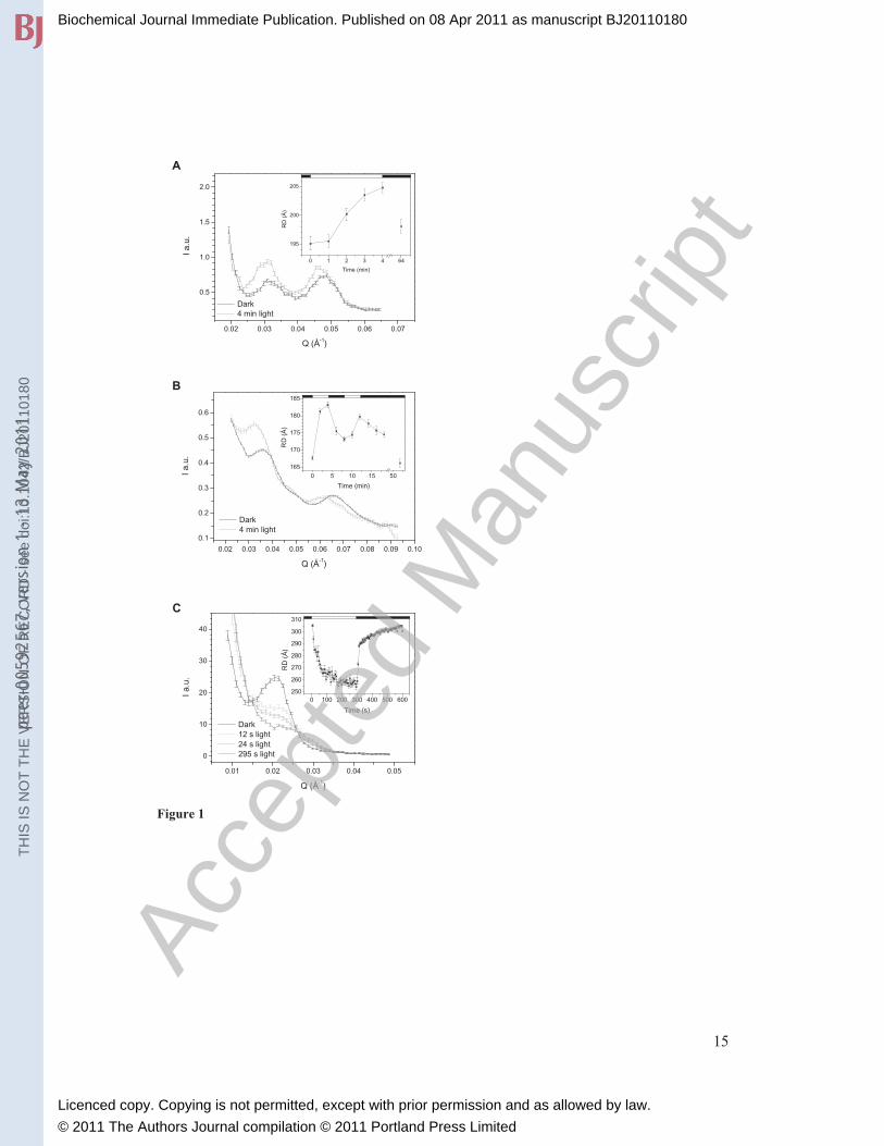
(Figure 1A), corresponding to a RD of ~ 190 Å
(Table 1). The measured RD values, representing statistically averaged values, are
somewhat higher than expected based on electron microscopy data: a thylakoid vesicle
thickness of 125 Å plus the 40 Å interthylakoidal space [18].
In diatoms the chloroplast thylakoid membranes are loosely appressed and organised into
groups of three [21;22]. Each of these membranes contains both PSII and PSI, along with
their common light harvesting antenna complexes, the fucoxanthin-chlorophyll proteins
(FCPs). In the diatom Phaeodactylum tricornutum, we obtain a RD of ~170 Å (Table 1).
This RD evidently requires a tight packing of membranes – given the even distribution of
PSII, PSI and FCPs [21]. In plants, such an arrangement of PSII and PSI complexes
would require a somewhat larger RD of ~185 Å: PSI protrudes about 40 Å while both
PSII and its main light harvesting complexes (LHCII) are extended by about 20 Å into
the interthylakoidal space; the membrane thicknesses are ~40 Å each, and the lumenal
spacing is ~45 Å [12;19]. In diatoms, however, FCPs possess considerably smaller loop
segments [21;23] than LHCII - thus allowing a smaller interthylakoidal space and smaller
RD. Concerning the location of the ATP synthase in diatoms, with its estimated large,
~140 Å protrusion [24], it is clear that this enzyme can only be accommodated in the
‘end’ membrane, i.e. on the outer surfaces of the groups of three thylakoid membranes.
This implies that the thylakoid in the middle must be energetically coupled, evidently via
interconnected lumenal spaces and contiguous bilayer membranes, to the two end
membranes.
In the chloroplasts of higher plants, cylindrical stacks of granum thylakoids, with typical
diameters of 300–600 nm and containing 5-20 thylakoids are interconnected by non-
appressed, so-called stroma thylakoid membranes [25]. With this lateral heterogeneity,
there are two RDs in the system, corresponding to the grana stacks and stroma lamellae,
respectively. In grana, which in the stacked region contain PSII and LHCII but no PSI,
RD can be small indeed, a RDgrana of 157 Å (with 45 Å lumenal and 32 Å interthylakoidal
space) has recently been determined by cryo-EM for spinach chloroplasts [19]. RDgrana
was found to be 167 Å in dark-adapted Arabidopsis leaves after cryo-immobilization and
freeze-substitution (H. Kirchhoff and Z. Reich, personal communication). In contrast,
RDstroma must be considerably larger, due to the presence of PSI and the ATP synthase in
these membrane sections. Indeed, in freshly isolated intact spinach thylakoids we find
well defined Bragg peaks corresponding to RDs between 285-300 Å (Figure 1C, Table
1). These RDs can accommodate a ~125 Å thick thylakoid vesicle and allow �160 Å
protrusions between the lamellae. For reasons probably related to symmetry and/or lack
Biochemical Journal Immediate Publication. Published on 08 Apr 2011 as manuscript BJ20110180T
HIS
IS N
OT
TH
E V
ER
SIO
N O
F R
EC
OR
D -
see
doi
:10.
1042
/BJ2
0110
180
Acce
pted
Man
uscr
ipt
Licenced copy. Copying is not permitted, except with prior permission and as allowed by law.
© 2011 The Authors Journal compilation © 2011 Portland Press Limited
peer
-005
9256
7, v
ersi
on 1
- 13
May
201
1

6
of contrast, the first order Bragg peak of the multilamellar grana stacks is missing – under
all experimental conditions tested. (For theoretical treatment of the small angle scattering
of granal thylakoid membranes, see [26].) Instead, as will be shown elsewhere, grana, as
well as PSII membrane-pairs devoid of the interconnecting stroma thylakoids, i.e. pairs of
adjacent stacked membranes isolated from granal chloroplasts, display characteristic
scattering in the Q-range between 0.065 and 0.07 Å-1
.
In order to investigate if these membrane ultrastructures are stable under different
conditions and in particular during the photosynthetic functions, we carried out time
resolved SANS experiments. These measurements on the PAL mutant of SynechocystisPCC 6803 revealed, to our knowledge for the first time, rapid light-induced RD changes
of the thylakoid membranes in living cyanobacterial cells (Figure 1A). The magnitude of
these changes, which probably originate from variations in the surface charges during
photosynthesis, is rather small (~ 10 Å), rendering them almost certainly undetectable by
EM. It is also noteworthy that the changes were fully reversible in the dark. This phase
was rather slow, which suggests the existence of two states, a light adapted state and a
dark adapted state of the thylakoid membrane RDs. Similar membrane reorganizations
are observed in Phaeodactylum tricornutum cells (Figure 1B), where the full reversibility
is also slow (30-60 min), similar to the relaxation after the photoprotective non-
photochemical quenching of the fluorescence in the antenna in the dark [27;28], and to
the light-induced reorganizations in the chiral macroorganization of the pigment system
detected by CD spectroscopy [29]. The structural flexibility of diatoms might be related
to their well known ability to survive large fluctuations in light intensity in mixing waters
[30;31]. With regard to higher plants, our studies reveal unexpectedly high structural
flexibility of stroma thylakoids. Fully reversible light-induced reorganizations,
observable already in 2-dimensional scattering profiles (Supplementary Figure 1), could
be recorded with a time-resolution of several seconds, which affected both RD and the
degree of the lamellar order - manifested in shifted peak positions and broadened and
diminished Bragg peaks, respectively (Figure 1C). It is interesting to note that the light-
induced changes in the RD were smaller when whole chain, instead of PSI cyclic electron
transport, was operating, i.e. when PMS was replaced with methylviologen
(Supplementary Figure 2). These reorganizations in plant thylakoid membranes closely
resemble the light-induced shrinking of thylakoid membranes, detected earlier by
electron microscopy as well as by light scattering, and which has been shown to be driven
by transmembrane �pH generated during photosynthesis [32]. In perfect agreement with
these data, we show that uncouplers eliminate the light-induced RD changes, a shrinkage,
in isolated plant thylakoid membranes (Supplementary Figure 3A and B). In contrast,
NH4Cl had no significant effect on the light-induced RD change, a swelling, in the
diatom P. tricornutum (Supplementary Figure 3C and D), showing that the light-induced
transmembrane �pH in the two systems do not play the same role in the membrane
reorganizations. While the mechanism of the light-induced membrane reorganizations in
diatoms remains to be explored, it is to be noted that the macro-organization of
membranes, the roles of transmembrane proton gradients generated upon illumination
and via chlororespiration in the dark as well as some details of the regulatory mechanisms
of non-photochemical quenching display significant differences in the two systems
[33;34].
Biochemical Journal Immediate Publication. Published on 08 Apr 2011 as manuscript BJ20110180T
HIS
IS N
OT
TH
E V
ER
SIO
N O
F R
EC
OR
D -
see
doi
:10.
1042
/BJ2
0110
180
Acce
pted
Man
uscr
ipt
Licenced copy. Copying is not permitted, except with prior permission and as allowed by law.
© 2011 The Authors Journal compilation © 2011 Portland Press Limited
peer
-005
9256
7, v
ersi
on 1
- 13
May
201
1

7
In summary, in the past decades a wealth of information has accumulated on different
reorganizations which are part of the multilevel regulatory mechanisms of photosynthetic
organisms and which affect the overall membrane organization [1-6;32-38]. As
demonstrated in this paper, SANS allows the determination of RDs of the membrane
ultrastructure in different photosynthetic organisms and the monitoring of their time -
resolved reorganizations during photosynthesis, information hitherto not available. This
technique is thus suitable for the fast and accurate determination of repeat distances on
the entire population of multilamellar, inherently heterogeneous biological membrane
systems in vivo. Our data provide clear evidence for the occurrence of small but well
discernible membrane reorganizations during photosynthesis, which appear to be linked
with basic regulatory mechanisms.
Biochemical Journal Immediate Publication. Published on 08 Apr 2011 as manuscript BJ20110180T
HIS
IS N
OT
TH
E V
ER
SIO
N O
F R
EC
OR
D -
see
doi
:10.
1042
/BJ2
0110
180
Acce
pted
Man
uscr
ipt
Licenced copy. Copying is not permitted, except with prior permission and as allowed by law.
© 2011 The Authors Journal compilation © 2011 Portland Press Limited
peer
-005
9256
7, v
ersi
on 1
- 13
May
201
1

8
Acknowledgements We would like to thank the Institut Laue-Langevin (Grenoble, France) for providing us
beamtime for the experiments. We are indebted to Dr. Ghada Ajlani (CEA, Saclay) for
providing us the PAL mutant of Synechocystis PCC 6803 and for critical reading of a
version of this manuscript and to Dr. Charles Dewhurst, Dr. Philip Callow, Dr. Lionel
Porcar and Peter Cross (ILL, Grenoble) for helping us in configuring of the experiments
and advice on data treatment. We also wish to thank Kasper H. Swiatek (Roskilde
University) and Dr. Eszter Rétfalvi (RISP, Budapest) for participating in some of the
experiments and Dr. Zoltán Gombos (BRC, Szeged) for helpful discussions.
Funding: This work was supported by the Marie Curie Initial Training Network ‘HARVEST’
sponsored by the 7th
Framework Program of the European Union [No. 238017] and by
the Hungarian Scientific Research Fund/National Office for Research and Technology
[No. 80345] grants to G.G., National Office for Research and Technology [NAP-
VENEUS05] grant to L.R. and Bourse du Gouvernement Français to G.N.; D.P. thanks
the Danish Centre for the use of Synchrotron X-ray and Neutron facilities (DANSCATT)
for financial support.
Biochemical Journal Immediate Publication. Published on 08 Apr 2011 as manuscript BJ20110180T
HIS
IS N
OT
TH
E V
ER
SIO
N O
F R
EC
OR
D -
see
doi
:10.
1042
/BJ2
0110
180
Acce
pted
Man
uscr
ipt
Licenced copy. Copying is not permitted, except with prior permission and as allowed by law.
© 2011 The Authors Journal compilation © 2011 Portland Press Limited
peer
-005
9256
7, v
ersi
on 1
- 13
May
201
1

9
References
1 Anderson, J. M. and Andersson, B. (1988) The dynamic photosynthetic membrane
and regulation of solar-energy conversion. Trends Biochem. Sci. 13, 351-355
2 Allen, J. F. (2003) State transitions - a question of balance. Science 299, 1530-1532
3 Kargul, J. and Barber, J. (2008) Photosynthetic acclimation: structural
reorganisation of light harvesting antenna - role of redox-dependent
phosphorylation of major and minor chlorophyll a/b binding proteins. FEBS J. 275,
1056-1068
4 Horton, P., Ruban, A. V., and Walters, R. G. (1996) Regulation of light harvesting
in green plants. Annu. Rev. Plant Physiol. Plant Mol. Biol. 47, 655-684
5 Mullineaux, C. W., Tobin, M. J., and Jones, G. R. (1997) Mobility of
photosynthetic complexes in thylakoid membranes. Nature 390, 421-424
6 Eberhard, S., Finazzi, G., and Wollman, F. A. (2008) The dynamics of
photosynthesis. Annu. Rev. Genet. 42, 463-515
7 Renger, G. (2010) The light reactions of photosynthesis. Curr. Sci. 98, 1305-1319
8 Sadler, D. M. and Worcester, D. L. (1982) Neutron-scattering studies of
photosynthetic membranes in aqueous dispersion. J. Mol. Biol. 159, 485-499
9 Fitter, J., Gutberlet, T. and Katsaras, J. (2006) Neutron Scattering in Biology,
Springer-Verlag, Berlin, Heidelberg
10 Heller, W. T. (2010) Small-angle neutron scattering and contrast variation: a
powerful combination for studying biological structures. Acta Crystallogr., Sect. D:
Biol. Crystallogr. 66, 1213-1217
11 Serdyuk, I. N., Zaccai, N. R., and Zaccai, J. (2007) Methods in Molecular
Biophysics: Structure, Dynamics, Function, Cambridge University Press,
Cambridge
12 Dekker, J. P. and Boekema, E. J. (2005) Supramolecular organization of thylakoid
membrane proteins in green plants. Biochim. Biophys. Acta 1706, 12-39
13 Wilson, A., Ajlani, G., Verbavatz, J. M., Vass, I., Kerfeld, C. A., and Kirilovsky, D.
(2006) A soluble carotenoid protein involved in phycobilisome-related energy
dissipation in cyanobacteria. Plant Cell 18, 992-1007
14 Lepetit, B., Volke, D., Szabó, M., Hoffmann, R., Garab, G. Z., Wilhelm, C., and
Goss, R. (2007) Spectroscopic and molecular characterization of the oligomeric
antenna of the diatom Phaeodactylum tricornutum. Biochemistry 46, 9813-9822
Biochemical Journal Immediate Publication. Published on 08 Apr 2011 as manuscript BJ20110180T
HIS
IS N
OT
TH
E V
ER
SIO
N O
F R
EC
OR
D -
see
doi
:10.
1042
/BJ2
0110
180
Acce
pted
Man
uscr
ipt
Licenced copy. Copying is not permitted, except with prior permission and as allowed by law.
© 2011 The Authors Journal compilation © 2011 Portland Press Limited
peer
-005
9256
7, v
ersi
on 1
- 13
May
201
1

10
15 Várkonyi, Z., Nagy, G., Lambrev, P., Kiss, A. Z., Székely, N., Rosta, L., and Garab,
G. (2009) Effect of phosphorylation on the thermal and light stability of the
thylakoid membranes. Photosynth. Res. 99, 161-171
16 Holm, J. K. (2004) Structure and Structural Flexibility of Chloroplast Thylakoid
Membranes, Roskilde University. Ph.D. Thesis
17 Liberton, M. and Pakrasi, H. (2008) Membrane systems in cyanobacteria. In The
Cyanobacteria: Molecular Biology, Genomics and Evolution (Herrero, A. and
Flores, E., eds.), pp. 271-289, Caister Academic Press, Norfolk
18 Olive, J., Ajlani, G., Astier, C., Recouvreur, M., and Vernotte, C. (1997)
Ultrastructure and light adaptation of phycobilisome mutants of Synechocystis PCC
6803. Biochim. Biophys. Acta 1319, 275-282
19 Daum, B., Nicastro, D., Il, J. A., McIntosh, J. R., and Kühlbrandt, W. (2010)
Arrangement of photosystem II and ATP synthase in chloroplast membranes of
spinach and pea. Plant Cell 22, 1299-1312
20 Ajlani, G. and Vernotte, C. (1998) Construction and characterization of a
phycobiliprotein-less mutant of Synechocystis sp. PCC6803. Plant Mol. Biol. 37,
577-580
21 Wilhelm, C., Büchel, C., Fisahn, J., Goss, R., Jakob, T., LaRoche, J., Lavaud, J.,
Lohr, M., Riebesell, U., Stehfest, K., Valentin, K., and Kroth, P. G. (2006) The
regulation of carbon and nutrient assimilation in diatoms is significantly different
from green algae. Protist 157, 91-124
22 Pyszniak, A. M. and Gibbs, S. P. (1992) Immunocytochemical localization of
photosystem I and the fucoxanthin-chlorophylla/c light-harvesting complex in the
diatom Phaeodactylum tricornutum. Protoplasma 166, 208-217
23 Eppard, M. and Rhiel, E. (1998) The genes encoding light-harvesting subunits of
Cyclotella cryptica (Bacillariophyceae) constitute a complex and heterogeneous
family. Mol. Gen. Genet. 260, 335-345
24 Stock, D., Leslie, A. G. W., Walker, J. E. (1999) Molecular architecture of the
rotary motor in ATP synthase. Science 286, 1700-1705
25 Mustárdy, L. and Garab, G. (2003) Granum revisited. A three-dimensional model -
where things fall into place. Trends Plant Sci. 8, 117-122
26 Kirkensgaard, J. J. K., Holm, J. K., Larsen, J. K., and Posselt, D. (2009) Simulation
of small-angle X-ray scattering from thylakoid membranes. J. Appl. Crystallogr. 42,
649-659
Biochemical Journal Immediate Publication. Published on 08 Apr 2011 as manuscript BJ20110180T
HIS
IS N
OT
TH
E V
ER
SIO
N O
F R
EC
OR
D -
see
doi
:10.
1042
/BJ2
0110
180
Acce
pted
Man
uscr
ipt
Licenced copy. Copying is not permitted, except with prior permission and as allowed by law.
© 2011 The Authors Journal compilation © 2011 Portland Press Limited
peer
-005
9256
7, v
ersi
on 1
- 13
May
201
1
11
27 Ruban, A. V., Lavaud, J., Rousseau, B., Guglielmi, G., Horton, P., and Etienne, A.
L. (2004) The super-excess energy dissipation in diatom algae: comparative
analysis with higher plants. Photosynth. Res. 82, 165-175
28 Goss, R., Pinto, E. A., Wilhelm, C., and Richter, M. (2006) The importance of a
highly active and Delta pH-regulated diatoxanthin epoxidase for the regulation of
the PSII antenna function in diadinoxanthin cycle containing algae. J. Plant Physiol.
163, 1008-1021
29 Szabó, M., Lepetit, B., Goss, R., Wilhelm, C., Mustárdy, L., and Garab, G. (2008)
Structurally flexible macro-organization of the pigment–protein complexes of the
diatom Phaeodactylum tricornutum. Photosynth. Res. 95, 237-245
30 Wagner, H., Jakob, T., and Wilhelm, C. (2006) Balancing the energy flow from
captured light to biomass under fluctuating light conditions. New Phytol. 169, 95-
108
31 Lavaud, J., Strzepek, R. F., and Kroth, P. G. (2007) Photoprotection capacity differs
among diatoms: Possible consequences on the spatial distribution of diatoms related
to fluctuations in the underwater light climate. Limnol. Oceanogr. 52, 1188-1194
32 Murakami, S. and Packer, L. (1970) Protonation and chloroplast membrane
structure. J. Cell Biol. 47, 332-351
33 Cruz, S., Goss, R., Wilhelm, C., Leegood, R., Horton, P., and Jakob, T. (2011)
Impact of chlororespiration on non-photochemical quenching of chlorophyll
fluorescence and on the regulation of the diadinoxanthin cycle in the diatom
Thalassiosira pseudonana. J. Exp. Bot. 62, 509-519
34 Lepetit, B., Goss, R., Jakob, T., and Wilhelm, C. (2011) Molecular dynamics of the
diatom thylakoid membrane under different light conditions. Photosynth. Res. doi:
10.1007/s11120-011-9633-5
35 Chuartzman, S. G., Nevo, R., Shimoni, E., Charuvi, D., Kiss, V., Ohad, I.,
Brumfeld, V., and Reich, Z. (2008) Thylakoid membrane remodeling during state
transitions in Arabidopsis. Plant Cell 20, 1029-1039
36 Iwai, M., Yokono, M., Inada, N., and Minagawa, J. (2010) Live-cell imaging of
photosystem II antennna dissociation during state transitions. Proc. Natl. Acad. Sci.
U. S. A. 107, 2337-2342
37 Istokovics, A., Simidjiev, I., Lajkó, F., and Garab, G. (1997) Characterization of the
light induced reversible changes in the chiral macroorganization of the
chromophores in chloroplast thylakoid membranes. Temperature dependence and
effect of inhibitors. Photosynth. Res. 54, 45-53
38 Dobrikova, A. G., Várkonyi, Z., Krumova, S. B., Kovács, L., Kostov, G. K.,
Todinova, S. J., Busheva, M. C., Taneva, S. G., and Garab, G. (2003) Structural
Biochemical Journal Immediate Publication. Published on 08 Apr 2011 as manuscript BJ20110180T
HIS
IS N
OT
TH
E V
ER
SIO
N O
F R
EC
OR
D -
see
doi
:10.
1042
/BJ2
0110
180
Acce
pted
Man
uscr
ipt
Licenced copy. Copying is not permitted, except with prior permission and as allowed by law.
© 2011 The Authors Journal compilation © 2011 Portland Press Limited
peer
-005
9256
7, v
ersi
on 1
- 13
May
201
1

12
rearrangements in chloroplast thylakoid membranes revealed by differential
scanning calorimetry and circular dichroism spectroscopy. Thermo-optic effect.
Biochemistry 42, 11272-11280
Biochemical Journal Immediate Publication. Published on 08 Apr 2011 as manuscript BJ20110180T
HIS
IS N
OT
TH
E V
ER
SIO
N O
F R
EC
OR
D -
see
doi
:10.
1042
/BJ2
0110
180
Acce
pted
Man
uscr
ipt
Licenced copy. Copying is not permitted, except with prior permission and as allowed by law.
© 2011 The Authors Journal compilation © 2011 Portland Press Limited
peer
-005
9256
7, v
ersi
on 1
- 13
May
201
1
13
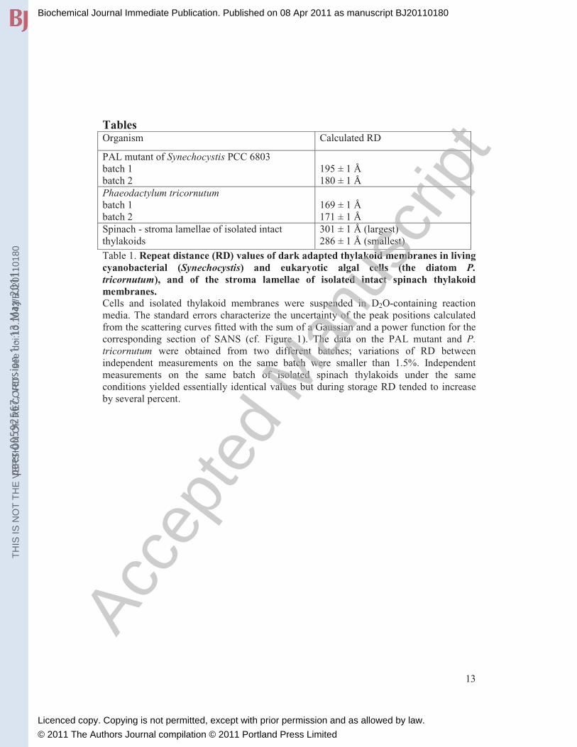
Tables Organism Calculated RD
PAL mutant of Synechocystis PCC 6803
batch 1
batch 2
195 ± 1 Å
180 ± 1 Å
Phaeodactylum tricornutum batch 1
batch 2
169 ± 1 Å
171 ± 1 Å
Spinach - stroma lamellae of isolated intact
thylakoids
301 ± 1 Å (largest)
286 ± 1 Å (smallest)
Table 1. Repeat distance (RD) values of dark adapted thylakoid membranes in living cyanobacterial (Synechocystis) and eukaryotic algal cells (the diatom P. tricornutum), and of the stroma lamellae of isolated intact spinach thylakoid membranes. Cells and isolated thylakoid membranes were suspended in D2O-containing reaction
media. The standard errors characterize the uncertainty of the peak positions calculated
from the scattering curves fitted with the sum of a Gaussian and a power function for the
corresponding section of SANS (cf. Figure 1). The data on the PAL mutant and P. tricornutum were obtained from two different batches; variations of RD between
independent measurements on the same batch were smaller than 1.5%. Independent
measurements on the same batch of isolated spinach thylakoids under the same
conditions yielded essentially identical values but during storage RD tended to increase
by several percent.
Biochemical Journal Immediate Publication. Published on 08 Apr 2011 as manuscript BJ20110180T
HIS
IS N
OT
TH
E V
ER
SIO
N O
F R
EC
OR
D -
see
doi
:10.
1042
/BJ2
0110
180
Acce
pted
Man
uscr
ipt
Licenced copy. Copying is not permitted, except with prior permission and as allowed by law.
© 2011 The Authors Journal compilation © 2011 Portland Press Limited
peer
-005
9256
7, v
ersi
on 1
- 13
May
201
1

14
Figure 1. Effect of illumination on the SANS profiles of thylakoid membranes in living cyanobacterial cells (A), and diatoms (B) and in suspensions of isolated plant thylakoid membranes (C). (A) the PAL mutant of Synechocystis PCC 6803, (B) the diatom Phaeodactylum tricornutum, and (C) isolated spinach thylakoid membranes – the SANS signal of stroma
lamellae. Insets, time courses of the light-induced variations in the repeat distances,
calculated from the first order Bragg peaks, and their dark recovery phases. The
additional peaks seen at Q values at around 0.048 Å-1
(A) and 0.065 Å-1
(B) are proposed
to originate from paired membranes of adjacent thylakoids. The samples were illuminated
for different time periods with white light of 2000 (A), 1200 (B) and 1700 (C) μmol
photons m-2
s-1
photon flux densities, as indicated by the light (light on) and dark (light
off) horizontal bars.
Biochemical Journal Immediate Publication. Published on 08 Apr 2011 as manuscript BJ20110180T
HIS
IS N
OT
TH
E V
ER
SIO
N O
F R
EC
OR
D -
see
doi
:10.
1042
/BJ2
0110
180
Acce
pted
Man
uscr
ipt
Licenced copy. Copying is not permitted, except with prior permission and as allowed by law.
© 2011 The Authors Journal compilation © 2011 Portland Press Limited
peer
-005
9256
7, v
ersi
on 1
- 13
May
201
1
15
0.02 0.03 0.04 0.05 0.06 0.07
0.5
1.0
1.5
2.0
0 1 2 3 4 64
195
200
205
RD
(Å
)
Time (min)
A
I a.u
.
Q (Å-1)
Dark 4 min light
0.02 0.03 0.04 0.05 0.06 0.07 0.08 0.09 0.10
0.1
0.2
0.3
0.4
0.5
0.6
0 5 10 15 50
165
170
175
180
185
RD
(Å
)
Time (min)
I a.u
.
Q (Å-1)
Dark 4 min light
B
0.01 0.02 0.03 0.04 0.05
0
10
20
30
40
0 100 200 300 400 500 600250
260
270
280
290
300
310
RD
(Å
)
Time (s)
C
I a.u
.
Q (Å-1)
Dark 12 s light 24 s light 295 s light
Figure 1
Biochemical Journal Immediate Publication. Published on 08 Apr 2011 as manuscript BJ20110180T
HIS
IS N
OT
TH
E V
ER
SIO
N O
F R
EC
OR
D -
see
doi
:10.
1042
/BJ2
0110
180
Acce
pted
Man
uscr
ipt
Licenced copy. Copying is not permitted, except with prior permission and as allowed by law.
© 2011 The Authors Journal compilation © 2011 Portland Press Limited
peer
-005
9256
7, v
ersi
on 1
- 13
May
201
1