Photosynthesis research in Italy: a review
30
Abstract This historical review was compiled and edited by Giorgio Forti, whereas the other authors of the different sections are listed alphabetically after his name, below the title of the paper; they are also listed in the individual sections. This review deals with the research on photosynthesis performed in several Ital- ian laboratories during the last 50 years; it includes research done, in collaboration, at several international laboratories, particularly USA, UK, Switzerland, Hungary, Germany, France, Finland, Denmark, and Austria. Wherever pertinent, references are provided, especially to other historical papers in Govindjee et al. [Govindjee, Beatty JT, Gest H, Allen JF (eds) (2005) Discoveries in Photosynthesis. Springer, Dordrecht]. This paper covers the physical and chemical events starting with the absorption of a quantum of light by a pigment molecule to the conversion of the radiation energy into the stable chemical forms of the reducing power and of ATP. It describes the work done on the structure, function and regulation of the photosyn- thetic apparatus in higher plants, unicellular algae and in photosynthetic bacteria. Phenomena such as photoinhibition and the protection from it are also included. Research in biophysics of photosynthesis in Padova (Italy) is discussed by G.M. Giacometti and G. Giacometti (2006). Keywords Antenna structure and function ATP synthase Bacterial photosynthesis Biophysics of photosynthesis Chemiosmosis G. Forti (&) F. M. Garlaschi R. C. Jennings G. Zucchelli Istituto di Biofisica del CNR, Sezione di Milano e Dipartimento di Biologia dell’Universita ` degli Studi di Milano, Via Celoria 26, Milano 20133, Italy e-mail: [email protected] A. Agostiano Dipartimento di Chimica, Universita ` degli Studi di Bari, Via Orabona 4, Bari, Italy R. Barbato Dipartimento di Scienze dell’Ambiente e della Vita, Universita ` del Piemonte Orientale, Via Bellini 25/G, Alessandria 15100, Italy R. Bassi Dipartimento Scientifico Tecnologico, Universita ` di Verona, Strada Le Grazie 37134 Verona, Italy; and Faculte ´ des Sciences de Luminy, Universite ´ Aix-Marseille II, Case 901, 136 Avenue de Luminy, Marseilles, France e-mail: [email protected] E. Brugnoli Istituto di Biologia Agroalimentare del Consiglio Nazionale delle Ricerche, Laboratorio di Porano, Via Marconi 2, Porano TR 05010, Italy G. Finazzi Institut de Biologie Physico-Chimique, 13 Rue Pierre et Marie Curie, Paris 75005, France B. A. Melandri G. Venturoli D. Zannoni Dipartimento di Biologia Evoluzionistica Sperimentale, Universita ` degli Studi di Bologna, Via Irnerio 42, Bologna 40126, Italy M. Trotta Dipartimento di Chimica, CNR Istituto per i Processi Chimico Fisici, sede di Bari, Universita ` degli Studi di Bari, Via Orabona 4, Bari, Italy G. Zanetti Dipartimento di Scienze Biomolecolari e Biotecnologie, Universita ` degli Studi di Milano, Via Celoria 26, Milano 20133, Italy Photosynth Res (2006) 88:211–240 DOI 10.1007/s11120-006-9054-z 123 HISTORICAL MINIREVIEW Photosynthesis research in Italy: a review Giorgio Forti Angela Agostiano Roberto Barbato Roberto Bassi Enrico Brugnoli Giovanni Finazzi Flavio M. Garlaschi Robert C. Jennings Bruno Andrea Melandri Massimo Trotta Giovanni Venturoli Giuliana Zanetti Davide Zannoni Giuseppe Zucchelli Received: 24 October 2005 / Accepted: 24 February 2006 / Published online: 6 June 2006 ȑ Springer Science+Business Media B.V. 2006
-
Upload
independent -
Category
Documents
-
view
1 -
download
0
Transcript of Photosynthesis research in Italy: a review

Abstract This historical review was compiled and
edited by Giorgio Forti, whereas the other authors of
the different sections are listed alphabetically after his
name, below the title of the paper; they are also listed
in the individual sections. This review deals with the
research on photosynthesis performed in several Ital-
ian laboratories during the last 50 years; it includes
research done, in collaboration, at several international
laboratories, particularly USA, UK, Switzerland,
Hungary, Germany, France, Finland, Denmark, and
Austria. Wherever pertinent, references are provided,
especially to other historical papers in Govindjee et al.
[Govindjee, Beatty JT, Gest H, Allen JF (eds) (2005)
Discoveries in Photosynthesis. Springer, Dordrecht].
This paper covers the physical and chemical events
starting with the absorption of a quantum of light by a
pigment molecule to the conversion of the radiation
energy into the stable chemical forms of the reducing
power and of ATP. It describes the work done on the
structure, function and regulation of the photosyn-
thetic apparatus in higher plants, unicellular algae
and in photosynthetic bacteria. Phenomena such as
photoinhibition and the protection from it are also
included. Research in biophysics of photosynthesis in
Padova (Italy) is discussed by G.M. Giacometti and
G. Giacometti (2006).
Keywords Antenna structure and function ÆATP synthase Æ Bacterial photosynthesis ÆBiophysics of photosynthesis Æ Chemiosmosis Æ
G. Forti (&) Æ F. M. Garlaschi Æ R. C. Jennings ÆG. ZucchelliIstituto di Biofisica del CNR, Sezione di Milano eDipartimento di Biologia dell’Universita degli Studi diMilano, Via Celoria 26, Milano 20133, Italye-mail: [email protected]
A. AgostianoDipartimento di Chimica, Universita degli Studi di Bari,Via Orabona 4, Bari, Italy
R. BarbatoDipartimento di Scienze dell’Ambiente e della Vita,Universita del Piemonte Orientale, Via Bellini 25/G,Alessandria 15100, Italy
R. BassiDipartimento Scientifico Tecnologico, Universitadi Verona, Strada Le Grazie 37134 Verona,Italy; and Faculte des Sciences de Luminy, UniversiteAix-Marseille II, Case 901, 136 Avenue deLuminy, Marseilles, Francee-mail: [email protected]
E. BrugnoliIstituto di Biologia Agroalimentare del Consiglio Nazionaledelle Ricerche, Laboratorio di Porano, Via Marconi 2,Porano TR 05010, Italy
G. FinazziInstitut de Biologie Physico-Chimique, 13 Rue Pierre etMarie Curie, Paris 75005, France
B. A. Melandri Æ G. Venturoli Æ D. ZannoniDipartimento di Biologia Evoluzionistica Sperimentale,Universita degli Studi di Bologna, Via Irnerio 42, Bologna40126, Italy
M. TrottaDipartimento di Chimica, CNR Istituto per i ProcessiChimico Fisici, sede di Bari, Universita degli Studi di Bari,Via Orabona 4, Bari, Italy
G. ZanettiDipartimento di Scienze Biomolecolari e Biotecnologie,Universita degli Studi di Milano, Via Celoria 26, Milano20133, Italy
Photosynth Res (2006) 88:211–240
DOI 10.1007/s11120-006-9054-z
123
HISTORICAL MINIREVIEW
Photosynthesis research in Italy: a review
Giorgio Forti Æ Angela Agostiano Æ Roberto Barbato Æ Roberto Bassi ÆEnrico Brugnoli Æ Giovanni Finazzi Æ Flavio M. Garlaschi Æ Robert C. Jennings ÆBruno Andrea Melandri Æ Massimo Trotta Æ Giovanni Venturoli ÆGiuliana Zanetti Æ Davide Zannoni Æ Giuseppe Zucchelli
Received: 24 October 2005 / Accepted: 24 February 2006 / Published online: 6 June 2006� Springer Science+Business Media B.V. 2006

Excitation energy transfer Æ FNR Æ International
congress on photosynthesis in Stresa ÆPhotoinhibition Æ Photophosphorylation ÆPhotosynthesis research in Italy ÆPhotosynthetic electron transport Æ Photosynthetic
membranes Æ Q-cycle Æ Reaction center structure and
function Æ Respiration Æ State changes
Abbreviations
AFR ascorbate free radical
Chl chlorophyll
DBMIB 2,5-Dibromo-3-methyl-6-isopropyl
benzoquinone
DCCD Dicyclohexyl carbodiimide
DCMU 3-(3,4-Dichlorophenyl)-1,1-dimethylurea
DNP-INT 2,4-(Dinitrophenol)-dinitrophenylether
of iodonitrothymol
Fd ferredoxin
FNR ferredoxin-NADP+ reductase
HiPIP high-Potential Iron-sulfur Proteins
LHC light harvesting complex
NADP nicotinamide adenine dinucleotide
phosphate
NPQ non-photochemical quenching of the
excited state of Chl
PS I photosystem I
PS II photosystem II
THC-RC tetra-heme RC-bound cytochrome
Introduction (by Giorgio Forti)
History is the orderly narration of events, with the aim
that ‘‘the memory of the enterprises of humans are not
erased by time’’ (Herodotus, V century B.C.). Herodo-
tus insisted on the necessity of thorough investigation
before accepting any ‘‘fact’’ as truly documented. In
modern times, Science requires that the ‘‘facts’’ are, or
should be, documented ‘‘objectively,’’ i.e., by repro-
ducible experiments reported and discussed in papers
accepted in peer-reviewed scientific journals. But what
can be defined as an ‘‘objective’’ description, and, even
more difficult, an ‘‘acceptable’’ interpretation of
experiments? We quote an outstanding ancient histo-
rian (Thucidides, V century B.C.): ‘‘...it has been an ar-
duous undertaking, because the persons present at every
event did not report on it in the same way, but everyone
according to what he remembered or his personal pre-
ferences for one or the other of the conflicting parts’’.
Keeping in mind the ancient historians, we will
adopt here the criteria of the critical description and
discussion of experiments, documented by the pub-
lished papers: this is the reason for including a long list
of references, which were prepared by the authors of
the nine sections of this chapter of history.
The scientists involved in photosynthesis research in
Italy are many, and most of their names can be found
in the list of references. Extensive collaboration with
scientists from many countries is documented in the
text and in the references. The authors of the nine
sections, except for the first author (GF), are listed in
alphabetical order in the authors’ list; they are fully
responsible for their contributions: their opinions and
personal views on historical events may differ on some
points, as observed by Thucidides long time ago.
For time-lines of discoveries in anoxygenic photo-
synthesis and oxygenic photosynthesis, see, in Gov-
indjee et al. (eds) (2005), Gest and Blankenship (pp
51–62), and Govindjee and Krogmann (pp 63–105),
respectively. Readers are requested to consult other
chapters in Govindjee et al. (2005), which are related
to the topics presented here. A limited number of
examples of some of these chapters are included in this
paper.
Photosynthetic electron transport andphotophosphorylation in oxygenic photosynthesis
(by Giorgio Forti and Giovanni Finazzi)
The studies on photosynthesis after World War II in
Italy were initiated, in the Department of Botany at
the University of Milano, by Erasmo Marre and one
of us (Giorgio Forti); the late Sergio Tonzig led the
department in those years. Marre was mostly inter-
ested, at that time, in the mechanism of action of
auxins at the metabolic level; most of the research in
the field of photosynthesis was pursued by Forti, who
made it his life-time scientific interest. He spent
2 years (1958–1960) in the laboratory of Andre T.
Jagendorf, at the Johns Hopkins University in Balti-
more, MD. There he investigated the effect of
ascorbate on the so-called ‘‘endogenous’’ photophos-
phorylation (endogenous because it was observed
without the addition of any electron transport carrier
or acceptor to the washed chloroplast fragments): the
large stimulation observed was connected with the
Mehler reaction by the observation that O2 was re-
quired by the electron transport coupled to ATP
synthesis. This was the first demonstration that the
Mehler reaction is coupled to photophosphorylation
(Forti and Jagendorf 1961). [For a historical per-
spective on ‘photophosphorylation’, see Jagendorf
(pp 561–569) in Govindjee et al. (2005).]
212 Photosynth Res (2006) 88:211–240
123

Many years later, after the discovery of ascorbate
peroxidase in plants by Asada (1992), Forti and Eh-
renheim (1993) showed that the ascorbate free radical
(AFR), generated either via oxidation of ascorbate by
O2) formed at the reducing side of Photosystem I (PS I),
or by peroxidation of ascorbate, catalyzed by the per-
oxidase, is photoreduced by PS I in competition with
NADP. The rate of this reaction is about 50% of the
maximal rate of NADP reduction and the process shows
an Emerson enhancement effect of the same magnitude
as NADP reduction. [For the discovery of the Emerson
enhancement effect in NADP reduction, see R. Gov-
indjee et al. (1962, 1964).] The overall balance of O2
changes is zero (Forti and Elli 1995), as it is well known
to be the case for the Mehler reaction followed by the
combined action of superoxide dismutase and catalase.
Forti and Elli (1995) showed that this pathway of elec-
tron transport (also called the ascorbate-Mehler reac-
tion) is triggered by the slow electron transport to O2 of
the Mehler reaction, and is coupled with ATP synthesis
with the same ATP/2 electron ratio of 1 as for the NADP
reduction.
Forti and Gerola (1977) showed the involvement of
the water to O2 electron transport (the Mehler reac-
tion) in steady-state photosynthesis by isolated intact
chloroplasts, performing CO2 assimilation at high rates,
to be an essential part of higher plant photosynthesis, in
agreement with the earlier results of Egneus et al.
(1975). The two laboratories used different methods to
estimate O2 photoreduction: Forti and Gerola mea-
sured the initial rate of H2O2 formation upon the
addition of KCN (an inhibitor of both catalase and
ascorbate peroxidase) to the illuminated chloroplasts,
while Egneus et al. measured the initial rate of incor-
poration of 18O2 into H2O during steady-state photo-
synthesis. The ratios of the rates of O2 reduction to CO2
reduction were found to be of 0.25–0.30 in both the
laboratories. In the Mehler reaction, the ratio O2 up-
take/electron transported by the linear system through
the two Photosystems in series is 0.25, when the
ascorbate/AFR turnover, superoxide dismutase, and
catalase are inhibited (see line 1 in the scheme in Ta-
ble 1, calculated from Forti and Elli 1995).
Forti (1996) proposed an overall picture of the con-
tinuous alternation of electron transport at the reducing
side of PS I to NADP or to O2)AFR (see Table 1) for
higher plant photosynthesis. The regulation is affected
by the redox state of NADP, because NADPH cannot
be reoxidized, if ATP is not produced in the linear
electron transport to NADP in the ratio of not less than
1.5/1, required for the operation of the Calvin–Benson
cycle. Thus, the electron transport pathways of higher
plant photosynthesis, using alternatively NADP or
O2)AFR as the final acceptor at the reducing side of PS
I, involves the two Photosystems operating in series;
this allows a more substantial utilization of light energy
absorbed than would be possible in alternating linear
electron transport to NADP and cyclic electron trans-
port around PS I to produce the needed ATP. Indeed,
in the latter process the energy absorbed by PS II is
totally wasted. This of course does not minimize the
importance of Daniel Arnon’s discovery of cyclic
electron flow and photophosphorylation in isolated
chloroplasts (Arnon et al. 1954).
Though cyclic photophosphorylation has been
shown to occur in vivo in higher plants, it seems to be
limited to particular conditions of stress, in the
presence of the PS II inhibitor DCMU (3-(3,4-Di-
chlorophenyl)-1,1-dimethylurea) (Forti and Parisi
1963), and it has been shown to occur extensively
during the dark–light transition, at variance with
steady-state photosynthesis (Joliot and Joliot 2005). In
cyanobacteria, Myers (1987) found no evidence for
cyclic electron transport, and Xu et al. (2005) discov-
ered that a thylakoid protein was putatively required
for P700+ reduction in the presence of DCMU and for
photoheterotrophic growth in the presence of diuron
and glycerol, but not for normal photoautotrophic
growth and photosynthetic O2 evolution, indicating
that cyclic electron transport around PS I is not re-
quired for steady-state photosynthesis.
On the other hand, a new research line started in the
1990 by Giovanni Finazzi indicated that cyclic electron
transport coupled to ATP synthesis might represent a
major process for the utilization of light energy in the
unicellular green alga Chlamydomonas reinhardtii. In
Table 1 Scheme of electron transport in the Mehler-ascorbate reaction
DO2 O2/e) ATP ATP/e)
2H2Oþ 4O2 �����!PSIIþPSI
O2 þ 4O�2 þ 4Hþ )3 )0.75 2 0.5
4O�2 þ 4AsAþ 4Hþ �����!spontaneous4H2O2 þ 4AFR 0.0 0.0 0.0 0.0
4H2O2 þ 8AsA �����!ApX8AFRþ 8H2O 0.0 0.0 0.0 0.0
6H2Oþ 12AFR �����!PSIIþPSI3O2 þ 12AsA +3.0 0.5 6 0.0
Overall balance 0.0 )0.25 8 0.5
(Abbreviations: ApX: ascorbate peroxidase; AFR: ascorbate free radical; AsA: ascorbate)
Photosynth Res (2006) 88:211–240 213
123

this alga, onset of cyclic electron flow was suggested by
Wollman and co-workers (Vallon et al. 1991) to be
regulated by the State 1–State 2 transition. While in
plants the occurrence of State transitions involves a
limited fraction of the major light-harvesting complex
(LHC) of Photosystem II (LHC II), and is supposed to
represent a physiological device to allow optimization
of light redistribution between Photosystem I and
Photosystem II (Allen 1992, see also a later section, by
Jennings), this process apparently involves a larger
fraction of LHC II in Chlamydomonas, up to 85% of
the LHC II, according to Delosme et al. (1996). Due to
this large redistribution of the LHC II complexes, State
transitions in Chlamydomonas would hardly be able to
fulfill the role of balancing light absorption between the
two Photosystems. (For identification of the mobile
LHC II polypeptides in Chlamydomonas, see Takah-
ashi et al. 2006.) Instead, they would tend to increase
PS I performance at the expense of PS II, therefore
representing a mechanism to allow the switching on
and off of cyclic electron flow around PS I (see e.g.,
Vallon et al. 1991). This hypothesis was experimentally
tested by the application of pump and probe spectros-
copy to intact cells of Chlamydomonas (Finazzi et al.
1999). When light induced electron injection into
cytochrome b6f complex was probed in cells that were
adapted to either State 1 or State 2 in the dark
(Wollman and Delepelaire 1984), a differential sensi-
tivity of electron injection into the cytochrome b6f
complex to the addition of the PS II inhibitor DCMU
was observed. Consistent with this hypothesis, we
found that this inhibitor blocked electron flow to
Cytochrome b6f in State 1 only, suggesting that PS II
activity was not required to reduce the plastoquinone
(PQ) pool in State 2 (Finazzi et al. 1999). On the other
hand, an identical sensitivity to the addition of the
cytochrome b6f inhibitor DBMIB (2,5-dibromo-
3-methyl-6-isopropyl benzoquinone) was observed in
both State 1 and State 2 conditions (Finazzi et al. 1999).
This result is consistent with the occurrence of a switch
between linear and cyclic flow upon State 2 transition.
The strict relationship that exists between State
transitions and DCMU sensitivity of electron flow
through Cytochrome b6f complex was confirmed by the
analysis of the stt7 mutant of Chlamydomonas, which is
locked in State 1 because the LHC II kinase is knocked
out in this mutant (Fleischmann et al. 1999). In this
mutant, electron flow remained sensitive to DCMU
inhibition under both State 1 and State 2 promoting
conditions (Finazzi et al. 2002). The study of the rela-
tionship between State transitions and the occurrence
of cyclic electron flow has been extended to conditions
approaching the physiological ones (i.e., phototrophic
growth under moderate light intensity). Under these
conditions, cells appear to be in an intermediate state
between State 1 and State 2, and both linear and cyclic
flows seem to take place at the same time (Forti et al.
2003). Measurement of thylakoid swelling upon illu-
mination of Chlamydomonas mutants devoid of the
ATP-synthase CF0-F1 (Majeran et al. 2001) indicated
that the building of a transthylakoid DpH can be
inhibited by DCMU addition in State 1, but not in
State 2 conditions, consistent with the notion that the
DpH is built by linear flow in State 1, and by cyclic flow
around PS I in State 2 in this alga. Cardol et al. (2003)
showed that a reduced metabolic interaction between
mitochondria and chloroplasts, which is observed in
respiratory mutants of Chlamydomonas, promotes a
systematic transition to State 2; this results in a reduced
oxygen evolution capacity and in an enhanced cyclic
flow activity around PS I, as indicated by photoacoustic
measurements.
The above observations indicated that, from an
energetic point of view, State transitions in Chla-
mydomonas seem to represent a shift from an oxygenic
type of photosynthesis (that generates both reducing
power and ATP, ‘State 1’) to an anoxygenic bacterial
one, where only ATP is synthesized (‘State 2’). This
switch may represent an advantage in terms of the
capacity of adaptation to environmental changes. By
maintaining a high quantum yield of ATP synthesis in
State 2, cells might be able to maintain vital processes
and therefore face successfully stress conditions, where
photosynthetic CO2 assimilation and respiration are
inhibited. Consistently, it had been observed that
a systematic transition to State 2 is induced in
Chlamydomonas under nutrient deprivation conditions
(reviewed in Davies and Grossman (1998)), which
often lead to a systematic decrease of PS II activity,
and therefore of the linear electron flow. Under more
physiological conditions, i.e., intermediate conditions
between State 1 and State 2, cyclic flow might provide
the extra ATP required by the Calvin–Benson–
Bassham cycle in excess of that produced by the linear
electron transport (Forti et al. 2003). It seems there-
fore that Chlamydomonas is able to match the extent
of cyclic flow to the energetic cellular needs by mod-
ulating the amplitude of State transitions. This would
occur, it seems, due to the sensing of the redox state of
the plastoquinone pool, which is expected to become
more reduced upon decrease in the CO2 fixation per-
formance of the chloroplasts. [For a historical per-
spective on ‘state changes’, see Allen (pp 177–186) in
Govindjee et al. (2005).]
On the other hand illumination, when electron flow
is mainly occurring with cycling, is expected to pro-
214 Photosynth Res (2006) 88:211–240
123

mote back electron flow to PS II from reduced qui-
nones. This is expected to increase photoinhibition of
PS II (see e.g., Keren and Ohad 1998, for discussion).
In this sense, the ‘‘removal’’ of the antennae from PS II
might represent a useful way to protect it.
Binding of PQH2 to the quinol-binding site of the
cytochrome b6f complex (Vener et al. 1995; Zito et al.
1999) leads to the activation of the stt7/stn7 kinase
responsible for the phosphorylation of LHC II (Allen
1992, Depege et al. 2003, Bellafiore et al. 2005). This
is likely to stem from conformational changes occur-
ring in the lumenal portion of the Rieske Iron–sulfur
subunit of the Cytochrome b6f complex upon binding
of PQH2 to its quinol binding pocket (reviewed in
Breyton 2000, see also Kurisu et al. (2003) for a dis-
cussion). A role of the conformational changes in the
activation of the kinase has been proposed in Chla-
mydomonas. This has been done in Milano, by com-
paring the effect of three well known competitive
inhibitors of the Cytochrome b6f complex: stigmatel-
lin, DBMIB, and DNP-INT (2,4 Dinitrophenol)-dini-
trophenylether of iodonitrothymol) (see e.g., Frank
and Trebst 1995) on both the catalytic mechanism of
the Cytochrome b6f complex (Barbagallo et al. 2000)
and the induction of the State 1–State 2 transition
(see e.g., Finazzi et al. 2001). Thus, a number of rel-
evant phenomenological information has emerged
that will lead to final understanding of the relation-
ship between reduction of the plastoquinone pool to
activation of the kinase.
Figure 1A illustrates a model for the relationship
between the State transitions and its relationship with
the linear and cyclic electron flow in the green alga
Chlamydomonas reinhardtii. Fig. 1B shows a 2005
photograph of Giovanni Finazzi. Finazzi, who was
associated with Giorgio Forti in the Department of
Plant Physiology at the University of Milano from 1989
to 2003, is now a researcher at the Institut de Biologie
Physico-Chimique in Paris, France.
Detailed information is not available on the
mechanism leading to the State 2–State 1 transition.
We all recognize that the oxidation of the plasto-
quinone pool is an essential step in the recovery of
State 1 in State 2-adapted plants or algae (Allen
1992). However, we also recognize that this transition
is under the control of the size of the cellular ATP
pool. When the ADP/ATP ratio exceeds a certain
critical level, cells are locked in State 2 (Bulte et al.
1990). Only when both conditions are fulfilled (i.e.,
re-oxidation of the PQ pool and enhancement of the
ATP/ADP ratio), State 1 can be attained (Bulte et al.
1991). In Chlamydomonas, cyclic flow is responsible
for the regeneration of a high ATP/ADP ratio upon
illumination of State 2 cells (Forti et al. 2003).
However, this process cannot provide any means to
oxidize the plastoquinone pool, since it is also ki-
netically limited by the rate of plastoquinol oxidation.
We have shown that this latter function might be
performed by the Mehler reaction in Chlamydo-
monas. By reducing molecular oxygen with electrons
coming from PS I photochemistry, this reaction might
allow sustained oxidation of the plastoquinone pool,
and therefore inactivation of the stt/stn 7 kinase,
followed by the transition to State 1 (Forti and
Caldiroli 2005).
We acknowledge the contribution of Alberto Via-
nelli, who measured the changes of PS I activity, at
different wavelengths, in isolated spinach thylakoids in
States 1 and 2, helping to establish the difference
Fig. 1 (A) State transitions in Chlamydomonas reinhardtii. Thephysical movement of the light-harvesting complex II (LHC II)and the cytochrome b6f complex (Cyt b6f) complex, that followsthe transition to State 2, modify the diffusion properties ofplastoquinone (PQ), thus preventing the functional connectionbetween photosystem II (PS II) and Cyt b6f. This promotescyclic electron flow around photosystem I (PS I), and the over-reduction of the PQ pool connected to PS II. This enhancesacceptor side photoinhibition while protecting PS II degradation(dashed subunit) through PQH2 binding to its quinone-bindingsite. Calvin cycle should be read as Calvin–Benson–BasshamCycle (Source: Giorgio Forti and Giovanni Finazzi). (B) A 2005photograph of Giovanni Finazzi
Photosynth Res (2006) 88:211–240 215
123

between higher plants and Chlamydomonas’ state
transitions (Forti and Vianelli 1988). (Dr. Vianelli is
now on the Staff of the University of Insubria, Varese,
Italy.)
Studies on ferredoxin-NADP+ reductase (FNR)
and ferredoxin (Fd) (by Giuliana Zanetti)
In the 1960s, it was shown in the photosynthesis lab-
oratory in Milano that the thylakoid flavoprotein
(ferredoxin-NADP+ reductase, FNR), which has the
main function of reducing NADP+ in photosynthesis
(Shin and Arnon 1965), is also active in the reduction
of solubilized cytochrome f (Zanetti and Forti 1966),
and forms a complex with it during the catalysis (Forti
and Sturani 1968); this observation preceded the dis-
covery that FNR is able to associate with the Cyto-
chrome b6f complex (Zhang et al. 2001). The
discovery that this flavoprotein is required for pho-
tophosphorylation coupled to cyclic electron transport
around PS I was based on the observation that the
activity could be titrated with an antibody against
pure FNR (Forti and Zanetti 1969; Forti and Rosa
1971).
Zanetti and co-workers have extensively studied the
properties of this important electron carrier. A thor-
ough characterization of the spinach leaf enzyme was
achieved through kinetic studies with various electron
acceptors (Zanetti and Curti 1980), and preparation of
the apoprotein and reconstitution with flavin adenine
dinucleotide (FAD) or its analogues (Zanetti et al.
1983). Using chemical modification, it was suggested
that different residues of the enzyme are involved in
the catalytic mechanism: a cysteine (Zanetti and Forti
1969) and a lysine important for NADP+/NADPH
binding (Zanetti 1976), later on shown to be Lys 116
(Cidaria et al. 1985). Further, many studies were con-
cerned with the interaction of FNR with the protein
substrate ferredoxin (Fd). By using a cross-linking
agent, a soluble carbodiimide, a covalent complex be-
tween the two proteins was obtained which behaved
the same as the natural dissociable complex. Peptide
mapping of the cross-linked complex allowed us to
define the polypeptide regions of the two proteins
which interact in the complex: the polypeptide segment
72–91, containing Lys 85 and 88, was found to be
covalently linked to the Fd region 76–97 containing the
acidic cluster Glu 92–94 (Zanetti et al. 1988). Studies
of limited proteolysis of FNR in the presence and ab-
sence of its substrates further identified the 25–35 N-
terminal region as part of the binding site for Fd and
the 235–250 polypeptide segment for interaction with
NADP+/NADPH (Gadda et al. 1990). Thus, the N-
terminal moiety of the reductase, where the flavin-
binding domain is localized, was shown to be that
mainly involved in the binding of Fd.
The powerful technique of chemical cross-linking
was used also to study the interaction of purified Fd
with PS I particles. A covalent, active complex PS I–Fd
was successfully isolated and the subunit PsaD was
identified as the main partner for Fd docking to PS I
(Zanetti and Merati 1987). Later on, the interaction of
Fd in solution with a recombinant form of PsaD was
demonstrated (Pandini et al. 1999). All these studies
supported the hyphothesis that Fd acts as a shuttle
between PS I and the FNR.
A real breakthrough in the structure–function rela-
tionship of FNR was the achievement of the
recombinant form of the enzyme by A. Aliverti and the
fruitful collaboration with the crystallographer P.A.
Karplus who had solved the three-dimensional struc-
ture of the spinach leaf FNR (see Fig. 2A). A series of
studies of selected FNR mutants, designed on the basis
of the previous chemical modification results, and of
the three-dimensional structure, allowed us to define
the roles for specific residues in the reaction mecha-
nism of this essential catalyst of photosynthesis. Thus,
Lys116 and Lys 244 (Aliverti et al. 1991), Glu 312
(Aliverti et al. 1998) were demonstrated to be required
for NADP+/NADPH binding and Lys 88 (Aliverti
et al. 1994) for interaction with Fd. A direct involve-
ment in the catalytic mechanism was then shown for
Cys 272 (Aliverti et al. 1993) and Ser 96 (Aliverti et al.
1995). Mutants of the C-terminal tyrosine allowed us to
finally have a picture of how the nicotinamide group of
NADP+/NADPH could come close to the isoalloxa-
zine ring of FAD in the enzyme’s active center for
electron transfer to occur (Deng et al. 1999). Further-
more, these mutant proteins showed an impressive
increase (up to 1700-fold) in catalytic efficiencies with
NADH as compared with wild-type FNR. Thus, the
C-terminal tyrosine plays a key role in the discrimi-
nation of the pyridine nucleotides by destabilizing the
interaction with the nicotinamide. A bipartite binding
mode for NADP+/NADPH interaction with FNR was
proposed (Piubelli et al. 2000).
Figure 2A shows a three dimensional structure of
ferredoxin-NADP reductase (FNR) showing the
NADP and the ferredoxin sites, and Fig. 2B shows a
2005 photograph of the photosynthesis research group
in the Laboratory of Biochemistry, at the University of
Milano.
Spinach leaf ferredoxin I was also produced in a
recombinant form and its plasmid has been distributed
to many laboratories over the world. Mutants of the
216 Photosynth Res (2006) 88:211–240
123

conserved Glu 92 allowed us to define its role in the
modulation of the iron-sulfur cluster redox potential as
well as in the interaction with FNR (Piubelli et al.
1996) and to obtain the three-dimensional structure of
the spinach protein (Binda et al. 1998). By protein
engineering, the genes of Fd and the reductase were
fused to yield a non-dissociable protein complex, which
possesses many of the properties of the physiological
one (Aliverti and Zanetti 1997). [For historical per-
spectives on FNR and ferredoxin, respectively, see
Buchanan et al. (pp 859–866) and M. Shin (pp
867–873) in Govindjee et al. (2005).]
Recently, new forms of FNR and Fd have come to
the fore: the so-called non-photosynthetic ones. The
physiological reactions catalyzed by these FNRs run in
the opposite direction with respect to that occurring in
photoysnthesis, i.e., electrons are transferred from
NADPH to Fd. The structure–function relationship of
corn root FNR (Aliverti et al. 2001) was thoroughly
evaluated and more recently, involvement in the
characterization of the FNR/Fd system present in the
apicoplast of the apicomplexan, protozoan parasites
(Toxoplasma gondii and Plasmodium falciparum)
(Pandini et al. 2002) is keeping the laboratory busy to
find how these redox systems (photosynthetic and non-
photosynthetic), that are highly similar to each other,
are modulated in the redox potential of their prosthetic
groups to accomplish their different metabolic roles.
The Second International Congress on Photosynthesis
in Stresa, Italy (by Giorgio Forti)
In June 1971, Giorgio Forti and his associates orga-
nized the Second International Congress on Photo-
synthesis in Stresa, on the Lago Maggiore. (For a list of
International Congressses on Photosynthesis, see
Appendix F in Govindjee et al. 2005, pp 96–97.) A
total of 438 scientists, from all over the world, partic-
ipated in this Congress. The opening lecture was
delivered by Robin Hill (1972), who commemorated
Joseph Priestley in the second centenary of his
‘‘Discovery of Photosynthesis’’; he discussed Priestley’s
experiments and their interpretations. The Proceedings
of that Congress, edited by Forti, Avron and Melandri,
were published in 1972 by Dr W. Junk N.V. Publishers,
The Hague.
Mechanisms and regulation of energy transduction
and electron transport pathways in bacterialphotosynthesis (by Bruno Andrea Melandri,
Giovanni Venturoli, and Davide Zannoni)
Studies on the ATP synthase of purple
photosynthetic bacteria
The research activities in this field began in 1970 when
the existence of F1 in Rhodobacter (Rb) capsulatus was
firmly established (Baccarini-Melandri et al. 1970;
Melandri et al. 1970). This discovery, made when the
authors were working at the University of Indiana,
Bloomington, represents the first demonstration that
photosynthetic phosphorylation in bacteria is catalyzed
by an enzyme homologous to that previously demon-
strated in chloroplasts, and was also the first demon-
stration of the presence of F-type ATP synthases in
prokaryotes (Baccarini-Melandri and Melandri 1972).
Subsequently, the presence of this enzyme was dem-
onstrated also in Rhodospirillum rubrum (Johansson
et al. 1973). More importantly the same enzyme was
shown to be active in the respiratory ATP synthesis of
Fig. 2 (A) A three dimensional structure of Ferredoxin-NADPreductase (FNR) showing the NADP and the ferredoxin sites(Source: A Aliverti et al. 2001). (B) A 2005 photograph ofmembers of the Laboratory of Biochemistry at the University ofMilano. Back row: from left to right: Andrea Pennati, VittoriaPandini, Giuliana Zanetti, Bruno Curti and Alessandro Aliverti.Front row: from left to right: Gianluca Caprini, Sara Baroni andMatteo De Rosa
Photosynth Res (2006) 88:211–240 217
123

Rb. capsulatus, thereby unifying the enzymology of
ATP synthesis in the dual functional membrane of
facultative photosynthetic prokaryotes (Melandri et al.
1971).
The functional properties of the ATP synthase were
further characterized with the demonstration that the
ATPase activity was strongly stimulated by illumina-
tion (Melandri et al. 1972) and by inorganic phosphate
(Baccarini-Melandri et al. 1975), pioneering the work
on the activation of the enzyme by DlH+ in prokaryotic
ATP synthases (Turina et al. 1992). Further, Turina
et al. (2004) have discovered subtle regulatory mech-
anisms on the proton translocation efficiency operating
on the enzyme by ADP at submicromolar concentra-
tions and by phosphate at submillimolar concentra-
tions.
The ATP synthase of Rb. capsulatus has been cho-
sen as a model for functional studies on prokaryotic
ATP synthases, since its association with a photosyn-
thetic cyclic electron transport system allows experi-
ments with excellent time resolution, unattainable in
respiratory systems. The complete sequence of the two
operons coding for the enzyme in Rb. capsulatus B100
are available (Borghese et al. 1998a, b) and genetic
techniques for site-specific mutagenesis have been set
up (Turina and Melandri, 2002). These studies have
lead to the production of a number of site- specific
mutants and, for some of these, to the functional
characterization of their phenotypes (Turina and
Melandri 2002). [For a historical perspective on ATP
Synthase, see W Junge (pp 571–595) in Govindjee
et al. (2005).]
Relationship between photosynthetic and
respiratory chains in Rhodobacter capsulatus
The study of the respiratory chain in Rb. capsulatus was
pioneered in Bologna, partly in collaboration with
Antony Crofts’ laboratory, then in Bristol, UK, during
the 1970s; these studies established the branched nature
of the chain (Zannoni et al. 1976a), and the involve-
ment in the two branches, respectively, of a cytochrome
c2 oxidase and of the UQH2 (Ubiquinol) oxidase
(Zannoni et al. 1976b). For these studies, the avail-
ability of mutant strains with lesions in different points
of the respiratory chain, isolated by Barry Marrs (St.
Louis University, USA) was essential. Further, these
studies led also to the discovery that the cytochrome c2
oxidase was associated with a cytochrome b-type heme
(Zannoni et al. 1974) (today, the oxidase is known to be
a cbb3 oxidase) and that also the quinol oxidase con-
tained a CO-inhibited cytochrome b (Zannoni et al.
1976b) (today referred as a bb3 quinol oxidase). The
sites of oxidative phosphorylation were also localized
utilizing the then recently introduced technique of the
quenching of fluorescent acridines (Baccarini-Melandri
et al. 1973). The coincidence of several components of
the photosynthetic and of the respiratory chain led to a
scheme in which the quinone pool, the cytochrome bc1
complex and the cytochrome c2 pool are shared by the
two electron transfer systems, whereas the NADH
dehydrogenase complex and the two oxidases operate
uniquely in respiration. The role of the photosynthetic
Reaction Center (PRC) therefore is that of reversing
the central sector of the respiration pathway from
cytochrome c2 to ubiquinone. The concept that quinone
molecules directly connect respiratory and photosyn-
thetic components in membranes from facultative
phototrophs was further supported by studies of Davide
Zannoni in collaboration with Barry Marrs, indicating
that a strong quinol oxidase activity (Q-bb3 pathway)
was stimulated by light (LIOU-reaction or light-in-
duced oxygen uptake) also in the absence of a func-
tional cytochrome bc1 complex (Zannoni et al. 1978).
This overall scheme is, of course, in full agreement with
the double role of the ATP synthetase in the respiratory
and photosynthetic ATP synthesis (see above).
Membrane-anchored Cytochrome c (cy)
and high-potential iron–sulfur proteins (HiPIP)
as electron carriers in bacterial photosynthesis
and respiration
Prior to the mid-1980s, models of bacterial photosyn-
thetic (Ps) and respiratory (Res) electron transfer
chains (ETC) always involved a soluble, freely diffus-
ible protein, called cytochrome c2 (Prince et al. 1975).
Consistent with this dogma, a cytochrome c minus
mutant of Rhodobacter capsulatus (MT113), isolated by
Zannoni et al. (1980), was unable to grow, using only
photosynthetic electron transport. However, our grasp
of the actual role of cytochrome c2 in photosynthesis
had to await the availability of a mutant lacking only
cytochrome c2. Indeed, subsequent studies by Davidson
and collaborators in Philadelphia, USA (Davidson
et al. 1987) demonstrated that mutant MT113 lacked
not only cytochrome c2 but also the Cytochrome bc1
complex including its cytochrome c1 subunit. Thus the
lack of photosynthesis (Ps) could not be attributed so-
lely to the absence of cytochrome c2. It is noteworthy
that already in 1986, Fevzi Daldal, in collaboration with
Roger Prince, demonstrated that a mutant, MT-G4/S4
that lacked only cytochrome c2 was Ps+ like its wild-
type parent. This showed, for the first time, that
bacterial photosynthesis in the absence of cyt c2 is
possible. Despite this progress, the molecular nature of
218 Photosynth Res (2006) 88:211–240
123

the critical component(s) of the cytochrome c2-inde-
pendent Ps growth remained obscure until 1990.
Indeed, an important finding came from the work of
J. (Baz) Jackson and his colleagues in Birmingham, UK
(Jones et al. 1990). They discovered in a double mutant
(constructed by F. Daldal), lacking both cytochrome c2
and the Cytochrome bc1 complex, photo-oxidation of a
membrane-associated cytochrome c (which they called
cyt cx), and proposed it to be the electron donor to PRC
alternative to cytochrome c2. Resolution of this
intriguing story awaited the discovery of the structural
gene (cycY) of the membrane-associated cytochrome c
(re-named cyt cy) by F. Daldal and co-workers in
Philadelphia and the demonstration by Zannoni and
Venturoli, in collaboration with F. Daldal (Zannoni
et al. 1992), that cytochrome cy steady-state amount
depended on the composition of the growth medium
used. Finding cycY ended all controversies on the
molecular nature of cytochrome cy and focused the
attention on the question of whether cytochrome
c2-independent pathways existed and to how such
pathways operated. Zannoni in Bologna, in collabora-
tion with the Daldal’s group, undertook a series of de-
tailed studies indicating that in Rb. capsulatus, unlike
Rb. sphaeroides, two independent electron carriers with
distinct properties operate simultaneously. The mem-
brane-attached cytochrome cy communicates with only
a subset of Reaction Centers in Rb. capsulatus while it is
not involved in Ps electron transport (ET) in Rb. sph-
aeroides although working efficiently in respiratory ET
of both bacterial species (Myllykallio et al. 1999; see
review by Daldal et al. 2001).
Ironically, after almost three decades, it became
clear in the mid-1990s that high-potential iron–sulfur
proteins (HiPIP) are the soluble electron carriers of
choice in most photosynthetic bacteria. The first func-
tional evidence of the role of HiPIP in bacterial pho-
tosynthesis was published in 1995 by Zannoni and
collaborators (Hochkoeppler et al. 1995). Our research
group in Bologna demonstrated that the rate of the
light-induced oxygen uptake (LIOU) in membranes
from Rhodoferax fermentans, a phototroph lack-
ing cytochrome c2 but expressing high amounts of Hi-
PIP, was concentration-dependent on the HiPIP
(E�¢ = +351 mV), isolated from the same microrgan-
ism. Subsequent studies by the Bologna research group
supported and extended the previous report on the
involvement of HiPIP in the photosynthetic electron
transfer. In particular, kinetic evidence was provided,
for the first time, that HiPIP rapidly re-reduces the
Reaction Center (RC) following a single turnover
excitation flash identifying the tetra-heme cytochrome
RC-bound (THC-RC), cytochrome c556, as the elec-
tron acceptor (Hochkoeppler et al. 1995). The con-
clusive evidence that HiPIP was indeed the direct
electron donor to the THC-RC of R. fermentans was
reported in 1996 by our research group in collaboration
with a group at the University of Arizona in Tucson
(Hochkoeppler et al. 1996). Laser excitation of P to P*,
and the subsequent oxidation to P+, caused a rapid
photo-oxidation of cytochrome c556 f; this was fol-
lowed, in the presence of HiPIP, by a slower re-
reduction of cytochrome c556 with a second-order rate
constant of 4.8·107 M)1 s)1. The amplitude of this
slower process was shown to decrease with increasing
HiPIP concentration, with the amplitude of a faster
phase (3·105 s)1) increasing concomitantly and inde-
pendently of the HiPIP concentration (a first-order
process). These results were interpreted as evidence
for the formation of a complex between HiPIP and
THC-RC (Kd = 2.5 lM) prior to the excitation flash.
Thus the multiphasic kinetics, observed for the reac-
tion between soluble HiPIP and the THC-RC in Rf.
fermentans, turned out to be similar to that of soluble
cytochrome c2 with the RCs from Rb. sphaeroides and
Rb. capsulatus: the circle was closed. [For historical
perspectives on electron transfer proteins and on
membrane-anchored cytochrome c, see Meyer and
Cusanovich (pp 455–470) and Daldal et al. (pp 471–
478), respectively, in Govindjee et al. (2005).]
Chemiosmotic circuits in bacterial chromatophores
Over the period from the 1970s to the early 1990s
systematic studies on chemiosmotic coupling to phos-
phorylation in chromatophores from Rb. capsulatus,
which in many ways is an ideal model system, were
performed. Bacterial ‘chromatophores’ from Rb. cap-
sulatus possess several characteristics that make them
an ideal model system for the study of chemiosmotic
coupling in photophosphorylation with an excellent
time resolution (Jackson et al. 1980). Due to the
inverse polarity of the membrane with respect to its
orientation in whole cells (Prince et al. 1975), the light-
induced proton pumping is inwardly directed, produc-
ing an acidic lumen of the chromatophores (DpH) and
a positive inside membrane potential difference (Dw).
The extent of these two forces was continuously
monitored, using 9-amino-acridine for DpH (Casadio
et al. 1974), and the electrochromic spectral shift of
endogenous carotenoids (for Dw). The responses of
these two indicators are fast enough for following in
real time the transients of DlH+ (Turina et al. 1990), and
have been conveniently calibrated by Baccarini-Mel-
andri et al. (1977), Venturoli et al. (1986a) and
Casadio (1991).
Photosynth Res (2006) 88:211–240 219
123

Systematic studies on chemiosmotic coupling were
performed: the ‘‘static head’’ concentrations of sub-
strates and products of phosphorylation were mea-
sured while the extent of steady-state DlH+ was varied
by the addition of small concentrations of uncouplers
or of electron transfer inhibitors. The results obtained
were compatible with a H+/ATP ratio in the range of
2–3 (Baccarini-Melandri et al. 1977). The rates of
photophosphorylation were also measured under the
same varied conditions and the results obtained were
surprising: the rates of ATP synthesis appeared more
related to the rates of electron transfer than to the
extent of DlH+ (Baccarini-Melandri et al. 1977; Casadio
et al. 1978), an observation hardly compatible with a
totally delocalized chemiosmotic coupling (Westerhoff
et al. 1984). Ensuing researches indicated that [ATP]/
[ADP] [Pi] ratio was a decisive factor affecting the
onset rate of photophosphorylation and its pre-steady-
state kinetics (Melandri et al. 1980), and that effects of
the titration of the electron transport chain with anti-
mycin A or of the ATP synthase with DCCD (dic-
yclohexyl carbodiimide) suggested localized coupling
(Venturoli and Melandri 1982). Subsequent studies,
however, performed with ‘chromatophores’ from cells
grown at very high light intensity (and therefore con-
taining high concentrations of ATP synthases with
respect to the photosynthetic antenna size), indicated
delocalized coupling when the ATP synthase was
titrated with DCCD (Virgili et al. 1986). A model in
which the coupling unit for photophosphorylation is
the entire vesicle, under certain growth conditions,
can reconcile these apparently conflicting results. This
aspect is being investigated at the moment.
Q-pool function and Q-cycle mechanism
in ‘chromatophores’
Starting from the initial paper (Baccarini-Melandri and
Melandri 1977), the relevance of the Q-pool for elec-
tron transport has been extensively studied over the
past 30 years. Chromatophores contain a large pool of
ubiquinone, in a 30/60-fold molar excess over the
Reaction Centers. The relevance of this pool for the
electron transfer reactions has been the topic of
extensive studies (Casadio et al. 1984). The optimal
redox poise of the quinone pool, clamped at set redox
potentials by the use of redox mediators and small
addition of oxidants or reductants, was found to be
around 100 mV for photophosphorylation (Baccarini-
Melandri et al. 1979). Reversible extraction of ubi-
quinone from lyophilized ‘chromatophores’ and their
reconstitution with ubiquinone-10, or with its lower
molecular weight homologues, indicated the need of a
large pool for an effective electron transfer rate and
the relative unimportance of the length of polyprenyl
lateral chain (for n (= or) >3) (Baccarini-Melandri
et al. 1980). [For a personal historical perspective on
the ‘‘Q-cycle’, see Crofts (pp 479–499) in Govindjee
et al. (2005).]
Extraction and reconstitution of UQ-10 was also
utilized for studying the kinetic behavior of the qui-
none pool at the Qo and at the Qi sites of the cyto-
chrome bc1 complex. The size of the pool was found to
affect the optimal redox poise for cytochrome b
reduction in the presence of antimycin, a reaction
occurring through the Qo site, and the duration of the
lag between the firing of the flash and the onset of
cytochrome b reduction (Venturoli et al. 1986b).
Reactions at the Qi site were followed indirectly by
measuring the associated electrochromic signal as a
function of the redox potential and in differently
extracted chromatophores (Venturoli et al. 1988). The
results demonstrated that the reaction rates depended
on the concentrations of quinol or of quinone,
respectively at the quinol-oxidizing (Qo) or at the
quinone-reducing (Qi) site. These data offer a clear
kinetic evidence for a Q-cycle operating in ‘chro-
matophores’, in which ubiquinone species act both
upstream and downstream the cytochrome bc1 com-
plex.
The behavior of ubiquinone-10 was also studied in
reconstituted systems. Purified complexes (Gabellini
et al. 1982) and ubiquinone were incorporated into
phospholipid liposomes containing cytochrome c2 and
the kinetics of electron transfer were studied, obtaining
information on the pool behavior and on the exchange
kinetics of cytochrome c2 in its interaction with the
Reaction Center (Venturoli et al. 1989a, b). These
studies were extended to the successful reconstitution
of photophosphorylation, obtained when purified ATP
synthase was inserted into the lipid bilayer in addition
to the electron transfer chain components (Gabellini
et al. 1989).
In 1990, the level of reduction of the Q-pool of
Rhodobacter capsulatus was determined voltametri-
cally, for the very first time, by Zannoni in collabora-
tion with Moore in Brighton, UK (Zannoni and Moore
1990), using a glassy carbon working electrode and a
platinum electrode. The results obtained with this non-
invasive technique indicated that unlike the cyto-
chrome bc1 complex, through which electron transport
appears to be linearly related to the Q-pool redox state
(between 4–5% and 30% of the quinol pool, i.e., re-
duced quinone, Qr), net flux through the Qr-oxidizing
step of the alternative pathway is strongly limited until
the Qr pool reaches ~25%. Based on a Q-pool
220 Photosynth Res (2006) 88:211–240
123

containing ~60 quinones per Reaction Center (RC), it
was concluded that the Km of QH2 at the Qo site is
~2.4–3 Q per RC while at the Qo site (Q-bb3 oxidase
pathway) the Km of QH2 is ~15 Q per RC.
Subsequent studies were aimed at elucidating ther-
modynamic and mechanistic aspects of the interaction
of the Reaction Center (RC) with its diffusive electron
transfer partners, i.e., quinone at the QB acceptor site
and cytochrome c2. In collaboration with P. Mathis in
Saclay, France, the docking of cytochrome c2 and the
electron transfer to the primary photooxidized donor
P+, within the preformed RC–cyt c2 complex, were
kinetically characterized, both in detergent suspensions
and in proteoliposomes. This last intracomplex process
was shown to involve a substantial structural re-orga-
nization, being totally impaired at 230 K (Venturoli
et al. 1993). The reorganization energy involved in
cytochrome c2 oxidation could be determined by
studying the reaction kinetics in a series of specifically
mutated Reaction Centers characterized by altered
midpoint redox potentials of the P+/P couple. This
analysis also showed that the semi-classical Marcus
equation fully accounts for the temperature and free
energy dependence of the kinetics, restricting the range
of coupled vibrational modes to frequencies lower than
200 cm)1 (Venturoli et al. 1998). Information on the
interaction of the ubiquinone pool with the QB site was
obtained by studying the recombination kinetics of the
flash-induced PþQ�B state in detergent suspensions and
in different reconstituted systems, i.e., reverse micelles
of phospolipids in hexane (Mallardi et al. 1997; Palazzo
et al. 2000a) and lecithin vesicle (Ambrosone et al.
2002; Palazzo et al. 2000b). These comparative analy-
ses, in which the dependence of the recombination
kinetics upon the size of the quinone pool and upon
temperature were simultaneously examined, allowed
proper separation of the thermodynamic parameters
governing the binding at QB and electron transfer from
reduced QA. They clearly showed that in reverse
micellar solutions, as well as in liposomes, quinone
molecules are in fast exchange with the QB site on the
time scale of electron transfer. A new model was
developed which accounted for the non-exponential
character of the recombination kinetics by considering
the distribution of ubiquinone molecules in the vesicle
populations and the size polidispersity of proteolipo-
somes, as probed by quasi-elastic light-scattering
(Ambrosone et al. 2002; Palazzo et al. 2000b).
The operon in Rb. sphaeroides coding for the L and
M subunits of the Reaction Center and the a and bsubunits of the internal antenna system (LH I) contains
an additional gene (pufX) whose deletion impairs the
capacity of photosynthetic growth. The role of PufX in
the photosynthetic electron transport chain was studied
in details. PufX is associated with the Reaction Cen-
ter–LHI complex in a stoichiometry of 1:1 (Francia
et al. 1999). The PufX protein was demonstrated
to have a role in facilitating the exchange reaction
between the Reaction Center and the cytochrome bc1
complex. In ‘chromatophores’ from cells carrying a
deletion of pufX (Barz et al. 1995a, 1995b), or even
truncation of its C-terminal sequence (residues 68–82)
(Francia et al. 2002), the lag period encompassing the
production of ubiquinol at the QB site of the Reaction
Center and its oxidation at the Qo site of the
cytochrome bc1 complex was markedly prolonged as
compared to the wild-type (from £1 ms to 5–10 ms). A
possible explanation of this phenotype, when the
structural effects of PufX were demonstrated, was:
PufX is necessary to form an S-shaped dimeric struc-
ture of the LHI–RC complex, a structure that allows a
passage for fast diffusion of reduced and oxidized
ubiquinone in exchange with the quinone pool (Sche-
uring et al. 2004). Consistent with this model, the ef-
fects of PufX deletion were alleviated, and ultimately
eliminated, when the quinone pool was maintained
largely reduced, both in vitro and in vivo. Surprisingly,
the LHI–RC preparations, as isolated by sucrose
gradient centrifugation, contained an amount of
ubiquinone much larger than the average content in
un-fractionated membranes (Francia et al. 2004a); this
observation indicates the possibility that ubiquinone
could be associated in vivo to specific protein com-
plexes (in this case, the LHI–RC complex), casting
some doubt on the accuracy of kinetic models assum-
ing a totally delocalized diffusional behavior of the
quinone pool.
Dynamics of the Reaction Center protein
in trehalose glassy matrices
A new research program was initiated in 2002 in which
RC protein dynamics were studied in trehalose glassy
matrices. Trehalose, a non-reducing disaccharide of
glucose, is found in large amounts in organisms (like
yeast cells, soil-dwelling animals, and desert resurrec-
tion plants) that can survive extreme conditions (such
as high temperatures (>60�C) and lack of water) in a
state of suspended animation called anhydrobiosis.
This property has been related to the extraordinary
efficacy of dehydrated trehalose matrices in preserving
biological structures (isolated proteins, membranes
and tissues). Several experimental and simulative
studies, mostly performed on myoglobin containing
CO, have unambiguously shown that incorporation of
a soluble protein into extremely dehydrated trehalose
Photosynth Res (2006) 88:211–240 221
123

glassy matrices severely inhibits internal protein
dynamics at room temperature. Starting from these
observations, we have shown that bacterial photosyn-
thetic RCs can be functionally incorporated into tre-
halose matrices and that trehalose coating can be used
to condition protein dynamics coupled to electron
transfer in this large integral membrane protein
(Palazzo et al. 2002). In extensively dehydrated tre-
halose matrices, RC relaxation from the dark-adapted
to the light-adapted conformation and inter-conversion
between conformational substates could be blocked at
room temperature over the time scale of hundreds of
milliseconds: the impairment was reflected in inho-
mogeneous kinetics of the PþQ�A charge recombina-
tion (Francia et al. 2004b). By exploiting a similar
strategy we have shown that Q�A -to-QB electron
transfer can be slowed down by orders of magnitude at
room temperature when the water content of the tre-
halose matrix is decreased over a limited range. These
findings were consistent with conformational gating of
the reaction and demonstrated that the conformational
dynamics controlling electron transfer to QB was
strongly dependent on the structure and dynamics of
the surrounding medium (Francia et al. 2003). As a
whole, it appeared that incorporation into dried tre-
halose matrices mimics, at room temperature, several
effects observed on electron transfer at cryogenic
temperatures, when the protein dynamics is frozen.
This approach has allowed us to uncouple temperature
and solvent effects on electron transfer, yielding direct
information on the dynamics of solvent–protein inter-
actions (Cordone et al. 2005). The extraordinary
stability at room temperature of RCs, when incorpo-
rated into trehalose-dehydrated glasses, could be of
great relevance for biotechnological applications.
Figure 3 shows a 2005 photograph of some of the
members of the photosynthesis research group in
Bologna.
Radiation absorption, transfer of excitation energy
and primary photochemical reactions: structure
and function of the antennae and Reaction Centersof the two photosystems (by Robert C. Jennings,
Flavio M. Garlaschi, and Gıuseppe Zucchelli)
Robert Jennings arrived in Italy in the December of
1972, fresh from a postdoctoral period in Itzhak Ohad’s
laboratory in Jerusalem, Israel, to take up a postdoc-
toral fellowship in Giorgio Forti’s laboratory in Naples,
Italy. This was the beginning of a long and fruitful
association between Jennings and the Italian photo-
synthesis researchers. During 1973–1977, some of the
first chlorophyll (Chl)a fluorescence induction experi-
ments in Europe were performed with research direc-
ted towards an understanding of cation effects on the
organization of the two photosystems in membranes
and in particular on the ‘‘spillover’’ of excitation
energy among the two photosystems. In 1974, an early
study on cation effects on Chl fluorescence induction
was published (Jennings and Forti 1974) and it was
subsequently demonstrated, by measuring fluorescence
induction at 682 nm, with selective excitation into the
long wavelength band of Photosystem I at 722 nm, that
an ‘‘uphill’’ energy transfer from Photosystem I to
Photosystem II may occur (Jennings and Forti 1975).
This was the first demonstration of thermally activated
energy transfer in plant photosystems. (For a detailed
discussion on Chl fluorescence as a probe of photo-
synthesis, see Papageorgiou and Govindjee 2004.)
Gotthard Krause (in Germany) had, at that time,
discovered what is now known as ‘‘non-photochemical
quenching’’ in isolated, intact chloroplasts and this was
further studied in the Italian laboratory where it was
demonstrated that the phenomenon almost exclusively
affects the variable fluorescence of Photosystem II,
with little or no effect on the minimum fluorescence
level Fo (Jennings et al. 1976; also see Papageorgiou
and Govindjee 2004). An early modeling study on
fluorescence induction was also presented in which it
was proposed that Mg2+ ions promote energy transfer
between the Reaction Center and the main light-har-
vesting (antenna) complex of Photosystem II, LHC II.
This investigation heralded the beginning of an almost
lifelong collaboration between Robert Jennings and
Flavio Garlaschi, which continues to the present day.
The experimental demonstration that electrostatic
screening by cations, including protons, promotes en-
Fig. 3 A 2005 photograph of some of the staff members of theresearch group in Bologna. From left to right: Davide Zannoni,Andrea Melandri and Giovanni Venturoli. Photo taken onSeptember 29, 2005
222 Photosynth Res (2006) 88:211–240
123

ergy coupling between the Reaction Center and LHC
II had to wait another 12 years (Zucchelli et al. 1988)
and the arrival of Giuseppe Zucchelli as a new and
lasting partner of the research group in 1985.
Returning to the late 1970s and the early 1980s, the
main research effort concerned membrane stacking,
which at that time was a very active field of photo-
synthesis research. Studies by Jim Barber’s group (in
UK) had established the electrostatic screening
mechanism, which constituted the basis for grana for-
mation and much effort was spent in Italy, and else-
where, in an attempt to understand its biological
implication. The close relation between grana forma-
tion and ‘‘spillover’’ interruption, previously suggested
in the literature, was experimentally confirmed (Jen-
nings et al. 1978) by the use of mild trypsin digestion to
selectively unstack thylakoids. Subsequently, Charles
Arnzten and his co-workers (at the University of Illi-
nois at Urbana, Illinois) demonstrated that this mild
trypsin digestion was due to the cleavage of a small
N-terminal polypeptide segment from LHC II con-
taining the phosphorylation site. Furthermore,
Jennings et al. (1980b) demonstrated that grana for-
mation establishes energy transfer between Photosys-
tem II units (i.e., cooperativity). An interesting
observation made in this period concerns the promo-
tion of normal grana formation by protons at micro-
molar concentrations (Jennings et al. 1979b, 1981),
which means that protons are orders of magnitude
more effective than metal cations in bringing about this
process. The lateral separation of the two photosys-
tems, which is associated with grana formation, does
not apply to plastoquinone, found to be uniformly
distributed throughout the thylakoid membrane (Jen-
nings et al. 1983) while the NADPH-ferredoxin oxido-
reductase is restricted to the stromal membranes
(Jennings et al. 1979a). These latter points were
established using techniques that did not involve
membrane fractionation, which may itself modify
component distribution (Jennings et al. 1980a). An
unexpected offshoot of the studies on grana function
was the demonstration of the optical sieve effect asso-
ciated with the formation of this structure (Jennings
and Zucchelli 1985). This decreases light absorption
specifically by Photosystem II by as much as 20%.
Following the investigation of State transitions in
isolated spinach thylakoids (see e.g., Jennings et al.
1986), a novel research line of a completely different
nature was begun and which involved the study of leaf
optical properties. Garlaschi et al. (1989) concluded
that the highly scattering internal leaf environment
(detour effect) increases light harvesting by both the
photosystems by 35–40%. As light is filtered across
successive vegetation layers, however, there will be an
increase in the relative Photosystem I absorption,
associated with the long wavelength spectral forms
(red forms) in this photosystem. Rivadossi et al. (1999)
further analyzed the role of the Photosystem I red
forms in light harvesting in crowded vegetation systems
and established its relation with increased synthesis of
Photosystem II antenna (LHC II). Further, Rivadossi
et al. (2004) analyzed the influence of the leaf optical
properties on the effective absorption spectrum of the
four chlorophyll a/b proteins of Photosystem II. The
most unexpected findings are that (i) in leaves, in the
Qy absorption region, it is the chlorophyll b absorption
band, which dominates that of chlorophyll a in LHC II
and (ii) the carotenoids would appear to have only a
rather slight light-harvesting role. The vibrational
bands coupled to the Qy transition have a very signif-
icant light harvesting function in all complexes in a
leaf, which is many times greater than one would
expect from their weak oscillator strength.
From the earlier studies of excitation energy trans-
fer processes between the photosystems, the main
research effort of Jennings et al. group, over the past
15 years, has been on a detailed understanding of the
biophysical characteristics of antenna structure and
function of both the plant photosystems. Initially,
the characterization and function of the different pools
of spectral Chl forms in Photosystem II was exam-
ined. For LHC II, the lowest energy pigment pool
with significant oscillator strength was identified at
683–684 nm by gaussian decomposition of absorp-
tion, fluorescence as well as linear dichroism spectra
(Zucchelli et al. 1990, 1992, 1994). The small Stokes
shift (2 nm) was subsequently demonstrated to be a
general feature of most Photosystem II antenna chlo-
rophylls and is due to the small reorganization energy
of these pigments in the excited state (Zucchelli et al.
1996). The thermal broadening analysis of LHC II led
to the suggestion that the red-most form of Chl a in
LHC II is generated by coupling to a protein phonon
frequency of the order of 120 cm)1. Analysis of the
Reaction Center trapping efficiency as a function of the
different Chl spectral pools, present in the Photosys-
tem II antenna, revealed that a 683–684 spectral form
was most strongly coupled to the Reaction Center
(Jennings et al. 1991b, 1992). This spectral pool was
subsequently shown to be also an important compo-
nent of the Reaction Center complex (Cattaneo et al.
1995) and compartment modeling of Photosystem II in
terms of its component chlorophyll/protein complexes
shows that this determines its strong coupling to
Photosystem II primary photochemistry (Jennings
et al. 2000).
Photosynth Res (2006) 88:211–240 223
123
A useful collaboration with the laboratory of
Roberto Bassi (in Verona, Italy) developed which led
to a detailed absorption and fluorescence analysis of all
the Photosystem II chlorophyll/protein complexes
(Jennings et al. 1993a, b). The main conclusion was
that all the Photosystem II complexes are approxi-
mately isoenergetic with the main spectral pools having
a similar distribution in all the chlorophyll/protein
complexes. This demonstrated that energy funneling in
Photosystem II antenna is almost absent. The DGo for
energy transfer from the outer antenna to the core was
estimated as )0.2 kBT at room temperature. (It is
interesting to note that many Plant Physiology and
Biochemistry text books written before these studies,
and quite a number written since, show elegant little
funnel shaped diagrams for Photosystem II antenna
spectral forms though there was no experimental evi-
dence at all for this and it has now been disproved.)
Jennings et al. (1991a) made the novel observation
of photophysical activity associated with an isolated
antenna complex (LHC II). The reversible, light-
induced fluorescence quenching was subsequently
studied in detail in collaboration with the laboratory of
Gyozo Garab (Barzda et al. 1995, 1999) who had been
analyzing light-induced circular dichroism signals in
isolated LHC II. This opened up the interesting pos-
sibility that this antenna complex could, under certain
light-induced conditions, down-regulate the excited
state level in the Photosystem II antenna. This phe-
nomenon is still under investigation in several labora-
tories.
In the mid-1990s, the study of photosynthetic
antenna systems expanded to include Photosystem I
with particular interest being dedicated to the low en-
ergy red Chl spectral forms. Of particular interest was
the finding, based on steady-state fluorescence tech-
niques that the excited state population of the red Chl
forms accounts for 80–90% of the steady-state antenna
population at room temperature. This means that most
excited states generated in the antenna are trapped on
the red forms (Croce et al. 1996a). Jennings et al.
(2003b) subsequently demonstrated, on the basis of a
time resolved fluorescence analysis, that energy trans-
fer from the red antenna forms to ‘‘bulk’’ antenna
molecules is energetically ‘‘uphill,’’ and is thermally
activated. The activation energy increases with wave-
length above 700 nm and lies in the range of 2–4 kBT
at room temperature. Furthermore, Croce et al. (1998)
demonstrated that most of the red chlorophyll states
are in the external antenna complexes and that they
are almost absent in the core complex. In this paper an
initial attempt was also made at establishing the optical
reorganization energy of the red forms, which was
shown to be much greater than for normal antenna
chlorophylls. Subsequent studies, in which the lowest
energy form in isolated LHC I (F735, fluorescence
band at 735 nm, at 77 K) was selectively excited,
demonstrated that the very high reorganization energy
has the effect of producing a half absorption band
width of more than 50 nm at room temperature (five
times greater than for normal antenna chlorophylls)
with a massive Stokes shift of 27 nm from a thermally
equilibrated excited state manifold (Jennings et al.
2003a, 2004). The ‘‘in band’’ selective excitation tech-
nique, furthermore, demonstrated the presence of two
low energy states in LHC I (F735, F713), both of which
are present in the reconstituted Lhca4 monomer
(Zucchelli et al. 2005).
The large energy shift of the red forms with
respect to normal chlorophyll a antenna molecules was
often suggested to be due to strong excitonic coupling
between pigments, with the red form representing the
low energy excitonic state. The first clear experimental
evidence for this was obtained by selective bleaching
combined with circular dichroism measurements of a
red form of the cyanobacterial core complex (Cometta
et al. 2000; Engelmann et al. 2001). The matrix inter-
action energy between the chlorophyll a dimer was
estimated as about 300 cm)1. Subsequently the labo-
ratory of Roberto Bassi provided similar evidence for
the F735 of plant LHC I.
The presence of the low energy chlorophyll states in
the external antenna complexes of Photosystem I leads
to a situation in which antenna energy equilibration is
not fast, with respect to Reaction Center trapping, as is
thought to occur in most other photosystems. Analysis
of the first spectral moment of the fluorescence decay
of isolated Photosystem I–LHC I and that of the
spectral equilibration itself show that the two processes
occur on a similar time scale (Croce et al. 2000b). Thus
the antenna, due to the presence of the red forms, is
kinetically limiting in Photosystem I primary charge
separation.
Research on the Photosystem II antenna continued
with the doctoral studies of Stefano Santabarbara on
photoinhibition. It was shown that while this process
followed the linear dose/response reciprocity rule, only
a slight decrease in photoinhibition was observed,
when the antenna excited state population was drasti-
cally reduced by added singlet excited state quenchers
(Santabarbara et al. 1999). This phenomenon was also
observed in intact algal cells (Santabarbara et al. 2003)
and led to the suggestion that thylakoids contain a
small number of energetically uncoupled chlorophyll
molecules, which are active in photoinhibition of
Photosystem II. Both chlorophylls a and b seem
224 Photosynth Res (2006) 88:211–240
123

involved with a model-based estimate of 1–3 uncou-
pled chlorophylls per Photosystem II (Santabarbara
and Jennings 2005). In agreement with this the pho-
toinhibition action spectrum for thylakoids is blue
shifted with respect to the Photosystem II absorption
spectrum (Santabarbara et al. 2001). The search for
chlorophyll triplets in thylakoids, which are not asso-
ciated with the Reaction Center recombination triplet,
was successful (Santabarbara et al. 2002).
The first attempt at calculating the in vivo, protein
bound, chlorophyll a band shape using the published
vibrational frequency modes and Franck–Condon fac-
tors was successfully carried out (Zucchelli et al. 2002)
and now a new project on the influence of antenna size
and characteristics on the effective photosystem trap-
ping rate has been initiated: Engelmann et al. (2005),
using time resolved fluorescence techniques and an-
tenna mutants, have shown, for the first time, that the
outer antenna of this photosystem slows down the
effective photochemistry by a factor of 2.5–3. Similar
studies on Photosystem I are now being explored.
The most recent contribution, which is probably the
most interesting of all, is the demonstration that the
primary photochemistry of the core complexes of
higher plants may occur with an overall decrease in the
total entropy (Jennings et al. 2005). Thus, primary
photochemistry in principle constitutes an exception to
the second law of thermodynamics and this is basically
due to its extremely high thermodynamic efficiency.
The inevitable entropy increase associated with most
energy (heat) conversion and transfer processes does
not apply to the quantum machine of primary plant
photochemistry.
[For historical perspectives on early studies on
excitation energy transfer, see S.S. Brody (pp 165–170)
and M. Mimuro (pp 171–176) in Govindjee et al.
(2005).]
Figure 4 shows a 2005 photograph of the photosyn-
thesis research group in the Laboratory of Plant
Physiology at the University of Milano.
On the composition and structure of thephotosynthetic apparatus (by Roberto Bassi)
Giorgio Forti, of Milano, initiated photosynthesis
studies in Italy for plants and Assunta Baccarini and
Andrea Melandri, of Bologna, for bacteria. These two
laboratories were well connected with the international
research community of photosynthesis research and
contributed to the development of the basic aspects of
photosynthetic electron transport. The organization of
the International Congress of Photosynthesis in 1971 at
Stresa was a seal for their status (see above). This built
around scientists working in these groups kind of an
‘‘aura’’ made of respect and admiration. When a talk
by one of them was scheduled at the meeting of the
‘‘Societa Italiana di Fisiologia Vegetale,’’ even Italian
scholars, notoriously noisy, were speaking less loudly,
like in a church, waiting for the verb which could
connect the Italian University plant community, rather
provincial, to the wonderful world of Robin Hill, Peter
Mitchell, and Melvin Calvin through the wire of a red
fluorescence beam.
At the Botanical Institute of the University of
Padua, one of the older Universities in the world
(University of Bologna being the oldest), systematic
and pharmaceutical botany were the subjects of study
considered convenient for a young undergraduate
student longing to study plant science. Electron
microscopy (EM), though, was admitted, although
considered a bit too fashionable for a serious botanist,
since you could produce nice pictures that did not
really need explanations in English. The idea of joining
photosynthesis studies through the mainstream led by
Giorgio Forti was unthinkable there, due to the lack of
both the equipment and cultural background. This led
the early undergraduate student Roberto Bassi, slightly
disgusted from the bloody aspects of experimental
pharmacology, where he had a previous experience, to
observe plant tissues from C3 and C4 plants grown in
shade and in full sunlight by EM. The network
of thylakoid membranes was clearly affected by the
light gradient, with preponderance of stroma-exposed
membranes and grana stacks made up by merely two
membranes in the chloroplasts of the first layer of cells
facing full sunlight and stacks of up to 25 membranes in
the shade (Bassi 1986). What was the difference in
composition of stacked versus unstacked membranes
Fig. 4 A 2005 photograph of the members of the Laboratory ofPlant Physiology at the University of Milano. From left to right:Giorgio Forti, Anna Paola Casazza, Flavio M Garlaschi, EnricoEngelmann, Giuseppe Zuccheli and Robert C Jennings
Photosynth Res (2006) 88:211–240 225
123

became an obvious question that could be answered by
analyzing membranes in the bundle sheath tissue of the
C4 plants, which did not have grana at all. Using
selective cellulase and pectinase digestion of cell walls
separated these cells. Green-gel fractionation of thy-
lakoid pigment binding proteins showed that Photo-
system II was absent from unstacked bundle sheath cell
chloroplasts, thus suggesting that grana stacking was
associated with Photosystem II Reaction Center rather
than with LHC II, as previously suggested. LHC II, in
fact, was found not only in grana but also in unstacked
bundle sheath membranes (Bassi and Simpson 1986),
where, most unexpectedly, it was associated with
Photosystem I as shown by chlorophyll fluorescence
excitation and emission spectra (Bassi and Simpson
1987).
The above-described work suggested two additional
questions: the first was: what was the lonely PS I doing
in stroma membranes without its companion PS II?
The second asked if the LHC II could jump from grana
to stroma membranes in mesophyll chloroplast, as it
was being proposed by Bennett (1983). Finally, the two
questions, together, showed that there was a strong
need for the precise identification of the many subunits
that made up PS I and PS II in order to elucidate both
their function and regulation during the ever-changing
conditions of photosynthesis in natural environment. A
genetic approach was badly needed but no plant
geneticists interested in photosynthesis were working
in Italy at that time. This is why, a few months after
being appointed assistant professor, Bassi undertook a
long visit to the Carlsberg Research Center (Copen-
hagen), where Diter von Wettstein and David Simpson
were constructing the barley collection of photosyn-
thetic mutants. Chlorina f2 had no chlorophyll b and
lacked LHC II. Yet, EM showed it had grana larger
than in wild-type plants, consistent with its higher PS
II/PS I ratio (Bassi et al. 1985). Elucidation of the
architecture of PS I, and of PS II, was the long range
goal. The subunit composition of PS I–LHC I complex
was defined as well as their spectral characteristics
leading to a tentative model for their organization
(Bassi and Simpson 1987), including a binding site for
LHC II, which was found associated with the PS I
complex in some conditions. During this period,
Roberto Bassi became associated with the laboratory
of Giorgio M. Giacometti, a young full professor who
had made his career in Rome with hemoglobins, and
later dedicated his life to the green side of the world.
The topological work initiated with PS I was extended
to PS II by isolating the most intact system and then
dissociating it into the component subunits, one of
which was the new component CP 26 (Bassi et al.
1987), later called Lhcb5 when the new nomenclature
for light-harvesting protein was defined (X-P. Li et al.
1992). The problem of the double localization for LHC
II in both PS I, and PS II (and with PS I in bundle
sheath membranes) was of great interest and appeal
since it was a sign of dynamics in the system. Following
an overnight discussion with David Simpson, and in
collaboration with Giacometti, State transition was
obtained in vivo by illuminating dark-adapted leaves
with PS I light. Leaves were cooled in ice water and
stroma membranes isolated. LHC II was there and was
able to transfer energy to PS I, while stroma mem-
branes from control plants did not show any trace of it
(Bassi et al. 1988a). Symmetrically, a LHC II isoform
disappeared from grana membranes (Bassi et al.
1988b). Moreover, permanence of LHC II into stroma-
exposed membranes was dependent on the presence of
phosphatase inhibitors.
Although John Allen and John Bennet had defined
State transitions ‘‘the carburettor of photosynthesis’’
(Allen et al. 1981), it did not sound right that this
complex mechanism was activated just for increasing
PS I antenna size by 10% (at least in higher plants). At
that time, in Paris, Francis Andre Wollman was
studying State transitions in Chlamydomonas rein-
hardtii while Pierre Joliot was studying cyclic electron
transfer. This unicellular alga undergoes huge fluores-
cence changes following switch from PS I to PS II light.
The fact that State transitions could be induced by
changes in the level of ATP metabolic demand in
C.reinhardtii (Bulte et al. 1990) led Wollman to pro-
pose that a switch from linear to cyclic electron transfer
could be the ‘underground’ reason for State transitions.
Of course, this could be the only possible explanation
for the case of the agranal bundle sheath chloroplasts
where the LHC II binding to PS I was first observed
(Bassi et al. 1985). In fact, bundle sheath chloroplasts
do not evolve oxygen and can only perform ATP
synthesis required for CO2 fixation by cyclic electron
transfer around PS I. A joint French–Italian team
performed the comparative analysis of thylakoid pro-
tein redistribution upon State transition in C. rein-
hardtii and Zea mays showing that in both organisms
LHC II was not the only complex migrating from grana
to stroma membranes: in particular, Cytochrome b6/f
complex accumulated in the stromal regions thus
making, together with LHC II and PS I, a supercom-
plex well suited for cyclic electron transport and ATP
synthesis (Vallon et al. 1991). Like bacterial systems,
cyclic electron transport needs a low-rate of electron
donor for refilling lost electrons. Such a donor was
found to be a monomeric PS II complex present at low
level in both bundle sheath thylakoids and mesophyll
226 Photosynth Res (2006) 88:211–240
123

stroma-exposed membranes. This PS II complex was
able to reduce plastoquinone pool at a 50 times lower
rate with respect to dimeric PS II in grana stacks (Bassi
et al. 1995). Thus, what had been long considered
merely an example of chromatic adaptation of PS I
versus PS II antenna size was, in fact, a more complex
mechanism able to comply with the changes of the
cellular needs for ATP versus NADPH.
At the end of the 1980s, the knowledge of the
structure of higher plant photosystems was limited to
indirect evidence and homology to bacterial systems.
Cartoons of monomeric PS II core surrounded by Lhc
proteins were the only pictures available. In this con-
text, even low resolution EM data were welcome such
as those obtained from 2D arrays self organizing in
grana membranes (Bassi et al. 1989, Santini et al. 1994)
showing a dimeric PS II core complex. Still, the
architecture of the Lhc antenna system was unknown
and had to wait for a low-resolution model by an
integrated approach of cross-linking of near-neighbor
subunits (Harrer et al. 1998) and single particle anal-
ysis (Boekema et al. 1999). At that time, our knowl-
edge of PS II antenna system structure was still
unsatisfactory.
While the overall structure of the photosystems
was emerging, the structure and function of antenna
proteins was a complete mystery, which allowed
multiple and contradictory models derived from
spectroscopic studies. Even the number of chromo-
phores bound per protein subunit was uncertain till
the non-denaturing isoelectric focusing method was
devised for purification of individual subunits (Dai-
nese et al. 1990) and the determination of their pig-
ment to protein stoichiometry (Dainese and Bassi
1991) yielding a value of 12–13, 8, 9 and 10 chloro-
phyll molecules per polypeptide for, respectively,
LHC II, CP 29, CP 26, and CP 24. Soon these data
were confirmed by Kuhlbrandt et al. (1994): it was
the most remarkable paper published in the field of
antenna proteins showing the overall structure of the
LHC II trimer with two central carotenoids and
location for 12 chlorophylls (Kuhlbrandt et al. 1994).
The availability of a structural model for antenna
proteins allowed the study of structure–function
relationships by using site-directed mutagenesis. The
absence of a suitable expression system able to syn-
thesize the different chromophores led us to try in
vitro reconstitution of the apoproteins overexpressed
in E. coli with purified pigments. (Gerald Plumley
and Harald Paulsen had already established this
method; they were the first to obtain green bands, in
polyacrylamide gel electrophoresis by mixing re-
combinant proteins and pigments.) The procedure,
once refined, yielded pigment–protein complexes
identical to those purified from thylakoids, thus dif-
ferential biochemistry and spectroscopy between wild-
type and single point mutants on chlorophyll binding
residues could be used for the determination of
spectral properties, orientation and chemical identity
of individual chromophores within the complex (Bassi
et al. 1999; Remelli et al. 1999; Croce et al. 1999a, b;
Simonetto et al. 1999). It was interesting to see that
each chlorophyll is tuned to its specific wavelength of
absorption, presumably for optimization of excitation
energy transfer and that even carotenoid binding sites
were not equivalent: not only lutein, e.g., absorbed at
two different wavelengths when bound to one of the
two central binding sites but also the occupancy of
site L1 was indispensable for folding while site L2
could be empty or filled with any of the three xan-
thophyll species (Formaggio et al. 2001) that, in turn,
would set LHC II protein to different conformations
(Moya et al. 2001) having distinct efficiency in light
harvesting versus heat dissipation. When recombinant
pigment-proteins were used for ultra-fast measure-
ments in collaboration with Alfred Holzwarth
(in Germany), functional pattern of energy transfer
could be elucidated and basic aspects such as the
energy transfer rate between individual carotenoids
and chlorophyll could be measured (Croce et al.
2000a, 2000c; Cinque et al. 2000).
The finding that binding of different xanthophyll
species to allosteric binding sites of Lhc proteins could
regulate 1Chl* excited state lifetime had obvious
importance for physiology and prompted us to study
the dynamics of carotenoid binding in vivo and in vitro.
An additional xanthophyll binding site was found in
LHC II, called V1, whose affinity for violaxanthin was
decreased at low lumenal pH (Caffarri et al. 2004) thus
making the substrate available for violaxanthin de-
epoxidase enzyme whose product could be rebound to
the allosteric site L2 of Lhc proteins to a different
extent and rate (Morosinotto et al. 2002a) thus con-
tributing to the photoprotection of PS II in vivo
(Dall’Osto et al. 2005). Further insight in the function
of zeaxanthin was obtained by constructing a plant
with zeaxanthin as the only xanthophyll (Havaux et al.
2004); it had a reduced antenna size by selectively
affecting the major LHC II degradation. These results
changed the view on carotenoid function in plants from
accessory pigments for light harvesting to modulators
of photosynthesis, in some instances, like violaxanthin
bound to site V1, completely unable to transfer energy
to chlorophyll.
During many years the laboratory focused on an-
tenna proteins of PS II. Still, PS I had its own antenna
Photosynth Res (2006) 88:211–240 227
123

system made of homologous Lhc proteins which,
surprisingly enough, had very similar chemical com-
position and very different spectroscopic properties.
Actually, Lhca gene products confer to the PS I–LHC I
complex its characteristic fluorescence emission at
735 nm, strongly shifted towards low energy if one
considers that chlorophyll a absorbs at around 660 nm
in organic solvent and the red-most chlorophyll bound
to LHC II was found to have a 682 nm absorption
(Remelli et al. 1999). The fact that these properties
could be reconstituted in recombinant proteins (Croce
et al. 2002) made them amenable to mutation analysis:
Chl A5 was responsible for red-shifted spectral forms
(Morosinotto et al. 2002b) by interacting with the
neighboring Chl B5. Responsible for all this was a
single amino acid substitution of histidine with
asparagine implying a shorter coordination distance
(Morosinotto et al. 2003) and a position for Chl A5
closer to B5 thus yielding into an excitonic interaction
shifting absorption to lower energy.
The high-resolution structure of LHC II (Li et al.
2004) opens now the possibility to proceed to a far
more detailed analysis of structure–function relations
in antenna proteins. However, basic research in Wes-
tern Europe is becoming more and more difficult:
everything must be applied to short-term industrial
productivity. In Italy, genomic and proteomic studies
aimed at identifying genes involved in crop stress
resistance and productivity are the main possibility of
financial support offered by the government. In this
context, part of the work in the laboratory has been
dedicated to the molecular basis of abiotic stress
resistance and hardening. It was shown that the ability
of reversible phosphorylation of the CP29 (Lhcb4) PS
II subunit correlates with cold-stress resistance (Berg-
antino et al. 1995) through the conformational change
of this antenna subunit (Croce et al. 1996b; Testi et al.
1996). Further basis to the concept that Lhc genes
could be stress-resistance factors came from the evi-
dence of their differential expression under stress
(Caffarri et al. 2004, 2005) while the concept emerged
that constitutive expression of stress induced genes,
although increasing resistance, decreases productivity
and increased light energy dissipation (Dall’Osto et al.
2005) through mechanisms that involve the coordi-
nated role of PS II Reaction Center (Finazzi et al.
2004) and the antenna proteins. These dissipative
mechanisms were activated by low lumenal pH
through a molecular trigger: PsbS (Li et al. 2000;
Dominici et al. 2002). The analysis of chloroplast lo-
cated stress-resistance proteins was extended to solu-
ble, non-pigment binding components, in close
collaboration with Luigi Cattivelli from Piacenza.
Crosatti et al. (1999) and Dal Bosco et al. (2003)
showed that cold-stress response needs light and orig-
inates in the chloroplast. This research is being devel-
oped now by using chloroplast development mutants
and concepts derived from basic photosynthesis. Inte-
gration of the photosynthesis studies into general plant
physiology and molecular biology is certainly a positive
step for plant biology after so many years of virtual
separation. Nonetheless, it is unwise to decrease
research on the basic process of photosynthesis, as
further applications will depend upon what we still
learn from the newer details of this basic process.
Many aspects of plant stress biology need the contri-
bution of this unique combination of biophysics, bio-
chemistry, photobiology and molecular biology that
has identified the studies in photosynthesis during the
last 50 years.
I acknowledge here the essential contribution of my
former and present students: Paola Dainese, Roberta
Croce, Stefano Caffarri, Tomas Morosinotto, Luca
Dall’Osto, and many others: they did most of the work
and often had the best ideas. Moreover, I want to
thank my masters in science to whom I owe the little I
have done: Diter von Wettstein, David Simpson, Pierre
Joliot, and Jean-David Rochaix. Last but not the least,
I want to mention Professor Mario Orsenigo who was
in charge of the EM laboratory at the Botanical Gar-
den of Padua University, where I was admitted for my
graduation thesis work.
Figure 5 shows a 2004 photograph of the photosyn-
thesis research group in the Laboratory of Plant
Physiology at the University of Verona.
Photoinhibition of the donor side of Photosystem II(by Roberto Barbato)
Research in photoinhibition and D1 protein in Padova
began in 1988, during discussions between Maria Ter-
esa Giardi and Roberto Barbato. At that time, Maria
Teresa was visiting the University of Padova, after she
had worked at the Imperial College of Science in
London, UK, in Jim Barber’s laboratory. Soon after
Roberto Barbato went, in 1989, to the Imperial College
to work in Jim’s laboratory with a project related to the
isolation of a cytochrome b559-free Reaction Center
complex, using sucrose gradient centrifugation instead
of ionic exchange chromatography, with the aim to
retain in the preparation at least the primary quinone
QA. At that time, Cathie Shipton, at Imperial College,
had just shown that, in the presence of artificial elec-
tron acceptors, light could promote degradation of the
D1 protein in isolated Reaction Center complex, giving
228 Photosynth Res (2006) 88:211–240
123
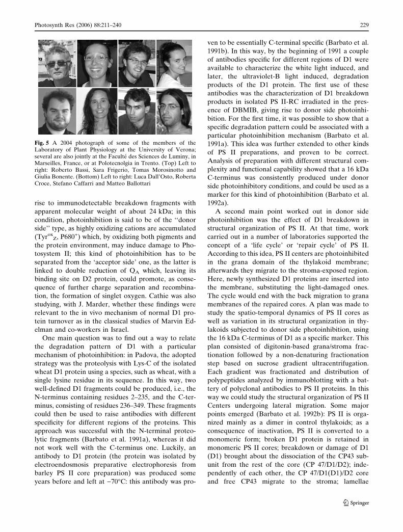
rise to immunodetectable breakdown fragments with
apparent molecular weight of about 24 kDa; in this
condition, photoinhibition is said to be of the ‘‘donor
side’’ type, as highly oxidizing cations are accumulated
(TyroxZ, P680+) which, by oxidizing both pigments and
the protein environment, may induce damage to Pho-
tosystem II; this kind of photoinhibition has to be
separated from the ‘acceptor side’ one, as the latter is
linked to double reduction of QA which, leaving its
binding site on D2 protein, could promote, as conse-
quence of further charge separation and recombina-
tion, the formation of singlet oxygen. Cathie was also
studying, with J. Marder, whether these findings were
relevant to the in vivo mechanism of normal D1 pro-
tein turnover as in the classical studies of Marvin Ed-
elman and co-workers in Israel.
One main question was to find out a way to relate
the degradation pattern of D1 with a particular
mechanism of photoinhibition: in Padova, the adopted
strategy was the proteolysis with Lys-C of the isolated
wheat D1 protein using a species, such as wheat, with a
single lysine residue in its sequence. In this way, two
well-defined D1 fragments could be produced, i.e., the
N-terminus containing residues 2–235, and the C-ter-
minus, consisting of residues 236–349. These fragments
could then be used to raise antibodies with different
specificity for different regions of the proteins. This
approach was successful with the N-terminal proteo-
lytic fragments (Barbato et al. 1991a), whereas it did
not work well with the C-terminus one. Luckily, an
antibody to D1 protein (the protein was isolated by
electroendosmosis preparative electrophoresis from
barley PS II core preparation) was produced some
years before and left at )70�C: this antibody was pro-
ven to be essentially C-terminal specific (Barbato et al.
1991b). In this way, by the beginning of 1991 a couple
of antibodies specific for different regions of D1 were
available to characterize the white light induced, and
later, the ultraviolet-B light induced, degradation
products of the D1 protein. The first use of these
antibodies was the characterization of D1 breakdown
products in isolated PS II-RC irradiated in the pres-
ence of DBMIB, giving rise to donor side photoinhi-
bition. For the first time, it was possible to show that a
specific degradation pattern could be associated with a
particular photoinhibition mechanism (Barbato et al.
1991a). This idea was further extended to other kinds
of PS II preparations, and proven to be correct.
Analysis of preparation with different structural com-
plexity and functional capability showed that a 16 kDa
C-terminus was consistently produced under donor
side photoinhibitory conditions, and could be used as a
marker for this kind of photoinhibition (Barbato et al.
1992a).
A second main point worked out in donor side
photoinhibition was the effect of D1 breakdown in
structural organization of PS II. At that time, work
carried out in a number of laboratories supported the
concept of a ‘life cycle’ or ‘repair cycle’ of PS II.
According to this idea, PS II centers are photoinhibited
in the grana domain of the thylakoid membrane;
afterwards they migrate to the stroma-exposed region.
Here, newly synthesized D1 proteins are inserted into
the membrane, substituting the light-damaged ones.
The cycle would end with the back migration to grana
membranes of the repaired cores. A plan was made to
study the spatio-temporal dynamics of PS II cores as
well as variation in its structural organization in thy-
lakoids subjected to donor side photoinhibition, using
the 16 kDa C-terminus of D1 as a specific marker. This
plan consisted of digitonin-based grana/stroma frac-
tionation followed by a non-denaturing fractionation
step based on sucrose gradient ultracentrifugation.
Each gradient was fractionated and distribution of
polypeptides analyzed by immunoblotting with a bat-
tery of polyclonal antibodies to PS II proteins. In this
way we could study the structural organization of PS II
Centers undergoing lateral migration. Some major
points emerged (Barbato et al. 1992b): PS II is orga-
nized mainly as a dimer in control thylakoids; as a
consequence of inactivation, PS II is converted to a
monomeric form; broken D1 protein is retained in
monomeric PS II cores; breakdown or damage of D1
(D1) brought about the dissociation of the CP43 sub-
unit from the rest of the core (CP 47/D1/D2); inde-
pendently of each other, the CP 47/D1(D1)/D2 core
and free CP43 migrate to the stroma; lamellae
Fig. 5 A 2004 photograph of some of the members of theLaboratory of Plant Physiology at the University of Verona;several are also jointly at the Faculte des Sciences de Luminy, inMarseilles, France, or at Polotecnolgia in Trento. (Top) Left toright: Roberto Bassi, Sara Frigerio, Tomas Morosinotto andGiulia Bonente. (Bottom) Left to right: Luca Dall’Osto, RobertaCroce, Stefano Caffarri and Matteo Ballottari
Photosynth Res (2006) 88:211–240 229
123
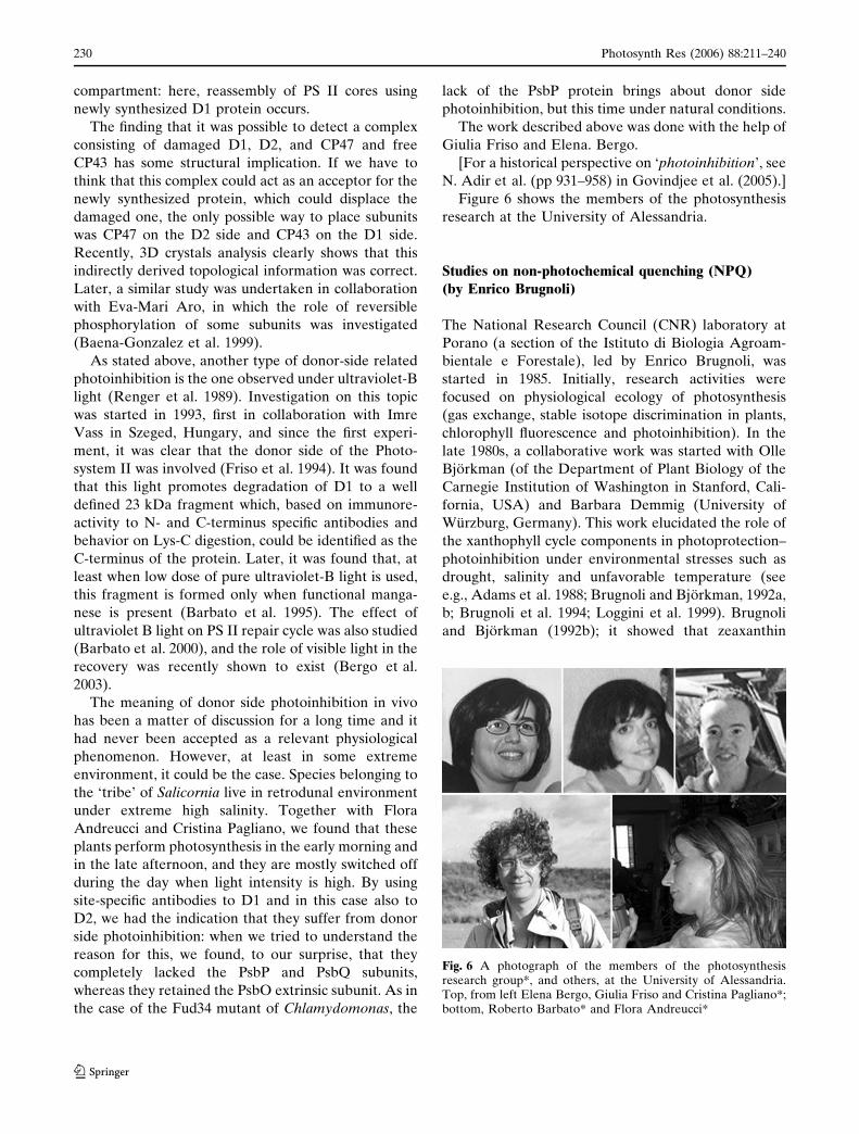
compartment: here, reassembly of PS II cores using
newly synthesized D1 protein occurs.
The finding that it was possible to detect a complex
consisting of damaged D1, D2, and CP47 and free
CP43 has some structural implication. If we have to
think that this complex could act as an acceptor for the
newly synthesized protein, which could displace the
damaged one, the only possible way to place subunits
was CP47 on the D2 side and CP43 on the D1 side.
Recently, 3D crystals analysis clearly shows that this
indirectly derived topological information was correct.
Later, a similar study was undertaken in collaboration
with Eva-Mari Aro, in which the role of reversible
phosphorylation of some subunits was investigated
(Baena-Gonzalez et al. 1999).
As stated above, another type of donor-side related
photoinhibition is the one observed under ultraviolet-B
light (Renger et al. 1989). Investigation on this topic
was started in 1993, first in collaboration with Imre
Vass in Szeged, Hungary, and since the first experi-
ment, it was clear that the donor side of the Photo-
system II was involved (Friso et al. 1994). It was found
that this light promotes degradation of D1 to a well
defined 23 kDa fragment which, based on immunore-
activity to N- and C-terminus specific antibodies and
behavior on Lys-C digestion, could be identified as the
C-terminus of the protein. Later, it was found that, at
least when low dose of pure ultraviolet-B light is used,
this fragment is formed only when functional manga-
nese is present (Barbato et al. 1995). The effect of
ultraviolet B light on PS II repair cycle was also studied
(Barbato et al. 2000), and the role of visible light in the
recovery was recently shown to exist (Bergo et al.
2003).
The meaning of donor side photoinhibition in vivo
has been a matter of discussion for a long time and it
had never been accepted as a relevant physiological
phenomenon. However, at least in some extreme
environment, it could be the case. Species belonging to
the ‘tribe’ of Salicornia live in retrodunal environment
under extreme high salinity. Together with Flora
Andreucci and Cristina Pagliano, we found that these
plants perform photosynthesis in the early morning and
in the late afternoon, and they are mostly switched off
during the day when light intensity is high. By using
site-specific antibodies to D1 and in this case also to
D2, we had the indication that they suffer from donor
side photoinhibition: when we tried to understand the
reason for this, we found, to our surprise, that they
completely lacked the PsbP and PsbQ subunits,
whereas they retained the PsbO extrinsic subunit. As in
the case of the Fud34 mutant of Chlamydomonas, the
lack of the PsbP protein brings about donor side
photoinhibition, but this time under natural conditions.
The work described above was done with the help of
Giulia Friso and Elena. Bergo.
[For a historical perspective on ‘photoinhibition’, see
N. Adir et al. (pp 931–958) in Govindjee et al. (2005).]
Figure 6 shows the members of the photosynthesis
research at the University of Alessandria.
Studies on non-photochemical quenching (NPQ)(by Enrico Brugnoli)
The National Research Council (CNR) laboratory at
Porano (a section of the Istituto di Biologia Agroam-
bientale e Forestale), led by Enrico Brugnoli, was
started in 1985. Initially, research activities were
focused on physiological ecology of photosynthesis
(gas exchange, stable isotope discrimination in plants,
chlorophyll fluorescence and photoinhibition). In the
late 1980s, a collaborative work was started with Olle
Bjorkman (of the Department of Plant Biology of the
Carnegie Institution of Washington in Stanford, Cali-
fornia, USA) and Barbara Demmig (University of
Wurzburg, Germany). This work elucidated the role of
the xanthophyll cycle components in photoprotection–
photoinhibition under environmental stresses such as
drought, salinity and unfavorable temperature (see
e.g., Adams et al. 1988; Brugnoli and Bjorkman, 1992a,
b; Brugnoli et al. 1994; Loggini et al. 1999). Brugnoli
and Bjorkman (1992b); it showed that zeaxanthin
Fig. 6 A photograph of the members of the photosynthesisresearch group*, and others, at the University of Alessandria.Top, from left Elena Bergo, Giulia Friso and Cristina Pagliano*;bottom, Roberto Barbato* and Flora Andreucci*
230 Photosynth Res (2006) 88:211–240
123

formation plays a relevant role under stress conditions
in preventing photoinhibition of photosynthesis and
photo-oxidation: this photoprotective role contributed
in maintaining the full photosynthetic capacity ex-
pressed as carbon fixation per unit leaf area or on
chlorophyll basis.
In addition, the light-induced absorbance change
caused by violaxanthin deepoxidation in intact leaves
was studied by Brugnoli and Bjorkman (1992a). The
correlation between the light-induced absorbance
change at 508 nm and zeaxanthin formation in leaves is
relevant for physiological and ecological applications.
Light-induced chloroplast movements were found to
cause changes in chlorophyll fluorescence emission and
matching changes in leaf absorptance both in kinetics
and in maximum extent. These results clearly showed
that chloroplast movements could strongly influence
the efficiency of light utilization in photosynthesis.
The capacity for non-radiative energy dissipation,
measured as maximum non-photochemical quenching
(NPQ), and its dependence on carotenoid composition
were studied in sun and shade leaves of several species.
The amount of photoconvertible violaxanthin was
higher in the sun compared to the shade leaves. Hence
sun leaves contain a greater pool size of xanthophyll
cycle components and these are more convertible to
zeaxanthin compared to that in shade leaves (Brugnoli
et al. 1994). The amount of photoconvertible viola-
xanthin in vivo is strongly dependent on the degree of
thylakoid membrane appression and on the stoichi-
ometries of PS II antennae (LHC II) and minor PS II
complexes. In a study on C3 and C4 plants (Brugnoli
et al. 1998), shade-grown leaves showed more grana
stacking, higher content of LHC II and lower contents
of minor complexes compared to sun-grown leaves.
This caused a much higher proportion of photocon-
vertible violaxanthin and a higher capacity for NPQ in
the sun compared to the shade leaves (see Fig. 7A).
Figure 7B shows members of the photosynthesis
research group at Porano.
Photoinhibition, photoprotection and desiccation
tolerance were studied in the poikilohydric (resur-
rection) plant Ramonda serbica. Changes in zeaxan-
thin and antheraxanthin contents and in the pools of
reduced ascorbate and glutathione were evident
during dehydration and rehydration and these
changes were related to increases in NPQ (Augusti
et al. 2000). In addition, fully dehydrated leaves
maintained some zeaxanthin and this may be rele-
vant to cause energy dissipation during subsequent
rehydration. Reduced ascorbate and reduced gluta-
thione were found to be most relevant for preventing
photo-oxidation at the lowest water content. Chan-
ges in the pools of antioxidants and of zeaxanthin
were found to be crucial for photoprotection and to
confer desiccation tolerance to Ramonda serbica
leaves.
Fig. 7 (A) Relationship between photoconvertible violaxanthin(i.e., the maximum amount of violaxanthin converted tozeaxanthin under high light and low CO2) and maximum non-photochemical quenching, which is related to the capacity fornon-radiative energy dissipation. Experiments run on intact fullsun exposed leaves (open symbols) and shade adapted leaves(closed symbols) in different species possessing the C3 (Hederaelix L., Ligustrum ovalifolium Hassk. and Laurus nobilis L.) andthe C4 photosynthetic pathway (Zea mays L. and Sorgum bicolorMoench.). The correlation coefficient r = 0.86 is indicative ofhigh level of significance. NPQ stands for non-photochemicalquenching of the excited state of chlorophyll molecule (Source:Brugnoli et al. 1998). (B) A photograph of members of thephotosynthesis research group in their laboratory in Porano.Enrico Brugnoli (middle), with Filip Volders (right) and MarcoLauteri (left)
Photosynth Res (2006) 88:211–240 231
123

[For a historical personal account of linking the
xanthophyll cycle to thermal energy dissipation, see
B. Demmig-Adams (pp 923–930) in Govindjee et al.
(2005).]
Kinetics and energy modulation of the quinone
acceptor complex in photosynthetic ReactionCenters from purple bacteria in different solubilizing
environments (by Angela Agostiano and Massimo
Trotta)
The research group on photosynthesis-related topics in
Bari, Italy, was initiated by Angela Agostiano in the
late 1980s, after she returned form the laboratory of
Francis Fong at the Purdue University, West Lafayette,
Indiana, USA, where she worked on the electro-
chemistry of aggregated species of chlorophyll a gen-
erated in several environmental conditions (Agostiano
et al. 1990, 2002, 2003). Her research topic expanded to
include bacterial photosynthesis in the 1980s in close
collaboration with Massimo Trotta who had returned
from a first 18-month stay at the University of Bologna
collaborating with G. Venturoli, A Melandri and D
Zannoni, working on the charge recombination from
the final quinone acceptors in Reaction Centers iso-
lated from the thermophilic bacterium Chloroflexus
aurantiacus (Venturoli et al. 1991) and a subsequent
second 2-year stay at the University of California at
San Diego working with George Feher and Mel
Okamura on NMR applied to Reaction Centers
(Trotta et al. 1992). Francesco Milano, who had spent
some time with Wolfgang Lubitz, and Livia Giotta,
with Peter Rich, joined this group in 2001 and 2002,
respectively. (Livia is presently at the University of
Lecce, Italy.)
The photocycle and the associated electron transfer
rate of the Reaction Center (RC) from the purple
bacterium Rhodobacter sphaeroides were well estab-
lished (Feher et al. 1989). The rate by which the
loosely bound quinone (as well as the quinol) enters
and leaves its binding site in the Reaction Center for
closing the photocycle and restart the cascade of the
electron transfer reactions was, and partially is, not yet
clear. The investigation on the quinone exchange rate
at QB site was performed in our laboratory by studying
the role of different solubilizing environments of the
enzyme. The first investigated environment was the
zwitterionic detergent N,N-Dimethyl-N-dodecylamine
oxide (LDAO) and in particular the influence on the
functioning of the secondary electron acceptor played
by the size, i.e., the number of LDAO monomers (in
the polymer), of the solubilizing micelles surrounding
the RC (Agostiano et al. 1999, Halmschlager et al.
2002). Subsequently the investigation was extended to
the case of RC containing proteoliposomes formed by
the zwitterionic phospholipid phosphatidylcholine
(PC) (Trotta et al. 2002, Milano et al. 2003). These
studies allowed us to assess kinetic constants for the
ubiquinone 10 uptake into—and release from—the
QB-binding site which were found to be 7 · 107 M)1s)1
and 40 s)1, respectively.
The effect of the solubilizing environment was also
studied in terms of the stability of the semiquinones at
the QA and the QB binding sites in RC incorporated
into proteoliposomes containing negatively charged
phospholipids, namely phosphatidylglycerol (PG) and
cardiolipin (CL). These studies were performed in
collaboration with Peter Maroti and Laszlo Nagy
(of the Department of Biophysics of the University of
Szeged, Hungary) and allowed us to determine the
standard free energy of the primary electron acceptor
Q�A (with the neutral state QA, as a reference) by using
the delayed luminescence technique (Turzo et al.
2000). This value was strongly dependent upon the
solubilizing environment and it increased by as much
as 46 meV when PG is added either to Triton X-100
micelles or to phosphatidylcholine proteoliposomes.
An even larger increase was found in the free energy of
Q�A upon addition of CL. On the other side, the free
energy of Q�B was found to be rather insensitive to the
presence of the negative charge on the phospholipids.
The presence of both PG and CL as phospholipids of
the Intra Cytoplasmic Membrane (ICM) of the pho-
tosynthetic system as well as their role in stabilizing the
lifetime of the Q�B state (mostly by increasing the
energy level of Q�A) was interpreted as possible
regulating mechanism for Reaction Centers under
physiological conditions (Nagy et al. 2004). This sta-
bilization effect of negatively charged phospholipids
was found to be extremely efficient in the case of
proteoliposomes formed exclusively by PG or phos-
phatidylinositol (PI) or phosphatidylserine (PS), when
the charge separated state generated by continuous
illumination was found to last up to 60 min (Agostiano
et al. 2005).
In close collaboration with researchers from Hungary,
Agostiano et al. (2004) and Milano et al. (2005) have
investigated the possibility of studying in the RC, the
proton uptake or release associated with the quinone
reduction and quinol oxidation, respectively, by using
pyranine, a pH sensitive fluorescence probe. Preliminary
information on this technique has shown its validity and
further experiments are presently being performed.
Figure 8 shows members of the photosynthesis
research group at the University of Bari.
232 Photosynth Res (2006) 88:211–240
123

Acknowledgments Govindjee invited us to write this historicalreview on ‘photosynthesis research’ in Italy. We thank him forhis many suggestions and for his painstaking work in editing thismanuscript. The content of each of the nine sections, however,was the sole responsibility of the individual authors, while theIntroduction is the sole responsibility of Giorgio Forti. All theauthors are thankful to Itzhak Ohad and Gernot Renger forreading this manuscript and for making suggestions for itsimprovement although we were unable to incorporate all ofthem.
References
Adams WW, Terashima I, Brugnoli E, Demmig B (1988) Com-parisons of photosynthesis and photoinhibition in the CAMvine Hoya australis and several C3 vines growing on thecoast of eastern Australia. Plant Cell Environ 11:173–181
Agostiano A, Cosma P, Della Monica M, Fong FK (1990)Spectroscopic and electrochemical characterization of pho-tocatalytic Chla in different water–organic solvent mixtures.Bioelect Bioenerg 23:311–324
Agostiano A, Milano F, Trotta M (1999) Investigation on thedetergent role in the function of secondary quinone inbacterial Reaction Centers. Eur J Biochem 262:358–364
Agostiano A, Cosma P, Trotta M, Monsu-Scolaro L, Micali N(2002) Chlorophyll a behaviour in aqueous solvents: for-mation of nanoscale self-assembled complexes. J Phys ChemB 106:12820–12829
Agostiano A, Catucci L, Cosma P, Fini P (2003) Aggregationprocesses and photophysical properties of chlorophyll a inaqueous solutions modulated by the presence of cyclodex-trines. Phys Chem Chem Phys 5:2122–2128
Agostiano A, Mavelli F, Milano F, Giotta L, Trotta M, Nagy L,Maroti P (2004) pH-sensitive fluorescent dye as probe forproton uptake in photosynthetic Reaction Centers. Bio-electrochem 63:125–128
Agostiano A, Milano F, Trotta M (2005) Trapping of the chargeseparated state of photosynthetic Reaction Centers frompurple bacteria in proteoliposomes of negatively chargedphospholipids. Photosynth Res 83:53–61
Aliverti A, Zanetti G (1997) A three-domain iron–sulfur flavo-protein obtained through gene fusion of ferredoxin andferredoxin-NADP+ reductase from spinach leaves. Bio-chemistry 36:14771–14777
Aliverti A, Lubberstedt T, Zanetti G, Herrmann GR, Curti B(1991) Probing the role of Lys116 and Lys244 in the spinachferredoxin-NADP+ reductase by site-directed mutagenesis.J Biol Chem 266:17760–17763
Aliverti A, Piubelli L, Zanetti G, Lubberstedt T, Herrmann RG,Curti B (1993) The role of cysteine residues of spinachferredoxin-NADP+ reductase as assessed by site-directedmutagenesis. Biochemistry 32:6374–6380
Aliverti A, Corrado ME, Zanetti G (1994) Involvement of ly-sine-88 of spinach ferredoxin-NADP+ reductase in theinteraction with ferredoxin. FEBS Lett 343:247–250
Aliverti A, Bruns CM, Pandini VE, Karplus AP, Vanoni MA,Curti B, Zanetti G (1995) Involvement of Serine 96 in thecatalytic mechanism of ferredoxin-NADP+ reductase:structure–function relationship as studied by site-directedmutagenesis and X-ray crystallography. Biochemistry34:8371–8379
Aliverti A, Deng Z, Ravasi D, Piubelli L, Karplus PA, Zanetti G(1998) Probing the function of the invariant glutamyl resi-due 312 in spinach ferredoxin-NADP+ reductase. J BiolChem 273:34008–34015
Aliverti A, Faber R, Finnerty CM, Ferioli C, Pandini V, Negri A,Karplus PA, Zanetti G (2001) Biochemical andcrystallographic characterization of Ferredoxin-NADP+
reductase from nonphotosynthetic tissues. Biochemistry40:14501–14508
Allen JF (1992) Protein phosphorylation in regulation of pho-tosynthesis. Biochim Biophys Acta 1098:275–335
Allen JF, Bennett J, Steinback KE, Arntzen CJ (1981) Chloro-plast protein phosphorylation couples plastoquinone redoxstate to distribution of excitation energy between Photo-systems. Nature 291:25–29
Ambrosone L, Mallardi A, Palazzo G, Venturoli G (2002) Effectof heterogeneity in the distribution of ligands and proteinsamong disconnected particles: the binding of ubiquinoneto bacterial Reaction Center. Phys Chem Chem Phys4:3071–3077
Arnon DI, Allen MB, Whatley FR (1954) Photosynthesis byisolated chloroplasts II. Photophosphorylation, the conver-sion of light into phosphate bond energy. J Am Chem Soc76:6324–6329
Asada K (1992) Ascorbate peroxidase: hydrogen peroxidescavenging enzyme in plants. Physiol Plant 85:235–242
Augusti A, Scartazza A, Navari-Izzo F, Sgherri CLM, StevanovicB, Brugnoli E (2000) Photosystem II photochemical effi-ciency, zeaxanthin and antioxidant contents in the poiki-lohydric Ramonda serbica during dehydration andrehydration. Photosynth Res 67:79–88
Baccarini-Melandri A, Melandri BA (1972) Energy transductionin photosynthetic bacteria. III Coincidence of the couplingfactor of photosynthesis and respiration in Rps capsulata.FEBS Lett 21:131– 134
Baccarini-Melandri A, Melandri BA (1977) A role for ubiqui-none-10 in the b–c segment of the photosynthetic electrontransport chain in bacteria. FEBS Lett 80:459–464
Baccarini-Melandri A, Gest H, San Pietro A (1970) A couplingfactor in bacterial photophosphorylation. J Biol Chem245:1224–1226
Baccarini-Melandri A, Zannoni D, Melandri BA (1973) Energytransduction in photosynthetic bacteria. VI Respiratory sitesof energy conservation in membrane from dark grown cellsof Rps. capsulata. Biochim Biophys Acta 314:289– 311
Fig. 8 A 2005 photograph of members of the photosynthesisresearch group at the University and Istituto per i ProcessiChimico Fisici in Bari. From left to right: Massimo Trotta,Francesco Milano, Angela Agostiano and Francesca Italiano
Photosynth Res (2006) 88:211–240 233
123

Baccarini-Melandri A, Fabbri E, Melandri BA (1975) Energytransduction in photosynthetic bacteria.VIII Activation ofthe energy transducing ATPase by O-phosphate. BiochimBiophys Acta 376:82–88
Baccarini-Melandri A, Casadio R, Melandri BA (1977) Ther-modynamics and kinetics of photophosphorylation in bac-terial chromatophores and their relation with thetransmembrane electrochemical potential difference ofprotons. Eur J Biochem 78:389–402
Baccarini-Melandri A, Melandri BA, Hauska G (1979) Stimu-lation of photophosphorylation and ATPase by artificialredox mediators in chromatophores of Rps. capsulata atdifferent redox potentials. Bioenerg Biomemb 11:1–16
Baccarini -Melandri A, Gabellini N, Melandri BA, Hurt E,Hauska G (1980) Structural requirements of quinonecoenzymes for endogenous and dye-mediated coupledelectron transport in bacterial photosynthesis. J BioenergBiomemb 12:95–110
Baena-Gonzalez E, Barbato R, Aro EM (1999) Role of phos-phorylation in the repair cycle and oligomeric structure ofPhotosystem II. Planta 208:196–204
Barbagallo RP, Finazzi G, Forti G (2000) Effects of inhibitors onthe activity of the cytochrome b6f complex: evidence for theexistence of two binding pockets in the lumenal site. Bio-chemistry 38:12814–12821
Barbato R, Shipton CA, Giacometti GM, Barber J. (1991a) Newevidence suggests that the initial photoinduced cleavage ofthe D1-protein may not occur near the PEST sequence.FEBS Lett 290:162–166
Barbato R, Friso G, Giardi MT, Rigoni F, Giacometti GM(1991b) Breakdown of the Photosystem II Reaction CenterD1 protein under photoinhibitory conditions: identificationand localization of the C-terminal degradation products.Biochemistry 30:10220–10226
Barbato R, Frizzo A, Friso G, Rigoni F, Giacometti GM (1992a)Photoinduced degradation of the D1 protein in isolatedthylakoids and various Photosystem II particles after donor-side inactivations. Detection of a C-terminal 16 kDa frag-ment. FEBS Lett 304:136–140
Barbato R, Friso G, Rigoni F, Dalla Vecchia F, Giacometti GM(1992b) Structural changes and lateral redistribution ofPhotosystem II during donor side photoinhibition of thy-lakoids. J Cell Biol 119:325–335
Barbato R, Frizzo A, Friso G, Rigoni F, Giacometti GM (1995)Degradation of the D1 protein of Photosystem-II ReactionCentre by ultraviolet-B radiation requires the presence offunctional manganese on the donor side. Eur J Biochem227:723–729
Barbato R, Bergo E, Szabo I, Dalla Vecchia F, Giacometti GM(2000) Ultraviolet B exposure of whole leaves of barleyaffects structure and functional organization of PhotosystemII. J Biol Chem 275:10976–10982
Barz HP, Francia F, Venturoli G, Melandri BA, Vermeglio A,Oesterhelt D (1995a) Role of PufX protein in photosyn-thetic growth of Rhodobacter sphaeroides. 1. PufX isrequired for efficient light-driven electron transfer andphotophosphorylation under anaerobic conditions. Bio-chemistry 34:15235–15247
Barz WP, Vermeglio A, Francia F, Venturoli G, Melandri BA,Oesterhelt D (1995b) Role of the PufX protein in photo-synthetic growth of Rhodobacter sphaeroides.2. PufX isrequired for efficient ubiquinone/ubiquinol exchange be-tween the Reaction Center QB site and the cytochrome bc1complex. Biochemistry 34:15248–14258
Barzda V, Simididjiev I, Vianelli A, Jennings RC, Garab G(1995) Light induced reversible structural changes in chirallyorganised macroaggregates of purified chlorophyll a/b pro-tein complexes. In: Mathis P (ed) Photosynthesis: fromLight to Biosphere, vol I. Kluwer Academic Publishers,Dordrecht, pp 315–318
Barzda V, Jennings RC, Zucchelli G, Garab G (1999) Kineticanalysis of the light induced fluorescence quenching in lightharvesting chlorophyll a/b protein complex of PhotosystemII. Photochem Photobiol 70:751–759
Bassi R (1986) Studies on the leaf of Trapa natants: polymor-phism of chloroplasts and microbodies. Cytobios 45:109–121
Bassi R, Simpson DJ (1986) Differential expression of chl a/bgenes in bundle sheath and mesophyll plastids of maize.Calsberg Res Commun 51:363–370
Bassi R, Simpson DJ (1987) Chlorophyll–proteins of barleyPhotosystem I. Eur J Biochem 163:221–230
Bassi R, Peruffo A, Barbato R, Ghisi R (1985) Differences inchlorophyll–protein complexes and composition of poly-peptides between thylakoids from bundle sheath andmesophyll cells in maize. Eur J Biochem 146:589–595
Bassi R, Simpson D, Barbato R, Hoyer-Hansen G, GiacomettiGM (1987) Chlorophyll–proteins of the PS II antenna sys-tem. J Biol Chem 262:13333–13341
Bassi R, Giacometti GM, Simpson DJ (1988a) Changes in theorganization of stroma membranes induced by in vivo StateI–State 2 transitions. Biochim Biophys Acta 935:152–165
Bassi R, Rigoni F, Barbato R, Giacometti GM (1988b) Lightharvesting chlorophyll a/b-proteins in phosphorylatedmembranes. Biochim Biophys Acta 936:29–38
Bassi R, Ghiretti-Magaldi A, Tognon G, Giacometti GM, MillerK (1989) Two dimensional cristals of Photosystems IIReaction Centre complex from higher plants. Eur J CellBiol 50:84–93
Bassi R, Marquardt J, Lavergne J (1995) Biochemical andfunctional properties of Photosystem II in agranal mem-branes from maize mesophyll and bundle sheath chlorop-lasts. Eur J Biochem 233:709–719
Bassi R, Croce R, Cugini D, Sandona D (1999) Mutation analysisof an higher plant antenna protein provides identification ofchromophores bound into multiple sites. Proc Natl Acad SciUSA 96:10056–10061
Bellafiore S, Barneche F, Peltier G, Rochaix J-D (2005) Statetransitions and light adaptation require chloroplast thyla-koid protein kinase STN7. Nature 433:892–895
Bennett J (1983) Regulation of photosynthesis by reversiblephosphorylation of the light-harvesting chlorophyll a/bprotein. Biochem J 212:1–13
Bergantino E, Dainese P, Cerovic Z Sechi S, Bassi R (1995) Apost-translational modification of the Photosystem II sub-unit CP29 protects maize from cold stress. J Biol Chem270:8474–8481
Bergo E, Segalla A, Giacometti GM, Tarantino D, Soave C,Andreucci F, Barbato R (2003) Role of visible light in therecovery of Photosystem II structure and function fromultraviolet-B stress in higher plants. J Exp Bot 54:1665–1673
Binda C, Coda A, Aliverti A, Zanetti G, Mattevi A (1998)Structure of the mutant E92K of [2Fe-2S] ferredoxin I fromSpinacia oleracea at 1.7 A resolution. Acta CrystD54:1353–1358
Boekema E, van Roon H, Calkoen F, Bassi R, Dekker J (1999)Different types of association of Photosystem II and its lightharvesting antenna in partially solubilized Photosystem IImembranes. Biochemistry 38:2233–2239
234 Photosynth Res (2006) 88:211–240
123

Borghese R, Crimi M, Fava L, Melandri BA (1998a) The ATPsynthase atpHAGDC (F1) operon from Rb caspulatus. JBacteriol 180:416–421
Borghese R, Turina P, Lambertini L, Melandri BA (1998b) TheatpIBEXF operon coding for the Fo sector of the ATPsynthase from the nonsulfur purple bacterium Rb. capsula-tus. Arch Microbiol 170:385–388
Breyton C (2000) The cytochrome b6f complex: structural studiesand comparison with the bc1 complex. Biochim BiophysActa 1459: 467–474
Brugnoli E, Bjorkman O (1992a) Chloroplast movements inleaves: influence on chlorophyll fluorescence and measure-ments of light-induced absorbance changes related to DpHand zeaxanthin formation. Photosynth Res 32:23–35
Brugnoli E, Bjorkman O (1992b) Growth of cotton undercontinuous salinity stress: influence on allocation pattern,stomatal and non-stomatal components of photosynthesisand dissipation of excess light energy. Planta 187:335–347
Brugnoli E, Cona A, Lauteri M (1994) Xanthophyll cycle com-ponents and capacity for non-radiative energy dissipation insun and shade leaves of Ligustrum ovalifolium exposed toconditions limiting photosynthesis. Photosynth Res41:451–463
Brugnoli E, Scartazza A, De Tullio MC, Monteverdi MC,Lauteri M, Augusti A (1998) Zeaxanthin and non-photo-chemical quenching in sun and shade leaves of C3 and C4
plants. Physiol Plant 104:727–734Bulte L, Gans L, Rebeille F, Wollman FA (1990) ATP control
on State transitions in vivo in Chlamydomonas reinhardtii.Biochim Biophy Acta 1020:72–80
Caffarri S, Croce R, Cattivelli L, Bassi R (2004) A look withinLHC II: differential analysis of the Lhcb 1–3 gene productsbuilding the major trimeric antenna complex of higher plantphotosynthesis. Biochemistry 43:9467–9476
Caffarri S, Frigerio S, Olivieri E, Righetti PG, Bassi R (2005)Differential accumulation of Lhcb gene products in thyla-koid membranes of Zea mays plants grown under contrast-ing light and temperature conditions. Proteomics 5:758–768
Cardol P, Gloire G, Havaux M, Remacle C, Matagne R, FranckF (2003) Photosynthesis and State transitions in mitochon-drial mutants of Chlamydomonas reinhardtii affected inrespiration. Plant Physiol 133:2010–2020
Casadio R (1991) Measurements of transmembrane pH differ-ences of low extents in bacterial chromatophores: a studywith the fluorescent probe 9-amino, 6-chloro, 2-methoxyacridine. Eur Biophys J 19:189–201
Casadio R, Baccarini-Melandri A, Melandri BA (1974) On thedetermination of the transmembrane pH difference inbacterial chromatophores using 9-amino acridine. Eur JBiochem 47:121–128
Casadio R, Baccarini-Melandri A, Melandri BA (1978) Limitedcooperativity in the coupling between electron flow andphotosynthetic ATP synthesis. FEBS Lett 87:323–328
Casadio R, Venturoli G, Di Gioia A, Castellani P, Leonardi L,Melandri BA (1984) Phospholipid enriched chromato-phores: a system suited to investigate the ubiquinone- med-iated interactions of protein complexes in photosyntheticoxidoreduction processes. J Biol Chem 259:9149–9157
Cattaneo R, Zucchelli G, Garlaschi FM, Finzi L, Jennings RC(1995) A thermal broadening analysis of absorption spectraof the D1/D2/cytochrome b559 complex in terms of gaussiandecomposition sub-bands. Biochemistry 34:15267–15275
Cidaria D, Biondi PA, Zanetti G, Ronchi S (1985) The NADP+
binding site of ferredoxin-NADP+ reductase: sequenceof the peptide containing the essential lysine residue. EurJ Biochem 146:295–299
Cinque G, Croce R, Holzwarth A, Bassi R (2000) Energytransfer among CP29 chlorophylls: calculated Forster ratesand experimental transient absorption at room temperature.Biophys J 79:1706–1717
Cometta A, Zucchelli G, Karapetyan NV, Engelmann E, Garl-aschi FM, Jennings RC (2000) Thermal behavior of longwavelength absorption transitions in Spirulina platensisPhotosystem I trimers. Biophys J 79:3235–3243
Cordone L, Cottone G, Giuffrida S, Palazzo G, Venturoli G,Viappiani C (2005) Internal dynamics and protein-matrixcoupling in trehalose-coated proteins. Biochim BiophysActa 1749:252–281
Croce R, Zucchelli G, Garlaschi FM, Bassi R, Jennings RC(1996a) Excited State equilibration in the Photosystem Ilight-harvesting I complex: P700 is almost isoenergetic withits antenna. Biochemistry 35:8572–8579
Croce R, Breton J, Bassi R (1996b) Conformational changesinduced by phosphorylation on the Photosystem II subunitCP29. Biochemistry 35:11142–11148
Croce R, Zucchelli G, Garlaschi FM, Jennings RC (1998) Athermal broadening study of the antenna chlorophylls in PSI-200, LHC I, and PS I core. Biochemistry 37:17355–17360
Croce R, Remelli R, Varotto C, Breton J, Bassi R (1999a) Theneoxanthin binding site of the major light harvesting com-plex (LHC II) from higher plants. FEBS Lett 456:1–6
Croce R, Weiss S, Bassi R (1999b) Carotenoid binding sites ofthe major Light Harvesting complex (LHC II) of higherplants. J Biol Chem 274:29613–29623
Croce R, Muller MG, Bassi R, Holzwarth AR (2000a) Carot-enoid to chlorophyll energy transfer in recombinant majorlight-harvesting complex (LHC II) of higher plants withvarious carotenoid contents 1. Femtosecond transientabsorption measurements. Biophys J 80:901–915
Croce R, Dorra D, Holzwarth A, Jennings RC (2000b) Fluo-rescence decay and spectral evolution in intact PhotosystemI of higher plants. Biochemistry 39:6341–6348
Croce R, Cinque G, Holzwarth AR, Bassi R (2000c) The Soretabsorption properties of carotenoids and chlorophylls in an-tenna complexes of higher plants. Photosynth Res 64:221–231
Croce R, Morosinotto T, Castelletti S, Breton J, Bassi R (2002)The Lhca antenna complexes of Photosystem I from higherplants. Biochim Biophys Acta 1556:29–40
Crosatti C, Polverino de Laureto P, Bassi R, Cattivelli L (1999) Theinteraction between cold and light controls the expression ofthe cold-regulated barley gene cor14b and the accumulationof the corresponding protein. Plant Physiol 119:671–680
Dainese P, Bassi R (1991) Stoichiometry of the chloroplastPhotosystem II antenna system and aggregation state of thecomponent Chl a/b proteins. J Biol Chem 266:8136–8142
Dainese P, Hoyer-Hansen G, Bassi R (1990) The resolution ofchlorophyll a/b binding proteins by a preparative method onflat bed isofocusing. Photochem Photobiol 51:693–703
Dal Bosco C, Busconi M, Govoni C, Baldi P, Stanca AM,Crosatti C, Bassi R, Cattivelli L (2003) Gene expression inbarley mutants affected in chloroplast development andphotosynthetic electron transport. Plant Physiol 131:793–802
Daldal F, Mandaci S, Winterstein C, Myllykallio H, Duyck K,Zannoni D (2001) Mobile cyt c2 and membrane-anchoredcyt cy are both efficient electron donors to the cbb3- andaa3-type cyt c oxidases during respiratory growth of Rho-dobacter sphaeroides. J Bacteriol 183(6):2013–2024
Davidson E, Prince RC, Daldal F, Hauska G, Marrs BL (1987)Rhodopseudomonas capsulata MT113: a single mutationresults in the absence of c-type cytochromes and the cyto-chrome bc1 complex. Biochim Biophys Acta 890: 292–301
Photosynth Res (2006) 88:211–240 235
123

Dall’Osto L, Caffarri S, Bassi R (2005) A mechanism of non-photochemical energy dissipation, independent from PsbS,revealed by a conformational change in the antenna proteinCP26. The Plant Cell 17:1217–1232
Davies JP, Grossman AR (1998) Responses to deficiencies inmicronutriens. In: Rochaix JD, Goldschmidt-Clermont M,Merchant S (eds) The molecular biology of chloroplasts andmitochondria in Chlamydomonas. Kluwer Academic Pub-lishers (now Springer), Dordrecht, pp 613–635
Delosme R, Olive J, Wollman FA (1996) Changes in light energydistribution upon State transitions: an in vivo photoacousticstudy of the wild type and photosynthesis mutants fromChlamydomonas reinhardtii. Biochim Biophys Acta1273:150–158
Deng Z, Aliverti A, Zanetti G, Arakaki AK, Ottado J, OrellanoE, Calcaterra NB, Ceccarelli EA, Carrillo N, Karplus PA(1999) A productive NADP+ binding mode of ferredoxin-NADP+ reductase revealed by protein engineering andcrystallographic studies. Nature Struct Biol 6:847–853
Depege N, Bellafiore S, Rochaix JD (2003) Role of chloroplastprotein kinase Stt7 in LHC II phosphorylation and Statetransition in Chlamydomonas. Science 299:1572–1575
Dominici PM, Crimi S, Ceoldo S, Caffarri F, Armenante M,Bassi R (2002) Biochemical properties of the Photosystem IIPsbS subunit subunit: either recombinant or purified fromchloroplasts. J Biol Chem 277:22750–22758
Egneus H, Heber U, Matthiesen U, Kirk M (1975) Reduction ofoxygen by the electron transport chain of chloroplasts dur-ing assimilation of carbon dioxide. Biochim Biophys Acta408:252–268
Engelmann E, Tagliabue T, Karapetyan NV, Garlaschi FM,Zucchelli G, Jennings RC (2001) CD spectroscopy pro-vides evidence for excitonic interactions involving red-shifted chlorophyll forms in Photosystem I. FEBS Lett499:112–115
Engelmann E, Zucchelli G, Casazza A, Jennings R (2005) Theeffect of outer antenna complexes on the photochemicaltrapping rate in barley thylakoid Photosystem II. BiochimBiophys Acta 1706:276–286
Feher G, Allen JP, Okamura MY, Rees DC (1989) Structure andfunction of bacterial photosynthetic Reaction Centres.Nature 339:111–116
Finazzi G, Furia A, Barbagallo RP, Forti G (1999) State tran-sitions, cyclic and linear electron transport and photophos-phorylation in Chlamydomonas reinhardtii. BiochimBiophys Acta 1413:117–129
Finazzi G, Zito F, Barbagallo RP, Wollman FA (2001) Contrastedeffects of inhibitors of cytochrome b6f complex on Statetransitions in C. reinhardtii: the role of Qo site occupancy inLHC II-kinase activation. J Biol Chem 276:9770–9774
Finazzi G, Rappaport F, Furia A, Fleischmann M, Rochaix JD,Zito F, Forti G (2002) Involvement of State transitions inthe switch between linear and cyclic electron flow in Chla-mydomonas reinhardtii. EMBO Rep 3:280–285
Finazzi G, Johnson GN, Dall’Osto L, Joliot P, Wollman FA,Bassi R (2004) A zeaxanthin-independent non-photochem-ical quenching mechanism localized in the Photosystem IIcore complex. Proc Natl Acad Sci USA 101:12375–12380
Fleischmann MM, Ravanel S, Delosme R, Olive J, Zito F,Wollman FA, Rochaix J-D (1999) Isolation and character-ization of photoautotrophic mutants of Chlamydomonasreinhardtii deficient in State transition. J Biol Chem274:30987–30994
Formaggio E, Cinque G, Bassi R (2001) Functional architectureof the major light harvesting complex of Photosystem II. JMol Biol 314:1157–1166
Forti G (1996) Photosynthesis: an overview. In: Jennings RC,Zucchelli G, Ghetti F, Colombetti G (eds) Light as an en-ergy source and information carrier in plant physiology.Plenum Press, NewYork and London, pp 1–16
Forti G, Caldiroli G (2005) State transitions in Chlamydomonasreinhardtii: the role of the Mehler reaction in State 2 to State1 transition. Plant Physiol 137:492–499
Forti G, Ehrenheim AM (1993) The role of ascorbic acid inphotosynthetic electron transport. Biochim Biophys Acta1183:408–412
Forti G, Elli G (1995) The function of ascorbic acid in photo-synthetic phosphorylation. Plant Physiol 109:1207–1211
Forti G, Gerola P (1977) Inhibition of photosynthesis by azideand cyanide and the role of oxygen in photosynthesis. PlantPhysiol 59:859–862
Forti G, Jagendorf AT (1961) Photosynthetic phosphorylation inthe absence of redox dyes: oxygen and ascorbate effects.Biochim Biophys Acta 54:322–330
Forti G, Parisi B (1963) Evidence for the occurrence of cyclicphotophosphorylation in vivo. Biochim Biophys Acta 71:1–6
Forti G, Rosa L (1971) On the pathway of electron transport incyclic photophosphorylation. FEBS Lett 18:55–58
Forti G, Sturani E (1968) On the structure and function of reducednicotinamide adenine dinucleotide phosphate-cytochromef reductase of spinach chloroplasts. Eur J Biochem3:461–472
Forti G, Vianelli A (1988) Influence of thylakoid protein phos-phorylation on Photosystem 1 photochemistry. FEBS Lett231:95–98
Forti G, Zanetti G (1969) The electron pathway of cyclic pho-tophosphorylation. In: Metzner H (ed) Progress in photo-synthesis research, vol III. Prof. Dr. H. Metzner Publisher,Tubingen, pp 1213–1216
Forti G, Furia A, Bombelli P, Finazzi G (2003) In vivo changesof the oxidation–reduction State of NADP and of the ATP/ADP cellular ratio linked to the photosynthetic activity inChlamydomonas reinhardtii. Plant Physiol 132:1464–1474
Francia F, Wang J, Venturoli G, Melandri BA, Barz WP,Oesterhelt D (1999) The Reaction Center–LH1 antennacomplex of Rhodobacter sphaeroides contains one PufXmolecule which is involved in dimerization of this complex.Biochemistry 38:6834–6845
Francia F, Wang J, Zischka H, Venturoli G, Oesterhelt D (2002)Role of the N- and C-terminal regions of the PufX proteinin the structural organization of the photosynthetic corecomplex of Rhodobacter sphaeroides. Eur J Biochem269:1877–1885
Francia F, Palazzo G, Mallardi A, Cordone L, Venturoli G(2003) Residual water modulates QA
- to QB electron transferin bacterial Reaction Centers embedded in trehaloseamorphous matrices. Biophys J 85:2760–2775
Francia F, Dezi M, Rebecchi A, Mallardi A, Palazzo G,Melandri BA, Venturoli G (2004a) Light-harvestingcomplex 1 stabilizes P+QB
- charge separation in ReactionCenters of Rhodobacter sphaeroides. Biochemistry43:14199–14210
Francia F, Palazzo G., Mallardi A, Cordone L, Venturoli G(2004b) Probing light-induced conformational transitions inbacterial photosynthetic Reaction Centers embedded intrehalose–water amorphous matrices. Biochim BiophysActa 1658:50–57
Frank K, Trebst A (1995) Quinone binding sites on cytochromeb/c complexes. Photochem Photobiol 61:2–9
Friso G, Spetea C, Giacometti GM, Vass I, Barbato R (1994)Degradation of Photosystem II Reaction Center D1proteininduced by UVB radiation in isolated thylakoids.
236 Photosynth Res (2006) 88:211–240
123

Identification and characterization of C- and N-terminalbreakdown products. Biochim Biophys Acta 1184:78–84
Gabellini N, Bowyer JR, Hurt E, Melandri BA, Hauska G (1982)A cytochrome bc complex with ubiquinol–cyt c oxidore-ductase activity from Rps. sphaeroides. Eur J Biochem126:105–111
Gabellini N, Venturoli G, Melandri BA, Gao Z, Oesterhelt D(1989) Reconstitution of cyclic electron transport and pho-tophosphorylation by reincorporation of Reaction Centers,cytochrome bc1 complex and ATP synthase from Rb.caspulatus in ubiquinone-10/phospholipid vesicles. BiochimBiophys Acta 974:202–210
Gadda G, Aliverti A, Ronchi S, Zanetti G (1990) Struc-ture–function relationship in spinach ferredoxin-NADP+
reductase as studied by limited proteolysis. J Biol Chem265:11955–11959
Garlaschi FM, Zucchelli G, Jennings RC (1989) Studies on lightabsorption and photochemical activity changes in chloro-plast suspensions and leaves due to light scattering and lightfiltration across chloroplast and vegetation layers. Photo-synth Res 20:207–220
Giacometti GM, Giacometti G (2006) Twenty years of bio-physics of photosynthesis in Padova (Italy): A tale of twobrothers. Photosynth Res, in press
Govindjee R, Govindjee, Hoch G (1962) The Emersonenhancement effect in TPN photoreduction by spinachchloroplasts. Biochem Biophys Res Comm 9:222–225
Govindjee R, Govindjee, Hoch G (1964) Emerson enhancementeffect in chloroplast reactions. Plant Physiol 39:10–14
Govindjee, Beatty JT, Gest H, Allen JF (eds) (2005) Discoveriesin photosynthesis. Springer, Dordrecht
Halmschlager A, Tandori J, Trotta M, Rinyu L, Pfeiffer I, NagyL (2002) A mathematical model for the quinone–herbicidecompetition in the Reaction Centers of Rhodobacter sph-aeroides. Funct Plant Physiology 29:443–449
Harrer R, Bassi R, Testi M-G, Shaefer C (1998) Nearest-neigh-bor analysis of a new Photosystem II preparation fromMarcantia polymorpha which contains Reaction Center andantenna proteins. Eur J Biochem 255:196–205
Havaux M, Dall’Osto L, Cuine S, Giuliano G, Bassi R (2004)The effect of zeaxanthin as the only xanthophyll on thestructure and function of the photosynthetic apparatus inArabidopsis thaliana. J Biol Chem 279:13878–13888
Hill R (1972) Joseph Priestley and his discovery of photosyn-thesis in 1771. In: Forti G, Avron M, Melandri BA (eds)Proceedings of the second international congress on pho-tosynthesis research. Dr. Junk NV Publishers, The Hague,pp 1–18
Hochkoeppler A, Moschettini G, Zannoni D (1995) The electrontransport chain of the facultative phototroph Rhodoferaxfermentans. I. A functional thermodynamic and spectro-scopic study of the respiratory chain of dark- and light-grown cells. Biochim Biophys Acta 1229:73–80
Hochkoeppler A, Zannoni D, Ciurli S, Meyer TE, CusanovichMA, Tollin G (1996) Kinetics of photo-induced electrontransfer from high-potential iron-sulfur protein to the pho-tosynthetic Reaction Center of the purple phototrophRhodoferax fermentans. Proc Natl Acad Sci USA93:6998–7002
Jackson JB, Venturoli G, Baccarini-Melandri A, Melandri BA(1980) Photosynthetic control and estimation of the optimalATP/e- stoichiometry during flash activation of chromato-phores from Rps. capsulata. Biochim Biophys Acta 636:1–8
Jennings RC, Forti G (1974) The influence of magnesium on thechlorophyll fluorescence of isolated chloroplasts. BiochimBiophys Acta 347:299–310
Jennings RC, Forti G (1975) Evidence for energy migration fromPhotosystem I and the effect of magnesium. Biochim Bio-phys Acta 376:89–96
Jennings RC, Zucchelli G (1985) The influence of membranestacking on light absorption by chloroplasts. PhotobiochemPhotobiophys 9:214–222
Jennings RC, Garlaschi FM, Forti G (1976) Studies on the slowfluorescence decline in isolated chloroplasts. Biochim Bio-phys Acta 423:264–274
Jennings RC, Garlaschi FM, Gerola PD, Forti G (1978) Studieson cation-induced thylakoid membrane stacking., fluores-cence yield and photochemical efficiency. Plant Physiol62:879–884
Jennings RC, Garlaschi FM, Gerola PD, Forti G (1979a) Parti-tion zone penetration by chymotrypsin and the localisationof the chloroplast flavoprotein and Photosystem II. BiochimBiophys Acta 546:207–219
Jennings RC, Gerola PD, Forti G, Garlaschi fm (1979b) Theinfluence of proton-induced grana formation on partialelectron transport reactions in chloroplasts. FEBS Lett106:247–250
Jennings RC, Gerola PD, Garlaschi FM, Forti G (1980a) Studieson the fractionation by digitonin of chloroplast membranes.FEBS Lett 115:39–42
Jennings RC, Garlaschi FM, Gerola PD, Forti G (1980b) Granaformation in chloroplasts may promote energy transferbetween Photosystem II units. FEBS Lett 117:332–334
Jennings RC, Garlaschi FM, Gerola PD, Etzion-Katz R, Forti G(1981) Proton induced grana formation in chlorop-lasts. Distribution of chlorophyll–protein complexes andPhotosystem II photochemistry. Biochim Biophys Acta638:100–107
Jennings RC, Garlaschi FM, Gerola PD (1983) A study on thelateral distribution of the plastoquinone pool with respect toPhotosystem II in stacked and unstacked spinach chlorop-lasts. Biochim Biophys Acta-Bioenergetics 722:144–149
Jennings RC, Islam K, Zucchelli G (1986) Spinach thylakoidphosphorylation: studies on the kinetics of change in Pho-tosystem antenna size, spillover and phosphorylation of lightharvesting chlorophyll a/b protein. Biochim Biophys Acta850:483–489
Jennings RC, Garlaschi FM, Zucchelli G (1991a) Light-inducedfluorescence quenching in the light-harvesting chlorophylla/b protein complex. Photosynth Res 27:57–64
Jennings RC, Zucchelli G, Garlaschi FM (1991b) The influenceof quenching by open Reaction Centres on the PhotosystemII fluorescence emission spectrum. Biochim Biophys Acta1060:245–250
Jennings RC, Zucchelli G, Garlaschi FM, Vianelli A (1992) Acomparison of the light-induced, non-reversible fluorescencequenching in Photosystem II with quenching due to openReaction Centres in terms of the chlorophyll emissionspectral forms. Biochim Biophys Acta 1101:79–83
Jennings RC, Bassi R, Garlaschi FM, Dainese P, Zucchelli G(1993a) Distribution of the chlorophyll spectral forms in thechlorophyll–protein complexes of Photosystem II antenna.Biochemistry 32:3203–3210
Jennings RC, Garlaschi FM, Bassi R, Zucchelli G, Vianelli A,Dainese P (1993b) A study of Photosystem-II fluorescenceemission in terms of the antenna chlorophyll–protein com-plexes. Biochim Biophys Acta 1183:194–200
Jennings RC, Elli G, Garlaschi FM, Santabarbara S, Zucchelli G(2000) Selective quenching of the fluorescence of corechlorophyll–protein complexes by photochemistry indicatesthat photosystem II is partly diffusion limited. PhotosynthRes 66:225–233
Photosynth Res (2006) 88:211–240 237
123

Jennings RC, Garlaschi FM, Engelmann E, Zucchelli G (2003a)The room temperature emission band shape of the lowestenergy chlorophyll spectral form of LHC I. FEBS Lett547:107–110
Jennings RC, Zucchelli G, Croce R, Garlaschi FM (2003b) Thephotochemical trapping rate from red spectral States in PSI-LHC I is determined by thermal activation of energytransfer to bulk chlorophylls. Biochim Biophys Acta-Bio-eneregtics 1557:91–98
Jennings RC, Zucchelli G, Engelmann E, Garlaschi FM (2004)The long wavelength chlorophyll States of plant LHC I atroom temperature: a comparison with PS I-LHC I. BiophysJ 87:488–497
Jennings RC, Engelmann E, Garlaschi FM, Casazza AP,Zucchelli G (2005) Photosynthesis and negative entropyproduction. Biochim Biophys Acta 1709:251–255
Johansson BJ, Baltcheffsky M, Baltcheffsky H, Baccarini-Melandri A, Melandri BA (1973) Purification and proper-ties of a coupling factor (Ca-dependent ATPase) from Rhrubrum. Eur J Biochem 40:109–117
Joliot P, Joliot A (2005) Quantification of cyclic and linearflows in plants. Proc Natl Acad Sci USA 102:4913–4918
Jones M, McEwan A, Jackson J (1990) The role of c-typecytochromes in the photosynthetic electron transportpathway of Rhodobacter capsulatus. Biochim Biophys Acta1019:59–66
Keren N, Ohad I (1998) State transitions and Photoinhibition. In:Rochaix J-D, Goldshmidt-Clermont M, Merchant S (eds)The molecular biology of chloroplasts and mitochondria inChlamydomonas. Kluwer Academic Pub., Dordrecht/Bos-ton/London, pp 569–596
Kuhlbrandt W, Wang DN, Fujiyoshi Y (1994) Atomic model ofplant light-harvesting complex by electron crystallography.Nature 367:614–621
Kurisu G, Zhang H, Smith JL, Cramer WA (2003) Structure ofthe cytochrome b6f complex of oxygenic photosynthesis:tuning the cavity. Science 302:1009–1014
Li X-P, Bjorkman O, Shih C, Grossmann AR, Rosenquist M,Jansson S, Niyogi KK (2000) A pigment-binding proteinessential for regulation of photosynthetic light harvesting.Nature 403:391–393
Li X-P, Gilmore AM, Caffarri S, Bassi R, Golan T, Kramer D,Niyogi KK (2004) Regulation of photosynthetic light har-vesting involves intrathylakoid lumen pH sensing by thePsbS protein. J Biol Chem 279:22866–22874
Loggini B, Scartazza A, Brugnoli E, Navari-Izzo F (1999)Antioxidative defence system, pigment composition, andphotosynthetic efficiency in two wheat cultivars subjected todrought. Plant Physiol 119:1091–1099
Majeran W, Olive J, Drapier D, Vallon O, Wollman FA (2001)The light sensitivity of ATP synthase mutants of Chla-mydomonas reinhardtii. Plant Physiol 126:421–433
Mallardi A, Palazzo G, Venturoli G (1997) Binding of ubiqui-none to photosynthetic Reaction Center: determination ofenthalpy and entropy changes of the binding process inreverse micelles. J Phys Chem B 101:7850–7857
Melandri BA, Baccarini-Melandri A, San Pietro A, Gest H(1970) Role of phosphorylation coupling factor in light-dependent proton translocation by Rps. capsulatamembrane preparations. Proc Natl Acad Sci USA67:477–484
Melandri BA, Baccarini-Melandri A, San Pietro A, Gest H(1971) Interchangeability of phosphorylation coupling fac-tor in photosynthetic and respiratory energy conversion.Science 17:514– 516
Melandri BA, Baccarini-Melandri A, Fabbri E (1972) Energytransduction in photosynthetic bacteria. IV Light-dependentATPase in photosynthetic membranes from Rps. capsulata.Biochim Biophys Acta 275:383–394
Melandri BA, Venturoli G, De Santis A, Baccarini-Melandri A(1980) The induction kinetics of bacterial photophosphory-lation: threshold effect by phosphate potential and its cor-relation with the absorption band shift of carotenoids.Biochim Biophys Acta 592:38–52
Milano F, Agostiano A, Mavelli F, Trotta M (2003) Kinetics ofthe quinone binding reaction at the QB site of ReactionCenters from the purple bacteria Rhodobacter sphaeroidesreconstituted in liposomes. Eur J Biochem 270:4595–4605
Milano F, Gerencser L, Agostiano A, Giotta L, Nagy L, TrottaM, Maroti P (2005) Kinetics of proton uptake duringphotocycle of Reaction Center of photosynthetic bacteria.In: van der Est A, Bruce D (eds) Photosynthesis: funda-mental aspects to global perspectives. Cheryl EvilsizorPublisher, pp 213–215
Morosinotto T, Baronio R, Bassi R (2002a) Dynamics ofchromophore binding to Lhc proteins in vivo and in vitroduring operation of the xanthophyll cycle. J Biol Chem277:36913–36920
Morosinotto T, Castelletti S, Breton J, Bassi R, Croce R (2002b)Mutation analysis of Lhca1 antenna complex. Low energyabsorption forms originate from pigment–pigment interac-tions. J Biol Chem 277(39):36253–36261 (Epub 2002 Jul 2)
Morosinotto T, Breton J, Bassi R, Croce R (2003) The nature ofa chlorophyll ligand in Lhca proteins determines the far redfluorescence emission typical of Photosystem I. J Biol Chem278:49223–49229
Moya I, Silvestri M, Vallon O, Cinque G, Bassi R (2001)Time resolved fluorescence analysis of the Photosystem IIantenna proteins in detergent micelles and liposomes.Biochemistry 40:12552–12561
Myers J (1987) Is there significant cyclic electron flow aroundPhotosystem I in cyanobacteria? Photosynth Res 14:55–69
Myllykallio H, Zannoni D, Daldal F (1999) Rhodobacter sph-aeroides cyt cy is a membrane-attached electron carrier thatis deficient in photosynthesis but proficient in respiration.Proc Natl Acad Sci USA 96:4348–4353
Nagy L, Milano F, Dorogi M, Agostiano A, Laczko G, SzebenyiK, Varo G, Trotta M, Maroti P (2004) Protein/lipid inter-action in bacterial photosynthetic Reaction Center: the roleof phosphatidylcholine and phosphatidylglycerol in chargestabilization. Biochemistry 43:12913–12923
Palazzo G, Mallardi A, Giustini M, Della Monica M, VenturoliG (2000a) Interactions of photosynthetic Reaction Centerwith 2,3-dimethoxy-5-methyl-1,4-benzoquinone in reversemicelles. Phys ChemChem Phys 2:4624–4629
Palazzo G, Mallardi A, Giustini M, Berti D, Venturoli G (2000b)Cumulant analysis of charge recombination kinetics inbacterial Reaction Centers reconstituted into lipid vesicles.Biophys J 79:1171–1179
Palazzo G, Mallardi A, Hochkoeppler A, Cordone L, VenturoliG (2002) Electron transfer kinetics in photosyntheticReaction Centers embedded in trehalose glasses: trapping ofconformational subStates at room temperature. Biophys J82:558–568
Pandini V, Aliverti A, Zanetti G (1999) Interaction of the sol-uble recombinant PsaD subunit of spinach Photosystem Iwith ferredoxin. Biochemistry 38:10707–10713
Pandini V, Caprini G, Thomsen N, Aliverti A, Seeber F, ZanettiG (2002) Ferredoxin-NADP+ reductase and ferredoxin ofthe protozoan parasite Toxoplasma gondii interact produc-tively in vitro and in vivo. J Biol Chem 277:48463–48471
238 Photosynth Res (2006) 88:211–240
123

Papageorgiou GC, Govindjee (eds) (2004) Chlorophyll a fluo-rescence: a signature of photosynthesis. Springer, Dordrecht
Piubelli L, Aliverti A, Bellintani F, Zanetti G (1996) Mutations ofGlu92 in ferredoxin I from spinach leaves produce proteinsfully functional in electron transfer but less efficient in sup-porting NADP photoreduction. Eur J Biochem 236:465–469
Piubelli L, Aliverti A, Arakaki AK, Carrillo N, Ceccarelli E,Karplus PA, Zanetti G (2000) Competition betweenC-terminal tyrosine and nicotinamide modulates pyridinenucleotide affinity and specificity in plant Ferredoxin-NADP+ reductase. J Biol Chem 275:10472–10476
Prince RC, Baccarini-Melandri A, Hauska GA, Melandri BA,Crofts (1975) The asymmetry of energy transducing mem-branes. The location of cytochrome c in Rps. capsulata andRps. sphaeroides. Biochim Biophys Acta 387:212–227
Remelli R, Varotto C, Sandona D, Croce R, Bassi R (1999)Chlorophyll binding sites of monomeric recombinant lightharvesting complex (LHC II) reconstituted in vitro. J BiolChem 274:33510–33521
Renger G, Volker M, Eckert HJ, Fromme R, Holm-Veit S,Graber P (1989) On the mechanism of Photosystem IIdeterioration by UV-B irradiation. Photochem Photobiol49:97–105
Rivadossi A, Zucchelli G, Garlaschi FM, Jennings RC (1999)The importance of PS I chlorophyll red forms in light-har-vesting by leaves. Photosynth Res 60:209–215
Rivadossi A, Zucchelli G, Garlaschi F, Jennings RC (2004) Lightabsorption by the chlorophyll a–b complexes of Photosys-tem II in a leaf with special reference to LHC II. PhotochemPhotobiol 80:492–498
Santabarbara S, Jennings RC (2005) The size of the populationof weakly coupled chlorophyll pigments involved inthylakoid photoinhibition determined by steady-State fluo-rescence spectroscopy. Biochim Biophys Acta 1709:138–149
Santabarbara S, Garlaschi FM, Zucchelli G, Jennings RC (1999)The effect of excited State population in Photosystem II onthe photoinhibition-induced changes in chlorophyll fluores-cence parameters. Biochim Biophys Acta 1409:165–170
Santabarbara S, Neverov KV, Garlaschi FM, Zucchelli G, Jen-nings RC (2001) Involvement of uncoupled antenna chlo-rophylls in photoinhibition in thylakoids. FEBS Lett491:109–113
Santabarbara S, Bordignon E, Jennings RC, Carbonera D (2002)Chlorophyll triplet States associated with Photosystem II ofthylakoids. Biochemistry 41:8184–8194
Santabarbara S, Cazzalini I, Rivadossi A, Garlaschi FM, Zuc-chelli G, Jennings RC (2003) Photoinhibition in vivo and invitro involves weakly coupled chlorophyll–protein com-plexes. Photochem Photobiol 75:613–618
Santini C, Tidu V, Tognon G, Ghiretti-Magaldi A, Bassi R(1994) Three dimensional structure of higher plantsPhotosystem II Reaction Center: evidences for its dimericorganization in vivo. Eur J Biochem 221:307–315
Scheuring S, Francia F, Busselez J, Melandri BA, Rigaud JL,Levy D (2004) Structural role of PufX in the dimerization ofthe photosynthetic core complex of Rhodobacter sphaero-ides. J Biol Chem 279:3620–3626
Shin M, Arnon DA (1965) Enzymic mechanisms of pyridinenucleotide reduction in chloroplasts. J Biol Chem240:1405–1411
Simonetto R, Crimi M, Sandona D, Croce R, Cinque C, BretonJ, Bassi R (1999) Orientation of chlorophyll transitionmoments in the higher plant light harvesting complex CP29.Biochemistry 38:12974–12983
Takahashi H, Iwai M, Takahashi Y, Minagawa J (2006) Identi-fication of the mobile light-harvesting complex II polypep-tides for State transitions in Chlamydomonas reinhardtii.Proc Natl Acad Sci USA 103:477–482
Testi MG, Croce R, Polverino-De Laureto P, Bassi R (1996) ACK2 site is reversibly phosphorylated in the Photosystem IIsubunit CP29. FEBS Lett 399:245–250
Trotta M, Feher G, Okamura M (1992) NMR studies of quinonebinding to Reaction Centers from Rb. sphaeroides. BiophysJ 61:A101
Trotta M, Agostiano A, Milano F, Nagy L (2002) Responseof membrane protein to the environment: the case ofphotosynthetic Reaction Centre. Mat Sci & Eng C22:263–267
Turina P, Melandri BA (2002) A point mutation in the ATPsynthase of Rhodobacter capsulatus results in differentialcontributions of DpH and Dw in driving the ATP synthesisreaction. Eur J Biochem 26:91984–91992
Turina MP, Venturoli G, Melandri BA (1990) Evaluation of thebuffer capacity and permeability constant for protons inchromatophores from Rb. capsulatus. Eur J Biochem192:39–47
Turina P, Rumberg B, Melandri BA, Graber P (1992) Activationof the H+-ATP synthase in the photosynthetic bacteriumRhodobacter capsulatus. J Biol Chem 267:11057–11063
Turina P, Giovannini D, Gubellini F, Melandri BA (2004)Physiological ligands ADP and Pi modulate the degree ofintrinsic coupling in the ATP synthase of the photosyntheticbacterium Rhodobacter capsulatus. Biochemistry 43:11126–11134
Turzo K, Laczko G, Filus Z, Maroti P (2000) Quinone dependentdelayed fluorescence from the Reaction Center of photo-synthetic bacteria. Biophys J 79:14–25
Vallon O, Bulte L, Dainese P, Olive J, Bassi R, Wollman FA(1991) Lateral redistribution of cytochrome b6/f complexesalong thylakoid membranes upon State transitions. Proc NatAcad Sci USA 88:8262–8266
Vener AV, Van Kan PJ, Gal A, Andersson B, Ohad I (1995).Activation/deactivation cycle of redox-controlled thylakoidprotein phosphorylation. Role of plastoquinol bound tothe reduced cytochrome bf complex. J Biol Chem 270:25225–25232
Venturoli G, Melandri BA (1982) The localized coupling ofbacterial phosphorylation. Direct evidence from the analysisin single turnover of the effect of antimycin A in chro-matophores of Rps. sphaeroides GA. Biochim BiophysActa 680:8–16
Venturoli G, Virgili M, Melandri BA, Crofts AR (1986a) Kineticmeasurements of electron transfer in coupled chro-matophores: a method of correction for the electrochromiceffects. FEBS Lett 219:477–484
Venturoli G, Fernandez-Velasco JG, Crofts AR, Melandri BA(1986b) Demonstration of a collisional interaction ofubiquinol with ubiquinol–cytochrome c2 oxidoreductasecomplex in chromatophores from Rb sphaeroides. BiochimBiophys Acta 851:340–352
Venturoli G, Fernandez-Velasco J, Crofts AR, Melandri BA(1988) The effect of the size of the ubiquinone pool onthe electrochrogenic reactions in the ubiquinol–cyt. c2
oxidoreductase of Rb. capsulatus. Biochim Biophys Acta935:258–272
Venturoli G, Melandri BA, Gabellini N, Oesterhelt D (1989a)Kinetics of photosynthetic electron transfer in artificial vesi-cles reconstitute with purified complexes of Rb. caspulatus: I.
Photosynth Res (2006) 88:211–240 239
123

Direct electron transfer between the Reaction Center andthe bc1 complex and the role of cytochrome c2. Eur J Bio-chem 189:95–103
Venturoli G, Melandri BA, Gabellini N, Oesterhelt D (1989b)Kinetics of photosynthetic electron transfer in artificialvesicles reconstitute with purified complexes of Rb. caspul-atus: II. The interaction of cytochrome c2 with the reactioncenter. Eur J Biochem 189:105–112
Venturoli G, Trotta M, Feick R, Melandri BA, Zannoni D(1991) Temperature dependence of charge recombinationfrom PþQ�A and PþQ�B states in photosynthetic ReactionCenters isolated from the thermophilic bacterium Chloro-flexus aurantiacus. Eur J Biochem 202:625–634
Venturoli G, Mallardi A, Mathis P (1993) Electron transfer fromcytochrome c to the primary donor of Rhodobacter sph-aeroides Reaction Centers. A temperature dependencestudy. Biochemistry 32:13245–13253
Venturoli G, Drepper F, Williams JC, Allen JP, Lin X, Mathis P(1998) Effects of temperature and DG� on electron transferfrom cytochrome c2 to the photosynthetic Reaction Centerof the purple bacterium Rhodobacter sphaeroides. Biophys J74:3226–3240
Virgili M, Pietrobon D, Venturoli G, Melandri BA (1986) In:Biggins J (ed) Single and double inhibitor titrations ofbacterial photophosphorylation. Proc VII Int Congress onPhotosynthesis, vol III(2). Martinus Nijhoff Pub., Dordr-echt, pp 193–200
Westerhoff HV, Melandri BA, Venturoli G, Azzone GF, KellDB (1984) A minimal hypothesis for membrane-linked freeenergy transduction: the role of independent, small couplingunits. Biochim Biophys Acta 768:257–292
Wollman FA, Delepelaire P (1984) Correlation between changesin light energy distribution and changes in thylakoid mem-brane polypeptide phosphorylation in Chlamydomonasreinhardti. J Cell Biol 98:1–7
Xu D, Liu X, Zhao Y, Zhao Y (2005) FesM, a membrane ironsulfur protein, is required for cyclic electron transportaround Photosystem I and photoheterotrophic growth of thecyanobacterium Synechococcus sp. PCC 7002. Plant Physiol138:1586–1595
Zanetti G (1976) A lysyl residue at the NADP binding site of fer-redoxin-NADP reductase. Biochim Biophys Acta 445:14–24
Zanetti G, Curti B (1980) Ferredoxin-NADP oxidoreductase.Meth Enzymol 64: 250–255
Zanetti G, Forti G (1966) Studies on the triphosphopyridin-enucleotide-cytochrome f reductase from chloroplasts. JBiol Chem 241:279–285
Zanetti G, Forti G (1969) On the reactivity of the sulfhydrylgroups of ferredoxin nicotinamide adenine dinucleotidephosphate reductase. J Biol Chem 244:4757–4760
Zanetti G, Merati G (1987) Interaction between Photosystem Iand ferredoxin. Identification by chemical cross-linking ofthe polypeptide which binds ferredoxin. Eur J Biochem169:143–146
Zanetti G, Massey V, Curti B (1983) FAD analogues asmechanistic and ‘‘binding domain’’ probes of spinachferredoxin-NADP+ reductase. Eur J Biochem 132:201–205
Zanetti G, Morelli D, Ronchi S , Negri A, Aliverti A, Curti B(1988) Structural studies on the interaction between ferre-doxin and ferredoxin-NADP+ reductase. Biochemistry27:3753–3759
Zannoni D, Moore AL (1990) Measurement of the redox state ofthe ubiquinone pool in Rhodobacter capsulatus membranefragments. FEBS Lett 271:123–127
Zannoni D, Baccarini-Melandri A, Melandri BA, Evans EH,Prince RC, Crofts AR (1974) Energy transduction in pho-tosynthetic bacteria. IX The nature of cytochrome c oxidasein the respiratory chain of Rps. capsulata. FEBS Lett48:152–155
Zannoni D, Melandri BA, Baccarini-Melandri A (1976a) Energytransduction in photosynthetic bacteria. X Composition andfunction of the branched oxidase system in wild type andrespiratory deficient mutants of Rps. capsulatua. BiochimBiophys Acta 423:423–430
Zannoni D, Melandri BA, Baccarini-Melandri A (1976b)Further resolution of cytochrome of b-type and the natureof the CO sensitive oxidase present in the respiratorychain of Rps. capsulata. Biochim Biophys Acta 449:386–400
Zannoni D, Jasper P, Marrs BL (1978) Light-induced oxygenuptake as a probe of electron trasnport between resp-iratory and photosynthetic components in membranesof Rhodobacter capsulatus. Arch Biochem Biophys191:625–631
Zannoni D, Prince RC, Dutton PL, Marrs BL (1980) Isolationand characterization of a cytochrome c2-deficient mutant ofRhodobacter capsulatus. FEBS Lett 113:289–293
Zannoni D, Venturoli G, Daldal F (1992) The role of themembrane-bound cytochrome b- and c-type in the electrontrasport chain of Rhodobacter capsulatus. Arch Microbiol157:367–374
Zhang HM, Whitelegge JP, Cramer WA (2001) Ferre-doxin:NADP+ oxidoreductase is a subunit of the chloroplastcytochrome b6f complex. J Biol Chem 276:38159–38165
Zito F, Finazzi G, Delosme R, Nitschke W, Picot D, WollmanFA (1999). The Qo site of cytochrome b6f complexescontrols the activation of the LHC II kinase. EMBO J18:2961–2969
Zucchelli G, Garlaschi FM, Jennings RC (1988) Influence ofelectrostatic screening by cations on energy coupling be-tween Photosystem II Reaction Centres and the light-har-vesting chlorophyll a/b protein complex II. Biochim BiophysActa 934:144–150
Zucchelli G, Jennings RC, Garlaschi FM (1990) The presence oflong-wavelength chlorophyll a spectral forms in the light-harvesting chlorophyll a/b protein complex II. J PhotochemPhotobiol B 6:381–394
Zucchelli G, Jennings RC, Garlaschi FM (1992) Independentfluorescence emission of the chlorophyll spectral forms inhigher plant Photosystem II. Biochim Biophys Acta1099:163–169
Zucchelli G, Dainese P, Jennings RC, Breton J, Garlaschi FM,Bassi R (1994) Gaussian decomposition of absorption andlinear dichroism spectra of outer antenna complexes ofPhotosystem II. Biochemistry 33:8982–8990
Zucchelli G, Garlaschi FM, Jennings RC (1996) Thermalbroadening analysis of the light harvesting complex IIabsorption spectrum. Biochemistry 35:16247–16254
Zucchelli G, Jennings RC, Garlaschi FM, Cinque G, Bassi R,Cremonesi O (2002) The calculated in vitro and in vivochlorophyll a absorption bandshape. Biophys J 82:378–390
Zucchelli G, Morosinotto T, Garlaschi FM, Bassi R, JenningsRC (2005) The low energy emitting States of the Lhca4subunit of higher plant Photosystem I. FEBS Lett579:2071–2076
240 Photosynth Res (2006) 88:211–240
123