Prevention of estrogen–DNA adduct formation in MCF10F cells by resveratrol
Upload
independentCategory
view
4download
0
Resveratrol (trans-3,5,4'-trihydroxystilbene) suppresses EL4tumor growth by induction of apoptosis involving reciprocalregulation of SIRT1 and NF-κB
Narendra P. Singh1, Udai P. Singh1, Venkatesh L. Hegde1, Hongbing Guan1, LorneHofseth2, Mitzi Nagarkatti1, and Prakash S. Nagarkatti1,*
1Department of Pathology, Microbiology & Immunology, University of South Carolina School ofMedicine, University of South Carolina, Columbia, SC 292082Department of Pharmacy, University of South Carolina, Columbia, SC 29208
AbstractScope—Understanding the molecular mechanisms through which natural products and dietarysupplements exhibit anticancer properties is crucial and can lead to drug discovery andchemoprevention. The current study sheds new light on the mode of action of Resveratarol (RES),a plant-derived polyphenolic compound, against EL-4 lymphoma growth.
Methods and results—Immuno-compromised NOD/SCID mice injected with EL-4 tumor cellsand treated with RES (100 mg/kg body weight) showed delayed development and progression oftumor growth and increased mean survival time. RES caused apoptosis in EL4 cells throughactivation of aryl hydrocarbon receptor (AhR) and upregulation of Fas and FasL expression invitro. Blocking of RES-induced apoptosis in EL4 cells by FasL mAb, cleavage of caspases andPARP, and release of cytochorme c, demonstrated the participation of both extrinsic and intrinsicpathways of apoptosis. RES also induced upregulation of SIRT1 and downregulation of NF-kB inEL4 cells. SiRNA-mediated down regulation of SIRT1 in EL4 cells increased the activation ofNF-kB but decreased RES-mediated apoptosis, indicating the critical role of SIRT1 in apoptosisvia blocking activation of NF-kB.
Conclusion—These data suggest that RES-induced SIRT1 upregulation promotes tumor cellapoptosis through negative regulation of NF-kB, leading to suppression of tumor growth.
KeywordsCancer; Lymphoma; Resveratrol; AhR; Fas; FasL; Apoptosis; Extrinsic pathway; Intrinsicpathway
1. IntroductionRES (trans-3,5,4’-trihydorxystilbene) is a naturally occurring polyphenolic phytoalexincompound found in a large number of plant products including red grapes, mulberries,peanuts, and Japanese knotweed [1]. RES possesses structural similarities to estradiol anddiethylstilbestrol [2], and is a member of plant antibiotic compounds produced as a part of aplant's defense system against fungal infection [3]. Ko-jo-kon, an oriental medicine used to
*Address for Correspondence: Prakash S. Nagarkatti, Ph. D., Associate Dean and Carolina Distinguished Professor, Department ofPathology, Microbiology, and Immunology, University of South Carolina School of Medicine, Columbia, SC 29208, Tel (off):803-733-3181, FAX: 803-733-1515. [email protected].
The authors have declared no conflicts of interests
NIH Public AccessAuthor ManuscriptMol Nutr Food Res. Author manuscript; available in PMC 2012 December 07.
Published in final edited form as:Mol Nutr Food Res. 2011 August ; 55(8): 1207–1218. doi:10.1002/mnfr.201000576.
NIH
-PA Author Manuscript
NIH
-PA Author Manuscript
NIH
-PA Author Manuscript

treat diseases of the blood vessels, heart [3], and liver [3] contains RES as an essentialcomponent. Although, RES received attention initially as a compound in red wineresponsible for the "French Paradox" [4], in recent years, it has gained more attention for itsanticancer [5,6], antioxidant [7], anti-inflammatory [8,9], and anti-aging properties [10,11].
RES has been the focus of many studies for its anticancer and chemopreventive propertiesagainst various types of cancer [6,12–14]. Armour et al proposed that some of the beneficialeffects of RES may be due to its suppressing property of autopagy [15]. RES has beenshown to suppress proliferation of various types of cancer cells, including breast, lung,colon, pancreas, liver, stomach, prostrate, leukemia, and medulloblastoma [5,16]. This mayresult from induction of apoptosis by RES in cancer cells [5,6], although the precisemechanisms and pathways involved remain unclear.
RES is well known for its ability to induce the expression of SIRT, a NAD+-dependenthistone deacetylase that plays a role in chromatin silencing, longevity and genomic stability[17]. The question as to whether SIRT1 acts as a tumor promoter or tumor suppressor ismuch debated [18]. SIRT1 is overexpressed in certain types of cancer [19], which led to thehypothesis that SIRT1 may serve as a tumor promoter. In contrast, SIRT1 expression wasalso shown to be reduced in many other types of cancers [20]. Thus, it is likely thatincreased expression of SIRT1 may be a consequence, rather than a cause, of tumorigenesis[18]. The fact that SIRT1 serves as a tumor suppressor was suggested by the findings thatoverexpression of SIRT1 in APCmin/+ mice reduces development of colon cancer [21]. Thus,clearly, additional studies are necessary to understand the mode of action of RES and itsimpact on SIRT1 expression and consequent effect on tumor growth. In the current study,we investigated the effect of RES on a T cell lymphoma line, EL4. We demonstrate that theanticancer property of RES against EL4, can be attributed, at least in part, to its ability toinduce apoptosis in EL4 cells through reciprocal regulation in the expression of SIRT1(upregulation) and NF-kB (downregulation).
2. Materials and Methods2.1 Mice and Cell line
We purchased NOD/SCID/γcnull mice from Jackson Laboratory (Bar Harbor, Maine). Theanimals were housed in University of South Carolina Animal facility and care andmaintenance of the animals were in accordance with guide for the care and use of laboratoryanimals and according to the declaration of Helsinki and as adopted by Institutional and NIHguidelines.
Mouse T lymphoma cell line (EL4 cells) was maintained in complete RPMI 1640 mediumcontaining 10% heat-inactivated fetal bovine serum, 10 mM L-Glutamine, 10 mM HEPES,and 100 µg/ml penicillin/streptomycin at 37°C and 5% CO2.
2.2 Reagents and AntibodiesCulture medium and its reagents (RPMI 1640, L-Glutamine, HEPES, Gentamicin, DMEM,PBS, and FBS) were purchased from Invitrogen Life Technologies (Carlsbad, CA) andConA from Sigma-Aldrich (St. Louis, MO). The following mAbs: anti-mouse IgG-PE,FcBlock, CD3-PE (chain) purified anti-FasL (k-10), anti-FasL-PE (Kay-10), and anti-Fas-PE (Jo2) were purchased from BD Pharmingen (Carlsbad, CA). The following primary Abs:caspase-2, caspase-3, caspase-8, caspase-9 (Cell Signaling, Danvers, MA), cytochrome-c(Cell Signaling, Danvers, MA), PARP (Cell Signaling, Danvers, MA), Bax (Cell Signaling,Danvers, MA), Bid (R & D System, Minneapolis, MN), and β-actin (Sigma-Aldrich, St.Louis, MO)) for Western blots were used. All antibodies used for Western blots were diluted1:1000 fold except β-actin, which was used at 1:5000 dilution. HRP-conjugated secondary
Singh et al. Page 2
Mol Nutr Food Res. Author manuscript; available in PMC 2012 December 07.
NIH
-PA Author Manuscript
NIH
-PA Author Manuscript
NIH
-PA Author Manuscript
Ab (Cell Signaling, Danvers, MA) was used at 1:1000 dilution. Inhibitors against Caspase 3(Z-DEVD), Caspase 8 (Z-IETD-FMK), Caspase 9 (Z-LEHD-FMK) were purchased fromR&D Systems (Minneapolis, MN). RNeasy Mini kit and iScript cDNA synthesis kit werepurchased from Qiagen (Valencia, CA). Epicentre’s PCR premix F and Platinum TaqPolymerase kits were purchased from Invitrogen Life Technologies (Carlsbad, CA). TUNELkits were purchased from Roche (Indianapolis, IN). RES was purchased from Sigma-Aldrich(St. Louis, MO). RES suspended in DMSO was used in the in vitro studies and suspended inwater was used for in vivo studies, as described [22].
2.3 EAE-induced tumorigenesis in NOD/SCID mice and RES treatmentNOD/SCID mice on the background of BALB/c were subcutaneously injected with freshlycultured EL4 cells (1×10⁶/mice) suspended in 100 µl 1X PBS. Five days post injection,vehicle or various doses of RES suspended in water (10, 50, and 100 mg/kg body weight)were administered orally every day. The mice were scored for tumor growth every alternateday and tumor size was documented by direct measurement in two perpendicular directionsusing a Max-Cal caliper (Cole Parmer Instrument Co.). The experiments were terminatedwhen the tumors reached 18–20 mm in diameter, or severe ulceration and bleeding haddeveloped. The measurements were recorded as tumor area (mm2) from groups of 6 miceeach. The experiments were repeated with various groups three times.
2.4 RES induced apoptosis in EL4 cellsTo determine RES-induced apoptosis in EL4 cells in vitro, freshly cultured EL4 cells weretreated with vehicle (DMSO) or different concentrations (1–100 µM) of RES for 6, 12, and24 hrs. Apoptosis in EL4 cells post-RES treatment was determined by performing TUNELassays (FITC-dUTP nick-end labeling) using In situ Cell-Death Detection kit (Roche,Indianapolis, IN) as described previously [23].
2.5 Reverse Transcriptase PCR (RT-PCR) to determine the expression of AhR, Fas, andFasL in EL4 cells
First strand cDNA synthesis was performed using total RNA (2 µg) isolated from EL4 cellstreated with vehicle (DMSO) or RES using iScript Kit and following the protocol of themanufacturer (Bio-Rad). To detect the expression AhR, Fas, and FasL, sets of primersspecific to mouse AhR, Fas, and FasL as described earlier [22] were used and PCR wasperformed as described earlier [22]. The PCR products, generated from mouse AhR, Fas,and FasL primer pairs, were normalized against PCR products generated from mouse 18Sforward (5'-GCCCGAGCCGCCTGGATAC-3') and reverse (5'-CCGGCGGGTCATGGGAATAAC-3') primers after electrophoresis on 1.5% agarose geland visualization with UV light. The band intensity of PCR products was determined usingBioRad image analysis system (BioRad, Hercules, CA).
2.6 Role of FasL in RES-induced EL4 cell apoptosisEL4 cells were cultured in the absence or presence of antibody against mouse FasL (5 µg/ml) 1 hr prior to RES treatment. Apoptosis in EL4 cells was determined by performingTUNEL assays as described earlier [22]. At least three independent experiments wereperformed and the data shown represent one representative experiment. Data from 3 to 4independent experiments were also depicted as mean fluorescence unit ± SEM.
2.7 Analysis of caspase 3/7, caspase 8, and caspase 9 activity in EL4 cells post REStreatments
To determine the role of caspases 3/7, 8, and 9 in RES-induced apoptosis in EL4 cells,activity of these caspases were determined using the Apo-ONE Homogeneous Caspase-3/7,
Singh et al. Page 3
Mol Nutr Food Res. Author manuscript; available in PMC 2012 December 07.
NIH
-PA Author Manuscript
NIH
-PA Author Manuscript
NIH
-PA Author Manuscript
caspase-8, and caspase-9 Assays according to manufacturer’s instructions (PromegaCorporation, Madison, WI) and as described earlier [22].
2.8 Role of various caspases in RES-induced apoptosis in EL4 cellsTo investigate the role and participation of caspases in RES-induced apoptosis in EL4 cells,we performed in vitro assays as described earlier [22] and inhibitors specific to mousecaspase-3 (Z-DEVD), caspase-8 (Z-IETD-FMK), and caspase-9 (Z-LEHD-FMK) at aconcentration of 20 µM. EL4 cells were incubated with various caspase inhibitors for at least1 hr prior to RES treatment. The cells were harvested 24 hrs post-vehicle or RES treatmentand TUNEL assays were performed to determine apoptosis as described earlier. At leastthree independent experiments were performed and the data shown represent mean of threeindependent experiments.
2.9 Immunoblot analysisImmunoblotting was performed as described previously [22]. The following antibodies:caspase-3, caspase-8, caspase-9, PARP, cytochrome-c, Bax, caspase-2, and β-actin wereused at dilution of 1:2000 except β-actin, which was used at dilution of 1:5000. HRP-conjugated secondary Ab was used 1:4000 dilution (Cell Signaling). The proteinsfractionated in SDS-PAGE were transferred onto PVDF membranes using a dryblotapparatus (BioRad, Hercules, CA) and was first, incubated in blocking buffer for 1 hr at RT,followed by incubation in primary antibody at 4°C overnight. After washing several timeswith washing buffer, the membrane was then incubated for 1 hr in HRP-conjugatedsecondary antibody (Cell Signaling Technology, Danvers, MA). The membranes afterwashing several times were incubated in developing solution (ECL Western BlottingDetection Reagents, GE Life Sciences, England) and signal was detected using ChemiDocSystem (BioRad). Densitometric analyses of the Western blots were performed usingChemiDoc software (BioRad).
2.10 Mitochondrial membrane potential (Δψm) analysisTo examine the role of intrinsic pathway (mitochondrial pathway) in RES-mediatedapoptosis in EL4 cell, we determined mitochondrial membrane potential (Δψm) of EL4 cellspost-vehicle (DMSO) or RES treatment using 3,3’-dihexyloxacarboeczyme (DiOC6) dye asdescribed earlier [22]. Propidium iodide (PI) was used to differentiate the dead cells. At leastthree independent experiments were performed.
2.11 Expression SIRT1 in EL4 cells and effect of RESTo determine expression of SIRT1 in EL4 cells, we performed RT-PCR using mouseSIRT1-specific forward and reverse primers as described earlier [24]. In brief, total RNAfrom EL4 cells untreated or treated with vehicle or RES were prepared and cDNAs weresynthesized as described earlier [22]. PCR for SIRT1 expression in EL4 cells was performedas described earlier [24]. The PCR products, generated from mouse SIRT1 primer pairs,were normalized against PCR products generated from mouse 18S (215 bp) as describedearlier [22] after electrophoresis on 1.5% agarose gel and visualization with UV light. Theband intensity of PCR products was determined using Bio Rad image analysis system (BioRad Hercules, CA). The fold increase mRNA in each sample was evaluated by using theChemiDoc software (Bio-Rad, Hercules, CA).
2.12 Western blot analysis for expression of SIRT1 and p-IκBα-Ser32 in EL4 cellsWe performed immunoblotting to determine the expression of SIRT1 and phosphorylatedIκBα following the protocol as described earlier [24]. The proteins from various treatedgroups of EL4 cells were fractionated in 12% SDS-PAGE and transferred onto PVDF
Singh et al. Page 4
Mol Nutr Food Res. Author manuscript; available in PMC 2012 December 07.
NIH
-PA Author Manuscript
NIH
-PA Author Manuscript
NIH
-PA Author Manuscript
membranes using a DryBlot apparatus (BioRad, Hercules, CA). The membrane afterwashing 3 times (10–15 min) with washing buffer (PBS + 0.2% Tween 20) were incubatedin developing solution (ECL Western Blotting Detection Reagents, Amersham BiosciencesEngland) and signal was detected using Chemi Doc System (Bio-Rad, Hercules CA). Theprotein on the membrane was quantified using Densitometry analyses of the western blotsand Chemi Doc Software (Bio-Rad, Hercules CA).
2.13 Transfection of EL4 cells with mouse SIRT1 siRNAFreshly cultured EL4 cells (5 × 10⁶) were transfected with 1.7 µmol/L mouse SIRT1-specific siRNAs (Dharmacon RNA Technologies, Lafayette, CO) using nucleofection ofEL4 cells with EL4 Nucleofector Transfection Reagent kit and Nucleofactor IIelectroporation system and following the protocols of the company (Amaxa, Inc.,Gaithersburg, MD). As a control, 5 × 10⁶ EL4 cells were transfected with mouse-specificcontrol SiGLO RISK-free siRNA (3 µg; designated control siRNA) unconjugated andpmaxGFP plasmid (2 µg). Transfected T cells were cultured for 48 hours in completemedium at 37°C and 5% CO2. We observed >85% cell viability after transfection andobserved >65% cells expressing green fluorescent protein when examined by flowcytometry (Cytomics FC 500, Beckman Coulter). EL4 cells, untransfected or transfectedwith control siRNA or mouse-specific SIRT1 siRNAs, were treated with vehicle or RES (25µM/ml). EL4 cells, 24 hours after treatment, were harvested and apoptosis was determinedby performing TUNEL assay and using flow cytometry (Cytomics FC 500, BeckmanCoulter).
2.14 Electrophoretic Mobility Shift Assay (EMSA)To determine the expression of NF-kB post RES treatment, EMSA assays using mouse-specific NF-kB probes and nuclear proteins harvested from EL4 cells post vehicle or REStreatment were performed following the protocol as described previously [25].
Preparation of nuclear proteins/extracts from EL4 cells—Nuclear proteins/extractsfrom EL4 cells treated with vehicle or RES or SIRT1siRNA-transfected and not treated ortreated with RES for EMSA were prepared as described previously [25]. The supernatantcontaining nuclear proteins/extracts was collected and the protein concentration wasmeasured using the BCA protein determination kit from Pierce, snap frozen in liquidnitrogen, and stored at −80 °C.
Generation of NF-κB oligo probes, radio-labeling and EMSA assays—We useddouble stranded mouse NF-kB-specific wild-type and mutant oligo probes in this study. Thedouble stranded wild- types and mutant NF-κB oligonucleotide probes were 5-end-labeledas described previously [25]. Radiolabeled (50,000 cpm) NF-kB probes were then incubatedtogether with nuclear protein (3–5 µg) in a reaction mix containing 1 µl binding buffer (10mM Tris, 1 mM EDTA, 1 mM DTT, 100 mM KCl, 10% (v/v) glycerol) and 1 µg poly (dI-dC) (Amersham Biosciences) as a nonspecific inhibitor in a final volume of 25 µl for 30 minat 25 °C. The samples were resolved on a 6% polyacrylamide gel in Tris borate EDTA thathad been pre run for 30 min. The gels were dried and exposed to X-ray film.
2.15 Statistical analysisResults presented here represent at least three independent experiments and are presented asthe mean ± SEM. Statistical analyses were performed using Student's t-test or two-factorANOVA as appropriate, with a P-value of ≤0.05 considered to be statistically significant.For tumor size, significant difference between control and experimental groups wasdetermined using the Mann-Whitney U test (*p<0.01).
Singh et al. Page 5
Mol Nutr Food Res. Author manuscript; available in PMC 2012 December 07.
NIH
-PA Author Manuscript
NIH
-PA Author Manuscript
NIH
-PA Author Manuscript
3 Results3.1 RES suppresses EL4 tumor growth in NOD/SCID mice
To assess the efficacy of RES against EL4-mediated tumorigenesis in vivo, independent ofthe immune components, an immuno-compromised murine (NOD/SCID) model was used.NOD/SCID mice were challenged s.c. into the right flank with a lethal dose of EL4 cells (1× 10⁶) and generation and development of tumor growth was monitored for 40 days. All theNOD/SCID mice that received vehicle treatment post EL4 challenge developed tumor (Fig.1A and B) whereas the mice that received various doses of RES treatments, responded inaccordance with the dose of RES (Fig. 1A and B). The mice that received lower dose (10mg/kg bw) of RES showed minimal effect on tumor development and survival of the mice(Fig. 1A and B). Mice that received 50 mg/kg bw RES showed moderate effects of RES ontumor burden (Fig. 1A and B). However, mice that received highest dose (100 mg/kg bw) ofRES treatment, showed significant effects as there was a delay in tumor growth andsignificantly lower tumor burden (Fig. 1A and B). Also, RES showed a dose-dependentincrease in survival time of mice when compared to those treated with vehicle, which diedby day 24 (Fig 1C). These data together suggested that RES inhibits tumor growth as well asprolongs the survival of EL4 bearing mice.
3.2 RES induces apoptosis in EL4 cellsTo understand the mechanisms through which RES inhibited EL4 tumor growth, weperformed a series of in vitro assays. To this end, EL4 cells were treated with various dosesof RES (5–100 µM). The data obtained from TUNEL assays demonstrated that EL4 cellscultured in the presence of RES underwent significant levels of apoptosis in a dose-dependent fashion (Fig. 2A and B). Apoptosis was demonstrable as early as 6 hr and peakedat 24 hr. At 24 hrs, particularly at higher concentrations, RES induced 80–90% apoptosis inEL4 cells.
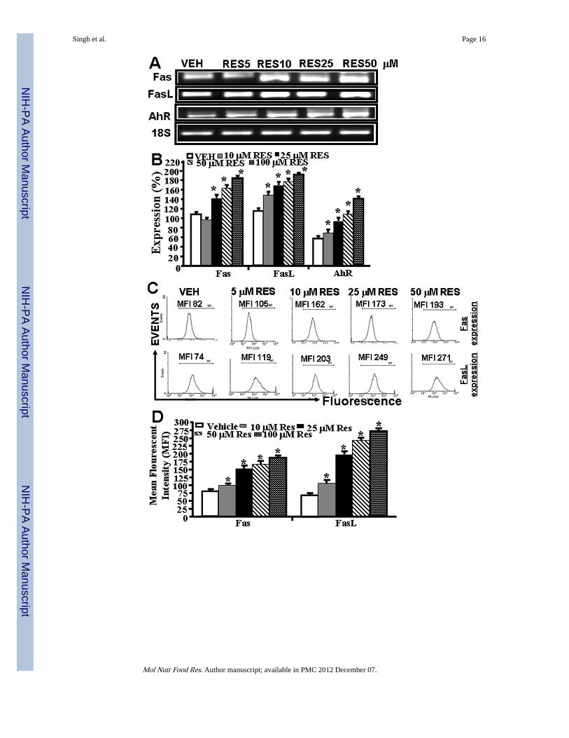
3.3 RES upregulates AhR, Fas, and FasL expression in EL4 cellsNext, we determined the expression of AhR, Fas, and FasL in EL4 cells in vitro post vehicleor RES treatment. To this end, we first performed RT-PCR to determine the expression ofAhR, Fas and FasL in EL4 cells. We also stained EL4 cells with mouse-specific anti-Fas andFasL antibodies respectively to determine the expression Fas and FasL in EL4 cells. Dataobtained from RT-PCRs showed constitutive expression of AhR, Fas, and FasL in EL4 cells.However, post RES-treatment, there was a significant increase in expression of all the threegenes in EL4 cells (Fig. 3A and B) when compared to vehicle-treated EL4 cells (Fig. 3A andB). Importantly, the increase in their expression in the presence of RES was dose-dependent(Fig. 3A and B). We observed similar 18S (a house keeping gene) expression between RES-and vehicle-treated EL4 cells (Fig. 3A). Upon examination of Fas and FasL expression inEL4 cells post RES/vehicle treatments, by flow cytometry, data corroborated with RT-PCRdata for increased expression of Fas and FasL (Fig 3C and D).
3.4 FasL plays a role in initiating death-receptor pathway during RES-induced apoptosis inEL4 cells
To test the role of FasL in RES-induced apoptosis, EL4 cells were cultured in the absence orpresence of anti-mouse FasL mAb (5 µg/ml) and various doses of RES (5–50 µM). Therewas significant reduction in RES-induced apoptosis in EL4 cells when Ab against FasL wasadded to the culture (Fig. 3E and F). Addition of isotype control Ab failed to exhibit anysignificant effect on RES-induced apoptosis (Fig. 3E and F). These data suggested a role forFasL in initiating RES-mediated death-receptor pathway leading to apoptosis in EL4 cells.
Singh et al. Page 6
Mol Nutr Food Res. Author manuscript; available in PMC 2012 December 07.
NIH
-PA Author Manuscript
NIH
-PA Author Manuscript
NIH
-PA Author Manuscript
3.5 RES triggers both extrinsic (death-receptor) and intrinsic (mitochondrial) pathways ofapoptosis in EL4 cells
We next investigated the role of various apoptotic pathways in RES-induced apoptosis inEL4 cells. To this end, we determined the role of various caspases (Caspase-3/7, caspase-8,and caspase–9) in RES-induced apoptosis in EL4 cells. To this end, we performed standardenzymatic assays in EL4 cells post vehicle or RES treatment measuring caspases asdescribed earlier [22]. Increased enzymatic activities were observed for all three caspasesexamined in RES-treated EL4 cells when compared to vehicle-treated EL4 cells (data notshown). These data were similar to our previous studies in which activated T cells wereshown to undergo apoptosis following resveratrol treatment that was mediated by caspases[22]. The role of various caspases was further confirmed by blocking caspase activity usingvarious caspase inhibitors (caspase-3; Z-DEVD, caspase-8; Z-IETD-FMK, and caspase-9;Z-LEHD-FMK). These data demonstrated significant blocking of RES-induced apoptosis inEL4 cells the presence of caspase-3 inhibitor and partial blocking in the presence ofcaspase-8 or caspase-9 inhibitors (data not shown), thereby suggesting that RES-inducedapoptosis in EL4 cells was caspase-dependent involving both intrinsic and extrinsicpathways. To further corroborate the role of caspases in RES-induced apoptosis in EL4cells, Western blot analysis for various caspases, PARP, BAX, Bid, and CYT-c wasperformed. The data demonstrated that RES treatment caused cleavage of caspases-8, -3, -9,-2, and PARP in EL4 cells and these data were similar to that reported by us earlier [22].Increased expression of BAX, Bid, and cytochrome-c was also observed in RES-treated EL4cells when compared to vehicle controls (data not shown). The increase in Bid expression inRES-treated EL4 cells demonstrated its role in initiating cross-talk between death-receptorand mitochondrial pathways. Moreover, release of cytochrome c from nucleus to cytoplasmin RES-treated EL4 cells, corroborated the role of mitochondrial pathway in RES-inducedapoptosis in EL4 cells.
To further corroborate the role of mitochondrial pathway in RES-induced apoptosis in EL4cells, we examined loss of mitochondrial membrane potential (Δψm) using DIOC6 dye post-RES or vehicle treatment. Reduction in Δψm in RES-treated EL4 cells compared to vehicle-treated EL4 cells was noted (data not shown), similar to our previous data on the effect ofRES on T cells[22]. However, even at a high dose of 50 µM RES, we noted moderate levelsof reduction in Δψm (data not presented) thereby suggesting that mitochondrial pathwayplays only a partial role in RES-mediated apoptosis in EL4 cells.
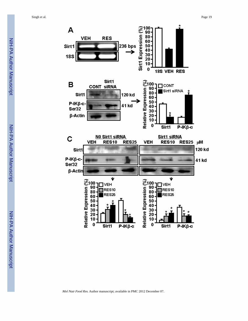
3.6 RES reciprocally regulates SIRT1 and NF-kB expression in EL4 cellsThere are reports demonstrating RES-mediated reciprocally regulates SIRT1 and NF-kBexpression [26–29]. To determine the effect of RES on SIRT1 and NF-kB activation in EL4cells, we performed a series of in vitro assays and determined the expression of SIRT1,phosphorylated IkK, and NF-kB in EL4 cells. As shown in Fig 4A, EL4 cells expressmoderate-level of SIRT1 but upon RES treatment, SIRT1 expression was significantlyupregulated (Fig. 4A).
Furthermore, we used mouse SIRT1-specific siRNAs to silence the expression of SIRT1 inEL4 cells and examined the expression of SIRT1 in the absence or presence of RES. Asshown in Fig 4B, there was significant blocking of SIRT1 expression in EL4 cells two dayspost transfection with SIRT1 siRNAs. However, RES treatment of SIRT1 siRNA-transfected EL4 cells led to upregulation of SIRT1 expression in transfected EL4 cellsalthough these levels were less than that seen in wild-type EL-4 cells (Fig 4C). We alsoexamined the posphorylation of IκBα which leads to release NF-kB that migrates to nucleusto regulate various genes. We observed lower level of phosphorylation of IκBα (Ser 32)upon RES treatment in these cells when compared to vehicle treatment (Fig 4C)
Singh et al. Page 7
Mol Nutr Food Res. Author manuscript; available in PMC 2012 December 07.
NIH
-PA Author Manuscript
NIH
-PA Author Manuscript
NIH
-PA Author Manuscript
demonstrating that RES-mediated induction in expression of SIRT1 blocks phosphorylationof IκBα (Ser 32) which in turn influences expression and release of NF-kB.
We also determined the activation of NF-kB in the nucleus. To this end, we performedEMSA assays using mouse NF-kB-specific probe and nuclear protein prepared fromuntreated or SIRT1 siRNA transfected EL4 cells treated with vehicle or RES. As shown inFig 4D, we observed shift in NF-kB probe when nuclear protein form vehicle-treated EL4cells was used. However, there was lesser shift in NF-kB probes when nuclear proteins fromResveatrol-treated EL4 cells were used (Fig 4D). Moreover, when nuclear protein fromSIRT1 siRNA-transfected EL4 cells was used, we observed relatively more shift in NF-kBprobes but RES treatment of transfected EL4 cells significantly affected the shift of NF-kBprobes (Fig. 4D). The nuclear extracts from various treated groups of EL4 cells did not showa shift in mutant NF-kB probes demonstrating NF-kB-specific binding of nuclear extracts ofEL4 cells (Fig. 4D). The data obtained from these studies demonstrated that RES treatmentof EL4 cells upregulated SIRT1 expression but downregulated NF-kB expression.
3.7 SIRT1 expression affects RES-mediated apoptosis of EL4 cellsWe next assessed the effects of SIRT1 on RES-mediated apoptosis in EL4 cells. To this end,we performed in vitro assays using EL4 cells untransfected or tranfected with SIRT1 siRNAand treated with vehicle or RES. We observed that untransfected EL4 cells were moresusceptible to RES-induced apoptosis (Fig. 4E and F), when compared to EL4 cellstransfected with SIRT1 siRNAs, particularly when the background apoptosis seen withvehicle was subtracted from RES-induced apoptosis (Fig. 4E and F). These datademonstrated that SIRT1 expression influences RES-mediated apoptosis in EL4 cells.
4 DiscussionDevelopment of anti-cancer drugs with high efficacy is a major challenge. The resistance ofmany cancers to various treatments throws obstacles and constitutes major problems incancer therapy. Usually, the abnormalities in apoptotic pathways of cancer cells make themresistant to various chemopreventive drugs. Successful induction of apoptosis in cancercells, therefore, is a key for the development of anti-cancer drugs and therapies.Chemoprevention is considered as a promising strategy to control cancer development[30,31]. There are many natural products and/or dietary supplements that have been shownto act as chemopreventive agents and inhibit experimental carcinogenesis [32]. In recentyears, RES, a natural plant-derived phytoalexin compound, which possesses anti-cancerproperties, has been the focus for its use as a chemopreventive agent against various types ofcancer [5,6]. RES has been shown to suppress the growth of transformed cells throughinduction of apoptosis [33]. There are studies demonstrating RES-mediated apoptosis inHL60 leukaemia cells as well as in T47D breast carcinoma cells [6]. RES-mediatedapoptosis has also been shown in human T-cell acute lymphoblastic leukemia MOLT-4 cells[34].
In the current study, we investigated the chemopreventive and anti-cancer properties of RESagainst EL4, a T lymphoma cell line, and evaluated its efficacy against EL4-inducedtumorigenesis in immunodeficient NOD/SCID mouse model. We used this model to directlytest the effect of RES on tumor cells because it is well known that RES also exhibitsimmunomodulatory properties, including the ability to enhance perforin expression and NKcell cytotoxicity through NKG2D-dependent pathways [36]. We noted that RES treatmentsignificantly suppressed EL4-tumor growth and increased the mean survival time. Theseresults correlated with the ability of RES to induce apoptosis in EL4 tumor cells. Analysis ofapoptotic pathways revealed that RES triggered both death-receptor (Fas/FasL-mediated)and mitochondrial pathways in EL4 cells. Although both death-receptor and mitochondrial
Singh et al. Page 8
Mol Nutr Food Res. Author manuscript; available in PMC 2012 December 07.
NIH
-PA Author Manuscript
NIH
-PA Author Manuscript
NIH
-PA Author Manuscript

pathways played a role in RES-mediated apoptosis in EL4 cells, the fact that only a smallproportion of apoptotic cells showed loss of mitochondrial membrane potential suggestedthat death-receptor pathway may play more critical role in RES-mediated apoptosis in EL4cells. The mitochondrial pathway may be activated through cross-talk via Bid as weobserved presence of cleaved Bid (22 kd) in EL4 cells in the presence of RES. We alsoobserved the presence of cleaved caspase-2, which can be activated by cytotoxic stress, andcaspase-2 is required for the permeabilization of mitochondria [35]. RES has been shown totrigger CD95 signaling-dependent apoptosis in human tumor cells [6]. RES may also induceFasL-mediated apoptosis through Cdc42 activation of ASK1/JNK-dependent signalingpathway in human leukemia HL-60 cells [37]. Dorrie et al have shown that RES inducesapoptosis by depolarizing mitochondrial membrane and activating caspase-9 in acutelymphoblastic leukemia cells [5].
We and others have shown that RES also acts as a AhR mixed agonist/antagonist [22,38]. Inthis study, we observed RES-mediated the activation of AhR in EL4 cells. Previously, wehave shown that activated AhR induced Fas and FasL expression in T cells leading toapoptosis [22,23]. The earlier study from our laboratory has also shown that AhR uponactivation upregulates the expression of Fas by interacting with dioxin response element(DRE) present as a regulatory element in murine Fas promoter, and FasL by interacting withNF-kB regulatory elements present in murine FasL promoter [25]. Thus, RES not onlyinduces AhR but also acts as an AhR ligand which may trigger the induction of Fas andFasL leading to apoptosis. There are reports demonstrating that RES can act as a ligand foraryl hydrocarbon receptor (AhR) thereby regulating the expression of various genes [39]. Insummary, we propose a model demonstrating participation of both extrinsic and intrinsicpathways in RES-mediated apoptosis in EL4 cells (Fig. 4G).
RES is one of the most potent natural compounds that activate SIRT1 [40,41]. Earlierstudies have demonstrated that SIRT1 acts as a longevity factor and its overexpressionincreases the life span of many organisms tested [42]. Many of the SIRT1 substrates aretranscription factors and key regulators described to take part in cancer development, suchas the nuclear factor-κB (NF-κB), the DNA repair factor Ku70, the tumor suppressor genep53, and the forkhead transcription factors (FoxOs) [43,44]. The relationship betweenSIRT1 activity and tumorigenesis is still open to debate [43]. Boily and others have reportedthat SIRT1 does not behave like a classical tumor-suppressor gene but the antitumor activityof RES is mediated, at least in part, by SIRT1 [45]. There are other reports demonstratinghigh expression of SIRT1 in several types of tumors [46] and may be responsible for thedevelopment of chemotherapy resistance [47]. SIRT1 is also an important mediator incalorie restriction-associated tumor prevention [48]. Although some studies have suggestedthat SIRT1 may function as a tumor promoter because of its increased expression in sometypes of cancers, other studies have demonstrated that SIRT1 levels are reduced in someother types of cancers thereby acting as a tumor suppressor [18]. Moreover, SIRT1deficiency promotes tumorigenesis, and overexpression of SIRT1 attenuates cancerformation in mice heterozygous for tumor suppressor p53 [49,50] or APC [45]. Thus,clearly, additional studies are necessary to address the precise role of SIRT1 in tumorgrowth and metastasis. In the current study we noted that upon RES treatment, SIRT1expression was significantly increased in EL4 tumor cells. It was also striking that there wasa significant decrease in p-IκBα expression and NF-κB nuclear translocation in EL4 cells.These data are consistent with the observation that IκBα levels were elevated in mouseembryonic fibroblasts derived from SIRT1−/− mice under basal conditions as well as inresponse to TNFα, thereby suggesting that SIRT1 suppresses the expression of NF-κB-dependent genes [51]. Moreover, SIRT1 regulates the transcriptional activity of NF-κB byphysically interacting with the RelA/p65 subunit of NF-κB and inhibiting transcription bydeacetylating RelA/p65 [52]. Together, these studies suggest that RES by enhancing SIRT1
Singh et al. Page 9
Mol Nutr Food Res. Author manuscript; available in PMC 2012 December 07.
NIH
-PA Author Manuscript
NIH
-PA Author Manuscript
NIH
-PA Author Manuscript
expression may downregulate NF-κB activation and thereby making tumor cells susceptibleto RES-induced apoptosis, specifically because NF-κB exhibits anti-apoptotic properties.
In the current study, we used a dose of 10, 50 and 100 mg/kg of RES administered by oralgavage. Previous in vivo studies to treat cancer suggested that low doses such as 40 mg/kgof RES are not effective while higher doses such as 80 mg/kg were partially effective [53].In another study, RES was shown to inhibit tumor growth in Balb/c mice at high doses suchas 500, 1000 and 1500 mg/kg in a dose dependent manner when administered for 10 days[54]. Also, in a rat model, 100 mg/kg of RES was very effective in delaying tumorigenesis[55]. In this study, we observed that lower dose (10 mg/kg) was not effective against EL4tumor growth while a higher dose (100 mg/kg) was very effective. It should be noted thatthe dose that we have used is feasible to achieve in humans because the human equivalentdose of 100 mg/kg in mouse is 486 mg, considering an average human weight of 60 kg.Currently, there are several nutraceutical companies selling purified resveratrol in 500-mgquantities in capsule form. Thus, our doses reflect the potential pharmacological/dietarysupplement dose available in market.
In summary, our study provides important information regarding the reciprocal regulation ofSIRT1 and NF-κB in tumor apoptosis in vitro and tumor growth in vivo. Our studydemonstrates that exposure of a T cell lymphoma line to RES induces SIRT1 and decreasesNF-κB activation thereby making the cells more susceptible to apoptosis and inhibition oftumor growth in vivo. Thus, our studies suggest that RES may serve as a chemopreventiveagent and make the tumor cells more susceptible to apoptosis when used either directly or incombination with other anti-cancer agents.
AcknowledgmentsN.P.S. designed and performed experiments, analyzed results, prepared the figures, and wrote the manuscript;U.P.S. helped in design of experiments and discussion, VGH, performed apoptotic assays and analyzed data, HG,performed in vitro experiments and analyzed data, LH, participated in discussion to examine data, M.N. designedthe experiments, edited the manuscript, and provided the resources, and P.S.N. assisted in project conception,design of experiments, provided resources, and edited the manuscript.
All authors read and approved the final manuscript.
This work was funded in part by National Institutes of Health Grants P01-AT03961, R01-ES09098, R01-DA016545).
Abbreviations used in this paper
Resveratrol (RES) trans-3,5,4'-trihydroxystilbene
SIRT1 silent mating type information regulation 2 homolog, 1
NF-kB Nuclear Factor Kappa B
AhR aryl hydrocarbon receptor
References1. Jang JH, Surh YJ. Protective effects of resveratrol on hydrogen peroxide-induced apoptosis in rat
pheochromocytoma (PC12) cells. Mutat Res. 2001; 496:181–190. [PubMed: 11551494]
2. Gehm BD, McAndrews JM, Chien PY, Jameson JL. Resveratrol, a polyphenolic compound found ingrapes and wine, is an agonist for the estrogen receptor. Proc Natl Acad Sci U S A. 1997;94:14138–14143. [PubMed: 9391166]
3. Soleas GJ, Diamandis EP, Goldberg DM. Wine as a biological fluid: history, production, and role indisease prevention. J Clin Lab Anal. 1997; 11:287–313. [PubMed: 9292395]
Singh et al. Page 10
Mol Nutr Food Res. Author manuscript; available in PMC 2012 December 07.
NIH
-PA Author Manuscript
NIH
-PA Author Manuscript
NIH
-PA Author Manuscript
4. Kopp P. Resveratrol, a phytoestrogen found in red wine. A possible explanation for the conundrumof the 'French paradox'? Eur J Endocrinol. 1998; 138:619–620. [PubMed: 9678525]
5. Dorrie J, Gerauer H, Wachter Y, Zunino SJ. Resveratrol induces extensive apoptosis bydepolarizing mitochondrial membranes and activating caspase-9 in acute lymphoblastic leukemiacells. Cancer Res. 2001; 61:4731–4739. [PubMed: 11406544]
6. Clement MV, Hirpara JL, Chawdhury SH, Pervaiz S. Chemopreventive agent resveratrol, a naturalproduct derived from grapes, triggers CD95 signaling-dependent apoptosis in human tumor cells.Blood. 1998; 92:996–1002. [PubMed: 9680369]
7. Ovesna Z, Kozics K, Bader Y, Saiko P, Handler N, Erker T, Szekeres T. Antioxidant activity ofresveratrol, piceatannol and 3,3',4,4',5,5'-hexahydroxy-trans-stilbene in three leukemia cell lines.Oncol Rep. 2006; 16:617–624. [PubMed: 16865264]
8. Cignarella A, Minici C, Bolego C, Pinna C, Sanvito P, Gaion RM, Puglisi L. Potential pro-inflammatory action of resveratrol in vascular smooth muscle cells from normal and diabetic rats.Nutr Metab Cardiovasc Dis. 2006; 16:322–329. [PubMed: 16829340]
9. Palsamy P, Subramanian S. Ameliorative potential of resveratrol on proinflammatory cytokines,hyperglycemia mediated oxidative stress, and pancreatic beta-cell dysfunction in streptozotocin-nicotinamide-induced diabetic rats. J Cell Physiol. 224:423–432. [PubMed: 20333650]
10. Mousa SA, Gallati C, Simone T, Dier E, Yalcin M, Dyskin E, Thangirala S, Hanko C, Rebbaa A.Dual targeting of the antagonistic pathways mediated by Sirt1 and TXNIP as a putative approachto enhance the efficacy of anti-aging interventions. Aging (Albany NY). 2009; 1:412–424.[PubMed: 20195491]
11. Orallo F. Trans-resveratrol: a magical elixir of eternal youth? Curr Med Chem. 2008; 15:1887–1898. [PubMed: 18691046]
12. Harikumar KB, Kunnumakkara AB, Sethi G, Diagaradjane P, Anand P, Pandey MK, Gelovani J,Krishnan S, Guha S, Aggarwal BB. Resveratrol, a multitargeted agent, can enhance antitumoractivity of gemcitabine in vitro and in orthotopic mouse model of human pancreatic cancer. Int JCancer. 127:257–268. [PubMed: 19908231]
13. Jang M, Cai L, Udeani GO, Slowing KV, Thomas CF, Beecher CW, Fong HH, Farnsworth NR,Kinghorn AD, Mehta RG, Moon RC, Pezzuto JM. Cancer chemopreventive activity of resveratrol,a natural product derived from grapes. Science. 1997; 275:218–220. [PubMed: 8985016]
14. Mao QQ, Bai Y, Lin YW, Zheng XY, Qin J, Yang K, Xie LP. Resveratrol confers resistanceagainst taxol via induction of cell cycle arrest in human cancer cell lines. Mol Nutr Food Res.54:1574–1584. [PubMed: 20521268]
15. Armour SM, Baur JA, Hsieh SN, Land-Bracha A, Thomas SM, Sinclair DA. Inhibition ofmammalian S6 kinase by resveratrol suppresses autophagy. Aging (Albany NY). 2009; 1:515–528.[PubMed: 20157535]
16. Aggarwal BB, Shishodia S. Molecular targets of dietary agents for prevention and therapy ofcancer. Biochem Pharmacol. 2006; 71:1397–1421. [PubMed: 16563357]
17. North BJ, Verdin E. Sirtuins: Sir2-related NAD-dependent protein deacetylases. Genome Biol.2004; 5:224. [PubMed: 15128440]
18. Deng CX. SIRT1, is it a tumor promoter or tumor suppressor? Int J Biol Sci. 2009; 5:147–152.[PubMed: 19173036]
19. Cha EJ, Noh SJ, Kwon KS, Kim CY, Park BH, Park HS, Lee H, Chung MJ, Kang MJ, Lee DG,Moon WS, Jang KY. Expression of DBC1 and SIRT1 is associated with poor prognosis of gastriccarcinoma. Clin Cancer Res. 2009; 15:4453–4459. [PubMed: 19509139]
20. Wang RH, Sengupta K, Li C, Kim HS, Cao L, Xiao C, Kim S, Xu X, Zheng Y, Chilton B, Jia R,Zheng ZM, Appella E, Wang XW, Ried T, Deng CX. Impaired DNA damage response, genomeinstability, and tumorigenesis in SIRT1 mutant mice. Cancer Cell. 2008; 14:312–323. [PubMed:18835033]
21. Firestein R, Blander G, Michan S, Oberdoerffer P, Ogino S, Campbell J, Bhimavarapu A,Luikenhuis S, de Cabo R, Fuchs C, Hahn WC, Guarente LP, Sinclair DA. The SIRT1 deacetylasesuppresses intestinal tumorigenesis and colon cancer growth. PLoS One. 2008; 3:e2020. [PubMed:18414679]
Singh et al. Page 11
Mol Nutr Food Res. Author manuscript; available in PMC 2012 December 07.
NIH
-PA Author Manuscript
NIH
-PA Author Manuscript
NIH
-PA Author Manuscript
22. Singh NP, Hegde VL, Hofseth LJ, Nagarkatti M, Nagarkatti P. Resveratrol (trans-3,5,4'-trihydroxystilbene) ameliorates experimental allergic encephalomyelitis, primarily via induction ofapoptosis in T cells involving activation of aryl hydrocarbon receptor and estrogen receptor. MolPharmacol. 2007; 72:1508–1521. [PubMed: 17872969]
23. Camacho IA, Singh N, Hegde VL, Nagarkatti M, Nagarkatti PS. Treatment of mice with 2,3,7,8-tetrachlorodibenzo-p-dioxin leads to aryl hydrocarbon receptor-dependent nuclear translocation ofNF-kappaB and expression of fas ligand in thymic stromal cells and consequent apoptosis in Tcells. J Immunol. 2005; 175:90–103. [PubMed: 15972635]
24. Singh UP, Singh NP, Singh B, Hofseth LJ, Price RL, Nagarkatti M, Nagarkatti PS. Resveratrol(trans-3,5,4'-trihydroxystilbene) induces silent mating type information regulation-1 and down-regulates nuclear transcription factor-kappaB activation to abrogate dextran sulfate sodium-induced colitis. J Pharmacol Exp Ther. 332:829–839. [PubMed: 19940103]
25. Singh NP, Nagarkatti M, Nagarkatti PS. Role of dioxin response element and nuclear factor-kappaB motifs in 2,3,7,8-tetrachlorodibenzo-p-dioxin-mediated regulation of Fas and Fas ligandexpression. Mol Pharmacol. 2007; 71:145–157. [PubMed: 16940415]
26. Gracia-Sancho J, Villarreal G Jr, Zhang Y, Garcia-Cardena G. Activation of SIRT1 by resveratrolinduces KLF2 expression conferring an endothelial vasoprotective phenotype. Cardiovasc Res.85:514–519. [PubMed: 19815564]
27. Lagouge M, Argmann C, Gerhart-Hines Z, Meziane H, Lerin C, Daussin F, Messadeq N, Milne J,Lambert P, Elliott P, Geny B, Laakso M, Puigserver P, Auwerx J. Resveratrol improvesmitochondrial function and protects against metabolic disease by activating SIRT1 andPGC-1alpha. Cell. 2006; 127:1109–1122. [PubMed: 17112576]
28. Markus MA, Morris BJ. Resveratrol in prevention and treatment of common clinical conditions ofaging. Clin Interv Aging. 2008; 3:331–339. [PubMed: 18686754]
29. Yang J, Kong X, Martins-Santos ME, Aleman G, Chaco E, Liu GE, Wu SY, Samols D, Hakimi P,Chiang CM, Hanson RW. Activation of SIRT1 by resveratrol represses transcription of the genefor the cytosolic form of phosphoenolpyruvate carboxykinase (GTP) by deacetylating hepaticnuclear factor 4alpha. J Biol Chem. 2009; 284:27042–27053. [PubMed: 19651778]
30. Rodriguez-Nieto S, Zhivotovsky B. Role of alterations in the apoptotic machinery in sensitivity ofcancer cells to treatment. Curr Pharm Des. 2006; 12:4411–4425. [PubMed: 17168751]
31. Testa U, Riccioni R. Deregulation of apoptosis in acute myeloid leukemia. Haematologica. 2007;92:81–94. [PubMed: 17229639]
32. Kelloff GJ, Crowell JA, Steele VE, Lubet RA, Malone WA, Boone CW, Kopelovich L, Hawk ET,Lieberman R, Lawrence JA, Ali I, Viner JL, Sigman CC. Progress in cancer chemoprevention:development of diet-derived chemopreventive agents. J Nutr. 2000; 130:467S–471S. [PubMed:10721931]
33. Ferry-Dumazet H, Garnier O, Mamani-Matsuda M, Vercauteren J, Belloc F, Billiard C, DupouyM, Thiolat D, Kolb JP, Marit G, Reiffers J, Mossalayi MD. Resveratrol inhibits the growth andinduces the apoptosis of both normal and leukemic hematopoietic cells. Carcinogenesis. 2002;23:1327–1333. [PubMed: 12151351]
34. Cecchinato V, Chiaramonte R, Nizzardo M, Cristofaro B, Basile A, Sherbet GV, Comi P.Resveratrol-induced apoptosis in human T-cell acute lymphoblastic leukaemia MOLT-4 cells.Biochem Pharmacol. 2007; 74:1568–1574. [PubMed: 17868649]
35. Lassus P, Opitz-Araya X, Lazebnik Y. Requirement for caspase-2 in stress-induced apoptosisbefore mitochondrial permeabilization. Science. 2002; 297:1352–1354. [PubMed: 12193789]
36. Lu CC, Chen JK. Resveratrol enhances perforin expression and NK cell cytotoxicity throughNKG2D-dependent pathways. J Cell Physiol. 223:343–351. [PubMed: 20082299]
37. Su JL, Lin MT, Hong CC, Chang CC, Shiah SG, Wu CW, Chen ST, Chau YP, Kuo ML.Resveratrol induces FasL-related apoptosis through Cdc42 activation of ASK1/JNK-dependentsignaling pathway in human leukemia HL-60 cells. Carcinogenesis. 2005; 26:1–10. [PubMed:15217905]
38. de Medina P, Casper R, Savouret JF, Poirot M. Synthesis and biological properties of new stilbenederivatives of resveratrol as new selective aryl hydrocarbon modulators. J Med Chem. 2005;48:287–291. [PubMed: 15634023]
Singh et al. Page 12
Mol Nutr Food Res. Author manuscript; available in PMC 2012 December 07.
NIH
-PA Author Manuscript
NIH
-PA Author Manuscript
NIH
-PA Author Manuscript
39. Casper RF, Quesne M, Rogers IM, Shirota T, Jolivet A, Milgrom E, Savouret JF. Resveratrol hasantagonist activity on the aryl hydrocarbon receptor: implications for prevention of dioxin toxicity.Mol Pharmacol. 1999; 56:784–790. [PubMed: 10496962]
40. Albani D, Polito L, Batelli S, De Mauro S, Fracasso C, Martelli G, Colombo L, Manzoni C,Salmona M, Caccia S, Negro A, Forloni G. The SIRT1 activator resveratrol protects SK-N-BEcells from oxidative stress and against toxicity caused by alpha-synuclein or amyloid-beta (1–42)peptide. J Neurochem. 2009; 110:1445–1456. [PubMed: 19558452]
41. Sulaiman M, Matta MJ, Sunderesan NR, Gupta MP, Periasamy M, Gupta M. Resveratrol, anactivator of SIRT1, upregulates sarcoplasmic calcium ATPase and improves cardiac function indiabetic cardiomyopathy. Am J Physiol Heart Circ Physiol. 298:H833–H843. [PubMed:20008278]
42. Tissenbaum HA, Guarente L. Increased dosage of a sir-2 gene extends lifespan in Caenorhabditiselegans. Nature. 2001; 410:227–230. [PubMed: 11242085]
43. Saunders LR, Verdin E. Sirtuins: critical regulators at the crossroads between cancer and aging.Oncogene. 2007; 26:5489–5504. [PubMed: 17694089]
44. Blander G, Guarente L. The Sir2 family of protein deacetylases. Annu Rev Biochem. 2004;73:417–435. [PubMed: 15189148]
45. Boily G, He XH, Pearce B, Jardine K, McBurney MW. SirT1-null mice develop tumors at normalrates but are poorly protected by resveratrol. Oncogene. 2009; 28:2882–2893. [PubMed:19503100]
46. Huffman DM, Grizzle WE, Bamman MM, Kim JS, Eltoum IA, Elgavish A, Nagy TR. SIRT1 issignificantly elevated in mouse and human prostate cancer. Cancer Res. 2007; 67:6612–6618.[PubMed: 17638871]
47. Chu F, Chou PM, Zheng X, Mirkin BL, Rebbaa A. Control of multidrug resistance gene mdr1 andcancer resistance to chemotherapy by the longevity gene sirt1. Cancer Res. 2005; 65:10183–10187. [PubMed: 16288004]
48. Bordone L, Guarente L. Calorie restriction, SIRT1 and metabolism: understanding longevity. NatRev Mol Cell Biol. 2005; 6:298–305. [PubMed: 15768047]
49. Peck B, Chen CY, Ho KK, Di Fruscia P, Myatt SS, Coombes RC, Fuchter MJ, Hsiao CD, LamEW. SIRT inhibitors induce cell death and p53 acetylation through targeting both SIRT1 andSIRT2. Mol Cancer Ther. 9:844–855. [PubMed: 20371709]
50. Yi J, Luo J. SIRT1 and p53, effect on cancer, senescence and beyond. Biochim Biophys Acta.1804:1684–1689. [PubMed: 20471503]
51. Kwon HS, Brent MM, Getachew R, Jayakumar P, Chen LF, Schnolzer M, McBurney MW,Marmorstein R, Greene WC, Ott M. Human immunodeficiency virus type 1 Tat protein inhibitsthe SIRT1 deacetylase and induces T cell hyperactivation. Cell Host Microbe. 2008; 3:158–167.[PubMed: 18329615]
52. Yeung F, Hoberg JE, Ramsey CS, Keller MD, Jones DR, Frye RA, Mayo MW. Modulation of NF-kappaB-dependent transcription and cell survival by the SIRT1 deacetylase. Embo J. 2004;23:2369–2380. [PubMed: 15152190]
53. Gao X, Xu YX, Divine G, Janakiraman N, Chapman RA, Gautam SC. Disparate in vitro and invivo antileukemic effects of resveratrol, a natural polyphenolic compound found in grapes. J Nutr.2002; 132:2076–2081. [PubMed: 12097696]
54. Liu HS, Pan CE, Yang W, Liu XM. Antitumor and immunomodulatory activity of resveratrol onexperimentally implanted tumor of H22 in Balb/c mice. World J Gastroenterol. 2003; 9:1474–1476. [PubMed: 12854144]
55. Bhat KP, Lantvit D, Christov K, Mehta RG, Moon RC, Pezzuto JM. Estrogenic and antiestrogenicproperties of resveratrol in mammary tumor models. Cancer Res. 2001; 61:7456–7463. [PubMed:11606380]
Singh et al. Page 13
Mol Nutr Food Res. Author manuscript; available in PMC 2012 December 07.
NIH
-PA Author Manuscript
NIH
-PA Author Manuscript
NIH
-PA Author Manuscript
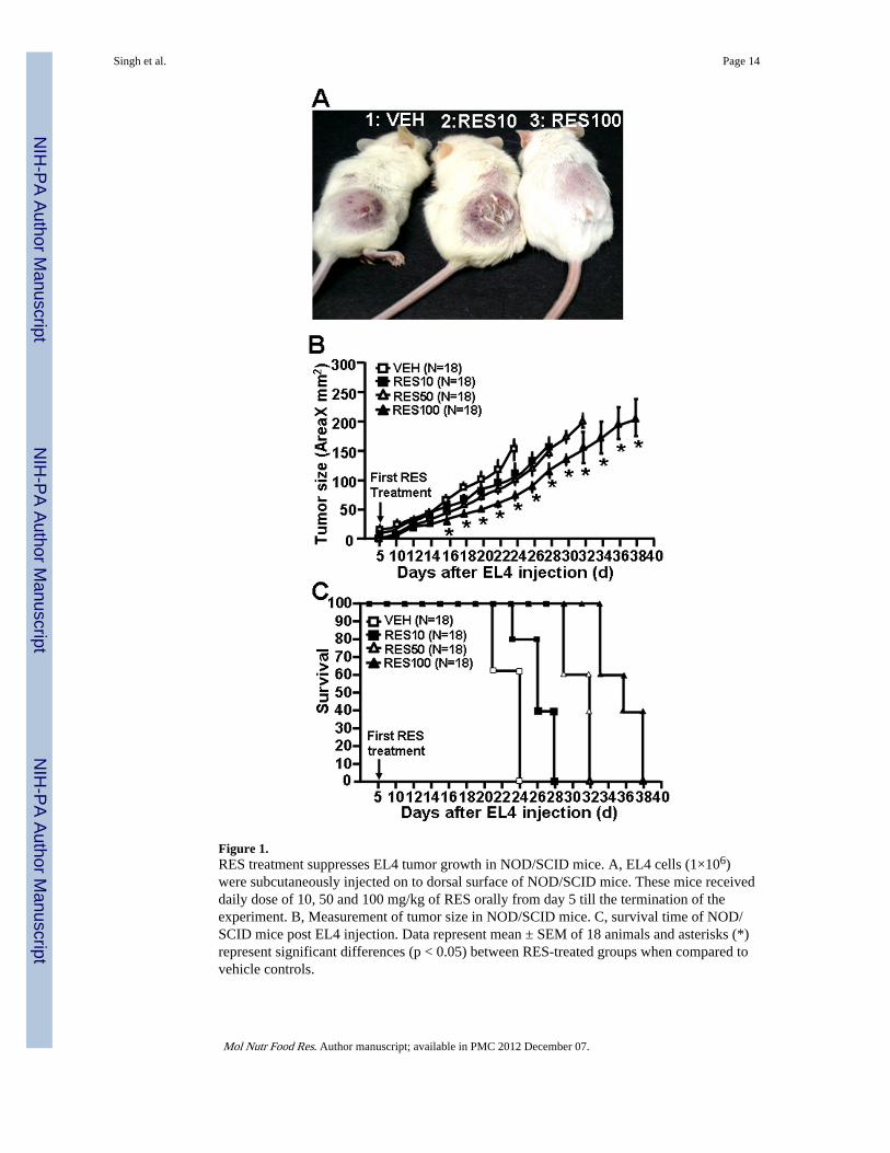
Figure 1.RES treatment suppresses EL4 tumor growth in NOD/SCID mice. A, EL4 cells (1×10⁶)were subcutaneously injected on to dorsal surface of NOD/SCID mice. These mice receiveddaily dose of 10, 50 and 100 mg/kg of RES orally from day 5 till the termination of theexperiment. B, Measurement of tumor size in NOD/SCID mice. C, survival time of NOD/SCID mice post EL4 injection. Data represent mean ± SEM of 18 animals and asterisks (*)represent significant differences (p < 0.05) between RES-treated groups when compared tovehicle controls.
Singh et al. Page 14
Mol Nutr Food Res. Author manuscript; available in PMC 2012 December 07.
NIH
-PA Author Manuscript
NIH
-PA Author Manuscript
NIH
-PA Author Manuscript
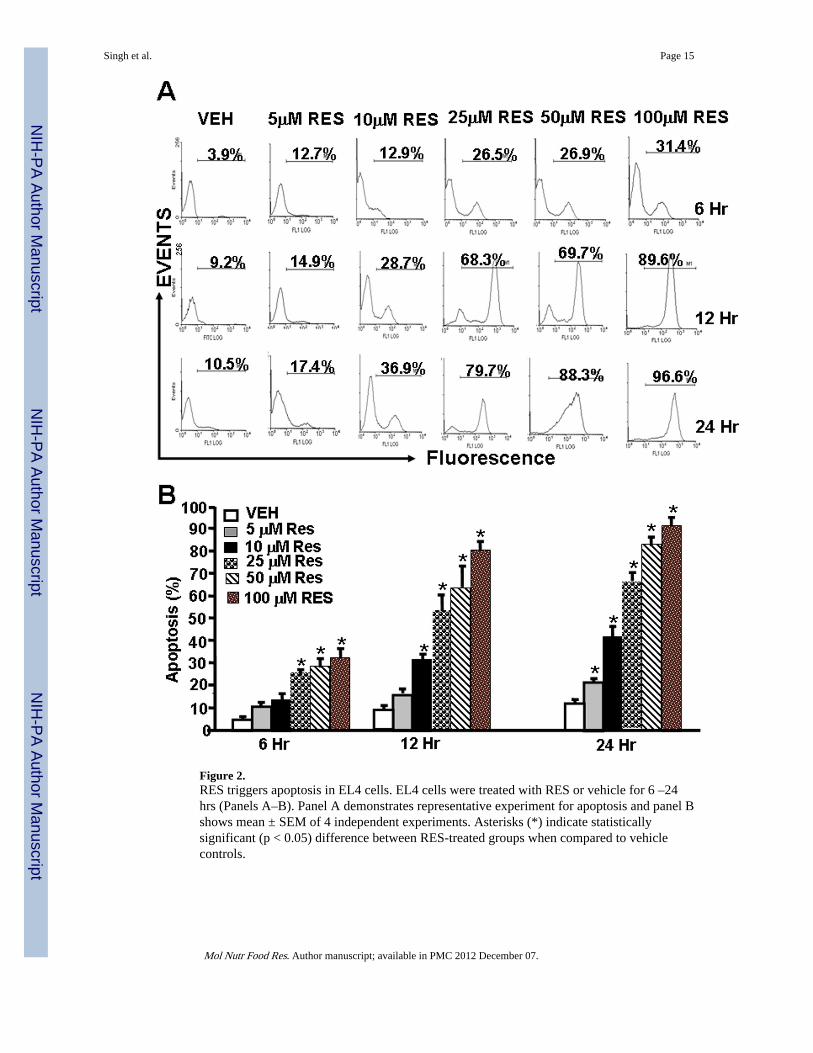
Figure 2.RES triggers apoptosis in EL4 cells. EL4 cells were treated with RES or vehicle for 6 –24hrs (Panels A–B). Panel A demonstrates representative experiment for apoptosis and panel Bshows mean ± SEM of 4 independent experiments. Asterisks (*) indicate statisticallysignificant (p < 0.05) difference between RES-treated groups when compared to vehiclecontrols.
Singh et al. Page 15
Mol Nutr Food Res. Author manuscript; available in PMC 2012 December 07.
NIH
-PA Author Manuscript
NIH
-PA Author Manuscript
NIH
-PA Author Manuscript
Singh et al. Page 16
Mol Nutr Food Res. Author manuscript; available in PMC 2012 December 07.
NIH
-PA Author Manuscript
NIH
-PA Author Manuscript
NIH
-PA Author Manuscript
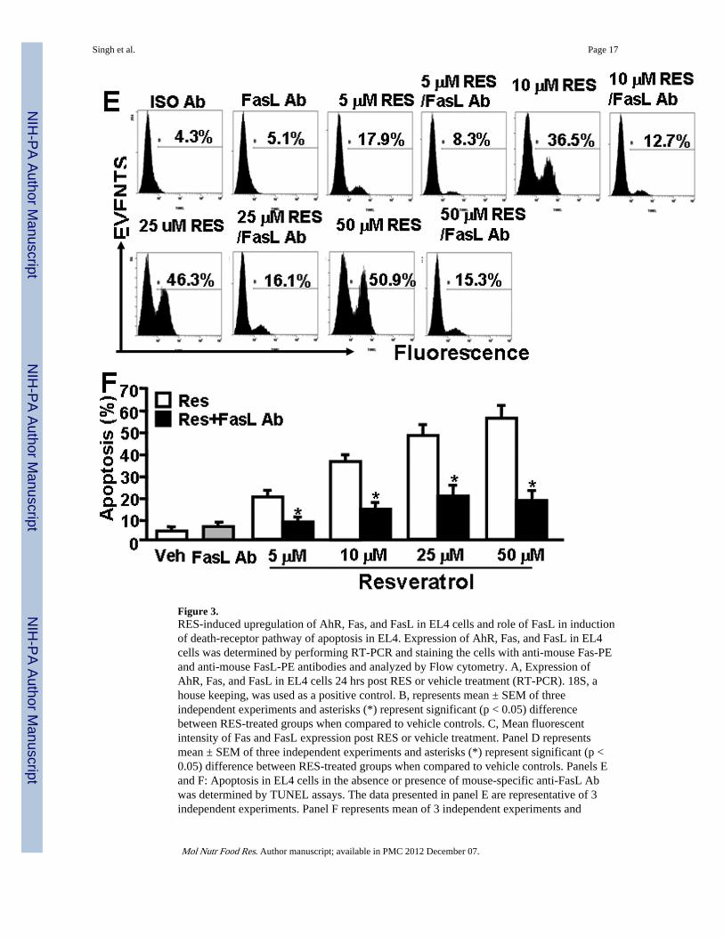
Figure 3.RES-induced upregulation of AhR, Fas, and FasL in EL4 cells and role of FasL in inductionof death-receptor pathway of apoptosis in EL4. Expression of AhR, Fas, and FasL in EL4cells was determined by performing RT-PCR and staining the cells with anti-mouse Fas-PEand anti-mouse FasL-PE antibodies and analyzed by Flow cytometry. A, Expression ofAhR, Fas, and FasL in EL4 cells 24 hrs post RES or vehicle treatment (RT-PCR). 18S, ahouse keeping, was used as a positive control. B, represents mean ± SEM of threeindependent experiments and asterisks (*) represent significant (p < 0.05) differencebetween RES-treated groups when compared to vehicle controls. C, Mean fluorescentintensity of Fas and FasL expression post RES or vehicle treatment. Panel D representsmean ± SEM of three independent experiments and asterisks (*) represent significant (p <0.05) difference between RES-treated groups when compared to vehicle controls. Panels Eand F: Apoptosis in EL4 cells in the absence or presence of mouse-specific anti-FasL Abwas determined by TUNEL assays. The data presented in panel E are representative of 3independent experiments. Panel F represents mean of 3 independent experiments and
Singh et al. Page 17
Mol Nutr Food Res. Author manuscript; available in PMC 2012 December 07.
NIH
-PA Author Manuscript
NIH
-PA Author Manuscript
NIH
-PA Author Manuscript

asterisks (*) represent significant (p < 0.05) reduction in RES-induced apoptosis of T cellscultured in the presence of FasL Ab when compared to the controls.
Singh et al. Page 18
Mol Nutr Food Res. Author manuscript; available in PMC 2012 December 07.
NIH
-PA Author Manuscript
NIH
-PA Author Manuscript
NIH
-PA Author Manuscript
Singh et al. Page 19
Mol Nutr Food Res. Author manuscript; available in PMC 2012 December 07.
NIH
-PA Author Manuscript
NIH
-PA Author Manuscript
NIH
-PA Author Manuscript
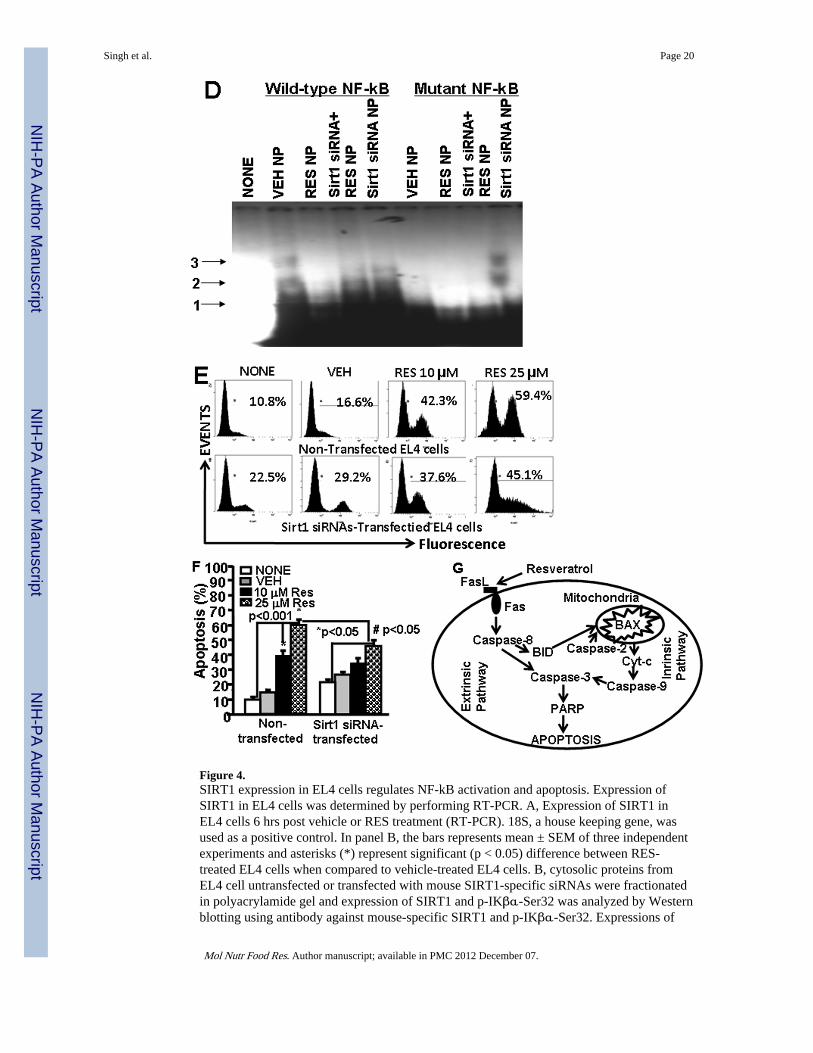
Figure 4.SIRT1 expression in EL4 cells regulates NF-kB activation and apoptosis. Expression ofSIRT1 in EL4 cells was determined by performing RT-PCR. A, Expression of SIRT1 inEL4 cells 6 hrs post vehicle or RES treatment (RT-PCR). 18S, a house keeping gene, wasused as a positive control. In panel B, the bars represents mean ± SEM of three independentexperiments and asterisks (*) represent significant (p < 0.05) difference between RES-treated EL4 cells when compared to vehicle-treated EL4 cells. B, cytosolic proteins fromEL4 cell untransfected or transfected with mouse SIRT1-specific siRNAs were fractionatedin polyacrylamide gel and expression of SIRT1 and p-IKβα-Ser32 was analyzed by Westernblotting using antibody against mouse-specific SIRT1 and p-IKβα-Ser32. Expressions of
Singh et al. Page 20
Mol Nutr Food Res. Author manuscript; available in PMC 2012 December 07.
NIH
-PA Author Manuscript
NIH
-PA Author Manuscript
NIH
-PA Author Manuscript
SIRT1 and and p-IKβα-Ser32 is presented as percentage of β-Actin expression on Y-axisand expression of β -Actin was considered to be 100% for each experiment. Vertical barsrepresent mean +/− SEM of three independent experiments and asterisks (*) representstatistically significant (p < 0.002) reduction in expression of SIRT1 (untransfected control;CONT vs SIRT1 siRNA transfected EL4 cells) and an increase in expression of p-IKβ-α-Ser32 (untransfected control; CONT vs SIRT1 siRNA transfected EL4 cells) in EL4 cells.C, cytosolic proteins from EL4 cell untransfected or transfected with mouse SIRT1-specificsiRNAs and treated with vehicle or RES were fractionated in polyacrylamide gel andexpression of SIRT1 and p- IKβα-Ser32 in EL4 cells was analyzed by Western blottingusing antibody against mouse-specific SIRT1 and p-IKβα-Ser32. Left panel in C representsexpressions of SIRT1 and p-IKβα-Ser32 in untransfected EL4 cells and treated with vehicleor RES whereas right panel represents expressions of SIRT1 and p-IKβα-Ser32 in EL4 cellstransfected with SIRT1 siRNA and treated with vehicle or RES. The vertical bars representpercentage of β-actin expression which was considered to be 100%. Also, vertical barsrepresent mean +/− SEM of three independent experiments and asterisks (*) representstatistically significant (p < 0.002) increase in expression of SIRT1 (VEH vs RES10 orRES25 µM) and decrease in p-IKβα-Ser32 (VEH vs RES10 or RES25 µM). D, EMSAanalysis of NF-kB motif. Double stranded probes containing wild-type NF-kB or mutantNF-kB motifs were generated and nuclear extracts (4–5 µg) generated from vehicle- or RES-treated untransfected or SIRT1 siRNA-transfected EL4 cells not treated or treated with RESwas used in each reaction. Radiolabeled (P32) wild-type or mutant NF-kB1 probes were usedeither without incubation with nuclear extracts or after incubation with nuclear extracts.Arrows 1 represents free probes and arrows 2 and 3 represent NF-kB+ nuclear protein (NP)complexes. E, EL4 cells non-transfected or transfected with SIRT1 siRNA were cultured inthe presence of vehicle or RES (10 or 25 µM) for 24 hrs and apoptosis was determined usingTUNEL assay. F, Vertical bars in the left panel of E represent mean +/− SEM of threeindependent experiments and asterisks (*) represent statistically significant (p< 0.001)increase in apoptosis in non-transfected EL4 cells or SIRT1-siRNA transfected EL4 cellspost RES treatment compared to vehicle-treated cells and (#) represents statisticallysignificant (p< 0.05) decrease in apoptosis in SIRT1-siRNA transfected EL4 cells post REStreatment compared to untransfected EL4 cells. G, Schematic diagram demonstratingpathways involved in RES-mediated apoptosis in EL4 cells.
Singh et al. Page 21
Mol Nutr Food Res. Author manuscript; available in PMC 2012 December 07.
NIH
-PA Author Manuscript
NIH
-PA Author Manuscript
NIH
-PA Author Manuscript
Copyright © 2022 FDOKUMEN