Resistance to cisplatin does not affect sensitivity of human ovarian cancer cell lines to...
13
BioMed Central TIONAL INTERNA CANCER CELL Page 1 of 13 (page number not for citation purposes) Cancer Cell International Open Access Primary research Resistance to cisplatin does not affect sensitivity of human ovarian cancer cell lines to mifepristone cytotoxicity Elizabeth M Freeburg, Alicia A Goyeneche, Erin E Seidel and Carlos M Telleria* Address: Division of Basic Biomedical Sciences, Sanford School of Medicine of The University of South Dakota, Vermillion, SD 57069, USA Email: Elizabeth M Freeburg - [email protected]; Alicia A Goyeneche - [email protected]; Erin E Seidel - [email protected]; Carlos M Telleria* - [email protected] * Corresponding author Abstract Background: The prototypical antiprogestin mifepristone exhibits potent growth inhibition activity towards ovarian cancer cells in vitro and in vivo. The aim of this research was to establish whether mifepristone is capable of inhibiting cell proliferation and inducing apoptotic cell death regardless of the degree of sensitivity ovarian cancer cells exhibit to cisplatin. Methods: OV2008, OV2008/C13, A2780, A2780/CP70, Caov-3, and SK-OV-3 cell lines exhibiting a range of sensitivities to cisplatin were used. Growth inhibition, cell viability, and sub-diploid DNA content in response to treatment with escalating doses of either mifepristone or cisplatin were assessed by microcapillary cytometry. Apoptotic cell death was evaluated by measuring genomic DNA fragmentation and cleavage of caspase-3 and poly (ADP ribose) polymerase (PARP). Results: The sensitivities to cisplatin manifested by the cell lines were OV2008 > A2780 > Caov- 3 > SK-OV-3 > OV2008/C13 > A2780/CP70. Mifepristone inhibited the growth of all six cell lines in a dose-related manner with IC 50s ranging from ~6–12 μM and without significant correlation with the relative sensitivities the cells displayed for cisplatin. Moreover, at the highest concentration studied, mifepristone triggered apoptotic death in all six cell lines as evidenced by the increase in sub-diploid fragmented DNA content and cleavage of caspase-3 and of its downstream substrate PARP. Conclusion: Mifepristone is cytotoxic towards ovarian cancer cells independent of the sensitivity exhibited by the cells to cisplatin, displaying cytostatic effects at lower concentrations and lethal effects at higher concentrations. Mifepristone monotherapy emerges as a valuable therapeutic alternative for platinum-resistant ovarian cancers. Background Current treatment for ovarian cancer begins with cytore- ductive surgery followed by platinum-based chemother- apy [1-3]. However, long-term survival remains low because acquisition of resistance to platinum derivatives is a common feature for this disease, taking place with high frequency in patients with recurrent ovarian cancer [4-7]. This resistance is caused by the failure of a sufficient amount of platinum to reach the target DNA and/or the failure to achieve cell death after platinum-DNA adduct formation by development of more efficient DNA repair mechanisms or by increased tolerance to platinum- Published: 17 February 2009 Cancer Cell International 2009, 9:4 doi:10.1186/1475-2867-9-4 Received: 18 December 2008 Accepted: 17 February 2009 This article is available from: http://www.cancerci.com/content/9/1/4 © 2009 Freeburg et al; licensee BioMed Central Ltd. This is an Open Access article distributed under the terms of the Creative Commons Attribution License (http://creativecommons.org/licenses/by/2.0 ), which permits unrestricted use, distribution, and reproduction in any medium, provided the original work is properly cited.
-
Upload
independent -
Category
Documents
-
view
4 -
download
0
Transcript of Resistance to cisplatin does not affect sensitivity of human ovarian cancer cell lines to...

BioMed CentralC
TIONALINTERNACANCER CELLCancer Cell International
ss
Open AccePrimary researchResistance to cisplatin does not affect sensitivity of human ovarian cancer cell lines to mifepristone cytotoxicityElizabeth M Freeburg, Alicia A Goyeneche, Erin E Seidel and Carlos M Telleria*Address: Division of Basic Biomedical Sciences, Sanford School of Medicine of The University of South Dakota, Vermillion, SD 57069, USA
Email: Elizabeth M Freeburg - [email protected]; Alicia A Goyeneche - [email protected]; Erin E Seidel - [email protected]; Carlos M Telleria* - [email protected]
* Corresponding author
AbstractBackground: The prototypical antiprogestin mifepristone exhibits potent growth inhibitionactivity towards ovarian cancer cells in vitro and in vivo. The aim of this research was to establishwhether mifepristone is capable of inhibiting cell proliferation and inducing apoptotic cell deathregardless of the degree of sensitivity ovarian cancer cells exhibit to cisplatin.
Methods: OV2008, OV2008/C13, A2780, A2780/CP70, Caov-3, and SK-OV-3 cell lines exhibitinga range of sensitivities to cisplatin were used. Growth inhibition, cell viability, and sub-diploid DNAcontent in response to treatment with escalating doses of either mifepristone or cisplatin wereassessed by microcapillary cytometry. Apoptotic cell death was evaluated by measuring genomicDNA fragmentation and cleavage of caspase-3 and poly (ADP ribose) polymerase (PARP).
Results: The sensitivities to cisplatin manifested by the cell lines were OV2008 > A2780 > Caov-3 > SK-OV-3 > OV2008/C13 > A2780/CP70. Mifepristone inhibited the growth of all six cell linesin a dose-related manner with IC50s ranging from ~6–12 μM and without significant correlation withthe relative sensitivities the cells displayed for cisplatin. Moreover, at the highest concentrationstudied, mifepristone triggered apoptotic death in all six cell lines as evidenced by the increase insub-diploid fragmented DNA content and cleavage of caspase-3 and of its downstream substratePARP.
Conclusion: Mifepristone is cytotoxic towards ovarian cancer cells independent of the sensitivityexhibited by the cells to cisplatin, displaying cytostatic effects at lower concentrations and lethaleffects at higher concentrations. Mifepristone monotherapy emerges as a valuable therapeuticalternative for platinum-resistant ovarian cancers.
BackgroundCurrent treatment for ovarian cancer begins with cytore-ductive surgery followed by platinum-based chemother-apy [1-3]. However, long-term survival remains lowbecause acquisition of resistance to platinum derivativesis a common feature for this disease, taking place with
high frequency in patients with recurrent ovarian cancer[4-7]. This resistance is caused by the failure of a sufficientamount of platinum to reach the target DNA and/or thefailure to achieve cell death after platinum-DNA adductformation by development of more efficient DNA repairmechanisms or by increased tolerance to platinum-
Published: 17 February 2009
Cancer Cell International 2009, 9:4 doi:10.1186/1475-2867-9-4
Received: 18 December 2008Accepted: 17 February 2009
This article is available from: http://www.cancerci.com/content/9/1/4
© 2009 Freeburg et al; licensee BioMed Central Ltd. This is an Open Access article distributed under the terms of the Creative Commons Attribution License (http://creativecommons.org/licenses/by/2.0), which permits unrestricted use, distribution, and reproduction in any medium, provided the original work is properly cited.
Page 1 of 13(page number not for citation purposes)

Cancer Cell International 2009, 9:4 http://www.cancerci.com/content/9/1/4
induced DNA damage [8,9]. Hence, finding new treat-ment alternatives for platinum-insensitive ovarian cancersis of critical importance.
In preclinical studies previously conducted in our labora-tory, the antiprogestin steroid mifepristone was found tobe highly effective as a single agent in vitro and in vivoabrogating growth of human epithelial ovarian cancercells [10]. We demonstrated that the growth inhibitoryeffect of mifepristone on ovarian cancer cells was associ-ated with inhibition of DNA synthesis, down-regulationof transcription factor E2F1 needed for S phase progres-sion, and inhibition of the activity of cell cycle regulatorykinase, cyclin dependent kinase 2 (Cdk2). This is likelydue to increased association of Cdk2 with the Cdk inhib-itors p21cip1 and p27kip1 which are greatly up-regulated inresponse to the drug. All these molecular events down-stream of mifepristone action lead to blockage of the cellcycle at the G1-to-S phase transition [10]. In the samestudy it was observed that mifepristone displayed similargrowth inhibition potency among SK-OV-3, OV2008, andCaov-3 cell lines [10]. To note is that whereas OV2008cells were reported as being highly sensitive to cisplatin[11], SK-OV-3 cells were originally obtained from apatient with intrinsic resistance to clinically achievabledoses of cisplatin [12], and Caov-3 cells were reported tobe resistant to cisplatin [13,14]. Based on this informa-tion, it is reasonable to speculate with the possibility thatmifepristone may be useful in abrogating ovarian cancercell growth irrespective of the sensitivity the cells displayfor cisplatin.
The tumor suppressor p53 encodes for a transcription fac-tor which is involved in a multiplicity of cellular functionsincluding cell cycle [15,16], cell death [17,18], cell differ-entiation [19], and DNA damage [18,20] and repair[21,22] pathways. In ovarian cancer, mutations in the p53gene correlate with resistance to platinum-based chemo-therapy and shortened survival [23]. In addition, p53 isnon-functional in 70% of ovarian tumors [24], whereaspreclinical studies suggest this tumor suppressor is a deter-minant of cisplatin sensitivity in ovarian cancer cells [25-27].
It is reasonable to contemplate the possibility that if boththe sensitivity to platinum and the p53 genetic back-ground of ovarian cancer cells do not condition theirresponse to the growth inhibition activity of mifepristone,such findings would have great clinical relevance. Thus, inthe present work we first set out to study the growth inhi-bition activity of mifepristone among ovarian cancer celllines having different sensitivities to platinum derivatives.We studied the action of mifepristone not only in ovariancancer cells with different genetic backgrounds, but alsoamong ovarian cancer cell line pairs consisting of cispla-
tin-sensitive parental lines and stable cisplatin-resistantsublines derived by in vitro selection with stepwise expo-sure to increasing doses of cisplatin. In addition, becauseof the differences in the p53 genetic status of the cell linesstudied, the experiments indirectly allowed us to provideevidence as to whether the p53 genetic backgroundimpacts the response of the ovarian cancer cells to mife-pristone.
MethodsCell lines and drugsThe human ovarian carcinoma cell lines, OV2008,OV2008/C13, A2780, and A2780/CP70, were obtainedfrom Dr. Stephen Howell (University of California, SanDiego) and were maintained in RPMI 1640 (Mediatech,Herndon, VA) supplemented with 5% heat inactivatedFBS (Atlanta Biologicals, Lawrencenville, GA) and 10 mMHEPES (Mediatech), 4 mM L-glutamine (Mediatech), 1mM sodium pyruvate (Mediatech), 1 X non-essentialamino acids (Mediatech), 100 IU penicillin (Mediatech)and 100 μg/ml streptomycin (Mediatech). Caov-3 and SK-OV-3 ovarian cancer cells were obtained from the Ameri-can Type Culture Collection (ATCC, Manassas, VA) andwere routinely maintained in RPMI 1640 (Mediatech)supplemented with 5% FBS (Atlanta Biologicals), 10 mMHEPES (Mediatech), 4 mM L-glutamine (Mediatech),0.45% D (+) glucose (Sigma Chemical Company, St.Louis, MO), 1 mM sodium pyruvate (Mediatech), 1 Xnon-essential amino acids (Mediatech), 100 IU penicillin(Mediatech), 100 μg/ml streptomycin (Mediatech), and0.01 mg/ml human insulin (Roche, Indianapolis, IN). Allcell lines were cultured at 37°C in a humidified atmos-phere in the presence of 5% CO2.
The stock of mifepristone (Sigma) was 116.5 mM solutionin DMSO. The maximal concentration of DMSO was0.02% (v/v). The stock of cisplatin (cis-diamminedichlo-roplatinum II) (Sigma) was 3 mM solution in 0.9% NaCl.Cells were exposed to cisplatin for only 1 h. Thereafter, themedium was replaced with fresh cisplatin-free medium.Cells exposed to mifepristone were cultured in the contin-uous presence of the drug throughout the studies.
Cell proliferation and viabilityTriplicate cultures were trypsinized, pelleted by centrifu-gation at 500 g for 5 min, and washed with PBS. The cellswere resuspended in ViaCount reagent (Guava Technolo-gies, Hayward, CA) and studied using the GuavaViaCount application in the Guava EasyCyte Mini micro-capillary cytometer (Guava Technologies). This assay pro-vides an absolute cell count and viability data on a cellsuspension, automating results like cell counts in a hemo-cytometer chamber with the trypan blue dye exclusionmethod for assessing cell viability. The cells are drawninto a capillary flow cell of known dimensions at a pre-
Page 2 of 13(page number not for citation purposes)
Cancer Cell International 2009, 9:4 http://www.cancerci.com/content/9/1/4
cisely controlled rate for measured periods of time. Abso-lute cell counts are obtained by knowing the exactsampling volumes. Viable and non-viable cells areassessed by the differential permeability of two DNA-binding dyes in the reagent. One dye is membrane perme-able and stains all nucleated cells. The other dye only pen-etrates cells with compromised membrane integrity (i.e.non-viable cells). The data are acquired and analyzedusing the CytoSoft 4.1 software (Guava Technologies).
For the cells treated with either cisplatin or mifepristone,three inhibition concentration 50% or IC50 values aver-aged for each cell line and drug were obtained. The IC50values were calculated using the drug interaction software(Calcusyn, Biosoft, Cambridge, UK), which was designedto study drug interaction and calculates the median effec-tive dose, Dm, which is analogous to the IC50.
Determination of sub-G1 DNA contentAfter treatment, cells were trypsinized, pelleted by centrif-ugation at 500 g for 5 min, washed with PBS, and fixedwith 4% paraformaldehyde. Cells were once again washedwith PBS and pelleted by centrifugation at 500 g for 5 min.Then, approximately 100,000–200,000 cells were resus-pended in 200 μl of cell cycle buffer [3.8 mM sodium cit-rate (Sigma), 7 U/ml RNase A (Sigma), 0.1% (v/v) TritonX-100 (Sigma), and 0.05 mg/ml propidium iodide(Sigma)] at a concentration of 500–1000 cells/μl. Cellswere analyzed for the capacity of their DNA to bind pro-pidium iodide utilizing the Guava EasyCyte Mini micro-capillary cytometer and the cell cycle application of theCytoSoft 4.1 software (Guava Technologies), with specialemphasis on the analysis of the cellular fragments withhypodiploid DNA content.
SDS-PAGE and Western blottingCells were scraped, pelleted, washed twice with PBS, andlysed by the addition of two volumes of radioimmuno-precipitation assay buffer (RIPA) containing 50 mM Tris-HCl (pH 7.4), 150 mM NaCl, 1% NP-40 (Sigma), 0.25%sodium deoxycholate (Sigma), 1 mM EDTA, 1 mM PMSF(Sigma), 1 μg/ml pepstatin (Sigma), 1 mM orthovanadate(Sigma) and 1 mM sodium fluoride (Sigma). Cells weredisrupted by passing them through a 21 gauge needle, andgently rocked on ice for 30 min. Lysates were centrifugedat 16,000 g for 15 min at 4°C, and the supernatant wasconsidered the whole cell extract, which was assayed forprotein content by using the bicinchoninic acid method(BCA; Pierce, Rockford, IL). Equivalent amounts of pro-tein (50 μg) per point were loaded in 12% (w/v) acryla-mide gels, subjected to SDS-PAGE and transferred toPVDF membranes. The blots were blocked in 5% (v/v)nonfat milk in TBS containing 0.1% (v/v) Tween 20 (T).Blots were then probed overnight with primary antibodiesagainst poly (ADP-ribose) polymerase (PARP) (#9542;1:1000; Cell Signaling Technologies, Danvers, MA) or cas-
pase-3 (#9662; 1:1000; Cell Signaling). The membraneswere washed 3 × 5 min in TBS-T and incubated with 1:10,000 dilution of peroxidase-conjugate secondary anti-body (#111-035-003; Jackson ImmunoResearch Labora-tories, West Grove, PA) for 30 min at room temperature.The blots were again washed, developed by chemilumi-nescence, and exposed to radiographic film. Blots werealso probed with an antibody directed against β-Actin(clone AC-15; 1:20,000; Sigma) to control for proteinloading.
DNA fragmentationFloating and adherent cells were pelleted and digestedovernight at 50°C in a buffer composed of 100 mM NaCl,10 mM Tris HCl (pH 8.0), 25 mM EDTA (pH 8.0), 0.5%SDS and 0.1 mg/ml proteinase K (Life Technologies,Rockville, MD). The genomic DNA was extracted from thedigested cells with phenol/chloroform/isoamyl alcohol(25:24:1, v/v/v), precipitated, and digested for 60 min at37°C with 1 μg/ml ribonuclease (deoxyribonuclease-free;Roche, Indianapolis, IN). After extraction and precipita-tion, an equal amount of DNA for each sample (2 μg) wasseparated by electrophoresis on a 2.5% agarose gel,impregnated with SYBR Gold nucleic acid gel stain(Molecular Probes, Eugene, OR) and photographed withthe Amersham Typhoon Fluorescence imaging system(Amersham Biosciences Corp., Piscataway, NJ). A 100 bpDNA ladder (Promega, Madison, WI) was utilized fordetermining the size of the fragments of DNA.
Statistical analysisAll data are reported as means ± SEM, and statistical sig-nificance was defined as p < 0.05. To compare cell growth,cell viability, sub-diploid DNA distribution, and IC50 val-ues, one-way ANOVA followed by the Newman-Keuls'multiple comparison test or two-way ANOVA followed bythe Bonferroni's multiple comparison test were used asappropriate. In addition, a Pearson correlation statisticaltest was utilized to quantify the degree to which the IC50sfor mifepristone and cisplatin for each cell line relate toone another.
ResultsGrowth inhibition and lethality of cisplatin towards ovarian cancer cells of similar genetic backgrounds but different platinum sensitivitiesWe utilized two pairs of human ovarian cancer cell lines,each pair consisting of a cisplatin-sensitive parental lineand a stably cisplatin-resistant subline derived by in vitroselection with cisplatin. The resistance of these cell lines tocisplatin, determined using a clonogenic survival assayand a 1-h exposure to cisplatin, has been previouslyreported to be 8.1 fold for A2780/CP70 vs. A2780, and5.7 fold for OV2008/C13 vs. OV2008 [11]. The differentsensitivity of these ovarian cancer cell lines to cisplatinwas further confirmed in our laboratory. Cells were
Page 3 of 13(page number not for citation purposes)

Cancer Cell International 2009, 9:4 http://www.cancerci.com/content/9/1/4
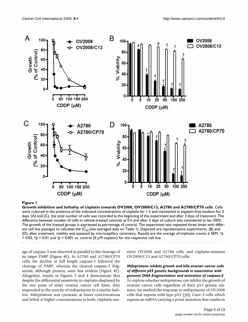
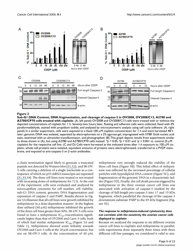
exposed to 0, 5, 10, 25, 50, 100, or 200 μM cisplatin for 1h, and the culture was continued for 72 h in cisplatin-freemedia. At the end of the experiment, non-adherent andadherent cells were harvested, and their number and via-bility was assessed by microcytometry. The experimentwas repeated three times utilizing different cell line pas-sages. The cisplatin IC50s calculated for the cell lines were,as expected, significantly lower for OV2008 cells whencompared to OV2008/C13 cells, and significantly lowerfor A2780 cells when compared to A2780/CP70 cells (Fig-ure 1A and 1C, and Table 1). These results confirm thepredictions that OV2008/C13 and A2780/CP70 are lesssensitive to cisplatin than their sisters OV2008 andA2780, respectively. The different sensitivity to cisplatinwas also manifested in the significant reduction in viabil-ity of OV2008 and A2780 cells in response to cisplatinexposure when compared to the response of OV2008/C13and A2780/CP70 cells, respectively (Figure 1B and 1D).Furthermore, sub-G1 DNA content, which is usually asso-ciated with apoptotic cell death [28], increased more sig-nificantly in response to cisplatin in OV2008 and A2780cells when compared to the levels observed in OV2008/C13 and A2780/CP70 cells (Figure 2, left panels in A andB). The apoptotic nature of the cell death process triggeredby cisplatin was confirmed by fragmentation of thegenomic DNA which was substantially more evident inOV2008 and A2780 cells when compared to the DNAfragmentation observed in OV2008/C13 and A2780/CP70 cells in response to treatment with 100 μM cisplatinfor 1 h (Figure 2, right panels in A and B). Finally, the dif-ferent response to cisplatin was highlighted by the cleav-age of the marker of apoptosis, 35 kDa procaspase-3, topresumably active 19 and 17 kDa fragments, which wasevident in OV2008 and A2780 cells, but not in OV2008/C13 and A2780/CP70 cells (Figure 2C and 2D). Together,results in Figures 1 and 2 confirm that the two sister ovar-ian cancer cell line pairs, OV2008 and OV2008/C13, andA2780 and A2780/CP70, although carrying similargenetic backgrounds, responded very differently to cispla-tin-induced lethality.
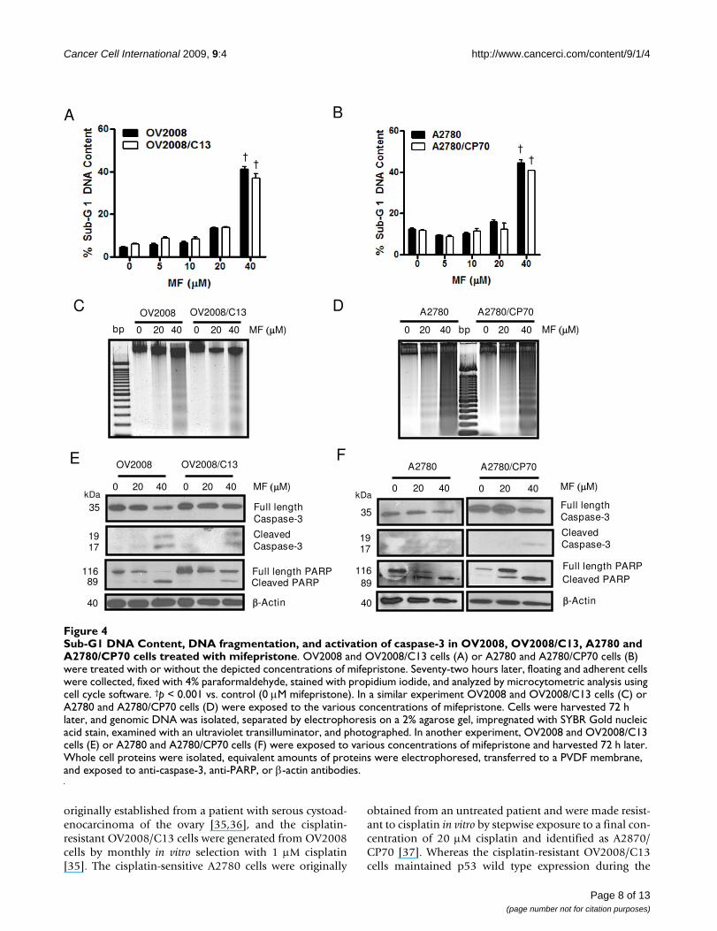
Growth inhibition and lethality of mifepristone towards ovarian cancer cells occurs regardless of cisplatin sensitivitiesTo study if the cell pairs OV2008 and OV2008/C13, andA2780 and A2780/CP70 responded similarly to the toxic-ity of mifepristone, the cells were plated and exposed for72 h to 0, 5, 10, 20 or 40 μM mifepristone. The antipro-gestin growth inhibited OV2008 and OV2008/C13 simi-larly, with IC50s that were indistinguishable from oneanother (Figure 3A and Table 1). In the case of the A2780and A2780/CP70 pair, mifepristone blocked growth in adose-dependent manner; however, the IC50 for A2780 wassignificantly lower than that for A2780/CP70 (Figure 3Cand Table 1). Mifepristone was cytostatic to all cell linesstudied at concentrations ranging from 0–10 μM, butshows some lethality towards the OV2008 and OV2008/C13 pair at the 20 μM concentration, which was more evi-dent at the concentration of 40 μM (Figure 3B). In theA2780 and A2780/CP70 pair, the lethality of mifepristonewas manifested only at the concentration of 40 μM (Fig-ure 3D). The lethality induced by mifepristone to theovarian cancer cell lines was further confirmed by thedetection of cellular particles containing hypodiploidDNA content in coincidence with the treatment done with40 μM of the drug in the four cell lines studied (Figure 4Aand 4B), without apparent differences in the behavior ofthe cisplatin-sensitive versus the cisplatin-resistant siblingcells. The presence of cellular particles with deficient DNAcontent as represented by the sub-G1 region of the cellcycle histogram in propidium iodide-stained cells, sug-gested that cell death occurred by apoptosis [28]. This wasconfirmed by the detection of the characteristic ladder ofDNA denoting DNA fragmentation in the four cell linesexposed to 40 μM mifepristone (Figure 4C and 4D).Finally, the lethality induced by mifepristone in all celllines, without apparent distinction of platinum-sensitivi-ties, was associated with another marker of apoptoticdeath, the cleavage of the caspase-3 target PARP from thefull length 116 kDa form to a 89 kDa fragment [29] (Fig-ure 4E and 4F). In OV2008 and OV2008/C13 cells, cleav-
Table 1: IC50s for mifepristone and IC50s for cisplatin in various ovarian cancer cell lines of different p53 genetic backgrounds and platinum sensitivities.
Cell line IC50 MF ( M) IC50CDDP ( M) Reported p53 status
OV2008 8.8 ± 0.8 (5) 0.2 ± 0.1 (3) wt [30]OV2008/C13 8.1 ± 0.6 (3) 49.8 ± 10 (3) wt [30,38]A2780 5.9 ± 0.6 (3) 6.0 ± 1.3 (3) wt [39,40,52]A2780/CP70 11.7 ± 0.6 (3)* 55.0 ± 11 (3) mut [30,38-40]SK-OV-3 12.6 ± 0.1 (3)* 18.1 ± 0.6 (3) mut [31,33,34]Caov-3 7.4 ± 1.8 (3) 15.2 ± 1.4 (3) mut [31,32]
Cells were treated with mifepristone (MF) or cisplatin (CDDP) as described in Figure 1A and 1C, Figure 3A and 3C, and Figure 5A, to calculate the concentration of the drugs needed to achieve 50% growth inhibition (IC50). The experiments were repeated at least three times with different cellular passages, each time in triplicates. The numbers in parentheses indicate the number of experiments carried out for each cell line. IC50s are expressed as the mean ± SEM. *p < 0.05 compared with the other cell lines treated with mifepristone. The numbers in brackets indicate references that reported the p53 genetic status of the depicted cell lines.
Page 4 of 13(page number not for citation purposes)
Cancer Cell International 2009, 9:4 http://www.cancerci.com/content/9/1/4
age of caspase-3 was observed in parallel to the cleavage ofits target PARP (Figure 4E). In A2780 and A2780/CP70cells, the decline in full length caspase-3 followed thecleavage of PARP, whereas the cleaved caspase-3 frag-ments, although present, were less evident (Figure 4F).Altogether, results in Figures 3 and 4 demonstrate thatdespite the differential sensitivity to cisplatin displayed bythe two pairs of sister ovarian cancer cell lines, theyresponded to the toxicity of mifepristone in a similar fash-ion. Mifepristone was cytostatic at lower concentrationsand lethal at higher concentrations in both, cisplatin-sen-
sitive OV2008 and A2780 cells, and cisplatin-resistantOV2008/C13 and A2780/CP70 cells.
Mifepristone inhibits growth and kills ovarian cancer cells of different p53 genetic backgrounds in association with genomic DNA fragmentation and activation of caspase-3To explore whether mifepristone can inhibit the growth ofovarian cancer cells regardless of their p53 genetic sta-tuses, we studied the response to mifepristone of OV2008cells that express wild type p53 [30], Caov-3 cells whichexpress an mRNA carrying a point mutation that results in
Growth inhibition and lethality of cisplatin towards OV2008, OV2008/C13, A2780 and A2780/CP70 cellsFigure 1Growth inhibition and lethality of cisplatin towards OV2008, OV2008/C13, A2780 and A2780/CP70 cells. Cells were cultured in the presence of the indicated concentrations of cisplatin for 1 h and maintained in cisplatin-free medium for 3 days. (A) and (C), the total number of cells was recorded at the beginning of the experiment and after 3 days of treatment. The difference between number of cells in vehicle-treated controls at 0 h and after 3 days of culture was considered to be 100%. The growth of the treated groups is expressed as percentage of control. The experiment was repeated three times with differ-ent cell line passages to calculate the IC50 (see averaged data on Table 1). Depicted are representative experiments. (B) and (D), after treatment, viability was assessed by microcapillary cytometry. Results are the average of triplicate counts ± SEM. *p < 0.05, #p < 0.01 and †p < 0.001 vs. control (0 μM cisplatin) for the respective cell line.
A B
††
† †
†
# # † †
†
C D
†
† †
* †† †
Page 5 of 13(page number not for citation purposes)
Cancer Cell International 2009, 9:4 http://www.cancerci.com/content/9/1/4
a chain termination signal likely to generate a truncatedpeptide not detected by Western blot [31,32], and SK-OV-3 cells carrying a deletion of a single nucleotide as a con-sequence of which no p53 mRNA transcripts are expressed[31,33,34]. The three cell lines were treated or not treatedwith increasing doses of mifepristone for 72 h. At the endof the experiment, cells were evaluated and analyzed bymicrocapillary cytometry for cell number, cell viability,sub-G1 DNA content, genomic DNA fragmentation andexpression of caspase-3 and PARP. Results shown in Fig-ure 5A illustrate that all cell lines were growth inhibited bymifepristone in a dose-dependent manner. At the highestdose utilized (40 μM) mifepristone inhibited the growthof the three cell lines greater than 90%. SK-OV-3 cells werefound to have a mifepristone IC50 concentration signifi-cantly higher than that of OV2008 and Caov-3 cells, bothof which had similar mifepristone IC50s (Figure 5A andTable 1). Mifepristone showed some lethality towardsOV2008 and Caov-3 cells at the 20 μM concentration, butnot on SK-OV-3 cells. At the concentration of 40 μM,
mifepristone very strongly reduced the viability of thethree cell lines (Figure 5B). This lethal effect of mifepris-tone was reflected by the increased percentage of cellularparticles with hypodiploid DNA content (Figure 5C), andfragmentation of the genomic DNA in a characteristic lad-der (Figure 5D). Finally, the cell death process triggered bymifepristone in the three ovarian cancer cell lines wasassociated with activation of caspase-3 marked by thecleavage of full-length pro-caspase-3 into 17 and 19 kDafragments, which paralleled the cleavage of the caspase-3downstream substrate PARP to the 89 kDa fragment (Fig-ure 5E).
The response to mifepristone-induced cytotoxicity does not correlate with the sensitivity the ovarian cancer cells displayed to cisplatinBecause we analyzed the response to six different ovariancancer cell lines to cisplatin and mifepristone in parallel,with experiments done separately three times with threedifferent cell line passages, we considered it valid to ana-
Sub-G1 DNA Content, DNA fragmentation, and cleavage of caspase-3 in OV2008, OV2008/C13, A2780 and A2780/CP70 cells treated with cisplatinFigure 2Sub-G1 DNA Content, DNA fragmentation, and cleavage of caspase-3 in OV2008, OV2008/C13, A2780 and A2780/CP70 cells treated with cisplatin. (A, left panel) OV2008 and OV2008/C13 cells were treated with or without the depicted concentrations of cisplatin for 1 h. Seventy-two hours later, floating and adherent cells were collected, fixed with 4% paraformaldehyde, stained with propidium iodide, and analyzed by microcytometric analysis using cell cycle software. (A, right panel) In a similar experiment, cells were exposed to a fixed 100 μM cisplatin concentration for 1 h and were harvested 48 h later; genomic DNA was isolated, separated by electrophoresis on a 2% agarose gel, impregnated with SYBR Gold nucleic acid stain, examined with an ultraviolet transilluminator, and photographed. (B) This graph depicts results from experiments similar to those shown in (A), but using A2780 and A2780/CP70 cells instead. *p < 0.05, #p < 0.01 and †p < 0.001 vs. control (0 μM cisplatin) for the respective cell line. (C and D) Cells were harvested at the indicated times after 1-h exposure to 100 μM cis-platin, whole cell proteins were isolated, equivalent amounts of proteins were electrophoresed, transferred to a PVDF mem-brane, and exposed to anti-caspase-3 or β-actin antibodies.
A
†
†
†
†
Hours after CDDP
OV2008
0 48 0 48
OV2008
/C13
#†
†
†
*
B
†
D
Full length Caspase-3
Cleaved Caspase-3
-actin
0 6 12 0 6 12 Hours after CDDP
A2780/CP70A2780
Hours after CDDP
0 48 0 48
A2780
A2780
/CP70
Full length Caspase-3
Cleaved Caspase-3
-actin
Hours after CDDP0 6 12 24 48 0 6 12 24 48
OV2008 OV2008/C13C
kDa
35
1917
40
kDa
35
1917
40
Page 6 of 13(page number not for citation purposes)

Cancer Cell International 2009, 9:4 http://www.cancerci.com/content/9/1/4
lyze whether or not the IC50s found for mifepristone andthe IC50s found for cisplatin among the cell lines relate toone another. To answer this question, a Pearson's correla-tion study statistics was performed to establish the rela-tionship between the IC50 values obtained for each drug.Analysis of data shown in Table 1 indicates the lack of sta-tistically significant difference in the response of the cellsto mifepristone despite the broad ranges of responses tocisplatin (r = 0.3931; p > 0.05). Whereas the IC50s for cis-
platin ranged from < 1 μM to as high as 55 μM, the IC50sfor mifepristone ranged only from ~6–12 μM.
DiscussionMifepristone inhibited ovarian cancer cell growth despitethe fact that the cell lines studied had similar genetic back-grounds but very different sensitivities to cisplatinacquired by in vitro selection of clones upon challengeswith cisplatin. The cisplatin-sensitive OV2008 cells were
Growth inhibition and lethality of mifepristone towards OV2008, OV2008/C13, A2780 and A2780/CP70 cellsFigure 3Growth inhibition and lethality of mifepristone towards OV2008, OV2008/C13, A2780 and A2780/CP70 cells. Cells were cultured in the presence of the indicated concentrations of mifepristone for 72 h. (A) and (C), the total number of cells was recorded at the beginning of the experiment and after the 3 days of treatment. The difference between number of cells in vehicle-treated controls at 0 h and after 3 days of culture was considered to be 100%. The growth of the treated groups is expressed as percentage of control. The experiment was repeated three times with different cell line passages to cal-culate the IC50 (see Table 1 for averaged data). Depicted are representative experiments. (B) and (D), after treatment, viability was assessed by microcapillary cytometry. Results are the average of triplicate counts ± SEM. †p < 0.001 vs. control (0 μM mifepristone) for the respective cell line.
C D
A B
† †
† †
††
Page 7 of 13(page number not for citation purposes)
Cancer Cell International 2009, 9:4 http://www.cancerci.com/content/9/1/4
originally established from a patient with serous cystoad-enocarcinoma of the ovary [35,36], and the cisplatin-resistant OV2008/C13 cells were generated from OV2008cells by monthly in vitro selection with 1 μM cisplatin[35]. The cisplatin-sensitive A2780 cells were originally
obtained from an untreated patient and were made resist-ant to cisplatin in vitro by stepwise exposure to a final con-centration of 20 μM cisplatin and identified as A2870/CP70 [37]. Whereas the cisplatin-resistant OV2008/C13cells maintained p53 wild type expression during the
Sub-G1 DNA Content, DNA fragmentation, and activation of caspase-3 in OV2008, OV2008/C13, A2780 and A2780/CP70 cells treated with mifepristoneFigure 4Sub-G1 DNA Content, DNA fragmentation, and activation of caspase-3 in OV2008, OV2008/C13, A2780 and A2780/CP70 cells treated with mifepristone. OV2008 and OV2008/C13 cells (A) or A2780 and A2780/CP70 cells (B) were treated with or without the depicted concentrations of mifepristone. Seventy-two hours later, floating and adherent cells were collected, fixed with 4% paraformaldehyde, stained with propidium iodide, and analyzed by microcytometric analysis using cell cycle software. †p < 0.001 vs. control (0 μM mifepristone). In a similar experiment OV2008 and OV2008/C13 cells (C) or A2780 and A2780/CP70 cells (D) were exposed to the various concentrations of mifepristone. Cells were harvested 72 h later, and genomic DNA was isolated, separated by electrophoresis on a 2% agarose gel, impregnated with SYBR Gold nucleic acid stain, examined with an ultraviolet transilluminator, and photographed. In another experiment, OV2008 and OV2008/C13 cells (E) or A2780 and A2780/CP70 cells (F) were exposed to various concentrations of mifepristone and harvested 72 h later. Whole cell proteins were isolated, equivalent amounts of proteins were electrophoresed, transferred to a PVDF membrane, and exposed to anti-caspase-3, anti-PARP, or β-actin antibodies.
A
MF M)
Cbp 0 20 40 0 20 40
OV2008E
kDa
35
1917
11689
40
Full length Caspase-3
Cleaved Caspase-3
Full length PARPCleaved PARP
-Actin
MF M)0 20 40 0 20 40
B
DMF M)bp0 20 40 0 20 40
A2780/CP70A2780
F
kDa
35
1917
11689
40
Full length Caspase-3
Cleaved Caspase-3
Full length PARPCleaved PARP
-Actin
MF M)0 20 40
A2780
0 20 40
A2780/CP70OV2008/C13
OV2008 OV2008/C13
††
††
Page 8 of 13(page number not for citation purposes)

Cancer Cell International 2009, 9:4 http://www.cancerci.com/content/9/1/4
process of in vitro challenge with cisplatin when comparedto their sister OV2008 cells [30,38], that was not the casefor the A2780/CP70 cells that acquired a p53 mutationduring such an in vitro selection process [39,40]. This phe-nomenon is not surprising as it has been shown that thereis a survival advantage of p53 mutant cells in the presence
of genotoxic cisplatin [27,41]. The tumor suppressor p53appears to be a determinant of cisplatin sensitivity sincemutant p53 status is often associated with cisplatin insen-sitivity [26], whereas reintroduction of wild type p53 viaadenovirus gene transfer into A2780/CP70 cells signifi-cantly sensitized these cells to cisplatin lethality [42].
Effect of mifepristone on growth, viability, fragmentation of genomic DNA, and on expression of caspase-3 and of its down-stream substrate poly (ADP) ribose polymerase (PARP) in OV2008, Caov-3, and SK-OV-3 cellsFigure 5Effect of mifepristone on growth, viability, fragmentation of genomic DNA, and on expression of caspase-3 and of its downstream substrate poly (ADP) ribose polymerase (PARP) in OV2008, Caov-3, and SK-OV-3 cells. Cells were plated in equal number, allowed to attach to the plate surface for 24 h, and were then treated with either vehicle (DMSO) or the indicated doses of mifepristone in cell specific culture media for 72 h. Cells were then trypsinized, stained, and counted by microcapillary cytometry. The experiments were repeated at least three times in triplicates for each of the doses tested. A representative experiment is shown for each of the cell lines. (A) The total number of cells was recorded at the beginning of the experiment and after 3 days of treatment. The difference between number of cells in vehicle-treated controls at 0 h and after 3 days of culture was considered to be 100%. The growth of the treated groups is expressed as percentage of control. (B) This experiment was similar to that described in previous panel. Following the plating and treatment protocol, cells were collected after 72 h and viable cells were recorded by microcytometry using the Guava ViaCount application. Bars, mean ± SEM. #p < 0.01 and †p < 0.001 when compared to the control (0 μM mifepristone) for each cell line. (C) Following plating and treatment protocol, cells were collected after 72 h, fixed in 4% paraformaldehyde, stained with propidium iodide, and analyzed by cytometry using the Guava cell cycle application. Bars, mean ± SEM. #p < 0.01; †p < 0.001 when compared to 0 μM mifepris-tone. (D) Genomic DNA was isolated and separated by electrophoresis on a 2% agarose gel, impregnated with SYBR Gold nucleic acid stain, examined with an ultraviolet transilluminator, and photographed with the Amersham Typhoon fluorescence imaging system. A 100 base pair marker and a positive control (+ Ctrl) of fragmented DNA generated by treating OV2008 cells with cisplatin were run in parallel. (E) Cells were treated with the indicated concentrations of mifepristone for 72 h, whole cell proteins were isolated, electrophoresed, electrotransferred to a PVDF membrane, and exposed to anti-caspase-3 and anti-PARP antibodies. β-Actin was used as loading control.
A
#
#
†
†
†
B C
#†
††
†
bp+ Ctrl
1500
600
300
100
0 20 40
SK-OV-3
0 20 40
Caov-3
20 40
OV2008
0MF ( M)
Caov-3 SK-OV-3
Full Length Caspase-3
Cleaved Caspase-3
Full Length PARPCleaved PARP
-Actin
0 10 20 40
OV2008
kDa
35
1917
MF ( M)
11689
40
0 10 20 40 0 10 20 40
ED
Page 9 of 13(page number not for citation purposes)

Cancer Cell International 2009, 9:4 http://www.cancerci.com/content/9/1/4
However, it has also been reported that the mutation ofthe p53 gene in the A2870/CP70 cells does not lead toexpression of a mutant p53 protein when the cells arechallenged with cisplatin; conversely, when the challengeis with ionizing radiation, they do express increasing lev-els of a mutant p53 protein capable of up-regulating thep53-target gene p21cip1, and of showing transcriptionalactivity in a functional assay [39]. Thus, it is the signaltransduction pathway connecting cisplatin action andp53 gene expression what appears to be impaired inA2780/CP70 cells. This might be related to the fact thatwhereas OV2008 and OV2008/C13 cells have similarmifepristone IC50s, the mifepristone IC50 of A2780/CP70was significantly higher than that of A2780 cells. Yet,although with slightly different potency, it can be con-cluded that mifepristone was very effective at blockinggrowth in the four ovarian cancer cell line pairs investi-gated considering the broad range of cisplatin IC50s (< 1 to55 μM) and the narrow range of mifepristone IC50s (~6–12 μM).
Notably, mifepristone was cytostatic at concentrationslower than 20 μM, but it was lethal at concentrationshigher than 20 μM. The cytostatic nature of concentra-tions of mifepristone up to 20 μM towards ovarian cancercells was previously shown in our laboratory by demon-strating the reversibility of the growth inhibition effectwhen the drug was removed from the culture [10]. Fur-thermore, we have recently demonstrated that intertwin-ing cytostatic concentrations of mifepristone in betweencourses of lethal cisplatin chemotherapy not only resultedin an efficacious strategy to prevent repopulation of can-cer cells in between lethal platinum treatment intervals,but it also potentiated cisplatin killing efficacy [43]. Inter-estingly, however, in the present work we are showing thatconcentrations of mifepristone higher than that needed toachieve cytostasis are per se lethal to ovarian cancer cells.This lethality was illustrated by the reduced viability of thecells, the increase in cellular particles with hypodiploidfragmented DNA content, and the cleavage of the celldeath associated caspase, caspase-3, in parallel with thecleavage of the widely accepted marker of cell death and asubstrate for caspase-3, poly (ADP) ribose polymerase(PARP) [29]. The lethality of concentrations of mifepris-tone over 40 μM towards ovarian cancer cells was first sug-gested in 1996 by Rose and Barnea in OVCAR-3 andA2780 cells [44]. Yet, the results presented here are thefirst to demonstrate that the lethality of mifepristonemonotherapy towards ovarian cancer cells is related to acaspase-associated apoptotic process. More importantlythe toxicity of mifepristone did not discriminate amongovarian cancer cell lines with very different sensitivities tocisplatin, suggesting that mifepristone monotherapycould be useful for treating patients who have becomeplatinum-resistant, for which the therapeutic alternativeshave very disappointing outcomes [1,4,7].
The dose-dependent cytostatic and lethal effects of mife-pristone towards ovarian cancer cells, which we will referto globally as cytotoxicity, have been shown to also occurin breast cancer cells. A recent work using the MCF-7breast cancer cell line illustrated that combination ofmifepristone and the antiestrogen 4-hydroxytamoxifenhad greater cytostatic and lethal activities than eithermonotherapy, whereas the lethality of the treatment wasassociated with genomic DNA fragmentation and cleav-age of PARP [45]. In addition, it has been shown thatMCF-7 made resistant to 4-hydroxytamoxifen alsorespond to mifepristone monotherapy undergoing apop-totic death [46]; finally, although at higher concentra-tions, mifepristone was also cytotoxic to progesteronereceptor- and estrogen receptor-negative MDA-MB-231breast cancer cells [47].
At present the role played by progesterone receptors in thecytotoxic activity of mifepristone remains unclear. Wehave reported that mifepristone has progesterone-likeactivity inhibiting growth of ovarian cancer cells [10].Likewise in MCF-7 breast cancer cells it was shown thatprogesterone, instead of reversing the growth inhibitoryactivity of mifepristone, contributed to its growth inhibi-tion effect [45]. A progesterone-like growth inhibitoryaction of mifepristone was also shown in estrogen-resist-ant, progesterone receptor expressing T47Dco breast can-cer cells [48]. In addition, although mifepristone can bindto glucocorticoid and progesterone receptors with similaraffinity [49], glucocorticoid receptors do not seem tomediate mifepristone action. In OV2008 ovarian cancercells we could not mimic the growth inhibition effect ofmifepristone when using equimolar concentrations of theglucocorticoid agonist dexamethasone (results notshown), whereas in MCF-7 breast cancer cells, dexameth-asone was unable to reverse the inhibitory action of mife-pristone [45]. Thus, it remains to be seen whethermifepristone utilizes cognate progesterone or glucocorti-coid receptor-mediated endocrine mechanisms to drive itscytostatic and lethal effects on cancer cells.
While indirectly, in the present work we also analyzedwhether the different p53 genetic status of the cellsimpacts the cytotoxicity of mifepristone. Normal functionof the p53 tumor suppressor gene is associated withenhanced sensitivity to chemotherapy; several studieshave suggested that loss of wild type p53 function may bea major cause of failure to respond to chemotherapy andradiotherapy [50,51]. Supporting this concept, in a studyconducted utilizing the 60 cancer cell lines of the NationalCancer Institute anticancer drug screen program, themajority of clinically active agents, including alkylatingagents, antimetabolites, and topoisomerase inhibitors,tended to exhibit growth suppression more in the celllines with normal p53 status than in the cell lines withmutant p53 status, with the exception of the anti-
Page 10 of 13(page number not for citation purposes)

Cancer Cell International 2009, 9:4 http://www.cancerci.com/content/9/1/4
mitogenic agents [33]. This exception was confirmed inovarian cancer cell lines where it was shown that the p53status does not affect the sensitivity of the cells to themicrotubule-stabilizing agent paclitaxel [52,53]. There-fore, although it is generally assumed that loss of normalp53 function can confer resistance to DNA-damage agentsas a consequence of reduced susceptibility to apoptosis,the relevance of p53 mutations in chemosensitivity hasexceptions and controversies particularly in terms of thepotential functional activity of mutant p53 proteins[51,54-56]. Consequently, it is apparent that there aredrugs for which the p53 background does not impactdrug-sensitivity; in the present work we provide evidencethat mifepristone behaves in that manner, inhibitinggrowth and triggering death of ovarian cancer cells whichhave been largely described as having different p53genetic statuses (Table 1). For instance, Caov-3 cells thatcarry a point mutation leading to expression of an abnor-mal transcript encoding an inactive p53 [31,32], and SK-OV-3 cells that carry a single nucleotide deletion in thep53 gene and are not able to generate a p53 transcript[31,33,56,57], both have defects in their apoptoticmachinery that associates with resistance to standard plat-inum therapy [58-60]. Conversely, OV2008 and A2780cells expressing wild type p53 are very sensitive to cyto-toxic drugs such as cisplatin [14,25], paclitaxel [61,62], ordoxorubicin [63-65], rapidly undergoing apoptosis. Ourdata demonstrate that mifepristone, at concentrationsbeyond those used to achieve cytostasis has lethal activitytriggering a caspase-associated apoptotic process in all sixovarian cancer cell lines studied regardless of their p53genetic backgrounds and sensitivities to cisplatin.Although the potency of the growth inhibition by mife-pristone was significantly higher in OV2008, OV2008/C13, and A2780 cells, all carrying wild type p53, whencompared with SK-OV-3 and A2780/CP70 cells carryingp53 mutations, the calculated IC50s only ranged from 8 to12 μM, suggesting that the inhibition is biologically rele-vant despite the p53 genetic backgrounds of the cells. Fur-thermore, the IC50 for mifepristone in Caov-3 carrying amutant p53 gene is indistinguishable from that of thethree wild type-carrying p53 cell lines (OV2008, OV2008/C13, and A2780), further supporting the notion that thep53 background is not relevant for the growth inhibitionand the lethality triggered by mifepristone in ovarian can-cer cells.
ConclusionThe results obtained and summarized in Table 1 highlightthe lack of correlation between the IC50s for mifepristoneand the IC50s for cisplatin obtained for the ovarian cancercell lines studied, and confirm the hypothesis that mife-pristone growth inhibits ovarian cancer cells regardless oftheir sensitivities to cisplatin. Furthermore, we found thatmifepristone, when used al lower concentration, showscytostatic effects, whereas at higher concentration, it
shows lethal effects towards all ovarian cancer cell linesstudied, triggering a caspase-associated apoptotic deathmechanism regardless of their degree of sensitivity to cis-platin and apparent p53 genetic status. The significance ofthis work lies in that it provides preclinical evidence sug-gesting that mifepristone monotherapy can be an alterna-tive to treat ovarian cancers intrinsically resistant toclinically achievable doses of cisplatin, or recurrent ovar-ian cancer tumors which frequently have become plati-num resistant and lack p53 function.
Competing interestsThe authors declare that they have no competing interests.
Authors' contributionsEMF carried out most of the experiments and participatedin the drafting of the manuscript. AAG participated in thedesign of the study and carried out and supervised someof the experiments. EES carried out some the experimentsinvolving SK-OV-3 cells. CMT conceived the study andcontributed to the writing of the manuscript. All authorsread and approved the final manuscript.
AcknowledgementsThis research was supported by Grant Number K22CA121991 from the National Cancer Institute, Grant Number 2 P20 RR016479 from the INBRE Program of the National Center for Research Resources, and funds from the Division of Basic Biomedical Sciences of The University of South Dakota. We thank Dr. Barbara Goodman for the critical reading of the manuscript.
References1. DiSaia PJ, Bloss JD: Treatment of ovarian cancer: new strate-
gies. Gynecol Oncol 2003, 90(2 Pt 2):S24-32.2. Aletti GD, Gallenberg MM, Cliby WA, Jatoi A, Hartmann LC: Cur-
rent management strategies for ovarian cancer. Mayo ClinProc 2007, 82(6):751-770.
3. Bhoola S, Hoskins WJ: Diagnosis and management of epithelialovarian cancer. Obstet Gynecol 2006, 107(6):1399-1410.
4. Herzog TJ: The current treatment of recurrent ovarian can-cer. Curr Oncol Rep 2006, 8(6):448-454.
5. Fraser M, Leung B, Jahani-Asl A, Yan X, Thompson WE, Tsang BK:Chemoresistance in human ovarian cancer: the role of apop-totic regulators. Reprod Biol Endocrinol 2003, 1(1):66.
6. Hamilton TC, Johnson SW: Recent insights into drug resistancein ovarian cancer. In Ovarian Cancer: Methods and Protocols Editedby: Barlett JMS. NJ: Humana Press; 2000:89-106.
7. Vasey PA: Resistance to chemotherapy in advanced ovariancancer: mechanisms and current strategies. Br J Cancer 2003,89(Suppl 3):S23-28.
8. Kelland L: The resurgence of platinum-based cancer chemo-therapy. Nat Rev Cancer 2007, 7(8):573-584.
9. Rabik CA, Dolan ME: Molecular mechanisms of resistance andtoxicity associated with platinating agents. Cancer Treat Rev2007, 33(1):9-23.
10. Goyeneche AA, Caron RW, Telleria CM: Mifepristone inhibitsovarian cancer cell growth in vitro and in vivo. Clin Cancer Res2007, 13(11):3370-3379.
11. Katano K, Kondo A, Safaei R, Holzer A, Samimi G, Mishima M, KuoYM, Rochdi M, Howell SB: Acquisition of resistance to cisplatinis accompanied by changes in the cellular pharmacology ofcopper. Cancer Res 2002, 62(22):6559-6565.
12. Ormerod MG, O'Neill C, Robertson D, Kelland LR, Harrap KR: cis-Diamminedichloroplatinum(II)-induced cell death throughapoptosis in sensitive and resistant human ovarian carci-noma cell lines. Cancer Chemother Pharmacol 1996, 37(5):463-471.
Page 11 of 13(page number not for citation purposes)
http://www.ncbi.nlm.nih.gov/entrez/query.fcgi?cmd=Retrieve&db=PubMed&dopt=Abstract&list_uids=8599870
http://www.ncbi.nlm.nih.gov/entrez/query.fcgi?cmd=Retrieve&db=PubMed&dopt=Abstract&list_uids=8599870

Cancer Cell International 2009, 9:4 http://www.cancerci.com/content/9/1/4
13. Arimoto-Ishida E, Ohmichi M, Mabuchi S, Takahashi T, Ohshima C,Hayakawa J, Kimura A, Takahashi K, Nishio Y, Sakata M, et al.: Inhi-bition of phosphorylation of a forkhead transcription factorsensitizes human ovarian cancer cells to cisplatin. Endocrinol-ogy 2004, 145(4):2014-2022.
14. Hayakawa J, Ohmichi M, Kurachi H, Ikegami H, Kimura A, MatsuokaT, Jikihara H, Mercola D, Murata Y: Inhibition of extracellular sig-nal-regulated protein kinase or c-Jun N-terminal proteinkinase cascade, differentially activated by cisplatin, sensitizeshuman ovarian cancer cell line. J Biol Chem 1999,274(44):31648-31654.
15. Giono LE, Manfredi JJ: The p53 tumor suppressor participatesin multiple cell cycle checkpoints. J Cell Physiol 2006,209(1):13-20.
16. Farid NR: P53 and other cell cycle regulators. Cancer Treat Res2004, 122:149-164.
17. Kuribayashi K, El-Deiry WS: Regulation of programmed celldeath by the p53 pathway. Adv Exp Med Biol 2008, 615:201-221.
18. Garner E, Raj K: Protective mechanisms of p53-p21-pRb pro-teins against DNA damage-induced cell death. Cell Cycle 2008,7(3):277-282.
19. Stiewe T: The p53 family in differentiation and tumorigenesis.Nat Rev Cancer 2007, 7(3):165-168.
20. Helton ES, Chen X: p53 modulation of the DNA damageresponse. J Cell Biochem 2007, 100(4):883-896.
21. Ford JM: Regulation of DNA damage recognition and nucle-otide excision repair: another role for p53. Mutat Res 2005,577(1–2):195-202.
22. Sengupta S, Harris CC: p53: traffic cop at the crossroads ofDNA repair and recombination. Nat Rev Mol Cell Biol 2005,6(1):44-55.
23. Reles A, Wen WH, Schmider A, Gee C, Runnebaum IB, Kilian U,Jones LA, El-Naggar A, Minguillon C, Schonborn I, et al.: Correlationof p53 mutations with resistance to platinum-based chemo-therapy and shortened survival in ovarian cancer. Clin CancerRes 2001, 7(10):2984-2997.
24. Astanehe A, Arenillas D, Wasserman WW, Leung PC, Dunn SE, Dav-ies BR, Mills GB, Auersperg N: Mechanisms underlying p53 reg-ulation of PIK3CA transcription in ovarian surfaceepithelium and in ovarian cancer. J Cell Sci 2008, 121(Pt5):664-674.
25. Yang X, Fraser M, Moll UM, Basak A, Tsang BK: Akt-mediated cis-platin resistance in ovarian cancer: modulation of p53 actionon caspase-dependent mitochondrial death pathway. CancerRes 2006, 66(6):3126-3136.
26. Fraser M, Bai T, Tsang BK: Akt promotes cisplatin resistance inhuman ovarian cancer cells through inhibition of p53 phos-phorylation and nuclear function. Int J Cancer 2008,122(3):534-546.
27. Mujoo K, Zhang L, Klostergaard J, Donato NJ: Emergence of cispl-atin-resistant cells from the OVCAR-3 ovarian carcinomacell line with p53 mutations, altered tumorigenicity, andincreased apoptotic sensitivity to p53 gene replacement. IntJ Gynecol Cancer 2000, 10(2):105-114.
28. Huang X, Halicka HD, Traganos F, Tanaka T, Kurose A, Darzynkie-wicz Z: Cytometric assessment of DNA damage in relation tocell cycle phase and apoptosis. Cell Prolif 2005, 38(4):223-243.
29. Scovassi AI, Poirier GG: Poly(ADP-ribosylation) and apoptosis.Mol Cell Biochem 1999, 199(1–2):125-137.
30. Fraser M, Leung BM, Yan X, Dan HC, Cheng JQ, Tsang BK: p53 is adeterminant of X-linked inhibitor of apoptosis protein/Akt-mediated chemoresistance in human ovarian cancer cells.Cancer Res 2003, 63(21):7081-7088.
31. Yaginuma Y, Westphal H: Abnormal structure and expression ofthe p53 gene in human ovarian carcinoma cell lines. CancerRes 1992, 52(15):4196-4199.
32. Reid T, Jin X, Song H, Tang HJ, Reynolds RK, Lin J: Modulation ofJanus kinase 2 by p53 in ovarian cancer cells. Biochem BiophysRes Commun 2004, 321(2):441-447.
33. O'Connor PM, Jackman J, Bae I, Myers TG, Fan S, Mutoh M, ScudieroDA, Monks A, Sausville EA, Weinstein JN, et al.: Characterizationof the p53 tumor suppressor pathway in cell lines of theNational Cancer Institute anticancer drug screen and corre-lations with the growth-inhibitory potency of 123 anticanceragents. Cancer Res 1997, 57(19):4285-4300.
34. Ikediobi ON, Davies H, Bignell G, Edkins S, Stevens C, O'Meara S,Santarius T, Avis T, Barthorpe S, Brackenbury L, et al.: Mutationanalysis of 24 known cancer genes in the NCI-60 cell line set.Mol Cancer Ther 2006, 5(11):2606-2612.
35. Andrews PA, Murphy MP, Howell SB: Differential potentiation ofalkylating and platinating agent cytotoxicity in human ovar-ian carcinoma cells by glutathione depletion. Cancer Res 1985,45(12 Pt 1):6250-6253.
36. Andrews PA, Murphy MP, Howell SB: Metallothionein-mediatedcisplatin resistance in human ovarian carcinoma cells. CancerChemother Pharmacol 1987, 19(2):149-154.
37. Hamilton TC, Winker MA, Louie KG, Batist G, Behrens BC, TsuruoT, Grotzinger KR, McKoy WM, Young RC, Ozols RF: Augmenta-tion of adriamycin, melphalan, and cisplatin cytotoxicity indrug-resistant and -sensitive human ovarian carcinoma celllines by buthionine sulfoximine mediated glutathione deple-tion. Biochem Pharmacol 1985, 34(14):2583-2586.
38. Sasaki H, Sheng Y, Kotsuji F, Tsang BK: Down-regulation of X-linked inhibitor of apoptosis protein induces apoptosis inchemoresistant human ovarian cancer cells. Cancer Res 2000,60(20):5659-5666.
39. Siddik ZH, Mims B, Lozano G, Thai G: Independent pathways ofp53 induction by cisplatin and X-rays in a cisplatin-resistantovarian tumor cell line. Cancer Res 1998, 58(4):698-703.
40. Lu X, Errington J, Curtin NJ, Lunec J, Newell DR: The impact of p53status on cellular sensitivity to antifolate drugs. Clin Cancer Res2001, 7(7):2114-2123.
41. Righetti SC, Perego P, Corna E, Pierotti MA, Zunino F: Emergenceof p53 mutant cisplatin-resistant ovarian carcinoma cells fol-lowing drug exposure: spontaneously mutant selection. CellGrowth Differ 1999, 10(7):473-478.
42. Song K, Li Z, Seth P, Cowan KH, Sinha BK: Sensitization of cis-platinum by a recombinant adenovirus vector expressingwild-type p53 gene in human ovarian carcinomas. Oncol Res1997, 9(11–12):603-609.
43. Freeburg EM, Goyeneche AA, Telleria CM: Mifepristone abro-gates repopulation of ovarian cancer cells in betweencourses of cisplatin treatment. Int J Oncol 2009, 34(3):743-755.
44. Rose FV, Barnea ER: Response of human ovarian carcinoma celllines to antiprogestin mifepristone. Oncogene 1996,12(5):999-1003.
45. Schoenlein PV, Hou M, Samaddar JS, Gaddy VT, Thangaraju M, LewisJ, Johnson M, Ganapathy V, Kallab A, Barrett JT: Downregulation ofretinoblastoma protein is involved in the enhanced cytotox-icity of 4-hydroxytamoxifen plus mifepristone combinationtherapy versus antiestrogen monotherapy of human breastcancer. Int J Oncol 2007, 31(3):643-655.
46. Gaddy VT, Barrett JT, Delk JN, Kallab AM, Porter AG, Schoenlein PV:Mifepristone induces growth arrest, caspase activation, andapoptosis of estrogen receptor-expressing, antiestrogen-resistant breast cancer cells. Clin Cancer Res 2004,10(15):5215-5225.
47. Liang Y, Hou M, Kallab AM, Barrett JT, El Etreby F, Schoenlein PV:Induction of antiproliferation and apoptosis in estrogenreceptor negative MDA-231 human breast cancer cells bymifepristone and 4-hydroxytamoxifen combination therapy:a role for TGFbeta1. Int J Oncol 2003, 23(2):369-380.
48. Horwitz KB: The antiprogestin RU38 486: receptor-mediatedprogestin versus antiprogestin actions screened in estrogen-insensitive T47Dco human breast cancer cells. Endocrinology1985, 116(6):2236-2245.
49. Mao J, Regelson W, Kalimi M: Molecular mechanism of RU 486action: a review. Mol Cell Biochem 1992, 109(1):1-8.
50. El-Deiry WS: The role of p53 in chemosensitivity and radio-sensitivity. Oncogene 2003, 22(47):7486-7495.
51. Ferreira CG, Tolis C, Giaccone G: p53 and chemosensitivity. AnnOncol 1999, 10(9):1011-1021.
52. Debernardis D, Sire EG, De Feudis P, Vikhanskaya F, Valenti M, RussoP, Parodi S, D'Incalci M, Broggini M: p53 status does not affectsensitivity of human ovarian cancer cell lines to paclitaxel.Cancer Res 1997, 57(5):870-874.
53. Kigawa J, Sato S, Shimada M, Takahashi M, Itamochi H, Kanamori Y,Terakawa N: p53 gene status and chemosensitivity in ovariancancer. Hum Cell 2001, 14(3):165-171.
Page 12 of 13(page number not for citation purposes)
http://www.ncbi.nlm.nih.gov/entrez/query.fcgi?cmd=Retrieve&db=PubMed&dopt=Abstract&list_uids=1638534
http://www.ncbi.nlm.nih.gov/entrez/query.fcgi?cmd=Retrieve&db=PubMed&dopt=Abstract&list_uids=1638534
http://www.ncbi.nlm.nih.gov/entrez/query.fcgi?cmd=Retrieve&db=PubMed&dopt=Abstract&list_uids=9331090
http://www.ncbi.nlm.nih.gov/entrez/query.fcgi?cmd=Retrieve&db=PubMed&dopt=Abstract&list_uids=9331090
http://www.ncbi.nlm.nih.gov/entrez/query.fcgi?cmd=Retrieve&db=PubMed&dopt=Abstract&list_uids=9331090
http://www.ncbi.nlm.nih.gov/entrez/query.fcgi?cmd=Retrieve&db=PubMed&dopt=Abstract&list_uids=4063975
http://www.ncbi.nlm.nih.gov/entrez/query.fcgi?cmd=Retrieve&db=PubMed&dopt=Abstract&list_uids=4063975
http://www.ncbi.nlm.nih.gov/entrez/query.fcgi?cmd=Retrieve&db=PubMed&dopt=Abstract&list_uids=4063975
http://www.ncbi.nlm.nih.gov/entrez/query.fcgi?cmd=Retrieve&db=PubMed&dopt=Abstract&list_uids=3568272
http://www.ncbi.nlm.nih.gov/entrez/query.fcgi?cmd=Retrieve&db=PubMed&dopt=Abstract&list_uids=3568272
http://www.ncbi.nlm.nih.gov/entrez/query.fcgi?cmd=Retrieve&db=PubMed&dopt=Abstract&list_uids=4040369
http://www.ncbi.nlm.nih.gov/entrez/query.fcgi?cmd=Retrieve&db=PubMed&dopt=Abstract&list_uids=4040369
http://www.ncbi.nlm.nih.gov/entrez/query.fcgi?cmd=Retrieve&db=PubMed&dopt=Abstract&list_uids=4040369
http://www.ncbi.nlm.nih.gov/entrez/query.fcgi?cmd=Retrieve&db=PubMed&dopt=Abstract&list_uids=9485023
http://www.ncbi.nlm.nih.gov/entrez/query.fcgi?cmd=Retrieve&db=PubMed&dopt=Abstract&list_uids=9485023
http://www.ncbi.nlm.nih.gov/entrez/query.fcgi?cmd=Retrieve&db=PubMed&dopt=Abstract&list_uids=9485023
http://www.ncbi.nlm.nih.gov/entrez/query.fcgi?cmd=Retrieve&db=PubMed&dopt=Abstract&list_uids=9563008
http://www.ncbi.nlm.nih.gov/entrez/query.fcgi?cmd=Retrieve&db=PubMed&dopt=Abstract&list_uids=9563008
http://www.ncbi.nlm.nih.gov/entrez/query.fcgi?cmd=Retrieve&db=PubMed&dopt=Abstract&list_uids=9563008
http://www.ncbi.nlm.nih.gov/entrez/query.fcgi?cmd=Retrieve&db=PubMed&dopt=Abstract&list_uids=8649817
http://www.ncbi.nlm.nih.gov/entrez/query.fcgi?cmd=Retrieve&db=PubMed&dopt=Abstract&list_uids=8649817
http://www.ncbi.nlm.nih.gov/entrez/query.fcgi?cmd=Retrieve&db=PubMed&dopt=Abstract&list_uids=4039656
http://www.ncbi.nlm.nih.gov/entrez/query.fcgi?cmd=Retrieve&db=PubMed&dopt=Abstract&list_uids=4039656
http://www.ncbi.nlm.nih.gov/entrez/query.fcgi?cmd=Retrieve&db=PubMed&dopt=Abstract&list_uids=4039656
http://www.ncbi.nlm.nih.gov/entrez/query.fcgi?cmd=Retrieve&db=PubMed&dopt=Abstract&list_uids=1614417
http://www.ncbi.nlm.nih.gov/entrez/query.fcgi?cmd=Retrieve&db=PubMed&dopt=Abstract&list_uids=1614417
http://www.ncbi.nlm.nih.gov/entrez/query.fcgi?cmd=Retrieve&db=PubMed&dopt=Abstract&list_uids=9041188

Cancer Cell International 2009, 9:4 http://www.cancerci.com/content/9/1/4
Publish with BioMed Central and every scientist can read your work free of charge
"BioMed Central will be the most significant development for disseminating the results of biomedical research in our lifetime."
Sir Paul Nurse, Cancer Research UK
Your research papers will be:
available free of charge to the entire biomedical community
peer reviewed and published immediately upon acceptance
cited in PubMed and archived on PubMed Central
yours — you keep the copyright
Submit your manuscript here:http://www.biomedcentral.com/info/publishing_adv.asp
BioMedcentral
54. Strano S, Blandino G: p73-mediated chemosensitivity: a prefer-ential target of oncogenic mutant p53. Cell Cycle 2003,2(4):348-349.
55. Sax JK, El-Deiry WS: p53 downstream targets and chemosensi-tivity. Cell Death Differ 2003, 10(4):413-417.
56. Berglind H, Pawitan Y, Kato S, Ishioka C, Soussi T: Analysis of p53mutation status in human cancer cell lines: a paradigm forcell line cross-contamination. Cancer Biol Ther 2008,7(5):699-708.
57. Hamroun D, Kato S, Ishioka C, Claustres M, Beroud C, Soussi T: TheUMD TP53 database and website: update and revisions. HumMutat 2006, 27(1):14-20.
58. Mabuchi S, Ohmichi M, Nishio Y, Hayasaka T, Kimura A, Ohta T, SaitoM, Kawagoe J, Takahashi K, Yada-Hashimoto N, et al.: Inhibition ofNFkappaB increases the efficacy of cisplatin in in vitro and invivo ovarian cancer models. J Biol Chem 2004,279(22):23477-23485.
59. Hayakawa J, Ohmichi M, Kurachi H, Kanda Y, Hisamoto K, Nishio Y,Adachi K, Tasaka K, Kanzaki T, Murata Y: Inhibition of BAD phos-phorylation either at serine 112 via extracellular signal-reg-ulated protein kinase cascade or at serine 136 via Aktcascade sensitizes human ovarian cancer cells to cisplatin.Cancer Res 2000, 60(21):5988-5994.
60. Mabuchi S, Altomare DA, Cheung M, Zhang L, Poulikakos PI, HensleyHH, Schilder RJ, Ozols RF, Testa JR: RAD001 inhibits humanovarian cancer cell proliferation, enhances cisplatin-inducedapoptosis, and prolongs survival in an ovarian cancer model.Clin Cancer Res 2007, 13(14):4261-4270.
61. Taylor SA, Marrinan CH, Liu G, Nale L, Bishop WR, Kirschmeier P,Liu M, Long BJ: Combining the farnesyltransferase inhibitorlonafarnib with paclitaxel results in enhanced growth inhibi-tory effects on human ovarian cancer models in vitro and invivo. Gynecol Oncol 2008, 109(1):97-106.
62. Huang Y, Fan W: IkappaB kinase activation is involved in regu-lation of paclitaxel-induced apoptosis in human tumor celllines. Mol Pharmacol 2002, 61(1):105-113.
63. Bruynzeel AM, Abou El Hassan MA, Torun E, Bast A, Vijgh WJ vander, Kruyt FA: Caspase-dependent and -independent suppres-sion of apoptosis by monoHER in Doxorubicin treated cells.Br J Cancer 2007, 96(3):450-456.
64. Vikhanskaya F, Clerico L, Valenti M, Stanzione MS, Broggini M, ParodiS, Russo P: Mechanism of resistance to cisplatin in a humanovarian-carcinoma cell line selected for resistance to doxo-rubicin: possible role of p53. Int J Cancer 1997, 72(1):155-159.
65. Zeng S, Chen YZ, Fu L, Johnson KR, Fan W: In vitro evaluation ofschedule-dependent interactions between docetaxel anddoxorubicin against human breast and ovarian cancer cells.Clin Cancer Res 2000, 6(9):3766-3773.
Page 13 of 13(page number not for citation purposes)
http://www.ncbi.nlm.nih.gov/entrez/query.fcgi?cmd=Retrieve&db=PubMed&dopt=Abstract&list_uids=9212237
http://www.ncbi.nlm.nih.gov/entrez/query.fcgi?cmd=Retrieve&db=PubMed&dopt=Abstract&list_uids=9212237