Expression of the RNA-binding protein RBM 3 is associated with a favourable prognosis and cisplatin...
12
RESEARCH Open Access Expression of the RNA-binding protein RBM3 is associated with a favourable prognosis and cisplatin sensitivity in epithelial ovarian cancer Åsa Ehlén 1 , Donal J Brennan 2 , Björn Nodin 1 , Darran P O’Connor 2 , Jakob Eberhard 3 , Maria Alvarado-Kristensson 1 , Ian B Jeffrey 4 , Jonas Manjer 5,6 , Jenny Brändstedt 1 , Mathias Uhlén 7 , Fredrik Pontén 8 , Karin Jirström 1* Abstract Background: We recently demonstrated that increased expression of the RNA-binding protein RBM3 is associated with a favourable prognosis in breast cancer. The aim of this study was to examine the prognostic value of RBM3 mRNA and protein expression in epithelial ovarian cancer (EOC) and the cisplatin response upon RBM3 depletion in a cisplatin-sensitive ovarian cancer cell line. Methods: RBM3 mRNA expression was analysed in tumors from a cohort of 267 EOC cases (Cohort I) and RBM3 protein expression was analysed using immunohistochemistry (IHC) in an independent cohort of 154 prospectively collected EOC cases (Cohort II). Kaplan Meier analysis and Cox proportional hazards modelling were applied to assess the relationship between RBM3 and recurrence free survival (RFS) and overall survival (OS). Immunoblotting and IHC were used to examine the expression of RBM3 in a cisplatin-resistant ovarian cancer cell line A2780-Cp70 and its cisplatin-responsive parental cell line A2780. The impact of RBM3 on cisplatin response in EOC was assessed using siRNA-mediated silencing of RBM3 in A2780 cells followed by cell viability assay and cell cycle analysis. Results: Increased RBM3 mRNA expression was associated with a prolonged RFS (HR = 0.64, 95% CI = 0.47-0.86, p = 0.003) and OS (HR = 0.64, 95% CI = 0.44-0.95, p = 0.024) in Cohort I. Multivariate analysis confirmed that RBM3 mRNA expression was an independent predictor of a prolonged RFS, (HR = 0.61, 95% CI = 0.44-0.84, p = 0.003) and OS (HR = 0.62, 95% CI = 0.41-0.95; p = 0.028) in Cohort I. In Cohort II, RBM3 protein expression was associated with a prolonged OS (HR = 0.53, 95% CI = 0.35-0.79, p = 0.002) confirmed by multivariate analysis (HR = 0.61, 95% CI = 0.40-0.92, p = 0.017). RBM3 mRNA and protein expression levels were significantly higher in the cisplatin sensitive A2780 cell line compared to the cisplatin resistant A2780-Cp70 derivative. siRNA-mediated silencing of RBM3 expression in the A2780 cells resulted in a decreased sensitivity to cisplatin as demonstrated by increased cell viability and reduced proportion of cells arrested in the G2/M-phase. Conclusions: These data demonstrate that RBM3 expression is associated with cisplatin sensitivity in vitro and with a good prognosis in EOC. Taken together these findings suggest that RBM3 may be a useful prognostic and treatment predictive marker in EOC. Background Epithelial ovarian cancer (EOC) is the leading cause of death from gynaecological malignancy and the fifth most common cause of cancer-related death in women. The poor ratio of survival to incidence in EOC is related to the high percentage of cases diagnosed at an advanced stage and the lack of effective therapies for advanced refractory disease. Despite improvements in surgical techniques and the advent of more targeted therapeutic agents, five year survival rates for EOC are only 45% [1]. Such poor statistics indicate an urgent requirement to improve on current understanding of the molecular mechanisms underlying EOC, so as to develop better early diagnostic and prognostic * Correspondence: [email protected] 1 Center for Molecular Pathology, Department of Laboratory Medicine, Lund University, Skåne University Hospital, Malmö, Sweden Full list of author information is available at the end of the article Ehlén et al. Journal of Translational Medicine 2010, 8:78 http://www.translational-medicine.com/content/8/1/78 © 2010 Ehlén et al; licensee BioMed Central Ltd. This is an Open Access article distributed under the terms of the Creative Commons Attribution License (http://creativecommons.org/licenses/by/2.0), which permits unrestricted use, distribution, and reproduction in any medium, provided the original work is properly cited.
Transcript of Expression of the RNA-binding protein RBM 3 is associated with a favourable prognosis and cisplatin...
RESEARCH Open Access
Expression of the RNA-binding protein RBM3 isassociated with a favourable prognosis andcisplatin sensitivity in epithelial ovarian cancerÅsa Ehlén1, Donal J Brennan2, Björn Nodin1, Darran P O’Connor2, Jakob Eberhard3, Maria Alvarado-Kristensson1,Ian B Jeffrey4, Jonas Manjer5,6, Jenny Brändstedt1, Mathias Uhlén7, Fredrik Pontén8, Karin Jirström1*
Abstract
Background: We recently demonstrated that increased expression of the RNA-binding protein RBM3 is associatedwith a favourable prognosis in breast cancer. The aim of this study was to examine the prognostic value of RBM3mRNA and protein expression in epithelial ovarian cancer (EOC) and the cisplatin response upon RBM3 depletionin a cisplatin-sensitive ovarian cancer cell line.
Methods: RBM3 mRNA expression was analysed in tumors from a cohort of 267 EOC cases (Cohort I) and RBM3protein expression was analysed using immunohistochemistry (IHC) in an independent cohort of 154 prospectivelycollected EOC cases (Cohort II). Kaplan Meier analysis and Cox proportional hazards modelling were applied toassess the relationship between RBM3 and recurrence free survival (RFS) and overall survival (OS). Immunoblottingand IHC were used to examine the expression of RBM3 in a cisplatin-resistant ovarian cancer cell line A2780-Cp70and its cisplatin-responsive parental cell line A2780. The impact of RBM3 on cisplatin response in EOC was assessedusing siRNA-mediated silencing of RBM3 in A2780 cells followed by cell viability assay and cell cycle analysis.
Results: Increased RBM3 mRNA expression was associated with a prolonged RFS (HR = 0.64, 95% CI = 0.47-0.86,p = 0.003) and OS (HR = 0.64, 95% CI = 0.44-0.95, p = 0.024) in Cohort I. Multivariate analysis confirmed that RBM3mRNA expression was an independent predictor of a prolonged RFS, (HR = 0.61, 95% CI = 0.44-0.84, p = 0.003) andOS (HR = 0.62, 95% CI = 0.41-0.95; p = 0.028) in Cohort I. In Cohort II, RBM3 protein expression was associated witha prolonged OS (HR = 0.53, 95% CI = 0.35-0.79, p = 0.002) confirmed by multivariate analysis (HR = 0.61, 95% CI =0.40-0.92, p = 0.017). RBM3 mRNA and protein expression levels were significantly higher in the cisplatin sensitiveA2780 cell line compared to the cisplatin resistant A2780-Cp70 derivative. siRNA-mediated silencing of RBM3expression in the A2780 cells resulted in a decreased sensitivity to cisplatin as demonstrated by increased cellviability and reduced proportion of cells arrested in the G2/M-phase.
Conclusions: These data demonstrate that RBM3 expression is associated with cisplatin sensitivity in vitro and witha good prognosis in EOC. Taken together these findings suggest that RBM3 may be a useful prognostic andtreatment predictive marker in EOC.
BackgroundEpithelial ovarian cancer (EOC) is the leading cause ofdeath from gynaecological malignancy and the fifthmost common cause of cancer-related death in women.The poor ratio of survival to incidence in EOC is related
to the high percentage of cases diagnosed at anadvanced stage and the lack of effective therapies foradvanced refractory disease. Despite improvements insurgical techniques and the advent of more targetedtherapeutic agents, five year survival rates for EOC areonly 45% [1]. Such poor statistics indicate an urgentrequirement to improve on current understanding ofthe molecular mechanisms underlying EOC, so as todevelop better early diagnostic and prognostic
* Correspondence: [email protected] for Molecular Pathology, Department of Laboratory Medicine, LundUniversity, Skåne University Hospital, Malmö, SwedenFull list of author information is available at the end of the article
Ehlén et al. Journal of Translational Medicine 2010, 8:78http://www.translational-medicine.com/content/8/1/78
© 2010 Ehlén et al; licensee BioMed Central Ltd. This is an Open Access article distributed under the terms of the Creative CommonsAttribution License (http://creativecommons.org/licenses/by/2.0), which permits unrestricted use, distribution, and reproduction inany medium, provided the original work is properly cited.
biomarkers. In addition, accurate predictive biomarkersare required to guide current treatment protocols, aswell as to guide the development and application of newtargeted therapies.Since its inception over 40 years ago, the platinum-
based agent cisplatin has had a major impact on cancertherapy, particularly in the treatment of testicular andovarian cancer [2]. Standard treatment for advancedEOC involves surgical debulking followed by adjuvantchemotherapy with a combination of a platinum com-pound (cisplatin or carboplatin) and taxane [3]. Despitean initial response to cisplatin treatment, many patientswith EOC develop resistance to the drug and relapsewithin a few years [4]. Cisplatin acts by forming covalentbonds with purine DNA bases which causes cross-link-ing of DNA and results in activation of several signaltransduction pathways involved in DNA-damage repair,cell cycle arrest and apoptosis [2,5,6]. Several mechan-isms have been implicated in cisplatin resistance, i.e.decreased drug uptake, insufficient DNA-binding of thedrug, increased DNA-repair of cisplatin adducts and fail-ure of induction of apoptosis, reviewed in [2,5,7].The RNA binding motif protein 3, RBM3, is a glycine
rich protein containing a RNA-recognition motif (RRM)through which it binds to both to DNA and RNA [8].Proteins containing specific RRMs play an importantrole in the stabilization of mRNA by reversibly bindingto conserved sequence elements, most often AU-richelements (AREs), in the untranslated regions (UTRs) ofthe mRNA resulting in either stabilization or destabiliza-tion of the mRNA [9]. The RBM proteins, 10 of whichhave been described, contain between one and fourcopies of the RRM consensus sequence [10]. The RRMdomain is evolutionary conserved across species andfound in virtually every cellular organelle in which RNAis present suggesting an important but as yet not fullyunderstood functionality [10]. RBM3, initially identifiedin a fetal brain cDNA library [11] is one of three X-chromosome related RBM-genes (RBMX, RBM3,RBM10) mapped to Xp11.23 [12] and is expressed invarious human fetal tissues as well as being one of theearliest proteins induced by hypothermia [13]. Followingan antibody-based proteomics biomarker discovery strat-egy using the Human Protein Atlas (HPA) (http://www.proteinatlas.org) [14,15] we recently demonstrated anassociation between nuclear RBM3 expression in breastcancer and a significantly improved survival, particularlyin estrogen receptor (ER) positive tumors [16].In the present study, the prognostic value of RBM3
was examined in two independent EOC cohorts, both atthe mRNA levels (Cohort I) and protein levels (CohortII), whereby RBM3 was found to be associated with agood prognosis in both cohorts. RBM3 expression wasalso examined in vitro using the cisplatin sensitive
ovarian cancer cell line A2780 and its cisplatin resistantderivative A2780-Cp70. The relationship between RBM3expression and cisplatin response in vitro was examinedusing small interfering RNA (siRNA) mediated RBM3knockdown in the A2780 cells which resulted in adecreased sensitivity to cisplatin as demonstrated by anincreased cell viability and reduced proportion of cellsG2/M-phase arrest following cisplatin treatment.
MethodsPatientsCohort ICohort I comprised of 285 cases of serous and endome-troid carcinoma of the ovary, fallopian tube and perito-neum. The cohort has been described previously [17].The majority of patients underwent laparotomy for sta-ging and debulking and subsequently received first-lineplatinum/taxane based chemotherapy. In most cases,tumor tissue was excised at the time of primary surgery,prior to the administration of chemotherapy. Eighteenpatients who had received neoadjuvant platinum basedchemotherapy were also included in the cohort butexcluded from this study hence the total number orpatients examined was 267. Optimal debulking wasdefined as less than 1 cm (diameter) residual disease,and sub-optimal debulking was more than 1 cm (dia-meter) residual disease. Recurrence-free survival (RFS)was defined as the time interval between the date ofdiagnosis and the first confirmed sign of disease recur-rence based on GCIG definitions. Overall survival (OS)was defined as the time interval between the date of his-tological diagnosis and the date of death from any cause.Median follow up was 29 months (range 0-214 months).RNA was extracted from tumors and hybridized to
Affymetrix U133 Plus 2 arrays as previously described[17]. Complete expression data were downloaded fromGEO (http://www.ncbi.nlm.nih.gov/geo) (accessionGSE9899). R package ‘’Affy’’ (http://www.bioconductor.org) was used to normalize the CEL files using theRMA method [18]. For RBM3 analysis normalized geneexpression values were extracted from the dataset andused without modification. Tumor samples were classi-fied using a previously published method [19].Cohort IIThis cohort is a merge of all incident cases of epithe-lial ovarian cancers in the large, population-based pro-spective cohort studies Malmö Diet and Cancer Study[20] (n = 101) and Malmö Preventive Medicine Study[21] (n = 108) until Dec 31st 2008. Thirty-five patientsparticipated in both studies, and archival tumor tissuecould be retrieved from 154 of the total number of174 cases. After a median follow-up of 2.65 years(range 0-21), 105 patients (68.2%) were dead and 49(31.8%) alive.
Ehlén et al. Journal of Translational Medicine 2010, 8:78http://www.translational-medicine.com/content/8/1/78
Page 2 of 12
All tumors were re-evaluated regarding histologicalsubtype and histological grade. Information regardingclinical stage was obtained from the medical charts, fol-lowing the standardized FIGO classification of tumorstaging. Information on residual tumor after surgery wasnot available. Standard adjuvant therapy was platinum-based chemotherapy, from the 1990s given in combina-tion with paclitaxel.
Tissue microarray constructionPrior to TMA-construction, all cases were histopatholo-gically re-evaluated on haematoxylin and eosin stainedslides. Areas representative of cancer were then markedand TMAs constructed as previously described [22]. Inbrief, 2-4 1.0 mm cores were taken from each tumorand mounted in a new recipient block using a semi-automated arraying device (TMArrayer; PathologyDevices, Inc, Westminster, MD, USA).
RBM3 antibody generation and immunohistochemistryPrEST [23,24] antigen was injected subcutaneously intoBALB/c mice (4-6 weeks old, female) at three weeksintervals. The antigen was mixed with completeFreund’s adjuvant for the first injection and incompleteFreund’s adjuvant for the following injections. Threedays before infusion, the mouse was last challenged withantigen intravenously. Hybridomas were generated byfusion of mouse splenocytes with the Sp2/0 myelomacell line. Cell lines that showed positive results inELISA, Western blot (WB) and immunohistochemistry(IHC) were selected for subcloning.For immunohistochemical analysis of RBM3 in Cohort
II, 4 μm TMA-sections were automatically pretreatedusing the PT-link system (DAKO, Copenhagen, Den-mark) and then stained in a Techmate 500 (DAKO,Copenhagen, Denmark) with the mouse monoclonalanti-RBM3 antibody (AAb030038, Atlas Antibodies AB,Stockholm, Sweden) diluted 1:5000. Estrogen receptor(ER) and progesterone receptor (PR) expression wereassessed following, heat-mediated antigen retrievalwhich was performed using microwave treatment for2 × 5 min in a citrate buffer before being processed inthe Ventana Benchmark system (Ventana MedicalSystems Inc, AZ) using pre-diluted antibodies to ER(Anti-ER, clone 6F11) and PR (Anti-PgR, clone 16).
Analysis of immunohistochemical stainingFor assessment of nuclear RBM3 expression, both thefraction of positive cells and staining intensity weretaken into account using a modification of the pre-viously applied semiquantitative scoring system [16].Nuclear fraction (NF) was categorized into four groups,namely 0 (0-1%), 1 (2-25%), 2 (26-75) and 3 (> 75%)and nuclear staining intensity (NI) denoted as 0-2,
whereby 0 = negative, 1 = intermediate and 2 = moder-ate-strong intensity. A combined nuclear score (NS) ofNFxNI, which had a range of 0 to 6, was then con-structed. Cytoplasmic staining intensity was denoted as0 = negative, 1 = mild and 2 = moderate-strong, andthe fraction of positive cells not taken into account. ERand PR negativity was defined as < 10% positively stain-ing nuclei.
Cell lines and reagentsThe human ovarian cancer cell line A2780 and the cis-platin-resistant variant A2780-Cp70 (received as a giftfrom Prof R Brown, Imperial College, London) weremaintained in RPMI-1640 supplemented with glutamine,10% fetal bovine serum and 1% pencillin/streptomycinin a humidified incubator of 5% CO2 at 37°C. Cisplatin(Sigma-Aldrich, St. Louis, MO, USA) was dissolved in0.9% NaCl to a stock solution of 1 mg/ml and added tocells to the final concentration (1-100 μM).
Real-time quantitative PCR and Western BlottingTotal RNA isolation (RNeasy, QIAgen, Hilden, Ger-many), cDNA synthesis (Reverse Transcriptase kit,Applied Biosystems, Warrington, UK) and quantitativereal-time PCR (qRT-PCR) analysis with SYBR GreenPCR master mix (Applied Biosystems) were performedas described [25,26]. Quantification of expression levelswere calculated by using the comparative Ct method,normalization according to house keeping genes; HMBS(forward primer: 5′-GGC AAT GCG GCT GCA A-3′,reverse primer: 5′-GGG TAC CCA CGC GAA TCA C-3′), SDHA (forward primer: 5′-TGG GAA CAA GAGGGC ATC TG-3′, reverse primer 5′-CCA CCA CTGCAT CAA ATT CAT G-3′) and UBC (forward primer:5′-ATT TGG GTC GCG GTT CTT G-3′, reverse pri-mer: 5′-TGC CTT GAC ATT CTC GAT GGT-3′). ForRBM3 amplification, forward primer with sequence 5′-CTT CAG CAG TTT CGG ACC TA-3′ and reverse pri-mer with sequence 5′-ACC ATC CAG AGA CTC TCCGT-3′ were used. All primers were designed using Pri-mer Express (Applied Biosystems).For immunoblotting, cells were lysed in ice-cold lysis
buffer (150 mM NaCl, 50 mM Tris-HCL pH 7.5, 1%Triton X-100, 50 mM NaF, 1 mM Na3VO4, 1 mM phe-nylmethylsulfonyl fluoride (PMSF)) and supplementedwith protease inhibitor cocktail Complete Mini (Roche,Basel, Switzerland). For Western blotting, 20-50 μg ofprotein were separated on 15% SDS-PAGE gels andtransferred onto nitrocellulose membranes (HybondECL, Amersham Pharmacia Biotech, Buckinghamshire,UK). The membranes were probed with primary antibo-dies followed by horseradish peroxidase (HRP)-conju-gated secondary antibodies (Amersham Life Science,Alesbury, U.K.) and visualized using the Enhanced
Ehlén et al. Journal of Translational Medicine 2010, 8:78http://www.translational-medicine.com/content/8/1/78
Page 3 of 12
ChemiLuminescence detection system (ECL) and ECLfilms (Amersham Pharmacia Biotech). RBM3 wasdetected by the mouse monoclonal anti-RBM3 antibody(AAb030038, Atlas Antibodies AB, Stockholm, Sweden)diluted 1:500 in blocking solution (5% BSA, 1× PBS,0.1% Tween20), Bax by a polyclonal antibody (BD Phar-mingen, San Diego, CA, USA) diluted 1:1000 and Bcl-2using a monoclonal antibody diluted 1:250 (Santa Cruz,Biotechnology, Santa Cruz, CA, USA). Membranes werestripped and re-probed with an anti-b-actin antibody(Santa Cruz, Biotechnology, Santa Cruz, CA, USA) at adilution of 1:1000, to provide a loading control.
Cell pellet arraysCell lines were fixed in 4% formalin and processed ingradient alcohols. Cell pellets were cleared in xylene andwashed multiple times in molten paraffin. Once pro-cessed, cell lines were arrayed in duplicate 1.0 mm coresusing a manual tissue arrayer (Beecher Inc, WI) andIHC was performed on 4 μm sections using the RBM31B5 antibody diluted 1:1000.
siRNA knockdown of RBM3 gene expressionTransfection with siRNA against RBM3 (Applied Biosys-tems, Carlsbad, Ca) or control siRNA (Applied Biosys-tems) was performed with Lipofectamine 2000(Invitrogen, Carlsbad, CA) with a final concentration of50 nM siRNA. All siRNA experiments were performedusing three independent RNA oligonucleotides (#58, #59and #60) targeting RBM3.
WST-1 cell viability assayThe effect of cisplatin on cell viability was determinedby the WST-1 assay (Roche Applied Science, Man-nheim, Germany) according to the manufacturer’srecommendation. A2780 and A2780-Cp70 cells wereseeded in 96-well plates at the density of 2500 cells/wellin 100 μl appropriate medium a day before addition ofcisplatin. Cells were treated with cisplatin (0-100 μM)for 1 h followed by 24, 48 or 72 hrs recovery in freshdrug-free media. Samples were made in triplicate. Tenmicroliters WST-1 solution was added per well andincubated at 37°C for 4 hrs. The absorbance of eachwell was measured using a scanning multiwell spectro-photometer, ELISA reader, at the wavelength of 450 nmand reference wavelength of 690 nm.
Flow cytometryFor cell cycle phase analysis, cells were fixed in 70%ethanol for 30 minutes at -20°C followed with washingwith PBS and centrifugation. In order to label DNA, pel-lets were resuspended in Vindelöv solution (3.5 μMTris-HCl pH 7.6, 10 mM NaCl, 10 μg/ml propidiumiodide, 20 ug/ml RNase and 0.1% v/v NP40) and
incubated in the dark for 20 minutes on ice. The cellcycle analysis was performed by flow cytometry analysisusing FACS Calibur (BD Biosciencies, San José, CA),counting in total 1 × 10⁴ cells. Gating of G0/G1-, S-and G2/M-populations was performed manually usingthe FlowJo software (version 6.4.7, Tree Star, Inc. Ash-land, OR).The fraction of apoptotic and necrotic cells were ana-
lyzed by flow cytometry using AnnexinV-APC and7AAD (BD Pharmingen, San Diego, CA) stainingaccording to manufacturer’s instructions. Briefly, cellswere harvested by trypsinization, washed twice in coldPBS and resuspended in 1× Annexin V Binding buffer(BD Pharmingen, San Diego, CA). Cells were stainedwith AnnexinV-APC antibody and 7AAD and subjectedto flow cytometric analysis using a FACSCalibur flowcytometry (BD Biosciencies, San José, CA) to determinethe percentage of AnnexinV and 7AAD positive cells.The results are given as the mean of three independentexperiments, bars indicate standard error of mean.
StatisticsSpearman’s Rho, Chi-square and Kruskal-Wallis testswere used for comparison of RBM3 expression and rele-vant clinicopathological characteristics. Kaplan-Meieranalysis and log rank test were used to illustrate differ-ences in recurrence free survival (RFS) and overall sur-vival (OS) according to RBM3 expression. Coxregression proportional hazards models were used toestimate the impact of RBM3 expression on RFS andOS in both uni- and multivariate analysis, adjusted forstage and differentiation grade (both cohorts) andvolume of residual tumor (0 vs > 0) in Cohort I.Patients who had received neoadjuvant chemotherapy inCohort I (n = 18) were excluded from the survival ana-lyses. All calculations were performed using SPSS ver-sion 15.0 (SPSS Inc, Chicago, IL). All statistical testswere two-sided and a p value < 0.05 was considered sta-tistically significant. The experimental data areexpressed as mean ± SEM of at least three independentexperiments. Statistical significance of differencesbetween means was determined by one-way ANOVAfollowed by Duncan’s multiple range test or Student’st-test.
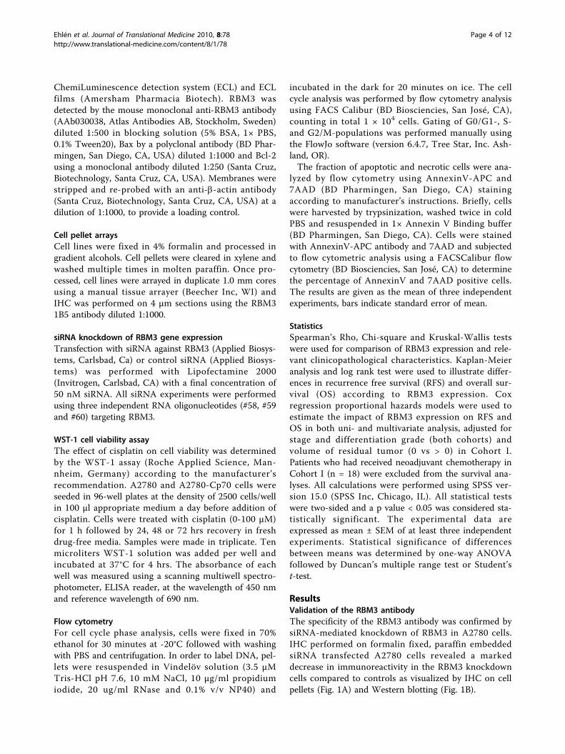
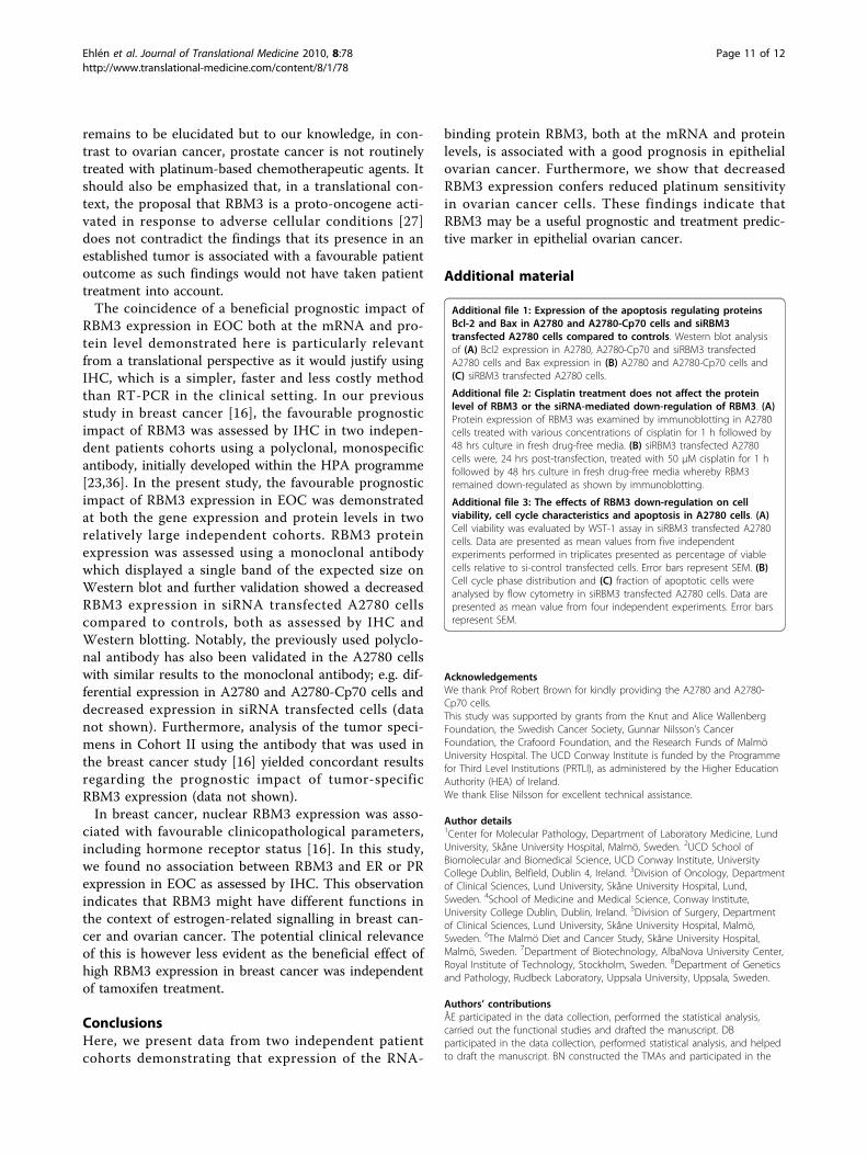
ResultsValidation of the RBM3 antibodyThe specificity of the RBM3 antibody was confirmed bysiRNA-mediated knockdown of RBM3 in A2780 cells.IHC performed on formalin fixed, paraffin embeddedsiRNA transfected A2780 cells revealed a markeddecrease in immunoreactivity in the RBM3 knockdowncells compared to controls as visualized by IHC on cellpellets (Fig. 1A) and Western blotting (Fig. 1B).
Ehlén et al. Journal of Translational Medicine 2010, 8:78http://www.translational-medicine.com/content/8/1/78
Page 4 of 12
RBM3 expression and association to clinicopathologicalcharacteristics in epithelial ovarian cancerHaving previously demonstrated that RBM3 was asso-ciated with a less aggressive breast cancer phenotype[16] we sought to examine the relationship betweenRBM3 mRNA and protein expression and clinicopatho-logical characteristics in two independent EOC cohorts.In Cohort I, increased RBM3 mRNA levels were notassociated with any clinicopathological characteristics(Table 1).In Cohort II, following antibody optimisation and stain-
ing, it was possible to evaluate the expression of RBM3protein in 151 cases (98%). Images representing differentpatterns of expression are shown in Figure 1C. Using thecombined score, 57 (38%) tumors lacked RBM3 nuclearRBM3 staining, and 94 (63%) tumors expressed RBM3 in
various intensities and fractions. For statistical purposes,tumors were grouped into negative = 0 (combined NS0-1), intermediate = 1 (combined NS 2-3) and strong = 2(combined NS > 3). As visualized in Table 1, RBM3 NSwas not associated with histological subtype, disease stageor differentiation grade. Cytoplasmic staining was onlypresent in 27 (18%) cases, and therefore not accountedfor in the statistics. There was no significant associationbetween RBM3 and ER or PR expression (data notshown).
Increased RBM3 mRNA levels and protein expression areassociated with a prolonged survival in ovarian cancerpatientsWe proceeded to investigate the relationship betweenRBM3 expression and clinical outcome. In Cohort I,
Figure 1 Specificity of the RBM3 antibody tested in A2780 ovarian cancer cells and immunohistochemical RBM3 expression in primaryovarian tumors. RBM3 protein expression was significantly decreased after transfection with siRNA against RBM3 in A2780 cells as shown by (A)immunocytochemistry 48 hrs post-transfection and (B) Western blot 48 and 72 hrs post-transfection. (C) Staining of RBM3 was denoted as (i)negative (nuclear score = 0), (ii) intermediate (nuclear score = 1-2) and (iii) strong (nuclear score > 2).
Ehlén et al. Journal of Translational Medicine 2010, 8:78http://www.translational-medicine.com/content/8/1/78
Page 5 of 12
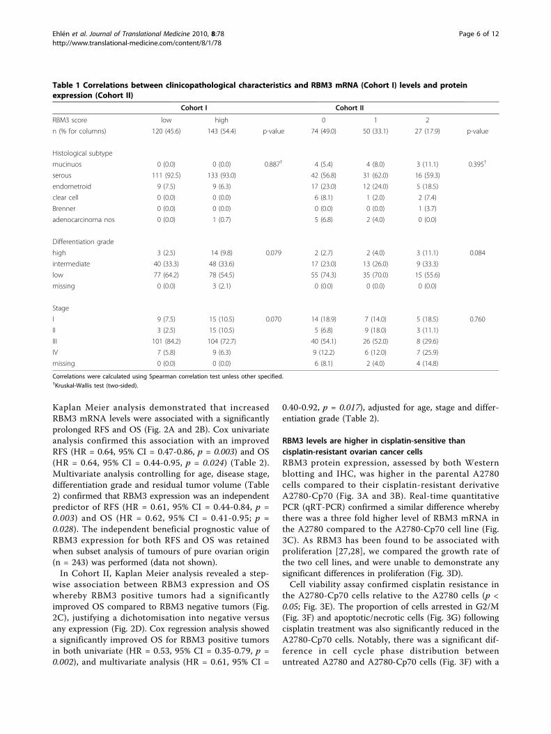
Kaplan Meier analysis demonstrated that increasedRBM3 mRNA levels were associated with a significantlyprolonged RFS and OS (Fig. 2A and 2B). Cox univariateanalysis confirmed this association with an improvedRFS (HR = 0.64, 95% CI = 0.47-0.86, p = 0.003) and OS(HR = 0.64, 95% CI = 0.44-0.95, p = 0.024) (Table 2).Multivariate analysis controlling for age, disease stage,differentiation grade and residual tumor volume (Table2) confirmed that RBM3 expression was an independentpredictor of RFS (HR = 0.61, 95% CI = 0.44-0.84, p =0.003) and OS (HR = 0.62, 95% CI = 0.41-0.95; p =0.028). The independent beneficial prognostic value ofRBM3 expression for both RFS and OS was retainedwhen subset analysis of tumours of pure ovarian origin(n = 243) was performed (data not shown).In Cohort II, Kaplan Meier analysis revealed a step-
wise association between RBM3 expression and OSwhereby RBM3 positive tumors had a significantlyimproved OS compared to RBM3 negative tumors (Fig.2C), justifying a dichotomisation into negative versusany expression (Fig. 2D). Cox regression analysis showeda significantly improved OS for RBM3 positive tumorsin both univariate (HR = 0.53, 95% CI = 0.35-0.79, p =0.002), and multivariate analysis (HR = 0.61, 95% CI =
0.40-0.92, p = 0.017), adjusted for age, stage and differ-entiation grade (Table 2).
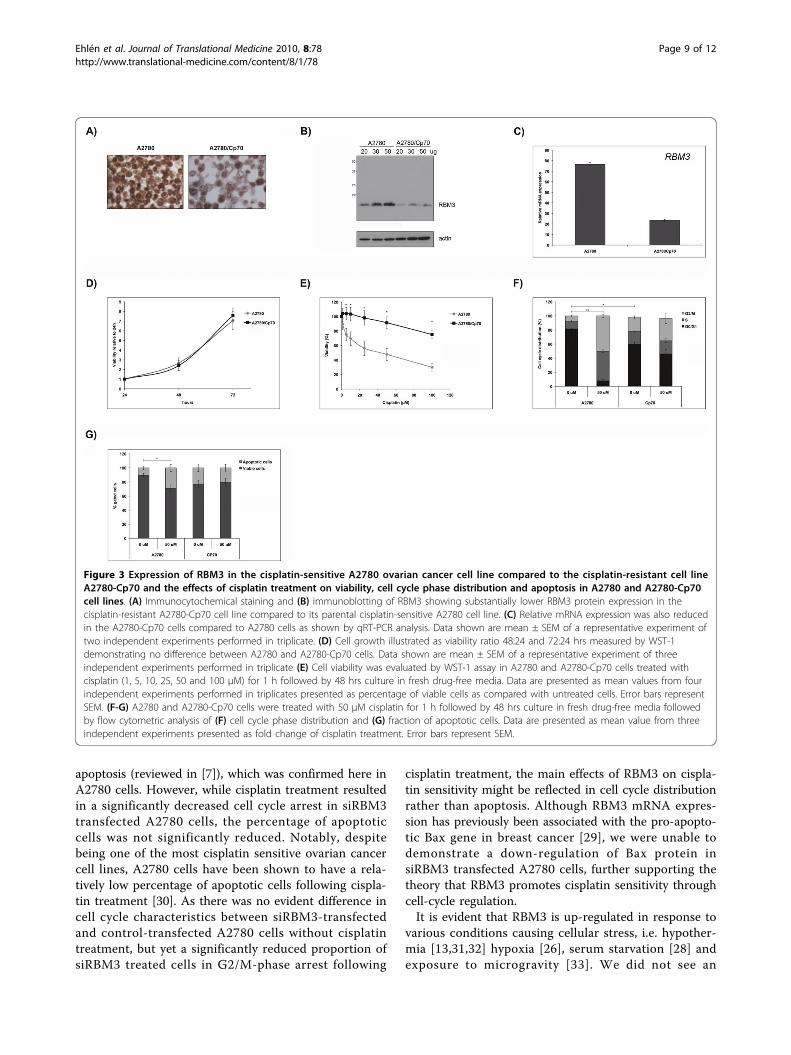
RBM3 levels are higher in cisplatin-sensitive thancisplatin-resistant ovarian cancer cellsRBM3 protein expression, assessed by both Westernblotting and IHC, was higher in the parental A2780cells compared to their cisplatin-resistant derivativeA2780-Cp70 (Fig. 3A and 3B). Real-time quantitativePCR (qRT-PCR) confirmed a similar difference wherebythere was a three fold higher level of RBM3 mRNA inthe A2780 compared to the A2780-Cp70 cell line (Fig.3C). As RBM3 has been found to be associated withproliferation [27,28], we compared the growth rate ofthe two cell lines, and were unable to demonstrate anysignificant differences in proliferation (Fig. 3D).Cell viability assay confirmed cisplatin resistance in
the A2780-Cp70 cells relative to the A2780 cells (p <0.05; Fig. 3E). The proportion of cells arrested in G2/M(Fig. 3F) and apoptotic/necrotic cells (Fig. 3G) followingcisplatin treatment was also significantly reduced in theA2780-Cp70 cells. Notably, there was a significant dif-ference in cell cycle phase distribution betweenuntreated A2780 and A2780-Cp70 cells (Fig. 3F) with a
Table 1 Correlations between clinicopathological characteristics and RBM3 mRNA (Cohort I) levels and proteinexpression (Cohort II)
Cohort I Cohort II
RBM3 score low high 0 1 2
n (% for columns) 120 (45.6) 143 (54.4) p-value 74 (49.0) 50 (33.1) 27 (17.9) p-value
Histological subtype
mucinuos 0 (0.0) 0 (0.0) 0.887† 4 (5.4) 4 (8.0) 3 (11.1) 0.395†
serous 111 (92.5) 133 (93.0) 42 (56.8) 31 (62.0) 16 (59.3)
endometroid 9 (7.5) 9 (6.3) 17 (23.0) 12 (24.0) 5 (18.5)
clear cell 0 (0.0) 0 (0.0) 6 (8.1) 1 (2.0) 2 (7.4)
Brenner 0 (0.0) 0 (0.0) 0 (0.0) 0 (0.0) 1 (3.7)
adenocarcinoma nos 0 (0.0) 1 (0.7) 5 (6.8) 2 (4.0) 0 (0.0)
Differentiation grade
high 3 (2.5) 14 (9.8) 0.079 2 (2.7) 2 (4.0) 3 (11.1) 0.084
intermediate 40 (33.3) 48 (33.6) 17 (23.0) 13 (26.0) 9 (33.3)
low 77 (64.2) 78 (54.5) 55 (74.3) 35 (70.0) 15 (55.6)
missing 0 (0.0) 3 (2.1) 0 (0.0) 0 (0.0) 0 (0.0)
Stage
I 9 (7.5) 15 (10.5) 0.070 14 (18.9) 7 (14.0) 5 (18.5) 0.760
II 3 (2.5) 15 (10.5) 5 (6.8) 9 (18.0) 3 (11.1)
III 101 (84.2) 104 (72.7) 40 (54.1) 26 (52.0) 8 (29.6)
IV 7 (5.8) 9 (6.3) 9 (12.2) 6 (12.0) 7 (25.9)
missing 0 (0.0) 0 (0.0) 6 (8.1) 2 (4.0) 4 (14.8)
Correlations were calculated using Spearman correlation test unless other specified.†Kruskal-Wallis test (two-sided).
Ehlén et al. Journal of Translational Medicine 2010, 8:78http://www.translational-medicine.com/content/8/1/78
Page 6 of 12
larger proportion of cells in G2/M in the A2780-Cp70cells, but no significant difference in apoptosis (Fig. 3G)was observed. However, there was a trend towards ahigher fraction of apoptotic cells in untreated A2780-Cp70 cells.
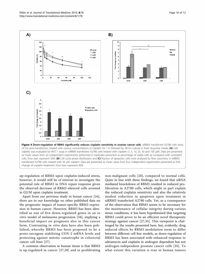
Downregulation of RBM3 significantly reduces cisplatinsensitivity in ovarian cancer cellsThe relationship between RBM3 and cisplatin responsewas then examined using siRNA mediated gene silen-cing, whereby the cisplatin-sensitive A2780 cells weretransfected with three different RBM3-specific siRNA:sor scrambled control siRNA prior to cisplatin treatment.Subsequent determination of cisplatin response as mea-sured by cell cytotoxicity assay and cell cycle analysisrevealed that RBM3 knockdown in A2780 cells
significantly decreased their sensitivity to cisplatin (Fig.4A). This effect was evident for three independentRBM3-specific siRNAs. Furthermore, cisplatin inducedG2/M arrest was significantly less pronounced insiRBM3 transfected A2780 cells compared to si-controltransfected cells (Fig. 4B). There was also a decreased,however non-significant, percentage of apoptotic cells insiRBM3 transfected cells compared to controls (Fig. 4C).Given the previously demonstrated relationship
between RBM3 and apoptosis-regulating proteins [29],we also compared the levels of Bcl-2 and Bax in siRBM3transfected cells and controls. In line with previous find-ings [30], Bcl-2 could barely be detected in the A2780cells while elevated levels were observed in A2780-Cp70cells. (Additional file 1A). In A2780-Cp70 cells, Baxlevels were lower than in A2780 cells, but Bax levels
Figure 2 Increased mRNA (Cohort I) and protein expression (Cohort II) of RBM3 are associated with a prolonged survival. Kaplan Meieranalysis of recurrence free survival (A) and overall survival (B) according to RBM3 mRNA levels in Cohort I. Kaplan Meier analysis of overallsurvival according to immunohistochemical RBM3 staining in Cohort II in strata defined as (C) negative, intermediate and strong expression and(D) negative versus positive expression.
Ehlén et al. Journal of Translational Medicine 2010, 8:78http://www.translational-medicine.com/content/8/1/78
Page 7 of 12
were not considerably altered by down-regulation ofRBM3 (Additional file 1B-C).Cisplatin treatment did not affect RBM3 protein
expression or the siRNA mediated down-regulation ofRBM3 (Additional file 2). The effects of RBM3 down-regulation in A2780 cells in the absence of cisplatinwere also investigated and, in agreement with previousstudies [27], siRBM3 transfected A2780 cells showed asignificantly reduced cell viability, a slightly higher pro-portion of cells in G2/M and no effect on apoptosis(Additional file 3).
DiscussionThis investigation of the prognostic value of RBM3 inEOC reveals that RBM3 is an independent prognosticmarker at both mRNA and protein levels. Gene expres-sion analysis in a cohort of 267 EOC cases showed thathigh RBM3 mRNA expression was an independent pre-dictor of a significantly improved RFS and OS. Immuno-histochemical analysis in an independent cohort of 154EOC cases demonstrated that RBM3 protein expressionwas associated with a significantly improved OS in uni-variate and multivariate analysis. This is in line withprevious findings from two independent breast cancercohorts where RBM3 was associated with more favour-able clinicopathological parameters and a significantlyimproved survival, irrespective of adjuvant treatment[16]. However, since platinum-based chemotherapy is afundamental aspect of current EOC treatment regimens,we hypothesized that RBM3 might enhance platinum-sensitivity in vitro. We initially confirmed lower RBM3
protein levels in the cisplatin-resistant ovarian cancercell line A2780-Cp70 compared to their parental cispla-tin-sensitive A2780 cells and using RNAi techniques, wedemonstrated that silencing of RBM3 led to a decreasedcisplatin response in ovarian cancer cells.Taken together, these data demonstrate that RBM3 is
a marker of good prognosis in EOC and a predictor ofresponse to platinum-based chemotherapy, most likely acombination of both, particularly in the light of the pre-viously demonstrated good prognosis associated withRBM3 expression in breast cancer patients, where thevast majority of patients received no adjuvant systemicchemotherapy [16]. While future in-depth studies arewarranted to further elucidate the functional mechan-isms underlying RBM3’s role in cisplatin-mediated celldeath, the in vitro data presented here provide sufficientevidence to support the hypothesis that, in addition tobeing a beneficial prognostic biomarker, RBM3 mightalso predict cisplatin response in EOC. Further studiesare required to evaluate the role of RBM3 in predictingresponse to other platinum based agents, particularly inthe setting of a prospective randomised control trialwhereby stratification according to different treatmentregimens can be performed.Some aspects on the results presented here merit
further attention. The reduced cytotoxic effect of cispla-tin in siRBM3 transfected cells was to a large extentreflected by cell cycle alterations, e.g. a lower percentageof cells arrested in G2/M phase rather than by adecreased percentage of apoptotic cells. Cisplatin treat-ment is known to induce both cell cycle arrest and
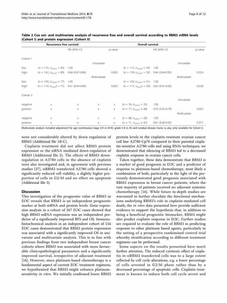
Table 2 Cox uni- and multivariate analysis of recurrence free and overall survival according to RBM3 mRNA levels(Cohort I) and protein expression (Cohort II)
Recurrence free survival Overall survival
HR (95% CI) p-value HR (95% CI) p-value
Cohort I
Univariate Univariate
low (n = 115, nevent = 85) 1.00 (n = 115, nevent = 54) 1.00
high (n = 142, nevent = 89) 0.64 (0.47-0.86) 0.003 (n = 139, nevent = 50) 0.64 (0.44-0.95) 0.024
Multivariate Multivariate
low (n = 103, nevent = 77) 1.00 (n = 103, nevent = 51) 1.00
high (n = 119, nevent = 71) 0.61 (0.44-0.84) 0.003 (n = 117, nevent = 50) 0.62 (0.41-0.95) 0.028
Cohort II
Univariate
negative x x x x (n = 74, nevent = 33) 1.00
positive x x x x (n = 77, nevent = 44) 0.53 (0.35-0.79) 0.002
Multivariate
negative x x x x (n = 68, nevent = 29) 1.00
positive x x x x (n = 71, nevent = 42) 0.61 (0.40-0.92) 0.017
Multivariate analysis included adjustment for age (continuous) stage (I-II vs III-IV), grade (I-II vs III) and residual disease (none vs any, only available for Cohort I).
Ehlén et al. Journal of Translational Medicine 2010, 8:78http://www.translational-medicine.com/content/8/1/78
Page 8 of 12
apoptosis (reviewed in [7]), which was confirmed here inA2780 cells. However, while cisplatin treatment resultedin a significantly decreased cell cycle arrest in siRBM3transfected A2780 cells, the percentage of apoptoticcells was not significantly reduced. Notably, despitebeing one of the most cisplatin sensitive ovarian cancercell lines, A2780 cells have been shown to have a rela-tively low percentage of apoptotic cells following cispla-tin treatment [30]. As there was no evident difference incell cycle characteristics between siRBM3-transfectedand control-transfected A2780 cells without cisplatintreatment, but yet a significantly reduced proportion ofsiRBM3 treated cells in G2/M-phase arrest following
cisplatin treatment, the main effects of RBM3 on cispla-tin sensitivity might be reflected in cell cycle distributionrather than apoptosis. Although RBM3 mRNA expres-sion has previously been associated with the pro-apopto-tic Bax gene in breast cancer [29], we were unable todemonstrate a down-regulation of Bax protein insiRBM3 transfected A2780 cells, further supporting thetheory that RBM3 promotes cisplatin sensitivity throughcell-cycle regulation.It is evident that RBM3 is up-regulated in response to
various conditions causing cellular stress, i.e. hypother-mia [13,31,32] hypoxia [26], serum starvation [28] andexposure to microgravity [33]. We did not see an
Figure 3 Expression of RBM3 in the cisplatin-sensitive A2780 ovarian cancer cell line compared to the cisplatin-resistant cell lineA2780-Cp70 and the effects of cisplatin treatment on viability, cell cycle phase distribution and apoptosis in A2780 and A2780-Cp70cell lines. (A) Immunocytochemical staining and (B) immunoblotting of RBM3 showing substantially lower RBM3 protein expression in thecisplatin-resistant A2780-Cp70 cell line compared to its parental cisplatin-sensitive A2780 cell line. (C) Relative mRNA expression was also reducedin the A2780-Cp70 cells compared to A2780 cells as shown by qRT-PCR analysis. Data shown are mean ± SEM of a representative experiment oftwo independent experiments performed in triplicate. (D) Cell growth illustrated as viability ratio 48:24 and 72:24 hrs measured by WST-1demonstrating no difference between A2780 and A2780-Cp70 cells. Data shown are mean ± SEM of a representative experiment of threeindependent experiments performed in triplicate (E) Cell viability was evaluated by WST-1 assay in A2780 and A2780-Cp70 cells treated withcisplatin (1, 5, 10, 25, 50 and 100 μM) for 1 h followed by 48 hrs culture in fresh drug-free media. Data are presented as mean values from fourindependent experiments performed in triplicates presented as percentage of viable cells as compared with untreated cells. Error bars representSEM. (F-G) A2780 and A2780-Cp70 cells were treated with 50 μM cisplatin for 1 h followed by 48 hrs culture in fresh drug-free media followedby flow cytometric analysis of (F) cell cycle phase distribution and (G) fraction of apoptotic cells. Data are presented as mean value from threeindependent experiments presented as fold change of cisplatin treatment. Error bars represent SEM.
Ehlén et al. Journal of Translational Medicine 2010, 8:78http://www.translational-medicine.com/content/8/1/78
Page 9 of 12
up-regulation of RBM3 upon cisplatin-induced stress,however, it would still be of interest to investigate thepotential role of RBM3 in DNA repair response giventhe observed decrease of RBM3-silenced cells arrestedin G2/M upon cisplatin treatment.Apart from our previous study in breast cancer [16],
there are to our knowledge no other published data onthe prognostic impact of tumor-specific RBM3 expres-sion in human cancer. However, RBM3 has been iden-tified as one of five down-regulated genes in an invitro model of melanoma progression [34], implying abeneficial impact on prognosis also in this cancerform. Contrasting in vitro data have also been pub-lished, whereby RBM3 has been proposed to be aproto-oncogene stabilizing COX-2 mRNA levels andprotecting against mitotic catastrophe in colorectalcancer cell lines [27].A common observation in human tissue is that RBM3
is up-regulated in cancer [27,28] and in proliferating
non-malignant cells [28], compared to normal cells.Quite in line with these findings, we found that siRNAmediated knockdown of RBM3 resulted in reduced pro-liferation in A2780 cells, which might in part explainthe reduced cisplatin sensitivity and also the relativelymodest reduction in apoptosis upon treatment insiRBM3 transfected A2780 cells. Yet, as a consequenceof the observation that RBM3 seems to be necessary forthe maintenance of cellular integrity during variousstress conditions, it has been hypothesized that targetingRBM3 could prove to be an efficient novel therapeuticstrategy against cancer [27,35]. This viewpoint is chal-lenged by the results presented here, but, evidently, druginduced effects by RBM3 modulation seem to differbetween different cell line models, as down-regulation ofRBM3 has been associated with enhanced response toadriamycin and cisplatin in androgen dependent but notandrogen-independent prostate cancer cells [35]. Towhat extent this variation is true in human tumors
Figure 4 Down-regulation of RBM3 significantly reduces cisplatin sensitivity in ovarian cancer cells. siRBM3 transfected A2780 cells were,24 hrs post-transfection, treated with various concentrations of cisplatin for 1 h followed by 48 hrs culture in fresh drug-free media. (A) Cellviability was evaluated by WST-1 assay in siRBM3 transfected A2780 cells treated with cisplatin (1, 5, 10, 25, 50 and 100 μM). Data are presentedas mean values from six independent experiments performed in triplicates presented as percentage of viable cells as compared with untreatedcells. Error bars represent SEM. (B) Cell cycle phase distribution and (C) fraction of apoptotic cells were analysed by flow cytometry in siRBM3transfected A2780 cells treated with 50 μM cisplatin. Data are presented as mean value from four independent experiments presented as foldchange of cisplatin treatment. Error bars represent SEM.
Ehlén et al. Journal of Translational Medicine 2010, 8:78http://www.translational-medicine.com/content/8/1/78
Page 10 of 12
remains to be elucidated but to our knowledge, in con-trast to ovarian cancer, prostate cancer is not routinelytreated with platinum-based chemotherapeutic agents. Itshould also be emphasized that, in a translational con-text, the proposal that RBM3 is a proto-oncogene acti-vated in response to adverse cellular conditions [27]does not contradict the findings that its presence in anestablished tumor is associated with a favourable patientoutcome as such findings would not have taken patienttreatment into account.The coincidence of a beneficial prognostic impact of
RBM3 expression in EOC both at the mRNA and pro-tein level demonstrated here is particularly relevantfrom a translational perspective as it would justify usingIHC, which is a simpler, faster and less costly methodthan RT-PCR in the clinical setting. In our previousstudy in breast cancer [16], the favourable prognosticimpact of RBM3 was assessed by IHC in two indepen-dent patients cohorts using a polyclonal, monospecificantibody, initially developed within the HPA programme[23,36]. In the present study, the favourable prognosticimpact of RBM3 expression in EOC was demonstratedat both the gene expression and protein levels in tworelatively large independent cohorts. RBM3 proteinexpression was assessed using a monoclonal antibodywhich displayed a single band of the expected size onWestern blot and further validation showed a decreasedRBM3 expression in siRNA transfected A2780 cellscompared to controls, both as assessed by IHC andWestern blotting. Notably, the previously used polyclo-nal antibody has also been validated in the A2780 cellswith similar results to the monoclonal antibody; e.g. dif-ferential expression in A2780 and A2780-Cp70 cells anddecreased expression in siRNA transfected cells (datanot shown). Furthermore, analysis of the tumor speci-mens in Cohort II using the antibody that was used inthe breast cancer study [16] yielded concordant resultsregarding the prognostic impact of tumor-specificRBM3 expression (data not shown).In breast cancer, nuclear RBM3 expression was asso-
ciated with favourable clinicopathological parameters,including hormone receptor status [16]. In this study,we found no association between RBM3 and ER or PRexpression in EOC as assessed by IHC. This observationindicates that RBM3 might have different functions inthe context of estrogen-related signalling in breast can-cer and ovarian cancer. The potential clinical relevanceof this is however less evident as the beneficial effect ofhigh RBM3 expression in breast cancer was independentof tamoxifen treatment.
ConclusionsHere, we present data from two independent patientcohorts demonstrating that expression of the RNA-
binding protein RBM3, both at the mRNA and proteinlevels, is associated with a good prognosis in epithelialovarian cancer. Furthermore, we show that decreasedRBM3 expression confers reduced platinum sensitivityin ovarian cancer cells. These findings indicate thatRBM3 may be a useful prognostic and treatment predic-tive marker in epithelial ovarian cancer.
Additional material
Additional file 1: Expression of the apoptosis regulating proteinsBcl-2 and Bax in A2780 and A2780-Cp70 cells and siRBM3transfected A2780 cells compared to controls. Western blot analysisof (A) Bcl2 expression in A2780, A2780-Cp70 and siRBM3 transfectedA2780 cells and Bax expression in (B) A2780 and A2780-Cp70 cells and(C) siRBM3 transfected A2780 cells.
Additional file 2: Cisplatin treatment does not affect the proteinlevel of RBM3 or the siRNA-mediated down-regulation of RBM3. (A)Protein expression of RBM3 was examined by immunoblotting in A2780cells treated with various concentrations of cisplatin for 1 h followed by48 hrs culture in fresh drug-free media. (B) siRBM3 transfected A2780cells were, 24 hrs post-transfection, treated with 50 μM cisplatin for 1 hfollowed by 48 hrs culture in fresh drug-free media whereby RBM3remained down-regulated as shown by immunoblotting.
Additional file 3: The effects of RBM3 down-regulation on cellviability, cell cycle characteristics and apoptosis in A2780 cells. (A)Cell viability was evaluated by WST-1 assay in siRBM3 transfected A2780cells. Data are presented as mean values from five independentexperiments performed in triplicates presented as percentage of viablecells relative to si-control transfected cells. Error bars represent SEM. (B)Cell cycle phase distribution and (C) fraction of apoptotic cells wereanalysed by flow cytometry in siRBM3 transfected A2780 cells. Data arepresented as mean value from four independent experiments. Error barsrepresent SEM.
AcknowledgementsWe thank Prof Robert Brown for kindly providing the A2780 and A2780-Cp70 cells.This study was supported by grants from the Knut and Alice WallenbergFoundation, the Swedish Cancer Society, Gunnar Nilsson’s CancerFoundation, the Crafoord Foundation, and the Research Funds of MalmöUniversity Hospital. The UCD Conway Institute is funded by the Programmefor Third Level Institutions (PRTLI), as administered by the Higher EducationAuthority (HEA) of Ireland.We thank Elise Nilsson for excellent technical assistance.
Author details1Center for Molecular Pathology, Department of Laboratory Medicine, LundUniversity, Skåne University Hospital, Malmö, Sweden. 2UCD School ofBiomolecular and Biomedical Science, UCD Conway Institute, UniversityCollege Dublin, Belfield, Dublin 4, Ireland. 3Division of Oncology, Departmentof Clinical Sciences, Lund University, Skåne University Hospital, Lund,Sweden. 4School of Medicine and Medical Science, Conway Institute,University College Dublin, Dublin, Ireland. 5Division of Surgery, Departmentof Clinical Sciences, Lund University, Skåne University Hospital, Malmö,Sweden. 6The Malmö Diet and Cancer Study, Skåne University Hospital,Malmö, Sweden. 7Department of Biotechnology, AlbaNova University Center,Royal Institute of Technology, Stockholm, Sweden. 8Department of Geneticsand Pathology, Rudbeck Laboratory, Uppsala University, Uppsala, Sweden.
Authors’ contributionsÅE participated in the data collection, performed the statistical analysis,carried out the functional studies and drafted the manuscript. DBparticipated in the data collection, performed statistical analysis, and helpedto draft the manuscript. BN constructed the TMAs and participated in the
Ehlén et al. Journal of Translational Medicine 2010, 8:78http://www.translational-medicine.com/content/8/1/78
Page 11 of 12
data collection. DPO participated in the data collection. JE participated inthe design of the study and helped draft the manuscript. MAK assisted withthe data collection and helped draft the manuscript. IBJ assisted with thestatistical analysis. JM assisted with data collection and helped to draft themanuscript. JB assisted with collection of clinical data. MU assisted with datacollection and participated in its design. FP assisted with data collection andhelped to draft the manuscript. KJ conceived of the study, participated in itsdesign and coordination and helped to draft the manuscript. All authorsread and approved the final manuscript.
Competing interestsThe authors declare that they have no competing interests.
Received: 14 April 2010 Accepted: 20 August 2010Published: 20 August 2010
References1. Jemal A, Siegel R, Ward E, Hao Y, Xu J, Thun MJ: Cancer statistics, 2009. CA
Cancer J Clin 2009, 59:225-249.2. Kelland L: The resurgence of platinum-based cancer chemotherapy. Nat
Rev Cancer 2007, 7:573-584.3. Han ES, Lin P, Wakabayashi M: Current status on biologic therapies in the
treatment of epithelial ovarian cancer. Curr Treat Options Oncol 2009,10:54-66.
4. Tummala MK, McGuire WP: Recurrent ovarian cancer. Clin Adv HematolOncol 2005, 3:723-736.
5. Siddik ZH: Cisplatin: mode of cytotoxic action and molecular basis ofresistance. Oncogene 2003, 22:7265-7279.
6. Jamieson ER, Lippard SJ: Structure, Recognition, and Processing ofCisplatin-DNA Adducts. Chem Rev 1999, 99:2467-2498.
7. Wang D, Lippard SJ: Cellular processing of platinum anticancer drugs.Nat Rev Drug Discov 2005, 4:307-320.
8. Wright CF, Oswald BW, Dellis S: Vaccinia virus late transcription isactivated in vitro by cellular heterogeneous nuclear ribonucleoproteins.J Biol Chem 2001, 276:40680-40686.
9. Burd CG, Dreyfuss G: Conserved structures and diversity of functions ofRNA-binding proteins. Science 1994, 265:615-621.
10. Sutherland LC, Rintala-Maki ND, White RD, Morin CD: RNA binding motif(RBM) proteins: a novel family of apoptosis modulators? J Cell Biochem2005, 94:5-24.
11. Kita H, Carmichael J, Swartz J, Muro S, Wyttenbach A, Matsubara K,Rubinsztein DC, Kato K: Modulation of polyglutamine-induced cell deathby genes identified by expression profiling. Hum Mol Genet 2002,11:2279-2287.
12. Derry JM, Kerns JA, Francke U: RBM3, a novel human gene in Xp11.23with a putative RNA-binding domain. Hum Mol Genet 1995, 4:2307-2311.
13. Danno S, Nishiyama H, Higashitsuji H, Yokoi H, Xue JH, Itoh K, Matsuda T,Fujita J: Increased transcript level of RBM3, a member of the glycine-richRNA-binding protein family, in human cells in response to cold stress.Biochem Biophys Res Commun 1997, 236:804-807.
14. Ponten F, Jirstrom K, Uhlen M: The Human Protein Atlas–a tool forpathology. J Pathol 2008, 216:387-393.
15. Bjorling E, Lindskog C, Oksvold P, Linne J, Kampf C, Hober S, Uhlen M,Ponten F: A web-based tool for in silico biomarker discovery based ontissue-specific protein profiles in normal and cancer tissues. Mol CellProteomics 2008, 7:825-844.
16. Jogi A, Brennan DJ, Ryden L, Magnusson K, Ferno M, Stal O, Borgquist S,Uhlen M, Landberg G, Pahlman S, et al: Nuclear expression of the RNA-binding protein RBM3 is associated with an improved clinical outcomein breast cancer. Mod Pathol 2009, 22:1564-1574.
17. Tothill RW, Tinker AV, George J, Brown R, Fox SB, Lade S, Johnson DS,Trivett MK, Etemadmoghadam D, Locandro B, et al: Novel molecularsubtypes of serous and endometrioid ovarian cancer linked to clinicaloutcome. Clin Cancer Res 2008, 14:5198-5208.
18. Bolstad BM, Irizarry RA, Astrand M, Speed TP: A comparison ofnormalization methods for high density oligonucleotide array databased on variance and bias. Bioinformatics 2003, 19:185-193.
19. Moody SE, Perez D, Pan TC, Sarkisian CJ, Portocarrero CP, Sterner CJ,Notorfrancesco KL, Cardiff RD, Chodosh LA: The transcriptional repressorSnail promotes mammary tumor recurrence. Cancer Cell 2005, 8:197-209.
20. Berglund G, Elmstahl S, Janzon L, Larsson SA: The Malmo Diet and CancerStudy. Design and feasibility. J Intern Med 1993, 233:45-51.
21. Berglund G, Eriksson KF, Israelsson B, Kjellstrom T, Lindgarde F, Mattiasson I,Nilsson JA, Stavenow L: Cardiovascular risk groups and mortality in anurban swedish male population: the Malmo Preventive Project. J InternMed 1996, 239:489-497.
22. Kononen J, Bubendorf L, Kallioniemi A, Barlund M, Schraml P, Leighton S,Torhorst J, Mihatsch MJ, Sauter G, Kallioniemi OP: Tissue microarrays forhigh-throughput molecular profiling of tumor specimens. Nat Med 1998,4:844-847.
23. Nilsson P, Paavilainen L, Larsson K, Odling J, Sundberg M, Andersson AC,Kampf C, Persson A, Al-Khalili Szigyarto C, Ottosson J, et al: Towards ahuman proteome atlas: high-throughput generation of mono-specificantibodies for tissue profiling. Proteomics 2005, 5:4327-4337.
24. Berglund L, Bjorling E, Jonasson K, Rockberg J, Fagerberg L, Al-KhaliliSzigyarto C, Sivertsson A, Uhlen M: A whole-genome bioinformaticsapproach to selection of antigens for systematic antibody generation.Proteomics 2008, 8:2832-2839.
25. Lofstedt T, Jogi A, Sigvardsson M, Gradin K, Poellinger L, Pahlman S,Axelson H: Induction of ID2 expression by hypoxia-inducible factor-1: arole in dedifferentiation of hypoxic neuroblastoma cells. J Biol Chem2004, 279:39223-39231.
26. Wellmann S, Buhrer C, Moderegger E, Zelmer A, Kirschner R, Koehne P,Fujita J, Seeger K: Oxygen-regulated expression of the RNA-bindingproteins RBM3 and CIRP by a HIF-1-independent mechanism. J Cell Sci2004, 117:1785-1794.
27. Sureban SM, Ramalingam S, Natarajan G, May R, Subramaniam D,Bishnupuri KS, Morrison AR, Dieckgraefe BK, Brackett DJ, Postier RG, et al:Translation regulatory factor RBM3 is a proto-oncogene that preventsmitotic catastrophe. Oncogene 2008, 27:4544-4556.
28. Wellmann S, Truss M, Bruder E, Tornillo L, Zelmer A, Seeger K, Buhrer C: TheRNA-binding protein RBM3 is required for cell proliferation and protectsagainst serum deprivation-induced cell death. Pediatr Res 2010, 67:35-41.
29. Martinez-Arribas F, Agudo D, Pollan M, Gomez-Esquer F, Diaz-Gil G, Lucas R,Schneider J: Positive correlation between the expression of X-chromosome RBM genes (RBMX, RBM3, RBM10) and the proapoptoticBax gene in human breast cancer. J Cell Biochem 2006, 97:1275-1282.
30. Kolfschoten GM, Hulscher TM, Schrier SM, van Houten VM, Pinedo HM,Boven E: Time-dependent changes in factors involved in the apoptoticprocess in human ovarian cancer cells as a response to cisplatin. GynecolOncol 2002, 84:404-412.
31. Danno S, Itoh K, Matsuda T, Fujita J: Decreased expression of mouseRbm3, a cold-shock protein, in Sertoli cells of cryptorchid testis. Am JPathol 2000, 156:1685-1692.
32. Williams DR, Epperson LE, Li W, Hughes MA, Taylor R, Rogers J, Martin SL,Cossins AR, Gracey AY: Seasonally hibernating phenotype assessedthrough transcript screening. Physiol Genomics 2005, 24:13-22.
33. Lebsack TW, Fa V, Woods CC, Gruener R, Manziello AM, Pecaut MJ,Gridley DS, Stodieck LS, Ferguson VL, Deluca D: Microarray analysis ofspaceflown murine thymus tissue reveals changes in gene expressionregulating stress and glucocorticoid receptors. J Cell Biochem 2010,110(2):372-81.
34. Baldi A, Battista T, De Luca A, Santini D, Rossiello L, Baldi F, Natali PG,Lombardi D, Picardo M, Felsani A, Paggi MG: Identification of genes down-regulated during melanoma progression: a cDNA array study. ExpDermatol 2003, 12:213-218.
35. Zeng Y, Kulkarni P, Inoue T, Getzenberg RH: Down-regulating cold shockprotein genes impairs cancer cell survival and enhanceschemosensitivity. J Cell Biochem 2009, 107:179-188.
36. Uhlen M, Bjorling E, Agaton C, Szigyarto CA, Amini B, Andersen E,Andersson AC, Angelidou P, Asplund A, Asplund C, et al: A human proteinatlas for normal and cancer tissues based on antibody proteomics. MolCell Proteomics 2005, 4:1920-1932.
doi:10.1186/1479-5876-8-78Cite this article as: Ehlén et al.: Expression of the RNA-binding proteinRBM3 is associated with a favourable prognosis and cisplatin sensitivityin epithelial ovarian cancer. Journal of Translational Medicine 2010 8:78.
Ehlén et al. Journal of Translational Medicine 2010, 8:78http://www.translational-medicine.com/content/8/1/78
Page 12 of 12