Research Article Exploring the Oxidative Stress Mechanism of ...
29
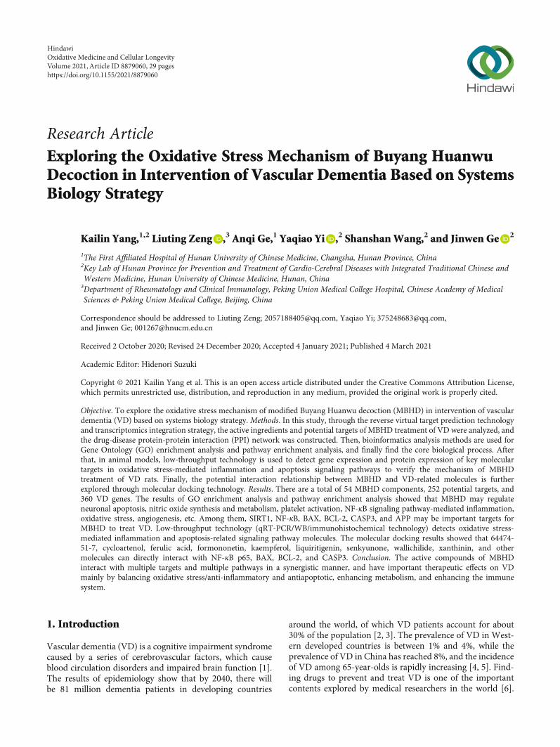
Research Article Exploring the Oxidative Stress Mechanism of Buyang Huanwu Decoction in Intervention of Vascular Dementia Based on Systems Biology Strategy Kailin Yang, 1,2 Liuting Zeng , 3 Anqi Ge, 1 Yaqiao Yi , 2 Shanshan Wang, 2 and Jinwen Ge 2 1 The First Affiliated Hospital of Hunan University of Chinese Medicine, Changsha, Hunan Province, China 2 Key Lab of Hunan Province for Prevention and Treatment of Cardio-Cerebral Diseases with Integrated Traditional Chinese and Western Medicine, Hunan University of Chinese Medicine, Hunan, China 3 Department of Rheumatology and Clinical Immunology, Peking Union Medical College Hospital, Chinese Academy of Medical Sciences & Peking Union Medical College, Beijing, China Correspondence should be addressed to Liuting Zeng; [email protected], Yaqiao Yi; [email protected], and Jinwen Ge; [email protected] Received 2 October 2020; Revised 24 December 2020; Accepted 4 January 2021; Published 4 March 2021 Academic Editor: Hidenori Suzuki Copyright © 2021 Kailin Yang et al. This is an open access article distributed under the Creative Commons Attribution License, which permits unrestricted use, distribution, and reproduction in any medium, provided the original work is properly cited. Objective. To explore the oxidative stress mechanism of modified Buyang Huanwu decoction (MBHD) in intervention of vascular dementia (VD) based on systems biology strategy. Methods. In this study, through the reverse virtual target prediction technology and transcriptomics integration strategy, the active ingredients and potential targets of MBHD treatment of VD were analyzed, and the drug-disease protein-protein interaction (PPI) network was constructed. Then, bioinformatics analysis methods are used for Gene Ontology (GO) enrichment analysis and pathway enrichment analysis, and finally find the core biological process. After that, in animal models, low-throughput technology is used to detect gene expression and protein expression of key molecular targets in oxidative stress-mediated inflammation and apoptosis signaling pathways to verify the mechanism of MBHD treatment of VD rats. Finally, the potential interaction relationship between MBHD and VD-related molecules is further explored through molecular docking technology. Results. There are a total of 54 MBHD components, 252 potential targets, and 360 VD genes. The results of GO enrichment analysis and pathway enrichment analysis showed that MBHD may regulate neuronal apoptosis, nitric oxide synthesis and metabolism, platelet activation, NF-κB signaling pathway-mediated inflammation, oxidative stress, angiogenesis, etc. Among them, SIRT1, NF-κB, BAX, BCL-2, CASP3, and APP may be important targets for MBHD to treat VD. Low-throughput technology (qRT-PCR/WB/immunohistochemical technology) detects oxidative stress- mediated inflammation and apoptosis-related signaling pathway molecules. The molecular docking results showed that 64474- 51-7, cycloartenol, ferulic acid, formononetin, kaempferol, liquiritigenin, senkyunone, wallichilide, xanthinin, and other molecules can directly interact with NF-κB p65, BAX, BCL-2, and CASP3. Conclusion. The active compounds of MBHD interact with multiple targets and multiple pathways in a synergistic manner, and have important therapeutic effects on VD mainly by balancing oxidative stress/anti-inflammatory and antiapoptotic, enhancing metabolism, and enhancing the immune system. 1. Introduction Vascular dementia (VD) is a cognitive impairment syndrome caused by a series of cerebrovascular factors, which cause blood circulation disorders and impaired brain function [1]. The results of epidemiology show that by 2040, there will be 81 million dementia patients in developing countries around the world, of which VD patients account for about 30% of the population [2, 3]. The prevalence of VD in West- ern developed countries is between 1% and 4%, while the prevalence of VD in China has reached 8%, and the incidence of VD among 65-year-olds is rapidly increasing [4, 5]. Find- ing drugs to prevent and treat VD is one of the important contents explored by medical researchers in the world [6]. Hindawi Oxidative Medicine and Cellular Longevity Volume 2021, Article ID 8879060, 29 pages https://doi.org/10.1155/2021/8879060
-
Upload
khangminh22 -
Category
Documents
-
view
3 -
download
0
Transcript of Research Article Exploring the Oxidative Stress Mechanism of ...
Research ArticleExploring the Oxidative Stress Mechanism of Buyang HuanwuDecoction in Intervention of Vascular Dementia Based on SystemsBiology Strategy
Kailin Yang,1,2 Liuting Zeng ,3 Anqi Ge,1 Yaqiao Yi ,2 ShanshanWang,2 and Jinwen Ge 2
1The First Affiliated Hospital of Hunan University of Chinese Medicine, Changsha, Hunan Province, China2Key Lab of Hunan Province for Prevention and Treatment of Cardio-Cerebral Diseases with Integrated Traditional Chinese andWestern Medicine, Hunan University of Chinese Medicine, Hunan, China3Department of Rheumatology and Clinical Immunology, Peking Union Medical College Hospital, Chinese Academy of MedicalSciences & Peking Union Medical College, Beijing, China
Correspondence should be addressed to Liuting Zeng; [email protected], Yaqiao Yi; [email protected],and Jinwen Ge; [email protected]
Received 2 October 2020; Revised 24 December 2020; Accepted 4 January 2021; Published 4 March 2021
Academic Editor: Hidenori Suzuki
Copyright © 2021 Kailin Yang et al. This is an open access article distributed under the Creative Commons Attribution License,which permits unrestricted use, distribution, and reproduction in any medium, provided the original work is properly cited.
Objective. To explore the oxidative stress mechanism of modified Buyang Huanwu decoction (MBHD) in intervention of vasculardementia (VD) based on systems biology strategy.Methods. In this study, through the reverse virtual target prediction technologyand transcriptomics integration strategy, the active ingredients and potential targets of MBHD treatment of VD were analyzed, andthe drug-disease protein-protein interaction (PPI) network was constructed. Then, bioinformatics analysis methods are used forGene Ontology (GO) enrichment analysis and pathway enrichment analysis, and finally find the core biological process. Afterthat, in animal models, low-throughput technology is used to detect gene expression and protein expression of key moleculartargets in oxidative stress-mediated inflammation and apoptosis signaling pathways to verify the mechanism of MBHDtreatment of VD rats. Finally, the potential interaction relationship between MBHD and VD-related molecules is furtherexplored through molecular docking technology. Results. There are a total of 54 MBHD components, 252 potential targets, and360 VD genes. The results of GO enrichment analysis and pathway enrichment analysis showed that MBHD may regulateneuronal apoptosis, nitric oxide synthesis and metabolism, platelet activation, NF-κB signaling pathway-mediated inflammation,oxidative stress, angiogenesis, etc. Among them, SIRT1, NF-κB, BAX, BCL-2, CASP3, and APP may be important targets forMBHD to treat VD. Low-throughput technology (qRT-PCR/WB/immunohistochemical technology) detects oxidative stress-mediated inflammation and apoptosis-related signaling pathway molecules. The molecular docking results showed that 64474-51-7, cycloartenol, ferulic acid, formononetin, kaempferol, liquiritigenin, senkyunone, wallichilide, xanthinin, and othermolecules can directly interact with NF-κB p65, BAX, BCL-2, and CASP3. Conclusion. The active compounds of MBHDinteract with multiple targets and multiple pathways in a synergistic manner, and have important therapeutic effects on VDmainly by balancing oxidative stress/anti-inflammatory and antiapoptotic, enhancing metabolism, and enhancing the immunesystem.
1. Introduction
Vascular dementia (VD) is a cognitive impairment syndromecaused by a series of cerebrovascular factors, which causeblood circulation disorders and impaired brain function [1].The results of epidemiology show that by 2040, there willbe 81 million dementia patients in developing countries
around the world, of which VD patients account for about30% of the population [2, 3]. The prevalence of VD in West-ern developed countries is between 1% and 4%, while theprevalence of VD in China has reached 8%, and the incidenceof VD among 65-year-olds is rapidly increasing [4, 5]. Find-ing drugs to prevent and treat VD is one of the importantcontents explored by medical researchers in the world [6].
HindawiOxidative Medicine and Cellular LongevityVolume 2021, Article ID 8879060, 29 pageshttps://doi.org/10.1155/2021/8879060
Modified Buyang Huanwu decoction (MBHD) consists ofHedysarum multijugum Maxim. (Huang Qi), Angelicaesinensis Radix [Dang Gui or Angelica sinensis (Oliv.) Diels],Pheretima Aspergillum (E. Perrier) (Di Long or Pheretima),Chuanxiong Rhizoma (Chuan Xiong or Ligusticum striatumDC.), Acoritataninowii Rhizoma (Shi Chang Pu or Acoruscalamus var. angustatus Besser), Panax notoginseng (Burk.)F. H. Chen ex C. Chow (San Qi), and Polygalae Radix (YuanZhi or Polygala tenuifolia Willd.) with a ratio of120 : 15 : 15 : 10 : 10 : 9 : 10 [7]. Clinical studies have shownthat MBHD can prevent and interfere with vascular dementia[8], and its mechanism may be achieved by participating inthe reconstruction of synapses and enhancing the expressionof LTP [8]. The prognosis of VD is related to the underlyingdisease causing vascular damage and the location of intracra-nial vascular disease. By improving cerebral circulation andpreventing the recurrence of cerebrovascular diseases, symp-toms can be reduced and the disease can be prevented fromfurther deterioration [9]. In cerebrovascular diseases, MBHDhas a good anti-ischemic effect, which can reduce the area ofcerebral infarction in animal models of cerebral ischemia andimprove the neurological dysfunction after cerebral ischemia[10]. MBHD can also promote angiogenesis and inhibit neu-ronal apoptosis and improve the learning and memory abili-ties of VD rats [11–14]. However, the specific mechanism ofMBHD to improve VD needs further study.
Currently, network pharmacology is considered as a newmodel for drug development [15, 16]. This method can ana-lyze and organize information and data in existing databases(genes, diseases, drug network libraries, etc.), and use profes-sional network analysis software to build a “drug-target-disease” interaction network [17]. In addition, through theuse of network analysis methods, network pharmacologycan systematically construct a disease network for drug inter-vention, comprehensively analyze the relationship betweendrug targets and disease targets, and evaluate the efficacyand side effects of drugs on this basis. Because traditionalChinese medicine (TCM) has the characteristics of multiplecomponents, multiple targets, and multiple channels, it isalso researched on a systematic and overall level, so theresearch ideas of network pharmacology have the same goalsas TCM prescriptions [18]. Network pharmacology providesnew ideas for the modernization and development of TCM[19, 20]. Therefore, in this study, bilateral common carotidartery ligation (2-VO) was used to replicate the VD ratmodel, and the microarray analysis technique (including net-work pharmacology) was used to explore the protective effectof MBHD on the hippocampal neurons of the VD model ratand its molecular mechanism.
2. Materials and Methods
2.1. Potential Components and Potential Targets Predictionand VD-Related Gene Collection. The compound informa-tion of MBHD comes from TCMSP (http://lsp.nwu.edu.cn/tcmsp.php) [21], TCM@Taiwan (http://tcm.cmu.edu.tw/zh-tw/) [22], and TCMID (http://www.megabionet. org/tcmid/)[23]. The TCMSP database was used to provide compoundsrelated to the pharmacokinetic properties including oral
bioavailability (OB), drug-likeness properties (DL), andCaco-2 parameters to predict the components of MBHD[21]. The standard was OB ≥ 30%, Caco − 2 > −0:4, and DL≥ 0:18. In addition, due to the limitations of pharmacoki-netic prediction, this study retrieved a large amount of liter-ature and included oral absorbable pharmacologically activecompounds for supplementation [24–27].
The predicted compounds are standardized by PubChem(https://pubchem.ncbi.nlm.nih.gov/) to obtain their SMILESformula. Those SMILES formulas of the components wereinput into the SwissTargetPrediction Database (http://www.swisstargetprediction.ch/) to predict the potential targets ofeach component [28]. The OMIM database (http://omim.org/) [29] and GeneCards (http://www.genecards.org) [30]were utilized to collect the VD-related disease genes andtargets. In GeneCards, the targets with relevant score ≥ 1:0were selected for sequence research.
2.2. Network Construction and Analysis Methods. In molecu-lar biology, the interactive gene/protein search tool STRING(https://string-db.org/) searches and predicts gene/proteininteractions based on biological databases and web resources[31]. Protein-protein interaction (PPI) network is an impor-tant tool for the system to understand cellular processes. Thisnetwork can be used to filter and evaluate functional genomicdata, and provide a visual platform for annotating the struc-ture and function of proteins [31]. In this study, the onlinedatabase STRING was used to obtain the PPI data of MBHDpotential targets and VD genes, and Cytoscape v3.7.2 wasused to construct a visual PPI network diagram. In the PPInetwork, the closely connected part of the nodes is calledcluster. The cluster module analysis of the PPI network canprovide information for studying the pathogenesis of diseasesand the mechanism of drugs [32]. In this study, the MCODE,a plug-in of Cytoscape, was used to analyze the PPI networksto detect the clusters. Finally, the genes and targets in clustersor in PPI networks were input into DAVID (https://david-d.ncifcrf.gov) for Gene Ontology (GO) enrichment analysisand pathway enrichment analysis.
2.3. Experimental Materials
2.3.1. Experimental Animal. One hundred and twenty (120)male specific pathogen-free (SPF) grade SD rats were pur-chased from Hunan Slack Jingda Experimental Animal Co.,Ltd. (experimental animal production license number isSCXK (xiang) 2013-0004), weighing 250-300 g and raised inthe Experimental Animal Center of Hunan University ofChinese Medicine. The breeding environment is at roomtemperature (24 ± 1) °C, relative humidity (50 ± 5)%, and12 h/12 h alternate day and night. The rats started the exper-iment after 2 weeks of adaptive feeding. The entire experi-mental procedure was approved by the ethics committee ofHunan University of Chinese Medicine (HCM-00150023).
2.3.2. Experimental Drugs.Modified Buyang Huanwu decoc-tion (MBHD) consists of Hedysarum multijugum Maxim.(Huang Qi; specimen number: 2015076211), Angelicaesinensis Radix (Dang Gui or Angelica sinensis (Oliv.) Diels;specimen number: 201504212), Pheretima Aspergillum
2 Oxidative Medicine and Cellular Longevity
(E.Perrier) (Di Long or Pheretima; specimen number:2015035123), Chuanxiong Rhizoma (Chuan Xiong or Ligus-ticum striatum DC.; specimen number: 2015074430), Acori-tataninowii Rhizoma (Shi Chang Pu or Acorus calamus var.angustatus Besser; specimen number: 201407210), Panaxnotoginseng (Burk.) F.H. Chen ex C. Chow (San Qi; specimennumber: 201505792), Polygalae Radix (Yuan Zhi or Polygalatenuifolia Willd.; specimen number: 201504212) with a ratioof 120 : 15 : 15 : 10 : 10 : 9 : 10. All herbs are certified by Associ-ate Professor Liu Lin of Hunan University of Chinese Medi-cine to be authentic products specified in the 2015 edition ofPharmacopoeia of the People’s Republic of China. The equiv-alent dose of MBHD rats is 17.01 g/kg through the conver-sion of the human/mouse coefficient. Oxiracetam capsuleswere purchased from CSPC. The clinical equivalent dosefor rats is 0.216 g/kg.
2.3.3. Preparation of Drugs. The herbs of MBHD were mixed,soaked in 5 times the volume of distilled water for 2 hours,and boiled for 0.5 hours on strong fire and 1 hour on slowfire. Then, the filtrate was collected, and the filtrate wasextracted again with 3 times the volume of distilled wateraccording to the above steps, and the filtrate was mixed twiceand concentrated by a rotary evaporator. The final drug con-centration was 5.1 g of crude drug/ml.
2.3.4. Instruments and Reagents. Rabbit anti-rat polyclonalantibodies SIRT1, nuclear factor κB which inhibits the pro-tein subunit (IκBα), and NF-κB p65 were purchased fromProteintech (Catalog No.: 10268-1-AP, 10268-1-AP, and10745-1-AP, respectively). Ordinary agarose was purchasedfrom Biomiga, Inc.; TRIzol reagent was purchased fromSigma, Inc.; mRNA reverse transcription kit and SYBR Pre-mix Ex Taq were purchased from Takara, Inc.; BSA was pur-chased from Huami, Inc.; PVDF membrane was purchasedfrom Amersham, Inc.; HRP-coupled goat anti-rabbit IgGwas purchased from Sigma, Inc.; PV-9000 two-step detectionkit was purchased from Beijing Zhongshan Jinqiao Biotech-nology Co., Ltd.; reverse transcription cDNA kit and SYBRdye were purchased from Takara, Inc. The primers weredesigned and synthesized by Shanghai Shenggong BiologyCo., Ltd. according to the nucleic acid sequence searched byNCBI.
MT-200 Morris water maze was purchased fromChengdu Taimeng Technology Co., Ltd.; Motic B5 imageanalysis system was purchased from McAudi IndustrialGroup; PCR amplification machine (2400 PCR system) waspurchased from PerkinElmer, Inc.; Eppendorf desktop low-temperature microcentrifuge was purchased from Eppen-dorf, Inc.; horizontal electrophoresis was purchased fromBio-Rad, Inc.; Gel Doc1000 gel imaging analysis system waspurchased from Bio-Rad, Inc.; Mixer Gentus was purchasedfromMicroBio, Inc.; J2-21 high-speed centrifuge, J6-HC cen-trifuge GS-15R, and high-speed desktop centrifuge were pur-chased from Beckman, Inc.; clean bench was purchased fromSuzhou Purification Equipment Co., Ltd.; automatic platewasher was purchased from Beijing Pulang New TechnologyCo., Ltd.; microscopic imaging system of Mike Audi was pur-chased from Motic, Inc.; CFX96 real-time PCR system was
purchased from Bio-Rad, Inc.; RE-5002 rotary evaporatorwas purchased from Ride Instruments Co., Ltd.; SMART3.0 Small Animal Behavior Video Collection and AnalysisSystem was purchased from Panlab, Inc.
2.4. Experimental Methods
2.4.1. Animal Modeling, Grouping, and Intervention. Twentyrats were randomly selected as the sham operation group byonly freeing bilateral common carotid arteries without liga-tion. The remaining 100 rats underwent bilateral commoncarotid artery ligation (2-VO) method to replicate the VDrat model according to Reference [33]. The Morris watermaze test was used to eliminate unqualified rats after model-ing (i.e., the escape latency was less than 5 s or greater than120 s). After excluding the rats that died during the modelingand administration process and the water maze experimentwas unqualified, the successful modeling rats were dividedinto model groups, MBHD high-, medium-, and low-dosegroups, and positive control group, and no less than 15 ratswere included in each group. According to the results of thewater maze test, 5 mice were selected for gene chip detectionin the model group and the MBHD dose group. The remain-ing rats are used for pathological morphology and molecularbiology testing.
Rats inMBHDhigh-, medium-, and low-dose groups wereintragastrically administered with MBHD 17.0, 34.0, and51.0 g/kg, respectively. Rats in the positive control group wereintragastrically administered with oxiracetam 0.216 g/kg. Theremaining 3 groups were given equal volume of distilled water.The drug intervention lasted 30 days.
2.4.2. Learning and Memory Ability Test. The learning andmemory ability of rats was tested by Morris water maze.The rats were trained for 5 consecutive days. On the 6thday, the water platform was removed, the rats were put intothe water from 4 quadrants, and the Motic B5 image analysissystem was used to record the number of times the ratscrossed the platform in 120 s.
2.4.3. Histopathological Observation of the Hippocampus. HEstaining was used to detect histopathological changes in thehippocampus of rats. The hippocampal tissue samples ofeach group of rats were fixed with 4% paraformaldehydeand dehydrated by ethanol gradient, sliced, stained, andfinally observed under a microscope.
2.4.4. Agilent mRNA Expression Profiling Chip Experiment.The freshly collected rat hippocampus tissue was placed ina 1.5ml EP tube, wrapped with gauze, quickly frozen in liquidnitrogen at −70°C, placed in a sufficient amount of dry ice,and sent to Beijing Boao Jingdian Biotechnology Co., Ltd.for mRNA extraction, purification, and detection. The quali-fied RNA was detected by Agilent mRNA chip hybridization,and image scanning and data analysis were performed toobtain the differential expression information of mRNAsbetween the two groups.
2.4.5. Quantitative Real-Time PCR (qRT-PCR). The totalRNA of the rat hippocampus was extracted by the TRIzol
3Oxidative Medicine and Cellular Longevity
method. Total RNA is reverse transcribed into cDNA accord-ing to the instructions of the reverse transcription cDNA kit.The qRT-PCR analysis was performed using the CFX96 real-time PCR system according to the method described in thekit instructions, and the specific primers are shown inTable 1.
2.4.6. SIRT1, IκBα, NF-κB p65, Bax, Bcl-2, and Caspase-3Protein Expression Detected by Immunohistochemistry. Afterthe rat hippocampus tissue was fixed with 4% paraformalde-hyde, section deparaffinized, 3% hydrogen peroxide treat-ment, antigen restoration, etc., the primary antibody(1 : 200) was added and placed overnight at 4°C, and then,HRP-IgG secondary antibody (1 : 400) was added. Finally,after operations such as color development, dehydration,and mounting, 5 fields of view were randomly selected undera light microscope, and the average gray value of the Moticimage analysis system was used to determine the average grayvalue. The smaller the gray value, the higher the positiveprotein expression.
2.4.7. SIRT1, IκBα, NF-κB p65, Bax, Bcl-2, and Caspase-3Protein Expression Detected by Western Blot. 100mg of tissuewas added to 200μl RIPA lysis buffer and 4μl PMSF. The tis-sue was ground and centrifuged under low temperature con-ditions, and the supernatant was collected. After collectingthe protein, the protein concentration was measured with amicroplate reader, and the protein concentration was calcu-lated according to the standard curve. Then, SDS-PAGE elec-trophoresis, film transfer, and other operations were carriedout. After ECL color development, observation and photo-graphing are carried out in the gel imaging system. Theimage analysis software IPP6.0 is used to analyze the integraloptical density value of the protein.
2.5. MBHD Quality Control via Reversed Phase High-Performance Liquid Chromatography (RP-HPLC)
2.5.1. Sample Preparation. MBHD sample: MBHD 6.94mlwas adjusted to 10.0ml with water. Standard sample: ferulicacid standard 9.4mg was placed in a 100ml volumetric flaskand dissolved with methanol-1% glacial acetic acid (1 : 1) anddiluted to the mark. Negative control solution: after Chuan-xiong Rhizoma in MBHD was removed, other herbs wereprepared as a negative control solution according to theMBHD preparation method.
2.5.2. RP-HPLC Condition. Chromatography column isHypersi1 C18 (200mm × 4:6mm, 5μm). Mobile phase ismethanol-1% glacial acetic acid (36 : 64). Column tempera-ture is 25°C. Flow rate is 1ml/min. Detection wavelength is322 nm.
2.5.3. Standard Curve Drawing. Standard solution 1ml wasdiluted to 10ml, and then according to the above chromato-graphic conditions, each injection volume was 2μl, 4μl, 8μl,12μl, 16μl, and 20μl. The regression equation of the stan-dard curve was obtained with the peak area as y and the injec-tion volume as xðngÞ: y = 3:782x − 4:4(r = 0:9995, n = 6) andthe linear range: 18.8 ng-188 ng. Ferulic acid: tR = 9:40 ±0:12 min, RSD = 1:4%(n = 8), peak area precision RSD =2:0%(n = 4) (Table 2 and Figure S1).
2.6. Statistical Analysis. Statistical analysis was performedusing SPSS 17.0. Measurement data are expressed as themean ± standard deviation (x ± s). The sample is testedfor the homogeneity of variance first. When the varianceis uniform, the one-way ANOVA test is used for analysis,and the LSD method is used for multiple comparisonsbetween groups. When the variance is not uniform, thenonparametric rank sum test is used for analysis. TheKruskal-Wallis H test is used to compare the total differ-ence, and then, the Mann–Whitney U test is used tocompare the two groups.
2.7. Molecular Docking Analysis. The molecular structure ofeach compound was searched in PubChem (http://scifinder.cas.org), and the structure of the compound wasreconstructed in ChemBioDraw strictly according to thestructure on PubChem. These compounds are then savedin the “mol2” file format and their energy is minimized.The PDB database (https://www.rcsb.org/) is used toretrieve the 3D structure of SIRT1, NF-κB p65 (RELA),
Table 2: Linear relationship of standard curve.
Injection volume (ng) 18.8 37.6 75.3 112.9 150.6 188.2
Peak area 67.2 134.0 278.0 422.3 565.1 709.2
Table 3: Precision test.
Sample 1 2 3 4 5 x RSD (%)
Peak area 2097 2077 2148 2054 2166 2108 2.24
Table 1: The primer.
Primer Direction Sequence (5′ to 3′)SIRT1 Forward AGGCAGACAATTTAATGGGGTGAA
SIRT1 Reverse GAAGTCCACAGCAAGGCGAG
NF-κB Forward TCCAGGTCATAGAGAGGCTCA
NF-κB Reverse CCGTGGAGTACGACAACATCT
IκBα Forward CTCAAGAAGGAGCGGTTGGT
IκBα Reverse CCAAGTGCAGGAACGAGTCT
GAPDH Forward AGTGCCAGCCTCGTCTCATA
GAPDH Reverse GACTGTGCCGTTGAACTTGC
Bax Forward AGGACGCATCCACCAAGAAG
Bax Reverse CAGTTGAAGTTGCCGTCTGC
Bcl-2 Forward GGATGACTTCTCTCGTCGCT
Bcl-2 Reverse GACATCTCCCTGTTGACGCT
Caspase-3 Forward CGCTGGACTGCGGTATTGAGA
Caspase-3 Reverse TAACCGGGTGCGGTAGAGTA
GAPDH Forward AGTGCCAGCCTCGTCTCATA
GAPDH Reverse GACTGTGCCGTTGAACTTGC
4 Oxidative Medicine and Cellular Longevity
BAX, BCL-2, and CASP3, and download the file in the“pdb” format [34]. Discovery Studio Client ver. 4.5 soft-ware is used to hydrogenate proteins, remove water, andremove ligand molecules. AutoDock ver. 4.2 software isused to convert compound molecules and protein mole-cules into “pdbqt” format, and finally run Vina for molec-ular docking. If the binding energy is less than 0, thecompound (ligand) and protein (receptor) can bind spon-taneously. The binding energy ≤ −5:0 kcal/mol is consid-ered that the ligand can bind to the receptor stably.
3. Results and Discussion
3.1. Research Results of RP-HPLC
3.1.1. Precision Test and Repeatability Test. Precision test:Standard solution (0.094mg/ml) of 5μl was taken,repeated injection 5 times; according to the above chro-matographic conditions, the peak area was determined(Table 3).
Repeatability test: 5 copies of MBHD were randomlytaken out, 5μl each, and the content of ferulic acid was deter-mined according to the above chromatographic conditions(Table 4 and Figure S1).
3.1.2. Recovery Test. Six copies of MBHD sample were addedto standard products 1.6ml, 2.0ml, and 2.4ml, respectively.Then, 5μl of each was taken, and the content of ferulic acidwas determined according to the above chromatographicconditions (Table 5).
3.1.3. Sample Determination. Three samples were processedin parallel, each sample was injected twice, each injectionvolume was 5μl, the HPLC chart and peak area wereobtained, and the content of ferulic acid (mg/100 g crudedrug) was determined according to the external standardmethod (see Table 6 and Figure S2). It can be calculatedthat the content of ferulic acid is 1:40 ± 0:03mg/g.
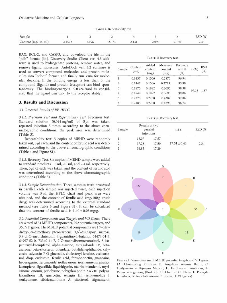
3.2. Potential Components and Targets and VD Genes. Thereare a total of 54MBHD components, 252 potential targets, and360VD genes. TheMBHDpotential components are 1,7-dihy-droxy-3,9-dimethoxy pterocarpene, 3,6′-disinapoyl sucrose,3,9-di-O-methylnissolin, 4-guanidino-1-butanol, 64474-51-7,64997-52-0, 73340-41-7, 7-O-methylisomucronulatol, 8-iso-pentenyl-kaempferol, alpha-asarone, astragaloside IV, beta-asarone, beta-sitosterol, bifendate, butylidenephthalide, caly-cosin, calycosin 7-O-glucoside, cholesteryl ferulate, cycloarte-nol, diop, eudesmin, ferulic acid, formononetin, guanosine,hederagenin, hyrcanoside, isoflavanone, isorhamnetin, jaranol,kaempferol, ligustilide, liquiritigenin, mairin, mandenol, myri-canone, ononin, perlolyrine, polygalasaponin XXVIII, polyga-laxanthone III, quercetin, senegin III, senkyunolide I,senkyunone, sibiricaxanthone A, sitosterol, stigmasterol,
Table 4: Repeatability test.
Sample 1 2 3 4 5 x RSD (%)
Content (mg/100ml) 2.1592 2.196 2.073 2.131 2.090 2.130 2.35
Table 5: Recovery test.
SampleContent(mg)
Addedcontent(mg)
Measuredcontent(mg)
Recoveryrate X(%)
x (%)RSD(%)
1 0.1457 0.1506 0.2870 96.94
97.15 1.87
2 0.1447 0.1506 0.2773. 93.90
3 0.1875 0.1882 0.3696 98.38
4 0.1848 0.1882 0.3695 99,06
5 0.2225 0.2258 0.4387 97.86
6 0.2185 0.2258 0.4298 96.74
Table 6: Recovery test.
SampleResults of two
parallelinjections
x ± s RSD (%)
1 18.07 17.5717:51 ± 0:40 2.342 17.28 17.50
3 16.83 17.29
A
7 B
1
C56
D
12
E
2F
18
G 14
H
327
1
Figure 1: Venn diagram of MBHD potential targets and VD genes(A: Chuanxiong Rhizoma; B: Angelicae sinensis Radix; C:Hedysarum multijugum Maxim.; D: Earthworm Lumbricus; E:Panax notoginseng (Burk.) F. H. Chen ex C. Chow; F: Polygalatenuifolia; G: Acoritataninowii Rhizoma; H: VD genes).
5Oxidative Medicine and Cellular Longevity
tenuifolin, tenuifoliose A, tenuifoliose H, tenuifoliose I, tenui-foliside A, tenuifoliside B, wallichilide, and xanthinin. Mean-while, there are some overlaps between MBHD potentialtarget set and VD gene set (Figure 1). The details of MBHDpotential targets and VD genes are shown in Table S1 and S2.
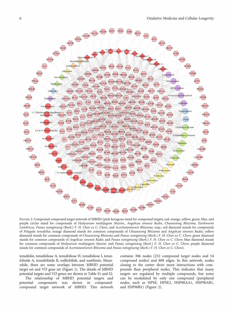
The relationship of MBHD potential targets andpotential components was shown in compound-compound target network of MBHD. This network
contains 306 nodes (252 compound target nodes and 54compound nodes) and 808 edges. In this network, nodesclosing to the center show more interactions with com-pounds than peripheral nodes. This indicates that manytargets are regulated by multiple compounds, but somecan be modulated by only one compound (peripheralnodes, such as HPSE, HPSE2, HSP90AA1, HSP90AB1,and HSP90B1) (Figure 2).
PTPN2MBNL2CA2
PRKCD
ALOXE3 MMP9PRKCB
MBNL3
HSD17B1
CA1
HSD11B1ABCB11
PTAFRCNR1
CA14
CNR2
CBR1
SQLE FNTA
CBR3
FNTB
CalycosinCalycosin7-O-glucoside
Ferulic Acid
ABCB4
HSD11B1L
ABCB1CYP1A2
NR3C1ERBB4
REL
LigustilideMyricanone
ERBB3
RELA
Wallichilide
MAPK10MAPK14
CA6
MAPK11
RDH8
ADK
KDR
ABCB5GRM5
ERBB2
STAT1CXCR4
DHPS
NR3C2
Perlolyrine
Senkyunone
Sitosterol
LGALS9CHyrcanoside
APLP2
APP
4-Guanidino-1-butanol
Xanthinin
LGALS3
LGALS9B
DHFRL1
LGALS9
DHFR
Guanosine
PIK3CD
PTPRF
PIK3CA
PIK3CG
8-Isopentenyl-kaempferol
Eudesmin
Cholesteryl ferulate
Diop
Liquiritigenin
DYRK1ACYP51A1
XDH
CA12AKR1E2
SLC6A5
STAT4
SLC6A4
FGFR4
FGFR2
NQO1
AKT3
AKT1
AKT2
Cycloartenol
Tenuifolin
beta-Asarone
Tenuifoliose A
Tenuifoliose H
FGFR3CYP1A1
PDE5A
PDE11A
AOX1
PTPRD
PPARA
PTPRS
ACP1
DUSP3
alpha-AsaroneACHE
CYP1B1
ABCC1
NOX4
EGFR
CRYZSLC6A14
CA9 LDLR
BCL2
NOS3
CA5B
BCL2L2
CA7
FRK
SRC
STAT3
ADORA3FGRADORA2AAAFYNYES1
SLC6A9
ALOX15B
ADORA1
AKR1B10
ESR2VLDLR
CHRM1CHRM2
MC1RTUBB8
CHRM4
FGF1FGF2
NQO2PPARD
HSD17B2
NR1H2AR
MBNL1 MMP13MMP1 MMP27
PLA2G1BHMGCRNR1H3
AKR1B1
MMP12
MMP2
PTPN1ALOX15
ESR1
ALOX5
ALOX12B
TDP1ALOX12
MAPTLRP8
AKR1B15
SLC6A7CHRM3
CHRM5 CDK1
BCL2L1
SLC6A3POLB HSD11B2
TUBB1 GFER
HPSE2
HGFAC
BLK
MPO
PRSS1
PLAU
FDFT1
64997-52-0
7-O-methylisomucronulatol
MAPK8MAPK9
EPHX2 MGLL
FLT3
ELANEAZU1 PRTN3
MMP8
HSP90B1
ALDH1A1
73340-41-7
SREBF2
HSP90AB1
ALDH1A2
CA5A
Mairin
HCK
ALDH1L2
BACE2
BCHE
FLT4
Bifendate
MAOA
LCK
F2
SREBF1
ALDH1B1
ABCG2
Astragaloside IV
PPP1CB
PPP1CA
PPP1CC
HSP90AA164474-51-7
Isorhamnetin
Isoflavanone
Jaranol
PTGS2
LYN
FLT1
MAOB
BACE1
ALDH1A3
Ononin
NOS2
F7
ALDH2F9
ALDH1L1
IL5
HTR1B
HTR1A
FAAH
TOP2B
senkyunolide I
CDC25ACDC25B
PLGLPA
MCL1AHRUBA2SAE1NR1H4
TOP2A
FGFR1
ABCC3
ABCC2
STAT2
TLR9
GLI1
PDE4A
GRM1
GLI2
PDE4C
PDE4D
PDE4B
GLI3
CA13AAAAAAAAAAAAAAAAAAAAAAA
SLC6A2
NOS1 CYP19A1PRKCG
CA3PTGS1
P18054F10
CDK4
3,9-di-O-methylnissolin JUND
JUNBHederagenin
MMP3METPRKCQ
PRKCA
MMP10
CYP17A1AKR1A1
PPARG
Senegin III
HPSE
Polygalasaponin
XXVIIIPolygalaxanthone III
Butylidenephthalide
Tenuifoliside A
Sibiricaxanthone A
VEGFA
Tenuifoliose I
3,6-Disinapoyl
sucroseTenuifoliside B
ADRA1B
ADRA1D
JUN
Quercetin
ADRA1A
beta-Sitosterol
Formononetin
Mandenol
1,7-Dihydroxy-3,9-dimethoxypterocarpene
Kaempferol
Stigmasterol
Figure 2: Compound-compound target network of MBHD (pink hexagons stand for compound targets; red, orange, yellow, green, blue, andpurple circles stand for compounds of Hedysarum multijugum Maxim., Angelicae sinensis Radix, Chuanxiong Rhizoma, EarthwormLumbricus, Panax notoginseng (Burk.) F. H. Chen ex C. Chow, and Acoritataninowii Rhizoma, resp.; red diamond stands for compoundsof Polygala tenuifolia; orange diamond stands for common compounds of Chuanxiong Rhizoma and Angelicae sinensis Radix; yellowdiamond stands for common compounds of Chuanxiong Rhizoma and Panax notoginseng (Burk.) F. H. Chen ex C. Chow; green diamondstands for common compounds of Angelicae sinensis Radix and Panax notoginseng (Burk.) F. H. Chen ex C. Chow; blue diamond standsfor common compounds of Hedysarum multijugum Maxim. and Panax notoginseng (Burk.) F. H. Chen ex C. Chow; purple diamondstands for common compounds of Acoritataninowii Rhizoma and Panax notoginseng (Burk.) F. H. Chen ex C. Chow).
6 Oxidative Medicine and Cellular Longevity
3.3. MBHD-VD PPI Network Analysis
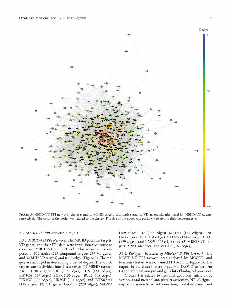
3.3.1. MBHD-VD PPI Network. The MBHD potential targets,VD genes, and their PPI data were input into Cytoscape toconstruct MBHD-VD PPI network. This network is com-posed of 512 nodes (213 compound targets, 267 VP genes,and 32 BHD-VP targets) and 8468 edges (Figure 3). The tar-gets are arranged in descending order of degree. The top 20targets can be divided into 3 categories: (1) MBHD targets:AKT1 (190 edges), SRC (176 edges), JUN (161 edges),PIK3CA (157 edges), EGFR (150 edges), BCL2 (148 edges),PIK3CG (136 edges), PIK3CD (131 edges), and HSP90AA1(127 edges); (2) VD genes: GAPDH (220 edges), MAPK3
(169 edges), IL6 (168 edges), MAPK1 (164 edges), TNF(163 edges), IGF1 (134 edges), CALM2 (134 edges), CALM1(129 edges), and CASP3 (125 edges); and (3) MBHD-VD tar-gets: APP (166 edges) and VEGFA (163 edges).
3.3.2. Biological Processes of MBHD-VD PPI Network. TheMBHD-VD PPI network was analyzed by MCODE, andfourteen clusters were obtained (Table 7 and Figure 4). Thetargets in the clusters were input into DAVID to performGO enrichment analysis and get a lot of biological processes.
Cluster 1 is related to neuronal apoptosis, nitric oxidesynthesis and metabolism, platelet activation, NF-κB signal-ing pathway-mediated inflammation, oxidative stress, and
0Degree
10
40
220
Figure 3: MBHD-VD PPI network (circles stand for MBHD targets; diamonds stand for VD genes; triangles stand for MBHD-VD targets,respectively. The color of the nodes was related to the degree. The size of the nodes was positively related to their betweenness).
7Oxidative Medicine and Cellular Longevity
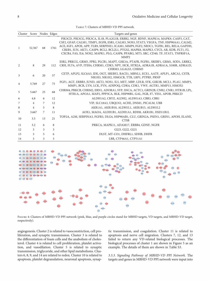
angiogenesis. Cluster 2 is related to vasoconstriction, cell pro-liferation, and synaptic transmission. Cluster 3 is related tothe differentiation of foam cells and the anabolism of choles-terol. Cluster 4 is related to cell proliferation, platelet activa-tion, and vasodilation. Cluster 5 is related to synaptictransmission, triglyceride, and other lipid metabolisms. Clus-ters 6, 8, 9, and 14 are related to redox. Cluster 10 is related toapoptosis, platelet degranulation, neuronal apoptosis, synap-
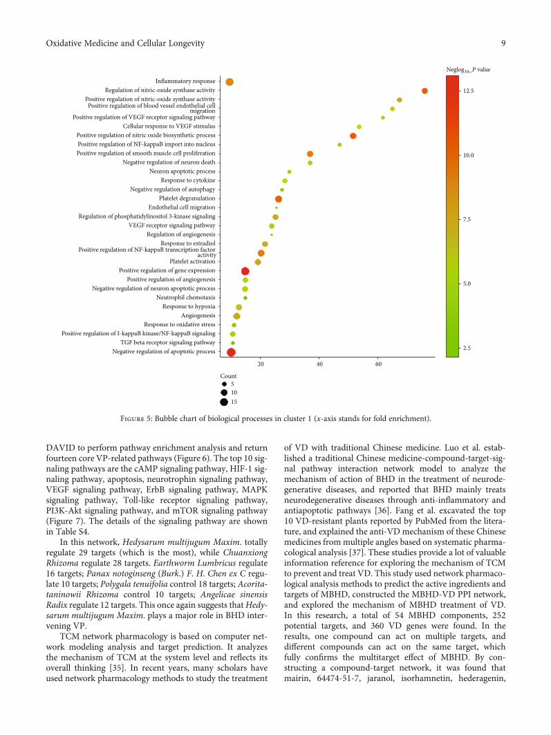
tic transmission, and coagulation. Cluster 11 is related toapoptosis and nerve cell migration. Clusters 7, 12, and 13failed to return any VD-related biological processes. Thebiological processes of cluster 1 are shown in Figure 5 as anexample. The details of them are shown in Table S3.
3.3.3. Signaling Pathway of MBHD-VD PPI Network. Thetargets and genes in MBHD-VD PPI network were input into
Table 7: Clusters of MBHD-VD PPI network.
Cluster Score Nodes Edges Targets and genes
1 52.567 68 1761
PIK3CD, PIK3CG, PIK3CA, IL1B, PLA2G1B, ERBB2, NGF, BDNF, MAPK14, MAPK9, CASP3, CAT,CSF2, GFAP, CALM1, TIMP1, EGFR, ESR1, CALM3, NOS3, STAT3, VEGFA, TNF, HSP90AA1, CALM2,ACE, IGF1, APOE, APP, TLR9, SERPINE1, ICAM1, MMP9, FGF2, NR3C1, TGFB1, REL, RELA, GAPDH,CREB1, JUN, AKT1, CASP9, BCL2, BCL2L1, PTGS2, MAPK8, MAPK3, CYCS, AR, KDR, FLT1, F2,CXCR4, FAS, IL6, NOS2, MAPK1, PLG, CASP8, PPARG, MT3, SRC, CD40, TF, STAT1, TNFRSF1A,
MMP2
2 8 29 112ESR2, PRKCG, GRM5, FPR2, PLCB1, MAPT, GSK3A, PTAFR, FGFR1, SREBF1, GRM1, SOD1, LRRK2,CRH, FLT4, AVP, ITIH4, CHRM1, CDK5, NPY, HCK, HTR2A, ADRA1B, ADRA1A, S100B, ADRA1D,
CHRM3, LGALS3, CHRM5
3 6 20 57CETP, APLP2, SLC6A3, IDE, OGT, SREBF2, BACE1, MBNL1, ECE1, AATF, APLP1, ABCA1, CSTB,
NR1H3, NR1H2, HMGCR, TTR, LRP1, PTPRF, PRNP
4 5.769 27 75FGF1, AGT, ERBB3, JUND, AKT2, NOS1, IL5, MET, MBP, LDLR, SYK, GSK3B, MCL1, PLAU, MMP3,
MMP1, BCR, LYN, LCK, FYN, ADIPOQ, CDK4, CDK1, VWF, AGTR1, MMP13, HMOX1
5 5.667 25 68CHRM4, PRKCB, CHRM2, ERN1, ADORA1, SYP, SNCA, ACTC1, GRIN2B, CNR2, CNR1, HTR1B, LPL,
HTR1A, APOA1, MAP2, PPP3CA, BLK, HSP90B1, GAL, FGR, F7, YES1, APOB, PRKCD
6 4.8 6 12 ALDH1A2, CRYZ, ALDH2, ALDH1A3, CBR3, CBR1
7 4 7 12 VIP, SLC18A3, UBQLN2, ACHE, DNM1, PICALM, UBB
8 4 5 8 AKR1A1, AKR1B10, ALDH1L1, AKR1B15, ALDH1L2
9 3.667 7 11 AOX1, MAOA, ALDH1B1, ALDH1A1, RDH8, AKR1B1, HSD11B1L
10 3.5 13 21TOP2A, A2M, SERPINA3, FGFR3, DLG4, HSP90AB1, CLU, GRIN2A, PSEN1, GRIN1, APOH, ELANE,
CTSB
11 3.2 6 8 PRKCA, MAPK11, ADAM17, ERBB4, GDNF, NGFR
12 3 3 3 GLI3, GLI2, GLI1
13 3 5 6 DLST, MT-CO1, DHFRL1, SDHB, DHFR
14 3 3 3 LBR, CYP46A1, CYP51A1
Figure 4: Clusters of MBHD-VD PPI network (pink, blue, and purple circles stand for MBHD targets, VD targets, and MBHD-VD target,respectively).
8 Oxidative Medicine and Cellular Longevity
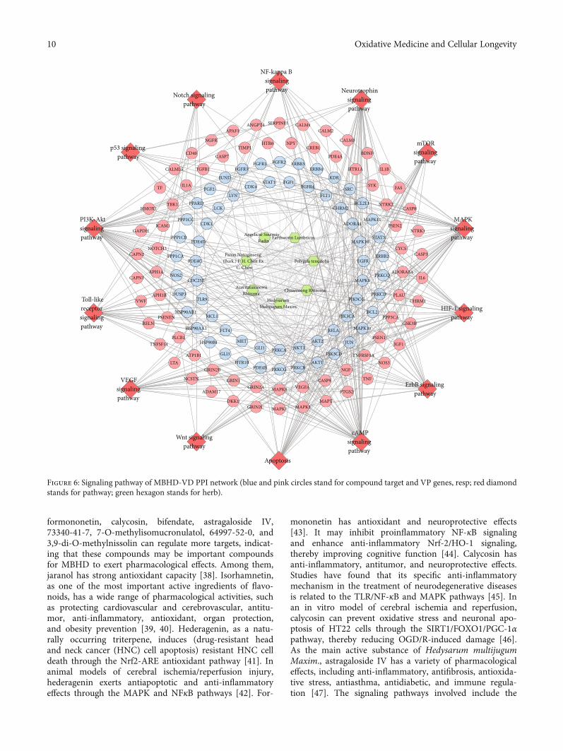
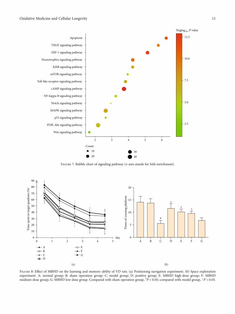
DAVID to perform pathway enrichment analysis and returnfourteen core VP-related pathways (Figure 6). The top 10 sig-naling pathways are the cAMP signaling pathway, HIF-1 sig-naling pathway, apoptosis, neurotrophin signaling pathway,VEGF signaling pathway, ErbB signaling pathway, MAPKsignaling pathway, Toll-like receptor signaling pathway,PI3K-Akt signaling pathway, and mTOR signaling pathway(Figure 7). The details of the signaling pathway are shownin Table S4.
In this network, Hedysarum multijugum Maxim. totallyregulate 29 targets (which is the most), while ChuanxiongRhizoma regulate 28 targets. Earthworm Lumbricus regulate16 targets; Panax notoginseng (Burk.) F. H. Chen ex C regu-late 10 targets; Polygala tenuifolia control 18 targets; Acorita-taninowii Rhizoma control 10 targets; Angelicae sinensisRadix regulate 12 targets. This once again suggests thatHedy-sarum multijugum Maxim. plays a major role in BHD inter-vening VP.
TCM network pharmacology is based on computer net-work modeling analysis and target prediction. It analyzesthe mechanism of TCM at the system level and reflects itsoverall thinking [35]. In recent years, many scholars haveused network pharmacology methods to study the treatment
of VD with traditional Chinese medicine. Luo et al. estab-lished a traditional Chinese medicine-compound-target-sig-nal pathway interaction network model to analyze themechanism of action of BHD in the treatment of neurode-generative diseases, and reported that BHD mainly treatsneurodegenerative diseases through anti-inflammatory andantiapoptotic pathways [36]. Fang et al. excavated the top10 VD-resistant plants reported by PubMed from the litera-ture, and explained the anti-VD mechanism of these Chinesemedicines frommultiple angles based on systematic pharma-cological analysis [37]. These studies provide a lot of valuableinformation reference for exploring the mechanism of TCMto prevent and treat VD. This study used network pharmaco-logical analysis methods to predict the active ingredients andtargets of MBHD, constructed the MBHD-VD PPI network,and explored the mechanism of MBHD treatment of VD.In this research, a total of 54 MBHD components, 252potential targets, and 360 VD genes were found. In theresults, one compound can act on multiple targets, anddifferent compounds can act on the same target, whichfully confirms the multitarget effect of MBHD. By con-structing a compound-target network, it was found thatmairin, 64474-51-7, jaranol, isorhamnetin, hederagenin,
Negative regulation of apoptotic processTGF beta receptor signaling pathway
Positive regulation of I-kappaB kinase/NF-kappaB signalingResponse to oxidative stress
AngiogenesisResponse to hypoxia
Neutrophil chemotaxisNegative regulation of neuron apoptotic process
Positive regulation of angiogenesisPositive regulation of gene expression
Platelet activation
Positive regulation of NF-kappaB transcription factoractivity
Response to estradiolRegulation of angiogenesis
VEGF receptor signaling pathwayRegulation of phosphatidylinositol 3-kinase signaling
Endothelial cell migrationPlatelet degranulation
Negative regulation of autophagyResponse to cytokine
Neuron apoptotic processNegative regulation of neuron death
Positive regulation of smooth muscle cell proliferationPositive regulation of NF-kappaB import into nucleusPositive regulation of nitric oxide biosynthetic process
Cellular response to VEGF stimulusPositive regulation of VEGF receptor signaling pathway
Positive regulation of blood vessel endothelial cellmigration
Positive regulation of nitric-oxide synthase activityRegulation of nitric-oxide synthase activity
Inflammatory response
20 40 60
Count51015
2.5
5.0
7.5
10.0
12.5
Figure 5: Bubble chart of biological processes in cluster 1 (x-axis stands for fold enrichment).
9Oxidative Medicine and Cellular Longevity
formononetin, calycosin, bifendate, astragaloside IV,73340-41-7, 7-O-methylisomucronulatol, 64997-52-0, and3,9-di-O-methylnissolin can regulate more targets, indicat-ing that these compounds may be important compoundsfor MBHD to exert pharmacological effects. Among them,jaranol has strong antioxidant capacity [38]. Isorhamnetin,as one of the most important active ingredients of flavo-noids, has a wide range of pharmacological activities, suchas protecting cardiovascular and cerebrovascular, antitu-mor, anti-inflammatory, antioxidant, organ protection,and obesity prevention [39, 40]. Hederagenin, as a natu-rally occurring triterpene, induces (drug-resistant headand neck cancer (HNC) cell apoptosis) resistant HNC celldeath through the Nrf2-ARE antioxidant pathway [41]. Inanimal models of cerebral ischemia/reperfusion injury,hederagenin exerts antiapoptotic and anti-inflammatoryeffects through the MAPK and NFκB pathways [42]. For-
mononetin has antioxidant and neuroprotective effects[43]. It may inhibit proinflammatory NF-κB signalingand enhance anti-inflammatory Nrf-2/HO-1 signaling,thereby improving cognitive function [44]. Calycosin hasanti-inflammatory, antitumor, and neuroprotective effects.Studies have found that its specific anti-inflammatorymechanism in the treatment of neurodegenerative diseasesis related to the TLR/NF-κB and MAPK pathways [45]. Inan in vitro model of cerebral ischemia and reperfusion,calycosin can prevent oxidative stress and neuronal apo-ptosis of HT22 cells through the SIRT1/FOXO1/PGC-1αpathway, thereby reducing OGD/R-induced damage [46].As the main active substance of Hedysarum multijugumMaxim., astragaloside IV has a variety of pharmacologicaleffects, including anti-inflammatory, antifibrosis, antioxida-tive stress, antiasthma, antidiabetic, and immune regula-tion [47]. The signaling pathways involved include the
HIF-1 signaling pathwayPPP3CA
BCL2
GSK3BPIK3CA
PRKCDChuanxiong Rhizoma
AcoritataninowiiRhizoma PLAU
MAPK9
HedysarumMultijugum Maxim.
PRKCQ IL6
CHRM1PIK3CG
ADORA2A
Toll-likereceptorsignalingpathway RELN
PSENEN MCL1HSP90AB1
VWFDUSP3
CDC25B
APH1A
APH1B
CAPN1
TLR9
NOS2
PPP1CB
NOTCH3CAPN2 PPP1CA
PDE4C
PDE4D
PI3K-Aktsignalingpathway
MAPK11PPP1CC
GAPDHPSEN2
NTRK1CDK1ICAM1
MAPKsignalingpathway
ADORA1
p53 signaling pathway
TBK1 PPARD
TF
CD40
IL1A
HMOX1 LCK
CALML5
CYCSMAPK14
CASP3Polygala tenuifolia
Panax Notoginseng (Burk.) F. H. Chen Ex
C. Chow
STAT3
EGFR
Angelicae Sinensis Radix
Earthworm Lumbricus
ERBB2
SYK
mTORsignalingpathway
NTRK2CHRM2
IL1B
FAS
CASP8BCL2L1
BDNF
Notch signaling pathway
LYNFGF2
APAF1
JUND
NGFR
TGFB1
CASP7
STAT1CDK4
ERBB3FGFR1 FGFR2
FGFR4
FGFR3
FGF1
ERBB4
TIMP1
CALM1ANGPT4
HTR6
SERPINE1
NPY
NF-kappa B signalingpathway
CREB1
Neurotrophinsignalingpathway
PDE4A
FLT1
KDR
CALM2
CALM3
SRC
HTR1A
Apoptosis
MAPK8
PRKCG
PRKCA
MAPK1
VEGFsignalingpathway
LTA
Wnt signaling pathway
ADAM17
NCSTN
GRIN2B
DKK1
PDE4B
MET
HTR1B
GRIN2C
GRIN2AGRIN1
GLI1
HSP90AA1
HSP90B1TNFSF14
FLT4
GLI3
PLCB1
ATP1B1
PRKCB
RELA
PIK3CD
MAPK3
CASP9
AKT2
VEGFA
AKT3
MAPT
AKT1
JUN
TNF
IGF1
ErbB signaling pathway
NOS3
cAMPsignalingpathway
PSEN1
NGF
TNFRSF1A
PTGS2
MAPK10
Figure 6: Signaling pathway of MBHD-VD PPI network (blue and pink circles stand for compound target and VP genes, resp; red diamondstands for pathway; green hexagon stands for herb).
10 Oxidative Medicine and Cellular Longevity
Wnt signaling pathway
PI3K-Akt signaling pathway
p53 signaling pathway
MAPK signaling pathway
Notch signaling pathway
NF-kappa B signaling pathway
cAMP signaling pathway
Toll-like receptor signaling pathway
mTOR signaling pathway
ErbB signaling pathway
Neurotrophin signaling pathway
HIF-1 signaling pathway
VEGF signaling pathway
Apoptosis
2 3 4 5 6
Count1020
3040
2.5
5.0
7.5
10.0
12.5
Neglog10_P value
Figure 7: Bubble chart of signaling pathway (x-axis stands for fold enrichment).
90
ABC
EFG
D
80
70
60
50
40
30
Tim
e spe
nt in
targ
et q
uadr
ant (
%)
20
10
01 2 3 4 5
day0
(a)
20
15
10
Tim
es o
f cro
ssin
g pl
atfo
rm
5
0A B C
#
D E F G
⁎⁎
⁎
(b)
Figure 8: Effect of MBHD on the learning and memory ability of VD rats. (a) Positioning navigation experiment. (b) Space explorationexperiment. A: normal group; B: sham operation group; C: model group; D: positive group; E: MBHD high-dose group; F: MBHDmedium-dose group; G: MBHD low-dose group. Compared with sham operation group, #P < 0:05; compared with model group, ∗P < 0:05.
11Oxidative Medicine and Cellular Longevity
(a) (b)
(c) (d)
(e) (f)
Figure 9: Continued.
12 Oxidative Medicine and Cellular Longevity
EGFR-Nrf2 signaling pathway, NF-κB signaling pathway,signaling pathway Nrf2 antioxidant signaling pathway,PI3K/Akt/mTOR signaling pathway, and many other sig-naling pathways [48].
Meanwhile, the further research (GO and pathwayenrichment analysis) showed that the mechanism of MBHDto treat VD is mainly related to neuronal apoptosis, nitricoxide synthesis and metabolism, platelet activation, NF-κBsignaling pathway-mediated inflammation, oxidative stress,angiogenesis, etc. Current studies have shown that the path-ogenesis of VD mainly includes (1) hypoperfusion and hyp-oxia caused by microvascular circulatory disorders, (2)increased blood-brain barrier permeability caused by endo-thelial dysfunction, (3) inflammation and oxidative stressdamage the nutritional interaction between neurovascularunit cells, (4) ROS and inflammation inhibit the survival ofneurons (neuron apoptosis), and (5) the destruction anddemyelination of myelin tablets caused by the oxidative andproinflammatory environment caused by cerebral ischemiaand hypoperfusion and the breakdown of the blood-brainbarrier [1, 49, 50].
3.4. General Condition. The normal group and sham opera-tion group: Before the operation, the rats had bright hair,red lips, bright red eyes, vigorous spirit, frequent fighting,and strong appetite. Within 3 days after the operation, thespirits were sluggish, less active, and appetite decreased, butgradually recovered, the mental state became better, themovement was agile, the voluntary activities increased, andthe hair, lips, nails, eyeballs, appetite, etc. recovered to a goodstate.
The model group: Before modeling, the experimentalrats’ hair, lips, nails, eyeballs, spirit, and appetite were gen-erally in good condition. After the model was created, var-ious degrees of lethargy, slow response, slow movement,dull fur, dark purple lip nails, increased sleep, and poorappetite appeared. Some rats have different degrees ofdrooping eyelids, diminished eye fissures, and slightsunken eyeballs; a small number of rats are irritable, more
active, irritable, have unstable walking, and turning in cir-cles. Moreover, the self-cleaning ability of rats is reduced,and the hair becomes sparse, dry, and erected or lacksgloss, and occasionally, there are transient paroxysmalconvulsions in rats. In some cases, the abdomen graduallyswelled into a spherical shape, accompanied by obviousreduction in eating and defecation, rough and dull coatcolor, gradual weight loss of limbs, and death.
The treatment groups: In the early stage after the suc-cessful modeling of the rat, the general condition andbehavior of the rat are similar to the VD model group.After a period of drug administration, compared with themodel group, the diet, spirit, and activity gradually recov-ered, and the general situation was better than that of themodel group.
3.5. Effect of MBHD on the Learning and Memory Ability ofVD Rats
(1) Positioning navigation experiment: the escapelatency of rats in each group was shortened with theextension of training time. There was no significantdifference between the blank group and the shamoperation group in the escape latency at the sametime point (P > 0:05). There was a significant differ-ence in the escape latency between the model groupand the sham operation group at the same time point(P < 0:05), indicating that the model was successful.Compared with the model group, the escape latencyin the positive group and the MBHD high- andmedium-dose groups at the same time point has sig-nificant differences (P < 0:05), suggesting thatMBHD and oxiracetam can improve learning andmemory in VD rats
(2) Space exploration experiment: the number ofcrossing platforms in the model group was signifi-cantly reduced (P < 0:05). Compared with themodel group, the number of times the rats crossedthe platform was significantly increased (P < 0:05)
(g)
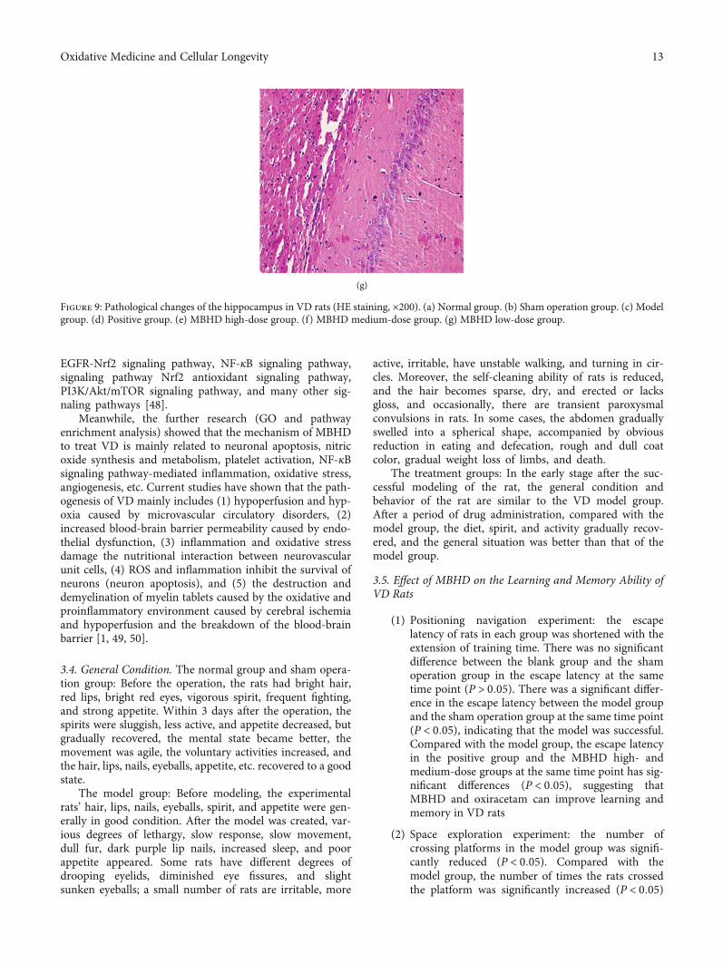
Figure 9: Pathological changes of the hippocampus in VD rats (HE staining, ×200). (a) Normal group. (b) Sham operation group. (c) Modelgroup. (d) Positive group. (e) MBHD high-dose group. (f) MBHD medium-dose group. (g) MBHD low-dose group.
13Oxidative Medicine and Cellular Longevity
in the oxiracetam group and the MBHD high- andmedium-dose groups, suggesting that the memoryability of the rats was improved after drug inter-vention (Figure 8)
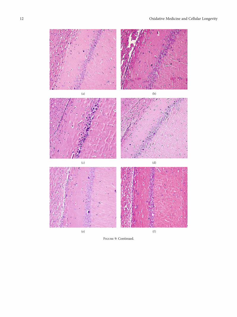
3.6. Effect of MBHD on the Pathological Morphology of theHippocampus in VD Rats. The results of HE staining showedthat hippocampal neurons in the model group were degener-ation and necrosis (as indicated by the arrow), and inflam-matory cell infiltration increased. Compared with the
model group, the hippocampal neuron pyramidal cells ofthe oxiracetam group and the MBHD high-, medium-, andlow-dose groups were arranged more neatly, the outlinewas clear, and the inflammatory cell infiltration was signifi-cantly reduced (Figure 9).
3.7. Differentially Expressed mRNA Analysis
3.7.1. Differentially Expressed mRNA Results. Agilent mRNAwas used to detect the mRNA expression profile of rats in the
5.92
Volcano plot
4.74
3.55
2.37−Log
10 (P
val
ue)
1.18
0−1.26 −0.9 −0.54 −0.18
Log2 (fold change)0.19 0.55 0.91
(a)
1
0
−1
BYHWD1
BYHWD2
BYHWD3
M1
M2
M3
Akrlb10
Cpa4
Sirt1
Gsg1
Il1a
Uncx
RTl-D
aFO
XOEsm1
Six1
Catsperg1
Olr7
18Liph
Iκ-Ba
Defb
1Npy2r
Mpzl2
Il1b
Srcrb4d
Prf1
Akap3
Klf2
Dusp1
Dio2
Abca9
Nr4a1
Rdh7
Fbxl14
iNOS
Dlc1
Wdfy3
Olr8
33Trib1
Apo
ld1Il6
Nfkbia
Ddit4
Bax
Plekhf1
Slc39a12
(b)
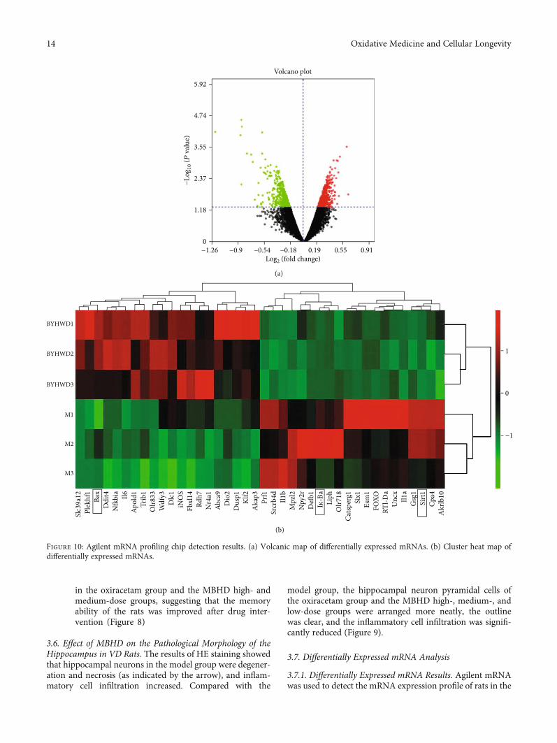
Figure 10: Agilent mRNA profiling chip detection results. (a) Volcanic map of differentially expressed mRNAs. (b) Cluster heat map ofdifferentially expressed mRNAs.
14 Oxidative Medicine and Cellular Longevity
MBHD group and model group. The sample similarity isreflected by sample principal component analysis (PCA).The PCA results show that the overall variance contributionrate is 51.17%.The data of the drug intervention group is rel-atively clustered (except GZ18), and the data of the modelgroup is relatively divergent. Through the analysis of thetwo sets of gene chip data, a total of 469 differentiallyexpressed mRNAs were screened, of which 180 were upregu-lated and 289 were downregulated. Among them, the mostupregulated was Slc39a12 (FC = 3:874), and the most down-regulated was Liph (FC = 5:986). The differentially expressedmRNA volcano map is shown in Figure 10(a), and the top 20differentially expressed mRNA cluster heat maps are shownin Figure 10(b).
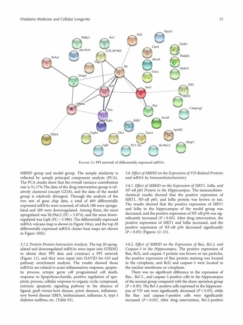
3.7.2. Protein-Protein Interaction Analysis. The top 20 upreg-ulated and downregulated mRNAs were input into STRINGto obtain their PPI data and construct a PPI network(Figure 11), and they were input into DAVID for GO andpathway enrichment analysis. The results showed thosemRNAs are related to acute inflammatory response, apopto-tic process, ectopic germ cell programmed cell death,response to lipopolysaccharide, positive regulation of apo-ptotic process, cellular response to organic cyclic compound,extrinsic apoptotic signaling pathway in the absence ofligand, graft-versus-host disease, prion diseases, inflamma-tory bowel disease (IBD), leishmaniasis, influenza A, type Idiabetes mellitus, etc. (Table S5).
3.8. Effect of MBHD on the Expression of VD-Related Proteinsand mRNA by Immunohistochemistry
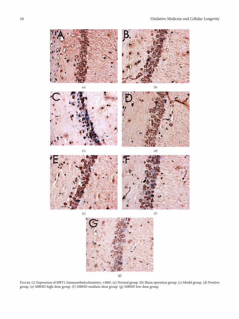
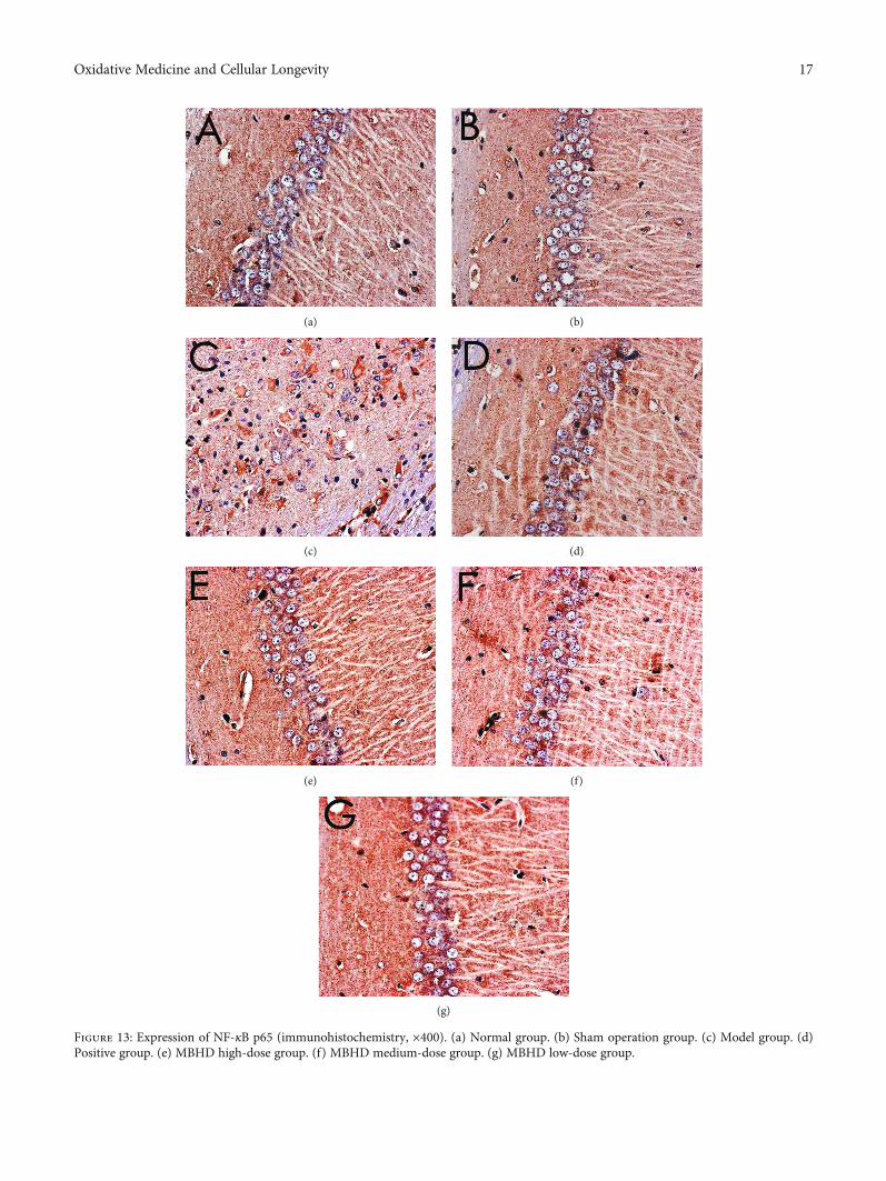
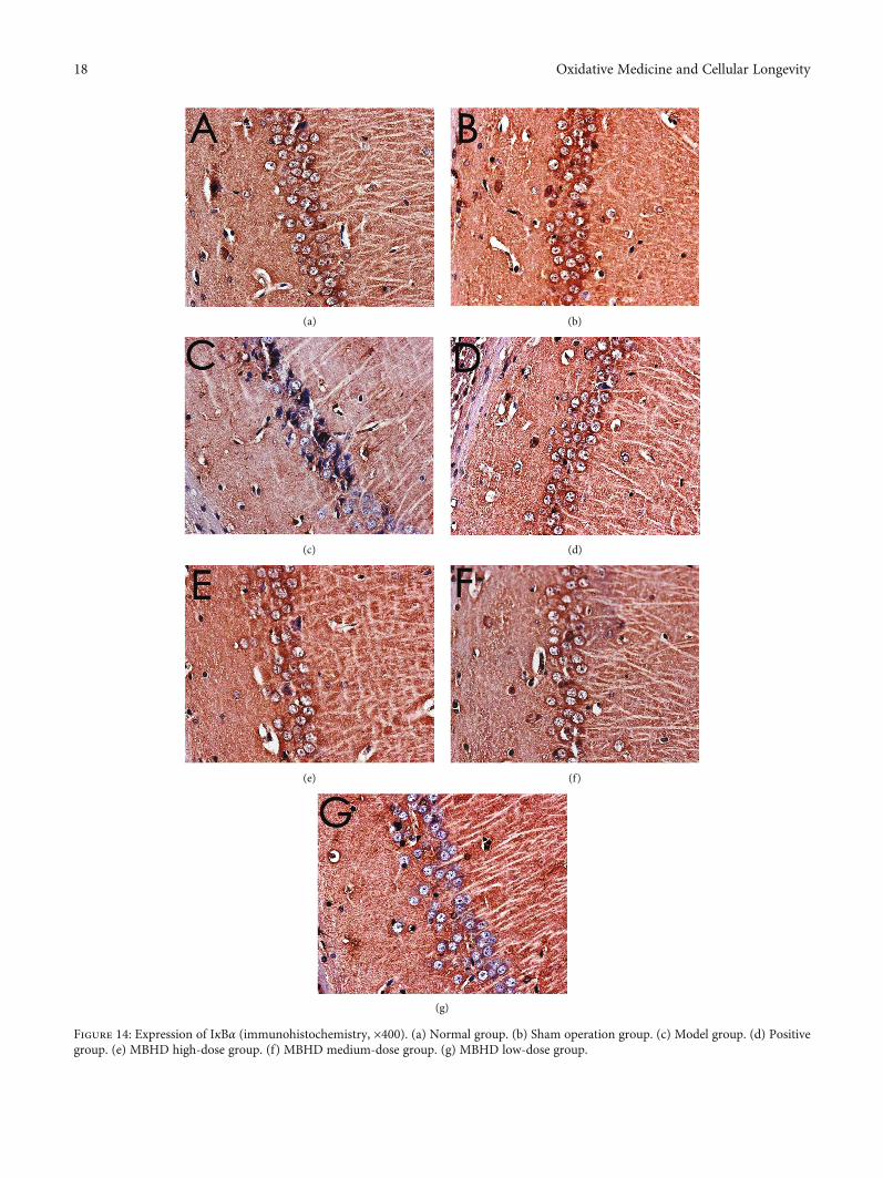
3.8.1. Effect of MBHD on the Expression of SIRT1, IκBα, andNF-κB p65 Protein in the Hippocampus. The immunohisto-chemical results showed that the positive expression ofSIRT1, NF-κB p65, and IκBα protein was brown or tan.The results showed that the positive expression of SIRT1and IκBα in the hippocampus of the model group wasdecreased, and the positive expression of NF-κB p56 was sig-nificantly increased (P < 0:05). After drug intervention, thepositive expression of SIRT1 and IκBα increased, and thepositive expression of NF-κB p56 decreased significantly(P < 0:05) (Figures 12–15).
3.8.2. Effect of MBHD on the Expression of Bax, Bcl-2, andCaspase-3 in the Hippocampus. The positive expression ofBax, Bcl2, and caspase-3 protein was brown or tan particles,the positive expression of Bax protein staining was locatedin the cytoplasm, and Bcl2 and caspase-3 were located inthe nuclear membrane or cytoplasm.
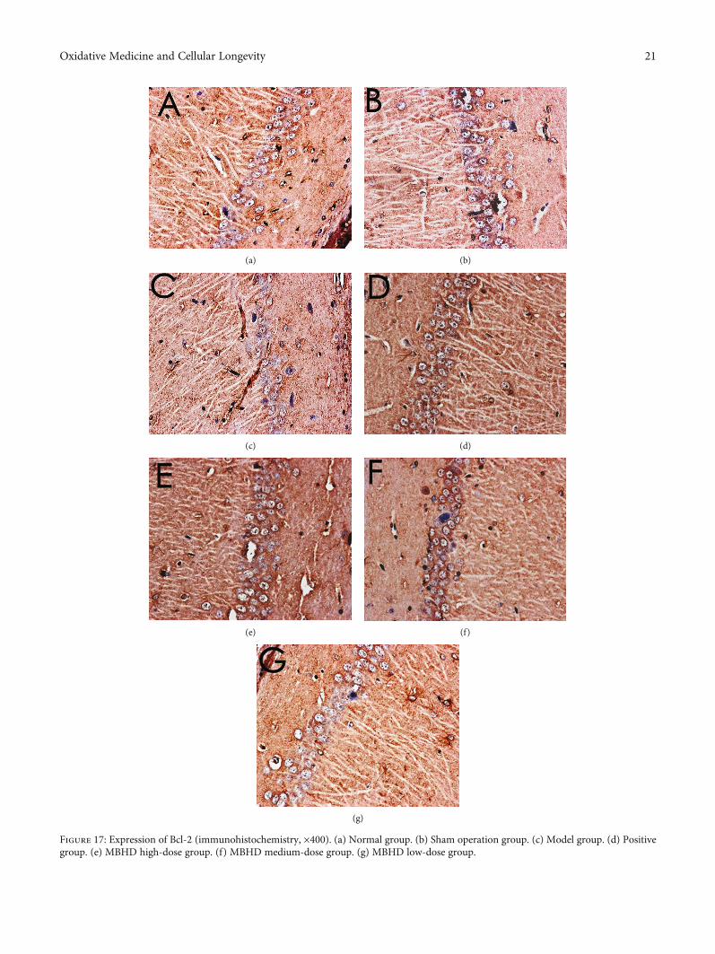
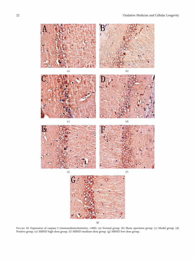
There was no significant difference in the expression ofBax-, Bcl-2-, and caspase-3-positive cells in the hippocampusof the normal group compared with the sham operation group(P > 0:05). The Bcl-2-positive cells expressed in the hippocam-pus of VD rats were significantly decreased (P < 0:05), whilethe Bax- and caspase-3-positive cells were significantlyincreased (P < 0:05). After drug intervention, Bcl-2-positive
Six1Prf1
Cpa4
Npy2r
Defb1
Plekhf1
Olr833
Klf2
Mpzl2
Catsperg1
Ssc4d
Apold1
Akap3Slc39a12RT1-Da
Esm1Dlc1
Liph
Veph1Ddit4
Trib1
Il1a
Rdh7
Bax
Sirt1Il1b
Il6Uncx
Abca9
Nfkbia
Wdfy3
Akr1b10 LOC497963
Gsg1
Dusp1
Nr4a1
Figure 11: PPI network of differentially expressed mRNA.
15Oxidative Medicine and Cellular Longevity
(a) (b)
(c) (d)
(e) (f)
(g)
Figure 12: Expression of SIRT1 (immunohistochemistry, ×400). (a) Normal group. (b) Sham operation group. (c) Model group. (d) Positivegroup. (e) MBHD high-dose group. (f) MBHD medium-dose group. (g) MBHD low-dose group.
16 Oxidative Medicine and Cellular Longevity
(a) (b)
(c) (d)
(e) (f)
(g)
Figure 13: Expression of NF-κB p65 (immunohistochemistry, ×400). (a) Normal group. (b) Sham operation group. (c) Model group. (d)Positive group. (e) MBHD high-dose group. (f) MBHD medium-dose group. (g) MBHD low-dose group.
17Oxidative Medicine and Cellular Longevity
(a) (b)
(c) (d)
(e) (f)
(g)
Figure 14: Expression of IκBα (immunohistochemistry, ×400). (a) Normal group. (b) Sham operation group. (c) Model group. (d) Positivegroup. (e) MBHD high-dose group. (f) MBHD medium-dose group. (g) MBHD low-dose group.
18 Oxidative Medicine and Cellular Longevity
cells increased significantly (P < 0:05); Bax- and caspase-3-positive cells decreased significantly (P < 0:05) (Figures 16–19).
3.8.3. Effect of MBHD on Expression of SIRT1, NF-κB, andIκBα mRNA in the Hippocampus. Compared with the shamoperation group, the level of SIRT1 and NF-κB mRNA inthe hippocampus of the model group was significantlydecreased (P < 0:05), while the level of IκBα mRNA wassignificantly increased (P < 0:05). After drug intervention,the levels of SIRT1 and IκBα mRNA in the hippocampusincreased significantly (P < 0:05), and the level of NF-κBmRNA decreased significantly (P < 0:05) (Figure 20).
3.8.4. Effect of MBHD on Expression of Bax, Bcl-2, andCaspase-3 mRNA in the Hippocampus. The level of Bcl-2mRNA in the hippocampus of the model group was signifi-cantly reduced, while the levels of Bax and caspase-3 mRNAwere significantly increased (P < 0:05). After drug interven-tion, the levels of SIRT1 and Bcl-2 mRNA expressed in thehippocampus were significantly increased, and the levels ofBax and caspase-3 mRNA were downregulated (P < 0:05)(Figure 21).
3.9. Effect of MBHD on the Expression of VD-RelatedProteins by Western Blot
3.9.1. Effect of MBHD on the Expression of SIRT1, IκBα, andNF-κB p65 Protein in the Hippocampus. Compared with thesham operation group, there were significant differences inthe protein levels of SIRT1, NF-κB, and IκBα in the modelgroup (P < 0:05), further suggesting the success of the model.After drug intervention, SIRT1 and IκBα protein levelsincreased significantly (P < 0:05), and NF-κB protein levelsdecreased significantly (P < 0:05) (Figures 22 and 23).
3.9.2. Effect of MBHD on the Expression of Bax, Bcl-2, andCaspase-3 in the Hippocampus. Compared with the shamoperation group, the Bcl-2 protein level in the hippocampusof the model group was significantly reduced, while the Baxand caspase-3 protein levels were significantly increased(P < 0:05). After drug intervention, the level of Bcl-2 proteinexpressed in the hippocampus increased significantly, andthe protein levels of Bax and caspase-3 decreased signifi-cantly (P < 0:05) (Figures 24 and 25).
3.10. Molecular Docking Results of MBHD Compound andSIRT1, RELA, BAX, BCL2, and CASP3.Due to the limitationsof the prediction database, this study used molecular dockingtechnology to further explore whether the compounds ofMBHD can directly interact with SIRT1, NF-κB p65 (RELA),BAX, BCL-2, and CASP3 molecules. It is generally believedthat when the conformation of ligand and receptor is stable,the lower the energy, the greater the possibility of interactionbetween ligand and receptor. The compounds in thecompound-compound target network of MBHD arearranged in descending order of degree, and the top com-pound of each herb is randomly selected. Finally, 15 com-pounds (64474-51-7, astragaloside IV, cycloartenol, ferulicacid, formononetin, hyrcanoside, kaempferol, ligustilide,liquiritigenin, mairin, mandenol, quercetin, senkyunone,wallichilide, and xanthinin) were randomly selected formolecular docking with SIRT1, RELA, BAX, BCL2, andCASP3 (Table 8).
In Table 8, the binding energy of the ligand and thereceptor is less than -5 kcal is considered to interact. BAXmay interact directly with cycloartenol, formononetin, andmairin. BCL2 may interact directly with cycloartenol, liquir-itigenin, mairin, and quercetin. CASP3 may interact directlywith 64474-51-7, cycloartenol, ferulic acid, formononetin,kaempferol, liquiritigenin, senkyunone, wallichilide, andxanthinin. NF-κB p65 may interact directly with 64474-51-7, cycloartenol, formononetin, kaempferol, ligustilide, liquir-itigenin, mairin, quercetin, wallichilide, and xanthinin(Figure 26). SIRT1 may not directly interact with the 15 smallmolecules mentioned above, and it may be indirectly regu-lated by MBHD compounds by interacting with otherproteins.
VD refers to a syndrome of learning, memory, and cogni-tive dysfunction caused by various cerebrovascular diseases.Its clinical characteristics are easy to forget when encounter-ing things, emotional disturbance, and misunderstanding[51]. The pathogenesis of VD is currently unknown. Studieshave found that it is closely related to cerebral ischemia andhypoxia and subsequent damage to specific nervous tissues[52]. Therefore, chronic cerebral ischemia and hypoxia maybe related to the onset of VD. Previous studies have shownthat the 2-VO method can induce ischemic and hypoxicdamage to the hippocampus, cortex, and other brain tissues,and then induce VD [53], and the method is easy to operateand reproducible. It has been widely used in VD research[53]. Meanwhile, based on our previous research foundation,we also use this mature model for research on MBHD inter-vention in VD.
150
100
50
Rela
tive
expr
essio
n of
pro
tein
0A B C D E F G A B C D E F G A
#
#
#
B C D E F G
⁎⁎ ⁎ ⁎ ⁎ ⁎
⁎ ⁎ ⁎
SIRT1NFκBIκB𝛼
Figure 15: Effect of MBHD on the expression of SIRT1, IκBα, andNF-κB p65 protein (A: normal group; B: sham operation group; C:model group; D: positive group; E: MBHD high-dose group; F:MBHD medium-dose group; G: MBHD low-dose group.Compared with the sham operation group, #P < 0:05; comparedwith model group, ∗P < 0:05).
19Oxidative Medicine and Cellular Longevity

(a) (b)
(c) (d)
(e) (f)
(g)
Figure 16: Expression of Bax (immunohistochemistry, ×400). (a) Normal group. (b) Sham operation group. (c) Model group. (d) Positivegroup. (e) MBHD high-dose group. (f) MBHD medium-dose group. (g) MBHD low-dose group.
20 Oxidative Medicine and Cellular Longevity
(a) (b)
(c) (d)
(e) (f)
(g)
Figure 17: Expression of Bcl-2 (immunohistochemistry, ×400). (a) Normal group. (b) Sham operation group. (c) Model group. (d) Positivegroup. (e) MBHD high-dose group. (f) MBHD medium-dose group. (g) MBHD low-dose group.
21Oxidative Medicine and Cellular Longevity
(a) (b)
(c) (d)
(e) (f)
(g)
Figure 18: Expression of caspase-3 (immunohistochemistry, ×400). (a) Normal group. (b) Sham operation group. (c) Model group. (d)Positive group. (e) MBHD high-dose group. (f) MBHD medium-dose group. (g) MBHD low-dose group.
22 Oxidative Medicine and Cellular Longevity
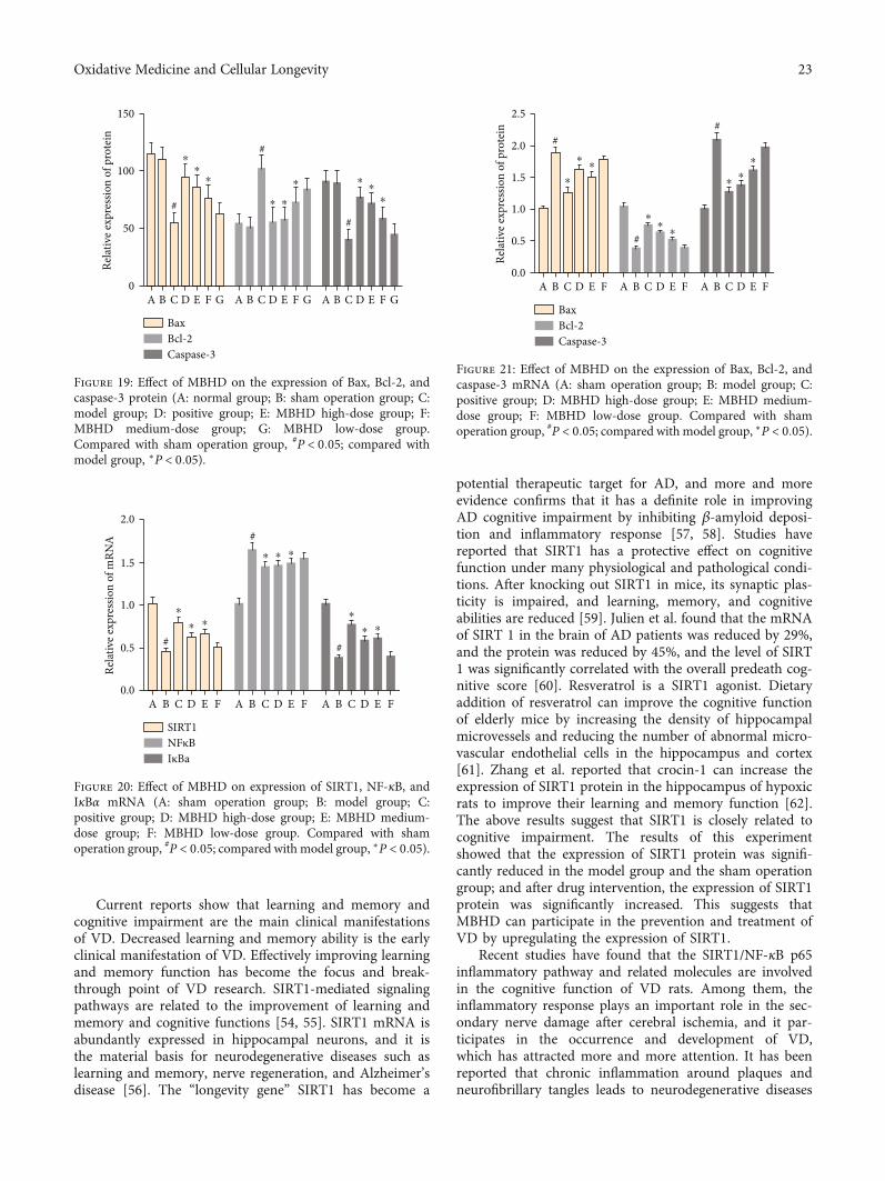
Current reports show that learning and memory andcognitive impairment are the main clinical manifestationsof VD. Decreased learning and memory ability is the earlyclinical manifestation of VD. Effectively improving learningand memory function has become the focus and break-through point of VD research. SIRT1-mediated signalingpathways are related to the improvement of learning andmemory and cognitive functions [54, 55]. SIRT1 mRNA isabundantly expressed in hippocampal neurons, and it isthe material basis for neurodegenerative diseases such aslearning and memory, nerve regeneration, and Alzheimer’sdisease [56]. The “longevity gene” SIRT1 has become a
potential therapeutic target for AD, and more and moreevidence confirms that it has a definite role in improvingAD cognitive impairment by inhibiting β-amyloid deposi-tion and inflammatory response [57, 58]. Studies havereported that SIRT1 has a protective effect on cognitivefunction under many physiological and pathological condi-tions. After knocking out SIRT1 in mice, its synaptic plas-ticity is impaired, and learning, memory, and cognitiveabilities are reduced [59]. Julien et al. found that the mRNAof SIRT 1 in the brain of AD patients was reduced by 29%,and the protein was reduced by 45%, and the level of SIRT1 was significantly correlated with the overall predeath cog-nitive score [60]. Resveratrol is a SIRT1 agonist. Dietaryaddition of resveratrol can improve the cognitive functionof elderly mice by increasing the density of hippocampalmicrovessels and reducing the number of abnormal micro-vascular endothelial cells in the hippocampus and cortex[61]. Zhang et al. reported that crocin-1 can increase theexpression of SIRT1 protein in the hippocampus of hypoxicrats to improve their learning and memory function [62].The above results suggest that SIRT1 is closely related tocognitive impairment. The results of this experimentshowed that the expression of SIRT1 protein was signifi-cantly reduced in the model group and the sham operationgroup; and after drug intervention, the expression of SIRT1protein was significantly increased. This suggests thatMBHD can participate in the prevention and treatment ofVD by upregulating the expression of SIRT1.
Recent studies have found that the SIRT1/NF-κB p65inflammatory pathway and related molecules are involvedin the cognitive function of VD rats. Among them, theinflammatory response plays an important role in the sec-ondary nerve damage after cerebral ischemia, and it par-ticipates in the occurrence and development of VD,which has attracted more and more attention. It has beenreported that chronic inflammation around plaques andneurofibrillary tangles leads to neurodegenerative diseases
150
100
50
Relat
ive e
xpre
ssio
n of
pro
tein
0A B C D E F G A B C D E F G A
#
#
#
B C D E F G
⁎⁎⁎
⁎ ⁎
⁎ ⁎⁎⁎
BaxBcl-2Caspase-3
Figure 19: Effect of MBHD on the expression of Bax, Bcl-2, andcaspase-3 protein (A: normal group; B: sham operation group; C:model group; D: positive group; E: MBHD high-dose group; F:MBHD medium-dose group; G: MBHD low-dose group.Compared with sham operation group, #P < 0:05; compared withmodel group, ∗P < 0:05).
2.0
1.5
1.0
Rela
tive
expr
essio
n of
mRN
A
0.5
0.0A B C D E F A B C D E F A B C D E F
#
⁎
⁎ ⁎
#⁎ ⁎ ⁎
#
⁎
⁎ ⁎
SIRT1NFκBIκBa
Figure 20: Effect of MBHD on expression of SIRT1, NF-κB, andIκBα mRNA (A: sham operation group; B: model group; C:positive group; D: MBHD high-dose group; E: MBHD medium-dose group; F: MBHD low-dose group. Compared with shamoperation group, #P < 0:05; compared with model group, ∗P < 0:05).
2.0
2.5
1.5
1.0
Rela
tive
expr
essio
n of
pro
tein
0.5
0.0A B C D E F A B C D E F A B C D E F
#
⁎
⁎⁎
#
⁎⁎
⁎
#
⁎⁎
⁎
BaxBcl-2Caspase-3
Figure 21: Effect of MBHD on the expression of Bax, Bcl-2, andcaspase-3 mRNA (A: sham operation group; B: model group; C:positive group; D: MBHD high-dose group; E: MBHD medium-dose group; F: MBHD low-dose group. Compared with shamoperation group, #P < 0:05; compared with model group, ∗P < 0:05).
23Oxidative Medicine and Cellular Longevity
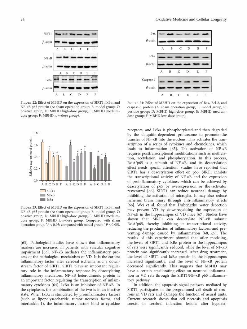
[63]. Pathological studies have shown that inflammatorymarkers are increased in patients with vascular cognitiveimpairment [63]. NF-κB mediates the inflammatory pro-cess of the pathological mechanism of VD. It is the earliestinflammatory factor after cerebral ischemia and a down-stream factor of SIRT1. SIRT1 plays an important regula-tory role in the inflammatory response by deacetylatinginflammatory mediators. NF-κB heterodimeric protein isan important factor regulating the transcription of inflam-matory cytokines [64]. IκBα is an inhibitor of NF-κB. Inthe cytoplasm, the combination of the two is in an inactivestate. When IκBα is stimulated by proinflammatory factors(such as lipopolysaccharide, tumor necrosis factor, andinterleukin 1), the inflammatory factors bind to cytokine
receptors, and IκBα is phosphorylated and then degradedby the ubiquitin-dependent proteasome to promote thetransfer of NF-κB into the nucleus. This activates the tran-scription of a series of cytokines and chemokines, whichleads to inflammation [65]. The activation of NF-κBrequires posttranscriptional modifications such as methyla-tion, acetylation, and phosphorylation. In this process,RelA/p65 is a subunit of NF-κB, and its deacetylationeffect needs special attention. Studies have reported thatSIRT1 has a deacetylation effect on p65. SIRT1 inhibitsthe transcriptional activity of NF-κB and the expressionof proinflammatory cytokines, which can be achieved bydeacetylation of p65 by overexpression or the activatorresveratrol [66]. SIRT1 can reduce neuronal damage byinhibiting the activation of microglia. It may also reduceischemic brain injury through anti-inflammatory effects[66]. Wei et al. found that Dishengzhu water decoctionmay prevent VD by downregulating the expression ofNF-κB in the hippocampus of VD mice [67]. Studies haveshown that SIRT1 can deacetylate NF-κB subunitRelA/p65, thereby inhibiting its transcriptional activity,reducing the production of inflammatory factors, and pre-venting damage caused by inflammation [68, 69]. Theresults of this experiment showed that after modeling,the levels of SIRT1 and IκBα protein in the hippocampusof rats were significantly reduced, while the level of NF-κBprotein was significantly increased. After drug treatment,the level of SIRT1 and IκBα protein in the hippocampusincreased significantly, and the level of NF-κB proteindecreased significantly. This suggests that MBHD mayhave a certain ameliorating effect on neuronal inflamma-tion in VD rats through the SIRT1/NF-κB p65 inflamma-tory pathway.
In addition, the apoptosis signal pathway mediated bySIRT1 participates in the programmed cell death of neu-rons in VD rats and damages the function of neural units.Current research shows that cell necrosis and apoptosiscoexist in cerebral infarction lesions after hypoxia-
SIRT1
𝛽-actin
𝛽-actin
𝛽-actin
NFκB
IκBa
A B C D E F
A B C D E F
A B C D E F
Figure 22: Effect of MBHD on the expression of SIRT1, IκBα, andNF-κB p65 protein (A: sham operation group; B: model group; C:positive group; D: MBHD high-dose group; E: MBHD medium-dose group; F: MBHD low-dose group).
0.6
0.8
0.4
Rela
tive
expr
essio
n of
pro
tein
0.2
0.0A B C D E F A B C D E F A B C D E F
#
⁎
⁎ ⁎
#
⁎⁎
⁎⁎
⁎
#
⁎⁎ ⁎
SIRT1NFκBIκBa
Figure 23: Effect of MBHD on the expression of SIRT1, IκBα, andNF-κB p65 protein (A: sham operation group; B: model group; C:positive group; D: MBHD high-dose group; E: MBHD medium-dose group; F: MBHD low-dose group. Compared with shamoperation group, #P < 0:05; compared with model group, ∗P < 0:05).
Bax
𝛽-actin
𝛽-actin
𝛽-actin
Bcl-2
Caspase-3
A B C D E F
A B C D E F
A B C D E F
Figure 24: Effect of MBHD on the expression of Bax, Bcl-2, andcaspase-3 protein (A: sham operation group; B: model group; C:positive group; D: MBHD high-dose group; E: MBHD medium-dose group; F: MBHD low-dose group).
24 Oxidative Medicine and Cellular Longevity
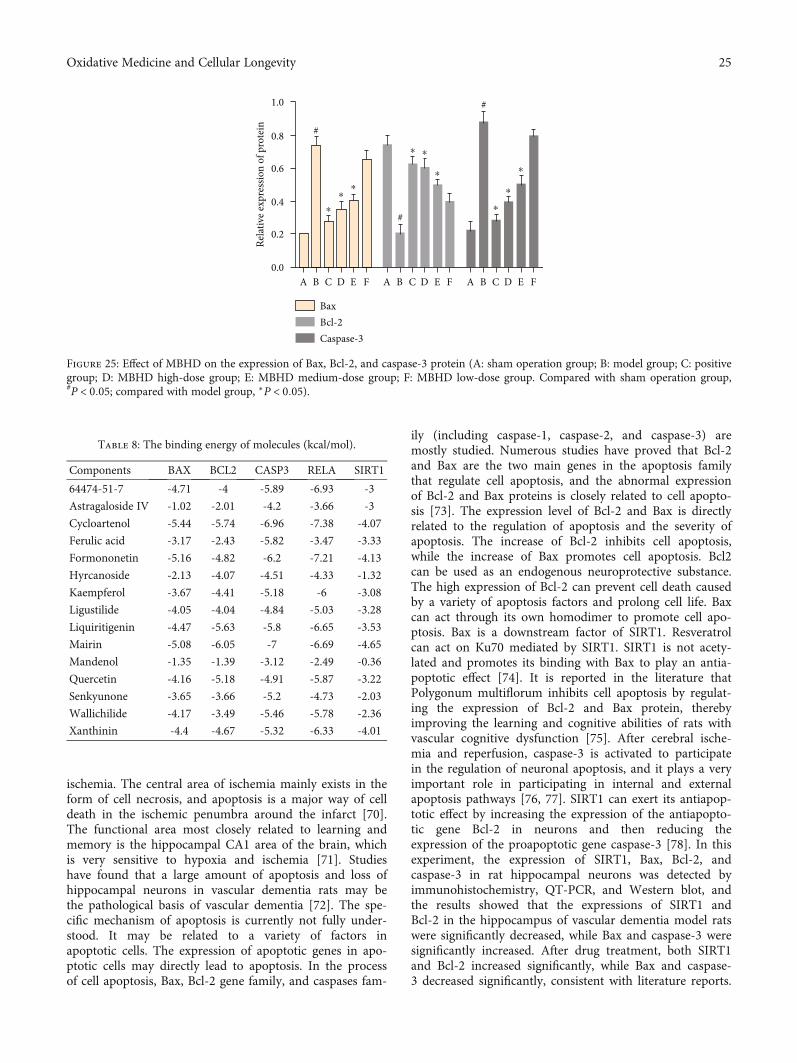
ischemia. The central area of ischemia mainly exists in theform of cell necrosis, and apoptosis is a major way of celldeath in the ischemic penumbra around the infarct [70].The functional area most closely related to learning andmemory is the hippocampal CA1 area of the brain, whichis very sensitive to hypoxia and ischemia [71]. Studieshave found that a large amount of apoptosis and loss ofhippocampal neurons in vascular dementia rats may bethe pathological basis of vascular dementia [72]. The spe-cific mechanism of apoptosis is currently not fully under-stood. It may be related to a variety of factors inapoptotic cells. The expression of apoptotic genes in apo-ptotic cells may directly lead to apoptosis. In the processof cell apoptosis, Bax, Bcl-2 gene family, and caspases fam-
ily (including caspase-1, caspase-2, and caspase-3) aremostly studied. Numerous studies have proved that Bcl-2and Bax are the two main genes in the apoptosis familythat regulate cell apoptosis, and the abnormal expressionof Bcl-2 and Bax proteins is closely related to cell apopto-sis [73]. The expression level of Bcl-2 and Bax is directlyrelated to the regulation of apoptosis and the severity ofapoptosis. The increase of Bcl-2 inhibits cell apoptosis,while the increase of Bax promotes cell apoptosis. Bcl2can be used as an endogenous neuroprotective substance.The high expression of Bcl-2 can prevent cell death causedby a variety of apoptosis factors and prolong cell life. Baxcan act through its own homodimer to promote cell apo-ptosis. Bax is a downstream factor of SIRT1. Resveratrolcan act on Ku70 mediated by SIRT1. SIRT1 is not acety-lated and promotes its binding with Bax to play an antia-poptotic effect [74]. It is reported in the literature thatPolygonum multiflorum inhibits cell apoptosis by regulat-ing the expression of Bcl-2 and Bax protein, therebyimproving the learning and cognitive abilities of rats withvascular cognitive dysfunction [75]. After cerebral ische-mia and reperfusion, caspase-3 is activated to participatein the regulation of neuronal apoptosis, and it plays a veryimportant role in participating in internal and externalapoptosis pathways [76, 77]. SIRT1 can exert its antiapop-totic effect by increasing the expression of the antiapopto-tic gene Bcl-2 in neurons and then reducing theexpression of the proapoptotic gene caspase-3 [78]. In thisexperiment, the expression of SIRT1, Bax, Bcl-2, andcaspase-3 in rat hippocampal neurons was detected byimmunohistochemistry, QT-PCR, and Western blot, andthe results showed that the expressions of SIRT1 andBcl-2 in the hippocampus of vascular dementia model ratswere significantly decreased, while Bax and caspase-3 weresignificantly increased. After drug treatment, both SIRT1and Bcl-2 increased significantly, while Bax and caspase-3 decreased significantly, consistent with literature reports.
0.6
0.8
1.0
0.4
Relat
ive e
xpre
ssio
n of
pro
tein
0.2
0.0A B C D E F A B C D E F A B C D E F
#
⁎
⁎⁎
#
⁎ ⁎
⁎
#
⁎
⁎
⁎
BaxBcl-2Caspase-3
Figure 25: Effect of MBHD on the expression of Bax, Bcl-2, and caspase-3 protein (A: sham operation group; B: model group; C: positivegroup; D: MBHD high-dose group; E: MBHD medium-dose group; F: MBHD low-dose group. Compared with sham operation group,#P < 0:05; compared with model group, ∗P < 0:05).
Table 8: The binding energy of molecules (kcal/mol).
Components BAX BCL2 CASP3 RELA SIRT1
64474-51-7 -4.71 -4 -5.89 -6.93 -3
Astragaloside IV -1.02 -2.01 -4.2 -3.66 -3
Cycloartenol -5.44 -5.74 -6.96 -7.38 -4.07
Ferulic acid -3.17 -2.43 -5.82 -3.47 -3.33
Formononetin -5.16 -4.82 -6.2 -7.21 -4.13
Hyrcanoside -2.13 -4.07 -4.51 -4.33 -1.32
Kaempferol -3.67 -4.41 -5.18 -6 -3.08
Ligustilide -4.05 -4.04 -4.84 -5.03 -3.28
Liquiritigenin -4.47 -5.63 -5.8 -6.65 -3.53
Mairin -5.08 -6.05 -7 -6.69 -4.65
Mandenol -1.35 -1.39 -3.12 -2.49 -0.36
Quercetin -4.16 -5.18 -4.91 -5.87 -3.22
Senkyunone -3.65 -3.66 -5.2 -4.73 -2.03
Wallichilide -4.17 -3.49 -5.46 -5.78 -2.36
Xanthinin -4.4 -4.67 -5.32 -6.33 -4.01
25Oxidative Medicine and Cellular Longevity
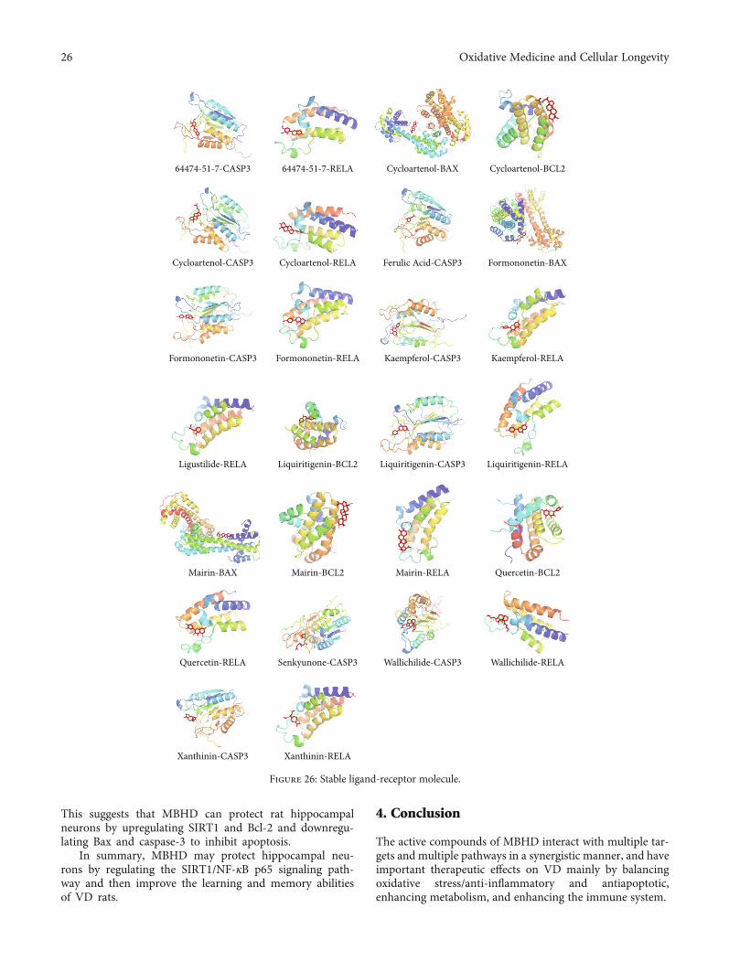
This suggests that MBHD can protect rat hippocampalneurons by upregulating SIRT1 and Bcl-2 and downregu-lating Bax and caspase-3 to inhibit apoptosis.
In summary, MBHD may protect hippocampal neu-rons by regulating the SIRT1/NF-κB p65 signaling path-way and then improve the learning and memory abilitiesof VD rats.
4. Conclusion
The active compounds of MBHD interact with multiple tar-gets and multiple pathways in a synergistic manner, and haveimportant therapeutic effects on VD mainly by balancingoxidative stress/anti-inflammatory and antiapoptotic,enhancing metabolism, and enhancing the immune system.
64474-51-7-CASP3
Cycloartenol-CASP3
Formononetin-RELAFormononetin-CASP3
Liquiritigenin-BCL2 Liquiritigenin-RELALiquiritigenin-CASP3
Mairin-BAX Mairin-BCL2 Mairin-RELA Quercetin-BCL2
Quercetin-RELA
Xanthinin-CASP3 Xanthinin-RELA
Senkyunone-CASP3 Wallichilide-CASP3 Wallichilide-RELA
Kaempferol-CASP3 Kaempferol-RELA
Ligustilide-RELA
Cycloartenol-RELA Ferulic Acid-CASP3 Formononetin-BAX
64474-51-7-RELA Cycloartenol-BAX Cycloartenol-BCL2
Figure 26: Stable ligand-receptor molecule.
26 Oxidative Medicine and Cellular Longevity
Data Availability
The data used to support the findings of this study are includedwithin the article and the supplementary information files.
Conflicts of Interest
We declare no competing interests.
Authors’ Contributions
Kailin Yang, Liuting Zeng, Anqi Ge, Yaqiao Yi, ShanshanWang, and Jinwen Ge are responsible for the study conceptand design. Kailin Yang, Liuting Zeng, AnqiGe and ShanshanWang are responsible for data analysis and interpretation inthe network pharmacology section. Yaqiao Yi is responsiblefor data analysis and interpretation in experiments. KailinYang, Liuting Zeng, Anqi Ge, Yaqiao Yi, and ShanshanWangdrafted the paper. JinwenGe supervised the study. All authorsparticipated in the analysis and interpretation of data andapproved the final paper. Liuting Zeng, Kailin Yang, AnqiGe, Yaqiao Yi, and Shanshan Wang should be consideredjoint first authors. Jinwen Ge is the first corresponding authorbecause he supervised the study and Hunan University ofChinese Medicine is the first corresponding address.
Acknowledgments
This work is supported by the National Natural Science Foun-dation of China (81774174) and the National Key Researchand Development Project of China (No. 2018YFC1704904).
Supplementary Materials
Supplementary 1. Figure S1: standard curve of ferulic acid.
Supplementary 2. Figure S2: HPLC results (A: ferulic acidstandard; B: negative control solution; C: MBHD sample).
Supplementary 3. Table S1: compound targets for MBHD.
Supplementary 4. Table S2: VD genes.
Supplementary 5. Table S3: biological processes of MBHD-VD PPI network.
Supplementary 6. Table S4: signaling pathway of MBHD-VDPPI network.
Supplementary 7. Table S5: enrichment analysis of differen-tially expressed protein PPI network.
References
[1] C. Iadecola, “The pathobiology of vascular dementia,” Neuron,vol. 80, no. 4, pp. 844–866, 2013.
[2] P. B. Gorelick, S. E. Counts, and D. Nyenhuis, “Vascular cog-nitive impairment and dementia,” Biochimica et BiophysicaActa (BBA) - Molecular Basis of Disease, vol. 1862, no. 5,pp. 860–868, 2016.
[3] J. T. O'Brien and A. Thomas, “Vascular dementia,” Lancet,vol. 386, no. 10004, pp. 1698–1706, 2015.
[4] K. Y. Chan, W. Wang, J. J. Wu et al., “Epidemiology of Alzhei-mer’s disease and other forms of dementia in China, 1990-
2010: a systematic review and analysis,” Lancet, vol. 381,no. 9882, pp. 2016–2023, 2013.
[5] J. Jia, F. Wang, C. Wei et al., “The prevalence of dementia inurban and rural areas of China,” Alzheimer's & Dementia,vol. 10, no. 1, pp. 1–9, 2014.
[6] M. K. Sun, “Potential therapeutics for vascular cognitiveimpairment and dementia,” CurrNeuropharmacol., vol. 16,no. 7, pp. 1036–1044, 2018.
[7] C. Liu, L. Zhou, and Z. Shui, “Tongqiao Huoxue Tang andBuyang Huanwu Tang for treatment of vascular dementia–areport of 36 cases,” Journal of Traditional Chinese Medicine,vol. 23, no. 4, pp. 243–245, 2003.
[8] T. Jinglong, G. Weijuan, L. Jun, Q. Tao, Z. Hongbo, andL. Shasha, “The molecular and electrophysiological mecha-nism of Buyanghuanwu Decoction in learning and memoryability of vascular dementia rats,” Brain Research Bulletin,vol. 99, pp. 13–18, 2013.
[9] J. M. Wardlaw, C. Smith, and M. Dichgans, “Small vessel dis-ease: mechanisms and clinical implications,” Lancet Neurol-ogy, vol. 18, no. 7, pp. 684–696, 2019.
[10] W. W. Zhang, F. Xu, D. Wang, J. Ye, and S. Q. Cai, “BuyangHuanwu Decoction ameliorates ischemic stroke by modulat-ing multiple targets with multiple components: _In vitro evi-dences_,” Chinese Journal of Natural Medicines, vol. 16,no. 3, pp. 194–202, 2018.
[11] R. Pan, J. Cai, L. Zhan et al., “Buyang Huanwu decoction facil-itates neurorehabilitation through an improvement of synapticplasticity in cerebral ischemic rats,” BMC Complementary andAlternative Medicine, vol. 17, no. 1, p. 173, 2017.
[12] B. Dou, W. Zhou, S. Li et al., “Buyang Huanwu decoctionattenuates infiltration of natural killer cells and protectsagainst ischemic brain injury,” Cellular Physiology and Bio-chemistry, vol. 50, no. 4, pp. 1286–1300, 2018.
[13] X. Wang, X. Li, L. N. Wang, J. J. Pan, X. Yang, and Y. L. Wei,“Buyang Huanwu decoction ameliorates bleomycin-inducedpulmonary fibrosis in rats via downregulation of related pro-tein and gene expression,” Evidence-Based Complementaryand Alternative Medicine, vol. 2018, Article ID 9185485, 8pages, 2018.
[14] J. Li, Effect of Buyang Huanwu Decoction on the Expression ofGlutamate Receptors in Hippocampal Neurons of VascularDementia Rats, Chengde Medical College, 2010, in Chinese.
[15] K. Yang, L. Zeng, A. Ge et al., “Exploring the regulation mech-anism of Xihuang pill, olibanum and β-boswellic acid on thebiomolecular network of triple-negative breast cancer basedon transcriptomics and chemical informatics methodology,”Frontiers in Pharmacology, vol. 11, 2020.
[16] L. Zeng, K. Yang, L. Liu et al., “Systematic biological and pro-teomics strategies to explore the regulation mechanism ofShoutai Wan on recurrent spontaneous Abortion's biologicalnetwork,” Journal of Ethnopharmacology, vol. 263, p. 113156,2020.
[17] L. Zeng and K. Yang, “Exploring the pharmacological mecha-nism of Yanghe decoction on HER2-positive breast cancer by anetwork pharmacology approach,” Journal of Ethnopharma-cology, vol. 199, pp. 68–85, 2017.
[18] E. H. Kim, S. Baek, D. Shin, J. Lee, and J.-L. Roh, “The effect ofHedysarum multijugum Maxim.-Chuanxiong Rhizoma com-pound on ischemic stroke: a research based on network andexperimental pharmacology,” Oxidative Medicine and CellularLongevity, vol. 2020, Article ID 5412108, 2020.
27Oxidative Medicine and Cellular Longevity
[19] K. Yang, L. Zeng, T. Bao, Z. Long, and B. Jin, “Exploring thepharmacological mechanism of quercetin-resveratrol combi-nation for polycystic ovary syndrome: a systematic pharmaco-logical strategy-based research,” Scientific Reports, vol. 9, no. 1,p. 18420, 2019.
[20] K. Yang, L. Zeng, and J. Ge, “Exploring the PharmacologicalMechanism of Danzhi Xiaoyao Powder on ER- Positive BreastCancer by a Network Pharmacology Approach,” Evidence-Based Complementary and Alternative Medicine, vol. 2018,Article ID 5059743, 20 pages, 2018.
[21] J. Ru, P. Li, J. Wang et al., “TCMSP: a database of systemspharmacology for drug discovery from herbal medicines,”Journal of Cheminformatics, vol. 6, no. 1, 2014.
[22] F.-P. Chen, C.-M. Chang, S.-J. Hwang, Y.-C. Chen, and F.-J. Chen, “Chinese herbal prescriptions for osteoarthritis inTaiwan: analysis of National Health Insurance dataset,”BMC Complementary and Alternative Medicine, vol. 14,no. 1, 2014.
[23] L. Huang, D. Xie, Y. Yu et al., “TCMID 2.0: a comprehensiveresource for TCM,” Nucleic Acids Research, vol. 46, no. D1,pp. D1117–D1120, 2018.
[24] Y. Yufen, S. Xu, and Y. Liang, “Determination of 10 chemicalcomponents in Chuanxiong under different storage conditionsby HPLC,” China Pharmacy, vol. 30, no. 6, pp. 92–97, 2019.
[25] X. Shi, Y. Tang, H. Zhu et al., “Pharmacokinetic comparison ofseven major bio-active components in normal and blood defi-ciency rats after oral administration of Danggui Buxue decoc-tion by UPLC-TQ/MS,” Journal of Ethnopharmacology,vol. 153, no. 1, pp. 169–177, 2014.
[26] B. Xu, C. Qu, W. Zheng et al., “UHPLC-MS/MS method forsimultaneous determination of Radix Polygalae glycolipidsand organic acids in rat plasma and application in a pharma-cokinetic study,” Journal of Chromatography B, vol. 1100-1101, pp. 165–173, 2018.
[27] J. Li, GC-MSMethod of Acori Tatarinovvii Rhizoma VolatileoilQuaility Analysis and β-Asarone, a-Asarone PharmacokineticStudy, Heilongjiang University of Chinese Medicine, 2014.
[28] A. Daina, O. Michielin, and V. Zoete, “SwissTargetPrediction:updated data and new features for efficient prediction of pro-tein targets of small molecules,” Nucleic Acids Research,vol. 47, no. W1, pp. W357–W364, 2019.
[29] A. Hamosh, “Online Mendelian Inheritance in Man (OMIM),a knowledgebase of human genes and genetic disorders,”Nucleic Acids Research, vol. 33, no. Database issue,pp. D514–D517, 2004.
[30] G. Stelzer, R. Rosen, I. Plaschkes et al., “The GeneCards suite:from gene data mining to disease genome sequence analyses,”Current Protocols in Bioinformatics, vol. 54, no. 1, pp. 1.30.1–1.30.33, 2016.
[31] D. Szklarczyk, A. Franceschini, S. Wyder et al., “STRING v10:protein–protein interaction networks, integrated over the treeof life,” Nucleic Acids Research, vol. 43, no. D1, pp. D447–D452, 2015.
[32] G. D. Bader and C. W. Hogue, “An automated method forfinding molecular complexes in large protein interaction net-works,” BMC Bioinformatics, vol. 4, no. 1, p. 2, 2003.
[33] E. Z. Longa, P. R. Weinstein, S. Carlson, and R. Cummins,“Reversible middle cerebral artery occlusion without craniect-omy in rats,” Stroke, vol. 20, no. 1, pp. 84–91, 1989.
[34] S. K. Burley, H. M. Berman, G. J. Kleywegt, J. L. Markley,H. Nakamura, and S. Velankar, “Protein Data Bank (PDB):
the single global macromolecular structure archive,” Methodsin Molecular Biology, vol. 2017, pp. 627–641, 2017.
[35] J. Fang, C. Liu, Q. Wang, P. Lin, and F. Cheng, “In silico poly-pharmacology of natural products,” Briefings in Bioinformat-ics, 2017.
[36] Y. Luo, Q. Wang, and Y. Zhang, “A systems pharmacologyapproach to decipher the mechanism of danggui-shaoyao-san decoction for the treatment of neurodegenerative dis-eases,” Journal of Ethnopharmacology, vol. 178, pp. 66–81,2016.
[37] J. Fang, L. Wang, T. Wu et al., “Network pharmacology basedstudy on the mechanism of action for herbal medicines in Alz-heimer treatment,” Journal of Ethnopharmacology, vol. 196,pp. 281–292, 2017.
[38] Z. Boghrati, M. Naseri, M. Rezaie et al., “Tyrosinase inhibitoryproperties of phenylpropanoid glycosides and flavonoids fromTeucrium polium L. var. gnaphalodes,” Iranian Journal ofBasic Medical Sciences, vol. 19, no. 8, pp. 804–811, 2016.
[39] G. Gong, Y. Y. Guan, Z. L. Zhang et al., “Isorhamnetin: areview of pharmacological effects,” Biomedicine & Pharmaco-therapy, vol. 128, p. 110301, 2020.
[40] S. Kim, C.-Y. Jin, C. Kim et al., “Isorhamnetin alleviateslipopolysaccharide-induced inflammatory responses in BV2microglia by inactivating NF-κB, blocking the TLR4 pathwayand reducing ROS generation,” International Journal of Molec-ular Medicine, vol. 43, no. 2, pp. 682–692, 2018.
[41] E. H. Kim, S. Baek, D. Shin, J. Lee, and J. L. Roh, “Hederagenininduces apoptosis in cisplatin-resistant head and neck cancercells by inhibiting the Nrf2-ARE antioxidant pathway,” Oxida-tive Medicine and Cellular Longevity, vol. 2017, Article ID5498908, 12 pages, 2017.
[42] H. Yu, L. Song, X. Cao et al., “Hederagenin attenuates cerebralischaemia/reperfusion injury by regulating MLK3 signalling,”Frontiers in Pharmacology, vol. 11, 2020.
[43] S. K. L. Ong, M. K. Shanmugam, L. Fan et al., “Focus on for-mononetin: anticancer potential and molecular targets,” Can-cers (Basel), vol. 11, no. 5, p. 611, 2019.
[44] X. Fu, T. Qin, J. Yu et al., “Formononetin ameliorates cognitivedisorder via PGC-1α pathway in neuroinflammation condi-tions in high-fat diet-induced mice,” CNS & Neurological Dis-orders Drug Targets, vol. 18, no. 7, pp. 566–577, 2019.
[45] J. Yang, M. Jia, X. Zhang, and P. Wang, “Calycosin attenuatesMPTP-induced Parkinson’s disease by suppressing the activa-tion of TLR/NF-κB and MAPK pathways,” PhytotherapyResearch, vol. 33, no. 2, pp. 309–318, 2019.
[46] X. Yan, A. Yu, H. Zheng, S. Wang, Y. He, and L. Wang, “Caly-cosin-7-O-β-D-glucoside attenuates OGD/R-induced damageby preventing oxidative stress and neuronal apoptosis via theSIRT1/FOXO1/PGC-1α pathway in HT22 cells,” Neural Plas-ticity, vol. 2019, Article ID 8798069, 2019.
[47] L. Li, X. Hou, R. Xu, C. Liu, and M. Tu, “Research review onthe pharmacological effects of astragaloside IV,” Fundamental& Clinical Pharmacology, vol. 31, no. 1, pp. 17–36, 2017.
[48] J. Zhang, C. Wu, L. Gao, G. Du, and X. Qin, “Astragaloside IVderived from _Astragalus membranaceus_ : A researchreview on the pharmacological effects,” PharmacologicalAdvances in Natural Product Drug Discovery, vol. 87,pp. 89–112, 2020.
[49] M. C. Romay, C. Toro, and M. L. Iruela-Arispe, “Emergingmolecular mechanisms of vascular dementia,” Current Opin-ion in Hematology, vol. 26, no. 3, pp. 199–206, 2019.
28 Oxidative Medicine and Cellular Longevity
[50] M. B. Paradise and P. S. Sachdev, “Vascular cognitive disor-der,” Seminars in Neurology, vol. 39, no. 2, pp. 241–250, 2019.
[51] F. J. Wolters and M. A. Ikram, “Epidemiology of vasculardementia,” Arteriosclerosis, Thrombosis, and Vascular Biology,vol. 39, no. 8, pp. 1542–1549, 2019.
[52] M. Dichgans and D. Leys, “Vascular cognitive impairment,”Circulation Research, vol. 120, no. 3, pp. 573–591, 2017.
[53] S. A. Choi, S. Chong, P. A. Kwak et al., “Impaired functionalrecovery of endothelial colony-forming cells from moyamoyadisease in a chronic cerebral hypoperfusion rat model,” Jour-nal of Neurosurgery: Pediatrics, vol. 23, no. 2, pp. 204–213,2019.
[54] M. Yao, Z. Zhao, L. Wei, D. Zhou, Z. Xue, and S. Ge,“HSF1/HSP pathway in the hippocampus is involved inSIRT1-mediated caloric restriction-induced neuroprotectionafter surgery in aged mice,” Experimental Gerontology,vol. 119, pp. 184–192, 2019.
[55] T.Wang, G. Di, L. Yang et al., “Saponins from Panax japonicusattenuate D-galactose-induced cognitive impairment throughits anti-oxidative and anti-apoptotic effects in rats,” The Jour-nal of Pharmacy and Pharmacology, vol. 67, no. 9, pp. 1284–1296, 2015.
[56] G. Ramadori, C. E. Lee, A. L. Bookout et al., “Brain SIRT1: ana-tomical distribution and regulation by energy availability,”Journal of Neuroscience, vol. 28, no. 40, pp. 9989–9996, 2008.
[57] J. Chen, Y. Zhou, S. Mueller-Steiner et al., “SIRT1 protectsagainst microglia-dependent amyloid-β toxicity through inhi-biting NF-κB signaling,” The Journal of Biological Chemistry,vol. 280, no. 48, pp. 40364–40374, 2005.
[58] J. Ye, Z. Liu, J. Wei et al., “Protective effect of SIRT1 on toxicityof microglial-derived factors induced by LPS to PC12 cells viathe p53-caspase-3-dependent apoptotic pathway,” Neurosci-ence Letters, vol. 553, pp. 72–77, 2013.
[59] S. Michan, Y. Li, M. M.-H. Chou et al., “SIRT1 is essential fornormal cognitive function and synaptic plasticity,” The Journalof Neuroscience, vol. 30, no. 29, pp. 9695–9707, 2010.
[60] C. Julien, C. Tremblay, V. Émond et al., “Sirtuin 1 Reductionparallels the accumulation of tau in Alzheimer disease,” Jour-nal of Neuropathology & Experimental Neurology, vol. 68,no. 1, pp. 48–58, 2009.
[61] C. Oomen, “Resveratrol preserves cerebrovascular density andcognitive function in aging mice,” Frontiers in Aging Neurosci-ence, vol. 1, 2009.
[62] X. Y. Zhang, J. Yu, X. J. Zhang, W. Jia, X. Y. Pu, and H. Liang,“The effect of crocin-1 on learning, memory and the expres-sion of hippocampal SIRT1 in rats under acute hypoxic condi-tions,” Chinese Herbal Medicine, vol. 44, no. 10, pp. 1314–1317, 2013.
[63] W. R. Brown, D. M. Moody, C. R. Thore, V. R. Challa, and J. A.Anstrom, “Vascular dementia in leukoaraiosis may be a conse-quence of capillary loss not only in the lesions, but in normal-appearing white matter and cortex as well,” Neurological Sci-ences, vol. 257, no. 1-2, pp. 62–66, 2007.
[64] J. Kang, M. H. Jiang, and H. J. Min, “IKK-β-mediated myeloidcell activation exacerbates inflammation and inhibits recoveryafter spinal cord injury,” European Journal of Immunology,vol. 41, no. 5, pp. 1266–1277, 2011.
[65] H. Yang, W. Zhang, H. Pan et al., “SIRT1 activators suppressinflammatory responses through promotion of p65 deacetyla-tion and inhibition of NF-κB activity,” PLoS One, vol. 7, no. 9,2012.
[66] H. Chen, H. Jin, J. Shao, W. Xia, and W. Ying, “NAD+ blocksH2O2-induced HMGBl translocation of PC12 cells anddecreases survival of microglia,” FASEB J, vol. 24, 2010.
[67] H. Wei, Y. Liu, X. Chen, and T. Gao, “Effect of Dishengzhudecoction on the expression of NF-κB and COX-2 in hippo-campus of mice with vascular dementia,” Sichuan TraditionalChinese Medicine, vol. 30, no. 11, pp. 45–47, 2012.
[68] X. Zhu, Q. Liu, M. Wang et al., “Activation of Sirt1 by resver-atrol inhibits TNF-α induced inflammation in fibroblasts,”PLoS ONE, vol. 6, no. 11, pp. 70–81, 2011.
[69] Y. J. Jung, J. E. Lee, A. S. Lee et al., “SIRT1 overexpressiondecreases cisplatin-induced acetylation of NF-κB p65 subunitand cytotoxicity in renal proximal tubule cells,” BiochemBio-phys Res Commun, vol. 419, no. 2, pp. 206–210, 2012.
[70] S. Yang, R. Cui, and Y. L. Liu, “Research progress in cytologyand molecular mechanism of vascular dementia,” ChineseJournal of Clinicians, vol. 10, no. 12, pp. 1785–1789, 2016.
[71] J. Burda, M. Matiašová, M. Gottlieb et al., “Evidence for a roleof second pathophysiological stress in prevention of delayedneuronal death in the hippocampal CA1 region,” Neurochem-ical Research, vol. 30, no. 11, pp. 1397–1405, 2005.
[72] Y. Luo, X. Lin, L. Ll et al., “Experimental study of hippocampalneuron apoptosis and pathological changes in vascular demen-tia model rats,” Chinese Journal of Gerontology, vol. 28, no. 18,pp. 1788–1790, 2008.
[73] A. Tichy, “Apoptotic machinery: the Bcl-2 family proteins inthe role of inspectors and superintendents,” Acta Medica,vol. 49, no. 1, pp. 13–18, 2006.
[74] M. Fulco, R. L. Schiltz, S. Iezzi et al., “Sir2 regulates skeletalmuscle differentiation as a potential sensor of the redox state,”Molecular Cell, vol. 12, no. 1, pp. 51–62, 2003.
[75] Z. Wang, G. Xia, G. Zhou et al., “Effects of Polygonum multi-florum on the learning and memory function of rats with vas-cular cognitive impairment,” Journal of Integrated Chinese andWestern Medicine Cardio-Cerebrovascular Disease, vol. 14,no. 9, pp. 960–962, 2016.
[76] H. Zhao, M. A. Yenari, D. Cheng, R. M. Sapolsky, and G. K.Steinberg, “Bcl-2 overexpression protects against neuron losswithin the ischemic margin following experimental strokeand inhibits cytochrome C translocation and caspase-3 activ-ity,” Journal of Neurochemistry, vol. 85, no. 4, pp. 1026–1036,2003.
[77] A. Rami, S. Jansen, I. Giesser, and J. Winckler, “Post-ischemicactivation of caspase-3 in the rat hippocampus: evidence of anaxonal and dendritic localisation,” Neurochemistry Interna-tional, vol. 43, no. 3, pp. 211–223, 2003.
[78] W. Yan, Z. Fang, Q. Yang et al., “Sirt1 mediates hyperbaricoxygen preconditioning-induced ischemic tolerance in ratbrain,” Journal of Cerebral Blood Flow & Metabolism, vol. 33,no. 3, pp. 396–406, 2013.
29Oxidative Medicine and Cellular Longevity