
Requirement of the C. elegans RapGEF pxf-1 and rap-1 for epithelial integrity
47
Requirement of the C. elegans RapGEF pxf-1 and rap-1 for epithelial integrity. W. Pellis-van Berkel 1,¶ , M.H.G. Verheijen 1, 6, ¶ , E. Cuppen 2 , M. Asahina 3 , J. de Rooij 1, 7 , G. Jansen 4, 5 , R.H.A. Plasterk 2, 4, , J.L. Bos 1 and F.J.T. Zwartkruis 1 1 Department of Physiological Chemistry and Centre for Biomedical Genetics, University Medical Center Utrecht, Universiteitsweg 100, 3584 CG Utrecht, The Netherlands, 2 Hubrecht Laboratory/NIOB, Uppsalalaan 8, 3584 CT Utrecht, The Netherlands. 3 Institute of Parasitology, ASCR, Branisovska 31, Ceske Budejovice, 370 05, Czech Republic, 4 Division of Molecular Biology, The Netherlands Cancer Institute, Plesmanlaan 121, 1066 CX Amsterdam. 5 Present adress: Institute for Cell Biology and Genetics, Erasmus University Rotterdam, Postbus 1738, 3000 DR Rotterdam, The Netherlands. 6 Present address: Department of Molecular and Cellular Neurobiology, Vrije Universiteit, De Boelelaan 1085, 1081 HV Amsterdam, The Netherlands. 7 Present address: Department of Cell Biology, The Scripps Research Institute 10550 North Torrey Pines Rd., La Jolla, CA 92037, USA ¶ These authors contributed equally to this work Tel: +31-30-253-8989 Fax: +31-30-253-9035 E-mail: [email protected] Running title: Hypodermal function of pxf-1 and rap-1. Total nr. of words: 9739 Keywords: GTPase, Rap1, PDZ-GEF, C. elegans, hypodermis.
-
Upload
independent -
Category
Documents
-
view
0 -
download
0
Transcript of Requirement of the C. elegans RapGEF pxf-1 and rap-1 for epithelial integrity

Requirement of the C. elegans RapGEF pxf-1 and rap-1
for epithelial integrity.
W. Pellis-van Berkel1,¶, M.H.G. Verheijen1, 6, ¶, E. Cuppen2, M. Asahina3, J. de Rooij1, 7,
G. Jansen4, 5, R.H.A. Plasterk2, 4,, J.L. Bos1 and F.J.T. Zwartkruis1
1Department of Physiological Chemistry and Centre for Biomedical Genetics, University
Medical Center Utrecht, Universiteitsweg 100, 3584 CG Utrecht, The Netherlands, 2Hubrecht
Laboratory/NIOB, Uppsalalaan 8, 3584 CT Utrecht, The Netherlands. 3Institute of
Parasitology, ASCR, Branisovska 31, Ceske Budejovice, 370 05, Czech Republic, 4Division
of Molecular Biology, The Netherlands Cancer Institute, Plesmanlaan 121, 1066 CX
Amsterdam. 5Present adress: Institute for Cell Biology and Genetics, Erasmus University
Rotterdam, Postbus 1738, 3000 DR Rotterdam, The Netherlands. 6Present address:
Department of Molecular and Cellular Neurobiology, Vrije Universiteit, De Boelelaan 1085,
1081 HV Amsterdam, The Netherlands. 7Present address: Department of Cell Biology, The
Scripps Research Institute 10550 North Torrey Pines Rd., La Jolla, CA 92037, USA
¶These authors contributed equally to this work
Tel: +31-30-253-8989
Fax: +31-30-253-9035
E-mail: [email protected]
Running title: Hypodermal function of pxf-1 and rap-1.
Total nr. of words: 9739
Keywords: GTPase, Rap1, PDZ-GEF, C. elegans, hypodermis.

2
ABSTRACT
The Rap-pathway has been implicated in various cellular processes but its exact physiological
function remains poorly defined. Here we show that the C. elegans homologue of the
mammalian guanine nucleotide exchange factors PDZ-GEFs, PXF-1, specifically activates
Rap1 and Rap2. Green fluorescent protein (GFP) reporter constructs demonstrate that sites of
pxf-1 expression include the hypodermis and gut. Particularly striking is the oscillating
expression of pxf-1 in the pharynx during the four larval molts. Deletion of the catalytic
domain from pxf-1 leads to hypodermal defects, resulting in lethality. The cuticle secreted by
pxf-1 mutants is disorganized and can often not be shed during molting. At later stages,
hypodermal degeneration is seen and animals that reach adulthood frequently die with a burst
vulva phenotype. Importantly, disruption of rap-1 leads to a similar, but less severe
phenotype, which is enhanced by the simultaneous removal of rap-2. In addition, the lethal
phenotype of pxf-1 can be rescued by expression of an activated version of rap-1. Together
these results demonstrate that the pxf-1/rap pathway in C. elegans is required for maintenance
of epithelial integrity, in which it probably functions in polarized secretion.
Keywords: PDZ-GEF/Rap1/Rap2/hypodermis/molting.

3
INTRODUCTION
Rap1 is a member of the family of Ras-like GTPases that function in signal transduction
processes by relaying signals received by transmembrane receptors to downstream effector
molecules. Ras-like GTPases switch between an active GTP-bound state and an inactive
GDP-bound state. Activation of Ras-like GTPases is mediated by guanine nucleotide
exchange factors (GEFs), which induce the dissociation of GDP to allow binding of the more
abundant GTP. Four different types of GEFs specific for Rap1 have been described in
vertebrates, namely C3G, CD-GEF, Epac and PDZ-GEF (also named RA-GEF (Liao et al.,
1999), Nrap-GEP (Ohtsuka et al., 1999) or CNrasGEF (Pham et al., 2000); reviewed in (Bos
et al., 2001)). These Rap1-GEFs share a highly homologous catalytic domain including a so-
called REM or LTE domain. In addition, each GEF has its own characteristic domains, which
regulate GEF activity and/or determine its sub-cellular location. Together these GEFs may
account for the various modes of Rap1 activation seen following ligand occupation of distinct
growth factor receptors (Altschuler et al., 1995; Franke et al., 1997; Vossler et al., 1997;
Posern et al., 1998; Zwartkruis et al., 1998), reviewed in (Bos et al., 2001). For example,
EPAC directly binds cAMP, which leads to stimulation of its exchange activity (de Rooij et
al., 1998; Kawasaki et al., 1998). Regulation of PDZ-GEFs is less well understood. The N-
terminal part of PDZ-GEF resembles a cAMP-binding domain, but since the absence of
certain conserved residues prohibits binding of cAMP (Liao et al., 1999; Ohtsuka et al., 1999;
Kuiperij et al., 2003) an as yet unidentified second messenger has been suggested to regulate
PDZ-GEFs (de Rooij et al., 1999). However, it has also been reported that cAMP can induce
Ras-GEF activity of PDZ-GEF, under conditions where its PDZ-domain is bound to the β1
adrenergic receptor (Pak et al., 2002). Apart from interacting with GTPases via its catalytic

4
domain, PDZ-GEF1 can stably interact with GTP-bound Rap1 via its Ras-associating (RA)-
domain in vitro (Liao et al., 1999), whereas PDZ-GEF2 interacts with M-Ras (Gao et al.,
2001). The C-terminal VSAV sequence of PDZ-GEF1 can bind to the second PDZ-domain of
the neural scaffolding protein S-SCAM, MAGI-1 or the related product of the KIAA0705
gene (Ohtsuka et al., 1999; Kawajiri et al., 2000; Mino et al., 2000), suggesting that PDZ-
GEF functions in a signaling complex with scaffolding proteins at sites of cell-cell contact.
Although cloning of the different Rap1-specific GEFs has opened the way to study the precise
mechanisms of Rap1 activation, the physiological function of these GEFs and their target
Rap1 is still enigmatic. Experiments in tissue culture cells have pointed towards a role for
Rap1 in modulation of the Ras-signaling pathway (Kitayama et al., 1989; Cook et al., 1993;
Vossler et al., 1997) and cell cycle control (Altschuler and Ribeiro-Neto, 1998); reviewed in
(Bos et al., 2001). More recently Rap1 was shown to be involved in activation of integrins in
T-cells and macrophages (Caron et al., 2000; Katagiri et al., 2000; Reedquist et al., 2000) and
overexpression of the Rap-1 specific GAP Spa1 was found to abolish adhesion of various cell
types to extracellular matrix proteins (Tsukamoto et al., 1999). The mechanism by which
Rap1 affects integrin function is largely unknown, but may depend on the polarizing activity
of Rap1 (Shimonaka et al., 2003). In addition, a role for Epac2 in cAMP-regulated insulin
secretion has been reported (Ozaki et al., 2000) and Epac1 has been implicated in the
regulation of intracellular calcium levels via phospholipase ε (Schmidt et al., 2001) or the
ryanodine receptor (Kang et al., 2003).
Genetic studies in Drosophila have revealed that Rap1 is an essential gene required during
embryogenesis. In the absence of maternally supplied Rap1, mutants display morphogenetic
defects including mesodermal cell migration, dorsal closure and head involution (Asha et al.,
1999). These defects have been attributed to a diminished activity of the AF-6 homologue
Canoe, which directly can bind to GTP-bound Rap1 and co-localizes in adherens junctions

5
(Boettner et al., 2003). Interestingly, generation of Rap1-/- clones in the wing results in
defective adherens junction formation (Knox and Brown, 2002), but it is unclear if Canoe is
involved in this as well. Apart from Canoe, the Drosophila RalGEF RGL has been proposed
to act as a Rap1 effector on the basis of genetic experiments (Mirey et al., 2003). Finally, in
the same organism loss of PDZ-GEF leads to larval lethality. Escapees and animals over-
expressing PDZ-GEF show defects that suggest a role for PDZ-GEF and Rap1 in ERK
activation (Lee et al., 2002).
We have turned to C. elegans as a model system to define its biological function and dissect
the Rap1 signaling pathway by genetic approaches. Here we show that the C. elegans
homologue of PDZ-GEF, pxf-1, acts upstream of rap-1 and rap-2. Animals, mutant for either
pxf-1 or double mutant for rap-1 and rap-2 remain scrawny, have a disorganized cuticle and
display molting defects. Furthermore, a progressive degeneration of the hypodermis is seen
and adults frequently die with a burst vulva phenotype. From this and the fact that an
activated version of RAP-1, RAP-1V12, rescues the lethal phenotype of pxf-1, we conclude
that the pxf-1/rap pathway is essential for maintenance of epithelial integrity in C. elegans.

6
MATERIALS AND METHODS
Worms.
General methods for culturing and manipulating worms used in these studies were as
described (Lewis and Fleming, 1995). Worms were cultured on NGM plates at 20 °C. Strains
and mutations used were Bristol N2, dpy-20 (e1362), mut-7 (pk204), ML652 [mcEx204:
pML902 + pRF4], PS3352 pkEx246 [pMH86 + JP#38 (cdh-3::GFP)], JR667 [unc-
119(e2498::Tc1) III; wIs51], veIS13 [col-19::GFP + pRF4], BC3106 (let-53 (s43) unc-22(s7)/
nT1 IV; +/nT1 V), BC2895 (let-73 (s685) unc-22(s7)/ nT1 IV; +/nT1 V), BC2049 (let-658
(s1149) unc-22 (s7 unc-331 (e169)/ nT1 IV), +/nT1 V), BC2067 (let-659 (s1152) unc-22 (s7
unc-331 (e169)/ nT1 IV; +/nT1 V). Transgenic animals were obtained by injection of plasmid
DNA into the gonad of worms (Mello et al., 1991). Transgenic arrays were integrated by
irradiating animals with 40 Gy of gamma radiation from a 137Cs source (Way et al., 1991).
Mutant animals isolated from frozen libraries were out-crossed at least five times to wild type
N2 animals before phenotypic analysis.
Constructs.
cDNAs from the T14G10.2 gene were obtained from Dr. Kohara (est yk222e6) or directly
isolated by PCR on cDNA and cloned into pGEM-T (Promega). Nucleotide and protein
sequences of the different isoforms have been deposited at Genbank under accession numbers
AF308447 (PXF-1-A), AF308448 (PXF-1-B) and AF308449 (PXF-1-C). To determine the
specificity of the catalytic domain of pxf-1 the region encompassing the REM-, the PDZ- and
the catalytic domain of PXF-1-A (amino acids 467-1173) was amplified using the primers
CTCGAGTTCAGAAGTTGAACGAAGGAC and

7
GCGGCCGCTGTTCAATTGCTGAAAAAACTG and cloned as an XhoI/Not1 fragment into
pMT2HA.
For reporter studies genomic fragments were obtained by PCR and inserted into the
SphI/BamHI site of the pPD95.75 GFP vector (Fire, pers. comm.). Primers used for
pT14G10.2::GFP-I were TAAGATCTTCCTGTCGAGGTTTCCGAGG and
ATAGCATGCCTTGGACCATTTTGCTATCTGTG, those for pT14G10.2::GFP-II were
ATAGCATGCGATGGAGATGTCGCACGAAG and
TAGGATCCCATTCTCGGACAGAATCTCG.
Isolation and rescue of pxf-1 and rap-1 mutant animals.
Worms mutant for pxf-1 (NL2808 (pk1331)) were isolated from a frozen library as described
in (Jansen et al., 1997). The first round of the nested PCR reactions were done with the
primers AATTGATGCTCTGGTTTGCC and GGTGGTAAACCTCGTGGAGA, followed by
a reaction with TCGAGACGAACGATGAGATG and AGGAAGAACACCCGGAAGAT.
To detect the wild type pxf-1 allele the primers CGATCT GAACCTCTTGTACCTG and
CCAAATTTTCCATTTGTGGTGG were used.
For rescue experiments worms with the genotype dpy-20 (e1362), pxf-1 (pk1331)/+, + IV
(NL2817) were generated. This line was then crossed with dpy-20 (e1362)/dpy-20 (e1362) IV
pkEx10[T14G10 + pMH86] (NL2850). Rescue of the lethal phenotype by the cosmid
T14G10 was evident from the appearance of F2 animals that segregated fertile,
phenotypically wild-type animals, and animals, that combined the pxf-1 mutant phenotype
with a dumpy appearance. In addition, genotyping by PCR confirmed that these latter animals
were homozygous pxf-1 mutants. Since cosmids K04D7 or K08F4 did not rescue, only
mutations in T14G10.1 and 2 could be causative of the mutant phenotype. Therefore, double
stranded RNA for either gene was fed to N2 and ML652 worms. For T14G10.2 this resulted

8
in a delayed progression through larval stages. In addition, microscopic analysis of dlg-
1::GFP showed loss of a restricted number of seam cells in 7% (n=100) of the animals.
Complementation tests with let-53, let-73, let-658 and let-659 mutants was done by first
placing these mutations in trans to a dpy-20 (e1362) allele, followed by mating to pxf-1 +/+
dpy-20 (e1362) animals. In all cases complementation was observed.
Mutant rap-1 animals (FZ0181; pk2082) were isolated from a frozen library by means of
target selected mutagenesis. To this end an 800 bp fragment was amplified using the primers
GACGAGAGTTTTAGTTACAG and GTGAGTTTCAAAAATGTGTG followed by a nested
reaction using TGTAAAACGACGGCCAGTCGAAAATGTATCATATCGAGAC and
AGGAAACAGCTATGACCATGTTAGCCTCCTTTTCATTGAG. Sequencing of
heteroduplex products revealed the presence of a C to T mutation, resulting in a premature
stop at position 130 of RAP-1. Rescue was performed by expression of heat shock promotor
driven rap-1 and restored alae formation and restored brood size, to the level seen for wt
animals, carrying the hsp::rap-1 construct.
Analysis of GFP-expression patterns and immunofluorescence.
Expression of pxf-1 was done by generating worms carrying pT14G10.2::GFP-I (FZ0116;
bjEx40[pT14G10.2::GFP-I /pMH86] and FZ0117; bjIs40[pT14G10.2::GFP-I/pMH86]) and
pT14G10.2::GFP-II (NL2815 or NL2821). NL2815 and NL2821 are independent lines
showing an identical expression pattern. These and other GFP lines described were mounted
on agarose pads and observed under a Nikon or Zeiss immunofluorescence microscope
equipped with Nomarski optics. For analysis of pharyngeal expression in NL2815 and
NL2821 eggs were collected from bleached gravid adults and viewed at various time-points
with a dissecting microscope under UV-light. DPY-7 localization was done using the DPY7-

9
5A antibody and procedures described in Roberts et al. (2003) using a Cy3-conjugated
donkey anti-mouse antibody (Jackson Lab) for detection.
Electron microscopy
Homozygous pxf-1 larvae that failed to molt to L3, N2 L2 and L3 larvae were fixed in 2.5%
glutaraldehyde in 0.2 M cacodylate buffer (pH7.2) at 4 °C for 4 hours, followed by post-
fixation in 2 % OsO4 at 4 °C for 2 hours. Samples were dehydrated by acetone series and
embedded in Epon resin. Ultrathin sections were double stained with uranyl acetate and lead
citrate and viewed with JOEL 1010 (Tokyo, Japan) transmission microscope.
In vivo activation of small GTPases.
The specificity of PXF-1 was determined essentially as described in de Rooij et al. (de Rooij
et al., 1999). Briefly, Cos-7 cells were transfected with HA-tagged versions of human Rap1,
Ral or Ras and increasing amounts of PXF-1. After 24 hours of serum starvation cells were
lysed and GTP-bound GTPases were isolated using activation-specific probes (GST-RalGDS-
RBD for Rap1, GST-RalBP for Ral and GST-Raf1-RBD for Ras). Isolated GTPases were
visualized by means of Western blotting using the 12CA5 monoclonal antibody (de Rooij et
al., 1999). Rap2 activation was measured in [32P]-orthophosphate labeled cells as described in
de Rooij et al., 1999.

10
RESULTS
Genomic organization of pxf-1 and analysis of cDNAs.
Analysis of the complete genome of C. elegans (Greenstein et al., 1998) reveals the presence
of a single homologue for each of the four types of Rap1-GEFs found in vertebrates (Fig. 1A).
Previously we had already reported the existence of a C. elegans homologue of the human
PDZ-GEF1, which we named CelPDZ-GEF at the time (de Rooij et al., 1999). However, in
order to comply with the international nomenclature, we renamed this gene pxf-1 for PDZ-
domain containing exchange factor. pxf-1 is located on LG IV and the complete coding
sequence is covered by cosmid T14G10 (gene T14G10.2). We cloned three different SL1-
spliced messengers from pxf-1 by PCR using primers designed on the basis of predictions
made by GENEFINDER, ACeDB as well as sequence information from ESTs of this gene.
The protein isoforms encoded by these mRNAs were named PXF-1A to C (Fig 1B and C).
PXF-1-A is the longest isoform (1470 amino acids) and has also been published under the
name RA-GEF (Liao et al., 1999). A profile-scan shows that it encodes two cNMP binding
motifs (RCBDs), followed by a Ras exchange motif (REM), a PDZ-domain, a Ras-association
(RA) domain and a catalytic domain (GEF). PXF-1-B is encoded by an alternatively spliced
messenger and lacks the consensus PDZ-binding motif VSRV at the C-terminus. PXF-1-C
resembles PXF-1-A, but does not contain the N-terminal cNMP-binding motif. It is encoded
by a transcript in which SL1 is transspliced to exon four. This transcript is derived from an
intragenic promoter (see below).

11
PXF-1 is a Rap-specific exchange factor.
To determine the specificity of PXF-1 for the various Ras-like GTPases co-transfection
studies were performed in Cos7 cells. A HA-tagged version of PXF-1, encompassing amino
acids 467 to 1173 of PXF-1-A, was co-expressed with HA-tagged human Rap1, Ral or Ras.
GTP-bound GTPases were isolated by virtue of their high affinity binding to activation-
specific probes (see materials and methods) and detected by Western blotting. Increasing the
amount of PXF-1 specifically activated Rap1 but not Ras or RalA (Fig. 2A). Since Rap2 has a
high basal level of GTP, the activation-specific probe assay is less sensitive for Rap2.
Therefore, the effect of PXF-1 on Rap2 was tested in [32P]-orthophosphate-labeled cells, from
which HA-tagged Rap2 was immunoprecipitated. Separation of nucleotides bound to Rap2 by
thin layer chromatography showed that co-expression of PXF-1 with HA-tagged Rap2A
resulted in a clear increase in the GTP/GDP ratio of Rap2A (Fig. 2B). Together, these results
show that PXF-1 is a Rap-specific exchange factor.
Expression of pxf-1 as determined by GFP reporter constructs.
Expression from the upstream pxf-1 promoter was studied by means of a GFP-reporter
construct, containing genomic sequences from nucleotide -2394 to +26 relative to the
translational start codon in exon I (pT14G10.2::GFP-I; Fig. 1B). Expression was first seen at
the comma stage in endodermal precursor and slightly later in the hypodermal cells of the
embryo (Fig. 3A, B and data not shown). During elongation and larval stages many cells of
the embryo express GFP, but particularly strong staining was seen in the hypodermis (Fig.
3C, D) and gut. In addition, several neurons in the head were brightly labeled. In general,
expression in the lateral seam cells (Fig. 3E, F) and hyp10 was most prominent. However,

12
expression could also clearly be seen in the other hypodermal cells, like the syncytial hyp7
and the Pn.p cells during and after the time of vulval induction (Fig. 3G, H and data not
shown). In the adult new sites of strong expression included the hermaphrodite-specific
neurons (HSNs) and the oviduct sheath cells. The lateral seam cells, which are fused in the
adult and other hypodermal cells were also GFP positive (data not shown).
To investigate expression from the downstream promoter a reporter construct was used that
contains sequences from nucleotide + 990 relative to the translation start of exon I to + 4500
located in exon VI (plasmid pT14G10.2::GFP-II; Fig. 1B). Expression was detected first in
neuronal precursor cells at the comma stage. Slightly later strong expression was found in the
hypodermal cells. In newly hatched L1 larvae this construct continued to be expressed in
neuronal cells in the head and tail, in cells of the ventral nerve cord and of the pharynx.
During the early L1 stage hypodermal expression disappeared. The GFP-positive neurons in
animals carrying pT14G10.2::GFP-II were identified as the RMDD, RMD, RMDV, SMDD
and SMDV, which are ring motoneurons. Also the more posteriorly located BDU cells and
the ALN, PVR and PVT cells in the tail expressed GFP. In the course of our studies we
noticed a striking difference in GFP expression levels in the pharynx. Analysis of
synchronized worms showed that GFP is present around the time of molting, but is barely
detectable during part of the intermolts (Fig. 3I, J) or in adults. During the outgrowth of the
gonadal primordium expression was seen in the distal tip cell and oviduct sheath cells (data
not shown). It is possible that the two reporter constructs used here contain enhancer
elements, which act on both promoters. However, the results clearly show that pxf-1 is widely
and dynamically expressed in the main epithelia during all stages.

13
Isolation of animals mutant for pxf-1.
To investigate the function of pxf-1, a deletion library of mutant worms was screened by PCR
(Jansen et al., 1997). A single mutant allele of pxf-1 was isolated, in which a 3.4 kb genomic
region (encoding amino acids 613 to 1306 of PXF-1-A; see Fig. 1B) was deleted. PXF-1
protein derived from this allele lacks most of the PDZ domain as well as the complete
catalytic domain, and therefore is likely to be non-functional, at least with respect to Rap
activation. No homozygous line could be established as deletion of pxf-1 results in lethality
during late larval stages or early adulthood. Timed egglays showed that mutant animals
progressed slower through larval stages than wild type animals. A fraction of the mutant
animals could be identified after 24 hours by their uncoordinated movement and their slightly
smaller size. Inspection by Nomarski optics at 48 hours after egg lay showed the appearance
of vacuoles just underneath the cuticle in a small fraction of mutant animals. Most commonly
these vacuoles were found in the tail region or around the anterior part of the pharynx (fig
4A). These vacuoles appear to be signs of degeneration of the hypodermis, which at later time
points can become more pronounced. From the L2 stage onwards, mutant animals displayed
molting defects: frequently animals were enclosed within their old cuticle or part of the
anterior cuticle remained attached to the pharynx, obstructing it (Fig. 4B, C, Table I). In some
instances animals that were encased in their old cuticle escaped from it and resumed feeding.
It is not uncommon for such animals to have trails of old cuticle attached to their bodies or
encircling it. The lumen of the gut was enlarged in most mutants and contained undigested
bacteria. About 25% of homozygous mutant animals reached adulthood (Table II). Almost all
of them moved in an uncoordinated fashion and their body size was strongly reduced as
compared to wild type animals (Fig. 9). Homozygous animals had developed a fairly normal
gonad and were able to lay a restricted number of fertilized eggs (see table II). Hereafter, the
14
proximal gonad started to degenerate and egg-laying stopped. Mutants then became immotile
and died as a "bag of worms". Alternatively, worms died as a result of a burst vulva (Fig. 4D).
Given the high expression of pxf-1 in seam cells, we inspected the alae, which are cuticular
structures secreted by lateral hypodermal (seam) cells after fusion. In virtually all mutant
adults alae were less distinct or interrupted (Fig. 4E, F). In males, the lateral hypodermal cells
V5, V6 and T form sensory rays in the tail. In pxf-1 mutant males these structures are
malformed and barely visible (n=9; Fig. 4G, H and data not shown). Formation of spicules on
the other hand appeared normal. Although clear cuticle defects in pxf-1 adults were noticed,
the earliest defects in pxf-1 mutants were observed at the L2 stage. At this stage the cuticle
does not contain any alae, but only circumferential ridges (the annuli), which appeared to be
present. To obtain a more detailed view of the cuticle, we immunostained wild type and pxf-1
mutant larvae with an antibody against the collagen DPY-7 (kindly provided by Dr. I.
Johnstone). In wild-type animals this antibody detects DPY-7 in very regular circumferential
bands (Roberts et al., 2003). In contrast, in pxf-1 animals DPY-7 was present in bands, which
not always ran parallel, were wider and often interrupted (fig. 4I and J). This demonstrates
that also at earlier stages the hypodermis is affected in secretion of the cuticle. Taken together,
these results indicate a requirement for PXF-1 in the hypodermis, especially in the seam cells.
Adult homozygous pxf-1 hermaphrodites produce a small brood. Only a minority of the
offspring from these mutants develops beyond the L3 stage (Fig. 4K). They display the same,
albeit more severe phenotypic traits. In addition, a low level of embryonic lethality is seen
(7%). This strongly suggests that there is maternal contribution of PXF-1 protein to embryos.
Yet it also demonstrates that significant development in the absence of PXF-1 is possible.
This is in striking contrast with the situation in Drosophila, where PDZ-GEF mutant animals
from heterozygous mothers show high level of embryonic lethality (Lee et al., 2002).

15
Transformation of mutant animals with the cosmid T14G10, carrying the complete pxf-1
gene, as an extra-chromosomal array led to complete rescue of all aspects of the above
described phenotype (see Table II). No rescue was seen with the overlapping cosmids K04D7
or K08F4. Since RNAi with T14G10.2 (see also materials and methods) resulted in a delayed
growth and a low frequency (7%) of hypodermal abnormalities described below we conclude
that the phenotype described is caused by disruption of pxf-1.
Hypodermal defects in pxf-1 mutant animals.
To analyze the hypodermal defects in more detail various GFP-reporter constructs were
introduced into a pxf-1 mutant background. The seam cells are known to be important for
cuticle secretion and their cell division is tightly coupled to molting events (Sulston and
Horvitz, 1977; Austin and Kenyon, 1994). When the seam cells of mutant L1 larvae were
marked using either the cdh-3::GFP (Pettitt et al., 1996) or the SCM::GFP construct (Koh and
Rothman, 2001), it was clear that the correct number of seam cells had been specified. The
SCM::GFP marker marks the nuclei of seam cells, which are very regularly spaced in wild
type animals (Fig. 5A, B). Using the SCM::GFP marker, no abnormalities were seen in the
division patterns of these seam cells during the L1 to L2 molt (Sulston and Horvitz, 1977;
Austin and Kenyon, 1994). However, at later stages mutant animals were seen in which
expression of the SCM::GFP was extremely weak or absent at various positions along the
body. The seam cell nuclei were often irregularly spaced in more affected animals (Fig 5C to
H). Weakly staining nuclei often had a shrinked appearance. Nomarski microscopy confirmed
that the lack of SCM::GFP expression resulted from a loss of seam cells (Fig 5G, H).
Importantly however, animals that could not complete molting frequently had normal
appearing seam cells, demonstrating that hypodermal degeneration does not necessarily

16
precede molting defects. We also noted a number of mutant animals in which seam cell
expression was weak in the mid-body and in which the nuclei of the seam cells still could be
seen by Nomarski optics. This most likely resulted from the inability of mutants to feed, since
starvation of wild type animals resulted in a similar decrease of SCM::GFP expression in the
mid-body.
Given the fact that lack of Rap1 in Drosophila perturbs adherens junction formation (Knox
and Brown, 2002), we used dlg-1::GFP to mark the adherens junctions of seam and other cells
(Fig. 6) (McMahon et al., 2001). Adherens junctions in pxf-1 mutant L1 larvae had a regular,
uninterrupted appearance and showed that also the shape of seam cells was normal. As
expected, also with this marker we noticed a loss of seam cells from the early L2 stage
onwards. Interestingly, in a restricted number of animals this marker revealed seam cells with
an abnormal morphology (Fig. 6C to F), suggesting that such cells were affected in their
ability to reorganize cell-cell contacts or the cytoskeleton. dlg-1::GFP staining in other parts
of the body like the pharynx and the Pn.p cells was normal. If present, the seam cells fused
normally at the L4 to adult molt. Analysis of pxf-1 mutants with various other GFP constructs,
including pT14G10.2::GFP-II, did not reveal any other abnormalities (Fig. 6G and H).
As a complementary approach in the analysis of pxf-1 mutants, electron microscopy was used
to investigate L2 and L3 larvae (Fig. 7). In contrast to wild type worms, which have a
characteristic round shape, pxf-1 mutants were deformed (Figure 7A, 7B). The lumen of the
gut was clearly enlarged, consistent with images from light microscopy. Microvilli are present
but have a shortened appearance. The most striking difference was found in the cuticle of
mutants, which was irregular around the entire circumference, varied in thickness and was
folded. Furthermore, the overall organization appears much more loose than in wild type
siblings. Importantly, both the underlying lateral hypodermis and hyp7 had a fairly normal
appearance, with a basal lamina and fibrous organelles being present (Fig. 7C, D, G, H).

17
Muscles of pxf-1 mutants were less regularly spaced as compared to those of wild type
animals, slightly disorganized and their dense bodies were less prominent (Figure 7E, 7F).
The defects in muscle organization were unexpected given the fact that no muscle expression
of pxf-1 was seen using GFP-reporters. However, it is well established that the hypodermis is
of crucial importance for muscle architecture and mechanical coupling of muscle cells with
the cuticle (for a review, see Michaux et al., 2001). Therefore, it is a realistic option that
disorganization of muscle cells may result from hypodermal defects. Other cells like the
ventral cord neurons appeared normal (not shown). In summary, ultrastructural analysis
demonstrates that pxf-1 is required in the hypodermis for proper formation of the cuticle and
that hypodermal degeneration does not precede cuticle defects.
pxf-1 acts upstream of rap-1 and rap-2.
To obtain genetic evidence for pxf-1 as a rap-specific GEF, rap mutants were investigated.
The C. elegans genome contains three genes with high homology to vertebrate Rap proteins.
Of these, rap-1 (C27B7.8) and rap-2 (C25D7.7) are clear homologues of Rap1 and Rap2,
respectively, whereas rap-3 (C08F8.7) encodes a more distinct member. rap-2 null mutants
(VC14; kindly provided by Dr. R. Barstead) did not display any detectable phenotype. We
therefore used target-selected mutagenesis and isolated a rap-1 mutant carrying a premature
stop-codon at position 130 (FZ0181, pk2082). Truncation of RAP-1 at this position leads to a
loss of the G5-loop, known to be required for GTP binding and of the C-terminal CAAX-box
motif, involved in membrane targeting. We therefore believe that this mutant represents a null
allele. Homozygous rap-1 mutants are viable and fertile, but show striking similarities to pxf-
1 mutants. First, they develop slower compared to wild type and especially the molting
process appears retarded. Alae of adults are almost always (94%) absent or interrupted (Fig.

18
8A) and loss of seam cells is seen during late larval stages using the SCM::GFP and dlg-
1::GFP markers (not shown). Adult hermaphrodites die with a burst vulva phenotype at a low
frequency (~5%, Fig. 8B) or die as a bag of worms after degeneration of the proximal gonad.
Consequently, rap-1 mutants have a reduced brood-size (36% percent of that of wild type
animals). In males, sensory rays are formed, but especially the most posterior rays are
frequently lost (Fig. 8C). To see if RAP-2 would function redundantly with RAP-1, double
mutants were generated. Whereas removal of a single rap-2 allele did not affect the rap-1
phenotype, removal of both alleles largely reproduced the pxf-1 phenotype: it caused lethality
at a late larval or early adult stage and animals had an enlarged gut lumen with undigested
bacteria (Fig 8D). Furthermore, animals remained scrawny (Fig. 9E), they showed molting
defects and adults almost invariably died with a burst vulva phenotype or as a bag of worms
as seen for pxf-1.
Given the fact that the rap-1 phenotype was most similar to that of pxf-1, we tried to rescue
pxf-1 mutants with an activated version of RAP-1, RAP-1V12. When placed under the control
of the heat shock promoter (hsp), expression of RAP-1V12 by a daily heat shock in wild type
animals slowed progress through larval stages, but did not affect viability. In pxf-1 mutants
expression of RAP-1V12 significantly increased the number of mutants that reached adulthood
as well as the number of progeny (Table II). Furthermore, when grown continuously at 20
degrees Celsius, lines homozygous for pxf-1 could be propagated. However, even though
RAP-1V12 largely rescued the lethality of the pxf-1 mutation, pxf-1-/-; hsp::rap-1V12 animals
develop slower and remain somewhat smaller as compared to wild type worms (Fig. 9F).

19
DISCUSSION
PXF-1 is an exchange factor for RAP1 and RAP2.
In this report we have characterized the pxf-1 gene (T14G10.2) of C. elegans, which encodes
three protein isoforms. When compared to human PDZ-GEF proteins, it is clear that PXF-1-C
is most similar to hPDZ-GEF1 since it contains a single RCBD. The other isoforms contain
two of these motifs and consequently resemble hPDZ-GEF2 (de Rooij et al., 1999; Liao et al.,
1999; Kuiperij et al., 2003). Strikingly, alternative splicing of mRNA from this latter gene
also yields proteins, that either contain a PDZ-binding motif at the C-terminus, resembling
PXF-1-A, or lack it as seen for PXF-1-B (Kuiperij et al., 2003). Like its mammalian
homologues, PXF-1 displays guanine nucleotide exchange activity towards Rap1 and Rap2.
Our transfection studies confirm the findings of Liao et al. (Liao et al., 1999), who showed
that in tissue culture cells PXF-1 activates Rap1. However, these authors also demonstrated
Ras-exchange activity in vitro, which we could not detect in our co-transfection experiments.
Ras activation by human PDZ-GEF1 in tissue culture cells has also been reported (Pak et al.,
2002) and we have currently no explanation for this discrepancy. Importantly, our genetic
experiments provide evidence that PXF-1 functions as a RAP-exchange factor in vivo. First,
disruption of pxf-1 or rap-1 shows that they both function in the hypodermis and in particular
the lateral hypdermal cells. Clearly, the pxf-1 phenotype is more severe than that of rap-1
mutants, which appears to result from functional redundancy of rap-1 with rap-2. The
presence of a single functional rap-1 or rap-2 allele is sufficient for viability, but double
mutants display a lethal phenotype, which is virtually identical to that of pxf-1 mutants.
Moreover, over-expression of an activated version of RAP-1 (RAP-1V12) rescues the lethal
phenotype of pxf-1, which strongly supports the notion that pxf-1 acts upstream of rap-1 in
vivo. pxf-1 -/-; hsp::rap-1V12 animals still develop more slowly and remain smaller than wild

20
type animals. It is possible that this is due to the fact that PXF-1 has functions other than
RAP-1 activation. However, we think it is more likely that heat-shock driven over-expression
of RAP-1V12 does not mimic the spatio-temporal control of RAP-1 activation by PXF-1.
Furthermore, despite being less sensitive to GTPase activating proteins, RAP-1V12 may still
require PXF-1 activity to become highly GTP-bound. Indeed, in an analogous situation
signaling by a gain-of-function allele of let-60/ras was found to be diminished by lowering
the activity of its upstream activator SOS-1 (Chang et al., 2000). So far, analysis of pxf-1
mutants did not reveal a role for PXF-1 as a LET-60/RAS-GEF: vulval development in
hermaphrodites and spicule formation in males, both known to be dependent on LET-60 RAS,
appear normal. This is consistent with a genetic analysis of PDZ-GEF in Drosophila, which
led to the conclusion that it functions as a Rap1-GEF rather then a Ras-GEF (Lee et al.,
2002).
pxf-1 is required in the hypodermis.
GFP-reporter studies show that pxf-1 is widely expressed in a specific spatio-temporal pattern,
suggesting a function for pxf-1 in multiple tissues. In line with this, pxf-1 mutants have a
pleiotropic phenotype, which includes uncoordinated movement, an enlarged gut lumen and a
strongly reduced fertility. Many aspects of the phenotype however, point to a critical function
for pxf-1 in seam cells, where it is highly expressed. For example, the alae, which are secreted
by the seam cells, are faint and interrupted in almost all mutant adults. Secondly, the sensory
rays in males, formed by the seam cells V5, V6 and T are malformed. Also the “burst vulva”
phenotype hints to a compromised seam cell function, as it is indicative for a loss of contact
between seam cells and the utse or VulE cells, which are connected to the uterus (Newman et
al., 2000) (Sharma-Kishore et al., 1999).

21
The molting defect seen in pxf-1 mutants provides the most direct evidence for a hypodermal
function of pxf-1. Molting is a complex process in which first a stage-specific, collageneous
cuticle is synthesized underneath the old one by cells of the pharynx and hypodermis. During
this period high secretory activity is seen in the hypodermis, including the lateral seam cells
(Singh, 1978). Simultaneously, the cortical actin cytoskeleton in the hypodermis undergoes a
dramatic reorganization into circumferential oriented actin bundles, which disappear again
during the final stages of cuticle formation (Costa et al., 1997). Two observations suggest that
the molting defect in pxf-1 mutants is not simply the result of the observed loss of seam cells,
but instead suggest that pxf-1 plays an active role in the process of molting. First, in many
young animals, which could not complete molting, all seam cells were present and no signs of
degeneration of the hypodermis were seen. Also ultra-structural analysis shows the presence
of hypodermal cells underneath an abnormally organized cuticle in pxf-1 mutants. Secondly,
pharyngeal expression of pxf-1 precisely coincides with molting and is barely detectable
during intermolts. Similar oscillating expression patterns have been reported for genes
encoding structural proteins of the cuticle like collagen genes (Johnstone, 2000) (McMahon et
al., 2003), but also for the lin-42 gene, which regulates the timing of molting events (Jeon et
al., 1999) and the nuclear hormone receptor nhr-23, disruption of which also causes molting
defects (Kostrouchova et al., 1998). Finally, loss of seam cells has also been described for
worms, mutant for plx-1, which encodes a plexin-type of transmembrane receptor for
semaphorins (Fujii et al, 2002). However, for plx-1 no molting defects have been reported.
Loss of seam cells in plx-1 mutants is caused by a reduced number of cell divisions during
larval stages. Following each division the anterior seam cell daughter fuses with hyp-7 in wild
type worms, while the posterior cell will undergo further divisions. These divisions require
that the posterior cell re-establishes contacts with its anterior and posterior neighbors (Austin
and Kenyon, 1994). In plx-1 mutants this does not occur, most likely due to changes in

22
adhesive properties. Given the fact that the Rap1 pathway is involved in cell-matrix and cell-
cell adhesion, we considered this an attractive scenario for our pxf-1 mutant. Although seam
cells sporadically display an abnormal morphology in pxf-1 mutants, analysis of seam cells
with the SCM::GFP marker show that the majority of cell divisions occurs normally and that
seam cells are lost by degeneration. The precise cause of this degeneration is currently
unknown, but it is possible that the disorganized cuticle provides insufficient protection
against mechanical stress.
The hypodermal degeneration and rupture of the vulval region distinguishes pxf-1 mutants
from previously described molting mutants like those carrying mutations in lrp-1, nhr-23 or
nhr-25 (Yochem et al., 1999), (Asahina et al., 2000), (Gissendanner and Sluder, 2000),
(Kostrouchova et al., 1998). Instead, the pxf-1 phenotype much more resembles that of
worms, in which sec-23 functioning is reduced by RNAi during larval stages (Roberts et al.,
2003). Apart from the molting defect and burst vulva phenotype, these animals also grow
slowly and have an enlarged gut lumen filled with undigested bacteria. In addition, both have
a dis-organized cuticle, as determined by detection of DPY-7 collagen using
immunofluorescence. Null mutants of sec-23 were isolated on the basis of their inability to
secrete cuticle components. Although sec-23 mutants are embryonic lethal and thus have a
more severe phenotype, the RNAi result shows that compromised secretion of cuticle
components can lead to molting defects. Likewise, loss of pxf-1 or rap-1 and rap-2 may
reduce the (polarized) secretory activity of the hypodermis to a level below that required for
molting. A role for pxf-1 in secretion is in line with the expression observed in tissues
involved in polarized secretion like the hypodermis and gut. Furthermore, also the Rap-
specific exchange factor, Epac2, has been shown to be involved in enhancing secretion,
namely of insulin from pancreatic beta-cells (Ozaki et al., 2000). The Rap1 pathway appears
not to be absolutely required for secretion, but may increase the efficiency indirectly by

23
organizing some spatial feature. Intriguingly, in yeast the RAP-1 homologue RSR1 is
essential for proper bud site selection, but not for bud formation per se. Bud site selection
relies on polarization of the cytoskeleton to target secretory vesicles to a specific site in the
cell. Also the well-documented stimulatory effect of Rap1 on integrin function in
lymphocytes may depend on the polarizing activity of Rap1 in these cells (Shimonaka et al.,
2003), possibly via its effector RapL (Katagiri et al., 2003). Definite proof for a role of pxf-1
and rap-1/2 in polarized secretion awaits further characterization of these mutants. In any
case, the described hypodermal defect offers a good starting point for identification of
additional components of the Rap1 pathway in C. elegans and opens the way to a comparative
analysis of this pathway in distinct organisms.
Acknowledgements.
We thank Dr. Y. Kohara for kindly providing us with cDNA clones and Dr. A. Fire for GFP
vectors. We thank Theresa Stiernagle and the CGC for sending various mutant and GFP-
marker strains. We thank Dr. M. Labouesse for ML652, Dr. A. Rougvie for veIS13, Dr. J.
Pettitt for PS3352, Dr. Barstead for VC14 (rap-2), Dr. J. Rothman for JR667 and Dr. I
Johnstone for the DPY-7 antibody. We thank Stephen Wicks for identifying GFP-expressing
neurons. We would like to thank the facility of Laboratory of Electron Microscopy, Institute
of Parasitology, ASCR. We would like to thank all members of the Plasterk and Bos lab for
critical comments and continuous support. We especially thank Karen Thijssen, Marieke van
de Horst and Rene Ketting for technical assistance. We thank Rik Korswagen for critically
reading the manuscript. This work was supported by the Dutch Cancer Society, KWF (M.V.),

24
the Center for Biomedical Genetics (F.Z.), the Netherlands Organization for Scienitific
Research NWO-ALW (J.d.R.) and grant GAAV KJB5022303 (M.A.).

25
REFERENCES
Altschuler, D.L., Peterson, S.N., Ostrowski, M.C., and Lapetina, E.G. (1995). Cyclic AMP-
dependent activation of Rap1b. J Biol Chem 270, 10373-10376.
Altschuler, D.L., and Ribeiro-Neto, F. (1998). Mitogenic and oncogenic properties of the
small G protein Rap1b. Proc Natl Acad Sci U S A 95, 7475-7479.
Asahina, M., Ishihara, T., Jindra, M., Kohara, Y., Katsura, I., and Hirose, S. (2000). The
conserved nuclear receptor ftz-F1 is required for embryogenesis, moulting and reproduction
in caenorhabditis elegans. Genes Cells 5, 711-723.
Asha, H., de Ruiter, N.D., Wang, M.G., and Hariharan, I.K. (1999). The Rap1 GTPase
functions as a regulator of morphogenesis in vivo. Embo J 18, 605-615.
Austin, J., and Kenyon, C. (1994). Cell contact regulates neuroblast formation in the
Caenorhabditis elegans lateral epidermis. Development 120, 313-323.
Boettner, B., Harjes, P., Ishimaru, S., Heke, M., Fan, H.Q., Qin, Y., Van Aelst, L., and Gaul,
U. (2003). The AF-6 homolog canoe acts as a Rap1 effector during dorsal closure of the
Drosophila embryo. Genetics 165, 159-169.
Bos, J.L., de Rooij, J., and Reedquist, K.A. (2001). Rap1 signalling: adhering to new models.
Nat Rev Mol Cell Biol 2, 369-377.
Caron, E., Self, A.J., and Hall, A. (2000). The GTPase Rap1 controls functional activation of
macrophage integrin alphaMbeta2 by LPS and other inflammatory mediators [In Process
Citation]. Curr Biol 10, 974-978.
Chang, C., Hopper, N.A., and Sternberg, P.W. (2000). Caenorhabditis elegans SOS-1 is
necessary for multiple RAS-mediated developmental signals. Embo J 19, 3283-3294.
26
Cook, S.J., Rubinfeld, B., Albert, I., and McCormick, F. (1993). RapV12 antagonizes Ras-
dependent activation of ERK1 and ERK2 by LPA and EGF in Rat-1 fibroblasts. Embo J 12,
3475-3485.
Costa, M., Draper, B.W., and Priess, J.R. (1997). The role of actin filaments in patterning the
Caenorhabditis elegans cuticle. Dev Biol 184, 373-384.
de Rooij, J., Boenink, N.M., van Triest, M., Cool, R.H., Wittinghofer, A., and Bos, J.L.
(1999). PDZ-GEF1, a guanine nucleotide exchange factor specific for Rap1 and Rap2. J Biol
Chem 274, 38125-38130.
de Rooij, J., Zwartkruis, F.J., Verheijen, M.H., Cool, R.H., Nijman, S.M., Wittinghofer, A.,
and Bos, J.L. (1998). Epac is a Rap1 guanine-nucleotide-exchange factor directly activated by
cyclic AMP. Nature 396, 474-477.
Franke, B., Akkerman, J.W., and Bos, J.L. (1997). Rapid Ca2+-mediated activation of Rap1
in human platelets. Embo J 16, 252-259.
Fujii, T., Nakao, F., Shibata, Y., Shioi, G., Kodama, E., Fujisawa, H., and Takagi, S (2002)
Caenorhabditis elegans PlexinA, PLX-1, interacts with transmembrane semaphorins and
regulates epidermal morphogenesis. Development 129, 2053-2063
Gao, X., Satoh, T., Liao, Y., Song, C., Hu, C.D., Kariya Ki, K., and Kataoka, T. (2001).
Identification and characterization of RA-GEF-2, a Rap guanine nucleotide exchange factor
that serves as a downstream target of M-Ras. J Biol Chem 276, 42219-42225.
Gissendanner, C.R., and Sluder, A.E. (2000). nhr-25, the Caenorhabditis elegans ortholog of
ftz-f1, is required for epidermal and somatic gonad development. Dev Biol 221, 259-272.
Greenstein, D., Hird, S., Plasterk, R.H., Andachi, Y., Kohara, Y., Wang, B., Finney, M., and
Ruvkun, G. (1998). Genome sequence of the nematode C. elegans: a platform for
investigating biology. The C. elegans Sequencing Consortium Science 282, 2012-2018.

27
Jansen, G., Hazendonk, E., Thijssen, K.L., and Plasterk, R.H. (1997). Reverse genetics by
chemical mutagenesis in Caenorhabditis elegans. Nat Genet 17, 119-121.
Jeon, M., Gardner, H.F., Miller, E.A., Deshler, J., and Rougvie, A.E. (1999). Similarity of the
C. elegans developmental timing protein LIN-42 to circadian rhythm proteins. Science 286,
1141-1146.
Johnstone, I.L. (2000). Cuticle collagen genes. Expression in Caenorhabditis elegans. Trends
Genet 16, 21-27.
Kang, G., Joseph, J.W., Chepurny, O.G., Monaco, M., Wheeler, M.B., Bos, J.L., Schwede, F.,
Genieser, H.G., and Holz, G.G. (2003). Epac-selective cAMP analog 8-pCPT-2'-O-Me-cAMP
as a stimulus for Ca2+-induced Ca2+ release and exocytosis in pancreatic beta-cells. J Biol
Chem 278, 8279-8285.
Katagiri, K., Hattori, M., Minato, N., Irie, S., Takatsu, K., and Kinashi, T. (2000). Rap1 is a
potent activation signal for leukocyte function-associated antigen 1 distinct from protein
kinase C and phosphatidylinositol-3-OH kinase. Mol Cell Biol 20, 1956-1969.
Katagiri, K., Maeda, A., Shimonaka, M., and Kinashi, T. (2003). RAPL, a Rap1-binding
molecule that mediates Rap1-induced adhesion through spatial regulation of LFA-1. Nat
Immunol 6, 6.
Kawajiri, A., Itoh, N., Fukata, M., Nakagawa, M., Yamaga, M., Iwamatsu, A., and Kaibuchi,
K. (2000). Identification of a novel beta-catenin-interacting protein. Biochem Biophys Res
Commun 273, 712-717.
Kawasaki, H., Springett, G.M., Mochizuki, N., Toki, S., Nakaya, M., Matsuda, M., Housman,
D.E., and Graybiel, A.M. (1998). A family of cAMP-binding proteins that directly activate
Rap1. Science 282, 2275-2279.
Kitayama, H., Sugimoto, Y., Matsuzaki, T., Ikawa, Y., and Noda, M. (1989). A ras-related
gene with transformation suppressor activity. Cell 56, 77-84.

28
Knox, A.L., and Brown, N.H. (2002). Rap1 GTPase regulation of adherens junction
positioning and cell adhesion. Science 295, 1285-1288.
Koh, K., and Rothman, J.H. (2001). ELT-5 and ELT-6 are required continuously to regulate
epidermal seam cell differentiation and cell fusion in C. elegans. Development 128, 2867-
2880.
Kostrouchova, M., Krause, M., Kostrouch, Z., and Rall, J.E. (1998). CHR3: a Caenorhabditis
elegans orphan nuclear hormone receptor required for proper epidermal development and
molting. Development 125, 1617-1626.
Kuiperij, H.B., de Rooij, J., Rehmann, H., van Triest, M., Wittinghofer, A., Bos, J.L., and
Zwartkruis, F.J. (2003). Characterisation of PDZ-GEFs, a family of guanine nucleotide
exchange factors specific for Rap1 and Rap2. Biochim Biophys Acta 1593, 141-149.
Lee, J.H., Cho, K.S., Lee, J., Kim, D., Lee, S.B., Yoo, J., Cha, G.H., and Chung, J. (2002).
Drosophila PDZ-GEF, a guanine nucleotide exchange factor for Rap1 GTPase, reveals a
novel upstream regulatory mechanism in the mitogen-activated protein kinase signaling
pathway. Mol Cell Biol 22, 7658-7666.
Lewis, J.A., and Fleming, J.T. (1995). Basic culture methods. Methods Cell Biol 48, 3-29.
Liao, Y., Kariya, K., Hu, C.D., Shibatohge, M., Goshima, M., Okada, T., Watari, Y., Gao, X.,
Jin, T.G., Yamawaki-Kataoka, Y., and Kataoka, T. (1999). RA-GEF, a novel Rap1A guanine
nucleotide exchange factor containing a Ras/Rap1A-associating domain, is conserved
between nematode and humans. J Biol Chem 274, 37815-37820.
McMahon, L., Legouis, R., Vonesch, J.L., and Labouesse, M. (2001). Assembly of C. elegans
apical junctions involves positioning and compaction by LET-413 and protein aggregation by
the MAGUK protein DLG-1. J Cell Sci 114, 2265-2277.

29
McMahon, L., Muriel, J.M., Roberts, B., Quinn, M., and Johnstone, I.L. (2003). Two sets of
interacting collagens form functionally distinct substructures within a Caenorhabditis elegans
extracellular matrix. Mol Biol Cell 14, 1366-1378.
Mello, C.C., Kramer, J.M., Stinchcomb, D., and Ambros, V. (1991). Efficient gene transfer in
C.elegans: extrachromosomal maintenance and integration of transforming sequences. Embo J
10, 3959-3970.
Michaux, G., Legouis, R., and Labouesse, M. (2001). Epithelial biology: lessons from
Caenorhabditis elegans. Gene 277, 83-100.
Mino, A., Ohtsuka, T., Inoue, E., and Takai, Y. (2000). Membrane-associated guanylate
kinase with inverted orientation (MAGI)-1/brain angiogenesis inhibitor 1-associated protein
(BAP1) as a scaffolding molecule for Rap small G protein GDP/GTP exchange protein at
tight junctions. Genes Cells 5, 1009-1016.
Mirey, G., Balakireva, M., L'Hoste, S., Rosse, C., Voegeling, S., and Camonis, J. (2003). A
Ral guanine exchange factor-Ral pathway is conserved in Drosophila melanogaster and sheds
new light on the connectivity of the Ral, Ras, and Rap pathways. Mol Cell Biol 23, 1112-
1124.
Newman, A.P., Inoue, T., Wang, M., and Sternberg, P.W. (2000). The Caenorhabditis elegans
heterochronic gene lin-29 coordinates the vulval-uterine-epidermal connections. Curr Biol 10,
1479-1488.
Ohtsuka, T., Hata, Y., Ide, N., Yasuda, T., Inoue, E., Inoue, T., Mizoguchi, A., and Takai, Y.
(1999). nRap GEP: a novel neural GDP/GTP exchange protein for rap1 small G protein that
interacts with synaptic scaffolding molecule (S-SCAM). Biochem Biophys Res Commun 265,
38-44.

30
Ozaki, N., Shibasaki, T., Kashima, Y., Miki, T., Takahashi, K., Ueno, H., Sunaga, Y., Yano,
H., Matsuura, Y., Iwanaga, T., Takai, Y., and Seino, S. (2000). cAMP-GEFII is a direct target
of cAMP in regulated exocytosis. Nat Cell Biol 2, 805-811.
Pak, Y., Pham, N., and Rotin, D. (2002). Direct binding of the beta1 adrenergic receptor to the
cyclic AMP-dependent guanine nucleotide exchange factor CNrasGEF leads to Ras
activation. Mol Cell Biol 22, 7942-7952.
Pettitt, J., Wood, W.B., and Plasterk, R.H. (1996). cdh-3, a gene encoding a member of the
cadherin superfamily, functions in epithelial cell morphogenesis in Caenorhabditis elegans.
Development 122, 4149-4157.
Pham, N., Cheglakov, I., Koch, C.A., de Hoog, C.L., Moran, M.F., and Rotin, D. (2000). The
guanine nucleotide exchange factor CNrasGEF activates ras in response to cAMP and cGMP.
Curr Biol 10, 555-558.
Posern, G., Weber, C.K., Rapp, U.R., and Feller, S.M. (1998). Activity of Rap1 is regulated
by bombesin, cell adhesion, and cell density in NIH3T3 fibroblasts. J Biol Chem 273, 24297-
24300.
Reedquist, K.A., Ross, E., Koop, E.A., Wolthuis, R.M., Zwartkruis, F.J., van Kooyk, Y.,
Salmon, M., Buckley, C.D., and Bos, J.L. (2000). The small GTPase, Rap1, mediates CD31-
induced integrin adhesion. J Cell Biol 148, 1151-1158 Press.
Roberts, B., Clucas, C., and Johnstone, I.L. (2003). Loss of SEC-23 in Caenorhabditis elegans
causes defects in oogenesis, morphogenesis, and extracellular matrix secretion. Mol Biol Cell
14, 4414-4426. Epub 2003 Aug 4417.
Schmidt, M., Evellin, S., Weernink, P.A., von Dorp, F., Rehmann, H., Lomasney, J.W., and
Jakobs, K.H. (2001). A new phospholipase-C-calcium signalling pathway mediated by cyclic
AMP and a Rap GTPase. Nat Cell Biol 3, 1020-1024.

31
Sharma-Kishore, R., White, J.G., Southgate, E., and Podbilewicz, B. (1999). Formation of the
vulva in Caenorhabditis elegans: a paradigm for organogenesis. Development 126, 691-699.
Shimonaka, M., Katagiri, K., Nakayama, T., Fujita, N., Tsuruo, T., Yoshie, O., and Kinashi,
T. (2003). Rap1 translates chemokine signals to integrin activation, cell polarization, and
motility across vascular endothelium under flow. J Cell Biol 161, 417-427.
Singh, R.N. (1978). Some observation on molting in Caenorhabditis elegans. Nematologica
24, 63-71.
Sulston, J.E., and Horvitz, H.R. (1977). Post-embryonic cell lineages of the nematode,
Caenorhabditis elegans. Dev Biol 56, 110-156.
Tsukamoto, N., Hattori, M., Yang, H., Bos, J.L., and Minato, N. (1999). Rap1 GTPase-
activating protein SPA-1 negatively regulates cell adhesion. J Biol Chem 274, 18463-18469.
Vossler, M.R., Yao, H., York, R.D., Pan, M.G., Rim, C.S., and Stork, P.J. (1997). cAMP
activates MAP kinase and Elk-1 through a B-Raf- and Rap1-dependent pathway. Cell 89, 73-
82.
Way, J.C., Wang, L., Run, J.Q., and Wang, A. (1991). The mec-3 gene contains cis-acting
elements mediating positive and negative regulation in cells produced by asymmetric cell
division in Caenorhabditis elegans. Genes Dev 5, 2199-2211.
Yochem, J., Tuck, S., Greenwald, I., and Han, M. (1999). A gp330/megalin-related protein is
required in the major epidermis of Caenorhabditis elegans for completion of molting.
Development 126, 597-606.
Zwartkruis, F.J., Wolthuis, R.M., Nabben, N.M., Franke, B., and Bos, J.L. (1998).
Extracellular signal-regulated activation of Rap1 fails to interfere in Ras effector signalling.
Embo J 17, 5905-5912.
32
FIGURE LEGENDS
Figure 1A. Phylogenetic tree of catalytic domains of the vertebrate Rap1 guanine nucleotide
exchange factors and their C. elegans homologues. The F25B3.3 gene may be more related to
the human RasGRF1 gene and thus might function as a RasGEF rather than a RapGEF.
Figure 1B. Genomic organization of the pxf-1 gene (T14G10.2), located on chromosome IV.
Exons are indicated by black boxes, start codons by arrows and stop codons by asterisks.
Differential promoter usage results in transsplicing of SL1 to exon 1 and exon 4. The
alternatively spliced exon 14 is indicated by a single white box, but in some cDNAs an intron
is removed from this exon (not shown). However, both of these splice variants encode PXF-1-
C. Promoter fragments used in the reporter constructs and the region deleted in pxf-1 mutants
(∆) are indicated as thin lines (see also materials and methods).
Figure 1C. Schematic representation of the three splice variants of pxf-1. RCBD = Related to
cNMP binding domain, REM = Ras Exchange Motif, RA = ras association domain, GEF =
guanine nucleotide exchange factor, VSRV = amino acid sequence of the PDZ-binding motif.
Numbers at the right indicate the number of amino acids for each isoform.
Figure 2A. In vivo activation of Rap1 by PXF-1. HA-Rap1, HA-RalA and HA-Ras were
cotransfected with increasing amounts of pxf-1 (0, 1 and 3 microgram) into Cos-7 cells. As
positive controls Epac, CalDAGGEFIII and Rlf were used as positive controls (lanes labeled
+) for Rap, Ras and Ral activation, respectively. GTP-bound Rap1, Ral or Ras was isolated
using activation specific probes and visualized with 12CA5 antibodies following Western
blotting (upper panels). Total lysates were blotted and probed with 12CA5 to show equal

33
levels of the transfected HA-tagged GTPases (middle panels) or increasing PXF-1 expression
(lower panels).
Figure 2B. Thin layer chromatography of nucleotides bound to HA-Rap2A, isolated from
[32P]-orthophosphate labeled Cos-7 cells, which had been transfected with HA-Rap2A in the
presence or absence of 3 microgram pxf-1. GTP/GDP ratios were measured using a phospho-
imager. Activation of HA-Rap2A by human EPAC1 is shown as a positive control.
Figure 3. Expression of pxf-1. DIC (A, C, E, G) and epifluorescence (B, D, F, H, I and J)
micrographs of embryos and larvae carrying pT14G10.2::GFP-I as an extra-chromosomal
array (A to H) or an integrated pT14G10.2::GFP-II construct (I and J). In each case the bar
indicates 10 µm. A, B: expression in endodermal cells of a comma stage embryo. C, D:
Expression in the hyp7 syncytium (anterior edge marked by arrow) and the annular hyp4
(anterior and posterior margins marked by arrowheads) of a L2 larva. GFP-expressing gut
cells are outside the focal plane. E, F: Expression in lateral seam cells in early L4 larva. G, H:
Expression in hypodermis during vulval formation. I: Expression in head neurons, but not in
the pharynx of a L3 larva during intermolt (see text for details). J: Expression in head neurons
and in the pharynx of a molting L3 larva.
Figure 4. Phenotype of pxf-1 (pk1331) mutant animal as seen by DIC microscopy (A to H) or
immunofluorescence microscopy (I, J). In each case the bar indicates 10 µm. A: Small
vacuoles (indicated by white arrows) underneath the cuticle in the pharynx region of a L4
larva. P indicates the pharynx. B: L4 larva encased in its old cuticle. Note that the pharyngeal
cuticle has been expelled. C: Unshed cuticle at the posterior part of a L4 larva. The unshed
cuticle is indicated with an arrowhead, the new cuticle with an arrow. D: Adult animal in

34
which the gonads are protruding from the body cavity. E: Alae of an adult wild type animal.
F: Disrupted alae of pxf-1 mutant. G: Sensory rays of wild type male. H: Lack of sensory rays
in pxf-1 mutant male. I, J, Localization of DPY-7 in the anterior part of wild type (I) and pxf-1
(J) L3 larvae, using a monoclonal anti-DPY7-5A antibody. K: Percentage of survival to the
indicated developmental stage of wild type animals (closed diamonds; n=75), pxf-1-/- mutants
from heterozygous hermaprodites (open squares; n=82) and from homozygous mutants
(closed triangles; n=76).
Figure 5. pxf-1 mutants display loss of seam cells. DIC (A, C, E, G) and epifluorescence (B,
D, F, H) micrographs of wt (A, B) and pxf-1 mutant animals (C to H) transgenic for
SCM::GFP. Scale bars in A and C equal 100 µm, in E and G 10 µm. A, B. Late wild type L2
larva, showing the regular spacing of seam cells. Descendants of the V1 and V2 cells are
indicated. C, D. pxf-1 mutant L2 larva, in which one of the V1 and V4 descendants are
missing. Descendants of the V1 and V2 cells are indicated. E, F. pxf-1 mutant L2 larva, in
which V3 descendants have degenerated. G, H. Enlargement of the region indicated in F. The
black and white arrow indicates the place in which a V3 descendant is expected, but where no
nucleus is seen. The brightly fluorescent dots may represent remnants of this nucleus. Other
seam cell nuclei are indicated with white arrows.
Figure 6. Morphology of seam cells in pxf-1 visualized by dlg-1::GFP. Epifluorescence (A,
B, D, F, H) and DIC (C, E, G) micrographs of wt (A) and pxf-1 mutant animals (B to H)
transgenic for dlg-1::GFP (A to F) or pT14G10.2::GFP-II (G, H). In each case the bar
indicates 10 µm. A. Wild type L2 larva, showing the regular organization of seam cells. B.
pxf-1 mutant L2 larva, in which anterior seam cells are missing (indicated by a black line
above their expected location). A white arrow indicates the array of PnP cells. C to F: pxf-1

35
mutant in which seam cells have undergone their first round of division during L2. E and F
are a higher magnification of the inset from C. Seam cells have formed processes extending in
anterior and posterior directions from their cell body. The descendant of H2 (H2pp) has a
dumb-bell shape with a very thin cytoplasmic bridge connecting the anterior part with a
nucleus (indicated by an arrow) and the posterior part (indicated by an arrowhead), which is
contacting the descendant of V1 (V1pa). G, H. Normal appearance of neurons labeled by
pT14G10.2::GFP-II in a pxf-1 mutant adult, that displayed uncoordinated movement and had
a molting defect.
Figure 7. Electron microscopic analysis of wild type (A, C, E, G) and pxf-1 mutants (B, D, F,
H). Pictures are taken from the midbody of L3 larvae. A. B. Cross sections, showing the
overall shape of wild type (A) and pxf-1 mutant (B). Note the widened gut lumen in the pxf-1
mutant. The gut cells (labeled G) contain fewer electron dense vesicles as compared to wild
type cells. Hypodermal cells are labeled h and in the pxf-1 mutant underlie a cuticle, which is
thin and irregular around the entire circumference. C. D. Lateral hypodermis (h). Abnormal
accumulation of cuticle structures in pxf-1 mutant is indicated by an arrow. E. Wild type
animal, showing the regular organization of muscle cells. F. Muscle quadrant of pxf-1 mutant.
White arrow indicates dense body. G. H. Hyp7 (h) and cuticle of wild type and pxf-1 mutant
animals. The cuticle has a striated and regular organization in the wild type animal (G),
whereas those of the pxf-1 mutant display extra folds and a looser organization (H). The basal
lamina underneath the hypodermis is indicated with white arrowheads, fibrous organelles are
marked with black arrowheads. Bar = 5 µm (A, B), 1 µm (C, D, E, F) or 200 nm (G, H).
Figure 8. DIC micrographs of rap-1 (A, B, C) and rap-1;rap-2 mutant animals (D). Scale
bars equal 10 µm in A, C, D and 100 µm in B. A. Disrupted alae in rap-1 mutant. B. Adult
36
with burst vulva. C. rap-1 male, lacking rays posterior to ray 6 (numbered as in Sulston and
Horvitz, 1977). D. Widened gut with undigested material in a L4 larva, double mutant for
rap-1 and –2.
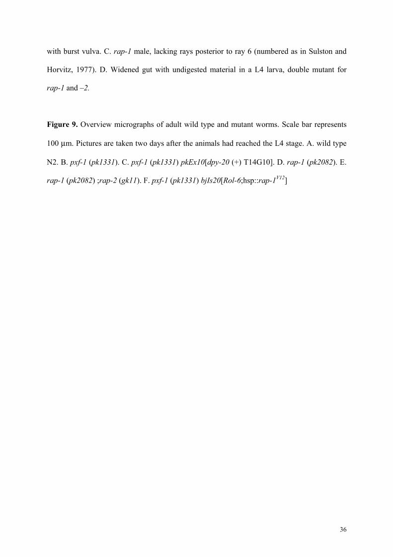
Figure 9. Overview micrographs of adult wild type and mutant worms. Scale bar represents
100 µm. Pictures are taken two days after the animals had reached the L4 stage. A. wild type
N2. B. pxf-1 (pk1331). C. pxf-1 (pk1331) pkEx10[dpy-20 (+) T14G10]. D. rap-1 (pk2082). E.
rap-1 (pk2082) ;rap-2 (gk11). F. pxf-1 (pk1331) bjIs20[Rol-6;hsp::rap-1V12]


TABLE I
Hours after hatching unc (%) vac (%) mol (%)
24 34 4 0
48 34 12 2
72 62 32 32
96 86 70 62
120 92 84 62
Table I. Cumulative percentage of pxf-1-/- animals (N = 62, analyzed in three separate
experiments), showing uncoordinated movement (unc), presence of vacuoles (vac) or
molting defect (mol).

TABLE II
Genotype % survival until adult stage # progeny
Wild type 100 165 + 20 (n = 5)
pxf-1 -/+ 100 155 + 36 (n
= 5)
pxf-1 -/- 25 (54/212) 5 + 3 (n = 14)
pxf-1 -/- pkEx10[dpy-20 (+) T14G10] 71 54+ 22
(n=14)
pxf-1 -/- bjIs20[Rol-6;hsp::rap-1V12;rap-1::GFP] 66 43+ 17 (n=14)
Table II. Survival and proliferation rates of pxf-1 mutant animals. Larvae (L1 or L2) from
heterozygous hermaphrodites were singled and scored for their survival to the adult stage. For
pxf-1 -/- bjIs20[Rol-6;hsp::rap-1V12;rap-1::GFP] counting of animals surviving to adulthood
and number of offspring was done on animals given a daily heat shock. Genotyping of
animals was done by PCR.