Regression of metastatic Merkel cell carcinoma following transfer of polyomavirus-specific T cells...
11
Research Article Regression of Metastatic Merkel Cell Carcinoma Following Transfer of Polyomavirus-Specific T Cells and Therapies Capable of Reinducing HLA Class-I Aude G. Chapuis 1 , Olga K. Afanasiev 3,4 , Jayasri G. Iyer 4 , Kelly G. Paulson 3,4 , Upendra Parvathaneni 8 , Joo Ha Hwang 9 , Ivy Lai 1 , Ilana M. Roberts 1 , Heather L. Sloan 1 , Shailender Bhatia 7 , Kendall C. Shibuya 1 , Ted Gooley 1 , Cindy Desmarais 10 , David M. Koelle 2,4,5,6,11 , Cassian Yee 1 , and Paul Nghiem 3,4 Abstract Merkel cell carcinoma (MCC) is an aggressive skin cancer that typically requires the persistent expression of Merkel cell polyomavirus (MCPyV) oncoproteins that can serve as ideal immunotherapeutic targets. Several immune evasion mechanisms are active in MCC, including downregulation of HLA class-I expression on tumor cells and dysfunctional endogenous MCPyV-specific CD8 T-cell responses. To overcome these obstacles, we combined local and systemic immune therapies in a 67-year-old man, who developed metastatic MCPyV- expressing MCC. Intralesional IFN-b-1b or targeted single-dose radiation was administered as a preconditioning strategy to reverse the downregulation of HLA-I expression noted in his tumors and to facilitate the subsequent recognition of tumor cells by T cells. This was followed by the adoptive transfer of ex vivo expanded polyclonal, polyomavirus-specific T cells as a source of reactive antitumor immunity. The combined regimen was well tolerated and led to persistent upregulation of HLA-I expression in the tumor and a durable complete response in two of three metastatic lesions. Relative to historical controls, the patient experienced a prolonged period without development of additional distant metastases (535 days compared with historic median of 200 days; 95% confidence interval, 154–260 days). The transferred CD8 þ T cells preferentially accumulated in the tumor tissue, remained detectable and functional for more than 200 days, persisted with an effector phenotype, and exhibited evidence of recent in vivo activation and proliferation. The combination of local and systemic immune stimulatory therapies was well tolerated and may be a promising approach to overcome immune evasion in virus-driven cancers. Cancer Immunol Res; 2(1); 1–10. Ó2013 AACR. Introduction Merkel cell carcinoma (MCC) represents a highly aggres- sive neuroendocrine skin malignancy with a high disease- associated mortality, early metastatic disease, and a high propensity for recurrence after initial treatment. The cause-specific mortality rate ranges from 23% to 80% at 5 years, thus making it three times as lethal as melanoma (1). The Merkel cell polyomavirus (MCPyV) is clonally integrated into at least 80% of MCC tumors and produces the viral T-antigen (T-Ag) oncoproteins that are persistently expressed by MCC and are necessary for the survival and proliferation of tumor cells (2–5). Compared with mammalian tumor-associ- ated antigens (TAA) that all have some degree of expression within normal tissue, MCPyV T-Ag expression is restricted to MCC, is a foreign antigen not subject to T-cell self-tolerance mechanisms, and is thus an optimal target for immunotherapy. Because no viral particles are formed in the tumor cells, antiviral agents are inefficient (6). As adoptive transfer has demonstrated clinical benefit for both viral and endogenous tumor antigens (7–9), we sought to apply the use of antigen-specific T cells to target the MCPyV large T-Ag (LT-Ag) oncoprotein. The HLA-A 2402- restricted MCPyV LT-Ag 92-101 -specific T cells (hereafter referred to as MCPyV-specific cells) were identified in a patient with metastatic MCC (10). These MCPyV-specificT cells, when isolated from tumors or peripheral blood mono- nuclear cells (PBMC) of patients with MCC, are largely Authors' Affiliations: 1 Program in Immunology; 2 Vaccine and Infectious Disease Division, Fred Hutchinson Cancer Research Center; Departments of 3 Pathology, 4 Medicine (Dermatology), 5 Laboratory Medicine, 6 Global Health, and 7 Medicine (Medical Oncology), University of Washington; Divisions of 8 Radiation Oncology and 9 Gastroenterology, University of Washington Medical Center; 10 Adaptive Biotechnologies; and 11 Benaroya Research Institute, Seattle, Washington Note: Supplementary data for this article are available at Cancer Immu- nology Research Online (http://cancerimmunolres.aacrjournals.org/). A.G. Chapuis and O.K. Afanasiev contributed equally to this work. Current address for I. Lai and C. Yee: Melanoma Medical Oncology, MD Anderson Cancer Center, Houston, TX; current address for I.M. Roberts: Surgery Branch, NIH, Bethesda, MD. Corresponding Authors: Paul Nghiem, Division of Dermatology, UWMC, 850 Republican Street, Box 358050, Seattle, WA 98195. Phone: 206-221- 2632; Fax: 206-221-4394; E-mail: [email protected]; and Cassian Yee, Department of Melanoma Medical Oncology, Department of Immunology Unit 0430, MD Anderson Cancer Center, 1515 Holcombe Blvd. Unit 904, Houston, TX 77030. Phone: 713-563-5272; Fax: 713-563-3424; E-mail: [email protected] doi: 10.1158/2326-6066.CIR-13-0087 Ó2013 American Association for Cancer Research. Cancer Immunology Research www.aacrjournals.org OF1 Research. on February 24, 2016. © 2013 American Association for Cancer cancerimmunolres.aacrjournals.org Downloaded from Published OnlineFirst November 4, 2013; DOI: 10.1158/2326-6066.CIR-13-0087
-
Upload
washington -
Category
Documents
-
view
1 -
download
0
Transcript of Regression of metastatic Merkel cell carcinoma following transfer of polyomavirus-specific T cells...

Research Article
Regression of Metastatic Merkel Cell Carcinoma FollowingTransfer of Polyomavirus-Specific T Cells and TherapiesCapable of Reinducing HLA Class-I
Aude G. Chapuis1, Olga K. Afanasiev3,4, Jayasri G. Iyer4, Kelly G. Paulson3,4, Upendra Parvathaneni8,Joo Ha Hwang9, Ivy Lai1, Ilana M. Roberts1, Heather L. Sloan1, Shailender Bhatia7, Kendall C. Shibuya1,Ted Gooley1, Cindy Desmarais10, David M. Koelle2,4,5,6,11, Cassian Yee1, and Paul Nghiem3,4
AbstractMerkel cell carcinoma (MCC) is an aggressive skin cancer that typically requires the persistent expression of
Merkel cell polyomavirus (MCPyV) oncoproteins that can serve as ideal immunotherapeutic targets. Severalimmune evasion mechanisms are active in MCC, including downregulation of HLA class-I expression on tumorcells and dysfunctional endogenous MCPyV-specific CD8 T-cell responses. To overcome these obstacles, wecombined local and systemic immune therapies in a 67-year-old man, who developed metastatic MCPyV-expressing MCC. Intralesional IFN-b-1b or targeted single-dose radiation was administered as a preconditioningstrategy to reverse the downregulation of HLA-I expression noted in his tumors and to facilitate the subsequentrecognition of tumor cells by T cells. This was followed by the adoptive transfer of ex vivo expanded polyclonal,polyomavirus-specific T cells as a source of reactive antitumor immunity. The combined regimen was welltolerated and led to persistent upregulation of HLA-I expression in the tumor and a durable complete response intwo of threemetastatic lesions. Relative to historical controls, the patient experienced a prolonged periodwithoutdevelopment of additional distant metastases (535 days compared with historic median of 200 days; 95%confidence interval, 154–260 days). The transferred CD8þ T cells preferentially accumulated in the tumor tissue,remained detectable and functional for more than 200 days, persisted with an effector phenotype, and exhibitedevidence of recent in vivo activation andproliferation. The combination of local and systemic immune stimulatorytherapies was well tolerated and may be a promising approach to overcome immune evasion in virus-drivencancers. Cancer Immunol Res; 2(1); 1–10. �2013 AACR.
IntroductionMerkel cell carcinoma (MCC) represents a highly aggres-
sive neuroendocrine skin malignancy with a high disease-
associated mortality, early metastatic disease, and a highpropensity for recurrence after initial treatment. Thecause-specific mortality rate ranges from 23% to 80% at 5years, thus making it three times as lethal as melanoma (1).The Merkel cell polyomavirus (MCPyV) is clonally integratedinto at least 80% of MCC tumors and produces the viralT-antigen (T-Ag) oncoproteins that are persistently expressedby MCC and are necessary for the survival and proliferation oftumor cells (2–5). Compared with mammalian tumor-associ-ated antigens (TAA) that all have some degree of expressionwithin normal tissue, MCPyV T-Ag expression is restricted toMCC, is a foreign antigen not subject to T-cell self-tolerancemechanisms, and is thus an optimal target for immunotherapy.Because no viral particles are formed in the tumor cells,antiviral agents are inefficient (6).
As adoptive transfer has demonstrated clinical benefit forboth viral and endogenous tumor antigens (7–9), we soughtto apply the use of antigen-specific T cells to target theMCPyV large T-Ag (LT-Ag) oncoprotein. The HLA-A�2402-restricted MCPyV LT-Ag92-101-specific T cells (hereafterreferred to as MCPyV-specific cells) were identified in apatient with metastatic MCC (10). These MCPyV-specific Tcells, when isolated from tumors or peripheral blood mono-nuclear cells (PBMC) of patients with MCC, are largely
Authors' Affiliations: 1Program in Immunology; 2Vaccine and InfectiousDisease Division, Fred Hutchinson Cancer Research Center; Departmentsof 3Pathology, 4Medicine (Dermatology), 5Laboratory Medicine, 6GlobalHealth, and 7Medicine (Medical Oncology), University of Washington;Divisions of 8Radiation Oncology and 9Gastroenterology, University ofWashington Medical Center; 10Adaptive Biotechnologies; and 11BenaroyaResearch Institute, Seattle, Washington
Note: Supplementary data for this article are available at Cancer Immu-nology Research Online (http://cancerimmunolres.aacrjournals.org/).
A.G. Chapuis and O.K. Afanasiev contributed equally to this work.
Current address for I. Lai and C. Yee: Melanoma Medical Oncology, MDAnderson Cancer Center, Houston, TX; current address for I.M. Roberts:Surgery Branch, NIH, Bethesda, MD.
Corresponding Authors: Paul Nghiem, Division of Dermatology, UWMC,850 Republican Street, Box 358050, Seattle, WA 98195. Phone: 206-221-2632; Fax: 206-221-4394; E-mail: [email protected]; and Cassian Yee,Department of Melanoma Medical Oncology, Department of ImmunologyUnit 0430, MD Anderson Cancer Center, 1515 Holcombe Blvd. Unit 904,Houston, TX 77030. Phone: 713-563-5272; Fax: 713-563-3424; E-mail:[email protected]
doi: 10.1158/2326-6066.CIR-13-0087
�2013 American Association for Cancer Research.
CancerImmunology
Research
www.aacrjournals.org OF1
Research. on February 24, 2016. © 2013 American Association for Cancercancerimmunolres.aacrjournals.org Downloaded from
Published OnlineFirst November 4, 2013; DOI: 10.1158/2326-6066.CIR-13-0087
dysfunctional and exhibit an immune inhibitory (PD-1þ/Tim3þ) phenotypic profile (11). We hypothesized that ex vivogeneration of polyclonal MCPyV-specific T cells might aug-ment the probability of including and expanding cells thathad an increased potential for proliferation, function, andpersistence after transfer as evidenced in murine and non-human primate models (12, 13).
Similar to the observations in other virus-associated can-cers, HLA class-I (HLA-I) downregulation is an immune escapemechanism present in the majority of MCC tumors (14–16).Single-dose low-dose radiation has been shown to upregulatecell surface HLA-I expression (17). Data from murine modelssuggest that single-dose radiation is more effective than frac-tionated radiation in promoting tumor immunity, because thelatter may suppress the function of lymphocytes that arerecruited to the tumor (18). Furthermore, IFNs direct theupregulation of HLA-I (19), and intralesional administrationof IFN-b has been observed to promote immune responses inMCC (14, 20).
Here, we investigated whether adoptive transfer of poly-clonal MCPyV-specific CD8þ T-cells following HLA-I upregu-lation strategies (intralesional IFN-b or local, single-fractionradiotherapy) could safely establish persistent anti-MCCresponses, migrate to tumor tissue, and induce regression ofMCPyV-positive, HLA-I–deficient MCC metastases.
Case reportA 67-year-old man with chronic renal failure secondary to a
nephrectomy for renal cell carcinoma (RCC) at the age of 50presented with a 1.6-cm (in largest dimension) MCC lesion onhis left upper thigh and a negative sentinel node biopsy(American Joint Committee onCancer stage IA). He underwenta wide local excision followed by 50 Gy of fractionated localradiation to the primary site. Eight months later while stillasymptomatic, a surveillance whole body positron emissiontomography (PET) scan detected a 2.9� 1.8 cm lesion adjacentto the pancreatic head. Merkel polyomavirus-specific CD8 Tcells were identified in the patient's tumor-infiltrating lym-phocytes (TIL) and peripheral blood using a MCPyV-tetramer(ref. 10; Supplementary Fig. S1). The patient underwent leu-kapheresis to allowMCPyV-specificT cells to be harvested. Thepatient was enrolled in a single-patient clinical trial of autol-ogous T-cell therapy for MCC [Fred Hutchinson Cancer
Research Center (FHCRC, Seattle, WA) protocol #2558]. Boththe primary tumor and pretreatment metastasis were con-firmed to be classical MCC by dot-like cytokeratin-20 staining.Both lesions expressed the MCPyV T-Ag, whereas HLA-Iexpression was absent or sparse. Two additional metastasesin the pancreatic head and neck appeared 137 days before theinitiation of therapy.
The patient underwent three rounds of treatment (Fig. 1).Intralesional IFN-b-Ib (3 � 106 IU) was first administered byendoscopic ultrasound-guided fine-needle injection to thelargest of the three detectable pancreatic metastases (desig-nated as metastasis A). After 24 hours, the patient receivedan infusion of 1010/m2 MCPyV tetramer-specific polyclonalCD8þ T cells followed by low-dose s.c. interleukin (IL)-2 (2.5�105 IU/m2) twice daily for 14 days as a means to increase T-cellpersistence (21). Thirty-seven days later, a single 8-Gy fractionof radiation was delivered to all three remaining lesions(designated as metastases A, B, and C) followed by a secondinfusion of 1010/m2MCPyV-specific CD8þ T cells and low-doses.c. IL-2. On day 148 after the first infusion, metastasis B wasinjected with IFN-b-1b followed by a third cell infusion andlow-dose s.c. IL-2� 14 days. The patient experienced low-gradefevers and a transient, less than 72-hour grade 1–2 lympho-penia after each infusion, consistent with the expected imme-diate toxicities associated with T-cell infusions (22, 23). Nochanges in end organ function, inflammation- or autoimmune-related complications were observed. As detailed in the Resultssection, follow-up scans showed no evidence of new distantdisease until 535 days after the original pancreatic metastasiswas detected. At that time, the patient developed new brainlesions that he declined to have worked up or treated. Thepatient died 1 month later without additional therapeuticinterventions.
Materials and MethodsClinical protocol and patient characteristics
All clinical investigations were conducted according tothe Declaration of Helsinki principles. The single-patientprotocol #2558 (described above) was approved by theFHCRC Institutional Review Board and the U.S. Food andDrug Administration. The patient provided written informedconsent.
Figure 1. Timeline for disease presentation and immune therapy. Events as indicated relative to immune therapy start date. The patient received a total ofthree treatments that consisted of HLA-I upregulation (either intralesional injection of 3� 106 IU of IFN-b-Ib or 8Gy radiotherapy) followed by administration of1010/m2 MCPyV-specific polyclonal CD8þ T cells and 14 days of twice-daily s.c. injections of IL-2 (2.5 � 105 IU/m2). XRT, radiotherapy.
Chapuis et al.
Cancer Immunol Res; 2(1) January 2014 Cancer Immunology ResearchOF2
Research. on February 24, 2016. © 2013 American Association for Cancercancerimmunolres.aacrjournals.org Downloaded from
Published OnlineFirst November 4, 2013; DOI: 10.1158/2326-6066.CIR-13-0087

Isolation and expansion of MCC-specific CTLsPBMCs were collected by leukapheresis and all ensuing ex
vivo manipulations involving processing of products destinedfor infusion were performed in the cGMP Cell ProcessingFacility of the FHCRC based on a protocol established forin vitro enrichment of low frequency antigen-specific cytotoxicT lymphocytes (CTL; refs. 24, 25). To facilitate the isolation ofantigen-specific CTL, PBMCswere depleted of CD25þT cells toeliminate regulatory T cells (Tregs; ref. 26;Miltenyi Biotec Inc.),and stimulated twice for 7 to 10 days with HLA-A�2402-restricted MCPyV LT-Ag92-101 peptide (CPC Scientific)-pulsedautologous dendritic cells. Each stimulation was supplemen-tedwith the gc-chain cytokines IL-2 (10 IU/mL), IL-7 (5 ng/mL),and IL-21 (30 ng/mL). Cultures that contained more than5% specific CD8þ T cells assessed by tetramer stains wereclinical-grade sorted (BD Influx cell sorter, BD Biosciences)before expansion to sufficient numbers for infusion as previ-ously described (21, 27, 28). Cell products bound the MCPyVLT-Ag92-101 peptide-HLA tetramer, secreted IFN-g , and lysedMCPyV LT-Ag92-101-pulsed FUJI (A24
þ cell line) pulsed with 10mg/mL peptide (Supplementary Fig. S2).
T-cell tracking by HLA-peptide tetramersTetramers (produced by the FHCRC immune monitoring
core facility) were used to detect transferred MCPyV LT-Ag92-101-specific CD8þ T cells in PBMCs collected after infusions.The sensitivity of the tetramer is 0.05% of total CD8þ T cells.Persistence after infusionswas calculated as the last time pointat which tetramerþ T cells were detected at 2 � backgroundlevels or �0.05%.
Flow cytometryBlood samples were collected at the indicated time points,
and the PBMCs were isolated by Ficoll-Hypaque gradient andcryopreserved. Cells that bound tetramer were analyzed byflow cytometry after staining with fluorochrome-conjugatedmonoclonal antibodies to CD14, CD16, CD19 (dump channel),CD8, CD4, CD137, CD28, CD127, CD62L, CCR7, PD-1, and TIM-3 (BD-Pharmingen). Intracellular cytokine production of IFN-g , TNF-a, and IL-2 by cells responding to in vitro stimulationwith MCPyV LT-Ag92-101 peptide for 4 to 5 hours were per-formed as described (29). Intranuclear expression of Ki-67 wasassessed after permeabilization (eBioscience). Cells were ana-lyzed on an LSRII cytometer (Becton Dickinson) using FACS-Diva software.Multiparametricflow cytometry staining, acqui-sition, and analyses were performed on all samples in a batchon the same day and one negative control for each parameterwas used. As the percentages of tetramerþ T cells in thesamples are often low, this method dedicated the majority ofthe PBMC for sample analyses.
Tracking polyclonal T cells using high-throughputTCR-b chain DNA sequencingA pool of primers to all V and J pairs specifically designed to
amplify the complete VDJ junction region, was designed suchthat only the minimal region (60 nucleotides) containing theantigen-specific nucleotide information for each T-cell recep-tor (TCR)-b CDR3 could be amplified and sequenced using the
immunoSEQ assay (Adaptive Biotechnologies; ref. 30). Usinggenomic DNA isolated from the blood and tumor samples as atemplate, this method was used to capture the frequency ofindividual TCRs in biologic samples with accurate reproduc-ibility and a sensitivity of 1/100,000 TCR-containing lympho-cytes (31, 32). DNA from the blood and tumor sampleswas prepared using the DNeasy Blood & Tissue Kit (Qiagen).The sources of available cells for DNA extraction includethe tetramer-sorted infusion product, cryopreserved PBMCderived using Ficoll isolation, phytohemagglutinin and IL-2–expanded TIL isolated from the primary tumor, and freshlyisolated cells from the pancreatic metastatic tumor biopsyfollowing immune therapy treatment. The results of individualTCR clonotypes are expressed as a percentage of all productive,in-frame TCRs sequenced.
ImmunohistochemistryAnti-CD8 clone 4B11 (Novocastra), anti-MCPyV T-Ag (5),
and anti-HLA-I clone EMR8-5 (MBL International) were usedat 1:200, 1:4,000, and 1:200 dilutions, respectively, after heat-induced epitope retrieval. Specimenswere assessed forMCPyVT-Ag andHLA-I expression and scored using theAllredmethod(33). An Allred score of 0 to 1.5 was considered negative (0), 2 to5.5 weak (þ), 6 to 7.5 moderate (þþ), and 8 strong (þþþ).Peritumoral and intratumoral CD8 infiltrates were scoredseparately as previously described (14). The numbers of CD8þ
cells were assessed and expressed on a 0-to-5 scale with ameanof 0, 90, 306, 508, 675, and 732þ CD8s/mm2, respectively.Intratumoral CD8 lymphocytes were surrounded by tumorcells only with no visualized contact with stroma.
Statistical analysisThe observed time between the first metastasis and the
second metastasis for 49 patients was available and these datawere used as historic controls. All of these patients presentedwith local or regional disease (stage I, II, or III) and laterdeveloped distant metastatic disease, for which they thenreceived treatment. The time between metastases was treatedas a time-to-event endpoint, and from these controls, themedian time to second metastasis was estimated along witha point-wise estimate of the 95% confidence interval for themedian time. We only included a highly analogous patientpopulation from our historical controls. Cases that would notbe eligible for the trial were eliminated, including those withprofound immune suppression and those who only had asingle subsequent skinmetastasis (these are often independentprimary lesions or represent a very benign subset of patients).Statistical tests were performed with the GraphPad prismsoftware version 3.0, with the R-package for statistical analysis(http://www.r-project.org), or SAS version 9.2.
ResultsHLA-I upregulation followed by adoptive transfer ofMCPyV-specific T cells induces regression of Merkel cellcarcinoma
Pre- and posttreatment tumor tissue immunohistochemis-try was used to evaluate viral oncoprotein expression, HLA-Iexpression, and CD8 lymphocyte infiltration (Fig. 2A). As
HLA-I Upregulation and Adoptive Immune Therapy in MCC
www.aacrjournals.org Cancer Immunol Res; 2(1) January 2014 OF3
Research. on February 24, 2016. © 2013 American Association for Cancercancerimmunolres.aacrjournals.org Downloaded from
Published OnlineFirst November 4, 2013; DOI: 10.1158/2326-6066.CIR-13-0087
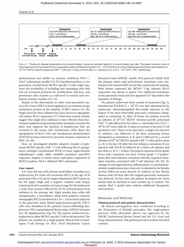
Figure 2. Response of individualMCC metastases after combinedimmune therapy. A, columns fromleft to right, hematoxylin and eosin(H&E) stains, immunohistochemistryfor MCPyV T-Ag, HLA-I, and CD8(red arrows, red asterisk indicatescell in the inset) of the primarytumor, pretreatment (metastasis A,biopsied immediately before startof immune therapy) andposttreatment (metastasis B,biopsied 148 days after start ofimmune therapy) tumors. Scalebar, 50 mm. B, MRI imaging (exceptthe first datapoint, day 146, whichwas obtained by PET/CT) ofindividual metastases thatincluded a peri-pancreatic lesionadjacent to the anterior duodenum(metastasis A, red), a pancreaticneck lesion (metastasis B, green),and a pancreatic head lesion(metastasis C, blue). The longestdiameter in cm (y-axis) of eachmetastasis is graphed over time(x-axis). Representative images ofpre- and posttreatment PET/CTscans are shown above the graph.Gray bars on x-axis indicate thetiming of each treatment as detailedin Fig. 1. The correspondingRECIST 1.1 criteria are indicatedbelow the graph. C, Kaplan–Meiercurve of the probability ofdeveloping a second distantmetastasis after the first detectedmetastasis (plotted at day zero) in49 patients with MCC whodeveloped distant disease. The95% confidence interval isindicated in red.
Chapuis et al.
Cancer Immunol Res; 2(1) January 2014 Cancer Immunology ResearchOF4
Research. on February 24, 2016. © 2013 American Association for Cancercancerimmunolres.aacrjournals.org Downloaded from
Published OnlineFirst November 4, 2013; DOI: 10.1158/2326-6066.CIR-13-0087

anticipated, the primary tumor and pretreatment metastasis Abiopsies were positive for the MCPyV T-Ag oncoprotein, buthad weak or no HLA-I expression. In contrast, HLA-I wasstrongly upregulated on the biopsy of the posttreatmentmetastasis (metastasis B) taken 146 days after the first treat-ment cycle, and as expected, MCPyV T-Ag expression waspersistently maintained. Scattered CD8þ T-cell infiltrates weredetected in the primary tumor, as well as in the pre- andposttreatment biopsies (Fig. 2A).Tumor burden and clinical efficacy were monitored using
MRI and PET/CT scans (Fig. 2B). Immediately before the firsttreatment, the patient's initial pancreatic metastasis hadenlarged, and two adjacent pancreatic tumors were newlydetected compared with the scan performed 146 days earlier.Restaging 30 days after the first treatment showed that thelargest lesion, which had undergone injection with IFN-b-Ib,had decreased in size, but the other two lesions had remainedstable or increased in size. The best overall response byResponse Evaluation Criteria in Solid Tumors (RECIST) wasa partial response observed following the second treatmentconsisting of localized, single-dose 8-Gy radiation before T-cellinfusion and low-dose s.c. IL-2. After the third treatment, twoof three lesions continued to regress and one lesion remainedrefractory to treatment.To determine systemic effects of adoptively transferred T
cells, the time for new metastasis to develop after treatmentwas assessed in comparison with 49 historical matchedcontrols undergoing standard, typically cytotoxic chemo-therapy (Fig. 2C). The patient showed no additional systemicmetastasis until 535 days after the appearance of the firstmetastasis, which greatly exceeds the estimated mediantime of 200 days (95% confidence interval, 154–260 days)for development of new distant metastatic disease in his-toric controls. The scan performed 535 days after the initialmetastasis revealed new brain lesions that the patientdeclined work-up. These likely represented MCC metastases;however, as the patient also had a history of remote RCC andno biopsy was performed, metastasis from an RCC origincould not be excluded. Overall, these data suggest thetreatment-induced regression in two of three metastasesand may have contributed to a delay in the development ofnew distant disease.
Enhanced ability to respond to antigen after transfer ofvirus-specific CD8þ T cellsThe frequency and persistence of MCPyV-specific CD8 T
cells in peripheral blood were assessed by their ability to bindthe MCPyV-specific tetramer (Fig. 3A, solid circles). The fre-quency of these cells peaked 4 to 7 days after infusions (up to8.8% of total CD8þ T cells) and remained detectable at the lastassessment (219 days after thefirst treatment) at frequencies ofapproximately 1%, which corresponded to a more than 3-foldincrease compared with preinfusion levels (0.26%).We next assessed whether the virus-specific T cells detected
after treatment were functional as assessed by their ability tosecrete IFN-g in response to cognate peptide. Before thetreatment cycles, cells that secreted IFN-g after exposure tothe MCPyV peptide could not be detected within PBMC,
suggesting that the MCPyV-specific cells binding to tetramerin the pretreatment PBMC were nonfunctional. However, thefrequency of CD8 T cells that secreted IFN-g in response toMCPyV peptide increased after each infusion (5.1, 4.5, and 3%after treatments 1, 2, and 3, respectively) and remained detect-able at frequencies of 1%ormore of total CD8þT cells up to 219days after thefirst infusion (Fig. 3A, open circles). Furthermore,the frequency of MCPyV-reactive peptide-responsive CD8þ Tcells correlated closely with the frequency of MCPyV tetramer-positive cells, suggesting that most of the persisting virus-specific cells remained functional in vivo.
Because an increased frequency of virus-specific T cells overtime may reflect antigen-specific cell division, we investigatedwhether the persistingMCPyV-specific T cells expressed Ki-67,a marker of proliferation (34). Before infusion, endogenous(preexisting) MCPyV-specific T cells did not express Ki-67,indicating minimal/absent proliferation. Cells from the infu-sion product were overwhelmingly positive for Ki-67 (92% atday 14 after the last stimulation cycle in vitro), consistent withrecent activation. Most transferred cells maintained Ki-67expression early after transfer (>80% on day 1 after infusion)and, as expected, Ki-67 expression decreased after each infu-sion but remained well above baseline levels (Fig. 3B, soliddiamonds) 219 days after the first infusion. Ki-67 expressionon MCPyV-specific CD8 T cells was overall significantly higher(P� 0.005) than Ki-67 expression of the host tetramer-negativeCD8 Tcells (Fig. 3B, open diamonds), suggesting that theinfused cells continued to divide in the presence of persistentantigen.
To validate the functional status and determine the differ-entiation profile of pre- and posttreatment virus-specific CD8T cells, we evaluated the phenotype of the persisting trans-ferred cells. Consistent with the absence of function andproliferation, both PD-1 (a marker of activation/exhaustion)and TIM-3 (a marker of exhaustion) were expressed onMCPyV-specific cells (90% and 78%, respectively) found inPBMC before the treatment cycles, suggesting endogenousCD8 responses were dysfunctional and exhausted (Fig. 3C andD). As expected, and following ex vivo expansion, the infusionproduct also expressed PD-1 (34%) and TIM-3 (93%). However,once infused into the patient, the transferred cells maintainedPD-1 expression but lost Tim-3 expression such that cellsfound after transfer had a nonexhausted phenotype (Fig.3C), consistent with their maintained function. Furthermore,7 days after transfer, 53% of MCPyV-specific cells transientlyexpressed CD137 (4-1BB), amarker associated with very recentactivation (Fig. 3E).
On the basis of the absence of expression of the costimu-latory molecule CD28, the IL-7 receptor CD127 as well as thelymph node homing molecules, CD62L and CCR7 (data notshown), the endogenous MCPyV-specific T cells found beforeinfusions exhibited a differentiated effector memory pheno-type (Fig. 3F and G). Consistent with the use of IL-21 in the exvivo cultures (24–26), a subset of the infusion product gener-ated for this patient expressed CD28 (78%) and CD127 (19%).After infusion into the patient, the transferred cellsmaintainedCD28 expression (Fig. 3F) and further upregulated CD127 (Fig.3G). CD62L and CCR7 were not detected (data not shown).
HLA-I Upregulation and Adoptive Immune Therapy in MCC
www.aacrjournals.org Cancer Immunol Res; 2(1) January 2014 OF5
Research. on February 24, 2016. © 2013 American Association for Cancercancerimmunolres.aacrjournals.org Downloaded from
Published OnlineFirst November 4, 2013; DOI: 10.1158/2326-6066.CIR-13-0087

These data suggest that the transferred cells persisted with aneffector memory phenotype (13), but in a less differentiatedstate compared with the endogenous response, with a main-tained capacity to signal through CD28 and CD127. Tregs canbe sensitive to exogenous IL-2, and could have a negativeimpact on the persistence and function of transferred cytotoxiclymphocytes. Therefore, we assessed Treg numbers after infu-sions based on the expression of surrogate markers CD4 andCD25 and the absence of CD127 (24). Treg frequenciesincreased after each infusion from baseline levels but returnedto near baseline levels within 28 days of the infusion (Supple-mentary Fig. S3). In TIL, unfortunately, wewere severely limitedby the quantity of tissue available for analysis from the patient'smetastases and thereforeTreg analysis could not be performed.
Overall, adoptive transfer of polyclonal, MCPyV-specific Tcells was associated with several key characteristics suggestiveof an enhanced ability to respond to antigen: (i) markedlyincreased frequency of MCPyV-specific cells that persistedin vivo, (ii) increased fraction of virus-reactive T cells, (iii)ability to proliferate (Ki-67), (iv) expression of activationmarkers (CD137, PD-1), and (v) facilitated costimulation andsurvival (CD28 and CD127; refs. 35, 36).
Transferred cells preferentially localize to metastaticMCC tissue
To capture the number and characteristics of the individualtransferred MCPyV-specific CD8þ T-cell clones, we analyzedtetramer-sorted infusion products, TIL, and PBMC for individ-ual TCR-b CDR3 using high-throughput TCR DNA sequencing(32). The infusion product (which was sorted a second time toyield>99% pure CD8þ tetramerþ cells for TCR-bCDR3 analysis)was polyclonal and consisted of 502 individual virus-specificclonotypes. Three clonotypes were predominant and repre-sented 99% of MCPyV-specific TCR reads (all three clonesshared V-b7-9 and differed by their J-b subunits as depictedin Fig. 4A and B). We then used this method to determine allTCR-b CDR3 present in unsorted TIL and PBMC obtainedbefore and after infusions and investigated whether the trans-ferred virus-specific clonotypes were present within these sam-ples, as well as their frequency. Before treatment, these three J-bvariant clones were present among primary tumor TIL (0.03%J-b1-5, 0.06% J-b1-1, 2.8% J-b2-3; Fig. 4C, left), and PBMC (0.06%J-b1-5, 0.15% J-b1-1, 0.01% J-b2-3; Fig. 4D, left), suggestingthat these preexisting clonotypes had been expanded in thePBMC-derived infusion product. Analysis of the posttreatment
Figure 3. Persistence, function, and phenotype of transferredMCPyV-specific CD8þ T cells. A, assessment of tetramerþCD8þ T cells (%, solid circles, solid line)and IFN-g–reactiveCD8þTcells (%,opencircles,dashed line) inPBMCcollectedatbaseline (137daysand immediatelybefore thefirst treatment)andat indicatedtime points after treatments. Gray bars on x-axis indicate the timing of each treatment as detailed in Fig. 1. B, intranuclear Ki-67 expression on baseline andposttreatment CD8þtetramerþ cells (solid diamonds, solid line), and CD8þtetramer� cells (open diamonds, dashed line). Gray bars on the x-axis indicatedthe timing of treatments as in A above. C–G, expression of PD-1, TIM-3, CD137, CD28, and CD127 on the infusion product, tetramerþCD8þ PBMC collected atbaseline and after treatment as indicated. All parameters were acquired from multiple time points on the same day using the same negative control foreach parameter as described in Materials and Methods, except for CD137 on day 45. A two-tailed paired t test was used for statistical analysis.
Chapuis et al.
Cancer Immunol Res; 2(1) January 2014 Cancer Immunology ResearchOF6
Research. on February 24, 2016. © 2013 American Association for Cancercancerimmunolres.aacrjournals.org Downloaded from
Published OnlineFirst November 4, 2013; DOI: 10.1158/2326-6066.CIR-13-0087

metastasis 146 days after the first infusion demonstrated thatclone Jb1-1 now represented 4.7% of all sequences, and theclones Jb1-5 and Jb2-3 were absent (Fig. 4C, right).We then investigated whether additional clonotypes that
were present in the posttreatment metastasis were also pres-ent within the low-prevalence MCPyV-specific clonotypes(<1%) in the infusion product. This analysis identified anadditionalMCPyV-specific clonotype (highlighted in red), withone nucleic-acid difference compared with the Jb1-1 clonotypehighlighted in blue that was present at 0.6% of all TCR reads inthe posttreatment metastatic biopsy and absent in the pre-treatment TIL. However, because the frequency in the infusionproduct was so low (2.6� 10-4%), we cannot formally excludethat this clone constituted a contaminant.Overall, MCPyV-specific CD8 T-cell clonotypes present in the
infused product increased from 2.9% in the primary tumor to5.3% in the postinfusion metastasis (Fig. 4C). Two clonesaccounted for the increase in MCPyV-specific cells present in
the biopsy metastasis following the treatments compared withtheprimary lesion.No increase in frequencyof these three cloneswasobserved in theperipheral bloodbefore andafter treatmentsat comparable timepoints (Fig. 4D, right). Using thismethod, thespecificity of themajority of the clonotypes isolated fromPBMCand TIL could not be determined and may have included (i) Tcells specific for other MCPyV epitopes; (ii) T cells recognizingnonviral TAAs such as survivin (37); HIP1 oncoprotein thatinteractswith c-KIT (38), CD56, or otherUV-inducedmutationalsignatures; or (iii) bystander or resident T cells. In summary, asthe infusion product contained clones that aggregated at ahigher frequency in the metastatic lesion compared with theperipheral blood, this suggests there was preferential localiza-tion of infused virus-specific T cells to the metastatic tissue.
DiscussionThe purpose of this study was to investigate the efficacy and
safety of HLA-I upregulating agents in combination with
Figure 4. TCR clonotypic analysis for infused T cells and TIL within MCC tumors. A, legend for the four Vb7-9 TCR clonotypes that were MCPyV-specific asassessed by HLA-A24–restricted MCPyV-specific tetramer binding. B, pie chart indicating the individual TCR-b CDR3 clonotypes composing the CD8þ
tetramerþ MCPyV-specific T cells isolated from the infusion product. Gray indicates other clonotypes found in the tetramer-sorted infusion product. C, piecharts indicating the prevalence of the individual TCR-b CDR3 clonotypes present in the infusion product among all TCR-b CDR3 clonotypes isolatedfrom expanded TIL from the primary tumor before treatment (left) and directly ex vivo from metastasis B after treatment (day þ148, right). The relativepercentages of the individual clones are indicated. Gray indicates all other clonotypes found in the biopsy sample. D, pie charts tracking the prevalence of theindividual TCR-b CDR3 clonotypes among PBMC before and at several time points after treatment.
HLA-I Upregulation and Adoptive Immune Therapy in MCC
www.aacrjournals.org Cancer Immunol Res; 2(1) January 2014 OF7
Research. on February 24, 2016. © 2013 American Association for Cancercancerimmunolres.aacrjournals.org Downloaded from
Published OnlineFirst November 4, 2013; DOI: 10.1158/2326-6066.CIR-13-0087
polyomavirus-specific adoptive T-cell therapy in the setting ofmetastatic MCC. Even though preexisting MCPyV-specificCD8þ T cells were present at low frequencies in the host beforeinfusions (10), these were unable to prevent the rapid progres-sion of metastatic disease. We addressed two key factors thatmay have contributed to the inefficiency of the endogenousvirus-specific T-cell responses: To reverse the observed down-regulated HLA-I expression on tumor cells necessary for thedisplay of the MCPyV viral peptides, and render the cellsaccessible to specific T-cell lysis, either intralesional IFN-b-1b and tumor-targeted, single-dose ionizing radiation (8 Gy)was administered 1 to 2 days before each T-cell infusion. Toaddress the absence of function of the endogenous CD8responses, polyclonal MCPyV-specific CD8 T cells were gen-erated ex vivo from PBMC, expanded, and reinfused. Thisresulted in the increased frequency of detectable and func-tional CD8 T cells for more than 3 months in vivo andpreferential localization of the infused cells in tumor tissueas evidenced by TCR clonotype analysis. The combination ofHLA-I upregulation followed by the infusion ofMCPyV-specificCD8 T cells was well tolerated and safe and mediated tumorregression in two of three detectable metastases consistentwith T-cell–mediated lysis. The absence of new metastaticdisease in this patient for a prolonged period of time comparedwith historic controls also suggests that the treatment mayhave delayed or prevented the progression of distant meta-static disease. Although the patient declined definitive work-up, it seems he developedmetastatic MCC disease in the brain,a site with limited lymphocyte accessibility (39).
Although a clear increase in antitumor reactivity wasachieved with this treatment, immune escape was not entirelyhalted. Although regressing lesions in the pancreas could notbe biopsied, the escaping pancreatic lesion was amenable to alimited fine-needle aspirate. Our data indicate that HLA-I andMCPyV T-Ag expression were maintained on the escapinglesion, and thus their respective downregulation was elimi-nated as having played a role in immune evasion in thepancreas. Othermechanismsmayhave contributed to immuneescape in some lesions in the patient. Infiltration of tumortissue by CD8þ T cells has been shown to have a positive effecton survival in numerous cancer settings. TIL density anddistribution were shown to independently predict sentinellymph node status and survival in patients with melanoma(40, 41). In MCC, a clear correlation between CD8þ T-cellinfiltration and favorable clinical outcome has been estab-lished, consistent with a role for the recognition of tumor cellsby host T cells (14). In this patient, CD8þ T-cell infiltration inthe primary tumor and pre- and posttreatment metastaticlesions remained scarce, suggesting that other factors mayhave prevented CD8þ infiltration and effective tumor lysis. A
recent report suggests that vascular E-selectin within MCCtumors is critical in mediating intratumoral CD8þ T-cellinfiltration (42); however, the intratumoral vascular architec-ture could not be assessed in this case. Furthermore, PD-1expression has been shown to be higher in the tumor-infil-trating CD8þ T cells and is associated with disease progression(43–45). Although transferred cells were PD-1þ and functionalin the peripheral blood, the function of the CD8þ T cellsinfiltrating the tumor could not be assessed. Ultimately, moreeffective results might also be achieved by combining adoptivetransfer with immunomodulatory strategies such as blockadeof the PD-1/PD-L1 axis or anti-CTLA-4 blockade (46–48).
Similar to other virus-driven cancers, MCC is an attractivetarget for immunotherapy because characterized, non-self,non-cross–reactive antigens are constitutively expressed andnecessary for tumor survival (3). The results of this single-patient study suggest that immune-mediated antitumor activ-ity can be established with a very limited side effect profile andoffers a strategy to rapidly elucidate the requirements for MCCtumor control directly in humans by bypassing the necessity ofmurinemodels (49, 50). Amultipatient clinical trial is currentlyunder way (NCT01758458) to further test this combinatorialimmunotherapy approach using additional viral CD8 T-cellepitopes in a broader cohort of patients with MCC.
Disclosure of Potential Conflicts of InterestC. Desmarais has ownership interest (including patents) in Adaptive Bio-
technologies. No potential conflicts of interest were disclosed by the otherauthors.
Authors' ContributionsConception and design: A.G. Chapuis, O.K. Afanasiev, J.G. Iyer, K.G. Paulson,D.M. Koelle, C. Yee, P. NghiemDevelopment of methodology: A.G. Chapuis, O.K. Afanasiev, J.G. Iyer, K.G.Paulson, D.M. Koelle, C. YeeAcquisition of data (provided animals, acquired and managed patients,provided facilities, etc.): A.G. Chapuis, O.K. Afanasiev, J.G. Iyer, K.G. Paulson,J.H. Hwang, I. Lai, H.L. Sloan, S. Bhatia, K.C. Shibuya, C. Desmarais, C. YeeAnalysis and interpretation of data (e.g., statistical analysis, biostatistics,computational analysis): A.G. Chapuis, O.K. Afanasiev, K.G. Paulson, I.M.Roberts, S. Bhatia, T. Gooley, C. Desmarais, C. Yee, P. NghiemWriting, review, and/or revision of the manuscript: A.G. Chapuis, O.K.Afanasiev, K.G. Paulson, J.H. Hwang, H.L. Sloan, S. Bhatia, C. Yee, P. NghiemAdministrative, technical, or material support (i.e., reporting or orga-nizing data, constructing databases): K.G. Paulson, I.M. Roberts, H.L. Sloan,C. Desmarais, P. NghiemStudy supervision: A.G. Chapuis, O.K. Afanasiev, C. Yee, P. Nghiem
AcknowledgmentsThe authors thankDr. James A. DeCaprio for providing theMCPyV large T-Ag
antibody Ab3.
The costs of publication of this article were defrayed in part by the payment ofpage charges. This article must therefore be hereby marked advertisement inaccordance with 18 U.S.C. Section 1734 solely to indicate this fact.
Received June 29, 2013; revised October 24, 2013; accepted October 25, 2013;published OnlineFirst November 4, 2013.
References1. Henness S, Vereecken P. Management of Merkel tumours: an evi-
dence-based review. Curr Opin Oncol 2008;20:280–6.2. Feng H, Shuda M, Chang Y, Moore PS. Clonal integration of a
polyomavirus in human Merkel cell carcinoma. Science 2008;319:1096–100.
3. Houben R, Shuda M, Weinkam R, Schrama D, Feng H, Chang Y, et al.Merkel cell polyomavirus-infected Merkel cell carcinoma cells requireexpression of viral T antigens. J Virol 2010;84:7064–72.
4. Shuda M, Arora R, Kwun HJ, Feng H, Sarid R, Fern�andez-Figueras M-T, et al. Human Merkel cell polyomavirus infection I. MCV T antigen
Chapuis et al.
Cancer Immunol Res; 2(1) January 2014 Cancer Immunology ResearchOF8
Research. on February 24, 2016. © 2013 American Association for Cancercancerimmunolres.aacrjournals.org Downloaded from
Published OnlineFirst November 4, 2013; DOI: 10.1158/2326-6066.CIR-13-0087

expression in Merkel cell carcinoma, lymphoid tissues and lymphoidtumors. Int J Cancer 2009;125:1243–9.
5. Rodig SJ, Cheng J, Wardzala J, DoRosario A, Scanlon JJ, Laga AC,et al. Improved detection suggests all Merkel cell carcinomas harborMerkel polyomavirus. J Clin Invest 2012;122:4645–53.
6. Hadrup S, Donia M, Thor Straten P. Effector CD4 and CD8 T cells andtheir role in the tumor microenvironment. Cancer Microenviron 2012;6:123–33.
7. Chapuis AG, Thompson JA,Margolin KA, RodmyreR, Lai IP, DowdyK,et al. Transferred melanoma-specific CD8þ T cells persist, mediatetumor regression, and acquire central memory phenotype. Proc NatlAcad Sci U S A 2012;109:4592–7.
8. HeslopHE,SlobodKS,PuleMA,HaleGA,RousseauA,SmithCA, et al.Long-term outcome of EBV-specific T-cell infusions to prevent or treatEBV-related lymphoproliferative disease in transplant recipients.Blood 2009;115:925–35.
9. Hunder NN, Wallen H, Cao J, Hendricks DW, Reilly JZ, Rodmyre R,et al. Treatment ofmetastaticmelanomawith autologousCD4þ T cellsagainst NY-ESO-1. N Engl J Med 2008;358:2698–703.
10. Iyer JG,AfanasievOK,McClurkanC,PaulsonK,NagaseK, Jing L, et al.Merkel cell polyomavirus-specific CD8(þ) and CD4(þ) T-cellresponses identified in Merkel cell carcinomas and blood. Clin CancerRes 2011;17:6671–80.
11. Afanasiev OK, Yelistratova L, Miller N, Nagase K, Paulson K, Iyer JG,et al. Merkel polyomavirus-specific T cells fluctuate with Merkel cellcarcinoma burden and express therapeutically targetable PD-1 andTim-3 exhaustion markers. Clin Cancer Res 2013;19:5351–60.
12. Wherry EJ, Teichgraber V, Becker TC,MasopustD, KaechSM, AntiaR,et al. Lineage relationship and protective immunity of memory CD8 Tcell subsets. Nat Immunol 2003;4:225–34.
13. Berger C, Jensen MC, Lansdorp PM, Gough M, Elliott C, Riddell SR.Adoptive transfer of effectorCD8þTcells derived fromcentralmemorycells establishes persistent T cell memory in primates. J Clin Invest2008;118:294–305.
14. Paulson KG, Iyer JG, Tegeder AR, Thibodeau R, Schelter J, Koba S,et al. Transcriptome-wide studies of Merkel cell carcinoma and vali-dation of intratumoral CD8þ lymphocyte invasion as an independentpredictor of survival. J Clin Oncol 2011;29:1539–46.
15. HaqueM,UedaK,NakanoK,HirataY,ParraviciniC,CorbellinoM, et al.Major histo-compatibility complex class I molecules are down-regu-lated at the cell surface by the K5 protein encoded by Kaposi'ssarcoma-associated herpes virus/human herpesvirus-8. J Gen Virol2001;82:1175–80.
16. Koopman LA, van Der Slik AR, Giphart MJ, Fleuren GJ. Humanleukocyte antigen class I gene mutations in cervical cancer. J NatlCancer Inst 1999;91:1669–77.
17. Reits EA, Hodge JW, Herberts CA, Groothuis TA, Chakraborty M,Wansley EK, et al. Radiation modulates the peptide repertoire,enhances MHC class I expression, and induces successful antitumorimmunotherapy. J Exp Med 2006;203:1259–71.
18. Lee Y, Auh SL, Wang Y, Burnette B, Wang Y, Meng Y, et al.Therapeutic effects of ablative radiation on local tumor require CD8þT cells: changing strategies for cancer treatment. Blood 2009;114:589–95.
19. Boss JM. Regulation of transcription of MHC class II genes. Curr OpinImmunol 1997;9:107–13.
20. Nakajima H, Takaishi M, Yamamoto M, Kamijima R, Kodama H,Tarutani M, et al. Screening of the specific polyoma virus as diagnosticand prognostic tools for Merkel cell carcinoma. J Dermatol Sci 2009;56:211–3.
21. Yee C, Thompson JA, Byrd D, Riddell SR, Roche P, Celis E, et al.Adoptive T cell therapy using antigen-specific CD8þ T cell clones forthe treatment of patients with metastatic melanoma: in vivo persis-tence, migration, and antitumor effect of transferred T cells. Proc NatlAcad Sci U S A 2002;99:16168–73.
22. National Cancer Institute NIoH. Common Terminology Criteria forAdverse Events (CTCAE) v4.0. 10/01/2009 ed: Cancer Therapy Eval-uation Program; 2009.
23. Ettinghausen SE, Moore JG,White DE, Platanias L, Young NS, Rosen-berg SA. Hematologic effects of immunotherapy with lymphokine-
activated killer cells and recombinant interleukin-2 in cancer patients.Blood 1987;69:1654–60.
24. Chapuis AG, Ragnarsson GB, Nguyen HN, Chaney CN, Pufnock JS,Schmitt TM, et al. TransferredWT1-reactive CD8þ T cells canmediateantileukemic activity and persist in post-transplant patients. Sci TranslMed 2013;5:174ra27.
25. Li Y, Bleakley M, Yee C. IL-21 influences the frequency, phenotype,and affinity of the antigen-specific CD8 T cell response. J Immunol2005;175:2261–9.
26. Li Y, Yee C. IL-21 mediated Foxp3 suppression leads to enhancedgeneration of antigen-specific CD8þ cytotoxic T lymphocytes. Blood2008;111:229–35.
27. HoWY, Nguyen HN,WolflM, Kuball J, Greenberg PD. In vitromethodsfor generating CD8þ T-cell clones for immunotherapy from the naïverepertoire. J Immunol Methods 2006;310:40–52.
28. Riddell SR, Watanabe KS, Goodrich JM, Li CR, Agha ME, GreenbergPD. Restoration of viral immunity in immunodeficient humans by theadoptive transfer of T cell clones. Science 1992;257:238–41.
29. Papagno L, Almeida JR, Nemes E, Autran B, Appay V. Cell permea-bilization for the assessment of T lymphocyte polyfunctional capacity.J Immunol Methods 2007;328:182–8.
30. Wang C, Sanders CM, Yang Q, Schroeder HW Jr, Wang E, BabrzadehF, et al. High throughput sequencing reveals a complex pattern ofdynamic interrelationships among human T cell subsets. Proc NatlAcad Sci U S A 2010;107:1518–23.
31. Robins H, Desmarais C, Matthis J, Livingston R, Andriesen J, ReijonenH, et al. Ultra-sensitive detection of rare T cell clones. J ImmunolMethods 2012;375:14–9.
32. Robins HS, Campregher PV, Srivastava SK, Wacher A, Turtle CJ,Kahsai O, et al. Comprehensive assessment of T-cell receptor beta-chain diversity in alphabeta T cells. Blood 2009;114:4099–107.
33. Allred DC,Harvey JM,BerardoM,ClarkGM. Prognostic and predictivefactors in breast cancer by immunohistochemical analysis.ModPathol1998;11:155–68.
34. Scholzen T, Gerdes J. The Ki-67 protein: from the known and theunknown. J Cell Physiol 2000;182:311–22.
35. Boise LH, Minn AJ, Noel PJ, June CH, Accavitti MA, Lindsten T, et al.CD28 costimulation can promote T cell survival by enhancing theexpression of Bcl-XL. Immunity 1995;3:87–98.
36. Kimura MY, Pobezinsky LA, Guinter TI, Thomas J, Adams A, Park JH,et al. IL-7 signaling must be intermittent, not continuous, during CD8(þ) T cell homeostasis to promote cell survival insteadof cell death. NatImmunol 2013;14:143–51.
37. Kim J, Mcniff JM. Nuclear expression of survivin portends a poorprognosis in Merkel cell carcinoma. Mod Pathol 2008;21:764–9.
38. Ames HM, Bichakjian CK, Liu GY, Oravecz-Wilson KI, Fullen DR,Verhaegen M, et al. Huntingtin-interacting protein 1: a Merkel cellcarcinoma marker that interacts with c-Kit. J Invest Dermatol 2011;131:1–8.
39. Engelhardt B, Ransohoff RM. The ins and outs of T-lymphocytetrafficking to the CNS: anatomical sites and molecular mechanisms.Trends Immunol 2005;26:485–95.
40. Azimi F, Scolyer RA, Rumcheva P, Moncrieff M, Murali R, McCarthySW, et al. Tumor-infiltrating lymphocyte grade is an independentpredictor of sentinel lymph node status and survival in patients withcutaneous melanoma. J Clin Oncol 2012;30:2678–83.
41. Gooden MJ, de Bock GH, Leffers N, Daemen T, Nijman HW. Theprognostic influence of tumour-infiltrating lymphocytes in cancer: asystematic review with meta-analysis. Br J Cancer 2011;105:93–103.
42. Afanasiev OK, Nagase K, Simonson W, Vandeven N, Blom A, KoelleDM, et al. Vascular E-selectin expression correlates with CD8 lym-phocyte infiltration and improved outcome in Merkel cell carcinoma. JInvest Dermatol 2013;133:2065–73.
43. Ghebeh H, Barhoush E, Tulbah A, Elkum N, Al-Tweigeri T, Dermime S.FOXP3þ Tregs and B7-H1þ/PD-1þ T lymphocytes co-infiltrate thetumor tissues of high-risk breast cancer patients: implication forimmunotherapy. BMC Cancer 2008;8:57.
44. Poschke I, DeBoniface J,MaoY, KiesslingR. Tumor-induced changesin the phenotype of blood-derived and tumor-associated T cells ofearly stage breast cancer patients. Int J Cancer 2012;131:1611–20.
HLA-I Upregulation and Adoptive Immune Therapy in MCC
www.aacrjournals.org Cancer Immunol Res; 2(1) January 2014 OF9
Research. on February 24, 2016. © 2013 American Association for Cancercancerimmunolres.aacrjournals.org Downloaded from
Published OnlineFirst November 4, 2013; DOI: 10.1158/2326-6066.CIR-13-0087

45. Sfanos KS, Bruno TC, Meeker AK, De Marzo AM, Isaacs WB, DrakeCG. Human prostate-infiltrating CD8þ T lymphocytes are oligoclonaland PD-1þ. Prostate 2009;69:1694–703.
46. Brahmer JR, Tykodi SS, Chow LQ, HwuWJ, Topalian SL, Hwu P, et al.Safety and activity of anti-PD-L1 antibody in patients with advancedcancer. N Engl J Med 2012;366:2455–65.
47. Topalian SL, Hodi FS, Brahmer JR, Gettinger SN, Smith DC,McDermott DF, et al. Safety, activity, and immune correlatesof anti-PD-1 antibody in cancer. N Engl J Med 2012;366:2443–54.
48. Hodi FS, O'Day SJ, McDermott DF, Weber RW, Sosman JA, HaanenJB, et al. Improved survival with ipilimumab in patients with metastaticmelanoma. N Engl J Med 2010;363:711–23.
49. GomezBP,WangC, Viscidi RP, PengS,HeL,WuTC, et al. Strategy foreliciting antigen-specific CD8þ T cell-mediated immune responseagainst a cryptic CTL epitope of Merkel cell polyomavirus large Tantigen. Cell Biosci 2012;2:36.
50. ZengQ,GomezBP, Viscidi RP, PengS, He L,MaB, et al. Developmentof a DNA vaccine targeting Merkel cell polyomavirus. Vaccine2012;30:1322–9.
Chapuis et al.
Cancer Immunol Res; 2(1) January 2014 Cancer Immunology ResearchOF10
Research. on February 24, 2016. © 2013 American Association for Cancercancerimmunolres.aacrjournals.org Downloaded from
Published OnlineFirst November 4, 2013; DOI: 10.1158/2326-6066.CIR-13-0087

Published OnlineFirst November 4, 2013.Cancer Immunol Res Aude G. Chapuis, Olga K. Afanasiev, Jayasri G. Iyer, et al. Capable of Reinducing HLA Class-ITransfer of Polyomavirus-Specific T Cells and Therapies Regression of Metastatic Merkel Cell Carcinoma Following
Updated version
10.1158/2326-6066.CIR-13-0087doi:
Access the most recent version of this article at:
Material
Supplementary
.DC1.html
http://cancerimmunolres.aacrjournals.org/content/suppl/2013/11/05/2326-6066.CIR-13-0087Access the most recent supplemental material at:
E-mail alerts related to this article or journal.Sign up to receive free email-alerts
Subscriptions
Reprints and
To order reprints of this article or to subscribe to the journal, contact the AACR Publications
Permissions
To request permission to re-use all or part of this article, contact the AACR Publications
Research. on February 24, 2016. © 2013 American Association for Cancercancerimmunolres.aacrjournals.org Downloaded from
Published OnlineFirst November 4, 2013; DOI: 10.1158/2326-6066.CIR-13-0087