Regional heterogeneity in the processing and the production of speech in the human planum temporale
40
Accepted Manuscript Title: Regional heterogeneity in the processing and the production of speech in the human planum temporale Authors: Pascale Tremblay, Isabelle Deschamps, Vincent L. Gracco PII: S0010-9452(11)00248-6 DOI: 10.1016/j.cortex.2011.09.004 Reference: CORTEX 712 To appear in: CORTEX Received Date: 16 December 2010 Revised Date: 12 June 2011 Accepted Date: 31 August 2011 Please cite this article as: Tremblay P, Deschamps I, Gracco VL. Regional heterogeneity in the processing and the production of speech in the human planum temporale, CORTEX (2011), doi: 10.1016/j.cortex.2011.09.004 This is a PDF file of an unedited manuscript that has been accepted for publication. As a service to our customers we are providing this early version of the manuscript. The manuscript will undergo copyediting, typesetting, and review of the resulting proof before it is published in its final form. Please note that during the production process errors may be discovered which could affect the content, and all legal disclaimers that apply to the journal pertain.
-
Upload
independent -
Category
Documents
-
view
4 -
download
0
Transcript of Regional heterogeneity in the processing and the production of speech in the human planum temporale

Accepted Manuscript
Title: Regional heterogeneity in the processing and the production of speech in thehuman planum temporale
Authors: Pascale Tremblay, Isabelle Deschamps, Vincent L. Gracco
PII: S0010-9452(11)00248-6
DOI: 10.1016/j.cortex.2011.09.004
Reference: CORTEX 712
To appear in: CORTEX
Received Date: 16 December 2010
Revised Date: 12 June 2011
Accepted Date: 31 August 2011
Please cite this article as: Tremblay P, Deschamps I, Gracco VL. Regional heterogeneity in theprocessing and the production of speech in the human planum temporale, CORTEX (2011), doi:10.1016/j.cortex.2011.09.004
This is a PDF file of an unedited manuscript that has been accepted for publication. As a service toour customers we are providing this early version of the manuscript. The manuscript will undergocopyediting, typesetting, and review of the resulting proof before it is published in its final form. Pleasenote that during the production process errors may be discovered which could affect the content, and alllegal disclaimers that apply to the journal pertain.

MANUSCRIP
T
ACCEPTED
ACCEPTED MANUSCRIPT
Title: Regional heterogeneity in the processing and the production of speech in the human planum temporale
Running title: Regional heterogeneity in the planum temporale
Authors and affiliations:
Pascale Tremblay1, Isabelle Deschamps2,3, and Vincent L. Gracco 2,3,4
1. Center for Mind & Brain Sciences (CIMeC), The University of Trento, Italy
2. McGill University, Faculty of Medicine, School of Communication Sciences and Disorders, Montreal,
Canada.
3. Centre for Research on Brain, Language & Music, McGill University
4. Haskins Laboratories, New Haven, Connecticut, USA
Corresponding author1:
Pascale Tremblay, Ph.D.
Center for Mind & Brain Sciences (CIMeC)
Università Degli Studi di Trento, Via delle Regole, 101
I - 38100 Mattarello (TN)
Italy
Tel. (+39) 39 0461 283660
Email: [email protected]
1 Corresponding author permanent address: 986 rue St-Philippe, Montréal, Québec, Canada, H4C2W3

MANUSCRIP
T
ACCEPTED
ACCEPTED MANUSCRIPT
Abstract
Introduction. The role of the left planum temporale (PT) in auditory language processing has been a central
theme in cognitive neuroscience since the first descriptions of its leftward neuroanatomical asymmetry.
While it is clear that PT contributes to auditory language processing there is still some uncertainty about its
role in spoken language production.
Methods. Here we examine activation patterns of the PT for speech production, speech perception and
single word reading to address potential hemispheric and regional functional specialization in the human PT.
To this aim, we manually segmented the left and right PT in three non-overlapping regions (medial, lateral
and caudal PT) and examined, in two complementary experiments, the contribution of exogenous and
endogenous auditory input on PT activation under different speech processing and production conditions.
Results. Our results demonstrate that different speech tasks are associated with different regional functional
activation patterns of the medial, lateral and caudal PT. These patterns are similar across hemispheres,
suggesting bilateral processing of the auditory signal for speech at the level of PT.
Conclusions. Results of the present studies stress the importance of considering the anatomical complexity
of the PT in interpreting fMRI data.
Keywords: posterior temporal plane, word production, word repetition, whispering, auditory feedback

MANUSCRIP
T
ACCEPTED
ACCEPTED MANUSCRIPT
Introduction
The planum temporale (PT) is a large sheet-like cortical area with a roughly triangular shape located on the
superior temporal gyrus (STG), posterior to the primary auditory area (PAC) which is located on Heschl’s
sulcus (Von Economo and Horn, 1930; Pfeifer, 1936; Galaburda and Sanides, 1980). The contribution of the
left PT (PT) to auditory language processing has been a central theme in cognitive neuroscience since the
first descriptions of its leftward asymmetry (Geschwind and Levitsky, 1968; Galaburda et al., 1978;
Steinmetz and Galaburda, 1991; for a systematic review, see Shapleske et al., 1999). It has been
hypothesized that this asymmetry reflects a functional specialization of the left cerebral hemisphere for
language (Goulven et al., 2003; Dorsaint-Pierre et al., 2006; but see also Marshall, 2000). Additional support
for this hypothesis comes from lesions studies that have shown that damage in or around PT is associated
with auditory language comprehension deficits (Tanaka et al., 1987; Praamstra et al., 1991; Caplan et al.,
1995). Furthermore, Jacquemot and colleagues (2003) have shown that the left superior temporal
gyrus/lateral PT selectively responds to language specific phonological manipulations. In other
neuroimaging experiments PT activation has been observed during passive listening to speech sounds
(Petersen et al., 1988; Wise et al., 1991; Zatorre et al., 1992; Mazoyer et al., 1993; Binder et al., 1996, 1997,
2000; Friederici et al., 2000; Wise et al., 2001; Belin et al., 2002, Callan et al., 2006; Wilson et al., 2008)
with increased activation in background noise (Wang et al., 2008). However, PT also responds to non-
language perceptual processing including melody (Griffiths et al., 1998; Zatorre et al., 1994), tones (Specht
and Reul, 2003; Hugdahl et al., 1999; Jäncke et al., 2003; Binder et al., 1996), musical instruments (Hugdahl
et al., 1999) and masking noise (Wise et al., 2001). Unilateral and/or bilateral STG lesions that include PT
result in impaired recognition of a wide range of non-verbal environmental sounds (including human non-
verbal sounds such as crying), manmade and natural non-living sounds and animal sounds (Schnider et al.,
1994). In terms of a possible functional specialization, passive and active listening results in greater PT
activation bilaterally for tones compared to words, suggesting a general auditory processing function rather
than language-specific processing (Binder et al., 1996, 1997). Consistent with these and other more recent
findings (e.g. Warren et al., 2005; Kumar et al., 2007) it has been suggested that the bilateral PT acts as a
computational hub whose function is to disambiguate complex sounds by isolating different properties of the
acoustic objects (e.g. temporal and spectral information) and matching them to stored templates (Griffith and

MANUSCRIP
T
ACCEPTED
ACCEPTED MANUSCRIPT
Warren, 2002). Despite some findings incompatible with the computational hub hypothesis (see for example
Hall and Plack, 2009), it is clear that PT is involved in the processing of sounds, including speech, a function
consistent with its connectivity pattern and anatomical location.
In addition to serving as a computational hub for auditory processing, PT may be involved in sensorimotor
(auditory-motor) transformation. According to this view, sensorimotor transformation is necessary to
establish parity between motor and perceptual units for speech (as in the Motor Theory of Speech
Perception; Liberman and Mattingly, 1985). Parity functions to link motor (articulatory) representations to
their auditory consequences. Sensorimotor transformation is necessary for both speech perception (as in the
Motor Theory of Speech Perception; Liberman & Mattingly, 1985) and speech production (Warren et al.,
2005; Hickok and Poeppel, 2007). This hypothetical function is most likely accomplished through
connections to and from motor regions through the arcuate fasciculus (AF) (i.e., the dorsal stream network)
(Hickok et al., 2000; Bushbaum et al., 2001; Hickok et al., 2003; Warren et al., 2005; Saito et al., 2006; see
also Hickock and Poeppel, 2000, 2004, 2007; Schmahmann and Pandya, 2006; Schmahmann et al., 2007;
Saur et al., 2008) as well as other networks that have yet to be identified. Support for the role for PT in
sensorimotor transformation comes mainly from imaging studies showing PT activation during overt speech
production (Petersen et al., 1988; Wise et al., 1991; Karbe et al., 1998; Schulz et al., 2005; Saito et al., 2006;
Tourville et al., 2008; Bohland and Guenther, 2006; Riecker et al., 2008; Zheng et al., 2010; Peschke et al.,
2009; Dhanjal et al., 2009), but also during silent speech production or speech rehearsal, which does not
involve self-generated auditory feedback (Hickok et al., 2000; Bushbaum et al., 2001; Wise et al., 2001;
Huang et al., 2001; Papathanassiou et al., 2000, Wise et al., 2001; Hickok et al., 2003; Okada et al., 2003;
Shergill et al., 2002; Callan et al., 2006; Pa and Hickok, 2008). Furthermore, it has been shown that an area
of the posterior temporal-parietal junction, often referred to as area Spt (which includes the caudal part of
PT) responds more strongly to sub-vocal rehearsal of auditory stimuli than to the perception of auditory
stimuli (e.g. Buschbaum et al., 2001; Hickock et al., 2003). Consistent with these results, left PT activation
co-varies with changes in the left motor cortex activation during whispered speech (Paus et al., 1996),
providing indirect evidence of a connection between motor regions and PT, and support for a role for PT in
sensorimotor transformation for speech.

MANUSCRIP
T
ACCEPTED
ACCEPTED MANUSCRIPT
While brain regions can support more than one function, it is possible that the apparent heterogeneity of
functions supported by PT is related to the fact that the human PT, like the non-human primate PT, contains
several cortical fields (e.g. Von Economo and Horn, 1930; Galaburda and Sanides, 1980; Rivier and Clarke,
1997; Tardif and Clarke, 2001; Scheich et al., 1998; Sweet et al., 2005; Fullerton and Pandya, 2007). Just
posterior to the transverse temporal sulcus, in a region corresponding to the rostral PT, two regions have
been identified by Rivier and Clarke (1997), one lateral (LA) and one more medial (PA). Areas LA and PA
are both part of the internal and external parabelt areas identified for example by Sweet et al. (2005) in
humans. The caudal most region of PT, often referred to as area Tpt, and sometimes as the vertical planum
temporale (vPT)2, is located outside of the auditory parabelt (Sweet et al., 2005). Since different cortical
fields are likely to support different functions, it seems reasonable to hypothesize that PT is a functionally
heterogeneous region, a claim that is supported by the literature. In sum, PT appears to be involved in
auditory speech processing and in transforming auditory signals into speech motor representations. In
addition, PT may also be activated as a result of auditory re-afference to monitor feedback during speech
production. In order to understand the role of PT in speech processing, the contribution of each of these three
sources of activation needs to be evaluated.
In the present study, we evaluated the hypothesis that the anatomically distinct regions of the left and right
PT are differentially activated depending on the source and function of the auditory processing involved. We
examined, in two complementary studies, the contribution of exogenous and endogenous auditory input on
PT activation under different speech processing and production conditions. Our main objective was to
investigate whether different regions of the human PT contribute to the perception and production of speech
in distinct ways. In the first study, we examined the processing of internally generated auditory stimulation
by comparing whispered speech production with and without feedback. In the second study, we compared
the processing of internally and externally generated auditory stimulation by comparing auditory word
repetition and word reading aloud. In general PT activation appears to be functionally differentiated
suggesting unique contributions to speech perception and production.
2 It should be noted that Tpt and vPT are not identical: the human Tpt extends onto the parietal convexity (Galaburda et al., 1978; Spocter et al, 2010), while the vPT does not.

MANUSCRIP
T
ACCEPTED
ACCEPTED MANUSCRIPT
Experiment 1.
1. Material and methods
1.1 Participants
Ten healthy adults (mean 25.1±3.8 years) balanced for gender comprised the experimental group. All
participants were right-handed according to the Edinburgh Handedness Inventory (Oldfield, 1971) and were
native speakers of Canadian English. All participants had normal or corrected-to-normal vision, and reported
no history of speech, language or learning difficulties. Participants were screened for any contraindication to
MRI. Informed written consent was obtained from each participant. The study was approved by the
Magnetic Resonance Research Committee (MRRC) and the Montreal Neurological Institute (MNI) Research
Ethics Committee.
2. Experimental procedures
The experiment consisted of two active speech conditions that were presented in pseudo-random blocks of
two or three trials. Subjects were instructed to whisper a printed word presented on the back-projected screen
under either normal feedback (Whisper) or in the presence of masking noise (Whisper masked). The masking
noise level was adjusted for each participant such that it was impossible for him or her to hear his or her own
voice while the masking was present. The noise begun at the time participants were instructed to start
whispering. The rationale for using whispered speech as a proxy of natural speech production, is that (1) in
whispered speech subjects articulate in a near normal manner, and (2) the self-generated auditory feedback
can be completely masked with noise, because of the absence of bone conducted feedback. These two
speaking conditions involved similar motor processes but differed in terms of the presence of auditory
feedback. Importantly, participants were trained not to change heir articulation in the masking condition, to
avoid a Lombard effect, characterized by an increase in voice intensity in the presence of background noise
(Lombard, 1911, as cited in H. Lane and B. Tranel, 1971). During the practice session, participants were
asked to produce visually presented words in the presence of noise and no noise and asked to keep
articulation and voice level constant. However, it should be noted that the acoustic recordings were not of
high enough quality to allow for acoustical analyses, which would have demonstrated whether or not there

MANUSCRIP
T
ACCEPTED
ACCEPTED MANUSCRIPT
was a Lombard effect. We are nevertheless confident that subjects maintained their voice constant between
the two conditions because we did not observe differences in motor cortex activation between the tasks.
Increased speaking intensity would typically translate into more effort and lead to increase motor activation
– this was not observed. The lack of increased activation in the primary motor area in the Whisper Masked
condition is shown in supplementary figure S2.
The printed stimuli used for the speech condition were three-syllable nouns presented on a screen one at a
time (see supplemental material for the list of all stimuli). Each experimental trial began with the
presentation of the visual instruction (ex. Whisper “tomato”) presented during the volume acquisition
(3.3sec) using SuperLabPro (Cedrus Corp, CA, USA). The visual instruction remained on the screen for the
duration of the volume acquisition in order to limit the need for participants to sub-vocally rehearse the
words. Participants were instructed to wait until the stimulus disappeared to respond, which occurred at the
end of the volume acquisition. All responses occurred during a silent interval of 6.2s – a delay that was
chosen such that at the time of the consecutive volume acquisition, the hemodynamic response to speech
production would be maximal (that is, 5-6 seconds after onset of speech production).
Fifty trials of the Whisper condition, and fifty trials of the Whisper Masked conditions were acquired in
total. The baseline condition, which was interleaved with the experimental conditions, was a visual fixation
condition during which participants fixated on the word “rest” (thirty trials). This was included for two
reasons: (1) to control for the visual stimuli that occurred during the experimental conditions, (2) to allow for
an examination of the effects of each tasks against a non-speech task, as opposed to looking at contrasts only.
A passive listening to masking noise condition (MN) was also added to the experimental paradigm (thirty
trials). During this task, participants fixated on the word “rest”. The noise was presented during the delay in
TR only. During the scanning session, participants also produced additional tasks that were not analyzed for
the current study (tongue raising and lowering, jaw openings and closings, silent articulation).
3. Image acquisition
The data were acquired on a 1.5T Siemens Sonata MR scanner at the Montreal Neurological Institute
(Montreal, Canada). In order to eliminate movement artifacts associated with producing speech in the
scanner, a sparse image acquisition technique (Eden et al., 1999; Edmister et al., 1999; Gracco et al., 2005)
was used. A silent period (6.2 seconds) was interleaved between each volume acquisition. All responses

MANUSCRIP
T
ACCEPTED
ACCEPTED MANUSCRIPT
occurred during the silent period. The subjects wore MR compatible headphones (Commander XG,
Resonance technology, CA, USA) and their responses were recorded though a MR compatible microphone
attached to the headphones and digitized directly onto a Toshiba laptop computer. Thirty-nine axial slices
(whole brain coverage) oriented parallel to the AC-PC line (thickness = 4 mm, no gap, FOV = 256 x 256
mm2, matrix = 64 X 64) were acquired in 3.3 sec using a multislice EPI sequence (TE = 50 ms, TR = 9.5s,
delay in TR = 6.2sec). The delay in TR occurred following each volume acquisition. The slices had a spatial
resolution of 4 x 4 x 4 mm. Two experimental runs (13 minutes each) resulted in the acquisition of 266 T2*-
weighted BOLD images acquired in a descending order. High-resolution T1-weighted volumes were
acquired for anatomical localization (matrix 256x256mm, 176 slices, 1x1x1mm, no gap, TE = 9.2ms, TR =
22ms). The participants’ head was immobilized by means of a vacuum-bag filled with polystyrene balls,
which was fitted around the subject’s head.
4. Image analysis
Images were spatially registered, motion-corrected, de-spiked, mean-normalized and smoothed with a
Gaussian 6-mm FWHM filter using AFNI (Cox, 1996). There were separate regressors for the experimental
conditions (Whisper, Whisper masked), as well as the control condition (Listen to masking noise).
Additional regressors were the mean, linear, and quadratic trend components, the six motion parameters (x,
y, z and roll, pitch and yaw). A linear least squares model was used to establish a fit to each time point of the
HRF for each condition. For the second level (group) analysis, subject data were transformed into stereotaxic
space using the MNI305 template (Collins et al., 1994), and re-sampled to 3x3x3mm. The group analyses
were performed on the subjects’ beta values resulting from the first level analysis. We conducted a two-way
mixed model ANOVA with repeated measurements on the task (Whisper, Whisper Masked). Subjects were
entered in the analysis as a random factor. A cluster correction for multiple comparisons across the brain was
implemented in the AFNI program 3dClusterSim. Based on 3dClusterSim results, we determined that a
family-wise error (FWE) rate of p < .05 is achieved with a minimum cluster size of 44 contiguous voxels
each significant at p < 0.01.
5. Region of interest (ROI) analysis

MANUSCRIP
T
ACCEPTED
ACCEPTED MANUSCRIPT
Three anatomically defined regions of interest were created to parcellate the left and the right PT.
Anatomical definitions were determined with reference to Rivier and Clarke (1997). For each subject, PT
was manually segmented into three non-overlapping regions (medial, lateral and caudal) in the following
manner: PT was first split into two regions (rostral and caudal) with the transverse temporal sulcus as the
rostral boundary. If two transverse temporal gyri were present, we used the gray matter caudal to the second
gyri. The rostral region was then split into two halves (lateral and medial), roughly corresponding to Rivier
and Clarke (1997) areas LA and PA respectively. The caudal boundary of the caudal PT was the ascending
ramus of the Sylvian fissure. An example of a parcellation is shown on Figure 1 (A, B). For each subject, we
calculated the mean percentage of signal change for each condition and PT region, as well as difference
scores between the two whispering conditions (Whisper - Whisper Masked) and entered these values into a
2-way ANOVA with repeated measurements, with factor ROI (Medial, Lateral, Caudal PT) and Hemisphere
(left, right). FDR corrected (i = 3; q = .05) post hoc tests (paired sample t-tests) were used to further examine
the effect of ROI.
6. SNR analyses
To evaluate the sensitivity of our experimental design, following Parrish et al. (2000) and Dick et al. (2009)
we conducted a series of simulations that determined, for various SNR values, the probability of identifying
a signal change of magnitude of .5% and 1%. For each participant, we then computed a signal-to-noise ratio
(SNR) map of PT using AFNI. SNR was defined here as the ratio of the signal’s mean to the standard
deviation of the signal. The simulation was conducted using R. We derived a synthetic model of the expected
activity by convolving the boxcar design with a canonical HRF (mean = 1). To this, varying amounts of
smoothed noise (modeled by an autoregressive moving average) were added to obtain different SNR ratios.
This process was repeated for 10,000 iterations for each noise level, where each iteration assigned a different
randomly generated noise value. We then determined the probability of finding a reliable correlation for that
noise level by evaluating the “boxcar-predictor-plus-noise” data against the original boxcar predictor via
regression (see Parrish et al., 2000 for details of method). This simulation method indicates the power to
reliably detect a given MR percent signal change as a function of SNR. Such a simulation determines
whether the design has sufficient power to detect results in cases where null results are reported.

MANUSCRIP
T
ACCEPTED
ACCEPTED MANUSCRIPT
Results
1. Behavioral data
No errors were committed in the whispering tasks.
2. Neuroimaging results
2.1 SNR analyses
The mean SNR in the bilateral PT for the group was 69.8 (±9.7 SD). The power simulations determined that,
given a power of .80, the minimum SNR to detect a MR signal change of .5% was 56, whereas the minimum
SNR to detect a MR signal change of 1% was 27 (Supplementary Figure S1). These results show that the
actual SNR of the study was sufficient to detect signal changes in PT as low as .5%.
2.2. Voxel-based analyses
As illustrated in Figure 2, the contrast of each whispering condition with the resting baseline revealed
activation in the ventral precentral gyrus (including PMv and M1) bilaterally, left IFG pars opercularis
(IFGop), left insula, transverse temporal gyrus and planum temporale bilaterally, and cerebellum, lobules V
and VI, bilaterally. In order to identify regions active for Whisper Masked above and beyond listening to
masking noise alone, we also subtracted the Noise condition from Whisper Masked. This contrast revealed
activation in the ventral precentral gyrus (including PMv and M1) bilaterally, left IFGop. The contrast of the
masking noise condition with the resting baseline revealed strong activation in transverse temporal gyrus and
planum temporale bilaterally. A list of all areas of activation, for each condition, is provided in Table 1.
2.3. ROIs
First, we computed a difference score between the two whispering conditions (Whisper – Whisper Masked),
for each ROI and each hemisphere. These scores were submitted to a 2-way ANOVA with repeated
measurements, with factor ROI (lateral PT, medial PT, caudal PT) and Hemisphere (left, right). Results show
a main effect of Region (F(2,9) = 5.91, p = .011), and a main effect of Hemisphere (F(1,9) = 19.56, p = .002),
but no interaction (F(2,18) = .14, p = .88). FDR-corrected paired sample t-tests indicate that the medial PT was
significantly different from the lateral PT (t(9df) = 3.11, p = .016). While the lateral PT was significantly more

MANUSCRIP
T
ACCEPTED
ACCEPTED MANUSCRIPT
active for Whisper than Whisper masked, the opposite effect was found at the level of the medial PT. There
was no difference between the two whisper conditions at the level of the caudal PT. These results are
illustrated in Figure 3. The laterality effect was due to an overall stronger difference between the Whisper
conditions in the left PT compared to the right PT.
We also computed the difference between the Whisper Masked condition and the Noise condition (Whisper
Masked - Noise listening). These difference scores were submitted to a 2-way ANOVA with repeated
measurements, with factor ROI (lateral PT, medial PT, caudal PT) and Hemisphere (left, right). Results show
a marginally significant laterality effect, with an overall stronger difference (Whisper Masked > Noise
listening) in the left than in the right PT (F(1,9) = 5.03, p = .052).
Discussion
In the first experiment we examined the pattern of functional activation in three PT regions (lateral, medial
and caudal) during speech production with and without masking of auditory feedback to evaluate the
contribution of endogenous (self-generated) and exogenous (masking noise) auditory input to PT activation.
Here we used whispered speech in order to evaluate the effects of auditory feedback, which could be
effectively masked through the use of masking noise. Under normal speech conditions, the use of masking
noise only reduces the air-borne feedback signal with limited effect on bone-conducted feedback. The results
of this study suggest, based on magnitude of activation in PT, that the left PT might be more strongly
involved in processing speech than the right PT, although the activation patterns, overall, were similar. There
was, however, evidence of functional heterogeneity within hemisphere. The ROIs differed in their response
to the presence of auditory feedback. For the whispered speech conditions (masked and not masked), the
lateral PT was most strongly activated for auditory feedback when it was perceptible, that is when subjects
were able to hear what they produced; in contrast, the medial PT exhibited a decrease in activation in the
presence of perceptible feedback. Finally, the caudal PT was not modulated by the presence or absence of
auditory feedback. When the masking noise was combined with the self-generated feedback there was no
significant increase in activation magnitude in any PT region over and above the auditory input from an
exogenous source (passive listening to masking noise). Hence, it appears that the PT, as a whole, does not
merely sum auditory input but selectively responds to auditory input that is of functional relevance, in this

MANUSCRIP
T
ACCEPTED
ACCEPTED MANUSCRIPT
case, self-generated feedback. In sum, our results demonstrate some degree of specialization within each PT
region.
Interestingly, while our results suggest regional specializations within PT, they do not support the hypothesis
of a functional specialization of the left PT in the processing of speech and language. We found similar
activation patterns in the left and right PT, with an overall greater activation magnitude in the left PT. In the
following experiment we extend the investigation of potential regional and hemispheric specialization in PT
during speech perception and production with a more naturalistic set of conditions comparing overt speaking
(with voiced feedback) when elicited from written or auditory stimuli.
Experiment 2.
Materials and Method
Participants
Participants were ten healthy adults (mean 27.3± 3.8years; 5 females). All participants were right-handed
according to the Edinburgh Handedness Inventory (Oldfield, 1971) and were native speakers of Canadian
English. All participants had normal or corrected-to-normal vision, and reported no history of speech,
language or learning difficulties. Participants were screened for any contraindication to MRI. Informed
written consent was obtained from each participant. The study was approved by the Magnetic Resonance
Research Committee (MRRC) and the Montreal Neurological Institute (MNI) Research Ethics Committee.
Experimental procedures
The experiment consisted of six experimental conditions that were presented in pseudo-random blocks of 12
or 13 trials. The conditions included (1) silent word reading (RS), (2) reading aloud (RA), (3) auditory word
listening (AWL), (4) auditory word repetition (AWR), (5) picture viewing and (6) picture naming. Each
condition was repeated 50 times each. Only the first four tasks were analyzed for the present experiment,
which resulted in a 2x2 design: modality (visual, auditory) and task (passive, active). The stimuli were fifty
concrete words derived from a set of objects pictures of chosen from Snodgrass and Vanderwart (1980) (see
supplemental material for the list of all stimuli). The words were highly concrete (mean 604 + 18), highly

MANUSCRIP
T
ACCEPTED
ACCEPTED MANUSCRIPT
imageable (mean 599 + 25) and highly familiar (mean 560 + 37). The mean written frequency of the words
was 40 + 29 (range 10-127) (Francis and Kucera, 1987). The mean number of letter was 4.6 + 1.2 and the
mean number of syllables was 1.3 + 0.5.
Each block began with the presentation of a visual instruction, which was followed by the presentation of an
auditory or a visual stimulus for 700ms. A sparse sampling image acquisition protocol was used in which, at
the end of each volume acquisition, a 2.36 s period of silence occurred during which the gradients were
switched off. The stimuli were presented, and the overt responses produced, during the silent interval. The
subjects wore MR compatible headphones (Commander XG, Resonance technology, CA, USA) and their
verbal responses were recorded though a MR compatible microphone attached to the headphones and
digitized directly onto a laptop computer. The baseline condition, which was also presented in blocks of 12
or 13 trials, was a visual fixation condition during which participants stared at the word “rest”. All stimuli
were presented through Presentation® software (Neurobehavioral Systems, Albany, CA, USA). The
experiment lasted approximately 40 minutes.
Image acquisition
The data were acquired on a 3T Siemens Trio MR scanner (Siemens AG, Erlangen, Germany) at the
Montreal Neurological Institute. Thirty-six axial slices (whole brain coverage) oriented parallel to the AC-
PC line (thickness=4mm, no gap, FOV=256×256mm2, matrix=64×64) were acquired in 2.64s using a multi-
slice EPI sequence (TE=50ms, TR=5s, delay in TR=2.36s.). The delay in TR occurred following each
volume acquisition. The slices had a spatial resolution of 4x4x4 mm. Two experimental runs (14 min each)
resulted in the acquisition of 382 T2*-weighted BOLD images acquired in descending order. High-resolution
T1-weighted volumes were acquired for anatomical localization (matrix 256×256 mm, 160 slices, 1×1×1
mm, no gap, TE=9.2 ms, TR=22ms). Participants’ head was immobilized by means of a vacuum-bag filled
with polystyrene balls and a forehead-restraining device (Hybex Innovations, St-Leonard, Qc, Canada).
Image analysis

MANUSCRIP
T
ACCEPTED
ACCEPTED MANUSCRIPT
The same analysis flow was used in both studies. T-statistical images were computed for four contrasts: (1)
AWL minus baseline; (2) AWR minus baseline; (3) RS minus baseline, and (4) RA minus baseline. A cluster
correction for multiple comparisons across the brain was implemented in the AFNI program 3dClusterSim,
which takes into account the amount of smoothing in the data, the number of voxels and their sizes. Based on
3dClusterSim results, we determined that a family-wise error (FWE) rate of p < 0.05 is achieved with a
minimum cluster size of 44 contiguous voxels each significant at p < 0.01.
ROI analysis
The process of identifying and extracting data in PT was identical to what was done in experiment #1 with a
manual segmentation of PT into three non-overlapping medial, lateral and caudal regions. We then extracted
the mean percentage of signal change for each condition and each PT region, as well as a number of
difference scores. All analyses focused on (1) the difference between the two reading conditions (Read aloud
- Read silent), (2) the two auditory conditions (Auditory word repetition - Auditory word listening and (3)
the two speaking conditions (Auditory word repetition - Read aloud). For each analysis, we and conducted a
2-way ANOVA with repeated measurements, with factor ROI (Medial, Lateral, Caudal PT) and Hemisphere
(left, right).
SNR analyses
The sensitivity of our experimental design was evaluated using the same procedure described in Experiment
1.
Results
1. Behavioral data
No errors were made in the speaking tasks.
2. Neuroimaging Results
2.1 SNR analyses

MANUSCRIP
T
ACCEPTED
ACCEPTED MANUSCRIPT
The mean SNR for the group was 83.5 (±10.3 SD). Our simulations determined that, given a power of .80,
the minimum SNR to detect a MR signal change of .5% was 59, whereas the minimum SNR to detect a MR
signal change of 1% was 30. Thus, the design had sufficient SNR to detect significant comparisons of
interest. These results are illustrated in supplementary figure S1.
2.2 Voxel-based analyses
As illustrated in figure 4, for RA, AWL and AWR, the whole brain analyses revealed significant activation
in the transverse temporal gyrus of Heschl and in the planum temporale, bilaterally. For the two speaking
conditions (RA and AWR), additional activity was found in the rostral and caudal regions of the precentral
gyrus, bilaterally, in the IFG, also bilaterally, as well as in the right cerebellum (lobules V and VI) and in the
left the rostral cingulate gyrus and sulcus. Activation in the insula was found during AWL, ARW and RA,
but not RS. A list of all areas of significant activation, for each of the condition compared with baseline, is
provided in Table 2.
2.3. ROI analysis
In order to examine hemispheric and regional differences in PT activation patterns during speech production,
we focused on three different contrasts: (1) Read Aloud compared to Read Silently (2) Auditory Word
Repetition (AWR) compared to Auditory Word Listening (AWL) and (3) AWR compared to Read Aloud.
These contrasts isolate (1) the effect of overt vs. covert word production, (2) the effect processing one’s own
auditory feedback over and above the processing of an external auditory stimulus, and, finally, (3) the
difference between auditorily and visually triggered word production.
In a first step, we computed a set of difference scores removing the effect of Read silently from the effect of
Read aloud (Read aloud – Read silently). These scores were entered in a 2-way ANOVA with repeated
measurements with factor ROI (lateral PT, medial PT, caudal PT) and Hemisphere (left, right). Results show
a main effect of ROI (F(2,9) = 6.79, p = .006), no main effect of Hemisphere (F(1,9) = .03, p = .86), and no
interaction (F(2,18) = .35, p = .71). As illustrated in Figure 5, two-tailed FDR-corrected (i = 3; q = .05) paired
sample t-tests reveal that the caudal PT was significantly different from both the lateral PT (t(9df) = 3.36, p =
.016), and from the medial PT (t(9df) = 3.14, p = .033). While the lateral and medial PT exhibited a significant
MANUSCRIP
T
ACCEPTED
ACCEPTED MANUSCRIPT
increase in activation for Read aloud compared to Read silently, the caudal PT showed no difference in
activation. None of the regions was significantly active in the read silent condition; the caudal PT was not
significantly active during Read silently or Read aloud.
In a second step, we computed a set of difference scores removing the effect of AWL from the effect of
AWR. These scores were entered in a 2-way ANOVA with repeated measurements with factor ROI (lateral
PT, medial PT, caudal PT) and Hemisphere (left, right). Results show no main effect of ROI (F(2,9) = .02, p =
.98), no main effect of Hemisphere (F(1,9) = .09, p = .77), and no interaction (F(2,18) = .39, p = .68). None of
the difference scores was significantly different from zero, indicating no difference in activation magnitude
between AWL and AWR. As shown in supplementary Figure S3, inspection of the activation patterns
reveals strong activation in both AWL and AWR.
Finally, in a third step, (1) we examined whether each of the ROIs was significantly active for both
production tasks (Read aloud and AWR) (p ≤ .05), and (2) we computed a set of difference scores removing
the effect of Read aloud from the effect of AWR. Results of the first analysis showed that the lateral and
medial PT were significantly active, bilaterally, for both speaking conditions, while the bilateral caudal PT
was only significantly active for AWR. For the second analysis, the difference scores were entered in a 2-
way ANOVA with repeated measurements with factor ROI (lateral PT, medial PT, caudal PT) and
Hemisphere (left, right). Results show a main effect of ROI (F(2,9) = 7.05, p = .005), no main effect of
Hemisphere (F(1,9) = .04, p = .84), and no interaction (F(2,18) = .19, p = .83). As illustrated in Figure 6, two-
tailed FDR-corrected paired sample t-tests indicate that the lateral PT showed a significantly greater increase
sensitivity for this contrast when compared to the medial PT (t(9df) = 3.29, p = .016) and the caudal PT (t(9df) =
4.29, p = .033).
2.5 Linear trend analyses
In order to further examine the contribution of the different sectors of PT to feedback processing, we
conducted a trend analysis across study 1 and 2, which was warranted by the fact that the same ROIs were
used across studies, the signal normalized to percentage of change, and the SNRs were comparable across

MANUSCRIP
T
ACCEPTED
ACCEPTED MANUSCRIPT
studies. The focus of this analysis was to test for a progressive increase in activation in each PT region, for
speaking tasks with increasing level of auditory feedback. For this analysis, hence, we examined a linear
trend from Whisper masked (articulation, masked voiceless feedback), to Whisper (articulation and voiceless
feedback), to Read aloud (articulation, voiced feedback). This analysis was conducted separately for the left
and right PT regions. P values were FDR corrected. As shown in Figure 7, this analysis revealed that activity
in the left and right lateral PT did not increase commensurate to an increase in the amount of auditory
feedback (left: F(1,9) = 4.37, p = .068; right: F(1,9) = 1.58, p = .24), similar results were obtained for the caudal
PT (left: F(1,9) = .90, p = .77; right: F(1,9) = .098, p = .76). In contrast, in the medial PT, there was a significant
linear trend on the left (F(1,9) = 6.99, p = .027) but not on the right (F(1,9) = .60 p = .45).
Discussion
The main focus of Experiment 2 was to extend and refine the results of Experiment 1, which suggested that
different parts of PT participate differentially in speech production and possibly in speech perception.
Overall, our results support and extend the results of experiment 1 in showing regional differences in the
processing of the auditory signal that accompany single word production in a variety of contexts.
Importantly, our results demonstrate that PT, as a whole, is activated during passive listening, reading aloud
and auditory word repetition, but not during silent reading. Activation of PT during reading aloud but not
during reading silently indicates that PT is activated by endogenous (self-generated) auditory feedback
consistent with the results from Experiment 1 using whispered speech. However, when comparing the
activation associated with normal (voiced) speech and whispered speech it becomes clear that when activated
by voiced feedback, the lateral and medial portions of PT are more responsive than the caudal portion. One
finding of note related to word reading is the lack of PT activation (in all three regions) during silent reading
(RS), a finding that cannot be attributed to a lack of sensitivity of our experimental design, as revealed by the
SNR analyses, or our spatial smoothing window (Buchsbaum et al., 2005). The lack of activation in the
silent reading condition contrasts with previous studies employing silent reading yielding activation in the
left posterior PT area identified as area Spt (Hickok et al, 2000; Hickok et al., 2003; Okada et al., 2003;
Buchsbaum et al., 2005; Pa and Hickok, 2008). In previous studies, however, silent reading was combined
with verbal working memory or articulatory rehearsal requirements. In the present study, participants were

MANUSCRIP
T
ACCEPTED
ACCEPTED MANUSCRIPT
simply asked to read silently each visually presented word, and similar to previous observations of silent
reading (see Fiez & Petersen, 1998; Turkeltaub et al., 2002; Mechelli et al., 2003) no activation in PT was
observed. This finding more clearly highlights the processing requirements needed to activate the left
posterior PT during reading and suggests that sound-symbol associations and sensorimotor transformations
may not be part of the silent reading process.
Another interesting result from Experiment 2 is the reduced level of activity in all regions of PT for word
repetition compared to word listening. Given that re-afference provides a substantial level of activation in all
regions of PT, no difference in activation magnitude suggests a modulation of auditory input during speech
production, reflecting a speech induced suppression of auditory cortex activity (Numminem et al., 1999;
Curio et al., 2000; Houde et al., 2002; Poulet and Hedwig, 2002). The role of speech-induced suppression is
unclear but it is most likely related to changing the sensitivity of auditory neurons to self-generated speech
(Behroozmand et al., 2010; Eliades and Wang, 2008; Poulet and Hedwig, 2003). It appears that the different
regions of PT exhibit both idiosyncratic as well as general processing features such as suppression.
General discussion
In the recent years, the role of the left planum temporale in speech and language has received much attention
(e.g. Hickock and Poeppel, 2000, 2004, 2007; Warren et al., 2005). Despite the renewed interest in this
region, the details of the functional and anatomical organization of the human planum temporale remain to
be uncovered. The present study is the first, to our knowledge, to examine the contribution of three
anatomically distinct regions of the left and right PT to speech production using a set of four overt word
production tasks with different levels of exogenous and endogenous auditory stimulation. Specifically, we
examined the potential regional and inter-hemispheric specialization in PT for the production and perception
of language. Our results demonstrate regional differences in the functional activation patterns in PT but
similar inter-hemispheric trends. In the following paragraphs, we summarize and interpret these results.
1. Regional specializations in PT

MANUSCRIP
T
ACCEPTED
ACCEPTED MANUSCRIPT
Our results highlight important differences in the manner in which different regions of PT are modulated by
different language tasks involving speech perception and speech production, and therefore emphasize the
importance of considering the anatomical complexity in PT in interpreting fMRI data. Based on these
findings, it appears that each region may be acting upon the incoming auditory signal in different ways and
for different purposes. Here we suggest (1) that the lateral PT acts as a general auditory processing area, (2)
that the medial PT is involved in processing speech feedback possibly as part of a feedback monitoring loop,
and (3) that the caudal PT is involved in auditory-motor transformation for speech production.
1.1 Lateral PT: general auditory processing
The lateral PT was the only region whose activation magnitude increased for whispered auditory feedback
relative to masked feedback. The apparent differential response to the presence of masking noise, whether
applied during passive listening or in combination with self-generated feedback, suggests a stronger or more
prominent role for this part of PT in auditory processing. In addition, the comparison of Experiments 1 and
2 reveals (1) that activation in lateral PT increased linearly as the amount of auditory stimulation increased,
and (2) that it was more sensitive than all other ROIs to the contrast of auditory word repetition and reading
aloud, which isolates the effect of adding external auditory stimulation. Taken together, these findings
suggest that the lateral PT is not specifically related to speech production or feedback control but instead in
general auditory processing, perhaps acting, as suggested by Griffiths and Warren (2002), as a computational
hub whose function is to disambiguate complex sounds, a process that would be achieved by isolating
different properties of the acoustic objects and matching them to previously stored auditory templates.
1.2 Medial PT processing speech feedback
The medial PT appears to be involved in processing self-generated speech possibly as part of a feedback
monitoring loop. The left medial PT was the only region to show an increase in activation magnitude
commensurate with an increase in the amount of feedback present. Compared to the lateral PT, it was less
sensitive to the comparison of auditory word repetition and reading aloud, suggesting a greater sensitivity to
internal than external feedback. This interpretation is consistent with previous studies showing sensitivity of
the medial PT to the presence of delayed (Takaso et al., 2010) and shifted auditory feedback (Tourville et al.,
2008), which supports a role for medial PT in an online feedback control system.

MANUSCRIP
T
ACCEPTED
ACCEPTED MANUSCRIPT
1.3 Caudal PT and auditory-motor transformation
For the caudal PT, the lack of a linear increase in activity with increasing amount of auditory feedback
during speech production is consistent with previous results showing caudal PT/Spt activation for tasks not
involving auditory feedback such as silent speech (Hickok et al, 2000; Bushbaum et al., 2001; Wise et al.,
2001; Huang et al., 2001; Papathanassiou et al., 2000; Hickok et al., 2003; Okada et al., 2003; Shergill et al.,
2002; Callan et al., 2006; Pa and Hickok, 2008). Moreover, the caudal PT was not activated for reading
aloud but only for auditory word repetition, a task requiring the conversion of an auditory target into a motor
program, as well as for auditory word listening This suggests that this region in not involved in processing
self-generated feedback per se, but it may be involved in monitoring external auditory input in order to
facilitate internally-generated motor commands. This interpretation is in line with a recent fMRI study in
which participants were asked to listen to songs and speech, as well as to speak and sing spontaneously and
in synchrony with external auditory stimuli. Results showed that activation in PT was found only in the
synchronized production tasks (Saito et al., 2006). In keeping with these and previous results, here we
suggest that the caudal PT may be involved in the process of integrating auditory and motor signals for
sensorimotor transformation for speech production (Hickock and Poeppel, 2000, 2004, 2007), or possibly
articulatory rehearsal, which would account for the activation in this region during auditory word listening.
2. Inter-hemispheric patterns
As discussed in the Introduction, the potentially “special” role of the left PT to auditory language processing
has been a central theme in cognitive neuroscience since the first discovery of a leftward asymmetry in this
region in the nineteen sixties (Geschwind and Levitsky, 1968; Galaburda et al., 1978; Steinmetz and
Galaburda, 1991). In the present study, we found a number of interesting regional differences in the
processing of speech across the different PT regions. Interestingly, our results demonstrate similar functional
activation patterns, and functional activation magnitude, in the left and right PT regions, which suggests a
bilateral processing of incoming auditory signal, which does not support the hypothesis of a distinct
contribution of the left PT to the processing and production of speech. Admittedly, however, it is possible
that some regions of the left PT contribute more strongly than the their corresponding regions in the right PT

MANUSCRIP
T
ACCEPTED
ACCEPTED MANUSCRIPT
during different tasks, for instance more demanding phonological tasks. This will have to be examined in
future studies.
3. Conclusion
In summary, the present findings stress the importance of considering the anatomical complexity in PT in
interpreting fMRI data. Indeed, the functional activation patterns of three PT regions suggest important
functional difference between the different regions of PT. The PT, as a whole, appears to be part of multiple
networks in which internal and external auditory input is acted upon in multiple ways and for multiple
purposes depending on the task.
Acknowledgements
We would like to thank Anthony S. Dick for his help with the analysis of SNR and his comments on several
versions of this manuscript, the research staff and MR technicians at the Montreal Neurological Institute
(Montreal, Canada) for their help in collecting the data, and all the participants. This study was supported by
research grants from the Canadian Institute of Health Research (CIHR) and Natural Science and Engineering
Council of Canada to V.L. Gracco and a CIHR postdoctoral fellowship to P. Tremblay.
References
Behroozmand R, Liu H, and Larson CR. Time-dependent Neural Processing of Auditory Feedback during
Voice Pitch Error Detection. Journal of Cognitive Neurosciences, 23 (5): 1205-1217, 2010.
Belin P, Zatorre RJ, and Ahad P. Human temporal-lobe response to vocal sounds. Brain research. Cognitive
brain research, 13 (1):17-26, 2002.
Binder JR, Frost JA, Hammeke TA, Rao SM, and Cox RW. Function of the left planum temporale in
auditory and linguistic processing. Brain, 119 (Pt 4):1239-1247, 1996.
Binder JR, Frost JA, Hammeke TA, Cox RW, Rao SM, and Prieto T. Human brain language areas identified
by functional magnetic resonance imaging. The Journal of neuroscience, 17 (1):353-362, 1997.
Binder JR, Frost JA, Hammeke TA, Bellgowan PSF, Springer JA, Kaufman JN, and Possing ET. Human
temporal lobe activation by speech and nonspeech sounds. Cerebral Cortex, 10 (5):512-528, 2000.

MANUSCRIP
T
ACCEPTED
ACCEPTED MANUSCRIPT
Bohland JW and Guenther FH. An fMRI investigation of syllable sequence production. NeuroImage, 32
(2):821-841, 2006.
Buchsbaum BR, Hickok G, and Humphries C. Role of left posterior superior temporal gyrus in phonological
processing for speech perception and production. Cognitive Science, 25:663-678, 2001.
Buchsbaum BR, Olsen RK, Koch PF, Koch PF, Kohn P, Kippenhan JS, and Berman KF. Reading, hearing,
and the planum temporale. NeuroImage, 24:444-454, 2005.
Callan DE, Tsytsarev V, Hanakawa T, Callan AM, Katsuhara M, Fukuyama H, and Turner R. Song and
speech: brain regions involved with perception and covert production. NeuroImage, 31 (3):1327-1342, 2006.
Caplan D, Gow D, and Makris N. Analysis of lesions by MRI in stroke patients with acoustic-phonetic
processing deficits. Neurology, 45 (2):293-298, 1995.
Collins DL, Neelin P, Peters TM, and Evans AC. Automatic 3D intersubject registration of MR volumetric
data in standardized Talairach space. Journal of computer assisted tomography, 18 (2):192-205, 1994.
Cox RW and Jesmanowicz A. Real-time 3D image registration for functional MRI. Magnetic resonance in
medicine, 42 (6):1014-1018, 1999.
Curio G, Neuloh G, Numminen J, Jousmaki V, and Hari R. Speaking modifies voice-evoked activity in the
human auditory cortex. Human brain mapping, 9 (4):183-191, 2000.
Dhanjal NS, Handunnetthi L, Patel MC, and Wise RJ. Perceptual systems controlling speech production. The
Journal of neuroscience, 28 (40):9969-9975, 2008.
Dick AS, Goldin-Meadow S, Hasson U, Skipper JI, and Small SL. Co-speech gestures influence neural
activity in brain regions associated with processing semantic information. Human brain mapping, 30
(11):3509-3526, 2009.
Dorsaint-Pierre R, Penhune VB, Watkins KE, Neelin P, Lerch JP, Bouffard M, and Zatorre RJ. Asymmetries
of the planum temporale and Heschl's gyrus: relationship to language lateralization. Brain, 129(Pt 5):1164-
1176, 2006.
Eden GF, Joseph JE, Brown HE, Brown CP, an Zeffiro TA. Utilizing hemodynamic delay and dispersion to
detect fMRI signal change without auditory interference: the behavior interleaved gradients technique.
Magnetic resonance in medicine, 41 (1):13-20, 1999.

MANUSCRIP
T
ACCEPTED
ACCEPTED MANUSCRIPT
Edmister WB, Talavage TM, Ledden PJ, and Weisskoff RM. Improved auditory cortex imaging using
clustered volume acquisitions. Human brain mapping, 7 (2):89-97, 1999.
Eliades SJ and Wang X. Neural substrates of vocalization feedback monitoring in primate auditory cortex.
Nature, 453:1102-1106, 2008.
Fiez JA and Petersen SE. Neuroimaging studies of word reading. Proceeding of the National Academy of
Sciences of the United States of America, 95:914–921, 1998.
Friederici AD, Meyer M, and von Cramon DY. Auditory language comprehension: an event-related fMRI
study on the processing of syntactic and lexical information. Brain and language, 75 (3):289-300, 2000.
Fullerton BC and Pandya DN. Architectonic analysis of the auditory-related areas of the superior temporal
region in human brain. The Journal of comparative neurology, 504 (5):470-98, 2007.
Galaburda AM, LeMay M, Kemper TL, and Geschwind N. Right-left asymmetrics in the brain. Science,
4331:852-856, 1978.
Galaburda A and Sanides F. Cytoarchitectonic organization of the human auditory cortex. The Journal of
comparative neurology, 190: 597-610, 1980.
Geschwind N and Levitsky W. Human brain: left-right asymmetries in temporal speech region. Science,
837:186-187, 1968.
Goulven J, Mazoyer B, Crivello F, and Tzourio-Mazoyer N. Left planum temporale: an anatomical marker
of the left hemispheric specialization for language comprehension. Cognitive Brain Research, 18:1-14, 2003.
Gracco VL, Tremblay P, and Pike B. Imaging speech production using fMRI. NeuroImage, 26 (1):294-301,
2005.
Griffiths TD, Buchel C, Frackowiak RS, and Patterson RD. Analysis of temporal structure in sound by the
human brain. Nature neuroscience, 1 (5):422-427, 1998.
Griffiths TD and Warren JD. The planum temporale as a computational hub. Trends in neurosciences, 25
(7):348-353, 2002.
Hall DA and Plack CJ. Pitch processing sites in the human auditory brain. Cerebral Cortex, 19 (3):576-585,
2009.
Hickok G and Poeppel D. Towards a functional neuroanatomy of speech perception. Trends in cognitive
sciences, 4 (4):131-138, 2000.

MANUSCRIP
T
ACCEPTED
ACCEPTED MANUSCRIPT
Hickok G, Erhard P, Kassubek J, Helms-Tillery AK, Naeve-Velguth S, Strupp JP, Strick PL, and Ugurbil K.
A functional magnetic resonance imaging study of the role of left posterior superior temporal gyrus in speech
production: implications for the explanation of conduction aphasia. Neuroscience Letters, 287:156-160,
2000.
Hickok G, Buchsbaum B, Humphries C, and Muftuler T. Auditory-motor interaction revealed by fMRI:
speech, music, and working memory in area Spt. Journal of cognitive neuroscience, 15 (5):673-682, 2003.
Hickok G and Poeppel D. Dorsal and ventral streams: a framework for understanding aspects of the
functional anatomy of language. Cognition, 92 (1-2):67-99, 2004.
Hickok G and Poeppel D. The cortical organization of speech processing. Nature Reviews Neuroscience, 8
(5):393-402, 2007.
Hickok G, Okada K, and Serences JT. Area Spt in the human planum temporale supports sensory-motor
integration for speech processing. Journal of Neurophysiology, 101:2725-2732, 2009.
Houde JF and Jordan MI. Sensorimotor adaptation in speech production. Science, 279 (5354):1213-1216,
1998.
Hugdahl K, Bronnick K, Kyllingsbaek S, Law I, Gade A, and Paulson OB. Brain activation during dichotic
presentations of consonant-vowel and musical instrument stimuli: a 15O-PET study. Neuropsychologia, 37
(4):431-440, 1999.
Huang J, Carr TH, and Cao Y. Comparing cortical activations for silent and overt speech using event-related
fMRI. Human brain mapping, 15:39-53, 2001.
Jacquemot C, Pallier C, LeBihan D, Dehaene S, and Dupoux E. Phonological grammar shapes the auditory
cortex: a functional magnetic resonance imaging study. The journal of Neuroscience, 23 (29):9541-9546,
2003.
Jancke L, Specht K, Shah JN, and Hugdahl K. Focused attention in a simple dichotic listening task: an fMRI
experiment. Brain research. Cognitive brain research, 16 (2):257-266, 2003.
Karbe H, Herholz K, Weber-Luxenburger G, Ghaemi M, and Heiss WD. Cerebral networks and functional
brain asymmetry: evidence from regional metabolic changes during word repetition. Brain and language, 63
(1):108-121, 1998.

MANUSCRIP
T
ACCEPTED
ACCEPTED MANUSCRIPT
Kumar S, Stephan KE, Warren JD, Friston KJ, and Griffiths TD. Hierarchical processing of auditory objects
in humans. PLoS computational biology, 3 (6):e100, 2007.
Liberman AM, and Mattingly IG. The motor theory of speech perception revised. Cognition, 21 (1):1-36,
1985.
Lombard E. Le signe de l’élévation de la voie. Annales des maladies de l’oreille, du larynx, du nez et du
pharynx, 37:101-119, 1911, as cited in H. Lane and B. Tranel, Journal of speech and hearing research, 14:
677, 1971.
Marshall JC. Planum of the apes: a case study. Brain and language, 71 (1):145-148, 2000.
Mazoyer BM, Tzourio N, Frak V, Syrota A, Murayama N, Levrier O, Salamon G, Dehaene L, Cohen L, and
Mehler J. The cortical representation of speech. Journal of cognitive neuroscience, 5 (4):467-479, 1993.
Mechelli A, Gorno-Tempini M, and Price CJ. Neuroimaging studies of word and pseudoword reading:
consistencies, inconsistencies, and limitations. Journal of cognitive neuroscience, 15:260– 271, 2003.
Numminen J, Salmelin R, and Hari R. Subject's own speech reduces reactivity of the human auditory cortex.
Neuroscience Letters, 265 (2):119-122, 1999.
Oldfield RC. The assessment and analysis of handedness: the Edinburgh inventory. Neuropsychologia, 9
(1):97-113, 1971.
Okada K, Smith KR, Humphries C, and Hickok G. Word length modulates neural activity in auditory cortex
during covert object naming. Neuroreport, 14 (18):2323-2326, 2003.
Pa J and Hickok G. A parietal-temporal sensory-motor integration area for the human vocal tract: evidence
from an fMRI study of skilled musicians. Neuropsychologia, 46 (1):362-368, 2008.
Papathanassiou D, Etard O, Mellet E, Zago L, Mazoyer B, and Tzourio-Mazoyer N. A common language
network for comprehension and production: a contribution to the definition of language epicenters with PET.
NeuroImage, 11 (4):347-357, 2000.
Parrish TB, Gitelman DR, LaBar KS, Mesulam MM. Impact of signal-to-noise on functional MRI. Magnetic
resonance in medicine, 44 (6):925-932, 2000.
Paus T, Perry DW, Zatorre RJ, Worsley KJ, and Evans AC. Modulation of cerebral blood flow in the human
auditory cortex during speech: role of motor-to-sensory discharges. The European journal of neuroscience, 8
(11):2236-2246, 1996.

MANUSCRIP
T
ACCEPTED
ACCEPTED MANUSCRIPT
Peschke C, Ziegler W, Kappes J, and Baumgaertner A. Auditory-motor integration during fast repetition: the
neuronal correlates of shadowing. NeuroImage, 47 (1):392-402, 2009.
Petersen SE, Fox PT, Posner MI, Mintun M, and Raichle ME. Positron emission tomographic studies of the
cortical anatomy of single-word processing. Nature, 331 (6157):585-589, 1988.
Pfeifer RA. Pathologie der Horstrahlung der corticalen Horsphare. In: Bumke O, Forster O, editors.
Handbuch der Neurologie. Berlin: Springer. 533-626, 1938.
Poulet JF and Hedwig B. A corollary discharge maintains auditory sensitivity during sound production.
Nature, 418 (6900):872-876, 2002.
Poulet JF and Hedwig B. Corollary discharge inhibition of ascending auditory neurons in the stridulating
cricket. The Journal of Neuroscience, 23 (11):4717-4725, 2003.
Praamstra P, Hagoort P, Maassen B, and Crul T. Word deafness and auditory cortical function. A case
history and hypothesis. Brain, 114 ( Pt 3):1197-1225, 1991.
Riecker A, Brendel B, Ziegler W, Erb M, and Ackermann H. The influence of syllable onset complexity and
syllable frequency on speech motor control. Brain and language, 107 (2):102-113, 2008.
Rivier F and Clarke S. Cytochrome oxidase, acetylcholinesterase, and NADPH-diaphorase staining in human
supratemporal and insular cortex: evidence for multiple auditory areas. Neuroimage, 6 (4):288-304, 1997.
Saito Y, Ishii K, Yagi K, Tatsumi IF, and Mizusawa H. Cerebral networks for spontaneous and synchronized
singing and speaking. Neuroreport, 17 (18):1893-1897, 2006.
Saur D, Kreher BW, Schnell S, Kümmerer D, Kellmeyer P, Magnus-Sebastian V, Umarova R, Musso M,
Glauche V, Abel Huber W, Rijntjes M, Hennig J, and Weiller C. Ventral and dorsal pathways for language.
Proceedings of the National Academy of Sciences of the United States of America, 105 (46):18035-18040,
2008.
Schmahmann JD and Pandya DN. Fiber pathways of the brain. New York: Oxford University Press, 2006.
Schmahmann JD, Pandya DN, Wang R, Dai G, D'Arceuil HE, de Crespigny AJ, and Wedeen VJ.
Association fibre pathways of the brain: parallel observations from diffusion spectrum imaging and
autoradiography. Brain, 130 (Pt 3):630-653, 2007.
Schnider A, Benson DF, Alexander DN, and Schnider-Klaus A. Non-verbal environmental sound
recognition after unilateral hemispheric stroke. Brain, 117 ( Pt 2):281-287, 1994.

MANUSCRIP
T
ACCEPTED
ACCEPTED MANUSCRIPT
Shapleske J, Rossell SL, and Woodruff PW. The planum temporale: a systematic, quantitative review of its
structural, functional and clinical significance. Brain Research and Brain Research Review, 29 :26-49, 1999.
Shergill SS, Brammer MJ, Fukuda R, Bullmore E, Amaro E Jr, Murray RM, McGuire PK. Modulation of
activity in temporal cortex during generation of inner speech. Human brain mapping, 16 (4):219-227, 2002.
Schulz GM, Varga M, Jeffires K, Ludlow CL, and Braun AR. Functional neuroanatomy of human
vocalization: an H215O PET study. Cerebral Cortex, 15 (12):1835-1847, 2005.
Scheich H, Baumgart F, Gaschler-Markefski B, Tegeler C, Tempelmann C, Heinze HJ, Schindler F, and
Stiller D. Functional magnetic resonance imaging of a human auditory cortex area involved in foreground-
background decomposition. The European journal of neuroscience, 10 (2):803-809, 1998.
Snodgrass JG and Vanderwart M. A standardized set of 260 pictures: norms for name agreement, image
agreement, familiarity, and visual complexity. Journal of experimental psychology. Human learning and
memory, 6 (2):174-215, 1980.
Specht K and Reul J. Functional segregation of the temporal lobes into highly differentiated subsystems for
auditory perception: an auditory rapid event-related fMRI-task. NeuroImage, 20 (4):1944-1954, 2003.
Steinmetz H and Galaburda AM. Planum temporale asymmetry: In-vivo morphometry affords a new
perspective for neuro-behavioral research. Reading Writing, 3:331-343, 1991.
Sweet RA, Dorph-Petersen K-A, and Lewis DA. Mapping auditory core, lateral belt, and parabelt cortices in
the human superior temporal gyrus. The journal of comparative neurology, 491:270-289, 2005.
Tanaka Y, Yamadori A, and Mori E. Pure word deafness following bilateral lesions. A psychophysical
analysis. Brain, 110 ( Pt 2):381-403, 1987.
Takaso H, Eisner F, Wise RJ, and Scott SK. The effect of delayed auditory feedback on activity in the
temporal lobe while speaking: a positron emission tomography study. Journal of speech, language, and
hearing research: JSLHR, 53 (2): 226-236, 2010.
Tardif E and Clarke S. Intrinsic connectivity of human auditory areas: a tracing study with DiI. The
European journal of neuroscience, 13 (5):1045-1050, 2001.
Tourville JA, Reilly KJ, and Guenther FH. Neural mechanisms underlying auditory feedback control of
speech. NeuroImage, 39 (3):1429-43, 2008.
Turkeltaub PE, Eden GF, Jones KM, and Zeffiro TA, Metaanalysis of the functional neuroanatomy of single-
word reading: method and validation. NeuroImage, 16:765– 780, 2002.

MANUSCRIP
T
ACCEPTED
ACCEPTED MANUSCRIPT
von Economo C and Horn L. ber Windungsrelief, Masse und Rindenarchitektonik der Supratemporalfläche,
ihre individuellen und ihre Seitenunterschiede. Zeitschrift für Neurologie und Psychiatrie, 130:678-757,
1930.
Wang P, Kjems U, Pedersen MS, Boldt JB, Lunner T. Speech perception of noise with binary gains.
Acoustic Society of America, 124 (4):2303-2307, 2008.
Warren JD, Jennings AR, and Griffiths TD. Analysis of the spectral envelope of sounds by the human brain.
NeuroImage, 24 (4):1052-1057, 2005.
Warren JE, Wise RJ, and Warren JD. Sounds do-able: auditory-motor transformations and the posterior
temporal plane. Trends in neurosciences, 28 (12):636-43, 2005.
Wilson SM, Molnar-Szakacs I, and Iacoboni M. Beyond superior temporal cortex: Intersubject correlations
in narrative speech comprehension. Cerebral Cortex, 18:230-242, 2008.
Wilson SM, Isenberg AL, and Hickok G. Neural correlates of word production stages delineated by
parametric modulation of psycholinguistic variables. Human brain mapping, 30 (11):3596-3608, 2009.
Wise R, Chollet F, Hadar U, Friston K, Hoffner E, and Frackowiak R. Distribution of cortical neural
networks involved in word comprehension and word retrieval. Brain, 114 ( Pt 4):1803-1817, 1991.
Wise RJ, Scott SK, Blank SC, Mummery CJ, Murphy K, and Warburton EA. Separate neural subsystems
within 'Wernicke's area'. Brain, 124 (Pt 1):83-95, 2001.
Zatorre RJ, Evans AC, Meyer E, and Gjedde A. Lateralization of phonetic and pitch discrimination in speech
processing. Science, 256(5058):846-849, 1992.
Zatorre RJ, Evans AC, and Meyer E. Neural mechanisms underlying melodic perception and memory for
pitch. The journal of Neuroscience, 14(4):1908-1919, 1994.
Zheng ZZ, Munhall KG, and Johnsrude IS. Functional overlap between regions involved in speech
perception and in monitoring one's own voice during speech production. Journal of Cognitive Neuroscience
22 (8):1770-1781, 2010.

MANUSCRIP
T
ACCEPTED
ACCEPTED MANUSCRIPT
Captions to figures
Figure 1. Example of a representative planum temporale parcellation. A. Axial view of subject 8 brain
showing the superior temporal plane. B. Zoomed view on the left superior temporal area showing the
parcellation in PT. Legend: mPT = medial PT, lPT = lateral PT, cPT = caudal PT, TTG = transverse
temporal gyrus, TTS = transverse temporal sulcus. C. Results of the ROI analyses for Experiment 1. The
vertical axis represents the mean percentage of BOLD signal with error bars representing standard error of
the means. C(i) lateral PT (purple bars), C(ii) medial PT (turquoise bars) and C(iii) caudal PT (white bars).
MN = masking noise; WM = Whisper with masked feedback; W = Whisper.
Figure 2. Cluster-corrected group-level activation (family-wise error (FWE) rate of p < 0.05, achieved with
a minimum cluster size of 44 contiguous voxels each significant at p < 0.01) from Experiment 1 for the
contrast of each Whispering condition against a resting baseline. Data are overlaid on axial views of a
participant’s T1-weighted MRI transformed into stereotaxic space using the MNI305 template. All
coordinates are in MNI space.
Figure 3. Results of the ROI analyses for Experiment 1. The vertical axis represents the difference between
activation levels for the whispering conditions (Whisper - Whisper masked) expressed as percentage of
BOLD signal change, for each of the left (A) and right (B) PT regions (medial, lateral, and caudal). The
asterisks indicate significance (FDR corrected; q = 0.05). The error bars represent the standard error of the
mean (SE).
Figure 4. Cluster-corrected group-level activation (family-wise error (FWE) rate of p < 0.05, achieved with
a minimum cluster size of 44 contiguous voxels each significant at p < 0.01) from Experiment 2 for the
contrast of each condition (RS, RA, AWL, AWR) against a resting baseline. Data are overlaid on axial views
of a participant’s T1-weighted MRI transformed into stereotaxic space using the MNI305 template. All
coordinates are in MNI space.

MANUSCRIP
T
ACCEPTED
ACCEPTED MANUSCRIPT
Figure 5. Results of the ROI analyses for Experiment 2. The vertical axis represents the difference between
activation level for the reading conditions (Read aloud - Read silent) expressed as percentage of BOLD
signal change, for each of the left (A) and right (B) PT regions (medial, lateral, and caudal). Asterisks
indicate significant differences between PT regions.
Figure 6. Results of the ROI analyses for Experiment 2. The vertical axis represents the difference between
activation level for the speaking conditions (Auditory word repetition - Read aloud) expressed as percentage
of BOLD signal change, for each of the left (A) and right (B) PT regions (medial, lateral, and caudal).
Asterisks indicate significant differences between PT regions.
Figure 7. Linear trend analyses for the speaking tasks across Experiments 1 and 2. The vertical axis
represents the mean percentage of BOLD signal change for the medial PT (A), lateral PT (B) and caudal PT
(C), for the left PT (top row) and for the right PT (bottom row). Error bars represent the 90% confidence
interval (CI) of the mean,

MANUSCRIP
T
ACCEPTED
ACCEPTED MANUSCRIPT
Table 1. Table of statistically significant activation at the whole-brain level, corrected for multiple comparison (family-wise error rate of p < 0.05, achieved with a minimum cluster size of 44 contiguous voxels each significant at p < 0.01), for Experiment 1, for each condition compared against a resting baseline: (1) Whisper, (2) Whisper Masked, (3) Listening to masking noise, and (4) for the conjunction of the two whispering condition,
Description Hemi x y z Voxels p value
1. Whisper
Precentral gyrus and sulcus, postcentral gyrus (including PMv and M1/S1), extending caudally into the
supramarginal gyrus, and rostrally into the inferior frontal gyrus, pars opercularis and medially into the insula.
L -50 -17 33 156 0.00008
Precentral gyrus and central sulcus (M1/S1) R 44 -11 42 104 0.00017
Inferior frontal gyrus, pars opercularis and trangularis, and transverse temporal gyrus L -53 2 12 80 0.00256
Posterior cingulate gyrus. R 2 -8 27 76 0.00000
Anterior cingulate, medial frontal gyrus (pre-SMA) L -5 -8 63 48 0.00005
2. Whisper with Masked Feedback
Medial occipital lobe including calcarine fissure. R 17 -83 -1 492 0.00000
Precentral gyrus and sulcus, and postcentral gyrus (including PMv and M1/S1), extending rostrally into the
inferior frontal gyrus, pars opercularis. L -50 -23 39 137 0.00004
Supplementary motor area. L -5 -5 60 117 0.00002
Precentral gyrus and sulcus, and postcentral gyrus (including PMv and M1/S1), extending rostrally into the
inferior frontal gyrus, pars opercularis . R 59 2 18 90 0.00002
Cerebellum, lobules V and VI. R 17 -59 -19 57 0.00001
Inferior frontal gyrus, pars opercularis L -56 -2 9 51 0.00016
Superior temporal sulcus and gyrus, caudal PT. R 59 -41 12 46 0.00004
3. Whisper Masked minus Listening to Masking Noise
Precentral gyrus and sulcus, and postcentral gyrus (including PMv and M1/S1), extending caudally into the
supramarginal gyrus, rostrally into the inferior frontal gyrus, pars opercularis.
L -53 -8 39 74 0.000299577

MANUSCRIP
T
ACCEPTED
ACCEPTED MANUSCRIPT
4. Listen to masking noise
Superior temporal gyrus, including the planum temporale and transverse temporal gyrus. R 50 -26 18 67 0.00030
Superior temporal gyrus, including the planum temporale and transverse temporal gyrus. L -38 -32 12 53 0.00071
Frontal operculum, anterior insula. L -29 23 15 48 0.00003
Calcarine fissure, occipito-temporal sulcus, lingual gyrus. L -14 -65 -1 45 0.00003
Left and right precuneus. R 2 -47 54 45 0.00018
5. Whisper ∩ Whisper Masked
Precentral gyrus and sulcus, postcentral gyrus (including PMv and M1/S1), extending caudally into the
supramarginal gyrus, and rostrally into the inferior frontal gyrus, pars opercularis and medially into the posterior
insula.
L -49 -8 24 239
Cerebellum, lobules V and VI. R 11 -64 -17 129
Posterior superior temporal sulcus. R 44 -38 11 54
Caudal thalamus. R 18 -33 6 46

MANUSCRIP
T
ACCEPTED
ACCEPTED MANUSCRIPT
Table 2. Table of statistically significant activation at the whole-brain level, corrected for multiple comparison (family-wise error rate of Auditory word listening and (4) Auditory word repetition).
A. Read silently
B. Read aloud
Transverse temporal gyrus and planum temporale, extending downward into the superior temporal sulcus, medially into the insula, and laterally and rostrally into the inferior frontal gyrus (pars opercularis and
Transverse temporal gyrus and planum temporale, extending into the superior temporal sulcus.
C. Auditory word listening
Transverse temporal gyrus and planum temporale, expanding medially into the posterior insula, and ventrally into the superior termporal sulcus and middle temporal gyrus.
Transverse temporal gyrus and planum temporale, expanding medially into the posterior insula, and ventrally into the superior termporal sulcus.
D. Auditory word repetition
Transverse temporal gyrus and planum temporale, superior temporal sulcus, expanding medially into the posterior insula and putamen and more dorsally and laterally on the pars opercularis of IFG, and on the
Transverse temporal gyrus and planum temporale, superior temporal sulcus, expanding medially into the posterior insula, the putalem, thalamus.

MANUSCRIP
T
ACCEPTED
ACCEPTED MANUSCRIPT
TTG
TTS
mPT
lPT
cPT
TTG
PT
L R
TTG
PT
A. B. Subject 8

MANUSCRIP
T
ACCEPTED
ACCEPTED MANUSCRIPTW
his
pe
r m
ask
ed
Wh
isp
er
Left Rightx = -48 x = 50
x = -54 x = 52
0.05
0.001
0.0000
p-values

MANUSCRIP
T
ACCEPTED
ACCEPTED MANUSCRIPT
.06
.03
.00
-.03
-.06
Difference scores (in percentage)
mPT lPT cPT
A. Left PT B. Right PT
**
mPT lPT cPT

MANUSCRIP
T
ACCEPTED
ACCEPTED MANUSCRIPT
0.05
0.001
0.0000
p-values
Left Right
A. RS
B. RA
Left Right
C. AWL
D. AWRx = -47 x = 49
x = -47 x = 49
x = -47 x = 49
x = -51 x = 49

MANUSCRIP
T
ACCEPTED
ACCEPTED MANUSCRIPT
.4
.3
.2
.1
.0
Difference scores (Ra -RS) (in percentage)
A. Left PT B. Right PT
**
*
*
m P T l P T c P T m P T l P T c P T
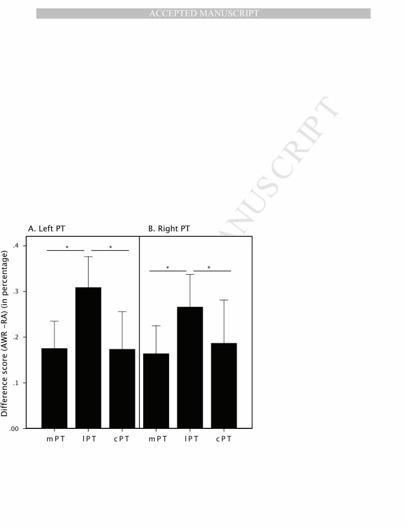
MANUSCRIP
T
ACCEPTED
ACCEPTED MANUSCRIPT
.4
.3
.2
.1
.00
Difference score (AWR -RA) (in percentage)
A. Left PT B. Right PT
**
**
m P T l P T c P T m P T l P T c P T