Regional Differences in Methyl Farnesoate Production by the Lobster Mandibular Organ
8
Reference: Biol. Bull. 186: 9- 16. (February, 1994) Regional Differences in Methyl Farnesoate Production by the Lobster Mandibular Organ DAVID W. BORST’, BRIAN TSUKIMURA’, HANS LAUFER2, AND ERNEST F. COUCH3- ‘Department of Biology, Illinois State University, Normal, Illinois 61790-4120; 2Department of Cellular Biology, University of Connecticut, Storrs, Connecticut 06268; and 3Department of Biology, Texas Christian University, Fort Worth, Texas 76129 Abstract. Visual examination of the mandibular organ (MO) from the lobster, Homarus americanus, disclosed two distinct morphological regions: a fan-folded region along one edge of the gland, and a smooth, unfolded region comprising the rest of the gland. Because MOs produce methyl farnesoate (MF), the MF content of both regions was measured. In freshly dissected glands, more than 95% of the MF was found in the fan-folded region of the gland. In MO sections incubated with [3H-methyl]methionine (a radiolabeled precursor of MF), more than 90% of MF synthesis was found in the fan-folded region. Eyestalk ablation, a procedure that increases MO activity, caused the MF content of MOs to increase more than 130-fold, but had little effect on the regional distribution of MF. Histological observations indicated that these two regions had different cellular compositions. The fan-folded region contained two cell types (A and B). The A cells were mi- totically active and appeared to be undifferentiated. The B cells contained a large number of small vacuoles. The unfolded region was largely composed of a third cell type (C). The C cells were large and morphologically complex, containing many mitochondria and large vacuolar-like structures. They contained relatively few small vacuoles. On the basis of appearance and location, B cells appear to be the likely site of MF synthesis. The physiological importance of C cells is unknown. Introduction Mandibular organs (MOs) from several crustacean decapods have been shown to produce methyl farnesoate, Received 20 July 1992; accepted 22 November 1993. Abbreviations: MO = mandibular organ; MF = methyl farnesoate; EF = ethyl farnesoate. a sesquiterpene structurally related to the insect juvenile hormones (JH) (Borst et al., 1987; Laufer et al., 1987; Tobe et al., 1989; Borst and Laufer, 1990). This synthetic activity is consistent with MO ultrastructure, which has features typical of endocrine cells producing lipid and ste- roid hormones (Aoto et al., 1974; Byard et al., 1975; De- meusy, 1975; Hinsch and al Hajj, 1975; Hinsch, 1977; Buchholz and Adelung, 1980). Similar ultrastructural features are observed in the insect corpus allatum (CA) (Cassier, 1979; Tobe and Stay, 1985), the site of JH syn- thesis. These similarities of ultrastructure and of bio- chemical product suggest that the crustacean MO is the homologue of the insect CA. Although MF is a major MO product, several reports have suggested that this tissue produces other endocrine products. For example, significant amounts of progester- one have been detected in lobster MOs by radioimmu- noassay (Couch et al., 1987). Moreover, lobster MOs con- vert pregnenolone to progesterone in vitro (Tsukimura, 1988). MOs from other species, such as the mud crab Scylla serrata and the crayfish Procambarus clarkii, pro- duce and release farnesoic acid in vitro (Tobe et al., 1989; Ding and Tobe, 199 1). However, the importance of either compound as a secretory product in vivo is still uncertain. Because of its large size, the lobster MO is an ideal tissue for investigating MO function (Byard et al., 1975). In this paper we describe studies on the structure and function of this tissue. Materials and Methods Animals Male lobsters (200-300 g) were obtained from the Ma- rine Resources Department at the Marine Biological Lab- oratory, Woods Hole, Massachusetts. Animals were kept 9
Transcript of Regional Differences in Methyl Farnesoate Production by the Lobster Mandibular Organ

Reference: Biol. Bull. 186: 9- 16. (February, 1994)
Regional Differences in Methyl Farnesoate Production by the Lobster Mandibular Organ
DAVID W. BORST’, BRIAN TSUKIMURA’, HANS LAUFER2, AND ERNEST F. COUCH3-
‘Department of Biology, Illinois State University, Normal, Illinois 61790-4120; 2Department of Cellular Biology, University of Connecticut, Storrs, Connecticut 06268; and 3Department of Biology,
Texas Christian University, Fort Worth, Texas 76129
Abstract. Visual examination of the mandibular organ (MO) from the lobster, Homarus americanus, disclosed two distinct morphological regions: a fan-folded region along one edge of the gland, and a smooth, unfolded region comprising the rest of the gland. Because MOs produce methyl farnesoate (MF), the MF content of both regions was measured. In freshly dissected glands, more than 95% of the MF was found in the fan-folded region of the gland. In MO sections incubated with [3H-methyl]methionine (a radiolabeled precursor of MF), more than 90% of MF synthesis was found in the fan-folded region. Eyestalk ablation, a procedure that increases MO activity, caused the MF content of MOs to increase more than 130-fold, but had little effect on the regional distribution of MF. Histological observations indicated that these two regions had different cellular compositions. The fan-folded region contained two cell types (A and B). The A cells were mi- totically active and appeared to be undifferentiated. The B cells contained a large number of small vacuoles. The unfolded region was largely composed of a third cell type (C). The C cells were large and morphologically complex, containing many mitochondria and large vacuolar-like structures. They contained relatively few small vacuoles. On the basis of appearance and location, B cells appear to be the likely site of MF synthesis. The physiological importance of C cells is unknown.
Introduction
Mandibular organs (MOs) from several crustacean decapods have been shown to produce methyl farnesoate,
Received 20 July 1992; accepted 22 November 1993. Abbreviations: MO = mandibular organ; MF = methyl farnesoate;
EF = ethyl farnesoate.
a sesquiterpene structurally related to the insect juvenile hormones (JH) (Borst et al., 1987; Laufer et al., 1987; Tobe et al., 1989; Borst and Laufer, 1990). This synthetic activity is consistent with MO ultrastructure, which has features typical of endocrine cells producing lipid and ste- roid hormones (Aoto et al., 1974; Byard et al., 1975; De- meusy, 1975; Hinsch and al Hajj, 1975; Hinsch, 1977; Buchholz and Adelung, 1980). Similar ultrastructural features are observed in the insect corpus allatum (CA) (Cassier, 1979; Tobe and Stay, 1985), the site of JH syn- thesis. These similarities of ultrastructure and of bio- chemical product suggest that the crustacean MO is the homologue of the insect CA.
Although MF is a major MO product, several reports have suggested that this tissue produces other endocrine products. For example, significant amounts of progester- one have been detected in lobster MOs by radioimmu- noassay (Couch et al., 1987). Moreover, lobster MOs con- vert pregnenolone to progesterone in vitro (Tsukimura, 1988). MOs from other species, such as the mud crab Scylla serrata and the crayfish Procambarus clarkii, pro- duce and release farnesoic acid in vitro (Tobe et al., 1989; Ding and Tobe, 199 1). However, the importance of either compound as a secretory product in vivo is still uncertain.
Because of its large size, the lobster MO is an ideal tissue for investigating MO function (Byard et al., 1975). In this paper we describe studies on the structure and function of this tissue.
Materials and Methods
Animals
Male lobsters (200-300 g) were obtained from the Ma- rine Resources Department at the Marine Biological Lab- oratory, Woods Hole, Massachusetts. Animals were kept
9
10 D. W. BORST ET AL.
in running seawater at ambient temperature ( 15 to 20°C) gentle shaking in Homarus saline (Welsh and Smith, 1960) until used. The eyestalks of some animals were severed supplemented with glucose (0.1%) and 200 PM [3H- at their base; bleeding was limited by cauterization. An- methyllmethionine. After two hours, MF was extracted imals were eyestalk-ablated 2 weeks before use. from the tissue and the incubation medium.
Materials
Acetonitrile and diethyl ether (both HPLC grade), and hexane (Optima grade) were purchased from Fisher Sci- entific (Pittsburgh, PA). [3H-methyl]methionine (specific activity = 200 mCi/mmol) was obtained from DuPont/ NEN (Boston, Massachusetts). JB-4 glycol methacrylate was purchased from Polysciences, Inc. (Warrington, Pennsylvania). MF was obtained from Dr. D. A. Schooley (Zoecon Research Institute, Palo Alto, California) as a mixture of two isomers (approximately 70% 2E,6E and 30% 2Z,6E). The 2E,6E isomer was purified by normal phase high performance liquid chromatography (np- HPLC; see below). Ethyl farnesoate was prepared from 2E,6E methyl farnesoate as previously described (Borst et al., 1987).
These extracts were then used to determine MF syn- thesis by two different methods. In one approach, np- HPLC was used to calculate the amount of MF synthe- sized during the incubation of MOs. This method is fa- cilitated by the observation that freshly dissected MO pairs from the same animal contain similar amounts of MF (see Results, Fig. 2). The MF contained in incubated gland sections and their culture media was quantified by npHPLC. The sum of these values was then compared to the MF found in sections of the unincubated contralateral pair. The difference between these values is the amount of MF synthesized by the MO during the incubation.
Mandibular organ dissection
MOs were removed from the area adjacent to the apo- deme of the mandibular abductor. Some MOs were cut transversely with a razor blade into four strips or sections (I-IV) of approximately equal width (see Fig. 1). In a few cases, each section was further divided into three or more pieces of equal size by cutting perpendicular to the first cut.
MF quantijcation
Whole MOs or MO sections were homogenized in dis- tilled water with a Dounce homogenizer; the homogenizer was then rinsed with 1% NaCl and the rinse combined with the homogenate. The homogenate (or the culture medium, see next section) was added to acetonitrile and sufficient saline to give a final acetonitrile:water ratio of 5:4. Ethyl farnesoate (5 or 10 ng) was added as an internal standard, and the samples were extracted twice with hex- ane. MF present in these pooled hexane extracts was de- termined by npHPLC. Briefly, this method involves sep- arating the hexane extract on a silica column (5 pm, 4.6 mm X 25 cm) with 1 .O% diethyl ether in hexane. Ma- terial appearing in the same elution volume as 2E,6E methyl farnesoate was quantified by absorption at 220 nm. This value was adjusted for recovery by comparison to the internal standard. This method has been validated by gas chromatography/mass spectroscopy and has a detec- tion limit of 0.1 rig/injection (Borst and Tsukimura, 1991).
In a second method, the relative level of MF synthesis was determined by calculating the transfer of radiolabeled methyl groups from [3H-methyl]methionine to MF during incubation (Borst et al., 1987; Laufer et al., 1987). Because lobster MOs synthesize and release more than one radio- labeled organosoluble compound (Borst et al., 1987), the hexane extracts of MO sections and their culture media were fractionated using the npHPLC conditions described above. The radioactive material eluting at the retention time of MF was quantified with a liquid scintillation counter. The sum of the radiolabeled MF in the tissue sections plus that released into their culture medium is a measure of MF synthesis by the MO. While this in vitro labeling procedure is more sensitive than the npHPLC procedure used above, it can be affected by factors other than the absolute synthetic rate of the MO. Therefore, the results are relative indicators of the level of MF syn- thesis and are best presented as DPM incorporated into MF (Tobe et al., 1989).
Histology
Whole MOs were fixed in modified Kamovsky’s ( 1965) fixative, which consists of 2.7% glutaraldehyde and 2% formaldehyde buffered with 0.1 M sodium cacodylate (pH 7.3) and adjusted to an osmolality of 835 milliosmoles with 10% sucrose. After fixation, the tissue was washed with cacodylate buffer, dehydrated in ethanol, and embedded at 5°C in glycol methacrylate by the method of Butler (1984). Sections were cut dry at 2 pm thickness with machine-made Ralph-Bennett knives (Butler, 1979) and stained with either Heidenhain’s iron hematoxylin or Lee’s stain (Bennett et al., 1976).
Measurement of MF synthesis
The synthesis of MF in vitro was measured in sections or pieces of MOs incubated at room temperature with
Morphology
Results
The MO has a fan-shaped appearance with a broad fan-folded edge at one end and a narrow smooth (un-
MF PRODUCTION IN THE LOBSTER MO 11
folded) edge at the other end (see Fig. 1). In situ, MOs are oriented with the fan-folded edge posterior and dorsal; this edge is the major site of attachment of the gland to the apodeme of the mandibular abductor. The narrow edge of the MO is anterior and ventral. Macroscopic ex- amination of several MOs suggested that they have two morphological regions. The fan-folded edge of the gland (approximately 25% of the glandular area) is thinner and has a light yellow color. The rest of the MO is thicker and dark beige or tan. MOs from animals that had been eye- stalk-ablated 14 days earlier had a larger and thicker fan- folded region.
MF content and distribution
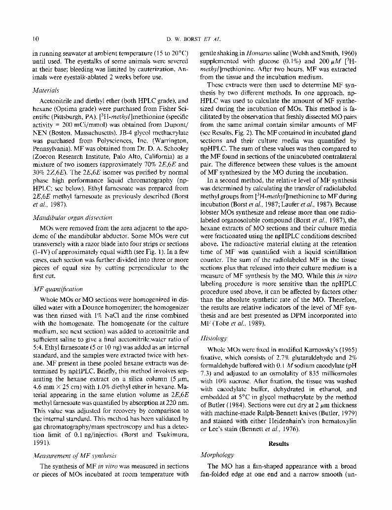
MF content was measured in freshly dissected whole glands. MF content varied markedly (from 3.2 to 196.8 rig/MO) between animals, with an average content of 55.5 (k12.7, SEM) rig/MO (n = 16). In individual an- imals, the MF contents of the left and right MOs were strongly correlated (r = 0.977; Fig. 2).
MOs from eyestalk-intact and eyestalk-ablated male lobsters were analyzed to determine regional content and synthesis of MF. MOs from these animals were divided into four sections of approximately equal width (Fig. 1). For MOs from eyestalk-intact animals, division of the gland into four sections placed the fan-folded region en- tirely in section I. For MOs from eyestalk-ablated animals, enlargement of the fan-folded region resulted in some of this region being included in section II.
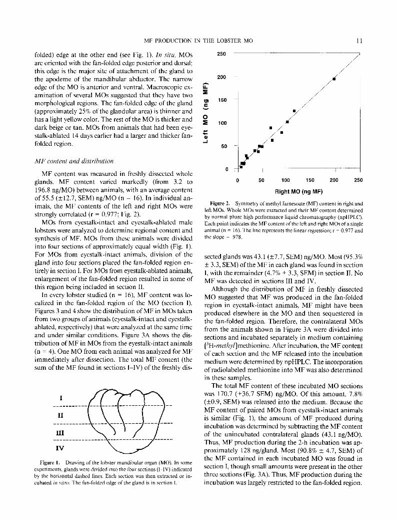
In every lobster studied (n = 16) MF content was lo- calized in the fan-folded region of the MO (section I). Figures 3 and 4 show the distribution of MF in MOs taken from two groups of animals (eyestalk-intact and eyestalk- ablated, respectively) that were analyzed at the same time and under similar conditions. Figure 3A shows the dis- tribution of MF in MOs from the eyestalk-intact animals (n = 4). One MO from each animal was analyzed for MF immediately after dissection. The total MF content (the sum of the MF found in sections I-IV) of the freshly dis-
I ___________ ___ __-__ __--______---- --
II
III m
________-_-___--- ---- ----- ____ ----
_-_______--_____-___------------ ------
Figure 1. Drawing of the lobster mandibular organ (MO). In some
experiments, glands were divided into the four sections (I-IV) indicated by the horizontal dashed lines. Each section was then extracted or in- cubated in vitro. The fan-folded edge of the gland is in section I.
250
200
ii+ a
z 150
V
: 100
c
z 50
0
0 50 100 150 200 250
Right MO (ng MF)
Figure 2. Symmetry of methyl farnesoate (MF) content in right and left MOs. Whole MOs were extracted and their MF content determined by normal phase high performance liquid chromatography (npHPLC).
Each point indicates the MF content of the left and right MOs of a single animal (n = 16). The line represents the linear regression; r = 0.977 and the slope = ,978.
sected glands was 43.1 (t7.7, SEM) rig/MO. Most (95.3% + 3.3, SEM) of the MF in each gland was found in section I, with the remainder (4.7% * 3.3, SEM) in section II. No MF was detected in sections III and IV.
Although the distribution of MF in freshly dissected MO suggested that MF was produced in the fan-folded region in eyestalk-intact animals, MF might have been produced elsewhere in the MO and then sequestered in the fan-folded region. Therefore, the contralateral MOs from the animals shown in Figure 3A were divided into sections and incubated separately in medium containing [3H-methyf]methionine. After incubation, the MF content of each section and the MF released into the incubation medium were determined by npHPLC. The incorporation of radiolabeled methionine into MF was also determined in these samples.
The total MF content of these incubated MO sections was 170.7 (k36.7 SEM) rig/MO. Of this amount, 7.8% (kO.9, SEM) was released into the medium. Because the MF content of paired MOs from eyestalk-intact animals is similar (Fig. l), the amount of MF produced during incubation was determined by subtracting the MF content of the unincubated contralateral glands (43.1 rig/MO). Thus, MF production during the 2-h incubation was ap- proximately 128 rig/gland. Most (90.8% +- 4.7, SEM) of the MF contained in each incubated MO was found in section I, though small amounts were present in the other three sections (Fig. 3A). Thus, MF production during the incubation was largely restricted to the fan-folded region.

12 D. W. BORST ET AL.
100
60
0 I
A 8 Freshly-dlmmcted MO 61 Incubated MO
B
q % Total DPM
I II III IV
MO Section
Figure 3. Regional distribution and synthesis of MF in MOs from eyestalk-intact lobsters. Both MOs from each animal (n = 4) were divided into four sections as in Figure 1. (A) MF content: Sections of the left MO were analyzed for MF content by npHPLC. The MF content of each section is expressed as a percentage of the total MF content of the MO (% Total MF Content). No MF was detectable in sections III and IV. Sections of the right gland were incubated individually for 2 hours in Homarus saline supplemented with [-‘H-methyllmethionine. The MF content ofeach incubated section and its culture medium was also quan- tified by npHPLC and is displayed as a percentage of the total MF content of the incubated MO. (B) Radiolabel incorporation: The radiolabeled MF in the MO sections incubated above was calculated from the amount of radioactivity present in the fractions eluting with MF during npHPLC. Results are expressed as a percentage of the total radioactive MF produced by the gland (% Total DPM).
This conclusion was confirmed by analyzing the in- corporation of [3H-methyZ]methionine into MF by the incubated MO sections (Fig. 3B). The total synthesis of radiolabeled MF by the MO sections was 66,600 (+ 17,760, SEM) DPM/gland. Of this amount, 14.3% (a3.1, SEM) was released into the medium. Most (95.7% 1- 3.6, SEM) of the radiolabeled MF found in each MO was in section I, though small amounts were found in the other sections.
The production of radiolabeled MF was studied further by dividing the four MO sections from two animals into several (3 or 6) pieces. Each piece was then incubated with [3H-methyl]methionine and MF synthesis deter- mined. More than 99% of the radiolabeled MF was pro- duced by pieces obtained from section I. Every piece pro- duced MF, and those from the middle of this section pro- duced the largest amounts (data not shown).
The distribution of MF was also determined in freshly dissected MOs from eyestalk-ablated animals (n = 4). The total MF content of these glands was 5,627 (+730, SEM) rig/gland, about 130-fold greater that the MF content of MOs from the group of eyestalk-intact animals (P < .OOO 1; t-test). As in the intact animals, the MF content of MOs from the eyestalk-ablated animals was localized to the fan-folded region of the MO (Fig. 4). Most (74.3% + 9.2, SEM) of the MF in each gland was found in section I; but a substantial amount (20.3% f 9.0, SEM) was found in section II due to the enlargement of the fan-folded region. The other two sections contained only small amounts of MF (~4% each).
Histology
Light microscopy showed that the two regions of the MO contain different cell types. Two cell types (A and B) are found in the fan-folded region of the gland. A single cell type (C) predominates in the rest of the gland. The A cells are a minor cellular constituent of the fan-folded region and were not always seen. When present, they were found along the outer edge of the fan-folded region and
II Ill
MO Section
Figure 4. Regional distribution of MF in MOs from eyestalk-ablated lobsters. The left MOs from each animal (n = 4) were divided into four sections. Each section was analyzed for MF content by npHPLC. The MF content of each section is expressed as a percentage of the total MF content of the MO (% Total MF Content).
MF PRODUCTION IN THE LOBSTER MO 13
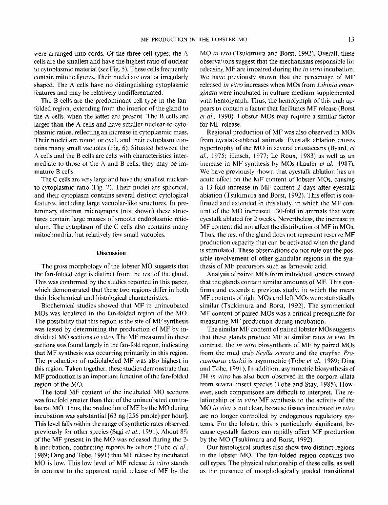
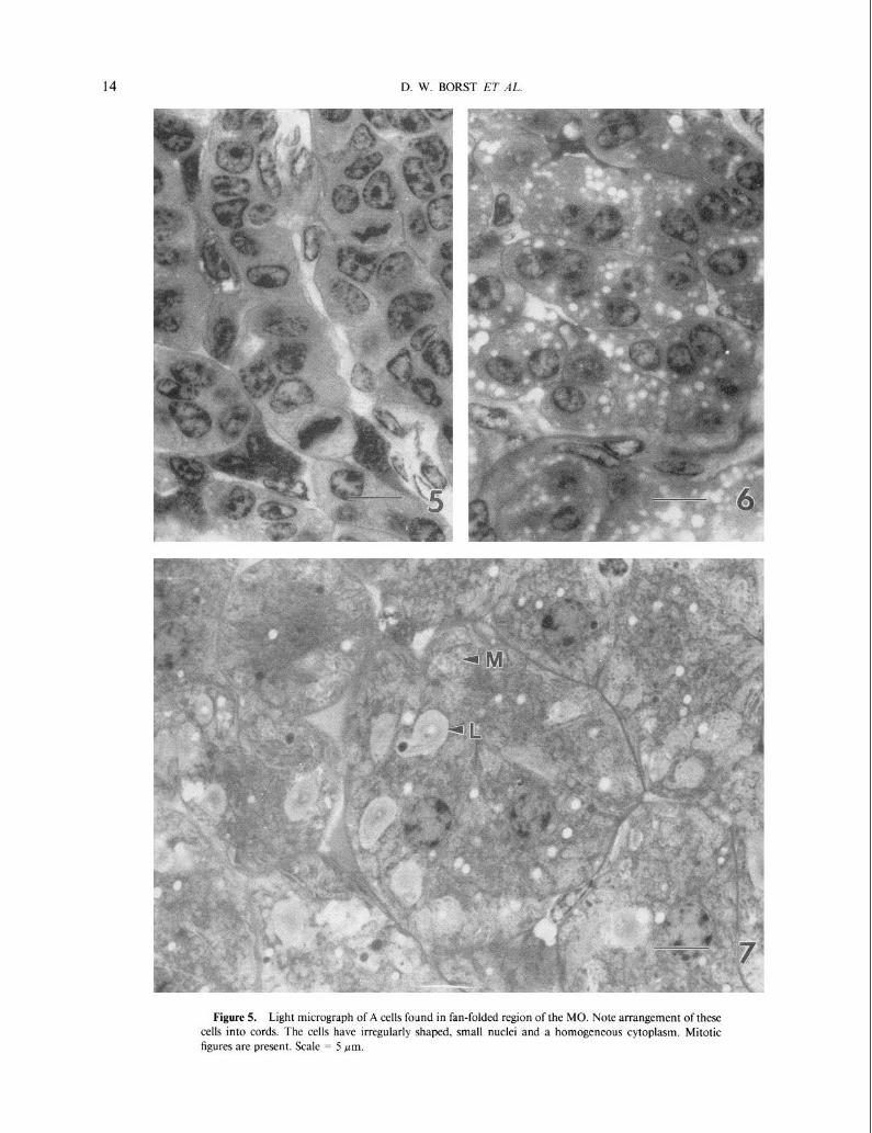
were arranged into cords. Of the three cell types, the A cells are the smallest and have the highest ratio of nuclear to cytoplasmic material (see Fig. 5). These cells frequently contain mitotic figures. Their nuclei are oval or irregularly shaped. The A cells have no distinguishing cytoplasmic features and may be relatively undifferentiated.
The B cells are the predominant cell type in the fan- folded region, extending from the interior of the gland to the A cells, when the latter are present. The B cells are larger than the A cells and have smaller nuclear-to-cyto- plasmic ratios, reflecting an increase in cytoplasmic mass. Their nuclei are round or oval, and their cytoplasm con- tains many small vacuoles (Fig. 6). Situated between the A cells and the B cells are cells with characteristics inter- mediate to those of the A and B cells; they may be im- mature B cells.
The C cells are very large and have the smallest nuclear- to-cytoplasmic ratio (Fig. 7). Their nuclei are spherical, and their cytoplasm contains several distinct cytological features, including large vacuolar-like structures. In pre- liminary electron micrographs (not shown) these struc- tures contain large masses of smooth endoplasmic retic- ulum. The cytoplasm of the C cells also contains many mitochondria, but relatively few small vacuoles.
Discussion
The gross morphology of the lobster MO suggests that the fan-folded edge is distinct from the rest of the gland. This was confirmed by the studies reported in this paper, which demonstrated that these two regions differ in both their biochemical and histological characteristics.
Biochemical studies showed that MF in unincubated MOs was localized in the fan-folded region of the MO. The possibility that this region is the site of MF synthesis was tested by determining the production of MF by in- dividual MO sections in vitro. The MF measured in these sections was found largely in the fan-fold region, indicating that MF synthesis was occurring primarily in this region. The production of radiolabeled MF was also highest in this region. Taken together, these studies demonstrate that MF production is an important function of the fan-folded region of the MO.
The total MF content of the incubated MO sections was fourfold greater than that of the unincubated contra- lateral MO. Thus, the production of MF by the MO during incubation was substantial [63 ng (256 pmole) per hour]. This level falls within the range of synthetic rates observed previously for other species (Sagi et al., 1991). About 8% of the MF present in the MO was released during the 2- h incubation, confirming reports by others (Tobe et al., 1989; Ding and Tobe, 199 1) that MF release by incubated MO is low. This low level of MF release in vitro stands in contrast to the apparent rapid release of MF by the
MO in vivo (Tsukimura and Borst, 1992). Overall, these observa! ions suggest that the mechanisms responsible for releasing MF are impaired during the in vitro incubation. We have previously shown that the percentage of MF released in vitro increases when MOs from Libinia emar- ginata were incubated in culture medium supplemented with hemolymph. Thus, the hemolymph of this crab ap- pears to contain a factor that facilitates MF release (Borst et al., 1990). Lobster MOs may require a similar factor for MF release.
Regional production of MF was also observed in MOs from eyestalk-ablated animals. Eyestalk ablation causes hypertrophy of the MO in several crustaceans (Byard, et al., 1975; Hinsch, 1977; Le Roux, 1983) as well as an increase in MF synthesis by MOs (Laufer et al., 1987). We have previously shown that eyestalk ablation has an acute effect on the MF content of lobster MOs, causing a 13-fold increase in MF content 2 days after eyestalk ablation (Tsukimura and Borst, 1992). This effect is con- firmed and extended in this study, in which the MF con- tent of the MO increased 130-fold in animals that were eyestalk ablated for 2 weeks. Nevertheless, the increase in MF content did not affect the distribution of MF in MOs. Thus, the rest of the gland does not represent reserve MF production capacity that can be activated when the gland is stimulated. These observations do not rule out the pos- sible involvement of other glandular regions in the syn- thesis of MF precursors such as farnesoic acid.
Analysis of paired MOs from individual lobsters showed that the glands contain similar amounts of MF. This con- firms and extends a previous study, in which the mean MF contents of right MOs and left MOs were statistically similar (Tsukimura and Borst, 1992). The symmetrical MF content of paired MOs was a critical prerequisite for measuring MF production during incubation.
The similar MF content of paired lobster MOs suggests that these glands produce MF at similar rates in vivo. In contrast, the in vitro biosynthesis of MF by paired MOs from the mud crab Scylla serrata and the crayfish Pro- cambarus clurkii is asymmetric (Tobe et al., 1989; Ding and Tobe, 199 1). In addition, asymmetric biosynthesis of JH in vitro has also been observed in the corpora allata from several insect species (Tobe and Stay, 1985). How- ever, such comparisons are difficult to interpret. The re- lationship of in vitro MF synthesis to the activity of the MO in vivo is not clear, because tissues incubated in vitro are no longer controlled by endogenous regulatory sys- tems. For the lobster, this is particularly significant, be- cause eyestalk factors can rapidly affect MF production by the MO (Tsukimura and Borst, 1992).
Our histological studies also show two distinct regions in the lobster MO. The fan-folded region contains two cell types. The physical relationship of these cells, as well as the presence of morphologically graded transitional
14 D. W. BORST ET AL.
Figure 5. Light micrograph of A cells found in fan-folded region of the MO. Note arrangement of these cells into cords. The cells have irregularly shaped, small nuclei and a homogeneous cytoplasm. Mitotic figures are present. Scale = 5 pm.

MF PRODUCTION IN
forms, suggests that A cells provide the precursor cells that differentiate into B cells. Based on their morphological characteristics and location within the fan-folded region, the B cells are the most likely site of MF synthesis.
The rest of the gland contains predominantly C cells, whose function and relationship to the other two cell types are unknown. Preliminary ultrastructural studies suggest that these cells are active metabolically and may be in- volved in the synthesis of lipids or steroids. We speculate that they may be the sites of the progesterone metabolism previously reported in MOs from female lobsters (Couch et al., 1987; Tsukimura, 1988). However, to the best of our knowledge MOs from male lobsters have never been investigated for the presence of this steroid. In any case, the lobster MO may have several products, so the phys- iological importance of this tissue may be complex.
The histological and biochemical complexity of the lobster MO was unexpected, partly because of the simi- larity between the MOs of other crustaceans and the CA of insects. In insects, the CA contains only one type of glandular cell (Tobe and Stay, 1985). Likewise, cytological observations of MOs from other crustaceans (Aoto et al., 1974; Byard et al., 1975; Demeusy, 197.5; Hinsch and al Hajj, 1975; Hinsch, 1977; Buchholz and Adelung, 1980) also suggest that the glands are relatively homogeneous. MOs from adult female spider crabs, L. emarginatu, have two cell types that differ in their staining properties, but have similar ultrastructural characteristics. In addition, there is no indication that these cells were regionally dis- tributed in the MO of this species (Hinsch, 198 1). Like- wise, we found no evidence for regional distribution of MF synthesis in MOs from L. cmarginata (Tsukimura, Martin, and Borst, unpub. data).
Our studies show that the lobster MO is more complex than the MOs of other species, containing at least three cell types localized in two areas of the gland. Thus, the lobster MO appears to be unique, and may represent the fusion of two tissues, one of which synthesizes MF and the other of which produces one or more other products.
Acknowledgments
This research was supported by grants to D.W.B. from the National Science Foundation (DCB-89 19833) and the Illinois-Indiana Sea Grant (project #R/A-2) under the National Sea Grant College Program (COMM NAX9AA- D-SG058) and to E.C. from the TCU Research Fund of
THE LOBSTER MO 15
Texas Christian University. This study is a publication of the Intensive Commercial Aquaculture Facility of Illinois State University. We thank Dr. J. K. Butler for his help in these studies, and express our appreciation for the helpful comments from two anonymous reviewers and the editors of this journal.
Literature Cited
Aoto, T., Y. Kamiguchi, and S. Hisano. 1974. Histological and ultra-
structural studies on the Y organ and the mandibular organ of the freshwater prawn, Pu/ucmon parrci&ns, with special reference to their relations with the molting cycle. J Fuc. Sci. Hokkaido Univ. 19: 295-308.
Bennett, H. S., A. D. Wyrick, S. W. Lee, and H. J. McNeil, Jr.
1976. Science and art in preparing tissues embedded in plastic for light microscopy with special reference to glycol methacrylate, glass knives and simple stains, Stuin Technol. 51: 7 l-97.
Borst, D. W., and H. Laufer. 1990. Methyl farnesoate, a JH-like com- pound in crustaceans. Pp. 35-60 in Rwen/ .4dvunce.s in Comparative
Arthropod Morphologic. PI~~~.sio/o,q~, und Lkwlopment, A. F. Gupta,
ed. Rutgers University Press, New Brunswick, NJ. Borst, D. W., and B. Tsukimura. 1991. Quantification of methyl far-
nesoate levels in hemolymph by high-performance liquid chroma-
tography. J Chromutogr. 545: 7 1-78. Borst, D. W., H. Laufer, M. Landau, E. S. Chang, W. A. Hertz, F. C.
Baker, and D. A. Schooley. 1987. Methyl farnesoate and its role in crustacean reproduction and development. Insc,ct Riochem. 17: 1123-l 128.
Borst, D. W., C. G. Buerkett, and B. Tsukimura. 1990. Factors affecting
methyl farnesoate release from mandibular organs (MOs) in vitro
.4m. Zoo/. 30: 29a. Buchhofc, C., and D. Adelung. 1980. The ultrastructural basis of steroid
production in the y-organ and the mandibular organ of the crabs IIemigrupws trndtr.v (Dana) and CurcYnrls mur~u.s I.. Cell Tiss. Res.
206: 83-94.
Butler, J. K. 1979. Methods for improved light microscope microtomy. Sluin Twhnol. 54: 53-69.
Butler, J. K. 1984. Improved methods for molding. facing, and sec- tioning of glycol methacrylate embedded tissue blocks. S/uin ‘Technol.
59: 315-321.
Byard, E. H., R. R. Shivers, and D. E. Aiken. 1975. The mandibular organ of the lobster. IIornurr~~ urncricumrs. Cell Ti.s.s. Res. 162: l3-
22. Cassier, P. 1979. The corpora allata of insects. I17l. Rev. C:,lol. 57: I-
73. Couch, E. F., N. Ilagino, and J. W. Lee. 1987. Changes in estradiol
and progesterone immunoreactivity in tissues of the lobster ffomarus
arncricumcx with developing and immature ovaries. Camp. Biochem. Ph,wiol. 87A: 765-770.
Demeusy, N. 1975. Observations sur le fonctionnement des glandes mandibulaires du Decdpode brachyoure Chrcimrs muenas L.: ani-
maux temoins et animaux sans ptdoncules ocularies. C’. R. Acud.
Ser. Pk.\ 281D: 1887-1889.
Figure 6. Light micrograph of B cells in the fan-folded region. The nuclei of these cells are oval or round and larger than those of A cells. The B cells have a more abundant ground cytoplasm than A cells and contain many clear vacuoles. Scale = 5 pm.
Figure 7. Light micrograph of C cells in the unfolded region. Note the large spherical nuclei and large cytoplasmic volume of these cells. The cytoplasm is morphologically complex, containing many mitochondria
(M). large vacuolar-like structures (L). and some clear vacuoles. Scale = 5 Frn.
16 D. W. BORST ET AL
Ding, Q., and S. S. Tobe. 1991. Production of famesoic acid and methyl
famesoate by mandibular organs of the crayfish, Procumbarus clurkii. Insect Biochem 21: 285-29 1.
Hinscb, G. W. 1977. Fine structural changes in the mandibular gland
of the male spider crab, Libinia emurginafa following eyestalk abla- tion. J. Morphol. 154: 307-3 16.
Hinscb, G. W. 1981. The mandibular organ of the female spider crab,
Libiniu emurginuta, in immature, mature, and ovigerous crabs. J. Morphol. 168: 181-187.
Hinscb, G. W., and H. al Hajj. 1975. The ecdysial gland of the spider
crab, Libiniu emurginata L. 1. Ultrastructure of the gland in the male. J. Morphol. 145: 179- 188.
Karnovsky, M. J. 1965. A formaldehyde-glutaraldehyde fixative of high osmolarity for use in electron microscopy. J. Cell Biol. 27: 137A.
Laufer, H., D. Borst, F. C. Carrasco, M. Sinkus, C. C. Reuter, L. W.
Tsai, and D. A. Scbooley. 1987. Identification of a juvenile hor- mone-like compound in a crustacean. Science 235: 202-205.
Le Roux, A. 1983. Reactions de I’organe mandibulaire a I’ablation des
pedoncules oculaires chez les larves et les juveniles de Puluemonetes
vuriuns (Leach) (Decapoda, Natantia). C. R. Acud. Sci. Paris 296D: 697-700.
Sagi, A., E. Homola, and H. Laufer. 1991. Methyl farnesoate in the prawn Mucrobruchium rosenbergii: synthesis by the mandibular organ in vitro, and titers in the hemolymph. Camp. Biochem. Physiol. 99B: 879-882.
Tobe, S. S., and B. Stay. 1985. Structure and regulation of the corpus allaturn. Adv. Insect Physiol. 18: 305-432.
Tobe, S. S., D. A. Young, and H. W. Kboo. 1989. Production of methyl
farnesoate by the mandibular organs of the mud crab, Scylia serrutu: validation of a radiochemical assay. Gen. Comp. Endocrinol. 73: 342- 353.
Tsukimura, B. 1988. The role of presumptive mandibular organ se- cretions in ovarian growth of the shrimp, Penueus vunnumei. Ph.D. dissertation, University of Hawaii.
Tsukimura, B., and D. W. Borst. 1992. Regulation of methyl famesoate levels in the lobster, Homarus americanus. Gen. Camp. Endocrinol. 86: 297-303.
Welsh, J. H., and R. I. Smith. 1960. Pp. 159-160 in InvertebrutePhys- iology. Burgess Publishing Co., Minneapolis, MN.