Integrability of reductions of the discrete KdV and potential KdV equations
Upload
independentCategory
view
4download
0
J7oumnal ofNeurology, Neurosurgery, and Psychiatry 1993;56:859-864
PAPERS
Reductions in parietal and temporal cerebralmetabolic rates for glucose are not specific forAlzheimer's disease
Mark B Schapiro, Pietro Pietrini, Cheryl L Grady, M J Ball, Charles DeCarli,Anand Kumar, Jeffrey A Kaye, James V Haxby
AbstractReduction in the regional cerebral meta-bolic rate for glucose (rCMRglc) in theparietal and temporal regions has beenshown in Alzheimer's disease (AD). Thespecificity of these findings for thisdisease state is uncertain. We repeatedlymeasured rCMRglc with positron emis-sion tomography and [18F]2-fluoro-2-deoxy-D-glucose in the resting state in a68 year old man with slowly progressivedementia who, during life, was initiallydiagnosed as having dementia of theAlzheimer type, then Parkinson diseasewith dementia, but was found to haveonly Parkinson's disease at necropsy.Metabolic ratios (rCMRglc/mean greyCMRglc) were significantly (p < 0 05)reduced in parietal and temporal regions,as well as in the prefrontal and premotorareas. This pattern was similar inregional distribution and magnitude ofthe defect to that seen in patients withprobable AD. These results suggest thatreductions of glucose metabolism in asso-ciation neocortex in AD are not specificto the disease process, but may be relatedto the dementia state.
(7 Neurol Neurosurg Psychiatry 1993;56:859-864)
Laboratory ofNeurosciences, Sectionon Brain Aging andDementia, NationalInstitute on Aging,Clinical Center,Bethesda, MD 20892,USAM B SchapiroP PietriniC L GradyC DeCarliA KumarJ A KayeJ V HaxbyDementia StudyLaboratory, AtkinsonNeurobiology ofAgingUnit, Department ofPathology, UniversityofWestern Ontario,London M6A SC1,CanadaM J BallCorrespondence to:Mark B Schapiro, MD,Laboratory of Neurosciences,National Institute on Aging,Bldg 10 Rm 6C414, 9000Rockville Pike, Bethesda,MD 20892, USA.Received 13 May 1991and in revised form11 November 1992Accepted 19 November 1992
Reductions in the regional cerebral metabolicrate for glucose (rCMRglc) in the parietal andtemporal regions have been shown inAlzheimer's disease (AD), using positronemission tomography (PET). Such reductionsoccur early in the disease and persist through-out its course.'2 However, the specificity ofthese findings for AD has not been deter-mined. To address this issue of specificity, wereport a necropsy proved case of Parkinson'sdisease (PD) in which reductions of parietaland temporal metabolism were found beforedeath, similar to those seen in AD.An abstract of this work has been
published.'
Case reportThe subject was a man who first presented on
31 October 1983 at age 65 years with a sixyear history of mild memory loss, difficultyfinding places, nightmares, and occasionalvisual hallucinations. He was forced to retireas a medical equipment salesman owing todifficulty with paperwork. History was notremarkable for neurological or medical dis-
ease. He received no medications. There wasno family history of AD or PD. Review of sys-tems was notable for impotence. On examina-tion, blood pressure was 140/90 (supine) and104/76 (standing), and heart rate was 54(supine) and 64 (standing). There was a mildtremor of outstretched hands.
Laboratory investigations revealed normalserum chemistries, cholesterol, thyroid func-tion test, VDRL, folate, vitamin B12, bloodcounts, and sedimentation rate. Chest x rayshowed borderline cardiomegaly and ECGwas normal. EEG showed 7-8 Hz, low ampli-tude background. CT scan showed mild corti-cal atrophy. CSF was clear with normalopening pressure.An extensive battery of neuropsychological
tests to assess memory, attention to simpleand complex sets, planning, language, visuo-spatial functions, and overall dementia sever-ity45 was administered to the patient five timesat roughly annual intervals (table 1). To testwhether scores differed significantly from thatwhich would be expected in an age, educa-tion, and gender-matched control, predictedscores were calculated based on a multipleregression equation derived from 108 healthycontrols. These controls ranged in age from45 to 90 years (mean 63) with 12 to 20 yearsof education (mean 16-7). Test scores werepredicted based on age, gender, and years ofeducation. The difference between actual andpredicted score was converted to a z-score bydividing by the standard deviation of controlresiduals, calculated with a jackknife proce-dure whereby the predicted score for eachcontrol subject was based on a multipleregression equation derived from all controlsbut that one. To test for significant impair-ments in AD patients, the difference betweenthe mean normalised discrepancy and zerowas tested with a t test. For the single patientwith PD, a score with normalised discrepancyless than -1-96 (p < 0-05) indicated signifi-cant impairment.On initial evaluation, dementia was mild
with significant impairments on two tests ofmemory, a test of the ability to shift attentionbetween two sequential sets (trails B), plan-ning (Porteus mazes), and visual abstract rea-soning (Raven's matrices). Performance on atest of memory for a prose passage (WMSstory recall), however, was within normal lim-its. Whereas all tests of visuospatial construc-tion and immediate visuospatial memory spanshowed significant impairment at the initialevaluation, all language tests were withinnormal limits on the first evaluation, and all
859
group.bmj.com on July 31, 2016 - Published by http://jnnp.bmj.com/Downloaded from
Shapiro, Pietnn4 Grady, Bali; DeCar4 Kiwr, Kawy, Haxhy
Table 1 Results oflongiud l neuropsychogical tests in the healhy convtls, Alzheine's diseas patenas, and theParkinson's disease subject Examinon onJanuay 1987 andJanuary 1988 lco ded with the paten's ifm adsecond PET scans, respectvdy.
Contros ADpaient Paa*nsn dsea subctTest (n=11) (n = 12) Nov '83 Jan '85 Ian '86 an '87 Jam '88
Overall Ability:MMSE (max = 30) 29-8 (0-4) 23-8 (2.0)* 24 t 22t 25t 23t 21tWAIS IQ 133 (5) 107 (12)* 105t 97t 87t 951t 86tMatis dementia RS 141(3) 121 (10)* 133t 130t 116t 120t 106t
Memory:WMS stories
Immediate recall 20 (7) 9 (5)* 12 St 7t 14 4tDelayed recall 17 (7) 4 (5)* 7 4t 2t it 3t
WMS FiguresImmediate recall 10 (3) 3 (2)* it it 5 6 itDelayedrecall 9 (4) 1 (2)* it 0t 0t I 1
Buschke TestTotal recall 64 (5) 40 (9)* 41t 41t 35t 39t 28tDelayed recall 8 (1) 2 (3)* it 3t 3t 2t 2t
Complex attention, planning, abstraction:Trails B (secs) 67 (27) 274 (119)* (Unable)Stroop interference 36 (8) 14 (10)* 22 9t 3t It 6tPorteusmazes (Test,years) 15-8 (1-1) 12-8 (4)* 7-5t 8-Ot 6-Ot 6-Ot 6-5tRaven's coloured malrices 33 (3) 25 (6)* 21t 15t 19t 16t 13t
WAIS VDQ 134 (9) 117 (15)* 123 109t 108t 114t 108tWAIS MDQ 124 (11) 97 (15)* 109 106 91t 97t 100Syntax comprehension 25 (1) 19-3 (3.9)* 23 21 19t 16t 17tBoston naming 38 (4) 31 (8)* 37 35 35 34 na.FAS serial naming 44 (10) 28 (14)* 34 lit lit 14t 16tWRAT oral reading 16-3 (2-4) 14-3 (2-5) 16-5 14-4 16-8 15-0 15-3
Visuospatial:WAIS PDQ 122 (14) 104 (17)* 87t 73t 60t 70t 57tExtended range drawing 20 (2) 17 (5)* lot 121 12t 8t 7tHiskey-Nebraska blocks 16 (5) 9 (4)* 6t 4t 4t 3t 3tBenton fcial recognition 46 (3) 43 (6) 43 36t 37t 36t 32tBlock tapping span 4-7 (0-6) 3-8 (0-5) 2-8t 3-0t 2-2t 2-5t 1-5t
Values are mean (SD).AD = Alzheimer's disease; MMSE = mini-mental statuseimn ; WAIS = Wechsler adult intelligence scale; IQ = ielligecequotient; WMS = Wechsler memory scale; VDQ = verbal comprehension deviation quotient; MDQ = memory and freedom fromdistractibility deviation quotient; WRAT = wide range achievement test; PDQ = perceptual organisation deviation quotent.*Mea in AD patients less than mean in controls by unpaired t test (p < 0-05).tParkinson subjects's value less than mean in controls by Z-score analysis (p < 0-05).
but two were normal on the second evalua-tion. Confrontation naming (Boston naming)and oral reading of irregularly spelled words(WRAT) remained normal on all evaluations.Performance on a test of face discrimination(Benton face recognition) demonstrated sig-nificant impairments on the second evaluationand on all evaluations thereafter. Mini-mentalstate examination (MMSE) score was 24/30.6His Hachinski ischaemic score was 1.7 Theclinical diagnosis at this time was dementia ofthe Alzheimer type.8At examination on 7 August 1985 there
were mild orthostatic blood pressure changes;increased tone in the upper extremities (withmild cogwheeling) which increased with acti-vation (L > R); tremor of outstretched arms;an intermittent, small amplitude fast tonguetremor; diminished rapid finger movements;and mild bradykinesia. His gait was charac-tensed by decreased arm swing, slow pace,moderate flexion posture, and decreasedstride length. He was unable to tandem walk.Vibratory sense and two point discriminationwere decreased in the toes. Extraocular move-ments were full. His MMSE score was 22. Atthis point the diagnosis was thought to beprobable Alzheimer's disease with extrapyra-midal signs.9 10 A trial of a levodopa/cardidopa combination (Sinemet) was initi-ated in September 1986 with improvement ofhis spontaneous movements. However, thisdrug was discontinued because of hallucin-ations, not because of any negative effect onhis cognitive performance. Other dopamineagonists were not tried.At the time of his first PET scan (11
January 1987) his MMSE score was 23. Aresting tremor and masked fades were alsonow present. The clinical diagosis waschanged to Parkinson's disease with dementia,due to the slow progression of cognitivechanges (table 1). Very slow deterioration ofmental status continued to be noticed at sub-sequent evaluations, with MMSE scores of 18on 22 July 1987 and 21 on 11 January 1988(date of his second PET scan). At his lastevaluation on 14 June 1988, his MMSE scoredecreased to 12 and it was noted that he nowwas dependent in daily living skills. Annualrates of decline (points/year) on both theWAIS IQ and Mattis dementia rating scalewere approximately half the mean rate forpatients with AD and were significant (p <0-05) in comparison with the mean in thehealthy controls (WAIS: +0-4 (SD 1 -8) forcontrols, - 8-3 (SD 4-4) for AD patients, -3-8for the PD patient, respectively. Mattisdementia rating scale: + 1-5 (SD 2-1) for con-trols, - 12-4 (SD 6-9) for AD patients, -6-1for the PD patients).
Progression of motor abnormalities contin-ued through the course of his illness.Extraocular movements remained full, butsaccades were hypometric.At necropsy by one of us (MJB), the fresh
brain weighed 1430 grams. On gross inspec-tion, there was no evidence of corticalatrophy, minimal ventricular dilatation, noatherosclerosis, and no cerebral infarctions.Transverse sections through the brainstemshowed striling pallor of the substantia nigraand locus ceruleus. Microscopic examinationshowed severe depletion of pigmented neu-
860
group.bmj.com on July 31, 2016 - Published by http://jnnp.bmj.com/Downloaded from
Cerebral glucose metabolism in Parkinson's disease
Table 2 Absolute rCMRglc values measured in the healthy volunteers, Alzheimer'sdisease subjects, and Parkinson's disease subject (first and second PET scans)
Brain Region Controls AD PatientAbsolute Values (n = 19) (n = 12) PET scan I PET scan 2(mgll00glmin) Mean (SD) Mean (SD) (01-15-87) (01-21-88)
Orbito frontalrightleft
Prefrontalrightleft
Premotorrightleft
Inferior parietalrightleft
Medial parietalrightleft
Superior parietalrightleft
Sensorimotorrightleft
Calcarinerightleft
Occipital associationrightleft
Inferior temporalrightleft
Middle temporalrightleft
Superior temporalrightleft
Anterior cingulaterightleft
Posterior cingulateAmygdala
rightleft
Insularightleft
Paracentralrightleft
Hippocampusrightleft
MidbrainCaudate nucleus
rightleft
Lenticular nucleusrightleft
Thalamusrightleft
Cerebellumrightleft
Global grey matter
7-42 (1-22)7-59 (1-16)
8-76 (1-45)8-78 (1-50)
9-38 (1-70)9-53 (1-56)
8-67 (1-61)8-89 (1-74)
9-53 (1-75)9 04 (2 02)
8-48 (1-52)8-47 (1-56)
8-48 (1-48)8-89 (1-62)
8-36 (1-32)8-47 (1-31)
8-05 (1-42)8 07 (1-51)
6-35 (1-31)6-08 (1-26)
7-99 (1-35)8-08 (1-28)
8-42 (1-52)8-59 (1-46)
8-66 (1-72)8-58 (1-44)9-33 (1-59)
5-57 (0-96)5-66 (1-11)
8-10 (1-56)8-17 (1-34)
8-26 (1-77)8-36 (1-67)
6-75 (1-15)6-78 (1-28)5-37 (0 60)
8-82 (1-93)9-27 (1-77)
9 54 (1-66)9-65 (1-34)
9-17 (1-55)9-22 (1-66)
6-84 (1 30)6-88 (1-58)8-41 (1-37)
5-84 (155)*6-10 (1 30)*
7-40 (0.98)*7-17 (0-92)*
7-48 (1-18)*7-58 (1-08)*
6-53 (1-22)*6-06 (1-38)*
6-68 (0-97)*6-64 (0-82)*
6-16 (1-06)*6-22 (1-04)*
7-38 (1-03)t7-42 (0-97)*
7 05 (1-29)*7-20 (1-24)*
6-95 (1-00)t6-73 (1-13)*
4-98 (0-72)*4-75 (0 67)*
6:00 (0-97)*6-14 (0-86)*
6-74 (0-88)*6-70 (1-07)*
7 05 (0.84)*7 05 (1-10)*7-01 (1-00)*
4-23 (1-35)*3-76 (0-98)*
6-53 (1-71)*6-62 (1-40)*
6-62 (1-01)*6-72 (0-84)*
5-47 (0 81)*5-20 (0-54)*4-74 (0-76)t8-11 (1-49)8-19 (1-67)
8-43 (1-75)8-53 (1-55)t8-05 (1-40)7-74 (l-58)t6-35 (0 57)6-27 (0-49)6-84 (0-82)*
5-656-39
5-36t§4-95*t5-21*5.50*
4-33*4-58*
3-69**4-34*t4-13*4-19*
5-44t5-89
6-105-25*
4.40*t4-60t
4-31*4-92*
4-42*t5-08*
6-886-823-95**6-655-77§
5-355-35t
5-926 30
5*104-414-87
7-857-08
8-118-08
7-108-69
5-695-385-47t
5-355 70
5-40t§5-23*t
5.56t4-80**3-80**3-81*
4-65*§3-63**4-06*4-37*
5.19tt5-25t§
5-38t4-87*
4-54**3-81**
3-803.49t
4-22*4-29*§
4-24**4-25*§
6-276-974-62t§
5-893.97
5-244.90*
6-655-89
4-624-12t5-41
5-546-61
8-438-24
7-536-64
4-86i4-37t5-10*
Values of rCMRglc are mean (SD) and are expressed in mg/100 g tissue/min. Differencesbetween mean values in the Alzheimer's disease and healthy volunteer groups were comparedwith an unpaired t test.PD patient was compared to both controls and AD patients by using a Z-score analysis.*Value differs from mean in controls at p < 0 01 level.fValue differs from mean in controls at p < 0-05 level.tValue differs from mean in AD patients at p < 0 01 level.§Value differs from mean in AD patients at p < 0-05 level.
rons in the substantia nigra, especially in thepars compacta, accompanied by extensiveastrocytic gliosis and free neuromelanin pig-ment. Frequent Lewy bodies were seen in theresidual substantia nigra neurons. The
remainder of the midbrain showed no signifi-cant abnormality. Neurofibrillary tangles were
not found. Even more depletion of pigmentednerve cells was shown in the locus ceruleus.The pontine raphe nuclei were normal. In thenucleus basalis of Meynert the average num-
ber of nucleolated neurons per 10 pM paraf-fin-embedded section was 38-5 in the anterior
division and 13-1 in the intermediate division(the posterior division was not available), areduction of 90% and 93%, respectively, incomparison with four non-demented subjectsaged 67-72 years. Neither Lewy bodies norneurofibrillary tangles were found in this sam-ple of nucleus basalis of Meynert. There wereno lesions seen in samples of the thalamuswith subthalamic nucleus, caudate, and puta-men, but there was suggestion of a mild,patchy neuronal loss in the globus pallidus.Within the hippocampal formation, there wasa relatively well preserved population of pyra-midal neurons, with only a very occasionalfocus of miniimal neuronal dropout. The adja-cent amygdala nuclei were also generallywithin normal limits. The neocortical popula-tion of nerve cells, including samples from themiddle frontal, inferior temporal, and superiorparietal gyri, and calcarine cortex, was wellpreserved. There were no neurofibrillary tan-gles or senile plaques in either the neocortexor hippocampal formation.A monoclonal mouse antibody to ubiquitin
(Chemicon International, Inc) showed typicalubiquitin-positive staining in the substantianigra in this case, while simultaneous stainingwith normal rabbit serum did not show thepositive staining. Lewy bodies or ubiquitin-positive inclusions were not seen in numer-ouos neocortical regions, including left middlefrontal gyrus, left inferior temporal gyrus, andleft superior parietal gyrus, hippocampus, cor-pus striatum, or basal forebrain. Pathology ofthe spinal cord and sympathetic ganglia wasnot obtained.
For the PET scan studies, the PD subjectwas compared with two groups of subjectswho were participants in the NationalInstitute on Aging-Laboratory of Neuro-sciences Studies of Aging and Dementia."'1One comparison group included 19 healthymale volunteers (mean age (SD) 64 (6) years,range 57-78), whose MMSE score was 29-8(0-4). The second group consisted of 12 menwith probable Alzheimer's disease (AD)7(mean age (SD): 65 (10) years, range 52-80),whose MMSE score was 23-8 (2-0), range 21to 27 (table 1). MMSE mean scores differedsignificantly in the two groups (p < 0-0001).These subjects were screened with medical,neurological, and laboratory tests. Subjectshad no history of neurological (other than ADin the patient group), psychiatric, or systemicmedical disorder. None was taking medicationfor at least two weeks before the study. All theAD patients were still alive at the time of pre-sent report; therefore, no necropsy data wereavailable to confirm diagnosis of probableAD.
Healthy volunteers and AD subjects (ortheir legal guardians) signed an informed con-sent describing the purpose of the study, thetests performed, and the risks involved. Theresearch was conducted under NIH protocols81-AG-10 (Parkinson's disease and AD sub-jects) and 80-AG-26 (controls).PET scans were performed using pro-
cedures previously described in subjects atrest with reduced auditory and visual
861
group.bmj.com on July 31, 2016 - Published by http://jnnp.bmj.com/Downloaded from
Schapiro, Pietrini, Grady, Ball, DeCarli, Kumar, Kaye, Haxby
Table 3 The ratio values ofrCMRgk to mean calcarine CMRgk measured in the healthyvolunteers, Alzheimer's disease subjects, and Parkinson's disease subject (first and secondPET scans).
Controls AD Patient(n = 19) (n = 12) PET Scan 1 PET Scan 2
Brain region Mean (SD) Mean (SD) (01-15-87) (01-21-88)
Orbitofrontalrightleft
Prefrontalrightleft
Premotorrightleft
Inferior parietalrightleft
Medial parietalrightleft
Superior parietalrightleft
Sensorimotorrightleft
Occipital associationrightleft
Inferior temporalrightleft
Middle temporalrightleft
Superior temporalrightleft
Anterior cingulaterightleft
Posterior cingulateAmygdala
rightleft
Insularightleft
Paracentralrightleft
Hippocampusrightleft
MidbrainCaudate nucleus
rightleft
Lenticular nucleusrightleft
Thalamusrightleft
Cerebellumrightleft
0-88 (0-08)0 90 (0 05)
1-04 (0-08)1-04 (0-08)
1-11 (0-10)1-13 (0-09)
1-03 (0-13)1-05 (0-10)
1-13 (0-14)1-07 (0-13)
1.01 (0-11)1.01 (0-11)
1.01 (0-09)1-06 (0-10)
0-96 (0 08)0-96 (0 08)
0-76 (0-09)0-72 (0-07)
0-95 (0-08)0-96 (0.07)
1-00 (0-09)1-02 (0-05)
1-03 (0.09)1-02 (0.07)1-11 (0-11)
0-66 (0-08)0-67 (0-08)
0-96 (0-10)0 97 (0 09)
0-99 (0-11)0 99 (0 09)
0-80 (0-08)0-80 (0-07)0-64 (0 07)
1-04 (0-13)111 (0-17)
1-14 (0-14)1-15 (0-10)
1-10 (0-17)1-10 (0-17)
0-82 (0-10)0-83 (0-11)
0-80 (0-18)0-84 (0-15)
1-05 (0-17)1-02 (0-14)
1-07 (0-19)1-09 (0-21)
0-92 (0-14)t0-86 (0 22)t
0-95 (0-14)*0-95 (0-15)t
0-88 (0-16)*0 90 (0-21)
1-05 (0-18)1-06 (0-21)
0-98 (0-11)0-96 (0-21)
0-72 (0-13)0-69 (0-12)
0-83 (0-09)*0-86 (0-14)t
0-97 (0-19)0-97 (0 24)
1-01 (0-19)1-01 (0.22)1-00 (0-14)t
0-59 (0-15)0 53 (0-13)*
0-92 (0-24)0-94 (0-22)
0-96 (0-19)0-98 (0-15)
0-77 (0-09)0-74 (0-12)0 70 (0-18)
1-15 (0.23)1-16 (025)
1-19 (0-17)1-21 (0 20)
1-14 (0-16)1-10 (0-19)
0-93 (0-15)0-92 (0-15)
1-001-12*
0940-87t
0-92097
0-76t0-81t
0-65*0-76t
0-73t0-74t
0-961-04
0-78t0-81
0-76t0-87
0-78t0-89*
1-21t1.20*0-70t§
1-17**1-02*t
094094
1-051-11
0900-780-86*
1-38*1-25
1-43t1-42*
1-251-53t
1-000 95
Values are mean (SD).To compare controls with AD patients an unpaired t test was employed.pared with both controls and AD patients by using a Z-score analysis.*Value differs from mean in controls at p < 0-01 level.tValue differs from mean in controls at p < 0-05 level.1:Value differs from mean in AD patients at p < 0-01 level.§Value differs from mean in AD patients at p < 0-05 level.
1-041-11*
1-051-02
1-080-94t
0-74t0.74*
0-910-71*
0-79t0-85
1-011-02
0-880.74*
0740-68
0-820-84
0-830-83*
1-22t1-36*090
1-15*t077
1-020-96
1-30*1-15
0900801-05*
1-081-29
1-64*t:1-61*§
1-47t§1-26
0-950-85
PD patient was com-
inputs'2 using a Scanditronix PC-1024-7Bscanner and [18] 2-fluoro-2-deoxy-D-glucose(18FDG). This is a seven slice machine with a
transverse resolution (full width at half maxi-mum) of 6 mm and an axial resolution of 10mm. Two interleaved transmission scans were
performed for correction of attenuation ofradiation by the skull. Forty five minutes afterintraveneous injection of 5 mCi of 18FDG,two interleaved emission scans were obtainedparallel to and from 10-100 mm above theIOM line. Arterial blood samples were drawnduring the uptake period and scan itself toobtain plasma for measurement of radioactiv-ity and glucose. Regional metabolic rates forglucose (rCMRglc) were calculated in units of
mg/100 g/min using Brooks' modification13 ofthe operational equation of Sokoloff et al.'4Regional brain radioactivities were deter-mined using a template of circular regions ofinterest 8 mm in diameter.At time of the first PET scan (15 January
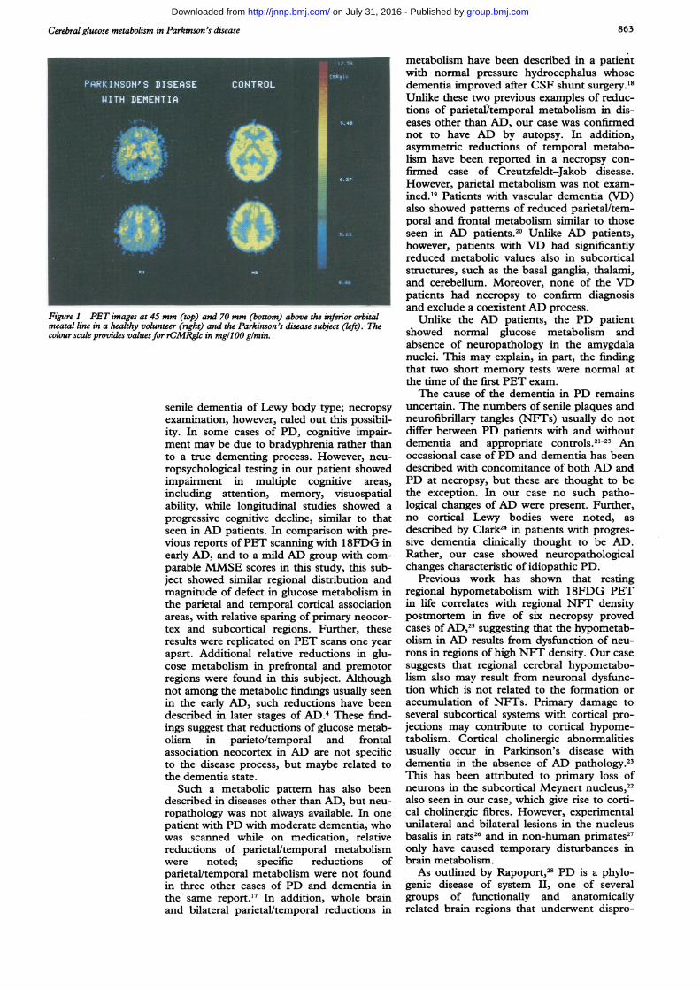
1987) global CMRglc equalled 5-47 mg/100g/min in the Parkinson's disease subject com-pared with 8&41 (SD 1-37) in the healthy vol-unteer group (p < 0-05) and 6-84 (SD 0-82)in the AD group (p > 0-05). Using a Z scoreanalysis,"5 the PD subject showed significantreductions in absolute rCMRglc values com-pared with healthy volunteers in all corticalregions, except the bilateral orbitofrontal,anterior cingulate, and paracentral regions,right calcarine, and left sensorimotor (table2). The later scan on 21 January 1988 showedadditional deficits in the right calcarine andleft sensorimotor regions. There were no sig-nificant reductions of glucose metabolism inthe thalamus and basal ganglia. A similar pat-tern of metabolic reduction was seen in theAD group compared with controls using anunpaired t test, with significant reductions inrCMRglc in all cortical regions. In compari-son with the AD group, the PD patientshowed significant reductions of absoluterCMRglc values in bilateral prefrontal andmedial parietal regions as well as in the rightoccipital association and right superior tempo-ral areas at the time of the first PET scan. Thesecond scan revealed additional deficits in theleft premotor, right inferior parietal, bilateralsensorimotor, left occipital association, leftsuperior temporal regions.To reduce the intersubject variance associ-
ated with absolute values of rCMRglc inhealthy controls and to examine patterns ofmetabolism,' we calculated ratios of rCMRglcto the mean calcarineCMRglc value (table 3),a region relatively spared in AD.16 In the PDsubject, relative reductions were shown in theparietal and temporal regions on both scanscompared with healthy volunteers. On secondPET scan, the left premotor became signifi-cantly reduced, whereas the left prefontalregion, which was decreased at the time of thefirst PET scan, was normal. These changesare illustrated in figure 1, which shows PETimages at two levels in the PD subject and ahealthy volunteer. Relative reductions inrCMRglc also were shown in parietal andmiddle temporal regions in AD subjects com-pared with healthy volunteers. Both the PDand AD subjects showed a similar pattern ofreduced rCMRglc in the parietal, temporal,and frontal association cortices, whereas therewas relative sparing of brain metabolism inprimary motor and sensory, and subcorticalregions.
DiscussionThis is a case of a slowly progressive dementiain a patient who, during life, was initially diag-nosed as having AD,8 then PD with dementia,but was found to have only PD at necropsy.Visual hallucinations, uncommon in earlyAD, may have suggested the diagnosis of
862
group.bmj.com on July 31, 2016 - Published by http://jnnp.bmj.com/Downloaded from
Cerebral glucose metabolism in Parkinson's disease
Figure 1 PET images at 45 mm (top) and 70 mm (bottom) above the inferior orbitalmeatal line in a healthy volunteer (right) and the Parkinson's disease subject (left). Thecolour scale provides values for rCMRglc in mg/i 00 glmin.
senile dementia of Lewy body type; necropsyexamination, however, ruled out this possibil-ity. In some cases of PD, cognitive impair-ment may be due to bradyphrenia rather thanto a true dementing process. However, neu-
ropsychological testing in our patient showedimpairment in multiple cognitive areas,including attention, memory, visuospatialability, while longitudinal studies showed a
progressive cognitive decline, similar to thatseen in AD patients. In comparison with pre-vious reports ofPET scanning with 18FDG inearly AD, and to a mild AD group with com-parable MMSE scores in this study, this sub-ject showed similar regional distribution andmagnitude of defect in glucose metabolism inthe parietal and temporal cortical associationareas, with relative sparing of primary neocor-tex and subcortical regions. Further, theseresults were replicated on PET scans one yearapart. Additional relative reductions in glu-cose metabolism in prefrontal and premotorregions were found in this subject. Althoughnot among the metabolic findings usually seenin the early AD, such reductions have beendescribed in later stages of AD.4 These find-ings suggest that reductions of glucose metab-olism in parieto/temporal and frontalassociation neocortex in AD are not specificto the disease process, but maybe related tothe dementia state.
Such a metabolic pattern has also beendescribed in diseases other than AD, but neu-
ropathology was not always available. In one
patient with PD with moderate dementia, whowas scanned while on medication, relativereductions of parietal/temporal metabolismwere noted; specific reductions ofparietal/temporal metabolism were not foundin three other cases of PD and dementia inthe same report.'7 In addition, whole brainand bilateral parietal/temporal reductions in
metabolism have been described in a patientwith normal pressure hydrocephalus whosedementia improved after CSF shunt surgery.'8Unlike these two previous examples of reduc-tions of parietalYtemporal metabolism in dis-eases other than AD, our case was confirmednot to have AD by autopsy. In addition,asymmetric reductions of temporal metabo-lism have been reported in a necropsy con-firmed case of Creutzfeldt-Jakob disease.However, parietal metabolism was not exam-ined.'9 Patients with vascular dementia (VD)also showed patterns of reduced parietal/tem-poral and frontal metabolism similar to thoseseen in AD patients.20 Unlike AD patients,however, patients with VD had significantlyreduced metabolic values also in subcorticalstructures, such as the basal ganglia, thalami,and cerebellum. Moreover, none of the VDpatients had necropsy to confirm diagnosisand exclude a coexistent AD process.
Unlike the AD patients, the PD patientshowed normal glucose metabolism andabsence of neuropathology in the amygdalanuclei. This may explain, in part, the findingthat two short memory tests were normal atthe time of the first PET exam.The cause of the dementia in PD remains
uncertain. -The numbers of senile plaques andneurofibrillary tangles (NFTs) usually do notdiffer between PD patients with and withoutdementia and appropriate controls.2'-2' Anoccasional case of PD and dementia has beendescribed with concomitance of both AD andPD at necropsy, but these are thought to bethe exception. In our case no such patho-logical changes of AD were present. Further,no cortical Lewy bodies were noted, asdescribed by Clark24 in patients with progres-sive dementia clinically thought to be AD.Rather, our case showed neuropathologicalchanges characteristic of idiopathic PD.
Previous work has shown that restingregional hypometabolism with 18FDG PETin life correlates with regional NET densitypostmortem in five of six necropsy provedcases of AD,25 suggesting that the hypometab-olism in AD results from dysfunction of neu-rons in regions of high NFT density. Our casesuggests that regional cerebral hypometabo-lism also may result from neuronal dysfunc-tion which is not related to the formation oraccumulation of NFTs. Primary damage toseveral subcortical systems with cortical pro-jections may contribute to cortical hypome-tabolism. Cortical cholinergic abnormalitiesusually occur in Parkinson's disease withdementia in the absence of AD pathology.2'This has been attributed to primary loss ofneurons in the subcortical Meynert nucleus,22also seen in our case, which give rise to corti-cal cholinergic fibres. However, experimentalunilateral and bilateral lesions in the nucleusbasalis in rats26 and in non-human primates27only have caused temporary disturbances inbrain metabolism.As outlined by Rapoport,28 PD is a phylo-
genic disease of system II, one of severalgroups of functionally and anatomicallyrelated brain regions that underwent dispro-
863
group.bmj.com on July 31, 2016 - Published by http://jnnp.bmj.com/Downloaded from
Schapiro, Pietrini, Grady, Ball, DeCarli, Kumar, Kaye, Haxby
portionate expansion during recent evolutionof primates, particularly humans. The regionsassociated in system II, including substantianigra, basal ganglia, thalamus, and frontalassociation and motor neocortical areas, par-ticipate in several functionally segregated cir-cuits which subserve cognition as well asmovement.29 The basal ganglia are linked withassociation cortex, by several of these segre-gated circuits. A major subcortical componentof system II that is disrupted in PD is thenigrostriatal system. Dementia is inverselyrelated to the neuronal cell count in themedial part of the substantia nigra.30 The cellsin this portion generally project to the cau-date, and along with cells in the ventraltegmental area which also are lost in PD, pro-ject to cortical and limbic regions. Therefore,significant involvement of parts of the seg-rated circuitry of system II may result indementia and cortical hypometabolism. Theinteraction between altered dopamine andcholinergic function may also have togetherresulted in the dementia and metabolic abnor-malities in our subject.
Dementia and an extrapyramidal syndromemay be seen in, among other diseases, AD,9PD with AD, and, as in this case, PD withoutAD. PET with 18FDG has shown similarreductions in parietal/temporal metabolism incases from each of these disease categoriesand therefore has not allowed differentiationof one entity from another. Preliminary workhas suggested that PET with [18F]6-fluoro-L-dopa (6-FD) differentiates clinically diag-nosed Alzheimer's disease with extra-pyramidal features from Parkinson's diseasewith dementia.3" However, it is not known if6-FD will allow differentiation of dementedParkinson's disease with and without the neu-ropathological changes of Alzheimer's disease.
1 Haxby JV, Grady CL, Duara R, Schlageter N, Berg G,Rapoport SI. Neocortical metabolic abnormalities pre-cede nonmemory cognitive defects in early Alzheimer's-type dementia. Arch Neurol 1986;43:882-5.
2 Grady CL, Haxby JV, Schlageter NL, Berg G, RapoportSI. Stability of metabolic and neuropsychological asym-metries in dementia of the Alzheimer type. Neurology1986;36:1390-2.
3 Schapiro MB, Grady CL, Ball MJ, DeCarli C, RapoportSI. Reductions in parieto/temporal cerebral glucosemetabolism are not specific for Alzheimer's disease.Neurology 1990;40(Suppl 1):152.
4 Haxby JV, Grady CL, Koss E, et al. Heterogeneous ante-rior-posterior metabolic patterns in dementia of theAlzheimer type. Neurology 1988;38:1853-63.
5 Haxby JV, Grady CL, Koss E, et al. Longitudinal study ofcerebral metabolic asymmetries and associated neuro-psychological patterns in early dementia of theAlzheimer type. Arch Neurol 1990;47:753-60.
6 Folstein MF, Folstein SI, McHugh PR. 'Mini MentalState'-a practical method for grading the cognitive stateof patients for the clinician. J Psychiatr Res 1976;12:189-98.
7 Hachinski VC, Iliff LD, Zilhka E, et al. Cerebral blood
flow in dementia. Arch Neurol 1975;32:632-7.8 American Psychiatric Association Task Force on
Nomenclature and Statistics. Diagnostic and statisticalmanual for mental disorders. 3rd ed. Washington, DC:American Psychiatric Association, 1980.
9 Kaye JA, May C, Daly E, et al. Cerebrospinal fluidmonoamine markers are decreased in dementia of theAlzheimer type with extrapyramidal features. Neurology1988;38:554-7.
10 McKhann G, Drachman D, Folstein M, Katzman R, PriceD, Stadlan EM. Clinical diagnosis of Alzheimer's dis-ease: report of the NINCDS-ADRDA work group underthe auspices of Department of Health and HumanServices task force on Alzheimer's disease. Neurology1984;34:939-44.
11 Duara R, Margolin RA, Robertson-Tchabo EA, et al.Cerebral glucose utilization as measured with positronemission tomography in 21 resting healthy men betweenthe ages of 21 and 83 years. Brain 1983;106:761-75.
12 Swedo SE, Schapiro MB, Grady CL, et al. Cerebral glu-cose metabolism in childhood-onset obsessive-compul-sive disorder. Arch Gen Psychiatry 1989;46:518-23.
13 Brooks RA. Alternative formula for glucose utilizationusing labeled deoxyglucose. J Nucl Med 1982;23:538-9.
14 Sokoloff L, Reivich M, Kennedy C, et al. The (14C)-deoxyglucose method for the measurement of local cere-bral glucose utilization: theory, procedure and normalvalues in the conscious and anesthetized albino rat. JNeuroch 1977;28:897-916.
15 Colton T. Statistics in medicine 1st ed. Boston: Little,Brown and Company, 1974:84-7.
16 Kumar A, Schapiro MB, Grady CL, et al. High-resolutionPET studies in Alzheimer's disease. Neuropsycho-pharmacology 1991;4:35-46.
17 Kuhl DE, Metter EJ, Riege WH. Patterns of local cerebralglucose utilization determined in Parkinson's disease bythe [18F]fluorodeoxyglucose method. Ann Neurol1984;15:419-24.
18 Friedland RP. 'Normal'-pressure hydrocephalus and thesaga of the treatable dementias. JAMA 1989;18:2577-81.
19 Friedland RP, Prusiner SB, Jaqust WJ, Budinger TF,Davis RL. Bitemporal hypometabolism in Creutzfeldt-Jakob disease measured by positron emission tomogra-phy with [18F]-2-fluorodeoxyglucose. Y Comput AssistTomogr 1984;8:978-81.
20 Mielke R, Herholz K, Grond M, Kessler J, Heiss W-D.Severity of vascular dementia is related to volume ofmetabolically impaired tissue. Arch Neurol 1992;49:909-13.
21 Ball MJ. The morphological basis of dementia inParkinson's disease. Can JNeurol Sci 1984; 1:180-4.
22 Mann DMA, Yates PO. Pathological basis for neurotrans-mitter changes in Parkinson's disease. Neuropathol ApplNeurobiol 1983;9:3-19.
23 Perry RH, Tomlinson BE, Candy JM, et al. Corticalcholinergic deficit in mentally impaired Parlinsonianpatients. Lancet 1983;2:789-90.
24 Clark AW, White CL, Manz HJ, et al. Primary degenera-tive dementia without Alzheimer pathology. Can YNeurol Sci 1986;13:462-70.
25 DeCarli C, Atack JR, Ball MJ, et al. Post-mortem regionalneurofibrillary tangle densities but not senile plaque den-sities are related to regional cerebral metabolic rates forglucose during life in Alzheimer's disease patients.Neurodegeneration 1992;1:1 1-9.
26 Orzi F, Diana G, Casamenti F, Palombo E, Fieschi C.Local cerebral glucose utilization following unilateraland bilateral lesions of the nucleus basalis magnocellu-laris in the rat. Brain Res 1988;462:99-103.
27 Kiyosawa M, Baron JC, Hamel E, et al. Time course ofeffects of unilateral lesions of the nucleus basalis ofmeynert on glucose utilization by the cerebral cortex.Brain 1989;112:435-55.
28 Rapoport SI. Integrated phylogeny of the primate brain,with special reference to humans and their diseases.Brain Research Reviews 1990;15:267-94.
29 Alexander GE, DeLong MR, Strick PL. Parallel organiza-tion of functionally segregated circuits linking basal gan-glia and cortex. Annu Rev Neurosci 1986;9:357-81.
30 Rinne JO, Rummukainen J, Paljarvi L, Rinne U. Dementiain Parkinson's disease is related to neuronal loss in themedial substantia nigra. Ann Neurol 1989;26:47-50.
31 Tyrrell PJ, Sawle GV, Frackowiak RSJ, Rossor MN.Extrapyramidal signs in dementia of the Alzheimer-type:18F-DOPA studies. J Cereb Blood Flow Metabol1989;9:S568.
864
group.bmj.com on July 31, 2016 - Published by http://jnnp.bmj.com/Downloaded from
for Alzheimer's disease.metabolic rates for glucose are not specific Reductions in parietal and temporal cerebral
Kaye and J V HaxbyM B Schapiro, P Pietrini, C L Grady, M J Ball, C DeCarli, A Kumar, J A
doi: 10.1136/jnnp.56.8.8591993 56: 859-864 J Neurol Neurosurg Psychiatry
http://jnnp.bmj.com/content/56/8/859Updated information and services can be found at:
These include:
serviceEmail alerting
box at the top right corner of the online article. Receive free email alerts when new articles cite this article. Sign up in the
Notes
http://group.bmj.com/group/rights-licensing/permissionsTo request permissions go to:
http://journals.bmj.com/cgi/reprintformTo order reprints go to:
http://group.bmj.com/subscribe/To subscribe to BMJ go to:
group.bmj.com on July 31, 2016 - Published by http://jnnp.bmj.com/Downloaded from
Copyright © 2022 FDOKUMEN