Red American Ginseng: Ginsenoside Constituents and Antiproliferative Activities of Heat-Processed...
11
Red American Ginseng: Ginsenoside Constituents and Antiproliferative Activities of Heat-Processed Panax quinquefolius Roots Chong-Zhi Wang 1,2 , Han H. Aung 1,2 , Ming Ni 1,2 , Ji-An Wu 1,2 , Robin Tong 1,2 , Sheila Wicks 1,2 , Tong-Chuan He 3 , and Chun-Su Yuan 1,2,4 1 Tang Center for Herbal Medicine Research, University of Chicago, Chicago, Illinois, USA 2 Department of Anesthesia and Critical Care, University of Chicago, Chicago, Illinois, USA 3 Department of Surgery, University of Chicago, Chicago, Illinois, USA 4 Committee on Clinical Pharmacology and Pharmacogenomics, University of Chicago, Chicago, Illinois, USA Abstract Red Asian ginseng (Panax ginseng C. A. Meyer, Araliaceae) is used in many Oriental countries. In this study, the saponin constituents and anticancer activities of steamed American ginseng (Panax quinquefolius L.) roots were evaluated. The contents of 12 ginsenosides in the roots were determined using high performance liquid chromatography (HPLC). After the steaming treatment (100 – 120 ° C for 1 h and 120 °C for 0.5 – 4 h), the quantity of 7 ginsenosides decreased and that of 5 others increased. The content of ginsenoside Rg3, a previously recognized anticancer compound, increased significantly when the root was steamed at 120 °C for 0.5 – 3 h. The antiproliferative effects of unsteamed and steamed (120 °C for 1 h and 2 h) American ginseng root extracts were assayed by the modified trichrome stain (MTS) method using three cancer cell lines (SW-480, HT-29, NSCLC). Heat-processing increased the antiproliferative effect of American ginseng significantly, and the activity of the extract from roots steamed for 2 h was greater than that of roots steamed for 1 h. Chemical constituents and antiproliferative activities of white and red Asian ginseng have also been evaluated. Five representative ginsenosides, Rb1, Rd, Re, Rg2 and Rg3, were studied. Ginsenoside Rg3 had the most potent effect. The antiproliferative activities of red American ginseng are augmented when ginsenoside Rg3 is increased. American ginseng (Panax quinquefolius L.) root is a herb commonly used in the USA. [1]. In oriental countries, Asian ginseng (Panax ginseng C. A. Meyer) root is air-dried into white ginseng or steamed at 100 °C to red ginseng [2]. A number of studies point to anticancer properties and other pharmacological activities of Asian ginseng [3], [4], [5], and ginsenosides Rg3 and Rh2 are recognized as active anticancer compounds [3]. Compared with Asian white ginseng, red ginseng has stronger anticancer activities [6], [7]. Recently, there was a report using a steaming process to treat American ginseng root [8]. In the study, however, the treatment temperature was 100 °C and thus, chemical constituents did not change significantly. Additionally, only 6 ginsenosides (excluding ginsenoside Rg3) were analyzed. Correspondence: Chun-Su Yuan, M.D., Ph. D., Tang Center for Herbal Medicine Research, and Department of Anesthesia & Critical Care, University of Chicago, 5841 South Maryland Avenue, MC 4028, Chicago, Illinois 60637, USA, Phone: +1-773-702-1916, Fax: +1-773-834-0601, E-mail: [email protected]. NIH Public Access Author Manuscript Planta Med. Author manuscript; available in PMC 2009 March 18. Published in final edited form as: Planta Med. 2007 June ; 73(7): 669–674. doi:10.1055/s-2007-981524. NIH-PA Author Manuscript NIH-PA Author Manuscript NIH-PA Author Manuscript
-
Upload
independent -
Category
Documents
-
view
3 -
download
0
Transcript of Red American Ginseng: Ginsenoside Constituents and Antiproliferative Activities of Heat-Processed...
Red American Ginseng: Ginsenoside Constituents andAntiproliferative Activities of Heat-Processed Panaxquinquefolius Roots
Chong-Zhi Wang1,2, Han H. Aung1,2, Ming Ni1,2, Ji-An Wu1,2, Robin Tong1,2, SheilaWicks1,2, Tong-Chuan He3, and Chun-Su Yuan1,2,4
1 Tang Center for Herbal Medicine Research, University of Chicago, Chicago, Illinois, USA
2 Department of Anesthesia and Critical Care, University of Chicago, Chicago, Illinois, USA
3 Department of Surgery, University of Chicago, Chicago, Illinois, USA
4 Committee on Clinical Pharmacology and Pharmacogenomics, University of Chicago, Chicago, Illinois,USA
AbstractRed Asian ginseng (Panax ginseng C. A. Meyer, Araliaceae) is used in many Oriental countries. Inthis study, the saponin constituents and anticancer activities of steamed American ginseng (Panaxquinquefolius L.) roots were evaluated. The contents of 12 ginsenosides in the roots were determinedusing high performance liquid chromatography (HPLC). After the steaming treatment (100 – 120 °C for 1 h and 120 °C for 0.5 – 4 h), the quantity of 7 ginsenosides decreased and that of 5 othersincreased. The content of ginsenoside Rg3, a previously recognized anticancer compound, increasedsignificantly when the root was steamed at 120 °C for 0.5 – 3 h. The antiproliferative effects ofunsteamed and steamed (120 °C for 1 h and 2 h) American ginseng root extracts were assayed bythe modified trichrome stain (MTS) method using three cancer cell lines (SW-480, HT-29, NSCLC).Heat-processing increased the antiproliferative effect of American ginseng significantly, and theactivity of the extract from roots steamed for 2 h was greater than that of roots steamed for 1 h.Chemical constituents and antiproliferative activities of white and red Asian ginseng have also beenevaluated. Five representative ginsenosides, Rb1, Rd, Re, Rg2 and Rg3, were studied. GinsenosideRg3 had the most potent effect. The antiproliferative activities of red American ginseng areaugmented when ginsenoside Rg3 is increased.
American ginseng (Panax quinquefolius L.) root is a herb commonly used in the USA. [1]. Inoriental countries, Asian ginseng (Panax ginseng C. A. Meyer) root is air-dried into whiteginseng or steamed at 100 °C to red ginseng [2]. A number of studies point to anticancerproperties and other pharmacological activities of Asian ginseng [3], [4], [5], and ginsenosidesRg3 and Rh2 are recognized as active anticancer compounds [3]. Compared with Asian whiteginseng, red ginseng has stronger anticancer activities [6], [7]. Recently, there was a reportusing a steaming process to treat American ginseng root [8]. In the study, however, thetreatment temperature was 100 °C and thus, chemical constituents did not change significantly.Additionally, only 6 ginsenosides (excluding ginsenoside Rg3) were analyzed.
Correspondence: Chun-Su Yuan, M.D., Ph. D., Tang Center for Herbal Medicine Research, and Department of Anesthesia & CriticalCare, University of Chicago, 5841 South Maryland Avenue, MC 4028, Chicago, Illinois 60637, USA, Phone: +1-773-702-1916, Fax:+1-773-834-0601, E-mail: [email protected].
NIH Public AccessAuthor ManuscriptPlanta Med. Author manuscript; available in PMC 2009 March 18.
Published in final edited form as:Planta Med. 2007 June ; 73(7): 669–674. doi:10.1055/s-2007-981524.
NIH
-PA Author Manuscript
NIH
-PA Author Manuscript
NIH
-PA Author Manuscript
We treated American ginseng root at a variety of temperatures and heating times to observechanges in ginsenoside contents and anticancer activities. After heat-processing, the root ofAmerican ginseng, like Asian ginseng, changes from white to red [2]; steamed P.quinquefolius root is therefore referred to as red American ginseng.
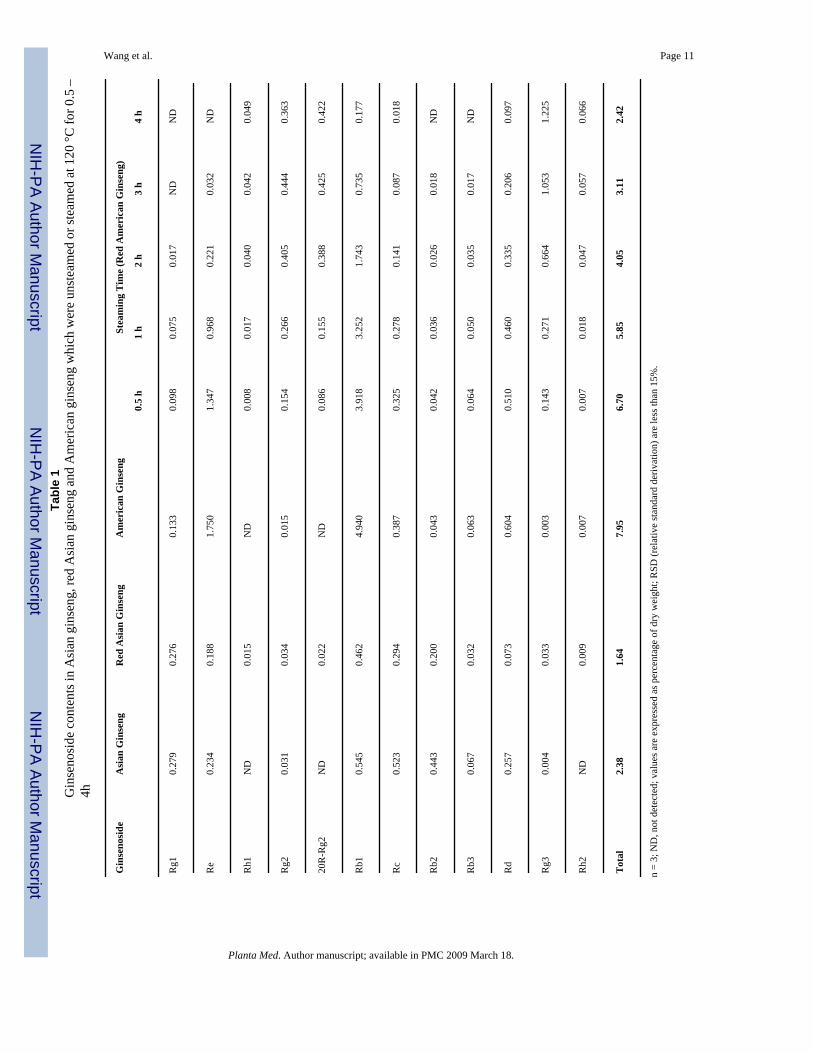
Twelve ginsenosides (Fig.1) were determined in American ginseng using high performanceliquid chromatography (HPLC). All the assayed ginsenosides were dammarane glycosides[9], [10]. The influence of steaming at 100 °C and 120 °C on the ginsenoside content ofAmerican ginseng was tested. Compared with unsteamed American ginseng, the roots treatedat 100 °C for 1 h had a slightly decreased total ginsenoside content, from 7.95% to 7.32%. Forthe main saponin contents, ginsenoside Rb1 was changed from 4.940% to 4.463%, and Refrom 1.756% to 1.630%; ginsenoside Rg3 increased from 0.003% to 0.048%. Steaming at 120°C for 1 h decreased the total ginsenoside content to 5.85%, as follows: Rb1, 3.252%; Re,0.968%; and Rg3, 0.271%. Ginsenoside Rg3 increased significantly at 120 °C.
Chromatograms of unsteamed and steamed American ginseng roots for 2 and 4 h at 120 °Care shown in Figs.2A–C. The peak areas of Rb1, Rd and Re decreased during the steamingprocess. On the other hand, ginsenoside Rg3, which is a trace saponin in unsteamed root[11], was augmented during the steaming process (Figs. 2B and C). The content of the 12ginsenosides in steamed American ginseng roots is shown in Table 1. During the steamingprocess, Rg1, Re, Rb1, Rc, Rb2, Rb3, and Rd decreased while Rh1, Rg2, 20R-Rg2, Rg3 andRh2 increased. Ginsenoside Rg3 increased significantly from 0.5 to 3 h of steaming.Ginsenoside Rh2 is also an active anticancer saponin, however, after steaming treatment, thecontent of Rh2 was very low (Table 1).
The chemical constituents of white ginseng and red ginseng (P. ginseng C. A. Meyer) werealso determined. Data shown in Table 1 indicated that for the Asian ginseng, after steaming at100 °C, the contents of the main ginsenosides (Rg1, Re, Rb1, Rb2, Rc and Rd) were decreased.Ginsenoside Rg3 increased from 0.004% to 0.033%. The changes of ginsenoside contents aresimilar to those of American ginseng steamed at 100 °C.
The antiproliferative effects on SW-480 and HT-29 human colorectal cancer cells and non-small cell lung cancer cells (NSCLC) of white ginseng and red ginseng extracts have beenevaluated. At the concentrations of 0.1, 0.25, 0.5 and 1 mg/mL, although the antiproliferativeactivities of red ginseng were stronger than that of white ginseng extract, in most cases, therewere no significant differences between them. The only exception was that at 0.5 mg/mL forHT-29 cells, the antiproliferative effect of red ginseng (42.9 ± 2.3%) was significantly higherthan that of white ginseng (26.4 ± 3.3%) (Fig. 3).
For the American ginseng, to evaluate the trend of anticancer activities, extracts from rootssteamed for 1 h and 2 h were added to SW-480, HT-29 and NSCLC cells (Fig. 4). UnsteamedAmerican ginseng root extract had antiproliferative effects on the three cancer cell lines, butthe effects were minimal at 0.1 – 0.5 mg/mL. Treated red American ginseng extracts hadsignificant antiproliferative effects. At 0.5 mg/mL, the steamed root extracts inhibited thegrowth of three cancer cell lines completely. Antiproliferative effects of red American ginsengwere influenced by the steaming times. The effects of extract from root steamed for 2 h werestronger than from root steamed for 1 h. Moreover, the antiproliferative effect of red Americanginseng is greater than that of red Asian ginseng (see Fig. 3 and Fig.4B).
The biological activity of red American ginseng extract is the result of saponins, believed tobe the main active constituents in American ginseng. In this diverse group of saponins, fiverepresentative ginsenosides, three major constituents in unsteamed root (Rb1, Rd and Re) andtwo main constituents in steamed root (Rg2 and Rg3), were used to test for antiproliferativeeffects on cancer cells. From the results shown in Fig. 5, at 30 – 300 μM, ginsenosides Rb1,
Wang et al. Page 2
Planta Med. Author manuscript; available in PMC 2009 March 18.
NIH
-PA Author Manuscript
NIH
-PA Author Manuscript
NIH
-PA Author Manuscript
Rd, Re, and Rg2 did not show strong antiproliferative effects on SW-480 cells. GinsenosideRg3, which is the main constituent in steamed American ginseng root, showed strongantiproliferative effects on SW-480 cells at 300 μM. Similar activities were observed on HT-29and NSCLC cells. Since several unconfirmed compounds were also detected (Fig. 2), thestructure and antiproliferative activities of these compounds will be the focus of future studies.
Human colorectal cancer is a significant public health problem in the Western world [12]. Inthe United States, this cancer is the second-leading cause of cancer-related deaths and thesecond most prevalent cancer worldwide [13]. The lung is one of the common sites ofmetastasis of colorectal cancer. About 85% of all lung cancers are of the non-small cell type[14], [15]. In this study, the anticancer activities of steamed American ginseng root extractson two human colorectal cancer cell lines and one non-small cell lung cancer cell line wereevaluated. After steaming at 120 °C, the antiproliferative effects of American ginseng rootextracts increased significantly in all the three cancer cell lines. The main anticancer ginsengsaponins, ginsenosides Rg3 and Rh2, can be obtained by hydrolysis from other ginsenosidessuch as Rb1, Rb2, Rc and Rd. Data from our study suggested that steaming at a highertemperature (120 °C) is advantageous for the conversion of Rg3 from other saponins. RedAmerican ginseng, which is the heat-processed root of P. quinquefolius, may be a potentanticancer herbal medicine.
Materials and MethodsMaterials
The root of P. quinquefolius L. (No. Pq2004003) was collected from Roland Ginseng, LLC(Wausau, WI, USA). White ginseng (P. ginseng C. A. Meyer) (No. Pg2005001) and redginseng (No. Pg2005006) were purchased from Tongrentang Pharmacy, Beijing, P. R. China.The voucher specimens were authenticated by Dr. Chong-Zhi Wang and deposited at the TangCenter for Herbal Medicine Research at University of Chicago (Chicago, IL, USA).Ginsenoside standards were purchased from Delta Information Center for Natural OrganicCompounds (Xuancheng, P. R. China). Other chemicals were obtained from Fisher Scientific(Norcross, GA, USA) and Promega (Madison, WI, USA).
Steaming treatment and extractionFor the heat-processing of American ginseng, the roots were steamed at 100 °C, and 120 °C(AMSCO 2022 Autoclave; AMSCO Scientific; Apex, NC, USA) for 1 h, or steamed at 120 °C for 0.5, 1, 2, 3, and 4 h. Fresh and steamed roots were lyophilized to obtain dried samples.For the HPLC analysis, dried root samples were ground and extracted with methanol in Soxhletextractors, and then assayed. For the in vitro anticancer studies, root samples unsteamed orsteamed at 120 °C for 1 h and 2 h were ground and extracted with 70% ethanol. The solventof the extract solution was evaporated under vacuum. The dried extract was dissolved in water,and then extracted with water-saturated n-butanol. The n-butanol phase was evaporated undervacuum and then lyophilized.
HPLC analysisThe HPLC system was a Waters 2960 instrument with a 996 photodiode array detector(Milford, MA, USA). The separation was carried out on an Alltech Ultrasphere C18 column(5 μ, 250×3.2 mm I.D.) (Deerfield, IL, USA) with a guard column (Alltech Ultrasphere C18,5 μ, 7.5×3.2 mm I.D.) [16]. Acetonitrile (solvent A) and water (solvent B) were used. Gradientelution started with 18% solvent A and 82% solvent B, was changed to 21% A for 20 min, to26% A for 3 min and held for 19 min; to 36% A for 13 min; to 50% A for 9 min; to 95% A for2 min and held for 3 min; and finally changed to 18% A for 3 min and held for 8 min. The flowrate was 1.0 mL/min and the detection wavelength was set to 202 nm. All tested solutions were
Wang et al. Page 3
Planta Med. Author manuscript; available in PMC 2009 March 18.
NIH
-PA Author Manuscript
NIH
-PA Author Manuscript
NIH
-PA Author Manuscript
filtered through Millex 0.2-μm nylon membrane syringe filters before use. The contents ofsaponins in each sample were calculated using standard curves of ginsenosides.
Cell cultureThe human colorectal cancer cell lines SW-480 (Leibovitz's L-15), HT-29 (McCoy's 5A), andnon-small cell lung cancer cells (NSCLC, DMEM) were purchased from American TypeCulture Collection, ATCC (Manassas, VA, USA) and grown in the indicated mediasupplemented with 10% FBS and 50 IU penicillin/streptomycin in a humidified atmosphereof 5% CO2 at 37 °C.
Cell proliferation analysisUnsteamed, steamed American ginseng root extract, and five ginsenosides were dissolved in50% ethanol. Cells were seeded in 96-well plates (1×10⁴ cells/well). After 1 d, variousconcentrations of extracts/ginsenosides were added to the wells. The final concentration ofethanol was 0.5%. Controls were exposed to culture medium containing 0.5% ethanol withoutdrugs. All experiments were performed in triplicate and repeated 3 times. Drug treatment timewas dependent on the cell growth rate. We selected 72 h for SW-480 and HT-29 cells, and 48h for NSCLC cells. Cell proliferation was evaluated using an MTS assay according to themanufacturer's instructions. Briefly, at the end of the drug exposure period, the medium wasreplaced with 100 μL of fresh medium, 20 μL of MTS reagent (CellTiter 96 Aqueous Solution)in each well, and the plate was returned to the incubator for 1 – 2 h. A 60-μL aliquot of mediumfrom each well was transferred to an ELISA 96-well plate and its absorbance at 490 nm wasrecorded [17]. Since 0.5% ethanol did not influence the proliferation of three cell lines, resultswere expressed as percent of control (ethanol vehicle set at 100%).
Statistical analysis of the resultsData are presented as mean ± standard deviation (SD) with n = 3. A one-way ANOVAdetermined whether the results had statistical significance. In some cases, Student's t test wasused for comparing two groups. The level of statistical significance was set at P < 0.05. Forthe antiproliferative effects of white and red ginseng extract on cancer cells, the significanceof red ginseng extract vs. white ginseng extract was assessed by Student's t test. For theantiproliferative effects of ginsenosides on SW-480 cells, the significance of ginsenosidetreatment vs. control was assessed by Student's t test.
AcknowledgementsThis work was supported in part by research grants from the American Cancer Society (RSG-05-254-01DDC), theNIH/NCI (1RO1 CA106569-01), and the NIH/NCCAM (AT002176 and AT002445).
References1. Ang-Lee MK, Moss J, Yuan CS. Herbal medicines and perioperative care. JAMA 2001;286:208–16.
[PubMed: 11448284]2. Takaku T, Kameda K, Matsuura Y, Sekiya K, Okuda H. Studies on insulin-like substances in Korean
red ginseng. Planta Med 1990;56:27–30. [PubMed: 2192378]3. Helms S. Cancer prevention and therapeutics: Panax ginseng. Altern Med Rev 2004;9:259–74.
[PubMed: 15387718]4. Xie JT, Mchendale S, Yuan CS. Ginseng and diabetes. Am J Chin Med 2005;33:397–404. [PubMed:
16047557]5. Liou CJ, Huang WC, Tseng J. Long-term oral administration of ginseng extract modulates humoral
immune response and spleen cell functions. Am J Chin Med 2005;33:651–61. [PubMed: 16173538]
Wang et al. Page 4
Planta Med. Author manuscript; available in PMC 2009 March 18.
NIH
-PA Author Manuscript
NIH
-PA Author Manuscript
NIH
-PA Author Manuscript
6. Yun TK, Lee YS, Lee YH, Kim SI, Yun HY. Anticarcinogenic effect of Panax ginseng C.A. Meyerand identification of active compounds. J Korean Med Sci 2001;16:S6–18. [PubMed: 11748383]
7. Yoo HH, Yokozawa T, Satoh A, Kang KS, Kim HY. Effects of ginseng on the proliferation of humanlung fibroblasts. Am J Chin Med 2006;34:137–46. [PubMed: 16437746]
8. Kim KT, Yoo KM, Lee JW, Eom SH, Hwang IK, Lee CY. Protective effect of steamed Americanginseng (Panax quinquefolius L.) on V79 – 4 cells induced by oxidative stress. J Ethnopharmacol.2007advance online publication
9. Park JD, Rhee DK, Lee YH. Biological activities and chemistry of saponins from Panax ginseng C.A. Meyer. Phytochem Rev 2005;4:159–75.
10. Wang CZ, McEntee E, Wicks S, Wu JA, Yuan CS. Phytochemical and analytical studies of Panaxnotoginseng (Burk.) F. H. Chen. J Nat Med 2006;60:97–106.
11. Wang CZ, Wu JA, McEntee E, Yuan CS. Saponins composition in American ginseng leaf and berryassayed by high-performance liquid chromatography. J Agric Food Chem 2006;54:2261–6.[PubMed: 16536605]
12. Francois F, Park J, Bini EJ. Colon pathology detected after a positive screening flexiblesigmoidoscopy: a prospective study in an ethnically diverse cohort. Am J Gastroenterol2006;101:823–30. [PubMed: 16494591]
13. Jemal A, Tiwari RC, Murray T, Ghafoor A, Samuels A, Ward E, et al. Cancer statistics, 2004. CACancer J Clin 2004;54:8–29. [PubMed: 14974761]
14. Penna C, Nordlinger B. Colorectal metastasis (liver and lung). Surg Clin North Am 2002;82:1075–90. [PubMed: 12507210]
15. Saijo N. Recent trends in the treatment of advanced lung cancer. Cancer Sci 2006;97:448–52.[PubMed: 16734721]
16. Wang CZ, Zhang B, Song WX, Wang A, Ni M, Luo X, et al. Steamed American ginseng berry:ginsenoside analyses and anticancer activities. J Agric Food Chem 2006;54:9936–42. [PubMed:17177524]
17. Cory AH, Owen TC, Barltrop JA, Cory JG. Use of an aqueous soluble tetrazolium/formazan assayfor cell growth assays in culture. Cancer Commun 1991;3:207–12. [PubMed: 1867954]
Wang et al. Page 5
Planta Med. Author manuscript; available in PMC 2009 March 18.
NIH
-PA Author Manuscript
NIH
-PA Author Manuscript
NIH
-PA Author Manuscript
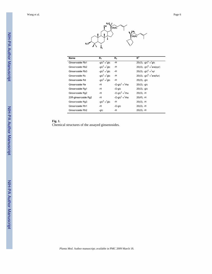
Fig. 1.Chemical structures of the assayed ginsenosides.
Wang et al. Page 6
Planta Med. Author manuscript; available in PMC 2009 March 18.
NIH
-PA Author Manuscript
NIH
-PA Author Manuscript
NIH
-PA Author Manuscript
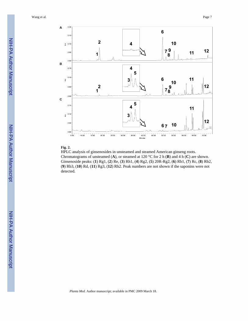
Fig. 2.HPLC analysis of ginsenosides in unsteamed and steamed American ginseng roots.Chromatograms of unsteamed (A), or steamed at 120 °C for 2 h (B) and 4 h (C) are shown.Ginsenoside peaks: (1) Rg1, (2) Re, (3) Rh1, (4) Rg2, (5) 20R-Rg2, (6) Rb1, (7) Rc, (8) Rb2,(9) Rb3, (10) Rd, (11) Rg3, (12) Rh2. Peak numbers are not shown if the saponins were notdetected.
Wang et al. Page 7
Planta Med. Author manuscript; available in PMC 2009 March 18.
NIH
-PA Author Manuscript
NIH
-PA Author Manuscript
NIH
-PA Author Manuscript

Fig. 3.Antiproliferative effects of Asian ginseng root extract on HT-29 cells. Cancer cells wereexposed to white ginseng and red ginseng extract (0.1, 0.25, 0.5, 1 mg/mL) for 72 h and cellproliferation was determined by the MTS assay. The results are expressed as the mean ± SDof three independent experiments. * p < 0.05, red ginseng vs. white ginseng.
Wang et al. Page 8
Planta Med. Author manuscript; available in PMC 2009 March 18.
NIH
-PA Author Manuscript
NIH
-PA Author Manuscript
NIH
-PA Author Manuscript

Fig. 4.Antiproliferative effects of unsteamed and steamed (120 °C for 1 h or 2 h) American ginsengroot extract on cancer cells determined by the MTS assay. Four doses (0.1, 0.25, 0.5 and 1 mg/mL) were used and the treatment time for SW-480 (A) and HT-29 (B) cells was 72 h; forNSCLC cells (C), it was 48 h. The results are expressed as the mean ± SD of three independentexperiments.
Wang et al. Page 9
Planta Med. Author manuscript; available in PMC 2009 March 18.
NIH
-PA Author Manuscript
NIH
-PA Author Manuscript
NIH
-PA Author Manuscript
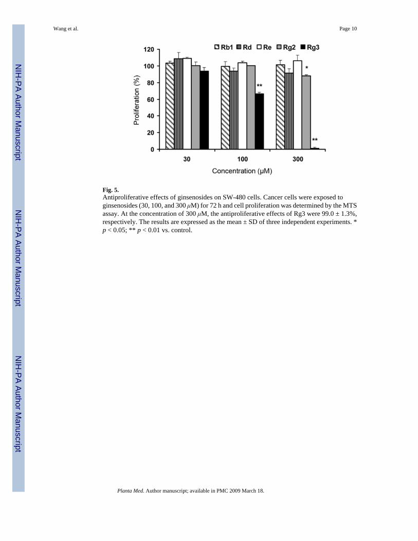
Fig. 5.Antiproliferative effects of ginsenosides on SW-480 cells. Cancer cells were exposed toginsenosides (30, 100, and 300 μM) for 72 h and cell proliferation was determined by the MTSassay. At the concentration of 300 μM, the antiproliferative effects of Rg3 were 99.0 ± 1.3%,respectively. The results are expressed as the mean ± SD of three independent experiments. *p < 0.05; ** p < 0.01 vs. control.
Wang et al. Page 10
Planta Med. Author manuscript; available in PMC 2009 March 18.
NIH
-PA Author Manuscript
NIH
-PA Author Manuscript
NIH
-PA Author Manuscript
NIH
-PA Author Manuscript
NIH
-PA Author Manuscript
NIH
-PA Author Manuscript
Wang et al. Page 11Ta
ble
1G
inse
nosi
de c
onte
nts i
n A
sian
gin
seng
, red
Asi
an g
inse
ng a
nd A
mer
ican
gin
seng
whi
ch w
ere
unst
eam
ed o
r ste
amed
at 1
20 °C
for 0
.5 –
4h
Gin
seno
side
Asi
an G
inse
ngR
ed A
sian
Gin
seng
Am
eric
an G
inse
ngSt
eam
ing
Tim
e (R
ed A
mer
ican
Gin
seng
)
0.5
h1
h2
h3
h4
h
Rg1
0.27
90.
276
0.13
30.
098
0.07
50.
017
ND
ND
Re
0.23
40.
188
1.75
01.
347
0.96
80.
221
0.03
2N
D
Rh1
ND
0.01
5N
D0.
008
0.01
70.
040
0.04
20.
049
Rg2
0.03
10.
034
0.01
50.
154
0.26
60.
405
0.44
40.
363
20R
-Rg2
ND
0.02
2N
D0.
086
0.15
50.
388
0.42
50.
422
Rb1
0.54
50.
462
4.94
03.
918
3.25
21.
743
0.73
50.
177
Rc
0.52
30.
294
0.38
70.
325
0.27
80.
141
0.08
70.
018
Rb2
0.44
30.
200
0.04
30.
042
0.03
60.
026
0.01
8N
D
Rb3
0.06
70.
032
0.06
30.
064
0.05
00.
035
0.01
7N
D
Rd
0.25
70.
073
0.60
40.
510
0.46
00.
335
0.20
60.
097
Rg3
0.00
40.
033
0.00
30.
143
0.27
10.
664
1.05
31.
225
Rh2
ND
0.00
90.
007
0.00
70.
018
0.04
70.
057
0.06
6
Tot
al2.
381.
647.
956.
705.
854.
053.
112.
42
n =
3; N
D, n
ot d
etec
ted;
val
ues a
re e
xpre
ssed
as p
erce
ntag
e of
dry
wei
ght;
RSD
(rel
ativ
e st
anda
rd d
eriv
atio
n) a
re le
ss th
an 1
5%.
Planta Med. Author manuscript; available in PMC 2009 March 18.