Pairwise interactions of polymerization inhibitory contact site mutations of hemoglobinS
Upload
independentCategory
view
3download
0
The importance of polymerization and galloylation forthe antiproliferative properties of procyanidin-richnatural extractsD. Lizarraga1, C. Lozano2, J. J. Briede3, J. H. van Delft3, S. Tourino2, J. J. Centelles1,J. L. Torres2 and M. Cascante1,2
1 Biochemistry and Molecular Biology Department, Biology Faculty, University of Barcelona, Biomedicine Institute from University of Barcelona
(IBUB) and Centre for Research in Theoretical Chemistry, Scientific Park of Barcelona (CeRQT-PCB), Associated Unit to CSIC, Spain
2 Institute for Chemical and Environmental Research (IIQAB-CSIC), Barcelona, Spain
3 Department of Health Risk Analysis and Toxicology, Maastricht University, the Netherlands
Colorectal cancer is the third most commonly diagnosed
cancer in the world and is one of the major causes of
cancer-associated mortality in the USA [1,2]. Epidemio-
logical studies indicate that colon cancer incidence is
inversely related to the consumption of fruit, vegetables
and green tea [3,4]. Specifically, the imbalance between
high-level oxidant exposure and antioxidant capacity in
the colon has been linked to increased cancer risk and
is strongly influenced by dietary antioxidants [5–7].
Several studies have demonstrated that polyphenolic
compounds are capable of providing protection against
cancer initiation and its subsequent development [8–11].
A variety of health-promoting products obtained
from grape seeds and skins, tea leaves, pine and other
plant byproducts are currently available and a great
deal of research is being devoted to testing the putative
beneficial effect of these products in relation to their
polyphenolic content [12–16]. Catechins and their poly-
meric forms (proanthocyanidins) are being studied in
particular depth. The composition of monomeric cate-
chins and their oligomers and polymers (proantho-
cyanidins), as well as the percentage of galloylated
species in these natural extracts, differs between tea,
grape and pine bark.
The antiproliferative activity of catechins and pro-
anthocyanidins is associated with their ability to inhi-
bit cell proliferation and to induce cell cycle arrest and
apoptosis [17,18]. Most of the polyphenols in tea are
monomers of gallocatechins and their gallates [19],
whereas grape contains monomers and oligomers of
Keywords
antiproliferative; apoptosis; cell cycle; colon
cancer; scavenger capacity
Correspondence
M. Cascante Serratosa, Department of
Biochemistry and Molecular Biology,
University of Barcelona, Biology Faculty,
Av. Diagonal 645, 08028 Barcelona, Spain
Fax: +34 934021219
Tel: +34 934021593
E-mail: [email protected]
(Received 2 May 2007, revised 3 July 2007,
accepted 18 July 2007)
doi:10.1111/j.1742-4658.2007.06010.x
Grape (Vitis vinifera) and pine (Pinus pinaster) bark extracts are widely
used as nutritional supplements. Procyanidin-rich fractions from grape and
pine bark extract showing different mean degrees of polymerization, per-
centage of galloylation (percentage of gallate esters) and reactive oxygen
species-scavenging capacity were tested on HT29 human colon cancer cells.
We observed that the most efficient fractions in inhibiting cell proliferation,
arresting the cell cycle in G2 phase and inducing apoptosis were the grape
fractions with the highest percentage of galloylation and mean degree of
polymerization. Additionally, the antiproliferative effects of grape fractions
were consistent with their oxygen radical-scavenging capacity and their
ability to trigger DNA condensation–fragmentation.
Abbreviations
DMPO, 5,5-dimethyl-1-pyrolline-N-oxide; FACS, fluorescence-activated cell sorter; FITC, fluorescein isothiocyanate; MTT,
3-(4,5-dimethylthiazol-2yl)-2,5-diphenyl-tetrazolium bromide; PI, propidium iodide.
4802 FEBS Journal 274 (2007) 4802–4811 ª 2007 The Authors Journal compilation ª 2007 FEBS

catechins with some galloylation and mainly poly-
merized procyanidins [20]. In contrast, procyanidin
fractions from pine bark extracts do not contain gallo-
catechins or gallates.
The influence of polyphenolic structure on antioxi-
dant activity, protective capacity and, particularly, on
the mechanism of action remains open to debate and
further study is required. Research with different cell
lines has shown that the most widely studied natural
polyphenol, epigallocatechin-3-gallate from green tea, is
a potent antioxidant and chemopreventive agent [21,22].
These and other results suggest that the galloylation of
catechins and the presence of gallocatechin moieties in
natural extracts could be important chemical character-
istics. They may be useful indicators in evaluating the
potential of natural plant extracts for colon cancer pre-
vention or treatment and the degree of polymerization
related to the bioavailability in the colon.
Procyanidins and monomeric catechins (Fig. 1) are
the main active polyphenols in grape and pine bark.
The difference between grape and pine catechins and
procyanidins is found in the presence of gallate esters
in position 3 (galloylation). Whereas grape flavanols
are galloylated to some extent [23,24], pine bark
appears to be devoid of gallate esters [25,26]. It has
been reported that oligomeric procyanidins are not sig-
nificantly absorbed in the intestinal tract, and reach
the colon mainly intact [27]. They are therefore bio-
available to the epithelial cells in the intestinal wall,
where procyanidins and other phenolics are extensively
degraded, metabolized and absorbed. In a first stage,
the oligomers are depolymerized and the constitutive
catechin units are partially absorbed as glucuronates,
sulfates and methyl esthers, as described for the small
intestine [28]. They are also, in part, extensively metab-
olized to phenolic acids such as 3-hydroxyphenylvaleric
acid and 3-hydroxyphenylpropionic acid, which are
then absorbed as glucuronates and sulfates [27,29].
The gallate esters are more stable than the simple cate-
chins upon being metabolized [30] and may be more
bioavailable in the colon. Gallates have been reported
to inhibit cell growth, trigger cell cycle arrest in tumor
cell lines and induce apoptosis [31,32]. Furthermore,
studies have shown that they also offer protection by
scavenging reactive oxygen species such as superoxide
anion, hydrogen peroxide and hydroxyl radicals, which
cause destruction of biochemical components that are
important in physiological metabolism [33,34]. This
capacity to prevent the imbalance between high-level
oxidant exposure and antioxidant capacity, which
leads to several pathological processes, may contribute
to the chemopreventive effect of the gallic acid deriva-
tives. Because grape is a rich source of procyanidins
and contains some galloylation, procyanidin fractions
from grape could be potential antiproliferative com-
pounds of interest in the prevention of colon cancer.
In the present study, we investigated the relationship
of different structural factors of procyanidins, such as
the mean degree of polymerization and percentage of
galloylation, with their antiproliferative potential and
their scavenging capacity for hydroxyl and superoxide
anion radicals.
Results and Discussion
Growth inhibition capacity
Table 1 shows that pine bark extracts containing
oligomers (XIP, VIIIP, IVP, VIP and OWP) reduced
proliferation of the carcinoma cell line HT29 dose-
dependently with IC50 values between 100 and 200 lm
and IC80 values between 200 and 300 lm, whereas the
IC50 and IC80 values of fraction VP containing mono-
mers were almost one order of magnitude higher (1551
and 2335 lm, respectively). If we consider that the pine
Fig. 1. Structure of the major polyphenols found in white grape
pomace.
D. Lizarraga et al. Antiproliferative properties of natural extracts
FEBS Journal 274 (2007) 4802–4811 ª 2007 The Authors Journal compilation ª 2007 FEBS 4803

fractions are not galloylated, it can clearly be con-
cluded that oligomers are much more efficient than
monomers at inhibiting colon carcinoma cell prolifera-
tion.
Under the same experimental conditions, the grape
polyphenolic fractions with an equivalent degree of
polymerization but also with a percentage of galloyla-
tion ‡ 15% (VIIIG, IVG, VIG and OWG) produced
IC50 and IC80 values that were approximately half
those of the homologous pine fractions. Moreover, as
was observed for pine fractions, the grape oligomers
were much more efficient than the monomers.
These results clearly show that both polymerization
and galloylation enhance the antiproliferative capacity
of polyphenolic fractions, which suggests that natural
polyphenolic extracts with a high degree of galloyla-
tion and containing oligomers are more suitable as
potential antiproliferative agents than those containing
monomers.
Cell cycle analysis
To examine the effects of grape and pine fractions on
the cell cycle pattern at concentrations equal to their
IC50 and IC80 values (Table 1), HT29 cells were treated
with each fraction for 72 h and then analyzed with a
fluorescence-activated cell sorter (FACS) (Fig. 2). The
cell cycle distribution pattern induced after grape poly-
phenolic treatments showed that, at IC50, the fractions
with the highest mean degree of polymerization and
percentage of galloylation (VIIIG and IVG) induced a
G2-phase cell cycle arrest, whereas the rest of the frac-
tions did not have a significant effect on the cell cycle
distribution. At IC80, the G2-phase arrest induced by
fractions VIIIG and IVG was enhanced, and fraction
VIG displayed a significant effect (Fig. 2A). Fraction
VIG is chemically classified in Table 1 as having the
third highest mean degree of polymerization and
galloylation, situated below fractions VIIIG and IVG,
respectively.
To determine whether galloylation was required to
induce the G2-phase arrest, we also examined the non-
galloylated pine fractions with high mean degrees of
polymerization (VIIIP and IVP) and observed that
they also induced a G2-phase arrest at their respective
IC50 values (Fig. 2B). These results showed that pro-
cyanidin polymerization plays a more important role
than galloylation in cell cycle arrest.
Apoptosis induction
HT29 cell incubations with polyphenolic fractions
were performed at the concentrations described in
Experimental procedures. As show in Fig. 3A, at
IC50, the grape polyphenolic fractions VIIIG and
IVG induced significant percentages of apoptosis in
HT29 cells (approximately 25% and 17%, respec-
tively) as measured by FACS analysis. Fraction VI-
IIG also induced a significant percentage of necrosis
(approximately 5%), which could be due to a pro-
oxidant effect at high concentration [35,36]. More-
over, this percentage is negligible in comparison to
the apoptotic effect induced by fraction VIIIG on
HT29 cells. At a concentration equal to IC80, frac-
tions VIIIG and IVG induced significant percentages
of apoptosis in HT29 cells (approximately 24% and
18%, respectively) and fraction VIG also displayed
a significant effect (approximately 22%) (Fig. 3A).
Fraction VIG is chemically classified in Table 1 as
having the third highest mean degree of polymeriza-
tion and galloylation, situated below fractions VIIIG
and IVG, respectively.
The pine fractions VIIIP and IVP were analyzed
to determine whether galloylation enhanced the apop-
totic induction observed; a significant percentage of
apoptosis was induced, but the percentages were
Table 1. Comparative chemical characteristics and HT29 cell growth inhibition of grape and pine fractions. Percentage of galloylation (%G),
mean degree of polymerization (mDP) and mean relative molecular mass (mMr) from Torres et al. [50] and Tourino et al. [26].
Fraction %G mDP mMr IC50 (lM) IC80 (lM)
Grape VIIIG 34 3.4 1160 55 ± 3 76 ± 3
IVG 25 2.7 880 67 ± 3 100 ± 3
VIG 16 2.4 751 56 ± 7 113 ± 7
OWG 15 1.7 552 99 ± 18 134 ± 18
VG 0 1 290 410 ± 10 483 ± 10
Pine XIP 0 3.4 999 108 ± 4 308 ± 4
VIIIP 0 3 876 123 ± 6 199 ± 6
IVP 0 2.9 833 127 ± 6 204 ± 6
VIP 0 2.7 777 143 ± 7 230 ± 7
OWP 0 2.1 601 190 ± 5 305 ± 5
VP 0 1 290 1551 ± 14 2335 ± 14
Antiproliferative properties of natural extracts D. Lizarraga et al.
4804 FEBS Journal 274 (2007) 4802–4811 ª 2007 The Authors Journal compilation ª 2007 FEBS

lower than those induced by the grape fractions
(Fig. 3B).
These results show that galloylation plays a more
important role than polymerization in apoptosis induc-
tion. Next, apoptosis induction by the two most highly
galloylated and polymerized fractions (VIIIG and
IVG) was analyzed by Hoescht staining, which
revealed early membrane alterations at the beginning
of the apoptotic process. Chromatin condensation was
also seen, and confirmed the induction of apoptosis by
fractions VIIIG and IVG (Fig. 4A). Finally, DNA
fragmentation was detected as a late marker of apop-
tosis by observing the pattern of DNA laddering at
IC50 and IC80 (Fig. 4B).
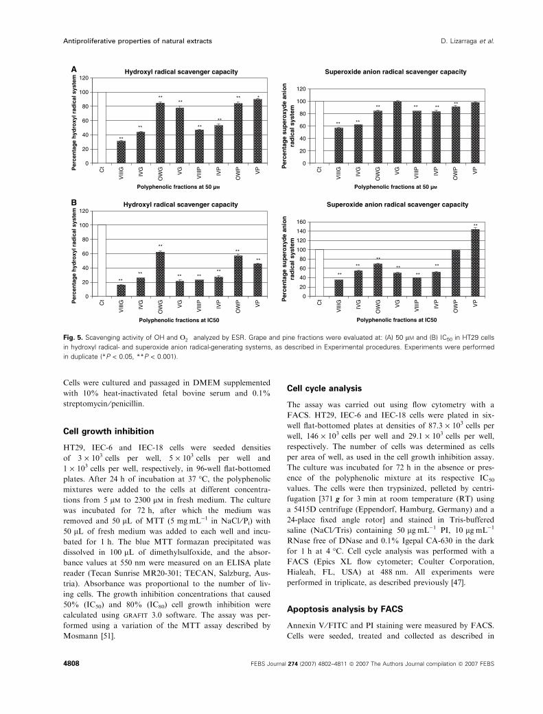
Oxygen radical scavenging activity as detected
by ESR spectroscopy
The next series of experiments used ESR spectroscopy
to test the radical-scavenging capacity of the fractions.
The results show that the oligomeric fractions (VIIIG,
IVG, VIIIP and IVP), which were the most effective in
the previous assays using HT29 cells, were also the
most efficient as hydroxyl radical and superoxide scav-
engers at 50 lm (Fig. 5A). Fraction VIIIG was the
most potent radical scavenger, followed by fraction
IVG and the pine fractions VIIIP and IVP. The same
levels of efficiency were also observed in the induction
of cell cycle arrest and apoptosis. When fractions were
tested at their respective IC50 values, fractions VIIIG,
IVG, VIIIP and IVP were again the most effective
(Fig. 5B). There is a clear relationship between high
scavenger capacity ⁄ lower IC50 and a high level of
apoptosis induction. Grape fractions proved to be
more potent scavengers than pine fractions in both
radical generation systems. The apparent high effi-
ciencies detected for the monomers (VG and VP) can
be largely attributed to the high concentrations used
(410 lm and 1551 lm, respectively).
Interestingly, the efficiencies observed for grape oligo-
meric fractions, which proved to be better apoptotic
inducers and better ROS scavengers than pine oligo-
meric fractions, are apparently related to the degree of
galloylation and are enhanced by the polymerization
of the fractions. Hydroxyl radical (OH) is the most
reactive product of reactive oxygen species formed by
successive one-electron reductions of molecular oxygen
(O2) in cell metabolism, is primarily responsible for the
Cell cycle at IC50 (Grape fraction)
ct
ct
ct
ct
ct
ct
VIIIG
VIIIG
VIIIG
VIIIG
VIIIG
VIIIGG
1S
G2
G1
SG
2
IEC
-6IE
C-1
8
Cell cycle at IC50 (Grape fractions)A
B C
0 10 20 30 40 50 60 70
ctVIIIG
IVGVIG
OWGVG
ctVIIIG
IVGVIG
OWGVG
ctVIIIG
IVGVIG
OWGVG
G1
SG
2
G1
SG
2
Cel
l cyc
le s
tag
es
Cel
l cyc
le s
tag
es
Cel
l cyc
le s
tag
es
Cel
l cyc
le s
tag
es
% Cell distribution (HT29) 0 10 20 30 40 50 60 70
% Cell distribution (HT29)
0 10 20 30 40 50 60 70
% Cell distribution (HT29) 0 10 20 30 40 50 60 70
% Cell distribution
*
*
***
*
*
Cell cycle at IC50 (Pine fractions)
ct
ct
IVP
ct
VIIIP
VIIIP
VIIIP
IVP
IVP
G1
SG
2
**
**
Cell cycle at IC80 (Grape fractions)
ctVIIIG
IVGVIG
OWGVG
ctVIIIG
IVGVIG
OWGVG
ctVIIIG
IVGVIG
OWGVG
**
*
***
*
*
Fig. 2. Cell cycle analysis of HT29, IEC-6 and IEC-18 cells treated with grape and pine polyphenolic fractions. (A) HT29 cells at their respec-
tive grape IC50 and IC80 values. (B) HT29 cells at pine IC50. (C) IEC-6 and IEC-18 cells treated with grape fraction VIIIG at HT29 IC50. Percent-
ages of cells in different cell stages are shown. Cell phases analyzed: G1, S and G2 (% cells ± SEM, *P < 0.05, **P < 0.001). Experiments
were performed in triplicate.
D. Lizarraga et al. Antiproliferative properties of natural extracts
FEBS Journal 274 (2007) 4802–4811 ª 2007 The Authors Journal compilation ª 2007 FEBS 4805

cytotoxic effects observed in aerobic organisms from
bacteria to plants and animals, and has been identified
as playing a role in the development of many human
cancers [37,38].
Cancer chemoprevention conducted by administering
chemical and dietary components to interrupt the initi-
ation, promotion and progression of tumors is consid-
ered to be a new and promising approach in cancer
prevention [39–41]. However, the development of effec-
tive and safe agents for the prevention and treatment
of cancer remains inefficient and costly, and falls short
of the requirements for primary prevention among the
high-risk population and for prevention in cancer sur-
vivors [42].
In recent years, many popular, polyphenol-enriched
dietary supplements have been commercialized, such as
tea catechins, grape seed proanthocyanidins and other
natural antioxidant extracts, each of which has been
claimed to exert chemopreventive activity in cellular
models of cancer [43,44]. Recent publications have sta-
ted that the antiproliferative activity of flavonoids is
dependent on particular structure motifs, such as gal-
late groups and degree of polymerization [45,46].
Our results suggest that polymerization plays a
greater role than galloylation in cell cycle arrest in
HT29 cells. Interestingly, galloylation appears to be
more influential than polymerization in the biological
apoptosis activities tested and in the hydroxyl and
superoxide anion radical-scavenging capacity of the
fractions when compared at the same concentration of
50 lm (Fig. 5A). The galloylated and polymerized
grape procyanidins were the most effective hydroxyl
radical scavengers and also triggered cell cycle arrest
and apoptosis, and although this does not necessarily
indicate that both effects are mechanistically related,
such as relationship cannot be ruled out. The present
results are in general agreement with previously
reported data for pure compounds [47]. Essentially, the
induction of apoptosis seems to be related to the elec-
tron transfer capacity of the phenolic extracts. Other
antioxidants with anti-inflammatory and anticancer
activities have been reported, such as edaravone [48]
and the flavonoid silydianin [49], both of which induce
apoptosis and act as radical scavengers.
It was also observed that the most efficient procyani-
din fraction, VIIIG, which induced approximately
Apoptosis at IC50 (Pine fractions)
ct
VIII
P
IVP ct
VIII
P
IVP ct
VIII
P
IVP
Early Late Necrotic
Cell stage
* *
Apoptosis at IC50 (Grape fractions)A
B C
0
5
10
15
20
ct
VIII
G
IVG
VIG
OW
G
VG ct
VIII
G
IVG
VIG
OW
G
VG ct
VIII
G
IVG
VIG
OW
G
VG
Early Late Necrotic
Cell stage
% C
ell d
istr
ibu
tio
n (
HT
29)
0
5
10
15
20
% C
ell d
istr
ibu
tio
n (
HT
29)
0
5
10
15
20
% C
ell d
istr
ibu
tio
n (
HT
29)
0
5
10
15
20
% C
ell d
istr
ibu
tio
n
**
* *
*
*
Apoptosis at IC80 (Grape fractions)
ct
VIII
G
IVG
VIG
OW
G
VG ct
VIII
G
IVG
VIG
OW
G
VG ct
VIII
G
IVG
VIG
OW
G
VG
Early Late Necrotic
Cell stage
*
** *
*
Apoptosis at IC50 (Grape fraction)
ct
VIII
G ct
VIII
G ct
VIII
G ct
VIII
G ct
VIII
G ct
VIII
G
Early Late Necrotic Early Late Necrotic
IEC-6 IEC-18
Cell stage
*
Fig. 3. Apoptosis was induced in HT29 tumor cells and did not affect normal epithelial cells. (A) HT29 cells after treatment with grape poly-
phenolic fractions at their respective IC50 and IC80 values. (B) HT29 cells after treatment with pine polyphenolic fractions at their respective
IC50 values. (C) IEC-6 and IEC-18 cells treated with grape fraction VIIIG at HT29 IC50. Percentages of cells in different cell stages are shown
(cell stages shown on the x-axis). (% cells ± SEM, *P < 0.05, **P < 0.001). Experiments were performed in triplicate.
Antiproliferative properties of natural extracts D. Lizarraga et al.
4806 FEBS Journal 274 (2007) 4802–4811 ª 2007 The Authors Journal compilation ª 2007 FEBS

30% apoptosis in HT29 cells, did not induce apoptosis
or affect the cell cycle of the intestinal nontumoral cell
lines IEC-18 and IEC-6, and even induced 10% necro-
sis in the IEC-6 cell line (Figs 2C and 3C). The results
obtained provide information about the activities of
procyanidin mixtures with different origins and struc-
tures on colon epithelial cells. These results should be
useful in defining the putative benefits of plant poly-
phenols in nutritional supplements. Additionally, this
study provides useful insights into the polyphenolic
structure, which should help in the rational design of
formulations for potent chemopreventive or antiprolif-
erative natural vegetable products on the basis of
apoptosis-inducing activity.
Experimental procedures
Materials
DMEM and Dulbecco’s phosphate-buffered saline (NaCl ⁄Pi) were obtained from Sigma Chemical Co. (St Louis,
MO, USA), antibiotics (10 000 UÆmL)1 penicillin, 10 000
lgÆmL)1 streptomycin) were obtained from Gibco-BRL
(Eggenstein, Germany), and fetal bovine serum was
obtained from Invitrogen (Carlsbad, CA, USA). Tryp-
sin ⁄EDTA solution C (0.05% trypsin ⁄ 0.02% EDTA) was
purchased from Biological Industries (Kibbutz Beit Ha-
emet, Israel). 3-(4,5-Dimethylthiazol-2yl)-2,5-diphenyl-tetra-
zolium bromide (MTT), dimethylsulfoxide, propidium
iodide (PI) and Igepal CA-630 were obtained from Sigma
Chemical Co. NADH disodium salt (grade I) was supplied
by Boehringer (Mannheim, Germany). RNase and agarose
MP were obtained from Roche Diagnostics (Mannheim,
Germany). Iron(II) sulfate heptahydrate was obtained from
Merck (Darmstadt, Germany) a-a-a-Tris(hydroxymeth-
yl)aminomethane was obtained from Aldrich-Chemie
(Steinheim, Germany) and moviol from Calbiochem (La
Jolla, CA, USA). The annexin V ⁄ fluorescein isothiocyanate
(FITC) kit was obtained from Bender System (Vienna, Aus-
tria), the Realpure DNA extraction kit, including protein-
ase K, was obtained from Durviz S.L. (Paterna, Spain),
and Blue ⁄Orange Loading dye and the 1 kb DNA ladder
were purchased from Promega (Madison, WI, USA).
5,5-Dimethyl-1-pyrolline-N-oxide (DMPO), hydrogen per-
oxide, phenazine methosulfate and Hoescht were obtained
from Sigma (St Louis, MO). DMPO was further purified
by charcoal treatment.
Fractions
The polyphenolic mixtures were obtained previously in our
laboratories [26,50] and contain mainly procyanidins.
OWG and OWP are composed of species that are soluble
in both ethyl acetate and water, and the rest of the frac-
tions (G for grape, P for pine) were generated by a combi-
nation of preparative RP-HPLC and semipreparative
chromatography on a Toyopearl TSK HW-40F column
(TosoHass, Tokyo, Japan), which separated the compo-
nents by size and hydrophobicity. The phenolics were
eluted from the latter column with MeOH (fractions VG,
VP) and water ⁄ acetone 1 : 1 (fractions IVG, VIG, VIIIG,
IVP, VIP, VIIIP and XIP), evaporated almost to dryness,
redissolved in Milli-Q water, and freeze-dried. The second
and third columns of Table 1 show the average chemical
composition of the fractions.
Cell culture
Human colorectal adenocarcinoma HT29 cells
(ATCC HTB-38) and two nontumoral intestinal rat cell
lines, IEC-6 (ECCAC no. 88071401) and IEC-18 (EC-
CAC no. 88011801), were used in all of the experiments.
HT29, IEC-6 and IEC-18 cells were maintained in mono-
layer culture in an incubator with 95% humidity and 5%
CO2 at 37 �C. HT29, IEC-6 and IEC-18 cells were passaged
at preconfluent densities using trypsin ⁄EDTA solution C.
M Ct1 Ct2 IVGA IVGB VIIIGA VIIIGB
Control 48H (VIIIG)
72H (VIIIG)
48H (IVG)
72H (IVG)Control
A
B
A= IC50
B= IC80
Fig. 4. Induction of apoptosis by grape fractions VIIIG and IVG in
HT29 cells. (A) Nuclear condensation of HT29 cells. Arrows indicate
the apoptotic cells with condensed and fragmented nuclei. (B) DNA
laddering induced in both treatments.
D. Lizarraga et al. Antiproliferative properties of natural extracts
FEBS Journal 274 (2007) 4802–4811 ª 2007 The Authors Journal compilation ª 2007 FEBS 4807
Cells were cultured and passaged in DMEM supplemented
with 10% heat-inactivated fetal bovine serum and 0.1%
streptomycin ⁄penicillin.
Cell growth inhibition
HT29, IEC-6 and IEC-18 cells were seeded densities
of 3 · 103 cells per well, 5 · 103 cells per well and
1 · 103 cells per well, respectively, in 96-well flat-bottomed
plates. After 24 h of incubation at 37 �C, the polyphenolic
mixtures were added to the cells at different concentra-
tions from 5 lm to 2300 lm in fresh medium. The culture
was incubated for 72 h, after which the medium was
removed and 50 lL of MTT (5 mgÆmL)1 in NaCl ⁄Pi) with
50 lL of fresh medium was added to each well and incu-
bated for 1 h. The blue MTT formazan precipitated was
dissolved in 100 lL of dimethylsulfoxide, and the absor-
bance values at 550 nm were measured on an ELISA plate
reader (Tecan Sunrise MR20-301; TECAN, Salzburg, Aus-
tria). Absorbance was proportional to the number of liv-
ing cells. The growth inhibition concentrations that caused
50% (IC50) and 80% (IC80) cell growth inhibition were
calculated using grafit 3.0 software. The assay was per-
formed using a variation of the MTT assay described by
Mosmann [51].
Cell cycle analysis
The assay was carried out using flow cytometry with a
FACS. HT29, IEC-6 and IEC-18 cells were plated in six-
well flat-bottomed plates at densities of 87.3 · 103 cells per
well, 146 · 103 cells per well and 29.1 · 103 cells per well,
respectively. The number of cells was determined as cells
per area of well, as used in the cell growth inhibition assay.
The culture was incubated for 72 h in the absence or pres-
ence of the polyphenolic mixture at its respective IC50
values. The cells were then trypsinized, pelleted by centri-
fugation [371 g for 3 min at room temperature (RT) using
a 5415D centrifuge (Eppendorf, Hamburg, Germany) and a
24-place fixed angle rotor] and stained in Tris-buffered
saline (NaCl ⁄Tris) containing 50 lgÆmL)1 PI, 10 lgÆmL)1
RNase free of DNase and 0.1% Igepal CA-630 in the dark
for 1 h at 4 �C. Cell cycle analysis was performed with a
FACS (Epics XL flow cytometer; Coulter Corporation,
Hialeah, FL, USA) at 488 nm. All experiments were
performed in triplicate, as described previously [47].
Apoptosis analysis by FACS
Annexin V ⁄FITC and PI staining were measured by FACS.
Cells were seeded, treated and collected as described in
Superoxide anion radical scavenger capacity
** **
**
**
**
**
**
Hydroxyl radical scavenger capacityA
B
0
20
40
60
80
100
120
Ct
VIII
G
IVG
OW
G
VG
VIII
P
IVP
OW
P
VP
Polyphenolic fractions at 50 µM
0
20
40
60
80
100
120
Ct
VIII
G
IVG
OW
G
VG
VIII
P
IVP
OW
P
VP
Polyphenolic fractions at 50 µM
Per
cen
tag
e h
ydro
xyl r
adic
al s
yste
m
0
20
40
60
80
100
120
Ct
VIII
G
IVG
OW
G
VG
VIII
P
IVP
OW
P
VPP
erce
nta
ge
hyd
roxy
l rad
ical
sys
tem
** **
**
**
**
** ** *
Superoxide anion radical scavenger capacity
Per
cen
tag
e su
per
oxy
de
anio
nra
dic
al s
yste
m0
20
40
60
80
100
120
140
160
Per
cen
tag
e su
per
oxy
de
anio
nra
dic
al s
yste
m
**
**
**
** ** **
Hydroxyl radical scavenger capacity
Polyphenolic fractions at IC50 C
t
VIII
G
IVG
OW
G
VG
VIII
P
IVP
OW
P
VP
Polyphenolic fractions at IC50
** ****
**
**
**
**
**
Fig. 5. Scavenging activity of OH and O��2 analyzed by ESR. Grape and pine fractions were evaluated at: (A) 50 lM and (B) IC50 in HT29 cells
in hydroxyl radical- and superoxide anion radical-generating systems, as described in Experimental procedures. Experiments were performed
in duplicate (*P < 0.05, **P < 0.001).
Antiproliferative properties of natural extracts D. Lizarraga et al.
4808 FEBS Journal 274 (2007) 4802–4811 ª 2007 The Authors Journal compilation ª 2007 FEBS
the previous section. Following centrifugation [371 g for
3 min at RT using a 5415D centrifuge (Eppendorf) with
24-place fixed angle rotor], cells were washed in binding
buffer (10 mm Hepes, pH 7.4, 140 mm sodium chloride,
2.5 mm calcium chloride) and resuspended in the same
buffer. Annexin V ⁄FITC was added using the annex-
in V ⁄FITC kit. Following 30 min of incubation at room
temperature and in the dark, PI was added 1 min before
the FACS analysis at 20 lgÆmL)1. Experiments were per-
formed in triplicate.
Apoptosis detection by DNA laddering
DNA isolation and purification were performed after 72 h
in the presence and absence of grape fractions VIIIG and
IVG. The fractions were assayed at their respective IC50
and IC80 values. After treatment, cells were scraped off
slides and collected by centrifugation at 14 000 g for 10 s
at RT using a 5415D centrifuge (Eppendorf) and 24-place
fixed angle rotor. Cells were then lysed by adding 600 lLof Realpure kit lysis buffer and 10 lL of proteinase K,
and incubated for 1 h at 55 �C. RNA digestion was per-
formed with 1.5 lL of RNase for 1 h at 37 �C, and this
was followed by protein precipitation with 360 lL of
Realpure kit buffer and centrifugation at 14 000 g for
10 min at RT using a 5415D centrifuge (Eppendorf) and
24-place fixed angle rotor. The DNA sample was
extracted with isopropanol ⁄ ethanol, dried, and eluted in
100 lL of Realpure kit DNA hydration solution. Equal
amounts of DNA (20 lg), estimated by measuring absorp-
tion at 260 ⁄ 280 nm, were electrophoretically separated on
1% TAE agarose gel and viewed under a UV transillumi-
nator (Vilber Lourmat, Marne-la-Vallee, France).
Apoptosis detection by Hoescht staining
Apoptotic induction was also studied using Hoescht stain-
ing. Samples were incubated with grape fractions VIIIG
and IVG at 0, 48 and 72 h. After incubation, cells were
trypsinized and fixed with cold methanol for 1 h at ) 20 �C.After being rinsed with NaCl ⁄Pi three times, cells were
stained in the dark with Hoescht (50 ngÆmL)1 in NaCl ⁄Pi)
for 50 min. Finally, cells were rinsed, suspended in NaCl ⁄Pi
and diluted 1 : 2 with moviol. The samples were mounted
on a slide and observed with a fluorescent microscope at an
excitation wavelength of 334 nm and an emission wave-
length of 365 nm.
ESR spectroscopy
ESR measurements were performed at concentrations that
caused 50% cell growth inhibition (IC50) and 50 lm grape
and pine fractions (VIIIG, IVG, OWG, VG, VIIIP, IVP,
OWP and VP). Molar concentrations were calculated from
the mean molecular mass of the fractions estimated by thiol-
ysis with cysteamine, as described in [52]. OH and O2– forma-
tion were detected by ESR spectroscopy using DMPO
(100 mm) as a spin trap. ESR spectra were recorded at room
temperature in glass capillaries (100 lL; Brand AG,
Wertheim, Germany) on a Bruker EMX 1273 spectrometer
(Bruker, Karlsruhe, Germany) equipped with an ER 4119HS
high-sensitivity cavity and a 12 kW power supply operating
at X-band frequencies. The modulation frequency of the
spectrometer was 100 kHz. Instrumental conditions for the
recorded spectra were: magnetic field, 3490 G; scan range,
60 G; modulation amplitude, 1 G; receiver gain, 1 · 105;
microwave frequency, 9.85 GHz; power, 50 mW; time
constant, 40.96 ms; scan time, 20.97 s; number of scans, 25.
Spectra were quantified by peak surface measurements using
the WIN-EPR spectrum manipulation program (Bruker).
All incubations were done at room temperature; the
hydroxyl radical generation system used 500 lm FeSO4 and
550 lm H2O2, and hydroxyl radicals generated in this system
were trapped by DMPO, forming a spin adduct detected by
the ESR spectrometer. The typical 1 : 2 : 2 : 1 ESR signal
of DMPO-OH was observed. The superoxide radical genera-
tion system used performed using 50 lm of the reduced form
of b-NADH and 3.3 lm phenazine methosulfate, and the
superoxide radicals generated in this system were trapped by
DMPO, forming a spin adduct detected by the ESR spec-
trometer. The typical ESR signal of DMPO-OOH ⁄DMPO-
OH was observed. The OH and O2-scavenging activity was
calculated on the basis of decreases in the DMPO-OH or
DMPO-OOH ⁄DMPO-OH signals, respectively, in which the
coupling constant for DMPO-OH was 14.9 G.
Data presentation and statistical analysis
Assays were analyzed using the Student’s t-test and
were considered statistically significant at P < 0.05 and
P < 0.001. The data shown are representative of three
independent experiments, with the exception of ESR experi-
ments, which were performed in duplicate. ESR experi-
ments were analyzed separately by radicals, Two-way
anova was applied (day was a block factor; due to the
nonsignificant effect of the day factor, we reanalyzed with a
one-way anova), and finally, a multicomparison between
compounds with respect to the control was performed.
anova with Bonferroni and Scheffe post hoc test was per-
formed in ESR experiments.
Acknowledgements
This work was supported by grants PPQ 2003-06602-
C04-01, PPQ 2003-06602-C04-04, AGL2004-07579-
C04-02 and AGL2004-07579-C04-03 from the Spanish
Ministry of Education and Science, and ISCIII-RTICC
(RD06 ⁄0020 ⁄ 0046) from the Spanish government and
D. Lizarraga et al. Antiproliferative properties of natural extracts
FEBS Journal 274 (2007) 4802–4811 ª 2007 The Authors Journal compilation ª 2007 FEBS 4809

the European Union FEDER funds. We thank Profes-
sor Francesc Oliva (Department of Statistics at the
University of Barcelona) for his assistance with statisti-
cal analysis.
References
1 Parkin DM (2004) International variation. Oncogene 23,
6329–6340.
2 Potter JD, Slattery ML, Bostick RM & Gapstur SM
(1993) Colon cancer: a review of the epidemiology.
Epidemiol Rev 15, 499–545.
3 Steinmetz KA & Potter JD (1991) Vegetables, fruit, and
cancer. I. Epidemiology. Cancer Causes Control 2, 325–
357.
4 Park OJ & Surh YJ (2004) Chemopreventive potential
of epigallocatechin gallate and genistein: evidence from
epidemiological and laboratory studies. Toxicol Lett
150, 43–56.
5 Bruce WR, Giacca A & Medline A (2000) Possible
mechanisms relating diet and risk of colon cancer.
Cancer Epidemiol Biomarkers Prev 9, 1271–1279.
6 Hietanen E, Bartsch H, Bereziat JC, Camus AM, McCl-
inton S, Eremin O, Davidson L & Boyle P (1994) Diet
and oxidative stress in breast, colon and prostate cancer
patients: a case-control study. Eur J Clin Nutr 48, 575–
586.
7 Theodoratou E, Kyle J, Cetnarskyj R, Farrington SM,
Tenesa A, Barnetson R, Porteous M, Dunlop M & Camp-
bell H (2007) Dietary flavonoids and the risk of colorectal
cancer. Cancer Epidemiol Biomarkers Prev 16, 684–693.
8 Mukhtar H & Ahmad N (1999) Green tea in chemopre-
vention of cancer. Toxicol Sci 52, 111–117.
9 Lee KW, Lee HJ & Lee CY (2004) Vitamins, phyto-
chemicals, diets, and their implementation in cancer
chemoprevention. Crit Rev Food Sci Nutr 44, 437–452.
10 Witschi H, Espiritu I, Ly M, Uyeminami D, Morin D
& Raabe OG (2004) Chemoprevention of tobacco
smoke-induced lung tumors by inhalation of an epigal-
locatechin gallate (EGCG) aerosol: a pilot study. Inhal
Toxicol 16, 763–770.
11 Delmas D, Lancon A, Colin D, Jannin B & Latruffe N
(2006) Resveratrol as a chemopreventive agent: a prom-
ising molecule for fighting cancer. Curr Drug Targets 7,
423–442.
12 Hakimuddin F, Paliyath G & Meckling K (2006) Treat-
ment of mcf-7 breast cancer cells with a red grape wine
polyphenol fraction results in disruption of calcium
homeostasis and cell cycle arrest causing selective cyto-
toxicity. J Agric Food Chem 54, 7912–7923.
13 Sime S & Reeve VE (2004) Protection from inflamma-
tion, immunosuppression and carcinogenesis induced by
UV radiation in mice by topical Pycnogenol. Photochem
Photobiol 79, 193–198.
14 Kumar N, Shibata D, Helm J, Coppola D & Malafa M
(2007) Green tea polyphenols in the prevention of colon
cancer. Front Biosci 12, 2309–2315.
15 McKay DL & Blumberg JB (2007) A review of the bio-
activity of south African herbal teas: rooibos (Aspala-
thus linearis) and honeybush (Cyclopia intermedia).
Phytother Res 21, 1–16.
16 Wright TI, Spencer JM & Flowers FP (2006) Chemo-
prevention of nonmelanoma skin cancer. J Am Acad
Dermatol 54, 933–946; quiz 947–950.
17 Tan XHD, Li S, Han Y, Zhang Y & Zhou D (2000)
Differences of four catechins in cell cycle arrest and
induction of apoptosis in LoVo cells. Cancer Lett 158,
1–6.
18 Kozikowski AP, Tuckmantel W, Bottcher G &
Romanczyk LJ Jr (2003) Studies in polyphenol
chemistry and bioactivity. 4 (1) Synthesis of trimeric,
tetrameric, pentameric, and higher oligomeric
epicatechin-derived procyanidins having all-4beta,
8-interflavan connectivity and their inhibition of cancer
cell growth through cell cycle arrest. J Org Chem 68,
1641–1658.
19 Nakamuta M, Higashi N, Kohjima M, Fukushima M,
Ohta S, Kotoh K, Kobayashi N & Enjoji M (2005)
Epigallocatechin-3-gallate, a polyphenol component of
green tea, suppresses both collagen production and col-
lagenase activity in hepatic stellate cells. Int J Mol Med
16, 677–681.
20 Shi J, Yu J, Pohorly JE & Kakuda Y (2003) Polypheno-
lics in grape seeds ) biochemistry and functionality.
J Med Food 6, 291–299.
21 Siddiqui IA, Adhami VM, SaleemM &Mukhtar H
(2006) Beneficial effects of tea and its polyphenols against
prostate cancer.Mol Nutr Food Res 50, 130–143.
22 Zhang Q, Tang X, Lu Q, Zhang Z, Rao J & Le AD
(2006) Green tea extract and (–)-epigallocatechin-3-gal-
late inhibit hypoxia- and serum-induced HIF-1alpha
protein accumulation and VEGF expression in human
cervical carcinoma and hepatoma cells. Mol Cancer
Ther 5, 1227–1238.
23 Prieur CRJ, Cheynier V & Moutounet M (1994) Oligo-
meric and polymeric procyanidins from grape seeds.
Phytochemistry 36, 781–784.
24 Souquet J-MCV, Brossaud F & Moutounet M (1996)
Polymeric proanthocyanidins from grape skins. Phyto-
chemistry 43, 509–512.
25 Rohdewald P (2002) A review of the French maritime
pine bark extract (Pycnogenol), a herbal medication
with a diverse clinical pharmacology. Int J Clin
Pharmacol Ther 40, 158–168.
26 Tourino S, Selga A, Jimenez A, Julia L, Lozano C,
Lizarraga D, Cascante M & Torres JL (2005)
Procyanidin fractions from pine (Pinus pinaster) bark:
radical scavenging power in solution, antioxidant
Antiproliferative properties of natural extracts D. Lizarraga et al.
4810 FEBS Journal 274 (2007) 4802–4811 ª 2007 The Authors Journal compilation ª 2007 FEBS

activity in emulsion, and antiproliferative effect in
melanoma cells. J Agric Food Chem 53, 4728–4735.
27 Gonthier MP, Donovan JL, Texier O, Felgines C,
Remesy C & Scalbert A (2003) Metabolism of dietary
procyanidins in rats. Free Radic Biol Med 35, 837–
844.
28 Kuhnle G, Spencer JP, Schroeter H, Shenoy B, Debnam
ES, Srai SK, Rice-Evans C & Hahn U (2000) Epicate-
chin and catechin are O-methylated and glucuronidated
in the small intestine. Biochem Biophys Res Commun
277, 507–512.
29 Rechner AR, Smith MA, Kuhnle G, Gibson GR, Deb-
nam ES, Srai SK, Moore KP & Rice-Evans CA (2004)
Colonic metabolism of dietary polyphenols: influence of
structure on microbial fermentation products. Free
Radic Biol Med 36, 212–225.
30 Meselhy MR, Nakamura N & Hattori M (1997) Biotrans-
formation of (–)-epicatechin 3-O-gallate by human intes-
tinal bacteria. Chem Pharm Bull (Tokyo) 45, 888–893.
31 Salucci M, Stivala LA, Maiani G, Bugianesi R & Van-
nini V (2002) Flavonoids uptake and their effect on cell
cycle of human colon adenocarcinoma cells (Caco2).
Br J Cancer 86, 1645–1651.
32 Stagos D, Kazantzoglou G, Magiatis P, Mitaku S,
Anagnostopoulos K & Kouretas D (2005) Effects of plant
phenolics and grape extracts from Greek varieties of Vitis
vinifera on mitomycin C and topoisomerase I-induced
nicking of DNA. Int J Mol Med 15, 1013–1022.
33 Subirade I, Fernandez Y, Periquet A & Mitjavila S
(1995) Catechin protection of 3T3 Swiss fibroblasts in
culture under oxidative stress. Biol Trace Elem Res 47,
313–319.
34 Cao Z & Li Y (2004) Potent induction of cellular anti-
oxidants and phase 2 enzymes by resveratrol in cardio-
myocytes: protection against oxidative and electrophilic
injury. Eur J Pharmacol 489, 39–48.
35 Alanko J, Riutta A, Holm P, Mucha I, Vapaatalo H &
Metsa-Ketela T (1999) Modulation of arachidonic acid
metabolism by phenols: relation to their structure and
antioxidant ⁄ prooxidant properties. Free Radic Biol Med
26, 193–201.
36 Azam S, Hadi N, Khan NU & Hadi SM (2004) Prooxi-
dant property of green tea polyphenols epicatechin and
epigallocatechin-3-gallate: implications for anticancer
properties. Toxicol In Vitro 18, 555–561.
37 Halliwell B & Gutteridge JM (1992) Biologically rele-
vant metal ion-dependent hydroxyl radical generation.
An update. FEBS Lett 307, 108–112.
38 Valko M, Leibfritz D, Moncol J, Cronin MT, Mazur M
& Telser J (2007) Free radicals and antioxidants in nor-
mal physiological functions and human disease. Int J
Biochem Cell Biol 39, 44–84.
39 Mathers JC (2002) Pulses and carcinogenesis: potential
for the prevention of colon, breast and other cancers.
Br J Nutr 88 (Suppl. 3), S273–S279.
40 Witte JS, Longnecker MP, Bird CL, Lee ER, Frankl
HD & Haile RW (1996) Relation of vegetable, fruit,
and grain consumption to colorectal adenomatous pol-
yps. Am J Epidemiol 144, 1015–1025.
41 Manju V & Nalini N (2005) Chemopreventive efficacy of
ginger, a naturally occurring anticarcinogen during the
initiation, post-initiation stages of 1,2-dimethylhydrazine-
induced colon cancer. Clin Chim Acta 358, 60–67.
42 Zou DM, Brewer M, Garcia F, Feugang JM, Wang J,
Zang R, Liu H & Zou C (2005) Cactus pear: a natural
product in cancer chemoprevention. Nutr J 4, 25–37.
43 Joshi SS, Kuszynski CA & Bagchi D (2001) The cellular
and molecular basis of health benefits of grape seed
proanthocyanidin extract. Curr Pharm Biotechnol 2,
187–200.
44 Depeint F, Gee JM, Williamson G & Johnson IT (2002)
Evidence for consistent patterns between flavonoid struc-
tures and cellular activities. Proc Nutr Soc 61, 97–103.
45 Fiuza SM, Gomes C, Teixeira LJ, Girao da Cruz MT,
Cordeiro MN, Milhazes N, Borges F & Marques MP
(2004) Phenolic acid derivatives with potential antican-
cer properties ) a structure–activity relationship study.
Part 1: methyl, propyl and octyl esters of caffeic and
gallic acids. Bioorg Med Chem 12, 3581–3589.
46 Brusselmans K, Vrolix R, Verhoeven G & Swinnen JV
(2005) Induction of cancer cell apoptosis by flavonoids
is associated with their ability to inhibit fatty acid syn-
thase activity. J Biol Chem 280, 5636–5645.
47 Lozano C, Torres JL, Julia L, Jimenez A, Centelles JJ
& Cascante M (2005) Effect of new antioxidant cyste-
inyl-flavanol conjugates on skin cancer cells. FEBS Lett
579, 4219–4225.
48 Kokura S, Yoshida N, Sakamoto N, Ishikawa T, Tak-
agi T, Higashihara H, Nakabe N, Handa O, Naito Y &
Yoshikawa T (2005) The radical scavenger edaravone
enhances the anti-tumor effects of CPT-11 in murine
colon cancer by increasing apoptosis via inhibition of
NF-kappaB. Cancer Lett 229, 223–233.
49 Zielinska-Przyjemska M & Wiktorowicz K (2006) An
in vitro study of the protective effect of the flavonoid
silydianin against reactive oxygen species. Phytother Res
20, 115–119.
50 Torres JL, Varela B, Garcia MT, Carilla J, Matito C,
Centelles JJ, Cascante M, Sort X & Bobet R (2002) Val-
orization of grape (Vitis vinifera) byproducts. Antioxi-
dant and biological properties of polyphenolic fractions
differing in procyanidin composition and flavonol con-
tent. J Agric Food Chem 50, 7548–7555.
51 Mosmann T (1983) Rapid colorimetric assay for cellular
growth and survival: application to proliferation and
cytotoxicity assays. J Immunol Methods 65, 55–63.
52 Selga A & Torres JL (2005) Efficient preparation of
catechin thio conjugates by one step extraction ⁄depolymerization of pine (Pinus pinaster) bark
procyanidins. J Agric Food Chem 53, 7760–7765.
D. Lizarraga et al. Antiproliferative properties of natural extracts
FEBS Journal 274 (2007) 4802–4811 ª 2007 The Authors Journal compilation ª 2007 FEBS 4811
Copyright © 2022 FDOKUMEN