Mining Efficiently Significant Classification Association Rules
Upload
independentCategory
view
1download
0
Journal of Cellular Biochemistry 104:202–212 (2008)
Zinc Depletion Efficiently Inhibits Pancreatic Cancer CellGrowth by Increasing the Ratio of Antiproliferative/Proliferative Genes
M. Donadelli,1 E. Dalla Pozza,1 C. Costanzo,1 M.T. Scupoli,2 A. Scarpa,3 and M. Palmieri1*1Department of Morphological and Biomedical Sciences, Section of Biochemistry,University of Verona, Verona, Italy2Interdepartmental Laboratory for Medical Research (LURM), University of Verona, Verona, Italy3Department of Pathology, Section of Anatomic Pathology, University of Verona, Verona, Italy
Abstract We investigated the ability of the zinc chelator N,N,N0,N0-tetrakis(2-pyridylmethyl)ethylenediamine(TPEN) to reduce pancreatic cancer cell viability. TPEN was much more efficient to inhibit pancreatic adenocarcinomacell growth than a panel of anti-cancer drugs, including 5-fluorouracil, irinotecan, cisplatin, edelfosine, trichostatin A,mitomycin C, and gemcitabine, the gold standard chemotherapeutic agent for pancreatic cancer. Moreover, TPENshowed a dose- and time-dependent anti-proliferative effect significantly higher on pancreatic cancer cells than on normalprimary fibroblasts. This effect may be explained by a significantly higher zinc depletion by TPEN in pancreatic cancercells as compared to fibroblasts. Cell viability reduction by TPEN was associated to both G1-phase cell cycle arrest andapoptosis, and to the increased ratio of the expression level of cyclin-Cdk inhibitor versus cyclin genes and apoptoticversus anti-apoptotic genes. Finally, we show that apoptotic cell death induced by TPEN involved mitochondrial injuryand caspase 3 and caspase 8 activation. In this study, we suggest that zinc depletion may be an efficient strategy in thetreatment of pancreatic cancer because of its reduced antiproliferative effect on normal cells. J. Cell. Biochem. 104: 202–212, 2008. � 2007 Wiley-Liss, Inc.
Key words: pancreatic adenocarcinoma; zinc; N,N,N0,N0-tetrakis(2-pyridylmethyl)ethylenediamine (TPEN); apoptosis;cell cycle
Zinc is a functionally versatile elementwith important structural and regulatory roles.It acts as a cofactor for several hundred enzymes[McCall et al., 2000] and facilitates the inter-action with DNA of metal-responsive (MTFs)and zinc finger-containing transcription fac-tors. Moreover, zinc modulates the activity ofvarious membrane receptors, transporters, and
channels [Hashemi et al., 2007] and regulatesspecific signal transduction pathways and theirtarget genes by interacting with intracellularsignaling molecules [Beyersmann and Haase,2001]. Because of these essential roles ofzinc, cells must maintain a strict intracellularzinc homeostasis, which is accomplished bytransporter families and sequestering metal-lothioneins [Gaither and Eide, 2001; Outtenand O’Halloran, 2001; Cousins et al., 2006]. Intheir physiological state, cells contain a poorlyexchangeable amount of zinc that is tightlybound within the tertiary structure of proteinsand a dynamic zinc pool subjected to ionic fluxesand readily influenced by zinc deprivation orsupplementation [Truong-Tran et al., 2000].
Many reports have shown that both an in-crease [Kagara et al., 2007] and a decrease[Gupta et al., 2005; Costello and Franklin, 2006]of the zinc level are associated with cancerdevelopment. Our group has previously shownthat intracellular zinc increase by the ionophore
� 2007 Wiley-Liss, Inc.
Grant sponsor: Fondazione Cassa di Risparmio di Verona(Bando 2004); Grant sponsor: Associazione Italiana Ricercasul Cancro, Milan, Italy; Grant sponsor: Italian Ministriesof University—Research and Health; Grant sponsor:European Community; Grant number: PL PL018771;Grant sponsor: Fondazione Giorgio Zanotto, Verona, Italy.
*Correspondence to: Dr. M. Palmieri, Department ofMorphological and Biomedical Sciences, Section of Bio-chemistry, Strada Le Grazie, 8, 37134 Verona, Italy.E-mail: [email protected]
Received 16 July 2007; Accepted 21 September 2007
DOI 10.1002/jcb.21613
compound pyrrolidine dithiocarbamate (PDTC)strongly inhibits p53-negative pancreatic can-cer cell growth in vitro and in vivo, while it doesnot affect normal fibroblast proliferation [Dona-delli et al., 2006].
The purpose of the present work was to studythe effect of the depletion of the endogenouszinc, by using the zinc chelator TPEN, onpancreatic cancer cell and normal fibroblastproliferation and to analyze the molecularmechanisms involved. The antiproliferativeefficiency of zinc depletion was comparedto that of various chemotherapeutic drugs,including gemcitabine and 5-fluorouracil thatrepresent the standard treatments for pan-creatic cancer. We report that TPEN inhibitedpancreatic cancer cells, by cell cycle arrest andapoptosis, much more efficiently than a panelof common anti-cancer drugs and showed asignificantly higher effect on cancer cells thanon normal primary fibroblasts.
MATERIALS AND METHODS
Chemicals
The chemotherapeutic drugs used were:gemcitabine (Gemzar; Eli Lilly), 5-fluorouracil(Teva Pharma), irinotecan (CPT11; Aventis),mitomycin C (Kyowa Italiana Farmaceutici),and cisplatin (Pharmacia). N,N,N0,N0-Tetra-kis(2-pyridylmethyl)ethylenediamine (TPEN)(Sigma), trichostatin A (Sigma), and edelfosine(Axxora, Alexis) were prepared in absoluteethanol and stored at �808C until use. Zincsulfate (Sigma) was prepared in sterile water.
Cell Culture
Human pancreatic adenocarcinoma cell lines(PaCa44, PSN1, HPAF II, PaCa3, T3M4, andCFPAC1; see Moore et al. [2001] for geneticcharacterization and primary tissue source)were grown in RPMI 1640 supplemented with20 mM glutamine, 10% FBS, and 50 mg/mlgentamicin sulfate (BioWhittaker) and wereincubated at 378C with 5% CO2. Normal primaryfibroblasts (Promocell PBI) were grown inDMEM supplemented with 20 mM glutamine,10% FBS, and 50 mg/ml gentamicin sulfate andwere incubated at 378C with 5% CO2.
Cell Proliferation Assay
Cells were plated in 96-well plates (4�103 cells/well) and after 24 h were treated with
the indicated compounds. Cells were stainedwith crystal violet (Sigma) and the dye wassolubilized in PBS containing 1% SDS andmeasured photometrically (A595nm) to deter-mine cell viability.
Cell Cycle Analysis
Cell cycle distribution was analyzed usingpropidium iodide (PI)-stained cells. Cellswere washed with PBS, incubated with 0.1%sodium citrate dihydrate, 0.1% Triton X-100,200 mg/ml RNase A, 50 mg/ml propidium iodide(Roche Molecular Biochemicals) and analyzedusing a flow cytometer (FACScalibur, BectonDickinson). The percentage of cells in thevarious stages of the cell cycle was determinedusing the ModFitLT software.
Apoptosis
The percentage of apoptotic cells was eva-luated by staining 2� 105 cells with annexinV-FITC (Bender Med System) and 5 mg/mlpropidium iodide in binding buffer [10 mMHEPES/NaOH (pH 7.4), 140 mM NaOH, and2.5 mM CaCl2] for 10 min at room temperaturein the dark. The samples were analyzed by flowcytometry (FACScalibur, Becton-Dickinson)within 1 h to determine the percentage of cellsdisplaying annexin Vþ/propidium iodide- (earlyapoptosis) or annexin Vþ/propidium iodideþ
staining (late apoptosis).Caspase 3 and caspase 8 activities were
assayed by cleavage of the fluorogenic sub-strates Ac-DEVD-AMC and Ac-IETD-AMC(Biomol), respectively. After the indicated treat-ments, cells were washed twice with ice-coldPBS and lysed in caspase buffer (100 mMHEPES, 200 mM NaCl, 1 mM EDTA, 20 mMCHAPS, 10% sucrose). Lysates were preparedby repeated freeze-thawing, centrifuged at14,000g for 10 min and the supernatant fractionsaved for analysis. One hundred microgramsof total protein extract were incubated with10 mg of the fluorogenic peptide substrates in atotal volume of 800 ml. After a 10 min (caspase 3)or 20 min (caspase 8) incubation at 308C,the release of 7-amino-4-methylcoumarin wasdetermined fluorometrically, using an excita-tion wavelength of 380 nm and an emissionwavelength of 460 nm. Optimal amounts of celllysate and duration of assay were determined inpreliminary experiments.
Effect of Zinc Depletion on Pancreatic Cancer 203
Analysis of the Intracellular Zinc Level
Cells (2� 105) were stained with 2 mMFluoZin-3 (Molecular Probes) at 378C for30 min. Before fluorescence measurements,cells were washed in probe-free medium toremove any dye nonspecifically associated andthen incubated at 378C for a further 10 min toallow complete intracellular de-esterification ofthe probe. Cells were then washed twice inPBS and analyzed by flow cytometry (Ex/Em494/516 nm).
Analysis of Mitochondrial MembranePotential (DCm)
Cells (2� 105) were stained with 40 nM 3,3-dihexyloxacarbocyanine (Molecular Probes)at 378C for 20 min and washed twice in PBS.The percentage of cells exhibiting a decreasedlevel of 3,3-dihexyloxacarbocyanine uptake,which reflects loss of DCm, was determined byflow cytometry.
RNA Extraction, RT-PCR, and Image Analysis
Total RNA was extracted from 5� 106 cellsusing TRIzol Reagent (Invitrogen) and 1 mg ofRNA was reverse transcribed using FirstStrand cDNA Synthesis for RT-PCR (Invitro-gen). For each sample, one-tenth of the cDNAswas used as template for the PCR amplificationsusing the following primers and cycling con-ditions: b-ACTINFor 50-ACCAACTGGGACGA-CATGGAGAA-30 and b-ACTINRev 50-GTGGT-GGTGAAGCTGTAGCC-30, 25 cycles of 948C for60 s, 558C for 60 s and 728C for 60 s; BCL-XLForand BCL-XSFor 50-CGGGCATTCAGTGACCT-GAC-30, BCL-XLRev and BCL-XSRev 50-TCAG-GAACCAGCGGTTGAAG-30, 35 cycles of 948Cfor 30 s, 508C for 30 s and 728C for 30 s; BCL-WFor 50-AAGCTGAGGCAGAAGGGTTA-30 andBCL-WRev 50-CCCAAAGACAAAGAAGGCTA-30, 28 cycles of 948C for 30 s, 608C for 30 s and728C for 30 s; P19For 50-CTCAACCGCTTCGG-CAAGAC-30 and P19Rev 50-CCTGAAGCAAC-GTGCACACT-30, CYCLINAFor 50-GCCTG-CGTTCACCATTCATG-30 and CYCLINARev50-CCAGTCCACGAGGATAGCTC-30, CYCLI-NB1For 50-AAGAGCTTTAAACTTTGGTCTG-GG-30 and CYCLINB1Rev 50-CTTTGTAAGTC-CTTGATTTACCATG-30, BCL-2For 50-TGCAC-CTGACGCCCTTCAC-30 and BCL-2Rev 50-AG-ACAGCCAGGAGAAATCAAACAG-30, 30cyclesof 948C for 30 s, 558C for 30 s and 728C for 30 s;P57For 50-TCGCTGCCCGCGTTTGCGCA-30 and
P57Rev 50-CCGAGTCGCTGTCCACTTCGG-30
30 cycles of 948C for 30 s, 608C for 30 s and 728Cfor 30 s. PCR products were separated byelectrophoresis through an ethidium bromide-stained 2.0% agarose gel and visualized withultraviolet light. Gels were photographed andthe bands were scanned as digital peaks. Theareas of the peaks were calculated in arbitraryunits using the public domain NIH Imagesoftware (http://rsb.info.nih.gov/nih-image/). Thevalue of b-ACTIN mRNA was used as a norma-lizing factor to evaluate the relative expressionlevels of the indicated genes.
Statistical Analysis
ANOVA (post hoc Bonferroni) analysis andgraphical presentations were performed byGraphPad Prism version 5. P values less than0.05, 0.01, or 0.001 were indicated as *, ** or ***,respectively. Data in Figure 2 were analyzed bypaired t-test.
RESULTS
TPEN Inhibits Pancreatic Adenocarcinoma CellProliferation More Efficiently Than a Panel of
Seven Anti-Cancer Drugs
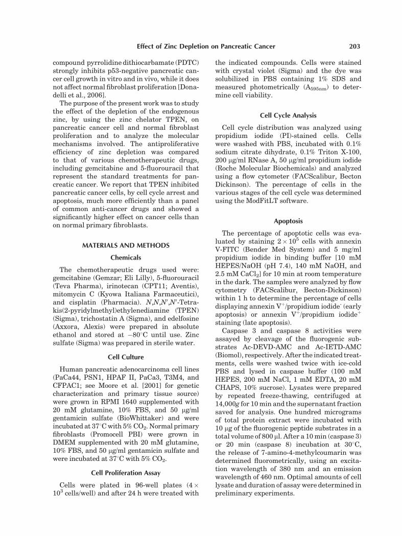
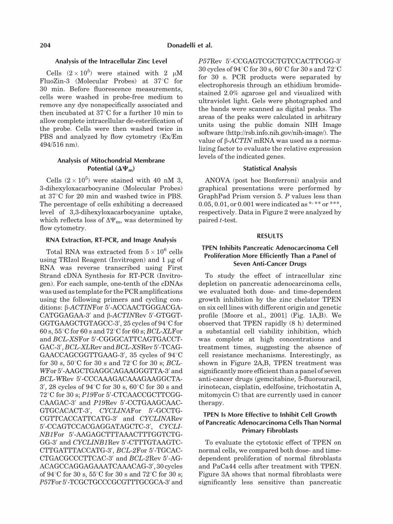
To study the effect of intracellular zincdepletion on pancreatic adenocarcinoma cells,we evaluated both dose- and time-dependentgrowth inhibition by the zinc chelator TPENon six cell lines with different origin and geneticprofile [Moore et al., 2001] (Fig. 1A,B). Weobserved that TPEN rapidly (8 h) determineda substantial cell viability inhibition, whichwas complete at high concentrations andtreatment times, suggesting the absence ofcell resistance mechanisms. Interestingly, asshown in Figure 2A,B, TPEN treatment wassignificantly more efficient than a panel of sevenanti-cancer drugs (gemcitabine, 5-fluorouracil,irinotecan, cisplatin, edelfosine, trichostatin A,mitomycin C) that are currently used in cancertherapy.
TPEN Is More Effective to Inhibit Cell Growthof Pancreatic Adenocarcinoma Cells Than Normal
Primary Fibroblasts
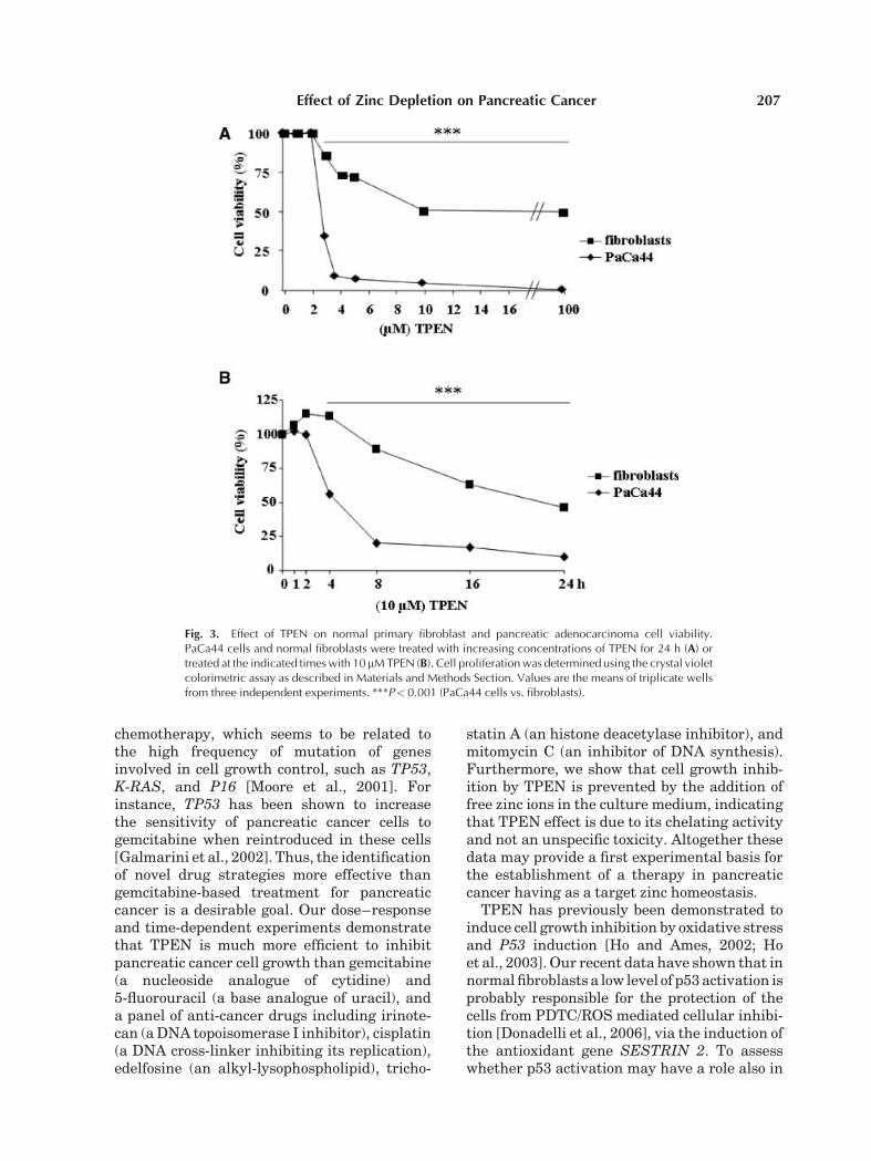
To evaluate the cytotoxic effect of TPEN onnormal cells, we compared both dose- and time-dependent proliferation of normal fibroblastsand PaCa44 cells after treatment with TPEN.Figure 3A shows that normal fibroblasts weresignificantly less sensitive than pancreatic
204 Donadelli et al.
adenocarcinoma cells to TPEN treatment.Moreover, normal fibroblasts showed featuresof cell resistance starting from a TPEN concen-tration of 10mM, which was more than sufficientto completely inhibit PaCa44 cell viability.Growth inhibition kinetics in fibroblasts wassignificantly slower than that obtained inPaCa44 cells, with the highest differencebetween the two cell types at 8 h (Fig. 3B).
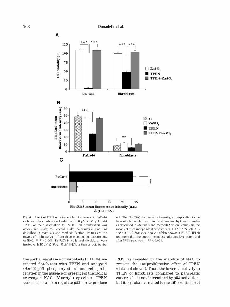
To verify whether the antiproliferative effectof TPEN was due to its chelating activity and didnot depend on an unspecific toxicity, we treatedcells with TPEN in the absence or presence ofsupplemented zinc ions in culture medium.We demonstrated that exogenous zinc ionscompletely counteracted the antiproliferativeeffect of TPEN both in PaCa44 cells andnormal fibroblasts (Fig. 4A). Furthermore,Figure 4B,C shows that TPEN determined anhigher intracellular zinc reduction in PaCa44cells (24 arbitrary units) than in normalfibroblasts (2.7 arbitrary units). This result
correlates with the stronger antiproliferativeeffect of TPEN on PaCa44 cells (see Fig. 3) andmay explain the lower sensitivity to TPEN ofnormal fibroblasts.
TPEN Inhibits Pancreatic AdenocarcinomaCell Cycle Progression
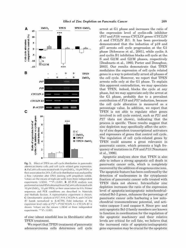
To examine the molecular mechanisms in-volved in the inhibition of PaCa44 cell viabilityby TPEN, we first analyzed cell distribution inthe various phases of the cell cycle. Figure 5Ashows that TPEN treatment increased thepercentage of cells in G1 phase (11% relativeto control), while the addition of exogenous zincions counteracted TPEN effect. Afterwards,we measured the expression of several genesrelated to the cell cycle. The intracellularzinc depletion by TPEN induced the expressionof P57 and P19 tumor suppressor genes andrepressed CYCLIN A and CYCLIN B1 (Fig. 5B),while it did not alter the expression of othergenes having a function in cell cycle checkpointsuch as P21 and P27 (data not shown).Figure 5C shows that the ratio of the expressionlevel of the cyclin-cdk inhibitors versus theCYCLIN genes was strongly enhanced byTPEN. Thus, TPEN treatment was able toregulate genes involved in both progressionand inhibition of the cell cycle thus to determinea global inhibitory effect.
TPEN Induces Apoptotic Cell Deathof Pancreatic Adenocarcinoma Cells
Figure 6A shows that TPEN caused PaCa44apoptotis by increasing the percentage of cells inthe early (annexin Vþ) and in the late (annexinVþ/propidium iodideþ) phases of the apoptoticprocess. This effect was completely recovered byzinc addition. Analyses of the expression ofmitochondrial related apoptotic genes demon-strated that TPEN induced the apoptoticBCL-XS expression, probably by regulating thealternative splicing of BCL-X, and inhibited theanti-apoptotic BCL-2, BCL-W, and BCL-XL
(Fig. 6B). Although TPEN did not induceother important apoptotic genes such as BIM,BAK, BAX, PUMA, and NOXA (data notshown), it is noteworthy that TPEN stronglyenhanced (9-fold) the ratio of the expressionlevel of apoptotic versus anti-apoptotic genes(Fig. 6C). Consistently with these results,Figure 6D reports that TPEN treatment in-creased the percentage of PaCa44 cells havinglow values of mitochondrial transmembrane
Fig. 1. Effect of TPEN on growth inhibition of a panel ofsix pancreatic adenocarcinoma cell lines. Cells were seeded in96-well plates and incubated overnight. TPEN was added atincreasing concentrations and cells were further incubated for24 h (A). Cells were treated at the indicated times with 10 mMTPEN (B). Cell proliferation was determined using the crystalviolet colorimetric assay as described in Materials and MethodsSection. Values are the means of triplicate wells from threeindependent experiments.
Effect of Zinc Depletion on Pancreatic Cancer 205
potential (DCm) and the addition of exogenouszinc ions completely recovered mitochondrialinjury. Figure 6E shows that TPEN inducedthe activity of caspase 3 and caspase 8 (11- and2.5-fold, respectively), demonstrating their in-volvement in TPEN-induced apoptotic celldeath.
DISCUSSION
We investigated the effect of intracellularzinc chelation on pancreatic adenocarcinomacell growth. Our results demonstrate that thedepletion of zinc ions determines cell cyclearrest and apoptosis via specific regulation ofgenes involved in both cellular processes.Zinc depletion has already been described to
determine cell cycle arrest and apoptosis innumerous cell types [Kolenko et al., 2001; Smithet al., 2002; Hashemi et al., 2007]. Our studyreports for the first time that pancreaticadenocarcinoma cell growth is inhibited by zincdepletion and that normal fibroblasts are muchless sensitive to the same treatment.
Pancreatic adenocarcinoma is one of the mostaggressive human cancers [Neoptolemos et al.,2003]. Standard treatments for advanced dis-ease include chemotherapy with 5-fluorouracil(5-FU) and gemcitabine. However, even gemci-tabine, which is now considered the gold stand-ard, has a response rate of less than 20%[Haller, 2003; Li et al., 2004]. A major cause oftreatment failure is believed to be resistance to
Fig. 2. Pancreatic adenocarcinoma cell growth inhibition by TPEN and chemotherapeutic agents.PaCa44 cells were seeded in 96-well plates and incubated overnight. Drugs were added at increasingconcentrations and cells were further incubated for 24 h (A). Cells were treated at the indicated times with10 mM of each drug (B). Cell proliferation was determined using the crystal violet colorimetric assay asdescribed in Materials and Methods Section. Values are the means of triplicate wells from three independentexperiments. ***P< 0.001 (TPEN vs. other drugs).
206 Donadelli et al.
chemotherapy, which seems to be related tothe high frequency of mutation of genesinvolved in cell growth control, such as TP53,K-RAS, and P16 [Moore et al., 2001]. Forinstance, TP53 has been shown to increasethe sensitivity of pancreatic cancer cells togemcitabine when reintroduced in these cells[Galmarini et al., 2002]. Thus, the identificationof novel drug strategies more effective thangemcitabine-based treatment for pancreaticcancer is a desirable goal. Our dose–responseand time-dependent experiments demonstratethat TPEN is much more efficient to inhibitpancreatic cancer cell growth than gemcitabine(a nucleoside analogue of cytidine) and5-fluorouracil (a base analogue of uracil), anda panel of anti-cancer drugs including irinote-can (a DNA topoisomerase I inhibitor), cisplatin(a DNA cross-linker inhibiting its replication),edelfosine (an alkyl-lysophospholipid), tricho-
statin A (an histone deacetylase inhibitor), andmitomycin C (an inhibitor of DNA synthesis).Furthermore, we show that cell growth inhib-ition by TPEN is prevented by the addition offree zinc ions in the culture medium, indicatingthat TPEN effect is due to its chelating activityand not an unspecific toxicity. Altogether thesedata may provide a first experimental basis forthe establishment of a therapy in pancreaticcancer having as a target zinc homeostasis.
TPEN has previously been demonstrated toinduce cell growth inhibition by oxidative stressand P53 induction [Ho and Ames, 2002; Hoet al., 2003]. Our recent data have shown that innormal fibroblasts a low level of p53 activation isprobably responsible for the protection of thecells from PDTC/ROS mediated cellular inhibi-tion [Donadelli et al., 2006], via the induction ofthe antioxidant gene SESTRIN 2. To assesswhether p53 activation may have a role also in
Fig. 3. Effect of TPEN on normal primary fibroblast and pancreatic adenocarcinoma cell viability.PaCa44 cells and normal fibroblasts were treated with increasing concentrations of TPEN for 24 h (A) ortreated at the indicated times with 10 mM TPEN (B). Cell proliferation was determined using the crystal violetcolorimetric assay as described in Materials and Methods Section. Values are the means of triplicate wellsfrom three independent experiments. ***P< 0.001 (PaCa44 cells vs. fibroblasts).
Effect of Zinc Depletion on Pancreatic Cancer 207
the partial resistance of fibroblasts to TPEN, wetreated fibroblasts with TPEN and analyzed(Ser15)-p53 phosphorylation and cell proli-feration in the absence or presence of the radicalscavenger NAC (N-acetyl-L-cysteine). TPENwas neither able to regulate p53 nor to produce
ROS, as revealed by the inability of NAC torecover the antiproliferative effect of TPEN(data not shown). Thus, the lower sensitivity toTPEN of fibroblasts compared to pancreaticcancer cells is not determined by p53 activation,but it is probably related to the differential level
Fig. 4. Effect of TPEN on intracellular zinc levels. A: PaCa44cells and fibroblasts were treated with 10 mM ZnSO4, 10 mMTPEN, or their association for 24 h. Cell proliferation wasdetermined using the crystal violet colorimetric assay asdescribed in Materials and Methods Section. Values are themeans of triplicate wells from three independent experiments(�SEM). ***P< 0.001. B: PaCa44 cells and fibroblasts weretreated with 10 mM ZnSO4, 10 mM TPEN, or their association for
4 h. The FluoZin3 fluorescence intensity, corresponding to thelevel of intracellular zinc ions, was measured by flow cytometryas described in Materials and Methods Section. Values are themeans of three independent experiments (�SEM). ***P<0.001,**P< 0.01. C: Statistical analysis of data shown in (B).D(C-TPEN)represents the difference of the intracellular zinc level before andafter TPEN treatment. ***P< 0.001.
208 Donadelli et al.
of zinc (about ninefold less in fibroblasts) afterTPEN treatment.
We report that TPEN treatment of pancreaticadenocarcinoma cells determines cell cycle
arrest at G1 phase and increases the ratio ofthe expression level of cyclin-cdk inhibitor(P57 and P19) versus CYCLIN genes (CYCLINA and CYCLIN B1). It has been previouslydemonstrated that the induction of p19 andp57 arrests cell cycle progression at the G1phase [Schwarze et al., 2001], while cyclin Aand cyclin B1 inhibition blocks cell cycle at theS and G2/M and G2/M phases, respectively[Desdouets et al., 1995; Porter and Donoghue,2003]. Our results demonstrate that TPENmodulates the expression of cell cycle relatedgenes in a way to potentially arrest all phases ofthe cell cycle. However, we report that TPENarrests cells only at the G1 phase. To explainthis apparent contradiction, we may speculatethat TPEN, indeed, blocks the cycle at anyphase, but we may appreciate only the arrest atthe G1 phase, probably due to a prevalentcontribution of P19 and P57 induction, becausethe cell cycle alteration is measured as apercentage value. In addition, we report thatTPEN is not able to regulate other genesinvolved in cell cycle control, such as P21 andP27 (data not shown), indicating that theprocess is specific. These results suggest thatzinc depletion may specifically affect the activ-ity of zinc dependent transcriptional activatorsand repressors of genes that control cell cycle.The regulation of cell cycle-related genes byTPEN could assume a great relevance inpancreatic cancer, which presents a high fre-quency of mutations in P16 and P15 [Naumannet al., 1996].
Apoptotic analyses show that TPEN is alsoable to induce a strong apoptotic cell death inpancreatic cancer cells, which is completelyrecovered by the addition of exogenous zinc ions.The apoptotic feature has been confirmed by thedetection of nucleosomes in the cytoplasmicfraction of pancreatic cancer cells treated withTPEN (data not shown). Intracellular zincdepletion increases the ratio of the expressionlevel of apoptotic/antiapoptotic mitochondrial-related Bcl-2 genes, enhances the percentage ofpancreatic cancer cells harboring a low mito-chondrial transmembrane potential, and acti-vates caspase 3 and caspase 8. Since pro- andanti-apoptotic Bcl-2 family members are knownto function in coordination for the regulation ofthe apoptotic machinery and their relativelevels are critical for cell fate, we believe thatthe increased ratio of apoptotic/antiapoptoticgene expression may be crucial for the apoptotic
Fig. 5. Effect of TPEN on cell cycle distribution in pancreaticadenocarcinoma cells and cell cycle related gene expression.A: PaCa44 cells were treated with 10 mM ZnSO4, 10 mM TPEN, ortheir association for 24 h. Cell cycle distribution was analyzed bya flow cytometer after DNA staining with propidium iodide.Values are the means of triplicate wells from three independentexperiments (�SEM). ***P<0.001. B: RT-PCR analysis wasperformed on total RNA obtained from PaCa44 cells treated with10 mM ZnSO4, 10 mM TPEN, or their association for 8 h. Primersequences and PCR conditions are described in Materialsand Methods Section. A representative experiment is shown.C: Densitometric analysis of P57, P19, CYCLIN A, and CYCLINB1 bands normalized to b-ACTIN. Fold induction of theexpression level ratio of P57þP19/CYCLIN AþCYCLIN B1 isshown. Values are the means (�SEM) of three independentexperiments. ***P<0.001.
Effect of Zinc Depletion on Pancreatic Cancer 209
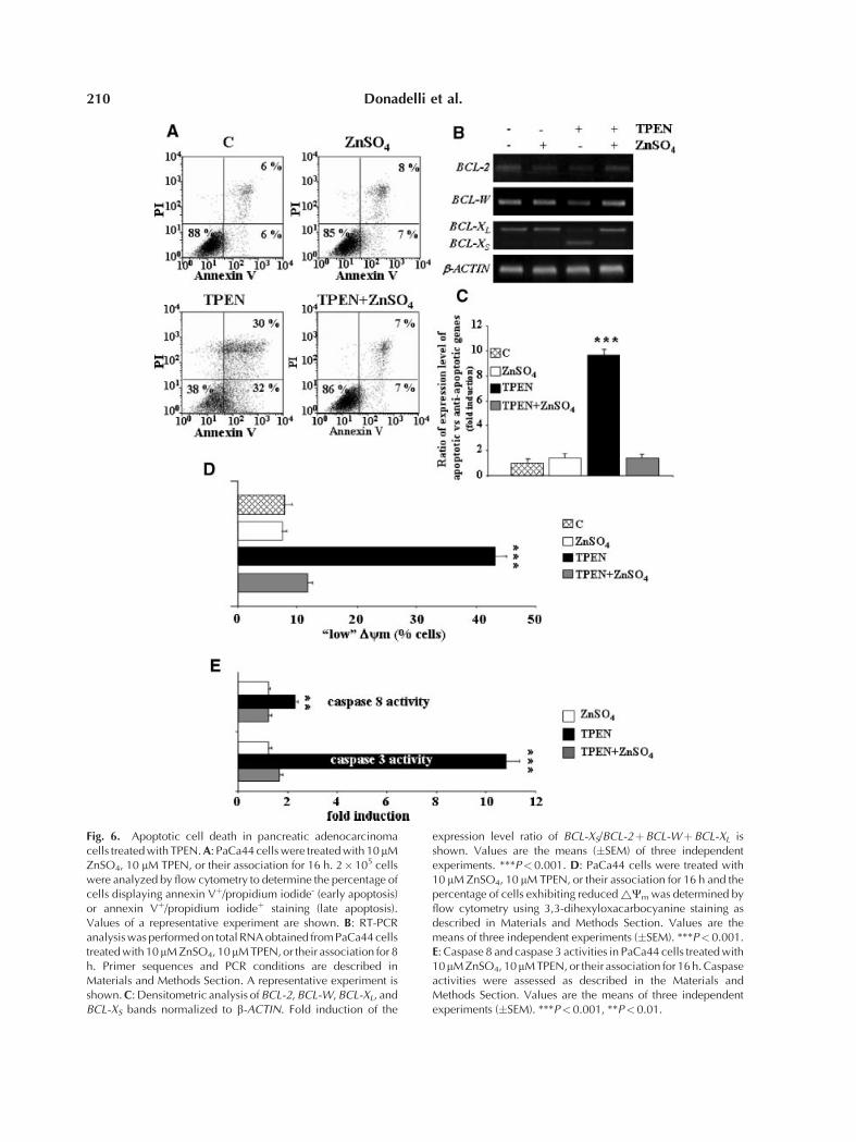
Fig. 6. Apoptotic cell death in pancreatic adenocarcinomacells treated with TPEN. A: PaCa44 cells were treated with 10 mMZnSO4, 10 mM TPEN, or their association for 16 h. 2�105 cellswere analyzed by flow cytometry to determine the percentage ofcells displaying annexin Vþ/propidium iodide- (early apoptosis)or annexin Vþ/propidium iodideþ staining (late apoptosis).Values of a representative experiment are shown. B: RT-PCRanalysis was performed on total RNA obtained from PaCa44 cellstreated with 10mM ZnSO4, 10mM TPEN, or their association for 8h. Primer sequences and PCR conditions are described inMaterials and Methods Section. A representative experiment isshown. C: Densitometric analysis of BCL-2, BCL-W, BCL-XL, andBCL-XS bands normalized to b-ACTIN. Fold induction of the
expression level ratio of BCL-XS/BCL-2þBCL-WþBCL-XL isshown. Values are the means (�SEM) of three independentexperiments. ***P< 0.001. D: PaCa44 cells were treated with10 mM ZnSO4, 10 mM TPEN, or their association for 16 h and thepercentage of cells exhibiting reduced ~Cm was determined byflow cytometry using 3,3-dihexyloxacarbocyanine staining asdescribed in Materials and Methods Section. Values are themeans of three independent experiments (�SEM). ***P<0.001.E: Caspase 8 and caspase 3 activities in PaCa44 cells treated with10mM ZnSO4, 10mM TPEN, or their association for 16 h. Caspaseactivities were assessed as described in the Materials andMethods Section. Values are the means of three independentexperiments (�SEM). ***P<0.001, **P<0.01.
210 Donadelli et al.
induction by TPEN. However, as zinc hasbeen shown to inhibit caspase 3 activation[Takahashi et al., 1996], a direct stimulation ofcaspase 3 proteolytic processing by zinc deple-tion may also contribute to TPEN inducedcell death in pancreatic cancer cells. Caspase8 activation, which could represent a mereconsequence of caspase 3 activation [Chimientiet al., 2001], may also have a role in TPENmediated apoptosis.
Metal chelators are currently used in clinicaltrials. Antitumoral activity of ICRF-159, achelator of divalent cations, has been demon-strated in patients with a variety of tumor typesincluding non-small cell lung cancer [Eaganet al., 1976], colorectal carcinoma [Marciniaket al., 1975; Bellet et al., 1976], Kaposi’s sarcoma[Olweny et al., 1976], non-Hodgkin’s lym-phoma [Flannery et al., 1978], acute leukemia[Bakowski et al., 1979], and head and neckcarcinoma [Shah et al., 1982]. Phase I studies ofdexrazoxane, a bidentate chelator that resem-bles EDTA, have been performed using a varietyof dosing schedules. Interestingly, the maximaltolerated dose (MTD) of dexrazoxane deter-mined by using short infusion schedules wasapproximately 10–15 times higher than theMTD determined with continuous infusionschedules [Tetef et al., 2001]. Myelosuppressionappeared as the strongest toxic effect and wasused to established the limiting dose of metalchelators [Tetef et al., 2001; Chow et al., 2004].Thus, in planning a therapy with this class ofagents, the infusion schedule must be carefullyevaluated.
In this work, we demonstrate that TPENefficiently inhibits pancreatic adenocarcinomacell growth providing opportunities for develop-ing potential pharmacological approaches toinhibit pancreatic cancer growth in vivo.
REFERENCES
Bakowski MT, Prentice HG, Lister TA, Malpas JS,McElwain TJ, Powles RL. 1979. Limited activity ofICRF-159 in advanced acute leukemia. Cancer TreatRep 63:127–129.
Bellet RE, Engstrom PF, Catalano RB, Creech RH,Mastrangelo MJ. 1976. Phase II study of ICRF-159 inpatients with metastatic colorectal carcinoma previouslyexposed to systemic chemotherapy. Cancer Treat Rep 60:1395–1397.
Beyersmann D, Haase H. 2001. Functions of zinc insignaling, proliferation and differentiation of mamma-lian cells. Biometals 14:331–341.
Chimienti F, Seve M, Richard S, Mathieu J, Favier A. 2001.Role of cellular zinc in programmed cell death: Temporalrelationship between zinc depletion, activation of cas-pases, and cleavage of Sp family transcription factors.Biochem Pharmacol 62:51–62.
Chow WA, Synold TW, Tetef ML, Longmate J, Frankel P,Lawrence J, Al-Khadimi Z, Leong L, Lim D, Margolin K,Morgan RJ, Jr., Raschko J, Shibata S, Somlo G,Twardowski P, Yen Y, Doroshow JH. 2004. Feasibilityand pharmacokinetic study of infusional dexrazoxaneand dose-intensive doxorubicin administered concur-rently over 96 h for the treatment of advanced malig-nancies. Cancer Chemother Pharmacol 54:241–248.
Costello LC, Franklin RB. 2006. The clinical relevance ofthe metabolism of prostate cancer; zinc and tumorsuppression: Connecting the dots. Mol Cancer 5:17.
Cousins RJ, Liuzzi JP, Lichten LA. 2006. Mammalian zinctransport, trafficking, and signals. J Biol Chem 281:24085–24089.
Desdouets C, Sobczak-Thepot J, Murphy M, Brechot C.1995. Cyclin A: Function and expression during cellproliferation. Prog Cell Cycle Res 1:115–123.
Donadelli M, Dalla Pozza E, Costanzo C, Scupoli MT,Piacentini P, Scarpa A, Palmieri M. 2006. Increasedstability of P21(WAF1/CIP1) mRNA is required for ROS/ERK-dependent pancreatic adenocarcinoma cell growthinhibition by pyrrolidine dithiocarbamate. Biochim Bio-phys Acta 1763:917–926.
Eagan RT, Carr DT, Coles DT, Rubin J, Frytak S. 1976.ICRF-159 versus polychemotherapy in non-small celllung cancer. Cancer Treat Rep 60:947–948.
Flannery EP, Corder MP, Sheehan WW, Pajak TF, Bate-man JR. 1978. Phase II study of ICRF-159 in non-Hodgkin’s lymphomas. Cancer Treat Rep 62:465–467.
Gaither LA, Eide DJ. 2001. Eukaryotic zinc transportersand their regulation. Biometals 14:251–270.
Galmarini CM, Clarke ML, Falette N, Puisieux A, MackeyJR, Dumontet C. 2002. Expression of a non-functionalp53 affects the sensitivity of cancer cells to gemcitabine.Int J Cancer 97:439–445.
Gupta SK, Singh SP, Shukla VK. 2005. Copper, zinc, andCu/Zn ratio in carcinoma of the gallbladder. J Surg Oncol91:204–208.
Haller DG. 2003. New perspectives in the management ofpancreas cancer. Semin Oncol 30:3–10.
Hashemi M, Ghavami S, Eshraghi M, Booy EP, Los M.2007. Cytotoxic effects of intra and extracellular zincchelation on human breast cancer cells. Eur J Pharmacol557:9–19.
Ho E, Ames BN. 2002. Low intracellular zinc inducesoxidative DNA damage, disrupts p53, NFkappa B, andAP1 DNA binding, and affects DNA repair in a rat gliomacell line. Proc Natl Acad Sci USA 99:16770–16775.
Ho E, Courtemanche C, Ames BN. 2003. Zinc deficiencyinduces oxidative DNA damage and increases p53expression in human lung fibroblasts. J Nutr 133:2543–2548.
Kagara N, Tanaka N, Noguchi S, Hirano T. 2007. Zinc andits transporter ZIP10 are involved in invasive behavior ofbreast cancer cells. Cancer Sci 98:692–697.
Kolenko VM, Uzzo RG, Dulin N, Hauzman E, Bukowski R,Finke JH. 2001. Mechanism of apoptosis induced by zincdeficiency in peripheral blood T lymphocytes. Apoptosis6:419–429.
Effect of Zinc Depletion on Pancreatic Cancer 211
Li D, Xie K, Wolff R, Abbruzzese JL. 2004. Pancreaticcancer. Lancet 363:1049–1057.
Marciniak TA, Moertel CG, Schutt AJ, Hahn RG, Reite-meier RJ. 1975. Phase II study of ICRF-159 (NSC-129943) in advanced colorectal carcinoma. Cancer Che-mother Rep 59:761–763.
McCall KA, Huang C, Fierke CA. 2000. Function andmechanism of zinc metalloenzymes. J Nutr 130:1437S–1446S.
Moore PS, Sipos B, Orlandini S, Sorio C, Real FX, LemoineNR, Gress T, Bassi C, Kloppel G, Kalthoff H, UngefrorenH, Lohr M, Scarpa A. 2001. Genetic profile of 22pancreatic carcinoma cell lines. Analysis of K-ras, p53,p16 and DPC4/Smad4. Virchows Arch 439:798–802.
Naumann M, Savitskaia N, Eilert C, Schramm A, KalthoffH, Schmiegel W. 1996. Frequent codeletion of p16/MTS1and p15/MTS2 and genetic alterations in p16/MTS1 inpancreatic tumors. Gastroenterology 110:1215–1224.
Neoptolemos JP, Cunningham D, Friess H, Bassi C,Stocken DD, Tait DM, Dunn JA, Dervenis C, Lacaine F,Hickey H, Raraty MG, Ghaneh P, Buchler MW. 2003.Adjuvant therapy in pancreatic cancer: Historical andcurrent perspectives. Ann Oncol 14:675–692.
Olweny CL, Masaba JP, Sikyewunda W, Toya T. 1976.Treatment of Kaposi’s sarcoma with ICRF-159 (NSC-129943). Cancer Treat Rep 60:111–113.
Outten CE, O’Halloran TV. 2001. Femtomolar sensitivity ofmetalloregulatory proteins controlling zinc homeostasis.Science 292:2488–2492.
Porter LA, Donoghue DJ. 2003. Cyclin B1 and CDK1:nuclear localization and upstream regulators. ProgCell Cycle Res 5:335–347.
Schwarze SR, Shi Y, Fu VX, Watson PA, Jarrard DF.2001. Role of cyclin-dependent kinase inhibitors inthe growth arrest at senescence in human prostateepithelial and uroepithelial cells. Oncogene 20:8184–8192.
Shah MK, Engstrom PF, Catalano RB, Paul AR, Bellet RE,Creech RH. 1982. Phase II trial of razoxane (ICRF-159)in patients with squamous cell carcinoma of the head andneck previously exposed to systemic chemotherapy.Cancer Treat Rep 66:557–558.
Smith PJ, Wiltshire M, Davies S, Chin SF, Campbell AK,Errington RJ. 2002. DNA damage-induced [Zn(2þ)](i)transients: Correlation with cell cycle arrest and apop-tosis in lymphoma cells. Am J Physiol Cell Physiol 283:C609–C622.
Takahashi A, Alnemri ES, Lazebnik YA, Fernandes-Alnemri T, Litwack G, Moir RD, Goldman RD, PoirierGG, Kaufmann SH, Earnshaw WC. 1996. Cleavage oflamin A by Mch2 alpha but not CP P32: Multipleinterleukin 1 beta-converting enzyme-related proteaseswith distinct substrate recognition properties areactive in apoptosis. Proc Natl Acad Sci USA 93:8395–8400.
Tetef ML, Synold TW, Chow W, Leong L, Margolin K,Morgan R, Raschko J, Shibata S, Somlo G, Yen Y,Groshen S, Johnson K, Lenz HJ, Gandara D, DoroshowJH. 2001. Phase I trial of 96-hour continuous infusion ofdexrazoxane in patients with advanced malignancies.Clin Cancer Res 7:1569–1576.
Truong-Tran AQ, Ho LH, Chai F, Zalewski PD. 2000.Cellular zinc fluxes and the regulation of apoptosis/gene-directed cell death. J Nutr 130:1459S–1466S.
212 Donadelli et al.
Copyright © 2022 FDOKUMEN