Performing High-Powered Studies Efficiently With Sequential Analyses
Upload
independentCategory
view
0download
0
ORIGINAL RESEARCH ARTICLEpublished: 15 October 2014
doi: 10.3389/fnmol.2014.00081
Intravenous AAV9 efficiently transduces myenteric neuronsin neonate and juvenile miceSara E. Gombash1, Christopher J. Cowley1, Julie A. Fitzgerald1, Jodie C. E. Hall 2, Christian Mueller3,
Fedias L. Christofi4 and Kevin D. Foust1*
1 Department of Neuroscience, Ohio State University, Columbus, OH, USA2 Department of Neuroscience, Center for Brain and Spinal Cord Repair, Ohio State University, Columbus, OH, USA3 Department of Pediatrics, Gene Therapy Center, University of Massachusetts Medical School, Worcester, MA, USA4 Department of Anesthesiology, Ohio State University, Columbus, OH, USA
Edited by:
Erwan Bezard, Institut des MaladiesNeurodegeneratives, France
Reviewed by:
Lucy Vulchanova, University ofMinnesota, USAJunling Yang, University ofIllinois, USA
*Correspondence:
Kevin D. Foust, Department ofNeuroscience, Ohio StateUniversity, 460 W 12th Ave., BRT698, Columbus, OH 43210, USAe-mail: [email protected]
Gene therapies for neurological diseases with autonomic or gastrointestinal involvementmay require global gene expression. Gastrointestinal complications are often associatedwith Parkinson’s disease and autism. Lewy bodies, a pathological hallmark of Parkinson’sbrains, are routinely identified in the neurons of the enteric nervous system (ENS)following colon biopsies from patients. The ENS is the intrinsic nervous systemof the gut, and is responsible for coordinating the secretory and motor functionsof the gastrointestinal tract. ENS dysfunction can cause severe patient discomfort,malnourishment, or even death as in intestinal pseudo-obstruction (Ogilvie syndrome).Importantly, ENS transduction following systemic vector administration has not beenthoroughly evaluated. Here we show that systemic injection of AAV9 into neonate orjuvenile mice results in transduction of 25–57% of ENS myenteric neurons. Transgeneexpression was prominent in choline acetyltransferase positive cells, but not withinvasoactive intestinal peptide or neuronal nitric oxide synthase cells, suggesting a biasfor cells involved in excitatory signaling. AAV9 transduction in enteric glia is very lowcompared to CNS astrocytes. Enteric glial transduction was enhanced by using a glialspecific promoter. Furthermore, we show that AAV8 results in comparable transduction inneonatal mice to AAV9 though AAV1, 5, and 6 are less efficient. These data demonstratethat systemic AAV9 has high affinity for peripheral neural tissue and is useful for futuretherapeutic development and basic studies of the ENS.
Keywords: AAV9, adeno-associated virus, enteric nervous system, myenteric plexus, functional gastrointestinal
motility disorders, enteric glia, enteric neuropathy, gene therapy
INTRODUCTIONThe recent identification of adeno-associated viral vector (AAV)serotypes that cross the blood brain barrier (BBB) resulted inunprecedented therapeutic benefit in animal models of neuro-logical disease (Foust et al., 2010, 2013; Fu et al., 2011; Ahmedet al., 2013; Garg et al., 2013; Haurigot et al., 2013). AAV9 wasthe first serotype described that efficiently crossed the BBB (Foustet al., 2009). Subsequently multiple groups have demonstratedthat intravenous injection of AAV9 into mice, rats, cats and non-human primates produces robust transduction in the centralnervous system (CNS) (Duque et al., 2009; Tatom et al., 2009;Bevan et al., 2011). Intravenous injection of AAV9 in neonatalmice results in primarily neuronal transduction throughout thebrain and spinal cord, but preferentially targets astrocytes wheninjected in mice greater than 2 weeks of age (Foust et al., 2009).Similar CNS transduction patterns have been reported with otherAAV serotypes following systemic delivery (Zhang et al., 2011).AAV’s affinity for nervous tissue suggests that the peripheral ner-vous systems are likely transduced following systemic injection.Gene delivery to large peripheral neuronal networks such as theautonomic nervous system (ANS) and enteric nervous system
(ENS) may be beneficial for treatment of global neurological dis-ease. To date, transduction in peripheral neurons has not beenthoroughly investigated.
The ENS is often referred to as “the little brain in the gut” andis estimated to have as many neurons as the spinal cord (Wood,2000). The ENS is embedded within the mucosal and muscu-lar layers throughout the length of the gastrointestinal (GI) tractand enteric neurons and glial cells are arranged into two gan-glionated plexuses called the submucosal and myenteric plexus,respectively. The submucosal plexus senses and reacts to stretchand chemical changes induced by luminal contents while themyenteric plexus coordinates motor function of smooth muscle.Dysfunction of the ENS is linked to a series of GI disorders thatare often chronic and prevalent in pediatric and adult populations(Fukudo et al., 2012; Yeung and Di Lorenzo, 2012). For example,functional GI motility disorders (FGIMD) are estimated to affect5–30% of adults and children in the United States (Saito et al.,2002; Camilleri et al., 2005). In FGIMDs, disruption of GI nervesand/or muscle can cause chronic gastroesophageal acid reflux,constipation, abdominal pain and bloating. However, etiology isnot well understood and suitable long-term therapies do not exist
Frontiers in Molecular Neuroscience www.frontiersin.org October 2014 | Volume 7 | Article 81 | 1
MOLECULAR NEUROSCIENCE
Gombash et al. AAV9 targets the myenteric plexus
(Fukudo et al., 2012). The ENS is also implicated in functionalgastrointestinal disorders (FGIDs) such as dyspepsia and irrita-ble bowel syndrome that affect 15–20% of the US population.Further, the ENS is a potential therapeutic target for diarrhealdisorders and inflammatory bowel diseases (IBD). GI neuromus-cular disorders, such as chronic intestinal pseudo-obstruction(CIP), are also poorly understood (Gabbard and Lacy, 2013). Insome CIP cases, myenteric neurons contain proteinaceous nuclearinclusions and evidence of apoptosis, presumably causing theGI dysfunction (El-Rifai et al., 2006; Gabbard and Lacy, 2013).Interestingly, GI dysfunction also commonly occurs in patientswith autism or Parkinson’s disease (Anderson et al., 2007; Buieet al., 2010; Wakabayashi et al., 2010; Natale et al., 2011; Mazureket al., 2012). Parkinson’s disease Lewy bodies in ENS neuronscan be identified in tissue biopsies (Derkinderen et al., 2011),suggestive that peripheral neurons are also affected in CNS dis-eases. Beyond diseases, aging is a risk factor for GI dysfunction(Camilleri et al., 2008; Wiskur and Greenwood-Van Meerveld,2010). Chronic constipation is common in the elderly and canbe associated with idiopathic ENS neuron degeneration (Sanchezand Bercik, 2011).
In addition to neurons, the ENS contains enteric glial cells(EGCs) that are similar to CNS astrocytes. Enteric glia, like CNSastrocytes, are essential for maintaining homeostasis and regulat-ing neural circuit activity (Gulbransen and Sharkey, 2012). EGCslikely serve as key regulators of intestinal inflammation in ani-mals and humans. Their ability to mediate immune responsesin vivo was suggested as a possible pathologic mechanism inCrohn’s Disease (Cornet et al., 2001). Data suggest that thereis an impairment of the glial network in non-inflamed regionsof the gut mucosa in patients with Crohn’s Disease, as evidenceby a decrease in GFAP immunoreactivity in glia (Cornet et al.,2001). Overall, EGCs like astrocytes in the brain mediate glialtransmission, and regulate synaptic signaling, synaptic plastic-ity, network excitability and inflammation. EGCs contribute tothe onset and development of intestinal inflammation’ and areimportant in the understanding of GI inflammation occurringin IBD, enterocolitis, and gut infections (Savidge et al., 2007;Vijayaraghavan, 2009; Cirillo et al., 2011; McClain et al., 2014;Turco et al., 2014).
Together, the lack of available therapies for ENS is a majorhealth problem and is an urgent need. Due to its safety and sus-tained expression, systemic AAV gene therapy may be a usefulapproach to treat and study the ENS and its associated disor-ders. AAV transduction of the ENS has been reported but not wellcharacterized (Fu et al., 2011; Rahim et al., 2011; Mattar et al.,2013; Schuster et al., 2014) likely due to the unique architectureand intricate dissection techniques required for study. The goalof the current work was to characterize AAV9 transduction effi-ciency and cell types targeted in the myenteric plexus followingintravenous injection into neonatal or juvenile mice. In con-trast to age dependent transduction patterns in the mouse CNS(Foust et al., 2009), we show that self complementary AAV9 injec-tion results in extensive myenteric neuron transduction at bothneonate and juvenile time points in all regions of the GI tract.Furthermore, AAV9 transduction of EGCs pales in comparisonto CNS astrocytes. Additionally, we also examined transduction
of self complementary AAV serotypes 1, 5, 6, and 8 in themyenteric plexus and show that they differ greatly in transductionefficiency.
MATERIALS AND METHODSANIMALSA total of 20 male or female FVB mice were used for thesestudies. Postnatal day 1 (P1) pups were used in all neonatal injec-tion studies and juvenile mice were used beginning at postnatalday 21 (P21). Following vector injection procedures, neonatalmice remained with the dam until weaning. Mice were housedwith same-sex littermates and given food and water ad libi-tum in a constant 12 h light/dark cycle room in the AAALACapproved Ohio State University Biomedical Research Towervivarium. All animal procedures were approved by the OhioState University Institutional Laboratory Animal Care and UseCommittee.
AAV VECTOR PRODUCTION AND PURIFICATIONAll vectors used in these studies were produced by the Universityof Massachusetts Medical School Viral Vector Core. Self com-plementary AAV (scAAV) genomes were engineered to encodethe green fluorescent protein (GFP) transgene under the controlof the chicken-β-actin/cytomegalovirus hybrid (CB) promoter,flanked by AAV2 inverted terminal repeats. Virus was packagedby triple transfection of HEK293 cells with an adenovirus helperplasmid and plasmids containing the AAV2 rep gene and AAV1,5, 6, 8, or 9 cap genes. Vectors were purified by cesium chloridegradient and titers were determined by qPCR. Vector titers werescAAV1- 2.4 × 10¹³, scAAV5- 9.0 × 10¹², scAAV6- 5.0 × 10¹²,scAAV8- 1.0 × 10¹³, and scAAV9- 1.0 × 10¹³ vector genomes(vg)/ml.
AAV INJECTIONSIntravaenous injections of scAAV9-CB-GFP or scAAV8-CB-GFPin neonatal (P1) and juvenile (P21) mice were completed as pre-viously described (Foust et al., 2009; Gombash Lampe et al.,in press)
NEONATESA dissecting microscope and fiber-optic light source were used forvisualization of the temporal face vein. First, neonatal mice wererested on a bed of ice for anesthetization and a 3/10 cc 30 gaugeinsulin syringe (Terumo Medical Corp., Elkton, MD) was loadedwith 1 × 10¹¹ vg/ml scAAV9-CB-GFP or 5 × 10¹⁰ vg/ml scAAV1,5, 6, 8, or 9-CB-GFP. Viral particles supplemented with phos-phate buffered saline (PBS, 0.01 M, pH 7.4) for a total volumeof 50 μl were injected into the face vein. Pups were then warmed,returned to their home cage, and rubbed in bedding to preventrejection by the mother. Neonatal animals were sacrificed 30–60days post-injection for myenteric analysis.
JUVENILESJuvenile tail vein injections were performed on P21 mice. Micewere restrained in an illuminated tail vein injection platform(Braintree Scientific Inc., Braintree, MA). Prior to injection, thetail was swabbed with alcohol, then 3/10 cc 30 gauge insulin
Frontiers in Molecular Neuroscience www.frontiersin.org October 2014 | Volume 7 | Article 81 | 2
Gombash et al. AAV9 targets the myenteric plexus
syringes were used to inject scAAV9-CB-GFP for a final dose of2 × 1012 vg. Mice were then returned to their home cages.
WHOLE MOUNT PREPARATIONSWhole mount longitudinal muscle-myenteric plexus (LMMP) tis-sues were microdissected as previously described (Wang et al.,2007). Briefly, the entire gastrointestinal tract beginning at thelower esophagus through the rectum was removed from eutha-nized mice and rinsed in room temperature PBS. Next, regions ofthe gastrointestinal tract (stomach, duodenum, jejunum, ileum,cecum, and colon) were separated, opened, and pinned flat withthe luminal side up on a Sylgard (Dow Corning, Midland, MI)base in 100 mm cell culture dishes. Under a dissecting micro-scope, fine forceps were used to remove the muscosa, submucosa,and circular muscle to expose the myenteric plexus on the surfaceof the longitudinal muscle. Immunohistochemistry was then usedto observe transgene expression, neurons, and glial cells withinthe myenteric plexus.
IMMUNOHISTOCHEMISTRYLMMP tissues were fixed in Zamboni’s fixative (4%paraformaldehyde and 0.2% piciric acid in 0.1 M PBS; #1459,Newcomer Supply, Middleton, WI) overnight at 4◦C. Thenext day, tissues were rinsed in PBS until clear. Tissues weretransferred to glass slides and incubated in blocking buffer [10%normal donkey serum (NDS), 0.5% Triton-X 100 in PBS] for 2 hat room temperature. Tissues were then incubated in appropriateprimary antisera overnight at 4◦C against green fluorescentprotein (GFP; 1:500; ab13970, Abcam, Cambridge, MA), HuD(1:25; A-21271, Life Technologies, Eugene, OR), S100 ProteinAb-2 (S100; 1:200; RB-044-A0, Thermo Scientific, UK), cholineacetyltransferase (ChAT; 1:50; AB144P, Millipore, Temecula,CA); vasoactive intestinal peptide (VIP; 1:200; Abcam), neuronalnitric oxide synthase (nNOS; 1:200; ab1376, Abcam), calretinin(1:200; CG1, Swant, Switzerland), or calbindin D-28K (1:200;AB1778, Millipore). The next day, tissues were rinsed 4 timesfor 10 min in PBS, then incubated in appropriate Alexa Fluorsecondary antibodies at 1:200 for 2 h at room temperature.Antibody dilutent was 3% NDS, 0.5% Triton-X 100 in PBS.Tissues were then rinsed and slides were coverslipped with 2.5%PVA/DABCO. Fluorescent images were captured on an OlympusFV 100 Spectral Confocal system (Melville, NY) in the Ohio StateUniversity Campus Microscopy and Imaging Facility.
QUANTIFICATION OF MYENTERIC NEURONSFluorescently stained whole mount tissues were viewed with aZeiss fluorescent microscope equipped with the appropriate fil-ters to distinguish CY3, CY5, and FITC flurophores. Myentericganglia in the stomach, duodenum, jejunum, ileum, cecum, andcolon were viewed at 40× for counting of HuD positive andGFP positive cells. Total neurons counted in each region arerecorded in Figure 3A. HuD and GFP positive neuron countswere collected only from the colon for scAAV8-CB-GFP andlow-dose scAAV9-CB-GFP (5 × 10¹⁰ vg) injected mice. For oral-aboral preference analysis in the colon, LMMP colon preparationsin the correct orientation were separated into nine 1 cm seg-ments. Numbers of HuD and GFP positive neurons in 10 ganglia
in each segment were recorded, resulting in approximately 5000HuD positive neurons counted within groups. Data is reported asthe percentage of GFP positive neurons ± standard error of themean (SEM).
STATISTICSA one-way repeated measures analysis of variance was used tocompare percentages of neuronal transduction across the oralto aboral axis of the colon in neonatal and juvenile injectedmice. A Student’s t-test was used to compare transduced neuroncounts following scAAV8-CB-GFP or scAAV9-CB-GFP injection.All results are listed as the mean ± s.e.m.
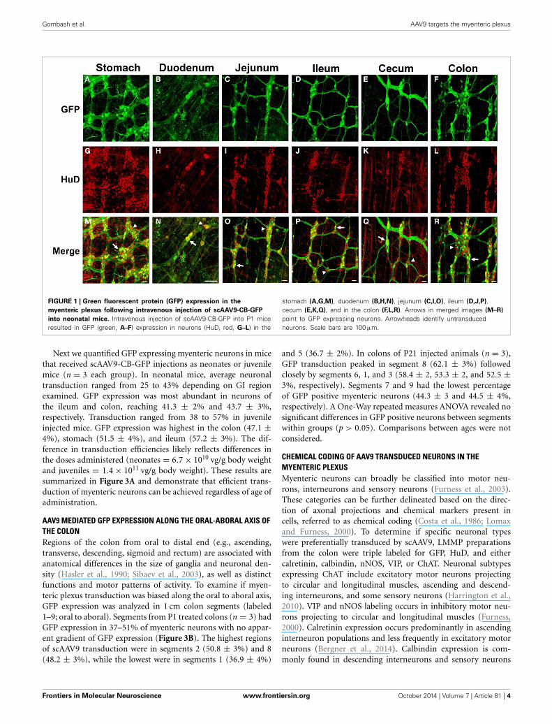
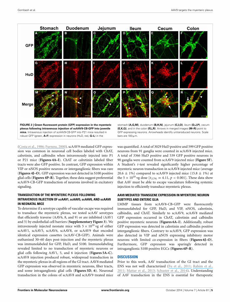
RESULTSAAV9 EFFICIENTLY TRANSDUCES MYENTERIC NEURONS FOLLOWINGINTRAVENOUS INJECTION INTO NEONATE AND JUVENILE MICETo investigate AAV9 ENS transduction, neonatal mice (P1)were injected in the temporal vein with 1 × 10¹¹ vg of selfcomplementary (sc) AAV9-CB-GFP. Three weeks post-injection,immunolabeling for GFP of the myenteric plexus revealed robust,widespread expression in neuronal cell bodies and fibers. GFPexpression was detected in all regions of the gastrointestinaltract, including the esophagus (not shown), stomach, duode-num, jejunum, ileum, cecum, and the colon (Figure 1). GFPlocalized in HuD positive cells in all regions of the GI tract. NoGFP expression was observed in S100 positive enteric glia in anyregion, suggesting expression was confined to myenteric neurons(Supplementary Figure 1). Intravenous AAV9 preferentially tar-gets neurons in neonatal mice and astrocytes in juvenile injectedanimals (Foust et al., 2009). To determine if ENS transductionalso followed this pattern, 2 × 10¹² vg of scAAV9-CB-GFP wasinjected into the tail vein of P21 mice. Similarly to injectedneonates, GFP and HuD immunolabeling revealed widespread,robust transgene expression through all regions of the gastroin-testinal tract (Figure 2). GFP expression was again limited tomyenteric neurons and their projections and was not expressedin intraganglionic glial cells along the entire tract. The paucityof enteric glia transduction was surprising because we previouslyreported that scAAV-CB-GFP expression was highly efficient inCNS astrocytes (Foust et al., 2009). Others have shown that theneuronal tropism of AAV vectors can be redirected to glia by driv-ing transgene expression under the GFAP promoter (Burger et al.,2004; Taymans et al., 2007; Lawlor et al., 2009; von Jonquiereset al., 2013; de Leeuw et al., 2014). GFAP expression is common toCNS astrocytes and enteric glia (Gulbransen and Sharkey, 2012).Therefore, we engineered a single stranded AAV9-GFAP-GFP vec-tor and intravenously injected neonate mice with 1 × 10¹¹ vg(n = 3). Mice were euthanized between 30 and 60 days post-injection and the myenteric plexus was labeled for GFP, GFAP,and S100. GFP expression was predominantly in enteric glia inmyenteric ganglia (Supplementary Figure 2). Though rare, anoccasional cell with neuronal morphology was detected (not pic-tured). Overall, the number of transduced glial cells was low(<5%). Further optimization is needed to increase glial targeting,however, these data show that AAV9 mediated transgene expres-sion in the ENS can be biased toward enteric glia by changing theviral genome.
Frontiers in Molecular Neuroscience www.frontiersin.org October 2014 | Volume 7 | Article 81 | 3
Gombash et al. AAV9 targets the myenteric plexus
FIGURE 1 | Green fluorescent protein (GFP) expression in the
myenteric plexus following intravenous injection of scAAV9-CB-GFP
into neonatal mice. Intravenous injection of scAAV9-CB-GFP into P1 miceresulted in GFP (green, A–F) expression in neurons (HuD, red, G–L) in the
stomach (A,G,M), duodenum (B,H,N), jejunum (C,I,O), ileum (D,J,P),cecum (E,K,Q), and in the colon (F,L,R). Arrows in merged images (M–R)
point to GFP expressing neurons. Arrowheads identify untransducedneurons. Scale bars are 100 μm.
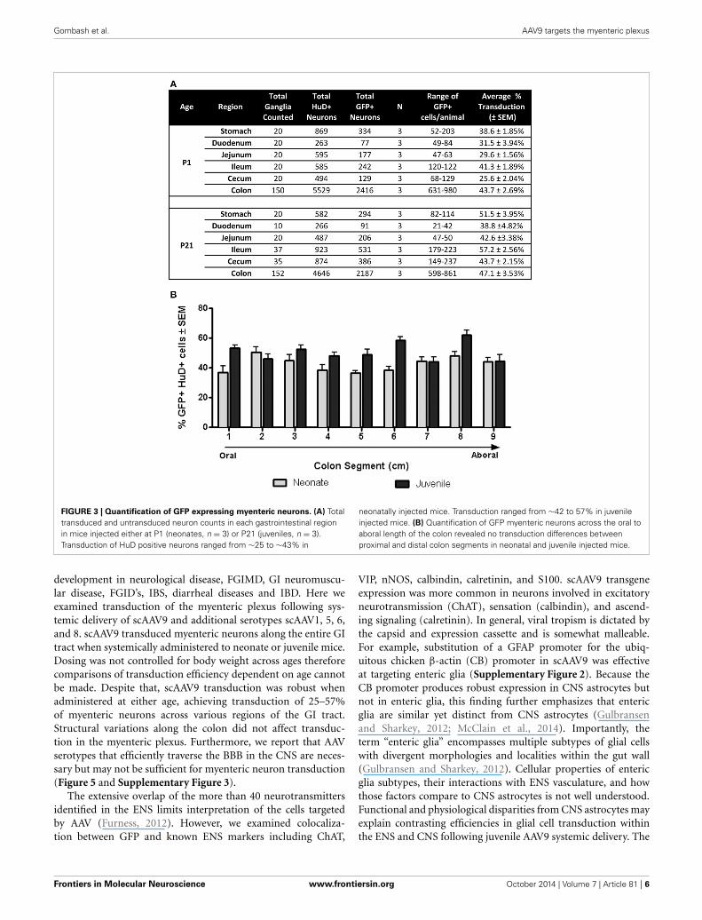
Next we quantified GFP expressing myenteric neurons in micethat received scAAV9-CB-GFP injections as neonates or juvenilemice (n = 3 each group). In neonatal mice, average neuronaltransduction ranged from 25 to 43% depending on GI regionexamined. GFP expression was most abundant in neurons ofthe ileum and colon, reaching 41.3 ± 2% and 43.7 ± 3%,respectively. Transduction ranged from 38 to 57% in juvenileinjected mice. GFP expression was highest in the colon (47.1 ±4%), stomach (51.5 ± 4%), and ileum (57.2 ± 3%). The dif-ference in transduction efficiencies likely reflects differences inthe doses administered (neonates = 6.7 × 10¹⁰ vg/g body weightand juveniles = 1.4 × 10¹¹ vg/g body weight). These results aresummarized in Figure 3A and demonstrate that efficient trans-duction of myenteric neurons can be achieved regardless of age ofadministration.
AAV9 MEDIATED GFP EXPRESSION ALONG THE ORAL-ABORAL AXIS OFTHE COLONRegions of the colon from oral to distal end (e.g., ascending,transverse, descending, sigmoid and rectum) are associated withanatomical differences in the size of ganglia and neuronal den-sity (Hasler et al., 1990; Sibaev et al., 2003), as well as distinctfunctions and motor patterns of activity. To examine if myen-teric plexus transduction was biased along the oral to aboral axis,GFP expression was analyzed in 1 cm colon segments (labeled1–9; oral to aboral). Segments from P1 treated colons (n = 3) hadGFP expression in 37–51% of myenteric neurons with no appar-ent gradient of GFP expression (Figure 3B). The highest regionsof scAAV9 transduction were in segments 2 (50.8 ± 3%) and 8(48.2 ± 3%), while the lowest were in segments 1 (36.9 ± 4%)
and 5 (36.7 ± 2%). In colons of P21 injected animals (n = 3),GFP transduction peaked in segment 8 (62.1 ± 3%) followedclosely by segments 6, 1, and 3 (58.4 ± 2, 53.3 ± 2, and 52.5 ±3%, respectively). Segments 7 and 9 had the lowest percentageof GFP positive myenteric neurons (44.3 ± 3 and 44.5 ± 4%,respectively). A One-Way repeated measures ANOVA revealed nosignificant differences in GFP positive neurons between segmentswithin groups (p > 0.05). Comparisons between ages were notconsidered.
CHEMICAL CODING OF AAV9 TRANSDUCED NEURONS IN THEMYENTERIC PLEXUSMyenteric neurons can broadly be classified into motor neu-rons, interneurons and sensory neurons (Furness et al., 2003).These categories can be further delineated based on the direc-tion of axonal projections and chemical markers present incells, referred to as chemical coding (Costa et al., 1986; Lomaxand Furness, 2000). To determine if specific neuronal typeswere preferentially transduced by scAAV9, LMMP preparationsfrom the colon were triple labeled for GFP, HuD, and eithercalretinin, calbindin, nNOS, VIP, or ChAT. Neuronal subtypesexpressing ChAT include excitatory motor neurons projectingto circular and longitudinal muscles, ascending and descend-ing interneurons, and some sensory neurons (Harrington et al.,2010). VIP and nNOS labeling occurs in inhibitory motor neu-rons projecting to circular and longitudinal muscles (Furness,2000). Calretinin expression occurs predominantly in ascendinginterneuron populations and less frequently in excitatory motorneurons (Bergner et al., 2014). Calbindin expression is com-monly found in descending interneurons and sensory neurons
Frontiers in Molecular Neuroscience www.frontiersin.org October 2014 | Volume 7 | Article 81 | 4
Gombash et al. AAV9 targets the myenteric plexus
FIGURE 2 | Green fluorescent protein (GFP) expression in the myenteric
plexus following intravenous injection of scAAV9-CB-GFP into juvenile
mice. Intravenous injection of scAAV9-CB-GFP into P21 mice resulted inrobust GFP (green, A–F) expression in neurons (HuD, red, G–L) in the
stomach (A,G,M), duodenum (B,H,N), jejunum (C,I,O), ileum (D,J,P), cecum(E,K,Q), and in the colon (F,L,R). Arrows in merged images (M–R) point toGFP expressing neurons. Arrowheads identify untransduced neurons. Scalebars are 100 μm.
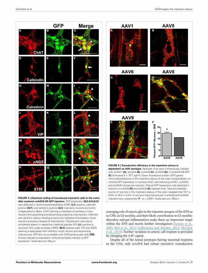
(Costa et al., 1986; Furness, 2000). scAAV9 mediated GFP expres-sion was common in neuronal cell bodies labeled with ChAT,calretinin, and calbindin when intravenously injected into P1or P21 mice (Figures 4A–I). ChAT or calretinin labeled fibertracts were also GFP positive. In contrast, GFP expression withinVIP or nNOS positive neurons or interganglionic fibers was rare(Figures 4J–O). GFP expression was not detected in S100 positiveglial cells (Figures 4P–R). Together, these data suggest preferentialscAAV9-CB-GFP transduction of neurons involved in excitatorysignaling.
TRANSDUCTION OF THE MYENTERIC PLEXUS FOLLOWINGINTRAVENOUS INJECTION OF scAAV1, scAAV5, scAAV6, AND scAAV8IN NEONATAL MICETo determine if a serotype capable of vascular escape was requiredto transduce the myenteric plexus, we tested scAAV serotypesthat efficiently traverse (AAV6, 8, and 9) or are inhibited (AAV1and 5) by endothelial cell barriers (Supplementary Figure 3). Weintravenously injected neonate mice with 5 × 10¹⁰ vg of eitherscAAV1, scAAV5, scAAV6, scAAV8, or scAAV9 that encodedidentical expression cassettes (scAAV-CB-GFP). Animals wereeuthanized 30–60 days post-injection and the myenteric plexuswas immunolabeled for GFP, HuD, and S100. Immunolabelingrevealed limited to no transduction of myenteric neurons orglial cells following AAV1, 5, and 6 injection (Figures 5A–C).scAAV8 injection produced robust, widespread transduction inthe myenteric plexus in all regions of the GI tract. AAV8 mediatedGFP expression was observed in myenteric neurons, fiber tracts,and some intraganglionic glial cells (Figures 5D, 6). Neuronaltransduction in the colons of scAAV8 and scAAV9 treated mice
was quantified. A total of 3029 HuD positive and 599 GFP positiveneurons from 91 ganglia were counted in scAAV8 injected mice.A total of 3366 HuD positive and 539 GFP positive neurons in90 ganglia were counted from scAAV9 injected mice (Figure 5F).A Student’s t-test revealed significantly higher percentage ofmyenteric neuron transduction in scAAV8 injected mice (average20.6 ± 1%) compared to scAAV9 injected mice (15.8 ± 1%) atthe 5 × 10¹⁰ vg dose [t(179) = 4.11, p < 0.001]. These data showthat AAV must be able to escape vasculature following systemicinjection to efficiently transduce myenteric plexus.
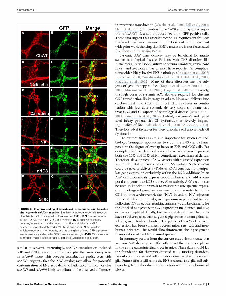
AAV8 MEDIATED TRANSGENE EXPRESSION IN MYENTERIC NEURONSUBTYPES AND ENTERIC GLIALMMP tissues from scAAV8-CB-GFP were fluorescentlyimmunolabeled for GFP, HuD, and VIP, nNOS, calretinin,calbindin, and ChAT. Similarly to scAAV9, scAAV8 mediatedGFP expression occurred in ChAT, calretinin and calbindinpositive myenteric neurons (Figures 6A–I). Additionally, robustGFP expression was detected in calretinin and calbindin positiveinterganglionic fibers. Contrary to scAAV9, GFP expression wasalso detected in VIP and nNOS expressing inhibitory motorneurons with limited co-expression in fibers (Figures 6J–O).Furthermore, GFP expression was sparingly detected inintraganglionic S100 positive EGCs (Figures 6P–R).
DISCUSSIONPrior to this work, AAV transduction of the GI tract and theENS was not well characterized (Fu et al., 2011; Rahim et al.,2011; Mattar et al., 2013; Schuster et al., 2014). Understandingof AAV transduction in the ENS is essential for therapeutic
Frontiers in Molecular Neuroscience www.frontiersin.org October 2014 | Volume 7 | Article 81 | 5
Gombash et al. AAV9 targets the myenteric plexus
FIGURE 3 | Quantification of GFP expressing myenteric neurons. (A) Totaltransduced and untransduced neuron counts in each gastrointestinal regionin mice injected either at P1 (neonates, n = 3) or P21 (juveniles, n = 3).Transduction of HuD positive neurons ranged from ∼25 to ∼43% in
neonatally injected mice. Transduction ranged from ∼42 to 57% in juvenileinjected mice. (B) Quantification of GFP myenteric neurons across the oral toaboral length of the colon revealed no transduction differences betweenproximal and distal colon segments in neonatal and juvenile injected mice.
development in neurological disease, FGIMD, GI neuromuscu-lar disease, FGID’s, IBS, diarrheal diseases and IBD. Here weexamined transduction of the myenteric plexus following sys-temic delivery of scAAV9 and additional serotypes scAAV1, 5, 6,and 8. scAAV9 transduced myenteric neurons along the entire GItract when systemically administered to neonate or juvenile mice.Dosing was not controlled for body weight across ages thereforecomparisons of transduction efficiency dependent on age cannotbe made. Despite that, scAAV9 transduction was robust whenadministered at either age, achieving transduction of 25–57%of myenteric neurons across various regions of the GI tract.Structural variations along the colon did not affect transduc-tion in the myenteric plexus. Furthermore, we report that AAVserotypes that efficiently traverse the BBB in the CNS are neces-sary but may not be sufficient for myenteric neuron transduction(Figure 5 and Supplementary Figure 3).
The extensive overlap of the more than 40 neurotransmittersidentified in the ENS limits interpretation of the cells targetedby AAV (Furness, 2012). However, we examined colocaliza-tion between GFP and known ENS markers including ChAT,
VIP, nNOS, calbindin, calretinin, and S100. scAAV9 transgeneexpression was more common in neurons involved in excitatoryneurotransmission (ChAT), sensation (calbindin), and ascend-ing signaling (calretinin). In general, viral tropism is dictated bythe capsid and expression cassette and is somewhat malleable.For example, substitution of a GFAP promoter for the ubiq-uitous chicken β-actin (CB) promoter in scAAV9 was effectiveat targeting enteric glia (Supplementary Figure 2). Because theCB promoter produces robust expression in CNS astrocytes butnot in enteric glia, this finding further emphasizes that entericglia are similar yet distinct from CNS astrocytes (Gulbransenand Sharkey, 2012; McClain et al., 2014). Importantly, theterm “enteric glia” encompasses multiple subtypes of glial cellswith divergent morphologies and localities within the gut wall(Gulbransen and Sharkey, 2012). Cellular properties of entericglia subtypes, their interactions with ENS vasculature, and howthose factors compare to CNS astrocytes is not well understood.Functional and physiological disparities from CNS astrocytes mayexplain contrasting efficiencies in glial cell transduction withinthe ENS and CNS following juvenile AAV9 systemic delivery. The
Frontiers in Molecular Neuroscience www.frontiersin.org October 2014 | Volume 7 | Article 81 | 6
Gombash et al. AAV9 targets the myenteric plexus
FIGURE 4 | Chemical coding of transduced myenteric cells in the colon
after systemic scAAV9-CB-GFP injection. GFP expression (B,E,H,K,N,Q)
was detected in choline acetyltransferase (ChAT, A,C) positive, calbindinpositive (D,F), and calretinin positive (G,I) myenteric neurons and someintraganglioninc fibers. ChAT staining is indicative of excitatory motorneurons and ascending and descending projecting interneurons. Calbindinand calretinin calcium binding proteins are indicative of excitatory motorneurons and some classes of interneurons. Transduction was rare orcompletely absent in vasoactive intestinal peptide (VIP, J,L) positive orneuronal nitric oxide synthase (nNOS, M,O) positive cells. VIP and nNOSstaining is associated with inhibitory motor neuron and descendinginterneurons. GFP did not co-localize with S100 positive glial cells (P,R).Arrows indicate co-expression while arrowheads indicate no GFPexpression. Scale bars are 100 μm.
FIGURE 5 | Transduction efficiency in the myenteric plexus is
dependent on AAV serotype. Neonatal mice were intravenously injectedwith scAAV1 (A), scAAV5 (B), scAAV6 (C), scAAV8 (D), or scAAV9-CB-GFP(E) (final dose 5 × 10¹⁰ vg/ml). Green fluorescent protein (GFP, green)immunofluorescence in the myenteric plexus of the colon reveled absent tominimal GFP expression in neurons (HuD, red) following scAAV1, scAAV5,and scAAV6 intravenous injection. Robust GFP expression was detected inneurons in scAAV8 (D) and scAAV9 (E) injected mice. Total enumerationcounts of neurons in the myenteric plexus of the colon revealed that 19.7 ±0.9% or 16.0 ± 0.2% of neurons were transduced in scAAV8 and scAAV9injected mice, respectively (F, ∗p < 0.001). Scale bars are 100 μm.
emerging role of enteric glia in the tripartite synapse of the ENS asin CNS, in GI motility, and their likely contribution to GI motilitydisorders and gut inflammation make them an important targetwithin the ENS and merits further investigation (Lomax et al.,2005; Ren et al., 2011; Gulbransen and Sharkey, 2012; McClainet al., 2014). Further variation in enteric cell tropism is providedby changing the AAV capsid.
Despite all of the tested serotypes having neuronal tropismsin the CNS, only scAAV8 had robust myenteric transduction
Frontiers in Molecular Neuroscience www.frontiersin.org October 2014 | Volume 7 | Article 81 | 7
Gombash et al. AAV9 targets the myenteric plexus
FIGURE 6 | Chemical coding of transduced myenteric cells in the colon
after systemic scAAV8 injection. Similarly to scAAV9, systemic injectionof scAAV8-CB-GFP produced GFP expression (B,E,H,K,N,Q) was detectedin ChAT (A–C), calbindin (D–F), and calretinin (G–I) positive excitatoryneurons, interneurons and intraganglionic fibers. Additionally, GFPexpression was also detected in VIP (J–L) and nNOS (M–O) positiveinhibitory neurons, interneurons, and intraganglionic fibers. GFP expressionwas occasionally detected in S100 positive enteric glia (P–R). White arrowsin merged images indicate transduced cells. Scale bars are 100 μm.
similar to scAAV9. Interestingly, scAAV8 transduction includedVIP and nNOS neurons and enteric glia that were rarely seenin scAAV9 tissue. This broader transduction profile seen withscAAV8 suggests that the AAV catalog may allow for powerfulcustomization of ENS gene delivery. Differences in receptors forscAAV8 and scAAV9 likely contribute to the observed differences
in myenteric transduction (Akache et al., 2006; Bell et al., 2011;Shen et al., 2011). In contrast to scAAV8 and 9, systemic injec-tion of scAAV1, 5, and 6 produced few to no GFP positive cells.These data suggest that vascular escape is a requirement for AAVmediated myenteric neuron transduction and is in agreementwith prior work showing that ENS vasculature is not fenestrated(Gershon and Bursztajn, 1978).
Systemic AAV gene delivery may be beneficial for multi-system neurological disease. Patients with CNS disorders likeAlzheimer’s, Parkinson’s, autism spectrum disorders, spinal cordinjury and neuromuscular diseases have reported GI complica-tions which likely involve ENS pathology (Anderson et al., 2007;Buie et al., 2010; Wakabayashi et al., 2010; Natale et al., 2011;Mazurek et al., 2012). Many of these disorders are the sub-jects of gene therapy studies (Kaplitt et al., 2007; Foust et al.,2010; Muramatsu et al., 2010; Garg et al., 2013). Currently,the high doses of systemic AAV delivery required for efficientCNS transduction limits usage in adults. However, delivery intocerebrospinal fluid (CSF) or direct CNS injection in combi-nation with low dose systemic delivery could simultaneouslytreat CNS and GI aspects of neurological disease (Bevan et al.,2011; Samaranch et al., 2012). Indeed, Parkinson’s and spinalcord injury patients list GI dysfunction as severely impact-ing quality of life (Sakakibara et al., 2001; Anderson, 2004).Therefore, ideal therapies for these disorders will also remedy GIdysfunction.
The current findings are also important for studies of ENSbiology. Transgenic approaches to study the ENS can be ham-pered by the degree of overlap between ENS and CNS cells. Forexample, most cre drivers designed for nervous tissue express inboth the CNS and ENS which complicates experimental design.Therefore, development of AAV vectors with restricted expressionwould be useful in basic studies of ENS biology. Such a vectorcould be used to deliver a cDNA or RNAi construct to manipu-late gene expression exclusively within the ENS. Additionally, anAAV can exogenously express cre-recombinase and add a tem-poral component to ENS studies. Alternatively, AAV vectors canbe used in knockout animals to maintain tissue specific expres-sion of a targeted gene. Gene expression can be restricted to theCNS by intracerebroventricular (ICV) injection. ICV injectionin mice results in minimal gene expression in peripheral tissues.Following ICV injection, resulting animals would be chimeric forthe knocked out gene; with CNS expression maintained and ENSexpression depleted. Finally, the current data can likely be trans-lated to other species, such as guinea pig or non-human primates,where genetic tools are limited. The pattern of scAAV9 transgeneexpression has been consistent across mice, rats, cats and non-human primates. This would allow fluorescent labeling or geneticmanipulation of the ENS in novel species.
In summary, results from the current study demonstrate thatsystemic AAV delivery can efficiently target the myenteric plexusin the entire gastrointestinal tract in mice. These data should laythe foundation for therapies directed at GI motility disorders,neurological disease and inflammatory diseases affecting entericglia. Future efforts will refine the ENS neuronal and glial cell sub-types targeted and evaluate transduction within the submucosalplexus.
Frontiers in Molecular Neuroscience www.frontiersin.org October 2014 | Volume 7 | Article 81 | 8
Gombash et al. AAV9 targets the myenteric plexus
AUTHOR CONTRIBUTIONSKevin D. Foust, Fedias L. Christofi, and Sara E. Gombash con-ceived and designed the studies. Sara E. Gombash, Christopher J.Cowley, and Julie A. Fitzgerald performed the studies. ChristianMueller and Jodie C. E. Hall helped in vector developmentand production. Sara E. Gombash and Kevin D. Foust wrotethe manuscript. All authors contributed to the editing andpreparation of the manuscript.
ACKNOWLEDGMENTSSara E. Gombash is supported by NIH T32# 5T32NS077984-02. We are grateful to Phil Popovich for his discussions on themanuscript.
SUPPLEMENTARY MATERIALThe Supplementary Material for this article can be foundonline at: http://www.frontiersin.org/journal/10.3389/fnmol.2014.00081/abstractSupplementary Figure 1 | GFP expression is confined to myenteric
neurons. Systemic injection of scAAV9-CB-GFP resulted in transduction of
myenteric neurons in both neonatal (P1, A–E) and juvenile (P21, F–J)
injected mice. Co-expression in neurons but not enteric glia was
confirmed by immunohistochemistry for GFP (A,F), S100 identifying
enteric glia (B,G), and HuD identifying myenteric neurons (D,I). Merged
images show GFP expression in neurons only (C,H,E,J). Arrows show
GFP expression in neurons that does no coincide with S100 glia staining.
Scale bars are 100 or 50 μm in insets.
Supplementary Figure 2 | Altered myenteric cell type transduction
following change in AAV promoter. GFP expression (A) was detected
exclusively in S100 positive (B,C) myenteric glial cells following
intravenous administration of ssAAV9-GFAP-GFP.
Supplementary Figure 3 | AAV Transduction in the Brain and Spinal Cord
following intravenous injection. GFP immunofluorescence was detected
in neurons (NeuN, cyan) and astrocytes [glial fibrillary acidic protein
(GFAP), red] in the brains and spinal cords of scAAV1 (A,F), scAAV6 (C,H),
scAAV8 (D,I), and scAAV9 (E,J) CB-GFP intravenously injected mice. No
CNS transduction occurred in scAAV5 (B,G) injected animals. Arrowheads
indicate transduced neurons (co-labeling with NeuN) and arrows indicate
transduced astrocytes (co-labeled with GFAP). Scale bars are 100 μm.
REFERENCESAhmed, S. S., Li, H., Cao, C., Sikoglu, E. M., Denninger, A. R., Su, Q., et al. (2013).
A single intravenous rAAV injection as late as P20 achieves efficacious and sus-tained CNS Gene therapy in Canavan mice. Mol. Ther. 21, 2136–2147. doi:10.1038/mt.2013.138
Akache, B., Grimm, D., Pandey, K., Yant, S. R., Xu, H., and Kay, M. A. (2006).The 37/67-kilodalton laminin receptor is a receptor for adeno-associatedvirus serotypes 8, 2, 3, and 9. J. Virol. 80, 9831–9836. doi: 10.1128/JVI.00878-06
Anderson, G., Noorian, A. R., Taylor, G., Anitha, M., Bernhard, D., Srinivasan, S.,et al. (2007). Loss of enteric dopaminergic neurons and associated changes incolon motility in an MPTP mouse model of Parkinson’s disease. Exp. Neurol.207, 4–12. doi: 10.1016/j.expneurol.2007.05.010
Anderson, K. D. (2004). Targeting recovery: priorities of the spinal cord-injured population. J. Neurotrauma 21, 1371–1383. doi: 10.1089/neu.2004.21.1371
Bell, C. L., Vandenberghe, L. H., Bell, P., Limberis, M. P., Gao, G. P., Van Vliet,K., et al. (2011). The AAV9 receptor and its modification to improve in vivolung gene transfer in mice. J. Clin. Invest. 121, 2427–2435. doi: 10.1172/JCI57367
Bergner, A. J., Stamp, L. A., Gonsalvez, D. G., Allison, M. B., Olson, D. P., Myers,M. G. Jr., et al. (2014). Birthdating of myenteric neuron subtypes in thesmall intestine of the mouse. J. Comp. Neurol. 522, 514–527. doi: 10.1002/cne.23423
Bevan, A. K., Duque, S., Foust, K. D., Morales, P. R., Braun, L., Schmelzer, L., et al.(2011). Systemic gene delivery in large species for targeting spinal cord, brain,and peripheral tissues for pediatric disorders. Mol. Ther. 19, 1971–1980. doi:10.1038/mt.2011.157
Buie, T., Campbell, D. B., Fuchs, G. J. 3rd., Furuta, G. T., Levy, J., Vandewater, J.,et al. (2010). Evaluation, diagnosis, and treatment of gastrointestinal disordersin individuals with ASDs: a consensus report. Pediatrics 125(Suppl. 1), S1–S18.doi: 10.1542/peds.2009-1878C
Burger, C., Gorbatyuk, O. S., Velardo, M. J., Peden, C. S., Williams, P., Zolotukhin,S., et al. (2004). Recombinant AAV viral vectors pseudotyped with viral cap-sids from serotypes 1, 2, and 5 display differential efficiency and cell tropismafter delivery to different regions of the central nervous system. Mol. Ther. 10,302–317. doi: 10.1016/j.ymthe.2004.05.024
Camilleri, M., Cowen, T., and Koch, T. R. (2008). Enteric neurodegeneration inageing. Neurogastroenterol. Motil. 20, 418–429. doi: 10.1111/j.1365-2982.2008.01134.x
Camilleri, M., Dubois, D., Coulie, B., Jones, M., Kahrilas, P. J., Rentz, A. M.,et al. (2005). Prevalence and socioeconomic impact of upper gastrointesti-nal disorders in the United States: results of the US Upper GastrointestinalStudy. Clin. Gastroenterol. Hepatol. 3, 543–552. doi: 10.1016/S1542-3565(05)00153-9
Cirillo, C., Sarnelli, G., Turco, F., Mango, A., Grosso, M., Aprea, G., et al. (2011).Proinflammatory stimuli activates human-derived enteroglial cells and inducesautocrine nitric oxide production. Neurogastroenterol. Motil. 23, e372–e382. doi:10.1111/j.1365-2982.2011.01748.x
Cornet, A., Savidge, T. C., Cabarrocas, J., Deng, W. L., Colombel, J. F., Lassmann,H., et al. (2001). Enterocolitis induced by autoimmune targeting of enteric glialcells: a possible mechanism in Crohn’s disease? Proc. Natl. Acad. Sci. U.S.A. 98,13306–13311. doi: 10.1073/pnas.231474098
Costa, M., Furness, J. B., and Gibbins, I. L. (1986). Chemical coding ofenteric neurons. Prog. Brain Res. 68, 217–239. doi: 10.1016/S0079-6123(08)60241-1
de Leeuw, C. N., Dyka, F. M., Boye, S. L., Laprise, S., Zhou, M., Chou, A. Y., et al.(2014). Targeted CNS delivery using human minipromoters and demonstratedcompatibility with adeno-associated viral vectors. Mol. Ther. Methods Clin. Dev.1:5. doi: 10.1038/mtm.2013.5
Derkinderen, P., Rouaud, T., Lebouvier, T., Bruley des Varannes, S., Neunlist,M., and De Giorgio, R. (2011). Parkinson disease: the enteric nervous sys-tem spills its guts. Neurology 77, 1761–1767. doi: 10.1212/WNL.0b013e318236ef60
Duque, S., Joussemet, B., Riviere, C., Marais, T., Dubreil, L., Douar, A. M.,et al. (2009). Intravenous administration of self-complementary AAV9 enablestransgene delivery to adult motor neurons. Mol. Ther. 17, 1187–1196. doi:10.1038/mt.2009.71
El-Rifai, N., Daoud, N., Tayyarah, K., Baydoun, A., and Jaubert, F. (2006).Neuronal intranuclear inclusion disease presenting as chronic intestinalpseudo-obstruction in the neonatal period in the absence of neu-rologic symptoms. J. Pediatr. Gastroenterol. Nutr. 42, 321–323. doi:10.1097/01.mpg.0000189331.39527.0b
Foust, K. D., Nurre, E., Montgomery, C. L., Hernandez, A., Chan, C. M.,and Kaspar, B. K. (2009). Intravascular AAV9 preferentially targets neona-tal neurons and adult astrocytes. Nat. Biotechnol. 27, 59–65. doi: 10.1038/nbt.1515
Foust, K. D., Salazar, D. L., Likhite, S., Ferraiuolo, L., Ditsworth, D., Ilieva, H., et al.(2013). Therapeutic AAV9-mediated suppression of mutant SOD1 slows diseaseprogression and extends survival in models of inherited ALS. Mol. Ther. 21,2148–2159. doi: 10.1038/mt.2013.211
Foust, K. D., Wang, X., McGovern, V. L., Braun, L., Bevan, A. K., Haidet, A. M.,et al. (2010). Rescue of the spinal muscular atrophy phenotype in a mousemodel by early postnatal delivery of SMN. Nat. Biotechnol. 28, 271–274. doi:10.1038/nbt.1610
Fu, H., Dirosario, J., Killedar, S., Zaraspe, K., and McCarty, D. M. (2011).Correction of neurological disease of mucopolysaccharidosis IIIB in adult miceby rAAV9 trans-blood-brain barrier gene delivery. Mol. Ther. 19, 1025–1033.doi: 10.1038/mt.2011.34
Frontiers in Molecular Neuroscience www.frontiersin.org October 2014 | Volume 7 | Article 81 | 9
Gombash et al. AAV9 targets the myenteric plexus
Fukudo, S., Kuwano, H., and Miwa, H. (2012). Management and pathophys-iology of functional gastrointestinal disorders. Digestion 85, 85–89. doi:10.1159/000334652
Furness, J. B. (2000). Types of neurons in the enteric nervous system. J. Auton. Nerv.Syst. 81, 87–96. doi: 10.1016/S0165-1838(00)00127-2
Furness, J. B. (2012). The enteric nervous system and neurogastroenterology. Nat.Rev. Gastroenterol. Hepatol. 9, 286–294. doi: 10.1038/nrgastro.2012.32
Furness, J. B., Alex, G., Clark, M. J., and Lal, V. V. (2003). Morphologies andprojections of defined classes of neurons in the submucosa of the guinea-pigsmall intestine. Anat. Rec. A Discov. Mol. Cell. Evol. Biol. 272, 475–483. doi:10.1002/ar.a.10064
Gabbard, S. L., and Lacy, B. E. (2013). Chronic intestinal pseudo-obstruction. Nutr.Clin. Pract. 28, 307–316. doi: 10.1177/0884533613485904
Garg, S. K., Lioy, D. T., Cheval, H., McGann, J. C., Bissonnette, J. M., Murtha, M. J.,et al. (2013). Systemic delivery of MeCP2 rescues behavioral and cellular deficitsin female mouse models of Rett syndrome. J. Neurosci. 33, 13612–13620. doi:10.1523/JNEUROSCI.1854-13.2013
Gershon, M. D., and Bursztajn, S. (1978). Properties of the enteric nervoussystem: limitation of access of intravascular macromolecules to the myen-teric plexus and muscularis externa. J. Comp. Neurol. 180, 467–488. doi:10.1002/cne.901800305
Gombash Lampe, S. E., Kaspar, B. K., and Foust, K. D. (in press). Intravenousinjections in neonatal mice. J. Vis. Exp.
Gulbransen, B. D., and Sharkey, K. A. (2012). Novel functional roles for enteric gliain the gastrointestinal tract. Nat. Rev. Gastroenterol. Hepatol. 9, 625–632. doi:10.1038/nrgastro.2012.138
Harrington, A. M., Hutson, J. M., and Southwell, B. R. (2010). Cholinergic neu-rotransmission and muscarinic receptors in the enteric nervous system. Prog.Histochem. Cytochem. 44, 173–202. doi: 10.1016/j.proghi.2009.10.001
Hasler, W. L., Kurosawa, S., and Chung, O. Y. (1990). Regional cholinergic differ-ences between distal and proximal colonic myenteric plexus. Am. J. Physiol. 258,G404–G410.
Haurigot, V., Marco, S., Ribera, A., Garcia, M., Ruzo, A., Villacampa, P., et al.(2013). Whole body correction of mucopolysaccharidosis IIIA by intracere-brospinal fluid gene therapy. J. Clin. Invest. 123, 3254–3271. doi: 10.1172/JCI66778
Kaplitt, M. G., Feigin, A., Tang, C., Fitzsimons, H. L., Mattis, P., Lawlor, P. A., et al.(2007). Safety and tolerability of gene therapy with an adeno-associated virus(AAV) borne GAD gene for Parkinson’s disease: an open label, phase I trial.Lancet 369, 2097–2105. doi: 10.1016/S0140-6736(07)60982-9
Lawlor, P. A., Bland, R. J., Mouravlev, A., Young, D., and During, M. J. (2009).Efficient gene delivery and selective transduction of glial cells in the mam-malian brain by AAV serotypes isolated from nonhuman primates. Mol. Ther.17, 1692–1702. doi: 10.1038/mt.2009.170
Lomax, A. E., Fernandez, E., and Sharkey, K. A. (2005). Plasticity of the entericnervous system during intestinal inflammation. Neurogastroenterol. Motil. 17,4–15. doi: 10.1111/j.1365-2982.2004.00607.x
Lomax, A. E., and Furness, J. B. (2000). Neurochemical classification of entericneurons in the guinea-pig distal colon. Cell Tissue Res. 302, 59–72. doi:10.1007/s004410000260
Mattar, C. N., Waddington, S. N., Biswas, A., Johana, N., Ng, X. W., Fisk, A. S., et al.(2013). Systemic delivery of scAAV9 in fetal macaques facilitates neuronal trans-duction of the central and peripheral nervous systems. Gene Ther. 20, 69–83.doi: 10.1038/gt.2011.216
Mazurek, M. O., Vasa, R. A., Kalb, L. G., Kanne, S. M., Rosenberg, D., Keefer,A., et al. (2012). Anxiety, sensory over-responsivity, and gastrointestinal prob-lems in children with autism spectrum disorders. J. Abnorm. Child Psychol. 41,165–176. doi: 10.1007/s10802-012-9668-x
McClain, J. L., Grubisic, V., Fried, D., Gomez-Suarez, R. A., Leinninger, G.M., Sevigny, J., et al. (2014). Ca2+ responses in enteric glia are medi-ated by connexin-43 hemichannels and modulate colonic transit in mice.Gastroenterology 146, 497–507.e1. doi: 10.1053/j.gastro.2013.10.061
Muramatsu, S., Fujimoto, K., Kato, S., Mizukami, H., Asari, S., Ikeguchi, K., et al.(2010). A phase I study of aromatic L-amino acid decarboxylase gene therapyfor Parkinson’s disease. Mol. Ther. 18, 1731–1735. doi: 10.1038/mt.2010.135
Natale, G., Pasquali, L., Paparelli, A., and Fornai, F. (2011). Parallel mani-festations of neuropathologies in the enteric and central nervous systems.Neurogastroenterol. Motil. 23, 1056–1065. doi: 10.1111/j.1365-2982.2011.01794.x
Rahim, A. A., Wong, A. M., Hoefer, K., Buckley, S. M., Mattar, C. N., Cheng, S. H.,et al. (2011). Intravenous administration of AAV2/9 to the fetal and neonatalmouse leads to differential targeting of CNS cell types and extensive trans-duction of the nervous system. FASEB J. 25, 3505–3518. doi: 10.1096/fj.11-182311
Ren, T., Grants, I., Alhaj, M., McKiernan, M., Jacobson, M., Hassanain, H. H.,et al. (2011). Impact of disrupting adenosine A(3) receptors (A(3)(-)/(-) AR)on colonic motility or progression of colitis in the mouse. Inflamm. Bowel Dis.17, 1698–1713. doi: 10.1002/ibd.21553
Saito, Y. A., Schoenfeld, P., and Locke, G. R. 3rd. (2002). The epidemiology of irrita-ble bowel syndrome in North America: a systematic review. Am. J. Gastroenterol.97, 1910–1915. doi: 10.1016/S0002-9270(02)04270-3
Sakakibara, R., Shinotoh, H., Uchiyama, T., Sakuma, M., Kashiwado, M.,Yoshiyama, M., et al. (2001). Questionnaire-based assessment of pelvicorgan dysfunction in Parkinson’s disease. Auton. Neurosci. 92, 76–85. doi:10.1016/S1566-0702(01)00295-8
Samaranch, L., Salegio, E. A., San Sebastian, W., Kells, A. P., Foust, K. D., Bringas,J. R., et al. (2012). Adeno-associated virus serotype 9 transduction in the cen-tral nervous system of nonhuman primates. Hum. Gene Ther. 23, 382–389. doi:10.1089/hum.2011.200
Sanchez, M. I., and Bercik, P. (2011). Epidemiology and burden of chronicconstipation. Can. J. Gastroenterol. 25(Suppl B), 11B–15B.
Savidge, T. C., Newman, P., Pothoulakis, C., Ruhl, A., Neunlist, M.,Bourreille, A., et al. (2007). Enteric glia regulate intestinal bar-rier function and inflammation via release of S-nitrosoglutathione.Gastroenterology 132, 1344–1358. doi: 10.1053/j.gastro.2007.01.051
Schuster, D. J., Dykstra, J. A., Riedl, M. S., Kitto, K. F., Belur, L. R., McIvor, R. S.,et al. (2014). Biodistribution of adeno-associated virus serotype 9 (AAV9) vectorafter intrathecal and intravenous delivery in mouse. Front. Neuroanat. 8:42. doi:10.3389/fnana.2014.00042
Shen, S., Bryant, K. D., Brown, S. M., Randell, S. H., and Asokan, A.(2011). Terminal N-linked galactose is the primary receptor for adeno-associated virus 9. J. Biol. Chem. 286, 13532–13540. doi: 10.1074/jbc.M110.210922
Sibaev, A., Franck, H., Vanderwinden, J. M., Allescher, H. D., and Storr, M. (2003).Structural differences in the enteric neural network in murine colon: impact onelectrophysiology. Am. J. Physiol. Gastrointest. Liver Physiol. 285, G1325–G1334.doi: 10.1152/ajpgi.00506.2002
Tatom, J. B., Wang, D. B., Dayton, R. D., Skalli, O., Hutton, M. L., Dickson, D. W.,et al. (2009). Mimicking aspects of frontotemporal lobar degeneration and LouGehrig’s disease in rats via TDP-43 overexpression. Mol. Ther. 17, 607–613. doi:10.1038/mt.2009.3
Taymans, J. M., Vandenberghe, L. H., Haute, C. V., Thiry, I., Deroose, C. M.,Mortelmans, L., et al. (2007). Comparative analysis of adeno-associated viralvector serotypes 1, 2, 5, 7, and 8 in mouse brain. Hum. Gene Ther. 18, 195–206.doi: 10.1089/hum.2006.178
Turco, F., Sarnelli, G., Cirillo, C., Palumbo, I., De Giorgi, F., D’alessandro, A.,et al. (2014). Enteroglial-derived S100B protein integrates bacteria-inducedToll-like receptor signalling in human enteric glial cells. Gut 63, 105–115. doi:10.1136/gutjnl-2012-302090
Vijayaraghavan, S. (2009). Glial-neuronal interactions–implications for plastic-ity and drug addiction. AAPS J. 11, 123–132. doi: 10.1208/s12248-009-9085-4
von Jonquieres, G., Mersmann, N., Klugmann, C. B., Harasta, A. E., Lutz, B.,Teahan, O., et al. (2013). Glial promoter selectivity following AAV-deliveryto the immature brain. PLoS ONE 8:e65646. doi: 10.1371/journal.pone.0065646
Wakabayashi, K., Mori, F., Tanji, K., Orimo, S., and Takahashi, H.(2010). Involvement of the peripheral nervous system in synucle-inopathies, tauopathies and other neurodegenerative proteinopathiesof the brain. Acta Neuropathol. 120, 1–12. doi: 10.1007/s00401-010-0706-x
Wang, C. H., Finkel, R. S., Bertini, E. S., Schroth, M., Simonds, A., Wong, B., et al.(2007). Consensus statement for standard of care in spinal muscular atrophy.J. Child Neurol. 22, 1027–1049. doi: 10.1177/0883073807305788
Wiskur, B., and Greenwood-Van Meerveld, B. (2010). The aging colon: the roleof enteric neurodegeneration in constipation. Curr. Gastroenterol. Rep. 12,507–512. doi: 10.1007/s11894-010-0139-7
Frontiers in Molecular Neuroscience www.frontiersin.org October 2014 | Volume 7 | Article 81 | 10
Gombash et al. AAV9 targets the myenteric plexus
Wood, J. D. (2000). Neuropathy in the brain-in-the-gut. Eur. J.Gastroenterol. Hepatol. 12, 597–600. doi: 10.1097/00042737-200012060-00002
Yeung, A. K., and Di Lorenzo, C. (2012). Primary gastrointestinal motility disordersin childhood. Minerva Pediatr. 64, 567–584.
Zhang, H., Yang, B., Mu, X., Ahmed, S. S., Su, Q., He, R., et al. (2011). SeveralrAAV vectors efficiently cross the blood-brain barrier and transduce neuronsand astrocytes in the neonatal mouse central nervous system. Mol. Ther. 19,1440–1448. doi: 10.1038/mt.2011.98
Conflict of Interest Statement: The authors declare that the research was con-ducted in the absence of any commercial or financial relationships that could beconstrued as a potential conflict of interest.
Received: 06 August 2014; accepted: 22 September 2014; published online: 15 October2014.Citation: Gombash SE, Cowley CJ, Fitzgerald JA, Hall JCE, Mueller C, Christofi FLand Foust KD (2014) Intravenous AAV9 efficiently transduces myenteric neurons inneonate and juvenile mice. Front. Mol. Neurosci. 7:81. doi: 10.3389/fnmol.2014.00081This article was submitted to the journal Frontiers in Molecular Neuroscience.Copyright © 2014 Gombash, Cowley, Fitzgerald, Hall, Mueller, Christofi and Foust.This is an open-access article distributed under the terms of the Creative CommonsAttribution License (CC BY). The use, distribution or reproduction in other forums ispermitted, provided the original author(s) or licensor are credited and that the originalpublication in this journal is cited, in accordance with accepted academic practice.No use, distribution or reproduction is permitted which does not comply with theseterms.
Frontiers in Molecular Neuroscience www.frontiersin.org October 2014 | Volume 7 | Article 81 | 11
Copyright © 2022 FDOKUMEN