A study of the quality of ginseng containing products in Nairobi ...
153
i A STUDY OF THE QUALITY OF GINSENG CONTAINING PRODUCTS IN NAIROBI, KENYA A thesis submitted in partial fulfillment of the requirements for the award of the degree of Masters of Science in Pharmacognosy and Complementary Medicine BY RONALD MWENDE INYANGALA B. Pharm. (Nairobi) U59/71906/08 Department of Pharmacology and Pharmacognosy School of Pharmacy UNIVERSITY OF NAIROBI 2012
-
Upload
khangminh22 -
Category
Documents
-
view
1 -
download
0
Transcript of A study of the quality of ginseng containing products in Nairobi ...

i
A STUDY OF THE QUALITY OF GINSENG CONTAINING
PRODUCTS IN NAIROBI, KENYA
A thesis submitted in partial fulfillment of the requirements for the
award of the degree of Masters of Science in Pharmacognosy and
Complementary Medicine
BY
RONALD MWENDE INYANGALA
B. Pharm. (Nairobi)
U59/71906/08
Department of Pharmacology and Pharmacognosy
School of Pharmacy
UNIVERSITY OF NAIROBI
2012

ii
DECLARATION
DECLARATION BY THE CANDIDATE
I hereby declare that this thesis is my original work and has not been presented for a
degree award in any other institution.
______________________ __________________
Ronald Mwende Inyangala Date
DECLARATION BY THE SUPERVISORS
This thesis has been submitted for examination with our approval as University
Supervisors
__________________________ __________________
Prof. I.O. Kibwage, PhD Date
Professor of Pharmaceutical Chemistry
Department of Pharmaceutical Chemistry
School of Pharmacy, University of Nairobi
________________________ __________________
Dr. K.O. Abuga, PhD Date
Department of Pharmaceutical Chemistry
School of Pharmacy, University of Nairobi
_______________________ __________________
Dr. M.O. Oluka, Msc Date
Department of Pharmacology and Pharmacognosy
School of Pharmacy, University of Nairobi

iii
DEDICATION
This thesis is dedicated to my children as it demonstrates that age and poor health are not
a limitation to acquiring knowledge. It is also dedicated to the Government of Kenya
especially Ministry of Medical Services which is responsible for regulation of herbal
medicines. Finally, I dedicate this to Kenyan researchers as a challenge that more research
is required in the field of herbal medicines and that this is achievable within our national
research framework.

iv
ACKNOWLEDGEMENTS
My heartfelt gratitude goes to my supervisors: Prof. I.O. Kibwage, Dr. K.O. Abuga and
Dr. M.O. Oluka upon whose fruitful directions I relied extensively.
I owe special thanks to Professor J. Mwangi of University of Nairobi and Dr. David
Olunya of Aga Khan University Hospital for effectively managing my meningioma, a
condition which I suffered while undertaking this study.
I appreciate the support I received from my colleagues at the School of Pharmacy, staff of
the National Quality Control Laboratory as well as the Pharmacognosy Laboratory and
Drug Analysis Research Unit (DARU) throughout the period of this research. I especially
express my gratitude to Mr. I.G. Mureithi for his advice and valuable information on the
operations of the densitometer.
I would like to thank the Pharmacy and Poisons Board, Kenya for sponsoring me to
undertake the Master of Science in Pharmacognosy and Complementary Medicine course
including financing this study.
I thank all the participants, my fellow pharmacists, pharmaceutical technologists and
nutritionists in Nairobi County for their cooperation that made this study successful.
Above all, I do acknowledge the Almighty God for all his blessings in my life.

v
TABLE OF CONTENTS
DECLARATION .................................................................................................................. i
DEDICATION ................................................................................................................... iii
LIST OF ABBREVIATIONS ........................................................................................... xiii
DEFINITION OF TERMS ................................................................................................ xvi
ABSTRACT ..................................................................................................................... xix
CHAPTER ONE .................................................................................................................. 1
1.1 Background to the Study ............................................................................................ 1
1.2 History of Ginseng Use ............................................................................................. 3
1.3 Taxonomy and Cultivation of Ginseng ...................................................................... 5
1.3.1 Asian White and Red Ginseng ............................................................................. 6
1.3.2 North American Ginseng ..................................................................................... 7
1.4 Chemical Constituents of Ginseng ............................................................................. 7
1.5 Pharmacological Effects of Ginseng .......................................................................... 9
1.5.1 Antioxidant Properties ....................................................................................... 10
1.5.2 Effect on Performance ....................................................................................... 10
1.5.3 Anti-Diabetic Effects ......................................................................................... 11
1.5.4 Immune Stimulatory Activities .......................................................................... 11
1.5.5 Impotency and Erectile Dysfunction .................................................................. 12
1.5.6 Psychomotor Activities ...................................................................................... 12
1.5.7 Effects on Haemolysis ....................................................................................... 12
1.5.8 Anti-Cancer Effects ........................................................................................... 13
1.5.9 Side Effects and Contraindications of Ginseng ................................................... 13
1.6 Ginseng Products in Kenya ...................................................................................... 14
1.7 Quality Control of Herbal Products .......................................................................... 15
1.7.1 Background ....................................................................................................... 15
1.7.2 Methods of Quality Control ............................................................................... 17
1.7.3 Use of Marker Compounds for Quality Control ................................................. 18
1.7.4 Quality Control of Ginseng Containing Products ............................................... 19
1.7.4.1 Detection and Analysis of Ginsenosides .......................................................... 20
1.7.4.2 Microbial Contamination of Ginseng .............................................................. 25
1.7.4.3 Microbial Identification .................................................................................. 27
1.8 Regulation of Herbal Medicines .............................................................................. 30

vi
1.9 Study Justification ................................................................................................... 36
1.10 Objectives of the Study .......................................................................................... 38
1.10.1 General Objectives ........................................................................................... 38
1.10.2 Specific Objectives .......................................................................................... 38
CHAPTER TWO ............................................................................................................... 39
METHODOLOGY ............................................................................................................ 39
2.1 Introduction ............................................................................................................. 39
2.2 Market Survey and Sampling ................................................................................... 39
2.2.1 Study Site .......................................................................................................... 39
2.2.2 Study Population Size or Sampling Frame ......................................................... 40
2.2.3 Study Design ..................................................................................................... 41
2.2.3.1 Sample Size Determination ............................................................................. 41
2.2.3.2 Sampling Procedure ........................................................................................ 41
2.2.4 Administration of Questionnaires ....................................................................... 42
2.2.5 Knowledge Assessment of Store Attendants ...................................................... 43
2.3 Quality Control Tests ............................................................................................... 44
2.3.1 Equipments ........................................................................................................ 44
2.3.3 Materials and Reagents ...................................................................................... 45
2.4 Methods .................................................................................................................. 47
2.4.1 Uniformity of Weight ........................................................................................ 47
2.4.2 Determination of Microbial Load ....................................................................... 47
2.4.2.1 Preparation of Nutrient Media ......................................................................... 47
2.4.2.2 Sample Preparation ......................................................................................... 48
2.4.2.3 Inoculation and Microbial Count ..................................................................... 49
2.4.2.4 Isolation and Storage of Pure Cultures ............................................................ 50
2.4.2.5 Microbial Identification by Staining and Microscopy ...................................... 50
2.4.2.6 Identification of Bacteria ................................................................................. 52
2.4.2.7 Bacteria Identification by API 20 E ................................................................. 53
2.4.3 TLC Analysis of Ginseng Samples and Standards ............................................... 55
2.4.3.1 Tablets ............................................................................................................ 55
2.4.3.2 Hard gelatin capsules ...................................................................................... 55
2.4.3.3 Soft gelatin capsules ....................................................................................... 55
2.4.3.4 Powdered Asian ginseng ................................................................................. 56
2.4.3.5 Radix ginseng extract ...................................................................................... 56

vii
2.4.3.6 Ginsenoside Rb1 and Rg1 ............................................................................... 56
2.4.3.7 TLC Analysis of ginseng samples ................................................................... 56
2.5 Data Analysis .......................................................................................................... 57
CHAPTER THREE ........................................................................................................... 58
RESULTS AND DISCUSSION ........................................................................................ 58
3.1 Results ..................................................................................................................... 58
3.2 Evaluation of Store Attendants’ Knowledge ............................................................ 58
3.2.1 Sample Size of Store Attendants ........................................................................ 58
3.2.2 Education Level of the Store Attendants ............................................................ 60
3.2.3 Attendants’ Knowledge on Ginseng products ..................................................... 61
3.2.4 Effects of Attendants Attributes on Ginseng Knowledge .................................... 62
3.2.5 Feedback on Use of Ginseng Products ............................................................... 63
3.3 Assessment of Label Information of Ginseng Samples ............................................. 64
3.3.1 Sample Size of Ginseng Products ....................................................................... 64
3.3.2 Evaluation of Label Information of Samples ...................................................... 64
3.3.3 Number of Active Ingredients per Sample/Product ............................................ 66
3.3.4 Country of Origin of the Samples ....................................................................... 67
3.4 Uniformity of Weight .............................................................................................. 70
3.5 Microbial Load ........................................................................................................ 70
3.5.1 Total Aerobic Microbial Count (AV.TAMC) ..................................................... 70
3.5.2 Total Combined Yeast and Mould Count (AV.TYMC) ...................................... 71
3.5.3 Total Viable Aerobic Microbial Count (TVAMC).............................................. 72
3.5.4 Compliance to Microbial Load by Country of Origin ......................................... 73
3.6 Positive Microbial Identification .............................................................................. 74
3.6.1 Gram Staining and Biochemical Characterization .............................................. 74
3.6.2 Positive Identifications for Microbial Contaminants ........................................... 75
3.7 Identification of Ginseng ......................................................................................... 78
3.7.1 Tablet Samples .................................................................................................. 78
3.7.2 Hard Gelatin Capsules Samples ......................................................................... 82
3.7.4 Occurrence of Ginsenosides in the Analyzed Samples ....................................... 85
3.8 Discussion ............................................................................................................... 86
3.9 Conclusion .............................................................................................................. 98
3.10 Recommendations ................................................................................................. 95
REFERENCES .................................................................................................................. 97

viii
APPENDICES ................................................................................................................. 109

ix
LIST OF TABLES
Table 1.1: Microbial growth on differential and selective media ................................................... 29
Table 1.2: Registered ginseng containing products in Kenya ......................................................... 36
Table 2.1: Retail outlets stocking ginseng per district ................................................................... 40
Table 2.2: Sampled ginseng retail outlets per district ..................................................................... 42
Table 2.3: Knowledge assessment of attendants ............................................................................. 43
Table 2.4: Quantity of nutrient media powder per 1000 ml of distilled water................................ 48
Table 2.5: Colony characteristics of bacteria and fungi .................................................................. 51
Table 3.1: Premises sampled per district ........................................................................................ 58
Table 3.2: Distribution of stores by type ........................................................................................ 59
Table 3.3: Effects of attendants attributes on knowledge about ginseng ........................................ 62
Table 3.4: Feedback from users of ginseng products ..................................................................... 63
Table 3.5: Sampled ginseng containing products per district ........................................................ 64
Table 3.6: Compliance to labeling requirements ............................................................................ 65
Table 3.7: Number of active ingredients per sample ...................................................................... 67
Table 3.8: Conformity to WHO labeling requirement by country of origin ................................... 68
Table 3.9: Average total aerobic microbial count (AV.TAMC) of the samples ............................ 70
Table 3.10: Average total combined yeast and mould count of the samples ................................. 72
Table 3.11: Total viable microbial count of the samples ............................................................... 73
Table 3.12: Compliance to microbial standards by country of origin ............................................ 74
Table 3.13: Numerical profiles of the microbes identified by API 20 E ........................................ 75
Table 3.14: Status of identified microbes by country of origin ...................................................... 78
Table 3.15: Rf and Rx values of ginsenosides of the tablet samples ............................................. 82
Table 3.16: Rf and Rx values of ginsenosides of the hard gelatin capsule samples ......................... 83
Table 3.17: Rf and Rx values of ginsenosides in the soft gelatin capsule samples .......................... 84
Table 3.18: Rf values of 8 ginsenoside (Courthout et al., 1999) ..................................................... 87

x
LIST OF FIGURES
Figure 1.1(a): Panax ginseng plant................................................................................... 5
Figure 1.1(b): Panax ginseng root .................................................................................... 5
Figure 1.2: Structures of 20(S)-protopanaxadiols and 20(S)-protopanaxatriols
ginsenosides .................................................................................................................... 8
Figure 1.3: TLC profile of ginsenosides occurring in Panax quinguefolium Root
(Ludwiczuk et al., 2006) ................................................................................................ 21
Figure 1.4: Densitogram of ginseng applied on a TLC plate (Courthout et al., 1999) ..... 22
Figure 1.5: Densitograms of ginsenosides occurring in ginseng root tissues ................... 24
Figure 1.6: Listed herbal products in Kenya by country of origin ................................... 33
Figure 1.7: Active ingredients per listed herbal product ................................................. 34
Figure 1.8: Therapeutic classification of listed herbal products ...................................... 35
Figure 3.1: Highest level of education attained by the store attendants ........................... 60
Figure 3.2: Distribution of attendants by professional training ....................................... 61
Figure 3.3 Compliance to Labeling Requirements Using all Characteristics ................... 66
Figure 3.4: Distribution of ginseng containing products by country of origin ................. 67
Figure 3.5: Occurrence of microbes in the sampled products .......................................... 76
Figure 3.6: Status of microbes identified in the sampled products .................................. 77
Figure 3.7: Densitogram of standard ginseng extract ...................................................... 79
Figure 3.8: Densitograms and Chromatograms of standard ginsenosides Rb1 and Rg1... 80
Figure 3.9: Typical Chromatograms of some samples and the standard ginseng extract .. 80
Figure 3.10: Densitogram of RMI 12 ............................................................................. 81
Figure 3.12: Presence of ginsenosides in the samples ..................................................... 85
Figure 3.13: Distribution of Rg1 and Rb1 ginsenosides in the samples ........................... 86

xi
LIST OF APPENDICES
Appendix 1: Listed Herbal Products in Kenya ............................................................. 109
Appendix 2: Questionnaire .......................................................................................... 112
Appendix 3: Interpretation of Chemical Reactions of Bacteria using API 20 E Strip .... 114
Appendix 4: Some Samples used in this Study ............................................................ 116
Appendix 5: Image of a Typical Ginseng Root used in this Study ................................ 116
Appendix 6: Typical Bacterial Colonies of RMI-19 ..................................................... 117
Appendix 7: Typical Fungal Colonies of RMI 37 ........................................................ 117
Appendix 8: TLC Densitogram for RMI 3 ................................................................... 118
Appendix 9: TLC Densitogram for RMI 4 ................................................................... 118
Appendix 10: TLC Densitogram for RMI 5 ................................................................. 119
Appendix 11: TLC Densitogram for RMI 6 ................................................................. 119
Appendix 12: TLC Densitogram for RMI 7 ................................................................. 120
Appendix 13: TLC Densitogram for RMI 8 ................................................................. 120
Appendix 14: TLC Densitogram for RMI 9 ................................................................. 121
Appendix 15: TLC Densitogram for RMI 10 ............................................................... 121
Appendix 16: TLC Densitogram for RMI 11 ............................................................... 122
Appendix 17: TLC Densitogram for RMI 13 ............................................................... 122
Appendix 18: TLC Densitogram for RMI 14 ............................................................... 123
Appendix 19: TLC Densitogram for RMI 16 ............................................................... 123
Appendix 20: TLC Densitogram for RMI 17 ............................................................... 124
Appendix 21: TLC Densitogram for RMI 18 ............................................................... 124
Appendix 22: TLC Densitogram for RMI 20 ............................................................... 125
Appendix 23: TLC Densitogram for RMI 23 ............................................................... 125
Appendix 24: TLC Densitogram for RMI 24 ............................................................... 126
Appendix 25: TLC Densitogram for RMI 26 ............................................................... 126
Appendix 26: TLC Densitogram for RMI 27 ............................................................... 127
Appendix 27: TLC Densitogram for RMI 28 ............................................................... 127
Appendix 28: TLC Densitogram for RMI 29 ............................................................... 128
Appendix 29: TLC Densitogram for RMI 30 ............................................................... 128
Appendix 30: TLC Densitogram for RMI 31 ............................................................... 129
Appendix 31: TLC Densitogram for RMI 32 .............................................................. 129
Appendix 32: TLC Densitogram for RMI 33 ............................................................... 130

xii
Appendix 33: TLC Densitogram for RMI 34 ............................................................... 130
Appendix 34: TLC Densitogram for RMI 35 ............................................................... 131
Appendix 35: TLC Densitogram for RMI 38 ............................................................... 131
Appendix 36: TLC Densitogram for RMI 39 ............................................................... 132
Appendix 37: TLC Densitogram for RMI 40 ............................................................... 132

xiii
LIST OF ABBREVIATIONS
ABC American Botanical Council
ADH Arginine Dihydrolase
AV. TAMC Average Total Aerobic Microbial Count
AV. TYMC Average Total Combined Yeast And Mould Count
BDS Botanical Dietary Supplements
BP British Pharmacopoeia
CDR Committee for Drug Registration
CFU Colony forming Units
CIT Trisodium Citrate
DARU Drug Analysis and Research Unit
EMB Ethyl Methyl Blue
FDA Food and Drug Administration
GEL Gelatin (bovine origin)
GEP Ginseng Evaluation Program
GLC Gas Liquid Chromatography
cGMP current Good Manufacturing Practices
HPLC High Performance Liquid Chromatography
IND Indole production test (L-tryptophane)
INN International Non Proprietary Name
IP Indian Pharmacopoeia
KEMRI Kenya Medical Research Institute
KNBS Kenya National Bureau of Statistics
KNDI Kenya Nutritionists and Dieticians Institute

xiv
KRA Kenya Revenue Authority
LDC Lysine Decarboxylase
MOH Ministry of Health
MOMS Ministry of Medical Services
MOPHS Ministry of Public Health and Sanitation
MSA Mannitol Salt Agar
ODC Ornithine Decarboxylase
OR
OTC
Odds Ratio
Over the Counter
PPB Pharmacy and Poisons Board
QC Quality Control
SDA Sabouroud Dextrose Agar
SSA Salmonella Shigella Agar
TAMC Total Aerobic Microbial Count
TDA Trytophane Deaminase
TK Traditional Knowledge
TLC Thin Layer Chromatography
TM Traditional Medicine
TNF- α Tumor Necrosis Factor- α
TVAMC Total Average Aerobic Microbial Count
UK United Kingdom
USA United States of America
USD United States of America Dollar
USP United States Pharmacopoeia
UTI Urinary Tract Infection

xv
VP Acetoin Production
WHO World Health Organization
XLD Xylose-Lysine Deoxycholate Agar

xvi
DEFINITION OF TERMS
“Yang” effects- Represent brightness, active, upward, hot, expanding and strong.
“Yin” effects- Represents darkness, passiveness, downward, cold, contraction and weak.
Adaptogen- A substance that stimulates adaptive mechanisms of the body, improves
human health and performance on long term use without causing dependence or negative
effects.
Counterfeit Medicine- WHO defines counterfeit medicine as “one which is deliberately
and fraudulently mislabeled with respect to identity and/or source. Counterfeiting can
apply to both branded and generic products and counterfeit products may include
products with the correct ingredients or with the wrong ingredients, without active
ingredients, with insufficient active ingredients or with fake packaging."
Finished herbal products- Consist of herbal preparations made from one or more herbs
and may contain excipients, animal materials and minerals in addition to the active
ingredients. In this study, the term “botanical dietary supplements” is used
interchangeably with “finished herbal products.
General sales medicine- a medical product or pharmaceutical formulation, the use or
supply of which does not require advice from a pharmacist or any other licensed person.
Herbs- Are crude plant materials such as leaves, flowers, fruits, seeds, stems, wood,
barks, roots, rhizomes, or other plant parts, which may be entire, fragmented or powdered
that are used as herbal medicines in the prevention or treatment of physical and mental
illness in man and animals.

xvii
Herbal materials- Are herbs, fresh juices, gums, fixed oils, essential oils and/or resins
that are processed by various local procedures such as steaming to be used as herbal
medicines.
Herbal medicines- Are preparations or formulations from herbs or herbal material or
finished herbal products used in the maintenance of health as well as in the prevention,
diagnosis, improvement or treatment of physical and mental illness.
Herbal preparations- Are the basis for finished herbal products and may include
powdered herbal materials, or extracts, tinctures and fatty oils of herbal materials. They
are produced by advanced processes like extraction, fractionation, purification or
concentration and together with additives they are formulated into herbal medicines.
Nutritionist- One who is registered by Kenya Nutritionists and Dieticians Institute to
practice as a nutritionist in Kenya.
Over the counter medicines- A medical product or pharmaceutical formulation, the safe
use of which may require advice from a pharmacist and which should be available from a
pharmacy or, where pharmacy services are not available, from a licensed person.
Pharmacist- One who is registered by the Pharmacy and Poisons Board to practice as a
pharmacist in Kenya.
Pharmaceutical Technologist- One who is enrolled by the Pharmacy and Poisons Board
to practice as a pharmaceutical technologist in Kenya.
Pharmacovigilance- Is the science and activities relating to detection, assessment,
understanding and prevention of adverse effects or any other drug related problems.

xviii
Prescription only medicines- A medical product or pharmaceutical formulation, the use
or supply of which should be by or on the orders of a person permitted by law to prescribe
and should be available from a pharmacist on prescription.
Quality- Is a measure of how inherent characteristics, such as bacterial contamination,
labeling and levels of active ingredients, of commercial ginseng containing products
comply with a set of requirements or standards including Monographs. The totality of
these attributes or properties bears upon the fitness of these ginseng containing products
for their intended purpose or use.
Traditional medicine- Is the sum total of the knowledge, skills and practices based on
the theories, beliefs and experiences indigenous to different cultures, whether explicable
or not, used in the prevention, diagnosis, and management of physical and mental illness
and for maintenance of well being of human beings or animals.

xix
ABSTRACT
The use and acceptance of nutritional and herbal supplements is popular in Kenya as is
the case in other developing countries. Ginseng containing products, for example, are
herbal formulations that are available as hard and soft gelatin capsules, dried roots,
tinctures and tea or beverage additives. These products are used based on the specific
indications for which they are promoted such as, adaptogenic effects, antioxidant, anti-
diabetic, anti-hypotensive, immune stimulatory and anti-cancer effects. Despite advances
made in understanding medicinal and toxic properties of many herbs, the consumer today,
is confronted with several unproven claims concerning the quality, efficacy and safety of
herbal preparations including ginseng containing products.
The main objective of this study therefore, was to evaluate the quality of ginseng
containing products and their use through retail outlets in Nairobi County, Kenya. Forty
commercial products labeled as containing ginseng were obtained through a structured
systematic random sampling technique from 76 retail outlets in Nairobi, Kenya. Seventy
six store attendants (one from each store) were assessed for their knowledge about
properties of ginseng based on their understanding of schedule or classification of ginseng
containing products, conditions for which they are prescribed and side effects.
The mean knowledge score, of the attendants, on ginseng products was 3.2 (+ 1.8 SD)
ranging between 0 and 8. Approximately one-third of the attendants (32.9%) had
adequate knowledge on ginseng products. The highest level of education attained was not
significantly associated with knowledge on ginseng products (P=0.823). However, the
type of professional training was significantly associated with knowledge on ginseng
products (P<0.001). An attendant with a Bachelor of Pharmacy degree (Pharmacist) was
3.11 times more likely to have adequate knowledge on ginseng products compared to an

xx
attendant with Diploma in Pharmaceutical Technology (Pharmaceutical Technologist).
The likelihood was equally high among attendants trained in other unspecified
professions (3.22-folds). A trained nutritionist attendant was 16.92 times more likely to
have adequate knowledge on ginseng products compared to an attendant who is a
pharmaceutical technologist.
Out of 40 ginseng products 41.5% were 80 – 100 % compliant to all labeling
requirements with 4.9% being fully compliant, 36.6% being 80 - <100 % compliant, 39%
being between 60 – <80% and 19.5% being <60% compliant.
All the products (100%) were compliant to microbial load requirements. Mean Total
Aerobic Microbial Count (AV.TAMC) was 6.5 (+ 6.0 SD) ranging between 0 and 29×
101 while Mean Total Combined Yeast and Mould Count (AV.TYMC) was 2.3 (+ 3.1
SD) ranging between 0 and 10 × 101. Mean Total Viable Aerobic Microbial Count
(TVAMC) was 8.9 (+ 7.1 SD) ranging between 0 and 29 × 101. However 22.0% of the
samples did not meet pharmacopoeia requirements for absence of high-risk microbes
(Salmonella species and Escherichia coli).
Of the 40 samples, 90 % (36) samples were analyzed for presence of ginseng, using a
Shimadzu Cs-9000 densitometer. Thirty (83.3%) of them tested positive for either Rb1 or
Rg1 groups of ginsenoside(s).
From this study, it is recommended that herbal or nutritional supplements containing
ginseng should be subjected to guided registration requirements and pharmacovigilance
programs. Special emphasis should be put on microbial contamination, especially
microbial identification, to avoid high risk microbes. Furthermore, assay for ginsenosides
should be carried out to establish levels of compliance to active ingredients. It is also
recommended that attendants selling these products should have basic training in herbal

xxi
and nutritional supplements and the pharmacy curriculum for Bachelor of Pharmacy
degree and Diploma in Pharmaceutical Technology should be reviewed in order to
empower the graduates to offer proper services in complementary medicines.

1
CHAPTER ONE
INTRODUCTION AND LITERATURE REVIEW
1.1 Background
Herbal products are commonly used in developing countries for management, treatment
or prevention of various diseases due to poverty and lack of access to modern medicines.
It is estimated that 65-80% of populations living in these countries depend on traditional
medicine (TM) for primary health care (WHO, 2005). The use and acceptance of
nutraceuticals and herbal supplements is also becoming popular in the developed
countries where they are marketed as Botanical Dietary Supplements (BDS) (Harkey et
al., 2001). For example, in the United States of America the market share for herbal and
nutraceutical preparations was over USD 4.2 billion in 2001 (Marcus and Grollman,
2002).
The medicinal use of herbs is deeply rooted in human history and folklore and has been
incorporated into traditional medicine (TM) of most cultures. In addition, strong religious
and mystical beliefs have been associated with healing properties of many herbs. These
beliefs, together with the definite physiological and pharmacological effects of various
herbs have been used by researchers and pharmaceutical companies to identify new lead
compounds for the development of new medicines (Dubic, 1986).
Unlike conventional medicines with clearly defined active ingredients, herbal medicines
may contain several active constituents with additive or synergistic effects (Evans, 2002).
Consequently, if every active constituent were to be isolated, assayed and tested for
biological activity, the time and financial investment would be immense and may prove to
be un-economical. This is particularly true for mixtures of herbal medicine formulations
containing extracts from several plants (Mukherjee et al., 2002).

2
Kenya is rich in traditional medicine (TM) and traditional knowledge (TK) that are
unique to each community. These traditional medicines are mainly formulated as
decoctions and infusions made from diverse plant species found within the country
(Mwangi et al., 2005). Like many developing countries, Kenya faces major challenges in
the development and implementation of a regulatory framework for herbal medicines
(WHO, 2005).
Despite advances made in understanding medicinal and toxic properties of many herbs,
consumers today, are confronted with several unproven claims concerning the efficacy
and toxicity of herbal preparations. In Kenya, for example, unauthorized and unregistered
conventional contraceptive medicines have been found in Chinese Herbal Clinics where
they are sold as Chinese herbal contraceptives. In children of less than 3 years of age, the
side effects reported were: early enlargement of breasts and uterus, swelling in the legs,
pain in the muscles and difficulty in walking. Tests carried out on these products
established that they contained quinesterol and levonegesterol in amounts 100 times more
than the normal doses for contraception and the children were getting the drug through
their mother’s milk (Pharmacy and Poisons Board, 2009(a)). Ginseng crude root products
are commonly stocked in these Chinese Herbal Clinics.
The use of ginseng as herbal medicine is well documented and dates as far back as 5000
years ago. The earliest written document on ginseng is Interpretation of Creatures written
by Shi You between 48 and 33 BC (Yun, 2001). According to this document ginseng
containing preparations were widely used for rejuvenation activities. North American
natives used ginseng as part of their traditional medicine practice (Banthorpe, 1994).
Ginseng is widely studied, with the first scientific research having been published in 1954
(Shibata et al, 1985). Ginseng plant contains ginsenosides which are pharmacologically

3
active ingredients and are unique to Panax species (But et al., 1995). Ginsenosides are
used as marker compounds for purposes of quality control of ginseng extracts (Harkey,
2001).
Currently, ginseng containing products account for a large proportion of sales for an
individual herb. In the United States of America, sales of ginseng containing preparations
for the year 1997 were about USD 100 million, with an estimated growth of 26% per year
(Brevoort, 1998). Ginseng is formulated into herbal supplements and nutraceuticals as
dried root, hard and soft gelatin capsules, tinctures, tea and as beverage additives. These
formulations are based on the specific indication for which they are promoted, which
include: adaptogenic effects, antioxidant, anti-diabetic, anti-hypotensive, immuno-
stimulatory and anti-cancer effects.
1.2 History of Ginseng Use
Historically, the term ginseng referred to herbs from Panax ginseng C.A.Meyer
indigenous to China and Korea (Lyons and Petrucelli, 1978). Ginseng in modern
literature refers to different herbs from the family Araliaceae of the genus Panax.
Common Panax species include Panax ginseng C.A.Meyer, indigenous to China and
Korea also referred to as Asian ginseng; Panax quinguefolius, L. indigenous to North
America also referred to as American ginseng, Panax pseudo-ginseng, Himalayan
ginseng; Panax vietnamensis, Vietnamese ginseng; and Panax japonicas, Japanese
ginseng (Harkey et al., 2001). Most studies have focused on the Asian and American
ginsengs.
Oriental ginseng from China and Korea is mentioned in one of the earliest Chinese herbal
document known as, the “Pen Tsao” written about 2800 BC (Lyons and Petrucelli, 1978).
Medical properties of ginseng were also described by Confucius around 500 BC (Dubic,

4
1986) and documented in first compendium of herbal knowledge for Chinese tradition,
“Classic Herbal of Shennong”, written around 100 AD (Dharmananda, 2002). Ginseng
was given special attention by Li Shih Chen in his 52 volume encyclopedia on plants
written in the sixteenth century (Dubic, 1986).
From the various literature sources, the effects of ginseng are described as: stimulation of
the “yin” (the cooling or negative force) and the “yang” (the hot or positive force), a
Chinese ancient concept that explains the balance in body cells and systems required to
promote general health and well being. It is believed that achieving the correct balance
between “yin” and “yang”, the opposing forces in the body cells and systems, give vitality
and good health (Dixon, 1976). Asian ginseng is thought to have more “yang” effect that
heats and energizes the body. Chinese traditional medicine practitioners have prescribed
Asian ginseng to counter the effects of aging, cold climate and to treat stress, asthma,
hypotension, digestive disorders and anaemia. Over-all good health is thought to be
achieved gradually (Barnthope, 1994; Li et al., 1996). However, North American
ginseng is thought to have the opposite effect of Asian ginseng, that is, stimulates the
“yin” more that produces cooling effect on the body. This ginseng is therefore used in
warmer climates, children, elderly and those with high blood pressure and diabetes (Li et
al., 1996).
Currently, ginseng is listed in many pharmacopoeias such as United States
Pharmacopoeia (USP), British Pharmacopoeia (BP) and Chinese Pharmacopoeia. There
are several ginseng containing products in the global market. Standardized ginseng
extracts are available in many formulations including capsules, tablets and herb powders.
Ginseng extracts have also been added into tea leaves, fruit juices, coffee, soups, soft
drinks and alcohol (Kitts and Popovich, 2003).

5
Ginseng has long been used for medicinal conditions in East Asia and has recently
become a popular complementary/alternative medicine in the Western countries as well as
the rest of the developing world mainly South America. Comprehensive studies on
ginseng have been carried out in East Asia, United States of America and the Western
countries. However, there is no much work done in Africa and Kenya in particular. A spot
check of the retail outlets in Nairobi indicates that there are many different types of
ginseng containing products which are relatively expensive with prices ranging from
USD 15 to 100.
1.3 Taxonomy and Cultivation of Ginseng
Ginseng is a perennial herb with characteristic branched taproot which often has two to
five laterals extending from the middle of the main root in the form of a human figure
{Figure 1.1(a),1.1(b) and Appendix (5) Panax ginseng plant and root respectively}.
Figure 1.1(a): Panax ginseng plant
Figure1.1 (b): Panax ginseng root
(Homeherbs, 2008).
The plant belongs to the family Araliaceae and species Panax. The most commonly used
species are Panax ginseng C.M. Meyer and Panax quinquefolius L. The crop usually
starts flowering in its fourth year and the roots take 4-6 years to reach maturity. The root
is the morphological part with highest concentration of active markers, the ginsenosides

6
(Ludwiczuki et al., 2006). The dry matter content of the root is about 30%. The crop is
produced mainly by cultivation in Eastern Asia and United States of America (WHO
Monographs, 1999).
Most problems leading to low quality roots start with poor growth conditions and can be
avoided. These include inadequate soil drainage, untimely and poorly applied pesticides,
and neglect of good agricultural practices, especially, during harvesting and post-harvest
processes. Furthermore, diseases remain the major problem in ginseng cultivation starting
from seed stratification, soil preparation prior to planting and drying of the roots (Persons,
1995). Seeding is still the major method of propagation of ginseng in spite of some
success in culturing different parts of the plant (Choi et al., 1995).
Ginseng is an expensive crop to produce so adulteration or substitution with other cheaper
products is likely to occur (Liberti and Der Marderosian, 1978). Furthermore, ginseng
attracts a high turnover with global sales of more than a billion US dollars per annum (Li
et al., 1996).
1.3.1 Asian White and Red Ginseng
Roots from the Asian ginseng, Panax ginseng C.A. Meyer, are often referred to as red or
white ginseng depending on the drying method used for preservation. Asian white
ginseng usually refers to the root of Panax ginseng that is air dried while red ginseng is
used to describe the root that is dried through steam heating process. Steaming ginseng
produces a red shade due to caramelization of sugars compared to the beige colour of air
dried root. Red ginseng is produced in Korea, China, Japan and the Russian Federation.
There are no major differences in terms of ginsenoside composition between the two
types of ginseng. However, red ginseng is believed to be more potent (Kim et al., 2000;
WHO, 1999).

7
1.3.2 North American Ginseng
North American plant, Panax quinquefolius, is identical in shape and physical
characteristics to Asian ginseng. It is typically grown in USA, Canada and Australia.
China introduced the cultivation of American ginseng for both local and export markets
in 1975 (Wang et al., 2001). Asian ginseng can be distinguished from American ginseng
using the ginsenoside profile because Asian ginseng contains Rf ginsenoside while
American ginseng does not (Tanaka et al., 1984). However, Rf Ginsenoside is often
misidentified during analysis. As a result, ginsenosides ratios such as Rb1/Rb2 have been
used to confirm North American ginseng (Hu and Kitts, 2001). Rb1 and Rb2 are some of
the common ginsenosides found in both Asian and American ginseng plants but in
different amounts. These two parameters, however, do not produce very clear distinctions
between the two types of ginseng. Protein analysis and genetic markers have also been
used to distinguish between ginseng species and are the definitive methods for
identification of authentic ginseng samples (Cui et al., 2003; Wang et al., 2001).
In Hong Kong, which is a world leading market for ginseng, North American ginseng is 5
to 10 times more expensive than Asian ginseng and this may lead to adulteration of the
American root by unscrupulous dealers (Wang et al., 2001).
1.4 Chemical Constituents of Ginseng
The major chemical constituents that have been demonstrated to be responsible for the
pharmacological properties of ginseng are the triterpene saponins called ginsenosides.
They are based on a shared common tetracyclic dammarane skeleton, a steroid nucleus
having 17 carbon atoms arranged in four rings. Ginsenosides are divided into two groups;
the Rg1 and Rb1 group (Figure 1.2).

8
CH3
CH3
H
H3CH
H
CH3 CH3
CH3
H
H3C OR2
H
OHH
CH3
OR1
CH3
CH3
H
H3CH
H
CH3 CH3
CH3
H
H3C OR4
H
OHH
CH3
HO
OR3
H
(S) protopanaxadiols20 20 protopanaxatriols(S)
O
OH
OH
OHO
OH
OH
OH
CH2OH
O
OH
OH
OH
CH3
O
OH
CH2OH OH
αααα L arabinofuranosyl(Araf)αααα L arabinofuranosyl(Arap) αααα L rhamnopyranosyl(Rha)ββββ L glucopyranosyl(Glc)
3
1
6
20
1717
20
1
36
Ginsenosides sugar substitution patterns
20(S)-Protopanaxadiols 20(S)-Protopanaxatriols
Ginsenoside R1 R2 Ginsenoside R3 R4
Rb1 Glc(β1-2)Glc Glc(β1-6)Glc Rg1 Glc Glc
Rb2 Glc(β1-2)Glc Arap(α1-6)Glc Rf Glc(β1-2)Glc H
Rc Glc(β1-2)Glc Araf(α1-6)Glc Re Rha(α 1-2)Glc Glc
Rd Glc(β1-2)Glc Glc
Figure 1.2: Structures of 20(S)-protopanaxadiols and 20(S)-protopanaxatriols ginsenosides

9
The differences in structure arise from the type, position and number of sugar moieties
(R1, R2, R3 and R4) attached by glycosidic bonds at positions C-3 and C-6 hence the
classification. The Rb1 group has 20(S) - protopanaxadiol as the aglycone with a sugar
moiety at position C-3. Ginsenosides in this group include Rb1, Rb2, Rc, and Rd. The
Rg1 group is also referred to as 20(S) –protopanaxatriol and consists of ginsenosides that
have sugar moiety attached at C-6 on the steroid nucleus. Ginsenosides in this group
include Rg1, Re, Rf, and Rg2 (Yat et al., 1998 and WHO Monographs, 1999). The root
also contains oleanolic acid structurally based ginsenosides, Ro (Tang and Eisenbrand,
1992).
More than thirty ginsenosides have been identified from Panax species six of which (Rg1,
Re, Rb1, Rc, Rb2 and Rd) constitute the major ginsenosides that account for over 90% of
saponin content of ginseng root (Yat et al., 1998). The six are normally considered
reference compounds and the total composition of ginsenosides is usually expressed as
percentage of one or all the six ginsenosides (Tang and Eisenbran, 1992). The
ginsenosides composition of the different ginseng plant species is affected by several
factors, including the region in which the crop is grown, the variety and age of ginseng
plant as well as light levels (Fournier et al. 2003; Li and Wardle, 2002).
1.5 Pharmacological Effects of Ginseng
Ginseng has been used in traditional Chinese medicine for a long time as a general tonic
and cardiotonic (Tang and Eisenbrand, 1992). These tonic effects of ginseng have led to
its classification as an adaptogen that can be used to modify favorably the effects of
various types of stress. According to Lee (1992), the systemic pharmacological effects of
ginseng include reducing fatigue, increasing stamina and strengthening the general
physical condition. It also exerts a hematopoietic action thus protecting against anaemia,

10
hypotension and other heart ailments. In addition, ginseng causes improved mental
condition by preventing neurosis and nervous breakdown. It also increases the secretions
of body fluids and thus quenches thirst. Other pharmacological effects include prevention
of coughing, tuberculosis, asthma, gastroenteritis, diarrhea and constipation. Ginseng has
also been shown to have antioxidant, antitumor, and immunomodulatory properties (Ni et
al., 2010).
Laboratory and clinical studies on ginsenosides appear to support some of the traditional
medical claims (But et al., 1995; WHO, 1999). However, findings from some other
clinical studies do not corroborate some of the traditional claims (Kiefer and Pantuso,
2003).
1.5.1 Antioxidant Properties
Antioxidant activities attributed to ginseng such as free radical scavenging have been
demonstrated from a number of in vitro studies. Ginseng extracts scavenge stable free
radicals such as 1, 1-diphenyl-2-picrylhydrazyl (DPPH) and the carbon- centered free
radical 1, 2’-azo bis (2-aminopropane) dihydrochloride (AAPH) {Kim et al., 2002; Kitts
et al., 2000}. North American ginseng has better free radical scavenging properties than
Asian ginseng (Hu and Kitts, 2001).
1.5.2 Effect on Performance
Clinical studies have been carried out to determine the effect ginseng has on participants’
physical performance and fatigue. These studies yielded conflicting results where in some
cases there was increase in performance for the participants who resisted getting fatigued.
In other cases, however, the opposite was true. In such studies participants taking ginseng
and placebos did not show significant differences. This may have been as a result of
poorly designed studies, lacking proper controls and using ginseng that was not

11
standardized. The effect of ginseng on substrate utilization, hormone production,
endurance, metabolism and perception of effort during consecutive days of exhaustive
exercises yielded no significant results (Wong et al, 2011).
1.5.3 Anti-Diabetic Effects
Animal studies have demonstrated that ginseng has anti-diabetic properties. In one such
study, using mice, it was demonstrated that administration of ginseng extract from white
ginseng root resulted in reduction of fasting blood sugar levels by 37% to 40% compared
to the untreated group (Chung et al., 2001). In another study, two diabetic animal
models; male KK-CAY mice and alloxan- diabetic mice were administered with 90mg/kg
body weight of water extract of ginseng through intraperitoneal route. The results showed
significant reduction in fasting glucose by 76% and 62% in the KK-CAY and alloxan-
diabetic mice respectively (Kimura et al., 1999).
Ginseng has been shown, in clinical studies, to have beneficial effect in both insulin-
dependent and non insulin-dependent patients (Sotaniemi et al., 1995). The effects
include elevation of mood, improved physical performance and reduced fasting blood
glucose.
1.5.4 Immune Stimulatory Activities
Some studies have shown that ginseng extracts stimulate human immune response system
(Scaglione et al., 1990). Administration of North American ginseng extract, of
concentration 200 µg/ml, given in a placebo controlled double blind study for
immunomodulatory actions revealed an increase in polymorphonuclear leucocytes, total
number of T3 and T4 lymphocytes. Ginseng also stimulated the production of tumour
necrosis factor α (TNF-α) (Zhou and Kitts, 2002). TNF-α is an important marker in early

12
immune response to immunomodulatory agents. Its production therefore implied that
ginseng is an immune booster.
1.5.5 Impotence and Erectile Dysfunction
Studies indicate that ginseng extracts improved sperm production in men and may have
some useful role in treating impotence (Owen, 1981). The ginsenosides are thought to
decrease blood prolactin levels thus increasing libido.
Panax ginseng appears to be effective in the treatment of erectile dysfunction as
suggested by a double blind crossover study evaluating the effect of Korean red ginseng
in patients with erectile dysfunction. In tests on 45 men with erectile dysfunction, those
who took Panax ginseng for eight weeks showed greater improvements than those given
a placebo for the same time period (Bumsik et al., 2002). In an earlier study on 90 men
with erectile dysfunction, 60% of the participants reported improvement in their
symptoms compared with 30% of those using the placebo.
1.5.6 Psychomotor Activities
Participants who use standardized ginseng extracts have been shown to improve on their
attention, processing integrated-sensory motor functions and auditory reaction time
(D’Angelo et al., 1986). These studies proved that ginseng was superior to placebo in
improving certain psychomotor functions in healthy subjects such as mental alertness.
1.5.7 Effects on Haemolysis
Using free- radical induced human erythrocyte haemolysis model, ginsenosides were
shown to exhibit different effects on haemolysis of red blood cells. However, the
majority of ginsenosides had a protective effect against haemolysis after exposing cells to

13
free-radical haemolysis (Hosettman and Marston 1995; Liu et al., 2002). Some
ginsenosides, like Rg3 and Rh2, increased the extent of free-radical induced haemolysis.
1.5.8 Anti-Cancer Effects
Regular consumers of ginseng have been shown to stand a lower risk of developing
different type of cancers than non ginseng consumers (Yun and Choi, 1998; Yun et al.,
2001). However, ginseng is reported to have no-specific preventive effect against the
following types of cancer: human liver (Park et al., 2002); leukemia (Lee et al., 2000;
Popovich and Kitts, 2002); prostate (Liu et al., 2000) and breast cancers (Oh et al., 1999).
1.5.9 Side Effects and Contraindications of Ginseng
Ginseng products should not be used by people with hypoglycemia, high blood pressure,
heart disorders, insomnia, acute asthma, nose bleeding or heavy menstruation. These
products may not be used by pregnant women or nursing mothers since the effects on
developing fetus or infants are not known (Balch, 2006). People with hormone-dependent
illnesses such as endometriosis, uterine fibroids or cancers of the breast, ovaries, uterus,
or prostate should avoid Panax ginseng because it may have estrogenic effects.
Ginseng generally is well tolerated, and its adverse effects are mild and reversible.
Associated adverse effects include nausea, diarrhea, euphoria, insomnia, headaches,
hypertension, hypotension, mastalgia, and vaginal bleeding (Kiefer and Pantuso, 2003).
Ginseng may interact with caffeine to cause hypertension and it may lower blood alcohol
concentrations. It also may decrease the effectiveness of warfarin. Concomitant use of
ginseng and the monoamine oxidase inhibitor, such as phenelzine, may result in manic-
like symptoms. Ginseng also causes hypoglycemic activity thus caution should be
exercised in the use of ginseng products in patients with diabetes. Care must be taken

14
when ginseng is used by diabetic persons because of possible interactions with oral
hypoglycemic agents and insulin (Chan et al., 2000; Kiefer and Pantuso, 2003).
1.6 Ginseng Products in Kenya
Commercially available ginseng products in the Kenyan market include those containing
ginseng as the only active ingredient of the finished product or those containing mixtures
of ginseng, vitamins, minerals and/or other complementary medicines such as royal jelly.
Crude root preparations are also available. These formulations are imported mainly from
China, the India and the Far East Asian countries.
These products are promoted as geriatric tonic for improving age related impairments of
cerebral function and adaptogenic therapy during periods of stress. Other
pharmacological properties for which they are promoted include adjuvant therapy for
improving peripheral blood flow, physical stamina and shortening convalescence time
after debilitating conditions through improved protein synthesis and body repairs.
Dealers in these products can broadly be divided into three classes: registered pharmacies
where they are sold as over the counter (OTC) medicines; nutritional stores where they
are treated as botanical dietary supplements (BDS) and supermarkets where they are sold
as general sales products (GSP). There is no data on the total market share of ginseng
trade in the Kenyan market but it suffices to presume that it contributes a substantial
amount to the total share of trade in herbal medicines.
Formulations containing ginseng are either classified as foods, nutritional supplements or
medicines. The use of these products, therefore, does not fall under strict regulations
applied to conventional medicines by the Pharmacy and Poisons Board.

15
1.7 Quality Control of Herbal Products
1.7.1 Background
The natural products industry requires standard criteria for the assessment of the quality
of these products for international and local markets. There are several attempts in many
countries to establish quality control standards for botanical dietary supplements or herbal
products. In the United States of America for example, botanical dietary supplements are
currently exempted from current Good Manufacturing Practice (cGMP) requirements, do
not require proof of efficacy and do not require pre- marketing approval by the FDA
unless medical or drug-like claims are made. The safety of botanical dietary supplements
is the responsibility of the manufacturer. The role of the FDA in safety assurance is
limited to post-marketing surveillance of adverse effects (Breemen et al., 2007).
Therefore, the safety and efficacy of these products lack documentation, which is an area
of concern for health care providers and consumers.
Kenya, like many other developing countries, faces major challenges in the development
and implementation of a regulatory framework for herbal medicines (WHO, 2005).
These challenges are related to regulation of the practice of herbal medicines and the
products themselves. Whereas herbal products are covered by the Pharmacy and Poisons
Act, Cap. 244 hence falling under the two ministries of health; Ministry of Medical
Services and Ministry of Public Health, the practice fall under the Ministry of Gender,
Children and Social Development. Herbal practitioners are registered and licensed by the
Department of Culture in Ministry of Culture and National Heritage. The registration
process for herbalists does not follow strict scientific and ethical standards observed by
medical practitioners.

16
Other challenges are related to definition and categorization, pharmacovigilance
framework and post market surveillance of herbal medicines. There is also lack of
knowledge and enabling legal framework on herbal medicines and the practice (WHO,
2005). Quality Control (QC) is an integral part of herbal medicines practice which
ensures delivery of required quantity of quality medicament for each herbal formulation
(Mukherjee, 2002).
The quality of plant raw material is determined by intrinsic and extrinsic factors (WHO,
2005; Evans, 2002, Mukherjee, 2002; Shinde et al., 2009). Intrinsic factors include inter/
intra-species variations and parts of plant used. Inter and intra-species variations affect the
levels of secondary metabolites which are normally the active constituents. Intrinsic
factors are influenced through gene and geographical variation. Because of these
variations it is difficult to standardize active ingredients from plants.
Concentrations of secondary metabolites vary in different morphological parts of plants.
Hence using a wrong part of the plant affects the quality of the final herbal medicine.
Environmental factors such as climate, altitude and rainfall affect quality and quantities of
active principles. It is important that the plants are cultivated in optimal climatic
conditions (Persons et al., 1995)
Good Manufacturing Practice (GMP) specifies many requirements for quality control of
starting materials including correct identification of species of medicinal plant, time and
procedure of harvesting, transport and storage conditions. Improper storage may lead to
microbial contamination whereas excessive drying may cause loss of thermo-labile
constituents (Shinde et al., 2009). Other contaminants of plant raw materials for the
manufacture of herbal medicines may include soil, heavy metals, pesticides and
fumigants (Persons et al., 1995).

17
It is, therefore, difficult to perform quality control tests on the raw materials of herbal
medicines. In the case of finished herbal medicinal products, particularly mixed herbal
products, it is more difficult to determine whether all the plants or starting materials have
been included (WHO, 2005).
1.7.2 Methods of Quality Control
Quality control is of paramount importance for efficacy and safety of herbal products.
Quality can be defined as the status of a drug that is determined by identity, purity,
content and other chemical, physical, or biological properties, or by the manufacturing
processes. Quality control is a term that refers to processes involved in maintaining the
quality and validity of a manufactured product. However, for traditional medicines, the
traditional methods of manufacturing such as harvesting and drying and traditional
information about the identity and quality assessment of medicinal formulations should
be studied and interpreted using modern technology. The modern quality control methods
for herbal products are documented in recent monographs such as Pharmacopoeia of the
Peoples Republic of China, United States Pharmacopeia and National Formulary, British
Pharmacopeia, French Pharmacopeia and the WHO Monographs on Selected Medicinal
Plants (Lee and Yuqing 2009).
Quality control methods include determination of microbial contamination or microbial
load and chromatographic techniques for identification and quantification of active
constituents. The quantitative determination of constituents has been made easy by recent
developments in analytical instrumentation. Recent advances in the isolation, purification,
and structure elucidation of naturally occurring metabolites have made it possible to
establish appropriate strategies for the determination and analysis of quality and the
process of standardization of herbal preparations. Thin Layer Chromatography (TLC),

18
High Performance Liquid Chromatography (HPLC), Gas Chromatography (GC),
quantitative TLC (QTLC), and high-performance TLC (HPTLC) can determine the
homogeneity of a plant extract. Infrared and Ultraviolet Visible (UV-VIS) spectrometry,
Mass Spectrophotometer (MS), Gas Chromatography (GC), liquid chromatography (LC)
used alone, or in combinations such as GC/MS, LC/MS, and MS/MS are powerful tools
used for quality control and standardization of both the raw materials and the finished
products. The results from these sophisticated techniques provide a chemical fingerprint
as to the nature of chemicals or impurities present in the plant or extract (Rozylo et al,
2002 and Ebel et al, 1978).
1.7.3 Use of Marker Compounds for Quality Control
The European Medicines Agency (EMEA) defines marker compound or chemical
markers as chemically defined constituents or groups of constituents of a herbal medicinal
product which are of interest for quality control purposes regardless of whether they
possess any therapeutic activity. Ideally, chemical markers should be unique components
that contribute to the therapeutic effects of a herbal medicine. Since only a small number
of chemical compounds are shown to have clear pharmacological actions, other chemical
components are also used as markers. The quantity of a chemical marker can be an
indicator of the quality of a herbal medicine (Songlin et al., 2008). A good marker should
be characteristic of or unique to the given herbal preparation and be a substance with an
established chemical structure. It should also not be present in other herbs contained in
the finished product when it is necessary to selectively quantify the content of one herb in
a multicomponent herbal product. For the purpose of proper quality control, a selected
marker should be present in sufficient quantities to develop a scientifically valid test
method, be accessible to quantify with common analytical equipment, be sufficiently
stable and commercially available.

19
Compounds with a proven therapeutic activity that have been used as markers for quality
assurance include kavalactones in Piper methysticum, silybins/silydinins in Silybum
marianume, piperine in Talisadi churna and ginsenosides in Panax ginseng (Shukla and
Saraf, 2009; Harkey et al, 2001). Several surveys have been carried out on the quality of
commercial ginseng containing products in the international market, particularly the USA
using ginsenosides as marker compounds. In one such study, twenty five commercial
ginseng samples were analyzed for levels of ginsenosides (Harkey et al., 2001). This
study showed that all the products were correctly identified as per botanical plant species
(Panax spp). However, concentration of ginsenosides differed significantly by 15 to 36
fold from the labeled amounts.
1.7.4 Quality Control of Ginseng Containing Products
There are several attempts in many countries to establish quality control standards for
products containing ginseng. In the United States of America, for example, the American
Botanical Council (ABC) initiated, in 1994, a comprehensive study of commercial
ginseng products sold throughout North America; the Ginseng Evaluation Program
(GEP). This program was meant to set standards for future studies, give guidance on
labeling brands and educate manufacturers on their responsibility towards ensuring safety
of ginseng containing products. This arose out of the fact that ginseng containing
products were classified as botanical dietary supplements following the passage of the
“Dietary Supplement and Health Education Act”, in 1994. These efforts led to inclusion
of ginseng in the United States Pharmacopoeia.
The WHO Monograph on Selected Medicinal Plants provides for description of ginseng
plant; properties of the root, which is the plant part used; purity tests; and chemical assays
of ginsenosides, the active constituents. Quality control methods for the root include:

20
general organoleptic properties, macroscopic, transverse section and powder
characteristics of the root (WHO, 1999).The finished ginseng containing formulation
must also meet some specific labeling regulatory considerations in the country where they
are to be marketed.
In Kenya, however, ginseng containing products are treated as nutritional supplements
and scheduled together with multivitamins. These products are not subjected to strict
regulatory requirements applicable to conventional medicines {Pharmacy and Poisons
Board, 2010(a)}.
1.7.4.1 Detection and Analysis of Ginsenosides
Ginsenosides are unigue to Panax species, and are responsible for the pharmacological
activity of Panax species. The compounds are also used as marker compounds for the
quality control of ginseng (Harkey et al., 2001). Ginseng extracts can be prepared in
several different ways for purposes of analysis. In some methods, only organic solvents
are used while others employ hot water or a mixture of the two. Organic solvents
reported to have been used for extraction of ginsenosides include 70% methanol
(Corthout et al., 1999; Bonfill et al., 2002), 50% methanol (Ludwiczuki et al., 2006) and
75% ethanol. Extraction methods have been found to have an impact on the
concentration of ginsenosides in the final extract (Yong et al., 2004; Ludwiczuki et al.,
2006). There is also variation in the concentration of ginsenosides depending on the part
of ginseng plant used. Ginseng root, leaf, flower, stem and seed all contain various levels
of ginsenosides (Tang and Eisenbran, 1992; Ludwiczuki et al., 2006). The methods for
isolation, identification and quantitative analysis of ginsenosides are varied as described
in the literature. Methods for qualitative and quantitative analysis of ginsenosides include
TLC, HPLC, GLC or Colorimetry ( Lui and Staba, 1980).

21
Thin layer chromatography (TLC) is the standard method used to analyse many different
phytochemicals including ginsenosides. Figure 1.3 illustrates analysis results of
ginsenosides using TCL.
Figure 1.3: TLC profile of ginsenosides occurring in Panax quinguefolium Root (Ludwiczuk et al., 2006)
Ginsenosides are named according to the retention times or distance migrated on a TLC
plate. A number of different solvent systems have been used to separate ginsenosides;
butyl alcohol- water- ethyl acetate (USP 2005); chloroform-methanol-ethyl-acetate-water-
hexane (Ludwiczuki et al., 2006; Corthout et al., 1999) and others. Using a lower phase
of a solvent system: chloroform (65ml), methanol (35ml), water (10ml) for TLC analysis,
the results showed that ginsenosides generally migrated according to their polarity where
the higher the polarity the less the distance of migration, Rf (Popovich and Kitts, 2003).

22
Ginsenosides Rb1 is found closer to the origin of the TLC plate than Rb2 (Figure 1.3).
However, ginsenosides Re and Rd are the exceptions, and can reverse their order of
migration when using CHCl3/MeOH/H2O solvents
Densitometry has been used to give faster scanning of TLC plates and has yielded more
accurate results as shown in Figure 1.4.
Figure 1.4: Densitogram of ginseng applied on a TLC plate (Courthout et al., 1999)
Typically, ginsenosides are extracted from ginseng containing products using methanol.
After the extraction of ginsenosides, a solid phase extraction (SPE) step often follows to
remove interfering components before TLC analysis (Corthout et al., 1999; Li et al.,
1996). Interfering components can also be removed by use of appropriate solvents by way
of fractionation (Kwon et al., 2001). The solvent combination of chloroform-ethyl
acetate-methanol-water is used as a mobile phase for separation of ginsenosides and p-
anisaldehyde used as a detection reagent.

23
Other types of columns and solvent systems have been used for purification and
separation of ginsenosides before densitometric analysis leading to densitograms like
those in Figure 5 (Shibata et al., 1985; Park et al., 2002; Yip et al., 1985; Ludwiczuk et
al, 2006).
Some researchers have used reverse phase HPLC analysis method, employing C18 column
to determine the quantity of ginsenosides in ginseng containing products (Meier et al.,
1985). Typically, the mobile phase of acetonitrile-water is used for separation. This
method has been successfully used to separate the main reference ginsenosides; Rb1,
Rg1, Rc, Rd, Re, from the rare ginsenosides; Rg3, Rh and other aglycones.
In addition, ginsenosides have been characterized by use of LC/MS (Chan et al., 2000;
Haijiang et al., 2003). This method has been used to distinguish between Asian ginseng
and American ginseng based on identification of ginsenoside, Rf and pseudoginsenoside,
F11 (Li et al., 2002). Scanning samples by way of MS-electrospray ionization (ESI-MS)
in positive and negative ionization mode have been reported (Haijaing et al., 2003; Kite et
al., 2003; Chan et al., 2000 and Wang et al., 1999).

24
Figure 1.5: Densitograms of ginsenosides occurring in ginseng root tissues
(Ludwiczuk et al., 2006)

25
1.7.4.2 Microbial Contamination of Ginseng
Most herbal products are simply washed and dried without any treatment for sterilization.
There is therefore a likelihood of microbial contamination from water, soil and the
environment during production and from human activities such as harvesting, drying and
sorting. For these reasons, the importance of sterilization has been pointed out for quality
control of crude drugs and herbal medicines. However, heat treatment is not suitable for
them as it may alter the active ingredients or scent of these natural plant derivatives.
Radiation treatment of <10 kGy on food has been approved by the Food and Agriculture
Organization of the United Nations, International Atomic Energy Agency and World
Health Organization and is widely practiced around the world. This radiation treatment
has inspired some countries such as Indonesia, to apply radiation on crude drugs.
However, this is not a common practice in many countries (Kimura, 1997).
The quality control of crude drugs has been at the discretion of each pharmaceutical
company; therefore, microbial contamination levels vary drastically amongst herbal drugs
from different manufacturers. Currently, microbial contamination on crude drugs has
become an issue. Consequently quality assurance and the Good Manufacturing Practices
have been instituted in some countries. Therefore, it is necessary to estimate the
microbial contamination level on crude drugs at each manufacturing stage and to establish
contamination assessment methods.
Commonly, microbial contamination is assessed by culture-dependent methods such as
the plate count technique. However, these methods may underestimate bacterial number
due to the difficulty of cultivation of starved or injured bacterial cells and by antibacterial
activities in some crude drugs. In addition, the plate count method may take up to several
days to detect bacteria (Amann et al., 1995; Vives-Rego et al., 2000).

26
Recent studies have revealed contamination of ginseng supplements. In one such study,
ginseng supplements have been found to be contaminated with moulds, yeast and bacteria
(Tournas et al., 2006). In the study, 46 ginseng supplements samples, including Siberian
ginseng root, Chinese ginseng (herb/root), and American ginseng (root/extract), were
tested for moulds and yeast contamination and the presence of bacteria. Results showed
that 100% of the Siberian ginseng samples were contaminated with fungi (up to 1,400 cfu
per gram) and bacteria (up to 1 million cfu per gram) (Tournas et al., 2006).
About 78% Chinese ginseng samples were contaminated with fungi (up to 60,000 cfu per
gram) and 89% with bacteria (up to 1.2 million cfu per gram). Of the root samples, 56 %
contained fungi (up to 1,400 cfu per gram) and 100 % contained bacteria (up to 680,000
cfu per gram). Out of the American ginseng root samples, 48 % contained moulds (up to
430,000 cfu per gram) and 30 % contained bacteria (up to 45,000cfu per gram). No
moulds or yeasts were found in the ginseng extract but it contained bacteria (up to 1,000
cfu per gram) (Tournas et al., 2006).
Most common fungi encountered in the study were Penicillium spp, Rhizopus spp,
Aspergillus spp. and Cladosporium spp. Whereas the common bacterial species found in
these products included Intrasporangium sp, Mycobacterium spp., Caulobacter leidyia,
Bifidobacterium spp. and Fusobacterium spp. However, virulent disease causing bacteria
like Salmonella and Escherichia coli were absent (Tournas et al., 2006).
A study of microbial contamination in concentrated Chinese medicine by Ku et al.,
(1994), where 61 semi-finished products and 20 finished products from five
pharmaceutical factories were sampled, established that crude drugs such as Ginseng
Radix, Glycyrrhizae Radix and Zingiber Rhizoma had serious microbial contamination.
In this study, it was established that using starch as excipients yielded lesser degree of

27
microbial contamination. Levels of contamination also depended on granulation
processes, tools and containers used and cleanliness of processing rooms and personnel
Studies have also shown potential contamination with the heavy metals such as lead,
cadmium and arsenic and the pesticides such as hexachlorobenzene, quintozene and
lindane. The last three are probable human carcinogens and have been banned from use
on food crops throughout the world (Ku et al., 1994). In another study by
ConsumerLab.com, (2000) White Plains, New York, out of 22 brands of ginseng dietary
supplements evaluated, eight were found to contain high levels of specific pesticides,
some of which also contained toxic levels of lead. In addition, three of the eight
contaminated products did not meet standards for their ginseng content, as did five other
products.
1.7.4.3 Microbial Identification
Microorganisms in their living state are very difficult to visualize because they are
minute, transparent and colorless when suspended in an aqueous medium. For studies
and diagnostic purposes, staining, biochemical and biological methods together with light
microscopy are major tools in microbial identification (Forles et al. 1994). Staining
method explores the affinity of bacterial cells to absorb dyes or stains while biochemical
and biological methods depend on biochemical characteristics of bacteria to grow on
selected media.
Staining and Biochemical Methods
A stain or dye can be defined chemically as an organic compound containing a
chromophore and auxochrome groups (Cappuccino and Sherman 2004). Most stains used
for bacterial staining are cationic, thus on ionization, exhibit a strong affinity for the
negative constituents of the cell. Staining techniques are divided into two; simple and

28
differential. Differential staining involves three or more chemical reagents applied
sequentially to a heat fixed smear. The primary stain which is the first reagent imparts its
color to all cells while the second decolorizes though it may or may not remove the
primary stain based on the chemical composition of the cellular components. The final
reagent called the counter stain has a contrasting color to that of the primary stain.
The gram stain is the most important type of differential stain in bacteriology which
divides bacterial cells into two major groups based on their cell wall difference (Preston
and Morrel 1962). Gram positive cells have a thick peptidoglycan layer while gram-
negative ones have a much thinner one surrounded by outer lipid containing layers. In
Gram staining, crystal violet is used as a primary stain and stains all cells purple. Gram’s
iodine is used as a mordant which is a substance that increases the affinity of bacterial
cells for a stain thus they all appear purple-black at this point. Under 95% ethanol, Gram-
negative bacteria cells will be decolorized to appear colourless while Gram-positive cells
retain the purple colour. Safranine is a counter stain that lastly stains red Gram-negative
cells while gram-positive cells retain the purple color of the primary stain.
Bacteria can also be identified through their biochemical characteristics that include
enzymes- catalase, oxidase and decarboxylase; fermentation of sugars, capacity to digest
or metabolize complex polymers and sensitivity to drugs. Identification methods that
exploit bacterial biochemical characteristics include hydrogen sulphide production, indole
test, phenylalanine deaminase test, nitrate reduction and methyl red test. The API 20 E
system for biochemical identification of bacteria was studied and evaluated by Michael
and Sharon (2002) who found out that 94.9 % of the microbes were correctly identified
while 13 % were assigned unacceptable or low discrimination profile.

29
Differential and Selective Media
Selective media are used to isolate specific groups of bacteria by incorporating chemical
substances that inhibit the growth of one type of bacteria while permitting the growth of
another (Table 1.1). Differential media distinguish among morphological and
biochemically related groups of microbes by incorporating chemical compounds that on
inoculation and incubation produce a characteristic change in the appearance of bacteria
growth or the medium surrounding the colonies thus permit differentiation. Differences
may also show up as colony size, media colour, gas bubble formation and precipitate
formation (Gracias and McKillip, 2004).
Table 1.1: Microbial growth on differential and selective media
S.NO Name of the Media Category Target Microorganism
1 Eosin Methylene Blue Differential E.coli, Enterobacter
aerogenes
2 MacConkey Agar differential/ selective Gram negative enteric
pathogens
3 RCA Selective Clostridium species
4 Cetrimede Agar Selective Pseudomonas species
5 XLD Agar Selective Salmonella
6 Salmonella Shigella Agar Selective Salmonella and Shigella
7 Mannitol Salt Agar Selective Staphylococci species
Mannitol salt agar (MSA) contains a high concentration of salt, 7.5 % NaCl, which is
inhibitory to the growth of most bacteria other than Staphylococci. It also performs a
differential function by having a carbohydrate mannitol which some Staphylococci
ferment, and phenol red, a pH indicator for detecting acid produced by mannitol

30
fermenting Staphylococci which exhibit a yellow zone surrounding their growth. Those
that do not ferment mannitol will not produce color change (Sandel, 2004; Smyth and
Kahlmeter, 2005).
1.8 Regulation of Herbal Medicines
The regulation of herbal medicines is characterized by large differences depending on the
ethnological, medical and historical background of each country (Benzi and Ceci, 1997).
The WHO Guidelines for the Assessment of Herbal Remedies, adopted by the
International Conference of Drug Regulatory Authorities , contain the basic elements of
legislation designed to assist those countries wishing to develop an appropriate legislation
and registration procedure for herbal medicines used (WHO, 1991). So far, herbal
medicines are regulated in United Kingdom (UK), China, India, Japan, France, Ghana
and Germany.
The European guidelines for the assessment of herbal remedies follow the WHO’s
Guidelines for the Assessment of Herbal Remedies, which state that a substance’s
historical use is a valid way to document safety and efficacy in the absence of scientific
evidence to the contrary. European drug regulations have made it easier to accord market
authorization to natural medicines and process of registration takes less time and is
cheaper. This is especially true of substances that have a long use history and can be
approved under the "doctrine of reasonable certainty" (Benzi and Ceci, 1997). These
products have been used since time immemorial by the respective communities hence can
be assumed to be effective and safe in the form and dose so used.
In France, traditional medicines are approved by the French Pharmacopoeia Committee
and licensed by the French Licensing Committee. The approval is highly dependent on
ethno-pharmacology. In Germany the whole herbal product is considered as one active

31
ingredient which makes it simpler to define and approve the product. The German
Federal Health Office regulates herbal products extracts so that potency and
manufacturing processes are standardized. England generally follows the rule of prior
use in relation to years of use with apparent positive effects and absence of detrimental
side effects in lieu of other scientific data that the product is safe (Saito, 2000).
In Kenya, Pharmacy and Poisons Act, Cap. 244, Legal notice 192 of December, 24Th
2010 defines herbal medicines as “ any medicinal substance derived from natural sources
(plants, animals, soil, crude salts, minerals, vitamins) in their processed form and includes
herbal preparations, ayurvedic medicines, nutraceuticals and homeopathic medicine”
(Republic of Kenya, 2010). The Pharmacy and Poisons Board, in pursuant of the legal
notice, has developed guidelines for registration (referred to as listing) of herbal
medicines (PPB, 2010). This process of registration of herbal medicines does not follow
the strict procedures followed for conventional medicines hence it is referred to as
“listing”. The guidelines conform to WHO guidelines for the assessment of herbal
remedies and reflect the domestic level of development in the pharmaceutical sector.
Registration of conventional medicines by the Pharmacy and Poisons Board follows a
procedure that is well documented and anchored in the Pharmacy and Poisons Act,
Chapter, 244. In this process, an applicant is expected to provide a drug registration
dossier for each drug. The dossier must contain the following information; name and
address of applicant (manufacturer), nature of product to be registered (name, dosage and
presentation), therapeutic classification, pharmaceutical formula of the product, names
and structural formulae and specifications of active ingredients. To be also included in the
dossier are specifications of raw materials used in the manufacturing process and
analytical control procedures performed on active and non active materials before they

32
are used in the manufacturing process. In process quality control procedures and their
frequencies are also documented. The dossier also contains analytical control procedures
and specifications of the final product. Stability data, manufacturing method and
pharmacological studies of the drug must also be availed. Other information required
include, tests on physiological availability and particulars of clinical studies.
The manufacturing site must also conform to WHO current GMP requirements while the
drug to be registered is subjected to laboratory quality control tests. The dossiers, GMP
and laboratory reports of each product are then assessed by a team of experts, referred to
as committees of drug registration (CDR), upon whose recommendations the PPB makes
a decision on whether to register or decline registration of the drug. However, this process
cannot be followed in the registration of herbal formulations. Name and address of
manufacturer, nature of product to be registered; name, dosage and presentation and
therapeutic classification are the only parameters considered for the purpose of listing of
herbal medicines.
A total of 71 herbal products have been listed (Appendix 1) by PPB, 58 (81.7%) of which
are imported from India. The countries of origin for the remaining 13 (18.3%) listed
herbal products are distributed as shown in Figure 1.6. Over 80% of the listed herbal
products originate from India because the Indian herbal products industry is highly
advanced. There are more than 500,000 non-allopathic practitioners trained in more than
400 medical colleges of their respective systems of health and are registered with the
official councils which monitor professionalism. The official non- allopathic systems of
health include Ayurveda, Yoga, Unani, Siddha, Homeopathy and Naturopathy. In India
more than 70% of the population uses herbal drugs for their health (Vaidya and
Devasagayam, 2007). Indian Pharmacopoeia, 2010 has 89 monographs under Herbs and

33
Herbal Products, these monographs are on extracts, plant parts, tinctures, fixed oils and
volatile oils.
Figure 1.6: Listed herbal products in Kenya by country of origin
Historically India and Kenya have had close commercial ties in the field of
pharmaceuticals. In a study carried out by the Pharmacy and Poisons Board in 2006, it
was established that 85% of pharmaceuticals used in Kenya were imported. Of the total
imports, India contributed 68% (PPB, 2006).
Out of the 71 listed products the number of active ingredients per product were
distributed as shown in Figure 1.7 with 25 products each having more than 26 active

34
ingredients. This clearly illustrates the difficulty of analysis of active ingredients in
finished herbal products.
Figure 1.7: Active ingredients per listed herbal product
Therapeutic claims for the listed products were as follows: ten (14.1%) for bronco-
dilatation, nine (12.7%) for anti-inflammatory activity, seven (9.9%) for adaptogenic and
anti-cough properties, six (8.5%) as aphrodisiac and four (5.6%) for haematinic and anti-
fungal actions respectively (Figure 1.8). There were very few products for diseases
endemic in Kenya like malaria, tuberculosis and HIV/AIDS. Manufacturers of these
products did not make many therapeutic claims on the labels or inserts.

35
None of the listed products contained ginseng as one of the active ingredients, however,
seven ginseng containing products were registered under Drug Registration Rules that
apply to conventional medicines (Table 1.2). All the listed products were therapeutically
classified as multivitamins and mineral supplements and each had 14 to 34 active
ingredients. In spite of ginseng being one of the active ingredients, it was noted that
manufacturers ignored its pharmacological effects and classified these products as
multivitamins and mineral supplements.
% o
f P
rodu
cts
Figure 1.8: Therapeutic classification of listed herbal products

36
Table 1.2: Registered ginseng containing products in Kenya
Name Active ingredients Country of origin Number of active
Ingredients
Ginsavit capsules
Ginseng extract
40mg + vitamins
and mineral
supplements
United Arab
Emirates 22
Revigin capsules
Ginseng extract
42.5mg + vitamins
and mineral
supplements
India 19
Neovita Fortified
capsules
Ginseng extract
100mg + vitamins
and mineral
supplements
United Kingdom 16
G-R Six capsules
Ginseng extract
40mg + Oils and
Yeast
Egypt
Ginsomine capsules
Ginseng extract
50mg + vitamins
and mineral
supplements
Thailand 21
Megavir capsules
Ginseng extract
3.6% + vitamins,
mineral and herbal
supplements
Italy 14
Vitamax capsules
Ginseng extract
40mg + vitamins
and mineral
supplements
Egypt 34
Pharmacy and Poisons Board, 2011
1.9 Study Justification
Herbal medicines play an important role in primary healthcare, especially for people
living in developing countries. Although herbal medicines are used extensively in Kenya,
they have not been investigated and their quality cannot be guaranteed. Furthermore,
there are no national standards and necessary enabling policy and legislation governing
their use. There is concern, therefore, among medical professionals and the general
public that these products may be adulterated, are of questionable potency and that they
may be toxic (Harkey et al., 2001).

37
The medicines regulatory authority, the Pharmacy and Poisons Board, lacks capacity to
evaluate applications for registration of herbal medicines both in terms of personnel with
necessary skills and laboratory to ascertain quality. Applications for registration of herbal
medicines, therefore, follow few steps including visual examination of commercial
samples for labeling details and consideration of general information like ethno
pharmacological activities, name and address of applicant (manufacturer), nature of
product to be registered (name, dosage and presentation) and therapeutic classification.
Ginseng is one of the herbs that is exploited immensely for its varied pharmacological
activities. Commercial products of ginseng are very expensive but readily available in the
Kenyan market. Since these products are not subjected to rigorous tests that apply to
conventional medicines, chances of poor quality products finding their way in the market
are higher. The present study, therefore, will investigate the quality and use of ginseng
containing products found in the Kenyan market and will serve as a baseline study for
further research work.
This study is significant and unique because it relates to the crucial problem of quality of
herbal medicines. The findings of this study are invaluable in so far as they will help the
Pharmacy and Poisons Board to strengthen the regulatory structure for quality herbal
medicines in Kenya. In addition, the results will be instrumental in activating the
Ministry of Health to equip National Quality Control Laboratory so as to be used for
assurance of quality of herbal medicines. The study will act as a catalyst for research
scientists to come up with national standards for herbal products and for research
institutions to train more of their personnel in quality assurance of herbal medicines. It is
hoped that the findings of this study will provoke training institutions to review their

38
pharmacy curricula to include more content on botanical dietary supplements or herbal
medicines.
1.10 Objectives of the Study
1.10.1 General Objectives
The main objective of the study was to evaluate the quality and use of ginseng containing
products commercially available in Nairobi, Kenya.
1.10.2 Specific Objectives
1. To investigate the distribution of ginseng containing products in Nairobi, Kenya.
2. To assess the knowledge, about ginseng, of persons selling the ginseng containing
products in the retail outlets.
3. To assess the sampled ginseng containing products for compliance to WHO
labeling requirements.
4. To determine the degree of microbial contamination of the samples obtained.
5. To use densitometric method to carry out identification of ginseng in the sampled
products.

39
CHAPTER TWO
METHODOLOGY
2.1 Introduction
The methodology for this study was divided into three sections, namely: market survey
and sampling; determination of microbial contamination and profiling of ginsenosides in
the samples. Market survey and sampling included study site, determination of study
population, study design and administration of questionnaires. The sample size was
determined according to study design while suitable questionnaires were used for the
assessment of knowledge of attendants found selling ginseng containing products in the
retail outlets visited. Microbial load determination covered the following areas; average
total aerobic microbial count (Av. TAMC), positive microbial identification for bacteria
and average total combined yeast and mould count (Av. TYMC) for fungi. Ginseng was
identified by profiling ginsenosides using TLC and densitometry. Standard ginseng
extract and reference standards for ginsenosides Rb1 and Rg1 were used to identify
ginseng in the samples.
2.2 Market Survey and Sampling
2.2.1 Study Site
Commercial products labeled as containing ginseng were obtained through a structured
systematic random sampling technique and were purchased from retail pharmacies and
distribution outlets in Nairobi County, Kenya. Nairobi County is divided into nine (9)
districts namely Westlands, Starehe, Langata, Madaraka, Embakasi, Kasarani, Dagoreti,
Kamukunji and Njiru. These districts were used as sampling blocks. Nairobi has the
highest number of registered pharmacies and herbal stores in Kenya. Therefore, the
samples from these stores were deemed to be representative of ginseng products in the

40
Kenyan market {Ministry of Medical Services and Ministry of Public Health and
Sanitation, 2010 and Pharmacy and Poisons Board, 2009(b)}. This study was carried out
from January 2009 to August 2010.
2.2.2 Study Population Size or Sampling Frame
There were 339 pharmacies, 18 supermarkets and 14 nutritional stores stocking ginseng
containing products in Nairobi County. These premises were distributed amongst the nine
Districts surveyed as shown in Table 2.1
Table 2.1: Retail outlets stocking ginseng per district
District Pharmacies Supermarkets Nutritional Stores
Total
Westlands 85 4 7 96
Starehe 87 8 3 98
Makadara 52 1 1 54
Langata 48 4 2 54
Kasarani 32 0 0 32
Dagoreti 1 1 1 3
Kamukunji 19 0 0 19
Embakasi 15 0 0 15
Njiru 0 0 0 0
Total 339 18 14 371
The population size of pharmacies was based on the data obtained from the Pharmacy and
Poisons Board and analyzed based on the assumption that ginseng containing products
were not stocked in un-registered pharmacies and low income neighbourhoods. This
assumption was tested through a pilot study where five unregistered premises from each
of the 4 slum areas in Nairobi namely, Kawangware, Kibera, Mathare and Zimmerman,

41
were surveyed for the presence of ginseng containing products. All the twenty premises
sampled did not stock ginseng containing products.
2.2.3 Study Design
2.2.3.1 Sample Size Determination
The number or sample size of premises selected from each category (j) of outlets per
district (i) was determined by the following formulae:
√Nij + 1
Where N was the number of outlets if they were between 4 and 300 but if the outlets were
equal to 1, 2 or 3, all of them were sampled (Torbeck, 2009 and Saranadasa, 2003). There
were 76 premises visited, 76 attendants interviewed and 40 products sampled.
2.2.3.2 Sampling Procedure
A wide variety of registered and un-registered ginseng containing products were found in
many outlets. The outlets were categorized into three broad classes namely registered
pharmaceutical outlets, herbal/nutritional shops and supermarkets. Distribution of
sampled outlets was as shown in Table 2.2.
Outlets stocking ginseng containing products were selected using the systematic random
sampling technique as follows: every nijth
(Nij/nij) outlet as shown by the distribution
matrix was selected based on the district (i) and the type of premises (j). Where Nij =1, 2
or 3, all the outlets were sampled. The samples obtained were in different formulations
including powders, hard and soft gelatin capsules and liquids. The formulations
comprised of several active ingredients such as one ginseng extract, many ginseng
extracts from multiple species of ginseng plants and ginseng extract(s) with
multivitamins, minerals and other herbs.

42
Table 2.2: Sampled ginseng retail outlets per district
District Pharmacies Supermarkets Nutritional
Stores Total
Westlands 10 3 4 17
Starehe 10 4 3 17
Makadara 9 1 1 11
Langata 7 4 2 13
Kasarani 6 0 0 6
Dagoreti 1 1 1 3
Kamukunji 5 0 0 5
Embakasi 4 0 0 4
Njiru 0 0 0 0
Total 52 15 12 76
A total of forty samples (40) were collected and subjected to evaluation and quality
analysis during this study (Appendix 4). Out of the 40 samples, 27 (68.3%) were soft gel
capsules followed by hard capsules 9 ((22.0%), tinctures 2 (4.9%), elixirs 1 (2.4%) and
powder sachets 1 (2.4 %).
2.2.4 Administration of Questionnaires
Details about the premises, sampled products and attendants selling them were collected
on a questionnaire administered at the time of sampling (Appendix 2). The details about
the premises included the name of the premises, nature of business as well as the physical
and postal address. The product details captured included the brand name, active
ingredients, manufacturer, batch number and the expiry date. The educational background
of the respondent and conditions for which he/she recommended the product were also
entered. In addition, product features such as dosage form, colour, contents of active
ingredients and labeling on the primary packets of the samples were also recorded. All

43
the information was obtained from questionnaires administered to the attendants selling
ginseng containing commercial products in the outlets and by observing the samples.
Where there was more than one attendant in the nutritional store or supermarket, the one
responsible for the section of ginseng containing products was interviewed. In the case of
pharmacies, only one enrolled pharmaceutical technologist or registered pharmacist
present was interviewed.
2.2.5 Knowledge Assessment of the Store Attendants
Assessment of store attendants for their knowledge about properties of ginseng was based
on their scores from the questions that referred to schedule or classification of ginseng
containing products, conditions for which they were prescribed and side effects (Table
2.3).
Table 2.3: Knowledge assessment of attendants
Category Score criteria Maximum
score
Schedule/Classification
‘2’ scores for
indicating
‘General sales’ or
‘for over the
counter
2
Pharmacological
Effects
‘1’ score for each
correct
condition×5
5
Side Effects
‘1’ score’ for each
correct response×3
3
Total 10

44
Adding up all the attained scores in the three knowledge aspects generated the overall
knowledge score. The maximum attainable overall knowledge score was 10. A mean of
the percentage score was used to determine those with adequate knowledge. Anyone
attaining the percentage score equal to the mean or more was considered to have adequate
knowledge.
2.3 Quality Control Tests
The 40 samples in this study were subjected to three quality control tests namely:
uniformity of weight, microbial load determination and identification of ginseng. Weight
uniformtiy tests were applicable to the samples that were formulated as capsules and
tablets. Microbial load determination was carried out on all samples. For the purpose of
this study, the following microbial contamination tests were performed: average total
aerobic microbial count (AV.TAMC) for bacteria, identification of aerobic bacteria that
were not supposed to be present in an oral preparation, average total combined yeast and
mould count (AV. TYMC) for fungi and average total viable aerobic microbial count,
which was the sum of AV.TAMC and AV.TYMC. Identification of ginseng was carried
out using TLC and densitometry and this only applied to tablet and capsule samples,
2.3.1 Equipments
A Mettler Toledo XP504 analytical balance (Mettler Toledo Inc. Polaris Parkway
Colombus, USA) was used for weighing samples. Ultra-violet absorption of cultures in
broth nutrient media was measured using spectronic 20D+ colorimeter Spectrophotometer
(Cole-Parmer Instrument Company, Vernon hills, Illinois USA). Volumes of samples
were measured by use of 200-1000 µl Pipetman micropipettes (Gilson inc. Middletown,
USA) fitted with sterile tips

45
An LG refrigerator (Digitalmax Co. Ltd, Uiwang-city, South Korea) was used for storage
of culture stocks and isolates at -20 °C. An All American 25 X-2 steam autoclave (Alfa
Medica Co., Hempstead New York, USA) was used for sterilization. A WTB Binder
incubator (Wolf Laboratories Ltd. Pocklington, U.K) was used for incubation of bacteria
and fungi.
A Leica BME 13395 H2X light microscope (SEO Enterprises Inc. Lakeland Florida,
USA) was used for examining stained microorganisms. Isolation, inoculation and transfer
of culture samples was done in a biosafety cabinet (The Baker Company, Sanford, USA).
Reflux apparatus and bath sonicator (Daihan Labotec Co. Ltd., Namyangju, Korea) were
used for extraction of ginsenosides from samples and the standard ginseng extract. A
Laborata 400 Rotary Evaporator (Heidolph, Schwabath, Germany) was used to reduce
extracts. Silica gel Si60G glass plates (Merk, Darmastdt, Germany) were used for Thin
Layer Chromatography of ginsenosides. The TLC plates were scanned using a Shimadzu
Cs-9000 densitometer (Shimadzu Corp., Kyoto, Japan).
2.3.3 Materials and Reagents
All the nutrient media used in this study were standard commercial powders
manufactured by Himedia Laboratories Pvt, Mumbai, India. Nutrient Agar, Nutrient
Broth, MacConkey Agar, Eosin Methylene Blue (EMB) Agar, Salmonella Shigella Agar
(SSA), Xylose-Lysine Deoxycholate Agar (XLD) and Mannitol Salt Agar were used for
bacterial growth. Sabouraud Dextrose Agar (SDA) and Sabouraud Dextrose Broth (SDB)
were used for growth of yeasts and moulds.
MacConkey Agar was used as a differential and selective nutrient media for growth of
Klebsiella pneumonia while Eosin Methylene Blue (EMB) agar was used as a differential
nutrient media for growth of Enterobacter aerogenes and Escherichia coli. Salmonella

46
Shigella Agar (SSA) and Xylose-Lysine Deoxycholate Agar (XLD) were used for
selective growth of Salmonella shigella while Mannitol Salt Agar was used for selective
isolation of pathogenic Staphylococcus aureus. Buffered Peptone Water was used as
diluents for ginseng samples before they were filtered through membrane filters
supported on membrane filter holders.
All the nutrient media were tested for viability using the standard micro-organisms from
National Collection of Type Cultures (NCTC), London, UK. They were; Bacillus pumilis,
NCTC 07447, Staphylococcus aureus, NCTC 8241, Pseudomonas aeruginosa, NCTC
12924, Saccharomyces cerevisiae, NCTC 10716, Candida albicans, NCPF 3179 and
Aspergillus niger, NCPF 2275.
The reagents used for Gram staining of bacteria were; Methylene blue alkaline solution or
Loffler’s reagent (Fine Chemicals Ltd, New Delhi, India) and Safranin, Malachite green
powder and Gram’s iodine, all from Rankem (RFCL) limited, New Delhi, India. An API
20 E – Biomerieux system (Biomerieux, I’Etoile, France) was used to carry out
biochemical tests for bacterial identification.
HPLC grade methanol (Merk, Darmstadt, Germany) was used in the extraction of
samples. Other solvents used included chloroform and Ethyl Acetate (Lab. Tech.
Chemicals, Mumbai, India), analytical grade absolute ethanol and hexane (Rankem
(RFCL) Ltd, New Delhi, India).
Anisaldehyde reagent was used for spraying the TLC plates before densitometric
analysis. Anisaldehyde reagent consisted of p-anisaldehyde (Riedeide, Haen AG,
Germany), glacial acetic acid (Scheriau Chemie S.A, Sentmenat, Spain) and sulfuric acid
(Thomas Baker Chemicals, Mumbai, India).

47
Standard Radix ginseng extract was a donation from Glaxo Smith Kline Beecham, Cairo,
Egypt while ginsenoside Rb1 and Rg1 (CAS No.41753-43-9 and 110703-200726) were
from Kunming Pharmaceutical Corporation, Yunnan, China.
2.4 Methods
2.4.1 Uniformity of Weight
Thirty capsule and five tablet samples were analyzed for uniformity of weight using the
methods described in British Pharmacopoeia (2008). The limits of uniformity of weight
for tablets and capsules given in the BP (2008) were: Not more than 2 of individual
tablet/capsule masses or weights deviated from the average mass by more than 10% and
none deviated by more than 20%.
From each tablet sample, twenty tablets were taken at random, weighed individually and
average weight determined. For hard and soft gelatin capsules, an intact capsule was
weighed and then opened without losing any part of the shell and the contents were
removed as completely as possible. Soft shell capsules were washed with methanol 70%
and allowed to dry until smell of methanol was no longer perceptible. The dry shell was
weighed. The mass of the content in the capsule was the difference between the weight
of the intact capsule and the dry empty shell.
2.4.2 Determination of Microbial Load
2.4.2.1 Preparation of Nutrient Media
A specific nutrient medium was weighed using a Mettler Toledo XP504 analytical
balance and added to one liter of distilled water in a conical flask and heated to dissolve.
The dissolved nutrient media was dispensed into 90 ml aliquots in culture media bottles
and sterilized by autoclaving at 15 lbs pressure and 121 °C for 15 minutes. The nutrient
media aliquots were cooled to about 50°C in a water bath and mixed well before being
poured into sterile petri dishes. The nutrient media that were prepared following this

48
method included: Nutrient Agar, Nutrient Broth, Sabouraud Dextrose Agar, Sabouraud
Dextrose Broth, Buffered Peptone Water, MacConkey Agar, EMB Agar and Mannitol
Salt Agar. However, Salmonella Shigella Agar (SSA) and Xylose-Lysine Deoxycholate
Agar (XLD) were not autoclaved since doing so would cause overheating that would
destroy their selectivity. The amount of nutrient media powder dissolved in l000 ml of
distilled water to prepare each nutrient medium for bacterial and fungal growth were as
shown in Table 2.4. All the media prepared were tested for viability using standard
reference micro-organisms.
Table 2.4: Quantity of nutrient media powder per 1000 ml of distilled water
Nutrient Media Amount (g) added to 1 litre of distilled water
Nutrient Agar 28.0
Nutrient Broth 13.0
Sabouraud Dextrose Agar 65.0
Sabouraud Dextrose Broth 30.0
Buffered Peptone Water 20.0
MacConkey Agar, 51.5
EMB Agar 37.5
Mannitol Salt Agar 111.0
Salmonella Shigella Agar (SSA) 63.0
Xylose-Lysine Deoxycholate Agar (XLD) 56.6
2.4.2.2 Sample Preparation
Ginseng samples were found to be fatty soluble. Solutions of these samples were prepared
as follows: 10 g or 10 ml or 10 capsules/tablets of the sample were homogenized with 5
ml of tween 80 polysorbate and warmed to 40 °C in a water bath. Using Buffered Peptone

49
Water, the volume of the homogenous mixture was made to 100 ml. This mixture was
stirred at 40ºC for 15 minutes then passed through a Cellulose Acetate membrane filter of
pore size 0.45µm mounted on a membrane filter holder. For each sample the procedure
was done in duplicate yielding two ‘contaminated’ membrane filters that were used for
bacterial and fungal growth respectively.
2.4.2.3 Inoculation and Microbial Count
All samples were analyzed for microbial contamination using membrane filtration
method. About 25 ml of molten Nutrient Agar and Sabouraud’s Dextrose Agar were
added to different 9 cm diameter Petri-dishes at 45 °C and allowed to solidify. The petri
dishes with solidified Nutrient Agar and Sabouraud’s Dextrose Agar media were used to
grow bacteria and fungi respectively. For each sample, the two ‘contaminated’ membrane
filters were placed over the surfaces of the solidified Nutrient Agar media and
Sabouraud’s Dextrose Agar media. The plates were then incubated in an inverted position
at 33 °C for bacteria and 23 °C for fungi respectively. The results were read after 48 hours
for bacteria and 72 hours for fungi (Tortora et al., 2008). A mechanical colony counter
was used to count the number of colonies. Only petri dishes that yielded between 30 and
300 colonies were evaluated. Plates with more than 300 colonies were designated as too
many to count (TMTC), while those below 30 were designated as too few to count
(TFTC) (Sutton, 2006).
The microbial load per ml/g/tablet/capsule was calculated by multiplying the number of
cfu by the reciprocal of the dilution factor divided by the amount of inoculum which was
usually 1ml (Cappuccino and Sherman, 2004). The limits were further interpreted as
recommended in the British Pharmacopeia, 2010.

50
2.4.2.4 Isolation and Storage of Pure Cultures
Based on morphologic differences, colonies were isolated from their axenic cultures
whereby form, elevation, pigmentation and size were the major distinguishing
characteristics. Petri-dishes containing solidified Nutrient Agar media were divided into
quadrants and sub-culturing done by the streak plate technique. The plates were then
incubated in an inverted position at 33 °C and bacterial results were read after 48 hours.
The colony cultures from each quadrant on the petri-dish were transferred to the nutrient
broth media using an iron loop. The colonies isolated to nutrient broth were incubated in a
shaking water bath overnight and the absorbance of the broth was read using a
spectrophotometer at 600 nm. They were ready for harvesting and storage once an
absorbance value of 0.06 was attained. Using a sterile pipette, 30µl broth of each isolate
was pipetted to 1.5ml Eppendorf tubes to which 70µl glycerol was added. Each isolate
was stocked in duplicate and assigned a 3 letter code derived from the sample they were
isolated and a number given in the order of isolation. The isolation date was also labeled
and the temperature at which storage was done. All the isolates were stored in a deep
freezer maintained at -23 °C. The stored isolates were thawed and recovered for use in the
subsequent identification tests. Viability of the stored isolates was confirmed after every
one month by way of streaking fungal and bacterial isolates to SDA and NA respectively
and incubated under conditions earlier stated. Growth in the agar for each isolate
confirmed viability.
2.4.2.5 Microbial Identification by Staining and Microscopy
Colony morphology was the major identification criteria for both bacteria and fungi. The
bacterial and fungal colony morphology characteristics were as shown in Table 2.5.

51
Table 2.5: Colony characteristics of bacteria and fungi
Micro-
organism
Colony Characteristics
Form Elavation Pigmentation Size Texture
Bacteria
Circular,
irregular,
spreading
Flat, slightly
raised, markedly
raised
Red, White,
Pink,
Colourless
Pinpoint,
small,
medium
Smooth,
serrated,
irregular
Fungi
Circular,
Branched
filaments,
multinucleated
hyaline, hyphae,
sporangium and
spores
Slightly raised,
markedly raised
Dark, white,
colourless
Small,
medium,
large
Moist,
smooth,
For Gram staining and microscopy, a clean microscope slide was labeled with the code of
the unknown organism and one loop full of its culture was applied on the slide using an
inoculating loop. The smear was then heat fixed on the slide by passing it over a Bunsen
flame.
The slide with the bacterial smear was held at the edge using a cloth peg over a staining
basin. It was then flooded with crystal violet for 1 minute. The stain was washed off
with tap water before applying Gram’s iodine for one minute. The Gram’s iodine was
washed off with tap water before adding 95 % ethyl alcohol drop by drop until the alcohol
was clear. The smear was counterstained with Safranin for about 45 seconds before it
was gently washed off with tap water. The smear was subsequently blow-dried before
viewing it under a microscope on the oil immersion objective lens at magnification of 45×
for fungi and 100× for bacteria (KEMRI, 2003).

52
2.4.2. 6 Identification of Specific Bacteria by Differential and Selective Media
Staphylococcus aureus
A pure culture from Nutrient agar was sub-cultured on a plate of mannitol salt agar
freshly prepared as per the manufacturer’s instructions. It was incubated at 33 °C for 72
hours. Growth of white colonies surrounded by yellow zones indicated the presence of
Staphylococcus aureus (Sandel, 2004; Smyth and Kahlmeter, 2005).
Escherichia coli
A pure culture from Nutrient agar was inoculated into Eosin Methylene Blue agar. The
innoculum was incubated at 33 °C for 72 hours. Colonies with dark centers and greenish
metallic sheen indicated the presence of lactose fermenters (Leininger et al., 2001).
Salmonella species
A pure culture from Nutrient agar was inoculated into Salmonella Shigella agar. It was
incubated at 33 °C for 48 hours. Growth of well developed red colonies with black
centers was considered to be the presence of Salmonella spp (Health Protection Agency,
2008).
Klebsiella pneumoniae
A pure culture from nutrient agar was inoculated into MacConkey agar. It was incubated
at 33 °C for 72 hours. Growth of round mucoid colonies with colorless edges was
considered to be the presence of Klebsiella pneumonia (Bruce et al., 1981).
Enterobacter aerogenes
A pure culture from nutrient agar was inoculated into Eosin Methylene Blue. It was
incubated at 33 °C for 72 hours. Growth of pinkish large mucoid colonies was considered
to be the presence of Enterobacter aerogenes (Betancourt et al., 2008).

53
Shigella species
A pure culture from nutrient agar was inoculated into Shigella Salmonella agar. It was
incubated at 33 °C for 72 h. Growth of mucoid colonies with black centers was
considered to be the presence of Shigella species (Maddocks et al., 2002).
All bacterial isolates; Staphylococcus aureus, Escherichia coli, Salmonella species
Klebsiella pneumoniae , Enterobacter aerogenes and Shigella species were further
subjected to gram staining and biochemical tests for confirmation.
Candida species
A pure culture isolated in Sabouraud’s Dextrose broth was inoculated into Sabouraud’s
dextrose Agar. It was incubated at 25 °C for 5 days. Growth of small round moist, white
to colorless colonies with even edges was considered to be the presence of Candida spp.
The colonies were further subjected to microscopic tests for confirmation (Shaheen and
Taha, 2006).
2.4.2.7 Bacteria Identification by API 20 E
The API 20 E strip was prepared by spraying 1 ml of distilled water into each
honeycombed wells of the tray to create a humid atmosphere. The isolate code was
labeled on the elongated flap of the tray. The strip was then removed from its packaging
and placed in the incubation box. The inoculum was prepared by using a test tube
containing 5 ml of 0.85 % sodium chloride. An isolated colony of 18 hours old from one
of the honeycombed wells was added to the 5ml test tube using a pipette and carefully
emulsified to achieve a homogenous bacterial suspension which was used immediately
after preparation as recommended by the manufacturer.
Using a pipette, both tube and cupule of the tests Trisodium Citrate (CIT), Sodium
pyruvate (VP) and Gelatin (GEL) were filled with the bacterial suspension while only the

54
tube was filled with bacteria for other tests. Anaerobic conditions were created in the
tests Arginine Dihydrolase (ADH), Lysine Decarboxylase (LDC), Ornithine
Decarboxylase (ODC), Sodium Thiosulphate (H2S) and UREA by overlaying with
mineral oil. The incubation box was closed and incubated at 37 °C for 24 hours. At the
end of the incubation period, the strip was read by referring to color changes described in
the literature or Appendix 3.
Some tests which required addition of specific reagents were also done. The Tryptophane
Deaminase (TDA) test was done by adding 1 drop of TDA reagent then colour changes
observed. The Indole production test (L-tryptophane) (IND) test was carried out by
adding 1 drop of JAMES reagent then observing colour changes while the VP test was
performed by adding 1 drop of VP 1 and VP 2 reagents and allowing the reaction to
proceed for 10 minutes before observations were recorded.
The oxidase test was carried out by using oxidase discs (Himedia laboratories, Mumbai,
India) to detect oxidase production by microorganisms. This was done by touching and
spreading a well-isolated colony on oxidase disks then reaction observed within 10
seconds at 25 °C. Change in colour was observed and if none after 60 seconds the test
was considered negative.
Identification was obtained with a numerical profile which was interpreted by use of a
result sheet that was supplied with the API 20 E kit. In the sheet, the tests are separated
into groups of 3 and a value 1, 2 or 4 was indicated for each. By adding together the
values corresponding to positive reactions within each group, a 7-digit profile number
was obtained for the 20 tests. The oxidase reaction constituted the 21st test and had a
value of 4 if positive. Identification was performed with the analytical profile index by
looking up at the numerical profile in the list of profile (Appendix 3 - KEMRI CMR).

55
2.4.3 TLC Analysis of Ginseng Samples and Standards
2.4.3.1 Tablets
There were five samples formulated as tablets. Twenty tablets of each sample were
crushed to a fine powder from which powder equivalent to about 100 mg of ginseng
extract was dissolved in 20 ml portion of mixture of methanol-distilled water (8:2). This
mixture was extracted for 30 minutes in a bath sonicator thermostated at 55ºC. The
extraction process was done three times each with 20 ml portion of mixture of methanol
and distilled water (8:2). The combined extract was evaporated to dryness under vacuum
at 55º C and the residue was dissolved in a 5 ml methanol. There were two 5 ml
preparations for each sample.
2.4.3.2 Hard gelatin capsules
Eight samples were extracted using this method. Contents of twenty hard gelatin capsules
were crushed to a powder. Powder equivalent to about 100 mg ginseng extract was put in
a conical flask and extracted with 3× 20 ml portion of a mixture of methanol and
water(4:1) for 30 minutes in a bath sonicator thermostated at 55ºC. The combined
extract was evaporated to dryness under vacuum at 55º C and the residue was dissolved in
5 ml of methanol.
2.4.3.3 Soft gelatin capsules
Twenty two samples were extracted under this method. For each sample, the contents of
20 soft gel capsules were stirred to a homogenous mixture. An accurately weighed
portion equivalent to about 100 mg of total ginseng extract from a homogenized mixture
was dissolved in 30 ml mixture of hexane-methanol-water (20:15:10) contained in a
separating funnel (USP, 2008). The lower layer of the mixture was collected while the
upper layer was washed with three-15 ml mixture of methanol and water (3:2). The

56
washings were combined with the lower layer evaporated to dryness under vacuum at
50ºC. The residue was dissolved in 5 ml methanol.
2.4.3.4 Powdered Asian ginseng
One product in this category was analyzed for identification of ginseng. A gram of
powdered ginseng root was transferred to a 25 ml flask fitted with a reflux condenser to
which 10 ml of a mixture of methanol-water (7:3) were added. This was heated under
reflux for 15 minutes. The resultant mixture was cooled, filtered and diluted with
methanol to 10 ml.
2.4.3.5 Radix ginseng extract
A gram of Radix ginseng extract powder was added to 10 ml of a mixture of methanol
and water (7:3) and heated under reflux for 15 minutes. The mixture was filtered and the
filtrate was dissolved in 10 ml of methanol.
2.4.3.6 Reference Ginsenoside Rb1 and Rg1
Solutions of Rb1 and Rg1 standard substance were prepared in methanol at a
concentration of 400 µg/ml.
2.4.3.7 TLC Analysis of ginseng samples
Thin layer chromatographic separation of ginsenosides was performed on analytical pre-
coated silica gel Si 60G glass plates using as mobile phase: chloroform- methanol-
ethylacetate- water-hexane (20:22:60:8:4 v/v) as described by Ludwiczuki et al.,( 2006).
The mobile phase was allowed to migrate to a distance of 150mm and after development,
the plates were dried and sprayed with p-Anisaldehyde reagent and heated at 106ºC for 10
minutes (USP, 2007). Qualitative densitometric evaluation was performed at wavelength
of 366 nm using a Cs-9000 Shimadzu densitometer.
The Rf values of the ginsenosides were calculated and Rx values were determined relative
to standard ginsenosides Rb1 and Rg1. The methods adopted for identification of

57
protopanaxadiols and the protopanaxatriols were as contained in USP, 2008. Accurate
measures of 20 µl of samples, standard extract of radix ginseng root powder and reference
standard ginsenosides Rb1 and Rg1 were spotted.
2.5 Data Analysis
Quantitative data from the field and the laboratory was coded and double entered into a
computer database designed using MS-Access version 2010 application. Exploratory data
techniques were used at the initial stage of analysis to uncover the structure of data and
identify outliers or unusually entered values. Data cleaning and validation was performed
in order to achieve a clean dataset that was then exported into a Statistical Package format
(SPSS). Back up files were stored in CDs and/or flash discs, this was done regularly to
avoid any loss or tampering. All the questionnaires were secured for confidentiality.
Data analysis was performed using Statistical Package for the Social Sciences (SPSS)
statistical software (Nie, Bent and Hull; New York, USA). Descriptive statistics such as
proportions were used to summarize categorical variables while measures of central
tendency were used for continuous variables. Graphs and summary tables were used in
data presentation. Pearson chi-square was used to test for the association between
categorical variables. Odds ratio (OR) with corresponding 95% confidence interval were
used to test for the strength of the association.

58
CHAPTER THREE
RESULTS AND DISCUSSION
3.1 Results
This chapter organizes, presents and explains the data collected and laboratory findings
established in this study. Data on quality of ginseng containing products was obtained
from an evaluation of 40 samples and assessment of 76 store attendants from nine
Districts of Nairobi County, Kenya. This chapter focuses on the assessment of attendants
for their knowledge about ginseng and evaluation of the samples for compliance to WHO
labeling requirements for market authorization. Furthermore, the samples were also
assessed for their compliance to pharmacopoeial requirements including microbial
contamination and identification of ginseng.
3.2 Assessment of Store Attendants’ Knowledge
3.2.1 Sample Size of Store Attendants
Table 3.1 shows number of premises sampled per 8 Districts where ginseng stores were
found.
Table 3.1: Premises sampled per district
Districts No. of outlets Percentage
Dagoreti 3 3.9
Embakasi 4 5.3
Kamukunji 5 6.6
Kasarani 6 7.9
Langata 13 17.1
Makadara 11 14.5
Starehe 17 22.4
Westlands 17 22.4
Total 76 100

59
Seventy six (76) premises were sampled from eight (8) districts of Nairobi as follows:
from Westlands, 17 (22.4%), Starehe 17 (22.4%), Langata 13 (17.1%), Makadara 11
(14.5%), Kasarani 6 (7.9%), Kamukunji 5(6.6%), Embakasi 4 (5.3%) and Dagoreti 3
(3.9%). This is shown in Table 3.1. There were large numbers of premises stocking
ginseng containing products in Westlands and Starehe Districts
Of the 76 stores stocking ginseng containing products, 14.5% were nutritional stores,
17.1% supermarkets and 68.4% pharmacies respectively (Table 3.2). A total of 76 store
attendants, one from each premises, were assessed by analyzing the information about
each one of them captured on the questionnaires.
Table 3.2: Distribution of stores by type
Type of
store Number %
Nutritional
stores 11 14.5
Pharmacies 52 68.4
Supermarkets 13 17.1
Total 76 100
Studies have shown that most people residing in Westlands and Starehe districts are
medium and high income earners (Mitullah, 2003). In Kenya, the poverty index is 52.2%
and income disparities are evident as 35% of the national income is controlled by 10% of
the population while the masses live below the poverty line defined as Ksh. 2,200 a
month (KNBS, 2007).
It appears that the traders wanted to capture the markets of the well to do social class
residing in the two districts. Going by pricing index, it was noted that the cheapest
product went for Ksh. 1200 (USD 15) and the highest Ksh.8, 300 (USD 100) for one

60
packet containing thirty dosage units. A supermarket chain had a store in low income
location that did not stock ginseng containing products, while its up market stores stocked
them.
3.2.2 Education Level of the Store Attendants
The education level of the store attendants was as illustrated in Figure 3.1. Out of 76
attendants that were interviewed, 48 (63.2%) had attained diploma education whereas 18
(23.7%) had university education. Ten attendants (13.2%) reported to have only attained
secondary school education.
Figure 3.1: Highest level of education attained by the store attendants
Out of 76 attendants interviewed, 10 (13.2%) were registered pharmacists, 33 (43.4%)
enrolled pharmaceutical technologists and 20 (26.3%) nutritionists. Other professions
accounted for 3 (3.9%) while 10 (13.2%) of the attendants had no professional training
(Figure 3.2). Of the 52 pharmacies, 39 were owned by pharmacists whereas 13 were
owned by pharmaceutical technologists. A majority of pharmacies owned by pharmacists
had one or more pharmaceutical technologists employed to dispense and attend to clients.
63.2%
23.7%
13.2%
Secondary College University

61
Figure 3.2: Distribution of attendants by professional training
3.2.3 Attendants’ Knowledge of Ginseng products
Assessment of attendants on their knowledge about ginseng containing products was
based on three aspects namely: classification, indications or pharmacological effects and
side effects associated with ginseng products. Out of the 76 attendants 2 (2.6%) classified
ginseng products as prescription only while the same number did not know where to
classify the products they were selling. In addition, 25 (33%) classified them as over the
counter only while a majority 40 (52.6%) classified them as general sales only.
Combination classification was reported by 7 (9.2%) of the attendants out of which 1
(1.3%) classified them as over the counter as well as general sales and 6 (7.9%) as
prescription, over the counter and general sales. The mean knowledge score on ginseng
products was 3.2 (+ 1.8 SD), range from 0 - 8. Approximately one-third of the attendants
(32.9%) had adequate knowledge.
13.2
43.4
26.3
17.1
0
5
10
15
20
25
30
35
40
45
50
Pharmacist Pharmaceutical
technologist
Nutritionist Others
Professional training
% of participants

62
3.2.4 Effects of Attendants Attributes on Knowledge about Ginseng
The relationship between knowledge on ginseng products and selected attributes of the
store attendants was analyzed as presented in Tables 3.3.
Table 3.3: Effects of attendants attributes on knowledge about ginseng
Education Variables
Knowledge about ginseng
Adequate (n=25) Inadequate
(n=51) Odds Ratio
95% Confidence Interval
N % N % Lower Upper P value
Highest level 0.823
University 7 38.9 11 61.1 1.48 0.28 7.74
Diploma College 15 31.3 33 68.8 1.06 0.24 4.68
Secondary 3 30.0 7 70.0 Reference
Professional training <0.001
Nutritionist 14 70.0 6 30.0 16.92 4.10 69.77 <0.001
Pharmacist 3 30.0 7 70.0 3.11 0.56 17.17
Others 4 30.8 9 69.2 3.22 0.67 15.56
Pharm. technologist 4 12.1 29 87.9 Reference
Highest level of education attained was not significantly associated with knowledge on
ginseng product (P=0.823). The proportion of attendants with adequate knowledge was
lowest among those with secondary school education (30.0%) followed by diploma
education (31.3%) and highest among those with university education (38.9%). An
attendant with diploma education was 1.06 times more likely to have adequate knowledge
on ginseng products compared to one with secondary school education. The likelihood
was higher in those with university education (Odds ratio, OR=1.48)).
The type and level of professional training significantly impacted on knowledge about
ginseng products (P≤0.001). The proportion of attendants with adequate knowledge was
lowest among the pharmaceutical technologists (12.1%) followed by pharmacists

63
(30.0%), other unspecified professions (30.8%) and highest among nutritionists (70.0%).
A pharmacist was 3.11 times more likely to have adequate knowledge on ginseng
products compared to a Pharmaceutical Technologist. The likelihood was equally high
among attendants trained in other unspecified type of professions (OR=3.22). A
nutritionist attendant was 16.92 times more likely to have adequate knowledge on ginseng
products compared to a pharmaceutical technologist. The other professions included
persons trained on the job, especially those who were not pharmaceutical technologists,
nutritionists or pharmacists.
3.2.5 Feedback on Use of Ginseng Products
Table 3.4 shows attendants’ responses on feedback from users of ginseng.
Table 3.4: Feedback from users of ginseng products
Feedback response Number of
Responses Percentage
Receipt status
Received 44 58.0
Not received 24 31.5
Not indicated 8 10.5
Total 76 100
Description
Somewhat
useful 3 6.8
Useful 28 63.6
Very useful 13 29.6
Not received 24 Nil
Not indicated 8 Nil
Total 76 100
Of the attendants interviewed, 58% affirmed that they received feedback from their
customers indicating that they benefited from using ginseng containing products. While

64
6.8% of the feedbacks indicated that the products were somewhat useful, the majority
(63.6%) indicated that they were useful and 29.5% indicated that they were very useful
3.3: Assessment of Label Information of Ginseng Samples
3.3.1: Sample Size of Ginseng Products
Out of 76 outlets visited, 40 unique ginseng products were identified and a sample of each
taken for laboratory evaluation (Appendix 4). The distribution of the products per district
is shown in Table 3.5.
Table 3.5: Sampled ginseng containing products per district
District Number of samples Percentage
Westlands 14 35.0
Embakasi 2 5.0
Langata 4 10.0
Makadara 5 12.5
Kamukunji 2 5.0
Kasarani 1 2.5
Dagoretti 3 7.5
Starehe 9 22.5
Total 40 100
3.3.2: Evaluation of Label Information of Samples
The WHO guidelines for registration of herbal medicines require that every herbal
formulation must be correctly labeled in clearly legible indelible letters in English or any
other language as determined by respective National Medicines Regulatory Authority
(NMRA). The label should state the name of the product, method of administration, shape
and colour of dosage form, list of active ingredients, date of manufacture and expiry,
address of manufacturer and batch number {Pharmacy and Poisons Board, 2010(b)}.

65
The guidelines further specify that a literature insert for the patient or prescriber be
included in the commercial pack stating the indications and recommended dose where
applicable. Further, storage conditions or handling precautions may be stated. Directions
for use and warning or precautions that may be necessary should also be included in the
literature insert {Pharmacy and Poisons Board, 2010 (b)}.
Of the forty samples, 7 (17.1%) were labeled as containing ginseng alone while 33
(82.9%) contained ginseng, vitamins and minerals. Further, 22 (55%) of the samples had
manufacturing and expiry dates correctly labeled. All the samples had correct labels for
batch number, dosage form and manufacturer’s address. About 13 (32%) were labeled as
containing ginseng root extract standardised to contain a stated percentage of
ginsenosides while 21 (53.7%) were labeled as containing a specified amount of ginseng
root extract without specifying the level of ginsenosides. Samples labeled as ginseng
without quantities of either ginseng root extract or ginsenosides being specified accounted
for 14.6%. Only 14 (34.1%) of products were correctly labeled for active ingredients
(Table 3.6).
Table 3.6: Compliance to labeling requirements
Label characteristics Number of
samples
Percentage
Manufacturing and expiry
date 22 55.0
Batch number 40 100
Patient information
literature 16 40.0
Active ingredients 14 35.0
Dosage form 40 100
Manufacturers’ address 40 100

66
Most labeling errors were observed with regard to active ingredients (65.9%), followed
by lack of patient information literature (61.0%) and failure to indicate manufacturing or
expiry date (46.3%). All the samples had batch numbers, dosage forms and
manufacturers’ addresses clearly indicated.
Of the 40 identified products 17 (41.5%) were 80 – 100 % compliant to all labeling
requirements constituted by 2 (4.9%) that were fully compliant and 15 (36.6%) that were
80 - <100 % compliant (Figure 3.3).
Figure 3.3 Compliance to Labeling Requirements Using all Characteristics
3.3.3: Number of Active Ingredients per Sample/Product
The number of active ingredients per sample was as illustrated in Table 3.7. Out of 40
samples 15% had active ingredients ranging from one to four. There were 7.3% of
samples that had one active ingredient (ginseng), 7.3% had two and 12.2% had five.
Those with more than one active ingredients contained either several species of ginseng
or a mixture of ginseng and other herbs, minerals and vitamins.
19.5
39.0 36.6
4.9
0
5
10
15
20
25
30
35
40
45
<60% 60 - <80 % 80 - <100 % 100%
Compliance
% Products

67
Table 3.7: Number of active ingredients per sample
Number of ingredients Number of
samples Percentage
1-4 6 15.0
5-8 8 20.0
9-12 3 7.5
13-16 5 12.5
17-20 5 12.5
21-24 7 17.5
25-28 2 5.0
29-43 4 10.0
Total 40 100
3.3.4: Country of Origin of the Samples
Samples were also analyzed with respect to the country of origin and the results were as
illustrated in Figure 3.4.
Figure 3.4: Distribution of ginseng containing products by country of origin
4.9
12.2
29.3
4.9 4.9
22.0 22.0
0
5
10
15
20
25
30
35
Egypt
India
South
Africa
Switzerland
Thailand
UK
USA
Country of origin
% of products

68
Most of the samples originated from South Africa followed by UK, USA and India
respectively. This is unlike the case for herbal medicines that were submitted to the
Pharmacy and Poisons Board for registration or listing where over 80% originated from
India (Appendix 1).
Analysis of compliance with WHO labeling requirement was done by country of origin as
shown in Table 3.8.
Table 3.8: Conformity to WHO labeling requirement by country of origin
Labeling variables
cc
Egypt(n=2)
India (n=4)
South Africa (n=12)
Switzer-land (n=2)
Thailand(n=2)
UK (n=9)
USA (n=9)
P value
Man. & Exp. Dates
Present 2 4 10 1 2 3 0 0.002
Not present 0 1 2 1 0 6 9
Patient Information Literature
Present 0 4 8 1 2 1 0 0.002
Not present 2 1 4 1 0 8 9
Active ingredients
Present 0 0 2 1 0 5 6 0.050
Not present 2 5 10 1 2 4 3
Overall compliance
Low (<60 %) 0 1 1 1 0 2 3
0.015
Medium (60 - <80
%) 2 0 3 0 0 5 6
High (80 - <100 %) 0 4 7 0 2 2 0
Full (100%) 0 0 1 1 0 0 0
Country of origin

69
There was a significant difference in distribution of products that met minimum labeling
requirements by dates of manufacturing and expiry between countries of origin
(P=0.002). All the sampled ginseng containing products originating from Egypt and
Thailand met minimum specifications for labeling of manufacture and expiry dates.
However, all the samples from the USA did not meet labeling requirements for the
manufacturing and expiry dates. Compliance with the batch numbers and dosage
forms requirements were observed for all the sampled products (100%).
Distribution by presence of patient information literature was significantly different
between countries of origin (P=0.002). All the sampled ginseng containing products
originating from Thailand met the specifications for patient information literature while
those from the USA and Egypt did not meet the requirements for patient information
literature.
Distribution by correct labeling of active ingredients was marginally different between
countries of origin (P=0.050). Correct labeling of active ingredients was observed in a
number of products originating from countries like the UK (55.6%) and USA (66.7%).
Of the samples from Switzerland, 50% met labeling specifications while only 16.7% from
South Africa satisfied the requirements. Complete non-compliance was observed in
products originating from Egypt, India and Thailand.
Assessment of the overall compliance using all characteristics revealed an interesting out
come. There was a significant difference in distribution of overall compliance by all
labeling requirements between countries of origin (P=0.015). Out of all the sampled
products only 8.3% of those originating from South Africa and 50.0% of those originating
from Switzerland fully complied with the five labeling requirements. High compliance to
the five labeling requirements was observed in products from India (80.0%), South Africa

70
(66.6%), Switzerland (50.0%), Thailand (100%) and the UK (22.2%). All the sampled
products from Egypt and the USA scored medium or low in compliance.
3.4 : Uniformity of Weight
All the thirty capsule and four tablet samples met the BP (2008) requirements for
uniformity of weight.
3.5: Microbial Load
3.5.1: Total Aerobic Microbial Count (AV.TAMC)
The limits of aerobic bacterial contamination given in the USP (2008) are: Average Total
Aerobic Microbial Count (AV.TAMC) not more than 105 bacteria for dried or powdered
botanicals and not more than 104 for powdered botanical extracts or nutritional
supplements with botanicals.
The results of the Mean Total Aerobic Microbial Count (AV.TAMC) for the samples
tested were as shown in Table 3.9.
Table 3.9: Average total aerobic microbial count (AV.TAMC) of the samples
AV.TAMC(cfu/ml, gm)
(n×101)
Number of
samples Percentage
0 4 10.0
1-2 9 22.5
2-4 7 17.5
4-6 2 5.0
6-8 2 5.0
8-10 8 20.0
10-12 3 7.5
12-14 2 5.0
Above 14 3 7.5
Total 40 100

71
The Mean Total Aerobic Microbial Count (AV.TAMC) for the samples tested was 6.5 (+
6.0 SD) ranging between 0 and 29× 101. Approximately 10% of the 40 samples did not
show any growth for aerobic bacteria while 41.4% samples had a colony count of 1.0 ×
101 to 4.0× 10
1. This means that none had a colony count of above 40cfu/g or 40cfu/ml.
Twenty nine samples (70.7%) contained an average total aerobic microbial load count of
1.0 × 101 to 1.0 × 10
2 cfu/ ml. Eight (19.5 %) of the samples had an average total
microbial load count of between 1.0 × 10² cfu/ ml and 2.9 × 10
2 cfu/ml (Table 3.9).
Typical bacterial colonies were as shown in Appendix 6.
All the 40 (100%) samples in this study met the USP (2008) specifications for Total
Aerobic Microbial Count.
3.5.2: Total Combined Yeast and Mould Count (AV.TYMC)
The limits of fungal contamination given in the USP (2008) are: Average total yeast and
mould count (AV.TYMC) not more than 103
fungi for dried or powdered botanicals and
AV.TYMC not more than 103
fungal for powdered botanical extracts or nutritional
supplements with botanicals.
The average total combined yeast and mould count (AV.TYMC) for the 40 ginseng
products was 2.3 (+ 3.1 SD) ranging between 0 and 10 × 101. Typical fungal colonies
were as shown in Appendix 7. Seventeen (41.5%) samples did not show any
fungal/mould growth while twenty four (56.3%) samples contained between 1.0 × 101 to
9.0 × 101
cfu/ ml of average total combined yeast and mould count (AV.TYMC). One
sample (2.4%) was contaminated to an extent of 1.0 × 10² cfu/ mg average total combined
yeast and mould count (Table 3.10). All the 40 (100%) samples under the study met the
USP (2005) specifications for Total Combined Yeast and Mould Count. Table 3.10
summarizes the results of the mean total combined yeast and mould count (AV.TYMC)
for the 40 ginseng products in this study.

72
Table 3.10: Average total combined yeast and mould count of the samples
AV.TYMC (cfu/ml, gm)
(n×101)
No. of Samples
Percentage
0-1 15 37.5
1-2 13 32.5
2-4 2 5.0
4-6 5 12.5
6-8 2 5.0
8-10 3 7.5
Total 40 100
.
3.5.3: Total Viable Aerobic Microbial Count (TVAMC)
The limits for Total Viable Aerobic Microbial Count (TVAMC) were not provided for in
the British and US Pharmacopeia. However, the limits of bacterial and fungal
contamination given in the Indian Pharmacopeia (IP) of 2008 were: Total Viable Aerobic
Microbial Count (TVMC) which is the sum of fungal and bacterial load not more than 102
bacteria and fungi per 1g or 1ml for preparations meant for oral administration containing
materials of natural origin
The Mean Total Viable Aerobic Microbial Count (TVAMC) for the 40 samples was 8.9
(+ 7.1 SD) ranging between 0 and 29 × 101. Four (9.8%) samples did not show any
contamination by either aerobic bacteria or moulds and yeast. However, this did not mean
that they were sterile since the samples under this study were not subjected to microbial
growth tests under anaerobic conditions. Twenty three samples (56.1%) contained
between 0 to 9×101 cfu/ml, gm total viable aerobic microbial count (TVAMC) while

73
eighteen (43.9%) were contaminated at the level of 1.0x10² to 2.9×10
2 cfu/ml or 2.9×10
2
cfu/gm TVAMC. These results are summarized in Table 3.11.
Table 3.11: Total viable microbial count of the samples
TVAMC (cfu/ml,
mg) (n×101)
Number of
samples % samples
0 4 10.0
1-4 10 25.0
5-9 8 20.0
10-14 8 20.0
15-19 8 20.0
>19 2 5.0
Total 40 100
Out of the 40 samples, 61.0% complied with the IP (2008) specifications for Total Viable
Aerobic Microbial Count.
3.5.4: Compliance to Microbial Load Standards by Country of Origin
Analysis of compliance to microbial standards was done by country of origin as shown in
Table 3.12. Compliance to Average Total Aerobic Microbial Count (AV.TAMC) and
Average Total Combined Yeast and Mould Count (AV.TYMC) was observed in all the
sampled products from each country of origin. Distribution of compliance by Total
Viable Aerobic Microbial Count (TVAMC) requirement was not significant between
different countries of origin (P=0.063). Products from Switzerland were 100% compliant
to this requirement. High compliance was also observed with products originating from
India (80.0%) and the USA (88.9%). While low compliance was recorded for products
originating from South Africa (50.0%) and the UK (55.6%). Products originating from
Egypt and Thailand were not compliant.

74
Table 3.12: Compliance to microbial standards by country of origin
Compliance Variables (cfu/ml,mg)
Country of origin
Egypt (n=2)
India (n=5)
South Africa (n=12)
Switzer-land (n=2)
Thai-
and
(n=2) UK (n=9)
USA (n=9)
P value
AV.TAMC
Compliant 2 5 12 2 2 9 9 -
AV.TYMC
Compliant 2 5 12 2 2 9 9 -
TVAMC
Compliant 0 4 6 2 0 5 8 0.063
Non-compliant 2 1 6 0 2 4 1
3.6: Positive Microbial Identification
3.6.1: Gram Staining and Biochemical Characterization
Gram Staining
A total of 121 isolates were initially tagged with the codes RMI-IS 1 to RMI-IS 121 of
which 118 isolates were found to be viable. Of the 118 viable isolates, 86 (72.8%) were
confirmed to be bacteria, 18 (15.3%) were fungus and 14 (11.9%) colonies were treated
as anaerobic isolates due to their growth characteristics. Out of the 86 aerobic bacterial
isolates 61 (70.9%) appeared negative to Gram’s staining, 16 (18.6%) positive and 9
(10.4%) were neither negative nor positive. Non response to gram stain indicates that the
isolates were likely to be anaerobic bacteria.

75
Biochemical Characterization by Differential and Selective Media and API 20 E
The test for specified microbes, carried out using differential and selective media, showed
growth of Escherichia coli and Enterobacter aerogenes in Eosin Methylene Blue (EMB),
Pseudomonas species in Cetrimide agar, Salmonella species in Salmonella Shigella Agar
(SSA) and Staphylococci species in Mannitol Salt Agar (MSA). The bacteria, Aeromonas
hydrophyla, Proteus penneri and Klebsiella species were diagnosed by API 20 E. This is
shown in Table 3.13.
Table 3.13: Numerical profiles of the microbes identified by API 20 E
ISOLATE CODE API CODE IDENTIFICATION
RMI IS – 5 7247124 Aeromonas hydrophila, V.
cholera (Presumptive ID)
RMI IS – 28 3243024 Salmonella species
RMIIS – 39 3207124 Aeromonas hydrophyla
RMIIS – 72 0026020 Proteus penneri
RMIIS – 77 7304552 Salmonella species
RMIIS – 90 6304112 Salmonella species
RMIIS – 92 0234020 Proteus penneri
RMIIS – 101 7404512 Salmonella species
RMIIS – 126 6204552 Klebsiella species
Adopted from biomeriux vitek,
3.6.2: Positive Identifications for Microbial Contaminants
The microbial contaminants profiled from the 40 ginseng containing samples are as
shown in Figure 3.5. Twenty-five (61.0%) samples were contaminated with one or more
bacteria and fungi. The contaminants included high risk microbes Salmonella spp and
E.coli that are pathogenic and should be absent in all oral formulations. Others were risky
microbes that form part of the normal flora in man but can cause diseases if products are

76
contaminated above the recommended levels especially in immune-compromised
persons.
Salmonella spp was the most frequently isolated bacteria from 19.5% (8) of the samples
followed by Pseudomonas and Staphylococci spp. The other contaminants were
Escherichia coli 7.3% (3), Aeromonas hydrophyla 4.8% (2), Proteus penneri 4.8% (2)
and Klebsiella species 2.4% (1). Rhizopus spp and Fusarium spp formed majority of the
perfect fungal isolates from 9.7 %( 2 samples for each) of the samples followed by
Candida albicans 2.4 % (1). The high level of contamination with Salmonella indicated
that the raw materials were not handled in a hygienic manner.
Figure 3.5: Occurrence of microbes in the sampled products
Figure 3.6 presents the distribution of samples by status of isolated microbes. A total of 9
(22.0%) samples did not meet pharmacopoeia requirements for absence of the high-risk
microbes, Salmonella species and Escherichia coli (USP, 2005). Of the 40 samples, 39%
39.0
4.92.4
7.34.9
2.44.9
17.1
2.4
19.517.1
0
5
10
15
20
25
30
35
40
45
Negative
Aeromonas hydrophyla
Candida albicans
Escherichia coli
Fusarium spp
Klebsiella spp
Proteus penneri
Pseudomonas spp
Rhizopus spp
Salmonella spp
Staphylococci spp
Identified microbes
% of products

were contaminated with risky microbes or contaminants that could be harmful in oral
preparations if they exceeded pharmacopeial limits.
Figure 3.6:
Analysis of status of identified microbes was done by country of origin as shown in Table
3.14. Distribution of status of identified microbes between countries of origin was not
significantly different (P=0.304). Microbial isolates were found in samples from every
country. However, high-risk microbes were found in samples originating from South
Africa (33.3%), Thailand (50.0%), UK (33.3%) and USA (11.1%).
39.0%
High risk microbes
77
minated with risky microbes or contaminants that could be harmful in oral
preparations if they exceeded pharmacopeial limits.
: Status of microbes identified in the sampled products
nalysis of status of identified microbes was done by country of origin as shown in Table
Distribution of status of identified microbes between countries of origin was not
significantly different (P=0.304). Microbial isolates were found in samples from every
risk microbes were found in samples originating from South
ca (33.3%), Thailand (50.0%), UK (33.3%) and USA (11.1%).
22.0%
39.0%
High risk microbes Risky microbes Negative
minated with risky microbes or contaminants that could be harmful in oral
Status of microbes identified in the sampled products
nalysis of status of identified microbes was done by country of origin as shown in Table
Distribution of status of identified microbes between countries of origin was not
significantly different (P=0.304). Microbial isolates were found in samples from every
risk microbes were found in samples originating from South

78
Table 3.14: Status of identified microbes by country of origin
Status of
microbes
Egypt
(n=2) India
(n=5)
South
Africa
(n=12)
Switzer-
land
(n=2)
Thailand
(n=2)
UK
(n=9)
USA
(n=9) P
value
% % % % % % %
High risk 0 0. 33.3 0 50 33.3 11.1
0.304 Risky 100 60 16.7 100 50 22.2 44.4
Negative 0 40 50.0 0 0 44.4 44.4
3.7 Identification of Ginseng
3.7.1 Tablet Samples Five out of 40 samples were tablet formulations. The five samples were labeled as
containing specified quantities of ginseng. Four were labeled as containing specific
levels of ginseng extract while one sample was labeled as having a stated amount of
ginseng root. The labeling did not indicate the species of ginseng plant.
Chromatograms and densitograms for all tablet samples were similar to those of standard
ginseng extract hence they probably had ginseng (Figure 3.7).
Two samples had densitogram peaks with Rf values similar to that of standard
ginsenoside Rb1 (Appendix 28 and 39) and two other samples had densitograms with
peaks that had Rf values similar to that of standard ginsenoside Rg1 (Appendix 22 and
26).

79
Figure 3.7: Densitogram of standard ginseng extract
Densitograms and chromatograms of standard ginsenosides Rb1and Rg1 were as shown
in Figure 3.8.
Although larger peaks are expected for standard substances, the peaks in this case were
small. This may have arisen out of the fact the densitometeric conditions were not
optimized for the standard ginsenosides because the standards were supplied in very small
quantities.
However, the peaks were good enough since the identification of ginsenoside was
dependant on position of the peaks or Rf values. This would have been different if it were
a quantitative experiment. The spots for the standard ginsenosides Rg1 and Rb1 were at
Rf values of 0.53 and 0.29 respectively.
Distance (mm)
De
tect
or
resp
on
se u
nit
s

Figure 3.8: Densitograms and Chromatograms of standard ginsenosides Rb1 and Rg1
Typical TLC chromatograms for the 5 tablet samples
Figure 3.9: Typical Chromatograms of some samplesextract-(s)
De
tect
or
resp
on
se u
nit
s
Distance (mm)
De
tect
or
resp
on
se u
nit
s
1 2 3 S 4 12 5 6 7 13 19 14 21 10 8 14 29 36 32 38
Dis
tan
ce (
mm
)
80
: Densitograms and Chromatograms of standard ginsenosides Rb1 and Rg1
chromatograms for the 5 tablet samples were as illustrated in Figure 3.9
Typical Chromatograms of some samples and the standard ginseng
Distance (mm)
Rb1 Rg1
5 6 7 13 19 14 21 10 8 14 29 36 32 38
Sample Identity –RMI
: Densitograms and Chromatograms of standard ginsenosides Rb1 and Rg1
ere as illustrated in Figure 3.9.
and the standard ginseng
Rb1
5 6 7 13 19 14 21 10 8 14 29 36 32 38
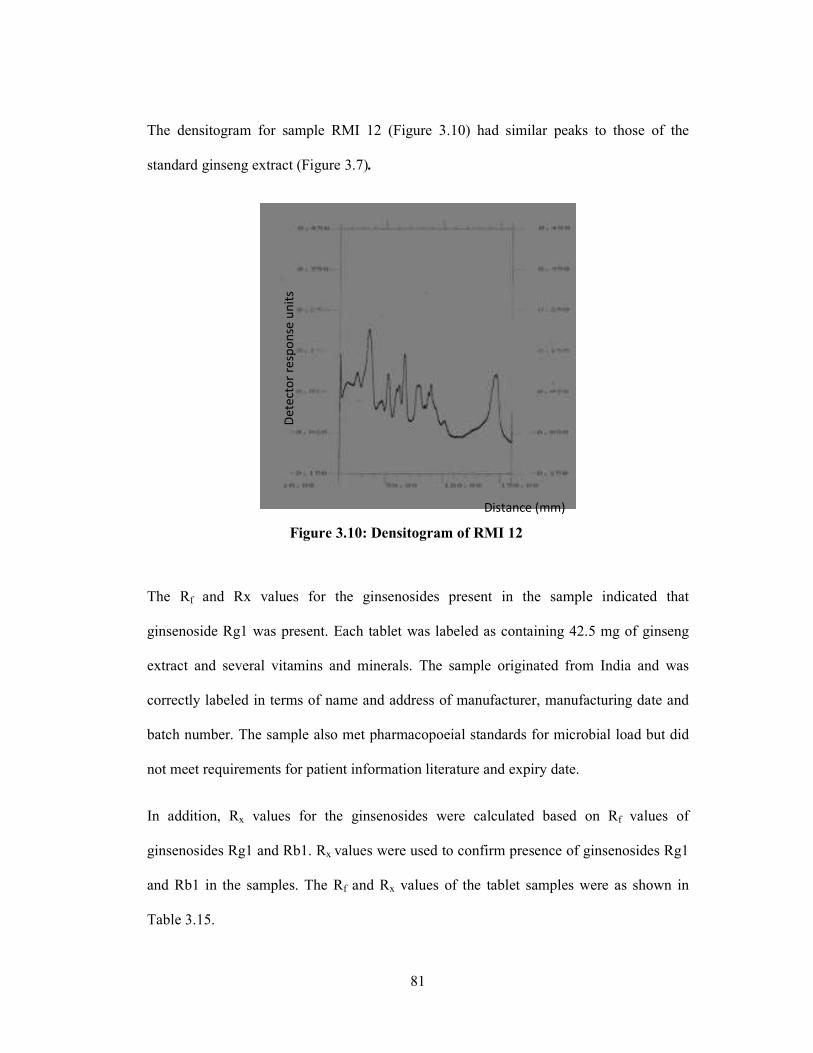
81
The densitogram for sample RMI 12 (Figure 3.10) had similar peaks to those of the
standard ginseng extract (Figure 3.7).
Figure 3.10: Densitogram of RMI 12
The Rf and Rx values for the ginsenosides present in the sample indicated that
ginsenoside Rg1 was present. Each tablet was labeled as containing 42.5 mg of ginseng
extract and several vitamins and minerals. The sample originated from India and was
correctly labeled in terms of name and address of manufacturer, manufacturing date and
batch number. The sample also met pharmacopoeial standards for microbial load but did
not meet requirements for patient information literature and expiry date.
In addition, Rx values for the ginsenosides were calculated based on Rf values of
ginsenosides Rg1 and Rb1. Rx values were used to confirm presence of ginsenosides Rg1
and Rb1 in the samples. The Rf and Rx values of the tablet samples were as shown in
Table 3.15.
Distance (mm)
De
tect
or
resp
on
se u
nit
s

82
Table 3.15: Rf and Rx values of ginsenosides of the tablet samples
Sample ID (RMI)
Rf Values×100 Rx Values based on reference ginsenosides Rg1 and Rb1
Rg1 Rb1
12 17, 18, 38, 44, 54, 63 0.32, 0.33, 0.71, 0.83, 1.01, 1.18 0.58, 0.62, 1.31, 1.51, 1.86,
2.17
16 10, 15, 22, 49, 50, 63 0.18, 0.28, 0.41, 0.92, 0.94, 1.18 0.34, 0.51, 0.75, 1.68, 1.72,
2.17
20 35, 57 0.66, 1.07 1.20, 1.96
24 15, 21, 29, 45, 59 0.28, 0.39, 0.54, 0.84, 1.11 0.51, 0.72, 1.00, 1.55, 2.03
39 8, 21, 31, 32, 45, 65 0.15, 0.39, 0.58, 0.60, 0.84, 1.22 0.27, 0.72, 1.06, 1.10, 1.55,
2.24
STD Rg1 53 1.00 1.86
STD Rb1 29 0.54 1.00
Std ginseng
extract 14, 16, 45, 53 ,59, 66 0.26, 0.30, 0.84, 1.00, 1.11, 1.24 0.48, 0.55, 1.55, 2.03, 2.27
STD=Standard, ID=Identity
3.7.2: Hard Gelatin Capsules Samples Of the eight hard gelatin samples, six (75%) had chromatograms and densitograms
similar to those of standard ginseng extract and showed presence of ginsenosides Rb1 and
Rg1. Two (25%) samples did not show any spots on the TLC chromatograms or peaks on
the densitograms hence a likelihood of them being falsified or counterfeit products. The
Rf and Rx values for the six hard gelatin capsule samples were as illustrated in Table 3.16.
Appendices 11, 12, 13, 15, 33 and 39 are densitograms for samples RMI 4, 5, 6, 8, 30 and
38 respectively. Samples RMI 19 and 21 had no spots on the TLC chromatograms and Rf
values corresponding to those of standard ginseng extract and this was taken to indicate
absence of ginseng.

83
Table 3.16: Rf and Rx values of ginsenosides of the hard gelatin capsule samples
Sample
Identity
(RMI)
Rf Values×100
Rx Values based on reference ginsenosides Rg1 and Rb1
Rg1 Rb1
4 19, 30, 40, 49, 57,
65
0.35, 0.56, 0.75, 0.92,
1.07,1.22
0.65, 1.03, 1.37, 1.68, 1.96,
2.24
5 16, 26, 46, 56 0.30, 0.49, 0.86, 1.06 0.55, 0.89, 1.58, 1.93
6 16, 26, 40, 57 0.30, 0.49, 0.75, 1.07 0.55, 0.89, 1.37, 1.96
8 18, 25, 37, 44, 55,
63
0.33, 0.47, 0.69, 0.83, 1.03,
1.18
0.62, 0.86, 1.27, 1.51, 1.89,
2.17
19 No spots N/A N/A
21 No spots N/A N/A
30 21, 30, 37, 45, 66 0.39, 0.56, 0.69, 0.84, 1.24 0.72, 1.03, 1.27, 1.55, 2.27
38 16, 26, 52, 59 0.30, 0.49. 0.98, 1.11 0.55, 0.89, 1.79, 2.03
The label for RMI-19 indicated that each capsule contained 150 mg of Panax ginseng
root standardized to 5% ginsenosides. In addition, the label stated that each capsule
contained 140 mg of Paullinia cupana seed, 100 mg of Camellia sinensis leaf, 75 mg
Eleutherococcus senticosus root, 50 mg of Rhodiola rosea root, 50 mg of Glycyrrhiza
glabra root, 50mg Centella asiatica, 25mg Ilex paraquariensis leaf, 12.5 mg Capsicum
annuum and 15 mg Octacosanol from spinach and several minerals and vitamins.
This sample comprised of many herbs that had several active ingredients hence the
difficulty in isolating and identifying ginsenosides. This sample is also likely to have been
falsely labeled thus qualifying to be a counterfeit.

84
3.7.3: Soft Gelatin Capsules Samples
There were 23 samples in this category but only 19 were found to have ginseng with the
Rf and Rx values illustrated in Table 3.17.
Table 3.17: Rf and Rx values of ginsenosides in the soft gelatin capsule samples
Sample Identity (RMI)
Rf Values×100
Rx Values based on reference ginsenosides Rg1 and Rb1
Rg1 Rb1
1 22, 33, 37, 45 0.41, 0.62, 0.69, 0.84 0.75, 1.13, 1.27, 1.55
2 26, 38, 44 0.49, 0.71, 0.83 0.89, 1.31, 1.51
3 18, 31, 41, 56, 60 0.33, 0.58, 0.77, 1.05, 1.13 0.62, 1.06, 1.41, 1.93,
2.06
7 16, 17, 39, 45, 57 0.30, 0.32, 0.73, 0.84, 1.07 0.55, 0.58, 1.34, 1.55,
1.96
9 16, 36, 41, 53 0.30, 0.67, 0.77, 1.00 0.55, 1.24, 1.41, 1.82
10 39, 44, 49, 56, 64 0.73, 0.83, 0.92, 1.20 1.34, 1.51, 1.68, 1.93,
2.20
11 48, 77 0.90, 1,45 1.65, 2.65
13 No spots N/A N/A
14 15, 21, 24, 38, 45,
56, 62
0.28, 0.39, 0.45, 0.71, 0.84, 1.05,
1.16
0.51, 0.72, 0.82, 1.31,
1.55, 1.93, 2.13
17 12, 40 0.22, 0.71 0.41, 1.37
18 36 0.67 1.24
23 21, 27, 36 0.39, 0.50, 0.67 0.72, 0.93, 1.24
26 26, 46 0.49, 0.86 0.89, 1.58
27 No spots N/A N/A
28 66, 86 1.24, 1.62 2.27, 2.96
29 No spots N/A N/A
31 43, 67, 78, 86 0.81, 1.26, 1.47, 1.62 1.48, 2.31, 2.68, 2.96
32 21, 34, 41, 68 0.39, 0.64, 0.77, 1.28 0.72, 1.06, 1.41, 2.34
33 28, 31, 36, 56, 86 0.52, 0.58, 0.67, 1.05, 1.62 0.96, 1.06, 1.24, 1.93,
2.96
34 10, 15, 41, 61, 71 0.18, 0.28, 0.77, 1.15, 1.33 0.34, 0.51, 1.41, 2.10,
2.44
35 10, 19, 43 0.18, 0.35, 0.81 0.34, 0.65, 1.48
36 No spots N/A N/A
40 21, 30, 32, 37, 45,
66, 68
0.39, 0.56, 0.60, 0.69, 0.84, 1.24,
1.28
0.72, 1.03, 1.10, 1.27,
1.55, 2.27, 2.34

85
Presence of ginseng in the samples was demonstrated by comparing the profiles of the
samples to that of standard ginseng extract and confirmed by having peaks with Rf values
corresponding to those of standard ginsenosides Rb1 and Rg1. The TLC chromatograms
for 4(17.4%) samples did not show any spots and the corresponding densitograms did not
have any peaks. The TLC chromatograms and densitograms for 19(82 .6%) samples
exhibited similar profiles to that of standard ginseng extract and reference ginsenosides
Rg1 and Rb1 respectively. Denistograms of the samples are as shown in the Appendix
chapter.
3.7.4: Occurrence of Ginsenosides in the Analyzed Samples
Of the 36 samples analyzed for identification of ginsenosides, 30 (83.3%) were positive
for ginseng. Figure 3.11 presents the distribution of samples by ginsenosides.
Figure 3.11: Presence of ginsenosides in the samples
Presence of ginsenosides was established by comparing ginsenoside profiles of the
samples to that of the standard ginseng extract. The ginseng profiles of the samples were
83.3%
16.7%
Positive for Ginseng Negative

86
also compared to the corresponding TLC chromatograms and densitograms of standard
ginsenosides Rb1 and Rg1. In this study, confirmed ginsenosides were those whose
reference standards were available hence the Rf values were ascertained. Of the 36
samples, 26 (72.2%) had confirmed ginsenosides Rb1 and Rg1. Broadly, 19 (52.7%)
samples were identified to have the Rb1 ginsenoside, 24 (66.6%) contained the Rg1
ginsenosides and 17 (47.2%) had both Rb1 and Rg1.
Further, 4 (11.1%) samples showed densitograms similar to that of the standard ginseng
extract (Figure 3.7) but did not show peaks corresponding to ginsenosides Rb1 or Rg1.
This indicates presence of the other ginsenosides other than Rb1 or Rg1. Another
observation was that 7 (19.4%) samples had Rg1 but no Rb1 while 2 (5.5%) samples only
had Rb1 ginsenosides. The distribution of ginsenosides in the 36 samples was as
illustrated in Figure 3.12.
Figure 3.12: Distribution of Rg1 and Rb1 ginsenosides in the samples
5.6
19.4
47.2
11.1
16.7
0
5
10
15
20
25
30
35
40
45
50
Rb1 Rg1 Rb1 and Rg1 Positive for
others
Negative
Ginsenosides
% Samples

87
Profiling of ginsenosides in the 40 samples in this study mirror the work done by
Courthout et al., (1999) who established Rf values for the 8 common ginsenosides as
illustrated in Table 3.18. Though the experimental conditions for the two studies were
different, the results had a lot of similarities.
Table 3.18: Rf values of 8 ginsenoside (Courthout et al., 1999)
Ginsenoside group Ginsenoside Rf×100 value
Protopanaxadiols( Rb1
Group)
Rb1 30
Rb2 35
Rc 39
Rd 49
protopanaxatriols(Rg1
Group)
Rg1 53
Re 43
Rf 56
Rg2 60
3.8: Discussion
In this study, commercial ginseng containing products from Nairobi County were
sampled. This decision was made out of the realization that Nairobi is the commercial
city of Kenya. All the big super market chains, nutritional stores, medical stores and
pharmacies have their head quarters in the Nairobi. It was, therefore, inferred that the
sampling results on the distribution of ginseng products are generalisable to all urban
areas in Kenyan.
The results of the survey in Nairobi County illustrated that there was a large number of
premises stocking ginseng containing products in Westlands and Starehe Districts.
Majority of the inhabitants of these two districts were medium and high income earners
with a significant number of foreigners (Mitullah, 2003). Of the eight stores of a leading
supermarket chain sampled, it was established that only the one in Githurai slums did not

88
stock any ginseng products. Githurai is an area in Nairobi County that is largely inhabited
by low income earners (Mitullah, 2003). This finding showed that ginseng products were
preferentially consumed by medium and high income earning population of Nairobi
County. The samples were purchased at various prices ranging from 12 to 100 USD per
monthly dose.
The assumption had been that ginseng containing products were not stocked in un-
registered pharmacies and in stores found in low income neighbourhoods. The study was
also limited to ginseng containing products of unique batch numbers. Same products with
different batch numbers were treated as distinct different products.
Persons selling ginseng containing products in the outlets were broadly classified as
pharmacists, pharmaceutical technologists, nutritionists and others who were none of the
three categories. One of the specific objectives for this study was to establish how much
knowledge they had about ginseng containing products. Their knowledge was tested
through questionnaires that referred to the classification or scheduling, pharmacological
uses and side effects of ginseng products.
A majority (52.6%) classified them as general sales food supplements. Indeed, this was
the most appropriate classification. However, for attendants in pharmacies and nutritional
stores a classification of ‘over the counter’ would suffice. Attendants who gave either of
the two answers were 32.9%. The registered ginseng containing products were scheduled
as multivitamins by the the Pharmacy and Poisons Board. This scheduling was wrong
and was not supported by any official legal instrument. In the United States of America,
ginseng containing products are classified as botanical dietary supplements following the
passage of the “Dietary Supplement and Health Education Act” of 1994 (Breemen et al.,

89
2007). The case in Kenya was similar to that in Canada where several efforts to develop a
comprehensive schedule for herbal products had not been successful.
This study brought out the fact that most personnel advising the public on use of ginseng
products had poor understanding of pharmacological activities of ginseng. Mean
knowledge score of the 76 store attendants was 3.2 (+ 1.8 SD) ranging between 0 and 8. It
was established that a trained nutritionist attendant was about 17 times more likely to
have adequate knowledge on ginseng products compared to one trained as a
pharmaceutical technologist or as pharmacist. Although nutritionists scored highly, most
of them had undergone in-service training. It is suggested from the results of this study
that the Bachelor of Pharmacy curriculum and Diploma in Pharmaceutical Technology
syllabus be reviewed to strengthen training on clinical aspects of specific herbal products.
This study exposed the difficulties that would arise when regulation of herbal products is
instituted by the Pharmacy and Poisons Board. Herbal products were not scheduled while
those who were supposed to practice herbal medicines had little understanding of what
the products entailed. There were risks pertaining to the then prevailing situation since
ginsenosides are phytoestrogens (steroidal glycosides) and their safety in infants and
pregnant women has not been studied (Osaki and Kennelly, 2003). Another worrying
trend was that there were no clear recommended daily nutritional doses both for adults
and children.
Forty samples were assessed for quality by way of compliance with WHO labeling
requirements, microbial load as per pharmacopeia standards and identification of ginseng.
Out of 40 identified samples 17 (41.5%) were 80 – 100 % compliant to all WHO labeling
requirements for market authorization. The high level of non conformity may have arisen
out of the fact that different countries followed different labeling regimes. The European
guidelines for the assessment of herbal remedies followed up on WHO’s Guidelines for

90
the Assessment of Herbal Medicines while the USA, Canada, China, India, Germany,
Britain and France had country specific guidelines that had different directives on
labeling. In Germany, for example, the whole herbal product is considered as one active
ingredient (Benzi and Ceci 1997; Saito, 2000). In the UK, The Traditional Herbal
Medicines Registration Scheme requires that relevant products are accompanied by the
necessary information for its safe use, such as safety warnings and contraindications. It is
therefore important to note that not all the samples complied with their country of origin
labeling requirements. In the absence of proper scheduling and poor understanding of
ginseng containing products by the professionals involved in dispensing them, the
chances of misuse are likely to be higher. This was worsened by the fact that 61% of the
sampled products did not have patient information literature. Furthermore, all the sampled
products had no labels that indicated storage conditions yet some were soft gel capsules
and others contained jellies that are susceptible to spoilage at higher temperatures.
Another important aspect of this study was to establish the level of contamination and
identify microbes that were not supposed to be present in ginseng containing products
prepared for oral use. Contamination by Salmonella Species and E. coli, in whatever
amounts, would render these products unfit for human consumption. There was 100%
compliance to microbial load, however, 22.0% of the samples did not meet
pharmacopoeial requirements of absence of high-risk microbes, Salmonella species and
Escherichia coli (USP NF, 2005). Salmonella species was the most frequently isolated
bacteria from 19.5% of the samples while Rhizopus and Fusarium spp formed majority of
the perfect fungal isolates from 9.7 % of the samples. Escherichia coli and Salmonella are
normally associated with infections such as diarrhea, urinary tract infections (UTI),
pneumonia and typhoid fever (Lessnau and Bronze, 2010).

91
The presence of several bacteria and fungi in the analyzed samples in this study may be
due to the methods of their preparation or the equipment and materials used in preparing
the ginseng containing samples. Other possible sources of contaminants are the
personnel(s) that could introduce the microbes when handling the raw materials during
processing. Therefore, the process of harvesting, drying, storage, handling and the soil
influence the microbial contamination of raw material which in turns affects the entire
quality of the finished herbal preparation. Thus manufacturers should ensure the highest
possible level of hygiene during manufacturing so as to maintain correct quality of the
final herbal preparations (Abba et al., 2009).
Gram negative organisms which were the majority of isolates in this study are pathogenic
and majority are associated with feacal contamination (Adenike et al. 2007). Presence of
E. coli may indicate a possible presence of harmful disease-causing organisms since the
bacteria constitute the intestinal flora of humans and other animals, and are therefore used
as indicator organisms and as an index of possible contamination by human pathogens.
The significance of faecal bacteria is that if these specific bacteria are present then other
harmful microorganisms may also be present, such as Salmonella (Abba et al., 2009).
Staphylococcus aureus isolated and identified in this study is a member of the normal
flora. However, higher contamination by these bacteria has been associated with a
number of complications especially in immune-compromised individuals. It disables the
immune system, destroying tissues and releases exotoxins which cause gastroenteritis,
toxic shock syndrome and scalded skin syndrome (Gladwin and Trattler 2004). It has also
been associated with pneumonia, osteomyelitis, acute bacterial endocarditis, septic
arthritis and urinary tract infections.

92
Klebsiella pneumonia, a Gram negative lactose fermenting bacterium found in the normal
flora of the mouth is ranked second to E. coli for urinary tract infections in older people
and also an opportunistic infection of people with weakened immunity. It is also a
common cause of pneumonia. Pseudomonas aeruginosa also a widespread
microorganism in soil, water and plants is pathogenic to humans and a common cause of
nasal-comical infections such as pneumonia and also UTI. Though rare to healthy
individuals, it is life threatening to immuno-compromised people (Lessnau and Bronze,
2010).
Fusarium is also a fungus common in soils and plants. Though most of its species are
harmless saprobes and abundant members of the soil microbial community, it has been
associated with mycotoxins at lower temperatures; it can also cause allergies and asthma
in immunocompromised individuals (WHO, 1999). Rhizopus spp. is frequently found in
house dust, soil, fruits, nuts and seeds. Exposure to large numbers of this fungus has been
reported as cause of respiratory complications. It is also an opportunistic pathogen for
immunocompromised individuals. People with diabetes, malnutrition and severe burns
are also at risk. The fungus Candida spp. also isolated and identified in this study is the
cause of many infections. Candida albicans causes mouth, skin, vaginal and penile
infections. In rare occasions, it has been traced in the blood stream causing infections to
vital organs especially in leukemia and HIV/ Aids (Dugdale, 2009).
These findings agreed with those of another study by Tournas et al., (2006), who
analyzed forty six samples, and concluded that most ginseng products were contaminated
with moulds and yeast as well as aerobic bacteria. A Study of microbial contamination in
concentrated Chinese medicine by Ku et al., (1994) established that crude drugs such as
Ginseng Radix, Glycyrrhizae Radix and Zingiber Rhizoma had serious contamination.
This study also corroborates the findings of Esimone et al., (2007) and Dnyaneshwar and

93
Bhushan (2005) who found Klebsiella pneumonia, Staphylococcus aureus, Escherichia
coli, Salmonella spp., and Pseudomonas aeruginosa as major bacterial contaminants of
herbal medicines sold and marketed in Nigeria.
Out of 36 samples that were analyzed for presence of ginseng, 83.3% were positive for
either Rb1 or Rg1 group of ginsenosides. Broadly, 19 (52.7%) samples were identified to
have the Rb1 ginsenoside, 24 (66.6%) contained the Rg1 ginsenoside and 17(47.2%) had
both Rb1 and Rg1. These results demonstrated that similar investigations can be done
within our local setting and with the resources available. Secondly, it was noteworthy that
the results obtained in this study were similar to those found in other studies elsewhere.
In one such study in the USA, twenty five commercial ginseng samples were analyzed for
levels of ginsenosides (Harkey et al., 2001). This study showed that all the products were
correctly identified as per botanical plant species (Panax spp); however, concentration of
ginsenosides differed significantly by 15-36 folds from the labeled amounts.
Further, the Rf values for ginsenosides contained in some of the samples in this study
were similar to those obtained in another one by Courthout et al., (1999) though under
different experimental conditions (Table 3.18).
This study was limited to identification because of lack of reference standards for all the
eight common ginsenosides. Furthermore, there were no columns for a solid phase
extraction (SPE) step coming immediately after the extraction of ginsenosides. A solid
phase extraction (SPE) step would have removed interfering components before
densitometric analysis (Corthout et al., 1999; Li et al., 1996). However, this study was
able to show that 5(13.9%) samples did not contain any ginsenosides. These results were
critical because ginsenosides are thought to be the active ingredients hence their chemical

94
standardization guarantees pharmacological activity of ginseng products (Harkey et al.,
2001). Absence of ginsenosides meant that these samples were counterfeits.
Whereas ginseng if used as unprocessed herb may be safe and effective as shown in
numerous studies, ginseng containing products formulated together with vitamins,
minerals and other herbs, as was the case in this study, should go through a registration
process under a Drug Regulatory Authority. A registration process where the
manufacturing plants and finished ginseng containing product must conform to cGMP
requirements. For market authorization, submissions should include the manufacturing
methods and analytical control procedures performed on raw materials used in the
manufacturing process. Other than using this information to grant market authorization,
medicine regulatory authorities will be able to carry out pharmacovigilance activities.
In the UK for instance, most herbal medicines remained as unlicensed herbal remedies up
to the year 2011 by which all were to be registered. There were no quality standards for
unlicensed herbal remedies. The same applies to South Africa where there is only a draft
policy on African Traditional Health. There are no prescribed quality standards for herbal
medicines. In the USA, herbs are defined as dietary supplements, and manufacturers can
therefore produce, sell, and market herbs without first demonstrating quality, safety and
efficacy, as is required for pharmaceutical drugs. The case in Thailand is different since
there is proper regulation of herbal products but compliance to current GMP requirements
is voluntary. The study did not establish the registration status of the samples that
originated from Thailand. It is therefore important that quality standards of herbal
medicines be enforced by PPB to safeguard public healthy.

95
3.9 Conclusion
This study established that ginseng products were found in medium to high income
neighborhoods of Nairobi County, especially Westlands and Starehe Districts and that
they were relatively expensive with a unit pack costing 15 to 100 USD. These findings
agreed with other studies conducted elsewhere such as those reported by Liberti and Der
Marderosian (1978), Wang et al., (2001). It was suggested that these products should be
regulated to avoid counterfeiting and adulteration.
The study also showed that ginseng containing products were poorly understood by the
attendants at selling points and were not scheduled by the Pharmacy and Poisons Board.
About 40% of the samples were 80 – 100 % compliant to all WHO labeling requirements
for market authorization. A majority of samples (59.5%) failed to comply with WHO
labeling requirements for market authorization. A significant number (22%) was
contaminated by Salmonella species and E. coli, bacteria that must not be present in oral
formulations. This study also revealed that 83.3% were positive for ginseng while 16.7%
of the samples did not contain ginseng.
Lack of regulation for ginseng products is likely to expose the public to some health
risks, a situation that is worsened by the fact that the attendants in the facilities had a poor
understanding of the pharmacological properties and side effects of ginseng. The present
study was significant and unique because it concerns quality and use of herbal medicines
in Kenya. The findings of this study are invaluable to policy makers and the Pharmacy
and Poisons Board as a stimulant to strengthen the regulatory framework for the quality
and practice of herbal medicines in Kenya.
3.10 Recommendations
From the foregoing, it is discernible that further studies need to be conducted to establish
the distribution and use of processed herbal products in Kenya and their value of trade.

96
This study also provides us with an opportunity to re-examine training curriculum for the
professional cadres involved in the practice of herbal medicines especially for
pharmaceutical technologists and pharmacists. With respect to quality of ginseng
containing products, more research for assay of ginsenosides, dissolution profile and
disintegration of some dosage forms, contamination by heavy metals and pesticides
residues is needed. The study affirms the need for regulation of herbal medicines so that
their quality, safety and efficacy can be assured. Herbal medicines require regulation with
respect to product development, quality control of finished products and their safety.
Meanwhile, the study has brought out the need to strengthen pharmacovigilance activities
in order to safeguard the public against poor quality herbal products.

97
REFERENCES
Abba D., Inabo H.I., YakubuS.E. and Olayeni S Olonitola O.S.(2009): Contamination of
Herbal Medicinal Products Marketed in Kaduna Metropolis with Selected
Pathogenic Bacteria. Afr. J. Trad. Compl. Alt. Med., 6(1): 70–77.
Adenike O., Adewoyin A. and Oluwatoyin A., (2007): Evaluation of pharmaceutical and
microbial qualities of some herbal medicinal products in South Western Nigeria.
Trop. J. Pharm. Res., 6 (1): 661-670.
Amann R.I., Ludwig W. and Schleifer K.-H., (1995): Phylogenetic identification and in
situ detection of individual microbial cells without cultivation. Microbiol. Rev., 59:
143–169.
Andrew M., (2000): The use of 16SrDNA methods in soil microbial ecology. Braz. J.
Microbiol., 31: 77-82.
Balch, P., (2006): Prescription for Nutritional Healing, New York, NY: Penguin Group.
ISBN-10-1583332367, ISBN-13-1583332368.
Banthorpe D.V., (1994): Terpenoids. Natural Products: Their Chemistry and Biological
Significance, Mann, J., Ed., Longman Scientific and Technical: Harlow, Essex,
England, 1994: 289-359.
Benzi G. and Ceci A., (1997): Herbal Medicines in European Regulation. Pharmacol.
Res., 35: 355-362.
Betancourt D.A., Loveless T.M., Brown J.W. and Bishop P.E., (2008): Characterization
of Diazotrophs Containing Mo-Independent Nitrogenases, Isolated from Diverse
Natural Environments. Appl. Environ. Microbiol, 74 (11):3471-3480.
Biomeriex Vitek, Inc, 20 E analytical profile index, enterobacteriaceae and other gram-
negative bacteria, 10th edition.
Bonfill M, Casals I, Palazon J, Mallol A and Morales C., (2002): Improved high
perfomance liquid chromatographic determination of ginsenosides in Panax
ginseng-based pharmaceuticals using a diol column. Biomed.Chromatogr. 16: 68-
72.
Breemen R.B., Harry H.S., Fong and Farmsworth, N.M., (2007): The Role of Quality
Assurance and Standardization in the Safety of Botanical Dietary Supplements.
Chem. Res. Toxicol., 20 (4): 577-582.
Brevoort P., (1998): The booming US botanical market-a new overview. Herbalgram, 44:
33-46.

98
British Pharmacopeia, (2008): The stationery office, London, 995-997. ISBN-13:
9780113227501, ISBN-10: 0113227507
Bruce S.K., Schick D.G., Tanaka L., Jimenez E.M., and Montgomerie J.Z., (1981): Selective medium for isolation of Klebsiella pneumoniae. J. Clin. Microbiol.,
13(6):1114-1116.
Bumsik H., Young H. J., Jun H.H., Yeul K.I. N. and Tai Y. A., (2002): A Double-Blind
Crossover Study Evaluating the Efficacy of Korean Red Ginseng in Patients with
Erectile Dysfunction: A Preliminary Report. J. Urology, 168 (5): 2070-2073.
But P.P.H., Hu S.Y. and Cao, H., (1995): The ginseng plant; products and quality. P. 44-
49. In: W.G. Bailey, C. Whitehead, J.T.A. Proctor, and J.T. Kyle, Proc. Int. ginseng
conf. Vancouver1994, Canada.
Cappuccino and Sherman (2004): Microbiology, a Laboratory Manual; Pearson
Education Inc. Sate University of New York.
Chan T.W.D., But P.P.H., Cheng S.W., Kwok I.M.Y., Lau F. W. and Xu H.W., (2000):
Differentiation and Authentication of Panax ginseng, Panax quinquefolius, and
Ginseng Products by Using HPCL/MS. J. Anal. Chem., 72 (10): 2329-2329.
Choi K.T., Park J.C., and Ahn I.O., (1995): Characteristics of the growth and suspension
of ginsenosides in the suspension-cultured cells of the Korean ginseng (Panax
ginseng, C.A.Meyer), Proc. Int. Ginseng Conf, Vancouver 1994, Canada, 259-269.
Chung S.H., Choi C.G., and Park S.H., (2001): Comparisons between white ginseng radix
and root for anti-diabetic activity and mechanism in KKAy mice. Arch. Pharm.
Res. 24(3): 214-218.
ConsumerLab, (2000): Independent tests of ginseng.
http://www.consumerlab.com/index.asp
Corthout J., Naessens T., Apers S. and Vlientinck A.J., (1999): Quantitative
Determination of Ginsenosides from Panax ginseng roots and ginseng preparations
by Thin Layer Chromatography-Densitometry. J. Pharm. Biomed. Anal., 21: 187-
192.
Cui X., Lo C., Yip K., Dong T. and Tsim K., (2003): Authentication of Panax
notoginseng by 5s-Rrna spacer domain and random amplified polymorphic DNA
(RAPD) analysis. Planta Med. 69: 584-586.

99
Cui Y., Shu X.O., Gao Y.T., Cai H., Tao M.H.and Zheng, W., (2006): Association of
Ginseng Use with Survival and Quality of Life among Breast Cancer Patients. Am.
J. Epidemiol., 7: 645-653.
D'Angelo L., Grimaldi R., Caravaggi M., Marcoli M., Perucca E., Lecchini S., Frigo
G.M., and Crema A., (1986): A double-blind, placebo-controlled clinical study on
the effect of a standardized ginseng extract on psychomotor performance in healthy
volunteers. J. Ethnopharmacol., 16 (1):15-22.
Dharmananda S., (2002): The Nature of Ginseng: From Traditional Use to Modern
Research. Internet Journal of the Institute for Traditional Medicine and Preventive
Health Care. http://www.itmonline.org/arts/ginsengnature.htm
Dixon P., (1976): Ginseng; Duckworth & Co: London. ISBN-10-071561007H.
Dnyaneshwar W. and Bhushan P. (2005): Botanicals: Quality and regulatory issues. J.
Sci. Ind. Res. 64: 83-92.
Dubic M.A., (1986): Historical perspective on the use of herbal preparations to promote
health. A paper presented to the 69th
Annual Meeting of the Federation of American
Societies for Experimental Biology, Anaheim, C.A. April 21-26, 1985.
Dugdale D.C., (2009): Candida Esophagitis. www.healthscout.com/ency/6813/main.html
Ebel S., Gigalke H.J. and Voelkl S. (1978): AMDHPTLC Analysis of Medicinal
Plants.Proceedings of 4th International Symposium of Instrumental HPTLC,
Selvino/Bargamo, Italy, 113.
Esimone C.O., Ibezima E.C. and Oleghe P.O., (2007): Gross microbial contamination of
herbal medicinal products marketed in Mid Western Nigeria. Int. J. Mol. Med. Adv.
Sci 3(2): 87-92.
European Medicine Agency Inspection, (2006): Guidelines on Quality of Herbal
Medicinal Products, 14-17.
European pharmacopoeia (2005): 5th
Edition, council of Europe, European Directorate for
the Quality of Medicines (EDQM), Strasbourg.
Evans W.C., (2007): Saponins, cardioactive drugs and other steroids. Trease and Evans,
15TH Edition, Elservier Ltd, London, 295-297.
Forles B. A., Sahm D. F. and Weissfield A. S., (1994): Balley and Scott’s diagnostic
Microbiology 10th edition. Elsevier Ltd, ISBN-978-0-323-08330-0

100
Fournier A.R., Proctor J.T., Gauthier L., Khanizadeh S., Belanger A. and Doras M.,
(2003): Understanding effect of light on root ginsenosides in forest grown Panax
quinquefolius. Phytochemistry, 63: 777-782.
Fuzzati N., Gabetta B., Jayakar K., Pace R. and Peterlongo F., (1999): Liquid
chromatography-electrospray mass spectrometric identification of ginsenosides in
Panax ginseng roots. J. Chromatogr. Anal., 854: 69-79.
Gladwin M. and Trattler B., (2004): Clinical microbiology made ridiculously simple, 3rd
edition, Medmaster Publishers. ISBN-10: 094-0780496, ISBN-13: 978-
0940780491.
Gracias S.K. and McKillip J.L., (2004): A review of conventional detection and
enumeration methods for pathogenic bacteria in food. Can. J. Microbiol., 50 (11):
883-890.
Haijiang Z.W., Yongjiang W. and Yiyu C., (2003): Analysis of SHENMAI Injection by
HPLC/MS/. J. Pharm. Biomed. Anal., 31: 175-183.
Harkeys M.R., Henderson G.L., Gershwin M.R., Stern J.S and Hackman, R.M., (2001):
Variability in commercial ginseng products: an analysis of 25 preparations .Am. J.
Clin. Nutr., 73 (6): 1106-1106.
Health Protection Agency UK, (2009): Salmonella Reference Unit. www.hpa.org.uk
Hiai S., Oura H., Hamanaka H. and Odaka Y., (1975): A colour reaction of panaxadiol
with vanillin and sulphuric acid. Planta Med. 28: 363-369.
Homeherbs (2008): Asian Ginseng (Panax ginseng) Medicinal, Historical and Botanical
Information http://www.panax-ginseng.co.uk.
Hosettman K. and Marston A., (1995): Analysis and Isolation. Chemistry and
Pharmacology of Natural Products; Cambridge University Press: New York, 122-
174.
Hu S. and Kitts D.D., (2001): Free radical scavenging capacity as related to antioxidant
activity and ginsenoside composition of Asian and North American ginseng
extracts. J. Am. Oil Chem. Soc., 78: 249-255.
KEMRI, (2003): Pelvic Inflammatory Disease (PID) Laboratory manual, KEMRI.
Kenya National Bureau of Statistics (KNBS), 2007: Basic report on well-being in Kenya:
Based on Kenya Integrated Household Budget Survey-2005/2006.

101
Kiefer D. and Pantuso T., (2003): Panax Ginseng. Am. Fam. Physician, 68(8): 1539-1542.
Indian Pharmacopoeia, (2008): A publication of the Government of India, Delhi, 9-10.
Kim W.Y., Kim J.M., Han S.B., Lee S.K., Kim N.D., Park M.K., Kim C.K. and Park J.H,
(2000): Steaming of ginseng at high temperature enhances biological activity. J.
Nat. Prod., 63: 1702-1704.
Kim Y.K., Guo Q. and Packer L., (2002): Free radical scavenging activity of red ginseng
aqueous extracts. Toxicology, 172: 149-156.
Kimura L., Nakashima N., Sugihara Y., Fu-jun C. and Kimura M., (1999): The
antihyperglyceamic blend effect of traditional Chinese medicine byakko-ka-ninjin-
to on alloxan and diabetic KK-CA(y) mice. Phytother. Res., 13: 484-488.
Kimura S., (1997): Use of the radiation method for sterilization of herbal medicines. J.
Antibiot. Japan, 25: 475–479.
Kite G.C., Howes M.J., Leon C.J. and Simmonds M.S., (2003): Liquid
chromatography/mass spectroscopy of malonyl- ginsenosides in the authentication
of ginseng. Rapid Commun. Mass Sp., 17: 238-244.
Kitts D.D. and Hu C., (2001): Efficacy and safety of ginseng. Public Health Nutr., 3: 473-
485.
Kitts D.D. and Popovich D.G. (2003): Ginseg: In Performance functional foods, Watson,
D., Ed., Woodhead Publishing Ltd., New York, 78-88.
Kitts D.D., Wijewickreme A.N. and Hu C., (2000): Antioxidant properties of a North
American ginseng extract. Mol. Cell. Biochem., 203: 1-10.
Kombo D.K. and Tromp D.L.A., Proposal and Thesis Writing, Pauline Publications
Africa, Nairobi, 2006.
Krochmal R., Hardy M., Boweman S., Wang Q.L., Elashoff R.M. and Heber D., (2004):
Phytochemical Assay of Commercial Botanical Dietary Supplements. Evid-based
Comp. Alt. 3:305-313.
Ku Y.R., Chou L.M., Jang C.F., Liu, Y.C. Lin, J.H. and Wen, G.C., (1994): Study of
microbial contamination in concentrated Chinese medicine. J. Food and Drug
Anal., 2(1): 49-62.

102
Kwon S.W., Han S.B., Park I.H., Kim J.M., Park M.K. and Park J.H., (2001): Liquid
chromatographic determination of less polar ginsenosides in processed ginseng. J.
Chromatogr. A, 921: 335-339.
Lee F.C., (1992): Facts about Ginseng, the Elixir of Life. Hollym Intern. Corp. Elizabeth,
New Jersey.
Lee J. and Yuqing Z., (2009): Current evaluation of millennium phytomedicine-Ginseng:
Etymology, Pharmacognosy, Phytochemistry, Market and Regulation. Curr. Med.
Chem.16: 2475-2484.
Lee S.J., Ko W.G., Kim J.H., Sung J.H., Moon C.K. and Lee B.H., (2000): Induction of
apoptosis by a novel intestinal metabolite of ginseng saponin via cytochrome c-
mediated activation of capase-3 protease. Biochem. Pharmacol., 60: 677-685.
Leininger D.J., Roberson J.R and Elvinger F., (2001): Use of eosin methylene blue agar
to differentiate Escherichia coli from other gram-negative mastitis pathogens. J.
Vet. Diagn. Invest., 13: 273-275.
Lessnau K. and Bronze M.S (2010): Pseudomonas aeruginosa
Infections. http://emedicine.medscape.com/article/226748.
Li T.S, and Wardle D. (2002): Seasonal fluctuations of leaf and weight and ginsenoside
contents of 2, 3 and 4 year old American ginseng plants. Hort. Technology, 12:
229-231.
Li T.S., Mazza G., Cotrell A.C. and Gao L., (1996): Ginsenosides in Roots and Leaves of
American Ginseng. J. Agr. Food Chem., 44: 717-720.
Liberti L., and Der Marderosian, A., (1978: Evaluation of Commercial Ginseng Products.
J. Pharm. Sci,, 67: 27-38.
Liu W.K., Xu S.K. and Che C.T., (2000): Anti-proliferative effect of ginseng saponins on
human prostate cancer cell lines. Life sci., 67: 1297-1306.
Liu Z.Q., Luo X.Y., Sun Y.X., Chen Y.P. and Wang Z.C., (2002): Can ginsenosides
protect human erythrocytes against free-radical-induced hemolysis? Biochem.
Pharmacol. 1572: 58-66.
Ludwiczuk A., Chmielewska E.W. and Wolski T., (2006): Localisation of ginsenosides in
Panax quinquefolium root tissues. Acta Agrobot, 59: 7-15.
Lui J.H. and Staba E.J., (1980): The ginsenosides of various ginseng plants and selected
products. J. Nat. Prod., 43: 340-346.

103
Lyons A. and Petrucelli J.R., (1978): Medicines: An Illustrated History, H.N. Abrams:
Baldwin, New York, USA, 604-607.
Maddocks S., Olma T and Chen S., (2002): Comparison of CHROMagar Salmonella
Medium and Xylose-Lysine-Desoxycholate and Salmonella-Shigella Agars for
Isolation of Salmonella Strains from Stool Samples. J. Clin. Microbiol.,
40 (8): 2999-3003.
Marcus D.M. and Grollman, A.P., (2002): Botanical Medicines—the need for new
regulations. New Engl. J. Med., 31(25):2073-6.
Meier B., Meir-Bratsch A., Dallenbach-Tolke K. and Sticher O., (1985): Quantitative
analysis of ginseng by HPLC. In Advances in Chinese Material Research, Chang
H.M., Yeung H.W., Tso W.W., Koo A. Edition; World Scientific Publication Co.
Singapore, 471-484.
Michael J. J and Sharon A, L., (2002): Bacterial Identification for Publication. J. Clin.
Microbiol. 40: 6, 1887-1891.
Ministry of Medical Services and Ministry of Public Health, (2010): Session Paper on
National Pharmaceutical Policy, Gov. Printers.
Mitullah W., (2003): The challenge of slums. A Case study of Nairobi. UN Habitat
Gliobal Report on Human Settlement. Londres, Earthscan.
Mugenda M.O., Mugenda A.G., (2003): Research Methods, Quantitative and Qualitative
Approaches, African Center for Technological Studies, Nairobi.
Mukherjee P.K., (2002): Quality Control of Herbal Drugs, Business Horizons, New
Delhi, 2.
Mwangi J. W., Mungai N.N., Thoithi G.N. and Kibwage, I.O., (2005): Traditional herbal
medicine in national healthcare in Kenya. East Cent. Afr. J. Pharm.Sci., 8 (2): 22-
26.
Ni W.,Zhang X., Wang B., Chen Y., Han H., Fan Y., Zhou Y. and Tai G., (2010):
Antntitumor activities and immunomodulatory effects of Ginseng neutral
polysaccharides in combination with 5- Fluorouracil. J. Med. Food, 13 (2): 270 –
277.
Oh M., Choi Y.H., Choi S., Chung H., Kim K., Kim S.I. and Kim. D.K., (1999): Anti-
proliferative effects of ginsenosides Rh2 on MC-7 human breast cancer cells. Int. J.
Oncol., 14: 869-875.

104
Osaki A.L and Kennelly E.J., (2003): Phytoestrogens: A review of the present state of
research. Phytother. Res., 17 (8): 845-869.
Owen R.T., (1981): Ginseng: A pharmacological profile. Drugs of today, 17:343–351.
Park I.H., Piao L.Z., Kwon S.W., Lee Y.J., Cho S.Y., Park M.K. and Park J.H., (2002):
Cytotoxic dammarane glycosides from processed ginseng. Chem. Pharm. Bull., 50:
538-540.
Persons W.C., (1995): American ginseng farming in its woodland habitat 78-83. Int.
Ginseng Conf.Vancouver, 1994.
Pharmacy and Poisons Board (2006): Data on Pharmaceutical Trade in Kenya for 2005.
www.pharmacyboardkenya.org
Pharmacy and Poisons Board (2009(a)): Public Alert on ‘Herbal Contraceptives’.
www.pharmacyboardkenya.org.
Pharmacy and Poisons Board (2009(b)): Lists of registered premises for pharmacists and
pharmaceutical Technologists. www.pharmacyboardkenya.org.
Pharmacy and Poisons Board (2010 (a)): Registration of Herbal and Complementary
Medicines; Guidelines for Submission of Applications.
www.pharmacyboardkenya.org.
Pharmacy and Poisons Board (2010 (b)): Registration of Drugs: Guidelines to Submission
of Applications. www.pharmacyboardkenya.org
Pharmacy and Poisons Board (2011): List of Registered Medicines.
www.pharmacyboardkenya.org
Popovich D.G. and Kitts D.D., (2002): Structure-function relationship exists for
ginsenosides in reducing cell proliferation and inducing apoptosis in human
leukemia (THP-1) cell line. Arch. Biochem. Biophys., 406: 1-8.
Popovich D.G. and Kitts D.D., (2003): Bioactive properties of ginseng and ginsenosides
constituent. Mol. Cell. Biochem, 1: 137-149.
Preston N.W. and Morrel A., (1962): Reproductive results with Gram stain. J. Pathol
Bacteriol. 84: 241-243.
Quiming N.S., Denola N.L., Soliev A.B., Saito Y.O. and Jino K., (2007): High
performance liquid chromatographic separation and quantitative analysis of

105
ginsenosides using a polyvinyl alcohol-bonded stationary phase. Chromatographia,
66 (1): 5-11.
Rózylo J.K., Zabinska A., Matysiak J., and Niewiadomy A.J., (2002): HPTLC Method in
Physicochemical Studies of a New Group of Antimycotic Compounds. J.
Chromatogr. Sci., 40: 581.
Saito H., (2000): Regulation of herbal medicines in Japan. Pharmaco. Res. 41, 5: 515-
519.
Sandel M., (2004): Virulence and recovery of Staphylococcus aureus relevant to the food
industry using improvements on traditional approaches. Food Control, 15 (1): 5-10.
Sang-Hyun P., Sangryeol R. and Dong-Hyun K., (2010): Improved Selective and
Differential Medium for Isolation of Escherichia coli O157:H7. J. Clin. Microbiol.,
41 (10): 839-841.
Saper B.R, Fletcher W.S, and Eamranond P., (2010): Overview of herbal medicines and
dietary supplements. Up To Date. http://www.uptodate.com.
Saranadasa H., (2003): "The Square Root of N Plus One Sampling Rule: How Much
Confidence Do We Have?" Pharm. Technol., 27 (5): 50.
Scaglione F., Ferrara F., Dugnani S.,Falchi M., Santoro G., and Fraschini F., (1990):
Immunomodulatory effects of two extracts of Panax ginseng C. A. Meyer. Drugs
Exp. Clin. Res., 16: 537-542.
Shaheen M.A. and Taha M., (2006): Species identification of candid isolates obtained
from oral lesions of hospitalized and non hospitalized patients with oral candiasis.
Egypt. Dermatol. Line J., 2 (1): 1-11.
Shibata S., Tanaka O., Shoji J. and Saito H., (1985): Chemistry and Pharmacology of
Panax. In Economic and Medicinal Plant Research, Wagner H, Edition; Academic
Press: London; Orlando, FLA., 217-284.
Shinde V.M., Dwalwal K., Potdar M., Mahadik K.M.,(2009): Application of quality
control principles to herbal drugs. Int. J. Phytochem., 1: 4-8.
Shukla S.S. and Saraf S., (2009): Development of spectrophotometric fingerprinting
method for Talisadi churna. East Cent. Afr.J. Pharm. Sci., 12: 52-54.
Smyth R.W and Kahlmeter G., (2005): Mannitol Salt Agar-Cefoxitin Combination as a
Screening Medium for Methicillin-Resistant Staphylococcus aureus. J.Clin.
Microbiol., 43 (8): 3797–3799.

106
Songlin L., Quanbin H., Chunfeng Q., Jingzheng S., Chuen L. C. and Hongxi X., (2008):
Chemical markers for the quality control of herbal medicines: an overview. Chin.
Med., 3 (7): 1-23.
Sotaniemi E.A., Haapakoski E., and Rautio A., (1995): Ginseng therapy in non-insulin-
dependent diabetic patients. Diabetes Care 18: 1373–1375.
Sundra- Rao and P.S.S., Richard J., (2006): Introduction to Biostatistics and Research
Methods, 4th Edition, Printice Hall of India Private LTD. New Delhi.
Sutton S., (2006): Microbiology Network-Counting Colonies: Dedicated to improvement
of regulatory science and compliance. [email protected](585): 594-8273.
Tanaka O. and Kasai R. (1984): Saponins of ginseng and related plants. In the Progress of
organic natural products, Herz, W., Grisebach, H., Kirby, G.W., Tamm, C.H., Eds;
Springer-Verag: New York, 2-75.
Tang W. and Eisenbrand G. (1992): Chinese Drugs of Plant Origin, Vol. 91, Springer:
Berlin 711-737.
Torbeck L., (2009): Is the square root of (N) + 1 a statistically valid scheme? Pharm.
Technol., 33 (10): 128.
Tortora J.G, Funke B.R, Case C.L and Al-Mukhtar T., (2008): Microbiology: An
Introduction, 10th
Ed. Benjamin/ Cummings-ISBN-13: 978-0-321-55007-1 ISBN-
10: 0-321-55007-2. San Francisco, California, USA.
Tournas V.H., Katsoudas E. and Miracco E.J., (2006): Moulds, yeasts and aerobic plate
counts in ginseng supplements. Int. J. Food Microbiol., 108 (2):178-181.
USP/NF, (2012): United States Pharmacopeia/ National Formulary, Twinbrook Parkway,
Rockville. 1174-1188
Vaidya A.D.B. and Devasagayam T.P.A., (2007): Current Status of Herbal Drugs in
India: An Overview. J. Clin. Biochem. Nutr., 41 (1): 1–11.
Vives-Rego J., Lebaron P. and Von Caron G.N., (2000): Current and future applications
of flow cytometry in aquatic microbiology. FEMS Microbiol. Rev., 24: 429–448.
Wang J., Ha W., Ngan F., But P. and Shaw (2001): Application of sequence characterized
amplified region analysis to authenticate Panax species and their adulterants.
Planta Med. 67: 781-783.

107
Wang X., Sakuma T., Asafu-Adjaye G.K. and Shiu G.K., (1999): Determination of
ginsenosides in plant extracts from Panax ginseng and Panax quinguefolius L. by
LC/MS. Anal. Chem., 71: 1579-1584.
WHO, (1991): (WHO/TRM/91.4):Guidelines for the Assessment of Herbal Medicines.
Geneva.
WHO, (1998): Quality Control Methods for Medicinal Materials, Geneva, 22.
http;//www.who.int/edicines/services/expertcommittee/pharmprep/QAS05-
131Rev1-QCM Methods-med-Plant Material UpdateSep05.pdf.
WHO, (1999): Monograph on selected medicinal plants, Vo. 1, Geneva.
WHO, (2005): National Policy on Traditional Medicine and Regulation of Herbal
Medicines- Report of a WHO Global Survey.
Wong F, Ping C and Amit B., (2011): Effects of acute supplementation of Panax ginseng
on endurance running in hot and humid environment. Indian J. Med. Res., 133, 1:
96-102.
Yat P.N., Arnason J.T. and Awang D.V.C., (1998): An improved extraction procedure for
rapid quantitative high performance liquid chromatographic estimation of the main
eleutheroside(B and E) in Eleutherococcus senticocus. Phytochem. Anal., 9: 291-
295.
Yip T.T., Lau C.N., But P.P. and Kong Y.C., (1985): Quantitative analysis of
ginsenosides in fresh Panax ginseng. Am. J. Chin. Med., 13: 77-88.
Yong L., Ming C., Gui C., Zheng W. and Zhi H.,(2004): Ruggedness/robustness
evaluation and system suitability test on United States Pharmacopeia XXVI assay
ginsenosides in Asian and American ginseng by high performance liquid
chromatography. J. Pharm. Biomed. Anal, 3: 1083-1091.
Yun T.K., (2001): Brief introduction of Panax ginseng C.M. Meyer. J. Kor. Med. Sci.,
16: 3-5.
Yun T.K. and Choi S.Y., (1998): Non –organ specific cancer prevention of ginseng: a
prospective study in Korea. Int. J. Epidemiol., 27: 359-364.
Yun T.K., Choi S.Y., and Yun H.Y., (2001): Epidemiological study on cancer prevention
by ginseng; are all kinds of cancer preventable by ginseng? J. Kor. Med. Sci., 16:
19-27.

108
Zhou D. and Kitts D.D., (2002): Peripheral blood mononuclear cell production of NTF-
alpha in response to North American ginseng stimulation. Can. J. Physiol.
Pharmacol., 80: 1030-1033.

109
APPENDICES
Appendix 1: Listed Herbal Products in Kenya
List adapted and modified from Pharmacy and Poisons Board (Kenya), 2011
NAME COMPOSITION
(No. of active ingredients)
THERAPEUTIC CLASSIFICATION
COUNTRY OF ORIGIN
Encof cough syrup 12 Cough syrup India
Antarth soft gel Several Anti inflammatory India
Spermon soft gel Several Skin ointment India
Vigomax capsules 16 sexual dysfunction India
Atochol tablets 1 Cholesterol booster India
Healthy betas
tablets 1
Anti-arthelo sclerotic India
Guava syrup 3 Cough syrup Egypt
Zecuf herbal cough
remedy 11
Cough syrup India
Zecuf lozenges 4 Cough lozenges India
Malbet capsules 7 Anti protozoan India
Burnactil cream 6 Tropical antiseptic India
Moov ointment 4 Analgesic ointment India
Krack SR cream 9 Antiseptic cream India
Moscand mosquito repellant jelly
2 Repellant
Not stated
Indiaga for men capsules
7 Aphrodisia
South Africa
Stamiforte capsules Several
Multivitamin/ nutritional suppliment
India
Hemon capsules 9 Hematinic India
Hemo-perfecta
tablets 17
Hematinic India
Dermoluconil
tablets 4
Not stated India
Enzoy powder 3 Aphrodisia Kenya
Sylovac – sf
powder 2
Laxative India
Zoom tablets 11 Aphrodisiac India
Superliv
concentrate premix 12
Procects from Mycotoxins
and aflatoxins India
Sylovac – sf
powder 2
Laxative India
Nikoff cough syrup 13 Cough Syrup India

110
NAME COMPOSITION
(No. of active ingredients)
THERAPEUTIC CLASSIFICATION
COUNTRY OF ORIGIN
Haleezy tablets
6 for Bronchial asthma
India
Haleezy syrup 7 For Bronchial asthma India
Paedritone
Drops 1
Not stated India
Aptizooom
syrup 8
For anorexia India
Livomyn
syrup 1
Hepatoprotective India
Livomyn
tablets 17
Hepato-protective India
Gum tone powder
20 For Gingivitis
India
Kofol syrup 14
Anti –inflammatory Broncho-dilator
India
M2 tone tablets
26 Menstrual disorder
India
M2 Tone
syrup Several
Menstrual disorder India
Addyzoa
capsules Several
Menstrual disorder India
Evanova
capsules Several
Peri menopausal syndrome India
Gum tone gel Several Antifungal (Gingivitis) India
Femiforte tablets
Several Leucorrhoea Vaginitis
India
Arthrella
tablets Several
Rheumatoid arthritis India
Hyponidd
tablets Several
Antidiabetic India
Streroak
premix 5
Immuno- modulator
Antistress Adaptogen
India
Toxiroak premix
9 Mineral & toxin binder
India
Apdyl-h syrup 4 Antitussive India
Femix herbal
capsules 2
Sexual disorders in Women India
Manix
capsules 12
General restorative tonic India
Kofol lozenges
(regular) 5
Cough and sore throat India

111
NAME COMPOSITION
(No. of active ingredients)
THERAPEUTIC CLASSIFICATION
COUNTRY OF ORIGIN
Kofol lozenges (honey &
lemon)
5 Cough and sore throat
India
Kofol lozenges
(pineapple) 5
Cough and sore throat India
Hapenz syrup 6 Appetive stimulant India
TIB tablets Several Anti- Retroviral China
Arthrella
ointment Several
Masculoskeletal disorders India
Kofol rub Several Nasal decongestant India
Arshonyt
ointment Several
For Haemorroids India
Arshonyt forte
tablets Several
For Haemorroids India
Honitus syrup Several Antitussive India
Canova liquid Several
Analgesic
Anti-inflammatory
Antiviral
Antiemetic
Botswana
Olbas oil drops Several Inhalant decogestant UK
Olbas pastilles Several Soothing agent UK
Kalmis tablets Several Stress Irritability Strains UK
Nexacof tablets 14 Cough remedy India
Imnohans 2 Immune enhancer India
Nokof syrup Several Cough suppressant India
Hormotex pills Several Aphrodisiac Germany
Emami Mentho
plus balm 7
Muscle relaxant &
Analgesic India

112
Appendix 2: Questionnaire
1. Premises Details
a) Name: ………………………………………………………
b) Nature of business: …………………………………………
c) Registration status: …………………………………….........
d) Physical address: …………………………………………….
e) Postal Address: ………………………………………………
2. Personal Details of the Attendant
a) Highest level of Education attained:
Primary � Secondary � College � University�
b) Relevant Professional Training (If any): …………………
c) Enrolment/Registration No. (If any): ……………………..
3. Product Details
a) Name………………………………………………
b) Active Ingredient(s): ………………………………
c) Manufacturer…………………………………… ….
d) Address…………………………............................
e) Manufacturing Date……………………………….
f) Expiry Date………………………………………
g) Batch No…………………………........................
h) Formulation Details………………………………
4. Product Classification
Please tick [√] the option that best fits the sell, control and use of the product in this outlet
a) 0=Prescription only………………… �
b) 2=Over the counter………………… �
c) 2=General sales…………………….. �
d) 0=All the above…………………….. �
5. Do you have any information about what this product contains?
a) 1=Yes …………………….� b) 2=No…………………. �
6. Indications as Understood by the Attendant: Please choose the option that
best describes the indications or the pharmacological effects of ginseng

113
a) Reduce fatigue, increase stamina and strengthen the general body conditions
b) Promotes formation of red blood cells and improves blood circulation
c) Promotes immunity and has ant-diabetic properties
d) All the above; a), b) and c)
7. (i) Does this product have any side effects?
a) 1=Yes …………………….� b) 2=No…………………. �
(ii) If the answer to 7(i) above is yes, please choose the option that best
describes the side effects
a) Causes dizziness, loss of libido or sexual desire and anaemia
b) Causes cancer if used for a long time
c) May lower blood sugar, increase blood pressure or increase lack of sleep
d) Is an immune suppressant, causes congestion and vaginal bleeding
8. (i) Do you receive any feedback from users of this product?
a) 1=Yes …………………….� b) 2=No…………………. �
(ii) If the answer to 8(i) above is yes, please choose the option that best
describes the user feedback:
a) 1=The product was not useful at all……………… �
b) 2=The product was hardly useful………………… �
c) 3=The product was somewhat useful…………….. �
d) 4=The product was useful………………………... �
e) 5=The product was very useful…………………… �

114
Appendix 3: Interpretation of Chemical Reactions of Bacteria using API 20 E Strip
Tests Active ingredients
Qty (mg/cu
p) Reactions/ Enzymes
Results
Negative Positive
ONPG
2-
nitropheni
le-βD-
galactopyranoside
0.223
β-galactosidase (ortho
NitroPhenyl-βD-
galactopyranosidase)
Colorless yellow
(1)
ADH L-arginine
1.9 Arginine DiHydrolase Yellow red/orange (2)
LDC L-lysine 1.9 Lysine DeCarboxylase Yellow red/ orange(
2)
ODC L-ornithine
1.9 Ornithine DeCarboxylase
Yellow red/orange(2)
CIT trisodium
citrate 0.756 CITrate utilization pale-green/yellow
blue-green/
blue (3)
H2S
sodium
thiosulpha
te
0.075 H2S production colorless/ grayish
black
deposit/
thin line
URE Urea 0.76 UREase Yellow
Red/
orange
(2)
TDA
L-
tryptopha
ne
0.38 Tryptophane
DeAminase
TDA/ immediate
Yellow reddish
brown
IND
L-
tryptopha
ne
0.19 INDole production
JAMES/immediate
colorless, pale-
green/yellow pink
VP Sodium
pyruvate 1.9
Acetoin production
(voges proskauer)
VP 1 + VP 2 / 10 min
Colorless Pink/red (5)
GEL
Gelatin
(bovine origin)
0.6 GELatinase no diffusion
diffusion of
black pigment
GLU D-glucose 1.9 Fermentation / oxidation (GLUcose) (4)
Blue / blue-green
Yellow / yellowish-
green
MAN D-mannitol
1.9 Fermentation / oxidation (MANnitol) (4)
Blue / blue-green
Yellow

115
Tests Active ingredients
Qty (mg/cup)
Reactions/ Enzymes
Results
Negative Positive
INO Inositol 1.9 Fermentation / oxidation
(INOsitol) (4)
Blue / blue-
green Yellow
SOR D-sorbitol 1.9 Fermentation / oxidation
(SORbitol) (4)
Blue / blue-
green Yellow
RHA L-
rhamnose 1.9
Fermentation / oxidation
(RHAmnose0 (4)
Blue / blue-
green Yellow
SAC D-sucrose 1.9 Fermentation / oxidation
(SACcharose) (4)
Blue / blue-
green Yellow
MEL D-
melibiose 1.9
Fermentation / oxidation
(MELibiose (4)
Blue / blue-
green Yellow
AMY Amygdali
n 0.57
Fermentation / oxidation
(AMYgdalin) (4)
Blue / blue-
green Yellow
ARA L-
arabinose 1.9
Fermentation / oxidation
(ARAbinose) (4)
Blue / blue-
green Yellow
OX Oxidase 0.18 Cytochrome- Oxidase No color change
Deep purple blue
(1) A very pale yellow should also be considered positive.
(2) An orange color after 36-48 h incubation must be considered negative.
(3) Reading made in the cupule (aerobic).
(4) Fermentation begins in the lower portion of the tubes, oxidation begins in the cupule.
(5) A slightly pink color after 10 minutes should be considered negative.
Table copied from biomerieux http://www.biomeriux.com

116
Appendix 4: Some Samples used in this Study
Appendix 5: Image of a Typical Ginseng Root used in this Study

117
Appendix 6: Typical Bacterial Colonies of RMI-19
Appendix 7: Typical Fungal Colonies of RMI 37

118
Appendix 8: TLC Densitogram for RMI 3
Appendix 9: TLC Densitogram for RMI 4
Distance (mm)
De
tect
or
resp
on
se u
nit
s
Distance (mm)
De
tect
or
resp
on
se u
nit
s

119
Appendix 10: TLC Densitogram for RMI 5
Appendix 11: TLC Densitogram for RMI 6
Distance (mm)
De
tect
or
resp
on
se u
nit
s D
ete
cto
r R
esp
on
se U
nit
s
Distance (mm)

120
Appendix 12: TLC Densitogram for RMI 7
Appendix 13: TLC Densitogram for RMI 8
Distance (mm)
De
tect
or
resp
on
se u
nit
s
Distance (mm)
De
tect
or
resp
on
se u
nit
s

121
Appendix 14: TLC Densitogram for RMI 9
Appendix 15: TLC Densitogram for RMI 10
Distance (mm)
De
tect
or
resp
on
se u
nit
s
Distance (mm)
De
tect
or
resp
on
se u
nit
s

122
Appendix 16: TLC Densitogram for RMI 11
Appendix 17: TLC Densitogram for RMI 13
Distance (mm)
De
tect
or
resp
on
se u
nit
s
Distance (mm)
De
tect
or
resp
on
se u
nit
s
Distance (mm)

123
Appendix 18: TLC Densitogram for RMI 14
Appendix 19: TLC Densitogram for RMI 16
De
tect
or
resp
on
se u
nit
s
Distance (mm)
De
tect
or
resp
on
se u
nit
s
Distance (mm)

124
Appendix 20: TLC Densitogram for RMI 17
Appendix 21: TLC Densitogram for RMI 18
Distance (mm)
De
tect
or
resp
on
se u
nit
s
Distance (mm)
De
tect
or
resp
on
se u
nit
s

125
Appendix 22: TLC Densitogram for RMI 20
Appendix 23: TLC Densitogram for RMI 23
De
tect
or
resp
on
se u
nit
s
Distance (mm)
De
tect
or
resp
on
se u
nit
s
Distance (mm)

126
Appendix 24: TLC Densitogram for RMI 24
Appendix 25: TLC Densitogram for RMI 26
Distance (mm)
De
tect
or
resp
on
se u
nit
s
Distance (mm)
De
tect
or
resp
on
se u
nit
s

127
Appendix 26: TLC Densitogram for RMI 27
Appendix 27: TLC Densitogram for RMI 28
Distance (mm)
De
tect
or
resp
on
se u
nit
s
Distance (mm)
De
tect
or
resp
on
se u
nit
s

128
Appendix 28: TLC Densitogram for RMI 29
Appendix 29: TLC Densitogram for RMI 30
Distance (mm)
De
tect
or
resp
on
se u
nit
s
Distance (mm)
De
tect
or
resp
on
se u
nit
s

129
Appendix 30: TLC Densitogram for RMI 31
Appendix 31: TLC Densitogram for RMI 32
Distance (mm)
De
tect
or
resp
on
se u
nit
s
Distance (mm)
De
tect
or
resp
on
se u
nit
s

130
Appendix 32: TLC Densitogram for RMI 33
Appendix 33: TLC Densitogram for RMI 34
Distance (mm)
De
tect
or
resp
on
se u
nit
s
Distance (mm)
De
tect
or
resp
on
se u
nit
s

131
Appendix 34: TLC Densitogram for RMI 35
Appendix 35: TLC Densitogram for RMI 38
De
tect
or
resp
on
se u
nit
s
Distance (mm)
De
tect
or
resp
on
se u
nit
s
Distance (mm)

132
Appendix 36: TLC Densitogram for RMI 39
Appendix 37: TLC Densitogram for RMI 40
De
tect
or
resp
on
se u
nit
s
Distance (mm)
De
tect
or
resp
on
se u
nit
s
Distance (mm)