Quantitative and Dynamic Assessment of the Contribution of the ER to Phagosome Formation
14
Cell, Vol. 123, 157–170, October 7, 2005, Copyright ©2005 by Elsevier Inc. DOI 10.1016/j.cell.2005.08.018 Matters Arising Quantitative and Dynamic Assessment of the Contribution of the ER to Phagosome Formation Nicolas Touret, 1,9 Paul Paroutis, 1,3,9 Mauricio Terebiznik, 2 Rene E. Harrison, 4 Sergio Trombetta, 5 Marc Pypaert, 5 Amy Chow, 5 Aimin Jiang, 5 James Shaw, 1,3 Christopher Yip, 3 Hsiao-Ping Moore, 6 Nicole van der Wel, 7 Diane Houben, 7 Peter J. Peters, 7 Chantal de Chastellier, 8 Ira Mellman, 5 and Sergio Grinstein 1,3, * 1 Programme in Cell Biology 2 Gastroenterology, Hepatology, and Nutrition Department Hospital for Sick Children 3 Department of Biochemistry and 4 Life Sciences University of Toronto Toronto, Ontario M5G 1X8 Canada 5 Department of Cell Biology Yale University School of Medicine New Haven, Connecticut 06520 6 Department of Molecular and Cell Biology University of California, Berkeley Berkeley, California 94720 7 Netherlands Cancer Institute Tumor Biology - H4 Plesmanlaan 121 1066 CX Amsterdam The Netherlands 8 Centre d’Immunologie de Marseille-Luminy INSERM-CNRS Université de la Méditerranée 13288 Marseille Cedex 09 France Summary Phagosomes were traditionally thought to originate from an invagination and scission of the plasma membrane to form a distinct intracellular vacuole. An alternative model implicating the endoplasmic reticu- lum (ER) as a major component of nascent and matur- ing phagosomes was recently proposed (Gagnon et al., 2002). To reconcile these seemingly disparate hypotheses, we used a combination of biochemical, fluorescence imaging, and electron microscopy tech- niques to quantitatively and dynamically assess the contribution of the plasmalemma and of the ER to phagosome formation and maturation. We could not verify even a transient physical continuity between the ER and the plasma membrane, nor were we able to detect a significant contribution of the ER to form- ing or maturing phagosomes in either macrophages or dendritic cells. Instead, our data indicate that the plasma membrane is the main constituent of nascent *Correspondence: [email protected] 9 These authors contributed equally to this work. and newly formed phagosomes, which are progres- sively remodeled by fusion with endosomal and eventu- ally lysosomal compartments as phagosomes mature into acidic, degradative organelles. Introduction Phagocytosis of invading pathogens by neutrophils and macrophages is an essential component of the immune response. Engulfment is initiated by interaction of re- ceptors on the surface of phagocytes with ligands on the target particles. Receptor activation triggers a com- plex signaling cascade that culminates with the exten- sion of pseudopods and the ingestion of the particle into a membrane bound vacuole known as the phago- some (Greenberg and Grinstein, 2002; Silverstein et al., 1977). Newly formed phagosomes undergo acute re- modeling by an orderly sequence of fusion events with organelles of the endocytic and biosynthetic pathways, while keeping their surface area nearly constant through concomitant fission of membrane vesicles (de Chastel- lier et al., 1995; Desjardins et al., 1994b; Pitt et al., 1992; Steinman et al., 1983). The net result of this process is an extensive restructuring of the membrane and contents of the phagosome (de Chastellier et al., 1995; Desjardins et al., 1994b; Lang et al., 1988) that is char- acterized by the acquisition of lysosomal enzymes, the ability to generate reactive oxygen intermediates, and luminal acidification (Tjelle et al., 2000; Vieira et al., 2002). For nearly four decades, the dominant paradigm was that the membrane of nascent phagosomes originates primarily from the plasma membrane (Silverstein, 1977; Ulsamer et al., 1971). Indeed, the major glycoproteins of phagosomes were found to be largely identical to those found on the cell surface (de Chastellier et al., 1995, 1983; de Chastellier and Thilo, 2002; Muller et al., 1980b). Recently, however, an alternative model was proposed implicating the endoplasmic reticulum (ER) in phagosome formation. Using electron microscopy (EM) and glucose 6-phosphatase cytochemistry, Desjardins and colleagues (Gagnon et al., 2002) suggested that during phagocytosis, the ER is recruited to sites of par- ticle ingestion, where it fuses with the plasma mem- brane. Fusion was proposed to establish continuity be- tween the two compartments, creating an opening at the junction that allows the particle access to the lumen of the ER. Resealing of the plasma membrane was en- visaged to complete the entry process (Desjardins, 2003; Gagnon et al., 2002). The concept of ER-medi- ated phagocytosis is appealing because it helps ex- plain how phagocytes are able to internalize multiple large particles without a correspondingly large de- crease in surface area. Even more provocatively, it pro- vides a framework for understanding how antigens de- rived from internalized material might be presented by MHC class I molecules, a process known as antigen cross-presentation and most often associated with dendritic cells (DCs) (Guermonprez et al., 2003; Houde et al., 2003).
Transcript of Quantitative and Dynamic Assessment of the Contribution of the ER to Phagosome Formation

Cell, Vol. 123, 157–170, October 7, 2005, Copyright ©2005 by Elsevier Inc. DOI 10.1016/j.cell.2005.08.018
Matters ArisingQuantitative and DynamicAssessment of the Contributionof the ER to Phagosome Formation
Nicolas Touret,1,9 Paul Paroutis,1,3,9
Mauricio Terebiznik,2 Rene E. Harrison,4
Sergio Trombetta,5 Marc Pypaert,5 Amy Chow,5
Aimin Jiang,5 James Shaw,1,3 Christopher Yip,3
Hsiao-Ping Moore,6 Nicole van der Wel,7
Diane Houben,7 Peter J. Peters,7
Chantal de Chastellier,8 Ira Mellman,5
and Sergio Grinstein1,3,*1Programme in Cell Biology2Gastroenterology, Hepatology, and Nutrition
DepartmentHospital for Sick Children3Department of Biochemistry and4Life SciencesUniversity of TorontoToronto, OntarioM5G 1X8Canada5Department of Cell BiologyYale University School of MedicineNew Haven, Connecticut 065206Department of Molecular and Cell BiologyUniversity of California, BerkeleyBerkeley, California 947207Netherlands Cancer InstituteTumor Biology - H4Plesmanlaan 1211066 CX AmsterdamThe Netherlands8Centre d’Immunologie de Marseille-LuminyINSERM-CNRSUniversité de la Méditerranée13288 Marseille Cedex 09France
Summary
Phagosomes were traditionally thought to originatefrom an invagination and scission of the plasmamembrane to form a distinct intracellular vacuole. Analternative model implicating the endoplasmic reticu-lum (ER) as a major component of nascent and matur-ing phagosomes was recently proposed (Gagnon etal., 2002). To reconcile these seemingly disparatehypotheses, we used a combination of biochemical,fluorescence imaging, and electron microscopy tech-niques to quantitatively and dynamically assess thecontribution of the plasmalemma and of the ER tophagosome formation and maturation. We could notverify even a transient physical continuity betweenthe ER and the plasma membrane, nor were we ableto detect a significant contribution of the ER to form-ing or maturing phagosomes in either macrophagesor dendritic cells. Instead, our data indicate that theplasma membrane is the main constituent of nascent
*Correspondence: [email protected]
9 These authors contributed equally to this work.and newly formed phagosomes, which are progres-sively remodeled by fusion with endosomal and eventu-ally lysosomal compartments as phagosomes matureinto acidic, degradative organelles.
Introduction
Phagocytosis of invading pathogens by neutrophils andmacrophages is an essential component of the immuneresponse. Engulfment is initiated by interaction of re-ceptors on the surface of phagocytes with ligands onthe target particles. Receptor activation triggers a com-plex signaling cascade that culminates with the exten-sion of pseudopods and the ingestion of the particleinto a membrane bound vacuole known as the phago-some (Greenberg and Grinstein, 2002; Silverstein et al.,1977). Newly formed phagosomes undergo acute re-modeling by an orderly sequence of fusion events withorganelles of the endocytic and biosynthetic pathways,while keeping their surface area nearly constant throughconcomitant fission of membrane vesicles (de Chastel-lier et al., 1995; Desjardins et al., 1994b; Pitt et al., 1992;Steinman et al., 1983). The net result of this process isan extensive restructuring of the membrane andcontents of the phagosome (de Chastellier et al., 1995;Desjardins et al., 1994b; Lang et al., 1988) that is char-acterized by the acquisition of lysosomal enzymes, theability to generate reactive oxygen intermediates, andluminal acidification (Tjelle et al., 2000; Vieira et al.,2002).
For nearly four decades, the dominant paradigm wasthat the membrane of nascent phagosomes originatesprimarily from the plasma membrane (Silverstein, 1977;Ulsamer et al., 1971). Indeed, the major glycoproteinsof phagosomes were found to be largely identical tothose found on the cell surface (de Chastellier et al.,1995, 1983; de Chastellier and Thilo, 2002; Muller et al.,1980b). Recently, however, an alternative model wasproposed implicating the endoplasmic reticulum (ER) inphagosome formation. Using electron microscopy (EM)and glucose 6-phosphatase cytochemistry, Desjardinsand colleagues (Gagnon et al., 2002) suggested thatduring phagocytosis, the ER is recruited to sites of par-ticle ingestion, where it fuses with the plasma mem-brane. Fusion was proposed to establish continuity be-tween the two compartments, creating an opening atthe junction that allows the particle access to the lumenof the ER. Resealing of the plasma membrane was en-visaged to complete the entry process (Desjardins,2003; Gagnon et al., 2002). The concept of ER-medi-ated phagocytosis is appealing because it helps ex-plain how phagocytes are able to internalize multiplelarge particles without a correspondingly large de-crease in surface area. Even more provocatively, it pro-vides a framework for understanding how antigens de-rived from internalized material might be presented byMHC class I molecules, a process known as antigencross-presentation and most often associated withdendritic cells (DCs) (Guermonprez et al., 2003; Houdeet al., 2003).

Cell158
Until now, cross-presentation of most peptide anti-gens was thought to involve the egress of antigensfrom endocytic compartments (e.g., phagosomes) intothe cytosol by an unspecified mechanism. Once in thecytosol, they would be digested by proteasomes andtranslocated into the ER by the ATP-dependent TAP1/TAP2 peptide transporter (Watts and Amigorena, 2001).Peptide loading onto MHC class I molecules wouldthen take place in the ER lumen. By placing ER compo-nents in the phagosome, the new model allows for spa-tial coordination of the cross-presentation machinery.Although this scheme would not obviate the need forantigen egress into the cytosol, the Sec61 translocationchannel, an ER protein, was proposed to fulfill thisfunction (Desjardins, 2003; Wiertz et al., 1996).
The work of Gagnon et al. (2002) was based largelyon a detailed proteomic analysis of isolated phago-somes containing latex beads, combined with staticelectron microscopic determinations. However, none ofthese data attempted to quantify the contribution of theER to the membrane of phagosomes or define the ki-netics of ER recruitment. Due to the profound implica-tions of the new paradigm, we sought to characterizethe role of the ER in phagocytosis by undertaking aquantitative assessment of the contribution of the plasmamembrane, endocytic compartment, and the ER to thecomposition of the nascent and maturing vacuole.
Results
Contribution of the Plasma Membraneto Early PhagosomesTo quantify the contribution of individual compartmentsto the phagosomal membrane, we imaged phagocyto-sis in living cells labeled with organelle-specific probes.First, RAW264.7 macrophages stably expressing GPI-GFP as a marker of the plasma membrane were al-lowed to internalize IgG-coated latex beads (Figure 1A)or IgG-coated red cells (Figure S1 in the SupplementalData available with this article online), and the distribu-tion of the fluorescent marker was monitored by confo-cal microscopy. Live-cell imaging (Movie S1) revealedthat GPI-GFP was readily detectable on the membraneof sealed phagosomes where it persisted for severalminutes. A quantitative assessment of the density ofGPI-GFP on the phagosomal membrane, relative to itsdensity at the plasma membrane, was performed onfixed samples at defined time points after completionof phagocytosis, using image analysis. Note that fixa-tion dissipates phagosomal proton gradients, obviatingpH-induced changes of GFP fluorescence. Representa-tive images are illustrated in Figure 1A, and a summaryof 200 independent measurements is presented in Fig-ure 1C. Phagosomal sealing was defined as the timewhen fluorescence formed a continuous ring aroundthe particles and was confirmed by exclusion of anti-bodies or of FM4-64 added extracellularly. The densityof GPI-GFP, which is comparable at the phagocytic cupto the rest of the plasma membrane, decreased onlymodestly (w25%) over the first 2 min following phago-cytosis. The probe disappeared gradually from thephagosomes at longer times (Figures 1A and 1C). Thepresence of GPI-YFP in the phagosomal membrane
wmftYa
mbtprci2bunss
CTbdpgaobgepnMmToncS5cpwmpiEbmrc(i
sRocmtn
as confirmed by immunoelectron microscopy (im-uno-EM) using anti-GFP antibodies (Figure 1D). Quanti-
ication of the density of gold particles indicated thathe phagosomal membrane retained z50% of the GPI-FP 5 min after particle ingestion (Figure 1E), in goodgreement with Figure 1C.Since GPI-GFP may associate with specialized lipidicrodomains on the plasma membrane, which mighte segregated during Fc receptor-mediated phagocy-osis (Kwiatkowska et al., 2003), we repeated these ex-eriments using a second plasmalemmal marker. Mac-
ophages were transfected with YFP-GT46, a chimericonstruct that is targeted to the plasma membrane, yet
s excluded from lipid microdomains (Kenworthy et al.,004). The behavior of YFP-GT46 was indistinguishableoth qualitatively and quantitatively from GPI-GFP (Fig-res 1B and 1C). These observations imply that compo-ents derived from the plasma membrane comprise aizable fraction of the early phagosomal membrane,etting an upper limit to the contribution of the ER.
ontribution of the ER to Early Phagosomeso quantify the contribution of the ER to phagosomeiogenesis, we examined various markers of the ERuring the course of particle engulfment. RAW macro-hages expressing either luminal (GFP-KDEL) or inte-ral membrane markers (Sec61α-GFP) of the ER werellowed to internalize beads. To ascertain the locationf the phagosomal membrane the cells were also la-eled with either FM4-64 or with fluorophore-conju-ated B subunit of cholera toxin (CTxB), a ligand ofxofacial gangliosides. Remarkably, even after re-eated imaging of living cells by spinning disk or scan-ing confocal fluorescence microscopy (Figure 2;ovie S2), we failed to detect colocalization of ERarkers with the limiting membrane of the phagosome.his impression was validated by quantitative analysisf colocalization (Figures 2A and 2B), yielding small oregative Pearson’s coefficients, indicative of no signifi-ant (p > 0.10) overlap of ER and phagosome markers.imilar quantitative analyses in phagosomes from over0 cells in four experiments yielded equally insignifi-ant Pearson’s coefficients for several combinations ofhagosomal and ER markers (Table S1). These findingsere not the consequence of overexpression of ERarkers, as immunostaining for an endogenous ER com-
onent (protein disulfide isomerase [PDI]) produced sim-lar negative results (Table S1). The observation that theR is not a major component of the phagosome mem-rane was not limited to RAW cells, as identical experi-ents performed in J774 murine macrophages and mu-
ine DCs yielded virtually identical results. Quantitativeolocalization analysis of GFP-KDEL with either CTxBFigures S2A and S2C) or FM4-64 (Figure S2B) yieldednsignificant Pearson’s coefficients (Table S1).
The ability to detect colocalization was verified bytaining the phagosomal membrane with FM4-64 inAW cells transfected with either GPI-YFP (Figure 2C)r with a GFP-tagged version of the human FcγRIIA re-eptor, both of which localized primarily to the plasmaembrane and the early phagosomal vacuole. In addi-
ion, transferrin receptors were found to colocalize withewly formed phagosomes (Figure 2D). Quantitation of

Matters Arising159
Figure 1. Contribution of the Plasma Membrane to Phagosome Formation
Confocal microscopy was used to monitor the distribution of the plasmalemmal markers GPI-GFP (A) and YFP-GT46 (B) in RAW cells. Imageswere acquired 2, 5, and 10 min after initiation of latex bead (3 �m) engulfment.(A and B) Representative images. Scale bars represent 5 �m.(C) Quantitation of >200 phagosomes (asterisks). Data in (C) were normalized to the fluorescence intensity of the surface membrane of thesame cell. Relative fluorescence intensity of unsealed phagocytic cup or phagosomes at various times after sealing shown.(D) Immunogold localization of GPI-YFP following phagocytosis. Arrows indicate location of gold particles. PM = plasma membrane ; LB =Latex bead ; C = Cytosol.(E) Quantification of the density of gold particles per �m on the plasma and phagosomal membranes.Data in (C) and (E) are the means ± SE of the number of cells (n).
these and other data is summarized in Table S1, whichdemonstrates that plasmalemmal and early endosomalmarkers were readily detectable in early (2–5 min)phagosomes, while ER-derived components were not.
A closer inspection of the relationship between theER and the phagosomal membrane was performed byimmuno-EM. Cryosectioned samples of RAW and J774macrophages that had previously ingested latex beadsfor 5 min were labeled with antibodies to either the lu-minal or cytosolic domain of calnexin, an ER-residentprotein. Despite the fact that the ER cisternae wereextensively labeled, calnexin could not be detected onthe phagosomal membrane (Figures 3 and S3). Quanti-tative analysis of multiple sections revealed that thedensity of gold particles per unit of phagosomal mem-brane was insignificantly small, comparable to the goldassociated nonspecifically with extracellular beads thatwere not ingested or organelles not expected to con-tain calnexin (Figure 3B). Similar results were obtained
in J774 cells using antibodies to another endogenousER marker, PDI (Figure 3D).
The sensitivity of immunofluorescence or immuno-gold labeling may be insufficient to detect a compara-tively scarce component of phagosomes. For this reason,immunocytochemical staining for the ER marker glucose6-phosphatase (G6Pase), as used by Gagnon et al.(2002), was employed as a third approach to assess thepresence of ER in phagosomes. As illustrated in Fig-ures 4A–4D, reaction products that appear as electron-dense deposits were readily detected on the nuclearmembrane and in branching tubules indicative of theER in all cells analyzed. That the method is sufficientlysensitive to detect phagosomes that fuse with the ERwas shown in macrophages that had ingested Brucellaabortus. This bacterium reroutes the normal maturationof the phagosome, dictating its fusion with componentsof the ER-to-Golgi intermediate compartment (Celli etal., 2003). As demonstrated recently, G6Pase activity

Cell160
Figure 2. Assessment of the Contribution of ER to Phagosomes by Fluorescence Microscopy
RAW cells expressing GFP-tagged organellar markers were allowed to internalize IgG-opsonized beads. In (A) and (C), FM4-64 was presentat the time of engulfment to label the phagosomal vacuole. In (B) and (D), the plasma membrane was prestained with Alexa 555-conjugatedB subunit of CTxB. Images were acquired 2–5 min after particle engulfment.(A) Comparison of GFP-KDEL (green) and FM4-64 (red).(B) Sec61α-GFP (green) and CTxB (red).(C) GPI-YFP (green) and FM4-64 (red).(D) Transferrin receptor-GFP (TfR-GFP; green) and CTxB (red). Phagosomes are indicated by asterisks. Middle panels show a magnifiedversion of the indicated phagosome. The rightmost panels are scatter plots of green vs. red fluorescence of individual pixels in the areashown in insets. Colocalization Pearson’s coefficients (r) are shown for each set of markers.Scale bars represent 5 �m.
was clearly detectable in phagosomes 24 hr after in-gestion of B. abortus (Figure 4A). In contrast, phagoso-mal cups (Figure S4A) or phagosomes studied 15 or 30min after uptake of latex beads were unlabeled (Figures4 and S4B). Similar negative results were obtained inJ774 cells (Figures 4F and S4) and in primary, bone mar-
rt4dne
ow-derived murine macrophages (Figure 4E), whetherhe beads were or not IgG-opsonized (Figures 4E andF) and regardless of whether their surface was hy-rophobic (Figure 4C) or hydrophilic (Figure 4D). No sig-ificant phagosomal staining was observed usingither small (1 �m) or large (3 �m) latex beads (Figure

Matters Arising161
Figure 3. Immunogold EM of Phagosomes in Macrophages
(A) Immunogold localization of calnexin (CNX) in RAW cells containing latex beads. An antibody recognizing the luminal domain of calnexinwas used.(B) Quantification of CNX in different organelles of RAW and J774 cells using antibodies directed to the luminal domain or cytosolic domainof CNX.(C) Immunogold localization of PDI in human DCs following uptake of latex beads (LB). Arrowheads: sites of apparent membrane fusion.(D) Summary of quantitation of gold particles for the indicated structures in J774 and human DCs using the method of Mayhew et al. (2002).PM = plasma membrane; N = nucleus; LB = Latex bead.
4E), indicating that particle size is not a critical determi-nant of ER association with the phagosome. Moreover,negative results were also obtained when Mycobacte-rium avium was used as prey (Figures 4B and 4E).Jointly, these findings suggest that the ER is not a nor-mal component of the phagosome, though it can beactively recruited by effectors of certain bacteria to pre-clude phagolysosome formation. Accordingly, G6Paseactivity was not detected in phagosomes containingB. abortus mutants lacking VirB, the virulence factorthat directs fusion with the ER (Celli et al., 2003).
Contribution of ER to Early Phagosomesin Dendritic CellsWe next examined the ER during phagocytosis in mu-rine bone marrow-derived DCs by live-cell imaging.GFP-KDEL was expressed by retroviral infection (Chowet al., 2002), while the plasma membrane was visual-ized using CTxB (Figure S2C). As found for RAW macro-
phages, the early phagosomal membrane in immatureDCs was rich in the plasma membrane marker, but con-tained no detectable ER marker. Quantitative colocali-zation analysis of over 20 phagosomes yielded a Pear-son’s coefficient of 0.065 (p > 0.10; Table S1), indicatingno significant overlap. Furthermore, EM analysis of im-munogold labeled samples confirmed the absence ofthe ER marker from phagosomes, while lgp120/LampI,a late endosomal/lysosomal marker, was readily de-tected on late phagosomes (data not shown). There-fore, the ER did not appear to be a major contributorto the formation of early phagosomes in mouse DCs.
We also examined the contribution of the ER in DCsderived from human monocytes. The distribution of PDIwas visualized by immuno-EM in cells that had in-gested latex beads. As shown in Figure 3C, abundantlabeling of typical ER elements was observed, while thelabel associated with phagosomes was negligible and,after quantitation, indistinguishable from the back-ground (Figure 3D). The PDI content of phagosomes

Cell162
Figure 4. G6Pase Staining in Macrophages
Bone marrow-derived macrophages (A–E) or J774 cells (F) were allowed to internalize particles, fixed and stained for G6Pase activity.(A) Macrophage containing Brucella abortus, fixed 24 hr after infection.(B) Macrophage containing Mycobacterium avium fixed after 2 hr of uptake.(C) Macrophage containing hydrophobic latex beads (LB), fixed after 30 min.(D) Macrophage containing hydrophilic (carboxylated) latex beads, fixed after 10 min.(E) Quantitation of G6Pase activity; 30–50 phagosomes counted per condition.(F) Quantification of G6Pase in J774 cells.

Matters Arising163
remained insignificant regardless of the duration of thephagocytosis pulse (0–30 min). By contrast, fusion ofPDI-negative tubulovesicular structures was commonlyseen (arrow Figure 3C), and immunogold-labeled trans-ferrin receptors were also readily observed in phago-somes (data not shown).
Is the Forming Phagosome Ever Continuouswith the ER?Experiments were designed to determine if the ER eventransiently fused with forming phagosomes, perhapsfailing to deliver the bulk of its contents. For this pur-pose, we turned to the dye FM4-64, which should dif-fuse into the ER membrane even if fusion created onlya small or evanescent fusion pore (see Figure 5A). Mac-rophages stably transfected with GFP-KDEL were ex-posed to opsonized beads in the presence of a highconcentration (10 �M) of the dye, which was kept in themedium during particle uptake to increase its opportu-nity to access the ER. A representative experiment isillustrated in Figure 5B, which demonstrates that, whileFM4-64 stained the plasma membrane and reached allaspects of the phagosomal membrane, it was neverfound to enter the ER.
Conceivably, fusion of the ER with the phagocyticcup during particle internalization may have been tootransient to allow detectable amounts of FM4-64 to en-ter its lumen. Indeed, Gagnon et al. (2002) found that innormal cells, sites of apparent continuity between theER and the phagocytic cup were difficult to observe.These authors found that arresting phagocytosis withwortmannin was able to stabilize the apparent connec-tions, facilitating their visualization by EM. We thereforetested the effects of the inositide kinase inhibitor on thedistribution of FM4-64. In agreement with earlier re-
Figure 5. Monitoring Accessibility of Extracellular Probes to the ER during Phagosome Formation
(A) Schematic representation of the experimental protocol. FM4-64 was present at the time of particle ingestion to monitor the continuity ofthe phagosomal membrane with the ER. RAW cells expressing GFP-tagged markers were used to visualize the ER.(B) FM4-64 (red), GFP-KDEL (green).(C) FM4-64 (red), Sec61α-GFP (green).(D) Cells transfected with Sec61α-GFP (green) were allowed to internalize latex beads and were gently permeabilized using 0.01% TritonX-100 before addition of FM4-64. (B)–(D) are representative of R3 experiments each. Scale bars represent 5 �m.
ports (Cox et al., 1999; Ninomiya et al., 1994), we foundthat wortmannin halted phagocytosis following exten-sion of incipient pseudopods. FM4-64 was readily ableto reach the base of such aborted phagosomal cups,implying that diffusion of the dye into the cleft is notrestricted. Nevertheless, despite the sustained arrest ofphagocytosis, the dye never reached any aspect of theER (Figure S5). We repeated these experiments usingSec61α-GFP, an ER integral membrane protein. Asshown in Figure 5C, neither did Sec61α-GFP enter thephagosome, nor did FM4-64 reach the ER. That FM4-64 could partition detectably into the ER was shown bygently permeabilizing the cells with 0.01% Triton X-100(Figure 5D).
The inability to detect FM4-64 in the ER followingphagocytosis might conceivably reflect an unsuspectedsubcompartmentalization in which only a specializedand small region of the ER engages in phagosomal fu-sion. Such as possibility would require that the ER itselfbe discontinuous, which is inconsistent with previousobservations in nonphagocytic cells (Lippincott-Schwartzet al., 2003; Nehls et al., 2000). To assess the continuityof the macrophage ER during phagocytosis, we moni-tored the distribution of GFP-KDEL fluorescence afterrepeated photobleaching of one half of a cell (FigureS6). The ER behaved like a single continuous structure,as there was no detectable residual GFP-KDEL fluores-cence in or around the phagosome formed in the un-bleached area (asterisk, Movie S3). Therefore, any FM4-64 entering through the putative fusion pore shouldhave access to ER elements spread throughout the cy-toplasm. Because this was not observed even in cellstreated with wortmannin, it is unlikely that physical con-tinuity between the phagosomal cup and the ER wasever established.

Cell164
The ER Does Not Fuse with the Plasma Membraneduring “Frustrated Phagocytosis”We next used a biophysical approach to assess whetherthe contents of the ER are released to the mediumthrough the connection proposed to form with nascentphagosomes. Due to the ephemeral nature of the con-nection and the unpredictable site of phagocytic eventsin cells ingesting small particles, we imaged fusion dur-ing “frustrated phagocytosis” (Figure 6A). Abortive pha-gosomal “cups” can be induced by plating macro-phages on IgG-coated coverslips, which engage Fcreceptors at the attached surface (Marshall et al., 2001;Silverstein et al., 1977). These surrogate cups are sus-tained for long periods and form at a predeterminedfocal plane, facilitating analysis by total internal reflec-
toGcsEGsp(HeapN
Figure 6. Assessment of ER Fusion Using TIR-FM
(A) Schematic representation of experimental protocol. Suspended macrophages expressing GFP-KDEL sedimented onto IgG-coated cover-slips to induce sustained frustrated phagocytosis at a defined focal plane. Release of fluorescent probes is monitored by TIR-FM.(B) Visualization of GPI-YFP over time (in min).(C) Time-lapse sequence of a cell expressing GFP-KDEL acquired by TIR-FM during spreading.(D) Time-lapse sequence of acridine orange in a spreading cell acquired by TIR-FM. The left-most panels in (C) and (D) show a whole cell,whereas the subsequent panels are of higher magnification. Numbers in (C) and (D) are time in min after acquisition was initiated.(E) Quantitation of organelle fusion events in >40 cells during frustrated phagocytosis. Left bar: cells expressing GFP-KDEL that were alsostained with acridine orange (AO). Right bar: cells expressing GFP-KDEL only (bar is not visible, mean = 0). Scale bars in (B) and (C) represent5 �m and in (D), they represent 0.5 �m.
ion fluorescence microscopy (TIR-FM). The suitabilityf this model was verified using cells expressing thePI-YFP. As shown in Figure 6B, cell spreading waslearly evident and sustained for many minutes. To as-ess the appearance of a connecting pore between theR and the frustrated phagocytic cup, cells expressingFP-KDEL were allowed to interact with the opsonizedurface. No fusion events, defined by the sudden ap-earance of a radially diffusing cloud of fluorescence
Fix et al., 2004), were detected (Figure 6C; Movie S4).owever, fusion events could be readily observed whenndosomes/lysosomes were loaded with acridine or-nge (Figure 6D; Movie S5), consistent with earlier re-orts (Braun et al., 2004; de Chastellier and Thilo, 1997;iedergang and Chavrier, 2004). Quantitation of fusion
Matters Arising165
events (Figure 6E) indicated that between one to twofusion events involving endosomes/lysosomes couldbe detected every 10 s, while in the same cells fusionof ER elements was never observed.
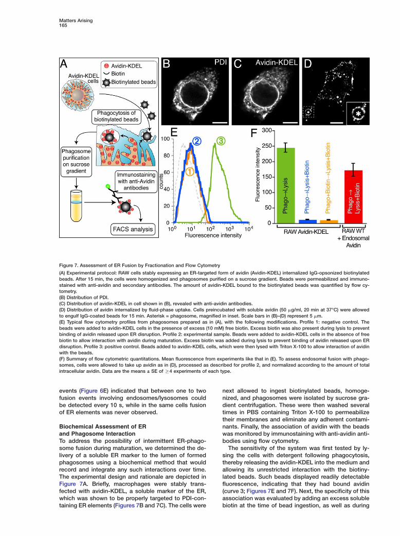
Biochemical Assessment of ERand Phagosome InteractionTo address the possibility of intermittent ER-phago-some fusion during maturation, we determined the de-livery of a soluble ER marker to the lumen of formedphagosomes using a biochemical method that wouldrecord and integrate any such interactions over time.The experimental design and rationale are depicted inFigure 7A. Briefly, macrophages were stably trans-fected with avidin-KDEL, a soluble marker of the ER,which was shown to be properly targeted to PDI-con-taining ER elements (Figures 7B and 7C). The cells were
Figure 7. Assessment of ER Fusion by Fractionation and Flow Cytometry
(A) Experimental protocol: RAW cells stably expressing an ER-targeted form of avidin (Avidin-KDEL) internalized IgG-opsonized biotinylatedbeads. After 15 min, the cells were homogenized and phagosomes purified on a sucrose gradient. Beads were permeabilized and immuno-stained with anti-avidin and secondary antibodies. The amount of avidin-KDEL bound to the biotinylated beads was quantified by flow cy-tometry.(B) Distribution of PDI.(C) Distribution of avidin-KDEL in cell shown in (B), revealed with anti-avidin antibodies.(D) Distribution of avidin internalized by fluid-phase uptake. Cells preincubated with soluble avidin (50 �g/ml, 20 min at 37°C) were allowedto engulf IgG-coated beads for 15 min. Asterisk = phagosome, magnified in inset. Scale bars in (B)–(D) represent 5 �m.(E) Typical flow cytometry profiles from phagosomes prepared as in (A), with the following modifications. Profile 1: negative control. Thebeads were added to avidin-KDEL cells in the presence of excess (10 mM) free biotin. Excess biotin was also present during lysis to preventbinding of avidin released upon ER disruption. Profile 2: experimental sample. Beads were added to avidin-KDEL cells in the absence of freebiotin to allow interaction with avidin during maturation. Excess biotin was added during lysis to prevent binding of avidin released upon ERdisruption. Profile 3: positive control. Beads added to avidin-KDEL cells, which were then lysed with Triton X-100 to allow interaction of avidinwith the beads.(F) Summary of flow cytometric quantitations. Mean fluorescence from experiments like that in (E). To assess endosomal fusion with phago-somes, cells were allowed to take up avidin as in (D), processed as described for profile 2, and normalized according to the amount of totalintracellular avidin. Data are the means ± SE of R4 experiments of each type.
next allowed to ingest biotinylated beads, homoge-nized, and phagosomes were isolated by sucrose gra-dient centrifugation. These were then washed severaltimes in PBS containing Triton X-100 to permeabilizetheir membranes and eliminate any adherent contami-nants. Finally, the association of avidin with the beadswas monitored by immunostaining with anti-avidin anti-bodies using flow cytometry.
The sensitivity of the system was first tested by ly-sing the cells with detergent following phagocytosis,thereby releasing the avidin-KDEL into the medium andallowing its unrestricted interaction with the biotiny-lated beads. Such beads displayed readily detectablefluorescence, indicating that they had bound avidin(curve 3; Figures 7E and 7F). Next, the specificity of thisassociation was evaluated by adding an excess solublebiotin at the time of bead ingestion, as well as during
Cell166
lysis. Only background levels of fluorescence were de-tected under these conditions (curve 1; Figures 7E and7F). Finally, we assessed if beads that had undergonephagocytosis gained access to avidin trapped in theER by allowing bead ingestion in the absence of biotin.Curve 2 in Figure 7E and the blue bar in Figure 7F indi-cated that no avidin was detectable on the beads. Sim-ilar negative results were obtained when phagosomeswere allowed to mature for up to 4 hr.
We used an analogous approach to assay the fusionof endosomes with phagosomes. Endosomes of RAWmacrophages were loaded with avidin by fluid phasepinocytosis (Figure 7D). When such preloaded cells in-gested biotinylated latex beads, avidin was consis-tently detected in the immediate vicinity of the internal-ized particles (Figure 7D). That the probe had in factreached the lumen of phagosomes was verified usingthe purification and flow cytometry approach describedabove. Endosomal avidin associated with the internal-ized biotinylated beads was readily detectable (Figure7F), despite the fact that the total amount of cellularavidin trapped in endosomes was substantially lowerthan that trapped in the ER in the preceding experi-ments.
Use of pH-Sensitive Proteins to Probe Associationof Phagosomes with the EROne of the defining characteristics of normal phago-somes is their ability to undergo rapid acidificationshortly after formation. Any components of the ER de-livered to the lumen of the phagosomes would there-fore experience an acute change in pH. We took advan-tage of the pH sensitivity of GFP and its derivatives(Tsien, 1998) to assess the fusion of phagosomes withthe ER by an independent approach. Macrophages sta-bly expressing the exofacial plasma membrane markerGPI-YFP, or the luminal ER marker GFP-KDEL, were al-lowed to internalize latex beads. The location of theseprobes with respect to the lumen of the phagosomecan be defined by monitoring the fluorescence changesinduced by rapidly dissipating the acidic pH of phago-somes. This can be simply accomplished by acute ad-dition of permeant weak bases, like NH3. As illustratedin Figures S7A and S7B, the emission of GPI-YFP lo-cated in endosomes and phagosomes increased mark-edly upon addition of NH3, indicating their presence inan acidic compartment. Ratio imaging of the fluores-cence before and after addition of NH3 yielded a 1.5-to 2.5-fold increase in emission intensity (Figures S7Cand S7D).
By contrast, addition of NH3 had no effect on thefluorescence of GFP-KDEL either in the immediate vi-cinity of phagosomes or elsewhere in the cell (FiguresS7E–S7H). The failure to see fluorescence changescannot be attributed to insensitivity of GFP-KDEL topH, since manipulation of the intracellular pH between5.5 and 7.5 caused pronounced changes in emission(Figures S7I–S7K). These findings are consistent withearlier conclusions that the ER is not acidic (Wu et al.,2001, 2000) and failed to give any indication of fusionof phagosomes with the ER.
QoWtnasmtipv1fsb1s6sFbddamr4cbwrtst
D
Tnaeswvspfptpa(SgppaatIt2
uantitative Assessment of the Contributionf Endosomes to the Phagosomal Membranehile we were unable to detect a significant contribu-
ion of the ER to the membrane of phagosomes, weevertheless found a comparatively rapid disappear-nce of plasmalemmal components from the vacuole,uch that only z30% could be accounted for by plas-alemmal components after 10 min (Figure 1). We
herefore sought to determine the source of membranenvolved in this remodeling. Because the classicalhagocytosis and maturation model postulates the in-olvement of endosomes (Lang et al., 1988; Pitt et al.,992), we devised a method for the quantitation of theraction of the phagosomal area contributed by endo-omal membranes. We selectively labeled endosomesy incubating macrophages at 37°C with FM4-64 for5 min prior to phagocytosis (Figure 8B), followed byelective removal of remaining plasma membrane FM4-4 by repeated washing in medium devoid of dye. Sub-equent addition of opsonized particles revealed thatM4-64 was rapidly delivered to the phagosomal mem-rane, verifying the occurrence of fusion between en-osomes and phagosomes (Figure 8C). The area of en-osomal membrane delivered to phagosomes was nextpproximated by comparing the intensity of phagoso-al FM4-64 fluorescence with plasma membrane fluo-
escence of the same cells incubated with FM4-64 at°C (Figure 8A). These calculations suggested that theontribution of endosomes to the phagosomal mem-rane increased between 2–10 min, from w40% to60%. When added to the fraction contributed by the
emaining plasma membrane, the endosomal contribu-ion accounts for most of the phagosomal area andets a very modest upper limit to any possible contribu-ion by the ER.
iscussion
he ability of macrophages and dendritic cells to elimi-ate invading microorganisms and to process theirntigens for presentation relies on the dynamic remod-ling of the membrane and contents of their phago-omes. Most of the functions attributed to phagosomesere thought to result from a maturation process in-olving sequential fusion to endosomes and lyso-omes. Recently, however, an alternate model was pro-osed by Gagnon et al. (2002), invoking fusion of the
orming phagosomes with the ER. This model is sup-orted by earlier observations that Dictyostelium mu-ants lacking calreticulin and calnexin have reducedhagocytosis (Muller-Taubenberger et al., 2001) and islso consistent with recent findings of Becker et al.2005) that impairment of the function of ERS24/ec22b, a SNARE protein of the ER, reduced the en-ulfment of 3 �m (but not 0.8 �m) particles by macro-hages. The possibility that the ER fused with thelasma membrane during phagocytosis had immenseppeal in the context of antigen cross-presentation. Asresult, several recent reports focusing on the capabili-
ies of phagosomes to process antigen for MHC classpresentation emphasized the ER-mediated phagocy-
osis model (Ackerman et al., 2003; Guermonprez et al.,003; Houde et al., 2003). Finally, the ER has also come

Matters Arising167
Figure 8. Quantitation of Endosomal Contribution to the Phagosome Membrane
(A) Labeling of the plasma membrane of RAW cells with FM4-64.(B) Labeling of endosomes with FM4-64 following incubation with dye for 15 min at 37°C and subsequent removal of the plasmalemmal FM4-64 by washing.(C) Endosomal fusion with phagosomes, assessed after labeling with FM4-64 as in (B), followed by ingestion of beads for 10 min. Scale barsin (A)–(C) represent 5 �m.(D) Mean phagosomal FM4-64 fluorescence/pixel measured as in (C) is expressed relative to the plasmalemmal FM4-64 fluorescence, mea-sured in the same cells as in (A). Normalized fluorescence intensities are illustrated (dark bars). Data are the means of 40 experiments. Forreference, the contribution of the plasma membrane to the phagosomes (open bars) is replotted from Figure 1.
to be viewed as providing a quantitatively importantreservoir of membrane inserted for the internalizationof large particles (Brode and Macary, 2004; Desjardins,2003; Niedergang and Chavrier, 2004).
On the other hand, several considerations make itdifficult to conceive that the ER would fuse directly withthe plasma membrane during particle uptake. A majorcontribution of the ER to the phagosomal membraneis difficult to reconcile with the well-established rapidacidification of the phagosomal lumen. The membraneof the ER is devoid of active proton-pumping V-ATPasesand is extremely leaky to proton equivalents (Paroutiset al., 2004; Wu et al., 2001). It was also unclear howthe proposed connection of the ER with the base of theforming phagosome would alter the ability of the ERto retain calcium, or nascent and misfolded secretoryproteins contained within its lumen.
This led us to reexamine the role of the ER duringphagocytosis. We devised six independent and quanti-tative methods to explore if the ER provides a substan-tial contribution to phagosomes in different cell typesfrom two species. Our experiments failed to produceany evidence in support of fusion of the ER with eithernascent or maturing phagosomes. It is noteworthy thatwe used phagocytic targets of various kinds that wereof similar size those employed by Gagnon et al. (2002)and that we tested primary and immortalized macro-phages, as well as dendritic cells. Therefore, neither dif-ference in particle size or cell type can readily accountfor the apparent discrepancies. By contrast, we couldeasily document the presence of plasmalemmal rem-nants in the early phagosomes as well as the fusion ofendosomes/lysosomes by several different approaches.The progressive replacement of plasma membranecomponents by endosomal membranes was verified inexperiments like that of Movie S6. The rapid and grad-ual insertion of membranes of the endocytic pathway,
which are endowed with V-ATPases and relatively tightto protons is eminently consistent with the develop-ment of phagosomal acidification.
The original hypothesis of ER-mediated phagocyto-sis stemmed from biochemical determinations of theprotein composition of purified phagosomes (Desjar-dins et al., 1994a; Garin et al., 2001). As in any otherbiochemical purification procedure, contaminants arean inevitable component of the final preparation. Owingto its abundance, the ER is the most likely source ofcontamination of particulate preparations, and may ac-count for the observed presence of the ER proteins inimmunoblots and mass-spectroscopy analyses of phago-somes. Accordingly, treatment of “purified” phagosomeswith ATP led to a 5- to 10-fold decrease in the content ofER markers and a concomitant enrichment for cathepsinD, a bona fide marker of phagolysosomes (Gotthardt etal., 2002). These observations imply that biochemicaldetection of ER markers on phagosomal preparationsis not necessarily evidence that the phagosome mem-brane is made up largely, or even partly, of ER.
Although contamination by ER may account for someof the biochemical observations, it cannot explain theevidence obtained by Gagnon et al. (2002) using EM.The striking observation of products of G6Pase in closeassociation with the phagosome membrane appears tolend strong support to the theory of ER-phagosome fu-sion. These findings contrast with our inability to detectmarkers of the ER within the phagosome by EM (Fig-ures 3, 4, S3, and S4). Cytochemical reactions such asthe cerium capture used for G6Pase determinations areoften more sensitive than immunogold, possibly ex-plaining the differential results. However, such reac-tions are not nearly as specific as immuno-EM and mayreflect a minor nonspecific contribution of other en-zymes, particularly in tissues with low G6Pase activity.The hydrolysis of glucose 6-phosphate can also be cat-
Cell168
alyzed by other enzymes, including alkaline and acidphosphatases, which are enriched in the plasma mem-brane and lysosomes, respectively (Matsubara et al.,1999; Nichols et al., 1984; Ulsamer et al., 1971). In ourexperiments, alkaline phosphatase activity at the plasmamembrane was not detectable, and reaction productdue to lysosomal acid phosphatase was rarely seen.Under these conditions, we consistently failed to detectG6Pase activity in phagosomes formed by ingestion ofdifferent types of particles, while clear and reproduciblestaining of the nuclear membrane and of the ER wasobtained routinely (Figures 4 and S4). Therefore, ourobservations are more consistent with previous studiesreporting low G6Pase activity in phagosomal mem-branes, while finding a 10-fold enrichment of plasma-lemmal NADPH oxidase and 5#-nucleotidase activity(Bellavite et al., 1982; Berton et al., 1982).
While our quantitative determination of the composi-tion of the phagosomal membrane leaves little room forER components, it does not rule out fusion of phago-somes with a small amount of ER, or the possibility thatthe phagosome communicates with the ER by alterna-tive pathways of vesicular traffic. Even a small frac-tional contribution of the ER to the membrane of thephagosome is compatible with its proposed role incross-presentation by MHC class I (Ackerman et al.,2003; Guermonprez et al., 2003; Houde et al., 2003).Alternatively, antigen-containing vesicles may pinch offthe phagosome and undergo retrograde transport tothe Golgi complex and ultimately the ER, as has beenshown for material that is internalized by endocytosis(Miesenbock and Rothman, 1995; Sandvig and vanDeurs, 2002). In this regard, it is noteworthy that com-ponents of the COPI coatomer system that mediatesretrograde transport from Golgi to the ER are alsofound on endosomes (Aniento et al., 1996; Daro et al.,1997; Whitney et al., 1995) and can affect the matura-tion of both endosomes and phagosomes. Material in-ternalized by caveolar endocytosis can be deliveredmore directly to the ER, possibly bypassing the Golgicomplex (Pelkmans et al., 2001), and a similar routemay apply to vesicles derived from phagosomes, whichappear to contain caveolar components (Dermine et al.,2001). Moreover, selected components of the ER couldbe delivered to the maturing phagosome via the trans-Golgi network in a manner akin to the delivery of newlysynthesized proteins to prelysosomes. Lastly, cross-presentation can also occur by the novel processdescribed recently by Neijssen et al. (2005), wherebypeptides are delivered by other cells to the cytosol ofantigen-presenting cells via gap junctions.
Our observation that the contribution of the plasmamembrane to phagosome formation decreases overtime, together with the notion that the total surface areaof macrophages remains constant (Hackam et al.,1998), or even increases (Holevinsky and Nelson, 1998),during particle engulfment, is suggestive of a mecha-nism for the recycling of plasmalemmal componentsback to the cell surface following phagosome for-mation.
The concept of phagosome membrane recyclingback to the cell surface is not without precedent, asstudies by Muller et al. and by de Chastellier et al. dem-onstrated that early phagosomes efficiently recycled
[bM
vitdbfato(vutqb
E
CRtmccdwauK
PPhpetAihpcIwba2
LTmPsci1IdCw(cs
FDg
125I]-labeled or [3H]galactose-labeled glycoconjugatesack to the cell surface (de Chastellier et al., 1983;uller et al., 1980a).It is important to note that the contribution of indi-
idual cellular compartments to forming phagosomess likely to vary with particle size (Cox et al., 1999). Inhe case of Fc receptors and particles of 1–3 �m iniameter, such as those used in our studies, the contri-ution of the plasma membrane and endosomes to the
orming and maturing phagosome appears indisput-ble. However, other compartments, possibly includinghe ER, may play a more important role for phagosomesf other sizes or those induced by different receptors
Cox et al., 1999; Henry et al., 2004). Therefore, the in-olvement of the ER in phagosome formation and mat-ration cannot be discounted, but direct demonstrationhat ER-to-phagosome fusion occurs and that it is re-uired for phagocytosis must be established on a case-y-case basis.
xperimental Procedures
ell Culture and TransfectionAW264.7 and J774 macrophages obtained from ATCC were cul-
ured in DMEM with 10% heat-inactivated fetal calf serum. Bone-arrow-derived mouse macrophages and DCs were obtained and
ultured as in (de Chastellier et al., 1995; Chow et al., 2002). RAWells stably transfected with GPI-GFP, GPI-YFP, GFP-KDEL, or avi-in-KDEL were selected using G418. Clones stably transfectedith avidin-KDEL were identified by immunostaining, using anti-vidin D antibody (Vector; 1:1000 dilution). Retroviral infection wassed for expression of GFP-KDEL in J774 macrophages and GFP-DEL and lgp120-GFP in DCs, as described by Chow et al. (2002).
hagocytosisolystyrene beads (Bangs; 3.1 �m) were opsonized with 1 mg/mluman IgG (Sigma) in PBS for 1 hr at room temperature. To inducehagocytosis, opsonized beads were added to cells and adher-nce synchronized by centrifugation for 20 s at 200 g. Adherenceo DC was induced by incubation with beads for 10 min at 4°C.fter completion of phagocytosis at 37°C, extracellular beads were
dentified by incubation with 1:500 dilution of Cy3-conjugated anti-uman antibody (Jackson). Where indicated, unopsonized hydro-hilic or hydrophobic latex beads (1 or 3 �m; Sigma) or sheep redells (MP Biomedicals) opsonized with rabbit anti-sheep IgG (1:50;
CN Biomedicals) were used. Bone-marrow-derived macrophagesere incubated for 15 or 30 min at 37°C with the different types ofeads or infected as previously described with Mycobacteriumvium (de Chastellier et al., 1995) or Brucella abortus (Celli et al.,003).
abeling and Immunostainingo label the plasma membrane, cells were incubated with 1 mg/l of Alexa 555-conjugated B subunit of cholera toxin (Molecularrobes) for 2–5 min prior to phagocytosis. Alternatively, a 10 �Molution of FM4-64 (Molecular Probes) was present during phago-ytosis. To label endosomes prior to phagocytosis, RAW cells were
ncubated with 2 �M FM4-64 in Hepes-buffered RPMI at 37°C for5 min, followed by washing to remove residual plasmalemmal dye.
mmunostaining of PDI was performed by incubating with a 1:500ilution of monoclonal anti-PDI antibody (Stressgen), followed byy3-conjugated goat anti-mouse antibody (Jackson). Avidin-KDELas detected using a 1:1000 dilution of a anti-avidin D antibody
Vector) followed by a Cy3-coupled anti-goat (Jackson). Fluores-ence microscopy and image analysis were essentially as de-cribed (Marshall et al., 2001).
rustrated Phagocytosis and TIR-FMetached RAW cells were allowed to spread onto IgG-opsonizedlass coverslips and analyzed by TIR-FM using an Olympus micro-

Matters Arising169
scope IX-70 equipped with TIRF accessories. To monitor fusion ofacidic organelles, macrophages were incubated with 5 �M acridineorange for 5 min and washed prior to deposition onto coverslips.
Immunogold Labeling, Immunocytochemistry, and EMPreparation of RAW, J774 cells and murine DCs for immuno-EM,cryosectioning and immunolabeling was as described before(Folsch et al., 2001). Cells were fixed for 1 hr in 4% paraformalde-hyde, followed by overnight fixation in 8% paraformaldehyde at4°C. Human DCs were fixed with 2% paraformaldehyde and pro-cessed as described (Peters and Hunziker, 2001). Quantification ofgold particles was according to Mayhew et al. (2002). Staining forG6Pase was performed as described by Griffiths et al. (1983) withthe modifications of Celli et al. (2003).
Biochemical Assessment of ER-Phagosome FusionRAW cells stably expressing avidin-KDEL were plated onto 10 cmpetri dishes and allowed to ingest biotin-coated polystyrene beads(3.0–3.9 �m) opsonized with 0.1 mg/ml IgG. Phagocytosis was ter-minated, and the cells were harvested with 1 ml of 0.25 M sucrose,3 mM imidazole, (pH 7.4) and disrupted using a Dounce homoge-nizer. Unless otherwise indicated, excess biotin (10 mM) was pres-ent during and after homogenization to prevent binding of avidinreleased upon disruption of the ER. Phagosomes were isolated asdescribed (Gagnon et al., 2002), permeabilized, and washed with0.1% Triton X-100, and immunostained with anti-avidin followed byAlexa 488-conjugated secondary. Endosomes were loaded by fluidphase uptake of 1 mg/ml avidin for 20 min at 37°C.
Supplemental DataSupplemental Data include seven figures, one table, SupplementalExperimental Procedures, and six movies and can be found with thisarticle online at http://www.cell.com/cgi/content/full/123/1/157/DC1/.
Acknowledgments
We thank Dr. A. Kenworthy (Vanderbilt University, Nashville, Ten-nessee) for the YFP-GT46 cDNA; Dr. A.D. Schreiber (University ofPennsylvania, Pennsylvania) for the FcγRIIa-GFP cDNA, Dr. S. High(University of Manchester, Manchester, United Kingdom) for theSec61-GFP cDNA, Dr. H. Ploegh (Harvard Medical School, Boston)for anti-PDI antibody, Drs. D. Williams (University of Toronto) and J.Bergeron (McGill University) for anti-calnexin antibodies. We thankDr. M. Michalak (University of Alberta) for useful suggestions. N.T.is supported by the Heart and Stroke Foundation of Canada, P.P.by a Canadian Cystic Fibrosis Foundation studentship; M.T. by theCanadian Association of Gastroenterology, S.G. by the CanadianInstitutes of Health Research; N.v.d.W. by the Netherlands LeprosyRelief and C.C. by the French Institut National de la Santé et de laRecherche Médicale (INSERM).
Received: April 8, 2005Revised: June 29, 2005Accepted: August 1, 2005Published: October 6, 2005
References
Ackerman, A.L., Kyritsis, C., Tampe, R., and Cresswell, P. (2003).Early phagosomes in dendritic cells form a cellular compartmentsufficient for cross presentation of exogenous antigens. Proc. Natl.Acad. Sci. USA 100, 12889–12894.
Aniento, F., Gu, F., Parton, R.G., and Gruenberg, J. (1996). An endo-somal beta COP is involved in the pH-dependent formation oftransport vesicles destined for late endosomes. J. Cell Biol. 133,29–41.
Becker, T., Volchuk, A., and Rothman, J.E. (2005). Differential useof endoplasmic reticulum membrane for phagocytosis in J774 mac-rophages. Proc. Natl. Acad. Sci. USA 102, 4022–4026.
Bellavite, P., Serra, M.C., Davoli, A., and Rossi, F. (1982). Selectiveenrichment of NADPH oxidase activity in phagosomes from guineapig polymorphonuclear leukocytes. Inflammation 6, 21–29.
Berton, G., Bellavite, P., de Nicola, G., Dri, P., and Rossi, F. (1982).Plasma membrane and phagosome localisation of the activatedNADPH oxidase in elicited peritoneal macrophages of the guinea-pig. J. Pathol. 136, 241–252.
Braun, V., Fraisier, V., Raposo, G., Hurbain, I., Sibarita, J.B.,Chavrier, P., Galli, T., and Niedergang, F. (2004). TI-VAMP/VAMP7 isrequired for optimal phagocytosis of opsonised particles in macro-phages. EMBO J. 23, 4166–4176.
Brode, S., and Macary, P.A. (2004). Cross-presentation: dendriticcells and macrophages bite off more than they can chew! Immunol-ogy 112, 345–351.
Celli, J., de Chastellier, C., Franchini, D.M., Pizarro-Cerda, J., Mor-eno, E., and Gorvel, J.P. (2003). Brucella evades macrophage killingvia VirB-dependent sustained interactions with the endoplasmic re-ticulum. J. Exp. Med. 198, 545–556.
Chow, A., Toomre, D., Garrett, W., and Mellman, I. (2002). Dendriticcell maturation triggers retrograde MHC class II transport from ly-sosomes to the plasma membrane. Nature 418, 988–994.
Cox, D., Tseng, C.C., Bjekic, G., and Greenberg, S. (1999). Arequirement for phosphatidylinositol 3-kinase in pseudopod exten-sion. J. Biol. Chem. 274, 1240–1247.
Daro, E., Sheff, D., Gomez, M., Kreis, T., and Mellman, I. (1997).Inhibition of endosome function in CHO cells bearing a temper-ature-sensitive defect in the coatomer (COPI) component epsilon-COP. J. Cell Biol. 139, 1747–1759.
de Chastellier, C., Lang, T., and Thilo, L. (1995). Phagocytic pro-cessing of the macrophage endoparasite, Mycobacterium avium,in comparison to phagosomes which contain Bacillus subtilis orlatex beads. Eur. J. Cell Biol. 68, 167–182.
de Chastellier, C., Ryter, A., and Thilo, L. (1983). Membrane shuttlebetween plasma membrane, phagosomes, and pinosomes in Dic-tyostelium discoideum amoeboid cells. Eur. J. Cell Biol. 30, 233–243.
de Chastellier, C., and Thilo, L. (1997). Phagosome maturation andfusion with lysosomes in relation to surface property and size ofthe phagocytic particle. Eur. J. Cell Biol. 74, 49–62.
de Chastellier, C., and Thilo, L. (2002). Pathogenic Mycobacteriumavium remodels the phagosome membrane in macrophages withindays after infection. Eur. J. Cell Biol. 81, 17–25.
Dermine, J.F., Duclos, S., Garin, J., St-Louis, F., Rea, S., Parton,R.G., and Desjardins, M. (2001). Flotillin-1-enriched lipid raft do-mains accumulate on maturing phagosomes. J. Biol. Chem. 276,18507–18512.
Desjardins, M. (2003). ER-mediated phagocytosis: a new mem-brane for new functions. Nat. Rev. Immunol. 3, 280–291.
Desjardins, M., Celis, J.E., van Meer, G., Dieplinger, H., Jahraus, A.,Griffiths, G., and Huber, L.A. (1994a). Molecular characterization ofphagosomes. J. Biol. Chem. 269, 32194–32200.
Desjardins, M., Huber, L.A., Parton, R.G., and Griffiths, G. (1994b).Biogenesis of phagolysosomes proceeds through a sequentialseries of interactions with the endocytic apparatus. J. Cell Biol.124, 677–688.
Fix, M., Melia, T.J., Jaiswal, J.K., Rappoport, J.Z., You, D., Sollner,T.H., Rothman, J.E., and Simon, S.M. (2004). Imaging single mem-brane fusion events mediated by SNARE proteins. Proc. Natl. Acad.Sci. USA 101, 7311–7316.
Folsch, H., Pypaert, M., Schu, P., and Mellman, I. (2001). Distribu-tion and function of AP-1 clathrin adaptor complexes in polarizedepithelial cells. J. Cell Biol. 152, 595–606.
Gagnon, E., Duclos, S., Rondeau, C., Chevet, E., Cameron, P.H.,Steele-Mortimer, O., Paiement, J., Bergeron, J.J., and Desjardins,M. (2002). Endoplasmic reticulum-mediated phagocytosis is amechanism of entry into macrophages. Cell 110, 119–131.
Garin, J., Diez, R., Kieffer, S., Dermine, J.F., Duclos, S., Gagnon, E.,Sadoul, R., Rondeau, C., and Desjardins, M. (2001). The phago-some proteome: insight into phagosome functions. J. Cell Biol. 152,165–180.
Gotthardt, D., Warnatz, H.J., Henschel, O., Bruckert, F., Schleicher,M., and Soldati, T. (2002). High-resolution dissection of phagosome
Cell170
maturation reveals distinct membrane trafficking phases. Mol. Biol.Cell 13, 3508–3520.
Greenberg, S., and Grinstein, S. (2002). Phagocytosis and innateimmunity. Curr. Opin. Immunol. 14, 136–145.
Griffiths, G., Simons, K., Warren, G., and Tokuyasu, K.T. (1983). Im-munoelectron microscopy using thin, frozen sections: applicationto studies of the intracellular transport of Semliki Forest virus spikeglycoproteins. Methods Enzymol. 96, 466–485.
Guermonprez, P., Saveanu, L., Kleijmeer, M., Davoust, J., Van En-dert, P., and Amigorena, S. (2003). ER-phagosome fusion definesan MHC class I cross-presentation compartment in dendritic cells.Nature 425, 397–402.
Hackam, D.J., Rotstein, O.D., Sjolin, C., Schreiber, A.D., Trimble,W.S., and Grinstein, S. (1998). v-SNARE-dependent secretion is re-quired for phagocytosis. Proc. Natl. Acad. Sci. USA 95, 11691–11696.
Henry, R.M., Hoppe, A.D., Joshi, N., and Swanson, J.A. (2004). Theuniformity of phagosome maturation in macrophages. J. Cell Biol.164, 185–194.
Holevinsky, K.O., and Nelson, D.J. (1998). Membrane capacitancechanges associated with particle uptake during phagocytosis inmacrophages. Biophys. J. 75, 2577–2586.
Houde, M., Bertholet, S., Gagnon, E., Brunet, S., Goyette, G.,Laplante, A., Princiotta, M.F., Thibault, P., Sacks, D., and Desjar-dins, M. (2003). Phagosomes are competent organelles for antigencross-presentation. Nature 425, 402–406.
Kenworthy, A.K., Nichols, B.J., Remmert, C.L., Hendrix, G.M., Ku-mar, M., Zimmerberg, J., and Lippincott-Schwartz, J. (2004). Dy-namics of putative raft-associated proteins at the cell surface. J.Cell Biol. 165, 735–746.
Kwiatkowska, K., Frey, J., and Sobota, A. (2003). Phosphorylationof FcgammaRIIA is required for the receptor-induced actin re-arrangement and capping: the role of membrane rafts. J. Cell Sci.116, 537–550.
Lang, T., de Chastellier, C., Ryter, A., and Thilo, L. (1988). Endocyticmembrane traffic with respect to phagosomes in macrophagesinfected with non-pathogenic bacteria: phagosomal membraneacquires the same composition as lysosomal membrane. Eur. J.Cell Biol. 46, 39–50.
Lippincott-Schwartz, J., Altan-Bonnet, N., and Patterson, G.H.(2003). Photobleaching and photoactivation: following protein dy-namics in living cells. Nat. Cell Biol. 46 (Suppl), S7–14.
Marshall, J.G., Booth, J.W., Stambolic, V., Mak, T., Balla, T.,Schreiber, A.D., Meyer, T., and Grinstein, S. (2001). Restricted accu-mulation of phosphatidylinositol 3-kinase products in a plasmalem-mal subdomain during Fc gamma receptor-mediated phagocytosis.J. Cell Biol. 153, 1369–1380.
Matsubara, S., Yamada, T., Minakami, H., Watanabe, T., Takizawa,T., and Sato, I. (1999). Polymorphonuclear leukocytes in the fetalmembranes are activated in patients with preterm delivery: ultra-structural and enzyme-histochemical evidence. Placenta 20, 185–188.
Mayhew, T.M., Lucocq, J.M., and Griffiths, G. (2002). Relative label-ling index: a novel stereological approach to test for non-randomimmunogold labelling of organelles and membranes on transmis-sion electron microscopy thin sections. J. Microsc. 205, 153–164.
Miesenbock, G., and Rothman, J.E. (1995). The capacity to retrieveescaped ER proteins extends to the trans-most cisterna of theGolgi stack. J. Cell Biol. 129, 309–319.
Muller, W.A., Steinman, R.M., and Cohn, Z.A. (1980a). The mem-brane proteins of the vacuolar system I. Analysis of a novel methodof intralysosomal iodination. J. Cell Biol. 86, 292–303.
Muller, W.A., Steinman, R.M., and Cohn, Z.A. (1980b). The mem-brane proteins of the vacuolar system. II. Bidirectional flow be-tween secondary lysosomes and plasma membrane. J. Cell Biol.86, 304–314.
Muller-Taubenberger, A., Lupas, A.N., Li, H., Ecke, M., Simmeth, E.,and Gerisch, G. (2001). Calreticulin and calnexin in the endoplasmicreticulum are important for phagocytosis. EMBO J. 20, 6772–6782.
NeSi
NNt
N6i1
Nd(
NaF
PsP
Pdt
PRg
PaI
SiF
Scs
Ss
SE9
Ta
Tc
UPJ
Vm
Ws
W(C
WTtt
WMs
WMs1
ehls, S., Snapp, E.L., Cole, N.B., Zaal, K.J., Kenworthy, A.K., Rob-rts, T.H., Ellenberg, J., Presley, J.F., Siggia, E., and Lippincott-chwartz, J. (2000). Dynamics and retention of misfolded proteins
n native ER membranes. Nat. Cell Biol. 2, 288–295.
eijssen, J., Herberts, C., Drijfhout, J.W., Reits, E., Janssen, L., andeefjes, J. (2005). Cross-presentation by intercellular peptide
ransfer through gap junctions. Nature 434, 83–88.
ichols, B.A., Setzer, P.Y., and Bainton, D.F. (1984). Glucose-phosphatase as a cytochemical marker of endoplasmic reticulum
n human leukocytes and platelets. J. Histochem. Cytochem. 32,65–171.
iedergang, F., and Chavrier, P. (2004). Signaling and membraneynamics during phagocytosis: many roads lead to the phagos
R)ome. Curr. Opin. Cell Biol. 16, 422–428.
inomiya, N., Hazeki, K., Fukui, Y., Seya, T., Okada, T., Hazeki, O.,nd Ui, M. (1994). Involvement of phosphatidylinositol 3-kinase inc gamma receptor signaling. J. Biol. Chem. 269, 22732–22737.
aroutis, P., Touret, N., and Grinstein, S. (2004). The pH of theecretory pathway: measurement, determinants, and regulation.hysiology (Bethesda) 19, 207–215.
elkmans, L., Kartenbeck, J., and Helenius, A. (2001). Caveolar en-ocytosis of simian virus 40 reveals a new two-step vesicular-
ransport pathway to the ER. Nat. Cell Biol. 3, 473–483.
eters, P.J., and Hunziker, W. (2001). Subcellular localization ofab17 by cryo-immunogold electron microscopy in epithelial cellsrown on polycarbonate filters. Methods Enzymol. 329, 210–225.
itt, A., Mayorga, L.S., Stahl, P.D., and Schwartz, A.L. (1992). Alter-tions in the protein composition of maturing phagosomes. J. Clin.
nvest. 90, 1978–1983.
andvig, K., and van Deurs, B. (2002). Transport of protein toxinsnto cells: pathways used by ricin, cholera toxin and Shiga toxin.EBS Lett. 529, 49–53.
ilverstein, S.C. (1977). Endocytic uptake of particles by mononu-lear phagocytes and the penetration of obligate intracellular para-ites. Am. J. Trop. Med. Hyg. 26, 161–169.
ilverstein, S.C., Steinman, R.M., and Cohn, Z.A. (1977). Endocyto-is. Annu. Rev. Biochem. 46, 669–722.
teinman, R.M., Mellman, I.S., Muller, W.A., and Cohn, Z.A. (1983).ndocytosis and the recycling of plasma membrane. J. Cell Biol.6, 1–27.
jelle, T.E., Lovdal, T., and Berg, T. (2000). Phagosome dynamicsnd function. Bioessays 22, 255–263.
sien, R.Y. (1998). The green fluorescent protein. Annu. Rev. Bio-hem. 67, 509–544.
lsamer, A.G., Wright, P.L., Wetzel, M.G., and Korn, E.D. (1971).lasma and phagosome membranes of Acanthamoeba castellanii.. Cell Biol. 51, 193–215.
ieira, O.V., Botelho, R.J., and Grinstein, S. (2002). Phagosomeaturation: aging gracefully. Biochem. J. 366, 689–704.
atts, C., and Amigorena, S. (2001). Phagocytosis and antigen pre-entation. Semin. Immunol. 13, 373–379.
hitney, J.A., Gomez, M., Sheff, D., Kreis, T.E., and Mellman, I.1995). Cytoplasmic coat proteins involved in endosome function.ell 83, 703–713.
iertz, E.J., Tortorella, D., Bogyo, M., Yu, J., Mothes, W., Jones,.R., Rapoport, T.A., and Ploegh, H.L. (1996). Sec61-mediatedransfer of a membrane protein from the endoplasmic reticulum tohe proteasome for destruction. Nature 384, 432–438.
u, M.M., Grabe, M., Adams, S., Tsien, R.Y., Moore, H.P., andachen, T.E. (2001). Mechanisms of pH regulation in the regulated
ecretory pathway. J. Biol. Chem. 276, 33027–33035.
u, M.M., Llopis, J., Adams, S., McCaffery, J.M., Kulomaa, M.S.,achen, T.E., Moore, H.P., and Tsien, R.Y. (2000). Organelle pH
tudies using targeted avidin and fluorescein-biotin. Chem. Biol. 7,97–209.