A Versatile Terahertz Chemical Microscope and Its Application ...
Yasuko Numata et al.: Qualitative Study of New Bone Formation
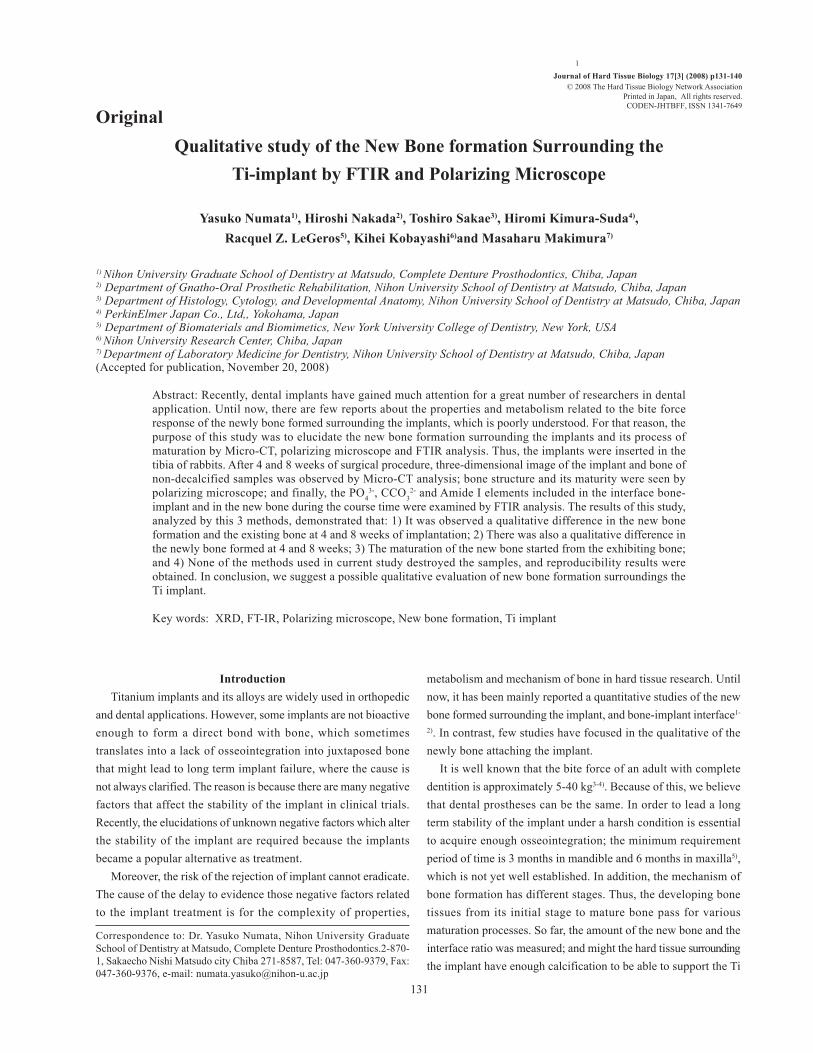
Qualitative study of the New Bone formation Surrounding theTi-implant by FTIR and Polarizing Microscope
Yasuko Numata1), Hiroshi Nakada2), Toshiro Sakae3), Hiromi Kimura-Suda4),Racquel Z. LeGeros5), Kihei Kobayashi6)and Masaharu Makimura7)
1) Nihon University Graduate School of Dentistry at Matsudo, Complete Denture Prosthodontics, Chiba, Japan2) Department of Gnatho-Oral Prosthetic Rehabilitation, Nihon University School of Dentistry at Matsudo, Chiba, Japan3) Department of Histology, Cytology, and Developmental Anatomy, Nihon University School of Dentistry at Matsudo, Chiba, Japan4) PerkinElmer Japan Co., Ltd,, Yokohama, Japan5) Department of Biomaterials and Biomimetics, New York University College of Dentistry, New York, USA6) Nihon University Research Center, Chiba, Japan7) Department of Laboratory Medicine for Dentistry, Nihon University School of Dentistry at Matsudo, Chiba, Japan(Accepted for publication, November 20, 2008)
Abstract: Recently, dental implants have gained much attention for a great number of researchers in dentalapplication. Until now, there are few reports about the properties and metabolism related to the bite forceresponse of the newly bone formed surrounding the implants, which is poorly understood. For that reason, thepurpose of this study was to elucidate the new bone formation surrounding the implants and its process ofmaturation by Micro-CT, polarizing microscope and FTIR analysis. Thus, the implants were inserted in thetibia of rabbits. After 4 and 8 weeks of surgical procedure, three-dimensional image of the implant and bone ofnon-decalcified samples was observed by Micro-CT analysis; bone structure and its maturity were seen bypolarizing microscope; and finally, the PO4
3-, CCO32- and Amide I elements included in the interface bone-
implant and in the new bone during the course time were examined by FTIR analysis. The results of this study,analyzed by this 3 methods, demonstrated that: 1) It was observed a qualitative difference in the new boneformation and the existing bone at 4 and 8 weeks of implantation; 2) There was also a qualitative difference inthe newly bone formed at 4 and 8 weeks; 3) The maturation of the new bone started from the exhibiting bone;and 4) None of the methods used in current study destroyed the samples, and reproducibility results wereobtained. In conclusion, we suggest a possible qualitative evaluation of new bone formation surroundings theTi implant.
Key words: XRD, FT-IR, Polarizing microscope, New bone formation, Ti implant
Correspondence to: Dr. Yasuko Numata, Nihon University GraduateSchool of Dentistry at Matsudo, Complete Denture Prosthodontics.2-870-1, Sakaecho Nishi Matsudo city Chiba 271-8587, Tel: 047-360-9379, Fax:047-360-9376, e-mail: [email protected]
IntroductionTitanium implants and its alloys are widely used in orthopedic
and dental applications. However, some implants are not bioactiveenough to form a direct bond with bone, which sometimestranslates into a lack of osseointegration into juxtaposed bonethat might lead to long term implant failure, where the cause isnot always clarified. The reason is because there are many negativefactors that affect the stability of the implant in clinical trials.Recently, the elucidations of unknown negative factors which alterthe stability of the implant are required because the implantsbecame a popular alternative as treatment.
Moreover, the risk of the rejection of implant cannot eradicate.The cause of the delay to evidence those negative factors relatedto the implant treatment is for the complexity of properties,
metabolism and mechanism of bone in hard tissue research. Untilnow, it has been mainly reported a quantitative studies of the newbone formed surrounding the implant, and bone-implant interface1-
2). In contrast, few studies have focused in the qualitative of thenewly bone attaching the implant. It is well known that the bite force of an adult with completedentition is approximately 5-40 kg3-4). Because of this, we believethat dental prostheses can be the same. In order to lead a longterm stability of the implant under a harsh condition is essentialto acquire enough osseointegration; the minimum requirementperiod of time is 3 months in mandible and 6 months in maxilla5),which is not yet well established. In addition, the mechanism ofbone formation has different stages. Thus, the developing bonetissues from its initial stage to mature bone pass for variousmaturation processes. So far, the amount of the new bone and theinterface ratio was measured; and might the hard tissue surroundingthe implant have enough calcification to be able to support the Ti
Journal of Hard Tissue Biology 17[3] (2008) p131-140 © 2008 The Hard Tissue Biology Network Association
Printed in Japan, All rights reserved.CODEN-JHTBFF, ISSN 1341-7649
Original
131
J.Hard Tissue Biology Vol. 17(3):131-140. 2008implant similar to the existing bone?. Isn’t that the bone tissueincludes various maturation levels being all of them summarizedin one word called new bone? The early stage of bone formationbecomes an important key to obtain enough osseointegration.Especially, there is a tissue that supports directly the implant afteroperation, which become a crucial element. In order to evaluatethe crystallinity in the existing bone and in the new bone, it wasused a polarizing microscope, Laboratory for Electron BeamResearch and Application Institute of Quantum Science, NihonUniversity-Parametric, X-ray radiation (LEBRA-PXR),microscope Fourier transform infrared (FTIR), Micro-XRD(Micro- X- ray Diffractometer).6) Moreover, Nakada et al. reportedthat X-ray Photoelectron Spectrometer detected the differenceabout the element contents of existing bone and new bone. 7). Onthe other hand, it has not still clarified about the qualitative changeof the new bone in relation with the position of new bone, implantand cortical bone. Until now, the crystal analysis of the hard tissueis not clear because its low crystallinity with a complex organicand inorganic structure. This could be the reason why there are afew researches investigating the qualitative difference of the newand existing bone. However, even when the prediction of earlyincrease of weight raises, it is possible to say that estimation ofnew bone is very important item same as the measurement ofbone amount
The main components of the bone are collagen fiber as anorganic component, and hydroxyapatite as an inorganiccomponent. It is well known that hydroxyapatite is one kind ofcalcium phosphate composed of Ca10(PO4)6OH2. But actually,various minuscule amount of molecules and elements are replacedCa2+,PO4
3-, OH- for CO32-, Na+, Mg 2+ in the apatite in vivo. On the
other hand, CO32- is included in the bone tissue in approximately
at 7% producing a distortion in the ideal crystal structure9).Therefore, hydroxyapatite included in the bone express PO4
3-;CO3
2- and collagen influencing in the distortion of hydroxyapatitecrystallinization express Amide I; for that reason, we believe thatan estimation of the qualitative difference of bone of them maybe performed by FT-IR analysis.
The purpose of the present study was to analyzed three-dimensional image of the implant and the bone of non-decalcifiedsamples by Microcomputed tomography (Micro-CT); bonestructure and their maturity by polarizing microscope; and finally,the PO4
3-,CO32- and Amide I elements included in the interface
bone-implant and in the new bone during the course time wereexamined by FTIR analysis.
Materials and Methods1. Animals
The procedure for the care and killing of the animals was inaccordance with the experimental animal committee approvalagreement of the Matsuo Dental School, Japan University(approval number 07-0016). In this study, fourteen male 18-weeks-
old rabbits (New Zealand White Rabbit from Sankyo Lab service,Japan) of approximately 3 kg were used. The animals were housedin a rabbit gauge and acclimatized with standard conditions(temperature: 23±1°C; humidity: 50±1%) with free access to waterand food pellets (rodent diet no. RC4).
2. MaterialsThe chemical composition of Ti alloy was (wt%): Ti15, Zr 4
and Nb 4 (Ti-15-4-4). Until now, Ti-15-4-4 implants showed agreat dynamic quality, corrosion fatigue strength, corrosionresistance and cell biocompatibility. For that reason, Ti alloy isexpected to be a future therapeutic strategy for medicalapplication10-11.
In the present study, we implanted Ti-15-4-4 (cylinder-shaped,2.8 mm x 7 mm) in the tibiae of rabbits. Two kind of treatment(Type 1:Acid etching + NAF acid,and Type 2:Acid etching+ NaF acid + Simulated body fluid, SBF) previously describedby Prof. LeGeros, R. Z and Prof. LeGeros J. P from New YorkUniversity College of Dentistry, Dept. Biomaterials andBiomimetics were performed on the surface of the implants. Weused mirror-polished implant surface as control group for surfacetreatment group
3. Surgical procedureThe surgical procedure was described previously by Tanaka et
al.12). The animals were intravenous anesthetized with 2mg/kg ofketalar. After that, bone defects of 20 mm were made in the tibiaeof rabbits. We used (IMPLANTOR-S®:kyocera, Japan) forinsertion of the implants irrigated with saline solution at 800 times/minutes (gear ratio 1:16) high speed times/minutes. The treatedgroups: treated group-type1 (surface of the implants treated withAcid etching + NAF acid) and treated group-type 2 (surface ofthe implants treated with Acid etching + NaF acid + SBF) anduntreated group (mirror-polished implant surface) as control wereimplanted in the left and right tibia of rabbits. After surgery, non-mobility of them was examined.
4. Preparation of non-decalcified specimensThe animals were sacrificed with an ear intravenous overdose
of pentobarbital natorium (nembutal®: dainippon seiyaku, Japan)after 4 and 8 weeks of surgery. The specimens were removed andfixed with 10% formalin and examined by Micro-TC analysis.Then, the sampled were dehydrated with 70~100 % ethanol, 100%acetone and finally embedded in resin (osteoresin embedment kit:Wako Pure Chemical Industries, Japan) placing them parallel tothe major axis. The specimens were then cut at 100 ìm with adiamond blade (isomet: buhler, usa).
5. Evaluation of the samplesMicro-CT analysis
The specimens were removed after 4 and 8 weeks of the132
Yasuko Numata et al.: Qualitative Study of New Bone Formation
operation, fixed with formalin solution and examined by Micro-TC analysis (R-mCT, Rigaku Corporation, Japan). The imageswere taken at 90kv, 85ìA, 4X of magnification for 2 minutes ofexposition. After that, the volume of the new bone surroundingthe implant observed in Micro-CT image was measured usinganalytic software (3D BON, RATOC System Engineering,Japan). The volume of the new bone formed was obtained asfollows:
Finally, differences among the results of the different groupswere analyzed using Student’s T test, with p < 0.05 considered asstatistically significant.
Polarizing microscope analysisThe hard tissue of non-decalcified specimens contained both
organic and inorganic components. For that reason, exhibitcharacteristic polarized light image as well as interference image
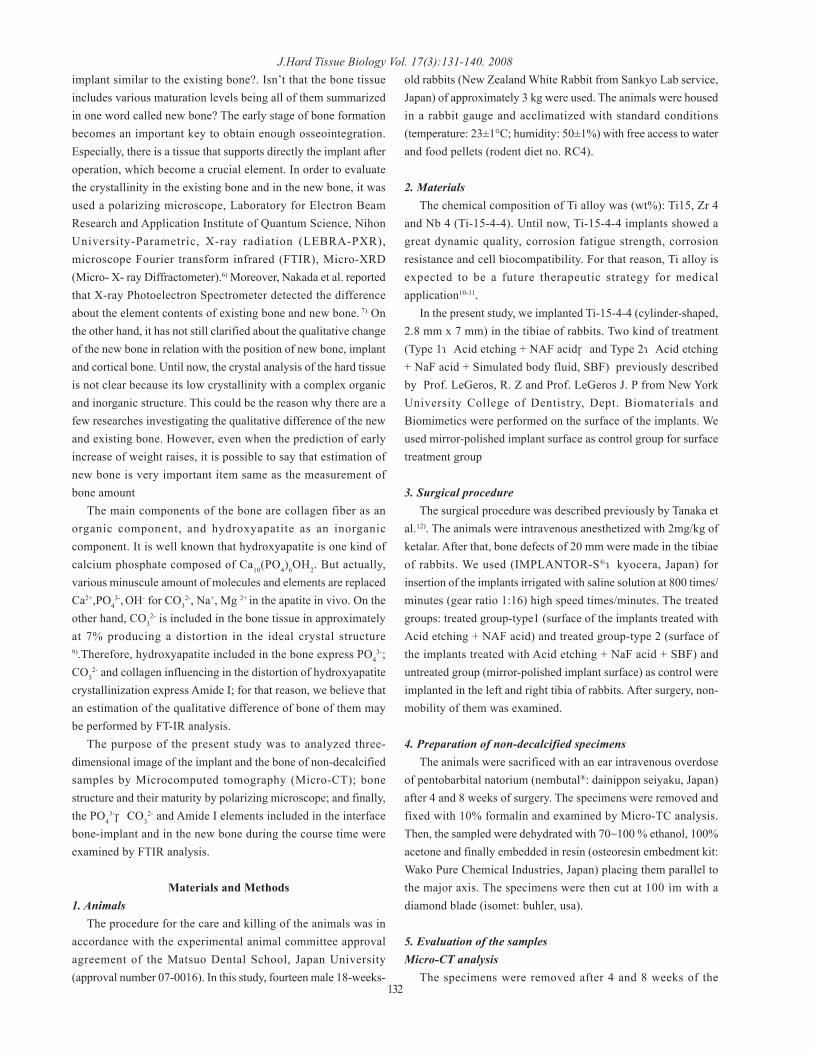
Fig. 1•Fig.1. Volume of the new bone detected by Micro-CT analysisTotal Volume (TV) = circumference 500µm of the implant ∧marrow space = ch 5 New Bone Volume (BV) = ch 1 ∧ ch 3∧ ch 5
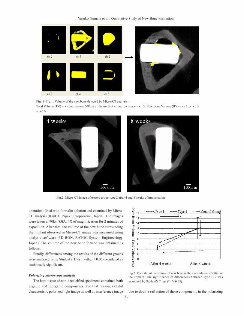
Fig.2. Micro-CT image of treated group-type 2 after 4 and 8 weeks of implantation.
Fig.3. The ratio of the volume of new bone in the circumference 500ìm ofthe implant. The significance of differences between Type 1, 2 wasexamined by Student’s T test (*: P<0.05).
due to double refraction of those components in the polarizing133
J.Hard Tissue Biology Vol. 17(3):131-140. 2008
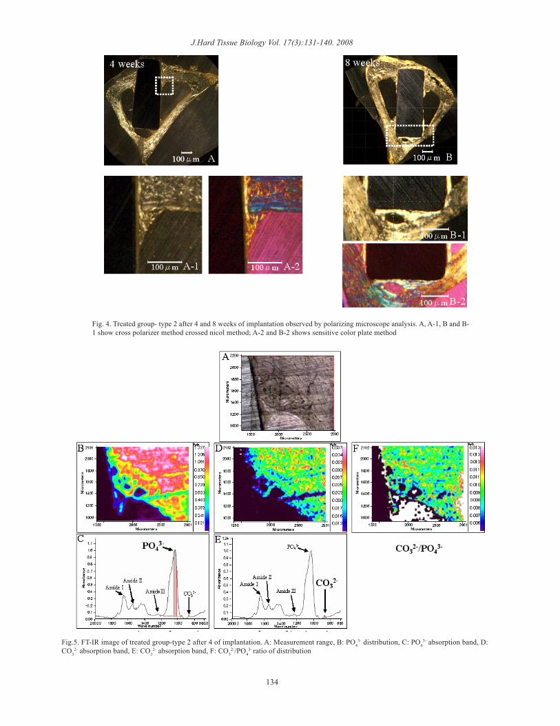
Fig. 4. Treated group- type 2 after 4 and 8 weeks of implantation observed by polarizing microscope analysis. A, A-1, B and B-1 show cross polarizer method crossed nicol method; A-2 and B-2 shows sensitive color plate method
Fig.5. FT-IR image of treated group-type 2 after 4 of implantation. A: Measurement range, B: PO43- distribution, C: PO4
3- absorption band, D:CO3
2- absorption band, E: CO32- absorption band, F: CO3
2-/PO43- ratio of distribution
134
Yasuko Numata et al.: Qualitative Study of New Bone Formationmicroscope13). For this observation, it was used polarizingmicroscope (OPTIPHOTO2-POL®: Nicon, Japan) with parallelnicol method, crossed nicol method and sensitive color platemethod (Gypsum Plate)
FT-IR analysisIn order to investigate the distribution of the chemical
component of the different groups, FTIR analysis was performed.For this examination, we made a reflection image using
Spotlight 400 (PerkinElmer, Inc.USA). We confirmed the correctarea of the newly bone formed from the polarizing microscopeview of the different groups at 4 and 8 weeks.
Analysis of the frequency area:4000~680cm-1
(In this study, it was excluded the analysis at more than 2000cm-1 because enters an artifact of measurement,)
Addition time:2 timesResolution :4cm-1
Conversion data:K-K conversion, PCA analysisImage pixel size:25ìm
Results1. Micro-CT analysis
We showed 3D image in thin sections evidencing new boneand Ti implant. The figure 2 shows Micro-CT images revealingpresence of cortical bone, Ti-implant and vessel canal system.The amount of the new bone from 4 to 8 weeks is observed in thefigure 3.
Treated groupIn this experiment, the implants inserted in the tibiae of rabbit
were touching the upper and lower side of cortical bone. Thetreated group showed new bone formation from the upper andlower side of the existing bone along the implant surface to thecenter at 4 and 8 weeks. Moreover, the bone-implant interfacewas clearly detected. The radiopacity of the newly bone formedwas increased from 4 to 8 weeks. On the other hand, the volumeof the new bone increased significantly from 4 to 8 weeks in treatedand control groups. However, there was no significant differencein new bone amount in the treated group- type A and B.
Control groupThe control group demonstrated scanty formation of new bone,
which was only seen attaching the existing bone in the upper sideof the implant at 4 and 8 weeks; in the lower side, the implant wasin contact with the existing bone without new bone formation.However, an increase of new bone from 4 to 8 weeks was revealed.
2. Polarizing microscope examinationFigure 4 shows clearly the orientation of collagen fibers by
sensitive color plate method and cross polarizer method. We couldalso observe some overlapped lamellae structure in the existingbone that is common at 4 and 8 weeks either in treated group aswell as control group. The lamellae structure of bone detected
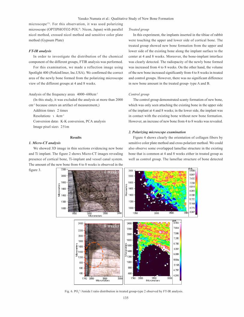
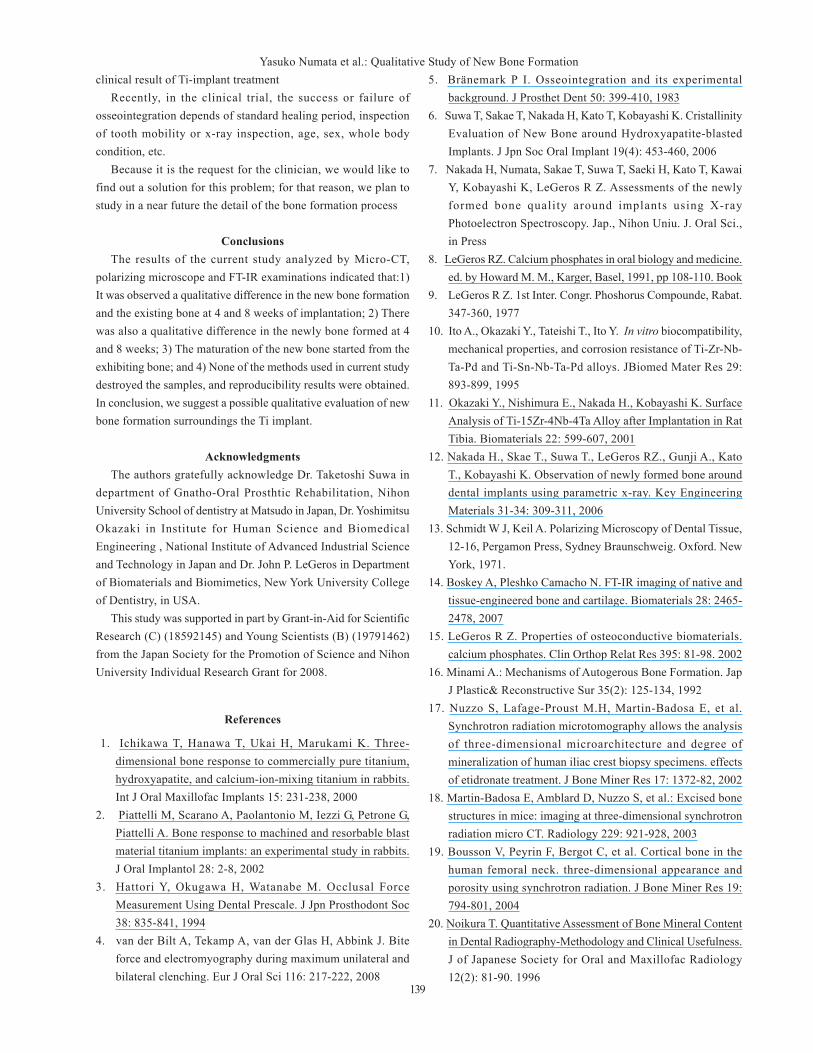
Fig. 6. PO43-/Amide I ratio distribution in treated group-type 2 observed by FT-IR analysis.
135
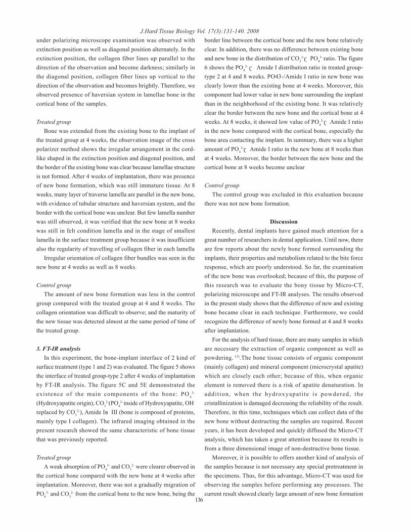
J.Hard Tissue Biology Vol. 17(3):131-140. 2008under polarizing microscope examination was observed withextinction position as well as diagonal position alternately. In theextinction position, the collagen fiber lines up parallel to thedirection of the observation and become darkness; similarly inthe diagonal position, collagen fiber lines up vertical to thedirection of the observation and becomes brightly. Therefore, weobserved presence of haversian system in lamellae bone in thecortical bone of the samples.
Treated groupBone was extended from the existing bone to the implant of
the treated group at 4 weeks, the observation image of the crosspolarizer method shows the irregular arrangement in the cord-like shaped in the extinction position and diagonal position, andthe border of the existing bone was clear because lamellae structureis not formed. After 4 weeks of implantation, there was presenceof new bone formation, which was still immature tissue. At 8weeks, many layer of traverse lamella are parallel in the new bone,with evidence of tubular structure and haversian system, and theborder with the cortical bone was unclear. But few lamella numberwas still observed, it was verified that the new bone at 8 weekswas still in felt condition lamella and in the stage of smallestlamella in the surface treatment group because it was insufficientalso the regularity of travelling of collagen fiber in each lamella
Irregular orientation of collagen fiber bundles was seen in thenew bone at 4 weeks as well as 8 weeks.
Control groupThe amount of new bone formation was less in the control
group compared with the treated group at 4 and 8 weeks. Thecollagen orientation was difficult to observe; and the maturity ofthe new tissue was detected almost at the same period of time ofthe treated group.
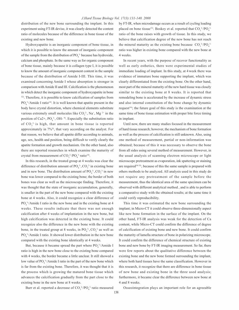
3. FT-IR analysisIn this experiment, the bone-implant interface of 2 kind of
surface treatment (type 1 and 2) was evaluated. The figure 5 showsthe interface of treated group-type 2 after 4 weeks of implantationby FT-IR analysis. The figure 5C and 5E demonstrated theexistence of the main components of the bone: PO4
3-
(Hydroxyapatite origin), CO32-(PO4
3- inside of Hydroxyapatite, OH-
replaced by CO32-), Amide I~III (bone is composed of proteins,
mainly type I collagen). The infrared imaging obtained in thepresent research showed the same characteristic of bone tissuethat was previously reported.
Treated groupA weak absorption of PO4
3- and CO32- were clearer observed in
the cortical bone compared with the new bone at 4 weeks afterimplantation. Moreover, there was not a gradually migration ofPO4
3- and CO32- from the cortical bone to the new bone, being the
border line between the cortical bone and the new bone relativelyclear. In addition, there was no difference between existing boneand new bone in the distribution of CO3
2-/PO43- ratio. The figure
6 shows the PO43-/Amide I distribution ratio in treated group-
type 2 at 4 and 8 weeks. PO43-/Amide I ratio in new bone wasclearly lower than the existing bone at 4 weeks. Moreover, thiscomponent had lower value in new bone surrounding the implantthan in the neighborhood of the existing bone. It was relativelyclear the border between the new bone and the cortical bone at 4weeks. At 8 weeks, it showed low value of PO4
3-/Amide I ratioin the new bone compared with the cortical bone, especially thebone area contacting the implant. In summary, there was a higheramount of PO4
3-/Amide I ratio in the new bone at 8 weeks thanat 4 weeks. Moreover, the border between the new bone and thecortical bone at 8 weeks become unclear
Control groupThe control group was excluded in this evaluation because
there was not new bone formation.
DiscussionRecently, dental implants have gained much attention for a
great number of researchers in dental application. Until now, thereare few reports about the newly bone formed surrounding theimplants, their properties and metabolism related to the bite forceresponse, which are poorly understood. So far, the examinationof the new bone was overlooked; because of this, the purpose ofthis research was to evaluate the bony tissue by Micro-CT,polarizing microscope and FT-IR analyses. The results observedin the present study shows that the difference of new and existingbone became clear in each technique. Furthermore, we couldrecognize the difference of newly bone formed at 4 and 8 weeksafter implantation.
For the analysis of hard tissue, there are many samples in whichare necessary the extraction of organic component as well aspowdering. 15).The bone tissue consists of organic component(mainly collagen) and mineral component (microcrystal apatite)which are closely each other; because of this, when organicelement is removed there is a risk of apatite denaturation. Inaddit ion, when the hydroxyapati te is powdered, thecristallinization is damaged decreasing the reliability of the result.Therefore, in this time, techniques which can collect data of thenew bone without destructing the samples are required. Recentyears, it has been developed and quickly diffused the Micro-CTanalysis, which has taken a great attention because its results isfrom a three dimensional image of non-destructive bone tissue.
Moreover, it is possible to offers another kind of analysis ofthe samples because is not necessary any special pretreatment inthe specimens. Thus, for this advantage, Micro-CT was used forobserving the samples before performing any processes. Thecurrent result showed clearly large amount of new bone formation
136

Yasuko Numata et al.: Qualitative Study of New Bone Formationattached to the existing bone in the control and treated groups.For that reason, we believe that osteoblast and precursor cellsclose to the existing bone have many supplied advantagessurrounding the implant. 16). Besides, in early stage at 4 weeks,the amount of the new bone formation was higher in the upperside of the implant than in the lower part. Thus could be due tothe presence of granulation tissue with endothelial cells, fibroblastsand macrophage with the secretion of cytokines and growth factorin the upper side of the implant. We believe that these cytokinesand growth factors act in an early stage inducing osteoblastdifferentiation and bone formation.
Therefore, surrounding the implant entrance, various cytokineand growth factor are secreted from vascular endothelial cells,fibroblast as well as from macrophage forming a granulation tissue;activation of cell occur concerning osteoblast as well as boneformation; we thought it is the region that can possibly grow thebone early result.
Nuzzo et al. reported that the changes of calcium depositiondegree from woven to matured bone induced by drugs can beestimated by Micro-CT analysis. 17). Moreover, It has beenpublished that trabecular bone, cortical bone and intracorticalporosity of mouse’s femur in three dimension can be estimated18);there is also reported that, three dimensionally, vessel canal systemin cortical bone of human femoral neck makes the quantificationpossible19).
In the current study, the observation target is the new bonesurrounding the implant, which could not obtain a high resolutionbeing influenced for an artifact. It could not do micro observationof bone structure but it could confirm an increase of radiopacityof new bone in the treated group from 4 to 8 weeks.
All collected elements crossing the x-ray were expressed asradiopacity by Micro-CT analysis. Especially the Ca included inbone which is a heavy chemical element with high radiopacity;when increase of amount of Ca increases also the radiopacity,that’s way it is possible to observe calcification and bone mineralamount as radiopacity by CT. Therefore, the CT image revealedhigher calcification in the new bone at 8 weeks compared to 4weeks. However, in this study, an evidence of artifact in the areaof new bone surrounding the Ti alloy was detected affecting theresults. We hope to get solution to reduce the artifact in both hardand soft tissues.
The polarizing microscope properties exhibit characteristic ofpolarized light image and interference image due to the doublerefraction in organic as well as inorganic component of hard tissue13). For that reason, we could easily observe the morphologicalinformation of bone tissue which is difficult to see from a commonlight microscope. Besides, Back Scattering Image (BSI) has thenecessity to evaporate carbon in the samples while in polarizingmicroscope is no necessary to apply a pretreatment in samples inthe existing bone and the new bone comparing with lightmicroscope which is commonly difficult for estimating the bone
tissue without staining, as well as it is possible to obtaininformation about the maturity stage of bone. Nakada et al.reported that is possible to obtain information about ontogenystage of bone surrounding the implant using polarizingmicroscope21). In this study, we could clarify the difference ofmaturity level of new bone at 4 and 8 weeks according to thecollagen fiber orientation inside the matrix of the new and existingbone. The new bone was still immature at 4 weeks, but in the newbone at 8 weeks partially mature bone was observed; there waspart in which exhibit irregular layer structure and part in whichexits together with scattered cord-like fiber, and the border ofnew and existing bone became unclear. This result indicates thatwe got rapid metabolic turnover in the new bone from 4 weeks to8 weeks; we thought there was a maturity process. It has beenpublished that it was observed bone tissue on the implant insertedin tibia of rabbits after 4 weeks of implantation by BSI; it isreported that it was possible to recognize the border of new boneand existing bone by different color tone. 22). In this research, thetreated group showed a clear border of new bone and existingbone from the orientation of collagen fibers at 4 weeks. At 8 weeks,the lamella structure in a part of new bone and the border with theexisting bone became unclear, but it did not reach to the regularlamella structure as in the existing bone. Therefore, the new bonesurrounding the implant did not get the maturity of the existingbone at 8 weeks. In the sensitive color plate method, because itcan identify the intensity and orientation of the double refractionof collagen fiber due to color, the difference of structure of theexisting bone and the new bone was observed more clearly. Fromthe results observed above, in the future we expect to getmaturation of formed bone similar to the existing bone after 8weeks; we would like to do further researches.
The polarizing microscope properties exhibit characteristic ofpolarized light image and interference image due to the doublerefraction in organic as well as inorganic component of hard tissue13). For that reason, we could easily observe the morphologicalinformation of bone tissue which is difficult to see from a commonlight microscope. Besides, Back Scattering Image (BSI) has thenecessity to evaporate carbon in the samples while in polarizingmicroscope is no necessary to apply a pretreatment in samples inthe existing bone and the new bone comparing with lightmicroscope which is commonly difficult for estimating the bonetissue without staining, as well as it is possible to obtaininformation about the maturity stage of bone.
The infrared absorption spectrum obtained by FT-IR, especiallyin 2000-700cm-1 called fingerprints region, is capable to obtaininformation about chemical structure of sample because they arecharacteristic molecule23). There are many comparative studies ofnormal bone and bone disease by FT-IR analysis reported untilnow 24-29); moreover, FT-IR is a reliable technique that makepossible the analysis of bone tissue 30-31). However, it is notpublished any case in which is observed chronological molecule
137
J.Hard Tissue Biology Vol. 17(3):131-140. 2008distribution of the new bone surrounding the implant. In thisexperiment using FT-IR devise, it was clearly detected the contentratio of molecules because of the difference in bone tissue of theexisting and new bone.
Hydroxyapatite is an inorganic component of bone tissue, inwhich it is possible to know the amount of inorganic componentof the sample from the distribution of PO4
3- because has hydroxide,calcium and phosphate. In the same way as for organic componentof bone tissue, mainly because it is collagen type I, it is possibleto know the amount of inorganic component content in the samplebecause of the distribution of Amide I-III. This time, it wasexamined concerning Amide I whose absorption is stronger incomparison with Amide II and III. Calcification is the phenomenonin which detect the inorganic component of hydroxyapatite in bone32). Therefore, it is possible to know calcification of samples fromPO4
3-/Amide I ratio14). It is well known that apatite present in thebody have crystal distortion, where chemical elements substitutevarious extremely small molecules like CO3
2-, Na+, Mg 2+ in theposition of Ca2+, PO4
3-, OH- 9). Especially the substitution ratioof CO3
2- is high, that amount in bone tissue is reportedapproximately in 7%8), that vary according on the analyst. Forthat reason, we believe that all apatite differ according to animals,age, sex, health and position, being difficult to verify clearly theapatite formation and growth mechanism. On the other hand, alsothere are reported researches in which examine the maturity ofcrystal from measurement of CO3
2-/PO43- ratio14).
In this research, in the treated group at 4 weeks was clear thedifference of distribution amount of PO4
3-, CO32- in existing bone
and in new bone. The distribution amount of PO43-, CO3
2- in newbone was lower compared to the existing bone; the border of bothbones was clear as well as the microscope finding. Therefore, itwas thought that the state of inorganic accumulation, generally,is smaller in the part of the new bone compared with the existingbone at 4 weeks. Also, it could recognize a clear difference ofPO4
3-/Amide I ratio in the new bone and in the existing bone at 4weeks. These results indicate that there was not enoughcalcification after 4 weeks of implantation in the new bone, buthigh calcification was detected in the existing bone. It couldrecognize also the difference in the new bone with the existingbone, in the treated group at 8 weeks, in PO4
3-, CO32- as well as
PO43-/Amide I ratio. It showed lower distribution in the new bone
compared with the existing bone identically at 4 weeks.But, because it became spread the part where PO4
3-/Amide Iratio is high in the new bone close to the existing bone comparedwith 4 weeks, the border became a little unclear. It still showed alow value of PO4
3-/Amide I ratio in the part of the new bone whichis far from the existing bone. Therefore, it was thought that it isthe process which is growing the matured bone tissue whichadvances the calcification gradually from the part close to theexisting bone in the new bone at 8 weeks.
Burr et al. reported a decrease of CO32-/PO4
3- ratio measured
by FT-IR, when microdamage occurs as a result of cycling loadingplaced on bone tissue33). Boskey et al. reported that CO3
2-/PO43-
ratio of the bone raises with growth of tissue. In this study, webelieve that calcification degree of the new bone has not reachthe mineral maturity as the existing bone because CO3
2-/PO43-
ratio was higher in existing bone compared with the new bone at4 weeks.
In recent years, with the purpose of recover functionality aswell as early esthetics, there were experimental studies ofimmediate loading of implant. In this study, at 4 week there wasevidence of immature bone supporting the implant, which wasclearly differentiated from the existing bone. On the other hand,most part of the mineral maturity of the new hard tissue was closelysimilar to the existing bone at 8 weeks. It is reported thatremodeling bone is accelerated by the increase of dynamic stressand also internal constitution of the bone change by dynamicrequest16); the future goal of this study is the examination at thesame time of bone tissue estimation with proper bite force timingin implant.
Until now, there are many studies focused in the measurementof hard tissue research; however, the mechanism of bone formationas well as the process of calcification is still unknown. Also, usingone method of measurement, partial or non-information wasobtained; because of this it was necessary to observe the bonefrom all sides using several method of measurement. However, inthe usual analysis of scanning electron microscope or lightmicroscope pretreatment as evaporation, ink spattering or stainingare required34-35), because of this the same sample is prepared withothers methods to be analyzed. All analysis used in this study donot require any pretreatment of the sample before themeasurement, thus the identical area of the same specimen can beobserved with different analytical method , and is able to performa comparative study with the obtained results; at the same time itcould verify reproducibility.
This time it was estimated the new bone surrounding theimplant; in Micro-CT it could observe three-dimensionally aspectlike new bone formation in the surface of the implant. On theother hand, FT-IR analysis was weak for the detection of Cacontent, while Micro-CT could confirm the difference of degreeof calcification of existing bone and new bone. It could confirmthe maturity of lamella structure of bone in polarizing microscope.It could confirm the difference of chemical structure of existingbone and new bone by FT-IR imaging measurement. So far, therewere few reports about the qualitative difference between theexisting bone and the new bone formed surrounding the implant,where both hard tissues have the same classification. However inthis research, it recognize that there are difference in bone tissueof new bone and existing bone in the three used analysis;furthermore, it became clear the difference between new bone at4 and 8 weeks.
Osseointegration plays an important role for an agreeable138
Yasuko Numata et al.: Qualitative Study of New Bone Formationclinical result of Ti-implant treatment
Recently, in the clinical trial, the success or failure ofosseointegration depends of standard healing period, inspectionof tooth mobility or x-ray inspection, age, sex, whole bodycondition, etc.
Because it is the request for the clinician, we would like tofind out a solution for this problem; for that reason, we plan tostudy in a near future the detail of the bone formation process
ConclusionsThe results of the current study analyzed by Micro-CT,
polarizing microscope and FT-IR examinations indicated that:1)It was observed a qualitative difference in the new bone formationand the existing bone at 4 and 8 weeks of implantation; 2) Therewas also a qualitative difference in the newly bone formed at 4and 8 weeks; 3) The maturation of the new bone started from theexhibiting bone; and 4) None of the methods used in current studydestroyed the samples, and reproducibility results were obtained.In conclusion, we suggest a possible qualitative evaluation of newbone formation surroundings the Ti implant.
AcknowledgmentsThe authors gratefully acknowledge Dr. Taketoshi Suwa in
department of Gnatho-Oral Prosthtic Rehabilitation, NihonUniversity School of dentistry at Matsudo in Japan, Dr. YoshimitsuOkazaki in Institute for Human Science and BiomedicalEngineering , National Institute of Advanced Industrial Scienceand Technology in Japan and Dr. John P. LeGeros in Departmentof Biomaterials and Biomimetics, New York University Collegeof Dentistry, in USA.
This study was supported in part by Grant-in-Aid for ScientificResearch (C) (18592145) and Young Scientists (B) (19791462)from the Japan Society for the Promotion of Science and NihonUniversity Individual Research Grant for 2008.
References
1. Ichikawa T, Hanawa T, Ukai H, Marukami K. Three-dimensional bone response to commercially pure titanium,hydroxyapatite, and calcium-ion-mixing titanium in rabbits.Int J Oral Maxillofac Implants 15: 231-238, 2000
2. Piattelli M, Scarano A, Paolantonio M, Iezzi G, Petrone G,Piattelli A. Bone response to machined and resorbable blastmaterial titanium implants: an experimental study in rabbits.J Oral Implantol 28: 2-8, 2002
3. Hattori Y, Okugawa H, Watanabe M. Occlusal ForceMeasurement Using Dental Prescale. J Jpn Prosthodont Soc38: 835-841, 1994
4. van der Bilt A, Tekamp A, van der Glas H, Abbink J. Biteforce and electromyography during maximum unilateral andbilateral clenching. Eur J Oral Sci 116: 217-222, 2008
5. Bränemark P I. Osseointegration and its experimentalbackground. J Prosthet Dent 50: 399-410, 1983
6. Suwa T, Sakae T, Nakada H, Kato T, Kobayashi K. CristallinityEvaluation of New Bone around Hydroxyapatite-blastedImplants. J Jpn Soc Oral Implant 19(4): 453-460, 2006
7. Nakada H, Numata, Sakae T, Suwa T, Saeki H, Kato T, KawaiY, Kobayashi K, LeGeros R Z. Assessments of the newlyformed bone quality around implants using X-rayPhotoelectron Spectroscopy. Jap., Nihon Uniu. J. Oral Sci.,in Press
8. LeGeros RZ. Calcium phosphates in oral biology and medicine.ed. by Howard M. M., Karger, Basel, 1991, pp 108-110. Book
9. LeGeros R Z. 1st Inter. Congr. Phoshorus Compounde, Rabat.347-360, 1977
10. Ito A., Okazaki Y., Tateishi T., Ito Y. In vitro biocompatibility,mechanical properties, and corrosion resistance of Ti-Zr-Nb-Ta-Pd and Ti-Sn-Nb-Ta-Pd alloys. JBiomed Mater Res 29:893-899, 1995
11. Okazaki Y., Nishimura E., Nakada H., Kobayashi K. SurfaceAnalysis of Ti-15Zr-4Nb-4Ta Alloy after Implantation in RatTibia. Biomaterials 22: 599-607, 2001
12. Nakada H., Skae T., Suwa T., LeGeros RZ., Gunji A., KatoT., Kobayashi K. Observation of newly formed bone arounddental implants using parametric x-ray. Key EngineeringMaterials 31-34: 309-311, 2006
13. Schmidt W J, Keil A. Polarizing Microscopy of Dental Tissue,12-16, Pergamon Press, Sydney Braunschweig. Oxford. NewYork, 1971.
14. Boskey A, Pleshko Camacho N. FT-IR imaging of native andtissue-engineered bone and cartilage. Biomaterials 28: 2465-2478, 2007
15. LeGeros R Z. Properties of osteoconductive biomaterials.calcium phosphates. Clin Orthop Relat Res 395: 81-98. 2002
16. Minami A.: Mechanisms of Autogerous Bone Formation. JapJ Plastic& Reconstructive Sur 35(2): 125-134, 1992
17. Nuzzo S, Lafage-Proust M.H, Martin-Badosa E, et al.Synchrotron radiation microtomography allows the analysisof three-dimensional microarchitecture and degree ofmineralization of human iliac crest biopsy specimens. effectsof etidronate treatment. J Bone Miner Res 17: 1372-82, 2002
18. Martin-Badosa E, Amblard D, Nuzzo S, et al.: Excised bonestructures in mice: imaging at three-dimensional synchrotronradiation micro CT. Radiology 229: 921-928, 2003
19. Bousson V, Peyrin F, Bergot C, et al. Cortical bone in thehuman femoral neck. three-dimensional appearance andporosity using synchrotron radiation. J Bone Miner Res 19:794-801, 2004
20. Noikura T. Quantitative Assessment of Bone Mineral Contentin Dental Radiography-Methodology and Clinical Usefulness.J of Japanese Society for Oral and Maxillofac Radiology12(2): 81-90. 1996
139

J.Hard Tissue Biology Vol. 17(3):131-140. 200821. Nakada H, Sakae T, Manabe T, Gunji A, Machida T, Suwa T,
Kato T, Kobayashi K. Observed by the Polarizing Microscopefor the Generation Stage of an Implant Surrounding Bone.Jap. Nihon Uniu. J. Oral Sci. 29: 160-163, 2003
22. Nakada H, Sakae T, LeGeros R Z, LeGeros J P, Suwa T,Numata Y, Kobayashi K. Early tissue response to modifiedimplant surfaces using back scattered imaging. Implant Dent16: 281-289, 2007
23. Carden A, Morris M D. Application of vibrational spectroscopyto the study of mineralized tissues. J Biomed Opt 5: 259-68,2000
24. Paschalis E P, Betts F, DiCarlo E, Mendelsohn R., BoskeyA.L. FTIR microspectroscopic analysis of human iliac crestbiopsies from untreated osteoporotic bone. Calcif Tissue Int61: 487–92, 1997
25. Huang R Y, Miller L M, Carlson C S, Chance M R. In situchemistry of osteoporosis revealed by synchrotron infraredmicrospectroscopy. Bone 33: 514-21, 2003
26. Boskey A. Mineral changes in osteopetrosis. Crit Rev EukaryotGene Expr 13: 109-16, 2003
27. Boskey A L, DiCarlo E, Paschalis E, West P. Mendelsohn R.Comparison of mineral quality and quantity in iliac crestbiopsies from high- and low-turnover osteoporosis: an FT-IRmicrospectroscopic investigation. Osteoporos Int 16: 2031-8, 2005
28. Faibish D, Gomes A, Boivin G, Binderman I, Boskey A.Infrared imaging of calcified tissue in bone biopsies fromadults with osteomalacia. Bone 36: 6-12, 2005
29. Faibish D, Ott S M, Boskey A L. Mineral changes in osteoporosis.a review. Clin Orthop Relat Res 443: 28-38, 2006
30. Boskey A, Mendelsohn R. Infrared analysis of bone in healthand disease. J Biomed Opt 10:031102–6, 2005
31. Miller LM, Dumas P. Chemical imaging of biological tissuewith synchrotron infrared light. Biochim Biophys Acta 1758:846–57, 2006
32. Bawden J W, Crenshaw M A, Wright J T and LeGeros R Z.Consideration of possible biologic mechanisms of fluorosis.J Dent Res 74(7):1349-52, 1995
33. Ruppel M E, Burr D B, Miller L M. Chemical makeup ofmicrodamaged bone differs from undamaged bone. Bone 39:318-24, 2006
34. Chu C, Lin P, Dong Y, Xue X, Zhu J, Yin Z. Fabrication andcharacterization of hydroxyapatite reinforced with 20 vol %Ti particles for use as hard tissue replacement, J Mater SciMater Med, 13: 985-992, 2002
35. Bûchter A, Joos U, Wiesmann HP, Seper L, Meyer U.Biological and biomechanical evaluation of interface reactionat conical screw-type implants. Head Face Med 21: 5-13, 2006
140
Copyright © 2022 FDOKUMEN