NOT ALWAYS AT STAGE. ATYPICAL PATTERNS IN SPANISH COPULAR CLAUSES
Upload
independentCategory
view
0download
0
Pure progressive amnesia: An atypical amnesticsyndrome?
Emmanuel J. Barbeau, Mira Didic, Olivier Felician, and Eve TramoniLaboratoire de Neurophysiologie et Neuropsychologie, INSERM EMI-U 9926, Faculte de Medecine, Universite de la Mediterranee
and Service de Neurologie et Neuropsychologie, AP-HM Timone, Marseille, France
Eric GuedjService Central de Biophysique et de Medecine Nucleaire, AP-HM Timone, Marseille, France
Mathieu Ceccaldi and Michel PoncetLaboratoire de Neurophysiologie et Neuropsychologie, INSERM EMI-U 9926, Faculte de Medecine, Universite de la Mediterranee
and Service de Neurologie et Neuropsychologie, AP-HM Timone, Marseille, France
We report on M.S., an 83-year-old patient with isolated pure progressive amnesia. This rare, recentlyidentified, form of amnesia has been described in elderly patients. Neuropathological studies suggestthat this syndrome is an atypical clinical presentation of Alzheimer’s disease. The aim of our study wasto characterize the neuropsychological pattern of pure progressive amnesia in comparison with otheramnestic syndromes and memory dissociations reported in the literature. Our results indicate thatpure progressive amnesia is characterized by a highly unusual dissociation in the realm of memory,with severe deficits on tests based on recognition and recall of verbal and visual single items, contrast-ing with relatively preserved anterograde autobiographical and spatial memory and normal recall ofcomplex material such as stories. These findings suggest that memory for single items coulddepend on an independent system. One hypothesis is that M.S.’s unusual memory profile resultsfrom relative dysfunction of the ventral medial temporal lobe pathway. An alternative explanationimplicates cognitive reserve. Further studies are required in order to progress on this matter. Inany case, pure progressive amnesia is a clinical syndrome that may provide further insight into theorganization of declarative memory.
INTRODUCTION
A wide variety of amnestic syndromes in relationto various pathological conditions and anatomicalsubstrates have been described (see Kopelman,2002, for a review). The characterization of these
syndromes has considerably improved the under-standing of how memory is organized in thehuman brain. Several dissociations followingmedial temporal lobe damage have been reported.For example, a dissociation between impaired epi-sodic and preserved semantic memory has been
Correspondence should be addressed to Emmanuel Barbeau, Service de Neurologie et de Neuropsychologie, CHU Timone, 264,
rue Saint-Pierre, 13385 Marseille Cedex 05, France (E-mail: [email protected]).
The authors wish to thank M.S. for his kindness and long-standing cooperation, as well as anonymous reviewers for their helpful
comments. EB is supported by a grant from the Conseil General des Bouches du Rhone. This study was partially financed by
AP-HM PHRC 2001/54.
# 2006 Psychology Press, an imprint of the Taylor & Francis Group, an informa business 1http://www.psypress.com/cogneuropsychology DOI:10.1080/02643290600893594
COGNITIVE NEUROPSYCHOLOGY, 0000, 00 (0), 1–18
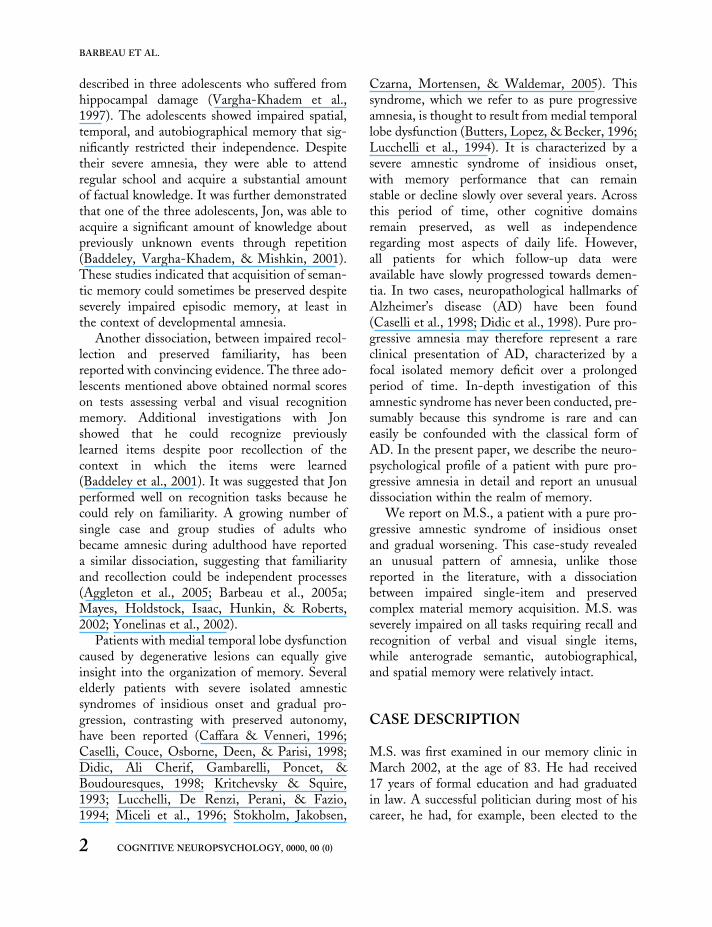
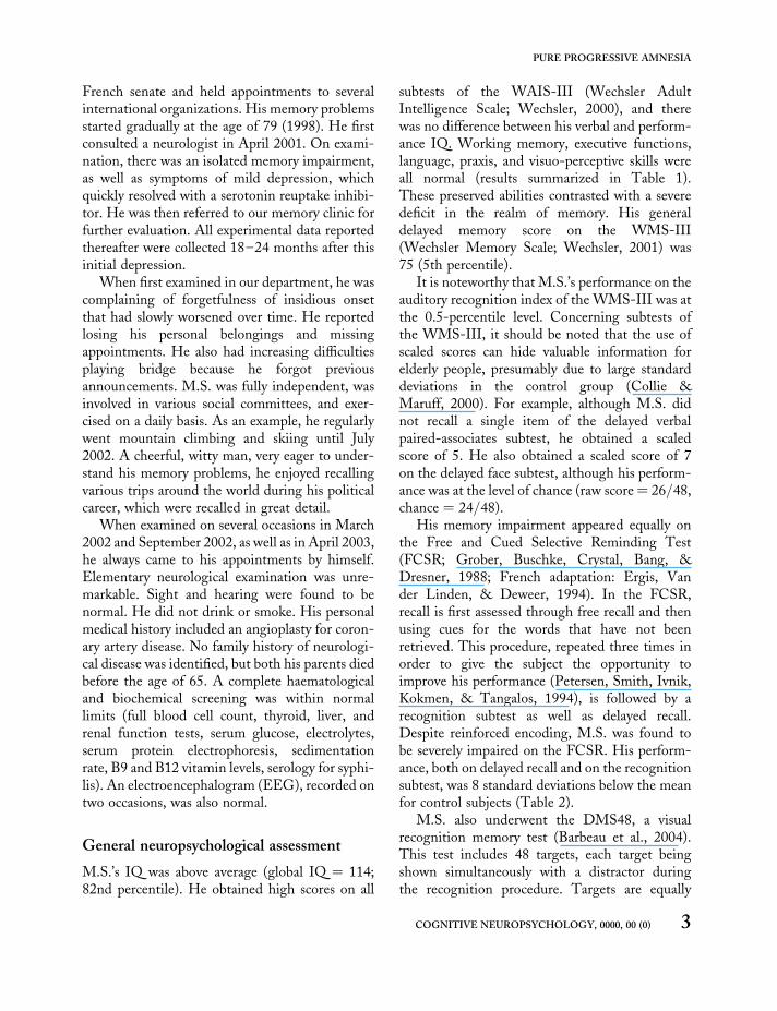
described in three adolescents who suffered fromhippocampal damage (Vargha-Khadem et al.,1997). The adolescents showed impaired spatial,temporal, and autobiographical memory that sig-nificantly restricted their independence. Despitetheir severe amnesia, they were able to attendregular school and acquire a substantial amountof factual knowledge. It was further demonstratedthat one of the three adolescents, Jon, was able toacquire a significant amount of knowledge aboutpreviously unknown events through repetition(Baddeley, Vargha-Khadem, & Mishkin, 2001).These studies indicated that acquisition of seman-tic memory could sometimes be preserved despiteseverely impaired episodic memory, at least inthe context of developmental amnesia.
Another dissociation, between impaired recol-lection and preserved familiarity, has beenreported with convincing evidence. The three ado-lescents mentioned above obtained normal scoreson tests assessing verbal and visual recognitionmemory. Additional investigations with Jonshowed that he could recognize previouslylearned items despite poor recollection of thecontext in which the items were learned(Baddeley et al., 2001). It was suggested that Jonperformed well on recognition tasks because hecould rely on familiarity. A growing number ofsingle case and group studies of adults whobecame amnesic during adulthood have reporteda similar dissociation, suggesting that familiarityand recollection could be independent processes(Aggleton et al., 2005; Barbeau et al., 2005a;Mayes, Holdstock, Isaac, Hunkin, & Roberts,2002; Yonelinas et al., 2002).
Patients with medial temporal lobe dysfunctioncaused by degenerative lesions can equally giveinsight into the organization of memory. Severalelderly patients with severe isolated amnesticsyndromes of insidious onset and gradual pro-gression, contrasting with preserved autonomy,have been reported (Caffara & Venneri, 1996;Caselli, Couce, Osborne, Deen, & Parisi, 1998;Didic, Ali Cherif, Gambarelli, Poncet, &Boudouresques, 1998; Kritchevsky & Squire,1993; Lucchelli, De Renzi, Perani, & Fazio,1994; Miceli et al., 1996; Stokholm, Jakobsen,
Czarna, Mortensen, & Waldemar, 2005). Thissyndrome, which we refer to as pure progressiveamnesia, is thought to result from medial temporallobe dysfunction (Butters, Lopez, & Becker, 1996;Lucchelli et al., 1994). It is characterized by asevere amnestic syndrome of insidious onset,with memory performance that can remainstable or decline slowly over several years. Acrossthis period of time, other cognitive domainsremain preserved, as well as independenceregarding most aspects of daily life. However,all patients for which follow-up data wereavailable have slowly progressed towards demen-tia. In two cases, neuropathological hallmarks ofAlzheimer’s disease (AD) have been found(Caselli et al., 1998; Didic et al., 1998). Pure pro-gressive amnesia may therefore represent a rareclinical presentation of AD, characterized by afocal isolated memory deficit over a prolongedperiod of time. In-depth investigation of thisamnestic syndrome has never been conducted, pre-sumably because this syndrome is rare and caneasily be confounded with the classical form ofAD. In the present paper, we describe the neuro-psychological profile of a patient with pure pro-gressive amnesia in detail and report an unusualdissociation within the realm of memory.
We report on M.S., a patient with a pure pro-gressive amnestic syndrome of insidious onsetand gradual worsening. This case-study revealedan unusual pattern of amnesia, unlike thosereported in the literature, with a dissociationbetween impaired single-item and preservedcomplex material memory acquisition. M.S. wasseverely impaired on all tasks requiring recall andrecognition of verbal and visual single items,while anterograde semantic, autobiographical,and spatial memory were relatively intact.
CASE DESCRIPTION
M.S. was first examined in our memory clinic inMarch 2002, at the age of 83. He had received17 years of formal education and had graduatedin law. A successful politician during most of hiscareer, he had, for example, been elected to the
2 COGNITIVE NEUROPSYCHOLOGY, 0000, 00 (0)
BARBEAU ET AL.
French senate and held appointments to severalinternational organizations. His memory problemsstarted gradually at the age of 79 (1998). He firstconsulted a neurologist in April 2001. On exami-nation, there was an isolated memory impairment,as well as symptoms of mild depression, whichquickly resolved with a serotonin reuptake inhibi-tor. He was then referred to our memory clinic forfurther evaluation. All experimental data reportedthereafter were collected 18–24 months after thisinitial depression.
When first examined in our department, he wascomplaining of forgetfulness of insidious onsetthat had slowly worsened over time. He reportedlosing his personal belongings and missingappointments. He also had increasing difficultiesplaying bridge because he forgot previousannouncements. M.S. was fully independent, wasinvolved in various social committees, and exer-cised on a daily basis. As an example, he regularlywent mountain climbing and skiing until July2002. A cheerful, witty man, very eager to under-stand his memory problems, he enjoyed recallingvarious trips around the world during his politicalcareer, which were recalled in great detail.
When examined on several occasions in March2002 and September 2002, as well as in April 2003,he always came to his appointments by himself.Elementary neurological examination was unre-markable. Sight and hearing were found to benormal. He did not drink or smoke. His personalmedical history included an angioplasty for coron-ary artery disease. No family history of neurologi-cal disease was identified, but both his parents diedbefore the age of 65. A complete haematologicaland biochemical screening was within normallimits (full blood cell count, thyroid, liver, andrenal function tests, serum glucose, electrolytes,serum protein electrophoresis, sedimentationrate, B9 and B12 vitamin levels, serology for syphi-lis). An electroencephalogram (EEG), recorded ontwo occasions, was also normal.
General neuropsychological assessment
M.S.’s IQ was above average (global IQ ¼ 114;82nd percentile). He obtained high scores on all
subtests of the WAIS-III (Wechsler AdultIntelligence Scale; Wechsler, 2000), and therewas no difference between his verbal and perform-ance IQ. Working memory, executive functions,language, praxis, and visuo-perceptive skills wereall normal (results summarized in Table 1).These preserved abilities contrasted with a severedeficit in the realm of memory. His generaldelayed memory score on the WMS-III(Wechsler Memory Scale; Wechsler, 2001) was75 (5th percentile).
It is noteworthy that M.S.’s performance on theauditory recognition index of the WMS-III was atthe 0.5-percentile level. Concerning subtests ofthe WMS-III, it should be noted that the use ofscaled scores can hide valuable information forelderly people, presumably due to large standarddeviations in the control group (Collie &Maruff, 2000). For example, although M.S. didnot recall a single item of the delayed verbalpaired-associates subtest, he obtained a scaledscore of 5. He also obtained a scaled score of 7on the delayed face subtest, although his perform-ance was at the level of chance (raw score¼ 26/48,chance ¼ 24/48).
His memory impairment appeared equally onthe Free and Cued Selective Reminding Test(FCSR; Grober, Buschke, Crystal, Bang, &Dresner, 1988; French adaptation: Ergis, Vander Linden, & Deweer, 1994). In the FCSR,recall is first assessed through free recall and thenusing cues for the words that have not beenretrieved. This procedure, repeated three times inorder to give the subject the opportunity toimprove his performance (Petersen, Smith, Ivnik,Kokmen, & Tangalos, 1994), is followed by arecognition subtest as well as delayed recall.Despite reinforced encoding, M.S. was found tobe severely impaired on the FCSR. His perform-ance, both on delayed recall and on the recognitionsubtest, was 8 standard deviations below the meanfor control subjects (Table 2).
M.S. also underwent the DMS48, a visualrecognition memory test (Barbeau et al., 2004).This test includes 48 targets, each target beingshown simultaneously with a distractor duringthe recognition procedure. Targets are equally
COGNITIVE NEUROPSYCHOLOGY, 0000, 00 (0) 3
PURE PROGRESSIVE AMNESIA
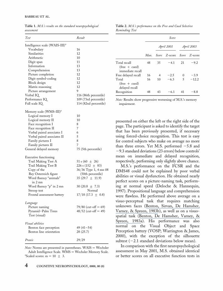
presented on either the left or the right side of thepage. The participant is asked to identify the targetthat has been previously presented, if necessaryusing forced-choice recognition. This test is easyfor control subjects who make on average no morethan three errors. Yet M.S. performed 25.8 and29.3 standard deviations (23 errors) below controls’mean on immediate and delayed recognition,respectively, performing only slightly above chance.
M.S.’s performance on the FCSR and theDMS48 could not be explained by poor verbalabilities or visual dysfunction. He obtained nearlyperfect scores on a picture-naming task, perform-ing at normal speed (Deloche & Hannequin,1997). Propositional language and comprehensionwere flawless. He performed above average on avisuo-perceptual task that requires matchingunknown faces (Benton, Sirvan, De Hamsher,Varney, & Spreen, 1983b), as well as on a visuo-spatial task (Benton, De Hamsher, Varney, &Spreen, 1983a). His performance was alsonormal on the Visual Object and SpacePerception battery (VOSP; Warrington & James,2000), with the exception of the silhouettesubtest (22.1 standard deviations below mean).
In comparison with the first neuropsychologicalassessment in May 2001, M.S. obtained identicalor better scores on all executive function tests in
Table 1. M.S.’s results on the standard neuropsychological
assessment
Test Result
Intelligence scale (WAIS-III)a
Vocabulary 16
Similarities 12
Arithmetic 10
Digit span 11
Information 13
Comprehension 13
Picture completion 12
Digit symbol-coding 12
Block design 12
Matrix reasoning 12
Picture arrangement 9
Verbal IQ 116 (86th percentile)
Performance IQ 109 (73rd percentile)
Full scale IQ 114 (82nd percentile)
Memory scale (WMS-III)a
Logical memory I 10
Logical memory II 10
Face recognition I 8
Face recognition II 7
Verbal paired associates I 6
Verbal paired associates II 5
Family pictures I 8
Family pictures II 7
General delayed memory 75 (5th percentile)
Executive functioning
Trail Making Test A 51 s (60 + 26)
Trail Making Test B 226 s (152 + 83)
Copy of the
Rey Osterreich figure
36/36 Type 1, 4 mn 08
(50th percentile)
Word fluency “animals”
in 2 mn
35 (29.7 + 11.9)
Word fluency “p” in 2 mn 30 (20.8 + 7.3)
Stroop test Normal
Frontal assessment battery 17/18 (17.3 + 0.8)
Language
Picture naming 79/80 (cut-off ¼ 69)
Pyramid–Palm Trees
Test (visual)
48/52 (cut-off ¼ 49)
Visual abilities
Benton face perception 49 (41–54)
Benton line orientation 28 (25.7)
Praxis 29/29
Note: Norms are presented in parentheses. WAIS ¼ Wechsler
Adult Intelligence Scale. WMS ¼Wechsler Memory Scale.aScaled scores: m ¼ 10 + 3.
Table 2. M.S.’s performance on the Free and Cued Selective
Reminding Test
Score
April 2001 April 2003
Max. Score Z-score Score Z-score
Total recall
(free þ cued)
immediate recall
48 35 24.1 21 29.2
Free delayed recall 16 4 22.5 0 23.9
Total
(free þ cued)
delayed recall
16 10 26.3 5 212.2
Recognition 48 43 26.1 41 28.8
Note: Results show progressive worsening of M.S.’s memory
impairment.
4 COGNITIVE NEUROPSYCHOLOGY, 0000, 00 (0)
BARBEAU ET AL.

April 2003 (for example, he was able to provide thename of 20 animals and 27 words beginning withletter “P” in 2001 compared to 30 animals and 35words in 2003; his digit span was 5 forward and 4backward in 2001, and 6 forward and 4 backwardin 2003). These results may be explained by hispositive response to the treatment of his slightinitial depression. At the same time, memory per-formance worsened, as shown by the scores at theFCSR in 2001 and 2003 (Table 2).
In summary, preliminary neuropsychologicaldata revealed an isolated anterograde amnesia ofinsidious onset and progressive worsening. It isgenerally considered that a score is impaired if itis 2 standard deviations below controls’ mean. Ithas to be noted that M.S.’s delayed MQ (75) didnot reach that criterion (21.67 standard devia-tions). However, his global IQ was of 1 standarddeviation above mean (114), and the differencebetween global IQ and delayed QM was foundto be statistically significant (p , .01, WMS-IIIstatistical manual). M.S.’s memory impairmentcould be considered as severe, since he performedwell below 2 standard deviations on both theFCSR and the DMS48, suggesting marked diffi-culties to process the kind of information used inthese tests. Both tests are thought to be relativelyindependent from executive functioning and sensi-tive to medial temporal lobe dysfunction (Barbeauet al., 2004; Petersen et al., 1994; Pillon et al.,1994). It is therefore plausible that M.S.’samnesia resulted from medial temporal lobe(MTL) dysfunction.
However, despite poor performance on stan-dard memory assessment, several intriguingfeatures were observed during his stay as an in-patient at the hospital. First, unlike otheramnesic patients, he seemed to have no difficultyrecalling what he had done during the previousdays, such as the different examinations he hadundergone, or the various members of the staffhe had met. Thus, some aspects of anterogradeautobiographical memory appeared to be pre-served. Secondly, he had no difficulty finding hisway in the hospital (in contrast to many healthyvisitors and out-patients) suggesting a preservedability to remember new routes and locations.
Finally, his recall of complex material seemed tobe more accurate than his ability to recall singleitems. For example, he obtained a normal scaledscore on the logical memory subtest of theWMS-III, which is based on the free recall oftwo complex stories (scaled score of 10 on imme-diate recall and of 10 on delayed recall; M ¼ 10,SD ¼ 3). A similar result was observed with theRey–Osterrieth complex figure. M.S. obtainednormal scores at both immediate recall (score ¼ 9;M ¼ 14.5, SD ¼ 6.3) and after a 30-min delay(score ¼ 13; M ¼ 13.8, SD ¼ 6.1). These resultscontrast with all scores obtained on singlestimuli-based tasks such as the FCSR, theDMS48, the verbal delayed paired associates, orthe face recognition tests. With these questionsin mind and M.S.’s informed consent, furtherevaluation was undertaken with the aim toinquire for possible dissociations.
Assessment of item recognition memory
M.S. underwent a battery of 11 recognitionmemory tests using single stimuli developedin our laboratory. Despite a large variety ofstimuli (words, faces, fruits, vegetables, abstractshapes) and procedures (forced-choice or yes/noresponses), M.S.’s performance was found to beconsistently impaired (Figure 1). On average, hisperformance was at 26.9 standard deviations(SD ¼ 4.0) below the mean of controls (meanage ¼ 79.3, SD ¼ 2.2). Level of chance for 8 ofthe 11 tests was 50% (one distractor for onetarget; the level of chance for the three othertests was 44, 33, and 25%). Performance ofcontrol subjects always remained well above thelevel of chance (the minimum difference betweenlevel of chance and performance on any of thetests for all control subjects being 20%), suggestingthat these tasks were on the whole relatively easy asthere was no floor effect.
Furthermore, M.S. performed below 10 stan-dard deviations on three tests. In the forced-choice (FC) verbal test, participants have to learna series of 48 words and recognize them after adelay of 3 and 60 minutes. M.S.’s Z-score afterthe 3-minute delay was 23.3 but dropped
COGNITIVE NEUROPSYCHOLOGY, 0000, 00 (0) 5
PURE PROGRESSIVE AMNESIA
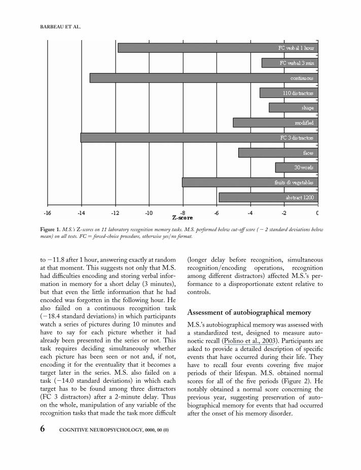
to 211.8 after 1 hour, answering exactly at randomat that moment. This suggests not only that M.S.had difficulties encoding and storing verbal infor-mation in memory for a short delay (3 minutes),but that even the little information that he hadencoded was forgotten in the following hour. Healso failed on a continuous recognition task(218.4 standard deviations) in which participantswatch a series of pictures during 10 minutes andhave to say for each picture whether it hadalready been presented in the series or not. Thistask requires deciding simultaneously whethereach picture has been seen or not and, if not,encoding it for the eventuality that it becomes atarget later in the series. M.S. also failed on atask (214.0 standard deviations) in which eachtarget has to be found among three distractors(FC 3 distractors) after a 2-minute delay. Thuson the whole, manipulation of any variable of therecognition tasks that made the task more difficult
(longer delay before recognition, simultaneousrecognition/encoding operations, recognitionamong different distractors) affected M.S.’s per-formance to a disproportionate extent relative tocontrols.
Assessment of autobiographical memory
M.S.’s autobiographical memory was assessed witha standardized test, designed to measure auto-noetic recall (Piolino et al., 2003). Participants areasked to provide a detailed description of specificevents that have occurred during their life. Theyhave to recall four events covering five majorperiods of their lifespan. M.S. obtained normalscores for all of the five periods (Figure 2). Henotably obtained a normal score concerning theprevious year, suggesting preservation of auto-biographical memory for events that had occurredafter the onset of his memory disorder.
Figure 1. M.S.’s Z-scores on 11 laboratory recognition memory tasks. M.S. performed below cut-off score ( 2 2 standard deviations below
mean) on all tests. FC ¼ forced-choice procedure, otherwise yes/no format.
6 COGNITIVE NEUROPSYCHOLOGY, 0000, 00 (0)
BARBEAU ET AL.

His ability to recall autobiographical events wasfurther evaluated in an ecological setting. M.S. wasaccompanied on a 30-minute walk through thehospital. After a 24-hour delay, he was asked toverbally recall this episode and all the events thathad occurred during the walk. He had no difficultyproviding a detailed report. This is a literal trans-lation of his spontaneous account: “We went tohave a drink on the ground floor, but we had towait a long time for the elevator. She [the examiner]went to buy Le Point [a magazine], but I did notbuy anything myself. We then drank a hot choco-late in the small cafe, which is near the entrance.We afterwards went to the children’s hospital,which we visited for some time and from wherewe saw the helicopter platform. Then we cameback.” All episodes were correctly recalled withthe exception of the episode concerning thepurchase of the magazine. He, not the examiner,bought the magazine on the examiner’s request.
Assessment of semantic memory
M.S.’s scaled-score on the information subtest ofthe WAIS-III (which assesses general, culturalknowledge about the world) was 1 standard devia-tion above mean. He also underwent a question-naire that assesses knowledge usually acquiredthrough school, designed for patients of hisage group. His score was perfect (20/20) forboth the historical (M¼ 16.6, SD¼ 4.0) and geo-graphical (M ¼ 18.8, SD ¼ 2.2) parts of the test.When shown the names of famous people, he
was able to provide detailed information on 9/10people (he failed concerning a young popstar;score ¼ 9, M ¼ 8.8, SD ¼ 1.2). He was alsoshown a series of photographs of famous peopleand was asked to provide the name, or alternativelyas many biographical details about the person aspossible. M.S. could only name 20 out of 40 faces(M ¼ 34.6, SD ¼ 5.4) and provided verbal detailson only 8 out of the remaining 20 faces (total ¼28, M ¼ 38.0, SD ¼ 2.4), performing lower than23 control subjects matched for age. He was thenshown a series of 10 photographs of famousevents. His performance did not differ from thatof control subjects (83%, M ¼ 87.8%, SD ¼
5.9%). Notably, he obtained a perfect score on allthree events that happened after the onset of hismemory problems. Finally, he was asked to recallpublic events that had occurred after the onset ofhis memory disorder (year 2001). He was spon-taneously able to provide an accurate account of14 events covering the major highlights in politicsand sporting events of this period.
Assessment of spatial memory
Route learning in a real environmentMethods. In order to evaluate to which extentspatial memory was preserved in M.S., wedesigned a route-learning test in which M.S. andnormal controls had to learn two circuits in thehospital (mean length: 267 m, SD ¼ 32). Thefirst route had 15 decision points (locationswhere participants have to make a decisionwhether to turn right, turn left, or continuestraight on), and the second had 13. Each itinerarywas completed in about 5 minutes. Participantswere shown the route once before the evaluationbegan. During the evaluation, each error was cor-rected. Trials were repeated until two consecutivetrials were successfully performed. After a 1-hourdelayed trial, participants were asked (a) to pointin the direction of three decision points (point ofdeparture, the nurse’s office, the library), (b) tocomplete the same itinerary in the reverse way,(c) to draw the two routes on a floor map, and(d) to verbally provide a detailed description ofeach route.
Figure 2. M.S.’s performance on a standard test of autobiographical
memory (TEMPau). He obtained normal performance concerning
all periods, including the last period (after presumable disease
onset). Vertical lines represent standard deviations.
COGNITIVE NEUROPSYCHOLOGY, 0000, 00 (0) 7
PURE PROGRESSIVE AMNESIA
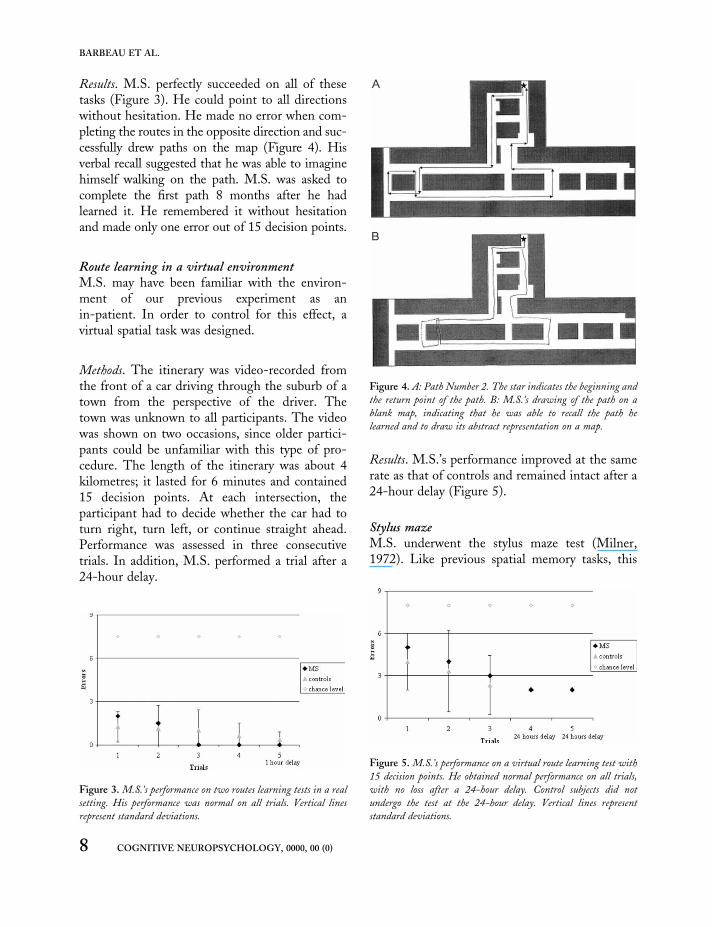
Results. M.S. perfectly succeeded on all of thesetasks (Figure 3). He could point to all directionswithout hesitation. He made no error when com-pleting the routes in the opposite direction and suc-cessfully drew paths on the map (Figure 4). Hisverbal recall suggested that he was able to imaginehimself walking on the path. M.S. was asked tocomplete the first path 8 months after he hadlearned it. He remembered it without hesitationand made only one error out of 15 decision points.
Route learning in a virtual environmentM.S. may have been familiar with the environ-ment of our previous experiment as anin-patient. In order to control for this effect, avirtual spatial task was designed.
Methods. The itinerary was video-recorded fromthe front of a car driving through the suburb of atown from the perspective of the driver. Thetown was unknown to all participants. The videowas shown on two occasions, since older partici-pants could be unfamiliar with this type of pro-cedure. The length of the itinerary was about 4kilometres; it lasted for 6 minutes and contained15 decision points. At each intersection, theparticipant had to decide whether the car had toturn right, turn left, or continue straight ahead.Performance was assessed in three consecutivetrials. In addition, M.S. performed a trial after a24-hour delay.
Results. M.S.’s performance improved at the samerate as that of controls and remained intact after a24-hour delay (Figure 5).
Stylus mazeM.S. underwent the stylus maze test (Milner,1972). Like previous spatial memory tasks, this
Figure 3. M.S.’s performance on two routes learning tests in a real
setting. His performance was normal on all trials. Vertical lines
represent standard deviations.
Figure 4. A: Path Number 2. The star indicates the beginning and
the return point of the path. B: M.S.’s drawing of the path on a
blank map, indicating that he was able to recall the path he
learned and to draw its abstract representation on a map.
Figure 5. M.S.’s performance on a virtual route learning test with
15 decision points. He obtained normal performance on all trials,
with no loss after a 24-hour delay. Control subjects did not
undergo the test at the 24-hour delay. Vertical lines represent
standard deviations.
8 COGNITIVE NEUROPSYCHOLOGY, 0000, 00 (0)
BARBEAU ET AL.

task evaluates sequence learning, but in intra-personal space. This task was chosen in referenceto the case study of H.M., whose performancewas consistently impaired (Milner, 1972).
Methods. Participants have to learn a path on aboard covered by a matrix of 10 � 10 bolts. Weused the same path that H.M. had to learn,which was made of 28 decision points. In orderto reach criterion, the path has to be repeatedwith no error on three consecutive trials.
Results. M.S. learned the procedure after 23 trials,at a similar rate to that of control subjects (Table 3,controls from Milner, 1972). This is in sharp con-trast with H.M., who never learned the taskdespite an impressive amount of trials (morethan 215 trials). The same procedure was repeatedat day þ 1 and day þ 2. M.S. consistentlyimproved his performance (number of errorsbefore reaching criterion at Day 0 ¼ 158; at Day1 ¼ 70; at Day 3 ¼ 13).
Brain imaging
Cerebral blood flow studyMethods. Acquisition of single photon emissioncomputed tomography (SPECT) images was per-formed with a double-head gamma camera (DST;Sopha Medical Vision International) with fan-beam collimators. The images were acquired 1hour after intravenous injection of 740 MBq99mTc-ethylcysteinate dimer (ECD) withdimmed lights, in a quiet surrounding. The par-ticipant’s head was safely positioned in an adjusta-ble head folder. A total of 64 angular views of 60 seach were obtained through a 3608 circular orbit.
The data were recorded in a 128 � 128 matrix.SPECT images were then reconstructed from pro-jection data using the filtered back-projectionalgorithm with a 0.30 cut-off frequencyButterworth filter and a software zoom of 2(matrix, 128 � 128 � 128; voxel size, 1.7 �1.7 � 1.7 mm). No attenuation correction wasperformed. Reconstructed brain slices were reor-iented according to the bicommissural line.
A proportional Talairach’s grid was semiauto-matically drawn by Neurogam@ (SEGAMI@Software), and 30 cortical regions of interest(ROIs) were defined on each hemisphere, usingstereotactic coordinates of the Talairach andTournoux atlas (Talairach & Tournoux, 1988),after segmentation. For each ROI, cerebral bloodflow (CBF) was evaluated using a perfusionindex calculated as the mean cortex-to-cerebellumratio and also expressed in number of standarddeviations compared to a reference population of24 participants matched for age contained in thesoftware. Perfusion was considered abnormal if itwas more than 2 standard deviations below thatof control subjects’ mean.
Results. Using a cut-off value of 22.0 standarddeviations below mean, there was marked hypo-perfusion of the medial temporal lobes bilater-ally, involving all hippocampal as well assubhippocampal structures predominating in thetemporal lobes medially, extending to ventraland anterior temporal lobe regions (Table 4).Within medial temporal lobe structures, hypo-perfusion predominated in the hippocampus(mean right and left Z-score: 24.40), veryclosely followed by the perirhinal (24.10) andentorhinal cortices (24.05). The subcallosalcortex showed hypoperfusion to a similar extent(24.45). Hypoperfusion, to a lesser degree, wasalso observed in the posterior cingulate gyrus(21.95) and in the occipito-temporo-parietaljunction bilaterally (22.30). Hypoperfusionswere bilateral in all cases. The remaining areaswithin the temporal, frontal, parietal, and occipi-tal cortex, as well as the thalami on both sides,showed normal perfusion (i.e., above cut-off).
Table 3. M.S.’s performance on the stylus maze test
M.S. Controls H.M.
Number of trials to criterion 23 17.0 215 þ
Number of errors to criterion 158 91.8 2,877 þ
Note: Results show that M.S. performed at the level of controls
and much better than H.M. Controls (younger in age than
M.S.) and H.M. reported by Milner (1972).
COGNITIVE NEUROPSYCHOLOGY, 0000, 00 (0) 9
PURE PROGRESSIVE AMNESIA

Morphometric study with MRI
Images of M.S.’s brain were acquired with a 1.5 TMagnetom MRI (Siemens, Erlangen), using astandard head coil. It revealed diffuse and homo-genous atrophy. The anterior temporal hornswere enlarged, and medial temporal lobe structureswere atrophied on both sides. T2-weighted imagesshowed punctuate foci of increased signal in thedeep white matter of both hemispheres as well asin the putamen and the pallidum. No suchlesions were observed in the thalami (with specialattention brought to the anterior and medio-dorsal nuclei), hippocampus, and parahippocampalgyrus.
Medial temporal lobes volumetryMethods. A magnetization prepared rapid gradientrecalled echo sequence allowing 3D reconstructionacquired in the sagittal plane, aligned on the axis ofthe hippocampus, and a tilted coronal gradientecho sequence was acquired on a 1.5 TMagnetom (TR ¼ 1,320 ms, TE ¼ 3.93 ms, TI ¼800 ms, flip angle ¼ 158, FOV ¼ 256 � 256,matrix ¼ 256 � 256, slice thickness ¼ 1.5). Theimages were reconstructed in the axial andcoronal planes and reformatted to obtain 1-mm3
isotropic voxels. Segmentation of the grey matterwas manually performed on coronal sections per-pendicular to the grand axis of the hippocampus.In order to measure the subregions of the medialtemporal lobe, regions of interest based on specificanatomical landmarks were defined (Duvernoy,1998; Insausti et al., 1998). They included the hip-pocampus (hippocampus proper, dentate gyrusand subicular complex) as well as entorhinal, peri-rhinal, and parahippocampal cortices. In order tocontrol the effect of interindividual variability ofparticipants’ head size on the volumes of thestudied structures, the volumes were normalizedto the intracranial area (Eritaia et al., 2000). Atotal of 5 participants (mean age: 72.0 years, SD¼ 4.8) served as controls for this study. All under-went three tests of visual and two tests of verbalrecognition memory, on which they obtainednormal results.
Results. Detailed results are summarized in Table 5.Within the medial temporal lobe, atrophy predomi-nated on the hippocampus when compared withresults for control subjects (mean Z-score, left andright: 21.27), followed by perirhinal cortices(20.93) and entorhinal cortices (20.56). Volumesof the parahippocampal cortices did not differfrom that of control subjects (20.03).
DISCUSSION
Our case study of M.S., an 83-year-old patientwith an amnestic syndrome and preserved overallintellectual efficiency, revealed a highly unusualdissociation in the realm of memory. M.S. failedon memory tasks for single items, such as recallof lists of words or recognition of verbal andvisual items. In contrast, recall of complex materialsuch as autobiographical and spatial memory wasrelatively preserved. Performance on formalmemory tests based on complex relationalmaterial, such as words embedded in stories(WMS-III logical memory) and the recall ofRey’s complex figure, was also preserved.
The comparison of two sets of tasks, one eval-uating memory of single items, the other
Table 4. SPECT results for M.S.
Region
Z-score
Left
hemisphere
Right
hemisphere
Area 38 (occipito-temporo-
parietal junction)
22.1 22.5
Area 23 (posterior cingulate) 22.2 21.7
Area 20 (anterior basal
temporal lobe)
22.9 21.8
Area 36 (perirhinal cortex) 24.8 23.4
Area 28 (entorhinal cortex) 24.3 23.8
Area 25 (subcallosal cortex) 24.3 24.6
Hippocampus 24.7 24.1
Note: Results expressed in number of standard deviations below
mean (Z-score). Hypoperfusion shown when more than 2.0
standard deviations below that of control subjects. The
remaining brain areas were found to show perfusion above
the cut-off of 2 2.0 standard deviations.
10 COGNITIVE NEUROPSYCHOLOGY, 0000, 00 (0)
BARBEAU ET AL.
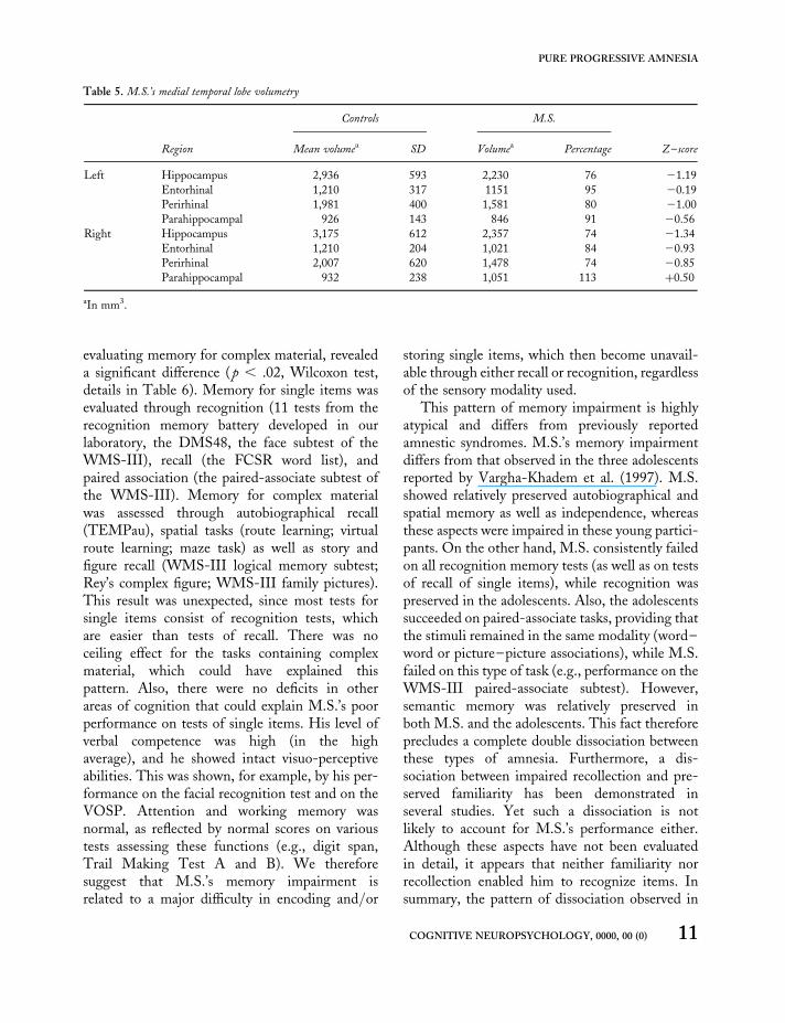
evaluating memory for complex material, revealeda significant difference (p , .02, Wilcoxon test,details in Table 6). Memory for single items wasevaluated through recognition (11 tests from therecognition memory battery developed in ourlaboratory, the DMS48, the face subtest of theWMS-III), recall (the FCSR word list), andpaired association (the paired-associate subtest ofthe WMS-III). Memory for complex materialwas assessed through autobiographical recall(TEMPau), spatial tasks (route learning; virtualroute learning; maze task) as well as story andfigure recall (WMS-III logical memory subtest;Rey’s complex figure; WMS-III family pictures).This result was unexpected, since most tests forsingle items consist of recognition tests, whichare easier than tests of recall. There was noceiling effect for the tasks containing complexmaterial, which could have explained thispattern. Also, there were no deficits in otherareas of cognition that could explain M.S.’s poorperformance on tests of single items. His level ofverbal competence was high (in the highaverage), and he showed intact visuo-perceptiveabilities. This was shown, for example, by his per-formance on the facial recognition test and on theVOSP. Attention and working memory wasnormal, as reflected by normal scores on varioustests assessing these functions (e.g., digit span,Trail Making Test A and B). We thereforesuggest that M.S.’s memory impairment isrelated to a major difficulty in encoding and/or
storing single items, which then become unavail-able through either recall or recognition, regardlessof the sensory modality used.
This pattern of memory impairment is highlyatypical and differs from previously reportedamnestic syndromes. M.S.’s memory impairmentdiffers from that observed in the three adolescentsreported by Vargha-Khadem et al. (1997). M.S.showed relatively preserved autobiographical andspatial memory as well as independence, whereasthese aspects were impaired in these young partici-pants. On the other hand, M.S. consistently failedon all recognition memory tests (as well as on testsof recall of single items), while recognition waspreserved in the adolescents. Also, the adolescentssucceeded on paired-associate tasks, providing thatthe stimuli remained in the same modality (word–word or picture–picture associations), while M.S.failed on this type of task (e.g., performance on theWMS-III paired-associate subtest). However,semantic memory was relatively preserved inboth M.S. and the adolescents. This fact thereforeprecludes a complete double dissociation betweenthese types of amnesia. Furthermore, a dis-sociation between impaired recollection and pre-served familiarity has been demonstrated inseveral studies. Yet such a dissociation is notlikely to account for M.S.’s performance either.Although these aspects have not been evaluatedin detail, it appears that neither familiarity norrecollection enabled him to recognize items. Insummary, the pattern of dissociation observed in
Table 5. M.S.’s medial temporal lobe volumetry
Region
Controls M.S.
Mean volumea SD Volumea Percentage Z–score
Left Hippocampus 2,936 593 2,230 76 21.19
Entorhinal 1,210 317 1151 95 20.19
Perirhinal 1,981 400 1,581 80 21.00
Parahippocampal 926 143 846 91 20.56
Right Hippocampus 3,175 612 2,357 74 21.34
Entorhinal 1,210 204 1,021 84 20.93
Perirhinal 2,007 620 1,478 74 20.85
Parahippocampal 932 238 1,051 113 þ0.50
aIn mm3.
COGNITIVE NEUROPSYCHOLOGY, 0000, 00 (0) 11
PURE PROGRESSIVE AMNESIA

M.S. cannot easily be related to previouslyreported amnestic syndromes.
Single-item memory and models ofdeclarative memory
By “single item”, we refer to a concrete or abstractconcept that is not embedded in a story (such as aword), or that is processed as an isolated or singlepart, such as a picture. The term single item is notvery well defined in the literature on memory,
although it is frequently used to refer to a singlestimulus that has to be memorized (Cohen,Poldrack, & Eichenbaum, 1997; Henke et al.,1999; Mayes, Isaac, Holdstock, Hunkin, &Montaldi, 2001; Turriziani, Fadda, Caltagirone,& Carlesimo, 2004). In fact, a wide variety ofmemory tests in the animal and human literatureare based on such material (e.g., visual recognitionmemory tasks, word lists). These tasks can be distin-guished from those that require establishing con-junctions or relations among elements (such asstimulus association tests, configurational tests,object–place tests, place/time discontiguous eventstests) or from those assessing context-rich memorysuch as spatial or autobiographical memory.
Eichenbaum and collaborators (Eichenbaum,2000; Eichenbaum, Otto, & Cohen, 1994;Eichenbaum, Schoenbaum, Young, & Bunsey,1996) have introduced a model of memory inwhich information is first memorized in an inflexi-ble manner at the single-item level. This stage isalso critical for paired-associate tasks within thesame modality when the relation between theitems is inflexible and can be stored as a single-item equivalent. In this model, it is thought thatflexible relations between items are established ina subsequent stage, in particular when they arespatially or temporally discontiguous, or whentransitivity across items is required (Wallenstein,Eichenbaum, & Hasselmo, 1998). Beyond theirrespective differences, other groups have elabo-rated similar hierarchical models of memory.Mishkin and collaborators have suggested that“context-free” memory such as recognition basedon familiarity or semantic memory could be disso-ciated from “context-rich” memory such asmemory for episodic events (Mishkin, Suzuki,Gadian, & Vargha-Khadem, 1997; Mishkin,Vargha-Khadem, & Gadian, 1998). Aggletonand Brown (1999) also provide evidence for a dis-tinction between two memory systems, one beingcrucial for familiarity judgements, while theother is thought to be critical for episodic andspatial memory. Although these models signi-ficantly differ in many aspects, all converge tothe notion that memory for single items could bedissociated from memory for complex material
Table 6. Comparison of results on single-item and complex-
material tests
Test Results
Single-item
FC verbal 1 hra 211.84
FC verbal 3 mina 23.33
Continuousa 218.38
110 distractorsa 23.29
Shapea 25.04
Modifieda 22.90
FC 3 distractorsa 214.05
Facesa 24.70
30 wordsa 22.51
Fruits & vegetablesa 28.06
Abstract 1200a 25.85
DMS48 29.30
WMS–III face subtestb 21.00
FCSR word list 28.80
WMS–III paired-associate subtest 21.67
Mean 26.7
SD 5.0
Complex-material
TEMPau 0.61
Route learning 0.71
Virtual route learning 20.32
Maze task 0.00
WMS–III logical memory subtest 0.00
Rey’s figure 20.13
Family pictures 21.00
Mean 0.0
SD 0.6
Note: Performance on delayed tests when available. FC ¼
forced choice. FCSR ¼ Free and Cued Selective Reminding
Test. WMS ¼ Wechsler Memory Scale.aTests from the recognition memory battery. bPerformance 1
standard deviation below mean but performed at the level of
chance by M.S. (see General Neuropsychological Assessment
section).
12 COGNITIVE NEUROPSYCHOLOGY, 0000, 00 (0)
BARBEAU ET AL.
and context-rich episodes. In addition, thesemodels converge concerning the involvement ofcritical brain structures for these stages: Item andcontext-free memory, as well as familiarity, arethought to depend on anterior subhippocampalareas (amongst which the perirhinal cortexplays a major role), while relational, context-richmemory is thought to depend on the hippocampalformation.
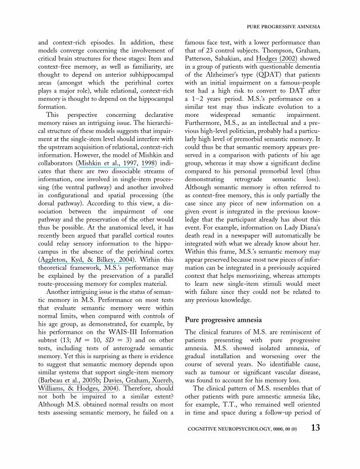
This perspective concerning declarativememory raises an intriguing issue. The hierarchi-cal structure of these models suggests that impair-ment at the single-item level should interfere withthe upstream acquisition of relational, context-richinformation. However, the model of Mishkin andcollaborators (Mishkin et al., 1997, 1998) indi-cates that there are two dissociable streams ofinformation, one involved in single-item proces-sing (the ventral pathway) and another involvedin configurational and spatial processing (thedorsal pathway). According to this view, a dis-sociation between the impairment of onepathway and the preservation of the other wouldthus be possible. At the anatomical level, it hasrecently been argued that parallel cortical routescould relay sensory information to the hippo-campus in the absence of the perirhinal cortex(Aggleton, Kyd, & Bilkey, 2004). Within thistheoretical framework, M.S.’s performance maybe explained by the preservation of a parallelroute-processing memory for complex material.
Another intriguing issue is the status of seman-tic memory in M.S. Performance on most teststhat evaluate semantic memory were withinnormal limits, when compared with controls ofhis age group, as demonstrated, for example, byhis performance on the WAIS-III Informationsubtest (13; M ¼ 10, SD ¼ 3) and on othertests, including tests of anterograde semanticmemory. Yet this is surprising as there is evidenceto suggest that semantic memory depends uponsimilar systems that support single-item memory(Barbeau et al., 2005b; Davies, Graham, Xuereb,Williams, & Hodges, 2004). Therefore, shouldnot both be impaired to a similar extent?Although M.S. obtained normal results on mosttests assessing semantic memory, he failed on a
famous face test, with a lower performance thanthat of 23 control subjects. Thompson, Graham,Patterson, Sahakian, and Hodges (2002) showedin a group of patients with questionable dementiaof the Alzheimer’s type (QDAT) that patientswith an initial impairment on a famous-peopletest had a high risk to convert to DAT aftera 1–2 years period. M.S.’s performance on asimilar test may thus indicate evolution to amore widespread semantic impairment.Furthermore, M.S., as an intellectual and a pre-vious high-level politician, probably had a particu-larly high level of premorbid semantic memory. Itcould thus be that semantic memory appears pre-served in a comparison with patients of his agegroup, whereas it may show a significant declinecompared to his personal premorbid level (thusdemonstrating retrograde semantic loss).Although semantic memory is often referred toas context-free memory, this is only partially thecase since any piece of new information on agiven event is integrated in the previous know-ledge that the participant already has about thisevent. For example, information on Lady Diana’sdeath read in a newspaper will automatically beintegrated with what we already know about her.Within this frame, M.S.’s semantic memory mayappear preserved because most new pieces of infor-mation can be integrated in a previously acquiredcontext that helps memorizing, whereas attemptsto learn new single-item stimuli would meetwith failure since they could not be related toany previous knowledge.
Pure progressive amnesia
The clinical features of M.S. are reminiscent ofpatients presenting with pure progressiveamnesia. M.S. showed isolated amnesia, ofgradual installation and worsening over thecourse of several years. No identifiable cause,such as tumour or significant vascular disease,was found to account for his memory loss.
The clinical pattern of M.S. resembles that ofother patients with pure amnestic amnesia like,for example, T.T., who remained well orientedin time and space during a follow-up period of
COGNITIVE NEUROPSYCHOLOGY, 0000, 00 (0) 13
PURE PROGRESSIVE AMNESIA

8 years (Caffara & Venneri, 1996).Autobiographical memory, evaluated five yearsafter the study onset, was still globally preserved.Like M.S., T.T. was severely impaired on theBuschke Selective Reminding Test (a word listmemory test), as well as on paired-associate learn-ing. A similar pattern of memory impairment wasreported in the patient of Lucchelli et al. (1994).She was also well oriented in time and space andobtained a normal score on a maze-learning task.This contrasted with impaired performance onpaired-associate learning and on face recognitiontasks. A recent case-study of pure progressiveamnesia with follow-up over a 8-year period hadvery similar features (Stokholm et al., 2005).Memory problems remained isolated until thelast year when a more widespread impairmentwas observed. At the time of the first examination,the patient was already found to be severelyimpaired at word list learning and on recognitionmemory tasks (22.8 standard deviations on theword part of the Recognition Memory Test).Memory performance on these tests slowly wor-sened over the years. Despite these severe deficitsin a formal evaluation, he was reported to be inde-pendent in daily life. For example, during most ofthe follow-up period, he remained able to travel byhimself, to shop for groceries, to continue garden-ing, and to take on small repair jobs in the house.From these ecological data, it is tempting todeduct that his spatial memory was globally pre-served and that his memory difficulties did not sig-nificantly interfere with his daily life. Overall, allpatients with pure progressive amnesia weredescribed as independent, with no impairment ineveryday activities (Bozoki, Giordani,Heidebrink, Berent, & Foster, 2001).
In previous reports on pure progressiveamnesia, most of the discussion has focused onthe pathological changes underlying the impair-ments. Although the possibility of ischaemicdamage has been raised (Kritchevsky & Squire,1993), a neurodegenerative condition seemsmore likely to be the origin of this disorder. Twopatients who had suffered from a long-standingisolated memory impairment ultimately pro-gressed to a pattern of cognitive deterioration
meeting the criteria for Alzheimer’s disease(Squire & Kritchevsky, 1996). In two otherpatients who came to autopsy, neuropathologicaldata showed widespread Alzheimer-type changes(Caselli et al., 1998; Didic et al., 1998). Inaddition, group studies have identified pure pro-gressive amnesia as a distinct and rare clinicalvariant of Alzheimer’s disease (Butters et al.,1996). Such a severe and isolated memory losshas been estimated to occur in about 3% of patientswith a newly recognized cognitive impairment(Bowen, Teri, Kukull, McCormick, McCurry, &Larson, 1997). Taken together, these data suggestthat pure progressive amnesia may represent anatypical presentation of Alzheimer’s disease.
The neural substrate of M.S.’s memoryimpairment: Hypotheses
Based on M.S.’s clinical features, Alzheimer’sdisease with a highly unusual clinical course isthe most likely pathological substrate of M.S.’scondition. Other conditions such as encephalitis,seizures, Korsakoff’s syndrome, paraneoplastic dis-orders, transient global amnesia, and tumour areunlikely to account for his memory impairment.Periventricular signal hyperintensities onT2-weighted magnetic resonance imaging(MRI), such as those found in our patient,deserve some consideration. Chronic ischaemicinjury could have damaged brain areas that arecrucial for memory, such as the hippocampus orthe anterior and dorso-medial thalamic nuclei(Aggleton & Brown, 1999). However, a neuroi-maging study on M.S. revealed no evidence of vas-cular damage in these structures.
In Alzheimer’s disease, neurofibrillary tangles(NFT) emerge in the ento- and perirhinal cortexstages (I and II) before spreading to the hippo-campus (Braak & Braak, 1991). The subsequentstages (III and IV) are characterized by severedestruction of the rhinal cortices and spreadingto the hippocampal formation. It is tempting tosuggest that M.S.’s memory disorder may rep-resent an atypical clinical correlate of StageII/III of Braak and Braak’s classification. In a pre-vious section, we referred to models of declarative
14 COGNITIVE NEUROPSYCHOLOGY, 0000, 00 (0)
BARBEAU ET AL.
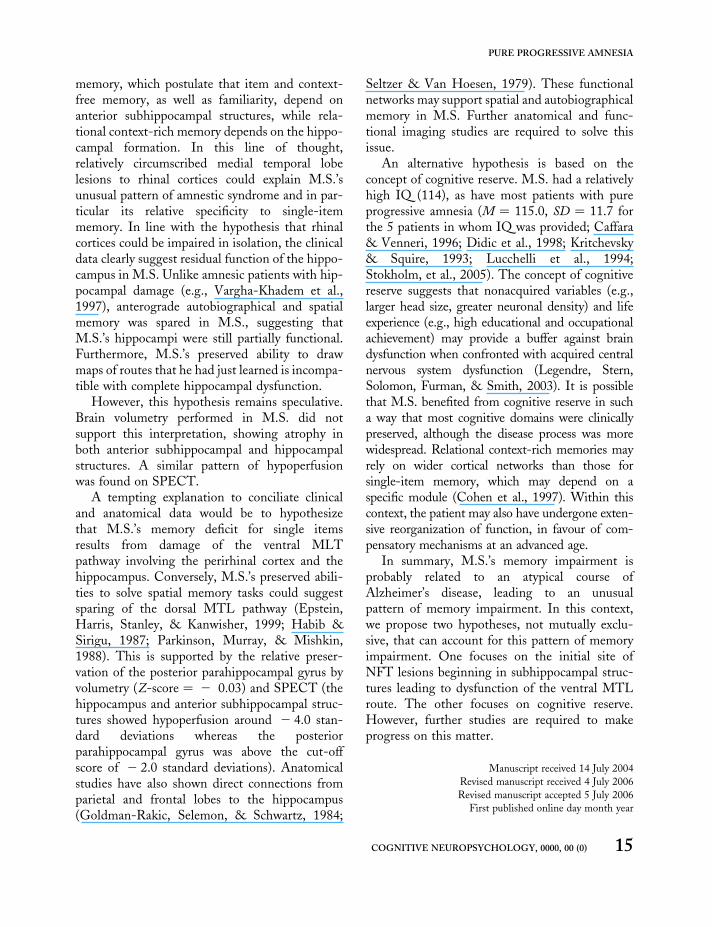
memory, which postulate that item and context-free memory, as well as familiarity, depend onanterior subhippocampal structures, while rela-tional context-rich memory depends on the hippo-campal formation. In this line of thought,relatively circumscribed medial temporal lobelesions to rhinal cortices could explain M.S.’sunusual pattern of amnestic syndrome and in par-ticular its relative specificity to single-itemmemory. In line with the hypothesis that rhinalcortices could be impaired in isolation, the clinicaldata clearly suggest residual function of the hippo-campus in M.S. Unlike amnesic patients with hip-pocampal damage (e.g., Vargha-Khadem et al.,1997), anterograde autobiographical and spatialmemory was spared in M.S., suggesting thatM.S.’s hippocampi were still partially functional.Furthermore, M.S.’s preserved ability to drawmaps of routes that he had just learned is incompa-tible with complete hippocampal dysfunction.
However, this hypothesis remains speculative.Brain volumetry performed in M.S. did notsupport this interpretation, showing atrophy inboth anterior subhippocampal and hippocampalstructures. A similar pattern of hypoperfusionwas found on SPECT.
A tempting explanation to conciliate clinicaland anatomical data would be to hypothesizethat M.S.’s memory deficit for single itemsresults from damage of the ventral MLTpathway involving the perirhinal cortex and thehippocampus. Conversely, M.S.’s preserved abili-ties to solve spatial memory tasks could suggestsparing of the dorsal MTL pathway (Epstein,Harris, Stanley, & Kanwisher, 1999; Habib &Sirigu, 1987; Parkinson, Murray, & Mishkin,1988). This is supported by the relative preser-vation of the posterior parahippocampal gyrus byvolumetry (Z-score ¼ 2 0.03) and SPECT (thehippocampus and anterior subhippocampal struc-tures showed hypoperfusion around 2 4.0 stan-dard deviations whereas the posteriorparahippocampal gyrus was above the cut-offscore of 2 2.0 standard deviations). Anatomicalstudies have also shown direct connections fromparietal and frontal lobes to the hippocampus(Goldman-Rakic, Selemon, & Schwartz, 1984;
Seltzer & Van Hoesen, 1979). These functionalnetworks may support spatial and autobiographicalmemory in M.S. Further anatomical and func-tional imaging studies are required to solve thisissue.
An alternative hypothesis is based on theconcept of cognitive reserve. M.S. had a relativelyhigh IQ (114), as have most patients with pureprogressive amnesia (M ¼ 115.0, SD ¼ 11.7 forthe 5 patients in whom IQ was provided; Caffara& Venneri, 1996; Didic et al., 1998; Kritchevsky& Squire, 1993; Lucchelli et al., 1994;Stokholm, et al., 2005). The concept of cognitivereserve suggests that nonacquired variables (e.g.,larger head size, greater neuronal density) and lifeexperience (e.g., high educational and occupationalachievement) may provide a buffer against braindysfunction when confronted with acquired centralnervous system dysfunction (Legendre, Stern,Solomon, Furman, & Smith, 2003). It is possiblethat M.S. benefited from cognitive reserve in sucha way that most cognitive domains were clinicallypreserved, although the disease process was morewidespread. Relational context-rich memories mayrely on wider cortical networks than those forsingle-item memory, which may depend on aspecific module (Cohen et al., 1997). Within thiscontext, the patient may also have undergone exten-sive reorganization of function, in favour of com-pensatory mechanisms at an advanced age.
In summary, M.S.’s memory impairment isprobably related to an atypical course ofAlzheimer’s disease, leading to an unusualpattern of memory impairment. In this context,we propose two hypotheses, not mutually exclu-sive, that can account for this pattern of memoryimpairment. One focuses on the initial site ofNFT lesions beginning in subhippocampal struc-tures leading to dysfunction of the ventral MTLroute. The other focuses on cognitive reserve.However, further studies are required to makeprogress on this matter.
Manuscript received 14 July 2004
Revised manuscript received 4 July 2006
Revised manuscript accepted 5 July 2006
First published online day month year
COGNITIVE NEUROPSYCHOLOGY, 0000, 00 (0) 15
PURE PROGRESSIVE AMNESIA

REFERENCES
Aggleton, J. P., & Brown, M. W. (1999). Episodicmemory, amnesia, and the hippocampal-anteriorthalamic axis. Behavioral and Brain Sciences, 22,425–444; discussion, 444–489.
Aggleton, J. P., Kyd, R. J., & Bilkey, D. K. (2004).When is the perirhinal cortex necessary for the per-formance of spatial memory tasks? Neuroscience and
Biobehavioral Reviews, 28, 611–624.Aggleton, J. P., Vann, S. D., Denby, C., Dix, S.,
Mayes, A. R., Roberts, N., et al. (2005) Sparing ofthe familiarity component of recognition memoryin a patient with hippocampal pathology.Neuropsychologia, 43, 1810–1823.
Baddeley, A., Vargha-Khadem, F., & Mishkin, M.(2001). Preserved recognition in a case of develop-mental amnesia: Implications for the acquisition ofsemantic memory? Journal of Cognitive Neuroscience,13, 357–369.
Barbeau, E., Didic, M., Tramoni, E., Felician, O.,Joubert, S., Sontheimer, A., et al. (2004).Evaluation of visual recognition memory in MCIpatients. Neurology, 62, 1317–1322.
Barbeau, E. J., Felician, O., Joubert, S., Sontheimer, A.,Ceccaldi, M., & Poncet, M. (2005a). Preservedvisual recognition memory in a patient with severehippocampal damage. Hippocampus, 15, 587–596.
Barbeau, E. J, Wendling, F., Regis, J., Duncan, R.,Poncet, M., Chauvel, P., et al. (2005b).Recollection of vivid memories after perirhinalregion stimulations: Synchronization in the thetarange of spatially distributed brain areas.Neuropsychologia, 43, 1329–1337.
Benton, A., Hamsher, K de S., Varney, N., &Spreen, O. (1983a). Judgment of line orientation,form H. In Benton, A. L., Sirvan, A. B.,Hamsher, K de S., Varney, N. R., & Spreen, O.(Eds.), Contributions to neuropsychological assessment.New York: Oxford University Press Inc.
Benton, A., Sirvan, A., Hamsher, K de S., Varney, N.,& Spreen, O. (1983b). Facial recognition: Stimulusand multiple choice pictures. In Benton, A. L.,Sirvan, A. B., Hamsher, L de S., Varney, N. R., &Spreen, O. (Eds.), Contributions to neuropsycho-
logical assessment. New York: Oxford UniversityPress Inc.
Bowen, J., Teri, L., Kukull, W., McCormick, W.,McCurry, S. M., & Larson, E. B. (1997).Progression to dementia in patients with isolatedmemory loss. Lancet, 349, 763–765.
Bozoki, A., Giordani, B., Heidebrink, J. L., Berent, S., &Foster, N. L. (2001). Mild cognitive impairmentspredict dementia in nondemented elderly patientswith memory loss. Archives of Neurology, 58, 411–416.
Braak, H., & Braak, E. (1991). Neuropathologicalstageing of Alzheimer-related changes. Acta
Neuropathologica (Berlin), 82, 239–259.Butters, M. A., Lopez, O. L., & Becker, J. T. (1996).
Focal temporal lobe dysfunction in probableAlzheimer’s disease predicts a slow rate of cognitivedecline. Neurology, 46, 687–692.
Caffara, P., & Venneri, A. (1996). Isolated degenerativeamnesia without dementia: An 8-year longitudinalstudy. Neurocase, 2, 99–106.
Caselli, R. J., Couce, M. E., Osborne, D., Deen, H. G., &Parisi, J. P. (1998). From slowly progressive amnesicsyndrome to rapidly progressive Alzheimer disease.Alzheimer Disease and Associated Disorders, 12,251–253.
Cohen, N. J., Poldrack, R. A., & Eichenbaum, H.(1997). Memory for items and memory for relationsin the procedural/declarative memory framework.Memory, 5, 131–178.
Collie, A., & Maruff, P. (2000). The neuropsychologyof preclinical Alzheimer’s disease and mild cognitiveimpairment. Neuroscience and Biobehavioral Reviews,24, 365–374.
Davies, R. R., Graham, K. S., Xuereb, J. H.,Williams, G. B., & Hodges, J. R. (2004). Thehuman perirhinal cortex and semantic memory.European Journal of Neuroscience, 20, 2441–2446.
Deloche, G., & Hannequin, D. (1997). Test de denomi-
nation orale d’images DO80. Paris: Les Editions duCentre de Psychologie Appliquee.
Didic, M., Ali Cherif, A., Gambarelli, D., Poncet, M.,& Boudouresques, J. (1998). A permanent pureamnestic syndrome of insidious onset related toAlzheimer’s disease. Annals of Neurology, 43, 526–530.
Duvernoy, H. M. (1998). The human hippocampus (2nded.). New York: Springler-Verlag Press.
Eichenbaum, H. (2000). A cortical-hippocampal systemfor declarative memory. Nature Review Neuroscience,1, 41–50.
Eichenbaum, H., Otto, T., & Cohen, N. (1994). Twofunctional components of the hippocampal memorysystem. Behavioral and Brain Sciences, 17, 449–472.
Eichenbaum, H., Schoenbaum, G., Young, B., &Bunsey, M. (1996). Functional organization of thehippocampal memory system. Proceedings of the
National Academy of Science USA, 93, 13500–13507.
16 COGNITIVE NEUROPSYCHOLOGY, 0000, 00 (0)
BARBEAU ET AL.

Epstein, R., Harris, A., Stanley, D., & Kanwisher, N.(1999). The parahippocampal place area:Recognition, navigation, or encoding? Neuron, 23,115–125.
Ergis, A.-M., Van der Linden, M., & Deweer, B.(1994). Investigation of memory performance witha cued recall test in Alzheimer’s disease. Revue de
Neuropsychologie, 4, 47–68.Eritaia, J., Wood, S. J., Stuart, G. W., Bridle, N.,
Maruff, P., Velakoulis, D., et al. (2000). An opti-mized method for estimating intracranial volumefrom magnetic resonance images. Magnetic
Resonance in Medicine, 44, 973–977.Goldman-Rakic, P. S., Selemon, L. D., & Schwartz,
M. L. (1984). Dual pathways connecting the dorso-lateral prefrontal cortex with the hippocampal for-mation and parahippocampal cortex in the rhesusmonkey. Neuroscience, 12, 719–743.
Grober, E., Buschke, H., Crystal, H., Bang, S., &Dresner, R. (1988). Screening for dementia bymemory testing. Neurology, 38, 900–903.
Habib, M., & Sirigu, A. (1987). Pure topographical dis-orientation: A definition and anatomical basis.Cortex, 23, 73–85.
Henke, K., Kroll, N. E., Behniea, H., Amaral, D. G.,Miller, M. B., Rafal, R., et al. (1999). Memory lostand regained following bilateral hippocampal damage.Journal of Cognitive Neuroscience, 11, 682–697.
Insausti, R., Juottonen, K., Soininen, H., Insausti, A.M., Partanen, K., Vainio P., et al. (1998). MRvolumetric analysis of the entorhinal and perirhinalcortices in Alzheimer’s disease. American Journal of
Neuroradiology, 19, 659–671.Kopelman, M. D. (2002). Disorders of memory. Brain,
125, 2152–2190.Kritchevsky, M., & Squire, L. R. (1993). Permanent
global amnesia with unknown etiology. Neurology,43, 326–332.
Legendre, S. A., Stern, R. A., Solomon, D. A.,Furman, M. J., & Smith, K. E. (2003). Theinfluence of cognitive reserve on memory followingelectroconvulsive therapy. The Journal of
Neuropsychiatry and Clinical Neurosciences, 15,333–339.
Lucchelli, F., De Renzi, E., Perani, D., & Fazio, F.(1994). Primary amnesia of insidious onset withsubsequent stabilisation. Journal of Neurology,
Neurosurgery and Psychiatry, 57, 1366–1370.Mayes, A. R., Holdstock, J. S., Isaac, C. L.,
Hunkin, N. M., & Roberts, N. (2002). Relativesparing of item recognition memory in a patient
with adult-onset damage limited to the hippo-campus. Hippocampus, 12, 325–340.
Mayes, A. R., Isaac, C. L., Holdstock, J. S.,Hunkin, N. M., & Montaldi, D. (2001). Memoryfor single items, word pairs, and temporal order ofdifferent kinds in a patient with selective hippocam-pal lesions. Cognitive Neuropsychology, 18, 97–123.
Miceli, G., Colosimo, C., Daniele, A., Marra, C.,Perani, D., & Fazio, F. (1996). Isolated amnesiawith slow onset and stable course, without ensuingdementia: MRI and PET data and a six-year neurop-sychological follow-up. Dementia, 7, 104–110.
Milner, B. (1972). Disorders of learning and memoryafter temporal lobe lesions in man. Clinical
Neurosurgery, 19, 421–446.Mishkin, M., Suzuki, W. A., Gadian, D. G., &
Vargha-Khadem, F. (1997). Hierarchical organiz-ation of cognitive memory. Philosophical transactions
of the Royal Society of London. Series B: Biological
Sciences, 352, 1461–1467.Mishkin, M. J., Vargha-Khadem, F., & Gadian, D. G.
(1998). Amnesia and the organization of the hippo-campal system. Hippocampus, 8, 212–216.
Parkinson, J. K., Murray, E. A., & Mishkin, M. (1988).A selective mnemonic role for the hippocampus inmonkeys: Memory for the location of objects.Journal of Neuroscience, 8, 4159–4167.
Petersen, R. C., Smith, G. E., Ivnik, R. J., Kokmen,E., & Tangalos, E. G. (1994). Memory functionin very early Alzheimer’s disease. Neurology, 44,867–872.
Pillon, B., Deweer, B., Michon, A., Malapani, C.,Agid, Y., & Dubois, B. (1994). Are explicitmemory disorders of progressive supranuclear palsyrelated to damage to striatofrontal circuits?Comparison with Alzheimer’s, Parkinson’s, andHuntington’s diseases. Neurology, 44, 1264–1270.
Piolino, P., Desgranges, B., Belliard, S.,Matuszewski, V., Lalevee, C., De La Sayette, V.,et al. (2003). Autobiographical memory and auton-oetic consciousness: Triple dissociation in neurode-generative diseases. Brain, 126, 2203–2219.
Seltzer, B., & Van Hoesen, G. W. (1979). A directinferior parietal lobule projection to the presubicu-lum in the rhesus monkey. Brain Research, 179,157–161.
Squire, L. R., & Kritchevsky, M. (1996). Selectivememory loss. Neurology, 47, 853–854.
Stokholm, J., Jakobsen, O., Czarna, J. M., Mortensen,H. V., & Waldemar, G. (2005). Years of severeand isolated amnesia can precede the development
COGNITIVE NEUROPSYCHOLOGY, 0000, 00 (0) 17
PURE PROGRESSIVE AMNESIA

of dementia in early-onset Alzheimer’s disease.Neurocase, 11, 48–55.
Talairach, J., & Tournoux, P. (1988). Co-planar stereo-
taxic atlas of the human brain. New York: Thieme.Thompson, S. A., Graham, K. S., Patterson, K.,
Sahakian, B. J., & Hodges, J. R. (2002). Is knowl-edge of famous people disproportionately impairedin patients with early and questionable Alzheimer’sdisease? Neuropsychology, 16, 344–358.
Turriziani, P., Fadda, L., Caltagirone, C., &Carlesimo, G. A. (2004). Recognition memory forsingle items and for associations in amnesic patients.Neuropsychologia, 42, 426–433.
Vargha-Khadem, F., Gadian, D. G., Watkins, K. E.,Connelly, A., Van Paesschen, W., & Mishkin, M.(1997). Differential effects of early hippocampalpathology on episodic and semantic memory.Science, 277, 376–380.
Wallenstein, G. V., Eichenbaum, H., & Hasselmo, M.E. (1998). The hippocampus as an associator ofdiscontiguous events. Trends in Neuroscience, 21,317–323.
Warrington, E., & James, M. (2000). Batterie de percep-
tion visuelle et spatiale des objets (VOSP). Paris:Editions et Applications Psychologiques.
Wechsler, D. (2000). Echelle d’Intelligence de Wechsler
pour Adulte III (WAIS-III). Paris: Les Editions duCentre de Psychologie Appliquee.
Wechsler, D. (2001). Echelle Clinique de Memoire de
Wechsler MEM-III (WMS-III). Paris: Les Editionsdu Centre de Psychologie Appliquee.
Yonelinas, A. P., Kroll, N. E. A., Quamme, J. R.,Lazzara, M. M., Sauve, M.-J., Widaman, K. F., &Knight, R. T. (2002). Effects of extensive temporallobe damage or mild hypoxia on recollection andfamiliarity. Nature Neuroscience, 5, 1236–1241.
18 COGNITIVE NEUROPSYCHOLOGY, 0000, 00 (0)
BARBEAU ET AL.
Copyright © 2022 FDOKUMEN