Specialization and Omnivory in Diverse Mammalian Assemblages

Neural specialization in autism 1
Atypical neural specialization for social percepts in autism spectrum disorder
IN PRESS, SOCIAL NEUROSCIENCE
James C. McPartland, Ph.D.1, Jia Wu, Ph.D.1, Christopher A. Bailey1, Linda C. Mayes, M.D.1, Robert T. Schultz, Ph.D.2, 3 & Ami Klin, Ph.D.4, 5
1Yale Child Study Center, New Haven, CT
2Department of Pediatrics, University of Pennsylvania, Philadelphia, PA
3Center for Autism Research, Children’s Hospital of Philadelphia, PA
4Marcus Autism Center, Atlanta, GA
5Department of Pediatrics, Emory University School of Medicine, Atlanta, GA
Correspondence to: Dr. James McPartland Yale Child Study Center 230 South Frontage Road New Haven, CT 06520 [email protected] Phone: (203) 785-7179 Fax: (203) 764-4373 Key words: Perceptual expertise, N170, event-related potential (ERP / EEG), face perception, autism spectrum disorder

Neural specialization in autism 2
Acknowledgements
This research was supported by NIMH R03 MH079908, NIMH K23 MH086785, NICHD
PO1HD003008, a NARSAD Atherton Young Investigator Award, and CTSA Grant
Number UL1 RR024139 from the National Center for Research Resources (NCRR), a
component of the National Institutes of Health (NIH), and NIH roadmap for Medical
Research (USA). Its contents are solely the responsibility of the authors and do not
necessarily represent the official view of NCRR or NIH.

Neural specialization in autism 3
Abstract
The social motivation hypothesis posits that aberrant neural response to human faces in
autism is attributable to atypical social development and consequently reduced exposure
to faces. The specificity of deficits in neural specialization remains unclear, and
alternative theories suggest generalized processing difficulties. The current study
contrasted neural specialization for social information versus non-social information in
36 individuals with autism and 18 typically developing individuals matched for age, race,
sex, handedness, and cognitive ability. Event-related potentials elicited by faces, inverted
faces, houses, letters, and pseudoletters were recorded. Groups were compared on an
electrophysiological marker of neural specialization (N170), as well as behavioral
performance on standardized measures of face recognition and word reading/decoding.
Consistent with prior results, individuals with autism displayed slowed face processing
and decreased sensitivity to face inversion; however, they showed comparable brain
responses to letters, which were associated with behavioral performance in both groups.
Results suggest that individuals with autism display atypical neural specialization for
social information but intact specialization for non-social information. They concord with
the notion of specific dysfunction in social brain systems rather than non-specific
information processing difficulties in autism.

Neural specialization in autism 4
Preserved neural specialization for non-social information in autism
The ability to efficiently perceive the human face is a crucial and early-emerging
social ability. Specialized processing for faces emerges in the first days of life (Bushnell,
Sai & Mullin, 1989; Goren, Sarty & Wu, 1975; Johnson, Dziurawiec, Ellis & Morton,
1991; Meltzoff & Moore, 1977) and is honed by developmental experience (Nelson,
2001). Faces come to be encoded using configural processing mechanisms (Farah,
Tanaka & Drain, 1995), reflected in disproportionate impairments in recognizing both
upside down faces (the inversion effect (Yin, 1970) and facial features out of context
(Tanaka & Farah, 1993). Functional neuroimaging studies show that faces elicit selective,
right-lateralized hemodynamic activity in a portion of occipitotemporal cortex, the
fusiform gyrus (Haxby et al., 1994; Kanwisher, McDermott & Chun, 1997; Puce, Allison,
Gore & McCarthy, 1995), and intra-cranial electrophysiological recordings reveal face-
related negative electrical activity originating from this portion of cortex (Allison,
McCarthy, Novbre, Puce & Belger, 1994). Likewise, event-related potentials (ERPs)
recorded from corresponding scalp regions show a negative-going electrical deflection
approximately 170 milliseconds after viewing a face (N170; Bentin, Allison, Puce, Perez
& McCarthy, 1996). The N170 putatively reflects structural encoding, an early stage of
face processing preceding higher-order processes like recognition (Bentin, Deouell &
Soroker, 1999), and is sensitive to perturbations in face configuration, including inversion
(Rossion et al., 2000). Neural generators of the N170 have been localized to
occipitotemporal sites including the fusiform gyrus (Itier & Taylor, 2002; Rossion, Joyce,
Cottrell & Tarr, 2003; Shibata et al., 2002), as well as the superior temporal sulcus (Itier

Neural specialization in autism 5
& Taylor, 2004), lingual gyrus (Shibata et al., 2002), and posterior inferotemporal gyrus
(Schweinberger, Pickering, Jentzsch, Burton & Kaufmann, 2002; Shibata et al., 2002).
These processing strategies and brain regions are also observed in the perception
of visual stimuli with which viewers have extensive experience (Diamond & Carey,
1986; Gauthier, 2000). Experts at perceiving and discriminating among exemplars within
a visually homogenous class (e.g., Greebles, birds, or cars (Gauthier, Skudkarski, Gore &
Anderson, 2000; Gauthier, Williams, Tarr & Tanaka, 1998) develop face-like patterns of
brain activity, in terms of both hemodynamic (Tarr & Gauthier, 2000) and
electrophysiological response, as indexed by the N170 (Rossion, Gauthier, Goffaux, Tarr
& Crommelinck, 2002). According to this model, these brain regions subserve a
processing style rather than specific content, and face-related brain activity reflects, in
large part, human beings’ extensive experience processing human faces during
development (Gauthier & Nelson, 2001).
Analogous specialization through developmental experience occurs in brain
mechanisms subserving letter and word processing. Perception of printed letters (James,
James, Jobard, Wong & Gauthier, 2005) and words (McCandliss, Cohen & Dehaene,
2003) selectively activates left fusiform gyrus and elicits a left-lateralized N170 in literate
children as young as eight years of age (Bentin, Mouchetant-Rostaing, Giard, Echallier &
Pernier, 1999; Maurer et al., 2006). A maturational course independent of higher-order
phonological or semantic processes (Grossi, Coch, Coffey-Corina, Holcomb & Neville,
2001; Holcomb, Coffey & Neville, 1992) and an early time course suggest that this
“letter N170” marks pre-linguistic processes related to visual perception of form (Bentin,
Mouchetant-Rostaing et al., 1999) and, like the N170 elicited by faces, automatic

Neural specialization in autism 6
perceptual categorization within a domain of expertise (Maurer, Brem, Bucher &
Brandeis, 2005). Neural specialization for letters is revealed by enhanced N170
amplitude to familiar alphabets but not foreign alphabets or nonsensical letter
approximations (pseudoletters; Wong, Gauthier, Woroch, DeBuse & Curran, 2005).
Converging evidence from neuroimaging studies and ERP source localization suggest
left-lateralized sources in the fusiform gyrus and the inferior occipitotemporal cortex
(Cohen et al., 2000; Maurer et al., 2005; Rossion et al., 2003). Though letter-related brain
activity is typically contralateral to face processing areas (Rossion et al., 2003), there is
some degree of functional overlap; under special circumstances, such as precocious
reading ability, right fusiform gyrus is recruited for letter and word recognition
(Turkeltaub et al., 2004).
Because face perception is a well-studied social behavior, it has been employed as
an avenue to understand social development in autism spectrum disorder (ASD). In ASD,
decreased attention to human faces is evident by 6 to 12 months (Maestro et al., 2002;
Osterling & Dawson, 1994), and abnormalities in face perception and recognition have
been observed throughout the lifespan (Hobson, 1986; Hobson, Ouston & Lee, 1988;
Klin et al., 1999; Langdell, 1978; Schultz, 2005; Wolf et al., 2008). Individuals with ASD
often exhibit abnormal viewing patterns to faces (Jones, Carr & Klin, 2008; Klin, Jones,
Schultz, Volkmar & Cohen, 2002) and hypoactivation in face-related brain areas
(Schultz, 2005; Schultz et al., 2000). Studies of electrophysiological markers of face
perception suggest delayed N170 to human faces and decreased sensitivity to face
inversion in individuals with ASD, as well as first degree relatives (Dawson et al., 2002;
Dawson, Webb, Wijsman et al., 2005; McCleery, Akshoomoff, Dobkins & Carver, 2009;

Neural specialization in autism 7
McPartland, Dawson, Webb, Panagiotides & Carver, 2004; O'Connor, Hamm & Kirk,
2005, 2007; Webb, Dawson, Bernier & Panagiotides, 2006) though some studies suggest
at least partially preserved face perception in some subgroups of individuals with ASD
(Webb et al., 2010; Webb et al., 2009).
One theoretical explanation for these observed differences in face perception in
ASD focuses on the role of developmental exposure to faces. The social motivation
hypothesis (Dawson, Webb & McPartland, 2005; Schultz, 2005) posits that, due to
abnormalities in social drive very early in childhood, children with ASD do not attend to
faces during sensitive developmental periods. Consequently, people with ASD fail to
develop typical proficiency in face processing and associated patterns of behavioral and
brain specialization (Behrmann, Thomas & Humphreys, 2006). Because the social
motivation hypothesis implicates social drive as the dysfunction from which face
perception difficulties originate (rather than specific dysfunction of brain regions
subserving face perception), it presumes that individuals with ASD, given appropriate
exposure to and interest in a stimulus class, should develop both behavioral and brain
specialization (Sasson, 2006). This notion is supported by a single-case study revealing
behavioral and neural indices of specialization in a child with ASD during perception of
cartoon characters associated with a circumscribed interest (Grelotti et al., 2005). Though
others have attempted to investigate brain response associated with experience in this
population (Boeschoten, Kenemans, van Engeland & Kemner, 2007), research has been
stymied by difficulty finding shared areas of expertise in ASD; whereas groups of study
participants experienced in perceiving faces are common, groups of individuals with
ASD who share a common non-face area of expertise are rare.

Neural specialization in autism 8
The current work circumvented this difficulty by examining brain activity
reflecting neural specialization for letters of the alphabet. As described above,
development of specialization for letters and words is well studied and elicits brain
activity similar to faces in terms of temporal characteristics and scalp topography (despite
lateralization differences). This is a novel and uniquely appropriate comparison because,
despite developmental disinterest towards faces and characteristic weakness in language,
facility with reading has been a noted strength in ASD since Kanner’s original account
(Kanner, 1943). High-functioning individuals on the autism spectrum display age-
appropriate skills in single word-reading and word-decoding ability (Huemer & Mann,
2009; Nation, Clarke, Wright & Williams, 2006; Newman et al., 2006), and a subgroup
possesses precocious interest and proficiency in reading, or hyperlexia (Burd, Kerbeshian
& Fisher, 1985; Grigorenko et al., 2002; Klin, 2004). In this study, electrophysiological
and behavioral methods were applied to compare neural specialization for faces and
letters in individuals with ASD. Experiments contrasted neural response to faces versus
houses, faces versus inverted faces, and letters versus pseudoletters and compared these
parameters to behavioral measures assessing proficiency in face recognition and letter
and word perception. Consistent with previous work, it was hypothesized that individuals
with ASD would exhibit impaired face recognition and delayed brain response to faces in
the right hemisphere, as well as decreased sensitivity to face inversion. In keeping with
the notion that these atypicalities reflect developmental sequelae of social deficits, it was
predicted that similar anomalies would not be observed for non-social stimuli; individuals
with ASD would show typical skills in terms of letter and word perception and
comparably enhanced response to letter stimuli with respect to unfamiliar pseudoletters.

Neural specialization in autism 9
As prior work has revealed relationships among neural correlates of face perception and
behavioral measures of face recognition (McPartland et al., 2004), exploratory analyses
examined relationships among neural and behavioral measures of face and letter
perception.

Neural specialization in autism 10
Methods
Participants
Two groups participated in the study: individuals with ASD and medically and
neuropsychiatrically healthy individuals with typical development. Exclusionary criteria
for participants with ASD included seizures, neurological disease, history of serious head
injury, sensory or motor impairment that would impede completion of the study protocol,
active psychiatric disorder (other than ASD; screened with the Child Symptom Inventory:
Fourth Edition (Gadow & Sprafkin, 1994), or medication known to affect brain
electrophysiology. Additional exclusionary criteria for typical participants included the
above plus learning/language disability or family history of ASD. From an existing pool
of subjects involved in ongoing research at the Yale Child Study Center, participants
were selected based on having a Full Scale IQ in the average range or higher (Standard
Score of 80 or above; Differential Ability Scales: Second Edition (Elliott, 2007);
Wechsler Intelligence Scale for Children – Fourth Edition (Wechsler, 2003); Wechsler
Adult Intelligence Scale: Third Edition (Wechsler, 1997)). All individuals with ASD had
a pre-existing diagnosis that was confirmed with gold standard diagnostic assessments for
research: combination of parent interview (Autism Diagnostic Interview-Revised (Lord,
Rutter & Le Couteur, 1994; ADI-R), semi-structured social behavior and communication
assessment (Autism Diagnostic Observation Schedule; Lord et al., 2000), and clinical
diagnosis based on DSM-IV-TR (American Psychiatric Association, 2000) criteria by an
expert clinician. The ADI was not administered to one subject because a parent was
unavailable for interviewing, and two individuals were included in the sample who failed
to meet ADI-R onset criteria; for both of these high-functioning, verbal individuals,

Neural specialization in autism 11
problems were not detected until enrolled in school with peers. In addition to the
aforementioned exclusionary criteria, typical participants were recruited to match the
ASD sample in terms of sex ethnicity, handedness (Edinburgh Handedness Inventory
(Oldfield, 1971), chronological age, and Full Scale IQ (Wechsler Abbreviated Scale of
Intelligence; Psychological Corporation, 1999). Groups did not significantly differ on any
of these variables. Behavioral assessments could not be administered to one typical
participant due to time limitations. All procedures were approved by the Human
Investigation Committee at Yale School of Medicine and were carried out in accordance
with the Declaration of Helsinki (1975/1983). Of an initial sample of 57 individuals with
ASD and 25 typically developing participants, adequate artifact-free data was obtained
from 36 and 18 participants, respectively, in the letter/pseudoletter experiment, and 32
and 17, respectively, in the face/house experiment. Table 1 displays demographic data for
the complete sample; variation in sample between experiments did not introduce
differences on demographic characteristics.
[PLEASE INSERT TABLE 1 ABOUT HERE]
EEG procedures
Stimuli. Stimuli were administered in pseudorandom sequence in two
counterbalanced blocks. The first block consisted of gray-scale digitized images of
neutral faces, houses, inverted faces, and inverted houses (not included in current
analyses), all displayed from a direct frontal perspective. The second block included
letters and a confabulated alphabet of pseudoletters (Wong et al., 2005). Example stimuli

Neural specialization in autism 12
are displayed in the legends of Figures 1 and 2. Subjects were presented with 23 stimuli
from each category four times, for a total of 92 stimuli per category. Stimuli were
standardized in terms of size (approximately five degrees of visual angle), background
color (gray), and average luminance. To maximally engage attention to individual
stimuli, participants were asked to press a button whenever a stimulus repeated (9 times
for each stimulus category). Because this behavioral task was confounded with face
recognition, attention to task was monitored in real time through closed-circuit video,
enabling pausing of data collection and redirection of attention to stimulus presentation if
needed.
Data collection. Stimuli were presented on a Pentium-IV computer controlling a
51 cm color monitor (75-Hz, 1024x768 resolution) running E-Prime 2.0 software
(Schneider, Eschman & Zuccolotto, 2002). Displays were viewed at a distance of 90 cm
in a sound attenuated room with low ambient illumination. EEG was recorded using
NetStation 4.3. A 256 lead Geodesic sensor net (Electrical Geodesics Incorporated;
(Tucker, 1993) was dampened with potassium-chloride electrolyte solution, placed on the
participant’s head, and fitted according to the manufacturer’s specifications. Impedances
were kept below 40 kilo-ohms. ERP was recorded continuously throughout each stimulus
presentation trial, consisting of a fixation cross (randomly varying from 250-750 ms),
stimulus (500 ms), and blank screen (500 ms). The EEG signal was amplified (x1000)
and filtered (0.1 Hz high-pass filter and 100 Hz elliptical low-pass filter) via a
preamplifier system (Electrical Geodesics Incorporated). The conditioned signal was
multiplexed and digitized at 250 Hz using an analog-to-digital converter (National
Instruments PCI-1200) and a dedicated Macintosh computer. The vertex electrode was

Neural specialization in autism 13
used as a reference, and data were re-referenced to an average reference after data
collection.
Data editing and reduction. Data were averaged for each subject by stimulus type
across trials. Averaged data were digitally filtered with a 30 Hz low-pass filter and
transformed to correct for baseline shifts. The window for segmentation of the ERP was
set from 100 ms before and 500 ms after stimulus onset. NetStation artifact detection
settings were set to 200 µv for bad channels, 150 µv for eye blinks, and 150 µv for eye
movements. Channels with artifacts on more than 50 percent of trials were marked as bad
channels and replaced through spline interpolation. Segments that contained eye blinks,
eye movement, and those with more than 20 bad channels were also excluded.
Participants with less than 46 good trials for any stimulus category were excluded from
analysis. Electrodes of interest were selected based on maximal observed amplitude of
the N170 to faces and letters in grand averaged data and to conform to those used in
previous research. Data were averaged across eight electrodes over the left (95, 96, 97,
106, 107, 108, 116, 117) and right lateral posterior scalp (151, 152, 153, 160, 161, 162,
170, 171). The time windows for P1 and N170 analysis, extending from 108 ms to 327
ms and 56 ms to 206 ms post-stimulus onset, respectively, were chosen by visual
inspection of grand averaged data and then customized for each subject to confirm that
the component of interest was captured at each electrode. Peak amplitude and latency to
peak were averaged across each electrode group within the specified time window and
were extracted for each participant for each stimulus category.
Data analysis. P1 and N170 amplitudes and latencies to peak were separately
analyzed using univariate repeated measures analyses of variance (ANOVA) with two

Neural specialization in autism 14
within-subject factors, condition (face/house; face/inverted face, letter/pseudoletter) and
hemisphere (left/right). The between subjects factor was Group (ASD/Typical). A
planned comparison using one-way ANOVA was employed to test the specific
hypothesis that N170 to faces would be delayed in the right hemisphere in the ASD group
relative to the typical group.
Behavioral procedures
Face perception. Face recognition was measured with the Benton Facial
Recognition Test (Benton, Sivan, Hamsher, Varney & Spreen, 1994). Participants viewed
a grayscale image of a face and specified one or three matches from an array of six faces,
varying in shadowing and orientation.
Letter perception. The Letter-Word Identification and Word Attack subtests of the
Woodcock-Johnson Tests of Achievement – Third Edition (Woodcock, McGrew &
Mather, 2001) required the participant to read words aloud, with the former using
genuine English words and the latter using novel words. Both subtests yielded a standard
score (Mean = 100, SD = 15) derived from an age-based standardization sample.
Data analysis. Between-group differences in behavioral measures were analyzed
with independent samples t-tests. Interrelationships among behavioral measures and ERP
parameters (N170 latency, amplitude) were computed using Pearson Product-Moment
Correlations.

Neural specialization in autism 15
Results
Electrophysiological measures
Faces versus houses: P1 Amplitude. Figure 1 displays waveforms depicting ERPs
to faces and houses, and Table 2 displays mean amplitudes and standard deviations for
both groups across both hemispheres and all conditions. Faces elicited smaller P1
amplitudes across hemisphere and group (main effect of Condition; F(1,47) = 18.01, p ≤
.01). No other significant effects were observed (all Fs < 2.44; all ps > .05).
Faces versus houses: P1 Latency. Table 3 displays mean latencies and standard
deviations for both groups across both hemispheres and all conditions. Faces elicited
shorter P1 latencies across hemisphere and group (main effect of Condition; F(1,47) =
63.92, p ≤ .01). No other significant effects were observed (all Fs < 3.10; all ps > .05).
Faces versus houses: N170 Amplitude. Faces elicited N170s with larger
amplitudes (main effect of Condition; F(1,47) = 49.77, p ≤ .01) across hemispheres for
both groups. N170 amplitude to houses was reduced in the left hemisphere across groups
(Hemisphere by Condition interaction; F(1,47) = 6.80, p ≤ .01), and, relative to typical
individuals, bilaterally in the ASD group (Condition by Group interaction; F(1,47) =
5.17, p ≤ .05). Right-lateralization was evident only in typically developing individuals
(Hemisphere by Group interaction; F(1,47) = 7.22, p ≤ .01). No other significant effects
were observed (all Fs < 0.90; all ps > .05).
Faces versus houses: N170 Latency. Faces elicited N170s with shorter latencies
(main effect of Condition; F(1,47) = 30.10, p ≤ .01) across hemispheres for both groups.
A planned comparison confirmed the predicted differences in latency between groups in
the right hemisphere, N170 latency to faces was significantly faster (a difference of
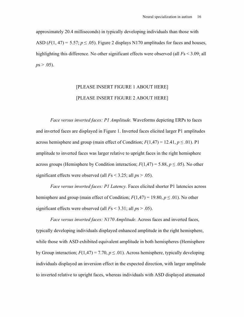
Neural specialization in autism 16
approximately 20.4 milliseconds) in typically developing individuals than those with
ASD (F(1, 47) = 5.57; p ≤ .05). Figure 2 displays N170 amplitudes for faces and houses,
highlighting this difference. No other significant effects were observed (all Fs < 3.09; all
ps > .05).
[PLEASE INSERT FIGURE 1 ABOUT HERE]
[PLEASE INSERT FIGURE 2 ABOUT HERE]
Face versus inverted faces: P1 Amplitude. Waveforms depicting ERPs to faces
and inverted faces are displayed in Figure 1. Inverted faces elicited larger P1 amplitudes
across hemisphere and group (main effect of Condition; F(1,47) = 12.41, p ≤ .01). P1
amplitude to inverted faces was larger relative to upright faces in the right hemisphere
across groups (Hemisphere by Condition interaction; F(1,47) = 5.88, p ≤ .05). No other
significant effects were observed (all Fs < 3.25; all ps > .05).
Face versus inverted faces: P1 Latency. Faces elicited shorter P1 latencies across
hemisphere and group (main effect of Condition; F(1,47) = 19.80, p ≤ .01). No other
significant effects were observed (all Fs < 3.31; all ps > .05).
Face versus inverted faces: N170 Amplitude. Across faces and inverted faces,
typically developing individuals displayed enhanced amplitude in the right hemisphere,
while those with ASD exhibited equivalent amplitude in both hemispheres (Hemisphere
by Group interaction; F(1,47) = 7.70, p ≤ .01). Across hemisphere, typically developing
individuals displayed an inversion effect in the expected direction, with larger amplitude
to inverted relative to upright faces, whereas individuals with ASD displayed attenuated

Neural specialization in autism 17
N170 amplitudes to inverted faces relative to upright faces (Condition by Group
interaction; F(1,47) = 5.84, p ≤ .05). No other significant effects were observed (all Fs <
1.40; all ps > .05).
Face versus inverted faces: N170 Latency. Inverted faces elicited N170s with
longer latencies than upright faces (main effect of Condition; F(1,47) = 4.66, p ≤ .05)
across hemispheres for both groups. No other significant effects were observed (all Fs <
2.69; all ps > .05).
Letters versus pseudoletters: P1 Amplitude. Figure 3 displays waveforms
depicting ERPs to letters and pseudoletters. No significant effects were observed (all Fs <
3.08; all ps > .05).
Letters versus pseudoletters: P1 Latency. Across hemisphere and group, letters
elicited shorter P1 latency than pseudoletters (main effect of Condition; F(1, 52) = 16.28,
p ≤ .01). Across group and condition, P1 latency was shorter in the right hemisphere
(main effect of Hemisphere; F(1, 52) = 4.15, p ≤ .05). No other significant effects were
observed (all Fs < 1.07; all ps > .05).
Letters versus pseudoletters: N170 Amplitude. For both groups, letters elicited
N170s with larger amplitudes than pseudoletters across hemispheres (main effect of
Condition; F(1, 52) = 14.67, p ≤ .01). As displayed in Figure 4, paired samples t-tests
revealed that this effect was carried by significantly enhanced amplitude to letters versus
pseudoletters in the typical group in the left hemisphere (t(1,17) = 2.12, p ≤ .05) and in
the ASD group in both left (t(1,35) = 2.90, p ≤ .01) and right hemispheres (t(1,35) = 3.34,
p ≤ .01). No other significant effects were observed (all Fs < 2.68; all ps > .05).

Neural specialization in autism 18
Letters versus pseudoletters: N170 Latency. No significant effects were observed
(all Fs < 3.58; all ps > .05).
[PLEASE INSERT FIGURE 3 ABOUT HERE]
[PLEASE INSERT FIGURE 4 ABOUT HERE]
Behavioral measures
Face perception. Table 2 displays mean score and standard deviation on
behavioral measures for both groups. Individuals with ASD obtained significantly lower
face recognition scores than typically developing individuals (t(1,51)=3.29, p ≤ .01). For
both groups, N170 latency to faces in the right hemisphere was correlated with face
recognition skill; individuals with faster N170s displayed better face recognition
performance (ASD: r = -.39, p ≤ .05; Typical: r = -.53, p ≤ .05). Among individuals with
ASD, N170 amplitude to inverted faces was correlated with face recognition
performance; those with better face recognition abilities were more likely to display an
enhanced N170 associated with inversion (r = -.47, p ≤ .01).
Letter perception. Groups performed comparably and in the average range on
word reading and decoding tasks. Among typically developing individuals, longer N170
latency to letters in the right hemisphere was correlated with word reading score; those
with longer latencies tended to perform better on the measure of single word reading (r =
.64, p ≤ .01).
[PLEASE INSERT TABLE 4 ABOUT HERE]

Neural specialization in autism 19
General Discussion
The current study contrasted neural specialization for social and non-social
information in individuals with ASD and a cohort of typically developing individuals of
comparable age, ethnicity, sex, handedness, and cognitive ability. A critical social
stimulus with which most adults possess great experience, the human face, was
contrasted with a comparably complex visual stimulus without interpersonal relevance,
houses. Consistent with predictions and with prior research (McPartland et al., 2004),
individuals with ASD displayed a selective processing delay for human faces in the right
hemisphere relative to typical counterparts. Individuals with ASD also showed reduced
hemispheric specialization compared to typical counterparts, who showed a marked right
lateralization effect for faces. The inversion effect, a marker of neural specialization and
processing experience for faces, was evident in typically developing individuals but not
those with ASD. On a behavioral measure of face recognition, individuals with ASD,
despite comparable intellectual ability, performed significantly worse than typically
developing counterparts. Face recognition performance was associated with processing
efficiency for faces; in both groups, individuals with better face recognition abilities
displayed faster N170 response. Among individuals with ASD, increased inversion
effects, as reflected by a stronger response to inverted faces, were associated with better
face recognition performance. This pattern of anomalies, i.e., decreased efficiency of
processing, insensitivity to inversion, and impaired face recognition, is hypothesized to
reflect underdeveloped specialization for faces, a downstream effect of decreased
attention to faces during childhood secondary to reduced social drive from infancy
(Dawson, Webb & McPartland, 2005). Indeed, the observed correlation between neural

Neural specialization in autism 20
response to face inversion and recognition performance suggests that, in this case,
development of expertise and processing proficiency are related.
Though differences related to condition and lateralization were observed in an
earlier component, the P1, no between-group differences were detected. Given the P1’s
role in basic visual attention and low-level sensory perception (Key, Dove, & Maguire,
2005), this pattern of results indicates that children with ASD did not differ from
typically developing peers in terms of fundamental sensory perception. Indeed, despite
observed differences in the basic sensory response to different classes of stimuli, groups
responded similarly. These findings suggest normative sensory perception for visual
information in ASD, with differences emerging at subsequent processing stages related to
social perception.
Current findings concord with prior work describing deviant social development
in ASD. However, scant evidence to-date has informed the specificity of the observed
neural processing anomalies to social information. By measuring responses to non-social
expert stimuli, the current study demonstrated the selectivity of perceptual deficits in
ASD. Following up on work showing N170-related expertise effects for letters, ERP
response to letters of the Roman alphabet were compared to a confabulated alphabet of
pseudoletters (Wong et al., 2005). In contrast to the discrepancies observed during
perception of social stimuli, individuals with ASD displayed neural responses
comparable to typical counterparts; both groups showed enhanced N170 to familiar
letters. Similar results were obtained on behavioral measures, with individuals with ASD
obtaining word reading and decoding scores that were comparable to the typical
participants in this study and within the average range. These results suggest intact

Neural specialization in autism 21
behavioral and brain specialization for non-social information in individuals with ASD.
These results are consistent with prior behavioral work demonstrating preserved word-
reading and word-decoding ability in cognitively able individuals with ASD (Huemer &
Mann, 2009; Nation et al., 2006; Newman et al., 2006) despite complications with
reading comprehension and more sophisticated aspects of language (O'Connor & Klein,
2004). They are consistent with the notion that the social element of communication
rather than language, more generally, may be most directly impacted in ASD (Paul,
2003).
The current findings have significant implications for understanding the
neuropathology of autism spectrum disorders. Two prevailing classes of theories attribute
autistic impairments to dysfunctional brain structures supporting social information
processing (Dawson, Webb & McPartland, 2005) or altered connectivity among
distributed brain regions (Minshew & Williams, 2007). The former emphasizes the
import of the content that is processed, and the latter accentuates the nature of processing
itself. Social brain theories posit that social information is qualitatively unique and that
specific brain systems have evolved to support this type of information processing.
Connectivity theories, in contrast, have traditionally argued that social information is
relevant only insofar as it relies on complex or cortically distributed processing
mechanisms. The current work demonstrates, for the first time in a substantial sample of
children with ASD, preserved specialization for a cognitive process subserved by
distributed cortical regions. Development of specialized letter processing is a process that
develops over time and requires elaborate communication of anterior and posterior
cortical regions (Krigolson, Pierce, Holroyd & Tanaka, 2009). The demonstration of

Neural specialization in autism 22
preserved neural specialization for this type of “expert” processing in ASD is not
consistent with models of non-specific, brain-wide dysfunction. Taking into account
consideration considerable evidence for atypical patterns of connectivity in ASD
(Minshew & Williams, 2007), current findings emphasize the potential value of studying
connectivity within specific brain systems in developmental context. The observed
latency delays may reflect reduced connectivity, and their specificity to social
information compared to non-social information may indicate system-specificity in terms
of atypical connectivity. By studying connectivity within specific neural circuits,
scientists may also extricate atypical connectivity as potential cause or consequence of
autistic dysfunction; it is likely that origins of dysfunction in functionally specific brain
systems would, through developmental maturation, lead to broader connectivity
problems. Such research may also clarify to what degree problems with connectivity
uniquely differentiate autism from the diversity of developmental and psychiatric
disorders also manifesting atypical connectivity, e.g. obsessive-compulsive disorder,
(Garibotto et al., 2010), schizophrenia (Friston, 2002), ADHD (Murias, Swanson &
Srinivasan, 2007), and intellectual impairment (Zhou et al., 2008).
This work yields clinically relevant implications for the detection and treatment of
ASD. Results are supportive of the broad class of interventions designed to direct the
attention of children with ASD to relevant social information. When children are
appropriately engaged and attuned to information, in this case, letters, typical patterns of
neural specialization develop; given the right input, the brain of a person with autism can
function like that a of a typical peer, without ostensible reliance on compensatory
mechanisms or alternative processing strategies. Findings add to a body of evidence that

Neural specialization in autism 23
electrophysiological brain activity to faces represents a viable bio-behavioral risk marker
for ASD, as temporal anomalies in neural correlates of face perception have been
observed in children with ASD (Dawson, Webb, Wijsman et al., 2005) and infants at-risk
for ASD (McCleery et al., 2009).
Though the current work replicates initial findings of temporal anomalies to faces
(McPartland et al., 2004), these findings have not fully replicated in all samples (Kemner,
Schuller & van Engeland, 2006; Senju, Tojo, Yaguchi & Hasegawa, 2005; Webb et al.,
2009). Some of this variability may reflect methodological inconsistencies in terms of
electrode selection (e.g., Webb et al., 2009) or employment of gold-standard diagnostic
procedures (e.g., Grice, Halit, Farroni, Baron-Cohen, Bolton & Johnson, 2005); however,
varied results may accurately reflect the phenotypic heterogeneity evident in ASD.
Despite the unifying characteristic of social impairment, ASD presents in a remarkable
diversity of manifestations, likely representing multiple etiologic pathways and
developmental experiences (Jones & Klin, 2009). Considering the manner in which face
processing (especially in older children and adults) has been actively shaped by
experience, it is intuitive that anomalies might emerge in different ways or might not
emerge universally (Jemel, Mottron & Dawson, 2006). In this regard, like any of the
symptoms characterizing autism, anomalous face perception is neither necessary nor
specific. It is one potential manifestation of atypical social development that, by virtue of
a deep understanding of behavioral and brain bases in typical social development, is a
viable avenue for investigating social disability. Variability in electrophysiological
studies of face perception may also relate to differences in visual attention (Webb et al.,
2009), a trend observed in hemodynamic studies (Dalton, Nacewicz, Alexander &

Neural specialization in autism 24
Davidson, 2007; Dalton et al., 2005). Our employment of a pre-stimulus fixation
crosshair reduces the likelihood that between-group differences are attributable to
differences in visual attention. Furthermore, given comparable N170 latencies to visual
fixations to eyes and mouths in typical development (McPartland, Cheung, Perszyk &
Mayes, 2010) and delayed processing in ASD irrespective of point of gaze on the face
(McPartland, Perszyk, Crowley, Naples & Mayes, 2011), it is unlikely that variation in
visual attention alone could account for observed latency differences; resolution of this
matter will ultimately require co-registration of eye-tracking and EEG.
There are several aspects of the current work that are being revisited and
improved upon in ongoing research. Limiting the sample to high-functioning individuals
was a necessary first step towards addressing the research questions posed in this study,
but it limits generalizability to the broader range of individuals with ASD. Given that
even many nonverbal children with ASD are capable of reading, these types of
experiments offer a window into domains of strength and preserved neural functions of
children on the autism spectrum, important goals for tailoring interventions and
proscribing specific treatments. The sample in the current study focused on pre-
adolescence, a time of rapid maturation of brain systems subserving face perception.
Additional research in younger and older children and adults will elucidate the protracted
maturational course of specialization for face perception in ASD and of letter expertise in
both typical and atypical development. Of note, many participants in the current study
displayed the bifid waveform morphology characteristic of pre-adult face responses
(Taylor, Batty & Itier, 2004); however, this was not evident for letter N170s. Exploiting
the dense spatial sampling afforded by the 256 electrode sensor net, analyses in progress

Neural specialization in autism 25
are using individual-specific three-dimensional head models (computed with sensor
registration images acquired with the Geodesic Photogrammetry System) to localize
potentially distinct neural sources for these facets of the developing N170 (Perszyk et al.,
2010).
Our current results reveal a different relationship between performance on the
behavioral face processing task and brain responses in individuals with ASD than
observed in a prior study (McPartland et al., 2004). Previously, results indicated slowed
processing to be associated with improved face recognition. In the current study,
however, the group of people with ASD, like the typically developing counterparts in the
current and prior study, displayed an association between faster processing and better
face recognition. We hypothesize that this reflects age-related differences in the
application of compensatory strategies over time. In this younger sample, more normative
brain responses correlated with more normative face recognition ability in ASD. In the
older sample studied previously, the opposite trend was observed in the hemisphere
contralateral to that typically associated with face perception; we interpret this as
reflective of effective compensatory processing strategies. In the approximately 10 years
between age 11 (current study) and age 21 (prior study), increased reliance on
compensatory strategies may “overtake” weakened default processing mechanisms,
ultimately resulting in better performance associated with these compensatory strategies.
Though we see the pattern of results as supportive of this interpretation, it is also possible
that the differences observed between studies simply reflect task effects, as the prior work
relied on a visual recall task and the current study utilized a visual discrimination task

Neural specialization in autism 26
with reduced memory demands. Face recognition performance in ASD is demonstrated to
vary with task characteristics (McPartland, Webb, Keehn & Dawson, 2011).
Understanding developmental factors is particularly important in the current
context in that neural specialization for letters is clearly a distinct phenomenon from face
expertise, occurring over a relatively compressed period of time rather than from birth.
Moreover, it is likely that qualitatively different types of experience are associated with
the accrual of proficiency in letter versus face processing. It will thus be essential to
examine development of specialized processing mechanisms for a greater variety of
stimuli. Though it has been proposed that, like faces, letters are encoded using a holistic
processing strategy (Martelli, Majaj & Pelli, 2005), unlike faces, letters are processed at a
basic rather than subordinate level of identification (James et al., 2005). The N170 has
been posited to denote specialization at this basic level of identification, while later
components, such as the N250, index expertise at the subordinate level of identification
(Scott, Tanaka, Sheinberg & Curran, 2006). Similar mechanisms underlying neural
specialization for both faces and letters exist at early processing stages as indexed by the
N170, but study of a broader range of electrophysiological components and expert stimuli
will paint a clearer picture of the development and potential limitations of neural
specialization in ASD.

Neural specialization in autism 27
References
Allison, T., McCarthy, G., Novbre, A., Puce, A. & Belger, A. (1994). Human extrastriate
visual cortext and the perception of faces, words, numbers, and colors. Cerebral
Cortex, 5, 544-554.
American Psychiatric Association (2000). Diagnostic and statistical manual of mental
disorders: DSM-IV-TR (4th ed.). Washington, DC: American Psychiatric
Association.
Behrmann, M., Thomas, C. & Humphreys, K. (2006). Seeing it differently: visual
processing in autism. Trends in Cognitive Sciences, 10(6), 258-264.
Bentin, S., Allison, T., Puce, A., Perez, E. & McCarthy, G. (1996). Electrophysiological
studies of face perception in humans. Journal of Cognitive Neuroscience, 8(6),
551-565.
Bentin, S., Deouell, L. Y. & Soroker, N. (1999). Selective visual streaming in face
recognition: evidence from developmental prosopagnosia. Neuroreport, 10(4),
823-827.
Bentin, S., Mouchetant-Rostaing, Y., Giard, M. H., Echallier, J. F. & Pernier, J. (1999).
ERP manifestations of processing printed words at different psycholinguistic
levels: time course and scalp distribution. Journal of Cognitive Neuroscience,
11(3), 235-260.
Benton, A., Sivan, A., Hamsher, K., Varney, N. & Spreen, O. (1994). Contributions to
Neuropsychological Assessment. New York: Oxford University Press.

Neural specialization in autism 28
Boeschoten, M. A., Kenemans, J. L., van Engeland, H. & Kemner, C. (2007). Face
processing in Pervasive Developmental Disorder (PDD): the roles of expertise
and spatial frequency. Journal of Neural Transmission, 114(12), 1619-1629.
Burd, L., Kerbeshian, J. & Fisher, W. (1985). Inquiry into the incidence of hyperlexia in
a statewide population of children with pervasive developmental disorder.
Psychological Reports, 57(1), 236-238.
Bushnell, I., Sai, F. & Mullin, J. (1989). Neonatal recognition of the mother's face.
British Journal of Developmental Pscyhology, 7, 3-15.
Cohen, L., Dehaene, S., Naccache, L., Lehericy, S., Dehaene-Lambertz, G., Henaff, M.
A. & Michel, F. (2000). The visual word form area: spatial and temporal
characterization of an initial stage of reading in normal subjects and posterior
split-brain patients. Brain, 123(2), 291-307.
Dalton, K. M., Nacewicz, B. M., Alexander, A. L. & Davidson, R. J. (2007). Gaze-
fixation, brain activation, and amygdala volume in unaffected siblings of
individuals with autism. Biological Psychiatry, 61(4), 512-520.
Dalton, K. M., Nacewicz, B. M., Johnstone, T., Schaefer, H. S., Gernsbacher, M. A.,
Goldsmith, H. H., Alexander, A.L. & Davidson, R. J. (2005). Gaze fixation and
the neural circuitry of face processing in autism. Nature Neuroscience, 8(4), 519-
526.
Dawson, G., Carver, L., Meltzoff, A. N., Panagiotides, H., McPartland, J. & Webb, S. J.
(2002). Neural correlates of face and object recognition in young children with
autism spectrum disorder, developmental delay, and typical development. Child
Development, 73(3), 700-717.

Neural specialization in autism 29
Dawson, G., Webb, S. J. & McPartland, J. (2005). Understanding the nature of face
processing impairment in autism: insights from behavioral and
electrophysiological studies. Developmental Neuropsychology, 27(3), 403-424.
Dawson, G., Webb, S. J., Wijsman, E., Schellenberg, G., Estes, A., Munson, J. & Faja, S.
(2005). Neurocognitive and electrophysiological evidence of altered face
processing in parents of children with autism: implications for a model of
abnormal development of social brain circuitry in autism. Development and
Psychopathology, 17(3), 679-697.
Diamond, R. & Carey, S. (1986). Why faces are and are not special: An effect of
expertise. Journal of Experimental Psychology, 115(2), 107-117.
Elliott, C. (2007). The Differential Ability Scales (2nd ed.). San Antonio, TX: Harcourt
Assessment.
Farah, M. J., Tanaka, J. W. & Drain, H. M. (1995). What causes the face inversion
effect? Journal of Experimental Psychology: Human Perception and
Performance, 21(3), 628-634.
Friston, K. J. (2002). Dysfunctional connectivity in schizophrenia. World Psychiatry,
1(2), 66-71.
Gadow, K. & Sprafkin, J. (1994). Child Symptom Inventories manual. Stony Brook, NY:
Checkmate Plus.
Garibotto, V., Scifo, P., Gorini, A., Alonso, C. R., Brambati, S., Bellodi, L. & Perani, D.
(2010). Disorganization of anatomical connectivity in obsessive compulsive
disorder: A multi-parameter diffusion tensor imaging study in a subpopulation of
patients. Neurobiology of Disease, 37(2), 468-476.

Neural specialization in autism 30
Gauthier, I. & Nelson, C. (2001). The development of face expertise. Current Opinion in
Neurobiology, 11, 219-224.
Gauthier, I., Skudkarski, P., Gore, J. & Anderson, A. (2000). Expertise for cars and birds
recruits brain areas involved in face recognition. Nature Neuroscience, 3(2), 191-
197.
Gauthier, I., Williams, P., Tarr, M. J. & Tanaka, J. (1998). Training 'greeble' experts: a
framework for studying expert object recognition processes. Vision Research,
38(15-16), 2401-2428.
Gauthier, I. (2000). What constrains the organization of the ventral temporal cortex?
Trends in Cognitive Sciences, 4(1), 1-2.
Goren, C. C., Sarty, M. & Wu, P. Y. (1975). Visual following and pattern discrimination
of face-like stimuli by newborn infants. Pediatrics, 56(4), 544-549.
Grelotti, D. J., Klin, A. J., Gauthier, I., Skudlarski, P., Cohen, D. J., Gore, J. C., Volkmar,
F.R. & Schultz, R. T. (2005). fMRI activation of the fusiform gyrus and amygdala
to cartoon characters but not to faces in a boy with autism. Neuropsychologia,
43(3), 373-385.
Grice, S.J., Halit, H., Farroni, T., Baron-Cohen, S., Bolton, P. & Johnson, M. (2005).
Neural correlates of eye-gaze detection in young children with autism. Cortex,
41(3), 342-353.
Grigorenko, E. L., Klin, A., Pauls, D. L., Senft, R., Hooper, C. & Volkmar, F. (2002). A
descriptive study of hyperlexia in a clinically referred sample of children with
developmental delays. Journal of Autism and Developmental Disorders, 32(1), 3-
12.

Neural specialization in autism 31
Grossi, G., Coch, D., Coffey-Corina, S., Holcomb, P. J. & Neville, H. J. (2001).
Phonological processing in visual rhyming: a developmental erp study. Journal of
Cognitive Neuroscience, 13(5), 610-625.
Haxby, J. V., Grady, C. L., Horwitz, B., Ungerleider, J. M., Maisog, M. & Pietrini, P.
(1994). The functional organization of human extrastriate cortext: A pet-
rCBFstudy of selective attention to faces and locations. Journal of Neuroscience,
14, 6336-6353.
Hobson, R. (1986). The autistic child's appraisal of expressions of emotion. Journal of
Child Psychology and Psychiatry, 27(3), 321-342.
Hobson, R., Ouston, J. & Lee, A. (1988). What's in a face? The case of autism. British
Journal of Psychology, 79, 441-453.
Holcomb, P., Coffey, S. & Neville, H. (1992). Visual and auditory sentence processing: a
developmental analysis using event-related brain potentials. Developmental
Neuropsychology, 8(2-3), 203-241.
Huemer, S. V. & Mann, V. (2009). A Comprehensive Profile of Decoding and
Comprehension in Autism Spectrum Disorders. Journal of Autism and
Developmental Disorders, 40(4), 485-493.
Itier, R. J. & Taylor, M. J. (2002). Inversion and contrast polarity reversal affect both
encoding and recognition processes of unfamiliar faces: a repetition study using
ERPs. Neuroimage, 15(2), 353-372.
Itier, R. J. & Taylor, M. J. (2004). Source analysis of the N170 to faces and objects.
Neuroreport, 15(8), 1261-1265.

Neural specialization in autism 32
James, K. H., James, T. W., Jobard, G., Wong, A. C. & Gauthier, I. (2005). Letter
processing in the visual system: different activation patterns for single letters and
strings. Cognitive, Affective, and Behavioral Neuroscience, 5(4), 452-466.
Jemel, B., Mottron, L. & Dawson, M. (2006). Impaired face processing in autism: fact or
artifact? Journal of Autism and Developmental Disorders, 36(1), 91-106.
Johnson, M. H., Dziurawiec, S., Ellis, H. & Morton, J. (1991). Newborns' preferential
tracking of face-like stimuli and its subsequent decline. Cognition, 40(1-2), 1-19.
Jones, W., Carr, K. & Klin, A. (2008). Absence of preferential looking to the eyes of
approaching adults predicts level of social disability in 2-year-old toddlers with
autism spectrum disorder. Archives of General Psychiatry, 65(8), 946-954.
Jones, W. & Klin, A. (2009). Heterogeneity and homogeneity across the autism
spectrum: the role of development. Journal of the American Academy of Child
and Adolescent Psychiatry, 48(5), 471-473.
Kanner, L. (1943). Autistic disturbances of affective contact. Nervous Child, 2, 217-250.
Kanwisher, N., McDermott, J. & Chun, M. M. (1997). The fusiform face area: a module
in human extrastriate cortex specialized for face perception. Journal of
Neuroscience, 17(11), 4302-4311.
Kemner, C., Schuller, A. M. & van Engeland, H. (2006). Electrocortical reflections of
face and gaze processing in children with pervasive developmental disorder.
Journal of Child Psychology and Psychiatry, 47(10), 1063-1072.
Key, A. P., Dove, G. O., & Maguire, M. J. (2005). Linking brainwaves to the brain: an
ERP primer. Developmental Neuropsychology, 27(2), 183-215.

Neural specialization in autism 33
Klin, A. (2004). Understanding circumscribed interests in autism spectrum disorders.
Paper presented at the The 25th Annual TEACCH Conference, Chapel Hill, NC.
Klin, A., Jones, W., Schultz, R., Volkmar, F. & Cohen, D. (2002). Visual fixation
patterns during viewing of naturalistic social situations as predictors of social
competence in individuals with autism. Archives of General Psychiatry, 59(9),
809-816.
Klin, A., Sparrow, S., De Bildt, A., Cicchetti, D., Cohen, D. & Volkmar, F. (1999). A
normed study of face recognition in autism and related disorders. Journal of
Autism and Developmental Disorders, 29(6), 499-508.
Krigolson, O. E., Pierce, L. J., Holroyd, C. B. & Tanaka, J. W. (2009). Learning to
become an expert: reinforcement learning and the acquisition of perceptual
expertise. Journal of Cognitive Neuroscience, 21(9), 1834-1841.
Langdell, T. (1978). Recognition of faces: an approach to the study of autism. Journal of
Child Psychology and Psychiatry, 19(3), 255-268.
Lord, C., Risi, S., Lambrecht, L., Cook, E. H., Leventhal, B. L., DiLavore, P. C., Pickles,
A. & Rutter, M. (2000). The Autism Diagnostic Observation Schedule--Generic:
A standard measure of social and communication deficits associated with the
spectrum of autism. Journal of Autism and Developmental Disorders, 30(3), 205-
223.
Lord, C., Rutter, M. & Le Couteur, A. (1994). Autism Diagnostic Interview-Revised: a
revised version of a diagnostic interview for caregivers of individuals with
possible pervasive developmental disorders. Journal of Autism and
Developmental Disorders, 24(5), 659-685.

Neural specialization in autism 34
Maestro, S., Muratori, F., Cavallaro, M. C., Pei, F., Stern, D., Golse, B. & Palacio-
Espasa, F. (2002). Attentional skills during the first 6 months of age in autism
spectrum disorder. Journal of the American Academy of Child and Adolescent
Psychiatry, 41(10), 1239-1245.
Martelli, M., Majaj, N. J. & Pelli, D. G. (2005). Are faces processed like words? A
diagnostic test for recognition by parts. Journal of Vision, 5(1), 58-70.
Maurer, U., Brem, S., Bucher, K. & Brandeis, D. (2005). Emerging neurophysiological
specialization for letter strings. Journal of Cognitive Neuroscience, 17(10), 1532-
1552.
Maurer, U., Brem, S., Kranz, F., Bucher, K., Benz, R., Halder, P., Steinhausen, H. &
Brandeis, D. (2006). Coarse neural tuning for print peaks when children learn to
read. Neuroimage, 33(2), 749-758.
McCandliss, B. D., Cohen, L. & Dehaene, S. (2003). The visual word form area:
expertise for reading in the fusiform gyrus. Trends in Cognitive Sciences, 7(7),
293-299.
McCleery, J. P., Akshoomoff, N., Dobkins, K. R. & Carver, L. J. (2009). Atypical face
versus object processing and hemispheric asymmetries in 10-month-old infants at
risk for autism. Biological Psychiatry, 66(10), 950-957.
McPartland, J.C., Cheung, C. H., Perszyk, D., & Mayes, L. C. (2010). Face-related ERPs
are modulated by point of gaze. Neuropsychologia, 48(12), 3657-3660.
McPartland, J.C., Dawson, G., Webb, S. J., Panagiotides, H. & Carver, L. J. (2004).
Event-related brain potentials reveal anomalies in temporal processing of faces in

Neural specialization in autism 35
autism spectrum disorder. Journal of Child Psychology and Psychiatry, 45(7),
1235-1245.
McPartland, J. C., Perszyk, D., Crowley, M., Naples, A. J. & Mayes, L. (2011, August).
Visual attention modulates neural response to faces in autism. Paper accepted for
presentation at the American Psychological Association Annual Convention.
McPartland, J. C., Webb, S. J., Keehn, B., & Dawson, G. (2011). Patterns of visual
attention to faces and objects in autism spectrum disorder. J Autism Dev Disord,
41(2), 148-157.
Meltzoff, A. N. & Moore, M. K. (1977). Imitation of facial and manual gestures by
human neonates. Science, 198(4312), 74-78.
Minshew, N. J. & Williams, D. L. (2007). The new neurobiology of autism: cortex,
connectivity, and neuronal organization. Archives of Neurology, 64(7), 945-950.
Murias, M., Swanson, J. M. & Srinivasan, R. (2007). Functional connectivity of frontal
cortex in healthy and ADHD children reflected in EEG coherence. Cerebral
Cortex, 17(8), 1788-1799.
Nation, K., Clarke, P., Wright, B. & Williams, C. (2006). Patterns of reading ability in
children with autism spectrum disorder. Journal of Autism and Developmental
Disorders, 36(7), 911-919.
Nelson, C. (2001). The development and neural bases of face recognition. Infant and
Child Development, 10, 3-18.
Newman, T. M., Macomber, D., Naples, A. J., Babitz, T., Volkmar, F. & Grigorenko, E.
L. (2006). Hyperlexia in Children with Autism Spectrum Disorders. Journal of
Autism and Developmental Disorders, 37(4), 760-774.

Neural specialization in autism 36
O'Connor, K., Hamm, J. P. & Kirk, I. J. (2005). The neurophysiological correlates of face
processing in adults and children with Asperger's syndrome. Brain and Cognition,
59(1), 82-95.
O'Connor, K., Hamm, J. P. & Kirk, I. J. (2007). Neurophysiological responses to face,
facial regions and objects in adults with Asperger's syndrome: An ERP
investigation. International Journal of Psychophysiology, 63(3), 283-293.
O'Connor, I. M., & Klein, P. D. (2004). Exploration of strategies for facilitating the
reading comprehension of high-functioning students with autism spectrum
disorders. J Autism Dev Disord, 34(2), 115-127.
Oldfield, R. (1971). The assessment and analysis of handedness; The Edinburgh
inventory. Neuropsychologia, 9, 97-113.
Osterling, J. A. & Dawson, G. (1994). Early recognition of children with autism: A study
of first birthday home videotapes. Journal of Autism and Developmental
Disorders, 24(3), 247-257.
Paul, R. (2003). Promoting social communication in high functioning individuals with
autistic spectrum disorders. Child & Adolescent Psychiatric Clinics of North
America, 12(1), 87-106.
Perszyk, D., Molfese, P., Kilroy, E., Mayes, L., Klin, A. & McPartland, J. (2010).
Developmental brain bases of face perception in autism as revealed by ERPs.
Paper presented at the International Meeting for Autism Research.
Psychological Corporation (1999). Wechsler Abbreviated Scale of Intelligence (WASI)
manual. San Antonio, TX: Author.

Neural specialization in autism 37
Puce, A., Allison, T., Gore, J. & McCarthy, G. (1995). Face-sensitive regions in human
extrastriate cortex studied by functional MRI. Journal of Neurophysiology, 74(3),
1192-1199.
Rossion, B., Gauthier, I., Goffaux, V., Tarr, M. J. & Crommelinck, M. (2002). Expertise
training with novel objects leads to left-lateralized facelike electrophysiological
responses. Psychological Science, 13(3), 250-257.
Rossion, B., Gauthier, I., Tarr, M. J., Despland, P., Bruyer, R., Linotte, S. &
Crommelinck, M. (2000). The N170 occipito-temporal component is delayed and
enhanced to inverted faces but not to inverted objects: an electrophysiological
account of face-specific processes in the human brain. Neuroreport, 11(1), 69-74.
Rossion, B., Joyce, C. A., Cottrell, G. W. & Tarr, M. J. (2003). Early lateralization and
orientation tuning for face, word, and object processing in the visual cortex.
Neuroimage, 20(3), 1609-1624.
Sasson, N. J. (2006). The Development of Face Processing in Autism. Journal of Autism
and Developmental Disorders, 36(3), 381-394.
Schneider, W., Eschman, A. & Zuccolotto, A. (2002). E-prime user's guide. Pittsburg,
Psychology Software Tools Inc.
Schultz, R. T. (2005). Developmental deficits in social perception in autism: the role of
the amygdala and fusiform face area. International Journal of Developmental
Neuroscience, 23(2-3), 125-141.
Schultz, R. T., Gauthier, I., Klin, A., Fulbright, R. K., Anderson, A. W., Volkmar, F.,
Skludlarski, P., Lacadie, C., Cohen, D.J. & Gore, J. C. (2000). Abnormal ventral

Neural specialization in autism 38
temporal cortical activity during face discrimination among individuals with
autism and Asperger syndrome. Archives of General Psychiatry, 57(4), 331-340.
Schweinberger, S. R., Pickering, E. C., Jentzsch, I., Burton, A. M. & Kaufmann, J. M.
(2002). Event-related brain potential evidence for a response of inferior temporal
cortex to familiar face repetitions. Cognitive Brain Research, 14(3), 398-409.
Scott, L. S., Tanaka, J. W., Sheinberg, D. L. & Curran, T. (2006). A reevaluation of the
electrophysiological correlates of expert object processing. Journal of Cognitive
Neuroscience, 18(9), 1453-1465.
Senju, A., Tojo, Y., Yaguchi, K. & Hasegawa, T. (2005). Deviant gaze processing in
children with autism: an ERP study. Neuropsychologia, 43(9), 1297-1306.
Shibata, T., Nishijo, H., Tamura, R., Miyamoto, K., Eifuku, S., Endo, S. & Ono, T.
(2002). Generators of visual evoked potentials for faces and eyes in the human
brain as determined by dipole localization. Brain Topography, 15(1), 51-63.
Tanaka, J. W. & Farah, M. J. (1993). Parts and wholes in face recognition. The Quarterly
Journal of Experimental Psychology Section A, 46(2), 225-245.
Tarr, M. J. & Gauthier, I. (2000). FFA: a flexible fusiform area for subordinate-level
visual processing automatized by expertise. Nature Neuroscience, 3(8), 764-769.
Taylor, M. J., Batty, M. & Itier, R. J. (2004). The faces of development: a review of early
face processing over childhood. Journal of Cognitive Neuroscience, 16(8), 1426-
1442.
Tucker, D. M. (1993). Spatial sampling of head electrical fields: the geodesic sensor net.
Electroencephalography and Clinical Neurophysiology, 87(3), 154-163.

Neural specialization in autism 39
Turkeltaub, P. E., Flowers, D. L., Verbalis, A., Miranda, M., Gareau, L. & Eden, G. F.
(2004). The neural basis of hyperlexic reading: an FMRI case study. Neuron,
41(1), 11-25.
Webb, S. J., Dawson, G., Bernier, R. & Panagiotides, H. (2006). ERP evidence of
atypical face processing in young children with autism. Journal of Autism and
Developmental Disorders, 36(7), 881-890.
Webb, S. J., Jones, E. J., Merkle, K., Murias, M., Greenson, J., Richards, T., Aylward, E.
& Dawson, G. (2010). Response to familiar faces, newly familiar faces, and novel
faces as assessed by ERPs is intact in adults with autism spectrum disorders.
International Journal of Psychophysiology, 77(2), 106-117.
Webb, S. J., Merkle, K., Murias, M., Richards, T., Aylward, E. & Dawson, G. (2009).
ERP responses differentiate inverted but not upright face processing in adults with
ASD. Social Cognitive and Affective Neuroscience. [Epub ahead of print]. doi:
10.1093/scan/nsp002.
Wechsler, D. (1997). Manual for the Wechsler Adult Intelligence Scale (3rd ed.). San
Antonio, Tx: The Psychological Corporation.
Wechsler, D. (2003). The Wechsler Intelligence Scale for Children - Fourth Edition. San
Antonio, TX: The Psychological Corporation.
Wolf, J.M., Tanaka, J.W., Klaiman, C., Cockburn, J., Herlihy, L., Brown, C, South, M.,
McPartland, J.C., Kaiser, M.D., Phillips, R. & Schultz, R.T. (2008). Specific
impairment of face processing abilities in children with autism spectrum disorder
using the Let’s Face It! Skills Battery. Autism Research, 1(6), 329-340.

Neural specialization in autism 40
Wong, A. C., Gauthier, I., Woroch, B., DeBuse, C. & Curran, T. (2005). An early
electrophysiological response associated with expertise in letter perception.
Cognitive, Affective, and Behavioral Neuroscience, 5(3), 306-318.
Woodcock, R., McGrew, K. & Mather, N. (2001). Woodcock-Johnson III Tests of
Achievement. Itasca, IL: Riverside Publishing.
Yin, R. (1970). Face recognition by brain-inujred patients: A dissociable ability.
Neuropsychologia, 8, 395-402.
Zhou, Y., Dougherty, J. H., Jr., Hubner, K. F., Bai, B., Cannon, R. L. & Hutson, R. K.
(2008). Abnormal connectivity in the posterior cingulate and hippocampus in
early Alzheimer's disease and mild cognitive impairment. Alzheimer’s and
Dementia, 4(4), 265-270.

Neural specialization in autism 41
Figure Legends
Figure 1. Grand averaged waveforms across entire scalp for faces, houses, and inverted
faces for typical participants and those with ASD. Electrodes of interest in right and left
hemisphere are highlighted. Subpanels display the averaged waveform across the eight
specified electrodes in each hemisphere for both groups.
Figure 2. Mean latency of the N170 component (in milliseconds) elicited by faces and
houses for both groups in both hemispheres. Error bars represent +/- 1 S.E. Significance
at the p ≤ .05 level is indicated by *.
Figure 3. Grand averaged waveforms across entire scalp for letters and pseudoletters for
typical participants and those with ASD. Electrodes of interest in right and left
hemisphere are highlighted. Subpanels display the averaged waveform across the eight
specified electrodes in each hemisphere for both groups.
Figure 4. Amplitude of the N170 component (in microVolts) elicited by letters and
pseudoletters for both groups in both hemispheres. Error bars represent +/- 1 S.E.
Significance at the p ≤ .05 level is indicated by *, and significance at the p ≤ .01 level is
indicated by **.

Neural specialization in autism 42
Tables
Table 1
Participant Characteristics
Typical (N=18) ASD (N=36)
Number male (Percent) 15 (83.3) 32 (88.9)
Number White (Percent) 15 (83.3) 34 (94.4)
Number right handed (Percent) 16 (88.9) 31 (86.1)
Mean age (SD) 12.6 (2.4) 11.2 (3.4)
Mean Full Scale IQ (SD) 112.9 (13.4) 105.2 (17.3)

Neural specialization in autism 43
Table 2
P1 and N170 amplitude
P1 N170
Hemisphere Condition M (microVolts) SD M (microVolts) SD
Typical group
Left Faces 7.77 4.4 0.39 1.7
Houses 8.60 5.7 2.49 3.5
Inverted faces 7.70 4.8 0.24 2.8
Letters 6.28 3.3 - 2.27 3.0
Pseudoletters 5.89 3.4 - 1.51 3.1
Right Faces 6.79 5.0 - 0.16 2.0
Houses 7.94 5.8 0.89 2.6
Inverted faces 7.50 5.5 - 0.58 2.8
Letters 5.03 2.5 - 3.03 4.2
Pseudoletters 5.10 3.0 - 2.53 3.9
ASD group
Left Faces 8.94 5.6 - 0.32 3.4
Houses 10.49 6.2 3.16 4.0
Inverted faces 9.64 5.4 0.59 3.6
Letters 6.26 3.8 - 3.09 3.6
Pseudoletters 6.31 3.6 - 2.22 3.3
Right Faces 10.10 6.5 0.76 3.6
Houses 11.11 6.7 3.44 4.2

Neural specialization in autism 44
Inverted faces 11.17 6.7 1.52 3.5
Letters 6.40 3.3 - 3.57 3.1
Pseudoletters 6.59 3.7 - 2.29 2.9

Neural specialization in autism 45
Table 3
P1 and N170 latency
P1 N170
Hemisphere Condition M (milliseconds) SD M (milliseconds) SD
Typical group
Left Faces 115.27 10.9 193.24 37.3
Houses 134.71 20.6 215.59 43.4
Inverted faces 119.44 9.7 194.82 35.3
Letters 106.67 22.0 177.25 26.9
Pseudoletters 111.58 25.6 179.56 26.8
Right Faces 118.44 12.2 181.38 31.6
Houses 133.97 15.0 217.50 36.4
Inverted faces 121.12 9.6 188.85 32.7
Letters 104.72 25.8 181.19 25.9
Pseudoletters 106.89 24.7 176.03 28.5
ASD group
Left Faces 118.73 10.3 200.13 24.3
Houses 134.05 11.3 221.67 31.2
Inverted faces 125.97 10.5 205.42 27.7
Letters 102.68 13.4 189.21 26.7
Pseudoletters 106.29 15.1 178.83 19.3
Right Faces 117.59 10.8 201.78 27.2
Houses 130.25 14.4 219.38 27.8

Neural specialization in autism 46
Inverted faces 122.78 15.4 204.66 31.3
Letters 101.53 14.1 188.42 28.0
Pseudoletters 103.97 14.9 183.51 23.6

Neural specialization in autism 47
Table 4
Performance on behavioral measures
Typical (N = 17) ASD (N = 36)
Measure M SD M SD
Benton Face Recognition Raw Score 41.41 3.6 37.11 4.8
Letter-Word Identification Standard Score 108.41 9.9 105.67 15.0
Word Attack Standard Score 101.41 9.7 103.86 11.6

Neural specialization in autism 48
Figure 1 TOP
N170 P1 N170 P1 N170 P1 N170 P1
!"#
$#
"#
%#
&#
'#
($#
("#
!($$# $# ($$# "$$# )$$# %$$# *$$#
!"#$%&%
'()*
!"''")+#%,-)*
./01*2"34(*
!"#
$#
"#
%#
&#
'#
($#
("#
!($$# $# ($$# "$$# )$$# %$$# *$$#
!"#$%&%
'()*
!"''")+#%,-)*
./01*5+6*
!"#
$#
"#
%#
&#
'#
($#
("#
!($$# $# ($$# "$$# )$$# %$$# *$$#
!"#$%&%
'()*
!"''")+#%,-)*
789"#:'1*5+6*
!"#
$#
"#
%#
&#
'#
($#
("#
!($$# $# ($$# "$$# )$$# %$$# *$$#
!"#$%&%
'()*
!"''")+#%,-)*
789"#:'1*2"34(*
+,-./#-01,-213#45678-9# +,-./#-01,-213#:;<#
!"#$%
&'($)*$+%!"#$%
,-./$%

Neural specialization in autism 49
Figure 2 TOP
!"#$
!%#$
!&#$
!'#$
(##$
(!#$
((#$
()#$
(*#$
+,-./01$ 234$ +,-./01$ 234$
567$ 8.9:;$
<.11.=6/>?
@=$
A>B=6$ C0/6$ D

Neural specialization in autism 50
Figure 3 TOP
N170 P1 N170 P1 N170 P1 N170 P1
!"#!$#!%#&#%#$#"#'#(#)#*#
!%&&# &# %&&# $&&# "&&# '&&# (&&#
!"#$%&%
'()*
!"''")+#%,-)*
./01*2+3**
!"#!$#!%#&#%#$#"#'#(#)#*#
!%&&# &# %&&# $&&# "&&# '&&# (&&#
!"#$%&%
'()*
!"''")+#%,-)*
./01*4"56(*
!"#!$#!%#&#%#$#"#'#(#)#*#
!%&&# &# %&&# $&&# "&&# '&&# (&&#
!"#$%&%
'()*
!"''")+#%,-)*
789"#:'1*4"56(*
!"#!$#!%#&#%#$#"#'#(#)#*#
!%&&# &# %&&# $&&# "&&# '&&# (&&#
!"#$%&%
'()*
!"''")+#%,-)*
789"#:'1*2+3**
+,-./#-01,-213#45678-9# +,-./#-01,-213#:;<#
!"#"$%
&'"()*+,"#"$%

Neural specialization in autism 51
Figure 4 TOP
!"#
!$#
!%#
!&#
!'#
(#)*+,-./# 012# )*+,-./# 012#
345# 6,789#:,-;<=<
/9>#
34?4;# @>4AB</4?4;#
C CC#CC#
Copyright © 2022 FDOKUMEN