Public Health Reports - NCBI
35
Public Health Reports Vol. 55 . JULY 19, 1940 * No. 29 THE GEOGRAPHIC DISTRIBUTION OF DISEASE III. A DECADE OF POLIOMYELITIS IN LOUISIANA' By ALBERT E. CASEY, and BRANCH J. AYMOND, Director of Research, Louisiana State Department of Health The finding of a definite concentration of cases of St. Louis enceph- alitis in the St. Louis area in weedy places about sewage-polluted streams (1) prompted the study of the geographic distribution of a somewhat similar disease, poliomyelitis, in Louisiana. The files of the State Department of Health for the decade ended January 1, 1939, contain reports of 676 cases, practically all of which, it may be assumed, are actual instances of paralysis, since it is not the custom in Louisiana to report abortive cases. Detailed maps of the 64 parishes in Louisiana were obtained from the Departments of Agriculture and Ilome Economics of Louisiana State University. Parish health unit directors and their staffs and other interested physicians generously took the time to secure and plot the exact home address of each patient at the time of the onset of the disease, thus circumventing the custom of rural residents of giving the nearest town as their home address. The population of all incorporated communities, wards, and parishes in Louisiana was obtained from the United States Census of 1930. The population of unincorporated communities was obtained from a commercial atlas and was corrected, whenever this was possible, by data in the pos- session of the State Department of Health. Accurate data were thus obtained for 59 parishes, in which the distribution of the population and of the cases of poliomyclitis, 1 From the Department of Pathology and Bacteriology of the School of Medicine of Louisiana State University, and the Louisiana State Department of Health. 2379210-40--1 (1295)
-
Upload
khangminh22 -
Category
Documents
-
view
1 -
download
0
Transcript of Public Health Reports - NCBI
Public Health ReportsVol. 55 . JULY 19, 1940 * No. 29
THE GEOGRAPHIC DISTRIBUTION OF DISEASEIII. A DECADE OF POLIOMYELITIS IN LOUISIANA'
By ALBERT E. CASEY, and BRANCH J. AYMOND, Director of Research, LouisianaState Department of Health
The finding of a definite concentration of cases of St. Louis enceph-alitis in the St. Louis area in weedy places about sewage-pollutedstreams (1) prompted the study of the geographic distribution of asomewhat similar disease, poliomyelitis, in Louisiana. The files ofthe State Department of Health for the decade ended January 1, 1939,contain reports of 676 cases, practically all of which, it may be assumed,are actual instances of paralysis, since it is not the custom in Louisianato report abortive cases.Detailed maps of the 64 parishes in Louisiana were obtained from
the Departments of Agriculture and Ilome Economics of LouisianaState University. Parish health unit directors and their staffs andother interested physicians generously took the time to secure andplot the exact home address of each patient at the time of the onset ofthe disease, thus circumventing the custom of rural residents of givingthe nearest town as their home address. The population of allincorporated communities, wards, and parishes in Louisiana wasobtained from the United States Census of 1930. The population ofunincorporated communities was obtained from a commercial atlasand was corrected, whenever this was possible, by data in the pos-session of the State Department of Health.
Accurate data were thus obtained for 59 parishes, in which thedistribution of the population and of the cases of poliomyclitis,
1 From the Department of Pathology and Bacteriology of the School of Medicine of Louisiana StateUniversity, and the Louisiana State Department of Health.
2379210-40--1 (1295)

July 19, 1940 1296
according to incorporated and unincorporated communities of variouspopulations, was as follows:
TABLE 1
Number ~~~Poulomyeultlses Rate perue of community Of Ter| Potl/J (8) 100
Places Actual Expected
Incorporated plae
Under 100-1 27 0100-499 -0 17, 60 12 6.0 6. 00 68.2500----___________--______________ 42 30,467 23 10.3 6.56675.61,000-1,499 - 25 29, 651 24 10.0 19.60 80.91,500-1,999 ......------------------15 25,88824 I-8.7 56.609272,0002,9 ---------------------------- 14 27, 681 219. 41 . 175. 93,000-4,999 - 18 68,629 29 23.2 1.45 42.26,00049,999 -12 117,822 36 39. 9 .38 30.650,000-499,99 - 2 535,417 128
Unincorporated places
Under 100 (rural) - - 890,208 286 301.4 0.79 8213100-499 - 195 143,342 26 '147 ' 8. 68 160.0500-999 -37 122,561 1 7 17.6 1.05 131.01,000-1,m ------------- 9 1 16,448 1 2
Total- ----------- 1,825,374 618 618.0 93.81+ 33.9
'Based on estimated populations and estimated community boundaries.
Statistically significant preponderances of poliomyelitis were foundin both incorporated and unincorporated communities of 100-499population. The highest preponderances were found in incorporatedcommunities of 500-2,999 inhabitants, the maximum incidence beingreached in communities of 1,500-1,999 inhabitants (table 1). Theincrease in incidence from rural communities to communities of 1,500-1,999 population, and the decrease from communities of this size tothose of 5,000-49,999 population, were orderly and formed a unimodalcurve when plotted (fig. 1).
It was of interest to find that rural areas, when interpreted as un-incorporated communities of less than 100 inhabitants, and urbancommunities of 5,000-49,999 inhabitants had the same low incidenceof poliomyelitis. The incidence in the only two large cities in Louisi-ana corresponded with the incidence of the disease in smaller cities,but the limited number of cities does not permit adequate analysis.Unincorporated communities of over 500 inhabitants, which for themost part are industrial in Louisiana, had an incidence of poliomyelitissimilar to that of cities and rural areas and significantly lower than theincidence for incorporated communities of the same size. The possibleexplanations for this discrepancy will be considered later in this paper.The age distribution of the cases of poliomyelitis in this series does
not differ from the age distribution in other reports, the mode being2 to 3 years and the mean age 7 years. Variations in age, however, donot explain the preponderance of the disease in small incorporated

communities. The average age of the patients in rural communitiesof less than 100 inhabitants was 6.0 years; of the patients in NewOrleans, where the disease was endemic, 6.8 years; of the patients inShreveport, where the disease was largely epidemic, 6.9 years; and ofthe patients in small towns of 500-2,999 inhabitants, 7.2 years. Allof these age distributions had the same modal points, and the mean
ages showed no statistically significant differences. Because of thelack of age variations, immunological differences probably do notaccount for the preponderance of poliomyelitis in small towns.
C*s$es OP PO1IONYIEL1/T/ /it LOUI/5AtI100
3~90A80
OhN
070
60
Q50
°4050
3|20-
lo%
O Y0 o
P0.PlIZ11r1ol a-IC ca1x7VI(ry( SCALE II GEOMIETRIC SERIES FOR4
PURPOSES OF ILLUSTRA0TION -)
FIGURZ 1.
The variations in incidence cannot be explained on the basis ofeither sex or race. Three hundred and sixty-four patients weremale and 324 female, which is a statistically insignificant sex differenceand compatible with the usual preponderance of males among children.The racial distribution in small towns is shown in the following table,there being no significant difference between the three types of com-munities in this regard (Chi square=3.44, n=2, P=0.21):
TABLE 2
Actual values Expected valuesS.ze of community - d'/m (8)
Colored White Total Colored White Total
Under 100 - -99 178 277 89.6 187.4 277 1.46600-2,999 . 23 M64 87 28.1 58.9 87 1.873,000-499,999-- 41 99 140 45.3 94.7 140 . 61
Total-163 341 604 163.0 341.0 604 44
1297 July 19. 1940

In summary, analysis of the incidence of poliomyelitis in Louisianaover a decade revealed the same low incidence of the disease in urbanand rural (under 100 inhabitants) commuties. There was, how-ever, a significant preponderance of cases in incorporated communitiesof 500-2,999 inhabitants. This preponderance formed a unimodalcurve with a peak at the communities of 1,500-1,999 and could not beexplained on the basis of age, sex, or race.The only characteristic in which small incorporated towns differ
from rural communities and which might have a bearing on the matterseemed to be the presence of a water supply and the absence of asewage disposal system. Unincorporated towns and rural com-munities dispose of human excreta by slow desiccation in the open air,
PERCUIT OF IOUI3hSIfA COPUPIYI7/!s Wih W17fRSUPPRY 5t7 10 Ji9GE OISPOJRL SYSTEM7 -
100 .
90 - \
80
70
60
50
20
10
IOit/OON OP COMMUNITY(SCALE IM rOtIfETRIC, SERIES POR.PURPOSES OC ILLUSTRATION)
FIGuRz 2.
and the first attempt in modernization is usually the construction of awater supply system which disposes of human excreta in a liquefiedform and permits the emulsions to accumulate and stagnate in ditches.A study of communities in Louisiana in regard to water supply and
sewage disposal2 was undertaken from official data obtained throughthe courtesy of the Bureau of Sanitary Engineering of the State Depart-ment of Health. The percentage of communities of various populationswhich had water supplies but no facilities for sewage disposal wasplotted, and the result was a unimodal curve which had a skew in theascending limb; a modal point, and a sharp descending limb. Thiscurve coincided remarkably with the curve for the preponderance ofpoliomyelitis (fig. 2), and the correlation was significant.
2 This study had been under way for more than a year and was in preparation for publication when thereeent interesting article by Paul, Trask, and Culotta (5) appeaed.
1298Jruly 19, 14

A further analysis of the data revealed that in those communitiesin which the daily per capita supply of water was less than 10 gallons,the rate of poliomyelitis for the decade was 32.1 cases per 100,000inhabitants. In those communities in which the daily per capitasupply was 10-49 gallons, the rate was 64.1 per 100,000 inhabitants;in communities where the daily per capita supply was 50-89 gallons,the rate was 120.0; and in communities in which the daily per capitasupply was 90-500 gallons, the rate was 39.0 per 100,000 inhabitants,which is approximately the rural-urban rate. This observation,which is statistically significant, was true only of communities withoutsewage disposal.The rate for poliomyelitis for the decade was over 120 per 100,000
(maximum rate, fig. 3) in 16 communities in Louisiana with more than
POL/OMYEL/7Js /At tOU/SIAI/ i9101ffCO/IlOIY/ITIzES WIT/I W9fER SUPPIY8IlT /O jrWi9E D1SPO&9L JF3Tt'
o 100
40 R R40 * * RATVES
10-49 50-89 90tDAoLY wRrre SUPFLYFER CAPJ/R COALW-1 S)
FIGURE 3.
500 inhabitants. In 8 of these communities, in which the populationwas 1,500-2,999, the average per capita daily water supply was 64.6gallons. In the other 8, in which the population was 500-1,499, theaverage per capita daily water supply was 42.0 gallons.
In contrast to these communities, there were 37 communities withpopulations of more than 1,200 inhabitants in which the rate for polio-myelitis for the decade was less than the rural-urban rate of 32 per100,000 inhabitants. Nineteen of the oommunities with populationsof 1,200-2,999 reported no cases, and in these localities the daily percapita water supply averaged 170.1 gallons. The other 18 communi-ties, in which the population was 3,000 or more inhabitants and therate for poliomyelitis was less than 32 per 100,000 (averaging 17.8),had an average daily per capita water supply of 101.4 gallons. The
1299 July 10, 1948

July it, I 1300
low rate for paralysis in unincorporated communities with more than500 inhabitants, which were usually industrial, was associated with anaverage per capita water supply of over 200 gallons daily. Inasmuchas the daily per capita water supply was for the most part independentof the size of the community, the data suggested the effect of largeamounts of fluid as a dilution factor or a factor increasing the rate offlow (fig. 3).
SUMMARY
Poliomyelitis seemed to be widely distributed in Louisiana duringthe decade under investigation, but the preponderant incidence was insmall towns. The question therefore arises as to whether the epi-demicity of the disease during the past 50 years has not been influeencedby the growing tendency of communities to liquefy excreta withoutmaking adequate provision for the disposal of the accumulated fluids.The 64 incorporated places in Louisiana without community water
supply and sewage disposal had 12 cases of poliomyelitis, or 39.7 casesper 100,000, which approximates the basal rural-urban rate. The 27incorporated places in Louisiana with both water supply and seweragesystem had a population of 691,881, and 184 reported cases of paraly-sis, a rate of 26.6 per 100,000 inhabitants. The 87 unincorporatedplaces in Louisiana with water supply but no sewerage system had120,811 inhabitants and 101 cases of paralysis, a rate of 83.6 per100,000 inhabitants.
REFERENCES
(1) Casey, Albert E., and Broun, G. 0.: Epidemiology of St. Louis encephalitis.Science, 88: 450-451 (1938).
(2) Paul, John R., Trask, James D., and Culotta, C. S.: Poliomyelitis virus insewage. Science, 90: 258-259 (1939).
(3) Fisher, R. A.: Statistical Methods for Research Workers. 4th ed. Oliverand Boyd, London, 1932.
NATURAL INFECTION OF TRIATOMA HEIDEMANNI WITHTRYPANOSOMA CRUZI IN TEXAS 1
By ARDZROONT PACKCEANIAN, Protozoologist, Division of Inwfectious Diseases, Na-tional Institute of Health, United States Public Health Service
INTRODUCTION
Previously the writer has demonstrated the natural infection ofTriatoma ger8takeri with Trypano8oma cruzi in the State of Texas (10).The object of the present communication is to show that Triatomaheidemanni ("bloodsucker," "Mexican bedbu," "kissing bug") isalso naturally infected with Trypanosoma cruzi in Texas, and, there-fore, that this blood sucking insect represents another potential vectorfor spreading Chagas' disease in man and in animals.
I The writer Is indebted to Dr. Charles Philips and his ooleagus at the Soott and White Clinics, Temple,Tex., fir their aid during the collection of live 7rWatoma Aedemenmd.

Fied4 studies.-In October 1937 and September 1938, the writer wassent by the Public Health Service to Temple, Tex., to investigatethe epidemiological significance of Triatoma heidemanni. A singleadult Triatoma previously received from there had been proved to benaturally infected with Trypanosoma cruzi (see fig. 1).Over 150 insects collected at Temple, Tex., were identified by Mr.
H. G. Barber, of the United States Department of Agriculture, asTriatoma heidemanni (2,5, 11). These bugs were collected chiefly inhomes in different sections of the city. The nymphs 2 were usuallyfound in mattresses and bedding and occasionally on the wallpaper orbetween cracks of wood in bedrooms. Most homes in which theinsects were located were fairly modern and the people living thereinwere of modest means. A few adults and two lots of nymphs werecollected in fields.
Laboratory studies.-The teclmique used in this study has beendescribed (10) and consists of (1) demonstration of natural infectionof Triatoma with trypanosomes; (2) use of experimental animals andmethods of inoculation; (3) microscopic examination of the blood fordemonstration of infection of test animals; (4) staining of trypanosomes.
EXPERIMENTAL DATA
Natural infection of Triatoma heidemanni with trypanosomes.-About 65 percent of 150 Triatoma heidemanni collected during 1937and 1938 in Temple, Tex., were found to be naturally infected withtrypanosomes. All adults examined were infected. Five lots ofnymphs representing 42 Triatoma collected in different homes werefree from flagellates, while of 2 lots of Triatoma representing 6 adultsand 44 nymphs collected in a cotton field near a farmhouse all wereinfected with trypanosomes.Two adult Triatoma heidemanni found in Three Rivers, Tex., were
likewise infected with trypanosomes. These two bugs were foundamong about 200 specimens of Triatoma gerstakeri collected during1938 in the same locality.The insects naturally infected with trypanosomes harbored the
flagellates in their intestines and eliiinated some of them in theirfecal excretion. The flagellates were not found in the saliva of 10adult Triatoma heidemanni examined. The parasite had the morphol-ogy of crithidia with long flagellum, herpetomonas, and slendermetacyclic trypanosomes; all these forms were often found in the fecalmaterial of both nymphs and adults. The forms of the trypanosomesobserved in cover-glass and stained preparations were identical with2The young Triatoma, which are known as nymphs, have no wings and cannot fly. They stay near or
around a location where a supply of blood is avaflable. The nymphal stage lasts several months. Thenymphs then mature, acquire wings, and are able to fly. "'Flying tick" stage lasts 1 or 2 months. Theadult female lays eggs which hatch within 3 weeks. The adults are usually found In Texas during themonths of May, June, July, and August. They are rarely found later.
1301 July 19, 190

July 19. I 1302
Rimilar preparations derived from naturally and experimentally in-fected Trhatoma megi8ta and Triaoma geretakeri (10) and were infectiveto guinea pigs, mice, rats, and rhesus monkeys (see table 1).TABLz 1.-Cultural and microscopic fndings in the animal inoculated with the
intatinal contents of Tratoma heidemanni
Microsoicaexamination for Cultural AutopsforPnce attempts fni
Key Exeimna Source ci 'UNo. anumal | nowlum
a ~~a
35-la Mwu muweutu# 2Wtoma he demnui 23 + 23 + 23 O +(Fecal material).
335-lb -do- do -13,132,184 +++ 184 + 184 ++ _35-2 do- - do -6,160,184 000 184 +184 +_
352-ia do- Mw musculw 335-la- 108 + 108 + 108352-lb - do-do- 108 0 108 + 188 O892-la-do Rhesus onkey388- 3,11 00 11 + 11 +39-lb - do-do -11, 202,308 0)0 308 + 308++o399-2a do - do -60139 CO 139 + 139 +1413-la -do- Trlatoma Ahedemcnni 53 0 53 + 5301413-lb-do- do -5,89 0+0 89 + 89 + -1419-4a do5do58,63 00 63 + 63O1419-4b do -----ddo .--5%8,83 00 83 + 83 ++.1465-4c do do11,24109 000 109 + 109 +++ -1456-b -do --do - 3,11,24 000 24 + 24 01456-le do- do -11,24,109 000 109 + 109 +++ _1461-la -do- Rhesus monkey 1442-1 52,282 +,0 282 0 282 +++1461-lb -do I--do5-2,282 0 282 0 282 +++ +1504-2a do_ -Triato=akhidemesnL. 59,63,100O0, 0O 100 + 100 ++++ +1-2bb---do do -59,63,169 O,,O0 169 0 169 ++ -1504-2c do - do -59,63,169 0,0,0 169 + 169 + -1504-2d do ---..do --59,6,169 0,00 169 ? 169 +++ +1505-la -----do_-------- -- _48 0 48+ 48+-1505 lb -do ------do -48 0 48 + 48+++++ +55-llc do - do -48 48 + 48 +++ -
1578s-a do---d ---do -------- 30,63 0,- 63 ? 63++1578-4a -do -do -30,630,+ 63 ? 63 ++++ +1578-4b -do - do - d 30,63 0,0 63 + 63 ++ I I -15s-3 do P. eremics 1579-1 ---- 1 22++1579-la P. ere=nwle -fatoma dWemenaL- 30 + 30 + 30 +++1579-ic do - do -30,37,63 +,+,O 63 + 63 ++1598-la do- P. eremi1cul5791- 33 0 33 7 63 + +1599-2a P. kucoptw o- do-33 0 33 + 33 ++_
ueboracsf.1599-2c- do-do-33 0 33 + 33 0-1I-la P. pVionot- do -33 0 33 + 33 ++ +
poltonatua.1600-la Rttue norueg- do--7,33 0,0 33 + 33 ++++16O0-lb do_ - 7,33 0,0 33 + 33 +_1600-c- do- do - 7,33 0,0 33 + 33 ++ +34-2 Guinea pig----- omaa idemanni- '8 + 58 + 580-834-3 do ---do -132,275 0,0 132,275 ++ 275 0 -1572-2 do....- do --- 12,170 O,0 12,170 + 170 ++_1600-2a- do- P.eremicu1579 127 0 127 + 127 ++1600-2b- do- do-127 0 127 + 127-88-1 Rhesusmonkey Culturefrom 334-2- 12,35,165+, +, 012,35,165 +++ 165 ++++1442-1 -do- Rhsus monkey 388-1_ 47,60 +,+ 47,60 ++ 60 ++++1460-1- do- Rheemonkeyi12-1 133 0 133 + 133 ++17-3 do -P.ereim 157979-1 127 0 127 + 127
I Dead.
Animal inocuklions and demonration of Trypanosoma cruzi in theblood of experimentally infected animal.-Sixty-eigbt susceptibleanimals were inoculated with the fecal material derived from seven

w
-j
N
N
...ot
' 0*4 1
-4-
~~~~~~~~~t
4444
NO4,
; 4::
*1~~~~~~~~~~~(
44e*
O
-2t
t-~
v;
0a.
-C
Lt
.0
b- i
#
1.
r7_ ,
wopl- W.
toW
vN

Public Health Reports, Vol. 55, No. 29, July 19, 1940 PLATE II
muclu 35l (tabe ) Not..segetn lesmai-lk trpnooe wit round or ovoid pale.. ~
fiW -I_
FisohiRE2.htmacronuceiand densraelyuabaophmi rod-shapedtn bepharmopas. Lruiinftcllesmdficton of8Romanowsky's stain. 'X 1600.)
groups of naturally infected Triatoma heidemanni or with the strainsof trypanosomes isolated from these bugs. For the sake of brevityonly 46 animal inoculations will be described in this commumcation, ofwhich 34 are mice (28 Mus musculus, 3 Peromysuw eremicu8 eremicu8,2 P. leucopus noveboracenai8, and 1 P. polionotw polionotw), 3 rats(Rattu norvegicu8), 5 guinea pig, and 4 monkeys (Macacus rhes8).Occasionally the blood of the inoculated animals was examinedmicroscopically under cover glass (objectives Nos. 21 and 45, ocularIOX). Trypanosomes were seen in the blood of 15 out of 45 animals(table 1). The number of trypanosomes seen in any given prepara-tion rarely exceeded 3 per microscopic field (45 x 10). Often over 5minutes search was necessary in order to demonstrate a single try-panosome in a cover-glass preparation. Once about 5 trypanosomeswere found in the blood of a mouse wbich had been inoculated with0.3 cc. of a rich culture of Trypanosoma cruzi. The movements andthe morphology of the trypanosomes in the peripheral blood of testanimals were similar to those previously observed (10), which arecharacteristic of Trypanosoma cruzi.
Culturing trypanosomes in vitro.-Growth of trypanosomes resultedin 42 out of 49 cultural attempts; of the remaining 7 negative cultures,4 were contaminated with bacteria. Trypanosomes were found bymicroscopic examination in the blood of only 8 animals (total of10 tests) at the time blood was taken. No trypanosomes weredemonstrated during 5 minutes of microscopic search in the bloodof the remaining 31 animals (total of 32 tests) at the time of culturalattempts, yet rich cultures of trypanosomes (in vitro) were obtainedfrom all of these animals. (See table 1.)The cultural forms, crithidia, herpetomonas, metacyclic trypano-
somes, dividing forms, and rosettes were similar in size, morphology,and movements to the forms found in a previous study (10). Monthlyor bimonthly subcultures were made in vitro from each strain forseveral generations. Some of these strains have been kept in vitroon Novy and MacNeal's media for over a year (14 generations).They grow luxuriantly and form colonies on blood agar slants andproduce typical infection in susceptible test animals. The culturalforms stain readily.
Gro88 and mcroscopic pathology.8-Among 64 inoculated animals, 1mouse died after 22 days of illness (1599-3). The remaining 63animals were sacrificed at various intervals; the minimum duration ofinfection was 11 days and the maximum 404 days. At autopsy nopronounced macroscopic changes were noted. The heart blood fromeach animal was introduced into N. N. tubes for cultural studies and
I The writer is indebted to Dr. Ralph D. LilUe and Dr. L. L. Ashburn, Division of Pathology, for theircooperation in this work and reports of histopathological findings.
1303 iwi S. am

pieces of tissue and organs were fixed in 10 percent formalin, or occa-sionally in saturated solution of mercury bichloride containing 10 per-cent formalin or in 20 parts of formalin and 80 parts of 95 percentalcohol. These were sent to the Division of Pathology. Afterdehydration of tissues they were imbedded in paraffin and sectionsstained by Lillie's modification of Romanowsky's stain. All the slideswere examined by either Dr. Ralph D. Lillie or Dr. L. L. Ashburn,and by the writer.
Leishmania-like segmenting trypanosomes were found in only 11cases out of 64 autopsies. These forms were found usually in thecardiac muscle fibers in the atrium (7 cases), in skeletal muscles (3cases), and in scattered fat cells (1 case). (See fig. 2.) The number ofsegmenting forms of Trypanosoma cruzi in a given cell varies from veryfew to many. These forns contain a round basophilic macronucleusand densely basophilic rod-shaped blepharoplasts. Lymphocytic in-filtration, mostly in the atrium, was noted in 51 cases. Marked myo-carditis was noted in 34 animals.
DISCLTSSION
The flagellates found in Triatoma heidemanni produced infection inexperimental animals similar to the infection produced by the strainsof Trypanosoma cruzi isolated directly from human sources. Themorphology and movements of the flagellates both in vitro and in tivoare also indistinguishable from known strains of Trypanosoma cruzi(8, 10). From the experimental data on hand it is concluded thatflagellates found in Triatoma heidemanni are Tr. cruzi. (See table 1.)The present study shows also that cultural tests are most valuable
in diagnosing Trypanosoma cruzi infections in experimental animals(6, 7, 8, 10). Cultures give positive results even when one is unableto demonstrate a single trypanosome in peripheral blood after a longmicroscopic search. Positive cultures were obtained from about 85percent of inoculated animals. (See table 1.) A few cultures whichhave been recorded as negative were contaminated with bacteria andmolds. If Novy and MacNeal's media are prepared properly and careis taken to prevent evaporation of water of condensation from testtubes, nearly 100 percent positive cultures may be obtained.Only 10 of 64 animals showed leishmania-like forms of trypanosomes
in muscle fibers, but this finding is sufficient to conclude that trypano-somes isolated from naturally infected Triatoma heidemanni arecapable of producing this condition in experimental animals. It wasinteresting to note that in one case leishmania-like forms of Try-panosoma cruzi were also found in the fat cells. (See fig. 2.)
There are about 15 species of Triatoma known to exist in the UnitedStates. Of these, 4 species, including Triatoma heidemanni, havebeen found naturally infected with Trypanosoma cruzi (2, 3, 5, 10, 11).
1304July 19, 194
1305 yiuy,WS
Tria*om4 heidmanni is already a "domesticated pest" in certainhomes and causes discomfort to the inhabitants. These insects, col-lected in homes and in bedding, were all free from Trypanosoma cmnszinfection, suggesting that the individuals from whom the bugs obtainedblood were not infected. The Trypanosoma cruzi infection is nottransmitted through the egg, and newly hatched nymphs are freefrom the infection. Such nymphs remain free from infection if theyfeed on normal individuals. Triatoma collected outside homes inTemple were found, however, to be infected with Trypanosoma cruzi.These bugs often get in homes during the months of May, June, andJuly, and thus represent a potential source of infection.
SUMMARY
1. The reduviid bug, Triatoma heidemanni, popularly known as"blood sucker," "Mexican bed bug," and "kissing bug," collected inor around dwellings in Temple, Tex., was found to be naturallyinfected with Trypanosoma cruzi. This blood-sucking insect hasalready become a household pest in certain localities and represents apotential vector for spreading Chagas' disease.
2. The strain of Trypanosoma cruzi collected in Temple producedinfection in monkeys (Macacus rhems), mice (Mus musculus), Ameri-can deer mice (Peromyscus eremicus eremicus, P. leuopus novebora-censi, P. polionotus polionotus), rats (Rattus norvegicus), and guineapigs.
3. Cultural tests proved to be very fruitful. Out of 49 culturalattempts from experimentally infected animals, 42 gave positivecultures in vitro. The subcultures of the Temple strain of Trypano-so0a cruzi have been maintained in vitro for over a year and are stillinfective to susceptible test animals.
4. Sixty-four animals, which were inoculated with the intestinalcontents of TriWoma heidemanni or trypanosomes derived therefrom,were sacrificed at various intervals. Histopathological studies inthese animals revealed 11 cases of intracellular leishmania forms ofTrypanosoma cruzi. These forms were found in cardiac muscle fibers(7 times), in skeletal muscles (5 times), and in scattered fat cells(once).
REFERENCES
(1) Chagas, C.: N6va tripanosomiaze humana. Mem. Inst. Oswaldo cruz, Riode Janeiro l1: 59 (1909).
(2) Del Ponte, i:.: Contribuci6n al estudio del gen. Triatoma Lap. Rev. d.Inst. Bact., 2: 133 (1921).
(3) Kofoid, C. A., and Whitaker, B. C.: Natural infection of American humantrypanosomiasis in two species of cone-nosed bugs, Triatoma protractaUhler and Triatoma ukeri Neiva, in the western United States. J.Parasitol 22: 259 (1936).
(4) Mazza, S.: inexistencia de un sintoma patognomonico en formas agudas deenfermedad de Chagas. Prensa Medica ienta, 26: 1 (1939).

JlMy 19, MO 1306
(6) Neiva, A., and Lent, H.: Notas e commentarios sobre triatomideos. Listade especies e sUS distribuicao geographica. Rev. de Entomol., 6: 153(1936).
(6) Novy, F. G., and MacNeal, W. J.: On the cultivation of Trypanosomabrucei. J. Infect. Dis., 1: 1 (1904).
(7) Novy, F. G., and MacNeal, W. J.: On the trypanosomes of birds. J. Infect.Dis., 2: 256 (1905).
(8) Packehanian, A.: On the cultivation of seven species of trypanosomes invitro. Science, 80: 407 (1934).
(9) Packchanian, A.: Agglutination and precipitation tests for the diagnosis ofTrypanosoma cruzi infection (Chagas' disease). J. Immunol., 29: 85(1935).
(10) Packehanian, A.: Natural infection of Triatoma gers!akeri with Trypanosomacruzi in Texas. Pub. Health Rep., 54: 1547 (1939).
(11) St&l, C.: Monographie der Gattung Conorhinus und Verwandten. BerlinEntomol. Zeitschr., 3: 111 (1859).
THE ISOLATION AND PATHOGENICITY OFPITYROSPORUM OVALE'
By C. W. EMMONS, Senior Mycologist, United States Public Health Service
A small yeast-like microorganism is almost always associated withthe dry or greasy scales of seborrhea capitis or dandruff (fig. 2). Itcan, in fact, be found on the majority of "normal" scalps (6) and itis present on other skin surfaces. Rivolta (14) is credited by manyinvestigators with being the first to describe this microorganism. Itseems doubtful, however, whether the Cryptococcus which he foundassociated with psoriasis is the same. In the following year Malassez(6) described it, and it has been called the spore of Malassez. It isbetter known as the "bottle bacillus" and Pityrosporum ovale.Although observers agree that P. ovale is usually found associated
with seborrhea, they do not agree on its cultural characteristics or itsetiologic significance. Since the careful but futile efforts of Sabouraudto obtain pure cultures of P. ovzale many investigators have tried invain to isolate this fungus. Many of them have observed somegrowth around the scales used as inoculum, but were unable to obtainsubcultures, further growth being inhibited by an inadequate culturemedium or by overgrowth of bacteria, yeast, or molds. The claims ofsome of those who believed they had successfully subcultured thisdelicate fungus have been later withdrawn or disproved. Attempts tostimulate growth by the addition of substances to the medium usuallyhave not entirely overcome the difficulties encountered. Marzinow-ski and Bogrow (7), Meirowsky (8), and Krauss (4) added lanolin,and Templeton (15) added oleic acid to the media. Panja (13) iso-lated a fungus which he believed was P. ovale by placing infected scaleson gentian violet glucose agar and then transferring them to 2 percentglycerin agar. Huang (12) isolated a strain on agar containing 8percent glucose and 2 percent lecithin. Ota and Huang (12) isolated
I From the Division of Infectious Diseases, National Institute of Health.

a second strain on agar containing 10 percent glucose and about thesame percentage of butter. Benham (2) tested a number of oilymaterials and found lanolin, oleic acid, and butter most effective inpromoting growth.Ota and Huang and Benham isolated and subcultured fungi which
appear to be strains of P. ovale. Ota and Huang state that strainsisolated by Acton and Panja and by Castellani (8) were essentiallylike their own. The other investigators mentioned, and others, prob-ably also observed growth of the fungus in primary cultures, but werenot successful in obtaining subcultures. In view of the recent studiesof Benham, and of the studies reported here, the correct identificationof some of the more easily cultured strains can well be questioned.The strains of Acton and Panja and of Castellani grew slowly onordinary media after the first isolation. Some investigators have iso-lated larger yeast-like forms which are easily subcultured on any ofthe ordinary mycological media. The strain isolated by Moore (9, 10)and used in inoculation experiments to prove the pathogenicity ofP. ovale grows readily and quickly on such media. Benham hasidentified this strain as a member of Group III of Cryptococcus.Species of Cryptococcus are known to be often on the skin.
Aside from the difficulty of obtaining pure cultures for use in ex-perimental inoculations, there is another serious obstacle to obtaningconvincing proof that P. ovale is pathogenic. It is normally presenton nearly all scalps, and adequate fulfillment of Koch's postulates istherefore difficult. Those who clam to have produced seborrhea byexperimental inoculation have specified that in order to produce lesionsit was necessary in most cases to use individuals who already exhibitedlesions of seborrhea, or who had the type of skin usually associatedwith seborrhea. Since such individuals almost certainly harbored P.ovale before the experimental inoculation, its demonstration after in-oculation cannot be taken as proof either that the fungus which wasused for inoculation produced the seborrhea, or that it was actually aculture of P. ovale.
It would appear that in most cases when P. ovale has been isolated,assuming that the microorganism obtained was correctly identified,the isolations were largely fortuitous, most attempted cultures yieldingno growth or only contaminants. It is, therefore, of interest to reporthere a method of isolating P. ovae which is easy and dependable.Strains of this fungus have been isolated repeatedly by plantinguntreated scales from the scalp of an individual with seborrhea oleosa(11) in dextrose broth (pH 5.5) to which varying amounts of glycerinhad been added. The glycerin broths were made up in flasks, tubed,sterilized in the autoclave, and planted by mixing the greasy scalesfrom the scalp as thoroughly as possible with the broth. Growth ofP. ovale in the lower concentrations of glycerin was inhibited by the
1307 .101 0, 19

July 19, IWO 1308
rapid growth of bacteria, but in 28 percent glycerin P. ovale grew welland bacterial growth was practically inhibited. Growth of P. ovalewas not entirely inhibited until the concentration of glycerin reached48 percent. Table 1 shows the estimated relative amounts of growthof P. oxale and of bacteria in various glycerin concentrations after 1week's incubation at 300 C. Growth was much better at 30°-37° C.than at room temperature.
TABLE 1.-Estimated relative growth of P. ovale and of bacteria on scales placed indifferent concentrations of glycerin, after 7 days incubation at 300 C. A smallamount of growth was observed in 48 percent glycerin after S weeks' incubation
Hypho. Hypho.myete?yctPercent e*ose Bcei Percent~mct
gyerin P.o Bacteria iornmor yeri P.ovk Bacterearin oneglycerin ~~~~~~ormore or more
tubes tubes
17-+------ ++++ - 34 -------- ++20 ++±+ +++ + 36 - +++ _
+23++++ + + 40-+ +26 ++++ _ + 44 _ _ +32----- - +++ + 48 - _ _-30-------+++ - + 53---------32-------- ++
Growth of P. ovale was most easily demonstrated in and aroundvery small scales which floated on the surface of the broth in a dust-like fim, but the fungus also grew in the scales which settled to thebottom of the tube. It was at first supposed that the glycerin sup-plied a nutrient required in the metabolism of the fungus. Furtherstudies indicated, however, that in these isolation cultures the nutrientrequirements of the fungus were met by the scales used as inoculum.P. ovale, after isolation in pure culture, does not grow readily in anyof the concentrations of glycerin found useful in its isolation. Theimportant function of the glycerin is to inhibit the growth of con-ta'inants, particularly of bacteria, which are not as tolerant asthe fungus of these high glycerin concentrations. A similar use ofglycerin broth may aid in the isolation of other fungi from mycoses.Experiments to test this are planned.In the experiments made to determine the glycerin tolerance of
P. ovale, the concentrations to be tested were set up and seeded fromportions of the same inoculum in order to minimize differences. Sev-eral tubes of each concentration were planted. A few tubes, afterincubation, contained Hyphomycetes, growths of these molds appear-ing in concentrations of glycerin as high as 44 percent. Isolation ofP. ovale was usualy possible even in these contaminated tubes unlessthe mold produced sprout cells or a fragile, easily tom mycelium.
In view of the conflicting claims for success in subculturing P.ovale, proof of the correct identification of any isolate is necessary.Evidence that the yeast-like fungus isolated by this method is actually
P. ovake is supplied by its peculiar nutritional requirements; its mor-phological resemblance to the budding cells seen in scales from thescalp; and a series of observations, which can readily be made, andwhich reveal a continuity of development between the budding cellsseen in the inoculum and those growing in the cultures (figs. 3-7).The strains of P. ovale isolgted by this method, like those isolated
by Benham, grow very poorly or not at all on all ordinary media.They grow readily, however, when planted on slants of acid dextroseor wort agar over which an ether extract of either lanolin, oleic acid,or the scales from seborrhea has been pipetted as described by Ben-ham (i). My strains have been compared with one which Dr.Benham kindly sent me as typical of hers, and with Dr. Moore's, to
Mas a
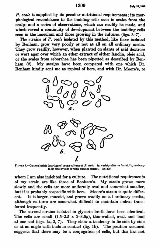
FiGuuz 1.-Camera lucida drawings of young cultures of P. ovak. la, variety of forms found; lb, tendencyto lie side by side or with buds In contact. (X1400)
whom I am also indebted for a culture. The nutritional requirementsof my strain are like those of Benham's. My strain grows moreslowly and the cells are more uniformly oval and somewhat smaller,but it is probably cospecific with hers. Moore's strain is quite differ-ent. It is larger, mucoid, and grows readily on all ordinary media,although cultures are somewhat difficult to maintain unless trans-ferred frequently.The several strains isolated in glycerin broth have been identical.
The cells are small (1.5-2.5 x 2-3.5u), thin-walled, oval, and budat one end (figs. la, 3, 7). They show a tendency to lie side by sideor at an angle with buds in contact (fig. lb). The position assumedsuggests that there may be a conjugation of cells, but this has not
1309 July 1, 1low
been actually demonstrated. The primary cultures on glycerinbroth and, to a slighter extent, subcultures on agar produce a notunpleasant fruity odor suggesting butyl acetate, and similar to thatsometimes detectable from the scalp. It is apparently a volatilesubstance formed through the utilization by P. ovate of the fats orfatty acids in the inoculum and on the ,gar.
Besides the fact that the strains isolated in glycerin broth cannotbe subcultured on ordinary media but will grow only when trans-ferred to media covered with a film of some suitable fatty material,and besides the morphological similarity between the fungus isolatedin culture and that seen in the scales, a further and perhaps moreconvincing proof of the identity of the isolate is furnished by follow-ing the development of the cells seen in the inoculum. Collectedscales are mixed in order to make the inoculum as nearly uniform aspossible, and tubes of 23-40 percent glycerin -broth are heavilyseeded with these scales. An immediate microscopic examinationmade by mounting some of the scales in broth under a cover slipshows that numerous cells of P. ovale are present (fig. 3). Theapparent size in glycerin broth is slightly larger (1.5-2.5 x 3.5.s,exclusive of buds) than in xylol, although the cells are not so easilyseen. If samples of the inoculum are now examined at intervals ofa few hours an increase in numbers of these cells can be clearlydemonstrated. The increasing numbers appear at innumerablepoints in situ in the scales, indicating that the microcolonies whichdevelop after a few days around the scales come from the cells ofP. ovale which were numerous on the scales, and do not arise by theproliferation of one or a few cells of a contaminating yeast whichmight have been present. Nearly all the small scales which floaton the surface of the broth thus become nuclei of microcolonies ofactively budding cells.When the inoculum is examined after 24 to 36 hours incubation
many of the individual scales are surrounded, when crushed under acover slip for microscopic examination, by an oily substance in whichan increased number of the cells of P. ovale can be seen (fig. 6). Someof the smaller scales have closely associated microcolonies of theorganism (fig. 4). After 4 or 5 days incubation the microcolonyincreases greatly in size, but the scale about which the growth centerscan still be seen (fig. 5). In older primary cultures further prolifera-tion of the cells is apparent (fig. 7). These budding cells almostexactly resemble those seen on the original scales, they are onlyslightly larger, and when transferred to agar media which has becomesomewhat dry before use, there appears to be no difference in size orappearance. Subcultures are best made by grinding some of theprimary culture with broth in a mortar and pipetting onto the pre-viously prepared agar slants.
1310Jubr 11§, NO

Public Health Reports, Vol. 55. No. 29, July 19, 1940
FIGURE 2.-P. ovale in defatted, heat-fixed, and methylene blue stained scales from seborrhea. (X 900.)FIGURE 3.-P. ovale in a scale a few hours after planting and before there has been any increase in numbers.
Note the apparent increase in size in 28 percent glycerin broth. (X 900.)FIGURE 4.-A microcolony of P. ovale after 54 hours' incubation, showing a close association with the epi-
thelial scale. (X 900.)FIGURE 5.-A microcolony of P. ovale surrounding an epithelial scale after 48 hours' incubation. The size ofthe colony which ^an be demonstrated at this stage depends upon the number of P. ovale cells in the scalewhen planted, and the number detached when mounting, as well as upon the time of incubation. (X 900 )
FIGURE 6.-Oily material exuding from epithelial scales incubated 48 hours. The scales were placed in adrop of the broth on a slide and flattened under a cover slip. The oily "fringe" contains a few dropletsand numerous cells of P. ovale, displaced when the scale was crushed. (X 100.)
FIGURE 7.--Primary culture of P. ovale after 5 days incubation. (X 900.)
PLATE I

1311 July 19, 18
Attempts were made to determine whether P. orale is pathogenic.The fungus ordinarily does not penetrate to the deeper layers of theskin, being found principally in the horny layers and the superficiallayers which line the hair follicle. It should, therefore, be possibleto simulate natural infection most closely by thoroughly rubbing aculture into the skin. It is desirable to avoid the trauma incident tointracutaneous injection or scarification because these operationsusually result in some scaling and increase in pigmentation. It wasfurther recognized that P. ovale is almost universally present on theskin areas subject to seborrhea. Therefore, instead of trying to findand experimentally infect an individual who did not already carry thefungus it was decided to inoculate a seborrheic individual and tomeasure any noticeable increase in the time required to developseborrhea in the area inoculated as compared with an uninoculatedarea. The entire scalp was thoroughly cleaned and a culture froman agar slant covered with a film of lanolin was removed and rubbedvigorously into areas on the scalp and over the shoulders. Lesionsof seborrhea did not appear in the inoculated areas over the shoulders,and did not appear any sooner in the inoculated areas on the scalpthan in the control areas. These experiments were repeated, but nopathogenic properties of the fungus could be demonstrated. Althoughsome features of seborrhea seem consistent with a parasitic etiology,the failure of these experimental inoculations would give support tothe contentions of many dermatologists and medical mycologists,that P. ovale is a saprophyte, especially adapted to growth on theskin, but without etiologic significance in seborrhea.
SUMMARY
P. ovale was repeatedly and easily isolated by planting scales fromseborrhea oleosa in acid dextrose broth containig 23 to 44 percentglycerin and incubating at 30°-37° C. Subcultures were success-fully carried on media prepared by pipetting ether extract of lanolin,oleic acid, or seborrheic scales over agar slants, as described byBenham.Evidence that the organism was actually P. ovale was furnished by
the necessity for using special media, the resemblance of the fungusin culture and in the skin, and a series of observations of the inocu-lum which showed a continuity of development of the cells of P. oralein the scales.
REFERENCES
(1) Acton, H. W., and Panja G.: Seborrhoeic dermatitis or Pityriasis capitis:A lesion caused by the Malassezia ovale. Ind. Med. Gaz., 62: 603-614(1927).
(2) Benham, R. W.: The cultural characteristics of Pityrosporum ovle-alipophylic fungus. J. Invest. Dermatol., 2: 187-203 (1939).
239210> 4_A e

JulyS19, 940 1312
(3) Castellani, A.: Note on three new yeast-like organisms and a new bacillus,etc. J. Trop. Med. and Hyg., 28: 217-223 (1925).
(4) Krauss, A.: Ueber das Wesen des sogenannten Unnaschen Flaschen-bazillus. Arch. f. Dermatol. u. Syph 116: 723-736 (1913).
(5) MacKee, G. M., Lewis G. M., Spence, i. J., and Hopper, M. E.: Dandruffand seborrhea. I. ilora of ' normal" and diseased scalps. J. Invest.Dermatol, 1: 131-139 (1938).
(6) Malassez, L.: Note sur le champignon du pityriasis simplex. Arch. dePhysiol., 1: 451-464 (1874).
(7) Marzinowski, E. J., and Bogrow, S. L.: Die Blastomyceten und ihre Bezie-hung zu Hautkrankheiten. Arch. f. Dermatol. u. Syph., 86: 215-238(1907).
(8) Meirowsky, E.: Ueber das Wesen der Unnaschen Flaschenbazillen undueber den feineren Bau einiger Hautpilze. Arch. f. Dermatol. u. Syph.,108: 129-140(1911).
(9) Moore, M.: LXVIII. Cultivation and study of Pityroaporum ovale, theso-called bottle bacillus of Unna. Arch. Dermatol. and Syph., 31: 661-671(1935).
(10) Moore, M., Kile,-R. L., Engman, M. F., Jr., and Engman, M. F.: LXXII.Pityrosporum ovale (bottle bacillus of Unna, spore of Malassez). Arch.Dermatol. and Syph., 33: 457-471 (1936).
(11) Ormsby, 0. S.: Diseases of the Skin. Lea and Febiger, Philadelphia, 1937.P. 1150.
(12) Ota, M., and Huang, Ping-Ting: Sur les champignons du genre Pityro-sporum Sabouraud. Ann. Parasitol. hum. et comp., 11: 49-69 (1933).
(13) Panja, G.: The Malassezia of the skin, their cultivation, morphology andspecies. Trans. Far Eastern Assoc. Trop. Med., 2: 442-456 (1927).
(14) Rivolta, S.: Parassiti vegetali. Torino, 1873. Pp. 469-470.(15) Templeton, H. J.: A studv of dandruff and of the Pityrosporon of Malassez.
Arch. Dermatol. and Syph., 14: 270-279 (1926).
CHIGGER MITES*
Chigger mites or "chiggers" I are the larval forms of various speciesof mites belonging to the family Trombidiidae, commonly known asharvest mites. Many different species of chiggers are known toattack vertebrate hosts, but only two chigger mites attacking manhave been recognized from the United States, one, the commonNorth American chigger,2 and the other a closely related form foundin the northern part of the Mississippi Valley.
Description and distribtdion.-The chigger or larv a of the commonNorth American species is oval, bright red, and, as in the first orlarval stage of all mites, possesses only 3 pairs of legs. In the unfed*A leaflet on this subject is available and may be obtained by addressing the Surgeon General, U. S.
Public Health Service, Washington, D. ¢.1 The term "chigger," with variations in spelling (chigoe, jigger, etc.), is also applied to a tropical fea,
Tusnga penefrans, but generally in this country the term is used to designate the larval forms of the trom-bidiid mites.
2 Our common North American chigger attacking man is now known under the scientific name of fLpturileyi Oudemans, 1939. In order to aid the reader in tracing the species under its scientific name in bothmedical and zoological literature, a list of synonyms follows:
Leptus irrilan8 Riley, 1873 (not Leptut irivan8 Lucas, 1847).TTranTychue tlalahuale Murray, 1877 (in part).Trorabidis irrilaas (Riley) Brumpt, 1910.Trombicula cinnabari Ewing, 1920.Lepus asn&w Hirst, 1921.TTrmbiCula irritaae (Riley) Ewing, 1925.Trombicula alfreddugsi (Oudemans, 1910) of Ewing, 1938.Eutrombicula alfreddugi (Oudemans, 1910) of Ewing, 193&

condition it measures about 150 microns in width, and is scarcelyvisible to the naked eye. The legs and surface of the body arecovered by numerous feathered hairs. The mouthparts consist of apair of hooked and ventrally barbed fingerlike mandibles, and 2five-jointed palpi, each of which is provided with a claw dividedinto 2 prongs at the tip. The adult is a large red hairy mite, withthe usual 4 pairs of legs, and with a marked constriction in the an-terior portion of the body. Unlike the larval form it is not parasiticbut is a scavenger, living largely on the fecal matter of arthropodsand on woody decaying substances. Eggs are laid in the ground andthe clhiggers hatch in the spring soon after warm weather begins.
Chiggers have a widespread distribution in the United States,occurring from Long Island to Mexico and from the Atlantic coastto the Rocky Miountains. They have been found in low lands andwell up in the mountains wherever there is rough growth of weedsand shrubbery. They may be encountered from the latter part ofApril until the last of October, depending upon conditions of tem-perature and moisture. In the southern United States they maybegin to cause annoyance early in May, while in the northern partof their range they seldom appear before the middle of June.The North American chigger is not only a pest of man but it has
been reported as attacking a wide range of vertebrates, includingdomestic animals, small mammals, birds, and reptiles. It is animportant pest of poultry, frequently causing the death of youngchickens.
MHethod of attack.--Chiggers attach themselves to the surface of theskin by means of their mouthparts and feed much as do ticks. Theyapparently feed upon epidermal tissue liquefied by a secretion whichthey themselves inject into the skin. When they become fully en-gorged they drop off. The localization of chigger attachment, toquote one author, is determined by two factors, the tightness of theclothing at certain parts of the body and the thickness of the skin.Experiments by the same writer have shown that chiggers attack bypreference where the skin is very thin and the flesh wrinlded ortender. Because of their size, 150 microns in width before theyhave become engorged, chiggers are unable to enter the pores of theskin (which range from 20 to 50 microns in diameter), but they fre-quently attach at the mouth of hair follicles. Although it is widelybelieved that chiggers burrow into the skin and embed their entirebody, this method of attack must be extremely uncommon; theywould be unable to accomplish such an invasion except in instanceswhere a large enough opening in the skin was already present.Symptoms.-An intense itching, apparently due to the liquefying
secretion injected by the chigger, develops within the first 24 hoursafter exposure, and this is followed by a breaking out of wheals or
1313 July 19, 1S
uyly 19, no 1314
papules surunded by an inflamed ara. The papules may be sur-mounted by a pinhead-sized vesicle containing clear fluid. The itchinggenerally reaches its Maximum on the second or third day, then gradu-ally subsides, though it may persist intermittently for several weeks.Scratching may be followed by secondary infection. If the lesionsare numerous, fever, headache, and temporary nervous upset mayresult, and the intense pruritus may lead to loss of sleep and digestivedisturbances. In this country chiggers are not known to transmitany disease, but in the Orient an allied species has been shown to bethe carrier of pseudotyphus or Japanese river fever.
Treatment and prevntion.-If it is known that there has beenexposure to chiggers the skin should be emined, preferably with ahand lens, for the active larvae. However, they are so mnute andthey move so rapidly over the surface of the skin before attachmentthat it is difficult to capture them. An application of kerosene or95 percent alcohol will kill the larvae quite rapidly. As soon as possi-ble after exposure, it is advantageous to apply a thick lather of soapto the affected parts, allowing it to remain for 10 minutes ormore beforebathing. Even though the larvae may be removed or killed soon afterattachment, usually enough secretion has been introduced into theskin to cause the characteristic itching lesion, and for this there is noknown specific remedy. The intense itching may be temporarilyrelieved by ammonia or strong salt water, or a calomel phenol lotion.Collodion with metaphen applied to the lesions is recommended bothto relieve the itching and to prevent infection.In the summer and early fall when it is necessary to go into fields
of tall weeds or grass, into berry patches, or wherever there is heavyundergrowth, an efficacious measure to prevent attack by chiggers isthe liberal spinklidng of the stockings and underclothing with flowers ofsulfur. Some authors have stated that the spraying of the shoes,stockings, and trouser legs with one of the proprietary fly-repellantpreparations is successful in warding off attacks by chiggers.
REFERENCES
Ewing, H. E.: Studies on the biology and control of chiggers. U. S. Dept. Agrie.Bull. No. 986, pp. 1-19 (Dec. 3, 1921).
Ewing, H. E.: The scientific name of the common North American chigger pre-occupied. Proc. Helminthol. Soc. Wash., 5: 26-27 (January 1938).
Ewing, H. E.: A key to the genera of chiggers (mite larvae of the subfamilyTrombiculinae) with descriptions of new genera and species. J. Wash. Acad.Sci., 28:288-295 (June 15, 1938).
Feng, L. C and Hoeppli, R.: On some histological changes caused by mites.Chinese ied. J., 47: 1191-1199 (1933).
Howard, C. W.: A preliminary report on the Trombidiidae of Minnesota. 17thRept. State Ent. of Minn., pp. 111-144 (1918).
Parkhurst, Howard J.: Trombidiosis (infestation with chiggers). Arch. Dermatol.and Syph., 35: 1011-1034 (June 1937).
Riley, Win. A., and JohannsOn. A.: Medical Entomology. 2d ed.; pp. 41-46(1938).

w
-J0L
l-
04
.0
0
0
0
~0
4)
0.
a;
0
s
U1)
0
.
0
._
._
m;0'0
c
._
._
PaH
E-cp
Un1:S
U%0
L;
i
POO
.,.,

1315
PHOTOGRAPH OF A SNEEZE
Sanitarians have long known that certain diseases are spread by thedischarges from the mouth and nose, and that droplet infection playsa role in the dissemination of pathogenic microorganisms. Theyhave also known that such microorganisms may be discharged intothe air in greater numbers and to greater distances by the uncoveredcough and sneeze than in ordinary breathinLg. But since such drop-lets are not visible under ordinary conditions, the risks of infectionby this means have not been fully appreciated by the public, and theprecaut.ionary warnings of health officers to "cover your cough andyour sneeze" are not generally heeded.
If any one has failed to appraise fully the potential danger ofspreading infection to others by an uncovered sneeze, he has only tostudy the accompanying photograph, taken by Prof. M. W. Jennison,of the Department of Biology and Public Health, MassachusettsInstitute of Technology, which shows the expulsion of droplets in aviolent, unstifled act of sneezing.According to Dr. C. E. Turner, who furnisbed the photograph, the
picture was taken by the technique of ultra high-speed photography,which substitutes an instantaneous flash of light for the opening andclosing of the camera shutter. This stroboscopic ligtht Muminatesthe object to be photographed with an intense flash of short duration,the light being placed in such a position in this picture as to illuminatethe droplets with a dark-field effect, so that they stand out sharplyeven in daylight and give photographic images larger than actualdroplet size. The time of exposure was about 1/30,000 of a second.
In such a sneeze as that illustrated here, the droplets are numberedin the thousands, varying with the intensity of the expiratory effort.The number of bacteria dispersed in a sneeze may also be very large.It is stated that most of the droplets are under 2 mm. in diameterand that many are less than 0.1 mm.The "muzzle velocity" of some droplets is said to be as great as
150 feet a second, and large droplets may be expelled to a distanceof 12 feet, although the majority do not travel more than 2 or 3 feet.The involuntary closing of the mouth near the end of a sneeze tendsto form a restricted orifice, resulting in the production of more andsmaller droplets, which probably come largely from the saliva in thefront of the mouth. Also it is apparent from the photograph thatthe number of droplets issuing from the nose in an unstifled sneezeis insignificant as compared with the number expelled from themouth. As stated by Jennison and Edgerton,' these observations are
I Droplet infection of air: High-speed photography of droplet production by sneezing. By M. W. Jeonion and B. E. Edgerton, Massachusetts Istituto of Technology. Proc. Soc. Zip. Biol. and Me.,
43: 455-458 (Marchi 1940).
July 19, 194

probably important in relation to infectivity, because of the differencesin the microbic flora of the two regions.Some droplets fall to the floor or ground, while others evaporate,
leaving their bacteria suspended in the air, through which they may bedisseminated by air currents.The bacteriologic and epidemiologic aspects of infection of the air
were discussed in a recent article by Wells, Wells, and Mudd,2 whoconducted experiments on the concentration of microorganisms in theair. They state that "the numbers of streptococci characteristic ofthe nasopharynx indicate a hazard of respiratory infection and havea sanitary significance comparable with the presence of Escherichiacoli in drinking water." They estimate that several thousand naso-pharyngeal streptococci per sneeze are contributed to the atmosphereand that "the sneeze thus almost seems to be a provision of naturefor the survival of nasopharyngeal parasites. Even where the mani-festations of a disease do not provide for the wide autodisseminationof the infection through the air it has been observed that an outbreakof colds will be followed by the rapid spread of contagion. Sneezinginduced by pollens might conceivably facilitate the spread of naso-pharyngeal infection * * *"Although much is yet to be learned experimentally regarding the
physical and other characteristics of expiratory droplets which arefactors in determining more accurately the role of droplet transmissionin those communicable diseases that are spread by nose and mouthdischarges,- there can be no question that covering the mouth incoughing and sneezing is an important preventive measure withrespect to such diseases.
COURT DECISION ON PUBLIC HEALTH
City ordinance regulting closing hour of barber shops held invalid.-(Sout.h Dakota Supreme Court; City of Huron v. Munson, 289 N. W.416; decided December 26, 1939.) A complaint, which charged aviolation of an ordinance of the city of Huron regulating the hour ofclosing of barber shops within the city, was dismissed by the trialcourt, and the city appealed. The power to regulate the business ofbarbering was not expressly granted to the municipalities of theState, but there was a grant of power to protect the public health.The supreme court said that, if it were conceded that certain generalgrants of power permitted the regulation of barbering by a city as ameans of safeguarding the public health, it did not necessarily followthat the city could regulate the hours during which that business couldbe carried on. It was pointed out that any such regulation, to come
Infection of air. Bacteri(ologic and epidemiologic factors. By W. F. Wells, M. W. Wells, and StuartMudd. Am. J. Pub. Health, 29: 863-880 (August 1939).
1316July 19, 1941
1317 luly 19, 1940
within the scope of the grant of power to protect the public health,had to be reasonable and, to qualify as reasonable, had to contributeim some real and substantial measure to the object sought to be ac-
complished by the grant of power. Continuing, the court said:"The conceded grant of power has as its purpose the protection ofpublic health. We are convinced that the hour of closing a barbershop bears no real or substantial relation to that purpose, and thatsuch a regulation contained in a city ordinance is therefore invalid as
beyond the scope of the power granted by the legislature."
DEATHS DURING WEEK ENDED JULY 6, 1940[From the Weekly Health Index, issued by the Bureau of the Census, Department of Commeroel
Week ended Correspond-July 6, 1940 ing week, 1939
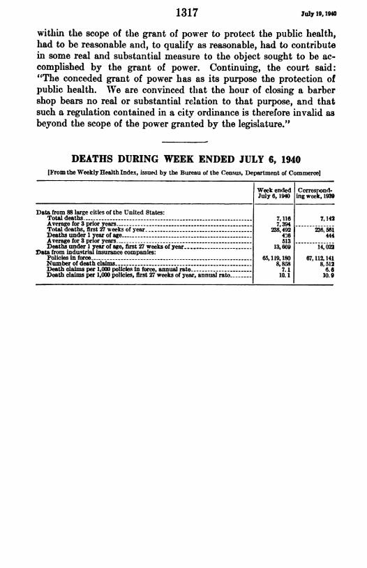
Data from 88 large cities of the United States:Total deaths -7,116 7,142Average for 3 prior years -7,394Total deaths, first 27 weeks of year -238,492 236, 561Deaths under 1 year of age -46 444Average for 3 prior years 513Deaths under I year of age, first 27 weeks of year -13,669 14, 022
Data from indtstrial insurance companies:Policies in force -65,119,180 67,112,141Number of death claims -8,858 8,512Death claims per 1,000 policies in force, annual rate -7.1 6.6Death claims per 1,000 policies, first 27 weeks of year, annual rato 10.1 10.9

PREVALENCE OF DISEASE
No health department, State or local, can effectively prevent or control disease tithoutknowledge of when, where, and under what conditio cases are occurring
UNITED STATES
REPORTS FROM STATES FOR WEEK ENDED JULY 13, 1940
Summary
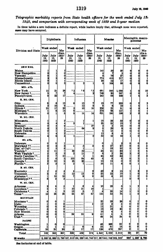
The incidence of poliomyelitis continues to attract interest despitethe favorable trend that has been evidenced throughout the currentseason. A report of 101 cases for the week ended July 13 comparesfavorably with 143 cases for the corresponding week in 1939 which wasalso the median week for the 1935-39 period. For the current weekCalifornia reported 27 cases and Washington 17. The other 57 caseswere scattered among 23 States.Typhoid fever increased from 215 cases for the preceding week to
238 cases, the largest numbers being reported from Arkansas, Louisiana,and Texas. The typhoid trend for 1940 has been below the seasonalexpectancy and lower than the 1939 incidence for each week ofthe year.
Slight increases were noted in the incidence of diphtheria, influenza,measles, meningitis, scarlet fever, smallpox, and whooping cough;however, with the exception of measles, the incidence of all the com-mon communicable diseases was below the 1935-39 median figure forthe corresponding week.Eighteen cases of Rocky Mountain spotted fever were reported, of
which 17 were in the Central and Eastern States. The 34 cases oftyphus fever reported were scattered among 8 South Atlantic andSouth Central States.
(1318)
1319 Julyl ,190
Telegraphic morbidity reports from State heath officers for the week ended July 1S31940, and comparison with corresponding week of 1939 and 5year median
In these tables a zero indicates a definite report, while leaders imply that, although none were reported,caes may have occurred.
Diphtheria Influenza Measl Meningitis, menin-
t Week ended Week ended Week ended Week endedDivision and State Me- _ _ _ Me- Me- Me-
dian, dian, dian, dian.July July 1935- July July 1935- July July 195- July July 1936-13, 15, 39 13, 15, 39 13, 15, 39 13, 15, 391940 1939 1940 1939 1940 19 1940 1939
NIW ENG.Maine .New Hampshlre--Vermont-Massachusetts.Rhode Island-Connecticut-
MD. ATLNew York-New Jersey .Pennsylvania-
Z. NO. CNN.Ohio -------Indiana-llinois -Michigan -----
Wisconsin-W. NO. C1N.
Minnesota-IowaMissouri-North DakotaSouth Dakota ____NebraskaKansas
30. AT.Delaware-Maryland IDist. of Col -Virginia 2 _______West Virginia 3North Carolina 2 4-South Carolina 4Georgia4-Florida 4
Z. SO. CNN.Kentucky-Tennessee-Alabama4-Mississippi 4-
W. so. CEN.Arkansas-Louisiana4.Oklahoma -Texas .
MOUNTAINMontana'-Idaho-Wyoming -Colorado'---
New MexicoArizon-aIJtah I
PAcIcWashington-OregonCalifornia-
TotaWl ----
28 weeks-
8
a
8
2
41
2
1
4
4
13
0106000
11
10
II1a10
217
22
6101761
432140
0
2
5
11
3
7
6
10
2
4
3
10
3
54
1
26
0
0
1
01
21
11
14
A
IC
9
3
4
3
10
3
5
7
3
20
1
0
0
3
0
1
I~
20
.j---2-1
26 1:2 1 6 1 37 4 -- 2
17--7-
ffE
9
30------i
2
28
2
1013
44
24
111I
A
it
191
8464
7---
7810
14
9
.2___2
2
7
140
.-
1410
877459
8
681749245
129
256370621
183520
0
1353
0
41
366
4861516
507
7641052108
8402066
776
2398190
295533915
810
1
273591282815
11
2 42 2
13 9 25 22
9 9 53
313
87
4
18
7
67
-9
10
4
125
221212
IC10
4169
1 35a 17 17 129
292
21
44
24
61479
50
a37
2171753
1,066247480
2331091137190
53151683810
2273360288280
8
151910
6914
76
2952
20
55
24
9218
397
000
Ccc1
O Oo a
10
0
0
2
0
0
0
1
I1
0
0
1
12
0
0
0
1
1
0
1
1
0000000
002
I15 2 421 307 32R .I- 3741I 3I,I622 -, i- 2i~~~~I =l -----
CC4
24cCc
c'I00000
000302201
0
001
000
0
0
1013
31
410
0
0
11
0
0
1
0
30
413
11
1
22
0
1
210
2
0
0
0
0
0
0
0
003
&796
See footnotes at end of table.
8,050110,6861l2,7961167,3131150, 2301140, 7431217,3671342,2491342,2491 9921 1,232
I
I
C
vc

July 19,19l0 1320
Telegraphic morbidity reports from State health officers for the week ended July 13,1940, and comparison with corresponding week of 1939 and 5-year median-Con.
Pollomyelitis Scarlet fever Smallpox Typhoid and par -_ _ _ _ _ _ _ ~~~~~~~~~~~~typhoidfever
Division and State Week ended Week ended Week ended Week endedDiviionand tat Wee en ed-e Me- Me- _ ___me-dian, dian, dan dian,
July July 193- July July 1935- July July 1935- July July 135-13, 15, 39 13, 15, 39 13, 15, 39 13, 15, 391940 193 1940 1939 19t0 1939 1940 1939
XEW ZNG.MaineNew Hampshire--..VermontMassachusettsRhode Ldiiand--Connecticut
MD. ATL
New YorklNew JerseyPennsylvania
E. NO. CNN.
OhioIndiana-Ilinob 2_-____-
MichiganWisconsin _________
W. NO. CNN.MinnesotaIowa -Mliouri-- ----
North Dakota .South DakotaNebraska .___Kans
so. AM
DelawareMaryland 2 8Dist. of Col-.
West Virginia'
North Carolina"-4__.South Carolina 4Georgia 4Florida 4 ---
E. SO. CNN.
KentuckyTennessee-4Alabama 4Mispps 4
W. 50. CNN.Akansas-
Lousana'OklahomaTexas 4
MOUNTAIN
MontanaIdaho-Wyoming-ColoradoNew Mexico-Arizona _------Utah'-
PACIF
Washington- 17Oregon-2CaHlfornia-27
Total 10
28 weeks--- -
0
0
0
0
2
1
0
0
1
0
4
0
5
0
0
0
0
4
I
0
0
0
10
0
20
5
15
5
2
21
I
I
1
1s
0
0
0
1
1
1
0
0
45
143j
C
I
0
0
19
143
,20 1,020j
0
0
11
0
0
62
011
5
2
0
1
0
10
0
11
11
2(
Ii
14
C
1itI
4
144
53
1,225
5, 292
3
1256
4
3141
42
14,
.0
5aa3
381]
I
1
102319E
911i608542
13138
24
5
23
2
161
149
9
1
10
5
4
15
10
2
75
17
8
2
7
9
4
1
4
5 14S 10
06 80
14
13
L.'
15131
144
91
2387
12966
34
1919104
5
27
2163
8
12152
2
10
10
3
6
717
835
215
29
9561 1, 391
LZ 6751160, 214
I
I
49
1,843
0
0
0
0
00
0
00
1
0
2
0
1I15
1610
0
0
1
DD74
00
0
0
0
0
0
12
2
310
21332
731
0
0
0
0
0
2DII
:I
0
0
0
0
0
0
0
0
0
021115
6
6
52
5
3
0
0
0
0
0
0
0
cI
II
IIi
i
32
103
7,557
Ii
21
31
S,
31
3
238
099
On footnotes at end of table.
2
0
0
2
2
a
1
8
14
6
0
9
6
0
3
1
4
1
0
1
1
I
3
1
2
2
1;
2
437
601
10
13
64
0
3
136
6
9
8
15
1
0
02
5
0
0
0
1
2
4
:7
7
9
1
0
130
3
146
14
12
823I1
0
2
11
0
0
1
12
318
9
2121371
37422011
23
21
39
12
0
2
4
1
2
3it
530
839_
_l
IaI
II
I
.L
1= =
5
LI
I
II
II
I
II
I
I
I
I
II
I
III 11
IfI.
IIII
I
11II I
II
IIIII
2

1321 Jtaly 31,14
Telegraphic morbidity report from State health officers for the week ended July 13,1940, and comparison with corresponding week of 1939 and 5-year median-Con.
Whooping ough Whoopingcough
Week ended Week endedDivision and State Division and State
July July July July13, 15, 13, is1,1940 1939 1940 1939
NEW ENO.
Maine -New Hampshire .VermontMassachusetts - .-.-------.-.-.----Rhode IslandConnecticut-
MM. ATL.
New YorkNow Jersey 2Pensylvania _
Z. NO. CEN.
OhioIndiana _Illinois'Michigan 'Wisconsin
W. NO. CEN.
MinnesotaIowa-Missouri .North DakotaSuth DakotaNebraska-Kansas .-------.
so. ATL.
Delaware - ---------------Maryland 'aDist. of Col'Virginia 'West Virginia
120
16105263
2B0
471403153
2 15 423142 239357 438
27012
157261108
437
339
6
6
61
1114413
11091
52
98362181212
353436583
3422
76538588
so. ATL-Cfotinued.North Carolina 2 4South Carolina 4 .Georgia4-Florida 4
X. 50. CEN.
KentuckyTennesseeAlabamaMississippi "4 ---------
W. 90. CIN.
Arkansas .- -----------------Louisiana 4Oklahoma 2Texas -
MOUNTAIN
Montana -
Idaho.WyomingColorado 'New MexicoArizona_--Utah '
PACIC
WashingtonOregon-California _
Total
28 weeks
12115
2010
924817
366419
210
814
61118
0
117
6528242
-13, 465.
90,001
27418
34
33
4413021
15
1594
115
60
1
13819
0
76
17
20
109
4,295
109,344
' New York City only.2 Rocky Mountain spotted fever, week ended July 13, 1940, 17 cases as follows: New Jersey, 2; Illnois, 2;
Maryland, 2; District of Columbia, 1; Virginia, 3; North Carolina, 3; Oklahoma, 4; Montana, 1.3 Period ended earlier than Saturday.4 Typhus fever, week ended July 13, 1940, 34 cases as follows: North Carolina, 3; South Carolina, 5;
Georgia, 9; Florida, 2; Alabama, 4; Missisippi, 1; Louisiana, 4; Texas, 6.1 Colorado tick fever, week ended July 13, 1940, Colorado, 2 cases.
PLAGUE INFECTION IN LICE FROM A MARMOT IN PARKCOUNTY, WYO.
Under date of July 5, 1940, Surgeon L. B. Byington reported plagueinfection proved in a pool of 14 lice from 1 marmot (Marmota flavi-ventri,s) shot 12 miles northwest of Cody, Park County, Wyo., onJune 17. This is stated to be the first proof of plague infection inthat county.
11
m-
July 19, imo 1322
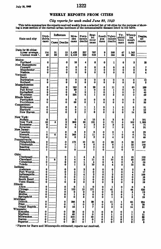
WEEKLY REPORTS FROM CITIESCity reports for week ended June 29, 1940
This table summarizes the reports received weekly from a selected list of 140 cities for the purpose of show-ing a cro section of the current urban incidence of the communicable diseases listed in the table.
Diph- Influenza Mea- Pneu- leart Small- Tuber. Ty. Whoop- Det_State and city theria sles monia let pox culosis phoid ing all
cases Cases Deathseathscaseshdeath e cases deaths fe o causesCasesDeaths ~~~~~cases cases
Data for 90 cities:5-year average. 114 31 15 2,433 337 787 9 3G6 47 1,243Currentweek I 49 32 13 2,519 232 616 1 364 34 935
Maine:Portland. 0- 0 _ 10 4 0 0 1 0 3 33
New Hampshire:Concor -- 0 _ 0 0 0 0 0 0 0 0 7Manchester-...0_0 0 0 2 0 0 0 0 9Nashua..- 0- 0 0 0 0 0 0 0 0 4
Vermont:Barre ---------Burlington 0- 0 0 0 0 0 0 0 0 12Rutland--0__0 0 0 0 0 0 0 0 2
Massachusetts:Boston - 0 0 O 165 6 28 0 11 2 60 189Fall River 0 -__ 0 89 1 0 0 1 0 9 21Springfield 0 0 3 2 1 0 1 0 0 40Worcester 0 0O 234 4 1 0 3 0 2 49
Rhode Island:Pawtucket 0 _ 0 0 0 0 0 0 0 0 19Providence 0- 0 06 0 1 0 0 0 6 50
Connecticut:Bridreport 0O0 6 1 1 0 1 0 0 31Hartford- 0- 0 0 5 7 0 1 0 2 43New Haven. 0- 0 _ 3 2 6 0 0 0 14 38
New York:Buffalo-- -- 0 0 4 15 O a O 2 101NewYork- 16 5 2 368 32 131 0 71 5 102 1,268Rochester 0 0 4 4 2 0 2 0 6 nSyracuse _ 0 0 0 1 0 1 0 4 31
New Jersey:Camden-_ -1 0 6 5 8 0 0 1 0 23Newark --0___O 6 0 248 3 11 0 7 0 6 74Trenton ___0 0 3 2 0 4 0 0 40
Pennsylvania:Philadelphia- 2 0 173 10 51 0 28 5 36 357Pittsburgh 3 0 1 6 5 0 6 1 33 146Reading- 0-----O0 1 2 0 0 2 0 15 27Scranton- 1 -- 0 __ 1 0 0 0
Ohio:CincinnatL 0 O 0 1 4 4 0 4 0 16 132Cleveland 1 8 2 4 4 11 0 10 0 48 163Columbus 0- 0 0 1 4 0 4 0 8 58Toledo-1 0 1 2 10 0 4 0 18 61
Indiana:Anderson- 0 0 0 2 0 0 0 0 2 6Fort Wayne 0 0 4 1 0 0 1 0 4 17IndTanapols 0 3 2 4 2 0 2 0 4 109Muncie- 0- 0 0 1 0 0 0 0 0 18SouthBend--- 0O 1 0 3 0 0 0 0 0 19Terr Haute.-- 0- 0 0 0 0 0 2 0 0 19
Illinob:Alton - 0- 0 0 1 0 0 1 0 0 8Chicago- 8 0 152 15 171 0 41 1 44 630Elgin - 0- 0 0 0 0 0 0 0 4 12Moline0 0 1 0 0 0 0 0 1 88pringfield 0O0 0 3 0 0 1 0 3 19Michin:Detroit- 0 296 6 29 0 11 1 65 234Flint- 0-- 1 0 3 0 2 0 0 2)Grand Rapids. 0O 0 16 1 7 0 2 0 14 31Wisconsin:Kenosha- 0 29 0 2 0 0 0 1 6Madison- 0 40 0 0 0 1 0 1 11Milwaukee 0 0 335 0 19 0 3 0 9 83Racine - 0 15 0 0 0 0 0 2 13Superior- 0 26 0 1 0 0 0 0 6
' Figures for Barre and Minneapolis estimated; reports not received.
I I

1323 July 10, 10
City reports for week ended June 29, 1940-Continued
Diph- uenia Mea- Pneu- ier- msn. Tuber- Ty- Whoo DeathsState and city theria ales monia POXr fever couthhsoa Cas ~~~deaths faevertcameideaths fever coe causesCa Detb .MB Cae M
Minnesota:Duluth-Minnapolis. --St. Paul
Iowa:Cedar Rapids~Davenport-De Moines...Sioux City-. .Waterloo
MIssouri:Kansas City-St. Joseph.St. Lous-
North Dakota:FargoGrand ForksaMinot
South Dakota:Aberdeen-
Nebraska:Lincoln-Omaha-
Kansa:Lawrence-Topeka _Wichita-
Delaware:Wilmington.---
Maryland:Baltimore-Cumberland-Frederick-
Dist. of Col.:Washington--
Virginia:Lynchburg-Norfolk-Richmond-Roanoke -
West Virginia:Charleston-Huntington_..WheelUn-
North Carolma:Gastonia-Raleigh-Wilmington-.Winston-Salem
South Carolina:Charleston-Florence-_Greenville --_-
Georgia:Atlanta-Brunswick-Savannah-
Florida:MiamiTampa-
Kentucky:Ashland-Covington.Lexington.Louisville.
Tennessee:Knoxville-Memphis.Nashville-
Alabama:Birmingham-Mobile-Montgomery-
o0 0 9 I1 1 01 0 0 15
57
8814
18313
-8032130
20
192118
152
1715381015
24
1221
21194
72225
4020
10181562
278057
5818
-i_
1. ..--- I - I - I- - -. - -. -. -.0
0
0
0
0
I
0
0
0
0
0
0
0
0
1
0
0
0
0
0
0
0
0
0
0
0
0
0
0
0
0
0
0
00
1
00
0
00
0
0
0
0
0
0
0
1
Arkansas:Fort Smith-.... O -
Little Rocklc- O.---
u
0
OO
1
2
0
8
0
2
8
0
4
0
0
0
0
0
2
0
110
1
4
0
0
1
290
17
0
0
0
0
00
0
0
1
9
x
4
0
6
0
0
2
1
0
8
0
0
1
1
0
2
0
0
0
0
0 0 10 5 10 40 0
0 6 2
0 5 0
0 4 20 3 3
1 5 4
0 0 0
------ 0 ----
*---- 0 ---
3
0
4a0
5
14
1
0
0
1
0
0
20
2
30
0
11
0
0
0
0
0
0
0
o
1
1
0
1
0
0
10
0
0
0
5
1
0
2
2
2
0
0
00200
000
000
0
00
000
0
000
0
0000
000
0000
000
00
00
00000
10
000
1
3010
0
0
0
1
010
010
12
00
0
002
10
0
0
3
41
0
11
3
41
--O 1
0
00000
001
000
0
01
001
1
000
0
0000
000
00001
001
10
0000
1011
00
4
30013
3010
050
7
11
302
3
12200
5013
1
1013040011801
0
332
098
200
20
0
O -- -
O -- -
I

July 19, 1r0 1324City reports for week ended June 29, 1940-Continued
Diph- Influenza Mea- Pneu- Sear- Small- Tuber- Ty- Whoop- Deaths,State and city thUeris|_|_ ] les moniaU ltfevef |pox c|ulosisl ing anStateand cty aters culdes h fevercae desi fever cough
cascssCases Deaths css cases case camse
Louisana:NewOrleans- 0 0 3 6 1 0 12 3 32 140Shreveport 0 0 0 0 0 0 1 3 0 42
Oklahoma:OklahomaCity 0 0 1 1 1 0 3 0 0 33Tulsa- 0-- 2 5 0 0 1 1 9 20
Texas:Dallas-2 0 34 2 0 0 3 2 7 78Fort Worth---- 0 0 0 2 1 0 1 0 0 37Galveston 0 0 0 0 0 0 2 0 0 25Houston - 0 1 0 14 4 2 0 7 1 1 110San Antonio ---- 0 0 0 3 0 0 6 0 8 63
Montana:Billings-0 0 0 0 0 0 1 0 0 7Great Falls 0 18 3 0 0 0 0 0 7Helena-0 0 0 0 0 0 0 0 0 4Missoula- 0 0 1 1 0 0 0 0 0 9
Idaho:Boise-0 0 0 0 0 0 1 0 0 5
Colorado:ColoradoSprings0 0 1 1 2 0 1 0 0 9
Denver-8 0 12 5 2 0 4 0 0 92Pueblo-0 4 1 0 0 0 0 3 8
New Mexico:Albuquerque 0 0 0 0 0 0 2 0 2 12
Utah:Salt Lake City 0 0 52 2 3 0 0 0 54 38
Washington:neattle0 0 33 3 3 0 3 0 8 78Spokane- 0 1 0 0 0 0 0 0 25Tacoma-0 1 0 1 0 1 0 0 33
Oregon:Portland- 2 0 7 3 3 0 0 0 10 76Salem- 0---- 0 0- 0 0
California:Los Angeles 3 6 0 6 1 12 0 23 0 77 326Sacramento 0 0 1 1 4 0 1 0 1 30San Francisco- 3 1 2 3 3 4 0 5 0 16 161
Meningitis, Polio- Meningitis, Polio-State and city meningococcus meningococcus m
- State and city litisCases Deaths cases Cases Deaths cases
Rhode Island: District of Columbia:Providence-1 0 0 Washington-1 1 1
New York: Virginia:Buffalo -3 1 0 Richmond-0 0 1New York- 2 0 0 Oklahoma:
Illinois: Oklahoma City 0 0 1Chicago-0 0 1 Washington:
Wisconsin: Tacoma-0 0 10Milwaukee -1 0 0 California:
Missouri: Los Angeles-0 0 5St. Joseph - 0 1 0 San Francisco 0 0 1
Nebraska:Omaha-0 0 1
Eneephalitis, epidemic or ltlhargic.-Cases: Neew York, 1; St. Louis, 1; Omaha, 1; Missoula, 1.PeUlagra.-Cases: Birmingham, 1.T"hufever.-Cases: Charleston, S. C., 1; Miami, 1; New Orleans, 2. Deaths: NTew York, 1.
I

FOREIGN REPORTS
CANADA
Provinces-Communicable diease&-Week ended June 8, 1940.-During the week ended June 8, 1940, cases of certain communicablediseases were reported by the Department of Pensions and NationalHealth of Canada as follows:
DiseasePrnceNova BNew Que- On- Man~i- kas Alber- Brim-totalDisease |E13sdriwarad Scotia | -B be tario toba tchtIsland wick ewan bla
Cerebrospinal menngiti ------1 6 ----- 6Chickenpo ---- 20 1 197 439 35 9 2 79 782Diphtheria.- --- 34 1 1 ---- 36Dysentery-------------- 1 1Infuenza ---- 8 --- 51 18 77Measles --- 6 116 289 158 146 2 94 810MUmPS 45 272 7 40 29 393PneUmOnia ---- 3- 17 2 --- 5 27Poliomyelitis--- 1 1 ---- 2Scarlet fever-------- 2 I 80 100 15 13 12-- 223Trachoma- --------- --- 2 2Tuberculosis -- 3 2 9 38 41 31 15 4-- 143TyphO{d and ParatY-.
.........PhOid feVer _ 1 13 2 1 6 1 1 AWhooping Cough_______-- _ 48 3 125 112 29 364 25 382
REPORTS OF CHOLERA, PLAGUE, SMALLPOX, TYPHUS FEVER, ANDYELLOW FEVER RECEIVED DURING THE CURRENT WEEK
NoTzE:-A cumulative table giving current information regarding the world prevalcncc of quarantinablediseases appeared in the PUBLIC HEALTH REPORTS of June 28, 190, pages 1188-1191. A similar table Willappear in future issues of the PUBMIC HEALTH REPORTS for the last Friday of each month.
Plague
CIina-South Hsingan Province-Tungliao (vicinity of).-A reportdated July 7, 1940, stated that up to July 6, 1940, 17 cases of plaguehad occurred in the vicinity of Tungliao, South Hsingan Province,China.
Hauw'aii Territory-Island of Hawaii-Hamakua District-Paauilo(vicinity of).-A rat found on June 7, 1940, another on June 18, andanother on June 20, 1940, in the vicinity of Paauilo, HamakuaDistrict, Island of Hawaii, T. H., have been proved positive forplague.
United St-atesWyoming-Park County.-A report of plagueinfection in Park County, Wyoming, appears on page 1319 of thisissue of PUBLIC HEALTH REPORTS.
Typhus Fever
&raits Settlements-Singapore.-During the week ended May 4,1940, 1 case of typhus fever was reported in Singapore, StraitsSettlements.
(1325)x