PTEN genomic deletion is associated with p-Akt and AR signalling in poorer outcome, hormone...
9
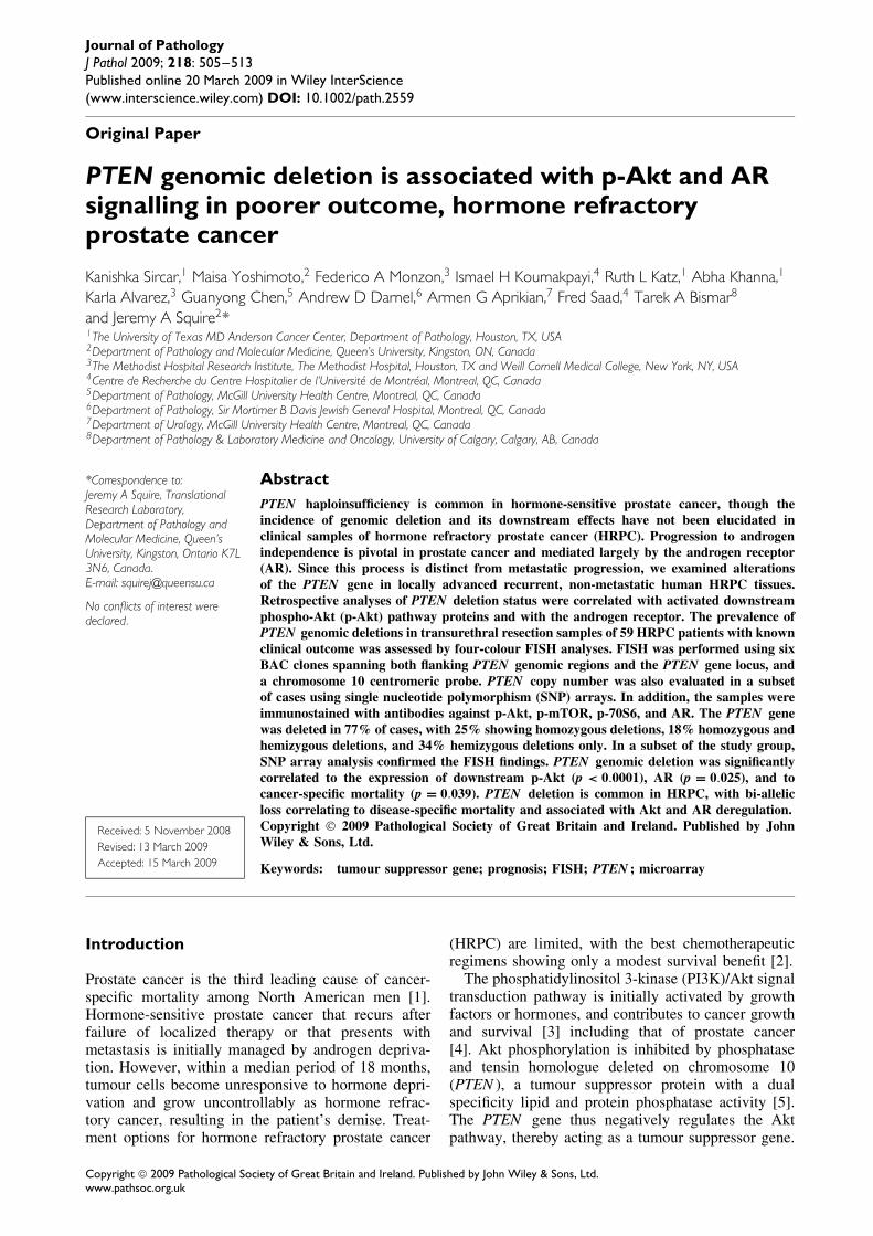
Journal of Pathology J Pathol 2009; 218: 505–513 Published online 20 March 2009 in Wiley InterScience (www.interscience.wiley.com) DOI: 10.1002/path.2559 Original Paper PTEN genomic deletion is associated with p-Akt and AR signalling in poorer outcome, hormone refractory prostate cancer Kanishka Sircar, 1 Maisa Yoshimoto, 2 Federico A Monzon, 3 Ismael H Koumakpayi, 4 Ruth L Katz, 1 Abha Khanna, 1 Karla Alvarez, 3 Guanyong Chen, 5 Andrew D Darnel, 6 Armen G Aprikian, 7 Fred Saad, 4 Tarek A Bismar 8 and Jeremy A Squire 2 * 1 The University of Texas MD Anderson Cancer Center, Department of Pathology, Houston, TX, USA 2 Department of Pathology and Molecular Medicine, Queen’s University, Kingston, ON, Canada 3 The Methodist Hospital Research Institute, The Methodist Hospital, Houston, TX and Weill Cornell Medical College, New York, NY, USA 4 Centre de Recherche du Centre Hospitalier de l’Universit´ e de Montr´ eal, Montreal, QC, Canada 5 Department of Pathology, McGill University Health Centre, Montreal, QC, Canada 6 Department of Pathology, Sir Mortimer B Davis Jewish General Hospital, Montreal, QC, Canada 7 Department of Urology, McGill University Health Centre, Montreal, QC, Canada 8 Department of Pathology & Laboratory Medicine and Oncology, University of Calgary, Calgary, AB, Canada *Correspondence to: Jeremy A Squire, Translational Research Laboratory, Department of Pathology and Molecular Medicine, Queen’s University, Kingston, Ontario K7L 3N6, Canada. E-mail: [email protected] No conflicts of interest were declared. Received: 5 November 2008 Revised: 13 March 2009 Accepted: 15 March 2009 Abstract PTEN haploinsufficiency is common in hormone-sensitive prostate cancer, though the incidence of genomic deletion and its downstream effects have not been elucidated in clinical samples of hormone refractory prostate cancer (HRPC). Progression to androgen independence is pivotal in prostate cancer and mediated largely by the androgen receptor (AR). Since this process is distinct from metastatic progression, we examined alterations of the PTEN gene in locally advanced recurrent, non-metastatic human HRPC tissues. Retrospective analyses of PTEN deletion status were correlated with activated downstream phospho-Akt (p-Akt) pathway proteins and with the androgen receptor. The prevalence of PTEN genomic deletions in transurethral resection samples of 59 HRPC patients with known clinical outcome was assessed by four-colour FISH analyses. FISH was performed using six BAC clones spanning both flanking PTEN genomic regions and the PTEN gene locus, and a chromosome 10 centromeric probe. PTEN copy number was also evaluated in a subset of cases using single nucleotide polymorphism (SNP) arrays. In addition, the samples were immunostained with antibodies against p-Akt, p-mTOR, p-70S6, and AR. The PTEN gene was deleted in 77% of cases, with 25% showing homozygous deletions, 18% homozygous and hemizygous deletions, and 34% hemizygous deletions only. In a subset of the study group, SNP array analysis confirmed the FISH findings. PTEN genomic deletion was significantly correlated to the expression of downstream p-Akt (p < 0.0001), AR (p = 0.025), and to cancer-specific mortality (p = 0.039). PTEN deletion is common in HRPC, with bi-allelic loss correlating to disease-specific mortality and associated with Akt and AR deregulation. Copyright 2009 Pathological Society of Great Britain and Ireland. Published by John Wiley & Sons, Ltd. Keywords: tumour suppressor gene; prognosis; FISH; PTEN ; microarray Introduction Prostate cancer is the third leading cause of cancer- specific mortality among North American men [1]. Hormone-sensitive prostate cancer that recurs after failure of localized therapy or that presents with metastasis is initially managed by androgen depriva- tion. However, within a median period of 18 months, tumour cells become unresponsive to hormone depri- vation and grow uncontrollably as hormone refrac- tory cancer, resulting in the patient’s demise. Treat- ment options for hormone refractory prostate cancer (HRPC) are limited, with the best chemotherapeutic regimens showing only a modest survival benefit [2]. The phosphatidylinositol 3-kinase (PI3K)/Akt signal transduction pathway is initially activated by growth factors or hormones, and contributes to cancer growth and survival [3] including that of prostate cancer [4]. Akt phosphorylation is inhibited by phosphatase and tensin homologue deleted on chromosome 10 (PTEN ), a tumour suppressor protein with a dual specificity lipid and protein phosphatase activity [5]. The PTEN gene thus negatively regulates the Akt pathway, thereby acting as a tumour suppressor gene. Copyright 2009 Pathological Society of Great Britain and Ireland. Published by John Wiley & Sons, Ltd. www.pathsoc.org.uk
-
Upload
independent -
Category
Documents
-
view
5 -
download
0
Transcript of PTEN genomic deletion is associated with p-Akt and AR signalling in poorer outcome, hormone...
Journal of PathologyJ Pathol 2009; 218: 505–513Published online 20 March 2009 in Wiley InterScience(www.interscience.wiley.com) DOI: 10.1002/path.2559
Original Paper
PTEN genomic deletion is associated with p-Akt and ARsignalling in poorer outcome, hormone refractoryprostate cancer
Kanishka Sircar,1 Maisa Yoshimoto,2 Federico A Monzon,3 Ismael H Koumakpayi,4 Ruth L Katz,1 Abha Khanna,1
Karla Alvarez,3 Guanyong Chen,5 Andrew D Darnel,6 Armen G Aprikian,7 Fred Saad,4 Tarek A Bismar8
and Jeremy A Squire2*1The University of Texas MD Anderson Cancer Center, Department of Pathology, Houston, TX, USA2Department of Pathology and Molecular Medicine, Queen’s University, Kingston, ON, Canada3The Methodist Hospital Research Institute, The Methodist Hospital, Houston, TX and Weill Cornell Medical College, New York, NY, USA4Centre de Recherche du Centre Hospitalier de l’Universite de Montreal, Montreal, QC, Canada5Department of Pathology, McGill University Health Centre, Montreal, QC, Canada6Department of Pathology, Sir Mortimer B Davis Jewish General Hospital, Montreal, QC, Canada7Department of Urology, McGill University Health Centre, Montreal, QC, Canada8Department of Pathology & Laboratory Medicine and Oncology, University of Calgary, Calgary, AB, Canada
*Correspondence to:Jeremy A Squire, TranslationalResearch Laboratory,Department of Pathology andMolecular Medicine, Queen’sUniversity, Kingston, Ontario K7L3N6, Canada.E-mail: [email protected]
No conflicts of interest weredeclared.
Received: 5 November 2008Revised: 13 March 2009Accepted: 15 March 2009
AbstractPTEN haploinsufficiency is common in hormone-sensitive prostate cancer, though theincidence of genomic deletion and its downstream effects have not been elucidated inclinical samples of hormone refractory prostate cancer (HRPC). Progression to androgenindependence is pivotal in prostate cancer and mediated largely by the androgen receptor(AR). Since this process is distinct from metastatic progression, we examined alterationsof the PTEN gene in locally advanced recurrent, non-metastatic human HRPC tissues.Retrospective analyses of PTEN deletion status were correlated with activated downstreamphospho-Akt (p-Akt) pathway proteins and with the androgen receptor. The prevalence ofPTEN genomic deletions in transurethral resection samples of 59 HRPC patients with knownclinical outcome was assessed by four-colour FISH analyses. FISH was performed using sixBAC clones spanning both flanking PTEN genomic regions and the PTEN gene locus, anda chromosome 10 centromeric probe. PTEN copy number was also evaluated in a subsetof cases using single nucleotide polymorphism (SNP) arrays. In addition, the samples wereimmunostained with antibodies against p-Akt, p-mTOR, p-70S6, and AR. The PTEN genewas deleted in 77% of cases, with 25% showing homozygous deletions, 18% homozygous andhemizygous deletions, and 34% hemizygous deletions only. In a subset of the study group,SNP array analysis confirmed the FISH findings. PTEN genomic deletion was significantlycorrelated to the expression of downstream p-Akt (p < 0.0001), AR (p = 0.025), and tocancer-specific mortality (p = 0.039). PTEN deletion is common in HRPC, with bi-allelicloss correlating to disease-specific mortality and associated with Akt and AR deregulation.Copyright 2009 Pathological Society of Great Britain and Ireland. Published by JohnWiley & Sons, Ltd.
Keywords: tumour suppressor gene; prognosis; FISH; PTEN ; microarray
Introduction
Prostate cancer is the third leading cause of cancer-specific mortality among North American men [1].Hormone-sensitive prostate cancer that recurs afterfailure of localized therapy or that presents withmetastasis is initially managed by androgen depriva-tion. However, within a median period of 18 months,tumour cells become unresponsive to hormone depri-vation and grow uncontrollably as hormone refrac-tory cancer, resulting in the patient’s demise. Treat-ment options for hormone refractory prostate cancer
(HRPC) are limited, with the best chemotherapeuticregimens showing only a modest survival benefit [2].
The phosphatidylinositol 3-kinase (PI3K)/Akt signaltransduction pathway is initially activated by growthfactors or hormones, and contributes to cancer growthand survival [3] including that of prostate cancer[4]. Akt phosphorylation is inhibited by phosphataseand tensin homologue deleted on chromosome 10(PTEN ), a tumour suppressor protein with a dualspecificity lipid and protein phosphatase activity [5].The PTEN gene thus negatively regulates the Aktpathway, thereby acting as a tumour suppressor gene.
Copyright 2009 Pathological Society of Great Britain and Ireland. Published by John Wiley & Sons, Ltd.www.pathsoc.org.uk

506 K Sircar et al
Akt activates downstream mTOR, which promotesphosphorylation of S6, leading to mRNA translation[6,7]. Akt pathway inhibitors, particularly mTORinhibitors, have shown promise in pre-clinical modelsof androgen-independent prostate cancer, underscoringthe biological relevance and therapeutic importance ofthis pathway [7,8].
PTEN haploinsufficiency has been described in pre-malignant high-grade prostatic intraepithelial neopla-sia (HGPIN), as well as organ-confined and metastaticprostate cancer [9]. Mouse models and cell line stud-ies have identified losses of PTEN with consequentactivation of Akt as contributing to the emergence ofandrogen-independent growth. Moreover, PTEN geneinactivation has been correlated to prostate cancer pro-gression using murine models [10,11] and in clini-cal specimens, where metastatic lesions showed botha greater frequency and extent of PTEN deletions[12–14]. No prior studies have specifically addressedthe copy number status of the PTEN gene in androgen-independent lesions from human tissues. However,pre-clinical models have shown direct links betweenPTEN and androgen signalling [15].
PTEN haploinsufficiency is common in hormone-sensitive prostate cancer, though the incidence ofPTEN genomic deletion and downstream effects havenot previously been elucidated in a distinct clinicalcohort derived from a large series of hormone refrac-tory prostate cancer (HRPC) patients. As the devel-opment of androgen independence is a key feature ofadvanced prostate cancer that is mediated largely bythe androgen receptor (AR) and since AR progressionis biologically distinct from metastasis, we undertookthis study to examine genomic alterations of the PTENgene in locally advanced recurrent, non-metastatichuman HRPC tissues. Furthermore, we examined thecorrelation of PTEN gene deletions, as evaluated byFISH, to activated downstream Akt pathway proteinsand to AR. Given the uncertainty regarding the bio-logical impact of PTEN pathway disruption in HRPC,these genotype–phenotype correlative studies will pro-vide valuable insights for future prostate cancer prog-nostication and treatment.
Materials and methods
Patients and tissue samples
Samples were obtained from 59 patients through insti-tutional review board-approved protocols and afterinformed consent from McGill University Health Cen-tre and the Centre Hospitalier de l’Universite deMontreal, Montreal, Quebec. Tissue samples con-sisted of transurethral resections performed on patientswith HRPC (median 77.5 years; range 57–95 years;PSA median 29.2; range 0.1–4537) to relieve urinaryobstructive symptoms. Tissue microarrays (TMAs)were constructed from the formalin-fixed, paraffin-embedded (FFPE) tissues using quadruplicate 1 mm
cores. Initial hormone deprivation therapy includedsurgical orchiectomy or luteinizing hormone-releasinghormone agonists with or without an anti-androgenagent. Hormone refractory disease was defined as twoconsecutive rises in serum PSA by more than 10%during therapy, despite castrate levels of testosterone.Patients were followed from initiation of hormonedeprivation therapy to death or last follow-up (median41 months), with a median time to the development ofHRPC of 19 months. Median time from detection ofhormone refractory cancer to death was 20 months.
Immunohistochemistry
Immunostaining on the TMAs was done using theLSAB 2 peroxidase system (Dako Diagnostics Inc,CA, USA). Staining was performed as previouslydescribed [16]. Briefly, TMAs were treated with 0.3%H2O2 –methanol for 30 min before performing antigenretrieval in boiling EDTA buffer (1 mM, pH 8.0) for15 min. After blocking with a protein-blocking serum-free reagent (Dako Cytomation) and incubating withprimary antibody for 90 min, TMAs were treated withthe secondary biotinylated antibody (Dako Cytoma-tion) and streptavidin–peroxidase (Dako Cytomation).Reaction products were developed with diaminoben-zidine (Dako Cytomation) and nuclei were coun-terstained with Harris haematoxylin (Sigma-Aldrich,MO, USA). Negative control involved using PBSinstead of the primary antibody.
The optimal dilution was determined for each anti-body by serial dilutions. The primary antibody incuba-tion was completed with the following antibodies: rab-bit monoclonal phospho-Akt antibody at 1/25 dilution(Ser 473, IHC-specific) (#3787; Cell Signaling, Dan-vers, MA, USA); rabbit monoclonal phospho-mTORat 1/100 dilution (Ser 2448, IHC-specific) (#2976; CellSignaling); rabbit monoclonal p70 S6 kinase at 1/60dilution (#2708; Cell Signaling); mouse monoclonalPTEN antibody at 1/15 dilution (Ab1809; Abcam,Cambridge, MA, USA); and mouse monoclonal andro-gen receptor at 1/100 dilution (AR #441; Neomarkers,Fremont, CA, USA).
Cytoplasmic and/or nuclear p-Akt, mTOR, AR,PTEN, and p70 S6 kinase immunoreactivities werescored by two independent observers according to theproduct of staining intensity (1–3) multiplied by theproportion of immunoreactive cells in the areas ofinterest (1–4) [17]. As such, the overall score rangewas from 1 to 12. Immunoreactivity scores were thendichotomized using a cut-off value of 6, with a scoreof ≥6 representing positive staining.
Fluorescence in situ hybridization (FISH)
The four-colour FISH strategy was employed on anarchival FFPE hormone-refractory prostatic adenocar-cinoma TMA, using six BAC clones spanning bothflanking PTEN genomic regions (BMPR1A and FASloci) and the PTEN gene locus, and a commer-cially available DNA probe for region 10p11.1–q11.1
J Pathol 2009; 218: 505–513 DOI: 10.1002/pathCopyright 2009 Pathological Society of Great Britain and Ireland. Published by John Wiley & Sons, Ltd.

PTEN loss in hormone refractory prostate cancer 507
(SpectrumAqua centromere of chromosome 10 probe)(CEP 10 — Abbott Molecular, Illinois, USA). Thefollowing BAC clones were used: (a) BMPR1A genelocus located at 10q23.2 (88.2–88.7 Mb, 900 kbupstream from the 5′PTEN gene): RP11-141D8,RP11-52G13, and RP11-420K10; (b) PTEN genelocus located at 10q23.31 (89.6–89.8 Mb): RP11-846G17; and (c) FAS gene locus located at 10q23.31(90.6–90.8 Mb): RP11-399O19 and RP11-360H20.The position and name of the BAC clones were takenfrom the Human March 2006 assembly of the UCSCGenome Browser 1 (Figure 1A). The presence ofPTEN, FAS sequences, and correct chromosome loca-tion of the BAC clones were verified by PCR andby hybridization to metaphase spreads from normalperipheral lymphocytes, respectively. BAC DNA wasextracted and labelled with either Spectrum Green-dUTP, SpectrumOrange-dUTP (Vysis Inc), or Cy5-dUTP (PerkinElmer Life and Analytical Sciences),using the nick-translation kit (Abbott Molecular).
PTEN copy number was evaluated for each probeby counting spots in 100 non-overlapped, intact inter-phase nuclei per tumour tissue core. The establishmentof PTEN gene copy number status was defined by con-sidering the adjacent probes (BMPR1A and FAS loci)used for the truncation artefacts, aneusomy, nuclearsize, and chromatin condensation. Based on hybridiza-tion in control cores (data not shown), hemizygousdeletions of PTEN were defined as more than 30%(mean ± 3 SD in non-neoplastic controls) of tumournuclei containing one PTEN locus signal and by thepresence of CEP 10 signals. Homozygous deletion ofPTEN was exhibited by the simultaneous lack of bothPTEN locus signals and by the presence of control sig-nals in more than 20% of cells. The LNCaP prostatecancer cell line with undeleted PTEN and the PC3 cellline with homozygously deleted PTEN were used ascontrols for our FISH procedure.
Androgen receptor gene copy number analysis wasperformed using LSI AR gene (Xq21, SpectrumOrangeprobe) and CEP X (DXZ1, SpectrumGreen, AbbottMolecular) (Figure 5). The dual-colour FISH methodwas applied to the FFPE prostate cancer TMA accord-ing to the manufacturer’s instructions.
SNP array assays
Tumour DNA was obtained from five of the above-mentioned samples by manually microdissectingtumour tissue from 10 µm FFPE sections according toa previously described protocol for deparaffinizationand DNA extraction [18]. DNA was quantitated ona NanoDrop ND-1000 spectrophotometer (NanoDropTechnologies, Wilmington, DE, USA). All samplesprocessed for downstream analysis in this study had anOD 260/280 ratio higher than 1.8. Samples were pro-cessed with an FFPE-optimized protocol based on theGeneChip Mapping 250 Nsp Assay Kits (Affymetrix,Santa Clara, CA, USA) and whose performance hasbeen described previously [18]. The samples were then
hybridized on GeneChip Mapping 250K Nsp arrays(Affymetrix) for 16 h at 48 ◦C in a GeneChip 450hybridization oven (Affymetrix) at 60 rpm. The arrayswere washed and stained according to the manufac-turer’s instructions.
Data acquired from the Affymetrix GeneChip Oper-ating System v4.0 (GCOS) was analysed usingAffymetrix Genotyping Console 2.1. The quality con-trol parameters evaluated for each sample were thesignal detection rate (the percentage of features inthe array that show adequate fluorescence intensity)and the SNP call rate (the rate of successful alleleidentification) [19]. Data from the X chromosomewere not analysed, as the samples were not gender-matched to controls. Loss of heterozygosity (LOH)and copy number (CN) estimates were obtained usinga publicly available analysis package: Copy Num-ber Analyzer for Affymetrix GeneChip arrays (CNAG3.0) and Genotyping Console [20,21], with unmatchednormal controls as described before [22]. We iden-tified the presence of genomic gains/losses in oneor two alleles in the PTEN genomic region. TheSNP array data discussed in this article have beendeposited in NCBI’s Gene Expression Omnibus (GEOhttp://www.ncbi.nlm.nih.gov/geo/) and are accessiblethrough GEO series accession number GSE13439.
Statistical analyses
Mean staining intensities of cores from HRPC patientswere calculated. Kruskal–Wallis non parametric testswere used to assess the statistical significance ofobserved differences in mean staining intensity. Allcorrelation coefficients were computed using Spear-man’s non-parametric test. The foregoing was doneusing Statistical Package for the Social Sciences(SPSS) version 12 (SPSS, Inc).
Results
PTEN assessment
The frequency of PTEN deletion was investigated inthe cohort of 59 tumour samples using a TMA inwhich anonymous annotation codes allowed interroga-tion of clinical outcome parameters. FISH was infor-mative in 56 cases, among which the PTEN gene wasdeleted in 43 cases (77%) with no deletions seen in 13cases (23%). Hemizygous PTEN deletion (single-copyloss) was found in 19 of the 56 (34%) and homozy-gous PTEN deletion (double-copy loss) was found in14 of the 56 (25%) prostate adenocarcinoma samples.The presence of both hemizygous and homozygousPTEN deletions was observed in ten of the 56 (18%)prostate adenocarcinomas (Figures 1B and 2). In twocases, it was possible to compare PTEN FISH in pri-mary adenocarcinoma with results from analysis oftumour tissue from the same patient’s HRPC tumoursamples (Table 1). In both cases, PTEN deletion was
J Pathol 2009; 218: 505–513 DOI: 10.1002/pathCopyright 2009 Pathological Society of Great Britain and Ireland. Published by John Wiley & Sons, Ltd.

508 K Sircar et al
Figure 1. (A) FISH probes used to detect the PTEN deletion in prostate cancer. The schematic chromosome 10 illustrates thegenomic localization of the commercially available alpha-satellite DNA sequences of the chromosome 10 probe (CEP10,Vysis Inc),the location and names of the BAC probes spanning the upstream genomic region of PTEN (BMPR1A gene locus), and PTEN andFAS loci at chromosome 10. The linear order and approximate distances of the BAC clones are based on the Human March2006 assembly of the UCSC Genome Browser. (B) Pie chart showing PTEN deletions: hemizygous (34%); homozygous (25%);homozygous and hemizygous (18%); and undeleted PTEN (23%)
Figure 2. Representative FISH images are shown for prostate cancer TMA applying the four-colour FISH. The panel showsa pseudo-colour image with the DAPI counterstained nuclei. Original magnification: ×100. (A) Four-colour FISH identifies twosignals of red (PTEN locus), green (BMPR1A locus), and yellow (FAS locus) BAC probes, as well as paired pale blue (CEP10, VysisInc) signals in most of the nuclei, indicating no deletion of PTEN in tumour cells. (B) Representative four-color PTEN FISH imageshows tumour cells with a single red signal for PTEN, BMPR1A, and FAS loci in most of the nuclei and paired blue signals forCEP10, indicating hemizygous deletion of the PTEN gene region in prostate cancer (arrow). (C) Representative PTEN FISH imageof homozygous PTEN deletion (arrow) in prostate cancer shows absence of a red signal for the PTEN locus in most of the nuclei.The retained single green signal (BMPR1A locus) and yellow signal (FAS locus) in most of the nuclei and paired blue signals forCEP10 indicate hemizygous deletion of the BMPR1A and FAS loci in prostate cancer
not present in the primary adenocarcinomas, but acqui-sition of either hemizygous or homozygous PTENdeletion was apparent in their clinically HRPC.
In addition to the evaluation of PTEN deletion statusby FISH, we performed whole genome CN and LOHanalysis with SNP arrays on five cases. One case
produced CN data with a high standard deviation inlog2 ratios for CN data (SD > 0.4) and was thereforeeliminated. Four cases showed adequate data (SD <
0.4) and all showed concordance with the FISH resultsfor loss of chromosomal material in the PTEN region(Figure 3).
J Pathol 2009; 218: 505–513 DOI: 10.1002/pathCopyright 2009 Pathological Society of Great Britain and Ireland. Published by John Wiley & Sons, Ltd.

PTEN loss in hormone refractory prostate cancer 509
Table 1. Correlation of PTEN copy number to downstreamAkt pathway in a subset of prostatic adenocarcinoma samplesobtained from two patients prior and subsequent to becomingclinically hormone refractory
PTEN deletion p-Akt
Sample 1Hormone-sensitive No NegativeHormone refractory Yes Positive
Sample 2Hormone-sensitive No NegativeHormone refractory Yes Positive
The relative deficiency of PTEN cellular protein lev-els assessed by immunohistochemistry revealed thatacquisition of the gene locus deletion and concomitantloss of PTEN functional activity was not consistentlysignificant. This discordance, as previously reportedby McCall et al [23], indicates that PTEN immunore-activity varies widely on FFPE tissue sections, mostlikely as a result of FFPE tissue preparation and thehigher sensitivity and specificity for FISH to detectsubtle clonal deletions in subsets of tumour cells [9].
Activation of downstream effectors in the p-Aktpathway and AR expression
As PTEN inhibits the Akt pathway, we examinedthe expression of the activated or phosphorylatedform of downstream agents: namely p-Akt, p-mTOR,and p-70S6. Activated p-Akt showed cytoplasmicexpression in 64% of cases, with nuclear stainingin 25%. Downstream, activated m-TOR and 70S6proteins showed cytoplasmic expression in 44% and55% of cases, respectively. As shown in Table 2, wefound immunoexpression of p-Akt to be positivelycorrelated with downstream p-mTOR (p = 0.016).Nuclear AR was expressed in 78% of the cases, with36% showing strong (3+) staining. Representativeimages are illustrated in Figure 4.
Correlation of PTEN copy number to downstreamp-Akt pathway, androgen receptor, anddisease-specific mortality
With respect to PTEN deletion, we analysed the sig-nificance of PTEN gene status, where we dividedpatients as either possessing or lacking PTEN dele-tion in their tumours. We also analysed the presenceor absence of PTEN genomic deletion, or overall genecopy number (ie 0, 1 or 2 copies), where our cohortwas grouped into patients harbouring undeleted, hem-izygously deleted or homozygously deleted PTEN intheir cancers. Our data indicated that both presence orabsence of PTEN deletion (ie grouped as undeletedversus deleted) and PTEN overall copy number (ieno deletion versus hemizygous versus homozygousdeletion) were associated with increased expressionof downstream p-Akt (p < 0.0001). Although concor-dance was observed between AR gene amplificationstatus and the overall cellular AR nuclear protein
expression levels (p = 0.021), there was no correla-tion of AR gene amplification, as assessed by FISH,with PTEN deletion, either homozygous or hemizy-gous gene copy number loss (data not shown). How-ever, PTEN genomic deletion correlated to AR pro-tein expression (p = 0.025) and to death from HRPC(p = 0.039). In contrast, presence versus absence ofPTEN gene deletion status was, by itself, not corre-lated to these parameters, highlighting the importanceof PTEN gene copy number in the androgen-resistantstage of prostate cancer. These data are summarizedin Table 2.
Two cases of HRPC had acquired PTEN genomicdeletions with concomitant p-Akt activation in tissuesfrom advanced disease. In both of the initial adeno-carcinoma samples from these patients, the tumourshad weak p-Akt immunostaining, indicative of inactiveAkt signalling and no apparent PTEN FISH deletion(Table 1).
Discussion
PTEN has previously been implicated in the patho-genesis and progression of hormone-sensitive prostatecancer. Recently, genome-wide scanning with an SNParray platform has revealed deletions at 10q23, whichmaps to the PTEN gene, to be among the mostfrequent recurrent changes in prostate cancer com-pared with non-neoplastic prostate tissues [24]. Sev-eral different methodologies have independently estab-lished PTEN loss to be involved in pre-invasiveprostatic intraepithelial neoplasia [9], as well as inthe grade and stage progression of invasive prostatecancer [12,25,26]. Moreover, PTEN genomic dele-tion was recently shown to be associated along withTMPRSS2:ERG fusion in earlier disease recurrenceof localized prostate cancer [27]. Hormone naivemetastatic samples have shown a higher frequency ofPTEN loss, often involving bi-allelic deletions [13].The importance of the PTEN gene is underscoredby murine models, where prostate-specific deletion ofPTEN resulted in advanced, metastatic prostate cancer[28,29].
To date, PTEN copy number changes have not beensystematically evaluated in clinical samples of HRPC.As hormone escape is the key factor presaging treat-ment failure in this disease, we sought to interrogateonly non-metastatic HRPC tissues so that we wouldbe able to evaluate transition to the hormone refrac-tory state without the process of metastasis being aconfounding factor. Androgen receptor signalling mayperpetuate tumour growth promotion when mutated ordefective [30]. Our results showing an increased inci-dence of 77% PTEN loss — with 43% harbouringhomozygous deletions — in HRPC implicates bothPTEN loss and gene dosage in hormonal escape. Ourresults are corroborated by independent copy numberanalysis with SNP arrays. PTEN loss was strongly cor-related to downstream p-Akt signalling (p < 0.0001),
J Pathol 2009; 218: 505–513 DOI: 10.1002/pathCopyright 2009 Pathological Society of Great Britain and Ireland. Published by John Wiley & Sons, Ltd.

510 K Sircar et al
Figure 3. Copy number (CN) data for the PTEN region in chromosome 10 from SNP arrays. For each sample (PR002 to PR005),the top graph shows log2 ratios for all SNPs in the region (red dots); the middle graph shows the CN estimate for 30 SNP average(blue line); and the bottom graph shows the CN estimate for allele-specific analysis (red/green lines). Samples PR003 and PR004show homozygous deletions (green arrows) and sample PR005 shows heterozygous deletion (blue arrow)
which it normally inhibits, suggesting a causal rela-tionship between PTEN loss and p-Akt activation inthis phase of prostate cancer.
Androgenic signalling mediated through the andro-gen receptor is well established as a major contrib-utor to cell survival in all stages of prostate cancer,
J Pathol 2009; 218: 505–513 DOI: 10.1002/pathCopyright 2009 Pathological Society of Great Britain and Ireland. Published by John Wiley & Sons, Ltd.

PTEN loss in hormone refractory prostate cancer 511
Table 2. Correlation of PTEN copy number to downstream Akt pathway, androgen receptor, and disease-specific mortality.p value with Spearman’s correlation coefficient
p-Akt p-mTOR p-70S6 AR Dead of disease
Overall PTEN gene copy number∗ <0.0001 0.621 0.076 0.025 0.039(0.462) (0.07) (0.368) (0.299) (0.332)
Presence versus absence of PTEN deletion† <0.0001 0.637 0.496 0.458 0.25(0.469) (0.067) (0.146) (0.101) (0.189)
p-Akt (IHC) 0.016 0.161 0.655 0.235(0.328) (0.283) (0.06) (−0.195)
∗ 0 = undeleted; 1 = hemizygous PTEN deletion; 2 = homozygous PTEN deletion.† 0 = undeleted; 1 = any PTEN deletionBold values: p < 0.05.
Figure 4. Hormone refractory prostate cancer immunostained with (A) phospho-Akt, (B) phospho-mTOR, (C) phospho-70 S6,and (D) androgen receptor (AR)
including hormone refractory cancer [31]. Indeed, up-regulation of AR mRNA and protein is now thoughtto be a principal pathway for the development of thecastrate-resistant or hormone refractory state [32]. Inthis model, cellular signalling in HRPC continues tooccur via the androgen receptor, which is hypersensi-tive to trace amounts of androgenic or non-androgenicstimuli. PTEN regulation of AR activity has been
demonstrated in studies on LNCaP prostate cancer celllines. Specifically, PTEN was shown to inhibit ARactivity directly by blocking AR nuclear translocationand to suppress AR activity indirectly through the Aktpathway [15].
Our data from clinical samples, with homozygousdeletions of the PTEN gene being associated with up-regulation of nuclear AR, are consistent with PTEN ’s
J Pathol 2009; 218: 505–513 DOI: 10.1002/pathCopyright 2009 Pathological Society of Great Britain and Ireland. Published by John Wiley & Sons, Ltd.

512 K Sircar et al
Figure 5. Section of prostate cancer showing nuclei stained with DAPI, CEPX (green), and AR gene locus (red). (A) Most nucleicontain one green and one red signal (arrow), indicating that each nucleus contains one X-chromosome, on which there is one ARgene. (B) Cells showing one or two green (X-chromosome) signals and three to six red AR gene signals (arrow). (C) Cells showingone or two X-chromosome green signals with multiple copies of red AR gene signals (arrow)
proposed suppression of the androgen receptor [33].Furthermore, the acquirement of PTEN deletion withconsequent Akt activation in a subset of two prostatecancers when becoming clinically hormone refractorysuggests the importance of PTEN loss with conse-quent Akt activation in driving hormone escape as analternative mechanism in hormone-sensitive prostatecancers. Thus, the biological and clinical importanceof PTEN inactivation in HRPC is suggested by itscorrelation to cancer-specific mortality in this cohort.
The Akt pathway is important in both androgen-sensitive and hormone refractory prostate cancer, withfunctional cross-talk between the Akt and AR path-ways involved in both prostate cancer initiation andprogression [4]. Both loss of PTEN expression andAkt activation have been correlated to poor clinicaloutcome in androgen-sensitive prostate cancer [34,35].Akt regulates androgen-mediated AR transcriptionalactivity in androgen-sensitive disease, while activa-tion of Akt has been shown to occur under condi-tions of androgen deprivation, where it supports cellu-lar survival [36]. This pathway is clinically germanesince it is amenable to therapeutic inhibition. Indeed,inhibitors of Akt are currently being tested in clinicaltrials across various malignancies. Our results, show-ing PTEN genomic deletions to be clinically signifi-cant and to be correlated to activation of Akt, wouldsupport their utility in hormone refractory prostate can-cer as well.
AcknowledgementsWe would like to thank HJ Kim, Joan O’Malley, and KimberleyR Stewart for valuable technical and secretarial assistance.This work was financially supported by grant number 018124(to JAS and MY) from the Canadian Cancer Society; theProstate Cancer Research Foundation, Canada (to TAB); anda foundation grant from the McGill University Health CentreResearch Institute (to KS).
References
1. Jemal A, Siegel R, Ward E, Murray T, Xu J, Smigal C, et al.Cancer statistics, 2006. CA Cancer J Clin 2006;56:106–130.
2. Calabro F, Sternberg CN. Current indications for chemotherapy inprostate cancer patients. Eur Urol 2007;51:17–26.
3. Testa JR, Bellacosa A. AKT plays a central role in tumorigenesis.Proc Natl Acad Sci U S A 2001;98:10983–10985.
4. Wang Y, Kreisberg JI, Ghosh PM. Cross-talk between theandrogen receptor and the phosphatidylinositol 3-kinase/Aktpathway in prostate cancer. Curr Cancer Drug Targets2007;7:591–604.
5. Myers MP, Stolarov JP, Eng C, Li J, Wang SI, Wigler MH, et al.P-TEN, the tumor suppressor from human chromosome 10q23,is a dual-specificity phosphatase. Proc Natl Acad Sci U S A1997;94:9052–9057.
6. Ruggero D, Pandolfi PP. Does the ribosome translate cancer?Nature Rev Cancer 2003;3:179–192.
7. Mamane Y, Petroulakis E, LeBacquer O, Sonenberg N. mTOR,translation initiation and cancer. Oncogene 2006;25:6416–6422.
8. Thomas GV. mTOR and cancer: reason for dancing at thecrossroads? Curr Opin Genet Dev 2006;16:78–84.
9. Yoshimoto M, Cutz JC, Nuin PA, Joshua AM, Bayani J, EvansAJ, et al. Interphase FISH analysis of PTEN in histologic sectionsshows genomic deletions in 68% of primary prostate cancer and23% of high-grade prostatic intra-epithelial neoplasias. CancerGenet Cytogenet 2006;169:128–137.
10. Di Cristofano A, De Acetis M, Koff A, Cordon-Cardo C,Pandolfi PP. Pten and p27KIP1 cooperate in prostate cancer tumorsuppression in the mouse. Nature Genet 2001;27:222–224.
11. Trotman LC, Niki M, Dotan ZA, Koutcher JA, Di Cristofano A,Xiao A, et al. Pten dose dictates cancer progression in the prostate.PLoS Biol 2003;1:E59.
12. McMenamin ME, Soung P, Perera S, Kaplan I, Loda M, Sell-ers WR. Loss of PTEN expression in paraffin-embedded primaryprostate cancer correlates with high Gleason score and advancedstage. Cancer Res 1999;59:4291–4296.
13. Schmitz M, Grignard G, Margue C, Dippel W, Capesius C,Mossong J, et al. Complete loss of PTEN expression as a possibleearly prognostic marker for prostate cancer metastasis. Int J Cancer2007;120:1284–1292.
14. Yoshimoto M, Cunha IW, Coudry RA, Fonseca FP, Torres CH,Soares FA, et al. FISH analysis of 107 prostate cancers shows thatPTEN genomic deletion is associated with poor clinical outcome.Br J Cancer 2007;97:678–685.
15. Lin HK, Hu YC, Lee DK, Chang C. Regulation of androgenreceptor signaling by PTEN (phosphatase and tensin homologdeleted on chromosome 10) tumor suppressor through dis-tinct mechanisms in prostate cancer cells. Mol Endocrinol2004;18:2409–2423.
16. Le Page C, Koumakpayi IH, Alam-Fahmy M, Mes-Masson AM,Saad F. Expression and localisation of Akt-1, Akt-2 and Akt-3correlate with clinical outcome of prostate cancer patients. Br JCancer 2006;94:1906–1912.
J Pathol 2009; 218: 505–513 DOI: 10.1002/pathCopyright 2009 Pathological Society of Great Britain and Ireland. Published by John Wiley & Sons, Ltd.

PTEN loss in hormone refractory prostate cancer 513
17. Al-Maghrabi J, Vorobyova L, Chapman W, Jewett M, Zielen-ska M, Squire JA. p53 alteration and chromosomal instabilityin prostatic high-grade intraepithelial neoplasia and concurrentcarcinoma: analysis by immunohistochemistry, interphase in situhybridization, and sequencing of laser-captured microdissectedspecimens. Mod Pathol 2001;14:1252–1262.
18. Lyons-Weiler M, Hagenkord J, Sciulli CM, Dhir R, Monzon FA.Optimization of the Affymetrix GeneChip mapping 10K 2.0 assayfor routine clinical use on formalin fixed paraffin embedded tissues.Diagn Mol Pathol 2008;17:3–13.
19. Thompson ER, Herbert SC, Forrest SM, Campbell IG. Wholegenome SNP arrays using DNA derived from formalin-fixed, paraffin-embedded ovarian tumor tissue. Hum Mutat2005;26:384–389.
20. Jacobs S, Thompson ER, Nannya Y, Yamamoto G, Pillai R,Ogawa S, et al. Genome-wide, high-resolution detection of copynumber, loss of heterozygosity, and genotypes from formalin-fixed,paraffin-embedded tumor tissue using microarrays. Cancer Res2007;67:2544–2551.
21. Nannya Y, Sanada M, Nakazaki K, Hosoya N, Wang L, HangaishiA, et al. A robust algorithm for copy number detection usinghigh-density oligonucleotide single nucleotide polymorphismgenotyping arrays. Cancer Res 2005;65:6071–6079.
22. Monzon FA, Hagenkord J, Lyons-Weiler M, Balani JP, Par-wani AV, Sciulli CM, et al. Whole genome SNP arrays as a poten-tial diagnostic tool for the detection of characteristic chromosomalaberrations in renal epithelial tumors. Mod Pathol 2008;21:1–10.
23. McCall P, Witton CJ, Grimsley S, Nielsen KV, Edwards J. IsPTEN loss associated with clinical outcome measures in humanprostate cancer? Br J Cancer 2008;99:1296–1301.
24. Liu W, Chang B, Sauvageot J, Dimitrov L, Gielzak M, Li T, et al.Comprehensive assessment of DNA copy number alterations inhuman prostate cancers using Affymetrix 100K SNP mappingarray. Genes Chromosomes Cancer 2006;45:1018–1032.
25. Majumder PK, Sellers WR. Akt-regulated pathways in prostatecancer. Oncogene 2005;24:7465–7474.
26. Dreher T, Zentgraf H, Abel U, Kappeler A, Michel MS, Bleyl U,et al. Reduction of PTEN and p27kip1 expression correlates withtumor grade in prostate cancer. Analysis in radical prostatectomyspecimens and needle biopsies. Virchows Arch 2004;444:509–517.
27. Yoshimoto M, Joshua AM, Cunha IW, Coudry RA, Fonseca FP,Ludkovski O, et al. Absence of TMPRSS2 : ERG fusions andPTEN losses in prostate cancer is associated with a favorableoutcome. Mod Pathol 2008;21:1451–1460.
28. Wang S, Gao J, Lei Q, Rozengurt N, Pritchard C, Jiao J, et al.Prostate-specific deletion of the murine Pten tumor suppressor geneleads to metastatic prostate cancer. Cancer Cell 2003;4:209–221.
29. Backman SA, Ghazarian D, So K, Sanchez O, Wagner KU,Hennighausen L, et al. Early onset of neoplasia in the prostateand skin of mice with tissue-specific deletion of Pten. Proc NatlAcad Sci U S A 2004;101:1725–1730.
30. Dagvadorj A, Tan SH, Liao Z, Cavalli LR, Haddad BR, NevalainenMT. Androgen-regulated and highly tumorigenic human prostatecancer cell line established from a transplantable primary CWR22tumor. Clin Cancer Res 2008;14:6062–6072.
31. Setlur SR, Rubin MA. Current thoughts on the role of theandrogen receptor and prostate cancer progression. Adv AnatPathol 2005;12:265–270.
32. Chen CD, Welsbie DS, Tran C, Baek SH, Chen R, Vessella R,et al. Molecular determinants of resistance to antiandrogentherapy. Nature Med 2004;10:33–39.
33. Jiao J, Wang S, Qiao R, Vivanco I, Watson PA, Sawyers CL, et al.Murine cell lines derived from Pten null prostate cancer showthe critical role of PTEN in hormone refractory prostate cancerdevelopment. Cancer Res 2007;67:6083–6091.
34. Bedolla R, Prihoda TJ, Kreisberg JI, Malik SN, KrishnegowdaNK, Troyer DA, et al. Determining risk of biochemicalrecurrence in prostate cancer by immunohistochemical detectionof PTEN expression and Akt activation. Clin Cancer Res2007;13:3860–3867.
35. Kreisberg JI, Malik SN, Prihoda TJ, Bedolla RG, Troyer DA,Kreisberg S, et al. Phosphorylation of Akt (Ser473) is an excellentpredictor of poor clinical outcome in prostate cancer. Cancer Res2004;64:5232–5236.
36. Mikhailova M, Wang Y, Bedolla R, Lu XH, Kreisberg JI,Ghosh PM. AKT regulates androgen receptor-dependent growthand PSA expression in prostate cancer. Adv Exp Med Biol2008;617:397–405.
J Pathol 2009; 218: 505–513 DOI: 10.1002/pathCopyright 2009 Pathological Society of Great Britain and Ireland. Published by John Wiley & Sons, Ltd.