Clozapine Alone versus Clozapine and Risperidone with Refractory Schizophrenia
Upload
independentCategory
view
0download
0
doi:10.1016/j.jacc.2004.06.056 2004;44;1415-1419 J. Am. Coll. Cardiol.
Therese Resink Paul Erne, Peiman Jamshidi, Peter Juelke, Hans-Peter Hafner, Peter Thum, and
Brachytherapy: Potential therapy for refractory coronary spasm
This information is current as of July 13, 2011
http://content.onlinejacc.org/cgi/content/full/44/7/1415located on the World Wide Web at:
The online version of this article, along with updated information and services, is
by on July 13, 2011 content.onlinejacc.orgDownloaded from

BTPPL
Isdnaccsi
moTmar(ciatgs
L
a
Journal of the American College of Cardiology Vol. 44, No. 7, 2004© 2004 by the American College of Cardiology Foundation ISSN 0735-1097/04/$30.00Published by Elsevier Inc. doi:10.1016/j.jacc.2004.06.056
rachytherapy: Potentialherapy for Refractory Coronary Spasm
aul Erne, MD,* Peiman Jamshidi, MD,* Peter Juelke, MD,† Hans-Peter Hafner, PHD,†eter Thum, MD,† Therese Resink, PHD‡uzern and Basel, Switzerland
OBJECTIVES We sought to demonstrate that brachytherapy reduces coronary spasm in refractory andhighly symptomatic variant angina.
BACKGROUND In some patients with variant angina due to extensive vasoconstriction, intensive drug therapyfails to sufficiently relieve symptoms.
METHODS In 18 patients with frequent angina episodes despite triple anti-anginal therapy, coronaryspasm was induced by intracoronary acetylcholine (ACh) infusion. Five patients had spasm ina second vessel. Intracoronary radiation (20 Gy) was applied to vasospastic segments using abeta-emitting (32P) wire source centered within a Galileo balloon. Parameters of vesselfunction before and after brachytherapy were investigated.
RESULTS Before brachytherapy, artery diameters decreased (p � 0.0001) from 2.8 � 0.4 mm to 1.0 �0.4 mm for the first vessels and from 3.1 � 0.3 mm to 1.0 � 0.2 mm for the second vessels.After brachytherapy (143 � 106 and 80 � 52 days for first and second vessels, respectively),ACh-induced vasoconstriction was significantly reduced. The ACh-induced changes in arterydiameter before and after brachytherapy were �1.5 � 0.5 mm and �0.5 � 0.3 mm (p �0.0001) for the first vessels and �1.4 � 0.3 mm and �0.4 � 0.2 mm (p � 0.01) for thesecond vessels, respectively. In non-irradiated spastic vessels, ACh-induced vasoconstrictionremained unchanged (e.g., �1.7 � 0.6 mm, �1.6 � 0.3 mm, and �1.5 � 0.5 mm for secondvessels, at first investigation, first follow-up, and immediately before brachytherapy, respec-tively). Angina frequency decreased from 15.6 � 6.0 to 2.2 � 2.4 angina episodes/week (p �0.001) in treated patients.
CONCLUSIONS Brachytherapy is a potential therapy in patients with highly symptomatic variantangina. (J Am Coll Cardiol 2004;44:1415–9) © 2004 by the American College ofCardiology Foundation
M
PfcaiwvrsacifeaT
cacsiwt
n most patients with variant angina, spasm occurs at theite of atheromatous plaque, although the degree of stenosisue to plaque is moderate and commonly �30% of theormal segment diameter (1). To document variant angina,provocative test by infusion of acetylcholine (ACh) into
oronary arteries is sometimes necessary (2). Most patientsan be medically treated with vasodilating drugs. However,ome are highly symptomatic and drug-resistant, and stent-ng has been used (3).
Although application of brachytherapy produces an im-ediate increase of vasoconstriction and spasms (4,5), a loss
f vasomotion some weeks after brachytherapy occurs (6).his effect renders brachytherapy as a potential therapeuticodality for patients with highly symptomatic variant
ngina. We reported an index case of a post-brachytherapyeduction of angina in a patient with coronary vasospasm7). This patient died subsequently, and autopsy revealedoronary thrombosis of the irradiated left anterior descend-ng (LAD) coronary artery and untreated intermediatertery. Nevertheless, given the significant effect of brachy-herapy in relieving angina, we cautiously further investi-ated the potential of brachytherapy in refractory coronarypasm.
From the Divisions of *Cardiology and †Radiotherapy, Kantonsspital Luzern,uzern; and the ‡Department of Research, Kantonsspital Basel, Basel, Switzerland.Manuscript received May 14, 2004; revised manuscript received June 8, 2004,
accepted June 22, 2004.
content.onlinejDownloaded from
ETHODS
atients. Eighteen patients with frequent angina and re-ractoriness to intensive anti-ischemic therapy were in-luded. Seven and four patients had ST-segment elevationsnd depressions, respectively, on the electrocardiogram dur-ng chest pain at rest. Patients underwent initial diagnosticork-up to exclude coronary artery disease and documentasoconstrictive responsiveness to ACh. If similar ACh-esponsiveness was documented within eight weeks (inclu-ion criteria), investigation was repeated and brachytherapypplied to the dominant vasospastic vessel. The local ethicalommittee approved the protocol. Patients gave written,nformed consent. Three months before brachytherapy andor the study duration, patients reported weekly anginalpisode occurrence and nitrate use. Relevant patient char-cteristics, risk factors, and drug therapies are presented inable 1.No patient had angiographically visible plaque. Acetyl-
holine was infused (10 �mol/l, constant) into coronaryrteries, and vasospasms were quantitated by quantitativeoronary angiography if vasoconstriction �50% diametertenosis occurred (8). Vasospastic vessel locations weredentified anatomically in a predefined projection, whichas repeated at follow-up investigations. A prophylactic
emporary pacing lead was placed into the right ventricular
pex at a backup rate of 40 beats/min. Nitroglycerin wasby on July 13, 2011 acc.org
irn(cdwfi(
Vstc�c(awivta
ob
fifb8wacsddmttistatc1
d(wdsSva
R
TTmvvreaf
tEb2ftvtf
T
GAHWC
I
E
D
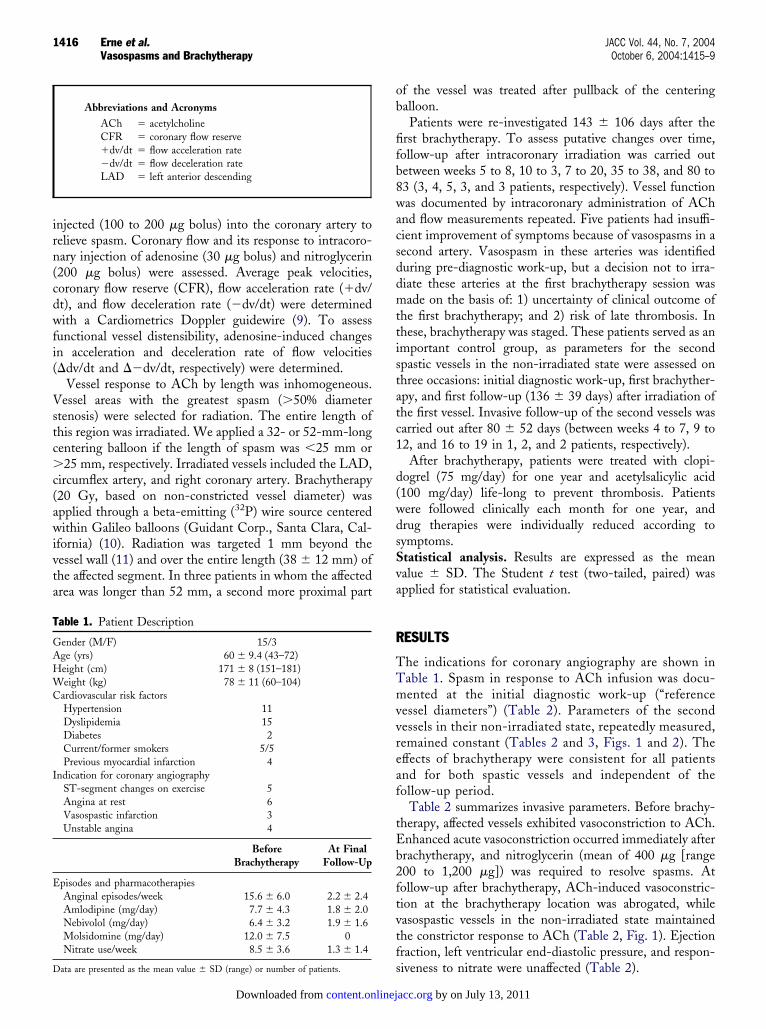
1416 Erne et al. JACC Vol. 44, No. 7, 2004Vasospasms and Brachytherapy October 6, 2004:1415–9
njected (100 to 200 �g bolus) into the coronary artery toelieve spasm. Coronary flow and its response to intracoro-ary injection of adenosine (30 �g bolus) and nitroglycerin200 �g bolus) were assessed. Average peak velocities,oronary flow reserve (CFR), flow acceleration rate (�dv/t), and flow deceleration rate (�dv/dt) were determinedith a Cardiometrics Doppler guidewire (9). To assess
unctional vessel distensibility, adenosine-induced changesn acceleration and deceleration rate of flow velocities�dv/dt and ��dv/dt, respectively) were determined.
Vessel response to ACh by length was inhomogeneous.essel areas with the greatest spasm (�50% diameter
tenosis) were selected for radiation. The entire length ofhis region was irradiated. We applied a 32- or 52-mm-longentering balloon if the length of spasm was �25 mm or25 mm, respectively. Irradiated vessels included the LAD,
ircumflex artery, and right coronary artery. Brachytherapy20 Gy, based on non-constricted vessel diameter) waspplied through a beta-emitting (32P) wire source centeredithin Galileo balloons (Guidant Corp., Santa Clara, Cal-
fornia) (10). Radiation was targeted 1 mm beyond theessel wall (11) and over the entire length (38 � 12 mm) ofhe affected segment. In three patients in whom the affectedrea was longer than 52 mm, a second more proximal part
Abbreviations and AcronymsACh � acetylcholineCFR � coronary flow reserve�dv/dt � flow acceleration rate�dv/dt � flow deceleration rateLAD � left anterior descending
able 1. Patient Description
ender (M/F) 15/3ge (yrs) 60 � 9.4 (43–72)eight (cm) 171 � 8 (151–181)eight (kg) 78 � 11 (60–104)
ardiovascular risk factorsHypertension 11Dyslipidemia 15Diabetes 2Current/former smokers 5/5Previous myocardial infarction 4
ndication for coronary angiographyST-segment changes on exercise 5Angina at rest 6Vasospastic infarction 3Unstable angina 4
BeforeBrachytherapy
At FinalFollow-Up
pisodes and pharmacotherapiesAnginal episodes/week 15.6 � 6.0 2.2 � 2.4Amlodipine (mg/day) 7.7 � 4.3 1.8 � 2.0Nebivolol (mg/day) 6.4 � 3.2 1.9 � 1.6Molsidomine (mg/day) 12.0 � 7.5 0Nitrate use/week 8.5 � 3.6 1.3 � 1.4
sata are presented as the mean value � SD (range) or number of patients.
content.onlinejDownloaded from
f the vessel was treated after pullback of the centeringalloon.Patients were re-investigated 143 � 106 days after the
rst brachytherapy. To assess putative changes over time,ollow-up after intracoronary irradiation was carried outetween weeks 5 to 8, 10 to 3, 7 to 20, 35 to 38, and 80 to3 (3, 4, 5, 3, and 3 patients, respectively). Vessel functionas documented by intracoronary administration of ACh
nd flow measurements repeated. Five patients had insuffi-ient improvement of symptoms because of vasospasms in aecond artery. Vasospasm in these arteries was identifieduring pre-diagnostic work-up, but a decision not to irra-iate these arteries at the first brachytherapy session wasade on the basis of: 1) uncertainty of clinical outcome of
he first brachytherapy; and 2) risk of late thrombosis. Inhese, brachytherapy was staged. These patients served as anmportant control group, as parameters for the secondpastic vessels in the non-irradiated state were assessed onhree occasions: initial diagnostic work-up, first brachyther-py, and first follow-up (136 � 39 days) after irradiation ofhe first vessel. Invasive follow-up of the second vessels wasarried out after 80 � 52 days (between weeks 4 to 7, 9 to2, and 16 to 19 in 1, 2, and 2 patients, respectively).After brachytherapy, patients were treated with clopi-
ogrel (75 mg/day) for one year and acetylsalicylic acid100 mg/day) life-long to prevent thrombosis. Patientsere followed clinically each month for one year, andrug therapies were individually reduced according toymptoms.tatistical analysis. Results are expressed as the meanalue � SD. The Student t test (two-tailed, paired) waspplied for statistical evaluation.
ESULTS
he indications for coronary angiography are shown inable 1. Spasm in response to ACh infusion was docu-ented at the initial diagnostic work-up (“reference
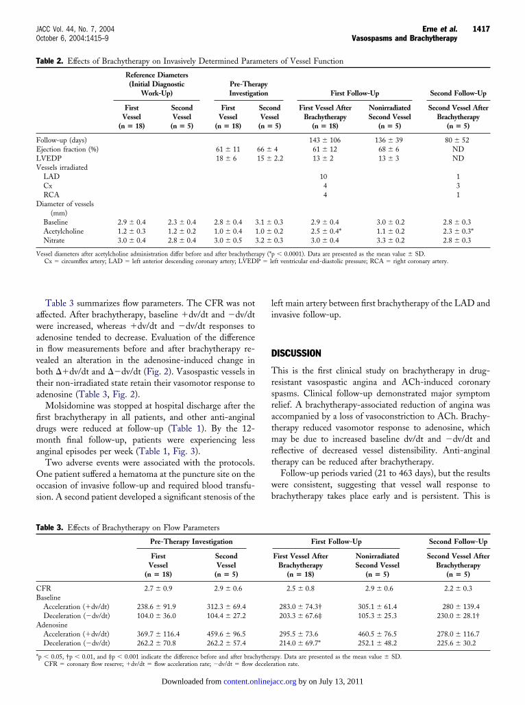
essel diameters”) (Table 2). Parameters of the secondessels in their non-irradiated state, repeatedly measured,emained constant (Tables 2 and 3, Figs. 1 and 2). Theffects of brachytherapy were consistent for all patientsnd for both spastic vessels and independent of theollow-up period.
Table 2 summarizes invasive parameters. Before brachy-herapy, affected vessels exhibited vasoconstriction to ACh.nhanced acute vasoconstriction occurred immediately afterrachytherapy, and nitroglycerin (mean of 400 �g [range00 to 1,200 �g]) was required to resolve spasms. Atollow-up after brachytherapy, ACh-induced vasoconstric-ion at the brachytherapy location was abrogated, whileasospastic vessels in the non-irradiated state maintainedhe constrictor response to ACh (Table 2, Fig. 1). Ejectionraction, left ventricular end-diastolic pressure, and respon-
iveness to nitrate were unaffected (Table 2).by on July 13, 2011 acc.org
awaivbta
fidma
Oos
li
D
Trsratmrt
wb
T
CB
A
*
T
FELV
D
V
1417JACC Vol. 44, No. 7, 2004 Erne et al.October 6, 2004:1415–9 Vasospasms and Brachytherapy
Table 3 summarizes flow parameters. The CFR was notffected. After brachytherapy, baseline �dv/dt and �dv/dtere increased, whereas �dv/dt and �dv/dt responses to
denosine tended to decrease. Evaluation of the differencen flow measurements before and after brachytherapy re-ealed an alteration in the adenosine-induced change inoth ��dv/dt and ��dv/dt (Fig. 2). Vasospastic vessels inheir non-irradiated state retain their vasomotor response todenosine (Table 3, Fig. 2).
Molsidomine was stopped at hospital discharge after therst brachytherapy in all patients, and other anti-anginalrugs were reduced at follow-up (Table 1). By the 12-onth final follow-up, patients were experiencing less
nginal episodes per week (Table 1, Fig. 3).Two adverse events were associated with the protocols.ne patient suffered a hematoma at the puncture site on the
ccasion of invasive follow-up and required blood transfu-ion. A second patient developed a significant stenosis of the
able 3. Effects of Brachytherapy on Flow Parameters
Pre-Therapy Investigation
FirstVessel
(n � 18)
SecondVessel
(n � 5)
FR 2.7 � 0.9 2.9 � 0.6aselineAcceleration (�dv/dt) 238.6 � 91.9 312.3 � 69.4Deceleration (�dv/dt) 104.0 � 36.0 104.4 � 27.2
denosineAcceleration (�dv/dt) 369.7 � 116.4 459.6 � 96.5Deceleration (�dv/dt) 262.2 � 70.8 262.2 � 57.4
p � 0.05, †p � 0.01, and ‡p � 0.001 indicate the difference before and after brach
able 2. Effects of Brachytherapy on Invasively Determined Para
Reference Diameters(Initial Diagnostic
Work-Up)Pre-TherInvestiga
FirstVessel
(n � 18)
SecondVessel
(n � 5)
FirstVessel
(n � 18)
ollow-up (days)jection fraction (%) 61 � 11VEDP 18 � 6essels irradiatedLADCxRCAiameter of vessels
(mm)Baseline 2.9 � 0.4 2.3 � 0.4 2.8 � 0.4 3Acetylcholine 1.2 � 0.3 1.2 � 0.2 1.0 � 0.4 1Nitrate 3.0 � 0.4 2.8 � 0.4 3.0 � 0.5 3
essel diameters after acetylcholine administration differ before and after brachytherCx � circumflex artery; LAD � left anterior descending coronary artery; LVED
CFR � coronary flow reserve; �dv/dt � flow acceleration rate; �dv/dt � flow deceler
content.onlinejDownloaded from
eft main artery between first brachytherapy of the LAD andnvasive follow-up.
ISCUSSION
his is the first clinical study on brachytherapy in drug-esistant vasospastic angina and ACh-induced coronarypasms. Clinical follow-up demonstrated major symptomelief. A brachytherapy-associated reduction of angina wasccompanied by a loss of vasoconstriction to ACh. Brachy-herapy reduced vasomotor response to adenosine, whichay be due to increased baseline dv/dt and �dv/dt and
eflective of decreased vessel distensibility. Anti-anginalherapy can be reduced after brachytherapy.
Follow-up periods varied (21 to 463 days), but the resultsere consistent, suggesting that vessel wall response torachytherapy takes place early and is persistent. This is
First Follow-Up Second Follow-Up
First Vessel AfterBrachytherapy
(n � 18)
NonirradiatedSecond Vessel
(n � 5)
Second Vessel AfterBrachytherapy
(n � 5)
2.5 � 0.8 2.9 � 0.6 2.2 � 0.3
283.0 � 74.3† 305.1 � 61.4 280 � 139.4203.3 � 67.6‡ 105.3 � 25.3 230.0 � 28.1†
295.5 � 73.6 460.5 � 76.5 278.0 � 116.7214.0 � 69.7* 252.1 � 48.2 225.6 � 30.2
py. Data are presented as the mean value � SD.
rs of Vessel Function
First Follow-Up Second Follow-Up
ndel5)
First Vessel AfterBrachytherapy
(n � 18)
NonirradiatedSecond Vessel
(n � 5)
Second Vessel AfterBrachytherapy
(n � 5)
143 � 106 136 � 39 80 � 524 61 � 12 68 � 6 ND2.2 13 � 2 13 � 3 ND
10 14 34 1
0.3 2.9 � 0.4 3.0 � 0.2 2.8 � 0.30.2 2.5 � 0.4* 1.1 � 0.2 2.3 � 0.3*0.3 3.0 � 0.4 3.3 � 0.2 2.8 � 0.3
� 0.0001). Data are presented as the mean value � SD.eft ventricular end-diastolic pressure; RCA � right coronary artery.
ythera
mete
apytion
SecoVess
(n �
66 �15 �
.1 �
.0 �
.2 �
apy (*pP � l
ation rate.
by on July 13, 2011 acc.org

cbc
awtalbs
vbiecrTeliSpinb
Caw
Ftcdb((bt
Ff
Fsiaavfe
1418 Erne et al. JACC Vol. 44, No. 7, 2004Vasospasms and Brachytherapy October 6, 2004:1415–9
onsistent with the reduction in angina within hours ofrachytherapy and the fact that molsidomine administrationould be halted at discharge (one day after the procedure).
We caution that brachytherapy can be associated withdverse events. Brachytherapy is known to be associatedith a risk of late thrombosis. However, our patients were
reated with clopidogrel for 12 months and continuouscetylsalicylic acid. One patient developed progression of aeft main plaque narrowing (75%) within 315 days afterrachytherapy of the LAD. This could result from progres-ion of native disease or stimulatory effects of irradiation.
Effects of brachytherapy on coronary artery tone andasomotion are poorly understood. Immediately afterrachytherapy, extensive vasoconstriction occurs, as shownn this study and others (4,5), and intracoronary nitroglyc-rin is necessary. Endothelium-dependent vasomotion oforonary segments treated with balloon angioplasty waseported to normalize at six months after brachytherapy (8).his study investigated patients with coronary artery dis-
ase, while herein no patient presented with coronaryesions. Our results suggest additional endothelium-ndependent effects of brachytherapy.tudy limitations. A larger study of less symptomaticatients is needed to assess the clinical impact of
ntracoronary irradiation for variant angina. The study didot address the cellular mechanism of action of
igure 1. Brachytherapy reduces acetylcholine (ACh)-induced vasocon-triction. Individual vessel diameter changes (with median values) afternfusion with ACh before and after brachytherapy. Circles � first vesselnd squares � second vessel before (open circles and open squares) andt follow-up after brachytherapy (solid circles and solid squares); secondessel in non-irradiated states at first investigation (open triangles) andollow-up after brachytherapy of first vessel (solid triangles). p indicatesffect of brachytherapy.
rachytherapy. c
content.onlinejDownloaded from
onclusions. Brachytherapy appears to be a potential ther-py in preventing the recurrence of vasospasm in patientsith refractory and highly symptomatic variant angina.
igure 2. Brachytherapy reduces adenosine-induced changes in accelera-ion and deceleration time of flow velocities. Individual adenosine-inducedhanges (with median values) in acceleration (��dv/dt; upper panel) andeceleration (��dv/dt; lower panel) flow velocities before and afterrachytherapy. Circles � first vessel and squares � second vessel beforeopen circles and open squares) and at follow-up after brachytherapysolid circles and solid squares). Second vessel in non-irradiated states atoth the first investigation (open triangles) and follow-up after brachy-herapy of first vessel (solid triangles). p indicates effect of brachytherapy.
igure 3. Brachytherapy reduces the frequency of anginal episodes. Weeklyrequency of anginal episodes for each patient before brachytherapy (open
ircles) and at follow-up (solid circles).by on July 13, 2011 acc.org

RCE
R
1
1
1419JACC Vol. 44, No. 7, 2004 Erne et al.October 6, 2004:1415–9 Vasospasms and Brachytherapy
eprint requests and correspondence: Dr. Paul Erne, Division ofardiology, Kantonsspital Luzern, 6006 Luzern 16, Switzerland.-mail: [email protected].
EFERENCES
1. Scholl JM, Benacerraf A, Ducimetiere P, et al. Comparison of riskfactors in vasospastic angina without significant fixed coronary nar-rowing to significant fixed coronary narrowing and no vasospasticangina. Am J Cardiol 1986;57:199–202.
2. Ludmer PL, Selwyn AP, Shook TL, et al. Paradoxical vasoconstrictioninduced by acetylcholine in atherosclerotic coronary arteries. N EnglJ Med 1986;315:1046–51.
3. Gupta S, Schiele F, Vuillemenot A, Appfel F, Bassand JP. Coronarystent for variant angina: atypical presentation. Cathet CardiovascDiagn 1998;45:439–41.
4. Wiedermann JG, Marboe C, Amols H, Schwartz A, Weinberger J.Intracoronary irradiation markedly reduces restenosis after balloonangioplasty in a porcine model. J Am Coll Cardiol 1994;23:1491–8.
content.onlinejDownloaded from
5. Scheinert D, Strnad V, Muller R, et al. High-dose intravascularbeta-radiation after de novo stent implantation induces coronary arteryspasm. Circulation 2002;105:1420–3.
6. Togni M, Windecker S, Wenawaeser P, et al. Deleterious effect ofbrachytherapy on vasomotor response to exercise. Circulation 2004;110:135–40.
7. Chatterjee T, Juelke PD, Thum P, Erne P. Successful brachytherapyof coronary vasospasm. Heart 2003;89:e25.
8. Sabate M, Kay IP, van Der Giessen WJ, et al. Preserved endothelium-dependent vasodilation in coronary segments previously treated withballoon angioplasty and intracoronary irradiation. Circulation 1999;100:1623–9.
9. Doucette JW, Corl PD, Payne HM, et al. Validation of a Dopplerguide wire for intravascular measurement of coronary artery flowvelocity. Circulation 1992;85:1899–911.
0. Raizner A, Eno R, Caffee R. The Guidant GalileoTM intravascularradiotherapy system. In: Waksman R, Serruys P, editors. Handbook ofVascular Brachytherapy. London: Martin Dunitz Ltd., 2000:83–93.
1. Waksman R, Raizner AE, Yeung AC, Lansky AJ, Vandertie L. Use oflocalised intracoronary beta radiation in treatment of in-stent resteno-sis: the INHIBIT randomised controlled trial. Lancet 2002;359:551–7.
by on July 13, 2011 acc.org

doi:10.1016/j.jacc.2004.06.056 2004;44;1415-1419 J. Am. Coll. Cardiol.
Therese Resink Paul Erne, Peiman Jamshidi, Peter Juelke, Hans-Peter Hafner, Peter Thum, and
Brachytherapy: Potential therapy for refractory coronary spasm
This information is current as of July 13, 2011
& ServicesUpdated Information
http://content.onlinejacc.org/cgi/content/full/44/7/1415including high-resolution figures, can be found at:
References
http://content.onlinejacc.org/cgi/content/full/44/7/1415#BIBLat: This article cites 10 articles, 6 of which you can access for free
Rights & Permissions
http://content.onlinejacc.org/misc/permissions.dtltables) or in its entirety can be found online at: Information about reproducing this article in parts (figures,
Reprints http://content.onlinejacc.org/misc/reprints.dtl
Information about ordering reprints can be found online:
by on July 13, 2011 content.onlinejacc.orgDownloaded from
Copyright © 2022 FDOKUMEN